500 mg/100 g of intraperitoneal L-arginine (Sigma) as a 20 ...
60
J. Physiol. (1992) Vol. 446. University College and R.C.S.I. Dublin 20-21 September 1991 402P PROCEEDINGS OF THE PHYSIOLOGICAL SOCIETY COMMUNICATIONS L-arginine: a role for inducing chronic pancreatitis in the anaesthetized rat C.P. Delaney, K. McGeeney, P.G. Horgan, N.F. Couse, T.F. Gorey and J.M. Fitzpatrick Professorial Surgical Unit, Mater Misericordiae Hospital and University College Dublin, Dublin, Ireland No simple experimental model exists for chronic pancreatitis in the rodent. Inducing a reproducible acute necrotizing pancreatitis in the rat using intra- peritoneal arginine has been only recently described (Tani et al. 1990). This study investigates the role of L-arginine in developing a model of chronic pancreatitis. Fasting male Sprague-Dawley rats (196-379 g) were divided into control (n = 10) and pancreatitis groups (n = 40). Each experimental animal received 500 mg/100 g of intraperitoneal L-arginine (Sigma) as a 20% solution in 0.15 M NaCI on days 1, 4, 7 and 10. Procedures were performed under ether anaesthesia. Each group then underwent serial assessment of pancreatic histology, serum amylase, weight, haemoglobin and white cell count with differential over a period of three months. At three months, plain abdominal X-rays were taken of each group. Although control animals had a 66% increase in weight, experimental animals gained only 25% (P < 0.01, Student's t test). Amylase levels increased from 8840 to 15918 i.u./l (Phadebas) in the acute phase, but were normal at three months. Haemoglobin dropped from 15.2 to 13.6 g/dl (P < 0.01) at one month but then returned to normal. Although the overall white cell count remained normal from day 1 to 14, the neutrophil count increased from 20.8% to 63.6% (P < 0.001). At three months the white count dropped from 18.2 to 12.8 x 10⁹/l (P < 0.05) although the differential returned to normal. Fasting glucose levels remained normal at all times. Light microscopy showed marked acinar degeneration with replacement by adipose tissue. Ductal, vascular and connective tissue structure was well preserved, as were the islets of Langer- hans. X-rays showed no evidence of pancreatic calcification. These data support L-arginine as a simple method of producing severe, non- resolving pancreatic damage. It is proposed as a new model of a chronic pancreatitis which warrants further investigation. REFERENCE Tani, S., Itoh, H., Okabayashi, Y., Nakamura, T., Fujii, M., Fujisawa, T., Koide, M. & Otsuki, M. (1990). Dig. Dis. Sci. 35(3), 367-374.
-
Upload
khangminh22 -
Category
Documents
-
view
0 -
download
0
Transcript of 500 mg/100 g of intraperitoneal L-arginine (Sigma) as a 20 ...

J. Physiol. (1992) Vol. 446. University College and R.C.S.I. Dublin 20-21 September 1991
402P PROCEEDINGS OF THE PHYSIOLOGICAL SOCIETY
COMMUNICATIONS
L-arginine: a role for inducing chronic pancreatitis in the anaesthetized rat
C.P. Delaney, K. McGeeney, P.G. Horgan, N.F. Couse, T.F. Gorey and J.M. FitzpatrickProfessorial Surgical Unit, Mater Misericordiae Hospital and University College Dublin,Dublin, IrelandNo simple experimental model exists for chronic pancreatitis in the rodent.
Inducing a reproducible acute necrotizing pancreatitis in the rat using intra-peritoneal arginine has been only recently described (Tani et al. 1990). Thisstudy investigates the role of L-arginine in developing a model of chronicpancreatitis.Fasting male Sprague-Dawley rats (196-379 g) were divided into control
(n = 10) and pancreatitis groups (n = 40). Each experimental animal received500 mg/100 g of intraperitoneal L-arginine (Sigma) as a 20% solution in 0.15 MNaCI on days 1, 4, 7 and 10. Procedures were performed under ether anaesthesia.Each group then underwent serial assessment of pancreatic histology, serumamylase, weight, haemoglobin and white cell count with differential over aperiod of three months. At three months, plain abdominal X-rays were takenof each group.Although control animals had a 66% increase in weight, experimental
animals gained only 25% (P < 0.01, Student's t test). Amylase levels increasedfrom 8840 to 15918 i.u./l (Phadebas) in the acute phase, but were normal atthree months. Haemoglobin dropped from 15.2 to 13.6 g/dl (P < 0.01) at onemonth but then returned to normal. Although the overall white cell countremained normal from day 1 to 14, the neutrophil count increased from 20.8%to 63.6% (P < 0.001). At three months the white count dropped from 18.2 to12.8 x 109/l (P < 0.05) although the differential returned to normal. Fastingglucose levels remained normal at all times. Light microscopy showed markedacinar degeneration with replacement by adipose tissue. Ductal, vascular andconnective tissue structure was well preserved, as were the islets of Langer-hans. X-rays showed no evidence of pancreatic calcification.These data support L-arginine as a simple method of producing severe, non-
resolving pancreatic damage. It is proposed as a new model of a chronicpancreatitis which warrants further investigation.
REFERENCE
Tani, S., Itoh, H., Okabayashi, Y., Nakamura, T., Fujii, M., Fujisawa, T., Koide, M. & Otsuki,M. (1990). Dig. Dis. Sci. 35(3), 367-374.

J. Physiol. (1992) Vol. 446. Proceedings of The Physiological Society
UNIVERSITY COLLEGE AND R.C.S.I. DUBLIN 20-21 SEPTEMBER 1991 403P
A longitudinal study of human erythrocyte choline transport after renaltransplantation
C.E. Poli de Figueiredo, J.C. Ellory and B.M. Hendry*University Laboratory of Physiology and *Nuffield Department of Clinical Medicine, JohnRadcliffe Hospital, OxfordHuman erythrocyte choline permeability is significantly elevated in patients
with chronic renal failure and this abnormality persists in patients on mainte-nance haemodialysis or continuous ambulatory peritoneal dialysis (Fervenza etal. 1991). It is not clear whether the altered membrane transport is due to acirculating plasma factor or due to abnormal erythrocyte development in thebone marrow. In order to explore this question we report a study of erythro-cyte choline transport in patients undergoing renal transplantation.Ten patients receiving cadaver renal transplants were studied immediately
prior to transplantation and at regular intervals for up to 6 months thereafter.Erythrocyte choline transport was measured with tracer [14C]-labelled cholineusing freshly-washed cells. Choline influx incubations were performed in salineof composition (mmol/1): NaCl, 140; KCI, 5; glucose, 5; MOPS, 10; pH 7.4. TheVmax for choline transport was estimated by using an extracellular cholineconcentration of 250 Mmol/l. The initial rate of choline uptake was measuredwith a 5 minute flux incubation at 370C.The mean plasma creatinine concentration fell from 860 (S.E.M. 74) ,umol/l
before transplantation to 140 (S.E.M. 11) ,umol/l after 6 months. The meanVmax for erythrocyte choline transport fell with a parallel time-course from 72(S.E.M. 6) ,umol I cells-' h-' to 42 (S.E.M. 6) ,umol I cells-' h-'. The time taken foreach of these means to fall halfway to its final value was 3.8 days for creatinineconcentration and 4.0 days for choline transport Vmax. There was a significantcorrelation between mean plasma creatinine and mean choline transport Vmax(Spearman, P < 0.01). Rejection episodes occurred in 2 patients with rapidincreases in plasma creatinine concentrations. In each case the increase increatinine concentration was accompanied by an increase in choline transportVmax. In one of these cases the increase in creatinine concentration wastransient and this was also reflected in a transient rise in choline transportVmax.These results suggest that the abnormal erythrocyte choline transport in
renal failure is due to a circulating plasma factor which is rapidly cleared by afunctional renal transplant and which reaccumulates following acute renaldysfunction.
This work was supported by the National Kidney Research Fund and was approved by theOxford Ethical Committee. C.E.P.F. is supported by CAPES (Brazil).
REFERENCE
Fervenza, F.C., Meredith, D., Ellory, J.C. & Hendry, B.M. (1991). Clin. Sci. 80, 137-141.

J. Physiol. (1992) Vol. 446. University College and R.C.S.I. Dublin 20-21 September 1991
404-P PROCEEDINGS OF THE PHYSIOLOGICAL SOCIETY
Furosemide induces cytoplasmic alkalinization in frog early distal tubules
G. Cooper, V. Stone and M. HunterDepartment ofPhysiology, Worsley Medical & Dental Building, University ofLeeds, LS2 9NQThe amphibian early distal tubule reabsorbs sodium and chloride in excess of
water and thus, like its mammalian counterpart, the thick ascending limb, iscalled the diluting segment (Guggino et al. 1988). We have shown previouslythat the apical K channels of this segment are stimulated by both cytoplasmicalkalinization and apical furosemide (Hurst & Hunter, 1990 a, b). We postulatedthat the furosemide-induced increase in channel activity may be due to anintracellular alkalinization and so have measured intracellular pH duringexposure to furosemide.Potassium-loaded adult frogs (Rana temporaria) were killed by decapitation
and destruction of the spinal cord. Single early distal tubules were dissectedand perfused using standard microperfusion techniques. The luminal and bathsolutions contained (mM): NaCl, 97; KCI, 3; MgCl2, 1; CaC12, 2; HEPES, 10,titrated to pH 7.4 with NaOH. Intracellular pH (pHi) was measured usingBCECF, which was loaded into the cells by incubating the tubules for 10 minin the dark with 5 pM BCECF-AM. The tubules were excited with light from a100W xenon bulb (0.1% transmission) at 440 and 490 nm; the emitted lightwas passed through a 520 nm barrier filter and measured with a photomultiplier.Cell viability was assessed by exposing the tubules to 5 mM ammoniumchloride, and monitoring the active recovery of pHi following the wash-out ofammonium from the bath. The effect of furosemide (5 x 10⁻⁵ M) was determinedin everted tubules, to simulate the conditions used in the previous patch clampstudies. Values are given as mean ± S.E.M. Statistical analysis was carried outusing Student's t tests. Significance was assumed at the 5% level.Following exposure to ammonium chloride the tubule cells rapidly recovered
their pH within about 2 minutes. The initial rate of recovery from the acidload (dpH/dt, 116 x 10-4 ± 21.4.10-4 sQ1, n = 6) was inhibited by the addition ofEIPA (1 x 10⁻⁵ M, an amiloride analogue with an enhanced ability to inhibitNa-H exchange) to the bath (9.27 x 10⁻⁴ + 3.22 x 10⁻⁴, n = 6), but not theluminal solution (90.2 x 10⁻⁴ + 19.1 x 10⁻⁴, n = 6). Furosemide caused a significantincrease in pHi, from 7.61 ± 0.11 to 7.92 ± 0.21 in six tubules.In conclusion, EIPA inhibits pH recovery from an acid load, suggesting that
Na-H exchange is responsible for this pH regulation. The exchangers seem tobe situated predominantly in the basolateral membrane. Furosemide caused acytoplasmic alkalinization. This is consistent with the theory that the previ-ously reported stimulation of apical K channels may be mediated by an intra-cellular alkalinization.
This work was supported by the Wellcome Trust.
REFERENCES
Guggino, W.B., Oberleithner, H. & Giebisch, G. (1988). Am. J. Physiol. 254, F615-F627.Hurst, A.M. & Hunter, M. (1990a). Am. J. Physiol. 259, C1005-C1009.Hurst, A.M. & Hunter, M. (1990b). J. Physiol. 426, 99P.
J. Physiol. (1992) Vol. 446. Proceedings of The Physiological Society
UNIVERSITY COLLEGE AND R.C.S.I. DUBLIV 20-21 SEPTEMBER 1991 405P
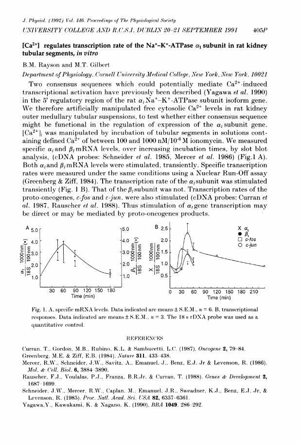
[Ca2+1 regulates transcription rate of the Na+-K+-ATPase a, subunit in rat kidneytubular segments, in vitro
B.M. Rayson and M.T. GilbertD)epartment of Physiology. Cornell University Medical College, New York, NVew York, 10021Two consensus sequences which could potentially mediate Ca2+-induced
transcriptional activation have previously been described (Yagawa et al. 1990)in the 5' regulatory region of the rat a, Na+-K+-ATPase subunit isoform gene.We therefore artificially manipulated free cytosolic Ca2+ levels in rat kidneyouter medullary tubular suspensions, to test whether either consensus sequencemight be functional in the regulation of expression of the a, subunit gene.[Ca2li] was manipulated by incubation of tubular segments in solutions cont-aining defined Ca2+ of between 100 and 1000 nM/106M ionomycin. We measuredspecific a, and /,B mRNA levels, over increasing incubation times, by slot blotanalysis, (cDNA probes: Schneider et al. 1985, Mercer et al. 1986) (Fig.1 A).Both a, and /3, mRNA levels were stimulated, transiently. Specific transcriptionrates were measured under the same conditions using a Nuclear Run-Off assay(Greenberg & Ziff, 1984). The transcription rate of the a,subunit was stimulatedtransiently (Fig. I B). That of the P/ subunit was not. Transcription rates of theproto-oncogenes, c-fos and c-jun, were also stimulated (cDNA probes: Curran etal. 1987, Rauscher et al. 1988). Thus stimulation of a,gene transcription maybe direct or may be mediated by proto-oncogenes products.
A 5.0 5.0 B 2.54 X ai PI
34.0 4.0 20 30 c2f2sEE ~~~~~~~~~EE EEj Oc-jun
3.0 ~~~~~3.0g 8 )g 1.5
2.0 12 .0 1. 0 >1
-CI) jcn~~~~~~~~U IC)
1.0 1.0 0.5
30 60 90 120 150 180 0 30 60 90 120 150 180 210Time (min) Time (min)
Fig. 1. A, specific mRNA levels. Data indicated are means ± SEM., n = 6. B, transcriptionalresponses. Data indicated are means ± S.E.MI., n = 3. The 18 s rDNA probe was used as aquantitative control.
R EFERENCES
Curran, T.. Gordon, M1.B., Rubino, K.L. & Sambucetti, L.C. (1987). Oncogene 2, 79-84.Greenberg, MI.E. & Ziff, E.B. (1984). Nature 311, 433--438.NIercer, R.W., Schneider, J.A., Savitz, A.. Emanuel, J., Benz, E.J. Jr & Levenson, R. (1986).Mol. & Cell. Riol. 6, 3884-3890.
Rauscher, F.J., Voulalas, P.J., Franza, B.R.Jr. & Curran, T. (1988). Genes & Development 2,1687-1699.
Schneider, J.W., MIercer, R.A., Caplan, A., Emanuel, J.R., Sweadner, K.J., Benz, E.J. Jr, &Levenson, R. (1985). Proc. Natl. Acad. Sci. USA 82, 6357-6361.
Yagawa,Y., Kawakami, K. & Nagano. K. (1990). BBA 1049, 286-292.
J. Physiol. (1992) I'ol. 446. (University (olleye and R.C.S.I. D)ublin 20-21 September 1991
406P PROCEEDINGS OF THE PHYSIOLOGICAL SOCIETY
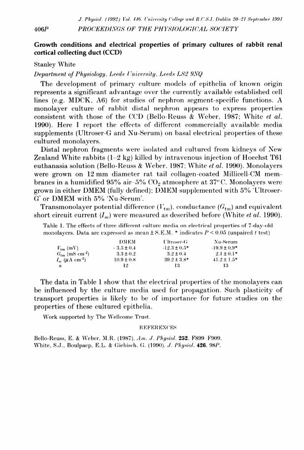
Growth conditions and electrical properties of primary cultures of rabbit renalcortical collecting duct (CCD)
Stanley WhiteDepartment of Physiology, Leeds University, Leeds L82 9NVQThe development of primary culture models of epithelia of known origin
represents a significant advantage over the currently available established celllines (e.g. MDCK, A6) for studies of nephron segment-specific functions. Amonolayer culture of rabbit distal nephron appears to express propertiesconsistent with those of the CCD (Bello-Reuss & Weber, 1987; White et al.1990). Here I report the effects of different commercially available mediasupplements (Ultroser-G and Nu-Serum) on basal electrical properties of thesecultured monolayers.
Distal nephron fragments were isolated and cultured from kidneys of NewZealand White rabbits (1-2 kg) killed by intravenous injection of Hoechst T61euthanasia solution (Bello-Reuss & Weber., 1987; White et al. 1990). Monolayerswere grown on 12 mm diameter rat tail collagen-coated Millicell-CM mem-branes in a humidified 95% air-5% CO2 atmosphere at 37") C. Monolayers weregrown in either DMEM (fully defined); DMEAI supplemented with 5% 'Ultroser-G or DMEM with 5% 'Nu-Serum'.Transmonolayer potential difference (1'tX0), conductance (Gt,,) and equivalent
short circuit current (i,J were measured as described before (White et al. 1990).Table 1. The effects of three diffetent culture media on electrical properties of 7-day-oldmonolayers. Data are expressed as mnean ± S. E.M. * indicates IP < 0.05 (unpaired t test)
DAI EM Ultrosr-(C Nu-SerumV' m (mV ) - 3.5 ± 0.4 -12:3 ± 0.5* -19.9 ± (09*Gtni (MS cm-2) 3.3 ± 0.2 3.2 ± 0.4 2.1 ± 0.1*ISe (,uA cm-2) 10.9 ± 0.8 39.2± 3.8* 41.2± 1.5*n 12 1'3 13
The data in Table 1 show that the electrical properties of the monolayers canbe influenced by the culture media used for propagation. Such plasticity oftransport properties is likely to be of importance for future studies on theproperties of these cultured epithelia.
XVork supported by The WNellcome Trust.
REFERENCES
Bello-Reuss, E. & Weber, M.R. (1987). Ant. J. Physiol. 252, F899 F909.White, S.J., Boulpaep, E.L. & Giebisch. G. (1990). J. I'hysiol. 426, 981'.

J. Physiol. (1992) V'ol. 446. Proceedinq.s of The I'hy.sioloyicol1 Society
ZN'(WIlERfSITY C(OLLEGE AND R.C.S.I. I)UBLIN 20-21 ASEPTEMBER 1991 407P
Effects of lithium on accumulation of inositol phosphates by rabbit blastocysts
MM.1I. Fahy and M.T. KaneDepartment of Physiology. University (Colleqe, Galivay, IrelandThe phosphatidylinositol (Ptdlns) cycle is involved in the control of cell
proliferation (Berridge, 1987). Lithium disrupts the Ptdlns cycle by inhibitingthe breakdown of inositol monophosphates (Sherman, 1989) thus preventinginositol recycling and causing an accumulation of inositol phosphates in cells.Treatment of Xenopus embryos with lithium disrupts differentiation by inter-fering with the Ptdlns cycle and starving the cells of recycled inositol (Busa &Gimlich, 1989). There is little published information on the role of the Ptdlnscycle or of lithium in preimplantation mammalian embryos although we haveshown that inositol is necessary for rabbit blastocyst growth (Kane, 1989) andthat the inositol phosphates and phosphoinositides of the Ptdlns cycle arepresent in rabbit blastoeysts (Fahy, 1991). We investigated the effects oflithium on the accumulation of inositol phosphates by rabbit blastocysts.New Zealand White does were anaesthetized with a combination of 0.3 ml/kg
Hypnorm I.M. (0.2 mg fentanyl base and 10 mg fluanisone/ml) and diazepamI.V. (5 mg/kg). Morulae were collected from the oviducts 40-44 h post hCGinjection and insemination. They were cultured for 2 days in basic culturemedium (Kane, 1989) containing 15 inositol; nyo-[2-3H]-inositol was addedfor a further 3 days culture. At the end of culture, the embryos were washedwith unlabelled medium and incubated with LiCl (0, 1, 5 and 20 mM, 90-92embryos per treatment in groups of 21-25 embryos) for 1 h. The embryos werethen lysed with perchloric acid and the inositol phosphates separated on aHPLC Alono Q anion exchange column and 1 ml fractions collected andcounted in a scintillation counter. Individual inositol phosphates were identi-fied based on the elution pattern of labelled standards. Lithium significantlystimulated inositol monophosphate accumulation (P < 0.01, analysis of vari-ance) with 20 mMA LiCl causing a 3-fold increase as compared with the 0control. Lithium also caused a significant (P < 0.05) increase in inositol (1, 4, 5)trisphosphate accumulation. These results indicate that the Ptdlns cycle isactive in rabbit blastocysts.Continuous culture of embryos for 5 days in 10 mM LiCl decreased blasto-
cyst growth as evidenced by decreased blastocyst diameter. However, it wasnot possible to counteract this effect by culturing the embryos in increasedconcentrations of inositol (75, 375, 1875 and 9375MM; 15 embryos per treat-ment in 2 replicates of 7-8 embryos) in an attempt to compensate for theinhibitory effect of lithium on inositol recycling. This suggests that lithiummay interfere with blastocyst function at sites additional to the Ptdlns cycle.
REFERENCES
Berridge, MI.J. (1987). Biochiml. Biophys. Acta 907, 33 45.Busa. W.B. & Gimlich, R.L. (1989). l)evel. Biol. 132. 315 324.Fahv, AMI.M. (1991). PhD. Thesis, National University of Ireland.Kane, M.T. (1989). J. Reprod. Fert. 87, 275-279.Sherman, W.R. (1989). In Inositol Lipids in (ell Signalling, pp. 39-79. Academic Press, London.

J. Physiol. (1992) Vol. 446. tUniversity College and 1.C!.S.1. Dublin 20-21 Septenmber 1991
408P PROCEEDINGS OF THE PHYSIOLOGICAL SOCIETY
Phagocytic properties can be endowed on a non-phagocytic cell line by expressionof a single receptor for immunoglobulin G (IgG)
Merav Socolovsky, A.R. Hockaday and Janet AllenPhysiological Laboratory, I)owning Street, Cambridge 1B2 3EGThe human high-affinity receptor for the constant (Fc) portion of IgG,
FcyRI, is found exclusively on cells of the macrophage-monocyte lineage. Itmediates internalization of IgG-coated particles (Silverstein et al. 1989). Macro-phages are 'professional phagocytes', cells uniquely able to ingest foreignparticles. The specific role of FcyRI in phagocytosis is unclear, as macrophagespossess a number of phagocytic receptors. Recently 3 cDNA clones for FcyRIhave been isolated. Two of these, p135 and p90, are almost identical; the third,p98/X2, has a different intracytoplasmic part. Transfection of cDNAs intoCOS cells (a simian fibroblast line) results in expression of receptors withsimilar ligand affinity and specificity to that of macrophages (Allen & Seed,1989). We have investigated whether transfection of FcyRI would convert thenon-phagocytic COS cells into phagocytes.Monomeric human IgGI was not endocytosed by transfected COS cells after
incubation at 37°C. However, when monomeric ligand was cross-linked with alabelled second antibody, a particulate labelling pattern was observed whichwas resistant to acid washes. The intracellular localization of this particulatelabel was shown by differential fluorescence labelling of intra- and extracellu-lar antigens. Endocytosis of cross-linked ligand was inhibited at 40C whereligand remained acid labile even after a 30 h incubation. Identical results wereobtained in COS cells transfected with either of the FcyRI clones, p135 andp98/X2. COS cells expressing FcyRI phagocytose IgG-opsonized yeast particlesat 370 C, as shown by an electron-microscopic study and by differentialfluorescence labelling. Only low levels of apparent internalization were seen at40C, when non-opsonized yeast was used, or when COS cells were transfectedwith a cDNA for an unrelated adhesion molecule, CD4.
Since both p135 and p98/X2 internalized ligand in spite of differing intracy-toplasmic tails, the role of this part of FcyRI was investigated by constructingtwo chimeric receptors. The FcyRI extracellular portion was transplanted ontothe intracellular parts of two cell adhesion molecules: CD7 and LFA3 (thelatter uses a glycosyl-phosphatidylinositol membrane anchor). Both chimericreceptors endowed COS cells with the ability to internalize ligand. We con-clude that expression of surface FcyRI is sufficient to confer phagocyticproperties on COS cells. Cross-linking of receptors is required for internaliza-tion. Endocytosis and phagocytosis are independent of the intracellular part ofthe receptor. The ability to study FcyRI phagocytosis in COS cells away fromthe complex environment of the macrophage should enable further elucidationof its signal transduction properties.
REFERENCES
Allen, J.M. & Seed, B. (1989). Science 243, 378-380.Silverstein, S.C., Greenberg, S., Di Virgilio F. & Steinberg. T.H. (1989). In FundamentalImmunology, ed. Paul, W. E., pp. 703-720. Raven Press.

J. 'hysiol. (1992) Vol. 446. P'roceedlins of 7'he Physiological Society
NUNIVERSITY COLLEGE AMD R.(.S.I. 1)UBLIN 20-21 SEPTEMBER 1991 409P
The effect of neutral amino acids on lysine transport in human erythrocytesdiscriminates between two high affinity transporters for cationic amino acids
R. Deves, P. Chivez and C.A.R. Boyd*Department of Physiology and Biophysics. University of Chile, Santiago 7. *Department ofHuman Anatomy. University of Oxford, Oxford OXi 3QXThe mediated transport of lysine across the red cell membrane has been
reported to occur through a single pathway specific for cationic amino acids(y+) that follows simple Michaelis-Alenten kinetics (Vmiax= 0.488 mmol h'Iand Km= 67.9,M). However, leucine has been shown to produce a smallinhibition of about 25% of lysine influx (0.2 mAI) when present in the externalmedium at very high concentrations (50 mM) (Young et al. 1980).We have studied the interaction of neutral and cationic amino acids in these
cells. All neutral amino acids tested (range 0.3-5 mM) inhibit the influx of L-lysine (1 MM). The inhibition pattern is biphasic, and tends to reach a maxi-mum at approximately 50% of the original flux. The concentrations that give25% inhibition are (mM) L-cysteine (2.7), L-alanine (1.3), L-serine (0.9), L-isoleucine (0.6), L-phenylalanine (0.35), L-methionine (< 0.3) and L-leucine(< 0.3); L-lysine and L-arginine completely abolish the rate at the highestconcentration.This observation can be quantitatively accounted for by assuming that the
total flux results from the contributions of two separate transporters (a and /)that differ in their affinity for neutral amino acids. A non-linear regressionanalysis of the effect of varying conicentrations of L-leucine (range 0.010-10mM) on the rate of entry of L-lysine (1 MM), yields two inhibition constants(± S.E.I.) for L-leucine: KiX0 0.022 ± 0.003 mM and Kip, 30.36 ± 7.9 mM. The( Irmaxa/Kma)/(Vmaxp/Kmp) ratio for the substrate (lysine) obtained from thisanalysis is 0.97 ± 0.066. If the sodium in the incubation medium is replaced bypotassium, the affinity for leucine (1/Kia) is reduced approximately 30-fold.Applying the same kind of analysis to the inhibitory effect of varying
unlabelled L-lysine (range 0.005-10 mM) on the rate of entry of 14C L-lysine(luM), showed that again two inhibition constants for L-lysine are obtained(Kina 0.014 ± 0.002 mAM and Ktnp, 0.112 ± 0.017 mMl). By inserting these valuesin the Vmnax/Kmn ratio, the maximum velocities of the two systems are found todiffer by a factor of 8.2 (Imaxf/lTmaxa). If one assumes that leucine interactswith system a, it can be predicted that the maximum inhibition caused byleucine should diminish as the lysine concentration is increased. This wasfound to be the case. Thus two systems appear to be responsible for the entryof lysine into human red blood cells, one of which (a) is a high-affinity, low-capacity system that recognizes neutral amino acids in the presence of sodiumand has not been previously described; we suggest it be called system y+L. Theother system (X3) which only interacts with cationic amino acids remains y+.
This research was supported by FONDECYT 1282/91. DT1 B2674 (Chile) and Dale Fund UK.
REFERENCE
Young, J.D., Jones, B.E.M1. & Ellory .J.C. (1980). 1'roc. R?. Soc. Lond. B 209. 355-375.

J. Physiol. (1992) Vol. 446. University College and RBC SI. Dublin 20-21 September 1991
410P PROCEEDINGS OF THE PHYSIOLOGICAL SOCIETY
Intestinal absorption of thyrotrophin-releasing hormone is not mediated by a Na+-or H-dependent carrier: in vitro studies with rat and rabbit brush-bordermembrane vesicles
David T. Thwaites, Nicholas L. Simmons and Barry H. HirstGastrointestinal Drug Delivery Research Centre and Department of Physiological Sciences,University of Newcastle upon Tyne, Medical School, Newcastle upon Tyne NE2 4HHOral administration of thyrotrophin-releasing hormone (TRH; pGlu-His-
Pro-NH2) is followed by an increase in plasma thyroid-stimulating hormonelevels (Haigler et al. 1972). It has been suggested that this oral bioavailabilityof TRH is mediated via a sodium-dependent carrier-mediated transport sys-tem, limited to the proximal small intestine (Yokohama et al. 1984). Thishypothesis for TRH absorption has been investigated using brush-bordermembrane vesicles (BBMV) prepared from rabbit and rat proximal intestine.Uptake into the vesicles was measured under proton (pH0 5.5 < pHi 7.4) orsodium ([Na]o 100 mM > [Na]i 0 mM) gradient conditions using a rapid filtrationtechnique. The uptake of [3H]TRH for each preparation was compared with['4C]glucose accumulation. All experiments were performed at room temperature(250C).In the presence of an inwardly-directed sodium gradient, [14C]glucose
accumulated within rabbit duodenal, rabbit jejunal and rat proximal intestinalBBMV (at 10 s) 7-14 fold above equilibrium values (65 min). No uptake aboveequilibrium was observed under control ([K]o = [K]i = 100 mM; pHo = pHi =7.4; [Na]o = [Na]i = 0 mM) or proton-gradient conditions. In contrast, in eachof the vesicle preparations no detectable [3H]TRH uptake could be measuredunder any of the three conditions after 10 or 60s. By 65 min, [3H]TRHappeared to fill an intravesicular space which approached that for ['4C]glucose,consistent with diffusive uptake. Stability of [3H]TRH when incubated withrabbit jejunal BBMV was investigated by high performance liquidchromatography (HPLC) analysis. Fractionation of the incubation mixture byHPLC indicated no [3H]TRH degradation after 10 or 60 s incubation. How-ever, after 65 min it appeared that approximately 6% of [3H]TRH had beendeamidated to form pGlu-His-Pro.These results provide no evidence for active absorption of TRH, by either
sodium- or proton-dependent mechanisms, from the intestine. Since oraladministration of TRH is followed by a biological response it may be con-cluded that sufficient TRH is absorbed by passive mechanisms (via theparacellular pathway) to attain a biological response.
Supported under the LINK Programme in Selective Drug Delivery & Targeting, funded bySERC/MRC/DTI and industry (SERC Grant GR/F 09747).
REFERENCES
Haigler, E.D. Jr, Hershman, J.M. & Pittman, J.A. Jr (1972). J. Clin. Endocrinol. Metab. 35, 631-635.Yokohama, S., Yoshioka, T., Yamashita, K. & Kitamori, N. (1984). J. Pharm. Dyn. 7, 445-451.
J. Physiol. (1992) Vol. 446. Proceedings of The Physiological Society
UNIVERSITY COLLEGE AND R.C.S.I. DUBLIN 20-21 SEPTEMBER 1991 411P
In vivo exposure of rat colonic mucosa to human semen induces mucosal cytolysis,abolishes fluid absorption and raises paracellular permeability
M.V. Mendizabal and R.J. NaftalinDivision of Biomedical Sciences, Physiology Group, King's College London, Strand,London WC2 2LS
It is uncertain whether semen facilitates the transmission of viral particlesfrom the colorectal lumen into the systemic circulation. To investigate thisquestion, the effects of freshly obtained human semen (HIV-negative) on therate of fluid absorption, the permeability to paracellular probe, 3H-labelledpolyethylene glycol (MW 4000) and on the histological appearance of ratcolonic mucosa were examined. Contact between human semen and ratdescending colonic mucosa for 3 hours in vivo, inhibits fluid absorption from52.0 ± 2.9 ul cm-2 h-' (n = 8; control) to 10.7 ± 3.4 ,l cm-2 h-' (n = 7; P < 0.001),increases the permeability to 3H-labelled polyethylene glycol 4000 from0.09 ± 0.006 cm h-' (control n = 8) to 0.31 ± 0.04 cm h-' (n = 7; P < 0.001) andcauses cytolysis of the surface mucosa of the rat colon. The rats weremaintained in anaesthesia with sodium pentobarbitone.Spermatazoa within the colonic lumen are destroyed within 1 hour and
release their acrosomal contents. The activity of the acrosomal proteolyticenzyme acrosin, which has trypsin-like specificity, is raised within the luminalcontents by 40-fold during the first hour (P < 0.005). Acrosin may enhanceseminal plasma metalloprotease (collagenase) within the lumen by activatingprocollagenase (mean luminal collagenase activity during 3 hours exposure1250 units/ml luminal fluid).The changes in colonic fluid absorption and permeability induced by seminal
plasma, which initially contains only collagenase activity (ca. 1000 units/ml),are similar to those induced by similar activities of the collagenase fromClostridium hystolyticum (Type II). Trypsin alone has no effect on colonicpermeability. These findings indicate that seminal collagenase causes localdamage to the colonic wall by destruction of the interstitial matrix.
M.V.M. is supported by the Wellcome Trust.

J. Physiol. (1992) Vol. 446. University College and R.C.S.I. Dublin 20-21 September 1991
412P PROCEEDINGS OF THE PHYSIOLOGICAL SOCIETY
Na-H exchange in isolated epithelial cells from sheep trachea
M. Acevedo and L.W. SteeleDepartment of Anatomy & Physiology, Dundee University, Dundee DD1 4HNThe presence of an Na-H antiport in isolated epithelial cells from sheep trachea
has been tested by measuring Na uptake induced by cell acidification.Tracheae were obtained from the local abattoir. The epithelium was me-
chanically stripped from the underlying tissue. The epithelial strips werewashed in Ca-free solution (2 mM EGTA) and then treated with collagenase(100 units/ml) for 1 h. The enzyme-containing solution was discarded and thetissue was gently vortexed in fresh, collagenase-free solution. The cell suspen-sion obtained was filtered (100 -pm mesh) and centrifuged (60 g, 5 min). Thisprocedure yielded around 5 million cells per trachea. Cell viability, assessed byTrypan Blue exclusion, ranged between 75 and 95%. Secretory cells repre-sented less than 1%.The cells were resuspended in a K-rich, Na- and Cl-free solution containing
10,M valinomycin, 1 mM ouabain and 10 mM ammonium sulphate. Na uptakewas started by adding 1 volume of the cell suspension to 3 volumes ofincubating solution, containing 5 mM Na and 0.2 MBq/ml 22Na. Incubationswere carried out at room termperature. Na uptake was stopped by passing thismixture through Dowex-50W (50-100 mesh, Tris form) columns. The eluate,containing 90% of the applied cells, was assayed for 22Na.In acidified cells (when the incubating solution was ammonium-free) the Na
uptake was significantly higher than in non-acidified cells (ammonium-con-taining incubating solution). These findings are consistent with the existenceof a Na-H antiport in these cells.EIPA (5-(N-ethyl-N-isopropyl) amiloride), an amiloride analogue of high
specificity for the Na-H exchanger, inhibited the Na uptake in acidified cellsin a dose-dependent manner. The dose-response curves showed more than onecomponent; the first component, maximally inhibited at micromolar EIPAconcentration, had a Ki of 10-8 M, adding support to the hypothesis of a Na-Hexchanger in sheep tracheal epithelial cells.Apart from the role of this mechanism in the maintenance of the intracellu-
lar pH, the Na-H exchange in the tracheal epithelial cells might be involved inthe regulation of the pH in the periciliary fluid.
M.A. is a Wellcome Research Fellow.

J. Physiol. (1992) Vol. 446. Proceedings of The Physiological Society
UNIVERSITY COLLEGE ANYD R.C.S.I. DUBLIN 20-21 SEPTEMBER 1991 413P
Inhibition of arachidonic acid metabolism by aspirin can promote thrombusformation in a stenosed coronary artery of the anaesthetized dog
S.J.G. McAuliffe, H.M. Snow, J. Moors, R. Jessup, M. Wayne and *M.I.M. NobleBioscience II, ICI Pharmaceuticals, Macclesfield, Cheshire. *The Academic Unit ofCardiovascular Medicine, Charing Cross & Westminster Medical School, LondonIn activated platelets arachidonic acid is converted sequentially by the
enzymes cyclo-oxygenase and thromboxane A2 (TXA2) synthase to form theeicosanoids PGH2 and TXA2; both metabolites stimulate the TXA2 receptorand induce platelet aggregation. However, in the presence of a TXA2 synthaseinhibitor, the concentration of PGH2 increases and degrades to form increasedamounts of the eicosanoids PGD2, PGE2 and PGF2a, of which PGD2 is anti-aggregatory. In these conditions inhibition of platelet cyclo-oxygenase byaspirin reduces PGD2, an effect which may be pro-aggregatory, particularly ifthe TXA2 receptor is simultaneously blocked, as demonstrated in vitro byWatts et al. (1991).We tested this hypothesis in vivo, using an anaesthetized dog preparation
(induction pentobarbitone 30 mg kg-' I.V.; maintenance 3 mg kg-' I.V. everyhalf hour) in which the left circumflex coronary artery was stenosed and theendothelium damaged to cause a site for platelet thrombus formation (Folts etal. 1982). The rate of decline of blood flow in the artery was used as an index ofthrombus formation (TF, ml min-2) and hence platelet aggregation (Cox et al.1991). Measurements of the metabolites thromboxane B2 and PGD2, in plate-lets activated ex vivo with collagen, were used as indices of the inhibition ofcyclo-oxygenase and TXA2 synthase.In 6 dogs dazoxiben (1 mg kg-' I.V.) inhibited TXA2 synthase by 89.6 ± 0.6%
(mean ± S.E.M.) and caused a consequent increase in PGD2 (14.9 ± 2.9 fold).The index of thrombus formation (TF) was reduced from 11.9 ± 1.75 ml min-2to zero but was restored by an infusion of adrenaline (6.7 ± 1.0,g kg-'). Despitethe inhibition of TXA2 formation TF was further reduced (7.3 ± 0.9 to 4.1 ± 0.9ml min-2, P < 0.05) by blockade of the TXA2 receptor with ICI 192605 (1 mgkg-' I.V.), a selective TXA2 receptor antagonist (Jessup et al. 1988), confirmingthe ability of PGH2 to activate the platelet TXA2 receptor and cause aggrega-tion.In these dogs, in the presence of simultaneous blockade of the TXA2 receptor
and inhibition of the enzyme TXA2 synthase, aspirin (5 mg kg-' I.V.) abolishedthe increase in PGD2 and allowed thrombus formation to increase (TF from4.1 ± 0.9 to 9.6 ± 2.1 ml min-2, P < 0.05); i.e. aspirin promoted thrombus forma-tion.
REFERENCES
Cox, B., McAuliffe, S.J.G., Moors, J.A., Noble, MI.M.N. & Snow, H.M. (1991). J. Physiol. 438,169P.
Folts, J.D., Gallagher, F.P. & Rowe, G.G. (1982). Circulation, 65, 248-255.Jessup, C.L., Jessup, R. & XVayne, M. (1988). Br. J. Pharmacol. 95, 676P.Watts, I.S., Wharton, K.A., WVhite, B.P. & Lumley, P. (1991). Br. J. Pharmacol. 102, 497-505.

J. Physiol. (19.92) Vol. 446. University (aollege and R.(C.S.I. l)ublin 20 21 September 1991
414P PROCEEDINGS OF THE PHYSIOLOGICAL SOCIETY
The relationship between portal pressure and portasystemic shunting in portalhypertension in the anaesthetized rat
R. Flynn, J.G. Geraghtv, W.J. Angerson, \V.A. Tanner. F.B.V. Keane and D.C.Carter*Meath and Adelaide Hospitals, Dlublin and *Royal Infirmnary. EdinburghThe relationship between portal pressure and degree of portasystemic shunt-
ing in portal hypertension is poorly understood. This study examines thisrelationship during the development of collateral vessels in a portal veinligated rat model. (Geraghty et al. 1989). The rats were anaesthetized withhalothane in a nitrous oxide and oxygen mixture (4% halothane in 2:1 mixturefor induction and maintained on 0.5% halothane in 2:1 mixture thereafter).Portal pressure was measured by cannulation of the portal vein and degree ofshunting quantified by intraportal injection of gamma-labelled microspheres.A total of 47 ligated and 27 control animals were studied over 28 days aftersurgery.Mean portal pressure in ligated animals fell from 23.4 ± 2.9 mmHg (S.D.) one
day after surgery to 11.6 ± 0.6 mmHg at 14 days and remained unchanged at28 days (11.3 ± 0.6 mmHg). Mean pressure did not exceed 8.3 mmHg in shamoperated controls.There was a positive correlation between pressure and shunting one day
after portal vein ligation (r = 0.08, P < 0.05, Student's t test). As the degree ofshunting increased at days 3, 5 and 7 after ligation there was no significantcorrelation between pressure and shunting. However, at 14 and 28 days therewas a significant linear correlation between pressure and shunting in portalhypertensive rats (r = 0.61, P < 0.01) This was best defined as a quadraticcurve showing that portal pressure increased with shunting up to a level of70% shunting. This study supports the traditional concept that portal pres-sure is the main factor controlling the opening of shunts in portal hyperten-sion.
REFERENCE
Geraghty, J.G., Angerson, W.J. & Carter, D.C. (1989). Gastroenterology 97, 1108-1114.

J. Physiol. (1992) V'ol. 446. Proceedings of The Physiological Society
UNIVERSITY COLLEGE AND R.C.S.I. DUBLIN 20-21 SEPTEMBER 1991 415P
Investigations of responses to 5-hydroxytryptamine in isolated human arteries andveins
E. Breslin, *T. Teoh, *M. Darling and J.R. DochertyDepartment of Clinical Pharmacology, Royal College of Surgeons in Ireland, Dublin 2 and*Rotunda Hospital, Dublin 15-Hydroxytryptamine (5-HT) produces contractions of human blood vessels
by action at both 5-HT, and 5-HT2 receptors (see Docherty & Hyland, 1986),but the distribution of these subtypes has not been well established. Theobject of this study was to investigate 5-HT receptor-mediated contractions inhuman umbilical artery and vein and human saphenous vein.Human umbilical vessels were obtained from normal pregnancies and human
saphenous veins were obtained from varicose vein surgery. In all cases, ringpreparations were employed in investigations of isometric contractions inducedby 5-HT, and the interaction with selective antagonists.The 5-HT2 receptor antagonist ketanserin (1 giM) produced approximately
parallel shifts in the concentration-response curve to 5-HT in human umbilicalarteries and veins, so that the predominant receptor present in these tissueswas clearly 5-HT2. However, evidence obtained with the selective 5-HT1receptor agonist sumatriptan suggested that a 5-HT1 receptor was also presentin these tissues.
In human saphenous vein. ketanserin (1,M) produced approximately paral-lel shifts in the potency of 5-HT in sections of main vein, but produced non-parallel shifts in the potency of 5-HT in side branches. In side branches,ketanserin shifted mainly the upper part of the concentration-response curveto 5-HT, whereas the 5-HT, receptor antagonists metitepine (0.1 M) andmetergoline (0.1 uM) also shifted the lower part of the concentration-responsecurve to 5-HT.
It is concluded that 5-HT2 receptors are the predominant receptors mediat-ing contractions in human umbilical arteries and veins and in the mainsaphenous vein. However, 5-HT1 receptors have an important role in contrac-tions of side branches of the human saphenous vein.
Supported by the Irish Heart Foundation and RCSI.
REFERENCE
Docherty, J.R. & Hyland, L. (1986). Br. J. Pharmacol. 89, 77-81.

J. Physiol. (1992) Vol. 446. University College and R(C.S.I. Dublin 20-21 September 1991
416P PROCEEDINGIS OF THE PHYSIOLOGICAL SOCIETY
The effect of loop diuretics on contraction in isolated rat arterial smooth muscle innormotension and hypertension
A.R. Chipperfield, J.P.L. Davis and A.A. HarperDepartment of Anatomy & Physiology, University of Dundee, Dundee DD1 4HNThe concept of co-transport of Na, K and Cl ions across cell membranes is
now well established (Haas, 1989) and in excitable cells it seems likely that the(Na-K-Cl) transporter system will contribute to the regulation of intracellularion concentrations and thus membrane potential. For example, we havedemonstrated that the co-transporter acts as an inward chloride pump inarterial smooth muscle thus causing depolarization and that its activity isincreased in rats with deoxycorticosterone acetate-salt (DOCA)-inducedhypertension (Davis et al. 1991).Here we report the action of a specific inhibitor of the co-transporter, the
loop diuretic bumetanide (Chipperfield, 1986), on the contractile state ofarterial smooth muscle. Rats were killed by concussion and cervical dislocationand the saphenous branch of the femoral artery removed. Isolated segments(approximately 12 mm in length) of the artery were perfused at 1.5 ml/minwith a nominally bicarbonate-free Ringer solution, and the pressure wasmeasured at the proximal end of the preparation to provide an index ofcontraction.Bumetanide (10,M) evoked a vasodilator response in arteries contracted
with the a-adrenergic agonist, phenylephrine (1-10,M), reducing the perfusionpressure to 64 ± 5% (S.D., n = 4) of control values in normotensive animalsand 65 ± 24% (n = 4) for arteries from rats with DOCA-induced hypertension(mean arterial systolic blood pressures 125 and 175 mmHg respectively).Whereas a reduced sensitivity of the contractile response to noradrenaline
by diuretics, and the presence of a diuretic-sensitive active chloride transport,in vascular smooth muscle has previously been reported (Kreye et al. 1981), themechanisms were unclear. The present results, taken with our earlierelectrophysiological evidence that (Na-K-Cl) co-transport exerts a depolarizinginfluence in this tissue (Davis et al. 1991), suggest that the contractile state ofarterial smooth muscle may be altered by cotransport through its effect onmembrane potential.
WN'e thank the British Heart Foundation for support.
REFERENCES
Chipperfield, A.R. (1986). Clin. Sci. 71, 465-476.Davis, J.P.L., Chipperfield, A.R. & Harper, A.A. (1991). Clin. Sci. 81, 73 78.Haas, AI. (1989). Ann. Rev. Physiol. 51, 443-457.Kreye, V.A.W., Bauer, P.K. & Vill Hauer, I. (1981). Eur. J. Pharinacol. 73, 91-95.

J. Physiol. (1992) IVol. 446. 1'roceeding.s of 7'he I'hysiological Society
UNIVERSITY COLLEGE AND R.C.S.I. DtBLIN 20-21 SEPTEM1BER 1991 417P
A factory study of vasospasm in riveters
Kathleen McKenna, S. McGrann, A.D. Blann* and Judith A. AllenSchool of Biomedical Science (Physiology), The Queen's U'niversity of Belfast andDepartment of Surgery, * University Hospital of South Mlanchester, Didsbury, MianchesterProlonged use of hand-held vibrating tools may result in vasospastic disease
(vibration white finger or VWF). Provocative testing which is helpful inobjective diagnosis (Allen et al. 1991) has been used on men in the aircraftindustry to investigate the acute vascular effects of riveting. Inclusion in thestudy depended upon absence of any history of trauma to the neck or upperlimbs. The project was approved by the Local Ethical Committee.Measurements were made on the fingers of 46 riveters and 46 controls before
work in the morning, and repeated 3 hours later within 5 minutes of stoppingwork. The controls had no history of vibration exposure, worked in the sameenvironment as the riveters, and were matched for age and smoking habit.Resting systolic pressure in fingertip skin (FSP) was measured by laserDoppler flowmetry (Perimed PF2B) before and after rapid cooling to 10°C for5 minutes. Absence of flux following cooling indicated complete vasospasm.Only 1 control subject exhibited cold-induced vasospasm before and after
work. Fifteen riveters exhibited cold-induced vasospasm before work and Iman had zero skin flux before cooling. After work 16 riveters had vasospasmafter finger cooling. So riveters have a higher incidence of vasospasm thancontrols, but the presence of vasospasm was not always related to a longhistory of vibration exposure (correlation coefficient 0.4). Before work, meanFSP in the riveters was 112 mmHg (S.E.M. ± 3.3) before cooling which de-creased significantly (P < 0.01, Wilcoxon signed-rank) to 77 ± 7 mmHg aftercooling. After work these values were very similar at 111 ± 1.9 and 74± 7.2respectively. So riveting for a morning did not significantly increase coldsensitivity in these men. Riveters do not all perform the same task and it waspossible to identify a subgroup who appeared to have an increased incidenceof vasospasm.Blood samples were obtained from 28 matched pairs of riveters and controls
before and immediately after work. The plasma levels of endothelin-l (a potentvasoconstrictor produced by the endothelium) and von Willebrand FactorAntigen (an indicator of endothelial damage) were measured. The earlymorning plasma levels of these substances were not significantly different inthe riveters and controls. The levels did not alter significantly following amorning's work.
Supported by the Health and Safety Agency for Northern Ireland & EMAS, Department ofEconomic Development. WNe are grateful to Mr WV. Leahey, Department of Therapeutics &Pharmacology, The Queen's University of Belfast for performing the endothelin assays.
REFERENCE
Allen. J.A., Doherty. C.C. & McGriann. S. (1991). J. P'hysiol. 435. 20P.

J. Physiol. (1992) Vol. 446. University College and R.C.S.I. Dublin 20-21 September 1991
418P PROCEEDINGS OF THE PHYSIOLOGICAL SOCIETY
Simultaneous measurement of intracellular pH (pHi) and contraction of isolated ratarteries
Clare Austin and Susan WrayPhysiology Department, The University of Liverpool, PO Box 147, Liverpool L69 3BThe dual emission fluoroprobe carboxy-SNARF-I has been successfully used
to record pHi in isolated cells (Buckler & Vaughan-Jones, 1990). Such probeshave advantages over previous methods of pHi measurement in smooth musclee.g. nuclear magnetic resonance, as they allow continuous recordings to bemade. As changes in pH1 have effects on the contractile properties of vascularsmooth muscle, we have investigated whether SNARF-1 could be used tomeasure changes in pHi in strips of vascular tissue, from which tension couldsimultaneously be recorded.Male rats were stunned and exsanguinated and small strips of femoral and
tail artery removed and mounted in a bath on an epifluorescence invertingmicroscope. Tissues were attached to a force transducer and incubated withthe acetoxymethyl ester of SNARF-1 (5 yuM), at room temperature for 30minutes. Tissue were then perfused with a HEPES-buffered Krebs solution,(100% 02 at 370C and pH 7.4). Tissues were excited at 540 nm and fluores-cence emitted at 590 and 640 nm measured.Loading with SNARF did not alter the contraction. As shown in Fig. 1
trimethylamine caused an intracellular alkalinization (a decrease in the ratio ofthe emissions), which elicited a contraction in the tissue (n = 15). Butyric aciddecreased pHi and caused relaxation of the tissue (not shown).
Trimethylamine
F 272.6L
690 2.4
Ratio 0.95[Fsgo:F. 0.90
Force (mg) [
1 minFig. 1. Force and pHi in SNARF-loaded rat femoral artery. F640 and F590 fluorescenceemissions.
It is thus possible, using SNARF-1, to simultaneously measure pHi and forcein small segments of blood vessels. A limitation however, is caused byextracellular SNARF contributing to the pH signal in strips, and thereforemaking calibration of pHi difficult.
Supported by the British Heart Foundation.
REFERENCE
Buckler, K.J. & Vaughan-Jones, R. (1990). Pflugers Arch. 417, 234-239.

J. Physiol. (1992) Vol. 446. Proceedings of The Physiological Society
UNIVERSITY COLLEGE AND R.C.S.I. DUBLIN 20-21 SEPTEMBER 1991 419P
The interrelations of blood pressure levels, circadian pressure alterations and leftventricular mass in mild to moderate hypertension in human subjects
Alice Stanton, Soon Tan, James Crowley, Neil Akins, O'Malley Kevin and EoinO'BrienBlood Pressure Unit, Beaumont Hospital, Dublin 9, Ireland
Left ventricular hypertrophy in hypertensive patients is an importantpredictor of cardiovascular morbidity and mortality. Blood pressure follows acircadian rhythm, elevated levels usually occurring during the day, and atrough at night. Some studies have suggested that hypertensive patients withmore pronounced circadian blood pressure changes may have a lower prevalanceof left ventricular hypertrophy. The aim of this study was to determine theinterrelations between blood pressure levels, circadian pressure alterations andleft ventricular mass in patients with mild to moderate hypertension.Thirty-seven previously untreated hypertensive patients underwent 24 hour
ambulatory BP monitoring and M-mode echocardiography. Mean 24 hour BP,crest and trough levels (mean BP of the 6 hour periods with highest and lowestaverage BP levels respectively), were calculated from the ambulatory profile,using a cumulative sums technique. Circadian alteration magnitude (CAM) wasdefined as the difference between crest and trough levels. Left ventricular mass(LVM) was calculated from echocardigraphic measurements according to thePenn Convention. LVM-index was calculated by dividing LVM by bodysurface area. The data were analysed using linear regression analysis (Table 1),and Student's unpaired t test
Table 1. The associations between BP levels and both CAM and LVM-index. BP unitsare mmHg; LVM-index units are g/m2; f= regression line slope; r = correlation coefficient;* indicates P < 0.05.
Independent Dependent Systolic Diastolicvariable variable
,B r rMean 24 hour BP CAM +0.36 * 0.45 * +0.36 * 0.38 *Crest BP CAM +0.45 * 0.69 * +0.52 * 0.69 *Trough BP CAM +0.04 0.05 +0.09 0.08Mean 24 hour BP LVM-index +0.87 * 0.47 * +0.83 0.35Crest BP LVM-index +0.76 * 0.50 * +0.77 * 0.39 *Trough BP LVM-index +0.89 * 0.45 * +0.64 0.26
In addition, LVM-index was significantly greater in those hypertensivesubjects whose CAM was greater than the median value in comparison withthose whose CAM was less than the median value (systolic, 119.1 versus102.6 g/m2, P < 0.05; diastolic, 118.6 versqus 103.1 g/m2, P < 0.05).The extent of the circadian BP change increased with increasing mean 24
hour and crest BP levels, but CAM was independent of trough BP. Leftventricular mass was positively associated with systolic mean 24 hour, crestand trough BP, and also with diastolic crest BP. In contrast to previousstudies, we found that patients with more pronounced circadian blood pressurealterations had increased left ventricular mass, in comparison with patientswho had similar trough blood pressure levels, but attenuated circadian pres-sure changes.

J. Physiol. (1992) lol. 446. niversity (ollege andl R.C.S.I. IDublin 20-21 September 1991
420P PROCEEDINGS OF THE PHYSIOLOGICAL SOCIETY
Augmentation of sheep lymphatic contractility by L-NG-nitro arginine methyl ester
M.A. Hollywood, K.D. Thornbury and N.G. McHaleSchool of Biomedical Science, Queen's University, 97 Lisburn Road, Belfast BT9 7BLSheep mesenteric lymphatic vessels respond to field stimulation with an
increase of frequency and a decrease in force of spontaneous contractions(McHale et al. 1990). The putative transmitter, noradrenaline, can mimic thechronotropic effect but it increases rather than decreases force of contraction.This suggests that an additional depressor substance is being released inresponse to field stimulation. The purpose of the present study was todetermine if nitric oxide (known to be an inhibitory transmitter in other typesof smooth muscle (Gillespie et al. 1989) might contribute to this negativeinotropic effect. The main lymph duct was dissected from the mesenteries ofsheep within 30 min after slaughter. Rings (2-3 mm in diameter and 8 mm inlength) were suspended in Krebs at 37"C and their spontaneous isometriccontractions were recorded.
2mN L-NAME 100gM
IminN,
0.5 Hz 0.5 Hz
Fig. 1. The effects of L-NAME on force of spontaneous contractions.
Figure 1 shows the effect of 1 min field stimulation at 0.5 Hz on lymphaticcontractions. There was a small increase in frequency of contraction anddramatic decrease in force. When the bath was perfused with L-ING-nitroarginine methyl ester, L-NAME (an inhibitor of nitric oxide synthesis), theforce of spontaneous contractions more than doubled. Xhen the vessel wasagain stimulated at 0.5 Hz the depression in contraction force was less thanthat observed under control conditions. In six similar experiments the meajspontaneous contraction force under control conditions was 2.2 ± 1.0 mN(S.E.M., n = 6) and this increased upon addition of L-NAME to 4.1 ± 0.7.There was a 56% depression of contraction force in response to field stimula-tion before L-NAME addition as compared to a 22% reduction afterwards.The above effects of L-NAME could be antagonized with 10-3 M L-arginine butnot D-arginine. These results suggest that nitric oxide or a related substancemay be released both tonically and in response to field stimulation and that itacts to depress contractility.
Supported by the British Heart Foundation.
REFERENCES
Gillespie, J.S., Xiarong, L. & Martin, WV. (1989). Br. J. IPharnacol. 98, 1080-1082McHale, N.G., Harty, H.R. & Thornbury, K.D. (1990). J. Physiol. 432, 9P.
J. Physiol. (1992) Vol. 446. Proceedings of The Physiological Society
UNIVERSITY COLLEGE AND R.C.S.I. DUBLIN 20-21 SEPTEMBER 1991 421P
Substance P effects in the isolated canine ureter
P.G. Horgan, N. Couse, J. Burke, C. Delaney, T. Gorey and J.M. FitzpatrickProfessorial Surgical Unit, Mater Misericordiae Hospital and University College Dublin,Ireland
Substance P (SP) innervation in the animal ureter has been described (Polak& Bloom, 1980). The role of SP in the intact ureter has not been confirmed.The aim was to determine the effect of SP on ureteric motility in an isolatedintact canine ureter model.Ten adult mongrel dogs were killed by a lethal I.V. dose of pentobarbitone.
Abdomens were quickly opened and intact ureters removed. Ureteric segments(3.0 cm) were prepared and one end ligated. A perfused open-ended catheterwas inserted and following exclusion of air, was ligated in place. The ureterwas then mounted in an oxygen-perfused water bath at 37°C.
Baseline regular ureteric contractions were observed in all segments. Addi-tion of SP (1.0 ng/ml) to the bathing fluid had no effect. Increased contractionfrequency first occurred at a dose of 10.0 ng/ml and incremental doses (1.0 pg/ml, 10.0 4ug/ml) produced dose-dependent responses. SP in doses of >10.0 pg/mlresulted in a tonic contraction of the isolated ureter. SP-augmented contrac-tion was not antagonized by atropine (3.0 Mg/ml) or diclofenac sodium (25.0 Mg/ml). Prostaglandin E2 (4.0 Mg/ml) reversed the increased frequency of contrac-tion. Verapamil (10.0 Mg/ml) consistently abolished all ureteric activity.These data suggest a role for SP in the mediation of ureteric contractility.
The mechanism of SP interactions in the ureter are unknown.REFERENCE
Polak, J.M. & Bloom, S.R. (1980). J. Histochem. Cytochem. 28, 918-924.

J. Physiol. (1992) V'ol. 446. t'niversity C'ollege and 1RP.S.I. Dublin 20-21 Septemlber 1991
422P PROCEEDLIGS OF THE PHYSIOLOOJ(.AL SOCIETY
Role of nitric oxide in neurogenic relaxation of the outlet sphincter of the urinarybladder in the sheep
K.D. Thornbury, M.A. Hollywood and N.G. McHaleDepartment of Physiology, School of Biomedical Science, The Queen's UJniversity ofBelfast, 97 Lisburn Road, Belfast BT9 7BLNeurogenic relaxation of the outlet of the bladder has been described though
the identity of the neurotransmitter is unknown (Klarskov et al. 1983). In thegastrointestinal system, non-adrenergic, non-cholinergic inhibitory responseshave recently been attributed to the release of nitric oxide (NO) (Gillespie et al.1989), therefore we have investigated the possibility that NO mediates nerve-evoked relaxation of the bladder outlet muscle. Sheep bladders were obtainedfrom a local abattoir. Circularly-orientated strips were cut from the region ofthe bladder just below the trigone and the mucosa removed by sharp dissec-tion. These were mounted in organ baths, with initial tension adjusted to6 mN, and perfused with Krebs solution containing atropine (1 MuM) andguanethidine (1 MM). Electrical field stimulation evoked biphasic responsesconsisting of a relaxation during the period of stimulation, and a rapidcontraction after cessation of the stimulus (Fig. 1, top panel). These responseswere completely blocked by TTX (1 pM), indicating that they were mediatedby nerves (n = 8).
NAME100,uM 2mNL
1 minNAME 100pM + L-arginine 1 mM
0.2 Hz 0.5 Hz 1 Hz 2 HzFig. 1. Relaxations of a bladder outlet muscle str'ip in response to 1 min periods ofelectrical field stimyulation.
The relaxations and after-contractions were abolished by L-1G-nitro argininemethyl ester (L-NAME; 100 pl'I), an inhibitor of nitric oxide synthase (Fig. 1,middle panel). Average relaxation in response to 1 Hz stimulation beforeexposure to L-NAME was 0.71 ± 0.24 mN (mean ± S.E.M.; n = 11), while in thepresence of L-NAME (100 uM) this was reduced to 0.02 ± 0.06 mN (P < 0.01,Student's paired t test). The inhibitory effect of L-NAME was partiallyreversed by L-arginine (1 mM; Fig. 1, bottom panel), but not by D-arginine(1 mM; not shown). S-Nitroso-L-cysteine (1.6 uM), a naturally occurring NO-releasing substance, mimicked the relaxation and after-contraction producedby nerve stimulation. These results suggest that nerve-evoked relaxation ofthe bladder outlet is mediated by NO, or a closely related substance such as S-nitroso-L-cysteine.
REFERENCES
Gillespie, J.S., Xiarong, L. & Martin, WV. (1989). Br. J. Pharmacol. 98, 1080-1082Klarskov, P., Gerstenberg, T.C., Ramirez, D. & Hald. T. (1983). J. Urol. 129, 848-850.

J. Physiol. (1992) V'ol. 446. Iroceedinys of Thve l'hysiological Society
U"N'i'ERSITY COLLEGE AND R.B.S.I. DUBLIN 20-21 SEPTEMBER 1991 423P
Intra-arterial 6-hydroxydopamine induces a regional chemical sympathectomy inthe rabbit
A. Grace, P.A. Grace and D.J. Bouchier-HavesDepartment of Surgery. Royal C(ollege of Surgeons in Ireland, Beaumont Hospital,Dublin 9A number of substances have been used parenterally to induce regional
chemical sympathectomy. Previous studies have shown that 6-OH dopamine(6-OHD) produces long lasting depletion of noradrenaline in peripheral sympa-thetic nerves. We tested the hypothesis that intra-arterial 6-OHD wouldinduce a regional sympathectomy in a rabbit hind limb.Following induction of anaesthesia (pentobarbitone, 30 mg/kg I.V.) the right
femoral artery was exposed in 17 male New Zealand White rabbits. Tenanimals received an I.A. injection of 6-OHD (20 mg/kg) while seven animalswere given an l.A. injection of ascorbic acid (the vehicle for 6-OHD) only.Using a Laser Doppler Flowmeter (DIODOPP) skin blood flow was measuredin both hind limbs pre-injection, one hour, one week and one month post-injection. The difference in skin blood flow between the limbs was calculatedfor each time point. Results (median and range) were:
Tabel 1. Alteration in blood flow in injected limb relative to contralateral limb.
Pre-inijection Post -in)jection
1 hour 1 week 1 month6-OHD 10 0(-3/+2) 6(+2/+14) * 18(+9/+26) 20(+13/+22) *Control 7 1(-2/+2) 1(-2/+3) 1(-2/+4) 0(-l/+2)
*1P < 0.02 vs. contralateral limb (W;'ilcoxon signed-rank test).
These data indicate that a single I.A. injection of 6-OHD results in asignificant increase in skin blood flow localized to the territory of the arteryinjected. This increase is seen at one hour and is maintained for at least onemonth post-injection. We speculate that 6-OHD may be useful for inducingsympathectomy in the clinical setting.

J. Physiol. (1992) Vol. 446. University College and R.C.S.I. Dublin 20-21 September 1991
424P PROCEEDINGS OF THE PHYSIOLOGICAL SOCIETY
Tracheal acid aspiration in patients in the Intensive Care Unit
*F. Kirby, C. McCaul, F. O'Donovan, K. McGrath, N.J. McDonald and A.J. McShaneDepartment of Anaesthesia, St Vincent's Hospital, Elm Park Dublin 4. *Department ofPhysiology & Histology, University College, Earlsfort Terrace, Dublin 2, Ireland
Patients who are being mechanically ventilated in the Intensive Care Unithave a high incidence of respiratory complications. Silent regurgitation orvomiting of gastric contents and subsequent tracheobronchial aspiration isthought to be a significant factor in the development of these problems.Previous studies using dye techniques have demonstrated that the presence ofa cuffed endotracheal tube does not guarantee protection of the airway andthat seepage of regurgitated gastric contents may occur around the inflatedcuff of the tube leading to contamination of the lung parenchyma (Bernhard etal. 1979; Seejobin et al. 1986). In this study pH probes were used to measurethe incidence of both gastroesophageal reflux and pulmonary acid aspiration inintubated patients in the Intensive Care Unit.Following local ethical committee approval twenty orally intubated patients
were studied. Informed consent was obtained prior to commencement of thestudy. Oesophageal and tracheal pH were simultaneously measured using fineantimony pH probes connected to a pH meter which recorded pH forsubsequent computer analysis. The oesophageal pH probe was passed nasallyand placed in the upper one third of the oesophagus. The tracheal pH probewas placed in a specially prepared cuffed endotracheal tube so that the tip ofthe probe was positioned below the inflated cuff of the endotracheal tube andtherefore could detect acid material that had seeped past the inflated cuff intothe lungs. Intracuff pressures were measured every two hours and kept withinthe recommended range of 15-25 cm H20. Continuous pH monitoring of bothprobes was carried out for a minimum of eighteen hours.Analysis of the results show that significant episodes of gastroesophageal
reflux (pH < 4) occurred in eleven patients (55%) while significant episodes oftracheal acidity (pH < 4) were recorded in six patients (33%). Pulmonarycomplications developed in four of the six patients with evidence of acidaspiration (66%), and in two of the fourteen patients with no evidence ofaspiration (14%). Using a Fischer's Exact Test there is a statistically signifi-cant difference between these two groups (P = 0.037).In conclusion this study has demonstrated two findings: firstly, the presence
of a cuffed endotracheal tube does not guarantee protection of the airway fromseepage of regurgitated acid material. Secondly, aspiration of acidic material isassociated with a statistically significant high incidence of pulmonarycomplications.
REFERENCES
McCleave, D.J. & Fisher, M. (1979). Anaesth. Intens. Care 5, 2, 167-168.Seegobin, R.D. & van Hasselt, G.L. (1986). Can. Anaesth. Soc. J. 33, 3 , 237-239.

J. Physiol. (1992) 1Vol. 446. Proceedinp;. of The I'hysiological Society
lUIVERSITY COLLEGE AND) R.C.S.I. DUBLIN 20-21 SEPTEmBER 1991 425P
Effect of halothane on the carotid chemoreceptor response to hypoxia inanaesthetized kittens
J. Morray, L. Bennet, R. Noble and AI.A. HansonDepts of Obstetrics & Gynaecology and Physiology, U'niversity College, London WCOE 6HXHalothane is known to reduce the steady-state sensitivity of the carotid
chemoreceptors to hypoxia in adults (Davies et al. 1982). It reduces the abilityof the kitten to sustain a ventilatory response to hypoxia, such that after aninitial increase, ventilation falls over 7-8 min of hypoxia at an age when a'biphasic' response is not present (Morray et al. 1991). We have examinedwhether halothane prevents the carotid body from sustaining its discharge rateduring hypoxia. Few-fibre preparations of carotid chemoreceptors were re-corded in ventilated, paralysed, 4-week-old anaesthetized (chloralose, 50-60 mg/kg I.V. after induction with halothane 1-2% in 02) kittens (n = 5).Depth of anaesthesia was monitored from blood pressure, heart rate andpupillary size. Responses were measured during isocapnic hypoxia (Pa 40-50 mmHg for 6-7 min) with or without halothane (steady end-tidal concentra-tions of 0.5 or 1.0% (Ohmeda 5250 RGM)).
Prior to halothane, chemoreceptor discharge in normoxia was 17.3 ± 6.1impulses/s (mean ± S.E.M.); with 0.5% halothane it was 4.4 ± 0.7 impulses/s(P < 0.01 by paired t test). Mean arterial pressure fell from 59 ± 7 to 42 ± 6 mmHg(P < 0.05). Discharge increased in hypoxia both with and without halothaneand, whilst the maximum discharge was less with halothane (22.4 ± 8.1 impulses/s) than without (36.9 ± 12.2 impulses/s; P < 0.05), there was no tendency fordischarge to decline at any time during hypoxia. Without halothane, bloodpressure increased in hypoxia to 82 ± 12 mmHg; with halothane it fell to37 ± 5 mmHg (P < 0.05). In 1.0% halothane, hypoxia resulted in a furtherdecrease in blood pressure (29 ± 3.8 mmHg; P < 0.05, n = 4); discharge increasedto only 9.2 ± 3.7 impulses/s during hypoxia, but this was not significantlydifferent from that during 0.5% halothane. Discharge increased towards 10-15minutes following withdrawal of 0.5 or 1.0% halothane.Halothane reduces carotid chemoreceptor activity in the newborn as in the
adult. It does not prevent a sustained increase in discharge during hypoxia.The fall in blood pressure during hypoxia with halothane suggests a depressedchemoreflex, but itself may have contributed to the sustained increase indisctiarge frequency (Biscoe & Millar, 1968). The fall in neonatal ventilationproduced by halothane in hypoxia may be due more to an effect at the brainstem than to an effect on the carotid body.
This work was supported by the WN'ellcome Trust, Ohmeda and The University of WashingtonDepartment of Anesthesiology.
REFERENCES
Biscoe, T.J. & Millar, R.A. (1968). Br. J. Anaesth. 40, 2-12.Davies. R.O., Edwards, M.W. & Lahiri, S. (1982). Anesthesiol. 57, 153-159.Mlorray, J., Noble, R. & Hanson, M.A. (1991). J. Physiol. 438, 244P.

J. Physiol. (1992) Vol. 446. University College and R.C.S.I. Dublin 20-21 September 1991
426P PROCEEDINGS OF THE PHYSIOLOGICAL SOCIETY
The effect of intravenous infusions of KCl and lactic acid on ventilation inanaesthetized cats
P. McLoughlin, R.A.F. Linton and D.M. BandLaboratory ofApplied Physiology, UMDS, Sherrinyton School of Physiology, St. Thomas'sHospital, London SE1 7EH
In humans during severe exercise release of both K+ and H+ from activemuscle leads to elevated arterial K+ and acidosis. Since both K+ and H+stimulate the carotid chemoreceptors it is possible that they have an interac-tive effect on ventilation in exercise. We have investigated the effect ofintravenous infusions of lactic acid alone or in combination with KCI onventilation in anaesthetized cats.Ten cats anaesthetized with pentobarbitone (40 mg kg' intraperitoneally
plus intravenous supplementation as required) breathed air through a trache-ostomy cannula. Tidal volume was measured with a pneumotachograph andrespired gas composition recorded by mass spectrometer. Arterial K+ and pHwere monitored using ion-selective catheter electrodes positioned in the ab-dominal aorta. Following a control period of one minute lactic acid (0.5mmol kg-' in 5 ml of water) was infused over one minute together with eitheran infusion of KCl (0.15 mmol kg-' as 300 mmol 1-' solution) or an equal volumeof normal saline. These protocols produced changes in arterial pH and K+similar to those seen in humans during severe exercise. The order of theinfusions was randomized. Arterial blood samples were taken during thecontrol periods and between seconds 40 and 50 of each infusion. Meanrespiratory minute volume (RMV), end tidal Pco , end tidal Po, pH and K+were calculated for each 10 second interval throughout the control andinfusion periods in each experiment and mean values of each of these variablesfor corresponding intervals across the group then determined.Mean control arterial PCO2, Po2, pH and K+ were not significantly different
prior to the two infusion protocols. When lactic acid and KCI were infusedtogether the mean increase in RMV was greater than when lactic acid alonewas infused (P < 0.05, paired t test). Arterial Pco2 was significantly lower(P < 0.05, paired t test) during the combined lactic acid and K+ infusion(35.3 ± 0.9 mmHg, mean ± S.E.M.) compared with lactic acid alone (38.2 ± 1.3mmHg). The change in ABE was similar following both protocols. At the endof the lactate and K+ infusion pH was higher (7.222 ± 0.011) than thatfollowing lactate alone (7.202 ± 0.01) though the difference was not statisti-cally significant (P = 0.056, paired t test). The KCI infusion resulted in amaximum arterial K+ value of 5.7 ± 0.13 mmol 1-1. These results suggest thatthe ventilatory response to the acidosis of severe exercise may be enhanced bythe concomitant hyperkalaemia.
P.McL. is supported by the Wellcome Trust.

J. Physiol. (1992) Vol. 446. Proceedinys of The Physioloyical Society
U,,'NIVERSITY COLLEGE AND) R.C.S.I. DUBLIN 20-21 SEPTEMBER 1991 427P
Regulation of perfusive 02 transport during exercise in humans: effects of changesin haemoglobin concentration
G. Ferretti, B. Kayser, F. Schena*, D.L. Turner** and H. Hoppeler**Depts of Physiology, University of Geneva, 1211 Geneva 4, Switzerland. *University of Verona,37134a Verona, Italy. **Dept of Anatomy. University of Bern, 3000 Bern 9, Switzerland
Recently it has been suggested that at any submaximal workload, cardiacoutput (Q) could vary in response to adaptive or induced changes in arterial02 concentration (Ca ) so that arterial 02 delivery (Qo = Q x Ca0, in mlminm') is kept constant (Ferretti et al. 1990). This hypothesis has been tested on8 healthy human subjects (25 ± 6 years; mean ± S.E.M.) following local ethicalapproval, making measurements at rest and during steady-state submaximalcycloergometric exercise (50, 100 and 150 W power output) in 3 conditions (1)untrained (U), (2) after 6 weeks endurance training (T; Hoppeler et al. 1985)and (3) 2 days after subsequent reinfusion of 2 units of autologous blood in thetrained subjects (R; Turner et al. 1991). The following variables were meas-ured: oxygen consumption (VO, ml min-' STPD) by standard open circuitrespirometry, cardiac output (ml min-') by a CO2 rebreathing method (Farhi etal. 1976) and haemoglobin concentration ([Hb], g dl-1) by a photometricmethod. Ca0 (ml 1-l) was calculated as the product of [Hb], arterial 02saturation (0.96 in normoxia) and the 02 binding coefficient (1.34 ml 02 g-I ofhaemoglobin).Haemoglobin concentration and thus Ca .,0) increased by 2.6% (T vs. U) and
by 5.8% (R vs. T). Both VO and Qo were liniearly related to power output andwere independent of the tested condition (i.e.Cao ). For a given workload, Qwas reduced in. proportion to the increase in Ma thereby maintaining aconstant QO . Q0, was linearly related to 02, the slope being equal to 1. and. theintercept equal to the rate of 02 transport in mixed venous blood (Qo -V0 ),was 1280 ± 150 ml minm. In conclusion, this study provides further support tothe stated hypothesis that arterial 02 delivery is kept constant for a givenworkload by adjusting cardiac output in response to changes in arterial oxygenconcentration. Although the mechanism by which this is achieved is unknown,it is notable that the rate Of 02 transport in mixed venous blood is constantand variations in this variable could thus provide a signal for regulating Qduring exercise.
This study was supported by NSF grant 3.172-0.88 SR.
REFERENCES
Farhi, L.E., Nesarajah, MS.. Olszowka, A.J., Metildi, A.J. & Ellis, A.K. (1976). Resp. Physiol.28, 141-159.
Ferretti, G., Boutellier, U.. Pendergast. D.R., Moia, C., Minetti, A.E.. Howald, H. & DiPrampero, P.E. (1990). Internat. J. Sports Med. 11, 815-820.
Hoppeler, H., Howald, H., Conley, K.E., Lindstedt, S.L., Claasen, H., Vock, P. & WVeibel, E.R.(1985). J. Applied Physiol. 59, 320-327.
Turner, D.L., Hoppeler, H., Noti, C., Gurtner, H.-P., Gerber, H. & Ferretti, G. (1991). Resp.Physiol. (submitted).

J. Physiol. (1992) V'ol. 446. University (olleye and R.C.S.I. Du6lin 20 21 Septem{ber 1.9.91
428P PROCEEDLVGS OF THE PHYSIOLOGICAL SOCIETY
Projections of neurones with respiratory-related discharge in the medulla of theanaesthetized rat
AIA. Parkes, J.P. Lara Alunoz. P.N. Izzo and KM.1. SpyerI)epartment of Physioloyy, Royal Free Hospital School of Medicine. Rowland Hill Street,London .NW3 2PFA group of neurones with respiratory-related discharge has been described in
the region of the nucleus ambiguus (NA) in the rat (Schwarzacher et al. 1991).Some of these project to the spinal cord and cranial nerves. Our interest is inthose that may be medullary interneurones and might mediate the centralinteractions between the respiratory and cardiovascular systems.Eleven rats were anaesthetized with pentobarbitone (60 mg/kg, I.P.), main-
tained with pentobarbitone (10 mg/kg, I.V.) or a-chloralose (17 mg/kg, I.V.)and artificially ventilated and paralysed (Schwarzacher et al. 1991). Extracel-lular recordings were made from 34 respiratory-related neurones in the regionof the nucleus ambiguus i.e. circa obex, 1.5-2.1 mm lateral and 1.5-2.6 mmdeep. Antidromic activation occurred in 2 neurones (one inspiratory, one post-inspiratory) from the ipsilateral cervical vagus and in 1 neurone (inspiratory)fromn both sides of the spinal cord. The other 31 neurones (20 inspiratory, 2post-inspiratory, 2 expiratory, and 7 phase-spanning) were not activatedantidromically from either the ipsilateral vagus or superior laryngeal nerve orfrom the spinal cord. Intracellular recordings were made in 6 other neurones(membrane potentials -32 to -70 mV) that were subsequently labelled withhorseradish peroxidase. Two post-inspiratory neurones had axons in the vagalefferent fibre bundle. One post-inspiratory neurone had physiological proper-ties similar to those of vagal cardio-inhibitorv neurones described in the cat(Gilbey et al. 1984) but its axon, and those of the remaining 3 neurones, werenot fully revealed.These data reveal that several respiratory-related neurones in a region of the
NA project via the cervical vagus. Others would appear however to beinterneurones. The role of these interneurones in cardio-respiratory homeostasisis under investigation.
This work is supported by the British Heart Foundlation.
REFERENCES
(Gilbev, M.P., Jordan. D., Richter. DAV. & Spyer, K.M. (1984). J. Physiol.356, 65-78.Schwarzacher. SW_.. W'ilhelim, Z.. Anders. K. & Richter. DAV'. (1991). J. IPhysiol. 435, 631-644.

J. Physiol. (1992) l'ol. 446. Proceedings of 7'he Phyqsiological Society
UN1IVERSITY COLLEGE AND R.C.S.I. DUBLIN 20-21 SEPTEMBER 1991 429P
The relationship between expansion and DNA synthesis in the lungs of fetal sheep
S.B. Hooper and R. HardingDepartment of Physiology, Monash University, Melbourne 3168, Australia
Prolonged over- or under-expansion of the fetal lungs has a profound effecton their DNA content (Moessinger et al. 1990). Our aim was to determine theeffects of short-term alterations in fetal lung expansion on DNA synthesis inthe lungs. Fifteen pregnant ewes underwent aseptic surgery 110-115 days aftermating; halothane anaesthesia (1.5% in 02/N20) was used. Catheters wereplaced in a fetal carotid artery and jugular vein, and a re-entrant cannula wasinserted into the fetal trachea. After 5-7 days, the tracheal cannula wasblocked in 5 fetuses causing liquid to accumulate in the lungs; in 5 others, lungliquid was drained from the trachea. Five fetuses with normal tracheal flowsserved as controls. Seven days later each fetus received (I.V.) [3H]thymidine (1mCi/kg fetal wt). After 8 hours the ewes and fetuses were killed withpentobarbitone Na (I.V.). Lung tissue DNA content was determined andrelative rates of DNA synthesis were calculated from the incorporation of[3H]thymidine into DNA. The effects of tracheal ligation and drainage onpulmonary wet weights, DNA concentrations and DNA synthesis rates(mean ± S.E.M.) are shown in Table 1.
Table 1.
Ligatiotn Control Drainage
Lung weight (g) 169.9 ± 17.4a 86.9 ± 7.2' 94.9 ± 18.5')Lung wvt:body wt (%o) 5.7 ± 0.5* 3.2 ±0.1') 3.1 ±+0.4'D)NA concentration 4.1 + 0.2') 4.3 ± (0.6al 5.5 ± 0.4a(mg/g tissue)DNA synthesis rate 118.0 ± 12.(a 76.7 + 12.6') 28.6 ± 4.3((DPMI/pg DNA)
Table 1. Significant differences between a, b and c: P < 0.05, unpaired t test.
The results show that over-expansion of the fetal lungs increases DNAsynthesis rates resulting in a doubling of lung weight (i.e. hyperplasia) within 7days. The drainage of tracheal fluid greatly reduces the rate of DNA synthesis,but a reduction in lung weight is not evident by 7 days. However, 25 days offluid drainage results in a significant reduction in pulmonary DNA content(Moessinger et al. 1990). The mechanism mediating the effects of lung liquidvolume on cell division in the fetal lungs is presently under investigation.
REFERENCE
Aloessinger, A.C., Harding, R., Adamson, T.M., Singh, Al. & Kiu, G.T. (1990). J. Clin. Invest. 86,1270-1277.

J. Physiol. (1992) Vol. 446. University College and ?.CJ.S.1. Dublin 20-21 September 1991
430P PROCEEDINCGS OF THE PHYSIOLOGICAL SOCIETY
Effect of step changes in intrathoracic pressure on left ventricular ejection in man
J.A. Innes, S.C. De Cort and A. GuzDepartment of Medicine, (Charing Cross & WVestminster Medical School, Fulhanm PalaceRoad, London W6 8RF
In normal man, left ventricular stroke volume (LVSV) is lowest on inspira-tion (-ve intrathoracic pressure (IP); Guz et al. 1987). In ventilated patientswith normal hearts and lungs, LVSV is highest during lung inflation (+ve IP;Innes et al. 1991) suggesting that LVSV depends more on IP than on lungvolume. To study the effect of changes in IP in the absence of changes in lungvolume, LVSV was measured beat-by-beat (Doppler ultrasound; Innes et al.1987) during voluntary step changes in IP of± 15cm H20 in 7 normal subjects.After a relaxed breath-hold, subjects blew or sucked against an occludedairway for 10 seconds, using visual feedback to achieve the desired pressure.Four to six 'sucks' and 'blows' were done by each subject. Systolic bloodpressure (SBP) was measured using a finger cuff (Finapres).
20 2Mouth oi Mouthpressure 0 pressure(cm H20) 0 (cmHO) °
20 H 1-0it0
510 :2 5
-mH)20|t|Ssoi P4
100Stroke 100- trk
-3-9 1234 5 1010,1tro0ke i 5690i
volume Time volume To(m) 60 (ml) 70 -
90 9
Heartrate1s Heart rate 80 a(basm)0 [-F-1-H (beats/min)70[--60~~~~~~~~~~~~~~~~~~6
50 ~~~~~~~~~~~~~~~~~~~50180 180160S -adue Systoicby [4ai
SystolicBPr140 140 P 'e t0(mmHg) 12 40mmg
I 20~~~~~~~~~~~~~~~~~~~2100 -2
*3 -2 10012 34 5 6 78 9 10 101.Time(s) ~~~~~~~~-321 0 12 34 5 6 78 91ICTime (s) ~~~~~~~~~~~~Time(s)
Fig. 1. This figure shows mean ± S.D. values (n =7), in is time bins. * = P < 0.05 vs.second before IP change (Dunnett's Test). Left = positive, right = negative pressureinterventions.
Fig. 1. shows that increased IP was associated with an immediate rise inLVSV, probably due to facilitation of ejection by raised IP, then a significantfall in LVSV, which may represent reduced LV filling due to impairment ofvenous return to the thorax. Decreased IP was followed by abrupt buttransient decreases in LVSV and SBP, probably due to impaired LV filling.Augmented RV filling and output due to low IP may later increase LV fillingand contribute to the recovery of LVSV.
REFERENCES
Guz, A., Innes, J.A. & Murphy, K. (1987). J. Physiol. 393, 499--512.Innes, J.A., De Cort, S.C., Kox, W. & Guz, A. (1991). J. Physiol. 432, 37P.Innes, J.A., Mills, C.J., Noble, M.I.M., Murphy, K., Pugh, S., Shore, A.C. & Guz, A. (1987).
Cardiovasc. Res. 21, 72-80.

J. Physiol. (1992) Vol. 446. Proceedings of The Physiological Society
UNIVERSITY COLLEGE AND R.C.S.I. DUBLIN 20-21 SEPTEMBER 1991 431P
Afferent carotid chemoreceptor response to step increases in arterial PCO2 inanaesthetized cats
D.M. Band, R.A.F. Linton and P. McLoughlinLaboratory of Applied Physiology, Sherrinyton School of Physiology, UMDS, St Thomas'sHospital, London SE1 7EHThe half-life of adaptation of the carotid boay chemoreceptor to a step
increase in Pa CO2 has been reported as being between 5 and 10 s (McCloskey,1968). This rate of adaptation is incompatible with the observation that someunits appear to have a true differential response between 14 and 20 breaths/min (Linton & Band, 1988). We have therefore reexamined the step responseusing single unit preparations.Six cats were anaesthetized with pentobarbitone (40 mg/kg) intraperitoneally
with further supplements (5 mg) administered intravenously to maintain anormal blood pressure. They were paralysed with alcuronium and ventilated at100 breaths/min. Afferent chemoreceptor discharge was recorded from a singlefibre preparation of the divided right carotid sinus nerve and arterial pH wasrecorded continuously using an electrode positioned in the abdominal aorta.Ten experiments were carried out in each cat. In each experiment there was aninitial 20 s control period. CO2 was then added in high concentration to theinspired gas to produce an abrupt increase in Pa,CO2 which was observed as acorresponding increase in arterial [H+] recorded by the in vivo pH electrode.This level of Paco2 was maintained for 20 s by reducing the Fi,cO2 so as tokeep pHa constant. Arterial samples were withdrawn during the control andhypercapnic periods. taCO increased from 27.1 ± 1.3 mmHg (mean ± S.E.M.)during the control period to 45.4 ± 1.4 mmHg during the hypercapnic period.This increase occurred within 2 s, with 73% of it occurring over 1 s. Inresponse to this PaCO2 change chemoreceptor discharge increased from a meanlevel during the control period of 0.8 impulses/s to a peak of 6.9 impulses/swithin 1.5 s, with 94% of this response occurring in 1 s. This was followed by aperiod of rapid adaptation towards the level of 2.7 impulses/s measured duringthe last 5 s of the hypercapnic phase. The rate of initial adaptation wasvariable. The mean discharge for all fibres had fallen from the peak of 6.9impulses/s to 4.4 impulses/s after 2 s, but the fastest adaptation occurred witha half-life of approximately 0.5 s.These results show that the adaptation of some receptors was sufficiently
rapid to account for the differential response described previously (Linton &Band, 1988). The slowest adapting receptors were within the range describedby McCloskey (1968), who used a much greater hypercapnic stimulus insteadof the averaging procedure of the present study.
This work is supported by the Wellcome Trust and St Thomas's Hospital ResearchEndowments.
REFERENCES
Linton, R.A.F. & Band, D.M. (1988). Respir. Physiol. 74, 49-54.McCloskey, D.I. (1968). In: Arterial Chemoreceptors. Ed. Torrance, R.W. Blackwell Scientific
Publications, Oxford and Edinburgh, pp. 279-295.

J. Physiol. (1992) V'ol. 446. U'nirersity (ollege and R.C.S.I. Dublin 20-21 September 1991
432P PROCEEDINGS OF THE PHYSIOLOGICAL SOCIETY
A comparison of plain radiology and flow volume loop curves in patients withgoitres
J.G. Geraghty, E.C. Coveney. AI. Kiernan and N.J. O'HigginsDepartment of Surgery, University College I)ublinAssessment of tracheal compression in patients with goitres is normally done
using plain radiology. This investigation however does not provide informationon airflow dynamics in the upper respiratory tree. Flow volume loop curves area simple non-invasive method of quantifying upper airway obstruction. Thisstudy compares plain radiology and flow volume loops in 51 patients beforeand after thyroidectomy. There was a significant increase in the post-operativeinspiratory variables of the flow volume loop (Table 1).
Table 1.
Pre-op Post-op 1' value
Max. inspiratory flow rate (1/s) 3.9 ± 0.2 4.9 ± 0.2 < 0.001Inspiratory max 50 (1) 3.7 ± 0.2 4.7 ± 0.2 < 0.(01Max. ex,oiratory flow rate (1/s) 5.4 ± 0.3 6.3 ± 0.3 < 0.001Expiratory V"max 50 (1) 3.8 ± 0.2 4.1 ± 0.2 n.s.
Values are mean ± S.E.M.: paired t test.
Patients with tracheal compression on plain radiology had a significantincrease in the mean post-operative inspiratory flow rate (P < 0.05). Further-more there was a significant increase in the maximum inspiratory flow rate insymptomatic patients with tracheal compression compared to those with nosymptoms (0.58 versus 1.63 I/min, P = 0.05). Eight of 12 patients with normaltracheal radiology had improved post-operative maximum inspiratory flowrates.The flow volume loop curve provides a quantitative assessment of airflow
dynamics in patients with goitres and may be superior to conventionalradiology.

J. Physiol. (1992) V'ol. 446. Proceedings of The Physiological Society
l,WNIVERSITY COLLEGE AiND R.C.S.I. DUBLINA 20-21 SEPTEMBER 1991 433P
Voltage-evoked currents in cultured equine sweat gland cells
S.M. Wilson, J.D. Pediani, H.Y. Elder and D. McEwan JenkinsonInstitute of Physiology, University of Glasgow, Glasgow G12 8QQEvaporation of sweat from the body surface is an important means of
thermoregulatory heat loss in equidae. Horses maintained in hot, humidenvironments, however, can develop a progressive failure of sweat glandfunction (anhidrosis) which compromises thermoregulation and impairs bothperformance and quality of life. Although this is a significant problem in theveterinary management of horses in tropical climates there have been rela-tively few studies of the secretory mechanism in equine sweat glands and sothe physiological basis of this condition remains unclear. The aim of thepresent experiments was therefore to establish a preparation which wouldpermit detailed study of stimulus-secretion coupling in equine sweat glandcells. 90= == mV
-40 mV I-1 00mV
f NJI~~~~~~TA.
L~~~~~~~~o
1000MA
Fig. 1. Records of whole cell currents in a single. cultured equine sweat gland cell whichwas held under voltage clamp and bathed with a HEPES-buffered. sodium-rich solution.The pipette contained a potassium-rich HEPES-buffered solution in which [Ca2+] was-0.2 pmol 1'1. Zero currient potential in this cell was -32 mV. The mnembrane potentialclamped to -40 mV and currents evoked by a series of depolarizing and hyperpolarizingvoltage steps (800 ms duration) were recorded.
Equine sweat gland cells were cultured using methods essentially similar tothose described for human cells (Lee et al. 1986; Brayden et al. 1988) andplated onto collagen-coated cover slips. Initial experiments clearly establishedthat these cells are suitable for patch clamp studies and a typical experimentalrecord is presented (Fig. 1). Hyperpolarization is associated with only smallinward currents whereas depolarization evokes a noisy, outward current.
The authors are grateful to the Royal Hong Kong Jockey Club for financial support andto D.V. Gallacher and S.C. AMartin for their helpful discussions.
REFERENCES
Brayden, D.J., Cuthbert, A.W. & Lee. C.M. (1988). J. Physiol. 405. 657 675.Lee, C.M., Carpenter, F., Coaker, T. & Kealey, T. (1986). J. Cell. Sci. 83, 103-118.

J. Physiol. (1992) V'ol. 446. University ('ollege an(ln I.C.S.I. Dublin 20-21 September 1991
434P PROCEEDLIGS OF THE PHYSIOLOGICAL SOCIETY
Autonomic reflexes in patients with carotid sinus syncope: a pilot study
Rose Anne Kenny, Judith A. Allen and Wrilliam F.M. \Vallace
School of Biomedical Science, The Queen's Utniversity, Belfast BT9 7BL
Ten patients, mean age 69.7, range 47-79 years, were compared with ninecontrol subjects, mean age 69.3, range 53-89 years. The Faculty of MedicineResearch Ethical Committee approved the study and subjects gave informedconsent. All patients had suffered syncopal episodes; they showed at least 3seconds asystole with carotid massage on at least one occasion during clinicaldiagnosis, whereas controls showed a mean prolongation of the RR interval,during massage, of only 0.31 (range 0.11 to 0.57) seconds.When subjected to lower body suction, -40 mmHg for three minutes, pa-
tients and controls showed virtually identical responses. Forearm blood flow,measured by venous occlusion plethysmography, fell by 29% (range17-41%) in the patients and by 28.3% (0-57%) in the controls. The RRinterval in the patients had shortened by 124 ms (37-218) at two minutes andin the controls it shortened by 132 ms (75-232). Arterial pressure (sphyg-momanometry) fell by 11/1 in the patients and 14/1 in the controls. Thus thesepatients showed no disturbance of sympathetic circulatory control.During the Valsalva manoeuvre the ratio longest/shortest RR interval in
patients was 1.304 (1-1.99) and in controls 1.396 (1.13-1.90), not significantlydifferent (normal 1.21 or more, Ewing & Clarke, 1982). Sinus arrhythmiaduring deep breathing (normal at least 15 beats per minute, Ewing & Clarke,1982) averaged 7.3 (0-20) beats per minute in the patients, but 15.2 (5.5-35) inthe controls (P = 0.05, Mann-Whitney IU test). Thus, together with an exag-gerated cardiac vagal response to carotid massage, sinus arrhythmia was alsoimpaired. The findings support the hypothesis that acquired hypersensitivityof the carotid sinus reflex may be due to a central nervous system abnormality(Kenny et al. 1987).
REFERENCES
Ewing, D.J & Clarke, B.F. (1982). Br. Med. J. 285, 916-918.Kenny, R.A, Lyon, C.C.. Ingram, AM.. Bayliss, J., Lightman, S.L. & Sutton, R. (1987).
Cardiovasc. Res. 21, 545-550.

J. Physiol. (1992) Vol. 446. Proceedings of The Physiological Society
UNIVERSITY COLLEGE AND R.C.S.I. DUBLIN 20-21 SEPTEMBER 1991 435P
Investigations of subtypes of functional pre- and post-junctional a2 adrenoceptors
K. Smith, D. Moore*, G. Shanik* and J.R. DochertyDepartment of Clinical Pharmacology, Royal College of Surgeons in Ireland, Dublin 2.*St James's Hospital, Dublin 8
a2 adrenoceptors have been subdivided into at least 3 subtypes, based onligand binding and gene cloning techniques (Bylund, 1988; Lorenz et al. 1990).However, functional correlates for this diversity have proved difficult toobtain. We now report evidence for at least two subtypes of functional pre-and post-junctional a2 adrenoceptor in the periphery.Antagonist potency at pre-junctional a2 adrenoceptors in rat atrium, vas
deferens and submandibular gland was expressed as an EC30 (concentrationproducing a 30% increase in the stimulation-evoked release of tritium intissues pre-incubated with [3H]noradrenaline). Antagonist potency at post-junctional a2 adrenoceptors in human saphenous vein was expressed as a KBfrom the shift in noradrenaline potency produced by a fixed concentration ofantagonist. In ligand binding studies, Ki values were obtained for the displace-ment by a2 adrenoceptor antagonists of [3H]yohimbine binding to humanplatelet (a2A) or rat kidney (a2B) membranes.Based on antagonist potencies, the pre-junctional a2 adrenoceptor of rat vas
deferens was similar to the pre-junctional a2 adrenoceptor of rat submandibu-lar gland in that a2B adrenoceptor-selective compounds showed low potency.The post-junctional a2 adrenoceptor of human saphenous vein differed fromthe pre-junctional a2 adrenoceptor of rat vas deferens, but showed similaritieswith the pre-junctional a2 adrenoceptor of rat atrium in that a2B adrenoceptor-selective compounds showed high potency.In conclusion, the pre-junctional a2 adrenoceptors of rat vas deferens and rat
submandibular gland resemble each other and resemble the a2A ligand bindingsite, and the pre-junctional a2 adrenoceptor of rat atrium and the post-junctional a2 adrenoceptor of human saphenous vein resemble each other andresemble the a2B ligand binding site. Hence, we are able to demonstratefunctional subtypes of a2 adrenoceptor.
Supported by the Health Research Board (Ireland) and RCSI.
REFERENCES
Bylund, D.B. (1988). Trends Pharmacol. Sci. 9, 356-361.Lorenz, W., Lomasney, J.W., Collins, S., Regan, J.W., Caron, M.G. & Lefkowitz, R.J. (1990).Mol. Pharmacol. 38, 599-603.

J. Physiol. (1992) V'ol. 446. University College anl 1.('.S.I. Dublin 20-21 September 1991
436P PROCEEDINGS OF THE PHYSIOLOCGICAL SOCIETY
The effect of loop diuretics on pacemaker activity in the isolated rat heart
A.R. Chipperfield, J.P.L. Davis, C.M. Gittins, A.A. Harper and F.J. Hunter
Department of Anatomy & Physiology, University of Dundee. Dundee DD1 4HN
We have investigated the effects of the loop diuretic bumetanide, a specificinhibitor of the (Na-K-Cl) co-transport system, on the frequency of spontane-ous action potentials recorded from an isolated sino-atrial node preparationfrom the rat. This agent (10 PM) decreased the cycle length (increased thefrequency of discharge) from 0.354 s (± 0.086, S.D.) to 0.318 s (± 0.075, n = 7,0.01 > P > 0.002, paired t test). The action of bumetanide was unaffected byatropine but was inhibited by the adrenergic ,3-antagonists propranolol (Fig. 1)and atenolol (10,M). Similar results were obtained for furosemide (100lPM).These results suggest that the positive chronotropic action of loop diuretics
is related in some way to catecholamine release. There is accumulatingevidence for intrinsic neurosecretory neurones in rat cardiac ganglia (Seabrooket al. 1990) and the (Na-K-Cl) co-transporter has been shown to accumulateCl- in nerve cells e.g. Russell (1983), Alvarez-Leefmans et al. (1988). It appears,therefore, that co-transport contributes to the regulation of [Cl]i and, by itsconsequent effect on Em, modulates excitability.
A 200ms B 0.50 -
c ] 50 mv 20.40 125
-044 o150 c
~0.35 -* 175 aC.0~~~~~~~~~~~~~0-
>'0.3020.-' ~~~~~~~~~ 0.25 -225<
Propranolol -250w Atropine______________\\ ~~~~~Bumetan'ide I I
2' ~~~~~ ~~~ ~~~~03060 90 120 150Time (min)
Fig 1. A, the effect of bumetanide on spontaneous action potentials recorded from anisolated sino-atrial node preparation, superfused with a nominally bicarbonate-freeRinger solution at 370(C; c, control; t test in presence of 10WPM bumetanide, 30 minutes;w, recovery. B, positive chronotropic action of bumetanide (100pM1) and its inhibition bypropranolol (10yMM); atropine (10 pM1) had no effect.
J.P.L.D. is supported by the British Heart Foundation.
REFERENC,ES'
Alvarez-Leefmans, F.J., Gamino, S.M., Giraldez. F. & Nogueron. I. (1988). J. Physiol. 406, 225--246.
Russell, J.M. (1983). J. Gen. Physiol. 81, 909-925.Seabrook, G.R., Fieber, L.A. & Adams. D.J. (1990). Amii. J. Physiol. 259. H997-1005.

J. Physiol. (1992) 1Vol. 446. 1'roceedings of The I'hysiological Society
UNIVERSITY COLLEGE AND R.C.S.I. I)UIBLIN 20-21 SEPTEMBER 1991 437P
The effect of 2,3-butanedione 2-monoxime (BDM) on the dihydropyridine-sensitive Ca-current in isolated guinea-pig ventricular myocytes
R.A. ChapmanBritish Heart Foundation Research Group, Department of Physiology, School of VeterinaryScience, Park Row, Bristol BSJ 5LSThe negative inotropic effects of BDM have been ascribed to its ability to
remove phosphorus moieties from proteins and because BDM has little effecton the Ca transient but inhibits Ca-activated contraction in skinned cardiacmuscle, a direct action on the contractile proteins has been proposed (Li et al.1985; Blanchard et al. 1990). A further action on the current carried by L-typeCa channels has been reported for rat myocytes but experimental data on slowrate of rise action potentials are equivocal (Bergey et al. 1981; Li et al. 1985;Coulombe et al. 1990).
Isolated guinea-pig ventricular myocytes were voltage-clamped using thewhole cell configuration and a switch clamp technique (see Rodrigo & Chapman,1991 for details and the composition of the Tyrode solution). The solutionfilling the patch electrode contained (in mM); 100 KCl, 50 CsCl, 1 BAPTA, 2Na4ATP, 3 MgCl2, 10 HEPES-KOH (pH 7.2). The fast Na current was inhib-ited by 10lM tetrodotoxin or inactivated by a prepulse protocol and thedihydropyridine-sensitive inward current (L-type current carried either byCa2+, Na+ or Ba2+) was recognized by being fully inhibited by 5 pM nifedipine.This L-type Ca current is inhibited by the presence of BDM with an ID50 of5.8 ± 0.4 mM (mean and S.E.M., n > 10), an effect that is insensitive to theholding potential (-100 to -50 mV) but which rarely fully reverses when [BDM]is 8 mM or greater. When 1mAM ATP7S replaced part of the ATP in the pipettesolution, the application of 1-2 jl\MI isoprenaline resulted in a marked increasein the L-type Ca current which persisted on removal of the isoprenaline.Subsequent application of BDM has less effect with 32 mM BDM reducing ICato 69.2 ± 7.0% of control; the recovery on removal of the oxime was stillgenerally incomplete but on re-exposure to isoprenaline the L-type Ca currentreturned to control levels. When 32 mMl BDMI is applied to myocytes, withATPRS in the patch electrode solution and isoprenaline in the bathing medium,the inhibitory action of BDM was further reduced (I(-a falling to 87.1 ± 10.4%of control). Under these conditions the Ca current recovered fully on removalof the oxime.These results confirm an inhibitory action of BDM on the current carried by
dihydropyridine-sensitive Ca channels that is consistent with BDM effecting adephosphorylation of these channels.
REFERENCES.
Bergey, J.L., Reiser, J., XViggins, J.R. & Freeman, A.R. (1981). Eur. J. Pharmacol. 71, 307-319.Blanchard, EM.M., Smith, G.L., Allen. D.G. & Alpert. N.R. (1990). Pflugers Arch. 416, 219-221.Coulombe, A., Lefevre, I.A.. Deroubaix, E., Thuringer. D. & Coraboeuf, E. (1990). J. Mol. Cell.
Cardiol. 22, 921--932.Li, T., Sperelakis, N., Teneick, R.E. & Solaro, J. (1985). J. Pharmacol. Expt. Ther. 232, 688-695.Rodrigo, G.C. & Chapman, R.A. (1991). J. Physiol. 434, 627 645.

J. Physiol. (1992) Vol. 446. University College and 1(C.S.I. Dublin 20-21 Septem)ber 1991
438P PROCEED)LVGS OF THE PHYSIOLOGICAL SOCIETY
The effect of stimulation on the refilling of the sarcoplasmic reticulum of singleisolated rat ventricular myocytes
N. Negretti, P. Donoso, S.C. O'Neill and D.A. EisnerDepartment of Veterinary Preclinical Sciences, University of Liverpool, PO Box 147,Liverpool L69 3BX
In mammalian cardiac muscle much of the calcium for contraction isreleased from the sarcoplasmic reticulum (SR). During relaxation this Ca mustbe pumped back into the SR. In the present study we have investigated therate at which the SR refills with Ca after it has been completely emptied.The experiments were performed on single rat myocytes loaded with Indo- 1
to measure [Ca2+]i. The SR was emptied by exposure to caffeine (10 mM).Figure 1 shows that this produced a transient rise of [Ca2+]i (cf. O'Neill, Donoso& Eisner, 1990). Caffeine was then washed off and the cell stimulated for about1 min. Caffeine was re-applied after a total of 1.5 min and produced a responsesimilar in size to the control. Caffeine was then removed and the cell rested for1.5 min after which caffeine was applied once more. The caffeine responsefollowing this rest period was smaller than the control, suggesting that the SRhad not re-filled completely. Prolonging the rest period between the caffeineexposures increased the size of the second response with a half time of 1-2 min(not shown).
.0.45 Caffeine
Ratio400:500
0.25
30 s
Fig. 1 The effect of electrical stimulation on the SR calcium store. The figure shows[Ca2+], measured by the ratio of 400:500 Indo-I emnission. The cell was stimulated (0.5Hz) initially and between the first two caffeine exposures. Caffeine (10 mAM) was appliedas indicated. Temperature 27"C.
This result shows that the rate of re-filling of the SR from the empty state isincreased by electrical stimulation. This contrasts with the normal behaviourof this tissue in which stimulation after a rest period (not preceded by caffeineexposure) leads to loss of calcium from the cell.
Supported by the WNellcome Trust. British Heart Foundation and CDCH. UCV &Fundayacucho.
REFERENC'E
O'Neill, S.C., Donoso. P. & Eisner. D.A. (1990). J. Physiol. 425, 55-70.

J. Physiol. (1992) l'ol. 446. P(roceedinys of The 1Phys4oloyical society
UNIVERSITY COLLEGE ANI) R.C.S.I. I)UBLIN 20-21 SEPTEMBER 1991 439P
A phospholipase A2 inhibitor (Ro 31-4493) prevents the protein loss associatedwith the Ca-paradox in isolated guinea-pig hearts and is without effect on thecurrents through L-type Ca channels of isolated myocytes
M.S. Suleiman, K.K. AMinezaki and R.A. ClhapmanBHF Research Group in Cellular C(ardiology, Departntent of Physiology, School ofVeterinary Science, U7niversity of Bristol, Park Rou, Bristol BSJ 5LSA rise in intracellular Ca2+ during the Ca-repletion which follows Ca-
depletion (the Ca-paradox) is supposed to be responsible for the ensuingcellular damage and is generally recognized by loss of cellular protein. The lossof protein may have a mechanical (weakened cellular connections plus contrac-ture) and/or a chemical basis (activation of intracellular proteases and/orphospholi-pases) (Chapman & Tunstall, 1987).Ro 31-4493 is a synthetic phospholipase A2 inhibitor (PLA2) which acts
intracellularly in human neutrophil-derived cytoplasts (Henderson et al. 1989;Davis et al. 1990). When guinea-pig hearts are perfused with Ca, Mg-freeTyrode for 10 minutes a small basal loss of protein and enzymes is observed.Re-perfusion with normal Tyrode triggers a marked loss of protein whichreaches a peak after 0.5 minutes. Total protein was measured at 280 nm andthe activity of both lactate dehydrogenase and creatine kinase were deter-mined using kits from Boehringer Maanheim. In the continuous presence of50 ,M Ro 31-4493, the release of enzymes and protein on Ca-repletion is fullyinhibited. This effect is not observed when Ro 31-4493 is present only duringCa-depletion and a small reduction in protein loss is observed when Ro 31-4493is present only during Ca-repletion. However, exposure to the drug only duringthe initial perfusion in normal Tyrode is equally effective in reducing theprotein loss during the Ca-paradox. This suggests that the drug requires thepresence of divalent cations for its action, which is then virtually irreversible.A number of agents protect against the Ca-paradox by blocking the Na
current through the L-type Ca channels (Chapman & Tunstall, 1987). Unlikethese agents Ro 31-4493, at 50 MM, has no effect on either the Ca currentsthrough the L-type Ca channels in normal Tyrode or the Na current observedduring perfusion in pCa 7, MIg-free Tyrode in isolated guinea-pig ventricularmyocytes.This work indicates that the com)ound Ro 31-4493 prevents protein loss
during the Ca-paradox by inhibiting PLA2 The protection seems to occur inspite of either Na loading during Ca-depletion or Ca loading, hypercontractureand mitochondrial swelling upon Ca-repletion.
Ro 31-4493 was a gift from Dr La'wton (Roche Products Ltd, Welwyn Garden City, Herts).
REFERENCESF
Chapman, R.A. & Tunstall, J. (1987). Proy. Biophys. Molec. Biol. 50, 67--96.Davis, P.D., Nixon, J.S., W\ilkinson, S.E. & Russell, M.G.N. (1990). Biochemn. Soc. Trans. 626th
Mieeting, 816-817.Henderson, L.N., Chappell. J.B. & Jones, O.T. (1989). Biocheml. J. 264. 249-255.

J. Physiol. (1992) Vol. 446. University (aolleye and R.C.S.I. Dublin 20 21 Septernber 1991
440P PROCEEDIVGS OF THE PHYSIOLOGICAL SOCIETI
In vitro receptor binding of C-type natriuretic peptide (CNP(1-22)) in rat brain
J. Brown and Z. ZuoPhysiological Laboratory. Dou-ning Stieet, Cambridge CB2 3EG
Two receptors with intrinsic guanylate cyclase activity, ANPR-A andANPR-B, and one without, ANPR-C, have been identified for natriureticpeptides (Koller et al. 1991). ANPR-A and ANPR-C have high affinities for a-ANP, and the cerebral distributions of these receptors are becoming clearer(Brown & Czarnecki, 1990). However, the lack of a high affinity ligand ofANPR-B has prevented investigation of its expression in vivo. It is now knownthat ANPR-B and ANPR-C differ from ANPR-A in their high affinities forCNP(1-22) (Koller et al. 1991). Here, we use this to define further the natriureticpeptide receptors of rat brain.Autoradiographs of brain excluding the olfactory bulbs were prepared from
six adult Wistar rats (260-330 g) (Brown & Czarnecki, 1990). Binding curveswere constructed for 100 pM [l2aIla-ANP or 250 pM [l25I]tyro-CNP(I-22) with arange of concentrations of up to 1 ,M of unlabelled a-ANP, tyr('-CNP(1-22) or C-ANP, a specific ligand for ANPR-C (Brown & Czarnecki, 1990). Binding wasmeasured from autoradiographs by computerized microdensitometry.High affinity specifically reversible binding sites for [l251]tyro-CNP(lI22)
occurred in the frontal cortex and cingulate gyrus, on choroid plexus and onarachnoid mater. All of this binding was inhibited in the presence of 1 PM a-ANP, and it was also inhibited by 1 MM C-ANP. The high affinity, specificallyreversible binding of [l251]a-ANP was distributed like that of [l25I]tyro-CNP(I122) but also occurred on structures, such as the subfornical organ, whichdid not bind [l25I]tyro-CNP(1-22) strongly. Consistent with this, CNP(I22)inhibited the subfornical binding of [1251]a-ANP relatively poorly.The results suggest that the main natriuretic peptide receptors in rat brain
conform to ANPR-A and ANPR-C, both receptors being found in nervousparenchyma. ANPR-B was not detected.
Supported by the British Heart Foundation.
REFERENCES
Brown, J. & Czarnecki, A. (1990). Am. J. Physiol. 258, R1078-R1083.Koller, K.J., Lowe, D.G., Bennett. G.L., MIinamino, N., Kangawa. K., Matsuo, H. & Goeddel,D.V. (1991). Science 252, 120-123.

J. ['hy4ol. (1992) Vol. 446. IProceedinys of 7'he Phy.soloylral S'oc4iety
U.'NIfVERSITY COLLEGE AND R.(C.S.I. DUBLIN 20-21 SEPTEMBER 1991 441P
Atrial natriuretic peptide (ANP) is a physiological inhibitor of adrenocortico-trophin (ACTH) secretion in the rat
U'. Fink. R.C. DoW, D. Caslev*. (C.I. Johnston*, A.T. Lim**, D1.L. Copolov**, J.Bennie, S. Carroll and H. DickMkIRC Brain Metabolism U.,'nit, Univ. I)ept. of Pharmacology, 1 George Square, Edinburgh,EH8 9JZ. *Dept. of Medicine, Austin Hospital. Heidelberg, Victoria 3084, Australia.**The Mental Health Research Institute of V"ictoria, Parkville, Victoria 3052, Australia
Several data suggest that ACTH release, as well as being stimulated by theCNS, is also under inhibitory CNS control. In vitro and hypophysial portalvessel data point to ANP as a possible inhibitor of ACTH release (Dayanithi &Antoni, 1989; Lim et al. 1990). Our aim was to determine by immunoneutral-ization whether ANP is indeed an inhibitor of ACTH secretion in vivo at 'rest'as well as after an ether stress.
Table 1. Mean ± S.E.M. (n = 8 group) )lasma concentrations of ACTH and corticosteronein conscious adult male WNistar rats infused with 0.8 mnl of sheep anti-ANP or pre-immuneserumn (AS arrow) The sera were infused and blood samples withdrawn through anindwelling intra-atrial cannula imp)lanted under halothane anaesthesia 2-3 days beforethe experiment. The animals were exposed to ether for precisely 30 s as indicated byether' arrow.
Hoormlonie Time (min)mneasturement Serum -3)0 AS + 15 +30 +60 Ether +15 +30 +60AC"H Pre-imm 247±79 142±48 127±27 119±22 307±93 237±52 110±35(pg/ml 1) Anti-ANP 252±81 705±163*** 517±184** 292±85* 421±127 259±80 193±52(orticosterone Pre-imm 160±39 109±32 108±23 103±29 224±22 169±32 78±32(ng/ml) Anti-ANP 106±23 V 241±39** 226±53** 187±50 V 212±46 187±46 131±42
Significantly greater than control values: *P < 0.002: **P < 0.02: * P < 0.05 (.Mann-WNhitnev U test).
Table 1 shows that infusion of anti-ANP but not pre-immune serum resultedin a significant increase in the plasma concentrations of ACTH and corticoster-one which were assayed (as described by Fink et al. 1988) in blood sampleswithdrawn from the intra-atrial cannula. The ACTH and corticosterone re-sponse to ether stress was not potentiated in rats infused with the anti-ANPserum; comparison of the hormone levels with those in the control rats suggestthat this might have been due to reduction of the readily releasable pool ofACTH. The fact that plasma corticosterone concentrations paralleled theACTHconcentrations suggests that the action of the anti-ANP serum was not due tointerference with glucocorticoid negative feedback. Intracerebroventricularinfusion of anti-ANP or pre-immune serum had no effect on the plasmaconcentrations of ACTH. These results show for the first time that ANP is aphysiological inhibitor of ACTH secretion which acts directly on the anteriorpituitary gland.
REFERENCES
Davanithi, G.A. & Antoni, F.A. (1989). J. Endocrinol. 125, 39-44.Fink, C., Robinson, I.C.A.F. & Tannahill, L.A. (1988). J. Physiol. 401, 329-345.Lim, A.T., Sheward, W.J., Copolov, D., WNindmill, D. & Fink, G. (1990). J. Neuroendocrinol. 2, 15-
18.

J. Physiol. (1992) Vol. 446. University College and R.C.S.I. Dublin 20-21 September 1991
442P PROCEEDINGS OF THE PHYSIOLOGICAL SOCIETY
Enhancement of long-term potentiation by an agonist of the metabotropicglutamate receptor in the rat hippocampal slice
N. McGuinness, R. Anwyl and M.J. RowanDepartment of Physiology, Trinity College, Dublin 2
At low concentrations, trans-l-amino-cyclo-pentane-1,3-dicarboxylic acid(trans-ACPD) has been shown to be a specific agonist of the metabotropicglutamate receptor, which is linked to the phosphoinositide second messengersystem (Palmer et al. 1989). In this study, the effects of trans-ACPD wereinvestigated on long-term potentiation (LTP) induced by tetanic stimulationin CAl of the rat hippocampal slice.
Perfusion of 10, 100 and 300 juM trans-ACPD caused an average maximumdepression of the low frequency (0.02 Hz) evoked population EPSP of10.9 ± 2.7%, n = 12, 77.0 ± 5.6%, n = 5 and 79.3 ± 6.8%, n = 5 respectively.Washout of the trans-ACPD resulted in a return of the population EPSP tothe control amplitude, with no induction of LTP.
In control media, tetanic stimulation (10 trains of 9 stimuli at 200 Hz, trainfrequency 0.5 Hz) produced an enhancement of the population EPSP of42+4.3, 23.7 ± 2.8 and 25.7±3.7%, n = 6, at 1, 5 and 20 min respectivelyfollowing the termination of the tetanus. Following 20 min pretreatmentwithlOMM trans-ACPD, tetanic stimulation produced an enhancement of thepopulation EPSP amplitude of 108.9 ± 16.1, 63.2 and 56.5 ± 8.2%, n = 6, at 1,5 and 20 min respectively post-tetanically, all three values significantly greaterthan control (P < 0.005, Mann-Whitney U test).
Tetanic stimulation following 35 min pretreatment with the protein kinaseinhibitor sphingosine (10 MM) produced decremental potentiation, the EPSPdeclining to baseline after 79.2 min. The amplitude of the population EPSPmeasured 44.5±0.6, 27.3±0.8 and 17.1±0.3%, n=5, at 1, 5 and 20 minrespectively post-tetanus. Tetanic stimulation following pretreatment withsphingosine and 10lM trans-ACPD produced an enhancement of the popula-tion EPSP of 85.5 ± 20, 48.3 ± 16 and 33.2 ± 13%, n = 5, at 1, 5 and 20 minrespectively following the tetanus, these values being significantly higher thanthose in sphingosine alone (P < 0.01).
The study suggests that activation of the metabotropic glutamate receptorenhances induction of LTP, possibly through intracellular inositol phosphatestimulation.
REFERENCE
Palmer, E., Monaghan, D.T. & Cotman, C.W. (1989). Eur. J. Pharmacol. 166, 585-587.

J. Physiol. (1992) Vol. 446. Proceedings of The Physiological Society
UNIVERSITY COLLEGE AND R.C.S.I. DUBLIN 20-21 SEPTEMBER 1991 443P
The effects of lithium on Syrian hamster suprachiasmatic circadian clock neuronesin vitro
S.M. Biello and R. MasonDepartment of Physiology & Pharmacology, University of Nottingham Medical School,Queen's Medical Centre, Nottingham NG7 2UHChronic lithium administration affects biological clock function, lengthening
the period of free-running circadian rhythms (Engelmann, 1987). Lithium saltsare effective in the treatment of manic-depressive disorders and it has beensuggested that certain mood disorders may result from a disturbed circadiansystem. One site of action of lithium is a blockade of the enzymatic hydrolysisof inositol monophosphate so preventing the resynthesis of phosphoinositidesecond messengers (Berridge & Irving, 1989). We addressed the question of thepossible involvement of the phosphoinositide system in circadian function byexamining the effect of lithium on the neurophysiology of the suprachiasmaticnuclei (SCN), identified as a central circadian clock in mammals.
Extracellular recordings were made from spontaneously discharging SCNneurones in male Syrian hamster (Mesocricetus auratus) hypothalamic slicesmaintained in vitro (Mason, 1991). Superfusion of hypothalamic slices withlithium chloride-containing aCSF (LiCl: 0.1-30 mM) produced a dose-depend-ent suppression of neuronal firing (n = 36/36 cells). Lithium superfusion be-came effective within 5 min and induced a steady response; switching back toLi'-free aCSF reversed this suppression with cells recovering their to pre-lithium control firing rates. lontophoretically applied Li' also suppressedspontaneous SCN cell firing (n = 14 cells). Perfusion with rubidium chloride-containing aCSF (RbCl: 0.1-10 mM), which does not affect phosphoinositidemetabolism, had little effect on neuronal firing (n = 9/9 cells).The Li'-induced suppression of spontaneous neuronal firing in SCN (n = 23/
23) cells was rapidly reversed in a dose-dependent manner by co-superfusionwith myo-inositol (0.1-10 mM). Superfusion of SCN slices with myo-inositol-containing aCSF had no effect on spontaneous discharge activity (n = 9/9cells). Lithium-induced suppression of firing was not reversed by co-superfusionwith additional glucose (10 mM; n = 4 cells), or with epi-inositol (n = 6 cells) atsimilar doses at which myo-inositol was effective. Epi-inositol while trans-ported across the cell membrane is, unlike myo-inositol, not incorporated intophospholipids.This neurophysiological study implicates the involvement of a lithium-
sensitive phosphoinositide system in maintenance of the spontaneous dischargeactivity of SCN neurones and may contribute to the action of lithium oncircadian function.
We acknowledge the support of The Royal Society.
REFERENCES
Berridge, M.J. & Irvine, R.F. (1989). Nature 341, 197-205.Mason, R. (1991). Neurosci. Lett. 123, 160-163.Engelmann, W. (1987). In Neuropsychiatric Disorders and Disturbances in the Circadian System,
ed. Halaris, A., pp. 263-289, Elsevier, New York.

J. Physiol. (1992) Vol. 446. University College and R.C.S.. Dublin 20-21 September 1991
444P PROCEEDINGS OF THE PHYSIOLOGICAL SOCIETY
Preventing regeneration abolishes the contralateral, transneuronal effects ofperipheral nerve section in anaesthetized rats
J. Kolston and S.J.W. LisneyDepartment of Physiology, School ofMedical Sciences, University Walk, Bristol BS8 lTD
In previous experiments we have shown that transection and subsequentregeneration of the saphenous nerve on one side of a rat reduces the ability ofthe contralateral saphenous nerve to evoke plasma extravasation in responseto antidromic nerve stimulation (Allnatt et al. 1990). We have proposed that atransneuronal signal is involved in initiating this response and the experimentsreported here were carried out to determine more about what triggers thissignal.Experiments were carried out on a group of normal rats and on 4 other
groups in which the right saphenous nerve of each animal had been injuredduring a brief operation under sodium pentobarbitone anaesthesia (48 mg/kgI.P.). The saphenous nerve was either cut and left to regenerate or cut andligated so that a nerve-end neuroma formed; in each case animals wereprepared for 2 and 10 week recovery times. For the final acute experiments theanimals were anaesthetized with sodium pentobarbitone (initially 48 mg/kgI.P.; subsequent doses as needed) and both saphenous nerves prepared forantidromic stimulation. Evans Blue dye was injected intravenously, to act as amarker of plasma extravasation, and after a wait of 10 min both nerves werestimulated antidromically at an intensity known to excite the unmyelinatedaxons involved in the plasma extravasation response (15 V, 1.0 ms, 5 impulsess-I for 5 min). After a further 10 min the animals were perfused with a prewashof Ringer followed by 4% formaldehyde solution, the skin up to 60 mm fromthe tip of toe III stripped off the hind-legs, and each specimen placed informamide and incubated at 60°C for 24 h. The amount of dye extracted fromeach skin specimen was measured photometrically as absorbance at 620 nmand then expressed as pg/g of skin.Table 1. Amounts of Evans Blue dye (mean ± S.D. gg/g skin) extracted from the leg skin.
Control 2 week 10 week 2 week 10 weekregen. regen. neuroma neuroma
R leg 82.4 ± 24.0 7.7 ± 2.0 11.1 ± 3.0 7.9 ± 2.0 6.0 ± 2.0L leg 86.2 ± 19.0 54.4 ± 15.0 48.4 ± 25.0 89.3 ± 28.0 90.8 ± 20.0n 7 6 7 6 6In both the regeneration groups there was a marked reduction in the amount
of plasma extravasation on the left, uninjured side when compared with theleft side of control animals (P < 0.01 Mann-Whitney U-test; see Table 1) butthere was no corresponding reduction in the animals where the contralateralnerve had been transected and ligated. This suggests that access to somethingat the injury site or in the distal stump of the damaged nerve, possibly achemical which could be transported retrogradely, may be involved in trigger-ing the transneuronal signal.
REFERENCE
Allnatt, J.P., Dickson, K.E. & Lisney, S.J.W. (1990). Neurosci. Lett. 118, 219-222.

J. Physiol. (1992) V'ol. 446. Proceedinqs of The Physiological Society
l7V\IVERSITY COLLEGE AND R.C.S.I. DUBLIN 20-21 SEPTEMBER 1991 445P
Localization of the effect of low energy laser irradiation upon conduction latenciesin the human median nerve in vivo
G.D. Baxter, J.M. Allen, D.M. Walsh, A.J. Bell* and J. RaveytBiomedical Sciences Research Centre, *Department of Occupational Therapy & Physiotherapyand tDepartment of Psychology, University of Ulster, Jordanstown, BT37 OQBWe have reported previously that irradiation of the human median nerve in
resting subjects using low intensity infrared laser (830 nm) produced anincrease in antidromic conduction latencies, measured over a 60 minute periodimmediately following irradiation (Baxter et al. 1991). In the current study,which was approved by the University's ethical committee, these experimentshave been extended to assess the localization of this laser-mediated effect.Healthy human volunteers (n 16) were recruited, screened for peripheral
neuropathy and allowed to rest for 10 minutes. After this, the skin over thepalm was prepared with alcohol, and the stimulation and recording sites withan abrasive (Omniprep). A bipolar muscle stimulator was used to identify themedian nerve at the elbow for antidromic stimulation using hydrogel elec-trodes. A velcro-supported earth was fastened approximately 10 cm above thewrist. Finally, 2 pairs of recording electrodes were positioned at the wrist andon the second digit.Electrodes were attached to a Mystro' recording and stimulation system
(Medelec, Woking). Stimulation consisted of 100 ,s pulses, 40-55 V (nominal).Stimulus voltage was increased until response amplitude was maximum. Oncethe recording had stabilized, 16 stimuli were delivered at a frequency of 1 Hzand the responses averaged and stored.Half of the subjects (n = 8) were irradiated at four points (830 nm, 1.2
J/point, 9.6 J/cm2) along the course of the nerve in the palm, using a continuouswave 40 mW GaAlAs laser diode. At the end of the irradiation period, subjectshad nerve conduction tests repeated as described above, and again every 5minutes for a total time of twenty minutes.Analysis of stored recordings using analysis of variance showed a small but
significant increase in conduction latency at the second digit after irradiation(P < 0.05). Before irradiation, conduction latency was 7.326 ± 0.164 ms (S.E.M.),20 minutes post irradiation, 7.445 ± 0.189 ms. No such increase in latency wasobserved at the wrist of laser irradiated subjects, nor at either point in placeboirradiated controls (n = 8).These results suggest that infrared laser at the irradiation parameters
described above produces a direct and localized effect upon nerve conductionlatency in vivo.
REFERENCE
Baxter, G.D., Allen, J.-M. & Bell, A.J. (1991). J. Physiol. 435, 63P.

J. Physiol. (1992) I ol. 446. tUniversity (Colleye and( R.C.S.I. Dublin 20-21 September 1991
446P PROCEEDINCGS OF THE PHYSIOLOCICAL SOCIETY
The relationship between changes in the excitability of the Hoffman reflex andmovement of the ankle joint during walking in freely moving man
AM. Garrett, *C. Murray and *Ade PaorSchool of Physiotherapy and *Departmtent of Electronic & Electrical Engineering, UniversityCollege Dublin, Earlsfort Terrace, Dublin 2, IrelandPrevious investigations have used the Hoffman (H-) reflex to demonstrate
changes in the excitability of the monosynaptic pathway from the ankleextensor during walking in man (Garrett et al. 1981; Garrett et al. 1984). Theresults obtained indicated a reduction in reflex excitability of 60% mid-swingcompared with control values , and an increase of 85% mid-stance. It wassuggested that the observed changes were due to some extent to programmedchanges in the sensitivity of the monosynaptic reflex arc at spinal level.Peripheral factors, such as muscle activity (Capaday & Stein, 1987) and jointangular displacement (Gottlieb & Agarwal, 1978), may also influence H-reflexamplitude. In this investigation the effect of ankle joint angular displacementon H-reflex amplitude in the ankle plantar-flexor was examined duringwalking in the alert freely moving man.Five healthy adults (2 male and 3 female) volunteered for testing. Three H-
reflex tests were carried out on the right leg in each subject. Each test involvedelectrical stimulation of the posterior tibial nerve in the popliteal fossa withincreasing voltages in order to determine the peak amplitudes of the M and Hwaves in the right calf muscle. The first test was a standing non-weightbearingcontrol. The second and third tests were carried out in the walking subject. Inthese, one stimulus was applied per random step-cycle at 50% and 75% ofstance phases, respectively. To eliminate intersubject differences, the maximalH-reflexes and M-waves obtained during stance in each subject were expressedas a percentage of that obtained in the control. In any one subject themaximal amplitude of the M-wave was similar in each test, indicating that thestimulating electrode remained in similar proximity to the nerve in each test inall subjects. Angular displacement of the right ankle was recorded by means ofa flexible electrogoniometer.The changes in amplitude of H-reflex previously described in relation to the
swing and stance phases of walking i.e. a reduction in reflex excitability at75% stance compared with control values, and an increase of 85% mid-stancewere confirmed. Position of maximal dorsiflexion was observed mid-stance.Earlier work in sitting suggests that plantar flexion will tend to increase theamplitude of the H-reflex and dorsiflexion would be expected to decrease theamplitude. The findings reported here suggest that dorsiflexion during walkingdoes not tend to decrease the amplitude of the H-reflex.
REFERENCES
Capaday, C. & Stein, R.B. (1987). J. Physiol. 392, 513-522.Garrett, M., Ireland, A. & Luckwill, R.G., (1984). J. Physiol. 355, 23P.Garrett, M., Luckwill, R.G. & MIcAleer, J.J.A. (1981). Bionmechanics, Vol. vii, 95-99. International
Series on Biomechanics, Vol. 3.Gottlieb, G.L. & Agarwal, G.C. (1978). Electroenceph. Clin. Neurophysiol. 44, 553-561.

J. Physiol. (1992) Vol. 446. Proceedinps of The Physiological Society
UNIVERSITY COLLEGE AND R.C.S.I. DU,tBLIN 20-21 SEPTEMBER 1991 447P
Inhibition of voluntary contraction by transcranial magnetic stimulation of thebrain subthreshold for excitation in man
N.J. Davey, P. Romaiguere, D.W. Alaskill and P.H. EllawayDepartment of Physiology, Charing Cross & Westnminster Medical School, London W6 SRFThere have been few reports of inhibition of spinal motoneurones unaccom-
panied by prior excitation in response to transcranial electrical stimulation ofthe brain in man (Cowan et al. 1986). With Local Ethics Committee approvalwe have examined the response of hand and arm muscles to transcranialmagnetic stimulation (TMS) of the brain to investigate whether inhibition ofvoluntary muscle contraction can be achieved in the absence of stimulus-evoked excitation of the homonymous muscle.TMS was delivered using a Novometrix Mlagstim 200 9 cm coil centred up to
1 cm lateral (left) and 1-2 cm posterior to the vertex with the initial currentflow anticlockwise. EMG was recorded from surface electrodes and 20-100recordings averaged after full wave rectification. The threshold strength ofTMS needed to evoke a muscle response in a right hand or arm muscle (dorsalinterossei, thumb, wrist and upper arm muscles) was determined during aweak voluntary contraction. The stimulus strength was then lowered by 5-13% of the Magstim output (scale 0-100%). In all seven subjects (ages 23-48)the voluntary contraction was inhibited at a latency a few milliseconds longerthan the excitation previously seen in the same muscle at higher stimulusstrength. To investigate the mechanism of this inhibition we have usedintramuscular needle electrodes and made simultaneous recordings from twomuscles to ascertain whether inhibition is accompanied by excitation of afunctionally related muscle. Eleven subjects (23-63 years) were asked to co-contract the second and third dorsal interosseus muscles and to relax othermuscles in the hand and arm. In seven subjects inhibition in one (n = 1) orboth (n = 6) muscles was obtained at stimulus strengths lower than thresholdfor excitation in either muscle. Threshold for inhibition was not assessed butinhibition was observed on average 4% of output below threshold for excitation.The inhibition lasted 8-27 ms (mean 13 ms) and was frequently terminated bya period of late facilitation. The latency of the inhibition was, on average, 4.2ms (range 3-5.1 ms) longer than the direct excitation seen at a higher stimulusstrength, except for one individual with inhibition at a particularly longlatency (42 ms) but who had a normal latency for excitation (22 ms). Finally,inhibition without concomitant excitation was seen in other combinations ofco-contracted muscles in the arm and hand.We propose that TAIS inhibits voluntary contraction at a cortical level since
it seems less likely that it excites corticospinal neurones that selectivelyactivate inhibitory interneurones in the spinal cord.
REFERENCE
Cowan. J.M.A.. Day, B.L. Alarsden. C.D. & Rothwell, J.C. (1986). J. Physiol. 377. 333-347.

J. Physiol. (1992) V'ol. 446. University (College and R.C.S.I. Dublin 20-21 September 1991
448P PROCEEDINGS OF THE PHYSIOLOGICAL SOCIETY
Evidence from slow wave potentials of functional asymmetry in the human brainduring an analytical task
E.R. Campbell*, M.F. Regan, P.J. McCullagh, R.J. McClelland and W.F.M. Wallace*Department of Mental Health and *School of Biomedical Science (Physiology), Queen'sUniversity, Belfast BT9 7BLA previous study (Davies et al. 1991) failed to demonstrate hemispheric
asymmetry of evoked potentials to auditory 'probe' stimuli in relation toanalytical and visuo-spatial tasks. In the present study (approved by theFaculty of Medicine Research Ethical Committee) there were eight male andten female strongly right-handed subjects. Slow wave potentials were recordedin the 5 second period between the appearance of task stimuli and the onset ofa tone signalling to the subject to respond. Recordings were made from rightand left temporal and central regions (T4, C4, C3, T3). As before, stimuliconsisted of an intact segmented circle, above which several segments werescattered. For the visuo-spatial task, subjects were required to decide whetherthe intact circle was a composite of the scattered segments. For the analyticaltask they decided whether the total of the numbers within the intact circle wasequal to that in the scattered segments. There were no significant differencesbetween males and females, and results were pooled.In the 4.5 second epoch before the auditory tone, the mean (standard
deviation) slow potential areas under baseline conditions (passive viewing ofstimuli) at T4, C4, C3 and T3 respectively were 3.2 (11.2), 5.4 (15.5), 6.1 (15.0)and 9.3 (14.7) ,uV s. There were no significant changes (analysis of variance)during the visuo-spatial task, but during the analytical task, correspondingvalues were 13.1 (20.7), 1.4 (23.7), -7.7 (19.2) and 2.6 (17.2). Not only were T4and C3 significantly (P < 0.05) different from baseline, but there was asignificant difference between hemispheres (T4 versus T3 P < 0.01 and C4versus C3 P < 0.05) with greater negativity on the left. The same pattern waspresent also in the 1.0 second epoch before the tone. Thus functional asymme-try was found during the analytical task.
REFERENCE
Davies, E.A., Regan, M.F., AMeCullagh, P.J., MIeClelland, R.J. & WVallace, W.F.M. (1991). J.Physiol. 438, 42P.

J. Physiol. (1992) V'ol. 446. Proceedings of The Physiological Society
lNIVERSITY COLLEGE AND R.C.S.I. I)DlTLIN 20-21 SEPTEMBER 1991 449P
Human visuo-motor tracking in cerebellar dysfunction
F.WT.J. Cody, Brenda Lovgreen and W. Schady*Department of Physiological Sciences, University of Manchester, .MWanchester M13 9PT.*Department of Neurology, Manchester Royal Infirmary, Manchester M13 9WLPatients with cerebellar disorders are known to perform badly during visuo-
motor tracking tasks (Beppu et al. 1984). We have tested whether abnormali-ties of sensory guidance may contribute to their impaired control of slowvoluntary movements.
Six patients (3 primary cerebellar degeneration, 1 Friedreich's ataxia, 1hereditary spino-cerebellar syndrome, 1 multiple sclerosis) and an equal numberof healthy, age-matched subjects were studied. Subjects made voluntary wristextensions of a manipulandum to superimpose two spots signalling, respec-tively, their actual movement ('movement' spot) and the target trajectory('target' spot) on a monitor screen. The 'target' trajectories each consisted of asequence of four ramp movements (20 deg amplitude; 5 s duration) presentedas a sawtooth pattern. Continuous visual displays of both 'target' andmovement' spots ('non-suppressed' condition) were provided in some trials.Alternatively, the 'target' spot, 'movement' spot or both spots disappearedfrom the screen for a period of 2.36 s during the mid-section of the secondramp movement. Trajectory errors were quantified over this period (or acorresponding period for 'non suppressed' trials).The tracking performance of the patients was inherently less accurate, in the
non-suppressed' condition, than that of control subjects (t test, P < 0.05). Inhealthy subjects, interference with visual feedback (whether by suppression of'target', 'movement' or both spots) failed to significantly alter movementerrors. This suggests that healthy subjects were able to learn and memorize thetarget trajectory; additionally, the movement was either largely pre-pro-grammed and independent of sensory feedback or could be regulated by non-visual (e.g. proprioceptive) information. Similarly, in the patient group, sup-pression of the 'target' spot failed to influence trajectory errors. Therefore, thebasic inaccuracy of their tracking would not appear to result from a learning ormemory deficit. In contrast to control subjects, however, suppression of themovement' or both spots significantly increased the patients' errors (Wilcoxon,P < 0.05). Thus, the cerebellar patients showed an increased reliance oncontinuous visual feedback of their motor performance during these simple,stereotyped motor tasks. This may reflect an impairment of proprioceptiveguidance of slow movements.
Protocols approved by Local Ethical Committee. Supported by the MRC and ActionResearch.
R EFERENCE
Beppu, H., Suda, M. & Tanaka, R. (1984). Brain 107, 787-809.

J. Physiol. (1992) Vol. 446. University College and R.C.S.1. Dublin 20-21 September 1991
450P PROCEEDINGS OF THE PHYSIOLOGICAL SOCIETY
The effect of competitive submaximal endurance exercise (The Marathon) onplasma 1-endorphin concentration in recreational runners
0. Rawas, B. Donne, M. Kelly and J.F. AndrewsDepartment of Physiology, Trinity College, Dublin 2, Republic of Ireland
P-Endorphin (,B-EP) is the C-terminal 31 amino acid peptide segment of itsprecursor in the pituitary gland, the 91-amino acid peptide P-lipotropin. ,B-EPhas central endogenous opiate activity. The role of the separate peripheral poolis uncertain, though recently it has been shown to have lipolytic activity onhuman adipose tissue cells in vitro (Fabris et al. 1991). Previous investigationson the effect of exercise on plasma 1-EP levels have found contradictoryresults as to the type and intensity of exercise which causes an increase inplasma P-EP concentrations (Goldfarb et al. 1987; Langfield et al. 1987) .
To investigate this question further, recreational runners were studied whenparticipating in the Dublin City Marathon of 1989 and 1990. The marathoninvolves submaximal endurance exercise, maintained for 3-4 hours in the caseof the volunteers. Nine subjects were studied in 1989; of these 5 were studied asecond time in 1990 together with a further 8 different subjects. Fifteenmillilitre blood samples were taken into chilled EDTA tubes just before therace and within 5 minutes of the finish. These were cold centrifuged for 30minutes, the plasma was separated and divided into 0.5 ml aliquots and storedat -70°C until assayed. P-EP concentration in plasma was measured using ahighly specific assay that involved an initial hydrochloric acid gel affinitychromatographic extraction to seperate fB-EP from the major cross reactingmolecule P-lipotrophin, followed by high specificity RIA (kit, Incstar, Ltd).For the 17 marathons run (excluding the second run of the 5 subjects studied
twice) the mean (± S.D.) pre- and post-marathon P-EP concentrations were 4.9(2.6) (range 0.9-10.5) pg/ml and 39.0 (39.1) (range 1.2-167) pg/ml respectively,that is a ca. 8-fold mean increase (P < 0.05, paired t test). These results clearlydemonstrate that competitive submaximal endurance exercise elevated P-EPlevels in the blood of all subjects, albeit with considerable individual variation.The physiological significance of this is unclear. For the 5 subjects studiedtwice their mean pre-run values in 1989 and 1990 were 4.8 (1.6) pg/ml and 6.4(4.9) pg/ml respectively. This increase is not statistically significant but iscontrary to the finding of Lobstein & Ismail (1989) that resting plasma p-endorphin levels fall with polonged training. Their post-exercise values in1989 and 1990 were respe6tive1ly31A22.8) pg/ml and 35.0 (30.0) pg/ml. Thisreduction is not statistically significant (P < 0.05, paired t test) which mayindicate that long-term training does not affect post exercise plasma,-endorphinconcentration.
REFERENCES
Fabris, R., Pagano, C., Lombardi, A.M., Macor, C., Mazzonetto, P. & Vettor, R. (1991). Int. J. Obesity15, (suppl. 1) 47.
Goldfarb, A.H., Hatfield, B.D., Sforzoga, & Flynn, M.G. (1987). Med. & Sci. in Sports & Exercise19, 78-82.
Langfield, M.E., Hart, L.S. & Kao, P.C. (1987). Med. & Sci. in Sports & Exercise 19, 83-86.Lobstein, D.D. & Ismail, A.H. (1989). Med. & Sci. in Sports & Exercise 21, 161-166.

J. Physiol. (1992) Vol. 446. Proceeding8 of The Phyeiological Society
UNIVERSITY COLLEGE AND R.C.S.I. DUBLIN 20-21 SEPTEMBER 1991 451P
The costs and savings of hibernation in the golden hamster
Gabrielle McKee and J.F. AndrewsDepartment of Physiology, Trinity College, Dublin 2, IrelandThe golden hamster (Mesocricetus auratus) is a facultative hibernator. Our
aim was to evaluate the cost and energy saving of hibernation in a populationof golden hamsters. Hibernation can be induced by exposing the animals tocold and < 8 h light (Lyman, 1948); in our case we used total darkness and atemperature of 9± 50C. After 18 days of this regime hibernation bouts of 1-5days commenced and continued until 120 days. The most intense hibernatingperiod occurred between days 30 and 90.
Table 1. Energy saving in the hibernating season (%)
Number Event Time Event oxygen Total oxygenof events (h) consumption consumption
(ml g-' h-') (ml g-')Energy expenditure over hibernating season
Entries into hibernation (n = 5) 144 8.14 1.69 1981Rest of day (n = 4) 144 15.86 3.90 8907Arousal (n = 5) 144 1.16 11.84 1978Rest of day (n = 6) 144 22.84 5.12 16839Hibernating days (n = 3) 236 24.00 0.30 1699Non-hibernating days (n = 280) 676 24.00 5.36 86988Total 118392
Energy expenditure - No hibernationDays without hibernation (n = 280) 1200 24.00 5.36 154368
Energy saved by hibernating 35976 (23%)
Table 1 shows the time spent in hibernation in a population of 20 animalsfrom day 30 to' 90 of cold exposure; the variation between individuals wasgreat, ranging from 0 to 24 days. Considering the group as a whole the totaltime spent in hibernation was 236 days. This is equivalent to 18% of thehibernating season, a value somewhat lower than previously recorded (25%,Kuhren et al. 1983). Table 1 also shows the oxygen consumption during coldexposure, entry into, arousal from and hibernation itself. The mean oxygenconsumption during hibernation at this temperature was 0.3 ± 0.9 ml g-I h-'(mean ± S.D., n = 3). This is only 9% of the mean normothermic oxygenconsumption. The average oxygen consumption of animals between bouts was5.36 ± 2.59 ml g-I h-' (mean ± S.D., n = 280). Table 1 shows the utilization ofenergy throughout the hibernating season and compares this to the energyexpenditure when no hibernation occurred under the same conditions. Underthese environmental conditions the mean energy saved by hibernating was23% (data considered unsuitable for further statistical analysis due to smallsample number), individual variation ranging from 0% to 53%. This demon-strates a substantial energy saving.
REFERENCES
Kuhnen, G., Peterson, P. & Wunnenberg, W. (1983). Experimentia 39, 1346-1347.Lyman, C.P. (1948). J. Expt. Zool. 109, 55-78.

J. Physiol. (1992) V'ol. 446. University (ollege and R.C.S.I. Dublin 20-21 September 1991
452P PROCEEDINGS OF THE PHYSIOLOGICAL SOCIETY
Water and sodium homeostasis during dietary sodium restriction in patients withcranial diabetes insipidus
AI. Sutters, E. Hatfield, C. Brace, A. Whitehurst, *S. Lightman and WV. PeartDepartments of Medicine of St. MIary's and *The Westminster Hospitals, London
The anterior hypothalamus may play a role in sodium and circulatoryhomeostasis in addition to its role in water homeostasis. We have studied theeffect of dietary salt restriction in 6 patients with cranial diabetes insipidus(CDI) due to destructive hypothalamic lesions. Patients had undetectableplasma arginine vasopressin (AVP) levels and were on glucocorticoid andthyroid hormone replacement. Studies were over a 5 day period, 3 days on 240mmol salt (HS) followed by 2 days on 20 mmol sodium (LS). Antidiuretichormone was given in the CDIs as the synthetic analogue dDAVP throughoutHS and LS. Our controls (C) were an age matched group of normal volunteers.Local Ethical Committee approval was obtained. Differences from day 3 (HS)within groups and in magnitude of change from HS to LS between groups wereassessed by analysis of variance. Urine flow (UF) increased on the first LS dayin the controls only (C, 66.5 ± 5.8 to 110 ± 11.9 vs. CDI, 106.2 ± 12.9 to 96 ± 17.7ml/h). Body weight fell 1.35 kg in the controls but was constant in the CDJs.Urine sodium excretion (Ux5V) fell at the same rate in both groups (C, from11.96 ± 0.7 to 4.1 + 0.49, CDIs from 12.27 ± 0.92 to 4.08 ± 0.4 mmol/h). Theusual nocturnal fall in UNaV and creatinine clearance (CRCL) was seen only inthe controls. Changes in plasma sodium concentration and osmolality (POsm)were similar. Plasma protein concentration increased further in the controls (C,69.1 ± 1.5 to 73 ± 1.2; CDI, 71.7 ± 1 to 73.2 ± 1.1 g/l). Changes in plasma atrialnatriuretic peptide (ANP) and plasma renin activity (PRA) during saltrestriction were similar between groups.
Release from the antidiuretic activity of AVP is the trigger for the low saltdiuresis. In the CDIs, loss of pituitary function and associated trauma to thehypothalamus did not affect renal adaptation to salt restriction. It is possiblethat such lesions, or factors associated with them such as hormone replace-ment, were responsible for the altered diurnal cycles of UNSal'and CRCL. Indicesof plasma volume suggested that in CDIs, osmoregulation of extracellular fluidwas achieved both by the movement of water into cells and of sodium into theextracellular fluid. Control of ANP and PRA release during dietary saltrestriction may depend on factors other than plasma volume and right atrialpressure.
Dr M. Sutters is a \Vellcome Junior Clinical Training Fellow.

J. Physiol. (1992) Vol. 446. Proceedings of The Physiological Society
UNIVERSITY COLLEGE AND R.C.S.I. DUBLIN 20-21 SEPTEMBER 1991 453P
The effects of time after orchidectomy (OCX) on the thymus and accessory sexorgans of 10-week- and 18-month-old rats
F.T.A. Fitzpatrick and B.D. GreensteinDivision of Pharmacological Sciences, United Medical & Dental Schools, St Thoma'sCampus, Lambeth Palace Road, London SE1 7EH
It has been known for many years that the thymus, important for normalmammalian development, atrophies during the ageing process and the cause ofatrophy is unknown. The thymus can be regenerated within 28 days in old ratsby chemical or surgical castration or after treatment with inhibitors of thearomatase system which converts androgens to oestrogens (Fitzpatrick et al.1987; Greenstein et al. 1986, 1988). It was considered important to ascertainthe effects of prolonged absence of the testes on the thymus, and to this end10-week- and 18-month-old rats were orchidectomized under pentobarbitoneanaesthesia (40 mg/kg. I.P.), killed at 1, 4 or 24 weeks after the operation andthymus, ventral prostate and seminal vesicles removed for weighing andhistology and serum prepared for measurment of testosterone byradioimmunoassay. The data were analysed using a multivariate ANOVAfollowed by Student's t test.
Table 1. The effects of time after OCX on thymus and accessory sex organs (mg/g bodyweight; mean ± S.E.M.; no. of animals in parentheses)
Time after OCX Age Organ(weeks)
Thymus Ventral Seminalprostate vesicles
0 10 weeks 1.75 ± 0.07(4) 1.01 ± 0.12(4) 1.06 ± 0.07(4)18 months 0.17 ± 0.07(4) 1.09 ± 0.11(4) 0.98 ± 0.03(4)
1 10 weeks 2.03 ± 0.08(3) 0.22 ± 0.04(3) 0.63 ± 0.06(3)18 months 0.63 ± 0.23(3) 0.54 ± 0.10(3) 0.80 ± 0.09(3)
4 10 weeks 2.15±0.14(7) 0.13 ±0.02(7) 0.33 ± 0.04(7)18 months 0.47 ±0.10(7) 0.40 ± 0.03(7) 0.41 ± 0.04(7)
24 10 weeks 0.79±0.16(3) ND 0.13 ±0.01(3)18 months 0.28 ± 0.02(3) 0.26 ± 0.01(3) 0.42 ± 0.03(3)
ND = not detectable.
Both in young and older rats (Table 1) the thymus was increased in size afterorchidectomy (P < 0.001), and in the latter animals appeared normalhistologically. But at 24 weeks the organ had been reduced to weightsmeasured in intact rats, and in older animals the thymus was atrophied bothin gross and histological appearance. The accessory sex organs, as expected,were greatly atrophied after orchidectomy and serum testosterone concentra-tions very much reduced.
REFERENCES
Fitzpatrick, F.T.A., Greenstein, B.D., Kendall, M.D. & Wheeler, M.J. (1987). J. Endocr. 112, 354-350.
Greenstein, B.D., Fitzpatrick, F.T.A., Adcock, I.M., Kendall, M.D. & Wheeler, M.J. (1986). J.Endocr. 110, 417-422.
Greenstein, B.D., Mander, J. & Fitzpatrick, F.T.A. (1988). J. Endocr. 119, 65-67.

J. Phy.siol. (1992) V'ol. 446. tniversity (ollege and R.C.S.I. Dublin 20-21 September 1991
454P PROCEEDLIGS OF THE PHYSIOLOGICAL SOCIETY
Recovery of ureteric motility following complete and partial ureteric obstruction inthe dog: an in vivol in vitro comparative study
G.M. Lennon and J.M. FitzpatrickDepartment of Surgery, Mater Hospital and University College Dublin
In clinical practice, calculi may cause either complete or partial obstructionof the ureter. The comparative motility of the ureter during and followingrelief of complete/partial ureteric obstruction is however unclear. The aim ofthis study was to compare the function of the canine ureter prior to, duringand following a period of complete and partial experimental ureteric obstruc-tion.Complete and partial ureteric obstruction was created in 2 groups of 10
female mongrel dogs (Groups 1 and 2). The animals were anaesthetized withpentobarbitone I.V. in a dose of 25 mg/kg. Two weeks prior to obstruction, aleft sided nephrostomy tube was inserted into the renal pelvis, the proximalend of which was tunnelled subcutaneously and connected to a heplock,allowing chronic access to the collecting system for pressure/motility studies.Partial obstruction was created by insertion of a stent of 0.3 mm internaldiameter in the lower end of the left ureter (Group 1). A silk ligature wasplaced around the lower ureter to effect complete obstruction (Group 2).Animals were given Buprenorphine 0.02 mg/kg I.M. for 4 hours post opera-tively. After 4 weeks, the stent/ligature was removed, a segment harvested forin vitro analysis and the ureter reimplanted into the bladder. Motility wasrecorded before, during and after the period of obstruction by passage of aureteral catheter percutaneously via the nephrostomy tube into the ureter.
Prior to obstruction, the resting intraureteric pressure was 3.5 ± 0.8 mmHg(mean ± S.E.M.). The ureter demonstrated regular contractions 10.2 ± 1.3/minwith an amplitude of 37.2 ± 9.5 mmHg. Following 4 weeks of obstruction,contractility was abolished in the complete group but increased in the partialgroup to 56.5 ± 5.1 mmHg, with irregular multiphasic contractions at bothnormal and increased urine flow rates. Baseline intraureteric pressure was12.1 ± 2.3 mmHg in Group 1 versus 5.1 ± 0.8 mmHg in Group 2. Induceddiuresis had no significant effect on resting intraureteric pressure or amplitudeof contraction in either group. In vitro experiments confirmed the disorderedpattern of ureteric motility seen in vivo. Eight weeks post-reimplantation theureter demonstrated a return to normal rhythm and rate in Group 1 but withpersisting evidence of increased contractility both in vivo and in vitro in Group 2.A high fluid intake has been the traditional recommendation 'to flush out' an
obstructing ureteric calculus. These findings show no physiological basis onwhich to support this practice.

J. Physiol. (1992) Vol. 446. Proceedings of The Physiological Society
UNIVERSITY COLLEGE AND R.C.S.I. DUBLIN 20-21 SEPTEMBER 1991 455P
A urinary excretory response to intragastric infusion of a bowel-lavage solution inthe anaesthetized minipig
E.M. Gebruers, W.J. Hall and A. HarrisDepartment of Physiology, University College, Cork
Infusion of a bowel-lavage solution into the stomach of man leads to ahypotonic diuresis (Crotty et al. 1988). Unlike physiological saline, bowel-lavage solutions, based on sodium sulphate and polyethylene glycol, areassociated with minimal absorption or secretion (Davis et al. 1980). The aim ofthe present study was to find an animal model to help establish how bowel-lavage solutions might induce a hypotonic diuresis. Female minipigs (Vietnam-ese-Yucatan hybrids) weighing about 20 kg, were sedated with intramuscular'Stresnil' (azaperone, 5 mg/kg) and anaesthetized with intravenous 'Saffan' (9mg alphaxalone and 3 mg alphadolone acetate/ml, in a dose of 0.2-0.3 ml/kg),with supplemental doses as required. A catheter was introduced into the greatveins for blood sampling and a size 10 Foley catheter inserted into the bladderfor urine collection. Each experiment consisted of control and test periods,with 15 min urine collection intervals during the test period. The data aresummarized using medians (with lower and upper quartiles) rather than meansand the Wilcoxon matched-pairs test was chosen for comparing the pre-infusion and second test interval values.The bowel-lavage solution, 'Klean-Prep' (Birex Pharmaceuticals Ltd), 20
ml/kg body wt, was infused into the stomach of the supine pig over tenminutes. In 8 experiments there was an approximate doubling of the urineflow in the second test interval from the preinfusion value of 0.37 (0.26, 0.80)to 0.87 (0.43, 1.18) ml/min (P < 0.02). The response began to wane after aboutan hour. Furthermore, there was only a transient dip in urine osmolality fromthe median control of 769 (633, 947) to 601 (507, 823) mosmol/kg H20(P < 0.05) in the second test interval. In addition, in contrast to the findings inman, there was an approximate doubling in solute excretion with osmolarclearance increasing from 0.99 (0.84, 1.87) to 1.60 (1.37, 2.33) ml/min in thesecond test interval (P < 0.02). Sodium excretion also increased in the secondtest interval, from 19 (13, 153) to 128 (52, 188) umol/min (P < 0.05) butincreases in potassium and urea excretion did not reach statistical significancein this interval. Also in the second test interval endogenous creatinine clear-ance did not differ significantly from control values. These findings are notunlike those made in the dog following the infusion of saline into its stomach,and attributed to changes in plasma protein concentration (O'Connor, 1962).
REFERENCES
Crotty, T.B., Gebruers, E.M. & Hall, W.J. (1988). J. Physiol. 406, 57P.Davis, G.R., Santa Ana, C.A., Morawski, S.G. & Fordtran, J.S. (1980). Gastroenterology 78, 991-
995.O'Connor, W.J. (1962). In Renal Function, Physiological Society Monograph, No. 10, pp. 4-16,Edward Arnold Ltd, London.

J. Physiol. (1992) Vol. 446. University College and R.C.S.I. Dublin 20-21 September 1991
456P PROCEEDINGS OF THE PHYSIOLOGICAL SOCIETY
Local hypothermia protects the small bowel from ischaemia in the ratA. Hennessy, P. Grace, M. Leader and D.J. Bouchier-HayesDepartments of Surgery and Pathology, Royal College of Surgeons in Ireland, BeaumontHospital, Dublin 9
Several authors have shown that a reduction in temperature decreases themetabolic rate of tissues. Hypothermia has been used clinically to prolongischaemia time and organ preservation. The effects of hypothermia on smallbowel ischaemia have not previously been studied. We tested the hypothesisthat hypothermia would protect the small bowel from ischaemic injury in therat model. In 38 Sprague-Dawley rats the small bowel was displaced via amidline abdominal incision. All animals were anaesthetized by intraperitonealinjection of sodium pentobarbitone and alcohol (0.75 mg/kg) augmented byhalothane inhalaiton where necessary. In 5 animals no further procedures wereperformed (group A), while the bowel was made hypothermic only in another 5rats, (group B). Hypothermia was achieved by immersing the bowel directly inslushed ice at 2°C. In the remaining 28 rats the superior mesenteric artery wasclamped for 30 and 60 min without (groups C & D) and with (groups E & F)hypothermia. The entire small bowel was examined histologically and thedegree of injury was graded from 0 to 5 by an independent observer. Results(median and range) were:
Group A B C D E Fn 5 5 7 7 7 7GradeMedian 0 1* 4* 5* 3*J 4*tRange (0-1) (1-2) (4-5) (4-5) (1-4) (2-4)
*P < 0.05 Vs. A. $P < 0.05 vs. C. tP < 0.05 vs. D (Mann-Whitney rank sum test).
In summary these data demonstrate that hypothermia protects the smallbowel from ischaemic injury at 30 and 60 min. We conclude that localhypothermia decreases small bowel injury and its use should be considered inpatients with potential mesenteric ischaemia, e.g. incarcerated inguinal hernia.

J. Physiol. (1992) I'ol. 446. I'roceedinys of The Physiological Society
UNIVERSITY COLLEGE AND R.C.S.I. DUBLIN 20-21 SEPTEMUBER 1991 457P
Failure of intracerebroventricular porcine relaxin to augment NaCl intake whengiven with systemic deoxycorticosterone to conscious male rats
J.T. Fitzsimons and S.N. ThorntonPhysiological Laboratory, Downing Street, Cambridge CR2 3EGDenton (1982) wrote, 'It will be of great interest to determine the role, if
any, of the ovarian hormone relaxin in the genesis of salt appetite in the laterstages of pregnancy'. Intracerebroventricular (I.C.V.) injection of porcinerelaxin (50-500 ng) caused water-replete male and female rats with access towater and 0.9 or 2.7% NaCl to drink on average about 3-8 ml of water within1 h of injection but NaCl intake was unaffected up to 24 h after injection(Thornton & Fitzsimons, 1989). A possible effect of relaxin on sodium appetiteis further examined in 9 male Wistar rats treated with systemicdeoxycorticosterone acetate (DOCA), which causes an increase in NaCl intakeand may also enhance the stimulating effect of angiotensin on sodium appetite(Fluarty & Epstein, 1983).One microlitre of 0.9% NaCl alone, or containing 50 ng porcine relaxin (gift
of G. Bryant-Greenwood) or 10 ng angiotensin II, was injected through anI.C.V. cannula previously implanted under equithesin (0.4 ml/100 g bodyweight) anaesthesia in the anterior region of the third ventricle, before and 5-12 days after the start of daily DOCA, 2 mg/rat S.C. Before DOCA, I.C.V.relaxin and angiotensin II caused significant short-latency increases in waterintake, but only angiotensin enhanced NaCl intake (Table 1). Relaxin andangiotensin remained effective dipsogens during DOCA, but no additionaleffect on NaCl intake was observed either at 1 h or 24 h after I.C.V. injectionsdespite the increased 24 h baseline intake during DOCA (the 24 h 2.7% NaClintake on non-I.C.V. injection days was 1.4 ± 0.3 ml on the last pre-DOCA dayand 9.7 ± 2.0 ml and 10.0 ± 2.6 ml on days 4 and 11 of DOCA).
Table 1. Mean amounts of water and 2.7% NaCl drunk (± S.E.M.) by 9 male rats in 1 hafter I.C.V. injections. Comparisons are by unpaired t tests with I.C.V. 0.9% NaCl.
0.9% NaCi Relaxin Angiotensin IIPre-rDOCA WNater 1.0 ± 0.54 4.7 ± 0.94 < 0.01 12.3 ± 1.90 < 0.001
2.7% NaC1 0.1 ± 0.04 0.2 ± 0.07 n.s. 1.3 ± 0.45 < 0.05
DOCA Water 3.3 ± 0.77 8.3 ± 1.25 < 0.01 7.9 ± 1.57 < 0.052.7% NaCl 0.7 ± 0.27 0.4 ± 0.11 n.s. 1.7 ± 0.63 n.s.
REFERENCES
Denton, D. (1982). The Hunger for Salt: an Anthropological, Physiological and Medical Analysis,p. 446. Springer-Verlag, Berlin.
Fluarty, S.J. & Epstein, A.N. (1983). Behav. Neurosci. 97. 746-758.Thornton, S.N. & Fitzsimons, J.T. (1989). Appetite 127 242.

J. Physiol. (1992) Vol. 446. University College and R.C.SJ. Dublin 20-21 September 1991
458P PROCEEDINGS OF THE PHYSIOLOGICAL SOCIETY
The genetic deletion of the 88KGKSEEE94 residues from the troponin-C centralhelix enhances the Ca2+-affinity in rabbit psoas skinned fast-twitch muscle fibres
J. Gulati, A. Babu, Hong Su and Martha GulatiThe Molecular Physiology Laboratory, Albert Einstein College of Medicine, Bronx, NY10461Troponin-C (TnC), a key molecule in the process of Ca2+-induced contrac-
tion, is dumbbell shaped with a long central helix connecting the Ca2+-bindingsites in the amino and carboxy domains (Herzberg & James, 1988). Tounderstand the function of the long helix in the contraction mechanism, wehave now prepared a mutant of rabbit muscle TnC in which the residues Lys-Gly-Lys-Ser-Glu-Glu-Glu (KGKSEEE) were deleted. The Lys and Glu residuesin this patch are electrically charged at physiological pH (K: positive; E:negative). Also, the SEEE part is highly conserved in the Ca2+-modulatingproteins.TnC encoding DNA was synthesized de novo from oligonucleotides designed
on the basis of the known amino acid sequence (Collins et al. 1977). Twenty-one different restriction sites were included in the full (wild type) TnC, and theshortened molecule was made by cassette mutagenesis. The proteins weresynthesized in E. coli and purified for fibre studies. The fibre responses wereassayed following the extraction of endogenous TnC and mutant replacement(Gulati, Babu & Putkey, 1989).The KGKSEEE-deleted mutant elicited normal contractile force under
conditions of maximal Ca2+-activation (pCa 4, 200 mM ionic strength, 50C).The pCa-force relation, however, was shifted leftwards, indicating highersensitivity. In a normal fibre (with normal TnC), the half maximal force wasreached at pCa = 6.35. In the presence of the mutant, the half-maximalresponse was observed at pCa = 6.65. The net shift in pCa50 (ApK) was0.30 ± 0.02 (S.E.M.) in 3 fibres.The results provide the first demonstration in the fiber that the KGKSEEE
cassette in the long helix of TnC plays a role in the Ca2+-switching mechanism.The properties of these residues may modulate Ca2+-sensitivity.
The grant support was from NIH and NY Heart Association.
REFERENCES
Collins, J.H., Greaser, M.L., Potter, J.D. & Horn, M.J. (1977). J. Biol. Chem. 252, 6356-6362.Gulati, J., Babu, A. & Putkey, J.A. (1989). FEBS Letters 248, 5-8.Herzberg, 0. & James, M.N.G. (1988). J. Molec. Biol. 203, 761-779.

J. Physiol. (1992) Vol. 446. Proceedings of The Physiological Society
UNIVERSITY COLLEGE AND R.C.S.I. DUBLIN 20-21 SEPTEMBER 1991 459P
The intracellular sodium and potassium activities, determined using ion-sensitivemicroelectrodes, in isolated soleus muscle fibres of hypothyroid rats
Mary MacDermottDepartment of Physiology, Royal College of Surgeons in Ireland, 123 St Stephen's Green,Dublin 2The number of Na-K pump units measured using [3H]ouabain binding
studies, is decreased considerably in the soleus muscles from hypothyroid rats(Kjeldsen et al. 1986). The objective of the present study was to determine ifthe decrease is associated with changes in the intracellular Na+ and K+activities of the muscles. The results are expressed as mean values ± 1 S.D.Four male Wistar rats (85-95 g) were rendered hypothyroid by administra-
tion of potassium perchlorate in their drinking water (1.7%) for six weeks.This treatment has been found to reduce the plasma T4 level by 74%. Controlrats were given tap water. During the six week period, the body weight ofcontrol rats increased by 250% while that of the perchlorate-treated ratsincreased by 116%. At the end of the treatment period, the heart weight of theperchlorate-treated rats was 0.48 ± 0.03 compared with that of 0.91 ± 0.04 forthe control rats.
Intracellular potentials were measured in paired control and hypothyroidmuscles in vitro. The membrane potential, Em, depolarized by 1 mV in thehypothyroid muscles, decreased from 73 +4 mV, (n = 29 fibres) in controlmuscles to 72 ± 3.7 mV (n = 40 fibres), in the hypothyroid muscles. The depo-larization was accompanied by a significant decrease in the intracellular K+activity, aKi which decreased from the value of 91 ± 18 mM (n = 53 fibres) incontrol muscles to that of 82 ± 17 mM (n = 58 fibres) in the hypothyroid muscles.The intracellular Na+ activity, aNai was measured in two pairs of muscles. Inthe control muscles aNai was 15 ± 4 mM (n = 6 fibres) and in the hypothyroidmuscles, aNai was 16 ± 5 mM (n = 8 fibres).Although the observed changes in Em and aNai in the hypothyroid muscles
were not significant, the decrease in aKi is in the direction expected if thepump activity is reduced. However, the reduction may be so small that thechanges in Em and aNal are undetectable. This follows from the observationthat only 2-6% of the total pump capacity of soleus muscles at rest, is utilized(Clausen et al. 1987). Consequently, it may be possible for a considerabledecrease in pump units to occur with little or no change in activity.
REFERENCES
Clausen, C., Everts, M.E. & Kjeldsen, K. (1987). J. Physiol. 388, 163-181.Kjeldsen, K., Everts, M.E. & Clausen, T. (1986). Pflugers Archiv. 406, 529-535.
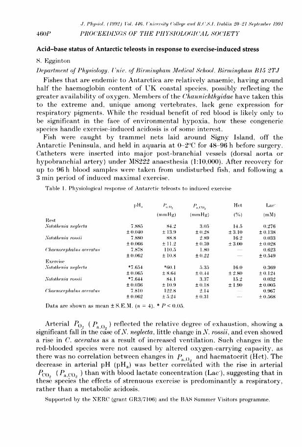
J. Physiol. (1992) IJol. 446. lUniv'ersity (olleye and IL(.CS.I. Dublin 20 21 Septenmber 1991
460P PRO(EEDINLVS OF THE PHYSIJOLOGI(CAL SOCIETY
Acid-base status of Antarctic teleosts in response to exercise-induced stress
S. EggintonDepartment of Physiology. Unit'. of Birmingham Medical School, Birmingham B15 2TJ
Fishes that are endemic to Antarctica are relatively anaemic, having aroundhalf the haemoglobin content of UK coastal species, possibly reflecting thegreater availability of oxygen. AMembers of the Channichthyidae have taken thisto the extreme and, unique among vertebrates, lack gene expression forrespiratory pigments. While the residual benefit of red blood is likely only tobe significant in the face of environmental hypoxia, how these congenericspecies handle exercise-induced acidosis is of some interest.Fish were caught by trammel nets laid around Signy Island, off the
Antarctic Peninsula, and held in aquaria at 0-2"C for 48-96 h before surgery.Catheters were inserted into major post-branchial vessels (dorsal aorta orhypobranchial artery) under MS222 anaesthesia (1:10,000). After recovery forup to 96 h blood samples were taken from undisturbed fish, and following a3 min period of induced maximal exercise.
Table 1. Physiological response of Antarctic teleosts to induced exercise
I)Ha a J)" o Hct Lac-
(mmHg) (mrtlHg) (%) (m1)RestNotothenia neylecta 7.885 84.2 3.05 14.5 0.276
± 0.040 ± 13.9 ± 0.28 ± 3.10 ± 0.138NArotothenia rossii 7.880 88.8 2.89 16.2 0.033
± 0.06( ± 11.2 ±0.59 ± 3.00 ± 0.028(haenocephalus aceratus 7.878 110.5 1.80 (0.623
±0.062 10.8 ±0 .22 ±±0.549ExerciseNotothenia nieylecta *7.654 *60.1 5.35 16.0 0.369
± 0.0605 + 8.64 ± 0.44 ±2.80 ±0.124oNotothenia roossii *7.644 84.1 3.37 15.2 0.032
±0.036 ± 10.9 ± 0.18 + 1.90 ±+0.005Chaenocephalu.s aceratu.s 7.810 122.8 2.14 0.967
± 0.062 ± 5.24 ± 0.31 ± 0.568
Data are shown as mean S.E.M. (n = 4). *P < 00.(5.
Arterial Po, ( Pa () ) reflected the relative degree of exhaustion, showing asignificant fall in the ca-se ofN. neglecta, little change in X. rossii, and even showeda rise in C. aceratus as a result of increased ventilation. Such changes in thered-blooded species were not caused by altered oxygen-carrying capacity, asthere wvas no correlation between changes in Pa.(, and haematocrit (Hct). Thedecrease in arterial pH (pHa) was better correlated with the rise in arterialP<.O (Paco} ) than with blood lactate concentration (Lac-), suggesting that inthese species the effects of strenuous exercise is predominantly a respiratory,rather than a metabolic acidosis.
Supported by the NERC (grant GR3/7106) and the BAS Summer Visitors programme.

J. Phy.iol. (1992) Vol. 446. Proceedinps of T'he P'hysioloyical Society
N,IWVERSITY COLLEGE ANI_V) R.C.S.I. D)UJ`BLLN 20-21 SEPTEMBER 1991 461P
No correlation exists between regional blood flow and regional aerobic, metaboliccapacity expressed as citrate synthase activity, within single cat skeletal muscle
P.O. Iversen, T. Flateb0 and G. NicolaysenDepartment of Physiology. University of Oslo, PO Box 1103 Blindern, 0317 Oslo, iNorwayThe perfusion variability among regions weighing about 0.25-1 g is considerable
within dog and rabbit single skeletal muscles at rest and during exercisehyperaemia (Piiper et al. 1985; Iversen et al. 1989). This regional heterogeneityin flow is not linked to specific fibre-type distributions or regional uptake ofglucose (Iversen & Nicolaysen, 1991). These findings could argue againstregional metabolic rate as an important determinator of regional perfusion.However, glucose is only one source of energy and furthermore, no distinctioncould be made between anaerobic and aerobic metabolism.We asked whether this perfusion inequality is present in a species with a
different pattern of locomotion, namely in the cat. We also aimed at testingwhether a possible regional inequality in flow could be correlated to theregional activity of the citric acid cycle enzyme citrate synthase (CS). Wereasoned that CS-activity could reflect aerobic, metabolic capacity.
Intraaortic infusions (30 s, 3 ml min') of radiolabelled microspheres (15 ,m)were given to 4 anaesthetized (pentobarbitone, 30 mg kg' I.V.) cats. Flow wasmeasured in resting as well as stimulated muscles. Post mortem (overdose ofpentobarbitone, I.V.) two hindleg muscles were removed in toto and immedi-ately frozen in liquid N2. Flow was measured in 0.25 g samples. The CS-activity was then determined spectrophotometrically in homogenates.Regional flow was markedly heterogenous both in resting and in stimulated
muscles. The coefficient of variation (CV) for regional flow was about 0.40,which is somewhat larger than that obtained in the rabbit. The CV for CS-activity was clearly smaller (about 0.15). Regional flow was not correlated toregional CS-activity either in the resting or stimulated cat muscles (P > 0.05).Furthermore, flow was not related to specific fibre-type distributions withinthe muscles. Flow was about 4.5 and 45 ml mim1' 100 g l at rest and duringstimulation, respectively. The CS-activity was approximately 1100 units 100 g-'.We conclude that regional perfusion is substantially variable also within
resting and stimulated cat skeletal muscles independent of fibre-type distribu-tions. This regional heterogeneity in flow is apparently not related to regionalaerobic, metabolic capacity as judged from regional citrate synthase activity.
REFER ENCES
Iversen, P.O. & Nicolavsen, G. (1991). Am. J. Physiol. 260, H1795-H1801.Iversen, P.O., Standa, AI. & Nicolaysen. G. (1989). Acta Physiol. Scand. 136, 17-28.Piiper, J., Pendergast, D.R., Marconi, C., Meyer, M., Heisler, N. & Cerretelli, P. (1985). J. Appl.
Physiol. 58. 2068-2074.