Role of the conserved arginine 507 and glutamate 727 residues
10
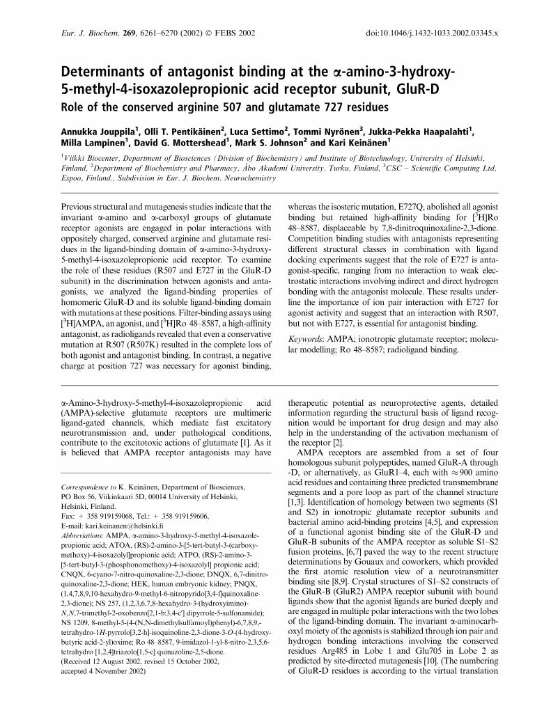
Determinants of antagonist binding at the a-amino-3-hydroxy- 5-methyl-4-isoxazolepropionic acid receptor subunit, GluR-D Role of the conserved arginine 507 and glutamate 727 residues Annukka Jouppila 1 , Olli T. Pentika ¨ inen 2 , Luca Settimo 2 , Tommi Nyro ¨ nen 3 , Jukka-Pekka Haapalahti 1 , Milla Lampinen 1 , David G. Mottershead 1 , Mark S. Johnson 2 and Kari Keina ¨ nen 1 1 Viikki Biocenter, Department of Biosciences (Division of Biochemistry) and Institute of Biotechnology, University of Helsinki, Finland, 2 Department of Biochemistry and Pharmacy, A ˚ bo Akademi University, Turku, Finland, 3 CSC – Scientific Computing Ltd, Espoo, Finland., Subdivision in Eur. J. Biochem. Neurochemistry Previous structural and mutagenesis studies indicate that the invariant a-amino and a-carboxyl groups of glutamate receptor agonists are engaged in polar interactions with oppositely charged, conserved arginine and glutamate resi- dues in the ligand-binding domain of a-amino-3-hydroxy- 5-methyl-4-isoxazolepropionic acid receptor. To examine the role of these residues (R507 and E727 in the GluR-D subunit) in the discrimination between agonists and anta- gonists, we analyzed the ligand-binding properties of homomeric GluR-D and its soluble ligand-binding domain with mutations at these positions. Filter-binding assays using [ 3 H]AMPA, an agonist, and [ 3 H]Ro 48–8587, a high-affinity antagonist, as radioligands revealed that even a conservative mutation at R507 (R507K) resulted in the complete loss of both agonist and antagonist binding. In contrast, a negative charge at position 727 was necessary for agonist binding, whereas the isosteric mutation, E727Q, abolished all agonist binding but retained high-affinity binding for [ 3 H]Ro 48–8587, displaceable by 7,8-dinitroquinoxaline-2,3-dione. Competition binding studies with antagonists representing different structural classes in combination with ligand docking experiments suggest that the role of E727 is anta- gonist-specific, ranging from no interaction to weak elec- trostatic interactions involving indirect and direct hydrogen bonding with the antagonist molecule. These results under- line the importance of ion pair interaction with E727 for agonist activity and suggest that an interaction with R507, but not with E727, is essential for antagonist binding. Keywords: AMPA; ionotropic glutamate receptor; molecu- lar modelling; Ro 48–8587; radioligand binding. a-Amino-3-hydroxy-5-methyl-4-isoxazolepropionic acid (AMPA)-selective glutamate receptors are multimeric ligand-gated channels, which mediate fast excitatory neurotransmission and, under pathological conditions, contribute to the excitotoxic actions of glutamate [1]. As it is believed that AMPA receptor antagonists may have therapeutic potential as neuroprotective agents, detailed information regarding the structural basis of ligand recog- nition would be important for drug design and may also help in the understanding of the activation mechanism of the receptor [2]. AMPA receptors are assembled from a set of four homologous subunit polypeptides, named GluR-A through -D, or alternatively, as GluR1–4, each with 900 amino acid residues and containing three predicted transmembrane segments and a pore loop as part of the channel structure [1,3]. Identification of homology between two segments (S1 and S2) in ionotropic glutamate receptor subunits and bacterial amino acid-binding proteins [4,5], and expression of a functional agonist binding site of the GluR-D and GluR-B subunits of the AMPA receptor as soluble S1–S2 fusion proteins, [6,7] paved the way to the recent structure determinations by Gouaux and coworkers, which provided the first atomic resolution view of a neurotransmitter binding site [8,9]. Crystal structures of S1–S2 constructs of the GluR-B (GluR2) AMPA receptor subunit with bound ligands show that the agonist ligands are buried deeply and are engaged in multiple polar interactions with the two lobes of the ligand-binding domain. The invariant a-aminocarb- oxyl moiety of the agonists is stabilized through ion pair and hydrogen bonding interactions involving the conserved residues Arg485 in Lobe 1 and Glu705 in Lobe 2 as predicted by site-directed mutagenesis [10]. (The numbering of GluR-D residues is according to the virtual translation Correspondence to K. Keina¨ nen, Department of Biosciences, PO Box 56, Viikinkaari 5D, 00014 University of Helsinki, Helsinki, Finland. Fax: + 358 919159068, Tel.: + 358 919159606, E-mail: kari.keinanen@helsinki.fi Abbreviations: AMPA, a-amino-3-hydroxy-5-methyl-4-isoxazole- propionic acid; ATOA, (RS)-2-amino-3-[5-tert-butyl-3-(carboxy- methoxy)-4-isoxazolyl]propionic acid; ATPO, (RS)-2-amino-3- [5-tert-butyl-3-(phosphonomethoxy)-4-isoxazolyl] propionic acid; CNQX, 6-cyano-7-nitro-quinoxaline-2,3-dione; DNQX, 6,7-dinitro- quinoxaline-2,3-dione; HEK, human embryonic kidney; PNQX, (1,4,7,8,9,10-hexahydro-9-methyl-6-nitropyrido[3,4-f]quinoxaline- 2,3-dione); NS 257, (1,2,3,6,7,8-hexahydro-3-(hydroxyimino)- N,N,7-trimethyl-2-oxobenzo[2,1-b:3,4-c¢] dipyrrole-5-sulfonamide); NS 1209, 8-methyl-5-(4-(N,N-dimethylsulfamoyl)phenyl)-6,7,8,9,- tetrahydro-1H-pyrrolo[3,2-h]-isoquinoline-2,3-dione-3-O-(4-hydroxy- butyric acid-2-yl)oxime; Ro 48–8587, 9-imidazol-1-yl-8-nitro-2,3,5,6- tetrahydro [1,2,4]triazolo[1,5-c] quinazoline-2,5-dione. (Received 12 August 2002, revised 15 October 2002, accepted 4 November 2002) Eur. J. Biochem. 269, 6261–6270 (2002) ȑ FEBS 2002 doi:10.1046/j.1432-1033.2002.03345.x
Transcript of Role of the conserved arginine 507 and glutamate 727 residues
Determinants of antagonist binding at the a-amino-3-hydroxy-5-methyl-4-isoxazolepropionic acid receptor subunit, GluR-DRole of the conserved arginine 507 and glutamate 727 residues
Annukka Jouppila1, Olli T. Pentikainen2, Luca Settimo2, Tommi Nyronen3, Jukka-Pekka Haapalahti1,Milla Lampinen1, David G. Mottershead1, Mark S. Johnson2 and Kari Keinanen1
1Viikki Biocenter, Department of Biosciences (Division of Biochemistry) and Institute of Biotechnology, University of Helsinki,
Finland, 2Department of Biochemistry and Pharmacy, Abo Akademi University, Turku, Finland, 3CSC – Scientific Computing Ltd,
Espoo, Finland., Subdivision in Eur. J. Biochem. Neurochemistry
Previous structural and mutagenesis studies indicate that theinvariant a-amino and a-carboxyl groups of glutamatereceptor agonists are engaged in polar interactions withoppositely charged, conserved arginine and glutamate resi-dues in the ligand-binding domain of a-amino-3-hydroxy-5-methyl-4-isoxazolepropionic acid receptor. To examinethe role of these residues (R507 and E727 in the GluR-Dsubunit) in the discrimination between agonists and anta-gonists, we analyzed the ligand-binding properties ofhomomeric GluR-D and its soluble ligand-binding domainwith mutations at these positions. Filter-binding assays using[3H]AMPA, an agonist, and [3H]Ro 48–8587, a high-affinityantagonist, as radioligands revealed that even a conservativemutation at R507 (R507K) resulted in the complete loss ofboth agonist and antagonist binding. In contrast, a negativecharge at position 727 was necessary for agonist binding,
whereas the isosteric mutation, E727Q, abolished all agonistbinding but retained high-affinity binding for [3H]Ro48–8587, displaceable by 7,8-dinitroquinoxaline-2,3-dione.Competition binding studies with antagonists representingdifferent structural classes in combination with liganddocking experiments suggest that the role of E727 is anta-gonist-specific, ranging from no interaction to weak elec-trostatic interactions involving indirect and direct hydrogenbonding with the antagonist molecule. These results under-line the importance of ion pair interaction with E727 foragonist activity and suggest that an interaction with R507,but not with E727, is essential for antagonist binding.
Keywords: AMPA; ionotropic glutamate receptor; molecu-lar modelling; Ro 48–8587; radioligand binding.
a-Amino-3-hydroxy-5-methyl-4-isoxazolepropionic acid(AMPA)-selective glutamate receptors are multimericligand-gated channels, which mediate fast excitatoryneurotransmission and, under pathological conditions,contribute to the excitotoxic actions of glutamate [1]. As itis believed that AMPA receptor antagonists may have
therapeutic potential as neuroprotective agents, detailedinformation regarding the structural basis of ligand recog-nition would be important for drug design and may alsohelp in the understanding of the activation mechanism ofthe receptor [2].
AMPA receptors are assembled from a set of fourhomologous subunit polypeptides, named GluR-A through-D, or alternatively, as GluR1–4, each with � 900 aminoacid residues and containing three predicted transmembranesegments and a pore loop as part of the channel structure[1,3]. Identification of homology between two segments (S1and S2) in ionotropic glutamate receptor subunits andbacterial amino acid-binding proteins [4,5], and expressionof a functional agonist binding site of the GluR-D andGluR-B subunits of the AMPA receptor as soluble S1–S2fusion proteins, [6,7] paved the way to the recent structuredeterminations by Gouaux and coworkers, which providedthe first atomic resolution view of a neurotransmitterbinding site [8,9]. Crystal structures of S1–S2 constructs ofthe GluR-B (GluR2) AMPA receptor subunit with boundligands show that the agonist ligands are buried deeply andare engaged in multiple polar interactions with the two lobesof the ligand-binding domain. The invariant a-aminocarb-oxyl moiety of the agonists is stabilized through ion pair andhydrogen bonding interactions involving the conservedresidues Arg485 in Lobe 1 and Glu705 in Lobe 2 aspredicted by site-directed mutagenesis [10]. (The numberingof GluR-D residues is according to the virtual translation
Correspondence to K. Keinanen, Department of Biosciences,
PO Box 56, Viikinkaari 5D, 00014 University of Helsinki,
Helsinki, Finland.
Fax: + 358 919159068, Tel.: + 358 919159606,
E-mail: [email protected]
Abbreviations: AMPA, a-amino-3-hydroxy-5-methyl-4-isoxazole-
propionic acid; ATOA, (RS)-2-amino-3-[5-tert-butyl-3-(carboxy-
methoxy)-4-isoxazolyl]propionic acid; ATPO, (RS)-2-amino-3-
[5-tert-butyl-3-(phosphonomethoxy)-4-isoxazolyl] propionic acid;
CNQX, 6-cyano-7-nitro-quinoxaline-2,3-dione; DNQX, 6,7-dinitro-
quinoxaline-2,3-dione; HEK, human embryonic kidney; PNQX,
(1,4,7,8,9,10-hexahydro-9-methyl-6-nitropyrido[3,4-f]quinoxaline-
2,3-dione); NS 257, (1,2,3,6,7,8-hexahydro-3-(hydroxyimino)-
N,N,7-trimethyl-2-oxobenzo[2,1-b:3,4-c¢] dipyrrole-5-sulfonamide);
NS 1209, 8-methyl-5-(4-(N,N-dimethylsulfamoyl)phenyl)-6,7,8,9,-
tetrahydro-1H-pyrrolo[3,2-h]-isoquinoline-2,3-dione-3-O-(4-hydroxy-
butyric acid-2-yl)oxime; Ro 48–8587, 9-imidazol-1-yl-8-nitro-2,3,5,6-
tetrahydro [1,2,4]triazolo[1,5-c] quinazoline-2,5-dione.
(Received 12 August 2002, revised 15 October 2002,
accepted 4 November 2002)
Eur. J. Biochem. 269, 6261–6270 (2002) � FEBS 2002 doi:10.1046/j.1432-1033.2002.03345.x

from the corresponding nucleotide sequences starting withthe initiator methionine. This differs, by the length of thepredicted signal peptide, from the numbering used byGouaux and coworkers [8,9] for the mature GluR-B(GluR2) polypeptide [8,9].) The three hydrogen bonds ofthe a-amino group (to Pro478, Thr480, both in Lobe 1, andGlu705) show a nearly perfect tetrahedral organizationaround the central nitrogen. The distal acidic group of theagonists interacts exlusively with Lobe 2 [9].
Importantly, the crystal structures of ligand-free GluR-BS1–S2 and one antagonist complex, 6,7-dinitroquinoxaline-2,3-dione (DNQX), have also been obtained [9]. Thesecomplexes are slightly more open than the agonist com-plexes, suggesting that agonist-induced closure of the lobesmay provide the driving force for channel opening [9].Binding of the DNQX is mediated by polar contacts to bothlobes and aromatic stacking with the side chain of a tyrosineresidue. Consistent with pharmacophore models [11,12]predicting that the carbonyl oxygens and iminic nitrogens ofthe quioxalinedione antagonists, indispensable for activity,mimic the a-aminocarboxylate core of the agonists, thecarbonyl oxygens of DNQX are hydrogen bonded toArg485. Glu705 does not, however, participate directly inDNQX binding. Interestingly, however, this residue showstwo different conformations, one of which is pointingdirectly below the quinoxalinedione ring and Glu705 is, infact, the only binding site residue which shows major sidechain reorientation between the agonist, antagonist andligand-free complexes [9].
Considering the large structural variation of AMPAreceptor antagonists [2,12], it can be expected that thereceptor–antagonist interactions may show differences tothose interactions seen in the DNQX complex structure. Inthe present study, we have analyzed the ligand-bindingproperties of mutated and wild-type homomeric GluR-DAMPA receptors in order to further characterize the roles ofArg507 and Glu727 (homologous to GluR-B residuesArg485 and Glu705, respectively) in antagonist binding.In order to separate the structural requirements necessaryfor antagonist binding from those important for agonistbinding, we have used a high-affinity antagonist [3H]Ro48-8587 [13] as a radioligand, in addition to [3H]AMPA.
M A T E R I A L S A N D M E T H O D S
Experimental materials
CNQX, DNQX and kainic acid were obtained from RBI-Sigma (Natick, MA, USA) and L-glutamate was fromSigma (St Louis, MO, USA). [3H]AMPA (specific activity,60 CiÆmmol)1) was from NEN Life Science Products(Boston, MA, USA). [3H]Ro 48-8587 (specific activity,44 CiÆmmol)1) was obtained from Amersham PharmaciaBiotech (Buckinghamshire, UK). ATPO and ATOA [14]were obtained from P. Krogsgaard-Larsen, Royal DanishSchool of Pharmacy, Copenhagen, Denmark. NS 257 [15],NS 1209 [16], and 1,4,7,8,9,10-hexahydro-9-methyl-6-nitro-pyrido[3,4-f]quinoxaline-2,3-dione (PNQX) [11] wereobtained from J. Drejer (NeuroSearch A/S, Glostrup,Denmark). Ro 48-8587 was obtained from R. Wyler andV. Mutel (F. Hoffmann-La Roche, Basel, Switzerland).Enzymes for molecular biology were purchased from NewEngland Biolabs (Beverly, MA, USA), Finnzymes (Espoo,
Finland), and Fermentas (Vilnius, Lithuania). Anti-FlagM1 was obtained from Sigma. Anti-c-myc antibody waspurified from the hybridoma cell line 9E10.2 originallyobtained from American Type Culture Collection (ATCCCRL1729).
DNA constructs and recombinant baculovirusexpression
Single point mutations were introduced into the flip isoform[17] of N-terminally Flag-tagged or C-terminally myc-tagged rat GluR-D in a pFASTBAC1 vector (BRL-LifeSciences) [6,18] by overlap extension PCR using mutagenicprimers, followed by restriction fragment replacement. Thepresence of the mutations was verified by DNA sequencing.Recombinant baculovirus vectors were then prepared byusing the Bac-to-Bac system according to the manufac-turer’s instuctions (Invitrogen Life Technologies, Carlsbad,CA, USA), and used to infect monolayers of Trichoplusia nicells (�High Five�, Invitrogen) grown in T25 or T175 tissueculture flasks at 27 �C. SF900-II (Life Technologies, Paisley,UK), supplemented with penicillin (100 lgÆmL-1), strepto-mycin (lgÆmL-1) and amphotericin B (0.25 lgÆmL-1) wasused as the culture medium. The cells were collected 4 daysafter infection by centrifugation (1000 g, for 10 min) andanalysed by immunoblotting. The cell pellets were stored at)20 �C until used for radioligand binding studies.
In some experiments, S1–S2 ligand-binding site con-structs were used instead of the full-length receptor. TheFlag-tagged GluR-D S1–S2 constructs (�wild-type�, E727Dand E727A mutants), and their expression as solubleproteins secreted in the culture medium have been describedpreviously [10].
Preparation of membranes
For radioligand binding experiments, membranes wereprepared as follows. Insect cells were homogenized in fivevolumes of 20 mM Hepes, pH 7.4, 2.5 mM EDTA, 0.1 mM
phenylmethanesulfonyl fluoride. The particulate fractionwas pelleted (30 000 g, 25 min) and washed extensively byrepeated homogenization and centrifugation in 20 mM
Hepes, pH 7.4, 200 mM NaCl, 0.5 mM EDTA, 0.1 mM
PMSF. Finally, the washed membranes were suspended in20 mM Hepes, pH 7.4, 200 mM NaCl, 10% glycerol,0.1 mM PMSF, and stored frozen or on ice until used inthe assay. Some experiments were performed with receptorpreparations that were solubilized in Triton X-100 asdescribed previously [19] with results essentially identical tothose obtained with membranes.
Radioligand binding assays
For the AMPA binding assays, the membranes weresuspended in 30 mM Tris/HCl, pH 7.2, 100 mM potassiumthiocyanate (KSCN), 2.5 mM CaCl2, 0.1% Triton X-100.For initial measurement of binding activity and for ligandcompetition experiments, 5 nM [3H]AMPA was used. Forsaturation analyses, [3H]AMPA was diluted to a specificactivity of 12 CiÆmmol-1 with unlabelled R,S-AMPA andused at 1–300 nM concentrations. Membranes (20–250 lgprotein) were incubated in a 0.5-mL volume for 1 h onice with the radioligand, whereafter the reactions were
6262 A. Jouppila et al. (Eur. J. Biochem. 269) � FEBS 2002

terminated by adding 5 mL of ice-cold 30 mM Tris/HCl,pH 7.2, 100 mM KSCN, 2.5 mM CaCl2 followed by rapidfiltration through Whatman GF/B filters with two 5-mLwashes with ice-cold buffer. Nonspecific binding wasdetermined in the presence of 1 mM L-glutamate. The filterswere solubilized in OptiPhase 3 HiSafe (Wallac) andsubjected to liquid scintillation counting.
[3H]Ro 48-8587 (0.5–2 nM final concentration) bindingassays were performed in 50 mM Tris/HCl, pH 7.0 [13]. Thefiltration assay was performed as described for [3H]AMPAbinding with the exception that 50 mM Tris/HCl, pH 7.0was used as the buffer. Saturation binding analysis with[3H]Ro 48-8587 was performed by diluting the radioligandwith increasing amounts of unlabelled compound.
The ligand binding data were analyzed by nonlinearcurve fitting using the PRISM 2.01 software (GraphPad Inc.).Student’s t-test was used for the statistical analyses.
Molecular modelling
Structural modelling. The three-dimensional structures ofGluR-B S1–S2 ligand-binding core complexed with DNQX(PDB accession no. 1ftl [9]); was obtained from the ProteinData Bank [20]. The sequences of GluR-D and GluR-Bwere aligned by using MALIGN [21] in the BODIL ModelingEnvironment (Lehtonen J.V., Rantanen V.-V., Still, D.-J.,Gyllenberg, M., and Johnson, M.S.; http://www.abo.fi/fak/mnf/bkf/research/johnson/bodil.html, personal communi-cation) using a structure-based sequence comparisionmatrix [22]. The program MODELLER 4.0 [23] was used toconstruct a three-dimensional model structure by satisfyingspatial restraints imposed on the GluR-D sequence by itsalignment with the known GluR-B structure. At the sametime, the structure of the ligand seen in the X-ray structurewas built using MODELLER 4.0.
Ligand minimization. Ligands were built with the programSYBYL 6.6 (Tripos, St Louis, MO, USA) and energyminimized prior to docking to the receptor models usingthe TRIPOS Force Field and conjugate gradient method untilthe energy gradient was less than 0.05 kcalÆmol)1. Protona-tion of the polar groups was evaluated by comparison of thefinal three-dimensional structures with similar substructuresobtained from the Cambridge Structural Data Bank (CSDSystem Documentation, 1992, Cambridge CrystallographicData Centre, Cambridge, UK). To calculate the optimizedstructure for Ro 48–8587, standard Hartree–Fock (HF/3-21G) methods in GAUSSIAN 98 [24] were used.
Receptor minimization. Polar hydrogens were added to thereceptor using SYBYL 6.6. In order to optimize theintramolecular interactions in the receptor model, hydrogenatoms were minimized (keeping the rest of the model rigid)using amber charges [25,26].
Ligand docking. AUTODOCK 3.0 [27] is a semirigid dockingprogram that considers the whole ligand molecule dockingto the binding site and it is possible to choose the torsionangles of the ligand that are allowed to rotate (AutoTors),while the bond angles and bond lengths are kept fixed. Theoverall interaction energy between chemical species isestimated by considering both Lennard–Jones atom–atompotentials and electrostatic effects, summed for the individ-
ual interactions between atoms. Partial charges for thereceptor model were calculated using the AMBER force fieldin SYBYL 6.6; and for ligands, the MMFF94 Force Field [28].The interaction of a probe group (corresponding to eachtype of atom in the ligand) with a receptor model wascalculated at grid positions 0.25 A apart in a(20 · 20 · 20) A3 box centered at the binding site usingthe program AUTOGRID in AUTODOCK 3.0. For each ligand,10 separate docking simulations were performed.
Other assays. SDS/PAGE and Western blotting usinganti-Flag M1 (Sigma Chemical, St. Louis, MO, USA) as theprimary antibody, were performed as described previously[6,19]. Protein content of the samples was measured byusing bicinchonic acid assay kit according to the manufac-turer’s instructions. (BCA, Pierce, Rockford, IL, USA).
R E S U L T S
Effects of mutations at residues Arg507 and Glu727on binding of [3H]AMPA to GluR-D
Structural and mutagenesis studies on soluble ligand-binding domains indicate that Arg507 and Glu727 in theAMPA receptor subunit GluR-D, and the equivalentresidues in the GluR-B subunit, serve as critical dockingsites for the a-amino and a-carboxylate groups of agonistligands. In the present study, we have examined the role ofthese residues in antagonist recognition in the full-length,membrane-bound AMPA receptor. Wild-type GluR-D andits R507K, E727D and E727Q mutants were expressed inrecombinant baculovirus-infected Trichoplusia ni (High
Fig. 1. Expression and [3H]AMPA binding activity of GluR-D mutant
receptors. Wild-type and mutated epitope-tagged GluR-D AMPA
receptors were expressed in recombinant baculovirus-infected High
Five insect cells. (A) Immunoblot analysis showing the expression of
Flag-tagged GluR-D (WT), GluR-D E727D, GluR-D E727Q and of
myc-tagged GluR-D R507K. Noninfected cells were used as controls.
All lanes were loaded with an equal amount (� 10 lg) of membrane
protein, and the blots were developed by using anti-Flag M1 or anti-
myc 9E10 as primary antibodies as indicated. The positions of
molecular size markers are shown at the right. (B) Binding of
[3H]AMPA to mutant GluR-D receptors. Binding of 5 nM [3H]AMPA
to insect cell membranes (50 lg protein for wild-type and E727
mutants; 100 lg for R507K) was determined in the presence (non-
specific binding) and absence (total binding) of 1 mM glutamate.
Specific binding was defined as the difference between total and non-
specific binding. The values (mean ± SD) are from a representative
assay performed in triplicate.
� FEBS 2002 Role of E727 of GluR-D in antagonist binding (Eur. J. Biochem. 269) 6263

Five) insect cells as homomeric, epitope-tagged receptors. Inimmunoblots, an � 105-kDa immunoreactive band, corres-ponding to the size expected for a glycosylated GluR-Dmonomer, was observed in baculovirus-infected cells ex-pressing Flag-tagged GluR-D and the E727D and E727Qmutants, and the myc-tagged R507K mutant, but not innoninfected control cells (Fig. 1A). The presence of anadditional and intense 90-kDa myc-immunoreactive bandin cells expressing the R507K mutant suggests partialproteolysis or defective glycosylation with this mutant(Fig. 1A). To determine the ligand binding activity of theGluR-D mutants, membrane preparations from the bacu-lovirus-infected cells were subjected to a filtration bindingassay by using [3H]AMPA (at 5 nM) as the radioligand. Inaccordance with a previous analysis conducted with thesoluble ligand-binding domain of GluR-D, [3H]AMPAbound to wild-type GluR-D and to the E727D mutant butnot to the R507K and E727Q mutants (Fig. 1B). In asaturation binding analysis,Kd values of 40 and 11 nM wereobtained for the wild-type and E727D receptors, respect-ively. The corresponding Bmax values were 12.3 pmolÆmg)1
of protein for GluR-D and 14.7 pmolÆmg-1 for GluR-DE727D. In a competition binding assay, unlabelledL-glutamate exhibited a Ki value of 0.20 ± 0.06 lM forthe wild-type GluR-D and 2.13 ± 0.13 lM for the GluR-DE727D mutant (mean ± SD; n ¼ 4). Kainate exhibited aKi value of 2.10 ± 0.14 lM (mean ± SD; n ¼ 4) with thewild-type GluR-D, but displayed a drastically decreasedaffinity to the E727D mutant (> 1000-fold), consistent withearlier results obtained with the soluble ligand-bindingdomain [10]. A reliable value for the inhibitory affinityconstant of kainate binding to E727D mutant receptorcould not be obtained, but 3 mM kainate caused only20–25% inhibition of [3H]AMPA binding (not shown).
Interaction of competitive antagonists with wild-typeand E727D mutant receptors
Next, we determined the ability of competitive antagonistsrepresenting different structural types (Fig. 2) to inhibit[3H]AMPA binding to GluR-D and to the E727D mutant.All eight antagonist ligands inhibited [3H]AMPA binding towild-type GluR-D in a concentration-dependent manner(Fig. 3, Table 1). With the exception of two compounds,Ro 48-8587 and NS 257, substituting an aspartate for theglutamate at position 727 of GluR-D, produced significantchanges in the apparent binding affinitites of the antagonists(Table 1). The closely related quioxaline-2,3-diones CNQXand DNQX were (� 20-fold) more potent inhibitors of[3H]AMPA binding at the mutant receptor, whereas anopposite effect was seen with PNQX, NS 1209 and the twoAMPA-derivatives, ATPO and ATOA. In fact, the highestconcentration of ATOA which was tested (1 mM), did notproduce any clear inhibition of [3H]AMPA binding to theE727D mutant. The competition curves were monophasicand displayed Hill coefficients close to unity for allantagonists, except for ATOA and ATPO whose lowaffinity to the E727D mutant receptor prevented thedetermination of more complete displacement curves.
Binding of [3H]Ro 48-8587 to wild-type and mutatedGluR-D receptors
As the E727Q and R507K mutant receptors were devoid ofany [3H]AMPA binding activity, the effect of these muta-tions in antagonist recognition could not be studied bybinding inhibition experiments. Therefore, we employed arecently introduced high-affinity antagonist radioligand,[3H]Ro 48-8587 [13], to analyze directly the antagonistbinding properties of the mutant receptors. In a filtrationassay using a single 1 nM concentration of [3H]Ro 48-8587,significant binding was observed to wild-type GluR-D, andto the E727D and E727Q mutants (Fig. 4A). In contrast, nobinding above nonspecific background produced by mem-branes prepared from noninfected cells was seen with theR507K mutant (Fig. 4A). In saturation binding experi-ments, Kd values of 8.3 ± 0.2 and 7.0 ± 1.2 nM wereobtained for the wild-type GluR-D and E727D, respect-Fig. 2. Structures of AMPA receptor antagonists used in this study.
Fig. 3. Inhibition of [3H]AMPA binding by competitive antagonists.
Wild-type (solid squares) and E727D mutant (open squares) GluR-D
receptors were expressed in recombinant baculovirus-infected insect
cells. Insect cell membranes were equilibrated with [3H]AMPA (5 nM)
in the presence of increasing concentrations of unlabelled antagonist
compounds, and the amount of bound [3H]AMPA was determined by
rapid filtration. Displacement of [3H]AMPA binding is shown for
DNQX, NS 1209, and Ro 48–8587. The curves represent a best fit to a
one-site model obtained by nonlinear curve fitting.
6264 A. Jouppila et al. (Eur. J. Biochem. 269) � FEBS 2002
ively, whereas a slightly higher Kd value, 20.0 ± 0.5 nM,was obtained for E727Q (mean ± SD; n ¼ 3; Fig. 4B).
In order to further substantiate the findings and tofacilitate comparision with earlier work, we performed some[3H]Ro 48-8587 binding studies with the separatelyexpressed S1–S2 ligand-binding domain of GluR-D(Fig. 4C). Consistent with the results obtained with themembrane-bound full-length receptors, [3H]Ro 48-8587 (at2 nM concentration) bound specifically to the wild-type andE727D S1–S2 proteins, whereas the S1–S2 R507K mutantdid not show binding activity (Fig. 4D). In a saturationbinding analysis, Kd values of 15.6 ± 6.6 and13.0 ± 1.8 nM were obtained for the binding of[3H]Ro 48-8587 to wild-type S1–S2 and to the E727Dmutant, respectively (mean ± SD; n ¼ 3). Furthermore, anS1–S2 protein carrying E727A mutation, previously shownto lack [3H]AMPA binding acitivity ([10,] Fig. 4D), did notshow any [3H]Ro 48-8587 binding in the filtration assay(Fig. 4D).
Ligand-binding properties of E727Q mutant receptors
Because earlier mutagenesis studies indicated that a negativecharge at position 727 is necessary for agonist binding, theability of the E727Q mutant receptor to bind [3H]Ro48-8587 was of considerable interest, and therefore weexamined further the [3H]Ro 48-8587 binding properties ofhomomeric E727Q receptors by using ligand competitionexperiments. Due to the limited amount of [3H]Ro 48-8587available, only semiquantitative analyses were performed.Using a single 1 mM concentration of unlabelled ligand, theagonists, L-glutamate and kainate, were practically inactiveas inhibitors of [3H]Ro 48-8587 binding to the E727Qreceptor, whereas DNQX inhibited > 90% of the binding(Fig. 5A). As expected, all three ligands strongly inhibited[3H]Ro 48-8587 binding to wild-type GluR-D, whereaskainate was a significantly weaker inhibitor than glutamateand DNQX at E727D membranes, consistent with thedramatic decrease in kainate affinity caused by E727Dmutation (Fig. 5A). Furthermore, DNQX and NS 1209inhibited [3H]Ro 48-8587 binding to GluR-D E727Qmembranes with Ki values of 0.033 ± 0.011 and
2.52 ± 0.15 lM, respectively (mean ± SD; n ¼ 3;Fig. 5B), indicating that binding of these two antagonists(as with Ro 48–8587) can bind to the receptor in thepresence of glutamine at position 727. Unfortunately, thesmall amount of labelled [3H]Ro 48-8587 available preven-ted more extensive displacement experiments.
Molecular model of GluR-D ligand-binding domain
Ligand binding studies were complemented by dockingsimulations in order to help us understand the role ofR507 and E727 in the interactions of the receptor withthe different antagonists. First, a three-dimensionalmodel of the ligand binding domain of GluR-D (S1–S2) was built using the 1.8 A resolution structure of theGluR-B S1–S2-DNQX complex [9] as a template. Thesequence identity between the molecules was high(� 90%) with only one difference located near thebinding site, Tyr703 in GluR-B being replaced by aphenylalanine in GluR-D.
Ligand docking
Quinoxalinediones. DNQX was docked automaticallyinto the GluR-D receptor model resulting in very similarinteractions to those present in the crystal structure ofGluR-B S1–S2-DNQX complex, including formation oftwo hydrogen bonds with R507 (Fig. 6A). Glu727 doesnot form hydrogen bonds with DNQX and is likely tohave the same two side chain orientations as theequivalent Glu705 has in the asymmetric unit of GluR-B S1–S2-DNQX complex. Both orientations could ac-commodate a weak electrostatic interaction with theDNQX ring structure, which carries a partial positivecharge due to two strongly electron-withdrawing nitrogroups. Furthermore, in the GluR-B S1–S2-DNQXcomplex [9], Tyr732 is seen to form a hydrogen bondwith the 6-nitro group of DNQX or with Glu705,depending on the orientation of the side chain ofGlu705. Similar interactions are likely to take placebetween DNQX and the corresponding residues, Glu727and Tyr754, in GluR-D (Fig. 7A and B).
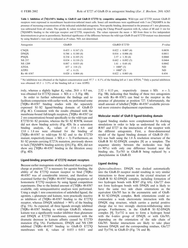
Table 1. Inhibition of [3H]AMPA binding to GluR-D and GluR-D E727D by competitive antagonists. Wild-type and E727D mutant GluR-D
receptors were expressed in recombinant baculovirus-infected insect cells. Insect cell membranes were equilibrated with 5 nM [3H]AMPA in the
presence of increasing concentrations of the indicated unlabelled antagonists. Non-specific binding, determined in the presence of 1 mM glutamate,
was subtracted from all values. The specific Ki values were calculated by using the Cheng–Prusoff equation with Kd values of 40 and 11 nM for
[3H]AMPA binding to the wild-type receptor and E727D, respectively. The values represent the mean ± SD from three to five independent
determinations (n given in parenthesis). Statistical significance of the difference between the wild-type GluR-D and E727D mutant was determined
by using Student’s t-test and is indicated as P-values. ND, not determined.
Antagonist GluRD GluRD E727D P-value
CNQX 0.433 ± 0.167 (3) 0.022 ± 0.007 (4) 0.0038
DNQX 0.546 ± 0.180 (4) 0.016 ± 0.006 (4) 0.0011
PNQX 0.334 ± 0.105 (5) 2.57 ± 1.30 (4) 0.0060
NS 257 0.834 ± 0.110 (5) 0.682 ± 0.092 (5) 0.0464
NS 1209 0.087 ± 0.035 (4) 1.61 ± 0.68 (4) 0.0042
ATOA 437 ± 118 (3) > 1000a (3) ND
ATPO 67 ± 6 (3) > 1000b (3) ND
Ro 48–8587 0.020 ± 0.004 (4) 0.022 ± 0.003 (4) 0.454
a No inhibition was obtained at the highest concentration used: 97.7 ± 4.1% of the binding left at 1 mM ATOA. b Only a partial inhibition
was obtained: 65.3 ± 3.5% of the binding left at 1 mM ATPO.
� FEBS 2002 Role of E727 of GluR-D in antagonist binding (Eur. J. Biochem. 269) 6265

The docked conformation of CNQX closely resemblesthat of DNQX. Because of the bulky methylpiperidinic ring,the nitro group of PNQX cannot be positioned to form ahydrogen bond with Tyr754, and therefore a hydrogenbonding pattern similar to that illustrated in Fig. 7A,cannot form, and a situation illustrated in Fig. 7B is morelikely. For the E727D mutant, the absence of a negativelycharged group equivalent to the nitro group in DNQX andthe presence of the positive charge would eliminate thepossibility for interactions of the type shown in (Fig. 7C).
NS 257 and NS 1219. The docked conformation of theantagonist, NS 257, resembles that of PNQX (Fig. 6B). Thecarbonyl oxygen and the oxime (-C¼N-OH) substituents of
the pyrrolidine ring in NS 257 carry partial negative chargesand interact with Arg507 in an analogous way to DNQX.No direct interaction is predicted between Glu727 andNS 257, consistent with the absence of any change in theability of NS 257 to inhibit [3H]AMPA binding uponthe mutation E727D (Table 1). In contrast, however, thehydroxyl group in the �side chain� of NS 1209, not present inNS 257, is ideally positioned to form a hydrogen bond withGlu727 in wild type GluR-D, but not with the E727Dmutant, providing an explanation for the observed� 18-fold decrease in affinity (Table 1). Interestingly, thecarboxylate group of the 4-hydroxy-2-methyleneamino-oxybutyrate �side chain� of NS 1209 may be able to interactwith the beginning of helix F, a partly positively chargedregion which binds the distal carboxylate groups of kainateand L-glutamate in the GluR-B crystal structures [8,9].
ATPO and ATOA. Two antagonists, ATOA and ATPO,are structurally related to AMPA and carry a bulky t-butylsubstituent at the 5-position of the isoxazole ring, and eithera carboxymethoxy group (ATOA) or a phosphonomethoxygroup (ATPO) at a position equivalent to the 3-hydroxylgroup in AMPA. ATPO and ATOA dock to the receptormodels in a fashion similar to that of AMPA in the GluR-BS1–S2-AMPA crystal structure (Fig. 6C). The essential
Fig. 5. [3H]Ro 48-8587 binding properties of GluR-D E727Q mutant.
Binding of [3H]Ro 48-8587 to membranes prepared from cells
expressing wild-type and mutated GluR-D receptors was analyzed. (A)
Inhibition of [3H]Ro 48-8587 (1 nM) binding to wild-type GluR-D and
to E727D and E727Q mutant receptors by 1 mM glutamate, kainate
and DNQX. The values are presented as percentage of the total
binding obtained in the absence of competing ligands (equal to
100%) ± SD, and are from a representative experiment performed in
triplicate. (B) Inhibition of [3H]Ro 48-8587 binding to GluR-D E727Q
membranes by DNQX and NS 1209. The values represent means from
a duplicate measurement. The displacement curves were obtained by
nonlinear curve-fitting to a one-site model. The corresponding Ki
values were 0.048 and 2.37 lM for DNQX (open circles) and NS 1209
(solid circles), respectively.
Fig. 4. Binding of [3H]Ro 48-8587 to GluR-D mutant receptors.
Wild-type and mutated GluR-D AMPA receptors and soluble ligand-
binding domains (�S1–S2�) were expressed in recombinant baculovirus-
infected insect cells, and binding of [3H]Ro 48-8587 to membranes and
to dialyzed culture supernanatants (in the case of S1–S2) was deter-
mined. (A) total binding of 1 nM [3H]Ro 48-8587 to membrane sam-
ples, each containing 50 lg of protein. The values represent
mean ± SD. from a representative experiment performed in triplicate.
The level of nonspecific binding is indicated by binding of
[3H]Ro 48-8587 to wild-type GluR-D membranes in the presence of
1 mM glutamate (�WT + Glu�) and by total binding of [3H]Ro 48-
8587 to membranes prepared from uninfected cells (�control�). (B)
Homologous displacement of [3H]Ro 48-8587 binding to wild-type
GluR-D (solid squares), GluR-D E727D (open squares) and GluR-D
E727Q (open circles) membranes by unlabelled Ro 48-8587. The
values represent mean ± SD from a triplicate measurement. The
displacement curves were obtained by nonlinear curve-fitting to a one-
site model.Kd values of 9.7 ± 2.0, 8.3 ± 2.0 and 25.2 ± 2.0 nM were
determined for the wild-type GluR-D, E727D and E727Q, respect-
ively. (C) Schematic structure of the constructs for expression of full-
length GluR-D and its ligand-binding domain (S1–S2), and an
immunoblot showing the expression of wild-type and mutated epitope-
tagged S1–S2 constructs as secreted 43-kDa anti-Flag-immunoreactive
species in recombinant baculovirus-infected insect cells. The samples
for immunoblotting correspond to 10 lL of culture supernatant. (D)
Binding of [3H]Ro 48-8587 to wild-type and mutated S1–S2 proteins.
Binding of [3H]Ro 48-8587 to extensively dialyzed culture superna-
tants was determined by using filtration through polyethylene–imine-
treated filters. The values correspond to total binding (mean ± SD)
from a representative experiment conducted in triplicate. The level of
nonspecific binding was determined for the wild-type S1–S2 in the
presence of 1 mM glutamate (S1–S2-WT + Glu).
6266 A. Jouppila et al. (Eur. J. Biochem. 269) � FEBS 2002
hydrogen bond and ion pair interactions with Arg507 andGlu727 are predicted to be closely similar to those inthe AMPA complex. In contrast to AMPA, however, thereceptor complexes of ATOA and ATPO would keep thereceptor in a much more open form because the bulkyt-butyl group and the acidic side chain substituents thatcontact the partly positively charged helix F region preventsterically further closure. In the E727D mutant, thecarboxylate group of the aspartate side chain cannot reachthe a-amino-group of the ligand, thus explaining theremarkable decrease in the binding affinity (Table 1).
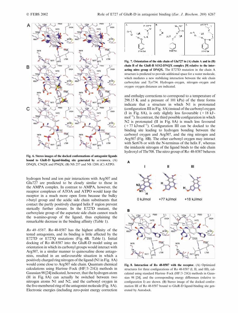
Ro 48–8587. Ro 48-8587 has the highest affinity of thetested antagonists, and its binding is little affected by theE727D or E727Q mutations (Fig. 4B, Table 1). Initialdocking of Ro 48-8587 into the GluR-D model using anorientation in which its carbonyl groups would interact withArg507, in a similar manner to quinoxaline dione antago-nists, resulted in an unfavourable situation in which apositively charged ring nitrogen of the ligand (N1 in Fig. 8A)would come close to Arg507 side chain. Quantum chemicalcalculations using Hartree–Fock (HF/3–21G) methods inGaussian 98 [24] indicated, however, that the hydrogen atom(H in Fig. 8A) can actually be switched between twonitrogen atoms N1 and N2, and the carbonyl oxygen inthe five-membered ring of the antagonist molecule (Fig. 8A).Electronic energies (including zero-point energy correction
and enthalpy corrections to correspond to a temperature of298.15 K and a pressure of 101 kPa) of the three formsindicate that a structure in which N1 is protonated(configuration III in Fig. 8A) instead of the carbonyl oxygen(I in Fig. 8A), is only slightly less favourable (+18 kJÆ-mol)1). In contrast, the third possible configuration in whichN2 is protonated (II in Fig. 8A) is much less favoured(+77 kJÆmol)1). Configuration III can be docked to thebinding site leading to hydrogen bonding between thecarbonyl oxygen and Arg507, and the ring nitrogen andArg507 (Fig. 8B). The other carbonyl oxygen may interactwith Ser676 or with the N-terminus of the helix F, whereasthe imidazole nitrogen of the ligand binds to the side chainhydroxyl of Thr708. The nitro group of Ro 48-8587 behaves
Fig. 6. Stereo images of the docked conformations of antagonist ligands
bound to GluR-D ligand-binding site generated by AUTODOCK. (A)
DNQX, CNQX and PNQX; (B) NS 257 and NS 1209; (C) ATPO.
Fig. 7. Orientation of the side chain of Glu727 in (A) chain A and in (B)
chain B of the GluR-B S1S2-DNQX complex [9] relative to the inter-
acting nitro group of DNQX. The E727D mutation in the chain A
structure is predicted to provide additional space for a water molecule,
which mediates a new stabilizing interaction between the side chain
carboxylate and Tyr754. Hydrogen–oxygen, nitrogen–oxygen and
oxygen–oxygen distances are indicated.
Fig. 8. Interaction of Ro 48-8587 with the receptor. (A) Optimized
structures for three configurations of Ro 48-8587 (I, II, and III), cal-
culated using standard Hartree–Fock (HF/3–21G) methods in Gaus-
sian 98 [24], and the corresponding energy differences (relative to
configuration I) are shown. (B) Stereo image of the docked confor-
mation III of Ro 48-8587 bound to GluR-D ligand-binding site gen-
erated by Autodock.
� FEBS 2002 Role of E727 of GluR-D in antagonist binding (Eur. J. Biochem. 269) 6267

in the same way as the nitro group in the DNQX complex.There is no direct interaction with E727, consistent with themutant binding data. Interestingly, we observed that for analternative configuration (I in Fig. 8A) of Ro 48–8587, anopposite orientation in which the 8-nitro group would bindArg507, may actually also provide some favourable hydro-gen bond interactions (not shown).
D I S C U S S I O N
In contrast to agonists, relatively little is known about thestructural requirements for the interaction between theAMPA receptor and competitive antagonists. The recentlydetermined crystal structures of the soluble ligand-bindingcore of the AMPA receptor subunit, GluR-B, included onecomplex with a bound antagonist, DNQX, and provided,for the first time, atomic resolution data on the antagonist–receptor interaction. The structures indicate a considerableoverlap in the key binding residues between DNQX andagonist compounds, and suggest that antagonists likeDNQX may not be able to stabilize a closed state of theligand-binding domain [9].
The AMPA receptor radioligands most suitable forligand binding studies, because of high binding affinity andlow nonspecific binding, are generally agonists like[3H]AMPA [29] or 5-[3H]fluorowillardiine [30]. In muta-genesis studies, however, the exclusive use of agonistradioligands means that only those mutations that preservethe essential features required for agonist binding, can beanalyzed. Availability of a high-affinity antagonist radio-ligand, like [3H]Ro 48–87587 [13] used in the present study,makes it possible to dissect the structural determinantsneeded for agonist binding from those required for inter-action with antagonist compounds [31]. Analysis of theeffects of mutations, at two key agonist-binding residues inGluR-D, Arg507 and Glu727, on the binding of [3H]AMPAand [3H]Ro 48–87587 revealed significant differences in theroles these two residues play in the recognition of agonistsvs. antagonists. Importantly, the results indicate thatArg507 plays an essential role in agonist and also antagonistbinding, whereas Glu727 is crucial for the interaction ofagonists with the receptor. Ligand diplacement assays wereused to obtain more information on the relative effects ofthe mutations at Glu727 on the binding of antagonistsrepresenting widely different structural types. Below, wediscuss the involvement of Arg507 and Glu727 in ligandinteractions in the light of radioligand binding experimentsand tentative antagonist docking models.
Role of Arg507
Arg507 in GluR-D is conserved across the superfamily ofiGluR subunits and mutations at this position result innonfunctional channels or a total loss or dramatic decreaseof agonist binding [10,32–36]. In GluR-B S1–S2 crystalstructures, the corresponding arginine residue (Arg485)forms hydrogens bonds with, and serves as a counterion forthe a-carboxylate of the three different agonist molecules[8,9]. A similar essential role for this arginine is seen in thestructure of the DNQX complex, where the carbonyloxygen atoms of the antagonist, carrying partial negativecharges, are both hydrogen bonded to Arg485. In thepresent study, no binding of either [3H]AMPA or
[3H]Ro 48-8587 to homomeric GluR-D receptors wasobserved with the mutant, R507K. This finding, consideredtogether with our ligand docking experiments, is consistentwith essential hydrogen bond interactions between Arg507and the carbonyl group of Ro 48–8587. Similar oxygenatoms, which are potentially isosteric with either one of thecarbonyl oxygens of DNQX are present in all AMPAantagonists [12], suggesting that docking to Arg507 (or anequivalent arginine residue in other AMPA receptorsubunits) may be a common feature for all (competitive)AMPA receptor antagonists. It must be noted, however,that the negative results in the present binding experimentsmay also be due to misfolding of the R507K mutant.
Role of Glu727 in agonist binding
All AMPA and kainate receptor subunits have a glutamateresidue at a position corresponding to Glu727 in the GluR-D subunit, whereas NMDA receptor subunits carry anaspartate residue. Mutations introduced at this position iniGluR subunits have been reported to abolish channelactivity [36,37] and ligand binding activity [10,32]. In thecrystal structure of the agonist complexes of GluR-B S1–S2,the a-amino nitrogen of the agonist is at the centre of atetrahedral arrangement of bonds which includes hydrogenbonds to Glu705, equivalent to Glu727 in GluR-D, and tworesidues from the opposite lobe of the ligand-bindingdomain, Pro478, and Thr480 and a covalent bond to thea-carbon of the agonist [8,9]. The arrangement is alsoconsistent with an ion pair interaction between the a-aminogroup and the carboxylate side chain of Glu705, which isconfirmed by the requirement for a negatively chargedresidue at position 727 for [3H]AMPA both in the S1–S2ligand binding domain [10] and in the full-length GluR-D(present study).
The interaction of the rigid ring imino nitrogen ofkainate with the carboxylate side chain at position 727 isconstrained by the partially closed state of the binding sitealready in the wild-type receptor, and therefore, the> 1000-fold loss in affinity in the E727D mutant is notunexpected. In contrast to kainate, the effects of the E727Dmutation on the affinities for AMPA and glutamate wereconsiderably smaller: glutamate bound to the mutant witha (10-fold) lower affinity, whereas the affinity for AMPAwas slightly increased by the mutation. The structural basisfor these opposing effects is not entirely clear, butinspection of the models provides a possible explanation,based on differences in the interactions of the watermolecule bound to Asp727 and occupying the extra spacecreated by the glutamate–aspartate mutation. In addition,this water is bound to the side chains of Thr502 and Ser676in the binding site, and has capacity for one more hydrogenbond interaction. In the AMPA complex, an ideal hydro-gen bond can form with another water molecule (water #4in [9]), which links the isoxazole hydroxyl with the base ofhelix F, whereas in the glutamate complex, the water facesthe planar surface of the carboxylate group, and only aweak interaction would be possible.
Role of Glu727 in antagonist binding
In a competitive [3H]AMPA binding assay, the E727Dmutation caused only modest (� 3–25-fold) increases or
6268 A. Jouppila et al. (Eur. J. Biochem. 269) � FEBS 2002

decreases in the binding affinities of most tested antagonists.A direct interaction with the antagonist takes place with twoantagonists, ATOA and ATPO, which are closely related toAMPA, and therefore likely to bind in a similar manner.Ligand docking experiments indicated, however, that for theother antagonists tested, the effects (increased affinity forDNQX and CNQX; decreased affinity for PNQX andNS 1209) are indirect in nature. One factor contributing tothe differences between these ligands is the charge andbulkiness of the ligand group that is positioned betweenTyr472 and Tyr754: CNQX and DNQX have a negativelycharged cyano or nitro group, Ro 48-8587 and NS 257 havea planar or almost planar and a (partially) positively chargedgroup, and PNQX and NS 1209 have a positively chargedtetragonal six-membered ring. In addition, occupation of theextra space resulting from the E727D mutation is achievedby two different mechanisms depending on the ligand (a) thepresence of a water molecule and the hydrogen-bond patternof the type shown in Fig. 7C; or (b) the e-amino group ofLys752 occupies this space forms hydrogen bonds to the sidechain oxygens of Asp727, Thr502 and Tyr754. Two antag-onists, NS 257 and Ro 48-8587 did not discriminate betweenwild-type receptor and the E727D mutant receptor, indica-ting that they have no interaction with E727, consistent withdocking experiments, or that the asparate can fully substitutefor glutamate at that position.
In conclusion, our results suggest that Arg507 is engagedin strong hydrogen-bonding interactions with all antago-nists and that Glu727 plays only a minor role except forthe AMPA derivatives, ATOA and ATPO. Analysis ofmutations at position 727 in GluR-D highlight theimportance of an ion-pair interaction with Glu727 (orequivalent residue) as an essential feature of agonist-receptor binding. Further studies, including electrophysio-logical analyses, are needed to clarify the role of Glu727 inreceptor activation.
A C K N O W L E D G E M E N T S
This work was supported by grants from the National Technology
Agency (TEKES), the Academy of Finland, the Sigrid Juselius
Foundation, and the Magnus Ehrnrooth Foundation. We thank Taru
Kostiainen for technical assistance. We are grateful to Dr J. Drejer
(NeuroSearch A/S, Glostrup, Denmark), Dr R. Wyler and Dr V. Mutel
(F. Hoffmann-La Roche, Basel, Switzerland), and Professor
P. Krogsgaard-Larsen (Royal Danish School of Pharmacy, Copenha-
gen, Denmark) for their generosity in providing compounds used in this
study.
R E F E R E N C E S
1. Dingledine, R., Borges, K., Bowie, D. & Traynelis, S.F. (1999) The
glutamate receptor ion channels. Pharmacol. Rev. 51, 7–61.
2. Brauner-Osborne, H., Egebjerg, J., Nielsen, E.Ø., Madsen, U. &
Krogsgaard-Larsen, P. (2000) Ligands for glutamate receptors:
design and therapeutic prospects. J. Med. Chem. 43, 2609–2645.
3. Madden, D.R. (2002) The structure and function of glutamate
receptor ion channels. Nature Rev. Neurosci. 3, 91–101.
4. Stern-Bach, Y., Bettler, B., Hartley, M., Sheppard, P.O., O’Hara,
P.J. & Heinemann, S.F. (1994) Agonist selectivity of glutamate
receptors is specified by two domains structurally related to bac-
terial amino acid-binding proteins. Neuron 13, 1345–1357.
5. Kuryatov, A., Laube, B., Betz, H. & Kuhse, J. (1994) Mutational
analysis of the glycine-binding site of the NMDA receptor:
structural similarity with bacterial amino acid-binding proteins.
Neuron 12, 1291–1300.
6. Kuusinen, A., Arvola, M. & Keinanen, K. (1995) Molecular dis-
section of the agonist binding site of an AMPA receptor. EMBO
J. 14, 6327–6332.
7. Arvola, M. & Keinanen, K. (1996) Characterization of the ligand-
binding domains of glutamate receptor (GluR) -B and GluR-D
subunits expressed in Escherichia coli as periplasmic proteins.
J. Biol. Chem. 271, 15527–15532.
8. Armstrong, N., Sun, Y., Chen, G.Q. & Gouaux, E. (1998)
Structure of a glutamate-receptor ligand-binding core in complex
with kainate. Nature 395, 913–917.
9. Armstrong, N. & Gouaux, E. (2000) Mechanism for activation
and antagonism of an AMPA-sensitive glutamate receptor: crystal
structures of the GluR2 ligand binding core. Neuron 28, 165–181.
10. Lampinen, M., Pentikainen, O., Johnson, M.S. & Keinanen, K.
(1998) AMPA receptors and bacterial periplasmic amino acid-
binding proteins share the ionic mechanism of ligand recognition.
EMBO J. 17, 4704–4711.
11. Bigge, C.F., Malone, T.C., Boxer, P.A., Nelson, C.B., Ortwine,
D.F., Schelkun, R.M., Retz, D.M., Lescosky, L.J., Borosky, S.A.,
Vartanian, M.G., Schwarz, R.D., Campbell, G.W., Robichaud,
L.J. & Watjen, F. (1995) Synthesis of 1,4,7,8,9,10-hexahydro-
9-methyl-6-nitropyrido[3,4-f]-quinoxaline-2,3-dione and related
quinoxalinediones: characterization of alpha-amino-3-hydroxy-
5-methyl-4-isoxazolepropionic acid (and N-methyl-D-aspartate)
receptor and anticonvulsant activity. J.Med.Chem. 38, 3720–3740.
12. Nikam, S.S. & Kornberg, B.E. (2001) AMPA receptor antago-
nists. Curr. Med. Chem. 8, 155–170.
13. Mutel, V., Trube, G., Klingelschmidt, A., Messer, J., Bleuel, Z.,
Humbel, U., Clifford, M.M., Ellis, G.J. & Richards, J.G. (1998)
Binding characteristics of a potent AMPA receptor antagonist
[3H]Ro 48–8587 in rat brain. J. Neurochem. 71, 418–426.
14. Madsen, U., Bang-Andersen, B., Brehm, L., Christensen, I.T.,
Ebert, B., Kristoffersen, I.T., Lan, Y. & Krogsgaard-Larsen, P.
(1996) Synthesis and pharmacology of highly selective carboxy
and phosphono isoxazole amino acid AMPA receptor antago-
nists. J. Med. Chem. 12, 1682–1691.
15. Nielsen, E.O., Johansen, T.H., Watjen, F. & Drejer, J. (1995)
Characterization of the binding of [3H]NS 257, a novel compet-
itive AMPA receptor antagonist, to rat brain membranes and
brain sections. J. Neurochem. 65, 1264–1273.
16. Nielsen, E.O., Varming, T., Mathiesen, C., Jensen, L.H., Moller,
A., Gouliaev, A.H., Watjen, F. & Drejer, J. (1999) SPD 502: a
water-soluble and in vivo long-lasting AMPA antagonist with
neuroprotective activity. J. Pharmacol. Exp. Ther. 289, 1492–1501.
17. Sommer, B., Keinanen, K., Verdoorn, T.A., Wisden, W., Burna-
shev, N., Herb, A., Kohler, M., Takagi, T., Sakmann, B. & See-
burg, P.H. (1990) Flip and flop: a cell-specific functional switch in
glutamate-operated channels of the CNS. Science 249, 1580–1585.
18. Kuusinen, A., Abele, R., Madden, D.R. & Keinanen, K. (1999)
Oligomerization and ligand-binding properties of the ectodomain
of the alpha-amino-3-hydroxy-5-methyl-4-isoxazole propionic
acid receptor subunit GluRD. J. Biol. Chem 274, 28937–28943.
19. Kuusinen, A., Arvola, M., Oker-Blom, C. & Keinanen, K. (1995)
Purification of recombinant GluR-D glutamate receptor produced
in Sf21 insect cells. Eur. J. Biochem. 233, 720–726.
20. Berman, H.M., Westbrook, J., Feng, Z., Gilliland, G., Bhat, T.N.,
Weissig, H., Shindyalov, I.N. & Bourne, P.E. (2000) The Protein
Data Bank. Nuceic. Acids Res. 28, 235–242.
21. Johnson, M.S. & Overington, J.P. (1993) A structural basis for
sequence comparisons. An evaluation of scoring methodologies.
J. Mol. Biol. 233, 716–738.
22. Johnson, M.S., May, A.C., Rodionov, M.A. & Overington, J.P.
(1996) Discrimination of common protein folds: application of
protein structure to sequence/structure comparisons. Methods
Enzymol. 266, 575–598.
� FEBS 2002 Role of E727 of GluR-D in antagonist binding (Eur. J. Biochem. 269) 6269

23. Sali, A. & Blundell, T.L. (1993) Comparative protein modelling by
satisfaction of spatial restraints. J. Mol. Biol. 234, 779–815.
24. Frisch, M.J., Trucks, G.W., Schlegel, H.B., Scuseria, G.E.,
Robb, M.A., Cheeseman, J.R., Zakrzewski, V.G., Montgomery,
J.A., Stratmann, R.E., Burant, J.C., Dapprich, S., Millam, J.M.,
Daniels, A.D., Kudin, K.N., Strain, M.C., Farkas, O., Tomasi,
J., Barone, V., Cossi, M., Cammi, R., Mennucci, B., Pomelli, C.,
Adamo, C., Clifford, S., Ochterski, J., Petersson, G.A., Ayala, P.Y.,
Cui, Q., Morokuma, K., Malick, D.K., Rabuck, A.D., Raghav-
achari, K., Foresman, J.B., Cioslowski, J., Ortiz, J.V., Stefanov,
B.B., Liu, G., Liashenko, A., Piskorz, P., Komaromi, I., Gom-
perts, R., Martin, R.L., Fox, D.J., Keith, T., Al-Laham, M.A.,
Peng, C.Y., Nanayakkara, A., Gonzalez, C., Challacombe, M.,
Gill, P.M.W., Johnson, B.G., Chen, W., Wong, M.W., Andres,
J.L., Head-Gordon, M., Replogle, E.S. & Pople, J.A. (1998)
Gaussian 98 (Revision A.7). Gaussian Inc., Pittsburgh, PA.
25. Weiner, S.J., Kollman, P.A., Nguyen, D.T. & Case, D.A. (1986)
An all atom force field for simulations of proteins and nucleic
acids. J. Comput. Chem. 7, 230–252.
26. Cornell, W.D., Cieplak, P., Bayly, C.I., Gould, I.R., Merz,K.M.
Jr, Ferguson, D.M., Spellmeyer, D.C., Fox, T., Caldwell, J.W. &
Kollman, P.A. (1995) A second generation force field for the
simulation of proteins, nucleic acids and organic molecules. J. Am.
Chem. Soc. 117, 5179–5197.
27. Morris, G.M., Goodsell, D.S., Halliday, R.S., Huey, R., Hart,
W.E., Belew, R.K. & Olson, A.J. (1998) Automated docking using
a Lamarckian genetic algorithm and empirical binding free energy
function. J. Comput. Chem. 19, 1639–1662.
28. Halgren, T.A. (1996) Merck molecular force field. II. MMFF94
van der Waals and electrostatic parameters for intermolecular
interactions. J. Comput. Chem. 17, 520–552.
29. Honore, T., Lauridsen, J. & Krogsgaard-Larsen, P. (1982) The
binding of [3H]AMPA, a structural analogue of glutamic acid, to
rat brain membranes. J. Neurochem. 38, 173–178.
30. Hawkins, L.M., Beaver, K.M., Jane, D.E., Taylor, P.M., Sunter,
D.C. & Roberts, P.J. (1995) Characterization of the pharmacology
and regional distribution of (S) -[3H]-5-fluorowillardiine binding
in rat brain. Br. J. Pharmacol. 116, 2033–2039.
31. Lampinen, M., Settimo, L., Pentikainen, O.T., Jouppila, A.,
Mottershead, D.G., Johnson, M. & Keinanen, K. (2002) Dis-
crimination between agonists and antagonists by the a-amino-
3-hydroxy-5-methyl-4-isoxazole propionic acid –selective glutam-
ate receptor. A mutation analysis of the ligand-binding domain of
GluR-D subunit. J. Biol. Chem. 277, 41940–41947.
32. Paas, Y., Eisenstein, M., Medevielle, F., Teichberg, V.I. & Dev-
illers-Thiery, A. (1996) Identification of the amino acid subsets
accounting for the ligand binding specificity of a glutamate
receptor. Neuron 17, 979–990.
33. Kawamoto, S., Uchino, S., Xin, K.Q., Hattori, S., Hamajima, K.,
Fukushima, J., Mishina, M. & Okuda, K. (1997) Arginine-481
mutation abolishes ligand-binding of the AMPA-selective gluta-
mate receptor channel alpha1-subunit. Brain Res. Mol. Brain Res.
47, 339–344.
34. Laube, B., Hirai, H., Sturgess, M., Betz, H. & Kuhse, J. (1997)
Molecular determinants of agonist discrimination by NMDA
receptor subunits: analysis of the glutamate binding site on the
NR2B subunit. Neuron 18, 493–503.
35. Abele, R., Keinanen, K. & Madden, D.R. (2000) Agonist-induced
isomerization in a glutamate receptor ligand-binding domain. A
kinetic and mutagenetic analysis. J. Biol. Chem. 28, 21355–21363.
36. Mano, I., Lamed, Y. & Teichberg, V.I. (1996) A venus flytrap
mechanism for activation and desensitization of alpha-amino-
3-hydroxy-5-methyl-4-isoxazole propionic acid receptors. J. Biol.
Chem. 271, 15299–15302.
37. Williams, K., Chao, J., Kashiwagi, K., Masuko, T. & Igarashi, K.
(1996) Activation of N-methyl-D-aspartate receptors by glycine:
role of an aspartate residue in the M3–M4 loop of the NR1 sub-
unit. Mol. Pharmacol. 50, 701–708.
6270 A. Jouppila et al. (Eur. J. Biochem. 269) � FEBS 2002