Interaction Between Cytokines and Oxidative Stress in Acute Pancreatitis
Upload
independentCategory
view
0download
0
RESEARCH ARTICLE
New Insights into the Methodology ofL-Arginine-Induced Acute PancreatitisBalázs Kui1‡, Zsolt Balla1‡, Béla Vasas2, Eszter T. Végh1, Petra Pallagi1
Eszter S. Kormányos1, Viktória Venglovecz3, Béla Iványi2, Tamás Takács1
Péter Hegyi1,4, Zoltán Rakonczay Jr.1*
1 First Department of Medicine, University of Szeged, Szeged, Hungary, 2 Department of Pathology,University of Szeged, Szeged, Hungary, 3 Department of Pharmacology and Pharmacotherapy, Universityof Szeged, Szeged, Hungary, 4 Hungarian Academy of Sciences-University of Szeged, TranslationalGastroenterology Research Group, Szeged, Hungary
‡ These authors contributed equally to this work.* [email protected]
AbstractAnimal models are ideal to study the pathomechanism and therapy of acute pancreatitis
(AP). The use of L-arginine-induced AP model is nowadays becoming increasingly popular
in mice. However, carefully looking through the literature, marked differences in disease se-
verity could be observed. In fact, while setting up the L-arginine (2×4 g/kg i.p.)-induced AP
model in BALB/c mice, we found a relatively low rate (around 15%) of pancreatic necrosis,
whereas others have detected much higher rates (up to 55%). We suspected that this may
be due to differences between mouse strains. We administered various concentrations
(5–30%, pH = 7.4) and doses (2×4, 3×3, or 4×2.5 g/kg) of L-arginine-HCl in BALB/c, FVB/n
and C57BL/6 mice. The potential gender-specific effect of L-arginine was investigated in
C57BL/6 mice. The fate of mice in response to the i.p. injections of L arginine followed one
of three courses. Some mice (1) developed severe AP or (2) remained AP-free by 72 h,
whereas others (3) had to be euthanized (to avoid their death, which was caused by the
high dose of L-arginine and not AP) within 12 h., In FVB/n and C57BL/6 mice, the pancreatic
necrosis rate (about 50%) was significantly higher than that observed in BALB/c mice using
2×4 g/kg 10% L–arginine, but euthanasia was necessary in a large proportion of animals,
The i.p. injection of lower L-arginine concentrations (e.g. 5–8%) in case of the 2×4 g/kg
dose, or other L-arginine doses (3×3 or 4×2.5 g/kg, 10%) were better for inducing AP. We
could not detect any significant differences between the AP severity of male and female
mice. Taken together, when setting up the L-arginine-induced AP model, there are several
important factors that are worth consideration such as the dose and concentration of the ad-
ministered L arginine-HCl solution and also the strain of mice.
PLOS ONE | DOI:10.1371/journal.pone.0117588 February 17, 2015 1 / 13
OPEN ACCESS
Citation: Kui B, Balla Z, Vasas B, Végh ET, Pallagi P,Kormányos ES, et al. (2015) New Insights into theMethodology of L-Arginine-Induced AcutePancreatitis. PLoS ONE 10(2): e0117588.doi:10.1371/journal.pone.0117588
Academic Editor: Juan Sastre, University ofValencia, SPAIN
Received: July 29, 2014
Accepted: December 29, 2014
Published: February 17, 2015
Copyright: © 2015 Kui et al. This is an open accessarticle distributed under the terms of the CreativeCommons Attribution License, which permitsunrestricted use, distribution, and reproduction in anymedium, provided the original author and source arecredited.
Data Availability Statement: All relevant data arewithin the paper.
Funding: Funding from the Hungarian NationalDevelopment Agency (TÁMOP-4.2.2.A-11/1/KONV-2012-0035, TÁMOP-4.2.2-A-11/1/KONV-2012-0052TÁMOP-4.2.2.A-11/1/KONV-2012-0073), theHungarian Scientific Research Fund (NF105758,NF100677, K101116), and the Hungarian Academy ofSciences (BO 00174/10/5, LP2014-10/2014). Thefunders had no role in study design, data collectionand analysis, decision to publish, or preparation ofthe manuscript.
IntroductionAcute pancreatitis (AP) is one of the most challenging diseases of the pancreas. The maincauses of AP are heavy alcohol consumption and gallstone disease [1]. The prevalence of bothetiological factors shows increasing tendency making the disease a widespread problem. Al-though, 80% of cases are mild, the remaining 20% of the patients suffer from severe AP form.The mortality of the latter group can reach 50%.
To study the pathomechanism and therapy of AP, usually animal models such as thesecretagouge-induced, the retrograde injection of bile acid-induced, the choline-deficient ethio-nine-supplemented (CDE) diet-induced and the basic amino acid (L-arginine, L-ornithine orL-lysine)-induced AP models are used [2][3]. None of these models is perfect; each has its ownadvantages and disadvantages. The most commonly investigated AP model is induced by re-petitive injections of secretagouges (like cholecystokinin or caerulein). This treatment causesmild, edematous pancreatitis in rats and severe inflammation and cell damage in mice [whichrequire six to ten intraperitoneal (i.p.) injections given at hourly intervals]. [4] The retrogradeinjection of bile acids produces patchy necrosis in the pancreatic head, but it involves the use ofgeneral anesthesia and a surgical procedure. The CDE diet-induced AP model, which causes se-vere necrotizing pancreatitis with hemorrhage, works only in young female mice, and this dietis very expensive. The L-arginine-induced AP model was first described by Mizunuma et al. [5]and Tani et al. [6] in rats. A single i.p. injection of 5 g/kg L-arginine selectively destroyed nearlyall of the pancreatic acinar cells [5]. It was not until 2007 that Dawra et al. [7] characterized theL-arginine-induced model (2×4 g/kg i.p.) in BALB/c and C57BL/6 mice as well. This basicamino acid-induced pancreatitis model is becoming increasingly popular as it is cheap, non-in-vasive and easy to induce as it only requires two i.p. injections to produce severe necrotizingdisease [8][9][10]. However, carefully looking through the literature, marked differences in dis-ease severity could be observed in mice. In fact, while setting up the L-arginine-induced APmodel in BALB/c mice, we found a relatively low rate (around 15%) of pancreatic necrosis inresponse to the basic amino acid, whereas others have detected much higher rates (up to 55%)in C57BL/6 mice [11][12][13].
Our overall aim was to characterize this increasingly popular AP model so that it can beused with the greatest efficiency. Therefore, we decided to test the effects of L-arginine dosesand concentrations in different mouse strains. In addition, the gender dependence of this basicamino acid-induced AP model was examined.
Materials and Methods
EthicsAll experiments were conducted in compliance with the Guide for the Care and Use of Labora-tory Animals (National Academies Press, Eight Edition, 2011), and were approved by the Insti-tutional Animal Care and Use Committee of the University of Szeged (I-74-3/2012 MÁB) andalso by an independent committee assembled by national authorities (XII./3773/2012.).
Animals and materialsMale and female mice weighing 20–25 g were used. FVB/n, C57BL/6 and BALB/c mousestrains were from Charles Rivers Laboratory. The animals were kept at a constant room tem-perature of 23°C with a 12 hour light–dark cycle and were allowed free access to water andstandard laboratory chow for rodents (Biofarm, Zagyvaszántó).
All chemicals were purchased from Sigma-Aldrich (Budapest, Hungary) unlessindicated otherwise.
Strain- and Dose-Dependency of Pancreatitis
PLOS ONE | DOI:10.1371/journal.pone.0117588 February 17, 2015 2 / 13
Competing Interests: The authors have declaredthat no competing interests exist.
Induction of acute pancreatitisL-arginine-HCl was dissolved in physiological saline (PS), and its pH was set to 7.4 withNaOH. The L-arginine solution was prepared freshly before each experiment. Different con-centrations (5, 8, 10 and 30%) and doses (2×4, 3×3 and 4×2.5 g/kg, administered at hourly in-tervals) of L-arginine-HCl were administered i.p. Control animals were treated i.p. with PSinstead of L-arginine. Mice were sacrificed at the peak of pancreatic injury, 72 h after the firsti.p. injection following anesthesia with i.p. 85 mg/kg pentobarbital (Bimeda MTC, Cambridge,Canada). Animals were exsanguinated through cardiac puncture, the pancreas was rapidlyremoved and trimmed from fat and lymph nodes. Parts of the pancreatic tissue were immedi-ately frozen in liquid nitrogen then stored at -80°C until biochemical assays were performed.Another part of the pancreas was fixed in 6% neutral formaldehyde solution for histologicalanalysis. Blood samples were centrifuged at 2500 RCF for 15 min at 4°C and the serum wasstored at -20°C until use.
The mice became sluggish and lethargic soon after the i.p. L-arginine-HCl injections. Somemice recovered within 12 h after the injections, but others remained unwell. Based on prelimi-nary experiments and literature data, since the core temperature (measured rectally with adigital thermometer, (TFC-305P, ONETEMP) of the latter mice decreased to critical levels(27–29 °C) 12 h after the L-arginine injections, these animals were euthanized by pentobarbitaloverdose (200 mg/kg i.p.) to minimize suffering [14][15]. The postmortem macroscopic andhistological analysis proved that these mice did not suffer from AP (data not shown). The sur-viving mice gradually recovered from the L-arginine injections and either developed necrotiz-ing AP or remained AP-free by 72 h (when the experiment was terminated). The effectivityrate of AP induction is defined as (the number of mice which showed signs of AP dividedby the total number of mice injected with L-arginine) × 100. The signs of AP on histologicalanalysis included pancreatic edema, acinar cell damage, and inflammatory infiltration. The eu-thanized animals and those that did not show any signs of AP according to the histologicalanalysis were excluded from the measurements (except in BALB/c mice treated with 2×4 g/kg5% L-arginine-HCl which recovered from the L-arginine-HCl injections and none of the ani-mals demonstrated signs of AP). Due to ethical issues, particularly in case of groups that need-ed euthanasia in large proportions, the laboratory and histological analyses were performed ona lower number of mice (in order to avoid the unnecessary death of animals).
Laboratory measurementsSerum amylase activity was measured with a colorimetric kinetic method using a commercialkit purchased from Diagnosticum ZRt. (Budapest, Hungary).
To evaluate the water content of the pancreas, the pancreatic wet weight was measured,then the tissue was dried for 24 h at 100°C and the dry weight was also measured. The dryweight (DW) and wet weight (WW) ratio was calculated as: (1-DW/WW)�100.
Pancreatic myeloperoxidase (MPO) activity is a hallmark of leukocytic infiltration and wasmeasured according to Kuebler et al. [16]. MPO activities were normalized to total protein con-tent measured by the Lowry method [17].
To determine the extent of inflammatory response in the pancreata, we measured interleu-kin (IL)-1β levels by a commercial ELISA kit from R&D Systems (Minneapolis, MN, USA) asdescribed previously [18].
Histological examinationHistological samples were prepared and stained with hematoxylin and eosin. Pancreatic sec-tions were analysed and scored by a pathologist blinded to the experimental protocol. Edema
Strain- and Dose-Dependency of Pancreatitis
PLOS ONE | DOI:10.1371/journal.pone.0117588 February 17, 2015 3 / 13
was scored from 0–3 points (0: none; 1: patchy interlobular; 2: diffuse interlobular; 3: diffuse in-terlobular and intraacinar), leukocytic infiltration from 0–3 points (0: none; 1: patchy interlob-ular; 2: diffuse interlobular; 3: diffuse interlobular and intraacinar), the percentage of acinarcell necrosis was evaluated by ImageJ software (NIH, Bethesda, MD, USA). Normal, non-APpancreatic samples from L-arginine-treated mice were eventually excluded from thedata analysis.
Statistical analysisData are presented as means ± SEM. Differences in euthanasia rates (mortality) were deter-mined by chi square test with Yates correction. Laboratory and histological parameters wereevaluated by using the one–way analysis of variance (ANOVA) followed by Bonferroni posthoc test, if the distribution of data was normal. Kruskal-Wallis non-parametric test with Dun-nett’s multiple comparison post hoc test was used, if the distribution of data was not normal.P<0.05 was accepted as statistically significant.
Results
The effects of L-arginine concentration on the development of acutepancreatitis in BALB/c, C57BL/6 and FVB/n miceIn the original article by Dawra et al. [7], the 2×4 g/kg L-arginine-HCl dose was administeredas an 8% L-arginine solution. Since this involves the injection of a relatively large amount offluid, first we wanted to characterize the effects of various L-arginine concentrations (5%, 8%,10% and 30%) on the development of AP in BALB/c mice. According to preliminary experi-ments, the administration of 30% L-arginine solution (which is commonly used in rats for APinduction) greatly increased the need of euthanasia (over 90% in all mouse strains), so we didnot proceed with this concentration any further. Interestingly, whereas treatment of mice with2×4 g/kg 5% L-arginine solution did not cause any mortality or pancreatic damage (therefore,its effectivity rate was 0) in the BALB/c strain, the effectivity rate of the 8 and 10% L-arginine-treated groups was over 90% (Fig. 1A).
Fig. 1B-H and Table 1 show various laboratory and histological parameters that confirm thedevelopment of experimental AP in the 8 and 10% L-arginine-treated groups, whereas no APwas seen in the 0 and 5% treated groups. Notably, the extent of acinar cell damage was relative-ly mild in pancreatitic animals. There were no significant differences in the measured parame-ters of the 8 and 10% treated groups, so to decrease the volume load, we used a 10% L-argininesolution for further studies.
Animals were treated as indicated in the legend of Fig. 1. The effects of L-arginine-HCl solu-tion concentration on the induction of AP are shown in BALB/c mice based on measurementsof pancreatic water content, leukocytic infiltration score and necrosis rate. L-arginine-treatedmice that had no pancreatic inflammation on histology at the time of sacrifice were excludedfrom the data analysis. �: p<0.05 vs the control (0% L-arginine). Control group: n = 8, 5%group: n = 12, 8% group: n = 11, 10% group: n = 11.
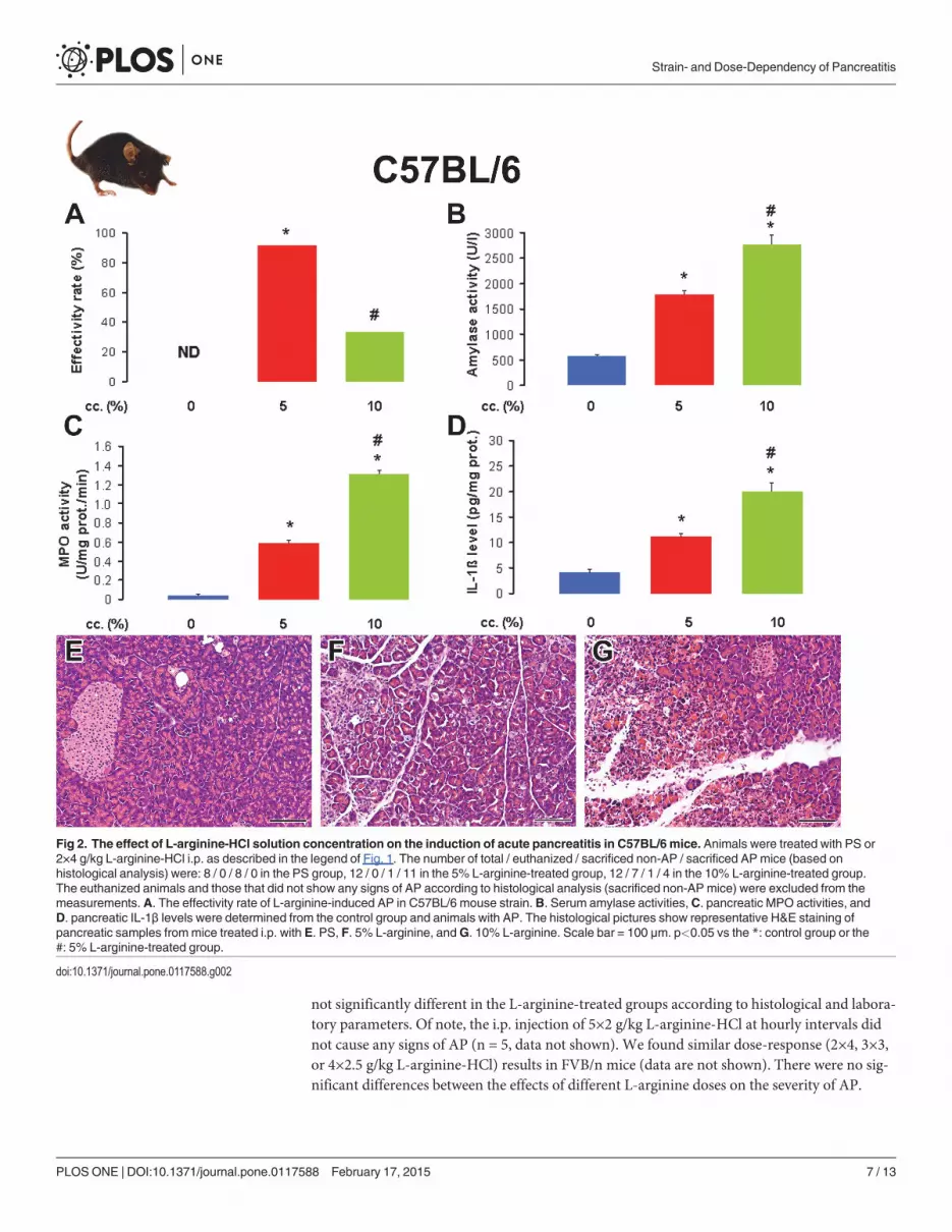
Next, we tested the effects of L-arginine concentration on the C57BL/6 mouse strain. Al-though the effectivity rate of AP induction was 93% in the 5% L-arginine-treated group, it wasonly 33% in the 10% L-arginine-treated group (Fig. 2A). The low effectivity rate in the lattergroup was caused by the high euthanasia rate. Both laboratory and histological parameters con-firmed the presence of AP in the groups treated with 5 and 10% L-arginine vs the physiologicalsaline-treated control group (Fig. 2B-H and Table 2). AP was more severe in the 10% vs 5% L-arginine-treated group.
Strain- and Dose-Dependency of Pancreatitis
PLOS ONE | DOI:10.1371/journal.pone.0117588 February 17, 2015 4 / 13
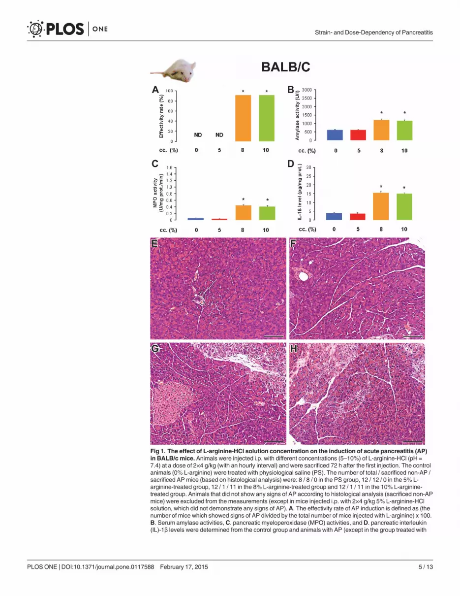
Fig 1. The effect of L-arginine-HCl solution concentration on the induction of acute pancreatitis (AP)in BALB/c mice. Animals were injected i.p. with different concentrations (5–10%) of L-arginine-HCl (pH =7.4) at a dose of 2×4 g/kg (with an hourly interval) and were sacrificed 72 h after the first injection. The controlanimals (0% L-arginine) were treated with physiological saline (PS). The number of total / sacrificed non-AP /sacrificed APmice (based on histological analysis) were: 8 / 8 / 0 in the PS group, 12 / 12 / 0 in the 5% L-arginine-treated group, 12 / 1 / 11 in the 8% L-arginine-treated group and 12 / 1 / 11 in the 10% L-arginine-treated group. Animals that did not show any signs of AP according to histological analysis (sacrificed non-APmice) were excluded from the measurements (except in mice injected i.p. with 2×4 g/kg 5% L-arginine-HClsolution, which did not demonstrate any signs of AP). A. The effectivity rate of AP induction is defined as (thenumber of mice which showed signs of AP divided by the total number of mice injected with L-arginine) x 100.B. Serum amylase activities, C. pancreatic myeloperoxidase (MPO) activities, andD. pancreatic interleukin(IL)-1β levels were determined from the control group and animals with AP (except in the group treated with
Strain- and Dose-Dependency of Pancreatitis
PLOS ONE | DOI:10.1371/journal.pone.0117588 February 17, 2015 5 / 13
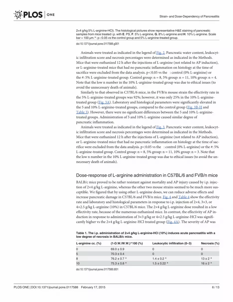
Animals were treated as indicated in the legend of Fig. 2. Pancreatic water content, leukocyt-ic infiltration score and necrosis percentages were determined as indicated in the Methods.Mice that were euthanized 12 h after the injections of L-arginine (not related to AP induction),or L-arginine-treated mice that had no pancreatic inflammation on histology at the time ofsacrifice were excluded from the data analysis. p<0.05 vs the �: control (0% L-arginine) orthe #: 5% L-arginine-treated group. Control group: n = 8, 5% group: n = 11, 10% group: n = 4.Note that the low n number in the 10% L-arginine-treated group was due to ethical issues (toavoid the unnecessary death of animals).
Similarly to that observed in C57BL/6 mice, in the FVB/n mouse strain the effectivity rate inthe 5% L-arginine-treated groups was 92%; however, it was only 25% in the 10% L-arginine-treated group (Fig. 3A). Laboratory and histological parameters were significantly elevated inthe 5 and 10% L-arginine-treated groups, compared to the control group (Fig. 3B-H andTable 3). However, there were no significant differences between the 5 and 10% L-arginine-treated groups. Administration of 5 and 10% L-arginine caused similar degree ofpancreatic inflammation.
Animals were treated as indicated in the legend of Fig. 3. Pancreatic water content, leukocyt-ic infiltration score and necrosis percentages were determined as indicated in the Methods.Mice that were euthanized 12 h after the injections of L-arginine (not related to AP induction),or L-arginine-treated mice that had no pancreatic inflammation on histology at the time of sac-rifice were excluded from the data analysis. p<0.05 vs the �: control (0% L-arginine) or the #: 5%L-arginine-treated group. Control group: n = 8, 5% group: n = 11, 10% group: n = 3. Note thatthe low n number in the 10% L-arginine-treated group was due to ethical issues (to avoid the un-necessary death of animals).
Dose-response of L-arginine administration in C57BL/6 and FVB/n miceBALB/c mice proved to be rather resistant against mortality and AP injury caused by i.p. injec-tion of 2×4 g/kg L-arginine, whereas the other two mouse strains seemed to be much more sus-ceptible. We figured that by using other L-arginine doses, we can reduce adverse effects andincrease pancreatic damage in C57BL/6 and FVB/n mice. Fig. 4 and Table 4 show the effectivityrate and laboratory and histological parameters in response to i.p. injection of 2×4, 3×3, or4×2.5 g/kg L-arginine (10%) in C57BL/6 mice. The 2×4 g/kg L-arginine dose resulted in a loweffectivity rate, because of the numerous euthanized mice. In contrast, the effectivity of AP in-duction in response to administration of 3×3 g/kg or 4×2.5 g/kg L-arginine-HCl was signifi-cantly higher vs the 2×4 g/kg L-arginine-HCl treated group (Fig. 4A). The severity of AP was
2×4 g/kg 5% L-arginine-HCl). The histological pictures show representative H&E staining of pancreaticsamples frommice treated i.p. with E. PS, F. 5% L-arginine,G. 8% L-arginine andH. 10% L-arginine. Scalebar = 100 μm.*: p<0.05 vs the control group and 5% L-arginine-treated group.
doi:10.1371/journal.pone.0117588.g001
Table 1. The i.p. administration of 2×4 g/kg L-arginine-HCl (10%) induces acute pancreatitis with alow degree of necrosis in BALB/c mice.
L-arginine cc. (%) (1-D.W./W.W.)*100 (%) Leukocytic infiltration (0–3) Necrosis (%)
0 69.0 ± 0.9 0 0
5 70.3 ± 0.4 0 0
8 76.2 ± 0.7 * 1.4 ± 0.2 * 13 ± 2 *
10 75.3 ± 0.8 * 1.5 ± 0.22 * 16 ± 2 *
doi:10.1371/journal.pone.0117588.t001
Strain- and Dose-Dependency of Pancreatitis
PLOS ONE | DOI:10.1371/journal.pone.0117588 February 17, 2015 6 / 13
not significantly different in the L-arginine-treated groups according to histological and labora-tory parameters. Of note, the i.p. injection of 5×2 g/kg L-arginine-HCl at hourly intervals didnot cause any signs of AP (n = 5, data not shown). We found similar dose-response (2×4, 3×3,or 4×2.5 g/kg L-arginine-HCl) results in FVB/n mice (data are not shown). There were no sig-nificant differences between the effects of different L-arginine doses on the severity of AP.
Fig 2. The effect of L-arginine-HCl solution concentration on the induction of acute pancreatitis in C57BL/6 mice. Animals were treated with PS or2×4 g/kg L-arginine-HCl i.p. as described in the legend of Fig. 1. The number of total / euthanized / sacrificed non-AP / sacrificed APmice (based onhistological analysis) were: 8 / 0 / 8 / 0 in the PS group, 12 / 0 / 1 / 11 in the 5% L-arginine-treated group, 12 / 7 / 1 / 4 in the 10% L-arginine-treated group.The euthanized animals and those that did not show any signs of AP according to histological analysis (sacrificed non-APmice) were excluded from themeasurements. A. The effectivity rate of L-arginine-induced AP in C57BL/6 mouse strain.B. Serum amylase activities,C. pancreatic MPO activities, andD. pancreatic IL-1β levels were determined from the control group and animals with AP. The histological pictures show representative H&E staining ofpancreatic samples frommice treated i.p. with E. PS, F. 5% L-arginine, andG. 10% L-arginine. Scale bar = 100 μm. p<0.05 vs the *: control group or the#: 5% L-arginine-treated group.
doi:10.1371/journal.pone.0117588.g002
Strain- and Dose-Dependency of Pancreatitis
PLOS ONE | DOI:10.1371/journal.pone.0117588 February 17, 2015 7 / 13
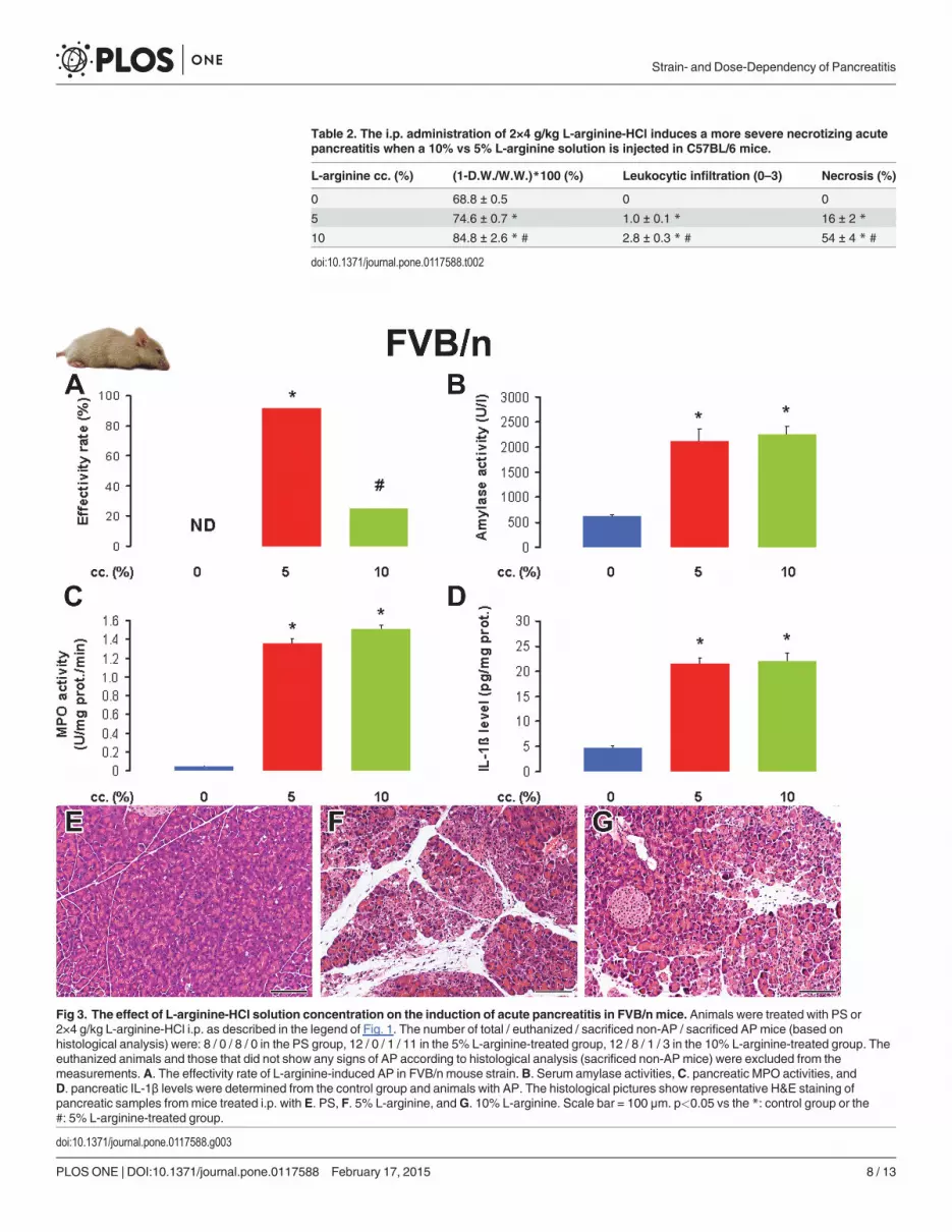
Table 2. The i.p. administration of 2×4 g/kg L-arginine-HCl induces a more severe necrotizing acutepancreatitis when a 10% vs 5% L-arginine solution is injected in C57BL/6 mice.
L-arginine cc. (%) (1-D.W./W.W.)*100 (%) Leukocytic infiltration (0–3) Necrosis (%)
0 68.8 ± 0.5 0 0
5 74.6 ± 0.7 * 1.0 ± 0.1 * 16 ± 2 *
10 84.8 ± 2.6 * # 2.8 ± 0.3 * # 54 ± 4 * #
doi:10.1371/journal.pone.0117588.t002
Fig 3. The effect of L-arginine-HCl solution concentration on the induction of acute pancreatitis in FVB/n mice. Animals were treated with PS or2×4 g/kg L-arginine-HCl i.p. as described in the legend of Fig. 1. The number of total / euthanized / sacrificed non-AP / sacrificed APmice (based onhistological analysis) were: 8 / 0 / 8 / 0 in the PS group, 12 / 0 / 1 / 11 in the 5% L-arginine-treated group, 12 / 8 / 1 / 3 in the 10% L-arginine-treated group. Theeuthanized animals and those that did not show any signs of AP according to histological analysis (sacrificed non-APmice) were excluded from themeasurements. A. The effectivity rate of L-arginine-induced AP in FVB/n mouse strain.B. Serum amylase activities,C. pancreatic MPO activities, andD. pancreatic IL-1β levels were determined from the control group and animals with AP. The histological pictures show representative H&E staining ofpancreatic samples frommice treated i.p. with E. PS, F. 5% L-arginine, andG. 10% L-arginine. Scale bar = 100 μm. p<0.05 vs the *: control group or the#: 5% L-arginine-treated group.
doi:10.1371/journal.pone.0117588.g003
Strain- and Dose-Dependency of Pancreatitis
PLOS ONE | DOI:10.1371/journal.pone.0117588 February 17, 2015 8 / 13
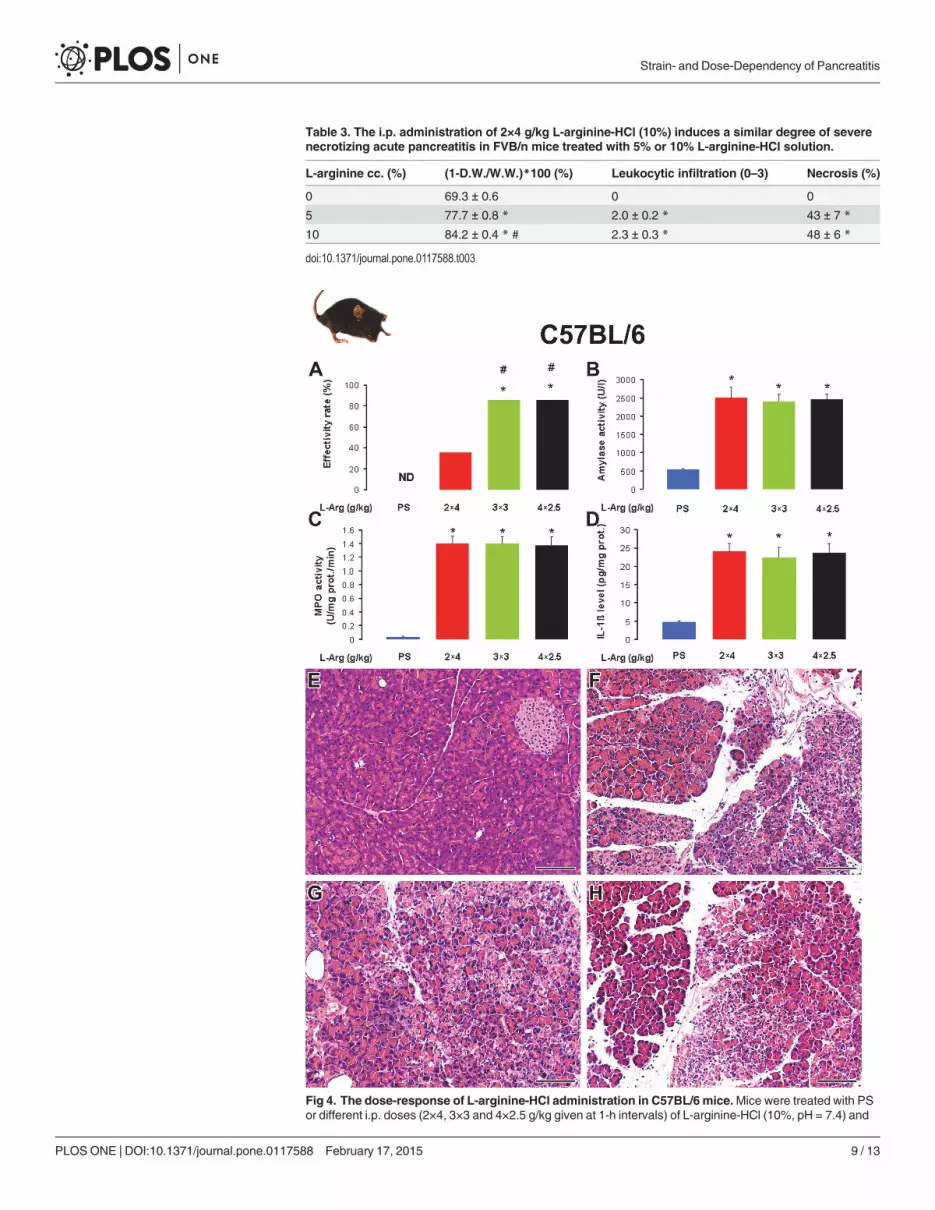
Table 3. The i.p. administration of 2×4 g/kg L-arginine-HCl (10%) induces a similar degree of severenecrotizing acute pancreatitis in FVB/n mice treated with 5% or 10% L-arginine-HCl solution.
L-arginine cc. (%) (1-D.W./W.W.)*100 (%) Leukocytic infiltration (0–3) Necrosis (%)
0 69.3 ± 0.6 0 0
5 77.7 ± 0.8 * 2.0 ± 0.2 * 43 ± 7 *
10 84.2 ± 0.4 * # 2.3 ± 0.3 * 48 ± 6 *
doi:10.1371/journal.pone.0117588.t003
Fig 4. The dose-response of L-arginine-HCl administration in C57BL/6 mice.Mice were treated with PSor different i.p. doses (2×4, 3×3 and 4×2.5 g/kg given at 1-h intervals) of L-arginine-HCl (10%, pH = 7.4) and
Strain- and Dose-Dependency of Pancreatitis
PLOS ONE | DOI:10.1371/journal.pone.0117588 February 17, 2015 9 / 13
Mice were treated as indicated in the legend of Fig. 4. Pancreatic water content, leukocyticinfiltration score and necrosis percentages were determined as indicated in the Methods. Micethat were euthanized 12 h after the injections with L-arginine, or L-arginine-treated mice thathad no pancreatic inflammation on histology at the time of sacrifice were excluded from thedata analysis. �: p<0.05 vs the control (0 g/kg L-arginine). Control group: n = 10, 2×4 g/kg L-arginine-treated group: n = 5, 3×3 g/kg L-arginine-treated group: n = 12, 4×2.5 g/kg L-arginine-treated group: n = 12.
The severity of L-arginine-induced acute pancreatitis is similar in maleand female miceAs some AP models show gender dependency (e.g. female mice are more susceptible to CDEdiet-induced AP), we wanted to test the effects of L-arginine administration in male and femalemice. C57BL/6 mice were subjected to hourly i.p. injections of 3×3 g/kg 10% L-arginine-HCl.The effectivity rate of AP induction was 87.5% in male (n = 8) and 100% in female (n = 8)mice, which was not significantly different. The laboratory and histological parameters weresimilar to that shown in Fig. 2B-G.
DiscussionAn important finding of our experiments is it that different mouse strains have varying sensi-tivities (resulting in AP, no pancreatic damage, or death) to administration of L-arginine-HCl(pH = 7.4) (Table 5.). In FVB/n and C57BL/6 mice, which are commonly used strains for gen-erating transgenic animals, the originally published i.p. 2×4 g/kg L-arginine-HCl dose may notbe appropriate and the use of other doses (like 3×3 g/kg or 4×2.5 g/kg) may be better for the in-duction of AP. Furthermore, the concentration of the administered L-arginine-HCl solutionmakes a huge difference in whether the animals survive the treatment. High L-arginine concen-trations like 30%, which are well tolerated by rats actually kill mice, thus lower (5–10%) con-centrations should be injected even if this means considerably more fluid volume.Interestingly, we could not detect any differences in AP severity of male and female mice.
were sacrificed 72 h after the first injection. The number of all total / euthanized / sacrificed non-AP / sacrificedAPmice (based on histological analysis) were: 10 / 0 / 10 / 0 in the PS group, 14 / 8 / 1 / 5 in the 2×4 g/kg L-arginine-treated group, 14 / 1 / 1 / 12 in the 3×3 g/kg L-arginine-treated group, 14 / 0 / 2 / 12 in the 4×2.5 g/kgL-arginine-treated group. The euthanized animals and those that did not show any signs of AP according tohistological analysis (sacrificed non-APmice) were excluded from the measurements. A. The effectivity rateof L-arginine-induced AP C57BL/6 mouse strain.B. Serum amylase activities,C. pancreatic MPO activities,andD. pancreatic IL-1β levels were determined from the control group and animals with AP. RepresentativeH&E staining of pancreatic samples frommice treated i.p. with E. PS, F. 2×4 g/kg L-arginine-HCl,G. 3×3 g/kgL-arginine-HCl andH. 4×2.5 g/kg L-arginine-HCl. Scale bar = 100 μm. p<0.05 vs the *: control group or the#: 2×4 g/kg L-arginine-treated group. Note that significance was borderline (p = 0.053) in case of PS- vs2×4 g/kg L-arginine-treated groups.
doi:10.1371/journal.pone.0117588.g004
Table 4. Acute pancreatitis severity in response to the i.p administration of 2×4, 3×3 or 4×2.5 g/kg L-arginine-HCl (10%) is similar in C57BL/6 mice.
L-arginine dose (g/kg) (1-D.W./W.W.)*100 (%) Leukocytic infiltration (0–3) Necrosis (%)
0 67.7 ± 1.1 0 0
2x4 82.8 ± 2.2 * 2.6 ± 0.3 * 60 ± 12 *
3x3 81.4 ± 0.9 * 2.3 ± 0.2 * 52 ± 6 *
4x2.5 80.8 ± 0.8 * 2.3 ± 0.2 * 50 ± 5 *
doi:10.1371/journal.pone.0117588.t004
Strain- and Dose-Dependency of Pancreatitis
PLOS ONE | DOI:10.1371/journal.pone.0117588 February 17, 2015 10 / 13
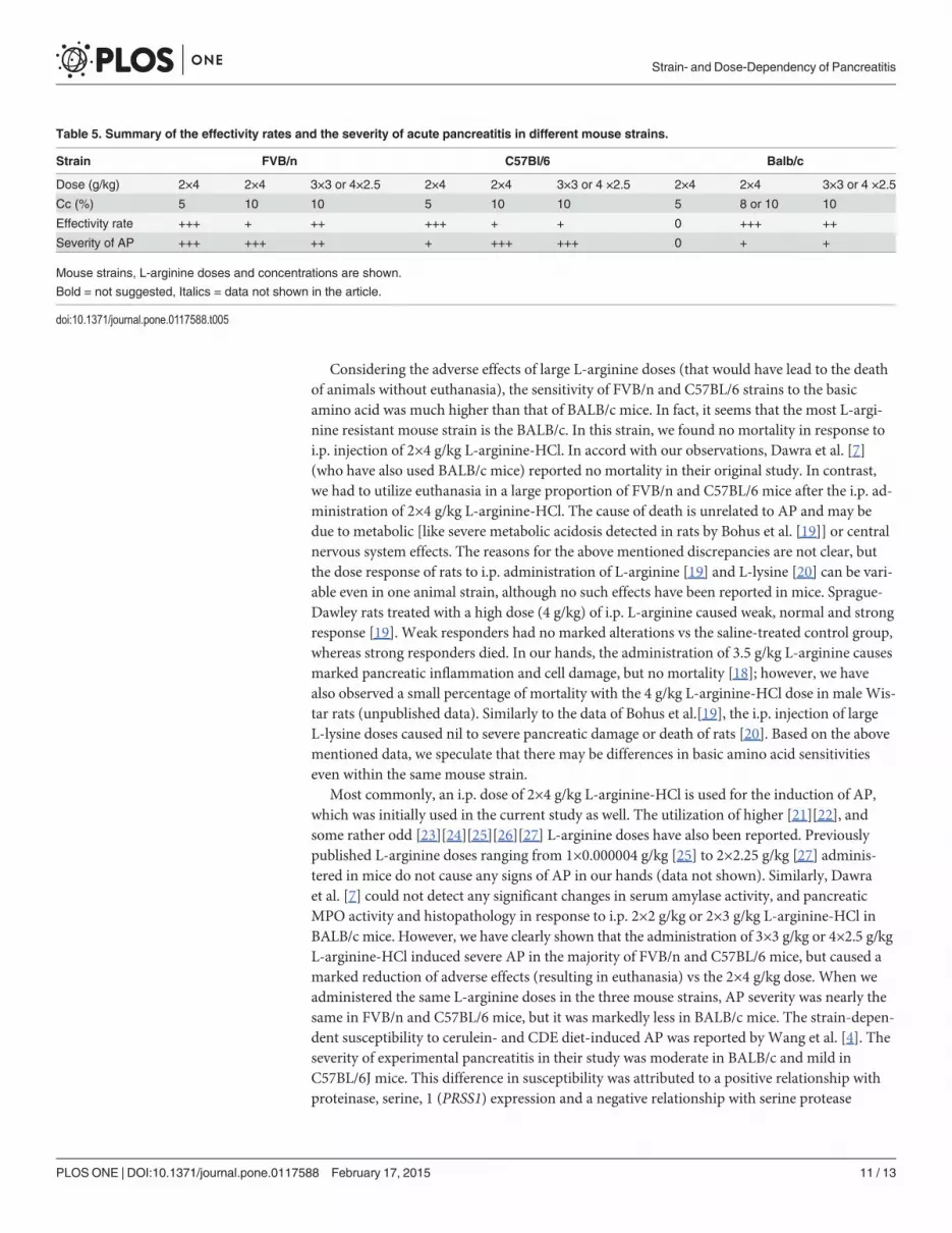
Considering the adverse effects of large L-arginine doses (that would have lead to the deathof animals without euthanasia), the sensitivity of FVB/n and C57BL/6 strains to the basicamino acid was much higher than that of BALB/c mice. In fact, it seems that the most L-argi-nine resistant mouse strain is the BALB/c. In this strain, we found no mortality in response toi.p. injection of 2×4 g/kg L-arginine-HCl. In accord with our observations, Dawra et al. [7](who have also used BALB/c mice) reported no mortality in their original study. In contrast,we had to utilize euthanasia in a large proportion of FVB/n and C57BL/6 mice after the i.p. ad-ministration of 2×4 g/kg L-arginine-HCl. The cause of death is unrelated to AP and may bedue to metabolic [like severe metabolic acidosis detected in rats by Bohus et al. [19]] or centralnervous system effects. The reasons for the above mentioned discrepancies are not clear, butthe dose response of rats to i.p. administration of L-arginine [19] and L-lysine [20] can be vari-able even in one animal strain, although no such effects have been reported in mice. Sprague-Dawley rats treated with a high dose (4 g/kg) of i.p. L-arginine caused weak, normal and strongresponse [19]. Weak responders had no marked alterations vs the saline-treated control group,whereas strong responders died. In our hands, the administration of 3.5 g/kg L-arginine causesmarked pancreatic inflammation and cell damage, but no mortality [18]; however, we havealso observed a small percentage of mortality with the 4 g/kg L-arginine-HCl dose in male Wis-tar rats (unpublished data). Similarly to the data of Bohus et al.[19], the i.p. injection of largeL-lysine doses caused nil to severe pancreatic damage or death of rats [20]. Based on the abovementioned data, we speculate that there may be differences in basic amino acid sensitivitieseven within the same mouse strain.
Most commonly, an i.p. dose of 2×4 g/kg L-arginine-HCl is used for the induction of AP,which was initially used in the current study as well. The utilization of higher [21][22], andsome rather odd [23][24][25][26][27] L-arginine doses have also been reported. Previouslypublished L-arginine doses ranging from 1×0.000004 g/kg [25] to 2×2.25 g/kg [27] adminis-tered in mice do not cause any signs of AP in our hands (data not shown). Similarly, Dawraet al. [7] could not detect any significant changes in serum amylase activity, and pancreaticMPO activity and histopathology in response to i.p. 2×2 g/kg or 2×3 g/kg L-arginine-HCl inBALB/c mice. However, we have clearly shown that the administration of 3×3 g/kg or 4×2.5 g/kgL-arginine-HCl induced severe AP in the majority of FVB/n and C57BL/6 mice, but caused amarked reduction of adverse effects (resulting in euthanasia) vs the 2×4 g/kg dose. When weadministered the same L-arginine doses in the three mouse strains, AP severity was nearly thesame in FVB/n and C57BL/6 mice, but it was markedly less in BALB/c mice. The strain-depen-dent susceptibility to cerulein- and CDE diet-induced AP was reported by Wang et al. [4]. Theseverity of experimental pancreatitis in their study was moderate in BALB/c and mild inC57BL/6J mice. This difference in susceptibility was attributed to a positive relationship withproteinase, serine, 1 (PRSS1) expression and a negative relationship with serine protease
Table 5. Summary of the effectivity rates and the severity of acute pancreatitis in different mouse strains.
Strain FVB/n C57Bl/6 Balb/c
Dose (g/kg) 2×4 2×4 3×3 or 4×2.5 2×4 2×4 3×3 or 4 ×2.5 2×4 2×4 3×3 or 4 ×2.5
Cc (%) 5 10 10 5 10 10 5 8 or 10 10
Effectivity rate +++ + ++ +++ + + 0 +++ ++
Severity of AP +++ +++ ++ + +++ +++ 0 + +
Mouse strains, L-arginine doses and concentrations are shown.
Bold = not suggested, Italics = data not shown in the article.
doi:10.1371/journal.pone.0117588.t005
Strain- and Dose-Dependency of Pancreatitis
PLOS ONE | DOI:10.1371/journal.pone.0117588 February 17, 2015 11 / 13
inhibitor, Kazal type 3 (SPINK3) expression. In L-arginine-induced AP, this may not be thecase since disease severity was significantly higher in C57BL/6 vs BALB/c mice.
The effects of the estrous cycle on AP development have always been a concern of research-ers, thus to exclude such potential effects, usually male animals are used. In fact, it has longbeen known that in case of the CDE diet model, the effects are gender specific, female mice aremuch more sensitive to treatment [28]. Therefore, we also tested the potential gender-specificeffects of L-arginine treatment, but we found no significant differences in AP severity of malevs female C57BL/6 mice.
Taken together, it is evident that setting up the L-arginine-induced AP model in mice isquite challenging. Overall, it seems that the borderline between the effective (AP inducing) andlethal doses of L-arginine is much thinner in mice vs rats. There are several important factorsthat need to be considered such as the concentration and dose of the injected L-arginine-HClsolution and also the strain of mice. The proper dosing of L-arginine to induce AP should betested by each laboratory and in each mouse strain. The reasons for the differences in L-argi-nine sensitivity of mice remain to be investigated.
AcknowledgmentsThe authors would like to thank Mihály Dezső (Department of Pathology) and Máté Katona(First Department of Medicine and Department of Pharmacology and Pharmacotherapy) fortheir help with picture editing.
Author ContributionsConceived and designed the experiments: ZR TT PH. Performed the experiments: BK ZB ESKBI BV ETV PP VV. Analyzed the data: BK ZB ESK. Contributed reagents/materials/analysistools: ZR TT PH. Wrote the paper: ZR BK ZB ESK.
References1. Pandol SJ, Saluja AK, Imrie CW, Banks PA (2007) Acute pancreatitis: bench to the bedside.
Gastroenterology 1127–51.
2. Hegyi P, Perides G, Steer ML, Rakonczay Z Jr (2013) Commonly Employed Rodent Models of Experi-mental Acute Pancreatitis: Their Strenghts andWeakness, Relevance to Human Disease, Selection,and Appropriate Use. The Pancreapedia: Exocrine Pancreas Knowledge Base. Available: http://www.pancreapedia.org/reviews/commonly-employed-rodent-models-of-experimental-acutepancreatitis-their-strengths-and-weakn
3. Su KH, Cuthbertson C, Christophi C (2006) Review of experimental animal models of acute pancreati-tis. HPB 8:264–86. doi: 10.1080/13651820500467358 PMID: 18333137
4. Wang J, Ohmuraya M, Suyama K, Hirota M, Ozaki N, et al. (2010) Relationship of strain-dependentsusceptibility to experimentally induced acute pancreatitis with regulation of Prss1 and Spink3 expres-sion. Lab Invest 90:654–64. doi: 10.1038/labinvest.2010.44 PMID: 20157294
5. Mizunuma T, Kawamura S, Kishino Y (1984) Effects of injecting excess arginine on rat pancreas.J Nutr 114:467–71.
6. Tani S, Itoh H, Okabayashi Y, Nakamura T, Fuji M, et al. (1990) Newmodel of acute necrotizing pancre-atitis induced by excessive doses of arginine in rats. Dig Dis Sci 35:367–74. PMID: 2307082
7. Dawra R, Sharif R, Phillips P, Dudeja V, Dhaulakhandi D, et al. (2007) Development of a newmousemodel of acute pancreatitis induced by administration of L-arginine. Am J Physiol Gastrointest LiverPhysiol 292:G1009–1018. PMID: 17170029
8. Hegyi P, Rakonczay Z Jr, Sári R, Góg C, Lonovics J, et al. (2004) L-arginine-induced experimental pan-creatitis.World J Gastroenterol 10:2003–9.
9. Kui B, Végh ET, Pallagi P, Venglovecz V, Iványi B, et al. (2014) Recent advances in the investigation ofpancreatic inflammation induced by large doses of basic amino acids in rodents. Lab Invest 138–49.
10. Dawra R, Saluja AK (2012) L-arginine-induced experimental acute pancreatitis. The Pancreapedia:Exocrine Pancreas Knowledge Base. Available: http://www.pancreapedia.org/?q = node/1394
Strain- and Dose-Dependency of Pancreatitis
PLOS ONE | DOI:10.1371/journal.pone.0117588 February 17, 2015 12 / 13
11. Shen J, Wan R, Hu G, Wang F, Shen J, et al. (2012) Involvement of thrombopoietin in acinar cell necro-sis in L-arginine-induced acute pancreatitis in mice. Cytokine 60:294–301.
12. Sharif R, Dawra R,Wasiluk K, Phillips P, Dudeja V, et al. (2009) Impact of toll-like receptor 4 on the sever-ity of acute pancreatitis and pancreatitis-associated lung injury in mice.Gut 58:813–9. doi: 10.1136/gut.2008.170423 PMID: 19201771
13. Hu G, Shen J, Cheng L, Guo C, Xu X, et al. (2011) Reg4 protects against acinar cell necrosis in experi-mental pancreatitis.Gut 60:820–8.
14. Toth LA, Rehg JE, Webster RG (1995) Strain differences in sleep and other pathophysiological sequel-ae of influenza virus infection in naive and immunized mice. J. Neuroimmunol. 58:89–99.
15. Toth LA (2000) Defining the moribund condition as an experimental endpoint for animal research.(2000) ILAR J. Natl. Res. Counc. Inst. Lab. Anim. Resour. 41:72–79.
16. Kuebler W, Abels C, Schuerer L, Goetz AE (1996) Measurement of neutrophil content in brain and lungtissue by a modified myeloperoxidase assay. Int J Microcirc Clin Exp 89–97.
17. Lowry OH, Rosebrough NJ, Farr AL, Randall RJ (1951) Protein Measurement with the Folin Phenol Re-agent. J Biol Chem 265–75.
18. Rakonczay Z Jr, Jármay K, Kaszaki J, Mándi Y, Duda E, et al. (2003) NF-kappaB activation is detrimen-tal in arginine-induced acute pancreatitis. Free Radic Biol Med 34:696–709. PMID: 12633747
19. Bohus E, Coen M, Keun HC, Ebbels TM, Beckonert O, et al. (2008) Temporal metabonomic modelingof l-arginine-induced exocrine pancreatitis. J Proteome Res 4435–45.
20. Biczó G, Hegyi P, Dósa S, Shalbuyeva N, Berczi S, et al. (2011) The crucial role of early mitochondrialinjury in L-lysine-induced acute pancreatitis. Antioxid Redox Signal 15:2669–81. doi: 10.1089/ars.2011.4065 PMID: 21644850
21. Lunova M, Zizer E, Kucukoglu O, Schwarz C, DillmannWH, et al. (2012) Hsp72 overexpression accel-erates the recovery from caerulein-induced pancreatitis. PloS One 7:e39972. doi: 10.1371/journal.pone.0039972 PMID: 22792201
22. Sakai A, Nishiumi S, Shiomi Y, Kobayashi T, Izumi Y, et al. (2012) Metabolomic analysis to discovercandidate therapeutic agents against acute pancreatitis. Arch Biochem Biophys 522:107–20. doi: 10.1016/j.abb.2012.03.025 PMID: 22483684
23. Bedrosian AS, Nguyen AH, HackmanM, Conolly MK, Malhotra A, et al. (2011) Dendritic cells promotepancreatic viability in mice with acute pancreatitis.Gastroenterology 141:1915–1926.e1–14.
24. Cui HF, Bai ZL (2003) Protective effects of transplanted and mobilized bone marrow stem cells on micewith severe acute pancreatitis.World J Gastroenterol 9:2274–7. PMID: 14562392
25. Huang L, Wang MH, Cheng ZY, Xue P, Yin T, et al. (2012) Effects of Chai-Qin-Cheng-Qi decoction onacute pancreatitis-associated lung injury in mice with acute necrotizing pancreatitis. Chin J Integr Med2012. Sep. 21; e-pub ahead of print.
26. Singh VP, Bhagat L, Navina S, Sharif R, Dawra RK, et al. (2007) Protease-activated receptor-2 protectsagainst pancreatitis by stimulating exocrine secretion.Gut 56:958–64. PMID: 17114298
27. Yuan H, Jin X, Sun J, Li F, Feng Q, et al. (2009) Protective effect of HMGB1 a box on organ injury ofacute pancreatitis in mice. Pancreas 38:143–8. doi: 10.1097/MPA.0b013e31818166b4 PMID:18665013
28. Lombardi B, Estes LW, Longnecker DS (1975) Acute hemorrhagic pancreatitis (massive necrosis) withfat necrosis induced in mice by DL-ethionine fed with a choline-deficient diet. Am J Pathol 79:465–80.PMID: 1094837
Strain- and Dose-Dependency of Pancreatitis
PLOS ONE | DOI:10.1371/journal.pone.0117588 February 17, 2015 13 / 13
Copyright © 2022 FDOKUMEN