Dendritic Cells Promote Pancreatic Viability in Mice With Acute Pancreatitis
18
Dendritic Cells Promote Pancreatic Viability in Mice with Acute Pancreatitis Andrea S. Bedrosian 1,* , Andrew H. Nguyen 2,* , Michael Hackman 1 , Michael K. Connolly 1 , Ashim Malhotra 1 , Junaid Ibrahim 1 , Napoleon E. Cieza-Rubio 1 , Justin R. Henning 1 , Rocky Barilla 1 , Adeel Rehman 1 , H. Leon Pachter 1 , Marco V. Medina-Zea 1 , Steven M. Cohen 1 , Alan B. Frey 2 , Devrim Acehan 2 , and George Miller 1,2 1 Department of Surgery, New York University School of Medicine, 550 First Avenue, New York, NY 10016 2 Department of Cell Biology, New York University School of Medicine, 550 First Avenue, New York, NY 10016 Abstract Background & Aims—Acute pancreatitis increases morbidity and mortality from organ necrosis by mechanisms that are incompletely understood. Dendritic cells (DCs) can promote or suppress inflammation, depending on their subtype and context. We investigated the roles of DC in development of acute pancreatitis. Methods—Acute pancreatitis was induced in CD11c.DTR mice using caerulein or L-arginine; DCs were depleted by administration of diphtheria toxin. Survival was analyzed using Kaplan- Meier analysis. Results—Numbers of MHC II + CD11c + DC increased 100-fold in pancreas of mice with acute pancreatitis, to account for nearly 15% of intra-pancreatic leukocytes. Intra-pancreatic DC acquired an immune phenotype in mice with acute pancreatitis; they expressed higher levels of MHC II and CD86 and increased production of interleukin-6, membrane cofactor protein (MCP)-1, and tumor necrosis factor (TNF)-α. However, rather than inducing an organ-destructive inflammatory process, DC were required for pancreatic viability; the exocrine pancreas died in mice that were depleted of DC and challenged with caerulein or L-arginine. All mice with pancreatitis that were depleted of DC died from acinar cell death within 4 days. Depletion of DC © 2011 The American Gastroenterological Association. Published by Elsevier Inc. All rights reserved. Address correspondence to: George Miller, MD, Departments of Surgery and Cell Biology, New York University School of Medicine, Medical Science Building 601, 550 First Avenue, New York, NY 10016, Tel: (212) 263-1479, Fax: (212) 263-6840, [email protected]. * ASB and AHN contributed equally to this work Disclosures: None Writing Assistance: None Author Contributions: Andrea S. Bedrosian (acquisition of data, drafting of manuscript), Andrew H. Nguyen (acquisition of data, analysis and interpretation of data, drafting of manuscript), Michael Hackman (acquisition of data), Michael K. Connolly (acquisition of data), Ashim Malhotra (acquisition of data, critical revision), Napoleon E. Cieza-Rubio (acquisition of data), Justin R. Henning (acquisition of data, critical revision), Junaid Ibrahim (acquisition of data), Rocky Barilla (acquisition of data), Adeel Rehman (acquisition of data), H. Leon Pachter (critical revision), Marco V. Medina-Zea (immunoblotting), Steven M. Cohen (critical revision), Alan B. Frey (critical revision; study design), Devrim Acehan (immunoblotting), George Miller (study concept and design, analysis and interpretation of data, drafting of manuscript) Publisher's Disclaimer: This is a PDF file of an unedited manuscript that has been accepted for publication. As a service to our customers we are providing this early version of the manuscript. The manuscript will undergo copyediting, typesetting, and review of the resulting proof before it is published in its final citable form. Please note that during the production process errors may be discovered which could affect the content, and all legal disclaimers that apply to the journal pertain. NIH Public Access Author Manuscript Gastroenterology. Author manuscript; available in PMC 2012 November 1. Published in final edited form as: Gastroenterology. 2011 November ; 141(5): 1915–1926.e14. doi:10.1053/j.gastro.2011.07.033. NIH-PA Author Manuscript NIH-PA Author Manuscript NIH-PA Author Manuscript
Transcript of Dendritic Cells Promote Pancreatic Viability in Mice With Acute Pancreatitis

Dendritic Cells Promote Pancreatic Viability in Mice with AcutePancreatitis
Andrea S. Bedrosian1,*, Andrew H. Nguyen2,*, Michael Hackman1, Michael K. Connolly1,Ashim Malhotra1, Junaid Ibrahim1, Napoleon E. Cieza-Rubio1, Justin R. Henning1, RockyBarilla1, Adeel Rehman1, H. Leon Pachter1, Marco V. Medina-Zea1, Steven M. Cohen1, AlanB. Frey2, Devrim Acehan2, and George Miller1,2
1Department of Surgery, New York University School of Medicine, 550 First Avenue, New York,NY 100162Department of Cell Biology, New York University School of Medicine, 550 First Avenue, NewYork, NY 10016
AbstractBackground & Aims—Acute pancreatitis increases morbidity and mortality from organnecrosis by mechanisms that are incompletely understood. Dendritic cells (DCs) can promote orsuppress inflammation, depending on their subtype and context. We investigated the roles of DCin development of acute pancreatitis.
Methods—Acute pancreatitis was induced in CD11c.DTR mice using caerulein or L-arginine;DCs were depleted by administration of diphtheria toxin. Survival was analyzed using Kaplan-Meier analysis.
Results—Numbers of MHC II+CD11c+DC increased 100-fold in pancreas of mice with acutepancreatitis, to account for nearly 15% of intra-pancreatic leukocytes. Intra-pancreatic DCacquired an immune phenotype in mice with acute pancreatitis; they expressed higher levels ofMHC II and CD86 and increased production of interleukin-6, membrane cofactor protein(MCP)-1, and tumor necrosis factor (TNF)-α. However, rather than inducing an organ-destructiveinflammatory process, DC were required for pancreatic viability; the exocrine pancreas died inmice that were depleted of DC and challenged with caerulein or L-arginine. All mice withpancreatitis that were depleted of DC died from acinar cell death within 4 days. Depletion of DC
© 2011 The American Gastroenterological Association. Published by Elsevier Inc. All rights reserved.Address correspondence to: George Miller, MD, Departments of Surgery and Cell Biology, New York University School of Medicine,Medical Science Building 601, 550 First Avenue, New York, NY 10016, Tel: (212) 263-1479, Fax: (212) 263-6840,[email protected].*ASB and AHN contributed equally to this workDisclosures: NoneWriting Assistance: NoneAuthor Contributions: Andrea S. Bedrosian (acquisition of data, drafting of manuscript), Andrew H. Nguyen (acquisition of data,analysis and interpretation of data, drafting of manuscript), Michael Hackman (acquisition of data), Michael K. Connolly (acquisitionof data), Ashim Malhotra (acquisition of data, critical revision), Napoleon E. Cieza-Rubio (acquisition of data), Justin R. Henning(acquisition of data, critical revision), Junaid Ibrahim (acquisition of data), Rocky Barilla (acquisition of data), Adeel Rehman(acquisition of data), H. Leon Pachter (critical revision), Marco V. Medina-Zea (immunoblotting), Steven M. Cohen (critical revision),Alan B. Frey (critical revision; study design), Devrim Acehan (immunoblotting), George Miller (study concept and design, analysisand interpretation of data, drafting of manuscript)Publisher's Disclaimer: This is a PDF file of an unedited manuscript that has been accepted for publication. As a service to ourcustomers we are providing this early version of the manuscript. The manuscript will undergo copyediting, typesetting, and review ofthe resulting proof before it is published in its final citable form. Please note that during the production process errors may bediscovered which could affect the content, and all legal disclaimers that apply to the journal pertain.
NIH Public AccessAuthor ManuscriptGastroenterology. Author manuscript; available in PMC 2012 November 1.
Published in final edited form as:Gastroenterology. 2011 November ; 141(5): 1915–1926.e14. doi:10.1053/j.gastro.2011.07.033.
NIH
-PA Author Manuscript
NIH
-PA Author Manuscript
NIH
-PA Author Manuscript
from mice with pancreatitis resulted in neutrophil infiltration and increased levels of systemicmarkers of inflammation. However, the organ necrosis associated with depletion of DC did notrequire infiltrating neutrophils, activation of NF-κB, or signaling by mitogen-activated proteinkinase or TNF-α.
Conclusions—DC are required for pancreatic viability in mice with acute pancreatitis and mightprotect organs against cell stress.
Keywordsimmune response; regulation; suppression; MCP-1; IL-6
IntroductionAcute pancreatitis has a considerable medical and economic impact. In the United States,this disease results in over 200,000 hospital admissions annually at a cost of over 2 billiondollars (1). While most patients will have an uncomplicated course, 10-20% of cases arecategorized as severe, resulting from organ necrosis, and are often associated with localizedinfection, systemic sepsis, and multi-organ failure. Acute pancreatitis carries a 2% overallmortality rate which increases to 10-30% in cases of pancreatic necrosis (2). These datademand an enhanced understanding of the pathophysiology of acute pancreatitis in order toimplement more effective treatments.
Acute pancreatitis usually results from either alcohol abuse or obstructing gallstones, whichare postulated to cause intra-acinar activation of proteolytic enzymes leading to intensepancreatic inflammation. The inflammatory mediators in this disease have been partiallydelineated in animal models mimicking human pancreatitis (3, 4). Massive leukocyteinfiltration is one of the early events of pancreatitis, and neutrophils play a distinctlyimportant role in its pathophysiology (5). Neutrophil recruitment worsens pancreatic injury(6) and, conversely, neutrophil depletion lessens tissue injury associated with acutepancreatitis (7). Similarly, CD4+ T cells are required for pancreatic acinar injury in rodentmodels of acute pancreatitis (8). Importantly, Gukovsky et al. (9) demonstrated that thenuclear transcription factor NF-κB, a key regulator of inflammation, is activated early inpancreatitis. Furthermore, NF-κB has recently been shown to regulate expression ofActivator Protein 1 which modulates the chemokine cascade after pancreatic insult (10). TheCXC-ELR family of chemoattractants, including CXCL1 and CXCL2, further regulate theinflux of inflammatory cells in acute pancreatitis (11).
Dendritic cells (DC) have emerged as important participants in inflammation. As instructorsof the T cell response, DC are the most powerful antigen-presenting cell in the immunesystem. Upon exposure to bacterial or viral infection or inflammatory stimuli, immature DCbecome activated and drive both adaptive and innate immune responses (12). Patternrecognition receptors on the DC surface, the most well-understood being toll-like receptors(TLRs), recognize pathogen-associated molecular patterns (PAMPs) on foreign antigens aswell as damage-associated molecular patterns (DAMPs) released by cellular injury (13).Immature DC become activated to maturity by the binding of PAMPs or DAMPs which maybe released secondary to tissue damage, or by cytokines generated during the inflammatoryresponse. Mature DC process and present antigen to T cells via MHC complexes. Thisclassical model dictates very tightly regulated pathways to DC activation, as mature DC arecentral to the development of inflammation and effective immunogenic responses.
Recent work by our group and others has shown a central role for DC in a number of organ-specific inflammatory diseases. We reported that DC tightly regulate the extent ofintrahepatic inflammation after liver insult from hepatotoxins via their production of TNF-α
Bedrosian et al. Page 2
Gastroenterology. Author manuscript; available in PMC 2012 November 1.
NIH
-PA Author Manuscript
NIH
-PA Author Manuscript
NIH
-PA Author Manuscript
(14). Bamboat et al. (15) also reported that DC modulate the extent of liver injury afterischemic insult via their production of IL-10. DC have also been assigned a central role inmodulating the severity of systemic sepsis (16, 17). Our preliminary investigations in thecurrent study revealed a marked increase in the numbers of intra-pancreatic DC in acutepancreatitis. Based on this, we postulated an important role for DC in modulating intra-pancreatic inflammation and thus determining the extent of endocrine and exocrinepancreatic injury.
Materials and MethodsAnimals
Male C57BL/6 (H-2Kb) and CD45.1 (B6.SJL-Ptprca/BoyAiTac) mice (6–8 weeks old) werepurchased from Taconic Farms (Germantown, NY). CD11b.DTR (18) and CD11c.DTR (19)mice were purchased from Jackson Laboratory (Bar Harbor, Maine). Mice were housed in apathogen-free environment and fed standard chow. Mouse serum was collected by retro-orbital bleeding. To effect macrophage or DC depletion, respectively, CD11b.DTR andCD11c.DTR mice were treated with a single intraperitoneal (i.p.) dose of diphtheria toxin(DPT; 4 ng/g; Sigma-Aldrich, Saint Louis, MO). Animal procedures were approved by theNew York University School of Medicine Institutional Animal Care and Use Committee.
Models of Acute Pancreatitis and In Vivo ExperimentsPancreatitis was induced using a regimen of seven hourly i.p. injections of caerulein (50 μg/kg; Sigma-Aldrich) for two consecutive days before sacrifice 12 hours later unless otherwisespecified. Alternatively, i.p. administration of two doses of L-arginine (40 mg/kg; Sigma-Aldrich) at hourly intervals was used to induce pancreatic injury (3, 4). In selectedexperiments, bone marrow chimeric animals were created by irradiating C57BL/6 mice (100Gy) before i.v. bone marrow transfer (1×10⁷) from CD45.1 or CD11c.DTR donors. In vivoTNF-α blockade was accomplished using MP6-XT22 (200 μg/day). IL-6 blockade wasaccomplished using MP5-20F3 (200 μg/day). MIP-1α blockade was accomplished usingclone 39624 (300 μg/day; all R&D Systems, Minneapolis, MN). NF-κB blockade wasaccomplished using both cell permeable inhibitors of the NF-κB p50 domain which preventsits nuclear translocation (NF-κB SN50) and the NEMO binding domain (NBD) inhibitor(both 1mg/kg/day) which prevents binding of NEMO to the IKK (IκB) – kinase complex(both EMD4Biosciences, Gibbstown, NJ). MAP Kinase (MAPK) blockade wasaccomplished using PD98059 (2.5mg/kg/day; Invivogen). To deplete Gr1+ cells or CD4+ Tcells, RB6-8C5 or GK1.5 (Monoclonal Antibody Core Facility, Sloan-Kettering Institute,New York, NY) were employed, respectively, as described (20, 21). In vivo plasmacytoidDC depletion was accomplished using either anti-mPDCA-1 (500 μg; Miltenyi, BergischGladbach, Germany) or 120G8 (200 μg; Imgenex, San Diego, CA) (22, 23). Serum levels ofpancreatic enzymes and glucose were measured using the Olympus AU400 ChemistryAnalyzer (Center Valley, PA).
StatisticsData is presented as mean +/- standard error of mean. Survival was measured according tothe Kaplan-Meier method. Statistical significance was determined by the Student's t test andor log-rank test using GraphPad Prism 5 (GraphPad Software, La Jolla, CA). P-values <0.05 were considered significant.
See additional Supplemental Materials and Methods
Bedrosian et al. Page 3
Gastroenterology. Author manuscript; available in PMC 2012 November 1.
NIH
-PA Author Manuscript
NIH
-PA Author Manuscript
NIH
-PA Author Manuscript
ResultsDC expand in acute pancreatitis
To assess the significance of DC in acute pancreatitis, we first tested whether the intra-pancreatic MHC II+CD11c+ DC population expands after pancreatic insult from 14caerulein injections over a 36-hour period. We found that while DC were rare in the normalpancreas, the total number of intra-pancreatic DC increased markedly in acute pancreatitis,reaching a peak at 72 hours after beginning caerulein challenge (Figure 1A). Intra-pancreaticDC numbers returned to normal by 7 days after beginning caerulein injections. The totalnumber of other leukocyte subgroups also increased markedly in acute pancreatitis;however, there was a disproportional increase in DC (Figure 1B). In particular, the fractionof intra-pancreatic MHC II+CD11c+ DC expanded from a baseline of 1-3% to nearly 15% ofall CD45+ intra-pancreatic leukocytes (Figure 1C-E). Conversely, the number of splenic DCremained constant in acute pancreatitis, suggesting that DC expansion is a pancreas-specificphenomenon (Figure 1C).
The origins of intra-pancreatic DC in acute pancreatitis are not entirely certain given theexperimental limitations of tracking DC in situ. However, we found that 48 hours afteradoptive transfer of CFSE-stained BM-DC to mice undergoing caerulein challenge, a largenumber of transferred BM-DC were found in the pancreas, suggesting that DC derived fromthe bone marrow may have a tendency to migrate to the pancreas in inflammatory conditions(Figure 1F). To further explore the issue of DC origins in pancreatitis, we made C57BL/6mice chimeric using bone marrow from CD45.1 mice. Two weeks later, when bone marrowleukocytes were almost entirely CD45.1+ (Supplemental Figure 1A) but only approximately50% of intra-pancreatic antigen-presenting cells were CD45.1+ (Supplemental Figure 1B),acute pancreatitis was induced. We found that in caerulein-challenged animals, the vastmajority of expanded intra-pancreatic DC were CD45.1+, suggesting a bone-marrow originof pancreatic DC in acute pancreatitis (Supplemental Figure 1C).
Pancreatic DC gain a distinct immune phenotype in acute pancreatitisTo determine whether intra-pancreatic DC are in an activated state in acute pancreatitis, wecompared the surface phenotype of pancreatic DC from caerulein-induced pancreatitis tothose of saline treated mice. Pancreatic DC in acute pancreatitis were markedly more maturethan controls, expressing higher levels of MHC II, CD40, CD80, and CD86 (Figure 2A). Inaddition, the DC population underwent a mild myeloid shift expressing slightly higher levelsof CD11b and lower CD8α (Figure 2A). Conversely, spleen DC surface phenotype wasunaltered in acute pancreatitis (Figure 2B). In acute pancreatitis, pancreatic inflammatorycells expressed TNF-α and IL-6 (Supplemental Figure 2A). To determine whether pancreaticDC become pro-inflammatory after pancreatic insult, we measured their production ofcytokines. Pancreatic DC produced markedly elevated levels of activating cytokines in acutepancreatitis, including TNF-α (Figure 2C, D), MCP-1 (Figure 2C), IL-6 (Figure 2C, D),IL-17a (Figure 2D), and IFN-γ (Figure 2D). Conversely, pancreatic DC production of IL-10and TGF-β, a regulatory cytokine, was unchanged in pancreatitis (Figure 2C). Furthermore,in consort with our previous findings, spleen DC production of inflammatory mediators wasunchanged in acute pancreatitis (Supplemental Figure 2B).
DC are necessary for pancreas viability after injurySince DC expand, mature, and become pro-inflammatory in acute pancreatitis, wepostulated that they contribute significantly to intra-pancreatic inflammation and end-organinjury. To test this hypothesis, we employed CD11c.DTR mice (19), in which transient DCdepletion can be effected for 48 hours (Supplemental Figure 3). Mice were either depleted ofDC (C-DC) or mock-depleted before caerulein challenge. Remarkably, rather than lessening
Bedrosian et al. Page 4
Gastroenterology. Author manuscript; available in PMC 2012 November 1.
NIH
-PA Author Manuscript
NIH
-PA Author Manuscript
NIH
-PA Author Manuscript
the severity of pancreatitis, DC depletion resulted in almost complete death of the exocrinepancreas (Figure 3A, B). Furthermore, all C-DC mice died of acinar cell death within fourdays of commencing caerulein injections (Figure 3C). Importantly, the severe effects on thepancreas associated with DC depletion were not related to either DPT treatment or theCD11c.DTR transgenic model, as DPT treated C57BL/6 or CD11b.DTR (macrophagedepleted) mice did not develop exacerbated injury (Figure 3D). Similarly, CD11c.DTR micechallenged with caerulein but not depleted of DC did not progress to acinar cell death(Figure 3D).
Inflammation can result in expansion of immature plasmacytoid DC (pDC) (24). We foundthat approximately 10% of intra-pancreatic DC in pancreatitis had the B220+ plasmacytoidphenotype (Supplemental Figure 4A). To investigate whether the pDC subpopulation wasrequired for protection, we selectively depleted pDC using either Anti-mPDCA-1 or 120G8.However, regardless of model of depletion, pDC depletion did not result in exacerbatedpancreatic injury in acute pancreatitis (Supplemental Figure 4B). Notably, adoptive transferof BM-DC or spleen DC were unable to rescue the pancreata of caerulein-challenged DCdepleted mice (Supplemental Figure 5).
To confirm that pancreatic parenchymal cells were not inadvertently targeted in depletingDC in CD11c.DTR mice, we tested their expression of CD11c. However, CD45- pancreaticcells did not express CD11c (Supplemental Figure 6A). To further exclude direct targetingof pancreatic acini or ducts in CD11c.DTR mice, we created CD11c.DTR bone marrowchimerics by irradiating C57BL/6 mice and transferring CD11c.DTR bone marrow cells. Forcontrols, we created C57BL/6 chimerics by transferring wild-type bone marrow to irradiatedmice. Six weeks later, chimeric mice were depleted of DC and challenged with caerulein.CD11c.DTR bone marrow chimerics developed severely exacerbated pancreatitis excludingdirect targeting of parenchymal cells in the CD11c.DTR model (Supplemental Figure 6B-D).
Effects of DC depletion are not model specificTo confirm the relevance of our findings, we next investigated whether the non-viablepancreatic parenchyma resulting from DC depletion in the context of acute pancreatitis wasrestricted to the caerulein model. To test this, we employed L-arginine to induce acutepancreatitis in DC depleted CD11c.DTR mice or controls. We again observed diffuselynonviable pancreata after DC depletion in the L-arginine model of acute pancreatitisconfirming that this finding is not model-specific (Figure 4).
Time course of acinar cell deathTo determine the time course of acinar death upon induction of acute pancreatitis in thecontext of DC depletion, we harvested pancreata at serial intervals after beginning caeruleintreatment. Approximately 20% of acini were non-viable after seven caerulein doses in micedepleted of DC. The fraction of non-viable acini increased to nearly 80% after 14 treatmentsby histologic examination. Acinar death was nearly complete after an additional 12 hourdelay and was associated with a robust inflammatory infiltrate (Figure 5A). TUNEL stainingalso revealed that acinar cell apoptosis increased significantly during the 12 hour lag periodafter cessation of caerulein treatment (Figure 5B).
DC have recently been assigned an important role in the clearance of cellular debris ininflammatory disease (25, 26). Since DC expand most sharply during the regeneration phaseof AP (Figure 1a), and DC depletion results in an marked increase in apoptotic cells andnecrotic elements, we postulated that DC may have a primary role in clearance of cellulardebris in pancreatitis, and, consequently, DC depletion may result in expansion of sterile
Bedrosian et al. Page 5
Gastroenterology. Author manuscript; available in PMC 2012 November 1.
NIH
-PA Author Manuscript
NIH
-PA Author Manuscript
NIH
-PA Author Manuscript

inflammation in AP. In support of this notion, we found that C-DC pancreata have markedlyhigher levels of HMGB-1 (Supplemental Figure 7A) which is also consistent with ourhistologic findings showing increased apoptotic and necrotic acini. We tested the relativecapacity of pancreatic DC to capture antigen as well as necrotic and apoptotic cells in AP.CD11c+MHCII+ DC from AP pancreata captured FITC-Dextran at a higher rate thanCD11c-MHCII+ antigen presenting cells from the same pancreata (Supplemental Figure 7B).In addition, we found that pancreatic DC captured 7AAD+ necrotic cells at a far greater ratethan other antigen presenting cells in AP (Supplemental Figure 7C). Moreover, the uptake of7AAD+ necrotic cellular debris (Supplemental Figure 7D) and Annexin+ apoptotic bodies(Supplemental Figure 7E) by MHC II+ cells was severely deficient in pancreata depleted ofDC compared with pancreata with a normal complement of DC. Taken together, these datasuggest that DC may exert their protective effects by clearance of necrotic and apoptoticcells thereby limiting sterile inflammation.
Systemic effects of DC depletion in acute pancreatitisConsistent with our findings of pancreatic end organ destruction, by 48 hours, serumamylase levels were lower in C-DC mice than in animals challenged with caerulein alone(Figure 6A). Serum levels of inflammatory cytokines, including MCP-1, IL-6, and TNF-αwere also elevated in C-DC mice, consistent with a severe inflammatory process (Figure6B). Notably, however, tissue necrosis was specific to the pancreas as other organs werepreserved when examined at 36 hours (Supplemental Figure 8) or 72 hours (not shown).Furthermore, cell death was limited to the exocrine pancreas. The endocrine pancreas wasentirely protected in C-DC treated mice as there was no evident islet cell destruction (Figure6C) and serum glucose levels were not elevated in C-DC mice (Figure 6D).
Elevated expression of IL6, MIP1A, and EGR1in C-DC pancreataSince TNF-α was upregulated in C-DC mice, we postulated that there may be differences inNF-κB signaling in pancreatitis in the context of DC depletion. Consistent with thishypothesis, we found lower IκBα and elevated pIκBα in C-DC pancreata (SupplementalFigure 9A) which is consistent with increased signaling via NF-κB (27). We performedadditional experiments investigating differential expression of genes that are knownmodulate pancreatic injury in C-DC pancreata compared with caerulein alone. We foundthat pancreatic expression of IL6 and EGR1, both positive regulators of pancreatic injury(28, 29), as well as MIP1A, were markedly increased in the pancreata of C–DC mice (Figure6E, F). Since the MAPK pathway is known to regulate EGR-1 via the phosphorylation of thetranscription factor Elk-1 we investigated MAPK signaling in C-DC mice. We found a smallelevation in pElk-1 in C-DC pancreata at early time points; however, there was no detectableincrease in Erk1/2 phosphorylation (Supplemental Figure 9B, C). Since there is increasedacinar cell death in C-DC pancreata, we also examined the relative expression of cell cycleregulator, BCL2; however, there was no change (Supplemental Figure 9D). We similarly didnot find differences in expression in additional genes related to cell cycle regulation,metabolism, or heat shock proteins which have been shown to regulate the severity ofpancreatitis (30) (Supplemental Figure 9E-G).
Effects of DC depletion are not contingent on secondary changes in cellular immunityTo determine whether a baseline level of endogenous DC directly protects the stressedpancreas from necrosis or whether DC depletion results in a secondary sequence of eventsleading to organ necrosis, we investigated the alterations in cellular immunity in acutepancreatitis upon DC depletion. We found that in C-DC mice, the total number of CD45+
pancreatic leukocytes was elevated (Figure 7A). Moreover, this infiltrate was attributable toa disproportionate increase in pancreatic Gr1+ neutrophils which increased in step withprogressive acinar necrosis (Figure 7B, C). Nevertheless, whereas neutrophil depletion
Bedrosian et al. Page 6
Gastroenterology. Author manuscript; available in PMC 2012 November 1.
NIH
-PA Author Manuscript
NIH
-PA Author Manuscript
NIH
-PA Author Manuscript
partially protected caerulein treated and C-DC mice from edema and inflammation(Supplemental Figures 10, 11) as expected (31), it did not protect C-DC pancreata fromcellular death (Figure 7D), suggesting that the neutrophil infiltration was a consequence ofthe exacerbated injury associated with DC depletion rather than a causative factor.Furthermore, since CD4+ T cells have been implicated in exacerbating acute pancreatitis (8),we postulated that CD4+ T cell depletion, in association with DC depletion, may protectpancreata from necrosis. However, whereas depletion of CD4+ T cells alone improvedpancreatic edema in non-DC-depleted mice (Supplemental Figure 10) and mitigatedinflammation in C-DC treated mice (Supplemental Figure 11), it did not protect againstcomplete exocrine destruction (Figure 7D).
To further investigate whether DC depletion in pancreatitic animals results in necrosis as aresult of secondary effects of DC depletion, rather than the specific absence of DC, weselectively blocked TNF-α, IL-6, and MIP-1α, each of which is markedly elevated in C-DCtreated animals (Figures 4B and 6E), or NF-κB, a key regulator in pancreatitis (9, 11).Blockade of TNF-α was not protective (Figure 7D and Supplemental Figures 10, 11). IL-6and MIP-1α blockade had variable effects on pancreatitis in absence of DC depletion(Supplemental Figure 10); however, selective cytokine or chemokine blockade offered noprotection against acute pancreatitis in the context of DC depletion (Figure 7D andSupplemental Figure 11). Inhibition of NF-κB using either an NBD inhibitor or a p50inhibitor mitigated pancreatic edema and inflammation as expected (32) (SupplementalFigures 10, 11). However, mice were not protected from acinar death in the context of DCdepletion despite abatement in inflammation (Figure 7D and Supplemental Figure 11).Similarly, MAPK blockade in vivo partially improved pancreatic edema and inflammationbut did not protect C-DC treated mice from organ destruction (Supplemental Figure 12).
DiscussionPancreatitis is a disease with significant medical and economic impact, yet one whosespecific immunological pathogenesis is uncertain. We have shown here that DC, principalmodulators of the immune response, play a key role in the progression of acute pancreatitisand the extent of organ-specific injury. Using mouse models of acute pancreatitis whichmimic human disease, we revealed an expansion of the DC population specific to thepancreas. DC also expressed a mature phenotype and produced high levels of pro-inflammatory cytokines without a commensurate increase in the regulatory cytokine IL-10.Again, these changes were noted to be pancreas-specific; cytokine levels of spleen DCremained constant in pancreatitic mice. The recognition of a discrete, mature andimmunogenic body of DC in the pancreas led us to investigate whether DC contribute to thedestructive inflammatory response in acute pancreatitis.
A very surprising and novel finding was that depletion of DC using CD11c.DTR micetreated with caerulein or L-arginine resulted in a massive increase in the severity ofpancreatitis, with near-complete pancreatic exocrine cell death and subsequent mortality ofall animals treated in this manner. Markers of injury and inflammation, including IL-6, Egr1,MCP-1, and TNF-α, were elevated in pancreatitic mice depleted of DC. Tissue necrosis waslimited to the exocrine pancreas and other organs were preserved. This was in considerablecontrast to our previous work in chronic liver disease, in which DC depletion abrogatedinflammatory markers in the fibrotic liver (14), suggesting that distinct regulatorymechanisms govern DC function in acute pancreatitis.
We next investigated the pancreas' immunological profile to shed light on the deleteriouseffects of DC depletion on acute pancreatitis. The severe necrosis observed in caerulein-treated, DC-depleted mice could be attributable to either a protective influence of
Bedrosian et al. Page 7
Gastroenterology. Author manuscript; available in PMC 2012 November 1.
NIH
-PA Author Manuscript
NIH
-PA Author Manuscript
NIH
-PA Author Manuscript
endogenous DC on the injured/inflamed pancreas, or a secondary destructive pathway inwhich DC depletion promotes other events culminating in organ necrosis. In the DC-depleted acute pancreatitis model, there was a disproportionate increase in Gr1+ neutrophilsamong the pancreatic leukocyte population with progressive acinar necrosis. However, thisneutrophil infiltration, which was confirmed by myeloperoxidase staining, appeared to bemerely a sequela of DC depletion-induced necrosis, as neutrophil depletion had noprotective effect on necrosis in DC-depleted pancreatitic mice. Demols et al. (8) establishedthat CD4+ T lymphocytes are key players in tissue injury in rodent models of acutepancreatitis. However, in the present study, CD4+ T cell depletion did not mitigate thedamage seen in DC-depleted pancreatitic mice. Selective blockade of the pro-inflammatorymediators IL-6, MIP-1α and TNF-α, and NF-κB and MAPK - all known to play a role inpancreatitis and showing differential activity in C-DC mice - did not protect from severeorgan destruction.
Another notable finding was that macrophage-depleted mice treated with caerulein did notexhibit the adverse effects associated with DC depletion (Figure 3D, E). Like DC,macrophages are antigen presenting cells with the capability of mobilizing other leukocytesagainst pathogenic insults. However, macrophages and DC appear to have contrasting rolesin acute pancreatitis. Pancreatic and peritoneal resident macrophages release TNF-α andIL-1β during the initial pancreatic insult (33, 34), and thus propagate local and systemicinflammation, as well as organ damage by recruitment of monocytes and neutrophils andpositive regulation of other pro-inflammatory chemokines, including MIP-1α and RANTES(35). In this manner, peritoneal macrophages can measurably expand the inflammatoryresponse to pancreatic injury and contribute to pancreatic necrosis. Administration of themacrophage-pacifying compound CNI-1493 prior to the induction of severe pancreatitis inrats resulted in an increased survival and a reduction in the severity of pancreatitis asexemplified by lower levels of circulating pancreatic enzymes and pro-inflammatorycytokines (36, 37). A protective effect was also observed by depleting macrophages with theinjection of liposome-encapsulated dichloromethylenediphosphonate in a mouse model ofvirus-induced pancreatitis (38). In the context of our work, we have found that, like theirmacrophage ‘cousins’, DC are pro-inflammatory in acute pancreatitis by releasing TNF-α,IL-6, and MCP-1. However, DC also protect the pancreas from severe injury. Themechanism underlying these paradoxical effects is unclear. It can be speculated that DCdepletion in acute pancreatitis prevents the induction of Tregs, allowing inflammation to gounchecked. Recent work by Tiemessen et al. (39) has shown that CD4+CD25+Foxp3+ Tregscan both inhibit the pro-inflammatory function of macrophages and direct theirdifferentiation toward the “alternatively activated”, or M2, anti-inflammatory phenotype.However, we did not appreciate a diminution in the intra-pancreatic Treg population uponDC depletion (not shown). Furthermore, the fact that depletion of CD4+ T cells did notexacerbate pancreatitis suggests that the DC-Treg axis is not a central modulator in acutepancreatitis.
Our findings implicating endogenous pancreatic DC in an innate protective mechanismagainst pancreatic necrosis are, in part, analogous to the role of DC in sepsis (40). Thedevelopment of shock and multiple organ system failure in sepsis has been attributed not tothe inciting infectious agent or the hyper-inflammatory response, but to progressiveimmunosuppression (41). In consort with these findings, studies in rodents have identified amarked depletion in splenic DC 12-36 hours after initiation of septic insult (42, 43). Thisimmune depletion results from apoptosis of dendritic cells, seen with increased activecaspase 3 and Annexin V staining in lymph nodes of mice undergoing cecal ligation andpuncture, simulating polymicrobial sepsis (44). Similarly, our study showed that a lack ofDC severely worsened organ injury in murine acute pancreatitis and resulted in frankpancreatic necrosis. Thus, in acute pancreatitis, DC appear to both galvanize the
Bedrosian et al. Page 8
Gastroenterology. Author manuscript; available in PMC 2012 November 1.
NIH
-PA Author Manuscript
NIH
-PA Author Manuscript
NIH
-PA Author Manuscript
inflammatory response to injury, by producing high levels of pro-inflammatory cytokines,but also protect the pancreas upon cellular stress. The physiologic or molecular switch thatgoverns DC functionality has yet to be determined in acute pancreatitis. However, given theapparent increase in sterile inflammation in acute pancreatitis associated with DC depletion(Supplemental Figure 7A), the emerging role of DC in clearance of the byproducts of injury(25), as well as our findings that DC are uniquely capable of clearance of antigen as well asapoptotic and necrotic debris (Supplemental Figure 7B, C) our work suggest that DC maylimit sterile inflammation in pancreatitis via this exact role. In support of this hypothesis, weshow that, in the context of DC depletion, intra-pancreatic antigen presenting cells have poorcapabilities in the clearance of the byproducts of pancreatic injury (Supplemental Figure 7D,E) suggesting that DC are required for optimal clearance of cellular debris. Further work toelucidate the regulatory function of DC in pancreatitis may be a key step in immune-directedtherapy against acute pancreatitis and its sequelae.
Supplementary MaterialRefer to Web version on PubMed Central for supplementary material.
AcknowledgmentsGrant Support: This work was supported in-part by grants from National Pancreas Foundation (AM), the Society ofUniversity Surgeons (GM), and National Institute of Health Awards CA108573 (ABF), DK085278 (GM),CA155649 (GM).
References1. Fagenholz PJ, Fernandez-del Castillo C, Harris NS, et al. Direct medical costs of acute pancreatitis
hospitalizations in the United States. Pancreas. 2007; 35:302–7. [PubMed: 18090234]2. Bradley EL 3rd, Dexter ND. Management of severe acute pancreatitis: a surgical odyssey. Ann
Surg. 2010; 251:6–17. [PubMed: 20009748]3. Hegyi P, Rakonczay Z Jr, Sari R, et al. L-arginine-induced experimental pancreatitis. World J
Gastroenterol. 2004; 10:2003–9. [PubMed: 15237423]4. Willemer S, Elsasser HP, Adler G. Hormone-induced pancreatitis. Eur Surg Res. 1992; 24 1:29–39.
[PubMed: 1601022]5. Glasbrenner B, Adler G. Pathophysiology of acute pancreatitis. Hepatogastroenterology. 1993;
40:517–21. [PubMed: 8119636]6. Sandoval D, Gukovskaya A, Reavey P, et al. The role of neutrophils and platelet-activating factor in
mediating experimental pancreatitis. Gastroenterology. 1996; 111:1081–91. [PubMed: 8831604]7. Bhatia M, Saluja AK, Hofbauer B, et al. The effects of neutrophil depletion on a completely
noninvasive model of acute pancreatitis-associated lung injury. Int J Pancreatol. 1998; 24:77–83.[PubMed: 9816540]
8. Demols A, Le Moine O, Desalle F, et al. CD4(+) T cells play an important role in acuteexperimental pancreatitis in mice. Gastroenterology. 2000; 118:582–90. [PubMed: 10702210]
9. Gukovsky I, Gukovskaya AS, Blinman TA, et al. Early NF-kappaB activation is associated withhormone-induced pancreatitis. Am J Physiol. 1998; 275:G1402–14. [PubMed: 9843778]
10. Vaquero E, Gukovsky I, Zaninovic V, et al. Localized pancreatic NF-kappaB activation andinflammatory response in taurocholate-induced pancreatitis. Am J Physiol Gastrointest LiverPhysiol. 2001; 280:G1197–208. [PubMed: 11352813]
11. Orlichenko LS, Behari J, Yeh TH, et al. Transcriptional regulation of CXC-ELR chemokines KCand MIP-2 in mouse pancreatic acini. Am J Physiol Gastrointest Liver Physiol. 2010; 299:G867–76. [PubMed: 20671197]
12. Dominguez PM, Ardavin C. Differentiation and function of mouse monocyte-derived dendriticcells in steady state and inflammation. Immunol Rev. 2010; 234:90–104. [PubMed: 20193014]
Bedrosian et al. Page 9
Gastroenterology. Author manuscript; available in PMC 2012 November 1.
NIH
-PA Author Manuscript
NIH
-PA Author Manuscript
NIH
-PA Author Manuscript
13. Zeytun A, Chaudhary A, Pardington P, et al. Induction of cytokines and chemokines by Toll-likereceptor signaling: strategies for control of inflammation. Crit Rev Immunol. 2010; 30:53–67.[PubMed: 20370620]
14. Connolly MK, Bedrosian AS, Mallen-St Clair J, et al. In liver fibrosis, dendritic cells governhepatic inflammation in mice via TNF-alpha. J Clin Invest. 2009; 119:3213–25. [PubMed:19855130]
15. Bamboat ZM, Ocuin LM, Balachandran VP, Obaid H, Plitas G, DeMatteo RP. Conventional DCsreduce liver ischemia/reperfusion injury in mice via IL-10 secretion. J Clin Invest. 2010; 120:559–69. [PubMed: 20093775]
16. Kerschen E, Hernandez I, Zogg M, et al. Activated protein C targets CD8+ dendritic cells to reducethe mortality of endotoxemia in mice. J Clin Invest. 2010; 120:3167–78. [PubMed: 20714108]
17. Pene F, Zuber B, Courtine E, Rousseau C, Ouaaz F, Toubiana J, Tazi A, Mira JP, Chiche JD.Dendritic cells modulate lung response to Pseudomonas aeruginosa in a murine model of sepsis-induced immune dysfunction. J Immunol. 2008; 181:8513–20. [PubMed: 19050269]
18. Cailhier JF, Partolina M, Vuthoori S, et al. Conditional macrophage ablation demonstrates thatresident macrophages initiate acute peritoneal inflammation. J Immunol. 2005; 174:2336–42.[PubMed: 15699170]
19. Jung S, Unutmaz D, Wong P, et al. In vivo depletion of CD11c+ dendritic cells abrogates primingof CD8+ T cells by exogenous cell-associated antigens. Immunity. 2002; 17:211–20. [PubMed:12196292]
20. Miller G, Lahrs S, Pillarisetty VG, et al. Adenovirus infection enhances dendritic cellimmunostimulatory properties and induces natural killer and T-cell-mediated tumor protection.Cancer Res. 2002; 62:5260–6. [PubMed: 12234994]
21. Connolly MK, Mallen-St Clair J, Bedrosian AS, et al. Distinct populations of metastases-enablingmyeloid cells expand in the liver of mice harboring invasive and preinvasive intra-abdominaltumor. J Leukoc Biol. 2010; 87:713–25. [PubMed: 20042467]
22. Yoneyama H, Matsuno K, Toda E, et al. Plasmacytoid DCs help lymph node DCs to induce anti-HSV CTLs. J Exp Med. 2005; 202:425–35. [PubMed: 16061729]
23. Asselin-Paturel C, Brizard G, Pin JJ, et al. Mouse strain differences in plasmacytoid dendritic cellfrequency and function revealed by a novel monoclonal antibody. J Immunol. 2003; 171:6466–77.[PubMed: 14662846]
24. Colonna M, Trinchieri G, Liu YJ. Plasmacytoid dendritic cells in immunity. Nat Immunol. 2004;5:1219–26. [PubMed: 15549123]
25. Sancho D, Joffre OP, Keller AM, et al. Identification of a dendritic cell receptor that couplessensing of necrosis to immunity. Nature. 2009; 458:899–903. [PubMed: 19219027]
26. Fransen JH, Hilbrands LB, Ruben J, et al. Mouse dendritic cells matured by ingestion of apoptoticblebs induce T cells to produce interleukin-17. Arthritis Rheum. 2009; 60:2304–13. [PubMed:19644874]
27. Liu S, Chen ZJ. Expanding role of ubiquitination in NF-kappaB signaling. Cell Res. 2011; 21:6–21. [PubMed: 21135871]
28. Suzuki S, Miyasaka K, Jimi A, Funakoshi A. Induction of acute pancreatitis by cerulein in humanIL-6 gene transgenic mice. Pancreas. 2000; 21:86–92. [PubMed: 10881937]
29. Ji B, Chen XQ, Misek DE, et al. Pancreatic gene expression during the initiation of acutepancreatitis: identification of EGR-1 as a key regulator. Physiological Genomics. 2003; 14:59–72.[PubMed: 12709512]
30. Kubisch C, Dimagno MJ, Tietz AB, et al. Overexpression of heat shock protein Hsp27 protectsagainst cerulein-induced pancreatitis. Gastroenterology. 2004; 127:275–86. [PubMed: 15236192]
31. Sandoval D, Gukovskaya A, Reavey P, et al. The role of neutrophils and platelet-activating factorin mediating experimental pancreatitis. Gastroenterology. 1996; 111:1081–91. [PubMed:8831604]
32. Ethridge RT, Hashimoto K, Chung DH, et al. Selective inhibition of NF-kappaB attenuates theseverity of cerulein-induced acute pancreatitis. J Am Coll Surg. 2002; 195:497–505. [PubMed:12375755]
Bedrosian et al. Page 10
Gastroenterology. Author manuscript; available in PMC 2012 November 1.
NIH
-PA Author Manuscript
NIH
-PA Author Manuscript
NIH
-PA Author Manuscript
33. Sameshima H, Ikei S, Mori K, et al. The role of tumor necrosis factor-alpha in the aggravation ofcerulein-induced pancreatitis in rats. Int J Pancreatol. 1993; 14:107–15. [PubMed: 8283075]
34. Lundberg AH, Eubanks JW 3rd, Henry J, et al. Trypsin stimulates production of cytokines fromperitoneal macrophages in vi- tro and in vivo. Pancreas. 2000; 21:41–51. [PubMed: 10881931]
35. Angus DC, Linde-Zwirble WT, Lidicker J, et al. Epidemiology of severe sepsis in the UnitedStates: analysis of incidence, outcome, and associated costs of care. Crit Care Med. 2001;29:1303–10. [PubMed: 11445675]
36. Yang J, Denham W, Tracey KJ, et al. The physiologic consequences of macrophage pacificationduring severe acute pancreatitis. Shock. 1998; 10:169–175. [PubMed: 9744644]
37. Yang J, Denham W, Carter G, et al. Macrophage pacification reduces rodent pancreatitis-inducedhepatocellular injury through down-regulation of hepatic tumor necrosis factor alpha andinterleukin-1 beta. Hepatology. 1998; 28:1282–1288. [PubMed: 9794913]
38. Shifrin AL, Chirmule N, Gao GP, et al. Innate immune responses to adenoviral vector-mediatedacute pancreatitis. Pancreas. 2005; 30:122–129. [PubMed: 15714134]
39. Tiemessen MM, Jagger AL, Evans HG, et al. CD4+CD25+Foxp3+ regulatory T cells inducealternative activation of human monocytes/macrophages. PNAS. 2007; 104(49):19446–51.[PubMed: 18042719]
40. Shields CJ, Winter DC, Redmond HP. Lung injury in acute pancreatitis: mechanisms, prevention,and therapy. Curr Opin Crit Care. 2002; 8:158–63. [PubMed: 12386518]
41. Hotchkiss RS, Tinsley KW, Swanson PE, et al. Depletion of dendritic cells, but not macrophages,in patients with sepsis. J Immunol. 2002; 168:2493–500. [PubMed: 11859143]
42. Tinsley KW, Grayson MH, Swanson PE, et al. Sepsis induces apoptosis and profound depletion ofsplenic interdigitating and follicular dendritic cells. J Immunol. 2003; 171:909–14. [PubMed:12847261]
43. Flohe SB, Agrawal H, Schmitz D, et al. Dendritic cells during polymicrobial sepsis rapidly maturebut fail to initiate a protective Th1-type immune response. J Leukoc Biol. 2006; 79:473–81.[PubMed: 16365154]
44. Efron PA, Martins A, Minnich D, et al. Characterization of the systemic loss of dendritic cells inmurine lymph nodes during polymicrobial sepsis. J Immunol. 2004; 173:3035–43. [PubMed:15322163]
Abbreviations
DC Dendritic cells
BM-DC Bone marrow derived dendritic cells
DAMP Damage-associated molecular patterns
DPT Diphtheria toxin
Ctl Saline
C Caerulein
MPO Myeloperoxidase
NBD NEMO Binding Domain inhibitor
PAMP Pathogen-associated molecular patterns
pDC Plasmacytoid DC
Bedrosian et al. Page 11
Gastroenterology. Author manuscript; available in PMC 2012 November 1.
NIH
-PA Author Manuscript
NIH
-PA Author Manuscript
NIH
-PA Author Manuscript
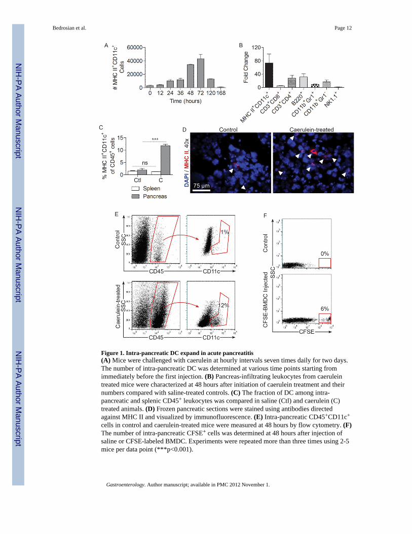
Figure 1. Intra-pancreatic DC expand in acute pancreatitis(A) Mice were challenged with caerulein at hourly intervals seven times daily for two days.The number of intra-pancreatic DC was determined at various time points starting fromimmediately before the first injection. (B) Pancreas-infiltrating leukocytes from caeruleintreated mice were characterized at 48 hours after initiation of caerulein treatment and theirnumbers compared with saline-treated controls. (C) The fraction of DC among intra-pancreatic and splenic CD45+ leukocytes was compared in saline (Ctl) and caerulein (C)treated animals. (D) Frozen pancreatic sections were stained using antibodies directedagainst MHC II and visualized by immunofluorescence. (E) Intra-pancreatic CD45+CD11c+
cells in control and caerulein-treated mice were measured at 48 hours by flow cytometry. (F)The number of intra-pancreatic CFSE+ cells was determined at 48 hours after injection ofsaline or CFSE-labeled BMDC. Experiments were repeated more than three times using 2-5mice per data point (***p<0.001).
Bedrosian et al. Page 12
Gastroenterology. Author manuscript; available in PMC 2012 November 1.
NIH
-PA Author Manuscript
NIH
-PA Author Manuscript
NIH
-PA Author Manuscript
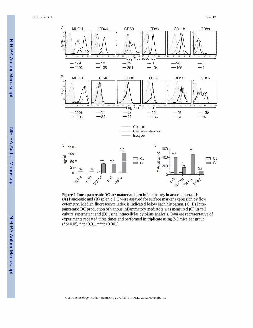
Figure 2. Intra-pancreatic DC are mature and pro-inflammatory in acute pancreatitis(A) Pancreatic and (B) splenic DC were assayed for surface marker expression by flowcytometry. Median fluorescence index is indicated below each histogram. (C, D) Intra-pancreatic DC production of various inflammatory mediators was measured (C) in cellculture supernatant and (D) using intracellular cytokine analysis. Data are representative ofexperiments repeated three times and performed in triplicate using 2-5 mice per group(*p<0.05, **p<0.01, ***p<0.001).
Bedrosian et al. Page 13
Gastroenterology. Author manuscript; available in PMC 2012 November 1.
NIH
-PA Author Manuscript
NIH
-PA Author Manuscript
NIH
-PA Author Manuscript
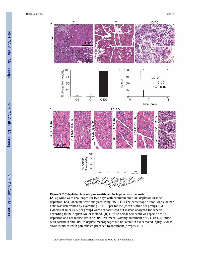
Figure 3. DC depletion in acute pancreatitis results in pancreatic necrosis(A-C) Mice were challenged for two days with caerulein after DC depletion or mockdepletion. (A) Pancreata were analyzed using H&E. (B) The percentage of non-viable acinarcells was determined by examining 10 HPF per mouse (mean 5 mice per group). (C)Cohorts of mice (4-5 per group) were not sacrificed but instead analyzed for survivalaccording to the Kaplan-Meier method. (D) Diffuse acinar cell death was specific to DCdepletion and not mouse strain or DPT treatment. Notably, treatment of CD11b.DTR micewith caerulein and DPT to deplete macrophages did not result in exacerbated injury. Mousestrain is indicated in parentheses preceded by treatment (***p<0.001).
Bedrosian et al. Page 14
Gastroenterology. Author manuscript; available in PMC 2012 November 1.
NIH
-PA Author Manuscript
NIH
-PA Author Manuscript
NIH
-PA Author Manuscript
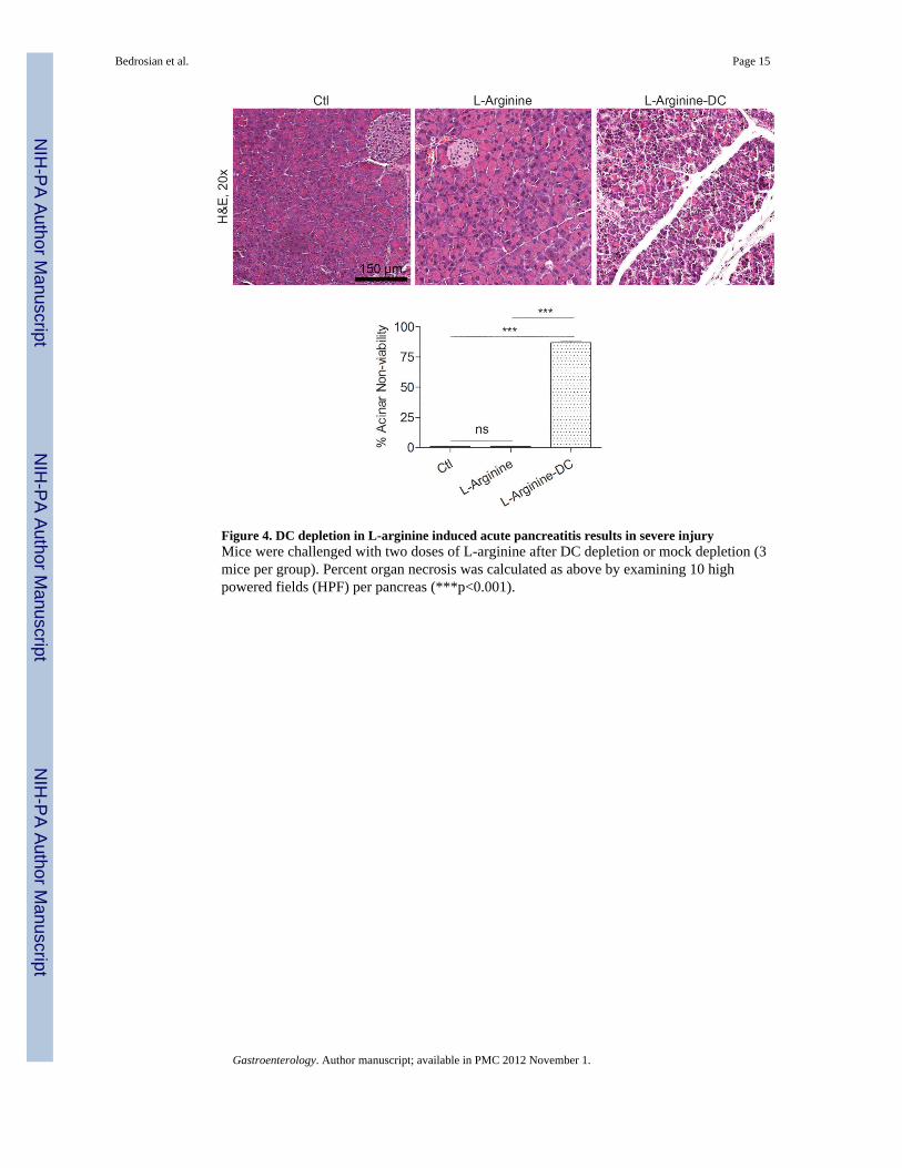
Figure 4. DC depletion in L-arginine induced acute pancreatitis results in severe injuryMice were challenged with two doses of L-arginine after DC depletion or mock depletion (3mice per group). Percent organ necrosis was calculated as above by examining 10 highpowered fields (HPF) per pancreas (***p<0.001).
Bedrosian et al. Page 15
Gastroenterology. Author manuscript; available in PMC 2012 November 1.
NIH
-PA Author Manuscript
NIH
-PA Author Manuscript
NIH
-PA Author Manuscript
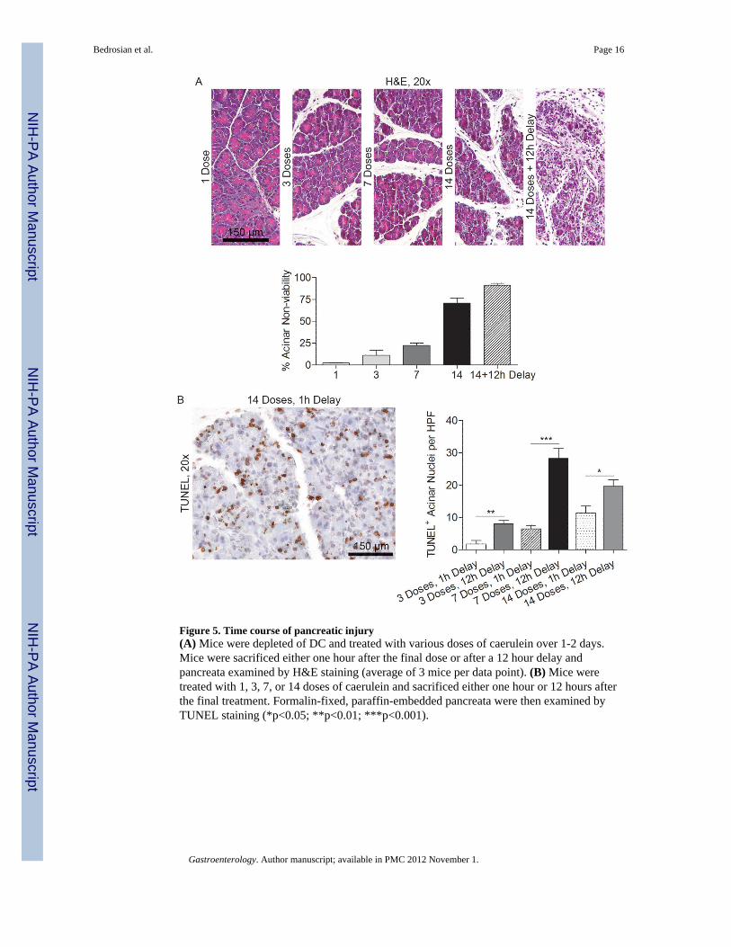
Figure 5. Time course of pancreatic injury(A) Mice were depleted of DC and treated with various doses of caerulein over 1-2 days.Mice were sacrificed either one hour after the final dose or after a 12 hour delay andpancreata examined by H&E staining (average of 3 mice per data point). (B) Mice weretreated with 1, 3, 7, or 14 doses of caerulein and sacrificed either one hour or 12 hours afterthe final treatment. Formalin-fixed, paraffin-embedded pancreata were then examined byTUNEL staining (*p<0.05; **p<0.01; ***p<0.001).
Bedrosian et al. Page 16
Gastroenterology. Author manuscript; available in PMC 2012 November 1.
NIH
-PA Author Manuscript
NIH
-PA Author Manuscript
NIH
-PA Author Manuscript
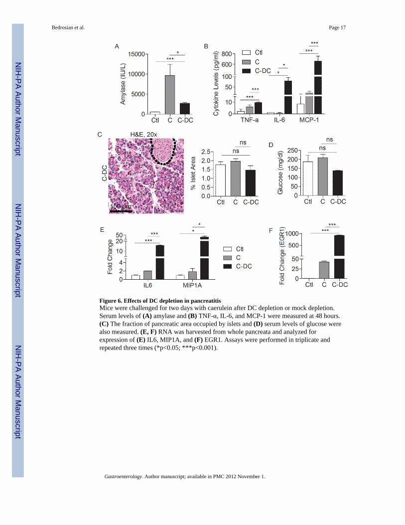
Figure 6. Effects of DC depletion in pancreatitisMice were challenged for two days with caerulein after DC depletion or mock depletion.Serum levels of (A) amylase and (B) TNF-α, IL-6, and MCP-1 were measured at 48 hours.(C) The fraction of pancreatic area occupied by islets and (D) serum levels of glucose werealso measured. (E, F) RNA was harvested from whole pancreata and analyzed forexpression of (E) IL6, MIP1A, and (F) EGR1. Assays were performed in triplicate andrepeated three times (*p<0.05; ***p<0.001).
Bedrosian et al. Page 17
Gastroenterology. Author manuscript; available in PMC 2012 November 1.
NIH
-PA Author Manuscript
NIH
-PA Author Manuscript
NIH
-PA Author Manuscript
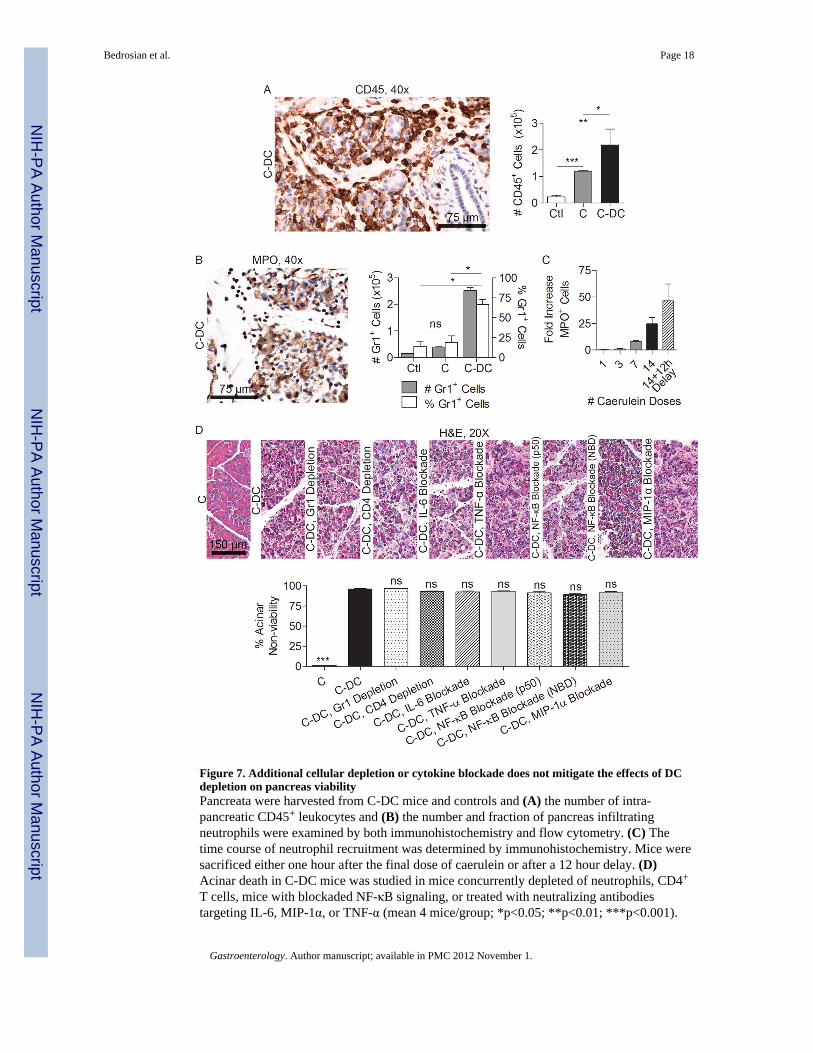
Figure 7. Additional cellular depletion or cytokine blockade does not mitigate the effects of DCdepletion on pancreas viabilityPancreata were harvested from C-DC mice and controls and (A) the number of intra-pancreatic CD45+ leukocytes and (B) the number and fraction of pancreas infiltratingneutrophils were examined by both immunohistochemistry and flow cytometry. (C) Thetime course of neutrophil recruitment was determined by immunohistochemistry. Mice weresacrificed either one hour after the final dose of caerulein or after a 12 hour delay. (D)Acinar death in C-DC mice was studied in mice concurrently depleted of neutrophils, CD4+
T cells, mice with blockaded NF-κB signaling, or treated with neutralizing antibodiestargeting IL-6, MIP-1α, or TNF-α (mean 4 mice/group; *p<0.05; **p<0.01; ***p<0.001).
Bedrosian et al. Page 18
Gastroenterology. Author manuscript; available in PMC 2012 November 1.
NIH
-PA Author Manuscript
NIH
-PA Author Manuscript
NIH
-PA Author Manuscript