MiR-1179 represses cell proliferation, migration and invasion ...
Upload
khangminh22Category
view
0download
0
細胞週期
Cell cycle細胞增生
Proliferation細胞活性
Cell viability細胞凋亡
Apoptosis

02
真核細胞,cell cycle 主要分成兩個階段
1. 間期 (Interphase):在此時期,細胞生長、累積進行有絲分裂所需要的養分及複製 DNA。
2. 分裂期 (Mitosis Phase):細胞在這個時候,分裂成兩個子細胞。藉由 cell cycle,生物體可以由 單一受精卵變成一個成熟的個體。並且藉此,生物體可以在生長發育時,更新毛髮、皮膚、血 球及一些器官和組織。
狀態 期間 簡稱 說明
靜止 /老化(quiescent/senescent)
Gap 0 G0休止期;細胞離開週期階段,停止
分裂生長。
間期
(Interphase)
Gap 1 G1細胞變大;合成 RNA及蛋白質,預
備進行 DNA合成
Synthesis S 此時 DNA進行複製。
Gap 2 G2在 DNA合成和有絲分裂之間,細胞
繼續成長。準備進入分裂期進行分
裂。
細胞分裂 (Mitosis phase)
Mitosis and cytokinesis
M這個時期,細胞停止生長。細胞集
中進行有絲分裂,由一個母細胞變
成兩個子細胞。
偵測 cell cycle 的工具和方式
細胞週期 Cell cycle
Measures Reagents Mechanism Technology Sample Types
DNA Propidium Iodide (PI)Interaction into DNA double strands
Flow cytometry
Fixed, permeabilized, and for live/dead discrimination in intact cells
Newly Synthesized DNA
BrdU and antibodies to BrdU
Bromodeoxyuridine replaces thymidine (T) in dividing DNA. It is then detected by antibodies to BrdU.
Flow cytometry, cell imaging, immunohistochemistry
Fixed and permeabilized cells, treated tissues (cell imaging, immunohistochemistry only)
Protein Level
Antibodies to cyclins, retinoblastoma (Rb), other cell cycle markers
Levels go up and down at different stages of the cell cycle.
Flow cytometry, bioimaging, immunohistochemistry, Western blot
Fixed cells, tissues, and extracts
Protein Modification
Antibodies to phosphorylated histone H3, cyclin dependent kinases (cdk)
Proteins become phosphorylated as a result of proliferation or changes to the cell cycle.
Flow cytometry, bioimaging, immunohistochemistry, Western blot
細胞週期 Cell cycle

03
PI是最常用來偵測 DNA含量的螢光染劑。其作用原理是細胞
固定打洞後,PI會染上核酸 (DNA & RNA) ,並搭配 RNase處理後,再進行染色後 , 以流式細胞儀測定 DNA含量。
• PI可用 488nm 雷射激發,散射波長約 617nm。
• Hoechst 33342 需用 Uv雷射激發,散射波長約 461nm。
Histone會和 DNA複合形成染色質 (Chromatin)。Histone H3蛋白質大小約 15kDa,在哺乳類動物
細胞進行有絲分裂或減數分裂時候,會在 serine 28 (S28)、 S10和 threonine 11等位置會磷酸化。
S10會在 G2 phase晚期到 anaphase期間發生磷酸化;S28會在 prophase到 anaphase期間發生磷酸化。
本試劑結合了Alexa Fluor® 488 Mouse anti-BrdU與Alexa Fluor® 647 Rat anti-Histone H3 (pS28),利用免疫螢光染色方式偵測實驗細胞其細胞週期的變化。
• 偵測 S phase - Bromodeoxyuridine (BrdU) 是核苷酸胸線嘧啶 (Thymidine) 的類似物,當細 胞要合成新的 DNA 時 (S phase),BrdU 可以被細胞攝入並取代 Tymidine 合成 DNA,利用 anti-BrdU 螢光抗體以辨識 BrdU,以鑑定細胞之 S phase。
• 偵測 M phase - 細胞在有絲分裂期發生染色體濃縮 ( chromosome condensation ) 時,其 Histone H3 serine 28位置發生磷酸化現象,故利用 Phospho-Histone H3抗體鑑定細胞之 M phase。
細胞週期指標 : DNA content的測定
Cell cycle kit
Cat. No. name Content Size
558662 Cell Cycle Kit
Alexa Fluor® 488 Mouse anti-BrdU
100 tests
Alexa Fluor® 647 Rat anti-Histone H3 (pS28)5x Fixation BufferPerm Buffer IIStain Buffer (FBS)PBS (10X) ConcentrateHoechst 33342 SolutionBrdUDNase
Confocal image, using the BD Pathway™ 435 Bioimaging system and a 20x (0.75 NA) objective, of HeLa cells that were stained with the three kit components, Alexa Fluor® 488 Mouse anti-BrdU (pseudo-colored green), Alexa Fluor® 647 Rat anti-Histone H3 (pS28) (pseudo-colored red) and Hoechst 33342 (pseudo-colored blue). Costaining of Hoechst 33342 and Histone H3 (pS28) appears pink.
Immunofluorescence 偵測
Flow cytometry 偵測
For Research Use Only. Not for use in diagnostic procedures.

04
Cat. No. Name Content Size
340242BD Cycletest™ Plus DNA Reagent Kit
Buffer Solution
40 Tests(3 vials, 50 mL per vial)Solution A (10 mL)Solution B (8 mL)Solution C (8 mL)
349523 BD™ DNA QC Particles
Vial A: 1-mL suspension of CEN in buffer and ethanol
25 Tests
Vial B: 1-mL suspension of CTN in buffer with formaldehyde and 0.01% thimerosalVial C: 1-mL suspension of 2-µm fluorescent beads in buffer with gelatin and 0.1% sodium azideVial D: 50 mL of 50-µg/mL solution of PI in buffer (25 tests using CEN and CTN)
BD Cycletest™ Plus DNA Reagent Kit適用於新鮮或冷凍固態組織亦或是懸浮細胞,來進行細胞核染色。利用流式細胞儀可以分析正常
細胞與腫瘤細胞染色上的差異。藉由此技術來鑑定異常 DNA stemline 的 DNA指數(DNA index, DI)和細胞週期相分佈。
搭配 BD DNA QC particles kit,可進行儀器電壓設定調整,確認儀器線性度及靈敏度。
細胞週期 Cell cycle
For Research Use Only. Not for use in diagnostic procedures.
05
Cell cycle 調控蛋白主要由 CDK、 Cyclins 與 CKI三種蛋白組成CDK(Cyclin-dependent Protein Kinase):主要執行磷酸化的激酶,受到 CKI與 Cyclins調節Cyclins: 細胞週期蛋白,用來活化 CDK 的活性
CKI (Inhibitor of CDK):負責與 CDK結合,以抑制 CDK的活性
Cell cycle 重組蛋白Cat. No. Description Size14-448 CDK2/CYCLINA , ACTIVE 10 UG 10 UG14-450 CDK1/CYCLIN B, ACTIVE 10 UG 10 UG14-475 CDK2/CYCLIN E, ACTIVE 10UG 10 UG
14-476CDK7/CYCLIN H/MAT1/CAK ACTVE 10UG
10 UG
Cat. No. Description Species Reactivity Key Applications05-507 Anti-Cdc25C Antibody, clone TC-15 H, M IP, WB, ICCMAB8878 Anti-Cdk1 Antibody, clone A17.1.1 Ch, H, M, R, Xn IF, IP, Enzyme Assays, WB, IH(P)
06-923 Anti-Cdk1/Cdc2 (PSTAIR) AntibodyAm, Ch, H, M, R, Sh, Dr, Mk, Ca
ICC, IP, WB
06-923 Anti-Cdk1/Cdc2 (PSTAIR) Antibody Am, Ch, H, M, R, Sh, Dr, Mk, Ca
ICC, IP, WB
06-966 Anti-CDK1/CDC2 Antibody (C-Term) M IP, WB07-631 Anti-cdk2 Antibody H IP, WB05-596 Anti-Cdk2 Antibody, clone AN4.3 H, M, Xn IP, WBMAB8879 Anti-Cdk4 Antibody, clone DCS-35 H, M, Po, R IF, IP, Enzyme Assays, WB, IHC05-364 Anti-Cdk5 Antibody, clone DC17 Dr, H, M, R IP, WB, ICC06-1398 Anti-CDK5RAP2 Antibody H WB, ICC06-138 Anti-Cyclin A Antibody H, M, R, Sh IP, WB, ICCMAB3680 Anti-Cyclin A Antibody, clone E23.1 B, H, M WB, ICCMAB3682 Anti-Cyclin A Antibody, clone E67.1 B, H, M, Mi IP, WB
05-373Anti-Cyclin B1 Antibody, clone GNS3 (8A5D12)
H, M IP, WB
MAB3684 Anti-Cyclin B1 Antibody, clone V152 H, M WB, IH(P)06-137 Anti-Cyclin D Antibody H, M, R ICC, IP, WBABE52 Anti-Cyclin D Antibody H, M, Pm WB, ICC, IPCC12-100UGCN Anti-Cyclin D1 (Ab-3) Mouse mAb (DCS-6) H, M, R WB, IHC, IH(P), FC, IF, IP, Neut05-362 Anti-Cyclin D1/2 Antibody, clone 5D4 H, Ht, M, R ICC, IP, WB05-363 Anti-Cyclin E Antibody, clone HE12 H IP, WB, ICC06-1289 Anti-Cyclin T1 Antibody H, M WB
MABE229Anti-phospho-Cdk1 (Thr14, Tyr15)Antibody, clone CP3.2
H WB, ICC
可應用於 Kinase Assay
cyclin D 表現量隨著細胞週期 (cell cycle)的推進而減緩cyclin E 在 G1 phase 與 合成期 (S)之間達到高峰 cyclin A 在 G2 phase 達到高峰cyclin B 在 G2 phase 與分裂期 (Mitosis) 之間達到高峰
細胞週期 Cell cycle
Cat. No. Description Size14-487 CDK3/CYCLIN E, ACTIVE 10UG 10 UG14-519 CDK6/CYCLIND3, ACTIVE 10UG 10 UG14-685 cdk9/cyclin T1, active 10ug 10 UG23-031M Cyclin E1 complex 250 ug 250 UG

06
細胞增生 Proliferation
Cell Proliferation 是指細胞生長、分裂導致細胞數目變多,可藉由細胞週期 (cell cycle) 來調控。
在原核細胞,cell cycle 主要藉由分裂生殖 (binary fission) 來完成;在真核細胞,cell cycle 主要可分成兩個階段:
1. 間期 (Interphase):在此時期,細胞生長、累積進行有絲分裂所需要的養分及複製 DNA。
2. 分裂期 (Mitosis Phase):細胞在這個時候,分裂成兩個子細胞。藉由 cell cycle,生物體可以由
單一受精卵變成一個成熟的個體。並且藉此,生物體可以在生長發育時,更新毛髮、皮膚、血球
及一些器官和組織。
Cell proliferation rate 細胞增生速率對於細胞研究相當重要,包括細胞生長、細胞分化、細胞毒
殺試驗等等,是探討細胞健康的重要指標之一。
試劑 樣品類型 機轉 偵測方式
BD Horizon™ cell proliferation dyes CFSE & Violet Proliferation Dye 450 (VPD 450)
Live proliferating cells
Diffuses into live cells and is hydrolyzed by intracellular non-specific esterases to become fluorescent products.
Flow cytometry
BrdU/EdU incorporation DNA Synthesis
Bromodeoxyuridine replaces thymidine (T) in dividing DNA and is then detected by antibodies to BrdU
Flow cytometry, cell imaging, immunohistochemistry
Antibodies to Ki-67, PCNA Protein levelLevels increase as a result of proliferation
Flow cytometry, cell imaging, immunohistochemistry, Western blot
偵測 Cell Proliferation 的工具和方式

07
細胞增生 Proliferation
原理
染劑本身是非螢光型態的物質,經由擴散作用到細胞內,其 ester group會被非專一性的酯酶(esterase)水解 (hydrolysis)成螢光型態,並以 succinimidyl ester group共價結合細胞內蛋白質的胺基 (amino group)。死細胞就不會發螢光。
• 活細胞分裂時,每個子細胞所含螢光量約為母細胞的一半,可藉由螢光量的減少偵測細胞增生。
• 能耐受細胞固定液與打洞液,能搭配muliti-color stain 增加實驗設計彈性。
BD Horizon™ Cell Proliferation DyesCFSE & Violet Proliferation Dye 450 (VPD 450)
活細胞染劑BioTracker 405 Blue SE Cell Proliferation Kit
Staining of intracellular phenotypes on activated mouse splenocytes
CD4+ mouse splenocytes 加入 1 µM VPD450。10分鐘後,細胞用 anti-CD3/CD28刺激。在收集細胞前的 4-6小時,細胞加入 PMA和 Ionomycin、BD Golgi-Stop™ (protein transport inhibitor)。 細胞在固定、打洞後,進行 IL-2染色。可見隨著時間增加,細胞增殖,VPD450的螢光強度也漸漸變弱。
Cat. No. Description Laser Equivalent Fluorochromes* Size562158 BD Horizon Violet Proliferation Dye 450 Violet V450, Pacific Blue™, BV421 1mg565082 BD Horizon CFSE Blue FITC, Alexa Fluor® 488 1mg
Flow cytometry 偵測
Flow cytometry/Microscope 偵測
• 可用於長期細胞標記。 • 偵測體內或體外細胞分裂。
• 利用螢光顯微鏡觀察被標記的細胞質。
• 利用 ELISA reader定量細胞數。
貨號 品名 應用 螢光
SCT110BioTracker 488 Green CFSE Cell Proliferation Kit
偵測 carboxyfluorescein succinimidyl ester (CFSE) used for fluorescent cell labeling in applications.
Green
SCT111BioTracker 405 Blue SE Cell Proliferation Kit
Live cell imaging cell proliferation kit used for fluorescent cell labeling in flow cytometry applications.
Blue
For Research Use Only. Not for use in diagnostic procedures.

08
Flow cytometry 偵測
Cell cycle analysis of a population stained for incorporated BrdU and total DNA levels (7-AAD).
Human PBMC用 anti-CD3和 anti-CD28刺激 48小時後,再用 PMA、Ionomycin 處理 4小時。在
最後一個小時加入 BrdU。收集細胞後進行 BrdU染色步驟。
可適用於體外偵測
可適用於體內偵測
Region Cell Cycle PhaseR3 G0/G1
R4 S
R5 G2+M
R6 Sub-G0/G1
C57BL/6在不同的時間點,IP注射 1mg BrdU。小鼠分別在注射後 40分鐘、2小時、4小時犧牲,取胸腺 (Thymus)及骨髓(Bone Marrow)細胞懸浮液進行 anti-BrdU FITC和 7-AAD染色。結果如左,40 分鐘細胞呈現馬蹄型。此種型態為短時間標定BrdU 的經典型態。隨著時間變常 ,原本在 S 期的細胞會回到G0/G1, 在 4 小時可以觀察到明顯 BrdU+ G0/G1週期的細胞。
偵測新合成 DNA : BrdU直接測定 DNA合成是細胞增殖檢測的最準確方法之一,是測定物質毒性、評估藥物安全評價、細
胞健康的基本方法,目前常用的方式是就是在實驗中加入 thymidine的類似物,Bromodeoxyuridine (BrdU)。在 S期的時候,BrdU可以代替 thymidine,崁入新合成的 DNA內。再用 BrdU的抗體與其結合,以流式細胞儀 (Flow Cytometry)、免疫化學染色 (Immunohistochemistry)及酵素免疫分析(ELISA)等常用的細胞分析方式來系列量化 BrdU-positive cell,進而得知細胞週期 S期的表現。
Cat. No. Description Content Size
559619 FITC BrdU Flow KitFITC-conjugated Anti-BrdU Antibody, BD Cytofix/Cytoperm™ Fixation/Permeabilization Solution, BD Perm/Wash™ Buffer (10×), BD Cytoperm™ Plus Permeabilization Buffer, 7-AAD, BrdU, Dnase
50 tests
557891 FITC BrdU Flow Kit 200 tests
552598 APC BrdU Flow KitAPC-conjugated Anti-BrdU Antibody, BD Cytofix/Cytoperm™ Fixation/Permeabilization Solution, BD Perm/Wash™ Buffer (10×), BD Cytoperm™ Plus Permeabilization Buffer, 7-AAD, BrdU, Dnase
50 tests
557892 APC BrdU Flow Kit 200 tests
細胞增生 Proliferation
For Research Use Only. Not for use in diagnostic procedures.

09
• 適用於免疫組織染色試驗 ( IHC )。• Kit 內附 anti-BrdU 螢光抗體辨識 BrdU,鑑定細胞之 S phase。
• 適用樣本為 - frozen sections, formalin-fixed paraffin-embedded sections, cultured isolated cells on slides。 • 適用樣本物種為 - human、mouse、rat。• Kit 內附實驗所需之抗體、buffer 和操作流程。
• Kit 內附 BD™ Retrievagen A,可幫助有效揭露抗原位置,保留組織型態,以便成功地在 DNA stands 進行 BrdU 染色。
• 實驗彈性,視實驗設計可再加入其他抗體或是偵測表面抗原之抗體。
• 提供 Control slides 以確定實驗的準確性。
Immunohistochemical staining of BrdU in paraffin sections. BALB/c mice were injected with 1 mg of BrdU via the intra-peritoneal route. After 24 hrs the spleen, thymus, and gastrointestinal tract were harvested and processed for paraffin sections. Mice injected with PBS served as the negative control. Immunohistochemical staining of BrdU was performed using the BrdU In-Situ Detection Kit on paraffin sections of the mouse gastro-intestinal tract. Proliferating cells in the crypts that incorporated BrdU can be identified by the dark brown color in their cell nuclei (A) in contrast to the control (B). Magnification 400×.
BrdU In-Situ Detection Kit
偵測 S phase - Bromodeoxyuridine (BrdU)是核苷酸胸線嘧啶 (Thymidine)的類似物,當細胞要合
成新的 DNA時 (S phase),BrdU可以被細胞攝入並取代 Tymidine 合成 DNA,利用 anti-BrdU 螢光抗體以辨識 BrdU,以鑑定細胞之 S phase 。
原理
實驗數據
Cat. No. Name Contents Size
550803BrdU In-SituDetection Kit
Biotin anti-BrdU antibody, BD™ Retrievagen A antigen retrieval solution, streptavidin-horseradish peroxidase (SAv-HRP) solution, DAB buffer, DAB chromogen, fixation buffer, diluent buffer, and control slides
50 Tests
Immunohistochemistry 偵測
For Research Use Only. Not for use in diagnostic procedures.
10
• 適用於酵素免疫分析試驗 (ELISA)。• 安全: 無須使用同位素即可進行體外定量。
• 應用性廣:適用物種為 Mouse、Human、Rat、Pig 、Horse、Rabbit、Guinea Pig、 Hamster、Non-human Primate、Canine。
• 適用樣本:貼附型 (Adherent) 與懸浮型 (Suspension) 細胞。
• 方便:Kit 內附 BrdU、Fixing Solution、Stop Solution 以及實驗所需之抗體與試劑。
• 偵測波長 - dual wavelength of 450/550 nm ( 替代波長 450/540 nm 或 450/595 nm 或 450 nm single)。
BrdU Cell Proliferation KitELISA 偵測
Cat. No. Description Content Size Detection
2750
BrdU Cell Proliferation Kit
BrdU Reagent ( 500X ) 、Fixing Solution、
Prediluted BrdU Detection Antibody、Stop Solution、Goat anti-Mouse IgG, Peroxidase labeled ( 2000X )、Conjugate Diluent、Substrate、Plate Wash Concentrate ( 50X )
200 assays
Colorimetric
2752 1000 assays
細胞增生 Proliferation
The BrdU cell proliferation kit (Cat. No. 2750) was used to measure proliferation of H9 human embryonic stem cells in HEScGRO™ and KOSR media, after cells were enzymaticallyexpanded for 12 passages. Increased BrdU incorporation indicated faster cell proliferation in HEScGRO™ medium.

11
偵測新合成 DNA : EdUEdU (5-ethynyl-2 -deoxyuridine)也是一種 thymidine類似物,在細胞增殖時能夠插入正在複製
的 DNA分子中,基於 EdU與染料的共軛反應可以有效地檢測處於 S期的細胞。EdU染料分子比BrdU抗體小,在細胞內更容易擴散,不需要嚴格的樣品變性 (酸解、熱解、酶解 )處理,有效地
避免了樣品損傷,有助於在組織、器官的整體水準上觀測細胞增殖的真實情況,具有更高的靈敏度
和更快的檢測速度。
EdU BrdU
檢測分子 很小 大
檢測方式 化學反應 免疫反應
DNA變性 不需 需要
靈敏度 靈敏 一般
Cat. No. Description Content Size DetectionEquivalent Fluorochromes
565456BD Pharmingen™ 647 EdU Click Proliferation Kit
EdU (5-ethynyl-2®-deoxyuridine) 、Eterneon™ Red 645 azide (10 mM)、Buffer Additive (10×)、Saponin-based Permeabilization and Wash Reagent (10×)、Fixative Solution (4% paraformaldehyde-based)、Catalyst Solution、
DMSO
50 testflow cytometry Bioimaging
APC
565455BD Pharmingen™ 488 EdU Click Proliferation Kit
EdU (5-ethynyl-2®-deoxyuridine)、6-FAM Azide (10 mM)、Buffer Additive (10×) 、Saponin-based Permeabilization and Wash Reagent (10×)、Fixative Solution (4% paraformaldehyde-based)、Catalyst Solution、
DMSO
50 testflow cytometry Bioimaging
FITC
BrdU與 EdU檢測原理示意圖BrdU與 EdU比較表
Cat. No. Description Content Size DetectionEquivalent Fluorochromes
17-10527EdU Cell Proliferation Assay (EdU-594)
5-Ethynyl-deoxyuridine (5-EdU)、5/6-Sulforhodamine101-PEG3-Azide (10 mM)、Buffer additive、
DMSO、10X Reaction Buffer 、Catalyst solution
100 reactionsFlow cytometryBioimaging
Cy5
17-10526EdU Cell Proliferation Assay (EdU-555)
5-Ethynyl-deoxyuridine (5-EdU)、5-TAMRA-PEG3-Azide (10 mM) 、Buffer additive、DMSO、10X Reaction Buffer、Catalyst solution
100 reactionsFlow cytometryBioimaging
Cy3
細胞增生 Proliferation
For Research Use Only. Not for use in diagnostic procedures.

12
細胞增生 Proliferation
粒腺體呼吸鏈上的酵素活性往往成為判定細胞是否存活的依據;當細胞死亡時,酵素也跟著失去
活性,只有活細胞的酵素才有活性。再加入呈色受質就可以定量存活的細胞量。
• 精確:細胞酵素代謝活性與細胞數量息息相關。
• 方便:馬上可用。 • 簡單:只要一個步驟,不用 wash,不需其他試劑。
• 安全: 沒有放射性物質,沒有有機溶劑。
• 完全水溶:不用再用 DMSO 回溶結晶。
Event Measure Product Package Size Cat. No. DectectionCell Proliferation
Reduction of WST-1 by live cells
Premix WST-1 Cell Proliferation Assay System
2,500 Tests MK400Colorimetric analysis
Cytotoxicity Release of LDH by dead or damaged cells
LDH Cytotoxicity Detection Kit 2,000 Tests MK401Colorimetric analysis
原理
當細胞膜受損或破裂時,位於細胞質的 LDH ( Lactate Dehydrase ) 便會釋放到細胞外,藉由加入
呈色受質就可以定量被毒害的細胞。
偵測 supernatant 內的 LDH 活性
• 快速:一小時收 Data。
• 簡單:不用 wash,不用 prelabeling。
• 超高敏感度:能測得 2000 個毒害細胞。
• 安全:非放射性材料,安全無虞。
LDH Cytotoxicity Detection Kit
Premixed WST-1 Cell Proliferation Reagent
Measure absorbanceAdd WST-1
30 min
13
細胞活性 Cell viability
實驗範例
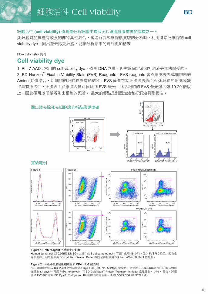
圈出跟去除死去細胞讓分析結果更準確
Figure 1: FVS reagent不受固定液影響Human Jurkat cell 以 0.025% DMSO (上圖 )或 5 µM camptothecin(下圖 )處理 16小時,並以 FVS780染色,藍色虛線和紅線分別是有無用 BD Cytofix™ Fixation Buffer做固定和有無用 BD Perm/Wash Buffer I 做打洞。
Figure 2 : 分析小鼠脾臟細胞增生和 CD4、IL-2的表現小鼠脾臟細胞先以 BD Violet Proliferation Dye 450 (Cat. No. 562158)做染色,之後以 BD anti-CD3e和 CD28抗體刺激細胞 (3 days)。再用 PMA, Ionomycin, 和 BD GolgiStop™ Protein Transport Inhibitor處理細胞 4小時。 最後,將細胞染 FVS780並用 BD Cytofix/Cytoperm™ Kit 細胞固定打洞後,染 BUV395 CD4和 FITC IL-2。
細胞活性 (cell viability) 偵測是分析細胞生長狀況和細胞健康重要的指標之一。
死細胞對於抗體有較強的非特異性結合,當進行流式細胞儀實驗的分析時,利用排除死細胞的 cell viability dye,圈出並去除死細胞,能讓分析結果的統計更加精確
Cell viability dye 1. PI , 7-AAD : 常用的 cell viability dye,偵測 DNA 含量,但對於固定液和打洞液是無法耐受的。
2. BD Horizon™ Fixable Viability Stain (FVS) Reagents : FVS reagents 會與細胞表面或細胞內的Amine 共價結合,活細胞的細胞膜沒有通透性,FVS僅會存於細胞膜表面;但死細胞的細胞膜變
得具有通透性,細胞表面及細胞內皆可偵測到 FVS 螢光,比活細胞的 FVS 螢光強度強 10-20 倍以上,因此便可以簡單辨別出細胞的死活。 最大的優點是對固定液和打洞液具耐受性。
Flow cytometry 偵測
圈出並去除死細胞
讓分析結果的統計更加精確
圈出並去除死細胞
讓分析結果的統計更加精確

14
Cat. No. Description size Laser Equivalent Fluorochromes對細胞固定與打洞液具耐受性
566332BD Horizon™
Fixable Viability Stain 440UV200ug UV/Violet BUV395
562247BD Horizon™ Fixable Viability Stain 450
0.1 mg Violet V450, Pacific Blue™, BV421
564406BD Horizon™ Fixable Viability Stain 510
0.1 mg Violet V500, BV510
565694BD Horizon™ Fixable Viability Stain 575V
200 µg Violet Brilliant Violet 605, Pacific Orange
564407BD Horizon™ Fixable Viability Stain 520
150 µg Blue FITC, Alexa Fluor® 488
564995BD Horizon™ Fixable Viability Stain 570
150 µg YG/Blue PE
564996BD Horizon™ Fixable Viability Stain 620
100 µg Yellow-Green/Blue PE-CF594
564405BD Horizon™ Fixable Viability Stain 660
100 µg Red APC, Alexa Fluor® 647
564997BD Horizon™ Fixable Viability Stain 700
100 µg Red APC-R700, Alexa Fluor® 700
565388BD Horizon™ Fixable Viability Stain 780
200 µg Red APC-Cy7
適用於只做細胞表面染色不需內染的實驗
565803BD Via-Probe™ Red Nucleic Acid Stain
0.1 mL Red APC
565804BD Via-Probe™ Red Nucleic Acid Stain
0.5 mL Red APC
565802BD Via-Probe™ Green Nucleic Acid Stain
0.5 mL Blue FITC
565799BD Via-Probe™ Green Nucleic Acid Stain
0.1 mL Blue FITC
564907BD Pharmingen™ DAPI Solution
1 mg UV/Violet DAPI (UV)/BV421 (V)
556463BD Pharmingen™ Propidium Iodide Staining Solution
2 mL YG/Blue PE
559925BD Pharmingen™ 7-AAD
2 mL YG/Blue PerCP-Cy™5.5
555815BD Via-Probe™ Cell Viability Solution
500 Tests YG/Blue PerCP-Cy™5.5
555816BD Via-Probe™ Cell Viability Solution
100 Tests YG/Blue PerCP-Cy™5.5
細胞活性 Cell viability細胞活性 Cell viability
For Research Use Only. Not for use in diagnostic procedures.
15
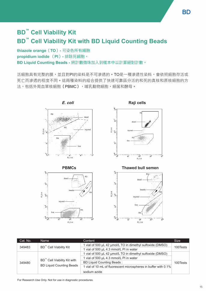
thiazole orange(TO)- 可染色所有細胞propidium iodide (PI)- 排除死細胞。 BD Liquid Counting Beads - 將計數微珠加入到樣本中以計算絕對計數。
活細胞具有完整的膜,並且對PI的染料是不可滲透的。TO是一種滲透性染料,會依照細胞存活或
死亡而滲透的程度不同。這兩種染料的組合提供了快速可靠區分活的和死的真核和原核細胞的方
法,包括外周血單核細胞(PBMC),哺乳動物細胞,細菌和酵母。
BD™ Cell Viability KitBD™ Cell Viability Kit with BD Liquid Counting Beads
Cat. No. Name Content Size
349483 BD™ Cell Viability Kit1 vial of 500 µL 42 µmol/L TO in dimethyl sulfoxide (DMSO)
100Tests1 vial of 500 µL 4.3 mmol/L PI in water
349480BD™ Cell Viability Kit with BD Liquid Counting Beads
1 vial of 500 µL 42 µmol/L TO in dimethyl sulfoxide (DMSO)
100Tests1 vial of 500 µL 4.3 mmol/L PI in waterBD Liquid Counting Beads :1 vial of 10 mL of fluorescent microspheres in buffer with 0.1% sodium azide
E. coli
PBMCs
Raji cells
Thawed bull semen
For Research Use Only. Not for use in diagnostic procedures.

16
Apoptotic cell
PS
Mitochondrial Potential decrease
Caspase cascade
PS exposure(Annexin Ca2+) 2
3
1
DNA - Fragmentation4
Apoptosis (細胞凋亡 )的概念在 1972年正式由 Kerr JF, Wyllie AH; Currie AR提出,到了 80年代後期,隨著分子生物學技術的進步,使細胞凋亡的研究迅速發展。細胞凋亡的起因有可能是因
為外界環境、生長發育等等,而細胞為維持生物體內環境的穩定,而進行有程序性的死亡現象 (Programmed cell death, PCD)。細胞凋亡發生時,細胞會先變圓,隨後細胞皺縮、細胞質密度增加,細胞膜內的 PS (phosphatidylserine)外翻、粒線體膜電位喪失 (depolarization)、DNA斷裂,最後細胞會裂解成多
個凋亡小體 (apoptotic body)進而被巨噬細胞清除。
Mesures Hot Sale Itemskey Features
Phosphatidylserine Exposure
Cat. No. Name
556547 Annexin V : FITC Apoptosis Detection Kit I • Detects early Apoptosis markers• Quick and easy• Flow cytometry application559763 Annexin V : PE Apoptosis Detection Kit I
MitochondrialChanges
551302 BDTM MitoScreen (JC-1) KitFast, easy, single cell resolution by flow cytometry or fluorescent microscopy
564696 MitoStatus TMRE
564697 MitoStatus Red
Caspase Activation550480 Caspase-3, Active Form, Apoptosis Kit : FITC • Quick and easy
• Flow cytometry application550914 Caspase-3, Active Form, Apoptosis Kit:PE
DNA Fragmentation556381 Apoptosis Detection Kit (APO-Direct) Works adherent cells, single cell
resolution in conjuunction with cell cycle analysis by flow cytometry556405 Apoptosis Detection Kit (APO-BRDU)
Apoptosis Study Tools
細胞凋亡 Apoptosis
For Research Use Only. Not for use in diagnostic procedures.

17
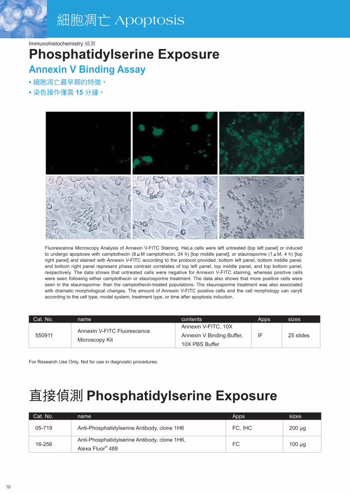
• 細胞凋亡最早期的特徵。
• 染色操作僅需 15分鐘。
質膜變化是在活細胞中檢測到細胞凋亡的第一個特徵。驗證細胞凋亡可以通過檢測磷脂醯絲氨酸
(PS)的存在,其通常位於質膜的細胞質表面 (如 )。 細胞凋亡過程中,PS會轉移到質膜的外層 (如 ) ,與螢光標記的Annexin V(含鈣)存在時結合,可以用流式細胞儀和細胞成像儀檢測。
Phosphatidylserine ExposureAnnexin V Binding Assay
Cat. No. Name Contents Apps Size
556547Annexin V : FITC Apoptosis Detection Kit I
Annexin V-FITC, Propidium Iodide Staining Solution, Annexin V Binding Buffer
FCM 100 Tests
559763Annexin V : PE Apoptosis Detection Kit I
Annexin V-PE, 7-AAD, and Annexin V Binding Buffer
FCM 100 Tests
原理示意圖
實驗範例
熱銷推薦
1 32
2 3
1
代表健康的細胞 代表細胞凋亡早期的細胞 代表細胞凋亡晚期 /壞死的細胞1 2 3
2 2
33
1
Flow cytometry 偵測
For Research Use Only. Not for use in diagnostic procedures.
18
細胞凋亡 Apoptosis
Fluorescence Microscopy Analysis of Annexin V-FITC Staining. HeLa cells were left untreated [top left panel] or induced to undergo apoptosis with camptothecin (8µM camptothecin, 24 h) [top middle panel]; or staurosporine (1µM, 4 h) [top right panel] and stained with Annexin V-FITC according to the protocol provided. bottom left panel, bottom middle panel, and bottom right panel represent phase contrast correlates of top left panel, top middle panel, and top bottom panel, respectively. The data shows that untreated cells were negative for Annexin V-FITC staining, whereas positive cells were seen following either camptothecin or staurosporine treatment. The data also shows that more positive cells were seen in the staurosporine- than the camptothecin-treated populations. The staurosporine treatment was also associated with dramatic morphological changes. The amount of Annexin V-FITC positive cells and the cell morphology can vary6 according to the cell type, model system, treatment type, or time after apoptosis induction.
Cat. No. name contents Apps sizes
550911Annexin V-FITC Fluorescence Microscopy Kit
Annexin V-FITC, 10X Annexin V Binding Buffer, 10X PBS Buffer
IF 25 slides
Cat. No. name Apps sizes
05-719 Anti-Phosphatidylserine Antibody, clone 1H6 FC, IHC 200 µg
16-256Anti-Phosphatidylserine Antibody, clone 1H6, Alexa Fluor® 488
FC 100 µg
• 細胞凋亡最早期的特徵。
• 染色操作僅需 15分鐘。
Phosphatidylserine ExposureAnnexin V Binding Assay
直接偵測 Phosphatidylserine Exposure
Immunohistochemistry 偵測
For Research Use Only. Not for use in diagnostic procedures.

19
JC-1
(FL2
)
JC-1(FL1)
J-aggregates (healthy cell)
monomers (apoptosis indicator)
J-aggregates (healthy cell)
monomers (apoptosis indicator)
JC-1 Staining in Control and Apoptotic Cells. Cells (1 × 10⁶ cells/ml) were untreated or treated with staurosporine (1µm, 4 h to induce apoptosis. Cells were stained with JC-1 according to the protocol and analyzed on a BD FACSCalibur™.
BD MitoScreen (JC-1) Kit analysis on the BD Accuri C6. K562 cells were treated with 100 µM of CCCP (in DMSO) for 5 minutes at 37℃ to induce mitochondrial membranes to decouple. Results: Compared to untreated controls (B), CCCP treatment (C) resulted in a shift in mitochondrial membrane potential.
• 可偵測粒線體膜電位變化。
• 適用於流式細胞儀及螢光顯微鏡偵測。
• 容易操作。
Mitochondrial ChangesBD Mitoscreen (JC-1) Kit
JC-1 (5,5' ,6,6' - tetrachloro-1,1' ,3,3' - tetraethylbenzimidazolcarbocyanine iodide)的兩種形式:
1. JC-1單體 ( Monomer ) :呈現綠色螢光,在大多數的流式細胞儀,可用 FL-1偵測器偵測。
2. JC-1多聚體 (Aggregates ) :呈現黃色螢光,在大多數的流式細胞儀,可用 FL-2偵測器偵測。
正常生理狀態下,細胞粒線體負電性高,細胞質內的 JC-1單體 (mononer) 進入粒線體以多聚體 (Aggregates ) 存在,此時會偵測到同時有綠色螢光跟黃色螢光的健康細胞較多,只呈現綠色螢光的細胞
較少。當細胞走向凋亡時,粒線體去極化產生,負電性降低,JC-1從粒線體釋放到細胞質,呈現單體形
式,因此會偵測到呈現綠色螢光的細胞增加 ,同時具有綠色螢光跟黃色螢光的細胞變少。
實驗範例
熱銷推薦
Cat. No. Name Contents Apps Size551302 BDTM MitoScreen (JC-1) JC-1 dye and assay buffer FCM 100 Tests
Flow cytometry 偵測
For Research Use Only. Not for use in diagnostic procedures.
20
實驗範例
Mitochondrial ChangesBD PharmingenTM MitoStatus Reagents
Cat. No.MitoStatus TMRE MitoStatus Red
564696 564697
Characteristic
Excitation peak 549 nm 622 nm
Emission peak 574 nm 648 nm
Laser 488 nm (blue) 640 nm (red)
Detector FL2 FL4
Equivalent fluorochromes* PE APC Alexa Fluor® 647
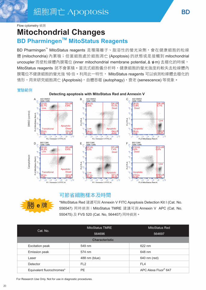
BD Pharmingen™ MitoStatus reagents 是種陽離子、脂溶性的螢光染劑,會在健康細胞的粒線
體 (mitochondria)內累積;但當細胞處於細胞凋亡 (Apoptosis)的狀態或是接觸到 mitochondrial uncoupler而使粒線體內膜電位 (inner mitochondrial membrane potential,Δψm)去極化的時候, MitoStatus reagents 就不會累積。當流式細胞儀分析時,健康細胞的螢光強度約較失去粒線體內
膜電位不健康細胞的螢光強 10倍。利用此一特性, MitoStatus reagents 可以偵測粒線體去極化的情形,用來研究細胞凋亡 (Apoptosis),自體吞噬 (autophagy),衰老 (senescence)等現象。
可節省細胞樣本及時間*MitoStatus Red 建議可與 Annexin V FITC Apoptosis Detection Kit I (Cat. No.
556547) 同時偵測;MitoStatus TMRE 建議可與 Annexin V APC (Cat. No.
550475)及 FVS 520 (Cat. No, 564407)同時偵測。
Detecting apoptosis with MitoStatus Red and Annexin V
勝 e牌
Flow cytometry 偵測
For Research Use Only. Not for use in diagnostic procedures.
細胞凋亡 Apoptosis

21
粒線體純化ProteoExtract® Cytosol/Mitochondria Fractionation Kit
粒線體活細胞染色
• 節省 : 一組 Kit 同時可得兩個不同位置的蛋白質。Cytosol 及 Mitochondria fraction 。• 可得 Native 的蛋白 : 適用於 WB, ELISA, Protein translocation 研究。 • 過程簡單 : 不需經過超高速離心。
活細胞影像觀察相較於固定樣本觀察,更能實現動態變化觀察,3D 結構觀察,避免固定細胞步驟
對樣品的影響。
• 粒線體膜活細胞染劑,無須固定即可直接檢測
• 應用 : 測細胞存活率,代謝活性和觀測整體細胞健康
Flow cytometry 偵測
貨號 品名 螢光 ChannelSCT136 BioTracker 488 Green Mitochondria Dye Green FITC/GFP SCT137 BioTracker 633 Red Mitochondria Dye Red Cy5SCT135 BioTracker 405 Blue Mitochondria Dye Blue DAPI
(Cat. No. QIA88-1KITCN)
細胞凋亡 Apoptosis

22
Jurkat T cells mouse thymocytes
Jurkat cells mouse thymocytes
Flow cytometric analysis of apoptotic and non-apoptotic populations using anti-active caspase-3 antibodies.Jurkat T cells (A, A1) or mouse thymocytes (B, B1) were left untreated (A, B) or treated for 4 h with camptothecin (A1) or a mouse Fas monoclonal antibody, clone Jo2 (Cat. No. 554254) to induce apoptosis (B1). Cells were permeabilized and then stained with PE-conjugated active caspase-3 antibodies (Cat. No. 557091). Untreated cells were primarily negative for the presence of active caspase-3, whereas about half of each population of cells induced to undergo apoptosis had detectable active caspase-3.
Caspase家族的活化caspase的活化在細胞凋亡反應扮演著重要的角色,活化的 caspase會進行一連串的蛋白質脢作用 (proteolysis cascade),將死亡訊號傳遞至下游,最後引發細胞凋亡。Caspase以未活化態存在 (procaspase),當細胞凋亡的訊號啟動,procaspase會自行裂解或被其他的蛋白脢切割而變成活化態的 caspase,進而進行一連串的下游反應。其中 caspase 8, 9, 3為此過程的關鍵角色,上
游的 caspase 8和 caspase 9是細胞凋亡起始訊號的 caspase, caspase 8透過不同路徑來活化caspase 3,而活化 caspase 3會將訊息放大傳遞至下游,最終執行細胞凋亡。
Caspase-3 AssayCaspase 3 : 細胞凋亡的關鍵人物Caspsae 3在細胞凋亡早期是主要的蛋白脢,未活化的 caspase 3會自行裂解或因其他蛋白脢 (如 : caspase 9)切割而成兩個次單位 (17-22kDa和 10-12 kDa)的二聚體,此活化態的 caspase 3會進一步活化下游的目標蛋白,例如胞漿內的 Bcl-2和 D4-GDI及核內的 PARP。
Caspase Acvtivation
Cat. No. Name Contents React. Apps Size
550480Caspase-3, Active Form, Apoptosis Kit : FITC
FITC anti-active Caspase-3 antibody, BD Cytofix/CytopermTM Fixation/Permeabilization Solution, and BD Perm/WashTM Buffer
Hu, Ms IC/FCM 100 Tests
550914Caspase-3, Active Form, Apoptosis Kit:PE
Anti-Active Caspase-3 Antibody, BD Cytofix/CytopermTM Fixation/Permeabilization Solution, and BD Perm/WashTM Buffer
Hu, Ms IC/FCM 100 Tests
Cat. No. Name Size550377 General Caspase Inhibitor, Z-VAD-FMK 1 mg563828 General Caspase Inhibitor, Q-VD-OPh 1 mg550380 Caspase-8 Inhibitor, Z-IETD-FMK 1 mg550381 Caspase-9 Inhibitor, Z-LEHD-FMK 1 mg550378 Caspase-3 Inhibitor, Z-DEVD-FMK 1 mg550411 Caspase Inhibitors Negative Control, Z-FA-FMK 1 mg
特別推薦 Caspase Inhibitor
熱銷推薦
Flow cytometry 偵測
細胞凋亡 Apoptosis
For Research Use Only. Not for use in diagnostic procedures.

23
其他 Caspase assay kit
特別推薦 - Activator
Cat. No. Description Assays AssaysAPT400 CaspaTag Pan-Caspase In Situ Assay Kit, Fluorescein FC, ACT 100 assaysAPT403 CaspaTag Caspase 3,7 In Situ Assay Kit, Fluorescein FC, ACT 100 assaysAPT408 CaspaTag Caspase 8 In Situ Assay Kit, Fluorescein FC, ACT 100 assaysAPT409 CaspaTag Caspase 9 In Situ Assay Kit, Fluorescein FC, ACT 100 assaysAPT420 CaspaTag Pan-Caspase In Situ Assay Kit, Fluorescein FC, ACT 25 assaysAPT423 CaspaTag Caspase 3,7 In Situ Assay Kit, Fluorescein FC, ACT 25 assaysAPT428 CaspaTag Caspase 8 In Situ Assay Kit, Fluorescein FC, ACT 25 assaysAPT429 CaspaTag Caspase 9 In Situ Assay Kit, Fluorescein FC, ACT 25 assays
Cat. No. Description Size
178497-10MGCNApoptosis Activator VII, Apoptozole - CAS 1054543-47-3- Calbiochem
10 mg
178493-10MGCN Apoptosis Activator III, Embelin - CAS 550-24-3 - Calbiochem 10 mg
178496-5MGCNApoptosis Activator VI, CD437/AHPN - CAS 125316-60-1 - Calbiochem
5 mg
5.08774.0001 Apoptosis Activator VIII, TP421 - Calbiochem 10 mg5.31709.0001 Apoptosis Activator IX, DPP-23- Calbiochem 25 mg
APT800 Apoptosis Inducer Set
· Actinomycin D (10 mM): 50 µL· Camptothecin (2 mM): 1 mL· Cycloheximide (100 mM): 1 mL· Dexamethasone (10 mM): 1 mL· Etoposide (10 mM): 100 µL
細胞凋亡 Apoptosis
CaspaTag Caspase 3,7,8,9 In Situ Assay Kit, Fluorescein• 應用 : 96-well plate-based fluorometry、 fluorescence microscopy 或 flow cytometry。
• 適用樣本:貼附型 (Adherent) 與懸浮型 (Suspension) 細胞。
• 方便:Kit 內附 FLICA Reagent、固定液、PI染劑、Hoechst Stain以及Wash buffer。
Flow cytometry 偵測
Fluorochrome Inhibitors of Caspases (FLICA)方法學,抑制劑利用帶有螢光標記,可通透細胞且
不具有毒性。抑制劑進入細胞後會共價結合上活性的 caspase,未結合的試劑將擴散出細胞並被洗
掉,螢光強度可直接代表 caspase 活性。
原理
24
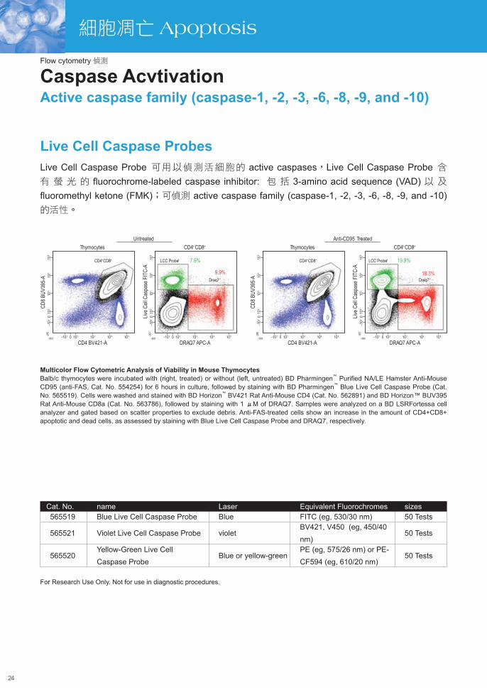
Multicolor Flow Cytometric Analysis of Viability in Mouse ThymocytesBalb/c thymocytes were incubated with (right, treated) or without (left, untreated) BD Pharmingen™ Purified NA/LE Hamster Anti-Mouse CD95 (anti-FAS, Cat. No. 554254) for 6 hours in culture, followed by staining with BD Pharmingen™ Blue Live Cell Caspase Probe (Cat. No. 565519). Cells were washed and stained with BD Horizon™ BV421 Rat Anti-Mouse CD4 (Cat. No. 562891) and BD Horizon™ BUV395 Rat Anti-Mouse CD8a (Cat. No. 563786), followed by staining with 1 µM of DRAQ7. Samples were analyzed on a BD LSRFortessa cell analyzer and gated based on scatter properties to exclude debris. Anti-FAS-treated cells show an increase in the amount of CD4+CD8+ apoptotic and dead cells, as assessed by staining with Blue Live Cell Caspase Probe and DRAQ7, respectively.
Cat. No. name Laser Equivalent Fluorochromes sizes565519 Blue Live Cell Caspase Probe Blue FITC (eg, 530/30 nm) 50 Tests
565521 Violet Live Cell Caspase Probe violetBV421, V450 (eg, 450/40 nm)
50 Tests
565520Yellow-Green Live Cell Caspase Probe
Blue or yellow-green PE (eg, 575/26 nm) or PE-CF594 (eg, 610/20 nm)
50 Tests
Caspase AcvtivationActive caspase family (caspase-1, -2, -3, -6, -8, -9, and -10)
Live Cell Caspase ProbesLive Cell Caspase Probe 可用以偵測活細胞的 active caspases,Live Cell Caspase Probe 含有 螢 光 的 fluorochrome-labeled caspase inhibitor: 包 括 3-amino acid sequence (VAD) 以 及 fluoromethyl ketone (FMK);可偵測 active caspase family (caspase-1, -2, -3, -6, -8, -9, and -10) 的活性。
細胞凋亡 Apoptosis
Flow cytometry 偵測
For Research Use Only. Not for use in diagnostic procedures.

25
Non-apoptotic cells
Non-apoptotic cells
Positive apoptotic cells in S phase
• 除偵測核內 DNA Fragmentation外,也可同時分析細胞週期 (Cell Cycle)。• 適用於懸浮型或貼附型細胞的實驗。
• 可利用流式細胞儀上機分析。
到了細胞凋亡後期,細胞核內的DNA會被內源性核酸內切脢降解,使DNA發生片段化的現象 (DNA fragmentation)。當 DNA被內切脢作用後,會產生單有 3'末端的切口或是斷裂 DNA片段,在 TdT (terminal transferase,末端轉移脢 )的作用下,使已標定螢光的核苷酸 (ex : FITC-dUTP)連結到DNA片段的 3'末端,螢光強度與 DNA片段量成正比。另外,可再搭配 PI染色來得知檢體中細胞凋亡細胞的百分比及細胞凋亡細胞所在的細胞週期 (G0/G1, S, G2/M phase)
Cells are labeled with both PI (DNA) and FITC-dUTP (Apoptotic Cells). Display 1:Non-clumped cells are gated.
APO-DIRECTTM Kit
Flow cytometry data of HPB-ALL human leukemia cells using an APO-BrdU assay.
套組
Cat. No. Description Contents Size
556381 APO-DIRECTTM FITC dUTP, PI/RNase staining buffer, reaction buffer, rinsing buffer, wash buffer, TdT enzyme, negative control cells, positive control cells
50 Tests
556405 APO-BRDUTM
FITC anti-BrdU antibody, PI/RNase staining buffer, reaction buffer, rinsing buffer, wash buffer, Br-dUTP, TdT Enzyme, negative control cells, positive control cells
60 Tests
APO-BRDUTM Kit
DNA FragmentationTUNEL Assay (Terminal deoxynucleotidyltransferase dUTP Nick End Labeling)
實驗數據
實驗數據
實驗原理
實驗原理
Flow cytometry 偵測
For Research Use Only. Not for use in diagnostic procedures.

26
細胞凋亡 Apoptosis
Catalog No. S7100 S7101 QIA33 S7110 S7111 S7165
DescriptionApopTag Peroxidase In Situ Apoptosis Detection Kit
ApopTag Plus Peroxidase In Situ Apoptosis Kit
FragEL DNA Fragmentation Detection Kit, Colorimetric - TdT Enzyme
ApopTag Fluorescein In Situ Apoptosis Detection Kit
ApopTag Fluorescein In Situ Apoptosis Detection Kit
ApopTag Red In Situ Apoptosis Detection Kit
Assays40 assays (~5 cm2 tissue specimens)
40 assays (~5 cm2 tissue specimens)
50 assays 40 assays 40 assays 40 assays (~5 cm2 tissue specimens)
Application ICC, IHC, IH(P) ICC, IHC, IH(P) IHC, IH(P), ICC ICC, IHC, IH(P), FC ICC, IHC, IH(P), FC ICC, IHC, IH(P)Methodology indirect indirect indirect (Biotin-labeled) indirect indirect indirect Detection Methods Chromogenic (HRP) Chromogenic (HRP) Chromogenic (HRP) Fluorescent (Fluorescein) Fluorescent (Fluorescein) Fluorescent (Rhodamine)
特點Anti-Digoxigenin抗體,不含
control slide與 DAB試劑Anti-Digoxigenin抗體,內含
control slide與 DAB試劑Anti-Biotin 抗體,內含
control slide與 DAB試劑綠螢光呈色,不含 control slide 綠螢光呈色,含 control slide 紅螢光呈色, 不含 control slide
抗體 Anti-Digoxigenin-HRP Anti-Digoxigenin-HRP Anti-Biotin-HRP Anti-Digoxigenin-Fluorescein Anti-Digoxigenin-Fluorescein Anti-Digoxigenin-RhodamineControl slide 有無 √ √ √
利用間接偵測 (Indirect) 法,將有接上 Digoxingenin (Dig) 或 Biotin 的 dNTP連結到 DNA片段的 3'端,接著利用帶有 HRP且可辨識到 Dig 或 Biotin的抗體,加入受質進行呈色。
DNA FragmentationTUNEL Assay
ICC / IHC 偵測
Indirect
End result of Apoptosis :Nucleosome-sized DNA fragments
Step 1Tail with digoxigenindNTP
Step 2Bind antibody conjugate
Step 3Stain with substrate and view bymicroscopy (peroxidase).Alternatively, analyze by microscopy or flow cytometry (fluorescein)

27
Catalog No. S7100 S7101 QIA33 S7110 S7111 S7165
DescriptionApopTag Peroxidase In Situ Apoptosis Detection Kit
ApopTag Plus Peroxidase In Situ Apoptosis Kit
FragEL DNA Fragmentation Detection Kit, Colorimetric - TdT Enzyme
ApopTag Fluorescein In Situ Apoptosis Detection Kit
ApopTag Fluorescein In Situ Apoptosis Detection Kit
ApopTag Red In Situ Apoptosis Detection Kit
Assays40 assays (~5 cm2 tissue specimens)
40 assays (~5 cm2 tissue specimens)
50 assays 40 assays 40 assays 40 assays (~5 cm2 tissue specimens)
Application ICC, IHC, IH(P) ICC, IHC, IH(P) IHC, IH(P), ICC ICC, IHC, IH(P), FC ICC, IHC, IH(P), FC ICC, IHC, IH(P)Methodology indirect indirect indirect (Biotin-labeled) indirect indirect indirect Detection Methods Chromogenic (HRP) Chromogenic (HRP) Chromogenic (HRP) Fluorescent (Fluorescein) Fluorescent (Fluorescein) Fluorescent (Rhodamine)
特點Anti-Digoxigenin抗體,不含
control slide與 DAB試劑Anti-Digoxigenin抗體,內含
control slide與 DAB試劑Anti-Biotin 抗體,內含
control slide與 DAB試劑綠螢光呈色,不含 control slide 綠螢光呈色,含 control slide 紅螢光呈色, 不含 control slide
抗體 Anti-Digoxigenin-HRP Anti-Digoxigenin-HRP Anti-Biotin-HRP Anti-Digoxigenin-Fluorescein Anti-Digoxigenin-Fluorescein Anti-Digoxigenin-RhodamineControl slide 有無 √ √ √
選擇指南
ApopTag® Peroxidase In Situ Apoptosis Detection Kit ApopTag® 系列產品採用 digoxigenin/anti-digoxigenin 偵測系統, 比起傳統 avidin/biotin 偵測系統更加專一, 背景值更低。抗體特選 sheep pAb 並且移除 Fc portion,讓非專一性降到最低。
經國內外多重驗證,適用多種檢體
1. 人前列腺,胸腺,和大腸
2. 鼠閹割後腹側前列腺3. 鼠胸腺的淋巴細胞 (dexamethasone treatment)4. 14天的小鼠胚胎肢體5. 大鼠斷奶後乳腺
人淋巴結組織
Chromogenic Fluorescent
Digoxigenin GreenBiotin Red
呈色方式 呈色方式
有 control#S7101
有 control#S7111
無 control#S7165
有 control#QIA33
(Cat. No. S7100 & S7101)
無 control#S7100
無 control#S7110
Reference
達150篇

28
Event Measure Product Package Size Cat. No. Dectection
ApoptosisDNA fragmentation
ApoAlert™ DNA Fragmentation Assay Kit
100 Assays 630108Fluorescence microscopy, Flow cytometryIn Situ Apoptosis Detection Kit 20 Assays MK500
ApopLadder Ex™ 24 Rxns MK600 Gel electrophoresis
ApoAlert™ DNA Fragmentation Assay Kit
DNA fragmentation in tissue sections.
原位檢測細胞核內核酸斷裂而片段化的現象,俗稱 TUNEL assay ( terminal deoxynucleotidyl transferase-mediated dUTP nick-end-labeling )。
• 適用於 adherent cells,suspension cells 或組織切片。
• 可用螢光顯微鏡或流式細胞儀作分析。
ApopLadder Ex™
利用 ApopLadder Ex 萃取出的 DNA,添加 SYBR Green I 後,測定螢光強度,即可得到片段化 DNA 量。由此結果可知 Staurosporine 的確可以誘發 HL-60 細胞引起 Apoptosis。
Staurosporine處理時間 (hr)
利用 ApopLadder Ex 檢測出 HL-60 細胞株處理 Staurosporine 後,產生 DNA 片段化的時間點
1-10 kbmarker
100 bpmarker
搭配 SYBR Green I 進行螢光定量
片段化
DN
A 量
(ng/
wel
l)
細胞凋亡 Apoptosis
只萃取小片段 DNA 觀察 DNA 片段化現象,減少大片段染色體的干擾。
• 方便:使用 Ready-to-Use reagent,只需簡單電泳設備即可觀察。
• 快速:僅需 2.5 小時。
• 專一:選擇性萃取片段化 DNA,去除染色體 DNA 的干擾。
• 安全:不需使用 phenol-chloroform 萃取。
• 可定量:搭配 DNA 染劑 (SYBR Green I 需添購),即可對片斷化 DNA 進行定量。

29
PBMCs were stimulated with Anti-CD3/CD28–coated Dynabeads for 3 days, then harvested and washed, and then replated with either 5 mM of camptothecin or left untreated as controls. The treated cell group was cultured for 3 hours with camptothecin, then washed and replated for an additional 2 hours allowing cells to recover. The untreated group was cultured for 5 hours. Both groups (control and treated) were pulsed with BrdU during the final 1 hour of culture. Cells were harvested, washed with staining buffer, then fixed then analyzed using the Apoptosis, DNA Damage, and Cell Proliferaiton Kit (Cat. No. 562253). Figures A–C are the untreated control group and figures D–F are the camptothecintreated group.
• 適用於藥物開發研究。
• 可同時偵測小分子 (small molecule)、放射物質 (Radiation)及環境壓力 (Environmental Stressors)等因子對於細胞週期 (Cell Cycle)、細胞增生 (Cell Proliferation)、細胞凋亡 (Apoptosis)、DNA受損 (DNA Damage)的影響。
• 已在人類及小鼠檢體測試過。
• 適用於大多數的流式細胞儀 (具有藍光及紅光雷射機型 )上分析。
Apoptosis, DNA Damage and Cell Proliferation Kit
眾多文獻,請連結搜尋
Apoptosis, DNA Damage, and Cell Proliferation Kit
Many factors including stress, radiation, environmental exposure, and treatment with small molecules can lead to changes in cell cycle, apoptosis, DNA damage, and cell proliferation. The Apoptosis, DNA Damage, and Cell Proliferation (ADDCP) Kit contains key markers for the simultaneous determination of these important cellular states by multicolor flow cytometry saving time and samples, and improving experimental results.
Detection of Cell Proliferation by BrdUBromodeoxyuridine (BrdU) is an analog of the DNA precursor thymidine. When cells are incubated in the presence of BrdU, the molecule is incorporated into newly synthesized DNA and can be detected with antibodies against BrdU. Thousands of scientific papers have been published using BrdU incorporation to measure cell proliferation.1
Determination of DNA Damage Using Phosphorylated H2AXH2AX is a member of the histone H2A protein family. When DNA strand breaks occur, H2AX is rapidly phosphorylated at serine 139 by ataxia telangiectasia mutated (ATM), ATM-Rad-3-related (ATR), DNA protein kinase (DNA-PK), and potentially other proteins. Hundreds to thousands of H2AX molecules are phosphorylated per double-stranded DNA break.
Phosphorylation of H2AX leads to the recruitment of DNA damage repair proteins at the site of DNA damage. It has been reported that detection of phosphorylated H2AX by flow cytometry is significantly more sensitive for the detection of DNA damage than the single-cell gel electrophoresis (comet) assay.2
Measurement of Apoptosis with Cleaved PARPPoly (ADP-ribose) polymerase-1 is a 116 kDa enzyme involved in DNA repair and maintenance of genomic integrity. During the early phases of apoptosis (programmed cell death), caspase-3 is activated by cleavage. Caspase-3, in turn, cleaves PARP into 24- and 89-kDa fragments, thereby inactivating it.3 Clone F21-852 specifically recognizes the 89-kDa cleaved fragment and does not recognize intact PARP. Detection of cleaved PARP by flow cytometry is routinely used for the study of apoptosis.
How the Kit WorksCells are in vitro–labeled, or mice can be in vivo–labeled with BrdU. After labeling, cells can be stained with cell surface markers, if desired. Samples are then fixed, permeabilized, and treated with DNase. The DNase treatment helps to expose the BrdU epitopes. Following this treatment, cells are simultaneously stained with fluorochrome-labeled anti-BrdU, cleaved PARP, and H2AX. DAPI staining can be performed at this step to determine DNA content. Cells are resuspended in staining buffer and analyzed by flow cytomtery.
Features
Useful for determining the effects of small molecules, radiation, and other environmental stressors on cell cycle, proliferation, apoptosis, and DNA damage
Conserve precious sample with the measurement of multiple parameters in a single tube
Tested on both human and mouse samples
Compatible with most flow cytometers with two or more lasers such as the BD Accuri® C6 and the BD FACSCalibur™*
*Detection of DAPI requires excitation by a UV laser.
This kit also contains BrdU, DNase, buffers, and a detailed protocol.
Visit bdbiosciences.com for more information.
Table 1. Kit Contents.
Name Clone Format Laser Purpose
Anti-BrdU 3D4 PerCP-Cy™5.5 BlueDetection of cell proliferation
Anti-H2AX (pS139) N1-431 Alexa Fluor® 647 RedDetection of DNA damage
Anti-Cleaved PARP (Asp214)
F21-852 PE Blue Detection of apoptosis
DAPI (optional) — — UVDetermination of DNA content
實驗範例
套裝組內含抗體種類
細胞凋亡 Apoptosis
(Cat. No. 562253)
For Research Use Only. Not for use in diagnostic procedures.

For ELISA
品號 規格 包裝
FALCON 351172 Falcon 96 Well Clear Flat Bottom Not Treated Cell Culture Plate, with Lid, Individually Wrapped, Sterile
50/case
For ELISA
品號 規格 包裝
EXCEL GP-PP-100 General Purpose Film 100
EXCEL GP-CS1X8-400 EZcap™ General Purpose FilmStrips™, 8 Strips per Sheet, 1X8 Rows per Strip
400
EXCEL SP-IDR-100 SealPlate® ColorTab™, Red 100
低殘留分液盤品號 規格 包裝
BIOTIX SR-0050-5SC50mL Reagent Reservoir, Ansi/Sbs Format, Clear Polystyrene, 5 Reservoir/Bag, Pre-sterile
5/pack, 40/case
耗材專區
ELISA 讀值好幫手
1.5mL Microcentrifuge 微量離心管 低殘留
品號 規格 包裝
ANCELL 1260-001.5 mL Microcentrifuge Tube, Clear, 好開、低殘留、管蓋大
500/pack, 10 packs/case
BIOTIX MTL-0150-BC1.5 mL Microcentrifuge Tube, Clear, 防爆、低殘留
500/pack, 10 packs/case
30

材質軟 契合度高符合人體工學 可輕鬆插入
自然材質 降低樣品殘留降低實驗誤差增加精準度
前端特殊薄壁設計防止水滴形成不需貼附管壁即可完整排出樣本降低樣本殘留於尖端處增加精準度
FLEXFIT™
X-RESIN™
BLADE™
Blade WithoutBlade
Tip 切口
低殘留 Pipette Tips
Biotix Tip 品號 規格 包裝
Non-Filter Tip (盒裝 )
M-0010-9SC BIOTIX 10 ul, Sterile 10 trays of 96/pack, 5 packs/case M-0011-9SC BIOTIX 10 ul XL, Sterile 10 trays of 96/pack, 5 packs/case M-0200-9SC BIOTIX 200 ul, Sterile 10 trays of 96/pack, 5 packs/case M-0300-9SC BIOTIX 300 ul, Sterile 10 trays of 96/pack, 5 packs/case M-1250-9SC96 BIOTIX 1250 ul, Sterile 10 trays of 96/pack, 4 packs/case
Non-Filter Tip (散裝 )
M-0010-9BC BIOTIX 10 ul 1000 tips/bag, 10 bags/case M-0011-9BC BIOTIX 10 ul XL 1000 tips/bag, 10 bags/case M-0200-9BC BIOTIX 200 ul 1000 tips/bag, 10 bags/case M-0300-9BC BIOTIX 300 ul 1000 tips/bag, 10 bags/case M-1250-9BC BIOTIX 1250 ul 1000 tips/bag, 4 bags/case
31

Unimed騰達行
台北 02-2720-2215 新竹 03-6684-586 台中 04-2463-3591 嘉義 05-2844-162 台南 06-2890-665 高雄 07-3470-143 花蓮 03-8573-757
www.unimed.com.tw
U080510
Copyright © 2022 FDOKUMEN