HaCaT Keratinocytes Response on Antimicrobial Atelocollagen Substrates: Extent of Cytotoxicity, Cell...
15
J. Funct. Biomater. 2014, 5, 43-57; doi:10.3390/jfb5020043 Journal of Functional Biomaterials ISSN 2079-4983 www.mdpi.com/journal/jfb Article HaCaT Keratinocytes Response on Antimicrobial Atelocollagen Substrates: Extent of Cytotoxicity, Cell Viability and Proliferation Jorge López-García 1 , Marián Lehocký 1, *, Petr Humpolíček 1,2 and Petr Sáha 1 1 Centre of Polymer Systems, Polymer Centre, Tomas Bata University in Zlin, T.G.Masaryk Sq. 5555, 76005 Zlin, Czech Republic; E-Mails: [email protected] (J.L.-G.); [email protected] (P.H.); [email protected] (P.S.) 2 Polymer Centre, Faculty of Technology, Tomas Bata University in Zlin, T.G.Masaryk Sq. 275, 76272 Zlin, Czech Republic * Author to whom correspondence should be addressed; E-Mail: [email protected]; Tel.: +420-608-616-048; Fax: +420-576-031-444. Received: 9 December 2013; in revised form: 7 March 2014 / Accepted: 2 April 2014 / Published: 8 May 2014 Abstract: The effective and widely tested biocides: Benzalkonium chloride, bronopol, chitosan, chlorhexidine and irgasan were added in different concentrations to atelocollagen matrices. In order to assess how these antibacterial agents influence keratinocytes cell growth, cell viability and proliferation were determined by using MTT assay. Acquired data indicated a low toxicity by employing any of these chemical substances. Furthermore, cell viability and proliferation were comparatively similar to the samples where there were no biocides. It means that regardless of the agent, collagen-cell-attachment properties are not drastically affected by the incorporation of those biocides into the substrate. Therefore, these findings suggest that these atelocollagen substrates enhanced by the addition of one or more of these agents may render effectiveness against bacterial stains and biofilm formation, being the samples referred to herein as “antimicrobial substrates” a promising view in the design of novel antimicrobial biomaterials potentially suitable for tissue engineering applications. Keywords: atelocollagen; antibacterial surface; cytotoxicity; cell proliferation; MTT assay OPEN ACCESS
-
Upload
independent -
Category
Documents
-
view
0 -
download
0
Transcript of HaCaT Keratinocytes Response on Antimicrobial Atelocollagen Substrates: Extent of Cytotoxicity, Cell...
J. Funct. Biomater. 2014, 5, 43-57; doi:10.3390/jfb5020043
Journal of
Functional
Biomaterials ISSN 2079-4983
www.mdpi.com/journal/jfb
Article
HaCaT Keratinocytes Response on Antimicrobial Atelocollagen
Substrates: Extent of Cytotoxicity, Cell Viability and Proliferation
Jorge López-García 1, Marián Lehocký
1,*, Petr Humpolíček
1,2 and Petr Sáha
1
1 Centre of Polymer Systems, Polymer Centre, Tomas Bata University in Zlin, T.G.Masaryk Sq. 5555,
76005 Zlin, Czech Republic; E-Mails: [email protected] (J.L.-G.);
[email protected] (P.H.); [email protected] (P.S.) 2 Polymer Centre, Faculty of Technology, Tomas Bata University in Zlin, T.G.Masaryk Sq. 275,
76272 Zlin, Czech Republic
* Author to whom correspondence should be addressed; E-Mail: [email protected];
Tel.: +420-608-616-048; Fax: +420-576-031-444.
Received: 9 December 2013; in revised form: 7 March 2014 / Accepted: 2 April 2014 /
Published: 8 May 2014
Abstract: The effective and widely tested biocides: Benzalkonium chloride, bronopol,
chitosan, chlorhexidine and irgasan were added in different concentrations to atelocollagen
matrices. In order to assess how these antibacterial agents influence keratinocytes cell
growth, cell viability and proliferation were determined by using MTT assay. Acquired
data indicated a low toxicity by employing any of these chemical substances. Furthermore,
cell viability and proliferation were comparatively similar to the samples where there were
no biocides. It means that regardless of the agent, collagen-cell-attachment properties are
not drastically affected by the incorporation of those biocides into the substrate. Therefore,
these findings suggest that these atelocollagen substrates enhanced by the addition of one
or more of these agents may render effectiveness against bacterial stains and biofilm
formation, being the samples referred to herein as “antimicrobial substrates” a promising
view in the design of novel antimicrobial biomaterials potentially suitable for tissue
engineering applications.
Keywords: atelocollagen; antibacterial surface; cytotoxicity; cell proliferation; MTT assay
OPEN ACCESS
J. Funct. Biomater. 2014, 5 44
1. Introduction
As a biomaterial for industrial application, collagen has been widely used in many fields, such as
cell cultures, cosmetics, foods and medicines [1]. With regard to medical applications, this protein
possesses an excellent biocompatibility, innocuousness and biodegradability. Due to these reasons, this
matrix is deemed as a primary resource in biomedical applications and one of the most useful
biomaterials that may be prepared in different forms, such as blocks, films, gels [2], pellets, sheets [3],
sponges [4] and tubes [5].
Skin comprises essentially three types of cell: keratinocytes, melanocytes and fibroblasts. It is
foreseen through wound healing, transplantation and cell culture studies that HaCaT cells may be used
as an in vitro model for highly proliferative epidermis in tissue engineering. The spontaneously
immortalised HaCaT cell line has been a widely employed keratinocyte model due to its ease of
propagation and near normal phenotype, but protocols for differentiation and gene delivery into
HaCaT cells are extensively found in the literature [6–8].
A serious difficulty in tissue replacement is biofilm formation, which is responsible for infections
over the treated areas. Several implants have to be removed by their poor performances. Indeed,
infections are the foremost common cause of biomaterial implant failure in medicine [9–12]. Different
types of polymers are often sterilised via dry/wet heating or irradiation. Nevertheless, these materials
may get contaminated by microbes once they are exposed to atmospheric conditions again. Hence, the
preparation of anti-infective polymeric implants is a powerful way to overwhelm this problem [13–16].
One method to develop these kinds of materials is by adding organic or inorganic antimicrobial agents
in the polymers during processing [17–21].
Antimicrobial agents are substances able to counteract or inhibit microorganisms [22]. Benzalkonium
chloride is a quaternary ammonium compound, which is one of most used and known synthetic biocides in
pharmaceutics [23,24]. Bronopol (2-bromo-2-nitropropane-1,3-diol) is a chemical compound which has a
low toxicity in mammals and a high activity against bacteria, being a popular preservative in many personal
care products as shampoos, colognes, deodorants, facial tissues, shaving creams amongst others personal
hygiene products [25,26] Chlorhexidine, (1,1-hexamethylene bis[5-(4-chlorophenyl)biguanide]), is
recognised as a chemical antiseptic by its effectiveness on both gram-positive and gram-negative bacteria.
It is the active ingredient in oral rinses, skin cleansers, topical solution for veterinary use and, in small
quantities, it is used as a preservative [27,28]. Another biocide, that holds immediate long term antibacterial
efficiency and marginal toxicity in clinical use, is Irgasan (5-chloro-2-(2,4-dichlorophenoxy)phenol). The
chemical structures are displayed in Figure 1 [29].
Chitosan is a deacetylated product of chitin, which is produced by chitin alkaline deacetylation
(Figure 2) and this product has properties, such as antimicrobial activity and low toxicity [30]. Besides,
it is highly synthesised because chitin is the second-most abundant biopolymer in nature. It is found in
the cell walls of fungi, the exoskeletons of arthropods, insects, molluscs and cephalopods [31].
On account of the high impact of nosocomial infections in hospitals, the state of art in antimicrobial
polymers is quite extensive and well documented. Nonetheless, there are few publications which have
been committed to study the incorporation of the above-mentioned antibacterial agents either into
biopolymers bulk or in their surfaces. Therefore, the main focus of this contribution is aimed at the
addition of these chemical substances onto collagen matrices and to evaluate how those may influence

J. Funct. Biomater. 2014, 5 45
keratinocyte cell response on atelocollagen films by means of cytotoxicity and cell proliferation
studies. The findings of this research may help to strengthen knowledge on fields, such as
antimicrobial biopolymers, human cell growth and tissue engineering.
Figure 1. Chemical structure of employed antibacterial agents.
Figure 2. Chitosan chemical structure.
2. Results and Discussion
2.1. Results
The extent of cytotoxicity from every single concentration of antibacterial agent was quantified as a
percentage of cell viability including the absorbance values obtained to each system (Figure 3A–F).
Pursuant to ISO 10993-5, percentages of cell viability above 80% are considered as non-cytotoxicity;
within 80%–60% weak; 60%–40% moderate and below 40% strong cytotoxicity respectively [32]. It
may be seen in the histograms that these percentages were high and consequently, these substances
were innoxious no matter the concentration that was used. The viability range was within 74%–99%
and merely two samples of benzalkonium chloride 2.0% and 1.0% presented a weak cytotoxicity. The
lowest value was found on collagen-benzalkonium chloride 2.0%, whilst the highest one was for the
matrix with bronopol 0.02%. Figure 3A–C depicts yields over 80%. For instance, the highest

J. Funct. Biomater. 2014, 5 46
concentration (Figure 3A) had a set of values within 74%–89%; it may be observed that the substrates
endowed with agents at this concentration exhibited the smallest viability rates and the maximum
value did not even reach 90%. All the experiments performed with 1.0% of biocide overcame 80% of
viability except the sample with benzalkonium chloride (Figure 3B 78–93). Figure 3C that corresponds
to 0.5%, the percentages were between 80% and 94% and the specimens with bronopol, chitosan and
chlorhexidine had viabilities over 90%.
On the other hand, the lower concentrations (Figure 3D–F) describe yields above 90% with two
exceptions, benzalkonium chloride 0.2% and 0.1%. (Figure 3D 85%–95%). The histograms of
Figure 3E disclose that solely the matrix with collagen-benzalkonium chloride 0.1% did not attain 90%
and the viability range for this concentration was within 89%–96%.
Figure 3. Effect of various concentrations of target compounds on HaCaT cell viability.
Cell line seeded on atelocollagen matrix with concentration of agent (A) 2.0% (w/w);
(B) 1.0% (w/w); (C) 0.5% (w/w); (D) 0.2% (w/w); (E) 0.1% (w/w); (F) 0.02% (w/w)
determined by MTT assay (the error bars depict standard deviations and dashed lines
define cytotoxicity ranges: non-cytotoxicity > 80%; weak > 60%; moderate > 40%;
strong < 40%). *p < 0.05 and **p < 0.01, compared with pristine atelocollagen film.

J. Funct. Biomater. 2014, 5 47
Figure 3. Cont.
With reference to the samples with the minimum concentration, which is 100 times lower than the
most concentrated one, 0.02% (Figure 3F). These showed the highest percentages 90%–99%. The data
demonstrates that these biocides do not drastically inhibit the viability of HaCaT keratinocytes cell
line. It may be noticed the increase of these values as concentration decreases, which points out the
intrinsic connection between cell growth and amount of cytotoxic drug [33–36]. Statistically speaking,
seven samples were found significantly different at a significance level of 0.05 (chlorhexidine
2.0% and 1.0%; irgasan 2.0% and 1.0%; benzalkonium chloride 1.0%, 0.5% and 0.2%) and one
(benzalkonium chloride 2.0%) as very significant.
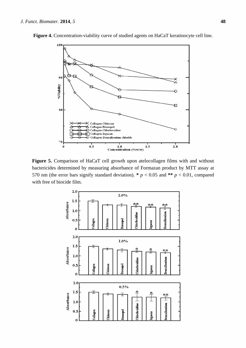
Figure 4 seeks to give other view of the results on Figure 3 by showing the dependence of cell
viability on concentration of agent in scatterplots instead of histograms. It shows the percentage of
viability with respect to this variable. It reveals a fall in cell viability by increasing concentration. In
general, all the plots have the same pattern. Chitosan is a particular case, because its curve evinces a
plateau followed by a decreasing trend at the highest concentrations (1.0% and 2.0%).
HaCaT cell proliferation on the substrates with and without biocides is given in Figure 5. It was
ascertained that cell attachment marginally diminished, as reflected by the spectrophotometric data of
each test, where all the absorbances corresponding to the added-agent samples were smaller than the
pristine ones. It may be noticed that only the substrates endowed with chitosan and bronopol did not
show a statistical significance. Conversely, all the samples with chlorhexidine, irgasan and benzalkonium
chloride have statistical significance and in five cases, these were considered as very significant ones,
chlorhexidine 2.0%; irgasan 2.0%; benzalkonium chloride 2.0%, 1.0% and 0.5% respectively.
These results were also qualitatively supported by the photomicrographs in Figure 6, where the
extent of cell adhesion on two non-cytotoxic substrates may be compared to a control specimen. As far
as surface morphology is concerned, the images show a vast amount of cell aggregates in form of
ripple-like areas adhered on the surfaces, with the typical keratinocyte cell shape.
J. Funct. Biomater. 2014, 5 48
Figure 4. Concentration-viability curve of studied agents on HaCaT keratinocyte cell line.
Figure 5. Comparison of HaCaT cell growth upon atelocollagen films with and without
bactericides determined by measuring absorbance of Formazan product by MTT assay at
570 nm (the error bars signify standard deviation). * p < 0.05 and ** p < 0.01, compared
with free of biocide film.

J. Funct. Biomater. 2014, 5 49
Figure 6. Photomicrographs of Human skin HaCaT keratinocytes in culture upon two
atelocollagen films compared with control: (A) Atelocollagen without biocides; (B) An
endowed film with cell viability above 80% (non-cytotoxic).
2.2. Discussion
As may be noted across this study, chitosan, bronopol and chlorhexidine have lower inhibition in
comparison with irgasan and benzalkonium chloride, which are the strongest ones in all the cases. As
well as it is important to emphasise the importance of pH and solubility on the yield of each agent,
since some of these bactericides do not possess the same effectiveness and stability in acid solution.
E.g., chitosan, bronopol and benzalkonium chloride are readily soluble and stable in water and acid
solution. In contrast to irgasan, which is slightly soluble in this medium and chlorhexidine is sparingly
soluble and unstable in acid pH [37–41]. As described in material section, the mixtures were prepared
by using a stirring machine for 4 h at 1000 rpm. When the biocide is soluble in the employed solvent, a
good dispersion (with cohesive character) and distribution of the agent into the mixture is obtained.
Contrariwise whether the substance is moderately soluble, the mixture is not uniform having loss of
agent during processing, and thus the final concentration is different and lower than the one that was
intended [42]. It means that irgasan, which has low solubility under the experimental conditions is
even able to suppress HaCaT cell growth. Whilst, despite bronopol and chitosan, are soluble do not
represent a serious risk to the viability of this cell line.
It is worth mentioning that for the studied samples preparation, the solvent (acetic acid 0.1 M) had
to be completely evaporated, since HaCaT as well as most cells require pHs around 7.0 and the
permanent control of pH is essential for optimal culture conditions [43,44].

J. Funct. Biomater. 2014, 5 50
Concerning the chosen method to estimate the extent of cytotoxicity, MTT is a rather cost-efficient
colorimetric technique, where the measurement strictly depends upon live cells, since the tetrazolium
salt 3-(4,5-dimethylthiazol-2-yl)-2,5-diphenyltetrazolium bromide is reduced to formazan product
exclusively by mitochondrial succinate dehydrogenase enzyme in the mitochondria of viable cells.
Hence, this assay ensures a good approximation in the study of cell viability and proliferation in cell
culture, where dead cells do not participate as interfering species. There are other factors that may
induce the reduction of MTT, such as times of incubation, age of culture, media poverty in glucose,
and reagents stability [45].
Cell proliferation under chitosan medium was higher than under the other media followed by
bronopol, chlorhexidine, irgasan and benzalkonium chloride respectively. This information coincides
with the cytotoxicity assay results, where the studied biocides performed in similar way in both
experiments (Chitosan, bronopol and chlorhexidine have lower inhibition ability than irgasan and
benzalkonium chloride).
Although the mechanisms of HaCaT cell adhesion and proliferation on different substrates are still
unclear, it is well-established that HaCaT keratinocytes proliferate better on rough, porous and
hydrophilic scaffolds. The cases of atelocollagen-chlorhexidine and irgasan substrates are a proof of that,
since these agents are toxic per se to this cell line and also because of their low solubility in the medium.
These substances may alter hydrophilicity, diminishing cell adhesion and proliferation [46,47].
In culture, keratinocyte cells behave in a similar way they do in vivo, where cells migrate towards
the air interface to form the epithelial surface. Epidermal substitutes require minimum two weeks to
expand keratinocytes population. For these reasons, it is necessary to pay heed to the stability of
keratinocytes attachment [48]. In that respect, the findings indicate that after four days in culture these
substrates hold low marginal toxicity, as well as suitability for cell proliferation. In fact, the MTT
assay was performed after 8 and 15 days showing no effect on the cell growth for and all the samples
with a concentration lower than 0.5% expect for the system with benzalkonium chloride. In contrast,
2.0% and 1.0% exhibited a decrease in cell growth by the passage of time. This drop was under 80% of
cell viability in the case of chlorhexidine, irgasan and benzalkonium systems, but none case reached
the moderate strong cytotoxicity range, which is within 60%–40%. The specimens endowed with
chitosan and bronopol 2.0% and 1.0% showed a slight alteration and all their systems kept as
non-cytotoxicity regardless of the experimentation time.
Surface adherence is a natural tendency, which is inherent to bacteria and other microorganisms. It
has four basic steps: adhesion, colonization, formation and the subsequent bacterial biofilm growth,
which is independent of the substrate. Biofilms act as defence mechanism against external agents; in
consequence, the aim of any antimicrobial materials is at preventing bacterial adhesion and colonization,
which are prerequisites to biofilm formation [49]. It is known by literature that benzalkonium chloride,
bronopol and chitosan hinder the adhesion of gram-positive strain, but do not behave satisfactorily
against gram-negative bacteria [50,51]; chlorhexidine and irgasan are efficacious against both
strains [52,53]. According to biothermodynamic studies, bacteria may attach to both hydrophobic and
hydrophilic surfaces; notwithstanding, hydrophobic surfaces are colonised faster than hydrophilic
ones [54,55]. This feature rises in importance, since the studied substrates are highly hydrophilic,
which is favourable to HaCaT cell adhesion but not to bacterial colonization.

J. Funct. Biomater. 2014, 5 51
The described phenomenon is largely surface specific and affects material functionality leading to loss
of physical and mechanical properties [56,57]. The overall outcome shows that by using a proper biocide
concentration cell viability and proliferation are barely impacted keeping the collagen-cell-attachment
properties almost steady (Cell viability rates beyond 80%). Consequently, these atelocollagen substrates
enhanced by the addition of one or more of these agents may render effectiveness against bacterial stains
and biofilm formation, being a promising view in the design of novel antimicrobial biomaterials
potentially suitable for tissue engineering applications, since atelocollagen is indeed prospective scaffold
for cell growth purposes, which also has the advantage of being eliminated by degradation processes
similar to the metabolism of endogenous collagen.
3. Experimental Section
3.1. Materials
Atelocollagen emulsion from bovine Achilles tendon (pH 3.5), which contains 1.4% of atelocollagen
was supplied by Vipo A.S (Partizánske, Slovakia). Acetic acid 99% was obtained from Penta (Prague,
Czech Republic). Bronopol (2-bromo-2-nitropropane-1,3-diol) C3H6BrNO4 98% was purchased from
Fluka (St. Louis, MO, USA). Benzalkonium chloride with a predominant formula of C12H25N(CH3)2C7H7Cl
98%; chitosan 98%; chlorhexidine (1,1-hexamethylene bis[5-(4-chlorophenyl)biguanide]) C22H30Cl2N10
98%; irgasan 5-chloro-2-(2,4-dichlorophenoxy)phenol C12H7Cl3O2 97% and Dimethyl sulfoxide
(DMSO) were provided by Sigma-Aldrich (St. Louis, MO, USA). The reagents in this study were used
as received without any further purification. Tissue culture dishes of 40 mm diameter and individual
wells of 96-well were commercially acquired from TPP (City, Switzerland). Vybrant®
MTT cell
proliferation Assay kit V-13154 was purchased from Invitrogen Corporation (Carlsbad, CA, USA).
3.2. Preparation of Collagen-Antibacterial Agent Substrates
Five mother mixtures of atelocollagen with each antibacterial agent (2.0% weight of agent/weight
collagen) were prepared by dissolving these compounds in 0.1 M water solution of acetic acid to
obtain a 0.1% weight of collagen/weight of solution, using an IKA RCT stirring machine (IKA®
works, Inc, Staufen, Germany) for 4 h at 1000 rpm. Less concentrated solutions (1.0%, 0.5%, 0.2%,
0.1% and 0.02%) were prepared by simple dilution. Each group of samples was casted on tissue
culture dishes and the solvent was evaporated at ambient conditions for three days. Thin films of pristine
atelocollagen were prepared and used as experimental blanks.
3.3. HaCaT Cell Incubation
Human immortalised non-tumorigenic keratinocyte cell line HaCaT, (Ethnicity, Caucasian; Age,
62 years; gender, Male and tissue, skin) were supplied by CLS Cell Lines Service (Eppelheim,
Germany). Dulbecco’s modified eagle medium, contains 4.5 g/L D-glucose, L-glutamine, and 110 mg/L
sodium pyruvate (DMEM; Invitrogen) supplemented with 2 mM L-glutamine, 10% foetal bovine
serum (FBS) and penicillin-streptomycin (100 U/mL-0.1 mg/mL) was used as a culture medium
(Biotech Inc., Oklahoma City, OK, USA). Cells were incubated at 37 °C for 24 h with 5% CO2 in
humidified air.

J. Funct. Biomater. 2014, 5 52
3.4. Evaluation of Cytotoxicity (in-Vitro)
3.4.1. Extract Preparation
The substrates obtained above were extracted according to ISO 10993-12 [58] in the ratio of 0.1 g
of the films per 1.0 mL of culture medium in chemically inert closed containers by using aseptic
techniques. Each extract was incubated in DMEM medium at 37 ± 1 °C with stirring for 24 h [59].
3.4.2. Cell Viability of HaCaT
All cells in the exponential growth phase were seeded in a concentration of 1 × 10⁵ cells/mL onto
the substrate extracts (2.0%, 1.0%, 0.5%, 0.2%, 0.1% and 0.02%). Cell viability as indicator of
cytotoxicity was determined after 4 days of culture by MTT assay. Absorbances were recorded by
using a Sunrise microplate ELISA reader at 570 nm (Tecan group, Männedorf, Switzerland), and all
determinations were performed in quadruplicate [60,61].
3.5. Cell Proliferation Test
HaCaT cell proliferation on thin films with the following specifications: collagen-benzalkonium
chloride, collagen-bronopol, collagen-chitosan, collagen-chlorhexidine and collagen-irgasan 2.0%,
1.0% and 0.5% was determined after 4 days of culture by MTT assay. A volume of 10 μL of 12 mM
MTT was taken for cell incubation performed at 37 °C for 4 h in the darkness. Thereafter, the media
were decanted and washed with phosphate-buffered saline solution (PBS). The produced formazan salts
were dissolved with dimethylsulphoxide (DMSO) and its concentration was measured in a spectrophotometer
at 570 nm [62]. The photomicrographs were taken by using an inverted phase-contrast microscope
Olympus CKX41 (Olympus, Münster, Germany) with an optical zoom of 40-times.
3.6. Statistical Analysis
All data were presented as the mean value ± standard deviation (SD) of each sample. Statistical
comparisons were performed using Student’s t-test with a confidence level of 95% (p < 0.05)
considered statistically significance and 99% (p < 0.01) considered very significant.
4. Conclusions
This contribution delved into the incorporation of bactericides to atelocollagen matrices. The
mixtures of atelocollagen with benzalkonium chloride, bronopol and chitosan are uniform and stable.
Cell viability of HaCaT is barely altered by the presence of these substances (74%–99%). Only in eight
from thirty samples the cell viability was statistically lower than that found on the substrates without
biocides. It means that any of these substrates provides an appropriate environment for this cell line.
Thus, the studied samples are perfectly apt for keratinocyte cell growth. Cytotoxicity is concomitant to
concentration and depends upon each agent. Bronopol and chitosan arise as the less hazardous to this
cell line having percentages of viability beyond 85% with a negligible cytotoxicity at lower
concentrations; whereas irgasan and benzalkonium chloride manifest more power of inhibition with

J. Funct. Biomater. 2014, 5 53
the highest rates of cytotoxicity throughout the study. This inhibition pattern might be observed in both
cytotoxicity and proliferation experiments and confirmed by statistic.
Acknowledgements
The authors would like to express their gratitude to the Operational Program Research and
Development for Innovations co-funded by the European Regional Development Fund (ERDF) and
national budget of Czech Republic, within the framework of project Centre of Polymer Systems
(registration number: CZ.1.05/2.1.00/03.0111), and The Advanced Theoretical and Experimental
Studies of Polymer Systems (registration number: CZ.1.07/2.3.00/20.0104).
Author Contributions
Jorge López-García and Petr Humpolíček made all the experiments, data analysis and build the
structure of the present text. Marián Lehocký and Petr Sáha supervised the data analysis, article writing
and financial support.
Conflicts of Interest
The authors declare no conflict of interest.
References
1. Parenteau-Bareil, R.; Gauvin, R.; Berthod, F. Collagen-based biomaterials for tissue enginnering
applications. Materials 2010, 3, 1863–1887.
2. López-García, J.; Humpolíček, P.; Lehocký, M.; Junkar, I.; Mozetič, M. Different source
atelocollagen thin films: Preparation, process optimisation and its influence on the interaction with
eukaryotic cells. Mater. Tehnol. 2013, 47, 473–479.
3. Bernal, A.; Balková, R.; Kuřítka, I.; Sáha, P. Preparation and characterisation of a new
double-sided bio-artificial material prepared by casting of poly(vinyl alcohol) on collagen. Polym.
Bull. 2012, 70, 431–453.
4. Tanaka, Y.; Yamaoka, H.; Nishizawa, S.; Nagata, S.; Ogasawara, T.; Asawa, Y.; Fujihara, Y.;
Takato, T.; Hoshi, K. The optimization of porous polymeric scaffolds for chondrocyte/atelocollagen
based tissue-engineered cartilage. Biomaterials 2010, 31, 4506–4516.
5. Banerjee, I.; Mishra, D.; Das, T.; Maiti, S.; Maiti, T.K. Caprine (Goat) collagen: A potential
biomaterial for skin tissue engineering. J. Biomater. Sci. Polym. Ed. 2012, 23, 355–373.
6. Lehmann, B. HaCaT cell line as a model system for vitamin D3 metabolism in human skin.
J. Invest. Dermatol. 1997, 108, 78–82.
7. Boukamp, P.; Petrussevska, R.T.; Breitkreutz, D.; Hornung, J.; Markham, A. Normal keratinization
in a spontaneously immortalized aneuploid keratinocyte cell line. J. Cell Biol. 1998, 106, 761–771.
8. García, J.L.; Pacherník, J.; Lehocký, M.; Junkar, I.; Humpolíček, P.; Sáha, P. Enhanced
keratinocyte cell attachment to atelocollagen thin films through air and nitrogen plasma treatment.
Prog. Colloid Polym. Sci. 2011, 138, 89–94.

J. Funct. Biomater. 2014, 5 54
9. Hacek, D.M.; Suriano, T.; Noskin, G.A.; Krusynski, J.; Reisberg, B.; Peterson, L.R. Medical and
economic benefit of a comprehensive infection control program that includes routine determination
of microbial clonality. Am. J. Clin. Pathol. 1999, 111, 647–654.
10. Bechert, T.; Steinrücke, P.; Guggenbichler, J.P. A new method for screening anti-infective
biomaterials. Nat. Med. 2003, 6, 1053–1056.
11. Popelka, A.; Novák, I.; Lehocký, M.; Junkar, I.; Mozetič, M.; Kleinová, A.; Janigová, I.;
Šlouf, M.; Bílek, F.; Chodák, I. A new route for chitosan immobilization onto polyethylene
surface. Carbohydr. Polym. 2012, 90, 1501–1508.
12. Kuceková, Z.; Humpolíček, P.; Kasparkova, V.; Perecko, T.; Lehocký, M.; Hauerlandová, I.;
Sáha, P.; Stejskal, J. Colloidal polyaniline dispersion: Antibacterial activity, cytotoxicity and
neutrophil oxidative burst. Colloids Surf. B 2014, 116, 411–417.
13. Hetrick, E.M.; Schoenfisch, M.H. Reducing implant-related infections: Active release strategies.
Chem. Soc. Rev. 2006, 35, 780–789.
14. Campoccia, D.; Montanaro, L.; Arciola, C.R. The significance of infection related to orthopedic
devices and issues of antibiotic resistance. Biomaterials 2006, 27, 2331–2339.
15. Kenawy, E.R.; Worley, S.D.; Broughton, R. The chemistry and applications of antimicrobial
polymers: A state-of-the-art review. Biomacromolecules 2007, 8, 1359–1384.
16. Sehgal, P.K.; Srinivasan, A. Collagen-coated microparticles in drug delivery. Expert Opin. Drug
Deliv. 2009, 6, 687–695.
17. Lee, J.E.; Park, J.C.; Kim, J.G.; Suh, H. Preparation of collagen modified hyaluronan microparticles
as antibiotics carrier. Yonsei Med. J. 2001, 42, 291–298.
18. Hume, E.B.H.; Baveja, J.; Muir, B.W.; Schubert, T.L.; Kumar, N.; Kjelleberg, S.; Griesser, H.J.;
Thissen, H.; Read, R.; Poole-Warren, L.A.; et al. The control of Staphylococcus epidermidis
biofilm formation and in vivo infection rates by covalently bound furanones. Biomaterials 2004,
25, 5023–5030.
19. Merchan, M.; Sedlaříkova, J.; Sedlařík, V.; Machovsky, M.; Svobodova, J.; Sáha, P. Antibacterial
polyvinyl chloride/antibiotic films: The effect of solvent on morphology, antibacterial activity and
release kinetics. J. Appl. Polym. Sci. 2010, 118, 2369–2378.
20. Bílek, F.; Sulovská, K.; Lehocký, M.; Sáha, P.; Humpolíček, P.; Mozetič, M.; Junkar, I.
Preparation of active antibacterial LDPE surface through multistep physicochemical approach II:
Graft type effect on antibacterial properties. Colloids Surf. B 2013, 102, 842–848.
21. López-García, J.; Kuceková, Z.; Humpolíček, P.; Mlček, J.; Sáha, P. Polyphenolic extracts of
edible flowers incorporated onto atelocollagen matrices and their effect on cell viability.
Molecules 2013, 18, 13435–13445.
22. Kenawy, E.R. Biologically active polymers. IV. Synthesis and antimicrobial activity of polymers
containing 8-hydroxyquinoline moiety. J. Appl. Polym. Sci. 2001, 82, 1364–1374.
23. Rees, E.N.; Tebbs, S.E.; Elliott, T.S.J. Role of antimicrobial-impregnated polymer and teflon in
the prevention of biliary stent blockage. J. Hosp. Infect. 1998, 39, 323–329.
24. Imbert, C.; Lassy, E.; Daniault, G.; Jacquemin, J.L.; Rodier, M.H. Treatment of plastic and
extracellular matrix components with chlorhexidine or benzalkonium chloride: Effect on Candida
albicans adherence capacity in vitro. J. Antimicrob. Chemother. 2003, 51, 281–287.

J. Funct. Biomater. 2014, 5 55
25. Bryce, D.M.; Croshaw, B.; Hall, H.E.; Holland, V.R.; Lessel, B. The activity and safety of the
antimicrobial agent bronopol (2-bromo-2-nitropropan-1,3-diol). J. Soc. Cosmet. Chem. 1978, 29,
3–24.
26. Legin, G.Y. 2-Bromo-2-nitro-1,3-propanediol (Bronopol) and its derivatives: Synthesis, properties,
and application (a review). Pharm. Chem. J. 1994, 30, 54–64.
27. Van Rijkom, H.M.; Truin, G.J.; van’t Hof, M.A. A meta-analysis of clinical studies on the
caries-inhibiting effect of chlorhexidine treatment. J. Dent. Res. 1996, 75, 790–795.
28. Lee, D.H.; Spångberg, L.S.W.; Bok, Y.B.; Lee, C.Y.; Kum, K.Y. The sustaining effect of three
polymers on the release of chlorhexidine from a controlled release drug device for root canal
disinfection. Oral Surg. Oral Med. Oral Pathol. Oral Radiol. Endodontol. 2005, 100, 105–111.
29. Junker, L.M.; Hay, A.G. Effects of triclosan incorporation into ABS plastic on biofilm
communities. J. Antimicrob. Chemother. 2004, 53, 989–996.
30. Rabea, E.I.; Badawy, M.E.T.; Stevens, C.V.; Smagghe, G.; Steurbaut, W. Chitosan as
antimicrobial agent: Applications and mode of action. Biomacromolecules 2003, 4, 1457–1465.
31. D’Ayala, G.G.; Malinconico, M.; Laurienzo, P. Marine derived polysaccharides for biomedical
applications: Chemical modification approaches. Molecules 2008, 13, 2069–2106.
32. ISO 10993-5:2009 Biological Evaluation of Medical Devices. Part 5: Tests for In Vitro
Cytotoxicity; International Organization for Standardization: Geneva, Switzerland, 2009.
33. Hasobe, M.; Mckee, J.G.; Borchardt, R.T. Relationship between intracellular concentration of
S-Adenosylhomocysteine and inhibition of vaccinia virus replication and inhibition of murine
L-929 cell growth. Antimicrob. Agents Chemother. 1989, 33, 828–834.
34. Monsk, A.; Scudiero, D.; Skehan, P.; Shoemaker, R.; Paull, K.; Vistica, D.; Hose, C.; Langley, J.;
Cronise, P.; Vaigro-Wolff, A.; et al. Feasibility of a high-flux anticancer drug screen using a
diverse panel of cultured human tumor cell lines. J. Natl. Cancer Inst. 1991, 83, 757–766.
35. Sun, T.; Li, Z.L.; Tian, H.; Wang, S.C.; Cai, J. Synthesis and biological evaluation of novel
1-alkyltryptophan analogs as potential antitumor agents. Molecules 2009, 14, 5339–5348.
36. Han, D.W.; Lee, M.H.; Kwon, B.J.; Kim, H.L.; Hyon, S.H.; Park, J.C. Selective inhibitory effect
of epigallocatechin-3-gallate on migration of vascular smooth muscle cells. Molecules 2010, 15,
8488–8500.
37. Stretton, R.J.; Manson, T.W. Some aspects of the mode of action of the antibacterial compound
bronopol (2-bromo-2-nitropropan-1,3-diol). J. Appl. Microbiol. 1973, 36, 61–76.
38. Russell, A.D.; Path, F.R.C. Chlorhexidine-antibacterial action and bacterial resistance. Infection
1986, 14, 212–215.
39. Marple, B.; Roland, P.; Benninger, M. Safety review of benzalkonium chloride used as a
preservative in intranasal solutions: An overview of conflicting data and opinions. Otolaryngol.
Head Neck Surgery 2004, 130, 131–141.
40. Qin, C.; Li, H.; Xiao, Q.; Liu, Y.; Zhu, J.; Du, J. Water-solubility of chitosan and its antimicrobial
activity. Carbohydr. Polym. 2006, 63, 367–374.
41. Aragón, D.M.; Ruidiaz, M.A.; Vargas, E.F.; Bregni, C.; Chiappetta, D.A.; Sosnik, A.; Martínez, F.
Solubility of the antimicrobial agent triclosan in organic solvents of different hydrogen bonding
capabilities at several temperatures. J. Chem. Eng. Data 2008, 53, 2576–2580.

J. Funct. Biomater. 2014, 5 56
42. Tadmor, Z.; Gogos, C.G. Principles of Polymer Processing, 2nd ed.; John Wiley & Sons:
Hoboken, NJ, USA, 2006; pp. 322–328.
43. Altenburger, R.; Kissel, T. The human keratinocyte cell line HaCaT: An in vitro cell model for
keratinocyte testosterone metabolism. Pharm. Res. 1999, 16, 766–771.
44. Deyrieux, A.F.; Wilson, V.G. In vitro culture conditions to study keratinocyte differentiation
using the HaCaT cell line. Cytotechnology 2007, 54, 77–83.
45. Vistica, D.T.; Skehan, P.; Scudiero, D.; Monks, A.; Pittman, A.; Boyd, M.R. Tetrazolium-based
assays for cellular viability: A critical examination of selected parameters affecting formazan
production. Cancer Res. 1991, 51, 2515–2520.
46. Peschel, G.; Dashe, H.M.; Kanrad, A. Growth of keratinocytes on porous films of
poly(3-hydroxybutyrate) and poly(4-hydroxybutyrate) blended with hyaluronic acid and chitosan.
J. Biomed. Res. Part A 2007, 85, 1072–1081.
47. García, J.L.; Asadinezhad, A.; Pacherník, J.; Lehocký, M.; Junkar, I.; Humpolíček, P.; Sahá, P.;
Valášek, P. Cell proliferation of HaCaT keratinocytes on collagen films modified by argon plasma
treatment. Molecules 2010, 15, 2845–2856.
48. Metcalfe, A.D.; Ferguson, M.W.J. Tissue engineering of replacement skin: The crossroads of
biomaterials, wound healing, embryonic development, stem cells and regeneration. J. R. Soc.
Interface 2007, 4, 413–437.
49. Lynch, A.S.; Robertson, G.T. Bacterial and fungal biofilm infections. Annu. Rev. Med. 2008, 59,
415–428.
50. Asadinezhad, A.; Novák, I.; Lehocký, M.; Sedlařík, V.; Vesel, A.; Junkar, I.; Sáha, P.; Chodák, I.
An in vitro bacterial adhesion assessment of surface-modified medical-grade PVC. Colloids Surf.
B 2010, 77, 246–256.
51. Asadinezhad, A.; Novák, I.; Lehocký, M.; Bílek, F.; Vesel, A.; Junkar, I. Polysaccharides coatings
on medical-grade PVC: A probe into surface characteristics and the extent of bacterial adhesion.
Molecules 2010, 15, 1007–1027.
52. Odore, R.; Valle, V.C.; Re, G. Efficacy of chlorhexidine against some strains of cultured and
clinically isolated microorganisms. Vet. Res. Commun. 2000, 24, 229–238.
53. Asadinezhad, A.; Novák, I.; Lehocký, M.; Sedlařík, V.; Vesel, A.; Junkar, I. A physicochemical
approach to render antibacterial surfaces on plasma-treated medical-grade PVC: Irgasan coating.
Plasma Process Polym. 2010, 7, 504–514.
54. Tsibouklis, J.; Stone, M.; Thorpe, A.A.; Graham, P.; Peters, V.; Heerlien, R.; Smith, J.R.;
Green, K.L.; Nevell, T.G. Preventing bacterial adhesion onto surfaces: The low-surface-energy
approach. Biomaterials 1999, 20, 1229–1235.
55. Neu, T.R. Significance of bacterial surface-active compounds in interaction of bacteria with
interfaces. Microbiol. Rev. 1996, 60, 151–166.
56. Esperanza, G.; Gottardi, G.; Pederzolli, C.; Lunelli, L.; Canteri, R.; Pasquardini, L.; Carli, E.;
Lui, A.; Maniglio, D.; Brugnara, M.; et al. Role of chemical interactions in bacterial adhesion to
polymer surfaces. Biomaterials 2004, 25, 2029–2037.
57. Lichter, J.A.; Thompson, M.T.; Delgadillo, M.; Nishikawa, T.; Rubner, M.F.; van Vliet, K.J.
Substrata mechanical stiffness can regulate adhesion of viable bacteria. Biomacromolecules 2008,
9, 1571–1578.

J. Funct. Biomater. 2014, 5 57
58. ISO 10993-12:2012 Biological Evaluation of Medical Devices. Part 12: Sample Preparation and
Reference Materials; International Organization for Standardization: Geneva, Switzerland, 2007.
59. Weyermann, J.; Lochmann, D.; Zimmer, A. A practical note on the use of cytotoxicity assays.
Int. J. Pharm. 2005, 288, 369–376.
60. Freshney, R.I. Culture of Animal Cells: A Manual of Basic Techniques, 4th ed.; John Wiley & Sons:
Hoboken, NJ, USA, 2005; pp. 359–373.
61. Roy, N.; Saha, N.; Humpoliček, P.; Sáha, P. Permeability and biocompatibility of novel
medicated hydrogel wound dressings. Soft Mater. 2010, 8, 338–357.
62. Mosmann, T. Rapid colorimetric assay for cellular growth and survival: Application to
proliferation and cytotoxicity assays. J. Immunol. Meth. 1983, 65, 55–63.
© 2014 by the authors; licensee MDPI, Basel, Switzerland. This article is an open access article
distributed under the terms and conditions of the Creative Commons Attribution license
(http://creativecommons.org/licenses/by/3.0/).