In Tech-Reproductive and developmental toxicity of insecticides
Upload
independentCategory
view
0download
0
Published by Oxford University Press 2005.
Effects of organochlorine insecticides on MAP kinase pathways in human HaCaT
keratinocytes : Key role of reactive oxygen species
Nathalie Ledirac*,‡,§, Sebastien Antherieu*,‡ , Anne Dupuy d’Uby*, Jean-Claude Caron† and
Roger Rahmani*
*Laboratoire de Toxicologie Cellulaire et Moléculaire, Centre de Recherche INRA, 400 route
des Chappes, 06903 Sophia-Antipolis, France.
†Galderma R&D, les Templiers, 2400 routes des Colles, 06410 Biot, France.
§Corresponding author. Tel. & Fax: +33 4 92 38 65 64
E-mail address: [email protected]
Section : Environmental Toxicology
ToxSci Advance Access published May 25, 2005

Abstract : Organochlorine pesticides (OCs) are reported as potential carcinogens in humans.
The aim of this study was to investigate the effects of four OCs (dieldrin, endosulfan,
heptachlor and lindane) on MAPK cascades and more specifically to identify the mechanism
underlying OCs-induced ERK1/2 activation. OCs increased phosphorylated Raf, MEK1/2,
ERK1/2 and c-Jun in human HaCaT cells, but had no effect on p38 MAPK activation.
Moreover, blockade of Raf, MEK1/2 or PKC activation with geldanamycin, U0126 or
calphostin C inhibited ERK1/2 phosphorylation, demonstrating a PKC-Raf-MEK1/2 pathway.
We also showed that these insecticides induced the production of reactive oxygen species
(ROS). Pre-treatment with antioxidant molecule N-acetyl cysteine sharply decreased the level
of phospho-ERK1/2, and had no effect on Raf and MEK1/2 activation, suggesting a Raf-
independent mechanism. This study indicates that OCs strongly activate the ERK1/2 pathway,
and identifies a critical role of ROS in OCs-induced ERK activation probably by stabilizing
its phosphorylation.
Keywords: ERK1/2; Keratinocytes; Mitogen-activated protein kinases; Organochlorines;
Reactive oxygen species; Signal transduction

1 Introduction
Organochlorine pesticides (OCs) are organic compounds that persist in the
environment, bioaccumulate through the food chain, and pose a risk of causing adverse
effects to human health and the environment. These pesticides, characterized by their cyclic
structure, number of chlorine atoms and low volatility, can be divided into four groups:
dichlorodiphenylethanes (such as DDT), cyclodienes (such as dieldrin, endosulfan,
heptachlor), chlorinated benzenes (such as hexachlorobenzene), and cyclohexanes (such as
lindane). Although these chemicals were widely used until the mid-1970s, most of them are
now banned from use in developed countries. However, they are still being produced and used
in other countries. Furthermore, one of these insecticides, endosulfan, is still in widespread
use throughout the world despite its known adverse effect on human as an endocrine-
disrupting compound (Andersen et al., 2002; Scippo et al., 2004). The majority of OCs is also
considered Persistent Organic Pollutants (POPs). These POPs chemicals, including nine OCs
(aldrin, chlordane, DDT, dieldrin, endrin, heptachlor, hexachlorobenzene, mirex and
toxaphen), were targeted by the Stockholm Convention in May 2001, which aimed to
eliminate their productions and restrict or ban their uses in the world (http://www.pops.int/).
Many human epidemiologic and animal studies have shown that exposure to OCs is
positively correlated with endocrine disruption (Lemaire et al., 2004), reproductive and
immune dysfunctions (Ayub et al., 2003; Reed et al., 2004; Saiyed et al., 2003), and cancers
(e.g. breast cancer (Kalantzi et al., 2004; Zou and Matsumura; 2003)). Human exposure
occurs mainly by ingestion (from eating contaminated foods), inhalation, absorption through
skin, and often during pest control operations both at home and in leisure areas. Therefore, to
investigate the toxicological effects of OCs after cutaneous exposure, we used the
spontaneously immortalized human keratinocyte cell line, HaCaT (Boukamp et al., 1988).
This well-characterized cell line is still able to proliferate and differentiate, and it has been
shown to be a good model in toxicology (Delescluse et al., 1998; Ledirac et al., 1997).
Moreover, these cells show good predictive results in comparison with keratinocyte and skin
models, and therefore constitute an appropriate model to study the molecular mechanisms
regulating keratinocyte growth and differentiation.
The mitogen-activated protein kinases (MAPKs) are serine/threonine kinases that
transduce signals from the plasma membrane to the nucleus, they play a critical role in
controlling cell survival, proliferation, and differentiation (Chang and Karin, 2001). In
epithelial cells, and in particular in keratinocytes, deregulation of MAPK signaling pathways
can lead to hyperproliferation and altered differentiation, which in turn contribute to
photoaging, psoriasic epidermis and malignant transformation in human cancers, including
those of the cutaneous epithelium (Bedogni et al, 2004; Chung et al., 2000; Takahashi et al.,
2002). Mammals express at least four MAPK subfamilies, extracellular signal-related kinases
(ERK), Jun amino-terminal kinases (JNK), p38 MAPK and ERK5 (Gutkind, 2000). ERK1/2
is mainly activated by mitogenic stimuli and has been linked to cell survival, whereas the JNK
and p38 MAPK pathways are primarily activated by stress stimuli and are linked to the
induction of differentiation and apoptosis.
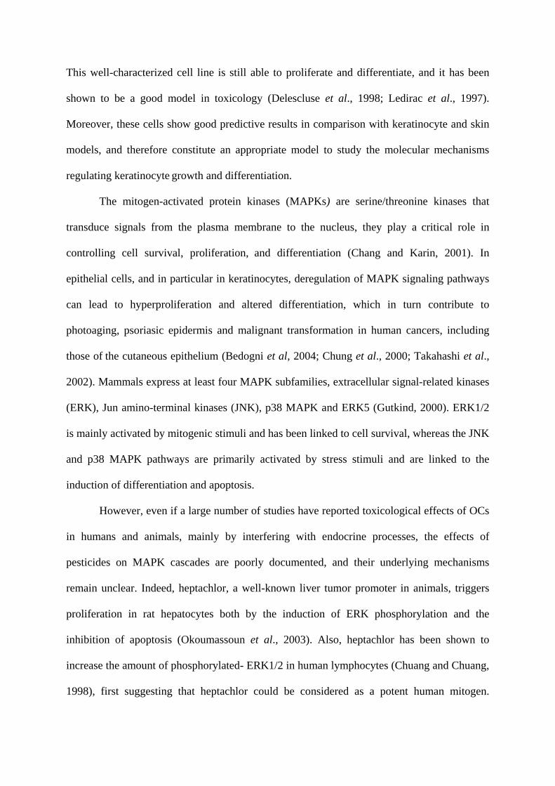
However, even if a large number of studies have reported toxicological effects of OCs
in humans and animals, mainly by interfering with endocrine processes, the effects of
pesticides on MAPK cascades are poorly documented, and their underlying mechanisms
remain unclear. Indeed, heptachlor, a well-known liver tumor promoter in animals, triggers
proliferation in rat hepatocytes both by the induction of ERK phosphorylation and the
inhibition of apoptosis (Okoumassoun et al., 2003). Also, heptachlor has been shown to
increase the amount of phosphorylated- ERK1/2 in human lymphocytes (Chuang and Chuang,
1998), first suggesting that heptachlor could be considered as a potent human mitogen.

However, more recent studies have demonstrated that heptachlor, like endosulfan, induces
apoptosis in those human lymphocytes (Kannan et al., 2000; Rought et al., 2000). In fact, in
contrast with the proliferating effects observed in animals, heptachlor provokes alterations of
cell cycle progression and induction of programmed cell death in human lymphocytes
(Chuang et al., 1999).
The aim of this study was to better understand the cellular events leading to OCs-
mediated toxicity and more particularly to investigate whether various compounds belonging
to the OCs family, such as dieldrin, endosulfan, heptachlor and lindane (figure 1), can activate
MAPKs and thus influence important cellular processes. In another respect, OCs such as
dieldrin and more recently endosulfan, have been reported to induce reactive oxygen species
(ROS) production in human liver. Therefore, the induction of ROS production by these
molecules and the involvement of that ROS production in MAPK signaling pathways were
investigated.

2 Materials and methods
2.1 Chemicals
Dulbeco’s modified Eagle’s medium (DMEM), fetal bovine serum (FBS), penicillin-
streptomycin, sodium pyruvate, Eagle’s non-essential amino-acids were from BioWhittaker
(Cambrex company, Walkersville, USA). Anti-phospho-p38, anti-phospho-c-Jun, anti-
phospho-MEK1/2, anti-phospho-Raf, anti-c-Jun, anti-p38, anti-MEK2 and MEK1/2 inhibitor
U0126 were purchased from Cell Signaling (Beverly, MA). Anti-ERK2 was from Santa Cruz
(Santa Cruz, CA). Anti-phospho ERK1/2 and N-acetyl cysteine (NAC) were from Sigma-
Aldrich (St. Louis, MO). Raf inhibitor geldanamycin and PKC inhibitor calphostin C were
purchased from Alexis Biochemicals. Organochlorine pesticides were from ChemService
(West Chester, PA).
2.2 Cell culture
HaCaT Cells (a generous gift from Prof. Fusenig, German Cancer Research Institute,
Heidelberg, Germany) were derived from a spontaneously immortalized human keratinocyte
but are non-tumorigenic epidermal cell line, and exhibit many of the morphological and
functional properties of normal human keratinocytes. HaCaT cells were cultured in DMEM
supplemented with 10% FBS, penicillin (100 U/ml), streptomycin (100 µg/ml), sodium
pyruvate (1mM) and non-essential amino acids (0.1 mM). Cultures were incubated at 37°C in
a humidified atmosphere with 95% air and 5% CO2.

2.3 Cell viability
The cytotoxicity of OCs was evaluated after 24 h of exposure by MTT (3-(4,5-
dimethylthiazol-2-yl)-2,5-diphenyltetrazolium bromide) colorimetric assay, according to
(Fautrel et al., 1991). Briefly, HaCaT cells were seeded in 96-well plates and grown to
confluence. Then cells were treated with various concentrations of the tested pesticides for 24
h. The next day medium was removed and 100 µL of serum-free DMEM containing MTT
(0.5 mg/ml) were added to each well and incubated 2 h at 37°C. Finally, solutions were
removed, the water-insoluble formazan was dissolved in 100 µl DMSO, and absorbance was
measured at 550 nm.
2.4 ROS measurement
Intracellular ROS generation was assessed by using 2’, 7’-dichlorodihydrofluorescein
diacetate (H2DCFDA) from Molecular Probes (Eugene, Oregon, USA). HaCaT cells were
seeded in 12-well plates and grown to confluence. Then cells were treated for 90 min at 37°C
with OCs in the presence of 100 µM H2DCFDA (the stock solution was made in ethanol, so
that final concentration in the medium was 0.33%). After the incubation time, cells were
washed twice with cold PBS, and scraped in potassium buffer (10 mM pH7.4) / methanol
(v/v) completed with Triton X100 (0.1%). An aliquot of 100 µL was incubated in a black 96-
well plate and relative fluorescence intensity was determined by spectrofluorimetry (λex = 488
nm, λem = 520 nm).

2.5 Western blot analysis
HaCaT cells were plated in 6-well plates and grown to confluence. The confluent cells were
serum starved for 24 h to establish quiescence (except for p38 MAPK), and then stimulated
with OCs for the indicated periods and at the indicated concentrations. After treatments, cells
were scraped and resuspended in buffer A containing the protease inhibitor cocktail (25 mM
HEPES (pH7.5), 5 mM MgCl2, 5 mM EDTA, 5 mM DTT, 2 mM PMSF, 10 µg/ml pepstatin
A and 10 µg/ml leupeptin). The protein concentration in each cell lysate was measured by
commercial method (BCA Protein Assay Kit), using BSA as the standard. Thirty micrograms
of total protein were resolved on 11% SDS-polyacrylamide gels electrophoresis and blotted
on PVDF membranes. Membranes were immunoblotted with anti-phospho-c-Jun (1:1000),
anti-phospho-p38 (1:2000) anti-phospho-Raf (1:1000), anti-phospho-MEK1/2 (1:1000), anti-
c-Jun (1:1000), anti-p38 (1:1000), or anti-MEK2 (1:1000) antibodies overnight at 4°C, or
anti-ERK2 (1:5000), anti-phospho-ERK1/2 (1:5000) antibodies for 1 h at room temperature.
Membranes were then reacted with horseradish peroxydase-conjugated secondary antibodies
(anti-mouse or anti-rabbit immunoglobulin G) for 1 h at room temperature. After washing,
blots were reacted using an ECL detection kit (Amersham Biosciences).
2.6 Western blot densitometric quantitation
Films were scanned and quantitated with the Chemi Genus Bio Imaging System (SynGene,
Sunnyvale, CA). The amount of phosphorylated protein detected was quantified and values
from multiple experiments were averaged and graphed. The Y-axis was presented as arbitrary
units. Error bars in each of the figures represent the standard deviation of the mean.

2.7 Statistical analysis
The statistical differences between different treatment groups were determined by
Student's t test and probability levels were noted as * (p<0.05) and ** (p<0.001). Data are
expressed as means ± standard deviations (SD) for at least three independent determinations
for each experimental point.

3 Results 3.1 Cytotoxicity Cytotoxicity studies of four OCs were performed by using two different endpoints – the MTT
reduction and neutral red (data not shown) tests. As shown in table 1, significant differences
were observed between the tested compounds. Indeed, endosulfan and heptachlor exerted a
cytotoxic effect with an IC50 ranging from ≈ 20-40 to 100 µM depending on the absence or
presence of serum in the medium, whereas dieldrin and lindane did not lead to cellular cell
death regardless of concentration and culture conditions. However, it should be pointed out
that due to the poor solubility (≤ 200 µM) of the two latter molecules in the culture medium,
their IC50 values could not be determined precisely. Nevertheless these experiments allowed
us to optimize experimental concentrations for the induction studies. Figure 2 illustrates the
dose-dependent toxicity of the two most toxic insecticides after 24 h exposure in complete
medium, and shows the absence of any relevant cytotoxicity at 25 µM, according to the
cytotoxicity tests used. We therefore used the subtoxic concentration 25 µM to study the early
events leading to toxic response.
3.2 OCs induce ERK1/2 and JNK but not p38 MAPK activation
To determine the effects of OCs on MAPK cascades, we studied the ability of these molecules
to activate the phosphorylation of ERK1/2, c-Jun and p38 MAPK after 1 h exposure on
HaCaT cells. As shown in figure 3, OCs activate both ERK1/2 and c-Jun, but have no
induction effect on p38 MAPK phosphorylation, compared with that obtained with sorbitol-
induced osmotic stress (Figure 3, line So). Endosulfan and heptachlor induce a strong
activation of ERK1/2, above that produced by FBS, whereas c-Jun is more strongly activated
by dieldrin and endosulfan, and to a lesser extent by heptachlor and lindane. As expected, no
effect was obtained on the inactive forms of ERK, c-Jun and p38 MAPK. These results are in

agreement with those previously obtained for ERK1/2 activation by 50 µM heptachlor in
human lymphocytes (Chuang and Chuang, 1998; Chuang et al., 1999), and clearly indicate
that OCs, endosulfan in particular, are able to strongly activate the ERK1/2 and JNK signaling
pathways, and therefore interfere with cellular functions.
3.3 Raf and MEK1/2 are required for OCs–induced ERK1/2 phosphorylation
Figure 4 shows the dose-dependent increase in ERK1/2 activation by the four OCs. Maximal
induction effect was observed for all the tested compounds at concentration of 25 µM, with a
strong effect of endosulfan as low as 10 µM. The phosphorylation of both MEK1/2 and Raf
was also induced by OCs, with a lesser effect of lindane, a less potent activator than other
OCs (fig. 5A). These results suggest the involvement of Raf and MEK1/2 in ERK1/2
activation by OCs, and clearly indicate that OCs of the cyclodiene group are more potent
activators. This difference may be explained by their different chemical structures.
Experiments with DDT, a dichlorodiphenylethane, have produced similar results to those we
obtained with cyclodienes (data not shown), and confirm the existence of a relationship
between the chemical structure (polyclyclic structures by comparison with that of lindane)
and the level of ERK1/2-induced phosphorylation.
To further confirm the upstream mechanism of OCs-induced ERK1/2 activation, we analyzed
the effects of U0126, a specific inhibitor of MEK1/2, and geldanamycin, which destabilizes
Raf-1 and disrupts the Raf-MEK-ERK pathway (Schutle et al., 1996), on induced-ERK1/2
phosphorylation. As shown in figure 5B, U0126 completely blocked ERK1/2 activation.
Surprisingly, although geldanamycin completely blocked any activation of Raf
phosphorylation by OCs (data not shown), ERK1/2 was still weakly activated by dieldrin,
endosulfan and heptachlor. These results suggest that OCs activate ERK1/2 through distinct
pathways including the Raf-MEK signaling cascade and a Raf-independent mechanism.

3.4 Involvement of PKC in OCs-induced ERK1/2 phosphorylation
Protein Kinase C (PKC) is involved in many cellular responses and in particular in Raf-1
stimulation. In addition, OCs were previously described as stimulating PKC activity both in
vitro and in vivo (Bagchi et al., 1997; Moser and Smart, 1989). Therefore, to examine the role
of PKC in OCs-induced ERK1/2 activation, experiments were performed with calphostin C, a
highly specific inhibitor of PKC (Fig. 5B). Pre-treatment of HaCaT cells during 4 h with
calphostin C, entirely blocked OCs-induced ERK1/2 phosphorylation, suggesting a key role
of PKC. We also noted that the amounts of phosphorylated Raf-1 were similar to those
obtained by treatment with geldanamycin (data not shown), suggesting a direct PKC-MEK1/2
mechanism for the residual ERK1/2 activation observed with geldanamycin. Taken together,
these data clearly indicate that OCs can activate ERK1/2 via PKC-Raf-MEK1/2 and PKC-
MEK1/2-dependent pathways (Schönwasser et al., 1998). This direct activation of MEK by
PKC, such as PKC-ζ, has already been demonstrated for insulin- and LPS-induced ERK1/2
phosphorylation (Monick et al., 2000; Sajan et al., 1999).
3.5 OCs- induced intracellular ROS generation
Many studies have shown that oxidative stresses induced JNK and p38 MAPK signaling
cascades, and to a lesser extent ERK1/2 pathway. Although OCs have been described to
increase ROS formation particularly in the liver, few studies have been conducted, which
involved other human tissue or cell types (Kannan and Jain, 2003). To determine whether
OCs treatments are associated with changes in intracellular ROS levels in HaCaT cells, we
measured oxidation of H2DCF diacetate (H2DCFDA). Intracellular esterases convert cell-
permeable H2DCFDA to HDCF, which is subsequently oxidized by ROS to fluorescent DCF
(Myhre et al., 2003). Primaquine, a known pro-oxidant compound (Magwere et al., 1997),

was chosen as a positive control. Figure 6 illustrates DCF fluorescence in response to two
insecticides concentrations (25 and 50 µM), and shows dose-dependent increase in ROS
generation with dieldrin, endosulfan and heptachlor, without dose-dependence with lindane.
At 25 µM, all the tested compounds induce a significant increase in DCF fluorescence : 1.6
fold increase with lindane, 2 fold increase with dieldrin and endosulfan, and 2.5 with
heptachlor. Maximum effect is observed at 50 µM heptachlor (3.4 fold over DMSO control).
3.6 Effect of oxidative stress on ERK and JNK signaling pathways
To determine the role of OCs-induced ROS generation on both ERK and JNK activation, we
studied the effects of the antioxidant molecule N-acetyl cysteine (NAC). In order to determine
the optimal concentration of NAC that inhibits OCs-induced oxidative stress, HaCaT cells
were pre-treated for 24 h with various concentrations of NAC (1-10 mM). Figure 7 shows a
dose-dependent decrease of OCs-induced oxidative stress after NAC pre-treatment, and
demonstrates that oxidative stress induced by lindane and dieldrin was completely blocked
with 5 mM of NAC, whereas 10 mM were necessary to inhibit ROS production induced by
endosulfan and heptachlor.
Next we examined the effects of NAC on ERK1/2 activation by OCs. HaCaT cells were pre-
treated for 24 h with 10 mM NAC, and then stimulated for 1 h with the four insecticides. As
indicated in figure 8A, NAC pre-treatment markedly decreases the OCs-induced ERK1/2
activation, whereas the corresponding level of phospho-MEK1/2 remains unchanged.
Moreover, NAC pre-treatment has no effect on serum-induced ERK1/2 activation, as
expected since this activation is ROS-independent, and has no significant effect on basal ERK
phosphorylation. These results indicate clearly that ROS contribute to increase the level of
OCs-induced ERK1/2 phosphorylation, but do not lead to the activation of the upstream
MAPKs. These data provide strong support of a crucial role of ROS in OCs-induced ERK1/2

phosphorylation, probably via a Raf-MEK-independent mechanism. Oxidative modifications
of other molecules including ERK-specific phosphatases may also contribute to an increase in
ERK1/2 phosphorylation level (Levinthal and Defranco, 2005). Results presented in figure
8B, indicate that pre-treatment with NAC partially prevents OCs-induced c-Jun
phosphorylation, suggesting the possibility of a similar mechanism in OCs-induced JNK
activation.

4 Discussion The association between the levels of exposure to organochlorine pesticides and cancers is
still largely debated. Indeed, in addition to their known estrogenic characteristics, OCs have
been especially implicated as risk factors for breast cancers. Although OCs such as lindane
and DDT have been shown to increase cell proliferation of MCF-7 cells (Steinmetz et al.,
1996; Zou and Matsumura, 2003), no mechanism has clearly been established yet, and
available data do not support the hypothesis that these chemicals increase the risk of breast
cancer. Nevertheless the toxicological effects of OCs have been extensively studied in
animals in which several studies have revealed that they provoke a vast range of disorders, but
they remain poorly documented in humans. Dietary contribution is probably the most
significant route of human exposure. Maximum levels were found in vegetables, specifically
in Egypt; plants like potato tubers or strawberries could contain high levels of lindane
residues (850 µg/kg) and dieldrine (220 µg/kg), respectively (Mansour SA, 2004). Although
dietary intake has decreased since 1970, tissue bioaccumulation, which is a common feature
of insecticides, should not be ruled out. This could lead to unexpectedly high intracellular OC
concentrations on hepatic and epidermal sites. Furthermore, during the application period,
OCs may be found in surface water and reservoirs. The general population can also be
exposed to OCs during pest control operations, both at home and in recreational areas.
Finally, the highest dermal and inhalation exposures documented were reported in farm
workers involved in the spraying of these insecticides. The mean dermal exposures to mixers
and sprayers of endosulfan via a tractor-mounted boom sprayer and highboy were 16.18 and
8.06 mg/kg/day, respectively (Lonsway JA et al., 1997). The authors concluded that not using
protective measures when spaying endosulfan could lead to poisoning. The present study was
therefore designed to assess the toxicological effects of OCs in humans after acute dermal
exposure by using the HaCaT cell line, the most widely studied keratinocyte cell line and to

try and better understand the effects of OCs on a population chronically exposed by dermal
contact.
Because MAPKs are key enzymes in signal transduction, we determined whether OCs had an
inducing effect on MAPK cascades, and provided data supporting the existence of an
additional ROS-dependent mechanism in MAPK activation by OCs. In this report we
demonstrate that multiple members of the MAPK family are stimulated by OCs and that
ERK1/2 is strongly activated in HaCaT cells. The OCs-induced ERK1/2 activation is
significantly reduced or completely blocked by inhibitors of MEK1/2 (U0126), Raf-1
(geldanamycin) and PKC (calphostin C). In addition, our data indicate that OCs induce Raf
and MEK1/2 phosphorylation, and therefore demonstrate that these pesticides induce ERK1/2
phosphorylation via successive activations of PKC, Raf-1 and MEK1/2. These results are
consistent with a previous study that reported ERK1/2 activation by 50 µM heptachlor in
human lymphocytes (Chuang and Chuang, 1998); they also clearly demonstrate that
insecticides of the organochlorine family induce the same MAPK activation profile in HaCaT
cells, namely ERK and JNK, but not p38 MAPK activation.
Furthermore OCs have been shown to induce oxidative stress in certain mammalian species
(Bayoumi et al., 2000), and the reactive oxygen species (ROS) generation has been described
to interfere with various signaling pathways, including MAPKs. Indeed all MAPK cascades
are known to be activated in response to oxidant injury (Gupta et al., 1999; Martindale and
Holbrook, 2002) and can therefore have an impact on cell survival and cell death. A recent
study has reported that exposure to 10-100 µM endosulfan levels induced apoptosis in human
T-cells via a bcl-2-independent mechanism and suggested that endosulfan-induced apoptosis
may be linked to excessive ROS production (Kannan et al., 2000). However, the mechanisms
that mediate MAPK activation by oxidants and the variability of mammalian sensitivity to
oxidant injury remain to be understood. Moreover, no data are currently available on OCs-

induced oxidative stress in human keratinocytes, or on the impact of this oxidant injury on
cell signaling.
Under our experimental conditions, the four OCs tested enhance the production of ROS in a
dose-dependent manner. When cells were pre-treated with increasing concentrations of the
antioxidant agent NAC, ROS induction was reduced, and the phosphorylation of both ERK1/2
and c-Jun was partially decreased. Interestingly, MEK and Raf phosphorylation was not
affected by NAC pre-treatment, suggesting that OCs-induced ERK1/2 phosphorylation is in
part dependent on a ROS-dependent mechanism downstream of the MEK1/2 level. Recent
studies have demonstrated the role of oxidative stress in the inactivation of phosphatase
activity, and have precisely described the mechanism of ROS regulation (Lee and Esselman,
2002; Peterson et al., 2004). Indeed, protein tyrosine phosphatases (PTPs) were shown to be
reversibly oxidized and inactivated after H2O2 treatment in various cellular systems, upon the
oxidation of cysteine thiols. Moreover, inactivation of phosphatase activity by H2O2 has been
demonstrated to contribute to MAPK activation, which could explain the ROS-dependent
increase of ERK and JNK phosphorylation by OCs. More recently, a study on phosphatase
inhibition during oxidative stress in murine neuronal cells has shown that glutamate-induced
oxidative stress specifically inhibited the phosphatase activity regulating ERK1/2 (PP2A,
MKPs), whereas other phosphatases, such as that regulating JNK were not affected (Levinthal
and Defranco, 2005). In contrast, our results clearly indicate that ROS-dependent events
induced by OCs contribute to both ERK and JNK activation.
Therefore, to identify the cellular events leading to ROS-induced MAPK phosphorylation
after OCs exposure, further experiments are under progress to investigate whether ERK and
JNK-directed phosphatases are inactivated. Nevertheless OCs-induced ERK activation is
completely blocked in the presence of U0126, indicating that inactivation of ERK
phosphatases would not be sufficient to drive by itself the increase in phosphorylated-

ERK1/2. Furthermore, the results presented here clearly demonstrate that OCs-induced ERK
activation require sequential activation of PKC, Raf and MEK1/2. Taken together, these
findings may contribute to better understand the mechanism underlying OCs-induced
alterations suspected to play a role in carcinogenesis.
In summary, we have shown that OCs activate ERK1/2 and JNK cascades, but have no effect
on p38 MAPK. This activation of ERK1/2 by OCs results in Raf and MEK1/2 activation, as
well as that of PKC. In addition the results presented here revealed that all the pesticides
tested stimulate ROS generation in HaCaT cells, and that this increase in ROS is partly
involved in ERK and JNK activation. However, MEK1/2 phosphorylation is not affected by
this oxidative stress. Thus we provide evidence that OCs-induced MAPK activation involves
both the classical MAPK signaling pathways, such as PKC and Raf-MEK1/2 activation
process for ERK1/2, and an additional ROS-dependent mechanism (figure 9) that should be
further investigated.

References Andersen, H.R., Vinggaard, A.M., Rasmussen, T.H., Gjermandsen, I.M., and Bonefeld-
Jorrgensen, E.C. (2002). Effects of currently used pesticides in assays for estrogenicity,
androgenicity, and aromatase activity in vitro. Toxicol. Appl. Pharmacol. 179, 1-12.
Ayub, S., Verma, J., and Das, N. (2003). Effect of endosulfan and malathion on lipid
peroxidation, nitrite and TNF-α release by rat peritoneal macrophages. Int.
Immunopharmacol. 3, 1819-1828.
Bagchi, D., Bagchi, M., and Tang, L., Stohs, S.J. (1997). Comparative in vitro oxygen radical
scavenging ability of zinc methionine and selected zinc salts and antioxidants. Toxicol.
Lett. 91, 31-37.
Bayoumi, A.E., Perez-Pertejo, Y., Ordonez, C., Reguera, R.M., Balana-Fouce, R., and
Ordonez, D. (2000). Changes in the glutathione-redox balance induced by the pesticides
heptachlor, chlordane, and toxaphene in CHO-K1 cells. Bull. Environ. Contam. Toxicol.
65, 748-755.
Bedogni, B., O'Neill, M.S., Welford, S.M., Bouley, D.M., Giaccia, A.J., Denko, N.C., and
Powell, M.B. (2004). Topical treatment with inhibitors of the phosphatidylinositol 3'-
kinase/Akt and Raf/mitogen-activated protein kinase kinase/extracellular signal-
regulated kinase pathways reduces melanoma development in severe combined
immunodeficient mice. Cancer Res. 64, 2552-2560.
Boukamp, P., Petrussevska, R.T., Breitkreutz, D., Hornung, J., Markham, A., and Fusenig,
N.E. (1988). Normal keratinization in a spontaneously immortalized aneuploid human
keratinocyte cell line. J. Cell. Biol. 106, 761-771.
Chang, L., and Karin, M. (2001). Mammalian MAP kinase signalling cascades. Nature 410,
37-40.

Chuang, L.F., and Chuang, R.Y. (1998). Heptachlor and the mitogen-activated protein kinase
module in human lymphocytes.Toxicology 128, 17-23.
Chuang, L.F., Rought, S.E., and Chuang, R.Y. (1999). Differential regulation of the major
cyclin-dependent kinases, cdk2 and cdc2, during cell cycle progression in human
lymphocytes exposed to heptachlor. In Vivo 13, 455-461.
Chung, J.H., Kang, S., Varani, J., Lin, J., Fisher, G.J., and Voorhees, J.J. (2000). Decreased
extracellular-signal-regulated kinase and increased stress-activated MAP kinase
activities in aged human skin in vivo. J. Invest. Dermatol. 115, 177-182.
Delescluse, C., Ledirac, N., de Sousa, G., Pralavorio, M., Lesca, P., and Rahmani, R. (1998)
Cytotoxic effects and induction of cytochromes P450 1A1/2 by insecticides, in hepatic
or epidermal cells: binding capability to the Ah receptor. Toxicol. Lett. 96-97, 33-39.
Fautrel, A., Chesne, C., Guillouzo, A., de Sousa, G., Placidi, M., Rahmani, R., Braut, F.,
Pichon, J., Hoellinger, H., Vintezou, P., Diarte, I., Melcion, C., Cordier, A., Lorenzon,
G., Benicourt, M., Vannier, B., Fournex, R., Peloux, A.F., Bichet, N., Gouy, D., and
Cano, J.P. (1991). A multi-laboratory evaluation of cryopreserved monkey hepatocyte
functions for use in pharmaco-toxicology. Toxicol. in vitro 5, 543-547.
Gupta, A., Rosenberger, S.F., and Bowden, G.T. (1999). Increased ROS levels contribute to
elevated transcription factor and MAP kinase activities in malignantly progressed mouse
keratinocyte cell lines. Carcinogenesis 20, 2063-73.
Gutkind J.S. (2000).Regulation of mitogen-activated protein kinase signaling networks by G
protein-coupled receptors. Sci STKE 40, re1.
Kalantzi, O.I., Hewitt, R., Ford, K.J., Cooper, L., Alcock, R.E., Thomas, G.O., Moris, J.A.,
McMillan, T.J., and Martin, K.C. (2004). Low dose induction of micronuclei by lindane.
Carcinogenesis 25, 613-622.

Kannan, K., and Jain, S.K. (2003). Oxygen radical generation and endosulfan toxicity in
Jurkat T-cells. Mol. Cell. Biochem. 247, 1-7.
Kannan, K., Holcombe, R.F., Jain, S.K., Alvarez-Hernandez, X., Chervenak, R., Wolf, R.E.,
and Glass, J. (2000). Evidence for the induction of apoptosis by endosulfan in a human
T-cell leukemic line. Mol. Cell. Biochem. 205, 53-66.
Ledirac, N., Delescluse, C., de Sousa, G., Pralavorio, M., Lesc52a, P., Amichot, M., Berge,
J.B., and Rahmani, R. (1997). Carbaryl induces CYP1A1 gene expression in HepG2 and
HaCaT cells but is not a ligand of the human hepatic Ah receptor. Toxicol. Appl.
Pharmacol. 144, 177-182.
Lee, K., and Esselman, W.J. (2002). Inhibition of PTPs by H(2)O(2) regulates the activation
of distinct MAPK pathways. Free Radic. Biol. Med. 33, 1121-1132.
Lemaire, G., Terouanne, B., Mauvais, P., Michel, S., and Rahmani, R. (2004). Effect of
organochlorine pesticides on human androgen receptor activation in vitro. Toxicol. Appl.
Pharmacol. 196, 235-246.
Levinthal, D.J., and Defranco, D.B. (2005). Reversible Oxidation of ERK-directed Protein
Phosphatases Drives Oxidative Toxicity in Neurons. J. Biol. Chem. 280, 5875-5883.
Lonsway, JA, Byers ME, Dowla HA Panemangalore, M, and Antonious GF. (1997). Dermal
and respiratory exposure of mixers/sprayers to acephate, methamidophos, and
endosulfan during tabacco production. Bull. Environ. Contam. Toxcicol. 52, 179-186.
Magwere, T., Naik, Y.S., and Hasler, J.A. (1997). Primaquine alters antioxidant enzyme
profiles in rat liver and kidney. Free Radic. Res. 27, 173-179.
Martindale, J.L., and Holbrook, N.J. (2002). Cellular response to oxidative stress: signaling
for suicide and survival. J. Cell. Phys. 192, 1-15.

Monick M.M., Carter A.B., Flaherty D.M., Peterson M.W., and Hunninghake G.W. (2000).
Protein kinase C zeta plays a central role in activation of the p42/44 mitogen-activated
protein kinase by endotoxin in alveolar macrophages. J. Immunol. 165, 4632-4639.
Monsour SA. (2004). Pesticide exposure--Egyptian scene. 198, 91-115.
Moser, G.J., and Smart, R.C. (1989). Hepatic tumor-promoting chlorinated hydrocarbons
stimulate protein kinase C activity. Carcinogenesis 10, 851-856.
Myhre, O., Andersen, J.M., Aarnes, H., and Fonnum, F. (2003). Evaluation of the probes
2',7'-dichlorofluorescin diacetate, luminol, and lucigenin as indicators of reactive species
formation. Biochem. Pharmacol. 65, 1575-1582.
Okoumassoun, L.E., Averill-Bates, D., Marion, M., and Denizeau, F. (2003). Possible
mechanisms underlying the mitogenic action of heptachlor in rat hepatocytes. Toxicol.
Appl. Pharmacol. 193, 356-369.
Persson, C., Sjoblom, T., Groen, A., Kappert, K., Engstrom, U., Hellman, U., Heldin, C.H.,
den Hertog, J., and Ostman, A. (2004). Preferential oxidation of the second phosphatase
domain of receptor-like PTP-alpha revealed by an antibody against oxidized protein
tyrosine phosphatases. Proc. Natl. Acad. Sci. U.S.A. 101, 1886-1891.
Reed, A., Dzon, L., Loganathan, B.G., and Whalen, M.M. (2004). Immunomodulation of
human natural killer cell cytotoxic function by organochlorine pesticides. Hum. Exp.
Toxicol. 23, 463-471.
Rought, S.E., Yau, P.M., Guo, X.W., Chuang, L.F., Doi, R.H., Chuang, R.Y. (2000).
Modulation of CPP32 activity and induction of apoptosis in Human CEM X 174
lymphocytes by heptachlor, a chlorinated hydrocarbon insecticide. J. Biochem. Mol.
Toxicol. 14, 42-50.

Saiyed, H., Dewan, A., Bhatnagar, V., Shenoy, U., Shenoy, R., Rajmohan, H., Patel, K.,
Kashyap, R., Kulkarni, P., Rajan, B., and Lakkad, B. (2003). Effect of endosulfan on
male reproductive development. Environ. Health Perspect. 111, 1958-1962.
Sajan M.P., Standaert M.L., Bandyopadhyay G., Quon M.J., Burke T.R .Jr, and Farese R.V.
(1999). Protein kinase C-zeta and phosphoinositide-dependent protein kinase-1 are
required for insulin-induced activation of ERK in rat adipocytes. J. Biol. Chem. 274,
30495-30500.
Schönwasser D.C., Marais R.M., Marshall C.J., and Parker P.J. (1998). Activation of the
mitogen-activated protein kinase/extracellular signal-regulated kinase pathway by
conventional, novel, atypical protein kinase C isotypes. Mol. Cell. Biol. 18, 790-798.
Schulte, T.W., Blagosklonny, M.V., Romanova, L., Mushinski, J.F., Monia, B.P., Johnston,
J.F., Nguyen, P., Trepel, J., and Neckers, L.M. (1996). Destabilization of Raf-1 by
geldanamycin leads to disruption of the Raf-1-MEK-mitogen-activated protein kinase
signalling pathway. Mol. Cell. Biol. 16, 5839-5845.
Scippo, M.L., Argiris, C., Van De Weerdt, C., Muller, M., Willemsen, P., Martial, J., and
Maghuin-Rogister, G. (2004). Recombinant human estrogen, androgen and progesterone
receptors for detection of potential endocrine disruptors. Anal. Bioanal. Chem. 378, 664-
669.
Steinmetz, R., Young, P.C., Caperell-Grant, A., Gize, F.A., Madhukar, B.V., Ben-Jonathan,
N., Bigsby, R.M. (1996). Novel estrogenic action of the pesticide residue beta-
hexachlorocyclohexane in human breast cancer cells. Cancer Res. 56, 5403-5409.
Takahashi, H., Ibe, M., Nakamura, S., Ishida-Yamamoto, A., Hasimoto, Y., and Iizuka, H.
(2002). Extracellular regulated kinase and c-Jun N-terminal kinase are activated in
psoriatic involved epidermis. J. Dermatol. Sci. 30, 94-99.

Zou, E., and Matsumura, F. (2003). Long-term exposure to beta-hexachlorocyclohexane
(beta-HCH) promotes transformation and invasiveness of MCF-7 human breast cancer
cells. Biochem. Pharmacol. 66, 831-840.
Acknowledgements
This work was supported by a grant from the Région Provence-Alpes-Côte d’Azur and
Galderma R&D. English proof-reading by Philip Rousseau-Cunningham.
Footnotes
‡ These authors have equally contributed to the work.

FIGURE LEGENDS
Fig. 1. Chemical structure of the organochlorine insecticides selected.
Fig. 2. In vitro cytotoxicity of endosulfan and heptachlor. HaCaT cells were incubated for 24
h with different concentrations (0-200 µM) of heptachlor or (0-150 µM) of endosulfan, and
cytotoxicity was measured by MTT test. Each point is the mean ± S.D. of three independent
experiments replicated five times.
Fig. 3. Activation of ERK1/2, c-Jun but not p38 MAPK by OCs in HaCaT cells. HaCaT cells
were serum-starved for 24 h (not for p38 MAPK analysis) and then treated with 0.25 %
DMSO (C), 10 % FBS (S), 0.6 M sorbitol (So), 25 µM dieldrin (Die), 25 µM endosulfan
(End), 25 µM heptachlor (Hep), or 25 µM lindane (Lin), for 1 h. Equal amounts of total
protein lysates were separated by SDS-PAGE, and phosphorylation levels of ERK1/2, c-Jun
and p38 MAPK were determined by immunoblotting using the appropriate phosphospecific
antibodies and developed with ECL reagent, as described in materials and methods. The
results are representative of three independent experiments.
Fig. 4. Dose-dependent increase in ERK1/2 activation by OCs in HaCaT cells. HaCaT cells
were serum-starved for 24 h and then treated with 0.25 % DMSO (C), or various
concentrations ranging from 1 to 25 µM of dieldrin (Die), endosulfan (End), heptachlor
(Hep), or lindane (Lin), for 1 h. Both Phospho-ERK1/2 and total ERK2 were determined by
immunoblotting. Levels of Phospho-ERK1/2 were determined by densitometric scanning of
autoradiograms. The results are expressed as fold increases in band densities above those

found for the control and values are the mean ± S.D. of three separate samples. (*, p < 0.05;
**, p < 0.001; as determined by Student’s t test).
Fig. 5. Involvement of MEK, Raf, and PKC in OCs-induced ERK1/2 activation in HaCaT
cells (A) HaCaT cells were treated with 0.25 % DMSO (C) or insecticides (25 µM) for 1 h
after starving in serum-free media for 24 h. Analysis of both Raf-1 and MEK1/2
phosphorylation was performed by immunoblotting using anti-phospho-Raf and anti-
phospho-MEK1/2 antibodies. The results are representative of three independent experiments.
(B) HaCaT cells serum-starved for 24 h, were preincubated (or not) with 10 µM U0126 for 30
min, 2 µM geldanamycin (GA) for 24 h, or 4 µM calphostin C (CC) for 4 h, followed by
stimulation with 0.25 % DMSO, or 25 µM insecticides for 1 h. Phosphorylation of ERK1/2
was analyzed by immunoblotting. Densitometric data of Phospho-ERK1/2 levels are plotted
as percentage of maximal induction caused by Die, End, Hep, Lin, respectively or observed in
presence of DMSO. Values are the mean ± S.D. of three independent experiments. (*, p <
0.05; **, p < 0.001; as determined by Student’s t test).
Fig. 6. Intracellular generation of ROS after OCs exposure. HaCaT cells were treated with
0.25% DMSO (C), 200 µM primaquine (PQ), or 25 and 50 µM insecticides, in the presence of
100 µM H2DCFDA for 90 min at 37 °C. Cells were collected in phosphate / methanol buffer
as described in materials and methods, and analyzed by spectrofluorimetry. The results were
expressed as means ± SD of at least three independent experiments. Asterisks indicate
significant differences from control (*, p < 0.05; **, p < 0.001; as determined by Student’s t
test).

Fig. 7. Effects of NAC on OCs-induced ROS generation. HaCaT cells were pre-incubated
with NAC (1-10 mM) for 24 h, and then treated with 25 µM insecticides or 0.25% DMSO
(C), in the presence of 100 µM of H2DCFDA for 90 min at 37°C. ROS generation was
quantified and analyzed as previously described. The results were expressed as means ± SD of
at least three independent experiments. Asterisks indicate significant differences from control
(*, p < 0.05; **, p < 0.001; as determined by Student’s t test).
Fig. 8. Effect of oxidative stress on ERK1/2 and JNK signaling pathways in HaCaT cells.
HaCaT cells were serum-starved for 24 h, followed by 24 h exposure with 10 mM N-acetyl
cysteine (NAC) or serum-free media. Then cells were stimulated with 0.25 % DMSO (C),
10% FBS (S) or 25 µM insecticides for 1 h. Phosphorylation of ERK1/2 and MEK1/2 (A), or
c-Jun (B), were analyzed by immunoblotting. The results shown are representative of three
independent experiments. Densitometric data of Phospho-ERK1/2 levels are plotted as
percentage of maximal induction caused by Die, End, Hep, Lin, respectively or observed in
presence of DMSO. Values are the mean ± S.D. of three independent experiments. (*, p <
0.05; **, p < 0.001; as determined by Student’s t test).
Fig. 9. Summary of the proposed mechanism for MAPK activation by OCs. Exposure of
HaCaT cells to organochlorine pesticides results in the activation of ERK1/2 and JNK
signaling pathways. In addition, these molecules induce the generation of intracellular ROS,
which appears to be critical for the OCs-induced MAPK activation. Our study shows that OCs
induce MAPK activation via PKC and Raf-MEK activation processes, but also demonstrate
that the oxidative stress induced by these chemicals contributes in part to the increase in
ERK1/2 phosphorylation in a MEK and Raf independent manner.

Table 1
Comparative in vitro cytotoxicity of organochlorine insecticides in HaCaT cells.
Insecticides I. C. 50 (µM)
Serum + Serum −
Dieldrin > 200.0 > 200.0
Endosulfan 92.10 ± 2.59 39.60 ± 0.69
Heptachlor 109.6 ± 8.98 17.4 ± 0.57
Lindane > 200.0 > 200.0
In vitro cytotoxicity of insecticides was determined using MTT test. Cells were treated for 24
h with various concentrations of insecticides in presence or absence of serum in the medium.
Insecticides solutions were prepared in DMSO so that the final concentration in the medium
was 0.25%. IC50 was the concentration that decreased viability to 50%.Values are expressed
as mean ± S.E. from at least three independent experiments.

Figure 1
O
Cl
Cl
ClCl
Cl
Cl
Cl
Cl
Cl
Cl
Cl
Cl
Cl
Cl
Cl
Cl
Cl
Cl
Cl
S
O
O
O
Cl
Cl
Cl
Cl
Cl
Cl
Dieldrin Endosulfan
Heptachlor Lindane

0
20
40
60
80
100
120
0 25 50 75 100 125 150 175 200
Concentration (µM)
Cel
l via
bilit
y (%
)Heptachlor
Endosulfan
Figure 2

Figure 3
C S Die End Hep Lin
P-ERK1/2
ERK2
C S Die End Hep Lin
P-c-Jun
c-Jun
C So Die End Hep Lin
P-p38
p38

*
*
**
*
*
0
5
10
15
20
25
30
Die End Hep Lin
Rel
ativ
e In
tens
ity (
Fol
d) 1 µM5 µM10 µM25 µM
Figure 4
P-ERK1/2
ERK2
DIE END HEP LIN
C 1 5 10 25 1 5 10 25 1 5 10 25 1 5 10 25

*
**
***
*****
** ** * **0
25
50
75
100
125
150
C Die End Hep Lin
Rel
ativ
e In
tens
ity
(% o
f con
trol
) CGACCU0126
**
Figure 5
P-MEK1/2
C Die End Hep Lin
P-Raf
MEK2
P-ERK1/2
P-ERK1/2
(B) C Die End Hep Lin C Die End Hep Lin
U 0126
ERK2
ERK2
GA CC
(A)

Figure 6
*
***
* **
****
0
100
200
300
400
500
600
700
C PQ Die End Hep Lin
Flu
ores
cenc
e (A
rbita
ry U
nits
) 25 µM
50 µM

Figure 7
***
** **
*
** **
***
0
100
200
300
400
500
0 1 5 10NAC (mM)
Flu
ores
cenc
e (A
rbita
ry U
nits
) CDieEndHepLin

****
**
0
50
100
150
200
C Die End Hep Lin S
Rel
ativ
e In
tens
ity
(% o
f con
trol
)
C
NAC
Figure 8
P-c-Jun
c-Jun
- + - + - + - + - + NAC
(B)
C Die End Hep Lin
P-MEK1/2
P-ERK1/2
C Die End Hep Lin S
- + - + - + - + - + - +
ERK2
NAC
(A)

Figure 9
Raf
MEK1/2
P-ERK1/2
PKC
Geldanamycin
U0126
NAC
Calphostin C
OCs?
JNK
ROS
ERK1/2 P-JNK
Ras
Cell Membrane
Copyright © 2022 FDOKUMEN