Delphinidin, an Anthocyanidin in Pigmented Fruits and Vegetables, Protects Human HaCaT Keratinocytes...
11
Delphinidin, an Anthocyanidin in Pigmented Fruits and Vegetables, Protects Human HaCaT Keratinocytes and Mouse Skin Against UVB-Mediated Oxidative Stress and Apoptosis Farrukh Afaq 1 , Deeba N. Syed 1 , Arshi Malik 1 , Naghma Hadi 1 , Sami Sarfaraz 1 , Mee-Hyang Kweon 1 , Naghma Khan 1 , Mohammad Abu Zaid 1 and Hasan Mukhtar 1 Solar UV radiation, in particular its UVB component, is the primary cause of many adverse biological effects, the most damaging of which is skin cancer. Here, we assessed the photochemopreventive effect of delphinidin, a major anthocyanidin present in many pigmented fruits and vegetables, on UVB-mediated responses in human immortalized HaCaT keratinocytes and SKH-1 hairless mouse skin. We found that pretreatment of cells with delphinidin (1–20 mM for 24 hours) protected against UVB (15–30 mJ/cm 2 , 24 hours)-mediated (i) decrease in cell viability and (ii) induction of apoptosis. Furthermore, we found that pretreatment of HaCaT cells with delphinidin inhibited UVB-mediated (i) increase in lipid peroxidation; (ii) formation of 8-hydroxy-2 0 - deoxyguanosine (8-OHdG); (iii) decrease in proliferating cell nuclear antigen expression; (iv) increase in poly(ADP-ribose) polymerase cleavage; (v) activation of caspases; (vi) increase in Bax; (vii) decrease in Bcl-2; (viii) upregulation of Bid and Bak; and (ix) downregulation of Bcl-xL. Topical application of delphinidin (1 mg/0.1 ml DMSO/mouse) to SKH-1 hairless mouse skin inhibited UVB-mediated apoptosis and markers of DNA damage such as cyclobutane pyrimidine dimers and 8-OHdG. Taken together our results suggest that treatment of HaCaT cells and mouse skin with delphinidin inhibited UVB-mediated oxidative stress and reduced DNA damage, thereby protecting the cells from UVB-induced apoptosis. Journal of Investigative Dermatology (2007) 127, 222–232. doi:10.1038/sj.jid.5700510; published online 10 August 2006 INTRODUCTION Human skin and particularly the epidermis, unlike all other organs, is continuously and directly exposed to numerous chemical and physical environmental stresses. Among these, solar UV radiation is the most ubiquitous damaging environ- mental factor (Bowden, 2004; Afaq et al., 2005a). Exposure of solar UV radiation, particularly its UVB component, to humans causes many adverse effects that include erythema, hyperpigmentation, hyperplasia, immune suppression, pho- taging, and skin cancer (F’guyer et al., 2003; Bowden, 2004; Matsumura and Ananthaswamy, 2004; Halliday, 2005; Afaq et al., 2005a). UVB exposure to skin cells results in several types of DNA damage such as the formation of cyclobutane pyrimidine dimers (CPDs), pyrimidine (6-4) pyrimidone photodimers, and 8-hydroxy-2 0 -deoxyguanosine (8-OHdG) (Budiyanto et al., 2000; Katiyar et al., 2000; Cadet et al., 2005). These effects of UVB radiation resulting in damaged DNA can initiate photocarcinogenesis. In addition, UV radiation produces reactive oxygen species (ROS) that can also damage DNA molecules and other lipid components, ultimately leading to carcinogenesis (F’guyer et al., 2003; Bowden, 2004; Nishigori et al., 2004). Studies have shown that excessive ROS production and/or their ineffective elimination is implicated in many cutaneous pathological processes (Katiyar et al., 2001; F’guyer et al., 2003). Skin appears to be endowed with a variety of enzymatic as well as small molecular antioxidants that can inhibit oxidative damage (Afaq and Mukhtar, 2001). However, the antioxidant capability of the skin is often overwhelmed by excessive production of ROS. To overcome this imbalance, the use of antioxidant has been considered. In this regard, emphasis in defining novel botanical agents capable of ameliorating the adverse effects of ROS has become an important area of research. This is also because primary prevention approaches of skin cancer have proven inade- quate in lowering the incidence of skin cancer, emphasizing the need to develop novel skin cancer chemopreventive ORIGINAL ARTICLE 222 Journal of Investigative Dermatology (2007), Volume 127 & 2006 The Society for Investigative Dermatology Received 15 February 2006; revised 9 June 2006; accepted 23 June 2006; published online 10 August 2006 1 Department of Dermatology, University of Wisconsin, Madison, Wisconsin, USA Correspondence: Dr Hasan Mukhtar, Department of Dermatology, University of Wisconsin, Medical Sciences Center, Room No. B25, 1300 University Avenue, Madison, Wisconsin 53706, USA. E-mail: [email protected] Abbreviations: CPD, cyclobutane pyrimidine dimer; LPO, lipid peroxidation; MTT, 3-[4,5-dimethylthiazol-2-yl]-2,5-diphenyl tetrazoliumbromide; 8-OHdG, 8-hydroxy-2 0 -deoxyguanosine; PARP, poly(ADP-ribose) polymerase; PBS, phosphate-buffered saline; PCNA, proliferating cell nuclear antigen; PI, propidium iodide; ROS, reactive oxygen species
-
Upload
independent -
Category
Documents
-
view
2 -
download
0
Transcript of Delphinidin, an Anthocyanidin in Pigmented Fruits and Vegetables, Protects Human HaCaT Keratinocytes...

Delphinidin, an Anthocyanidin in PigmentedFruits and Vegetables, Protects Human HaCaTKeratinocytes and Mouse Skin Against UVB-MediatedOxidative Stress and ApoptosisFarrukh Afaq1, Deeba N. Syed1, Arshi Malik1, Naghma Hadi1, Sami Sarfaraz1, Mee-Hyang Kweon1,Naghma Khan1, Mohammad Abu Zaid1 and Hasan Mukhtar1
Solar UV radiation, in particular its UVB component, is the primary cause of many adverse biological effects, themost damaging of which is skin cancer. Here, we assessed the photochemopreventive effect of delphinidin, amajor anthocyanidin present in many pigmented fruits and vegetables, on UVB-mediated responses in humanimmortalized HaCaT keratinocytes and SKH-1 hairless mouse skin. We found that pretreatment of cells withdelphinidin (1–20 mM for 24 hours) protected against UVB (15–30 mJ/cm2, 24 hours)-mediated (i) decrease in cellviability and (ii) induction of apoptosis. Furthermore, we found that pretreatment of HaCaT cells withdelphinidin inhibited UVB-mediated (i) increase in lipid peroxidation; (ii) formation of 8-hydroxy-20-deoxyguanosine (8-OHdG); (iii) decrease in proliferating cell nuclear antigen expression; (iv) increase inpoly(ADP-ribose) polymerase cleavage; (v) activation of caspases; (vi) increase in Bax; (vii) decrease in Bcl-2; (viii)upregulation of Bid and Bak; and (ix) downregulation of Bcl-xL. Topical application of delphinidin (1 mg/0.1 mlDMSO/mouse) to SKH-1 hairless mouse skin inhibited UVB-mediated apoptosis and markers of DNA damagesuch as cyclobutane pyrimidine dimers and 8-OHdG. Taken together our results suggest that treatment ofHaCaT cells and mouse skin with delphinidin inhibited UVB-mediated oxidative stress and reduced DNAdamage, thereby protecting the cells from UVB-induced apoptosis.
Journal of Investigative Dermatology (2007) 127, 222–232. doi:10.1038/sj.jid.5700510; published online 10 August 2006
INTRODUCTIONHuman skin and particularly the epidermis, unlike all otherorgans, is continuously and directly exposed to numerouschemical and physical environmental stresses. Among these,solar UV radiation is the most ubiquitous damaging environ-mental factor (Bowden, 2004; Afaq et al., 2005a). Exposure ofsolar UV radiation, particularly its UVB component, tohumans causes many adverse effects that include erythema,hyperpigmentation, hyperplasia, immune suppression, pho-taging, and skin cancer (F’guyer et al., 2003; Bowden, 2004;Matsumura and Ananthaswamy, 2004; Halliday, 2005; Afaqet al., 2005a). UVB exposure to skin cells results in severaltypes of DNA damage such as the formation of cyclobutane
pyrimidine dimers (CPDs), pyrimidine (6-4) pyrimidonephotodimers, and 8-hydroxy-20-deoxyguanosine (8-OHdG)(Budiyanto et al., 2000; Katiyar et al., 2000; Cadet et al.,2005). These effects of UVB radiation resulting in damagedDNA can initiate photocarcinogenesis. In addition, UVradiation produces reactive oxygen species (ROS) that canalso damage DNA molecules and other lipid components,ultimately leading to carcinogenesis (F’guyer et al., 2003;Bowden, 2004; Nishigori et al., 2004). Studies have shownthat excessive ROS production and/or their ineffectiveelimination is implicated in many cutaneous pathologicalprocesses (Katiyar et al., 2001; F’guyer et al., 2003).
Skin appears to be endowed with a variety of enzymatic aswell as small molecular antioxidants that can inhibitoxidative damage (Afaq and Mukhtar, 2001). However, theantioxidant capability of the skin is often overwhelmed byexcessive production of ROS. To overcome this imbalance,the use of antioxidant has been considered. In this regard,emphasis in defining novel botanical agents capable ofameliorating the adverse effects of ROS has become animportant area of research. This is also because primaryprevention approaches of skin cancer have proven inade-quate in lowering the incidence of skin cancer, emphasizingthe need to develop novel skin cancer chemopreventive
ORIGINAL ARTICLE
222 Journal of Investigative Dermatology (2007), Volume 127 & 2006 The Society for Investigative Dermatology
Received 15 February 2006; revised 9 June 2006; accepted 23 June 2006;published online 10 August 2006
1Department of Dermatology, University of Wisconsin, Madison, Wisconsin,USA
Correspondence: Dr Hasan Mukhtar, Department of Dermatology, Universityof Wisconsin, Medical Sciences Center, Room No. B25, 1300 UniversityAvenue, Madison, Wisconsin 53706, USA. E-mail: [email protected]
Abbreviations: CPD, cyclobutane pyrimidine dimer; LPO, lipid peroxidation;MTT, 3-[4,5-dimethylthiazol-2-yl]-2,5-diphenyl tetrazoliumbromide;8-OHdG, 8-hydroxy-20-deoxyguanosine; PARP, poly(ADP-ribose) polymerase;PBS, phosphate-buffered saline; PCNA, proliferating cell nuclear antigen;PI, propidium iodide; ROS, reactive oxygen species

agents. One approach to reduce its occurrence is through‘‘Photochemoprotection’’, which we define as ‘‘the use ofagents capable of ameliorating the adverse effects of UVB onthe skin’’. Among many photochemoprotective agents,botanical antioxidants are showing promise. With a notionto protect humans against the adverse effects of UV radiation,in recent years there is increasing use of botanicals in skincare products. Recently, we have shown that anthocyanidin-rich pomegranate fruit extract inhibits UVB-mediated:(i) phosphorylation of mitogen-activated protein kinase;(ii) activation of IKKa; (iii) degradation and phosphorylationof IkBa; and (iv) nuclear translocation and phosphorylation ofNF-kB at Ser536 in normal human epidermal keratinocytes(Afaq et al., 2005b). Among the six anthocyanidins present inpomegranate fruit extract, delphinidin (Figure 1) is the mostabundant and is known to be present in many other pig-mented fruits and vegetables like berries, dark grapes, eggplant, tomato, carrot, purple sweet potatoes, red cabbage,and red onion.
This study was designed to assess the photochemoprotec-tive effects of delphinidin on UVB-mediated responses inhuman immortalized HaCaT keratinocytes under in vitrosituation and SKH-1 hairless mouse model in vivo situation.Specifically, we determined the photochemopreventive effectsof delphinidin on UVB-mediated oxidative stress, biomarkersof DNA damage and repair, and regulatory pathways forapoptosis such as caspases and Bcl-2 family of proteins.
RESULTSDelphinidin treatment protects HaCaT cells fromUVB-mediated decrease in cell viability
Initially we investigated the effect of delphinidin (1–20 mM)against UVB-mediated decrease in cell viability. As expected,UVB (15–30 mJ/cm2) irradiation of HaCaT cells resulted in adecrease in cell viability (Figure 2a). This UVB-induced cellgrowth inhibition was significantly prevented by pretreatmentof cells with delphinidin (1–20 mM) in a dose-dependentmanner as determined by the MTT (3-[4,5-dimethylthiazol-2-yl]-2,5-diphenyl tetrazoliumbromide) assay (Figure 2a). Del-phinidin treatment to the control cells showed no cytotoxiceffects. For further experiments we selected a 10 mM dose ofdelphinidin, as this dose provided substantial protectionagainst UVB-mediated decrease in cell viability. UVB(15–30 mJ/cm2) irradiation resulted in distinct morphologicalchanges in HaCaT cells, as the cells became round anddetached from the surface of the plate, whereas pretreatment
of cells with delphinidin (10 mM) prevented these morpho-logical changes as assessed by phase contrast microscopy(Figure 2b).
Delphinidin treatment protects HaCaT cells fromUVB-mediated apoptosis
Next, we examined whether the protective effect ofdelphinidin on UVB-mediated decrease in cell viability wasdue to inhibition of apoptosis. The extent of apoptosis wasquantified by flow cytometry analysis of delphinidin (10 mM)treated and untreated cells exposed with 15 and 30 mJ/cm2
doses of UVB. As shown by the data in Figure 3a, UVBirradiation of cells resulted in 11.88 and 25.72% apoptoticcells at 15 and 30 mJ/cm2 of UVB, respectively. However,delphinidin pretreatment resulted in only 4.53 and 7.69%apoptotic cells at 15 and 30 mJ/cm2 of UVB, respectively(Figure 3a). We also performed fluorescence microscopy afterstaining the cells with Annexin V and propidium iodide (PI)because this method identifies the apoptotic (green fluores-cence) as well as necrotic (red fluorescence) cells. We foundthat pretreatment of HaCaT cells with delphinidin preventedUVB-mediated apoptosis (Figure 3b). Next, we examinedwhether delphinidin pretreatment could suppress poly(ADP-ribose) polymerase (PARP) cleavage. PARP is a 116 kDaprotein that is cleaved into 85 kDa fragment during apoptoticcell death. UVB (15–30 mJ/cm2) irradiation of HaCaT cellsresulted in cleavage of PARP protein 24 hours post-UVBexposure (Figure 3c). Western blot analysis and relativedensity of the bands suggest that pretreatment of cells withdelphinidin (10 mM) inhibited UVB-mediated cleavage ofPARP (116 kDa) into 85 kDa fragment (Figure 3c and d).
Delphinidin exerts strong antioxidant effect and inhibitsUVB-mediated lipid peroxidation in HaCaT cells
In order to determine whether the protective effect ofdelphinidin against UVB-mediated apoptosis is due to itsantioxidant activity, we performed trolox assay. Cells werepretreated with delphinidin (1–20 mM) for 24 hours and celllysates were prepared. Interestingly, we found that delphi-nidin possesses strong antioxidant activity as the troloxequivalent increased in cells treated with delphinidin in adose-dependent manner when compared to untreated cells(Figure 4a). Next, we evaluated the effect of delphinidin onUVB-mediated lipid peroxidation (LPO), which is a well-accepted marker of oxidative stress. As shown by the data inFigure 4b, UVB irradiation of cells resulted in a significantinduction of LPO (measured in terms of malondialdehydeproduction) by 122 and 186% at UVB doses of 15 and30 mJ/cm2, respectively, when compared to untreated cells.Pretreatment of cells with delphinidin (10 mM) significantlyinhibited UVB-mediated increased LPO by 64 and 75% atUVB doses of 15 and 30 mJ/cm2 respectively (Figure 4b).
Delphinidin treatment inhibits UVB-mediated formation of8-OHdG and degradation of proliferating cell nuclearantigen protein expression in HaCaT cells
UVB radiation induces oxidative damage in DNA, resultingin the formation of the adduct 8-OHdG. We therefore
OH
OH
OH
OH
OH
OH
H
O+
Figure 1. Structure of delphinidin.
www.jidonline.org 223
F Afaq et al.Delphinidin Protects Human Keratinocytes from UVB-Mediated Apoptosis

performed an immunocytochemical staining usingantibody specific for 8-OHdG to study the effect ofdelphinidin on UVB-mediated formation of 8-OHdG.We found stronger and more extensive staining in thenuclei of UVB- irradiated cells, 24 hours post-UVB(15–30 mJ/cm2) exposure compared to those of nonirradiatedcells (Figure 5a). However, treatment of cells prior toUVB irradiation resulted in a significant protectionagainst UVB-mediated formation of 8-OHdG (Figure 5a).Proliferating cell nuclear antigen (PCNA) is an activenuclear protein involved in DNA replication, recombinationand repair. We found that UVB (15–30 mJ/cm2) irradiationcompletely diminished PCNA protein expression comparedto untreated cells. Pretreatment of HaCaT cells with
delphinidin significantly restored UVB-induced PCNA pro-tein degradation (Figure 5b).
Delphinidin treatment modulates UVB-mediated changes inBcl-2 family of proteins in HaCaT cells
Bcl-2 is a member of the large Bcl-2 family and protects cellsfrom apoptosis. Studies have shown that the effect of Bcl-2against apoptosis is influenced by Bax. Bcl-2 homodimerreveals antiapoptotic effect, whereas this effect is inhibited byBax due to the formation of heterodimer complex of Bcl-2and Bax protein. Thus, the ratio of both these proteins iscritical for determining cell death or cell survival. UVB(15–30 mJ/cm2) irradiation of cells resulted in an increasedexpression of Bax protein and decreased expression of Bcl-2
120
100
80
60
40
20
00 15 30
Untreated10 �M delphinidin
1 �M delphinidin 5 �M delphinidin15 �M delphinidin 20 �M delphinidin
UVB (mJ/cm2)
UVB (15 mJ/cm2)
UVB (30 mJ/cm2) + UVB (15 mJ/cm2) + UVB (30 mJ/cm2)
Via
bilit
y (%
)
Untreated Delphinidin (10 �M)
Delphinidin (10 �M) Delphinidin (10 �M)
b
a
Figure 2. Delphinidin treatment protects HaCaT cells against UVB-mediated decrease in cell viability. HaCaT cells were treated with different doses of
delphinidin (1, 5, 10, 15, and 20mM) for 24 hours after which the media was removed and cells were washed once with PBS and then fresh PBS was added
and cells were then exposed to UVB (15–30 mJ/cm2) radiation. (a) At 24 hours after UVB irradiation, percent cell viability was assessed by MTT assay. Data
shown are mean7SD of three separate experiments in which each treatment was repeated in 10 wells. *Po0.001 versus control, #Po0.001 versus UVB.
(b) Phase contrast microscopy, HaCaT cells were incubated without or with delphinidin (10mM) for 24 hours and then irradiated with UVB (15–30 mJ/cm2).
A representative picture from three independent experiments with similar result is shown.
224 Journal of Investigative Dermatology (2007), Volume 127
F Afaq et al.Delphinidin Protects Human Keratinocytes from UVB-Mediated Apoptosis

protein (Figure 6a). However, delphinidin treatment resultedin a significant decrease in proapoptotic protein Bax withconcomitant increase in antiapoptotic protein Bcl-2 (Figure6a) resulting in a shift in Bax/Bcl-2 ratio that does not favorapoptosis (Figure 6b). We also evaluated the effect ofdelphinidin treatment on other members of Bcl-2 family,viz. Bid and Bak (proapoptotic) and Bcl-xL (antiapoptotic).Western blot analysis data and relative density demonstratedthat UVB irradiation of HaCaT cells resulted in a significantincrease in the protein levels of Bid and Bad (proapoptotic)and a decrease in Bcl-xL (antiapoptotic) protein, andpretreatment of cells with delphinidin modulates these effects(Figure 6c). These results suggest that the protective effect ofdelphinidin against UVB-mediated apoptosis may be viamodulation of Bcl-2 family members.
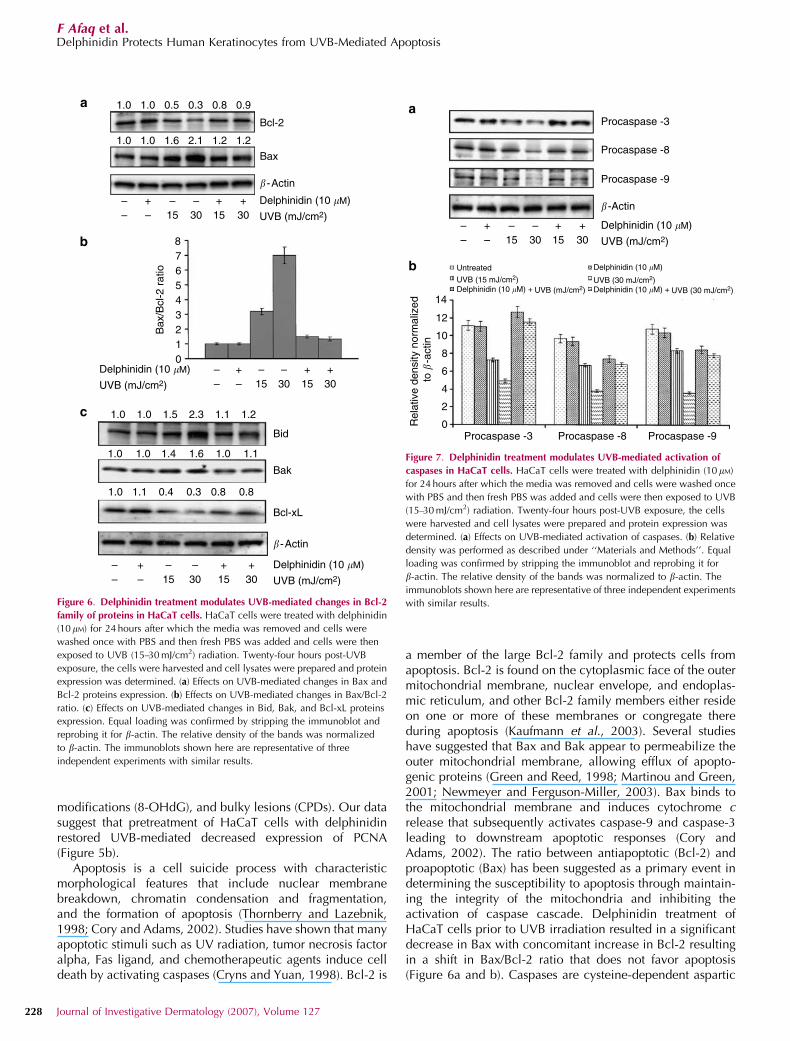
Delphinidin treatment modulates UVB-mediated activation ofcaspases in HaCaT cells
Exposure of cells to UVB induces activation of theproapoptotic protein including caspases. To further assessthe effect of delphinidin on UVB-induced apoptosis, we
examined whether delphinidin treatment would protect UVB-mediated activation of caspases. As expected employingWestern blot analysis we found that UVB irradiation resultedin a decrease in the protein expression of procaspase-3,-8, and -9, indicating that the pro form of caspase is cleavedinto active form. Our results suggest that pretreatment of cellswith delphinidin (10 mM) inhibited UVB-mediated decreasein the protein expression of procaspase-3, -8, and -9 asdemonstrated by relative density and Western blot analysis(Figure 7a and b).
Delphinidin treatment inhibits UVB-mediated formation ofCPDs, 8-OHdG, and TUNEL-positive cells in SKH-1 hairlessmouse skinTo investigate the relevance of our in vitro findings to in vivosituations, we determined the effect of topical application ofdelphinidin to SKH-1 hairless mouse on UVB-induceddamage. It is known that both CPDs and 8-OHdG are formedin epidermal DNA after UVB irradiation and are consideredas important biomarkers of DNA damage. Therefore, we firstassessed the effect of delphinidin (both pretreatment and
104
103
102
101
100
0 200 400 600 800 1,000FL3-area
0 200 400 600 800 1,000FL3-area
0 200 400 600 800 1,000FL3-area
0 200 400 600 800 1,000FL3-area
0 200 400 600 800 1,000FL3-area
0 200 400 600 800 1,000FL3-area
Ant
i-Brd
U F
ITC
104
103
102
101
100A
nti-B
rdU
FIT
C
104
103
102
101
100
Ant
i-Brd
U F
ITC
104
103
102
101
100
Ant
i-Brd
U F
ITC
104
103
102
101
100
Ant
i-Brd
U F
ITC
104
103
102
101
100A
nti-B
rdU
FIT
C
UVB (30 mJ/cm2) + UVB (15 mJ/cm2)
UVB (15 mJ/cm2)
UVB (30 mJ/cm2) UVB (15 mJ/cm2) UVB (30 mJ/cm2)
+ UVB (30 mJ/cm2)
UVB (15 mJ/cm2)Control
UntreatedUntreated
Delphinidin (10 �M)
Delphinidin (10 �M)
Delphinidin (10 �M)
Delphinidin (10 �M) +
UVB (mJ/cm2)
UVB (30 mJ/cm2)UVB (15 mJ/cm2)
Delphinidin (10 �M)
Delphinidin (10 �M)
Delphinidin (10 �M) + UVB (15 mJ/cm2) Delphinidin (10 �M) + UVB (30 mJ/cm2)
Delphinidin (10 �M) +
Delphinidin (10 �M)
PARP (116 KDa)
PARP (116 KDa)
PARP (85 KDa)
PARP (85 KDa)
� -Actin
– – –+ + +
– – 15 30 15 30
12
10
8
6
4
2
0
Rel
ativ
e de
nsity
nor
mal
ized
to �
-act
in
a
b
c
d
1.22%
25.72% 4.53% 7.69%
R2
R2 R2 R2
R2 R2
1.15% 11.88%
Figure 3. Delphinidin treatment protects HaCaT cells against UVB-mediated apoptosis. HaCaT cells were treated with delphinidin (10mM) for 24 hours after
which the media was removed and cells were washed once with PBS and then fresh PBS was added and cells were exposed to UVB (15–30 mJ/cm2) radiation.
(a) Effects on UVB-mediated apoptosis as detected by flow cytometry. Cells showing fluorescence (R2) are considered as apoptotic and their percentage
population is indicated. (b) Effects on UVB-mediated apoptosis as detected by fluorescence microscopy. The annexin-V-FLUOS staining kit was used for
detection of apoptotic and necrotic cells. The kit uses a dual staining protocol in which apoptotic cells are stained with annexin V (green fluorescence) and
the necrotic cells are stained with PI (red fluorescence). (c) Effects on UVB-mediated PARP cleavage. (d) Relative density was performed as described under
‘‘Materials and Methods’’. Equal loading was confirmed by stripping the immunoblot and reprobing it for b-actin. The relative density of the bands was
normalized to b-actin. Data shown are from representative experiments repeated thrice with similar results.
www.jidonline.org 225
F Afaq et al.Delphinidin Protects Human Keratinocytes from UVB-Mediated Apoptosis

post-treatment) on UVB-mediated DNA damage in mouseepidermis by immunohistochemical analysis. We selected atime point of 1 hour post-UVB irradiation because at this timepoint maximum formation of CPDs and 8-OHdG wasobserved (Lu et al., 1999; Gu et al., 2005; Afaq et al.,2006). As shown in Figure 8, UVB irradiation (180 mJ/cm2) toSKH-1 hairless mouse skin induced the formation of bothCPDs (Figure 8c) and 8-OHdG (Figure 8h) when compared totheir respective control groups (Figure 8a and f). Our dataclearly demonstrate that topical application of delphinidin(both pretreatment and post-treatment) resulted in markedinhibition in the number of CPDs (Figure 8d and e) and8-OHdG (Figure 8i and j) positive cells. Depending on theseverity of the DNA damage after UVB irradiation, keratino-cytes in the skin can undergo either DNA repair or apoptosis.Alternatively, UVB exposure leads to cutaneous neoplasia byimposing severe and sometimes irreparable damage. There-fore, we next assessed the effect of delphinidin (bothpretreatment and post-treatment) on UVB-mediated apoptosisby TUNEL assay, 8 hours post-UVB irradiation. Our datasuggest that UVB irradiation to SKH-1 hairless mouse skin
increased the number of apoptotic cells in the epidermis(Figure 8m) compared to control (Figure 8k) and delphinidinalone (Figure 8l) treated groups. Topical application ofdelphinidin (both pretreatment and post-treatment) inhibitedUVB-induced formation of apoptotic cells as determined byTUNEL staining (Figure 8n and o).
DISCUSSIONIn the United States alone, 1.2 million new cases of skincancer are identified each year and this accounts for 40% ofall new cancer cases diagnosed (Jemal et al., 2005). SolarUVB radiation has been implicated as the main cause for skincancer. The non-melanoma skin cancers comprising of basalcell carcinomas and squamous cell carcinomas are the mostfrequently diagnosed cutaneous malignancies. Therefore,additional efforts are needed to prevent skin cancers thatresult as a consequence of UVB exposure. Because skincancer is a significant problem associated with mortality andmorbidity, concerted efforts are needed to develop novelstrategies for the prevention of UV-mediated damages.Photochemoprevention has been appreciated as a viableapproach to reduce the occurrence of skin cancer and inrecent years, the use of agents, especially botanical anti-oxidants, present in the common diet and beveragesconsumed by human population have gained considerableattention as photochemopreventive agents for human use.Many such agents have also found a place in skin careproducts. A case–control study in Australia demonstrated asignificant inverse relationship between the risk of skincancer and a high intake of vegetables, b-carotene, andvitamin C containing foods (Kune et al., 1992). In the presentstudy, we demonstrated that delphinidin protected HaCaTcells from UVB-mediated decrease in cell viability (Figure 2).Our results further suggest that pretreatment of cells withdelphinidin inhibited UVB-mediated apoptosis as determinedby flow cytometry, confocal microscopy, and PARP cleavage(Figure 3). Moreover, we found that UVB radiation to SKH-1hairless mouse skin increased the number of apoptotic cells,and topical application of delphinidin (both pretreatmentand post-treatment) inhibited UVB-induced apoptosis(Figure 8).
UV radiation results in an increased generation of ROSthat overwhelms the antioxidant defense mechanisms of thetarget system. This condition of prooxidant/antioxidantdisequilibrium is defined as ‘‘oxidative stress’’. The epidermisis composed mainly of keratinocytes, which are rich in ROSdetoxifying enzymes such as superoxide dismutase, catalase,glutathione peroxidase, and in low-molecular-mass anti-oxidant molecules, and thus provide some natural protectionagainst ROS (Afaq and Mukhtar, 2001). Skin spontaneouslyresponds to increased ROS levels; however, this responsemay not be sufficient to prevent the progression of skincancer. Our studies suggest that delphinidin possesses strongantioxidant activity (Figure 4a). UV radiation to the skinresults in the formation of ROS that interact with proteins,lipids, and DNA (Halliday, 2005; Melnikova and Ananthas-wamy, 2005). LPO in biological membranes is a free radical-mediated event and is regulated by the availability of
14
12
10
8
6
4
2
0
350
300
250
200
150
100
50
0
0 �M 1 �M 5 �M 10 �M 20 �M
Delphinidin
Delphinidin (10 �M)
UVB (mJ/cm2)
Tro
lox
equi
vale
nts
(mM
)
Lipi
d pe
roxi
datio
n(%
of c
ontr
ol)
*
*
# #
–
– –
– –+ + +
15 30 15 30
a
b
Figure 4. Delphinidin exerts strong antioxidant effect and inhibits UVB-
mediated lipid peroxidation in HaCaT cells. HaCaT cells were treated with
different doses of delphinidin (1–20 mM) for 24 hours after which the media
was removed and cells were harvested and cell lysates were prepared for
antioxidant activity. (a) Effects on total antioxidant activity in cell lysate. The
cell lysate was added to the ABTS radical cation decolorization assay. HaCaT
cells were treated with delphinidin (10 mM) for 24 hours after which the media
was removed and cells were washed once with PBS and then fresh PBS
was added and cells were then exposed to UVB (15–30 mJ/cm2) radiation.
Twenty-four hours post-UVB exposure; the cells were processed for lipid
peroxidation as detailed in ‘‘Materials and Methods’’. (b) Effects on
UVB-mediated lipid peroxidation. Data shown are mean7SD of three
separate experiments. *Po0.001 versus control, #Po0.001 versus UVB.
226 Journal of Investigative Dermatology (2007), Volume 127
F Afaq et al.Delphinidin Protects Human Keratinocytes from UVB-Mediated Apoptosis

substrates in the form of polyunsaturated fatty acids andprooxidants that promote peroxidation (Katiyar et al., 2001;Girotti and Kriska, 2004). LPO is highly detrimental to cellmembrane structure and function, and its elevated level hasbeen linked to damaging effects such as loss of fluidity,inactivation of membrane enzymes, increased cell membranepermeability to ions, and finally rupture of cell membraneleading to release of cell organelles (Brunet et al., 2000;Cuzzocrea et al., 2001). Thus, inhibition of LPO bydelphinidin (Figure 4b) would result in reduction of the riskfactors associated with UVB radiation.
UVB irradiation results in DNA modification both directlyby forming CPDs and pyrimidine (6-4) pyrimidone photo-dimers, and indirectly via ROS that produces 8-OHdG(Budiyanto et al., 2000; Katiyar et al., 2000; Cadet et al.,2005). 8-OHdG is a mutation-prone (G:C to T:A transversion)DNA base-modified product generated by ROS and photo-dynamic action (Hattori et al., 1996). The transversion of G:Cto T:A in UVB-induced skin cancers of mice and humanssuggests that 8-OHdG plays an essential role in photocarci-nogenesis (Basset-Seguin et al., 1994; Nishigori et al., 1994).Recent studies have shown that 8-OHdG formation in miceepidermis was reduced by super virgin olive oil paintedimmediately after UV radiation (Budiyanto et al., 2000). The
formation of 8-OHdG after UVB exposure appears to beregulated by LPO and inflammation, and may play animportant role in photocarcinogenesis. UVB induces oxida-tive damage in DNA resulting in the formation of the adduct8-OHdG. The present study was undertaken to examine theeffect of delphinidin against UVB-mediated DNA damage.Our results suggest reduced formation of UVB-induced8-OHdG in HaCaT cells pretreated with delphinidin com-pared to UVB alone group (Figure 5a). Topical application ofdelphinidin (both pretreatment and post-treatment) to SKH-1hairless mouse skin inhibited UVB-induced formation ofCPDs and 8-OHdG (Figure 8). This indicates that the reducedformation of CPDs and 8-OHdG can be attributed to thescavenging activity of delphinidin against ROS (Figure 4aand 8).
PCNA is an active nuclear protein and serve as a marker ofDNA repair and indirectly as an indicator of UVB induceddamage (Akbari et al., 2004; Moore et al., 2004; Constantinet al., 2005). Studies have demonstrated that UVB irradiationcompletely diminished PCNA protein expression in mousemodel of photocarcinogenesis. Topical application of lyco-pene significantly reversed UVB-induced PCNA inhibition(Fazekas et al., 2003). Cells are equipped with extensiverepair capacity for removing strand breaks, small base
Control Delphinidin (10 �M)
Delphinidin (10 �M)
UVB (15 mJ/cm2)
UVB (15 mJ/cm2) + UVB (30 mJ/cm2)Delphinidin (10 �M)
Delphinidin (10 �M)
+ UVB (15 mJ/cm2)
UVB (15 mJ/cm2)
1.0 0.95 0.4 0.1 1.0 0.9
PCNA
� -Actin
– – –
– –
+ + +
15 30 15 30
a
b
Figure 5. Delphinidin treatment inhibits UVB-mediated formation of 8-OHdG and degradation of PCNA protein expression in HaCaT cells. HaCaT cells were
treated with delphinidin (10 mM) for 24 hours after which the media was removed and cells were washed once with PBS and then fresh PBS was added and
cells were then exposed to UVB (15–30 mJ/cm2) radiation. Twenty-four hours post-UVB exposure; the cells were processed for immunocytochemistry and
Western blot analysis as detailed in ‘‘Materials and Methods’’. (a) Effects on UVB-mediated formation of 8-OHdG as detected by immunocytochemistry.
A representative picture from three independent experiments with similar results is shown. (b) Effects on UVB-mediated degradation of PCNA. Equal loading
was confirmed by stripping the immunoblot and reprobing it for b-actin. The relative density of the bands was normalized to b-actin. The immunoblots
shown here are representative of three independent experiments with similar results.
www.jidonline.org 227
F Afaq et al.Delphinidin Protects Human Keratinocytes from UVB-Mediated Apoptosis
modifications (8-OHdG), and bulky lesions (CPDs). Our datasuggest that pretreatment of HaCaT cells with delphinidinrestored UVB-mediated decreased expression of PCNA(Figure 5b).
Apoptosis is a cell suicide process with characteristicmorphological features that include nuclear membranebreakdown, chromatin condensation and fragmentation,and the formation of apoptosis (Thornberry and Lazebnik,1998; Cory and Adams, 2002). Studies have shown that manyapoptotic stimuli such as UV radiation, tumor necrosis factoralpha, Fas ligand, and chemotherapeutic agents induce celldeath by activating caspases (Cryns and Yuan, 1998). Bcl-2 is
a member of the large Bcl-2 family and protects cells fromapoptosis. Bcl-2 is found on the cytoplasmic face of the outermitochondrial membrane, nuclear envelope, and endoplas-mic reticulum, and other Bcl-2 family members either resideon one or more of these membranes or congregate thereduring apoptosis (Kaufmann et al., 2003). Several studieshave suggested that Bax and Bak appear to permeabilize theouter mitochondrial membrane, allowing efflux of apopto-genic proteins (Green and Reed, 1998; Martinou and Green,2001; Newmeyer and Ferguson-Miller, 2003). Bax binds tothe mitochondrial membrane and induces cytochrome crelease that subsequently activates caspase-9 and caspase-3leading to downstream apoptotic responses (Cory andAdams, 2002). The ratio between antiapoptotic (Bcl-2) andproapoptotic (Bax) has been suggested as a primary event indetermining the susceptibility to apoptosis through maintain-ing the integrity of the mitochondria and inhibiting theactivation of caspase cascade. Delphinidin treatment ofHaCaT cells prior to UVB irradiation resulted in a significantdecrease in Bax with concomitant increase in Bcl-2 resultingin a shift in Bax/Bcl-2 ratio that does not favor apoptosis(Figure 6a and b). Caspases are cysteine-dependent aspartic
1.0 1.0 0.5 0.3 0.8 0.9
1.0 1.0 1.5 2.3 1.1 1.2
1.0 1.0 1.4 1.6 1.0 1.1
1.0 1.1 0.4 0.3 0.8 0.8
1.0 1.0 1.6 2.1 1.2 1.2
Bcl-2
Bax
Bak
Bid
Bcl-xL
� -Actin
�-Actin
–– –
– –+ + +15 30 15 30
–– –
– –+ + +15 30 15 30
Delphinidin (10 �M)
UVB (mJ/cm2)
–– –
– –+ + +15 30 15 30
Delphinidin (10 �M)
UVB (mJ/cm2)
Delphinidin (10 �M)
UVB (mJ/cm2)
876543210
Bax
/Bcl
-2 r
atio
a
b
c
Figure 6. Delphinidin treatment modulates UVB-mediated changes in Bcl-2
family of proteins in HaCaT cells. HaCaT cells were treated with delphinidin
(10mM) for 24 hours after which the media was removed and cells were
washed once with PBS and then fresh PBS was added and cells were then
exposed to UVB (15–30 mJ/cm2) radiation. Twenty-four hours post-UVB
exposure, the cells were harvested and cell lysates were prepared and protein
expression was determined. (a) Effects on UVB-mediated changes in Bax and
Bcl-2 proteins expression. (b) Effects on UVB-mediated changes in Bax/Bcl-2
ratio. (c) Effects on UVB-mediated changes in Bid, Bak, and Bcl-xL proteins
expression. Equal loading was confirmed by stripping the immunoblot and
reprobing it for b-actin. The relative density of the bands was normalized
to b-actin. The immunoblots shown here are representative of three
independent experiments with similar results.
–– –
– –+ + +15 30 15 30
Delphinidin (10 �M)
Delphinidin (10 �M)
UVB (mJ/cm2)
UVB (30 mJ/cm2)UVB (15 mJ/cm2)Delphinidin (10 �M) + UVB (mJ/cm2) Delphinidin (10 �M) + UVB (30 mJ/cm2)
Procaspase -3
Procaspase -3
Rel
ativ
e de
nsity
nor
mal
ized
Procaspase -8
Procaspase -8
Procaspase -9
Procaspase -9
� -Actin
to
�-a
ctin
14
12
10
8
6
4
2
0
Untreated
a
b
Figure 7. Delphinidin treatment modulates UVB-mediated activation of
caspases in HaCaT cells. HaCaT cells were treated with delphinidin (10 mM)
for 24 hours after which the media was removed and cells were washed once
with PBS and then fresh PBS was added and cells were then exposed to UVB
(15–30 mJ/cm2) radiation. Twenty-four hours post-UVB exposure, the cells
were harvested and cell lysates were prepared and protein expression was
determined. (a) Effects on UVB-mediated activation of caspases. (b) Relative
density was performed as described under ‘‘Materials and Methods’’. Equal
loading was confirmed by stripping the immunoblot and reprobing it for
b-actin. The relative density of the bands was normalized to b-actin. The
immunoblots shown here are representative of three independent experiments
with similar results.
228 Journal of Investigative Dermatology (2007), Volume 127
F Afaq et al.Delphinidin Protects Human Keratinocytes from UVB-Mediated Apoptosis

proteases that are activated by proteolytic cleavage into largeand small subunits and form tetrameric complexes (Changand Yang, 2000; Wang et al., 2003). Proapoptotic caspasesare grouped into initiator (caspase-8, -9, and -10) andexecutor (caspase-3, -6, and -7) caspases (Chang and Yang,2000; Pirnia et al., 2002; Milhas et al., 2005). Althoughcaspase activity is regulated primarily at the post-translationallevel, overexpression of caspases sensitizes cells for apopto-sis. The present study suggests that the protective effects ofdelphinidin against UVB-mediated apoptosis may be viamodulation of Bcl-2 family members (Figure 6) and inhibitionof activation of caspases (Figure 7).
In summary, our results suggest that treatment of HaCaTcells and mouse skin with delphinidin inhibited UVB-mediated oxidative stress and reduced DNA damage, therebyprotecting the cells from UVB-induced apoptosis. Theseeffects of delphinidin are because of its strong antioxidantactivity. Therefore, these results demonstrate that delphinidinhas the potential to protect against the adverse effects of UVBradiation via modulation of Bcl-2 family members andinhibition of activation of caspases, which in turn mayprotect the cellular targets against UVB-induced damage.Our results provide basis for the photochemopreventive effectof delphinidin and suggest that it may be a useful agentagainst UVB-induced damage for human skin.
MATERIALS AND METHODSMaterials
Delphinidin (498% pure) was purchased from Extrasynthase (Lyon,
France). The mAbs and polyclonal antibodies for PARP (116 kDa),
Bcl-2 Bax, Bak, Bcl-xL were obtained from Upstate Biotechnology
(Lake Placid, NY). mAb for PARP (85 kDa) was purchased from
Promega (Madison, WI). The other human reactive mAbs and
polyclonal antibodies (Procaspase 3, 8, and 9) were obtained from
Santa Cruz Biotechnology Inc. (Santa Cruz, CA). Anti-mouse or anti-
rabbit secondary antibody horse-radish peroxidase conjugate was
obtained from Amersham Life Science Inc. (Arlington Height, IL).
Annexin-V-FLOUS staining kit was purchased from Roche Diag-
nostic Corporation (Indianapolis, IN). The DC BioRad Protein assay
kit was purchased from BioRad Laboratories (Herculus, CA).
DeadEndTM Colorimetric TUNEL kit was procured from Promega
(Madison, WI). Novex pre-cast Tris-Glycine gels were obtained from
Invitrogen (Carlsbad, CA).
Treatment of cells
The HaCaT cells were cultured in DMEM medium, supplemented
with 10% fetal bovine serum, 1% penicillin/streptomycin and were
maintained at 95% humidity in 5% CO2 environment at 371C
(approved by the Institutional Review Board). Delphinidin (dissolved
in DMSO) was used for the treatment of cells. The final concentra-
tion of DMSO used was 0.1% (v/v) for each treatment. The cells
ControlT
UN
EL
8-O
HdG
CP
Ds
Delphinidin Delphinidin + UVB UVB + Delphinidin UVBa b c d e
f g h i j
k l m n o
Figure 8. Topical application of delphinidin inhibits UVB-mediated formation of CPD, 8-OHdG, and TUNEL-positive cells in SKH1-hairless mouse skin.
The groups of mice were either unexposed (control), treated topically on the dorsal skin with delphinidin (1 mg/100 ml DMSO/mouse), exposed to UVB
radiation (180 mJ/cm2), treated topically on dorsal skin with delphinidin (1 mg/100 ml DMSO/mouse) and then 30 minutes later exposed to UVB radiation,
exposed to UVB radiation and then treated immediately with delphinidin (1 mg/100 ml DMSO/mouse) as described under ‘‘Materials and Methods’’. One
and eight hours post-UVB irradiation, the animals were killed and skin biopsies were processed for staining. Immunohistochemical staining for (a–e) CPDs,
(f–j) 8-OHdG, and (k–o) TUNEL was carried out as described under ‘‘Material and Methods’’. The staining shown here are representative data from
different treatments. Bar¼ 25 mm.
www.jidonline.org 229
F Afaq et al.Delphinidin Protects Human Keratinocytes from UVB-Mediated Apoptosis
(70–80% confluent) were treated with delphinidin (10 mM) for
24 hours in DMEM medium after which the media was removed
and cells were washed with phosphate-buffered saline (PBS)
and then fresh PBS was added, and these delphinidin pretreated
cells were irradiated with UVB (15–30 mJ/cm2) with a custom
designed Research Irradiation Unit (Daavlin, Bryan, OH) that
consists of a fixture mounted on fixed legs. Mounted within
the exposure unit are four UVB lamps and the exposure
system is controlled using Daavlin Flex Control Integrating
Dosimeters. In this system, dose units can be entered in mJ/cm2
for UVB; variations in energy output are automatically compensated
for the delivery of the desired dose. Using this system, the cells
were exposed to accurate dosimetery of UVB radiation. Twenty-four
hours post-UVB exposure, cells were harvested and cell lysates
were prepared.
Cell viability
The effect of delphinidin or UVB (or both) on the viability of
cells was determined by MTT assay and phase contrast microscopy.
The cells were plated at 1� 104 cells per well in 200 ml of
complete culture medium containing 1, 5, 10, 15, and 20 mM
concentrations of delphinidin in 96-well microtiter plates for
24 hours at 371C in a humidified chamber. Each concentration
of delphinidin was repeated in 10 wells. After incubation for
specified times at 371C in a humidified incubator the cells were
washed and exposed to UVB (15–30 mJ/cm2). Twenty-four hours
post-UVB exposure, MTT reagent (4ml, 5 mg/ml in PBS) was added
to each well and incubated for 2 hours. The microtiter plate
containing the cells was centrifuged at 1,800 rpm for 5 minutes
at 41C. The MTT solution was removed from the wells by aspiration
and the formazan crystals were dissolved in DMSO (150 ml).
Absorbance was recorded on a microplate reader at 540 nm
wavelength.
Preparation of cell lysateFollowing treatment of cells with delphinidin or UVB (or both), the
medium was aspirated and the cells were washed twice in PBS
(10 mM, pH 7.4). The cells were incubated in 0.4 ml ice-cold lysis
buffer (50 mM Tris-HCl, 150 mM NaCl, 1 mM EGTA, 1 mM EDTA,
20 mM NaF, 100 mM Na3VO4, 0.5% NP-40, 1% Triton X-100, 1 mM
PMSF (pH 7.4)) with freshly added protease inhibitor cocktail
(Protease Inhibitor Cocktail Set III, Calbiochem, La Jolla, CA). The
cells were then centrifuged at 14,000 g for 25 minutes at 41C and the
supernatant (cell lysate) was collected, aliquoted, and stored at
�801C.
Western blot analysis
For Western blot analysis, 25–50 mg of protein was resolved over
8–12% polyacrylamide gels and transferred onto a nitrocellulose
membrane. The blot containing the transferred protein was blocked
in blocking buffer (5% nonfat dry milk, 1% Tween-20; in 20 mM Tris-
buffered saline, pH 7.6) for 1 hour at room temperature followed by
incubation with appropriate mAb or polyclonal primary antibody in
blocking buffer for 1 hour to overnight at 41C. This was followed by
incubation with anti-mouse or anti-rabbit secondary antibody horse-
radish peroxidase (Amersham Life Sciences, Inc.) for 1 hour. The blot
was then washed several times and detected by chemiluminescence
(ECL kit, Amersham Life Sciences, Inc.) and autoradiography using
XAR-5 film obtained from Eastman Kodak Co. (Rochester, NY).
Densitometric measurements of the band in Western blot analysis
were performed using digitalized scientific software program
UN-SCAN-IT (Silk Scientific Corporation, Orem, UT).
Quantification of apoptosis
The effect of delphinidin or UVB (or both) on the extent of apoptosis
was determined by TUNEL assay. The cells were treated with
delphinidin (10 mM) for 24 hours after which the cells were washed
and exposed to UVB (15 and 30 mJ/cm2). Twenty four hours post-
UVB exposure the cells were trypsinized, washed with PBS, and
processed for labeling with fluorescein-tagged deoxyuridine tripho-
sphate nucleotide and PI by use of an Apo-direct apoptosis kit
obtained from Phoenix Flow Systems (San Diego, CA) as per the
manufacturer’s protocol. The labeled cells were analyzed by flow
cytometry.
Apoptosis detection by confocal microscopy
The Annexin-V-FLUOS staining kit was used for the detection of
apoptotic and necrotic cells according to vendor’s protocol. This kit
uses a dual-staining protocol in which the apoptotic cells are stained
with annexin V (green fluorescence), and the necrotic cells are
stained with PI (red fluorescence). The fluorescence was measured
by a Zeiss 410 confocal microscope (Thornwood, NY). Confocal
images of green annexin-FITC fluorescence were collected using
488 nm excitation light from an argon/krypton laser, a 560 nm
dichroic mirror, and a 514–540 nm bandpass barrier filter. Images of
red PI fluorescence were collected using a 568 nm excitation light
from the argon/krypton laser, a 560 nm dichroic mirror, and a
590 nm long pass filter.
Measurement of antioxidant activity
To assess the antioxidant capacity of delphinidin (1–20 mM) in HaCaT
cells, trolox antioxidant assay was performed using the trolox
antioxidant assay kit from Cayman chemical (Ann Arbor, MI)
following the vendor’s protocol. Briefly, the assay relies on the
ability of antioxidants in the cell lysates to inhibit the oxidation of
ABTSR (2,20-Azino-di-[3-ethylbenzthiazoline sulphonate]) to
ABTSRKþ by metmyoglobin. The amount of ABTSRKþ produced
was monitored by reading the absorbance at 750 nm. The capacity of
the antioxidants in the sample to prevent ABTSR oxidation was
compared with that of trolox, a water-soluble tocopherol analogue
and was quantified as millimolar trolox equivalents. The reaction
was initiated by adding 40 ml of hydrogen peroxide working solution
(441mM) to all the wells being used. The plate was covered and
incubated at room temperature for 5 minutes and absorbance was
read at 750 nm using a plate reader.
Immunostaining for 8-OHdG
HaCaT cells, grown on cover slides and treated with delphinidin
(10 mM) for 24 hours, washed with PBS and exposed to UVB (15 and
30 mJ/cm2). Twenty four hours post-UVB exposure the cells were
fixed in methanol at �201C for 5 minutes. Briefly, the fixed cells
were incubated with primary antibody 8-OHdG (Chemicon Inter-
national Inc., Temecula, CA) followed by secondary antibody
conjugated with horseradish peroxidase. Cells were then
treated with 3,30-diaminobenzidine and counterstained with
hematoxylin.
230 Journal of Investigative Dermatology (2007), Volume 127
F Afaq et al.Delphinidin Protects Human Keratinocytes from UVB-Mediated Apoptosis
LPO assayAfter treatment with delphinidin or UVB (or both) cells were
harvested and washed with PBS, and microsomal fraction was
prepared as described earlier (Katiyar et al., 2001). Briefly, cells were
homogenized with a Polytron homogenizer in PBS buffer containing
potassium chloride (1.19%, w/v) and centrifuged at 18,000 g for
15 minutes at 41C to prepare microsomal fraction (Katiyar et al.,
2001). The LPO assay was performed in microsomal fraction
obtained from the different treatment groups. The generation of
malondialdehyde was employed as a marker of LPO and estimated
by the method of Wright et al., (1981). Briefly, microsomal fraction
(2.0 mg protein) was incubated for 1 hour at 371C in the presence of
ferric ions (1 mM) and ADP (5 mM) in Ca2þ -free phosphate buffer
(0.1 M; pH 7.4) containing MgCl2 (0.1 mM). The reaction was
terminated by addition of 0.6 ml of 10% (w/v) trichloroacetic acid
followed by 1.2 ml of 0.5% (w/v) 2-thiobarbituric acid. The mixture
was heated for 20 minutes at 901C in a water bath. After cooling, the
malondialdehyde levels were measured in the clear supernatant by
recording absorbance at 532 nm. The final concentration of
malondialdehyde generated during the reaction was calculated
using a molar extinction coefficient of 1.56� 105/M/cm.
Animals and treatmentFemale SKH-1 hairless mice (6-weeks-old) obtained from Charles
River Laboratories (Wilmington, MA) were used in this study. All
procedures were performed in accordance with the guidelines of the
Animal Care and Approved by Institutional Review Board. After their
arrival in the animal facility, the animals were allowed to
acclimatize for 1 week before the start of the experiments. The
animals were fed Purina Chow diet and water ad libitum. The mice
were maintained at standard conditions: temperature of 24þ 21C,
relative humidity of 50þ 10%, and 12 hours room light/12 hours
dark cycle. For UVB irradiation, the mice were housed in specially
designed cages where they were held in dividers separated by
Plexiglas. Sixty-four female SKH-1 hairless mice, maintained as
described, were divided into five groups of 16 animals each (except
in control and delphinidin alone groups, where eight animals were
used). The mice in the first group received a topical application of
100ml DMSO alone, and those in the second group received 1 mg
delphinidin in 100 ml DMSO/mouse. The mice in the third group
received a topical application of 100ml DMSO before a single UVB
(180 mJ/cm2) exposure with a custom designed Research Irradiation
Unit (Daavlin, Bryan, OH) as described above. The mice in the
fourth group received a topical application of delphinidin (1 mg/
100ml DMSO/mouse), 30 minutes before UVB (180 mJ/cm2) expo-
sure. The animals in the fifth group were exposed to UVB (180 mJ/
cm2), and immediately following UVB irradiation, they received
topical application of delphinidin (1 mg/100 ml DMSO/mouse). The
mice were then killed at 1 and 8 hours post-UVB exposure and skin
biopsies were harvested for immunohistochemical analysis of CPDs,
8-OHdG, and apoptotic cells. The selection of these time points are
based on recently published studies (Lu et al., 1999; Gu et al., 2005;
Afaq et al., 2006).
Immunohistochemical detection of CPDs and 8-OHdG
The skin biopsies were frozen in optimal cutting temperature
compound under liquid nitrogen immediately after removal.
The skin biopsies were stored at �801C for further use. Briefly,
5-mm-thick frozen skin sections were thawed and fixed in cold
acetone for 10 minutes and washed in PBS (pH 7.4). The sections
were then kept in 70 mmol/l NaOH in 70% ethanol for 2 minutes to
denature nuclear DNA followed by neutralization for 1 minute in
100 mmol/l Tris-HCl (pH 7.5) in 70% ethanol. Nonspecific binding
sites were blocked by 30 minutes incubation at room temperature in
PBS containing 10% goat serum. Sections were then incubated with
mouse mAbs to 8-OHdG (Nikken SEIL Corporation, Japan), CPDs
(Kamiya Biomedical Co., Seattle, WA). For negative control the
sections were incubated without primary antibody. After overnight
incubation at 41C and three washes in PBS, the sections were
incubated for 30 minutes with biotinylated rabbit anti-mouse link
antibody and then for 30 minutes with streptavidin conjugated to
horse radish peroxidase. After washing, sections were incubated
with diaminobenzidine plus peroxidase substrate and counterstained
with methyl green.
TUNEL staining for apoptotic cells
Apoptotic cells were detected using the DeadEndTM Colorimetric
TUNEL System (Promega, Madison, WI) using the manufacturer’s
protocol. Briefly, tissue sections were fixed in 4% paraformaldehyde,
permeabilzed with Proteinase K solution, re-fixed in 4% paraform-
aldehyde, and equilibrated with 100ml of equilibration buffer.
Sections were then labeled with TdT reaction mix by incubating at
371C for 1 hour, after which the reaction was stopped using 2� SSC.
Hydrogen peroxide (0.3%) was used for quenching endogenous
peroxidase activity followed by incubation with 100 ml streptavidin
horseradish peroxidase (1:500) for 30 minutes at room temperature.
Each step was followed by thorough washing with PBS. Finally,
sections were stained with 3,30-diaminobenzidine for 2–3 minutes
and visualized under microscope using � 400 maganification for
TUNEL-positive cells.
Statistical analysisThe results are expressed as means7SD Statistical analysis of all the
data between groups receiving UVB exposure alone and those with
delphinidin treatment plus UVB exposure were performed by
Student’s t-test. The P-value o0.05 was considered statistically
significant.
CONFLICT OF INTERESTThe authors state no conflict of interest.
ACKNOWLEDGMENTSThis work was supported by a grant from the Lynda and Stewart ResnickRevocable Trust and USPHS Grant R21 AT002429-01.
REFERENCES
Afaq F, Adhami VM, Mukhtar H (2005a) Photochemoprevention of ultravioletB signaling and photocarcinogenesis. Mutat Res 571:153–73
Afaq F, Hafeez BB, Syed DN, Kweon MH, Mukhtar H (2006) Oral feeding ofpomegranate fruit extract inhibits early biomarkers of UVB-inducedcarcinogenesis in SKH-1 hairless mouse epidermis. J Invest Dermatol126(Suppl 4):141
Afaq F, Malik A, Syed D, Maes D, Matsui MS, Mukhtar H (2005b)Pomegranate fruit extract modulates UV-B-mediated phosphorylationof mitogen-activated protein kinases and activation of nuclear factorkappa B in normal human epidermal keratinocytes. PhotochemPhotobiol 81:38–45
www.jidonline.org 231
F Afaq et al.Delphinidin Protects Human Keratinocytes from UVB-Mediated Apoptosis
Afaq F, Mukhtar H (2001) Effects of solar radiation on cutaneousdetoxification pathways. J Photochem Photobiol B 63:61–9
Akbari M, Otterlei M, Pena-Diaz J, Aas PA, Kavli B, Liabakk NB et al. (2004)Repair of U/G and U/A in DNA by UNG2-associated repair complexestakes place predominantly by short-patch repair both in proliferating andgrowth-arrested cells. Nucleic Acids Res 32:5486–98
Basset-Seguin N, Moles JP, Mils V, Dereure O, Guilhou JJ (1994) TP53 tumorsuppressor gene and skin carcinogenesis. J Invest Dermatol 103(Suppl5):102S–6S
Bowden GT (2004) Prevention of non-melanoma skin cancer by targetingultraviolet-B-light signalling. Nat Rev Cancer 4:23–35
Brunet S, Thibault L, Lepage G, Seidman EG, Dube N, Levy E (2000)Modulation of endoplasmic reticulum-bound cholesterol regulatoryenzymes by iron/ascorbate-mediated lipid peroxidation. Free RadicalBiol Med 28:46–54
Budiyanto A, Ahmed NU, Wu A, Bito T, Nikaido O, Osawa T et al. (2000)Protective effect of topically applied olive oil against photocarcinogen-esis following UVB exposure of mice. Carcinogenesis 21:2085–90
Cadet J, Sage E, Douki T (2005) Ultraviolet radiation-mediated damage tocellular DNA. Mutat Res 571:3–17
Chang HY, Yang X (2000) Proteases for cell suicide: functions and regulationof caspases. Microbiol Mol Biol Rev 64:821–46
Constantin N, Dzantiev L, Kadyrov FA, Modrich P (2005) Human mismatchrepair: reconstitution of a nick-directed bidirectional reaction. J BiolChem 280:39752–61
Cory S, Adams JM (2002) The Bcl2 family: regulators of the cellular life-or-death switch. Nat Rev Cancer 2:647–56
Cryns V, Yuan J (1998) Proteases to die for. Genes Dev 12:1551–70
Cuzzocrea S, Riley DP, Caputi AP, Salvemini D (2001) Antioxidant therapy: anew pharmacological approach in shock, inflammation, and ischemia/reperfusion injury. Pharmacol Rev 53:135–59
Fazekas Z, Gao D, Saladi RN, Lu Y, Lebwohl M, Wei H (2003) Protectiveeffects of lycopene against ultraviolet B-induced photodamage. NutrCancer 47:181–7
F’guyer S, Afaq F, Mukhtar H (2003) Photochemoprevention of skin cancer bybotanical agents. Photodermatol Photoimmunol Photomed 19:56–72
Girotti AW, Kriska T (2004) Role of lipid hydroperoxides in photo-oxidativestress signaling. Antioxid Redox Signal 6:301–10
Green DR, Reed JC (1998) Mitochondria and apoptosis. Science 281:1309–12
Gu M, Dhanalakshmi S, Singh RP, Agarwal R (2005) Dietary feeding ofsilibinin prevents early biomarkers of UVB radiation-induced carcino-genesis in SKH-1 hairless mouse epidermis. Cancer Epidemiol Bio-markers Prev 14:1344–9
Halliday GM (2005) Inflammation, gene mutation and photoimmunosuppres-sion in response to UVR-induced oxidative damage contributes tophotocarcinogenesis. Mutat Res 571:107–20
Hattori Y, Nishigori C, Tanaka T, Uchida K, Nikaido O, Osawa T et al. (1996)8-hydroxy-20-deoxyguanosine is increased in epidermal cells of hairlessmice after chronic ultraviolet B exposure. J Invest Dermatol 107:733–7
Jemal A, Murray T, Ward E, Samuels A, Tiwari RC, Ghafoor A et al. (2005)Cancer statistics. CA Cancer J Clin 55:10–30
Katiyar SK, Afaq F, Perez A, Mukhtar H (2001) Green tea polyphenol(�) epigallocatechin-3-gallate treatment of human skin inhibits ultra-violet radiation-induced oxidative stress. Carcinogenesis 22:287–94
Katiyar SK, Matsui MS, Mukhtar H (2000) Kinetics of UV light-inducedcyclobutane pyrimidine dimers in human skin in vivo: an immunohis-tochemical analysis of both epidermis and dermis. Photochem Photobiol72:788–93
Kaufmann T, Schlipf S, Sanz J, Neubert K, Stein R, Borner C (2003)Characterization of the signal that directs Bcl-x(L), but not Bcl-2, to themitochondrial outer membrane. J Cell Biol 160:53–64
Kune GA, Bannerman S, Field B, Watson LF, Cleland H, Merenstein D et al.(1992) Diet, alcohol, smoking, serum beta-carotene, and vitamin A inmale nonmelanocytic skin cancer patients and controls. Nutr Cancer18:237–44
Lu YP, Lou YR, Yen P, Mitchell D, Huang MT, Conney AH (1999) Time coursefor early adaptive responses to ultraviolet B light in the epidermis of SKH-1 mice. Cancer Res 59:4591–602
Martinou JC, Green DR (2001) Breaking the mitochondrial barrier. Nat RevMol Cell Biol 2:63–7
Matsumura Y, Ananthaswamy HN (2004) Toxic effects of ultraviolet radiationon the skin. Toxicol Appl Pharmacol 195:298–308
Melnikova VO, Ananthaswamy HN (2005) Cellular and molecular eventsleading to the development of skin cancer. Mutat Res 571:91–106
Milhas D, Cuvillier O, Therville N, Clave P, Thomsen M, Levade T et al.(2005) Caspase-10 triggers bid cleavage and caspase cascade activationin FasL-induced apoptosis. J Biol Chem 280:19836–42
Moore JO, Palep SR, Saladi RN, Gao D, Wang Y, Phelps RG et al. (2004)Effects of ultraviolet B exposure on the expression of proliferating cellnuclear antigen in murine skin. Photochem Photobiol 80:587–95
Newmeyer DD, Ferguson-Miller S (2003) Mitochondria: releasing power forlife and unleashing the machineries of death. Cell 112:481–90
Nishigori C, Hattori Y, Toyokuni S (2004) Role of reactive oxygen species inskin carcinogenesis. Antioxid Redox Signal 6:561–70
Nishigori C, Wang S, Miyakoshi J, Sato M, Tsukada T, Yagi T et al. (1994)Mutations in ras genes in cells cultured from mouse skin tumors inducedby ultraviolet irradiation. Proc Natl Acad Sci USA 91:7189–93
Pirnia F, Schneider E, Betticher DC, Borner MM (2002) Mitomycin C inducesapoptosis and caspase-8 and -9 processing through a caspase-3 andFas-independent pathway. Cell Death Differ 9:905–14
Thornberry NA, Lazebnik Y (1998) Caspases: enemies within. Science281:1312–6
Wang HQ, Quan T, He T, Franke TF, Voorhees JJ, Fisher GJ (2003) Epidermalgrowth factor receptor-dependent, NF-kappaB-independent activationof the phosphatidylinositol 3-kinase/Akt pathway inhibits ultravioletirradiation-induced caspases-3, -8, and -9 in human keratinocytes. J BiolChem 278:45737–45
Wright JR, Colby HD, Miles PR (1981) Cytosolic factors which affectmicrosomal lipid peroxidation in lung and liver. Arch Biochem Biophys206:296–304
232 Journal of Investigative Dermatology (2007), Volume 127
F Afaq et al.Delphinidin Protects Human Keratinocytes from UVB-Mediated Apoptosis