Whole broth cellulase production for use in simultaneous ...
Upload
independentCategory
view
0download
0
Cellulase Production by Pink Pigmented FacultativeMethylotrophic Strains (PPFMs)
Shanmugam Jayashree & Rajendran Lalitha &
Ponnusamy Vadivukkarasi & Yuko Kato &
Sundaram Seshadri
Received: 27 September 2010 /Accepted: 10 January 2011 /Published online: 28 January 2011# Springer Science+Business Media, LLC 2011
Abstract Pink pigmented facultative methylotrophs (PPFM) isolated from water samplesof Cooum and Adyar rivers in Chennai and soil samples of forests located in variousdistricts of Tamil Nadu, India were screened for cellulase production using carboxymeth-ylcellulose agar (CMC agar) medium. The strains showed wide variations in the productionof clearing zones around the colonies on CMC agar medium flooded with Congo red.CMCase and filter paper assays were used to quantitatively measure the cellulase activity of13 PPFM strains. Among the strains, Methylobacterium gregans, MNW 60, MHW 109,MSF 34, and MSF 40 showed cellulolytic activity ranging from 0.73 to 1.16 UmL−1 withwide temperature (35–65°C) and pH (5 to 8) tolerance. SDS-PAGE analysis of the crudeenzyme of PPFM strain MNW 60 exhibited several protein bands, and zymogram analysisrevealed two dimeric cellulase bands with molecular mass of ~92 and 42 kDa. Scanningelectron microscopic studies revealed significant morphological differences between thecells grown in normal and CMC amended medium. The strain MNW 60 was identified asMethylobacterium sp. based on biochemical, physiological, and morphological analyses,and the methylotrophic nature was authenticated by the presence of mxaF gene, encodingmethanol dehydrogenase as a key indicator enzyme of methylotrophs, with 99% similarityto Methylobacterium lusitanum. With the 16S ribosomal RNA sequence showing 97%similarity to M. lusitanum strain MP2, this can be proposed as a novel taxon of the genusMethylobacterium. The study forms the first detailed report on the extracellular cellulaseproduction by pink pigmented Methylobacterium sp., and it is expected that this might bethe basis for further studies on cellulase production by PPFMs to explore the molecularmechanism, strain improvement, and large-scale cellulase production for its application.
Keywords CMC . CMCase . FPase .mxaF .Methylobacterium
Appl Biochem Biotechnol (2011) 164:666–680DOI 10.1007/s12010-011-9166-6
S. Jayashree : R. Lalitha : P. Vadivukkarasi : S. Seshadri (*)Shri AMM Murugappa Chettiar Research Centre (MCRC), Taramani, Chennai 600113, Indiae-mail: [email protected];e-mail: [email protected]
Y. KatoMicrobiological and Analytical Group, Food Research Laboratories, Mitsui Norin Co. Ltd, 223-1,Miyahara, Fujieda, Shizuoka 426-0133, Japan
Introduction
Microbial degradation of lignocellulosic waste and the downstream products resulting fromit are accomplished by a concerted action of several enzymes, among which the mostprominent are the cellulases [1]. Cellulases assume greater importance in the currenteconomic scenario due to their versatile industrial and commercial applications [2–4].Though cellulase research has been concentrated mostly in fungi, actinomycetes, andprotozoa, there is an increasing interest in cellulose production by bacteria [5–7].
Pink pigmented facultative methylotrophs (PPFMs) are a physiologically interestinggroup of bacteria that are capable of growing on single carbon such as methanol andmethylamine, as well as on C2, C3, and C4 compounds [8]. The practical significanceof these bacteria, such as bioconversion of some substrates unusable by other organismsinto products with economic value, has brought them into prominence [9]. They aredescribed with the ability to stimulate seed germination, plant development, hormone orvitamin production, ACC deaminase activity, siderophore production, and flavordevelopment [10]. Though earlier studies indicate cellulase production by PPFMs [11],a detailed study is lacking. Similarly, while few attempts have been made to study thediversity of PPFMs in Tamil Nadu, India [12, 13], knowledge on their diversity inenvironments like in the rivers of Chennai and in the forests of Tamil Nadu and evaluationof their potential are lagging. Hence, the present study has been carried out to evaluatePPFM strains isolated from water samples of Cooum and Adyar rivers in Chennai and soilsamples of various forests located in Tamil Nadu, India along with standard strains of PPFMsviz Methylobacterium extorquens, Methylobacterium organophilum, Methylobacteriumgregans, and Methylobacterium komagatae for cellulase production. The study was directedtowards screening of selected PPFM isolates for cellulolytic activity on carboxymethylcel-lulose (CMC) amended media, estimation of the cellulolytic activity through CMCase andfilter paper (FPase) assays, optimization of pH and temperature for maximum cellulaseactivity by selected strains, characterization of crude enzyme present in the culture filtratethrough SDS-PAGE and zymogram analysis, study of the cellulose-induced changes on thecells of PPFMs using a scanning electron micrograph (SEM), and identification of apromising PPFM strain, MNW 60, through biochemical and molecular methods.
Materials and Methods
Screening of PPFM Strains for Cellulase Production
Sixteen PPFM strains isolated from water samples of Cooum and Adyar rivers in Chennaiand soil samples of forests in Tamil Nadu, India (Table 1) along with standard PPFMstrains, M. extorquens, M. organophilum (source: Dr. Mary Lidstrom, University ofWashington, Seattle, USA), M. gregans, and M. komagatae (source: Dr. Yuka Kato,Microbiological and Analytical Group, Food Research Laboratories, Japan), weresubcultured on methanol mineral salts medium (MMS) (L−1 distilled water: KNO3 1 g,MgSO4⋅7H2O 0.20 g, CaCl2⋅2H2O 0.02 g, Na2HPO4 0.23 g, NaH2PO4 0.07 g,FeSO4⋅7H2O 1 mg, CuSO4⋅5H2O 5 μg, H3BO3 10 μg, MnSO4⋅5H2O 1 μg, ZnSO4⋅7H2O70 μg, MoO3 10 μg, and 0.5% methanol, pH 6.8) and were preserved at 4°C. Preliminaryscreening of PPFM strains for cellulase production was carried out on selective CMC agar platesconsisting of (g L−1) K2HPO4 0.165 g, (NH4)2SO4 0.16 g, yeast extract 0.1 g, NaCl 0.096 g,cysteine HCl 0.05 g, CaCl2 0.0096 g, MgSO4 0.0096 g, 1% (w/v) carboxymethylcellulose
Appl Biochem Biotechnol (2011) 164:666–680 667
(pH 7.0) [14]. After incubating at 30±2 °C for 3 days, the plates were flooded with aqueoussolution of 1% (w/v) Congo red for 15 min and destained with 1 M NaCl for 15 min.Formation of a clear zone of hydrolysis around the colonies was construed as positive.
Enzyme Production
Based on CMC plate assay, 13 selected PPFM strains were inoculated in CMC brothamended with 1% (w/v) CMC as carbon source. After 72 h, the cultures were centrifuged at10,000 rpm and the supernatants were assayed for enzyme activity.
Enzyme Assay
The cellulase activity was estimated by both CMCase and FPase assay methods [15] usingglucose as standard. Reducing sugars released were determined using dinitrosalicylic acid(DNS) [16].
CMCase Activity A 0.5-mL culture filtrate was added to 0.05 g CMC in 0.1 Mphosphate buffer (pH 7) in a test tube and incubated at 50 °C for 30 min. Later, 3 mL
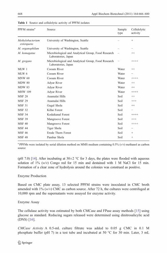
Table 1 Source and cellulolytic activity of PPFM isolates
PPFM strainsa Source Sampletype
Cellulolyticactivity
Methylobacteriumextorquens
University of Washington, Seattle – +
M. organophilum University of Washington, Seattle – −M. komagatae Microbiological and Analytical Group, Food Research
Laboratories, Japan– ++
M. gregans Microbiological and Analytical Group, Food ResearchLaboratories, Japan
− ++++
MLW 1 Cooum River Water ++
MLW 6 Cooum River Water −MNW 60 Cooum River Water ++++
MDW 80 Adyar River Water ++
MDW 83 Adyar River Water ++
MHW 109 Adyar River Water ++++
MSF 28 Anamalai Hills Soil ++
MSF 29 Anamalai Hills Soil +++
MSF 31 Gugal Shola Soil ++
MSF 32 Killai Forest Soil −MSF 34 Kodaikanal Forest Soil ++++
MSF 39 Mangroove Forest Soil +++
MSF 40 Mangroove Forest Soil ++++
MSF 44 Tiger Shola Soil −MSF 46 Erode Thorn Forest Soil +
MSF 48 Pambar Shola Soil +
a PPFMs were isolated by serial dilution method on MMS medium containing 0.5% (v/v) methanol as carbonsource
668 Appl Biochem Biotechnol (2011) 164:666–680
of DNS reagent was added to the test tube and incubated in a boiling water bath for5 min. After cooling, the glucose released in the assay liquid was measured by opticaldensity method at 540 nm.
FPase Activity A 0.5-mL culture filtrate was added to a test tube containing 0.05 g filterpaper strip (1×6 cm) and 1 mL of 0.1 M phosphate buffer (pH 7). The test tubes wereincubated at 50 °C for 30 min. After that, 3 mL of the DNS reagent was added to a test tubeand incubated in a boiling water bath for 5 min. The test tubes were cooled and the glucosereleased was measured using a UV spectrometer at 540 nm. One unit of enzyme activitywas defined as the amount of enzyme required for liberating 1 μM of glucose per milliliterper minute and was expressed as U mL−1 min−1 under standard assay conditions.
Enzyme Characterization
Effect of pH and Temperature on the Activity and Stability of the Enzyme
Five PPFM strains viz M. gregans, MNW 60, MHW 109, MSF 34, and MSF 40 showingsignificant cellulase activity were further characterized to determine the optimum pH andtemperature for cellulolytic activity. The optimum pH for the enzyme activity was determinedover a pH range of 2–10 at 50 °C using the following buffers: 50 mM citrate (pH 2–5), 50 mMphosphate (pH 6–7), and 50 mM glycine–NaOH (pH 8–10). The effect of temperature on thecellulase activity was determined by assaying the enzyme activity between 25 and 75 °C.
To determine pH stability, the enzyme was kept at room temperature for 1 h in the abovementioned buffers, and the residual cellulolytic activity was determined under standardassay conditions. Thermostability was measured by incubating the enzyme samples in50 mM phosphate buffer (pH 7) for 1 h at 20 to 60 °C. The enzyme solution was chilled inan ice bath for 5 min and then assayed using the standard assay procedures at 55 °C. For allthe assays, filter paper was used as the substrate.
SDS-PAGE and Zymogram Analysis
The crude enzyme preparation of MNW 60 was subjected to SDS-PAGE analysis [17].Zymogram analysis was done with the modified method reported earlier [18, 19]. Theculture supernatant along with the sample buffer was subjected to electrophoresis on SDS-PAGE (10%) containing 0.2% (w/v) CMC. After electrophoresis, the gel was soaked insolution A (sodium phosphate buffer, pH 7.2, containing 40% (v/v) isopropanol) for 1 hfollowed by solution B (sodium phosphate buffer, pH 7.2 containing 0.1% (v/v) TritonX-100) for 1 h at room temperature with gentle shaking to remove the SDS and to allow theproteins in the gel to renature. The gel was then washed four times in sodium phosphatebuffer (pH 7.2) for 30 min at 4 °C. After incubation for 60 min at 50 °C, the gel was soakedin 0.1% (w/v) Congo red solution for 30 min at room temperature and washed with 1 MNaCl until the activity bands were observed as clear colorless areas.
Scanning Electron Microscopy
After 72 h of growth, the cells of MNW 60 grown in the medium amended with celluloseand without cellulose were harvested and subjected to SEM. The cells were washed in
Appl Biochem Biotechnol (2011) 164:666–680 669

cacodylate buffer (0.1 M, pH 7.2) containing 0.22 M sucrose and were fixed in 2% (v/v)glutaraldehyde in cacodylate buffer at 4 °C for 2 h. The suspension was again centrifugedand washed in cacodylate buffer. The samples were post-fixed in 1% (v/v) OsO4 incacodylate buffer at 4 °C for 2 h, dehydrated by increasing the concentrations of alcohol,dried in hexamethyldisilazane, and mounted on aluminum stubs. The samples were sputter-coated with gold/palladium and examined using a FEI quanta 200 SEM operating at anaccelerating voltage of 10 keV.
Strain Characterization
The morphological, cultural, physiological, and biochemical characteristics of the cellulaseproducing pink pigmented facultative methylotropic strain, MNW 60, were determined byfollowing Bergey’s Manual of Systematic Bacteriology [20]. Gram stain reaction, catalaseand oxidase activity, indole production, methyl red and Voges–Proskauer reactions, ureaseactivity, nitrate reduction, and citrate production were carried out using standard procedures[21]. The ability of the PPFMs to grow in the presence of different substrates (ethanol,formaldehyde, acetic acid, methylamine, succinic acid, fumaric acid, formic acid, pyruvicacid, betaine, aspartic acid, glutamic acid, glycerol, glucose, fructose, lactose, maltose,mannose, ribose, rhamnose, xylose, and mannitol) other than methanol was studied usingthe MMS liquid medium with 0.5% for each amendment. Thermolabile substrates werefilter-sterilized using a 0.22-μm membrane filter (Millipore) and added to the medium. ForDNA extraction, the strain was grown in 100 mL of MMS medium supplemented with0.5% (v/v) methanol for 72 h at 35 °C. The biomass was harvested by centrifugation at10,000 rpm for 10 min and washed twice in sterile Tris–EDTA buffer (10:1 M ratio,pH 8.0). The extraction of DNAwas carried out by the phenol chloroform method [22]. Thepresence of mxaF gene was detected using specific primers 1003f (5′-GCG GCA CCAACT GGG GCT GGT-3′) and 1561r (5′-GGG CAG CAT GAA GGG CTC CC-3′) [23, 24].For amplification of 16S ribosomal RNA (rRNA) gene, universal 27F and 1492Rbacteria-specific primers were used [25]. Both the amplified DNA fragments were gel-purified using QIAquick Gel Extraction kit (Qiagen, Hilden, Germany) and sequencedusing an ABI377 sequencer (Applied Biosystems). The nucleotide sequence obtained wascompared with GenBank data (http://www.ncbi.nlm.nih.gov/) using BlastN search [26].Sequences were then aligned with the ClustalW software [27]. A phylogenetic tree wasconstructed by using the neighbor-joining DNA distance algorithm [28] with 1,000bootstrap resamplings.
Statistical Analysis
All the experiments were carried out in triplicates, and the data were evaluated by analysisof variance and means were compared by applying Tukey’s test using SPSS 14.0 statisticalpackage. The level of significance was accepted as P<0.05.
Results and Discussion
The importance of cellulases has attracted much interest because of their diversity inbiological, chemical, and industrial applications [29, 30]. Therefore, there is a continuousthrust to isolate efficient cellulose-producing microbes and optimize their ability to producemore. In this study, we report cellulase production by PPFMs isolated from the water
670 Appl Biochem Biotechnol (2011) 164:666–680
samples of Cooum and Adyar rivers in Chennai and soil samples of forests in Tamil Nadualong with four reference Methylobacterium strains (Table 1).
Screening for Cellulase-Producing PPFM
Among the 20 PPFM isolates, 13 strains showed clearing zones around the colonies inCMC agar plates flooded with Congo red solution indicating cellulase production (Table 1). Inthis study, detection of cellulolytic ability among the PPFMs isolated from differentenvironments with good organic components supports the argument that the presence ofdecaying organic matter acts as a natural enrichment for cellulolytic microbes [31–33].
Enzyme Activity and Characterization
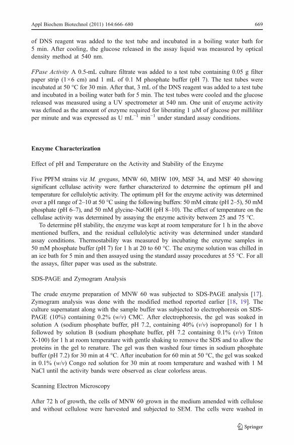
All the 13 cellulase-positive PPFM isolates were able to grow in the liquid medium withcellulose as a sole carbon source which induced cellulase production. The enzymeproduction reached maximum after 72 h, indicating a slow cellulose degradation processwhich could be related to catabolite repression by glucose [34]. Although many cellulolyticbacteria have been reported to produce cellulases that show both endo- and exoglucanaseactivities [35, 36], in this study, significantly higher FPase activity was observed thanCMCase activity. M. gregans, MNW 60, MHW 109, MSF 34, and MSF 40 showedCMCase activity in the range of 0.23–0.54 UmL−1 and higher FPase activity between 0.49and 0.84 UmL−1 than the other strains studied (Fig. 1). This could be related to theinfluence of substrate on the growth of cellulolytic organisms [37, 38], which seem to beregulated by induction repression mechanisms [39]. Observations on maximum FPaseactivities compared to CMase activity are similar to the studies expressed by Belghith et al.[40] and Emtiazi et al. [41].
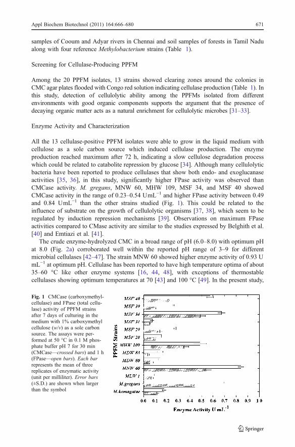
The crude enzyme-hydrolyzed CMC in a broad range of pH (6.0–8.0) with optimum pHat 8.0 (Fig. 2a) corroborated well within the reported pH range of 3–9 for differentmicrobial cellulases [42–47]. The strain MNW 60 showed higher enzyme activity of 0.93 UmL−1 at optimum pH. Cellulase has been reported to have high temperature optima of about35–60 °C like other enzyme systems [16, 44, 48], with exceptions of thermostablecellulases showing optimum temperatures at 70 [43] and 100 °C [49]. In the present study,
Fig. 1 CMCase (carboxymethyl-cellulase) and FPase (total cellu-lase) activity of PPFM strainsafter 7 days of culturing in themedium with 1% carboxymethylcellulose (w/v) as a sole carbonsource. The assays were per-formed at 50 °C in 0.1 M phos-phate buffer pH 7 for 30 min(CMCase—crossed bars) and 1 h(FPase—open bars). Each barrepresents the mean of threereplicates of enzymatic activity(unit per milliliter). Error bars(±S.D.) are shown when largerthan the symbol
Appl Biochem Biotechnol (2011) 164:666–680 671
Fig. 2 Effect of pH (a) and temperature (b) on enzyme activity. a Enzyme activity: Assays were done understandard FPase assay conditions with the following buffer systems: 50 mM citrate buffer (pH 2–5), 50 mMphosphate buffer (pH 6–7), and 50 mM glycine–NaOH buffer (pH 8–10) at 50 °C for 1 h. b Enzyme activity:Cellulase activity was measured under standard FPase assay conditions using 50 mM glycine–NaOH bufferpH 8 at different temperatures (25–75 °C). Values are expressed as mean ± standard deviation of threeindependent readings
672 Appl Biochem Biotechnol (2011) 164:666–680
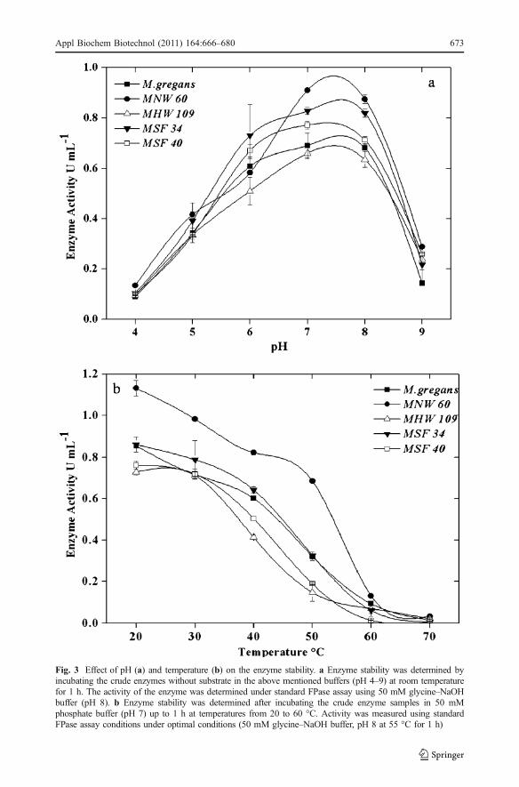
Fig. 3 Effect of pH (a) and temperature (b) on the enzyme stability. a Enzyme stability was determined byincubating the crude enzymes without substrate in the above mentioned buffers (pH 4–9) at room temperaturefor 1 h. The activity of the enzyme was determined under standard FPase assay using 50 mM glycine–NaOHbuffer (pH 8). b Enzyme stability was determined after incubating the crude enzyme samples in 50 mMphosphate buffer (pH 7) up to 1 h at temperatures from 20 to 60 °C. Activity was measured using standardFPase assay conditions under optimal conditions (50 mM glycine–NaOH buffer, pH 8 at 55 °C for 1 h)
Appl Biochem Biotechnol (2011) 164:666–680 673
significant enzyme activity was observed at temperatures between 35 and 65 °C (Fig. 2b).Though maximum activity was observed at 45 and 55 °C in the strains M. gregans, MNW60, MHW 109, MSF 34, and MSF 40 (0.74–1.12 UmL−1), the abrupt reduction in cellulaseactivity above 55 °C could be related to the denaturation of thermal sensitive componentsof the cellulase system [50].
Observations on the stability of the enzyme over the pH range of 5.0 to 8.0 withmaximum activity at pH 7.0 were in agreement with earlier reports [48, 51]. The activitywas 50% at pH 5, and at the alkaline pH 9, it was reduced to 30% (Fig. 3a). Thermostabilitystudies showed that the increase in temperature had a negative effect on the enzymeactivity. The enzyme was found stable at 20 to 50 °C, and although higher enzyme activitywas observed at 20 °C, it retained 60% of the activity at 40°C (Fig. 3b).
SDS-PAGE and Zymogram Analysis
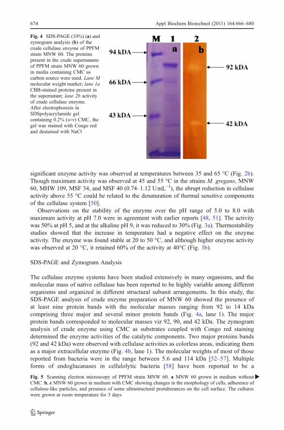
The cellulase enzyme systems have been studied extensively in many organisms, and themolecular mass of native cellulase has been reported to be highly variable among differentorganisms and organized in different structural subunit arrangements. In this study, theSDS-PAGE analysis of crude enzyme preparation of MNW 60 showed the presence ofat least nine protein bands with the molecular masses ranging from 92 to 14 kDacomprising three major and several minor protein bands (Fig. 4a, lane 1). The majorprotein bands corresponded to molecular masses viz 92, 90, and 42 kDa. The zymogramanalysis of crude enzyme using CMC as substrates coupled with Congo red stainingdetermined the enzyme activities of the catalytic components. Two major proteins bands(92 and 42 kDa) were observed with cellulase activities as colorless areas, indicating themas a major extracellular enzyme (Fig. 4b, lane 1). The molecular weights of most of thosereported from bacteria were in the range between 5.6 and 114 kDa [52–57]. Multipleforms of endoglucanases in cellulolytic bacteria [58] have been reported to be a
94 kDA
66 kDA
43 kDA
92 kDA
42 kDA
Fig. 4 SDS-PAGE (10%) (a) andzymogram analysis (b) of thecrude cellulase enzyme of PPFMstrain MNW 60. The proteinspresent in the crude supernatantsof PPFM strain MNW 60 grownin media containing CMC ascarbon source were used. Lane Mmolecular weight marker; lane 1aCBB-stained proteins present inthe supernatant; lane 2b activityof crude cellulase enzyme.After electrophoresis inSDSpolyacrylamide gelcontaining 0.2% (w/v) CMC, thegel was stained with Congo redand destained with NaCl
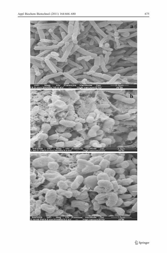
Fig. 5 Scanning electron microscopy of PPFM strain MNW 60. a MNW 60 grown in medium withoutCMC. b, c MNW 60 grown in medium with CMC showing changes in the morphology of cells, adherence ofcellulose-like particles, and presence of some ultrastructural protuberances on the cell surface. The cultureswere grown at room temperature for 3 days
�
674 Appl Biochem Biotechnol (2011) 164:666–680
a
b
c
Appl Biochem Biotechnol (2011) 164:666–680 675

consequence of multiple genes [59]. However, the dimeric form of the enzyme observedin this study could be related to the dissociation of the two subunits induced bySDS-PAGE [55].
SEM Analysis
Changes in the cell morphology of Methylobacerium sp. grown in the presence oftricalcium phosphate-containing medium was observed earlier (Jayashree et al. [60]communicated to Archives of Microbiology). MNW 60 cells grown in methanol mineralsalts medium were seen as long rods with smooth surfaces, and the cells grown in themedium containing CMC showed extensive morphological changes with adherence ofcellulose-like particles and presence of some ultrastructural protuberances on the surfaceof the cells (Fig. 5a–c). This structural change in the presence of cellulose coupled withenzyme production shows the hydrolyzing nature of the strain and leads to the conclusionthat these changes could be induced by the substrate, CMC, due to enzyme production.The presence of protuberance-like structures, though not prominent, seen in the bacterialcell mass has been reported earlier for efficient cellulose degradation [61–64]. Severalcellulolytic Clostridia sp. have also shown to produce an ultrastructural cellularprotuberance which is formed during the efficient degradation of cellulose and isconfirmed to be an exocellular protein complex of cellulolytic enzymes termedcellulosome [65]. Adhesion of cellulose-like particles observed in the surface of thebacterial cells grown in the presence of CMC has also been reported earlier in manybacteria [62, 66, 67]. According to Wilson et al. [68], adherence of bacteria to thecellulosic material is important for cellulase degradation. For these reasons it is believedthat the rate and extent to which microorganisms adhere to cellulose is important for thedigestibility of the material, and therefore the enzyme activity [67].
Fig. 6 Phylogenetic tree showing the relationships based on mxaF gene sequences betweenmethylotrophic strain (MNW 60) and other related taxa. MNW 60 which had maximum cellulase activityshowed the closest methanol dehydrogenase gene mxaF sequence relationships with the pink pigmentedfacultative methylotropic species of the genus Methylobacterium belonging to alpha proteobacteria withvalid published names. Accession number is given within brackets and the tree was drawn using theneighbor-joining method
676 Appl Biochem Biotechnol (2011) 164:666–680
Strain Characterization
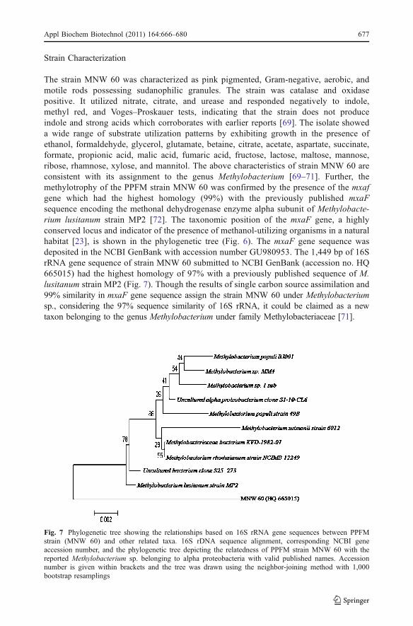
The strain MNW 60 was characterized as pink pigmented, Gram-negative, aerobic, andmotile rods possessing sudanophilic granules. The strain was catalase and oxidasepositive. It utilized nitrate, citrate, and urease and responded negatively to indole,methyl red, and Voges–Proskauer tests, indicating that the strain does not produceindole and strong acids which corroborates with earlier reports [69]. The isolate showeda wide range of substrate utilization patterns by exhibiting growth in the presence ofethanol, formaldehyde, glycerol, glutamate, betaine, citrate, acetate, aspartate, succinate,formate, propionic acid, malic acid, fumaric acid, fructose, lactose, maltose, mannose,ribose, rhamnose, xylose, and mannitol. The above characteristics of strain MNW 60 areconsistent with its assignment to the genus Methylobacterium [69–71]. Further, themethylotrophy of the PPFM strain MNW 60 was confirmed by the presence of the mxafgene which had the highest homology (99%) with the previously published mxaFsequence encoding the methonal dehydrogenase enzyme alpha subunit of Methylobacte-rium lusitanum strain MP2 [72]. The taxonomic position of the mxaF gene, a highlyconserved locus and indicator of the presence of methanol-utilizing organisms in a naturalhabitat [23], is shown in the phylogenetic tree (Fig. 6). The mxaF gene sequence wasdeposited in the NCBI GenBank with accession number GU980953. The 1,449 bp of 16SrRNA gene sequence of strain MNW 60 submitted to NCBI GenBank (accession no. HQ665015) had the highest homology of 97% with a previously published sequence of M.lusitanum strain MP2 (Fig. 7). Though the results of single carbon source assimilation and99% similarity in mxaF gene sequence assign the strain MNW 60 under Methylobacteriumsp., considering the 97% sequence similarity of 16S rRNA, it could be claimed as a newtaxon belonging to the genus Methylobacterium under family Methylobacteriaceae [71].
Fig. 7 Phylogenetic tree showing the relationships based on 16S rRNA gene sequences between PPFMstrain (MNW 60) and other related taxa. 16S rDNA sequence alignment, corresponding NCBI geneaccession number, and the phylogenetic tree depicting the relatedness of PPFM strain MNW 60 with thereported Methylobacterium sp. belonging to alpha proteobacteria with valid published names. Accessionnumber is given within brackets and the tree was drawn using the neighbor-joining method with 1,000bootstrap resamplings
Appl Biochem Biotechnol (2011) 164:666–680 677
Conclusion
The current study establishes a detailed report on the cellulase production by PPFM for thefirst time. A PPFM strain MNW 60 exhibiting higher cellulase activity (1.15 UmL−1) atpH 8 and at 55 °C was found to be related to Methylobacterium sp. Further investigation ongrowth, culture conditions, strain improvement for cellulase production, purification, andcharacterization of the cellulose enzyme would add value for its use in large-scaleapplication. Detection of genes responsible for cellulase production [73] in differentMethylobacterium sp. would help explore their functional aspects. The results of this studyalso indicate that the Cooum and Adyar rivers in Chennai and various forests in TamilNadu, India are rich sources of many pink pigmented facultative methylotropic bacteriawhich in turn could be a source of various enzymes, and further studies are recommendedon these sites including study of microbial biodiversity and the associated biotechnologicalpotential of the isolated strains.
Acknowledgments The authors thank the Life Sciences Research Board, DRDO, and the Department ofBiotechnology, Government of India for financial support; Dr. Mary E. Lidstorm, University of Washington,Seattle for providing the bacterial strain; and Shri AMM Murugappa Chettiar Research Centre for providingthe necessary facilities. The authors gratefully acknowledge Dr. Gerry Brennan and Ms. Sharon Elizhabeth,Queens University, Belfast, UK for their help in scanning electron microscopy.
References
1. Sukumaran, R. K., Singhania, R. R., & Pandey, A. (2005). Journal of Scientific and Industrial Research,64, 832–844.
2. Olsson, L., & Hahn-Hagerdahl, B. (1997). Enzyme and Microbial Technology, 18, 312–331.3. Levy, I., Shani, Z., & Shoseyov, O. (2002). Biomolecular Engineering, 19, 17–30.4. Van Wyk, J. P. H., & Mohulatsi, M. (2003). Bioresource Technology, 86, 21–23.5. Nipa, M. N., Sultana, S., & Hakim, M. A. (2006). Bangladesh Journal of Microbiology, 23(2), 174–176.6. Gosh, B. K., Gosh, A., & Salnar, A. (1987). In J. Chaloupka & V. Krumphanzl (Eds.), Extracellular
enzymes of microorganisms: Cellulase secretion from a hypercellulolytic mutant (pp. 157–172). NewYork: Plenum.
7. Lynd, R. L., Weimer, J. P., Van Zyl, H. W., & Pretorius, S. I. (2002). Microbiology and MolecularBiology Reviews, 66, 506–577.
8. Chistoserdova, L., Chen, S. W., Lapidus, A., & Lidstrom, M. E. (2003). Journal of Bacteriology, 185(10), 2980–2987.
9. Nemecek-Marshall, M., MacDonald, R. C., Franzen, J. J., Wojciechowski, C. L., & Fall, R. (1995). PlantPhysiology, 108, 1359–1368.
10. Idris, R., Kuffner, M., Bodrossy, L., Puschenreiter, M., Monchy, S., Wenzel, W. W., et al. (2006).Systematic and Applied Microbiology, 29, 634–644.
11. Madhaiyan, M., Park, M. S., Lee, H. S., Kim, C. W., Lee, K. H., Seshadri, S., et al. (2004). KoreanJournal of Soil Science and Fertilizer, 37(1), 46–53.
12. Raja, P., Balachandar, D., & Sundaram, S. P. (2008). Biology and Fertility of Soils, 45, 45–53.13. Balachandar, D., Raja, R., & Sundaram, S. P. (2008). Brazilian Journal of Microbiology, 39, 68–73.14. Kim, B. H., & Wimpenny, J. W. T. (1981). Canadian Journal of Microbiology, 27, 1260–1266.15. Mandels, M., Andreotti, R., & Roche, C. (1976). Biotechnology Biogen Symposium, 6, 21–31.16. Miller, G. C. (1959). Analytical Chemistry, 31, 426–428.17. Laemmli, U. K. (1970). Nature, 227, 680–685.18. Lee, S., Morikawa, M., Takagi, M., & Imankawa, T. (1994). Applied and Environmental Microbiology,
60, 3761–3773.19. Saul, D. J., Williams, L. C., Grayling, R. A., Chamley, L. W., Love, D. R., & Bergquist, P. L. (1990).
Applied and Environmental Microbiology, 56, 3117–3124.20. Bergy. (1984). Bergey’s manual of systematic bacteriology. Baltimore: Williams & Wilkins.
678 Appl Biochem Biotechnol (2011) 164:666–680
21. Norris, J.R., & Ribbons, D.W. (1972) (Eds.). Methods in microbiology. London: Academic.22. Sambrook, J., Fritsch, E. F., & Maniatis, T. (1989). Molecular cloning: A laboratory manual. Cold
Spring Harbor: Cold Spring Harbor Laboratory Press.23. Murrell, J. C., Mc Donald, I. R., & Bourne, D. G. (1998). FEMS Microbiology Ecology, 27, 103–114.24. Horz, H. P., Tchawa, Y. M., & Liesack, W. (2001). Applied and Environmental Microbiology, 67, 4177–4185.25. Christner, B. C., Mosley-Thompson, E., Thompson, L. G., & Reeve, J. N. (2001). Environmental
Microbiology, 3, 570–577.26. Altschul, S. F., Madden, T. L., Schaffer, A. A., Zhang, J., Zhang, Z., Miller, W., et al. (1997). Nucleic
Acid Program Research, 25, 3389–3402.27. Thompson, J. D., Higgins, D. G., & Gibson, T. J. (1994). Nucleic Acids Research, 22, 4673–4680.28. Saitou, N., & Nei, M. (1987). Molecular Biology and Evolution, 4, 406–425.29. Bhat, M. K., & Bhat, S. (1997). Biotechnology Advances, 15(3–4), 583–620.30. Jang, H. D., & Chen, K. S. (2003). World Journal of Microbiology & Biotechnology, 19, 263–268.31. Alani, F., Anderson, W. A., & Moo-Young, M. (2008). Biotechnology Letters, 30, 123–126.32. Jang, H., & Chang, K. (2005). Biotechnology Letters, 27, 239–242.33. Bischoff, K. M., Rooney, P. A., Li, X.-L., Liu, S., & Hughes, S. R. (2006). Biotechnology Letters, 28,
1761–1765.34. Hulme, M. A., & Stranks, D. W. (1970). Nature, 226, 469–470.35. Zverlov, V. V., Velikodvorskaya, G. V., Schwarz, W. H. J., Bronnenmeier, K., Kellermann, J., &
Staudenbauer, W. L. (1998). Journal of Bacteriology, 180, 3091–3099.36. Belghith, H., Semia, E.-C., & Gargouri, A. (2001). Journal of Bacteriology, 89, 257–262.37. Lakshmikant, K., & Mathur, S. N. (1990). World Journal of Microbiology & Biotechnology, 11, 23–26.38. Immanuel, G., Dhanusa, R., Prema, P., & Palavesam, A. (2006). International Journal of Environmental
Science and Technology, 3(1), 25–34.39. Reinhold-Hurek, B., Hurek, T., Claeyssens, M., & Van Montagu, M. (1993). Journal of Bacteriology,
175(21), 7056–7065.40. Belghith, H., Chaabouni, S. E., & Gargouri, A. (2001). Journal of Bacteriology, 89, 257–262.41. Emtiazi, G., Pooyan,M., & Shamalnasab,M. (2007).World Journal of Agricultural Sciences, 3(5), 602–608.42. Chakraborty, N., Sarkar, G. M., & Lahiri, S. C. (2000). The Environmentalist, 20, 9–11.43. Mawadza, C., Hatti-Kaul, R., Zvauya, R., & Mattiasson, B. (2000). Journal of Biotechnology, 83, 177–187.44. Coral, G., Arikan, B., Unaldi, M. N., & Guvenmez, H. (2002). Turkish Journal of Biology, 26, 209–213.45. Bakare, M. K., Adewalw, I. O., Ajayi, A. O., Okoh, A. I., & Shonukan, O. O. (2005). African Journal of
Biotechnology, 4(8), 838–843.46. Immanuel, G., Bhagavath, C. M. A., Raj, P. I., Esakkiraj, P., & Palavesam, A. (2007). The Internet
Journal of Microbiology, 3(1), 1–12.47. Dutta, T., Sahoo, R., Sengupta, R., Ray, S. S., Bhattacharjee, A., & Ghosh, S. J. (2008). Journal of
Industrial Microbiology & Biotechnology, 35, 275–282.48. Sanchez-Torres, J., Perez, P., & Santamaria, R. I. (1996). Applied Microbiology and Biotechnology, 46,
149–155.49. Bragger, J. M., Daniel, R. M., Coolbear, T., & Morgan, H. W. (1989). Applied Microbiology and
Biotechnology, 31, 556–561.50. Singh, R., Kumar, R., Bishnoi, K., & Bishnoi, R. N. (2009). Biochemical Engineering, 48, 28–35.51. Okeke, B. C., & Patterson, A. (1992). World Journal of Microbiology & Biotechnology, 8, 483–487.52. Bronnenmeier, K., & Staudenbauer, W. L. (1988). Applied Microbiology and Biotechnology, 27, 432–436.53. Hreggvidsson, G. O., Kaiste, E., Holst, E., Eggertssonh, G., Palsdottir, A., & Kristjansson, J. K. (1996).
Applied and Environmental Microbiology, 62(8), 3047–3049.54. Wang, W., Liu, J., Chen, G., Zhang, Y., & Gao, P. (2003). Current Microbiology, 46, 371–379.55. Huang, X. P., & Monk, C. (2004). World Journal of Microbiology & Biotechnology, 20, 85–92.56. Hong, S.-Y., Lee, J.-S., Cho, K.-M., Math, R. K., Kim, Y.-H., Hong, S.-J., et al. (2007). Biotechnology
Letters, 29, 931–936.57. Kim, E. S., Lee, H. J., Bang, W. G., Choi, I. G., & Kim, K. H. (2009). Biotechnology and
Bioengineering, 102, 1342–1353.58. Lamed, R., Setter, E., Kenig, R., & Bayer, E. A. (1983). Biotechnology and Bioengineering, 13, 163–181.59. Fukumori, F., Kudo, T., & Horikoshi, K. (1985). Journal of General Microbiology, 131, 3339–3345.60. Jayashree, S., Vadivukkarasi, P., Anand, K., Kato, Y., & Seshadri, S. (2010) Archives of Microbiology,
ArchMicr-2010-0079.R2.61. Bayer, E. A., Setter, E., & Lamed, R. (1985). Journal of Bacteriology, 163, 552–559.62. Lamed, R., Naimark, J., Morgenstern, E., & Bayer, E. A. (1987). Journal of Bacteriology, 169(8), 3792–3800.63. Bayer, E. A., Shimon, L. J., Shoham, Y., & Lamed, R. J. (1998). Journal of Structural Biology, 124,
221–234.
Appl Biochem Biotechnol (2011) 164:666–680 679

64. Blair, B. G., & Anderson, K. L. (1998). Biotechnic & Histochemistry, 73, 107–113.65. Bayer, E. A., & Lamed, R. (1986). Journal of Bacteriology, 167, 828–836.66. Pason, P., Kyu, K. L., & Ratanakhanokchai, I. (2006). Applied and Environmental Microbiology, 72,
2483–2490.67. Louime, C., Abazinge, M., & Johnson, E. (2006). International Journal of Molecular Sciences, 7, 1–11.68. Wilson, D. B., Lao, G., Ghangas, G. S., & Jung, E. (1991). Journal of Bacteriology, 173, 3397–3407.69. Green, P. N. (1992). In A. Balows, H. G. Truper, M. Dworkin, W. Harder, & K.-H. Schleifer (Eds.), The
prokaryotes: The genus Methylobacterium (2nd ed.). New York: Springer.70. Green, P. N., & Bousfield, I. J. (1983). International Journal of Systematic Bacteriology, 33, 875–877.71. Green, P. N. (2001). In M. Dworkin (Ed.), The prokaryotes: Methylobacterium (3rd ed.). New York:
Springer.72. Wang, X., Sahr, F., Ting, X., & Baolin, S. (2007). International Journal of Systematic and Evolutionary
Microbiology, 57, 1699–1703.73. Vuilleumier et al. (2010) putative cellulase, endoglucanase (celC) [Methylobacterium extorquens AM1]
unpublished data in NCBI gene database.
680 Appl Biochem Biotechnol (2011) 164:666–680
Copyright © 2022 FDOKUMEN