The putative protein methyltransferase LAE1 controls cellulase gene expression in Trichoderma reesei

The VELVET A Orthologue VEL1 of Trichoderma reeseiRegulates Fungal Development and Is Essential forCellulase Gene ExpressionRazieh Karimi Aghcheh1.", Zoltan Nemeth2.", Lea Atanasova1, Erzsebet Fekete2, Melinda Paholcsek3,
Erzsebet Sandor4, Benigno Aquino1, Irina S. Druzhinina1,5, Levente Karaffa2, Christian P. Kubicek1,6*
1 Institute of Chemical Engineering, Research Division Biotechnology and Microbiology, Microbiology Group, Vienna University of Technology, 1060 Vienna, Austria,
2 Department of Biochemical Engineering, Faculty of Sciences and Technology, University of Debrecen, H-4032 Debrecen, Hungary, 3 Department of Human Genetics,
Faculty of Medicine, University of Debrecen, H-4032 Debrecen, Hungary, 4 Faculty of Agricultural and Food Science and Environmental Management, Institute of Food
Science, H-4032 Debrecen, Hungary, 5 Austrian Center of Industrial Biotechnology, c/o Institute of Chemical Engineering, Vienna University of Technology, 1060 Vienna,
Austria, 6 Austrian Center of Industrial Biotechnology, 8010 Graz, Austria
Abstract
Trichoderma reesei is the industrial producer of cellulases and hemicellulases for biorefinery processes. Their expression isobligatorily dependent on the function of the protein methyltransferase LAE1. The Aspergillus nidulans orthologue of LAE1 -LaeA - is part of the VELVET protein complex consisting of LaeA, VeA and VelB that regulates secondary metabolism andsexual as well as asexual reproduction. Here we have therefore investigated the function of VEL1, the T. reesei orthologue ofA. nidulans VeA. Deletion of the T. reesei vel1 locus causes a complete and light-independent loss of conidiation, and impairsformation of perithecia. Deletion of vel1 also alters hyphal morphology towards hyperbranching and formation of thickerfilaments, and with consequently reduced growth rates. Growth on lactose as a sole carbon source, however, is even morestrongly reduced and growth on cellulose as a sole carbon source eliminated. Consistent with these findings, deletion ofvel1 completely impaired the expression of cellulases, xylanases and the cellulase regulator XYR1 on lactose as a cellulaseinducing carbon source, but also in resting mycelia with sophorose as inducer. Our data show that in T. reesei VEL1 controlssexual and asexual development, and this effect is independent of light. VEL1 is also essential for cellulase gene expression,which is consistent with the assumption that their regulation by LAE1 occurs by the VELVET complex.
Citation: Karimi Aghcheh R, Nemeth Z, Atanasova L, Fekete E, Paholcsek M, et al. (2014) The VELVET A Orthologue VEL1 of Trichoderma reesei Regulates FungalDevelopment and Is Essential for Cellulase Gene Expression. PLoS ONE 9(11): e112799. doi:10.1371/journal.pone.0112799
Editor: Jae-Hyuk Yu, University of Wisconsin - Madison, United States of America
Received August 13, 2014; Accepted October 15, 2014; Published November 11, 2014
Copyright: � 2014 Karimi Aghcheh et al. This is an open-access article distributed under the terms of the Creative Commons Attribution License, which permitsunrestricted use, distribution, and reproduction in any medium, provided the original author and source are credited.
Data Availability: The authors confirm that all data underlying the findings are fully available without restriction. All relevant data are within the paper and itsSupporting Information files. Strains will be available upon request to Prof. Irina S. Druzhinina ([email protected]).
Funding: This study was supported by grants of the Austrian Science Foundation (to CPK; P 21266 and I 1249), by the EU and co-financed by the European SocialFund under the project ENVIKUT (TA MOP-4.2.2.A-11/1/KONV-2012-0043), and also by the Hungarian Scientific Research Fund (OTKA Grant K1006600). LK is arecipient of a Bolyai Janos Research Scholarship. MP was supported by the EU and Hungary, co-financed by the European Social Fund in the framework of theTAMOP-4.2.4.A/2-11/1-2012-0001 ‘National Excellence Program.’ The funders had no role in study design, data collection and analysis, decision to publish, orpreparation of the manuscript.
Competing Interests: The authors have declared that no competing interests exist.
* Email: [email protected]
. These authors contributed equally to this work.
" These authors are listed in alphabetical order.
Introduction
Cellulose and hemicelluloses form the major amount of plant
biomass and thus represent the largest reservoir of renewable
carbon sources on Earth, which could potentially replace fuels and
refinery products derived from fossil carbon components [1]. To
this end, efficient hydrolysis of the plant cell wall polymers to
soluble oligo- and monomers is essential. The Sordariomycete
Trichoderma reesei is most widely used for the industrial
production of cellulolytic and hemicellulolytic enzymes and has
become a basis for the modern paradigm of these enzymes [2].
The T. reesei genome encodes two cellobiohydrolases, five endo-ß-
1,4-glucanases, and several ß-glucosidases, hemicellulases and
accessory enzymes [3]. Most of these genes are regulated in a
consistent manner, and are expressed only in the presence of an
inducer, which can be either cellulose itself, disaccharides
generated by its degradation (such as sophorose) or the galacto-
syl-b-1,4-glucoside lactose [3–7]. Today, seven transcription
factors have been identified that participate positively or negatively
in this regulation, of which XYR1 (xylanase regulator 1) is the
main activator of both cellulase and hemicellulase gene expression
[3,8]. However, we have recently shown that the expression of
genes for lignocellulose degradation in T. reesei is further
obligatorily dependent on the function of the protein methyltrans-
ferase LAE1 [9], the orthologue of the A. nidulans regulator of
secondary metabolism and development LaeA [10]. This regula-
tion requires a functional xyr1, but a lae1 loss-of-function cannot
be rescued by xyr1 overexpression [9], which would be consistent
with the hypothesis that LaeA acts by removing the repressive
PLOS ONE | www.plosone.org 1 November 2014 | Volume 9 | Issue 11 | e112799

chromatin [11]. However, a genome-wide analysis of H3K4 and
H3K9 methylation patterns in T. reesei lae1 mutants did not show
any methylation changes at the cellulase loci [12]. More recently,
the methyltransferase activity of A. nidulans LaeA was shown to
exclusively perform automethylation of LaeA, but ironically just
this automethylated methionine residue is not conserved in T.reesei [13]. Hence the mechanism of LAE1-dependence of
cellulase gene expression remains enigmatic.
In A. nidulans, LaeA is known to be part of the trimeric
VELVET protein complex, that consists of LaeA, VeA and VelB
and that regulates secondary metabolism and development in A.nidulans [14–16], and pathogenesis on plants and humans in
other genera [17–22]. Most studies on veA have been carried out
in Aspergillus spp., where this gene has been described to control
photodependent development, secondary metabolism and patho-
genesis-associated processes [14–16]. Thorough genetic, molecu-
lar, and biochemical work has recently shown that Velvet is part of
a high-molecular-weight complex containing at least 10 different
proteins, some of which have been assigned distinct regulatory
roles [15,22–26]. The VeA orthologue VEL1 of Trichodermavirens has been shown to regulate sporulation, chlamydospore
formation, secondary metabolite synthesis and mycoparasitism
[27].
Our finding that LAE1 is essential for cellulase gene expression
[9], independent of the underlying mechanism, raised the
hypothesis that this function may require a functional VELVET
complex. To test this hypothesis, we have therefore cloned and
functionally characterized the ortholog of the central component
of the VELVET complex, VeA, from T. reesei (VEL1; the gene/
protein name was chosen in view of the Sordariomycete
nomenclature, which uses three letters and a number instead of
only letters to designate a gene). We will show that T. reesei vel1 –
like lae1 – is essential for cellulase and hemicellulase gene
expression. In addition, we will show that vel1 is also essential for
asexual and sexual development of T. reesei in a photo-
independent manner.
Results
The VEL1 orthologue of T. reeseiThe genome of T. reesei contains a single copy of the vel1 gene
(Trire2:122284; Gene Bank accession number of the respective
protein VEL1: EGR48103.1). The ORF of vel1 consists of
1,801 bp, is interrupted by a single 79 bp intron and encodes a
574 amino acid protein. Inspection of the genome sequences of the
improved cellulase producer strains QM 9414, NG14 and RUT
C-30 [28,29] showed that they contain gene copies with identical
nucleotide sequences, proving that vel1 has not been altered by
mutagenesis towards improved cellulase formation.
A phylogenetic analysis of the T. reesei VEL1 protein sequence
using the 50 best blastp hits from NCBI produced a tree whose
shape was concordant with that of the species tree (data not
shown), thus confirming earlier data [27]. Similarity of VEL1 to
the VeA orthologues in T. virens and T. atroviride was
consequently high (80 and 78% similarity over the entire amino
acid sequence respectively). Highest identity outside of the genus
was observed with Nectria haematococca (60%, 2e-180, 99%
coverage), whereas it was only 36 and 38% with A. nidulans and
A. fumigatus, respectively.
In accordance with studies in A. nidulans and Neurosporacrassa [30–32], WoLF PSORT identified the protein to be able to
enter the nucleus, the responsible motif being located at the N-
terminus, and a leucine-rich nuclear import signal was putatively
identified in the C-terminal quarter of the protein sequence. Like
A. nidulans VeA, the T. reesei VEL1 protein also contains a
potential PEST region (a sequence rich proline, serine, threonine
Figure 1. Effect of carbon source and light on vel1 transcriptlevels in T. reesei. Transcript levels of vel1 during growth on glucose,glycerol and lactose in T. reesei QM 9414 in the presence of ambientlight (white bars) and darkness (full bars). Transcript levels are given inarbitrary units, which were calculated by normalizing the vel1/tef1 ratioto that on glucose (12 h, ambient light). Data are means of at least 3biological replicas. The asterisk indicates the time point where thecultures started to sporulate (this time point was the same in light anddarkness).doi:10.1371/journal.pone.0112799.g001
Figure 2. The impact of VEL1 on morphology of T. reesei insubmerged culture. Morphology of growing hyphae of T. reeseiQM9414 (A–E) and the Dvel1 (F–L) strains during submerged growth onMandels-Andreotti medium with glycerol (1%, w/v) as a carbon sourceat 12 (A–B and F–G), 48 (C and H–I) and 72 (D–E and J–K) hoursfollowing inoculation.doi:10.1371/journal.pone.0112799.g002
Trichoderma reesei VEL1
PLOS ONE | www.plosone.org 2 November 2014 | Volume 9 | Issue 11 | e112799

and glutamine that indicates a short half-life; HAP-
PPLPPPPPSSYDAPPPAAR; PEST score 9.97), but in contrast
to A. nidulans, where it is located at the C-terminal end of the
protein [32], T. reesei VEL1 displays it in the middle of the protein
immediately after the conserved N-terminal half (aa 290–311).
T. reesei vel1 transcript levels are carbon sourcedependent
We have examined vel1 transcript levels during hyphal growth
on plates and subsequent sporulation on three different carbon
sources and in light and darkness. During growth on glucose and
glycerol, two carbon sources that allow rapid growth of T. reesei,in the presence of light (empty bars), vel1 mRNA was most
abundant during the phase of most rapid growth (25 hrs) whereas
it declined once growth ceased (Figure 1). During growth on
lactose, which is much slower, the peak in vel1 transcript
accumulation in light occurred at 35 hrs, which again was the
point where the growth rate was highest. During growth in
darkness (full bars), the vel1 transcript accumulated to much
higher levels than under illumination (Figure 1). Again, the pattern
on glucose and glycerol was similar, but now vel1 mRNA levels
increased with progressing time and were highest when growth
already declined (35 hrs). During growth on lactose in the dark,
however, vel1 transcript levels were highest at the early time points
but significantly decreased at 35 hrs. No correlation was observed
between vel1 mRNA levels and the onset of sporulation (asterisks
in Figure 1). These data illustrate that the accumulation of vel1-
mRNA expression is regulated by light and darkness, but that this
regulation is further modulated by the carbon source in relation to
the growth rate.
VEL1 is required for normal hyphal tip growth, andessential for sexual and asexual development in T. reesei
To investigate the impact of vel1 on the development of T.reesei, vel1 null mutants (Dvel1) were generated by replacing the
vel1 coding region with the E. coli hygromycin B phosphotrans-
ferase gene hph in the T. reesei QM 9414. In addition, we
generated overexpressing (vel1OE) mutants by fusing the vel1ORF downstream of the strongly expressing tef1 (elongation factor
1a- gene) promoter. We also attempted to retransform the wild-
type vel1 gene into the Dvel1 mutant, but even screening of more
than 200 transformants did not yield a single stable one in which
the wild type gene was integrated (data not shown). In order to
identify true Dvel1 phenotypes, we therefore used three Dvel1 and
two vel1OE strains for the investigations, and they yielded
consistent results in all described cases. They had been purified
and verified by PCR or Southern blotting analysis (Figure S1).
The Dvel1 mutants displayed a distinct altered phenotype: when
growing in submerged culture, the hyphae developed swollen,
highly branched and much thicker hyphae (8–9 mm in diameter
Figure 2 H and I) than the parent (3–4 mm; Figure 2 C). The
hyphal tips of the Dvel1 mutants were often curved and filled with
small vacuoles that increased upon aging of the cells (Figure 2 F,G
and J, K).
On plates with glucose as a carbon source, the Dvel1 mutants
exhibited a slightly lower growth and also the hyperbranched
phenotype. More striking, however, was the completely impaired
conidiation, both in light as well as in darkness (Figure 3A and B).
Interestingly, the yellow pigment that is characteristic for T. reesei
Figure 3. VEL1 is required for T. reesei development. (A) Effect ofvel1 on asexual development of T. reesei. Formation of conidia in thepresence of ambient light (empty bars) and darkness (full bars) by T.reesei QM 9414 (1), in two vel1OE (2,3) and three Dvel1 strains (4–6).Data are means of at least three independent biological experiments,and only results with p,0.05 are shown. (B) Phenotype of the parentstrain and two Dvel1 strains (RKA14 and RKA17; see Table 1) after 5 daysof growth on PDA. (C) absence of fruiting body formation betweenDvel1 (top) and the compatible mating partner strain CBS 999.79(bottom) in the presence of ambient light,L, and darkness, D. (D) controlplate between QM 9414 (left) and CBS 999.79 (right) in the presence ofambient light. Note that fruiting in the wild-type does not occur indarkness [35], and thus no such plate is shown. (E) Fruiting bodyformation between vel1OE (bottom) and CBS 999.79 (top) underambient light (L) or darkness (D). One of at least three replicateexperiments is shown. The insert represents a magnification of theboxed part, highlighting the primordia.doi:10.1371/journal.pone.0112799.g003
Figure 4. The impact of VEL1 on the utilization of differentcarbon sources by T. reesei. Effect of vel1 on the growth of T. reeseion various carbon sources either in light or in darkness Photos weretaken 5 days (120 h) after inoculation of the plates. Additional 2 days ofcultivation did not alter the phenotypes shown.doi:10.1371/journal.pone.0112799.g004
Trichoderma reesei VEL1
PLOS ONE | www.plosone.org 3 November 2014 | Volume 9 | Issue 11 | e112799
and not formed by Dlae1 strains [9], was still formed. Sporulation
in the vel1OE strains appeared normal, as they formed conidia at
a similar intensity as the parent strain in darkness, and only
insignificantly stronger in the presence of light (Figure 3 A and B).
Sexual development of T. reesei (assayed by the formation of
fertile perithecia) requires a functional VEL1 protein: the Dvel1
strain (mat1-2) did not produce any fruiting bodies when mated
with the T. reesei tester strain CBS 999.79 (mat1-1; [33]) in the
presence of ambient light, whereas the parent strain QM 9414 did
(Figure 3 C). Since fruiting body formation in T. reesei obligatorily
requires light [34], the lack of mating of Dvel1 in darkness was the
same as that of the parent strain. Interestingly however, the
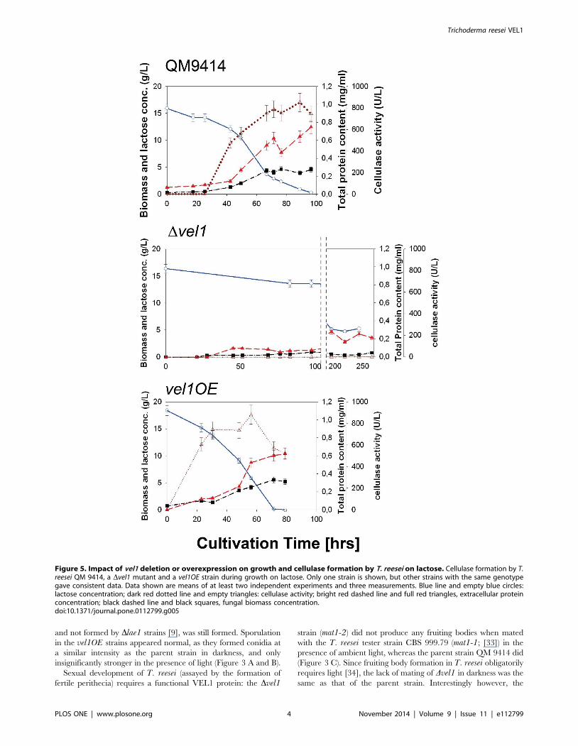
Figure 5. Impact of vel1 deletion or overexpression on growth and cellulase formation by T. reesei on lactose. Cellulase formation by T.reesei QM 9414, a Dvel1 mutant and a vel1OE strain during growth on lactose. Only one strain is shown, but other strains with the same genotypegave consistent data. Data shown are means of at least two independent experiments and three measurements. Blue line and empty blue circles:lactose concentration; dark red dotted line and empty triangles: cellulase activity; bright red dashed line and full red triangles, extracellular proteinconcentration; black dashed line and black squares, fungal biomass concentration.doi:10.1371/journal.pone.0112799.g005
Trichoderma reesei VEL1
PLOS ONE | www.plosone.org 4 November 2014 | Volume 9 | Issue 11 | e112799

vel1OE strain was able to form primordia also in darkness,
although with less frequency than in light (Figure 3 C). Yet these
primordia lacked asci and were thus not fertile (data not shown).
VEL1 is necessary for growth on cellulose and lactoseThe two Dvel1 strains exhibited a slightly reduced growth on
glucose as a carbon source on plates. To see whether this is a
consequence of the alterations in hyphal morphology, or a
different effect, we studied plate growth of T. reesei on several
carbon sources including such related to cellulase formation. As
can be seen (Figure 4), the slight reduction and altered hyphal
phenotype was indeed observed on all carbon sources, and
independent of growth in either light or in darkness. However, in
addition to that, growth was significantly reduced on lactose and
practically absent on cellulose. On the other hand, growth on
lactose, cellobiose and cellulose was phenotypically normal in the
vel1OE strain, and occurred at a faster rate. This suggests that, in
addition to the effect on hyphal morphology, VEL1 obviously also
impacts utilization of lactose and cellulose.
VEL1 is essential for cellulase and hemicellulase transcriptaccumulation
One hypothesis of this paper was that VEL1 would be necessary
for the formation of cellulases in T. reesei. The above reported
results with growth on cellulose were in accordance with this
hypothesis. To test this directly, we first cultivated the parent and
the two Dvel1 strains on lactose. The rationale for this was that
lactose induces cellulase expression [6], but its utilization - in
contrast to cellulose - is independent of the secreted cellulases [35].
Figure 6. Impact of vel1 deletion or overexpression on transcript levels of two cellulase, one hemicellulase and a cellulase regulatorgene in T. reesei during growth on lactose. mRNA levels of two cellulase genes (cel7a, cel6a), one xylanase gene (xyn2) and the cellulaseregulator xyr1 in T. reesei QM 9414 (full circles and solid line), a Dvel1 strain (empty circles and dotted line) and a vel1OE strain (full traingles anddashed line) under the fermentation conditions on lactose as shown in Figure 5. mRNA levels are given in arbitrary units, which were calculated bynormalizing the ratio of the respective gene transcript to that of the housekeeping gene tef1, and relating all ratios to that obtained for the givengene at 17.5 hrs in T. reesei QM9414. Only one mutant strain is shown, but other strains with the same genotype gave consistent data. Data shownare means of at least two independent experiments and three measurements.doi:10.1371/journal.pone.0112799.g006
Figure 7. Impact of vel1 deletion or overexpression ontranscript levels of two cellulase, one hemicellulase and acellulase regulator gene in T. reesei upon induction bysophorose. Transcript levels of two cellulase genes (cel7a, cel6a),one xylanase gene (xyn2) and the cellulase regulator xyr1 in T. reesei QM9414 (full symbols), a vel1 strain (empty symbols) and a vel1OE strain(grey symbols) upon induction of mycelia, pregrown on glycerol for20 hrs, by sophorose for 4 hrs. Transcript levels are given in arbitraryunits, as specified in legend to figure 6. The asterisks indicate caseswere the relative mRNA level was below the limit of detection. Only onemutant strain is shown, but other strains with the same genotype gaveconsistent data. Data shown are means of at least two independentexperiments and three measurements.doi:10.1371/journal.pone.0112799.g007
Trichoderma reesei VEL1
PLOS ONE | www.plosone.org 5 November 2014 | Volume 9 | Issue 11 | e112799
As shown in Figure 5, submerged growth of the Dvel1 mutants on
lactose occurred - as on plates - at a much lower rate, and while
the parent strain reached a final biomass concentration after 90–
100 hrs of growth comparable mycelial dry weights of the two
Dvel1 were only reached after 250 hrs of cultivation. Consistent
with the lack of growth on cellulose of these mutants, no cellulase
activity could be detected throughout the whole 250 hrs cultiva-
tion period (Figure 5 B). Also consistent with the faster growth of
the vel1OE strain on lactose on plates, this strain also grew faster
in submerged cultures on lactose, and started to produce cellulases
much earlier (Figure 5 C). However, the final values of cellulase
activity were only insignificantly higher in the vel1OE strain.
Transcript data for the two major cellulase genes cel7a and cel6a,
and the xylanase II-encoding gene xyn2 perfectly supported the
absence of cellulase activity in the two Dvel1 mutants, indicating
that the effect is on the level of gene transcription (Figure 6 A–C).
Also the abundance of the transcript of xyr1, which encodes the
major key cellulase and hemicellulase regulator XYR1, was
strongly reduced in the two Dvel1 mutants (Figure 6 D). This may
also explain the impaired growth on lactose, because loss of
function of xyr1 also strongly impairs growth on lactose [35,36]. In
the case of the vel1OE strain, and also consistent with the above
shown cellulose activities, the transcripts were not significantly
higher than in strain QM 9414 but did not decrease that rapidly as
in strain QM 9414 (Fig. 6 A–D).
In order to test the effect of vel1 on cellulase formation under
conditions where growth or inducer uptake are not affected, we
used the ß-linked disaccharide sophorose and resting mycelia
pregrown under non-inducing conditions (glycerol). Sophorose is a
very powerful inducer of cellulases and xylanases in T. reesei under
these conditions [3,37] and the ctr1 gene that encodes the sensor
mediating sophorose induction is still expressed at a high level in a
T. reesei mutant with impaired Dxyr1 function [37]. This
experiment should thus enable to detect effects of vel1 without
disturbance by effects of vel1 on growth and nutrient uptake.
As can be seen in Figure 7, addition of sophorose led to a strong
increase in the relative abundance of the cel7a, cel6a, xyn2 and
xyr1 transcripts with 4 hrs in the parent strain, and – with the
exception of xyn2 - roughly doubled in the vel1OE mutant.
whereas they remained barely detectable in the Dvel1 mutant.
Also the accumulation of the xyr1 transcript was strongly reduced,
yet clearly detectable and notably present in higher amounts than
in the parent strain at t = 0. This correlates with its partially
constitutive expression. We conclude therefore that a loss of vel1function impairs cellulase gene expression, and VEL1 is thus
essential for this process. It also shows that upregulation of vel1transcript levels increases cellulose gene transcription.
Discussion
In this paper, we have explored the function of the velvet gene
vel1 of T. reesei. While this global regulator is conserved in
Pezizomycota, studies of veA orthologs across several fungal
genera have now established a significant diversity in its impact on
fungal development [14,16,17]. The most striking example is
asexual sporulation, a trait influenced by VeA/VelA/VEL1 in all
fungi: whereas a veA/velA knock out in A. nidulans, P.chrysogenum and N. crassa increases conidiation [29,38,39], it
results in decreased conidiation in the corresponding knock-out
mutants of A. fumigatus, A. parasiticus, A. flavus, Fusariumfujikuroi, F. graminearum, Dothistroma septosporum and T. virens[19,22,27,39–44]. Our study shows that T. reesei belongs to the
second group as the vel1 knock-out strains almost completely
lacked conidiation. It is further interesting that this impairment
was independent on the presence or absence of light, a finding so
far only recently found in A. fumigatus [41]. Yet this coincides
with the findings that the T. reesei parent strain forms the same
number of conidia upon illumination as in the dark, and suggests
that, differently from A. nidulans [38], light is not a relevant factor
in the veA/vel1-mediated regulation of conidition in A. fumigatusand T. reesei.
Another example where the function of VEL1 differs from that
of its ortholog in other fungi, is the complete loss of sexual
development of T. reesei Dvel1 mutants. Opposite findings have
been reported for N. crassa [30], and while a similar elimination of
sexual reproduction has been reported for A. nidulans [31], the
effect of light on the action of veA/vel1 is reversed: fruiting body
formation in T. reesei only occurs in light [34] but in A. nidulansonly in the darkness [37]. Interestingly, overexpression of vel1under a constitutive, light-independent promoter (tef1) enabled T.reesei to form some sterile primordia in darkness, an ability not
shown by the wild type strains [34]. Thus an increased expression
of vel1 in the dark cannot overcome the inability of T. reesei to
initiate full fruiting body formation, but can only initiate an early
stage, suggesting that vel1 controls several steps in this process in
different ways. Thus, while vel1 is essential for sexual development
in T. reesei, it is only partially - if at all - responsible for its
dependence on light. Consequently, while we have shown that
VEL1 also regulates developmental processes in T. reesei, its mode
of action and its interaction with environmental triggers remains
unclear and cannot be deduced from analogy with other fungal
systems.
On the other hand, there are also effects that appear to be
conserved in all VeA/VelA/VEL1 orthologs, such as the
consequences of their knock out on hyphal morphology, which
are reflected in higher branching and shorter filaments caused by
changes in cell wall metabolism [45]. Our findings of crippled,
highly branched and thickened hyphae correlate well with
respective findings in P. chrysogenum, A. chrysogenum and
Fusarium spp. [40,41,44,45].
While the impact of the velvet complex on the regulation of
secondary metabolite production has been well documented in
numerous fungi (for review see [14,16]), only two papers have so
far reported the participation of veA/velA in the regulation of
extracellular enzyme synthesis: Kamerewerd et al. [46] reported
that the class V chitinase PcchiB1 of P. chrysogenum, which is
involved in cell wall turn-over, is strongly downregulated in a
delta-velA mutant. And while this paper was in preparation,
Duran et al. [47] reported that the expression of amylase and
protease activity in A. flavus is impaired in a veA mutant, while an
alpha-amylase was produced in greater quantities.
We have recently shown that LAE1 strongly impacts cellulase
gene transcription in T. reesei and cellulase expression is
completely abolished in lae1 loss-of-function mutants [9]. Since
LAE1 and VEL1, as in other fungi, can physically interact in T.reesei [12], we assumed that cellulase expression would also be
affected by vel1. In this paper, we now provide evidence that the
expression of cellulases in T. reesei is also completely dependent on
a functional vel1 gene, which agrees well with the stringent
requirement for a functional lae1 gene [9], and suggests that
cellulase expression is indeed regulated by the velvet complex. The
extension of our findings to all cellulases and hemicellulases
appears justified because they are corregulated by sophorose and
lactose [4–8], and thus - although we quantified only the
transcripts of two cellulases and one xylanase - it can be safely
assumed that the others are affected in the same manner too. In
addition, we have shown that also the transcript levels of the
central regulator of cellulase and hemicellulase biosynthesis,
Trichoderma reesei VEL1
PLOS ONE | www.plosone.org 6 November 2014 | Volume 9 | Issue 11 | e112799

XYR1, is strongly reduced albeit not eliminated. This fits perfectly
to the results with the T. reesei Dlae1 mutant, in which also a low
level of xyr1 transcript levels was still observed, and consequently
the expression of xyr1 under a constitutive promoter did not rescue
the Dlae1 phenotype [9].
Taken together, the above and our study show that the regulatory
targets of the velvet complex reach beyond mere secondary
metabolism and development. The extension of the function of
the velvet complex towards formation of extracellular hydrolytic
enzymes is intriguing, because a regulatory interaction of secondary
metabolism and fungal development is already well established [48],
whereas such a link with hydrolases is so far unknown. However, in
T. reesei a link between asexual sporulation and cellulase formation
has recently been demonstrated [49]. This presence of cellulases and
other plant cell wall hydrolases on the spores of the fungus was
interpreted in terms of an advantage because the disperged spores
could instantly initiate growth when arriving at a new substrate. In
this regards it is also interesting that many of the cellulase genes of T.reesei are clustered in the genome with genes encoding secondary
metabolite synthases [1], and indeed several of them were
demonstrated to be expressed under conditions inducing cellulase
biosynthesis [6,7,50]. However, the regulator coordinating sporu-
lation, secondary metabolite formation and cellulase gene expres-
sion in T. reesei has not yet been identified. It is not XYR1, because
Dxyr1 mutants do not form cellulases but are still able to sporulate
[49]. Likewise aconidial mutants of T. reesei still form cellulases (R.
Linke and C.P. Kubicek, unpublished data), and thus the two
processes are linked but not dependent on each other. This also
agrees with the findings that the strongly reduced conidiation in the
Dlae1 strain of T. reesei is xyr1 independent [9].
Based on the data of this paper, we therefore propose that the
velvet complex is a superimposed regulatory level that coordinates
the expression of cellulases, and secondary metabolites with asexual
development in T. reesei. We also conclude from the above data that
this coordination is not influenced by light, and this claim is further
supported by the findings that cellulase and secondary metabolite
gene expression is not significantly different (,1.5-fold at p,0.05;
[51]) in T. reesei wild-type strains growing on cellulose under either
illumination or in darkness. The identification of the signal
triggering this coordinated expression and how it interacts with
the velvet complex will be a challenge for further work, and may
also identify new regulatory levels for potential further improvement
of cellulase producing strains of T. reesei.
Materials and Methods
Strains and cultivation conditionsT. reesei strains used throughout this work are listed in Table 1.
They were maintained on potato dextrose agar (PDA). Escherichia
coli JM109 (Promega, Madison, Wisconsin) was used for plasmid
construction and amplification. Cultures were grown at 28uC in a
Sanyo incubator containing a Philips-master light source (TLD-
15 W/840), either with continuous illumination (light conditions)
or double wrapped in foil (dark conditions).
Cultivations on lactose were carried out in 20 L stainless steel
bioreactors (Zolend Ltd., Debrecen, Hungary) as described
previously [52], using Mandels-Andreotti medium [53], except
that agitation was 400 rpm. Induction experiments with sophorose
were performed as described [54], using 20 h old mycelia
pregrown on Mandels-Andreotti medium with glycerol (1%, w/
v) as a carbon source.
Growth tests on plates were performed on Mandels-Andreotti
medium, solidified with 2% (w/v) agar, but without peptone, and
the carbon source indicated (1%, w/v).
Nucleic acid isolation and hybridizationFungal mycelia were harvested by filtration, washed with
distilled cold water, frozen and ground under liquid nitrogen. For
extraction of genomic DNA, plasmid DNA and RNA, purification
kits (Wizard Genomic DNA Purification Kit, PureYield Plasmid
Midiprep System and RNeasy plant kit, respectively, all from
Promega) were used according to the manufacturer’s protocol.
Standard methods were used for electrophoresis, blotting and
hybridization of nucleic acids.
Construction of T. reesei recombinant strainsTo study the function of VEL1, we constructed T. reesei strains
in which vel1 was deleted and strains, which vel1 was expressed
under the strong constitutive expression signals of the tef1(translation elongation factor 1-alpha encoding) promoter region
[55].
To delete the vel1 gene of T. reesei, the 1.8-kb vel1 coding
region was replaced by the E. coli hygromycin B phosphotrans-
ferase (hph) gene. This was performed by amplifying around 1.2-
kb of the up- and downstream non-coding region of vel1 from
genomic DNA of T. reesei QM 9414 using the primer pairs given
in Table S1. The two resulting PCR fragments were digested with
ApaI/XhoI (upstream region) and XhoI/ClaI (downstream region)
and ligated into a ApaI/ClaI restricted vector pBluescript SK(+)
(Stratagene, La Jolla, California), followed by the insertion of the
2.4-kb SalI/XhoI fragment of the hph gene into the XhoI site
resulting in pRKA_D122284hph.
For expression of vel1 under a strong constitutive promoter, a
2,275-bp vel1 PCR fragment including the coding and terminator
region was amplified with the oligonucleotides Fw_ Ptef1:vel1_-
Cla1 and tef1:vel1-HindIII (Table S1) and then inserted down-
stream of the tef1 promoter region [56] into the ClaI/HindIII sites
of pLH1hphtef1 resulting in vector pRKA-OE122284hph, which
Table 1. Strains used in the present work.
Strain name Genotype Reference
QM 9414 mat1-2 [33]
CBS 999.79 mat1-1 [33]
RKA14 Dvel1/mat1-2 this work
RKA12 vel1OE, mat1-2 this work
RKA13 vel1OE, mat1-2 this work
RKA17 Dvel1/mat1-2 this work
RKA18 Dvel1/mat1-2 this work
doi:10.1371/journal.pone.0112799.t001
Trichoderma reesei VEL1
PLOS ONE | www.plosone.org 7 November 2014 | Volume 9 | Issue 11 | e112799
contains the E. coli hygromycin B phosphotransferase (hph) under
T. reesei expression signals as selection marker [57].
All vectors constructed were verified by nucleotide sequencing.
Fungal transformationProtoplast preparation and DNA mediated transformation was
performed as described [58]. The strains were purified twice for
mitotic stability, and integration of the expression cassettes was
verified by PCR analysis. Gene copy numbers of the integrated
constructs were determined by Southern analysis [59], using
chromosomal DNA cleaved with BamHI/HindIII.
Analysis of sexual and asexual developmentFor sexual reproduction, T. reesei parent and mutant strains
and the compatible mating partner strain CBS 999.79 [34] were
pre-grown on PDA for 4 days, and agar culture plugs then
transferred on fresh PDA (Difco, Lawrence, KS, USA) on opposite
sides of the plate at a 1 cm distance from the edge. The plates were
kept at room-temperature and exposed to day light or kept in
complete darkness for 4–7 days (see above). All pairs of strains
which formed fruiting bodies were visually inspected until the
maturation stage was achieved and ascospores were dispersed.
Monoascospore cultures were isolated by dispersing the solution
with a cotton swab on multiple PDA plates. After overnight
incubation several single germinated spores were selected with an
aid of a stereomicroscope, transferred to a new PDA plate and
cultivated at 28uC.
To test for photodependent conidiation, each PDA plate was
inoculated with a 5-mm diameter mycelial plug taken from the
edge of a 3-day-old colony. Three replications were done for each
treatment. Plates were incubated at 28uC for 8 days in either
complete darkness and cycles of 12 h illumination/12 h darkness
(see above), and conidia then harvested by gently rubbing them off
in an equal volume of physiologically salt (1%, w/v, Tween and
0.8% w/v NaCl), filtering through glass wool, and centrifugation
(50006 g, 10 min). The conidia were then suspended in 2.5 g/l
phytagel (Phytagel, SIGMA, Steinheim, Germany), mixed and
their transmission measured at 590 nm in a Biolog standard
turbidimeter. The number of conidia was calculated using a
calibration curve with T. reesei conidia.
Enzymatic assays and determination of fungal dry weightCellulase enzyme activities were determined using carboxy-
methylcellulose (1%, w/v) [56]. Protein in the culture supernatant
was determined by the method of Bradford [60]. Fungal dry
weight was determined by filtering an aliquot of the culture
through glass sinter funnels (porosity G1), washing with tap water
and drying at 80uC to constant weight. The lactose concentration
in the fermentor was determined by HPLC as described earlier
[52].
Gene expression by quantitative PCRDNase treated (DNase I, RNase free; Fermentas) RNA (5 mg)
was reverse transcribed with the RevertAid First Strand cDNA Kit
(Fermentas) according to the manufacturer’s protocol with a
combination of oligo-dT and random hexamer primers. All qPCR
assays were performed on a Bio-Rad (Hercules, CA) iCycler IQ.
For the reaction the IQ SYBR Green Supermix (Bio-Rad,
Hercules, CA) was prepared for 25 ml assays with standard MgCl2concentration (3 mM) and a final primer concentration of 100 nM
each. All assays were carried out in 96-well plates. The
amplification protocol consisted of an initial denaturation step
(3 min at 95uC) followed by 40 cycles of denaturation (15 sec at
95uC), annealing (20 sec; for primers and the respective temper-
ature see Table S2) and elongation (10 sec at 72uC). Determina-
tion of the PCR efficiency was performed using triplicate reactions
from a dilution series of cDNA (1; 0.1; 0.01; 0.001). Amplification
efficiency was then calculated from the given slopes in the IQ5
Optical system Software v2.0. Expression ratios were calculated
using REST Software [61]. All samples were analyzed in at least
two independent experiments with three replicates in each run.
Bioinformatic analysisIdentification of PEST regions (protein domains that are
enriched in proline, glutamic acid, serine, and threonine residues)
that may lead to rapid protein degradation, typical for unstable
proteins, was performed with epestfind ([62]; http://emboss.
bioinforatics.nl/cgi-bin/emboss/epestfind). The cellular localiza-
tion of proteins was analyzed by WoLF PSORT (Protein
Subcellular Localization Prediction tool; [63], http://wolfpsort.
org/), and leucine-rich nuclear export signals (NES) identified by
NetNES 1.1 Server ([64]; http://www.cbs.dtu.dk/services/
NetNES/).
Supporting Information
Figure S1 Verification of the recombinant T. reeseistrains. PCR verification of vel1 knock out in T. reesei: (A)
structure of the disrupted (top) and native vel1 locus (below).
Numbers indicate the size (in kb) of the respective areas. The
dotted line defines the gene construct present in the deletion
cassette. The arrows a–d specify the primers used for amplification
the homologous integrated knock-out construct (a and b; result
shown in B), and of the native vel1 gene (c and d; result shown in
C), respectively. a, pVel1; b, hph_int; c, Vel_int1; d, Vel_int2 (for
sequences see Table S2). Tracks: 1, parent strain QM9414; 2,
Dvel1 strain RKA14, 3, Dvel1 strain RKA17, Dvel1 strain
RKA18. Southern analysis: D, scheme of the wild-type vel1 locus.
DNA was cleaved by HindIII and BamHI, and hybridization was
done by a full-length 1.8 probe of vel1. E, resulting autoradio-
graph: Tracks: 1, RKA12; 2, RKA13; 3, parent strain QM9414; 4,
size marker ladder.
(DOCX)
Table S1 Oligonucleotide primers used in this work.
(XLSX)
Table S2 qPCR primers used in this work.
(XLSX)
Acknowledgments
Our special thanks are due to Michael Freitag, Oregon State University,
for making laboratory facilities available to RKA for performing some of
the described experiments during her stay in his institute on behalf of a
Marshal Stipendium granted to RKA. The expert help of Sara Ghassemi is
gratefully acknowledged.
Author Contributions
Conceived and designed the experiments: CPK ISD LK. Performed the
experiments: RKA ZN LA MP ES BA. Analyzed the data: RKA EF ISD
CPK. Contributed reagents/materials/analysis tools: LK ISD CPK. Wrote
the paper: CPK.
Trichoderma reesei VEL1
PLOS ONE | www.plosone.org 8 November 2014 | Volume 9 | Issue 11 | e112799

References
1. Kubicek CP (2012a) Systems biological approaches towards understandingcellulase production by Trichoderma reesei. J Biotechnol 163: 133–142.
2. Kubicek CP (2012b) Fungi and Lignocellulose Biomass. Wiley and Sons, NY,
USA.
3. Hakkinen M, Arvas M, Oja M, Aro N, Penttila M, et al. (2012) Re-annotation of
the CAZy genes of Trichoderma reesei and transcription in the presence oflignocellulosic substrates. Microb Cell Fact 11:134.
4. Foreman PK, Brown D, Dankmeyer L, Dean R, Diener S, et al. (2003)
Transcriptional regulation of biomass-degrading enzymes in the filamentousfungus Trichoderma reesei. J Biol Chem 278:31988–31997.
5. Ries L, Pullan ST, Delmas S, Malla S, Blythe MJ, et al. (2013) Genome-wide
transcriptional response of Trichoderma reesei to lignocellulose using RNAsequencing and comparison with Aspergillus niger. BMC Genomics 14:541.
6. Ivanova C, Baath JA, Seiboth B, Kubicek CP (2013) Systems analysis of lactose
metabolism in Trichoderma reesei identifies a lactose permease that is essential
for cellulase induction. PLoS One 8(5):e62631.
7. Bischof R, Fourtis L, Limbeck A, Gamauf C, Seiboth B, et al. (2013)Comparative analysis of the Trichoderma reesei transcriptome during growth on
the cellulase inducing substrates wheat straw and lactose. Biotechnol Biofuels6(1):127.
8. Seiboth B, Herold S, Kubicek CP (2012) Metabolic engineering of inducer
formation for cellulase and hemicellulase gene expression in Trichoderma reesei.Subcell Biochem 64:367–390.
9. Seiboth B, Karimi RA, Phatale PA, Linke R, Hartl L, et al. (2012) The putativeprotein methyltransferase LAE1 controls cellulase gene expression in Tricho-derma reesei. Mol Microbiol 84:1150–1164.
10. Bok JW, Noordermeer D, Kale SP, Keller NP (2006) Secondary metabolic genecluster silencing in Aspergillus nidulans. Mol Microbiol 61:1636–1645.
11. Reyes-Dominguez Y, Bok YW, Berger H, Shwab E, et al. (2010) Heterochro-
matic marks are associated with the repression of secondary metabolism clusters
in Aspergillus nidulans. Mol Microbiol 76:1376–1386.
12. Karimi-Aghcheh R, Bok JW Phatale PA, Smith KM, Baker SE, et al. (2013)Functional analyses of Trichoderma reesei LAE1 reveal conserved and
contrasting roles of this regulator. G3 3:369–378.
13. Patananan AN, Palmer JM, Garvey GS, Keller NP, Clarke SG (2013) A novelautomethylation reaction in the Aspergillus nidulans LaeA protein generates S-
methylmethionine. J Biol Chem 288:14032–14045.
14. Bayram O, Braus GH (2011) Coordination of secondary metabolism anddevelopment in fungi: the velvet family of regulatory proteins. FEMS Microbiol
Rev 36:1–24.
15. Bayram O, Krappmann S, Ni M, Bok JW, Helmstaedt K, et al. (2008) VelB/
VeA/LaeA complex coordinates light signal with fungal development andsecondary metabolism. Science 320:1504–1506.
16. Calvo AM (2008) The VeA regulatory system and its role in morphological and
chemical development in fungi. Fungal Genet Biol 45:1053–1061.
17. Kim HJ, Han JH, Kim KS, Lee YH (2014) Comparative functional analysis ofthe velvet gene family reveals unique roles in fungal development and
pathogenicity in Magnaporthe oryzae. Fungal Genet Biol 66:33–43.
18. Yang Q, Chen Y, Ma Z (2013) Involvement of BcVeA and BcVelB in regulating
conidiation, pigmentation and virulence in Botrytis cinerea. Fungal Genet Biol50:63–71.
19. Park HS, Bayram O, Braus GH, Kim SC, Yu JH (2012) Characterization of the
velvet regulators in Aspergillus fumigatus. Mol Microbiol 86:937–953.
20. Laskowski-Peak MC, Calvo AM, Rohrssen J, Smulian AG (2012) VEA1 isrequired for cleistothecial formation and virulence in Histoplasma capsulatum.Fungal Genet Biol 49:838–846.
21. Myung K, Zitomer NC, Duvall M, Glenn AE, Riley RT, et al. (2012) The
conserved global regulator VeA is necessary for symptom production andmycotoxin synthesis in maize seedlings by Fusarium verticillioides. Plant Pathol
61:152–160.
22. Chettri P, Calvo AM, Cary JW, Dhingra S, Guo Y, et al. (2012) The veA gene ofthe pine needle pathogen Dothistroma septosporum regulates sporulation and
secondary metabolism. Fungal Genet Biol 49:141–151.
23. Busch S, Schwier EU, Nahlik K, Bayram O, Helmstaedt K, et al. (2007) Aneight-subunit COP9 signalosome with an intact JAMM motif is required for
fungal fruit body formation. Proc Natl Acad Sci USA 104:8089–8094.
24. Fischer R (2008) Developmental biology. Sex and poison in the dark. Science
320:1430–1431.
25. Purschwitz J, Muller S, Fischer R (2009) Mapping the interaction sites ofAspergillus nidulans phytochrome FphA with the global regulator VeA and the
White Collar protein LreB. Mol Genet Genomics 281:35–42.
26. Sarikaya-Bayram O, Bayram O, Feussner M, Kim JH, Kim HS et al. (2014) Themembrane-bound VapA-VipC-VapB methyltransferase complex guides signal
transduction for epigenetic and transcriptional control of fungal development.
Dev Cell 29:406–420.
27. Mukherjee PK, Kenerley CM (2010) Regulation of morphogenesis andbiocontrol properties in Trichoderma virens by a VELVET protein, Vel1. Appl
Environ Microbiol 76:2345–2352.
28. Le Crom S, Schackwitz W, Pennacchio L, Magnuson JK, Culley ED et al.(2009) Tracking the roots of cellulase hyperproduction by the fungus
Trichoderma reesei using massively parallel DNA sequencing. Proc Natl Acad
Sci USA 106:16151–16156.
29. Vitikainen M, Arvas M, Pakula T, Oja M, Penttila ME et al. (2010) Array
comparative genomic hybridization analysis of Trichoderma reesei strains with
enhanced cellulase production properties. BMC Genomics 11:441.
30. Bayram OS, Krappmann S, Seiler S, Vogt N, Braus GH (2008) Neurosporacrassa ve-1 affects asexual conidiation. Fungal Genet Biol 45:127–138.
31. Stinnett SM, Espeso EA, Cobeno L, Araujo-Bazan L, Calvo AM (2007)
Aspergillus nidulans VeA subcellular localization is dependent on the importin
alpha carrier and on light. Mol Microbiol 63: 242–255.
32. Kim H, Han K, Kim K, Han D, Jahng K, et al. (2002) The veA gene activates
sexual development in Aspergillus nidulans. Fungal Genet Biol 37: 72–80.
33. Seidl V, Seibel C, Kubicek CP, Schmoll M (2009) Sexual development in the
industrial workhorse Trichoderma reesei. Proc Natl Acad Sci USA 106:13909–
13914.
34. Chen CL, Kuo HC, Tung SY, Hsu PW, Wang CL, et al. (2012) Blue Light Acts
as a Double-Edged Sword in Regulating Sexual Development of Hypocreajecorina (Trichoderma reesei). PLoS One 7:e44969.
35. Seiboth B, Pakdaman BS, Hartl L, Kubicek CP (2007) Lactose metabolism in
filamentous fungi: how to deal with an unknown substrate. Fungal Biol Reviews
21: 42–48.
36. Seiboth B, Gamauf C, Pail M, Hartl L, Kubicek CP (2007) The D-xylose
reductase of Hypocrea jecorina is the major aldose reductase in pentose and D-
galactose catabolism and necessary for beta-galactosidase and cellulase induction
by lactose. Mol Microbiol 66:890–900.
37. Ghassemi S, Lichius A, Bidard F, Herold S, Seidl-Seiboth V, et al. (2014) The
karyopherin KAP8 (Pse1/Kap121) mediates nuclear import of the cellulase
transcriptional regulator XYR1 of Trichoderma reesei and is essential for asexual
sporulation and stress resistance. Mol Microbiol., ms in revision.
38. Yager LN (1992) Early developmental events during asexual and sexual
sporulation in Aspergillus nidulans. Bio/Technology 23: 19–41.
39. Hoff B, Kamerewerd J, Sigl C, Mitterbauer R, Zadra I, et al. (2010) Two
components of a velvet-like complex control hyphal morphogenesis, conidio-
phore development, and penicillin biosynthesis in Penicillium chrysogenum.
Eukaryot Cell 9:1236–1250.
40. Wiemann P, Brown DW, Kleigrewe K, Bok JW, Keller NP, et al. (2010) FfVel1
and FfLae1, components of a velvet-like complex in Fusarium fujikuroi, affect
differentiation, secondary metabolism and virulence. Mol Microbiol 77: 972–
994.
41. Dhingra S, Andes D, Calvo AM (2012) VeA Regulates Conidiation, Gliotoxin
Production,and Protease Activity in the Opportunistic Human Pathogen
Aspergillus fumigatus. Eukaryot Cell 11:1531–1543.
42. Amaike S, Keller NP (2009) Distinct roles for VeA and LaeA in development
and pathogenesis of Aspergillus flavus. Eukaryot Cell 8:1051–1060.
43. Jiang J, Liu X, Yin Y, Ma Z (2011) Involvement of a velvet protein FgVeA in the
regulation of asexual development, lipid and secondary metabolisms and
virulence in Fusarium graminearum. PLoS One 6:e28291.
44. Merhej J, Urban JM, Dufresne M, Hammond-Kosack KE, Richard-Forget F,
et al. (2012) The velvet gene, FgVe1, affects fungal development and positively
regulates trichothecene biosynthesis and pathogenicity in Fusarium grami-nearum. Mol Plant Pathol 13:363–374.
45. Dreyer J, Eichhorn H, Friedlin E, Kurnsteiner H, Kuck U (2007) A homologue
of the Aspergillus velvet gene regulates both cephalosporin C biosynthesis and
hyphal fragmentation in Acremonium chrysogenum. Appl Environ Microbiol
73:3412–3422.
46. Kamerewerd J, Zadra I, Kurnsteiner H, Kuck U (2011) PcchiB1, encoding a
class V chitinase, is affected by PcVelA and PcLaeA, and is responsible for cell
wall integrity in Penicillium chrysogenum. Microbiology 157:3036–48.
47. Duran RM, Gregersen S, Smith TD, Bhetariya PJ, Cary JW, et al. (2014)The
role of Aspergillus flavus veA in the production of extracellular proteins during
growth on starch substrates. Appl Microbiol Biotechnol 98:5081–5094.
48. Yu JH, Keller N (2005) Regulation of secondary metabolism in filamentous
fungi. Annu Rev Phytopathol 43:437–458.
49. Metz B, Seidl-Seiboth V, Haarmann T, Kopchinskiy A, Lorenz P, et al.
(2011)Expression of biomass-degrading enzymes is a major event during
conidium development in Trichoderma reesei. Eukaryot Cell 10:1527–1535.
50. Arvas M, Pakula T, Smit B, Rautio J, Koivistoinen H, et al. (2011) Correlation
of gene expression and protein production rate - a system wide study. BMC
Genomics 12:616.
51. Tisch D, Kubicek CP, Schmoll M (2011) The phosducin-like protein PhLP1
impacts regulation of glycoside hydrolases and light response in Trichodermareesei. BMC Genomics 12:613.
52. Karaffa L, Coulier L, Fekete E, Overkamp KM, Druzhinina IS, et al.. (2013)
The intracellular galactoglycome in Trichoderma reesei during growth on
lactose. Appl Microbiol Biotechnol 97:5447–5456.
53. Mandels M, Andreotti RE (1978) Problems and challenges in the cellulose to
cellulase fermentation. Process Biochem 13: 6–13.
54. Sternberg D, Mandels GR (1979) Induction of cellulolytic enzymes in
Trichoderma reesei by sophorose. J Bacteriol 139:761–769.
Trichoderma reesei VEL1
PLOS ONE | www.plosone.org 9 November 2014 | Volume 9 | Issue 11 | e112799
55. Akel E, Metz B, Seiboth B, Kubicek CP (2009) Molecular regulation of arabinan
and L-arabinose metabolism in Hypocrea jecorina (Trichoderma reesei). EukaryotCell 8:1837–1844.
56. Uzbas F, Sezerman U, Hartl L, Kubicek CP, Seiboth B (2012) A homologous
production system for Trichoderma reesei secreted proteins in a cellulase-freebackground. Appl Microbiol Biotechnol 93:1601–1608.
57. Mach RL, Schindler M, Kubicek CP (1994) Transformation of Trichodermareesei based on hygromycin resistance using homologous expression signals. Curr
Genet 25: 567–570.
58. Guangtao Z, Hartl L, Schuster A, Polak S, Schmoll M, et al. (2009) Genetargeting in a nonhomologous end joining deficient Hypocrea jecorina 139:146–
151.59. Ausubel FM, Brent R, Kingston RE, Moore DD (1999) Short protocols in
molecular biology: a compendium of methods for current protocols in molecularbiology. John Wiley and Sons, 4th ed.
60. Bradford MM (1976) A rapid and sensitive method for the quantitation of
microgram quantities of protein utilizing the principle of protein-dye binding.
Anal Biochem 72:248–254.
61. Pfaffl MW, Horgan GW, Dempfle L (2002) Relative expression software tool
(REST) for group-wise comparison and statistical analysis of relative expression
results in real-time PCR. Nucleic Acids Res 30:e36.
62. Rechsteiner M, Rogers S, Rote K (1987) Protein structure and intracellular
stability. Trends Biochem Sci 12: 390–394.
63. Horton KJ, Park P, Obayashi T, Fujita N, Harada H, et al. (2007) WoLF
PSORT: protein localization predictor. Nucleic Acids Res 35:W585–W587.
64. la Cour T, Kiemer L, Mølgaard A, Gupta R, Skriver K, et al. (2004) Analysis
and prediction of leucine-rich nuclear export signals. Protein Eng Des Sel 17:
527–536.
Trichoderma reesei VEL1
PLOS ONE | www.plosone.org 10 November 2014 | Volume 9 | Issue 11 | e112799
Copyright © 2022 FDOKUMEN