TP0326, a Treponema pallidum β-barrel assembly machinery A (BamA) orthologue and rare outer...
32
TP0326, a Treponema pallidum β- Barrel Assembly Machinery A (BamA) Ortholog and Rare Outer Membrane Protein Daniel C. Desrosiers 1,# , Arvind Anand 1,ψ , Amit Luthra 1,ψ , Star M Dunham-Ems 1 , Morgan LeDoyt 1 , Michael A. D. Cummings 2 , Azad Eshghi 2 , Caroline E. Cameron 2 , Adriana R. Cruz 3 , Juan C. Salazar 3,4 , Melissa J. Caimano 1 , and Justin D. Radolf 1,4,5 1 Department of Medicine, University of Connecticut Health Center, Farmington, CT 06030 5 Department of Genetics and Developmental Biology, University of Connecticut Health Center, Farmington, CT 06030 2 Department of Biochemistry and Microbiology, University of Victoria, Victoria, British Columbia, Canada 3 Centro Internacional de Entrenamiento e Investigaciones Médicas (CIDEIM), Cali, Colombia 4 Department of Pediatrics, Connecticut Children's Medical Center, Division of Pediatric Infectious Diseases, Hartford, CT 06106 SUMMARY Definitive identification of Treponema pallidum (Tp) rare outer membrane proteins (OMPs) has long eluded researchers. TP0326, the sole protein in Tp with sequence homology to a Gram- negative OMP, belongs to the BamA family of proteins essential for OM biogenesis. Structural modeling predicted that five polypeptide transport-associated (POTRA) domains comprise the N- terminus of TP0326, while the C-terminus forms an 18-stranded amphipathic β-barrel. Circular dichroism, heat-modifiability by SDS-PAGE, Triton X-114 phase partitioning and liposome incorporation supported these topological predictions and confirmed that the β-barrel is responsible for the native protein's amphiphilicity. Expression analyses revealed that native TP0326 is expressed at low abundance, while a protease-surface accessibility assay confirmed surface exposure. Size-exclusion chromatography and blue native polyacrylamide gel electrophoresis revealed a modular Bam complex in Tp considerably larger than that of E. coli. Non-orthologous ancillary factors and self-association of TP0326 via its β-barrel may both contribute to the Bam complex. Tp-infected rabbits mount a vigorous antibody response to both POTRA and β-barrel portions of TP0326, whereas humans with secondary syphilis respond predominantly to POTRA. The syphilis spirochete appears to have devised a stratagem for harnessing the Bam pathway while satisfying its need to limit surface antigenicity. Keywords Treponema pallidum; Syphilis; BamA; Outer Membrane Protein; POTRA; β Barrel * To whom correspondence should be addressed: Justin D. Radolf, M.D. Department of Medicine The University of Connecticut Health Center 263 Farmington Avenue Farmington, CT 06030-3715 Phone: 860 679-8480; Fax: 860 679-1358 [email protected]. # AA and AL contributed equally to this work ψ Present address: Section of Microbial Pathogenesis, Yale University School of Medicine, Boyer Center for Molecular Medicine, 295 Congress Ave, New Haven CT 06536-0812 NIH Public Access Author Manuscript Mol Microbiol. Author manuscript; available in PMC 2012 June 1. Published in final edited form as: Mol Microbiol. 2011 June ; 80(6): 1496–1515. doi:10.1111/j.1365-2958.2011.07662.x. NIH-PA Author Manuscript NIH-PA Author Manuscript NIH-PA Author Manuscript
-
Upload
independent -
Category
Documents
-
view
1 -
download
0
Transcript of TP0326, a Treponema pallidum β-barrel assembly machinery A (BamA) orthologue and rare outer...
TP0326, a Treponema pallidum β-Barrel Assembly Machinery A(BamA) Ortholog and Rare Outer Membrane Protein
Daniel C. Desrosiers1,#, Arvind Anand1,ψ, Amit Luthra1,ψ, Star M Dunham-Ems1, MorganLeDoyt1, Michael A. D. Cummings2, Azad Eshghi2, Caroline E. Cameron2, Adriana R. Cruz3,Juan C. Salazar3,4, Melissa J. Caimano1, and Justin D. Radolf1,4,5
1Department of Medicine, University of Connecticut Health Center, Farmington, CT 060305Department of Genetics and Developmental Biology, University of Connecticut Health Center,Farmington, CT 060302Department of Biochemistry and Microbiology, University of Victoria, Victoria, British Columbia,Canada3Centro Internacional de Entrenamiento e Investigaciones Médicas (CIDEIM), Cali, Colombia4Department of Pediatrics, Connecticut Children's Medical Center, Division of Pediatric InfectiousDiseases, Hartford, CT 06106
SUMMARYDefinitive identification of Treponema pallidum (Tp) rare outer membrane proteins (OMPs) haslong eluded researchers. TP0326, the sole protein in Tp with sequence homology to a Gram-negative OMP, belongs to the BamA family of proteins essential for OM biogenesis. Structuralmodeling predicted that five polypeptide transport-associated (POTRA) domains comprise the N-terminus of TP0326, while the C-terminus forms an 18-stranded amphipathic β-barrel. Circulardichroism, heat-modifiability by SDS-PAGE, Triton X-114 phase partitioning and liposomeincorporation supported these topological predictions and confirmed that the β-barrel isresponsible for the native protein's amphiphilicity. Expression analyses revealed that nativeTP0326 is expressed at low abundance, while a protease-surface accessibility assay confirmedsurface exposure. Size-exclusion chromatography and blue native polyacrylamide gelelectrophoresis revealed a modular Bam complex in Tp considerably larger than that of E. coli.Non-orthologous ancillary factors and self-association of TP0326 via its β-barrel may bothcontribute to the Bam complex. Tp-infected rabbits mount a vigorous antibody response to bothPOTRA and β-barrel portions of TP0326, whereas humans with secondary syphilis respondpredominantly to POTRA. The syphilis spirochete appears to have devised a stratagem forharnessing the Bam pathway while satisfying its need to limit surface antigenicity.
KeywordsTreponema pallidum; Syphilis; BamA; Outer Membrane Protein; POTRA; β Barrel
*To whom correspondence should be addressed: Justin D. Radolf, M.D. Department of Medicine The University of ConnecticutHealth Center 263 Farmington Avenue Farmington, CT 06030-3715 Phone: 860 679-8480; Fax: 860 679-1358 [email protected].#AA and AL contributed equally to this workψPresent address: Section of Microbial Pathogenesis, Yale University School of Medicine, Boyer Center for Molecular Medicine,295 Congress Ave, New Haven CT 06536-0812
NIH Public AccessAuthor ManuscriptMol Microbiol. Author manuscript; available in PMC 2012 June 1.
Published in final edited form as:Mol Microbiol. 2011 June ; 80(6): 1496–1515. doi:10.1111/j.1365-2958.2011.07662.x.
NIH
-PA Author Manuscript
NIH
-PA Author Manuscript
NIH
-PA Author Manuscript
INTRODUCTIONThe cell envelope of Gram-negative bacteria is composed of outer and inner membranes(OM and IM, respectively) which demarcate the intervening periplasmic space andpeptidoglycan (PG) layer (Silhavy et al., 2010). While the IM is a phospholipid bilayercomposed entirely of glycerophospholipids, the prototypical OM is an asymmetrical bilayerwith lipopolysaccharide (LPS) in the outer leaflet and glycerophospholipids in the inner. Thegeneral topologies of IM and OM integral membrane proteins differ markedly. Integralmembrane proteins span the IM via one or more hydrophobic α-helices, whereas OM-spanning proteins generally are β-barrels consisting of anti-parallel amphipathic β-strands(Schulz, 2002; Wimley, 2003). The OM functions as a permeability barrier, protecting thebacterium against noxious environmental compounds, such as antibiotics and bile salts(Nikaido, 2003; Delcour, 2009), its numerous β-barrel outer membrane proteins (OMPs)forming aqueous channels for the passive or selective uptake of nutrients (Pages et al., 2008;Silhavy et al., 2010). Little was understood about the process by which Gram-negativebacteria assemble β-barrel OMPs into the OM until the discoveries of Omp85 of Neisseriameningitidis and YaeT of Escherichia coli (Voulhoux et al., 2003; Wu et al., 2005). TheseOMPs, since renamed BamA (β-barrel assembly machinery protein A), represent a family ofhighly conserved proteins essential for OM biogenesis (Knowles et al., 2009; Silhavy et al.,2010; Tommassen, 2010). Members of the BamA family also are required for insertion of β-barrel precursors into the OMs of mitochondria and chloroplasts, eukaryotic organellesderived from ancestral proto-bacterial and cyanobacterial endosymbionts, respectively(Walther et al., 2009; Tommassen, 2010). Bacterial BamA orthologs have a bipartitestructure comprised of a periplasmic N-terminal region with five polypeptide-transport-associated (POTRA) domains and a C-terminal OM spanning β-barrel region (Knowles etal., 2009; Silhavy et al., 2010; Tommassen, 2010). In E. coli, BamA is the centralcomponent of a multi-protein complex consisting of four ancillary factors (BamB-E) whichfunction collectively in OM biogenesis (Silhavy et al., 2010; Tommassen, 2010).Comparative studies of the Bam ancillary factors have demonstrated varying degrees ofconservation across bacterial species (Gatsos et al., 2008; Tommassen, 2010; Silhavy et al.,2010; Volokhina et al., 2009; Anwari et al., 2010). Current thinking holds that, as nascentOMPs exit from the Sec translocon, the principal periplasmic chaperone SurA ferries themto the Bam complex, which promotes proper folding into the OM (Sklar et al., 2007; Silhavyet al., 2010; Tommassen, 2010). This is believed to occur via a two-step process in whichindividual β-strands form alongside the β-sheets of POTRA segments (β-augmentation)while bending of the POTRA arm assists in the creation of β-hairpins that can then insertinto the bilayer (Tamm et al., 2004; Kim et al., 2007; Gatzeva-Topalova et al., 2008;Knowles et al., 2008).
Syphilis is a multistage, sexually transmitted disease caused by the spirochetal pathogenTreponema pallidum. Although T. pallidum is often analogized to Gram-negative bacteria,the structure, composition, and physical properties of its cell envelope diverge markedlyfrom those of Gram-negative bacteria. For example, the OM of T. pallidum is a fluid andfragile lipid bilayer devoid of LPS (Belisle et al., 1994; Fraser et al., 1998). Whereas inGram-negative bacteria the peptidoglycan (PG) layer is affixed to the underside of the OM,enhancing its rigidity, in T. pallidum the PG resides approximately midway within theperiplasmic space and lacks biochemical linkage with the OM; above the treponemal PGlayer are soluble proteins and flagella, while below are lipoproteins and integral IM proteins(Izard et al., 2009; Liu et al., 2010). In contrast to the canonical protein-rich Gram-negativeOM, the T. pallidum OM contains a paucity of membrane-spanning proteins; freeze-fractureelectron microscopy studies estimate that the OMPs of T. pallidum total <1% of those foundin E. coli (Walker et al., 1989; Radolf et al., 1989). The dearth of surface antigens is theultrastructural basis for the syphilis spirochete's impressive capacity to evade the robust
Desrosiers et al. Page 2
Mol Microbiol. Author manuscript; available in PMC 2012 June 1.
NIH
-PA Author Manuscript
NIH
-PA Author Manuscript
NIH
-PA Author Manuscript
immune responses it elicits within its obligate human host and its well earned reputation as astealth pathogen (Radolf, 1995; Lafond & Lukehart, 2006; Radolf & Lukehart, 2006).Definitive identification of these so-called rare OMPs has eluded investigators for nearlythree decades due to a variety of factors, most notably their extremely low abundance, theirlack of sequence relatedness to known OMPs of Gram-negative bacteria, and the fragility ofthe spirochete's OM, all set against the backdrop of the bacterium's recalcitrance to in vitrocultivation (Radolf, 1995; Fraser et al., 1998; Lafond & Lukehart, 2006; Cameron, 2006).
In 2000, Cameron et al. (2000) discovered the BamA ortholog TP0326 (Tp92) using adifferential screening strategy to identify E. coli clones expressing T. pallidum opsonictargets. At that time, virtually nothing was known about the Bam complex so the potentialcontribution of TP0326 to maintenance of the syphilis spirochete's OM was not appreciatedfor several years (Gentle et al., 2005). In a recent report (Cox et al., 2010), we employed abattery of cellular localization and topological prediction tools to generate ranked clusters ofcandidate rare OMPs, collectively referred to as the putative T. pallidum OMPeome.TP0326, the only protein encoded by the T. pallidum genome with sequence homology to aknown Gram-negative β-barrel OMP (Fraser et al., 1998), emerged from this analysis as oneof our two top-ranked candidates; attention also was called to its sequence and predictedstructural relatedness to BamA. The function of TP0326 as the central component of anOMP assembly platform also finds support in the report by Lenhart and Akins (2010) whoshowed that the related protein BB0795 is essential for the assembly of OMPs in Borreliaburgdorferi, the Lyme disease spirochete. The work reported herein demonstrates not onlythat TP0326 possesses the hallmark bipartite topology of a BamA, but that it also possessesthe key properties required to establish it as a bona fide rare OMP (Radolf, 1995; Nikaido,2003; Silhavy et al., 2010): low abundance, amphiphilicity, surface-exposure, and β-barrelstructure. Our studies, therefore, represent a major step in the longstanding quest for T.pallidum OMPs in addition to advancing our primitive understanding of OM biogenesis inthe syphilis spirochete (Radolf, 1995). Interestingly, in contrast to T. pallidum-infectedrabbits, which produce antibodies against both the POTRA and β-barrel regions of TP0326,humans with syphilis appear to direct their humoral responses only against the antibody-inaccessible portion of the molecule (i.e., POTRA domains). Thus, the syphilis spirocheteappears to have devised a stratagem for harnessing the Bam pathway while satisfying itsneed to limit surface antigenicity, a prerequisite for stealth pathogenicity.
RESULTSGenetic organization of the bamA locus in T. pallidum
More than ten years ago, it was reported that TP0326 (Tp92) is orthologous to severalGram-negative OMPs of then unknown function (Cameron et al., 2000), two of which arethe now well characterized BamAs of N. meningitidis and E. coli. Analysis using the mostrecent version of the BLAST algorithm (Altschul et al., 1990) confirmed these relationshipswith high probability values as well as the relatedness of TP0326 to the BamA orthologs ofT. denticola (Jun et al., 2008), L. interrogans (Haake & Matsunaga, 2010), and B.burgdorferi (Lenhart & Akins, 2010). Of note, recent experimental evidence has confirmedthat BB0795 is required for biogenesis of the B. burgdorferi OM (Lenhart & Akins, 2010).In many Gram-negative bacteria, bamA is flanked upstream by rseP, which encodes an IMzinc-metalloprotease that cleaves the anti-σE factor RseA in response to misfolded OMPs inthe periplasm (Li et al., 2009), and downstream by skp, which encodes a periplasmic OMPchaperone (Fig. S1). In T. pallidum, skp (tp0327) is immediately downstream of tp0326,while rseP (tp0600) is remotely downstream (Fig. S1). As in T. pallidum, the skp orthologsof T. denticola (Seshadri et al., 2004) and B. burgdorferi (Fraser et al., 1997) areimmediately downstream of bamA while L. interrogans (Nascimento et al., 2004) lacks this
Desrosiers et al. Page 3
Mol Microbiol. Author manuscript; available in PMC 2012 June 1.
NIH
-PA Author Manuscript
NIH
-PA Author Manuscript
NIH
-PA Author Manuscript
gene. In contrast to T. pallidum, in the other spirochetes the rseP genes are upstream andquite distant from bamA.
BamAs have a characteristic bipartite architecture consisting of a C-terminal β-barrel andone or more POTRA domains at the N-terminus (Tommassen, 2010; Silhavy et al., 2010).The SMART (Schultz et al., 1998) and Pfam (Finn et al., 2010) databases predict the N-terminal half of TP0326 contains five POTRA domains, while the C-terminal half consistsof a “bacterial surface antigen” domain belonging to the OMP β-barrel superfamily (Fig.1A). There are no solved structures containing all 5 POTRA domains, but there are,however, two structures representing flexed and extended POTRA1-4 (Kim et al., 2007;Gatzeva-Topalova et al., 2008) and a recently solved structure of POTRA4-5 (Gatzeva-Topalova et al., 2010) of the E. coli BamA. By aligning all three structures centered onPOTRA4, Gatzeva-Topalova et al. (2010) generated spliced models depicting the 5 POTRAdomains in both extended and flexed conformations. To create a structural representation ofall 5 POTRA domains of TP0326, we modeled POTRA1-5 using the SWISS-MODELserver (Arnold et al., 2006) and the combined E. coli structures. The root mean squaredeviation (RMSD) values of the predicted structures of the T. pallidum proteins (flexed andextended POTRA1-4 and POTRA45) are all < 0.1 Å with respect to the corresponding E.coli structures. In addition, the RMSD values of the T. pallidum spliced flexed and extendedmodels containing POTRA1-5, both aligned to POTRA4 of the POTRA45 model, are 0.896and 1.135 Å, respectively, which are in-line with those reported for the E. coli splicedmodels (Gatzeva-Topalova et al., 2010). As shown in Fig. 1B, all five domains are predictedto contain the hallmark POTRA fold consisting of a three-stranded β-sheet overlaid by a pairof anti-parallel α-helices (Kim et al., 2007; Gatzeva-Topalova et al., 2008). Furthermore, theedge of each POTRA domain is predicted to form a groove between α2 and β2 lined withhydrophobic residues (data not shown). In E. coli, these grooves are thought to bind and foldindividual β-strands by β-augmentation (Kim et al., 2007; Gatzeva-Topalova et al., 2008;Knowles et al., 2008). Also noteworthy is the β-bulge unique to POTRA3, which consists ofV225 and E226 (data not shown) (Kim et al., 2007). In tandem, the five domains can assumeeither flexed or extended conformations with the hinge located at the interface betweenPOTRA2 and 3. Depending on its overall orientation in relation to the plane of the OM(Gatzeva-Topalova et al., 2010), the extended POTRA arm could extend as much as 10 nminto the periplasm.
Currently, there are no structural templates that can be utilized to generate a threedimensional representation of the putative TP0326 β-barrel. The TbbPred (Natt et al., 2004)and PRED-TMBB (Bagos et al., 2004) servers predicted, however, that the β-barrel containsan even number (18) of OM-spanning, anti-parallel segments with both N- and C-terminiwithin the periplasm, short periplasmic turns, and long extracellular loops (Fig. 1C), allattributes shared by Gram-negative OMPs (Schulz, 2002; Wimley, 2003). Moreover, theClustalW alignment of the β-barrel domains from geographically diverse strains of T.pallidum indicates a high degree of conservation, including the predicted surface-exposedloops (Fig. S2). A serine-rich tract extending from residues 763 to 786, a unique feature ofTP0326, is predicted to reside on 1 of the 9 extracellular loops (Fig. 1C).
The C-terminal portion of TP0326 forms a β-barrelPrevious studies revealed that full-length recombinant TP0326 lacking a signal sequence ispartially degraded when expressed in E. coli (Cameron et al., 2000; Tomson et al., 2007). Inour hands as well, full-length recombinant TP0326 both expressed poorly and appeared tobe truncated in E. coli lysates (data not shown). To circumvent this problem, we generatedseparate POTRA and β-barrel constructs (Fig. 1A and Table 1). Both 326P1-5 and 326P1-4
expressed well but exhibited some degradation (data not shown). During the course of ourstudies, Kim et al. (2007) reported that addition of several residues from the 5th POTRA
Desrosiers et al. Page 4
Mol Microbiol. Author manuscript; available in PMC 2012 June 1.
NIH
-PA Author Manuscript
NIH
-PA Author Manuscript
NIH
-PA Author Manuscript

domain greatly enhanced the stability of the E. coli BamA POTRA1-4. Similarly, aPOTRA1-4 construct containing 14 residues from the 5th POTRA domain (326P1-4+, Table1) was stable throughout purification (Fig. S3A). A β-barrel construct with a C-terminal His-tag (326βb) expressed in E. coli and was stable during purification (Fig. S3B).
Upon folding, the fluorescence emission maximum of tryptophan-containing integralmembrane proteins “blue-shifts” as the tryptophan residues move from an aqueous to ahydrophobic environment (Heuck & Johnson, 2002). We, therefore, used tryptophanfluorescence to monitor folding of 326βb and a control β-barrel, E. coli OmpG, which haveten and 12 tryptophans, respectively. Fig. 2A shows that unfolded 326βB and OmpG hademission maxima at 346 nm as compared to the maxima of 337 nm for the folded proteins indetergent buffers; the folded proteins also displayed increased emission intensity. We nextemployed circular dichroism (CD) spectroscopy to assess the β-sheet content of the twofolded proteins. Both 326βB and OmpG displayed broad minima centering on 218 nm (Fig.2B), indicating a predominance of β-structure. Deconvolution of the spectra (Whitmore &Wallace, 2004) calculated β-structure contents of 48 % for both proteins. The spectrum of326P1-4+, in contrast, indicated a mixed α/β fold with a distinct minimum at 208 nmfollowed by a broad shoulder ranging from 215-222 nm (Fig. S3C). Deconvolutioncalculated α-helical (24%) and β-sheet (28%) contents similar to those reported for E. coliPOTRA (Kim et al., 2007; Gatzeva-Topalova et al., 2008; Knowles et al., 2008).
β-barrel forming proteins characteristically retain a high degree of β-sheet content whensolubilized in SDS at room temperature and, consequently, run with lower apparentmolecular masses by SDS-PAGE than when denatured by boiling; this property, termedheat-modifiability, has often been used to distinguish folded from unfolded populations ofOMPs and assess their structural stability (Conlan & Bayley, 2003; Burgess et al., 2008). Asexpected, boiling did not affect the electrophoretic mobility of 326P1-4+ (Fig. 2C). Folded326βB, in contrast, displayed heat-modifiability comparable to that of the E. coli BamA andOmpG controls (Fig. 2C). Surprisingly, given the results for 326βB and recombinant E. coliBamA, immunoblot analysis of T. pallidum lysates failed to reveal heat modifiability ofnative TP0326 (Fig. 2D). The immunoblot studies (included the stacking gels) also revealedthat a substantial amount of the native protein is lost (presumably degraded) during heatingeven in the presence of protease inhibitors; this odd but highly reproducible phenomenoncould be prevented by addition of 8M urea to the sample buffer (Fig. 2D). Lastly, weconfirmed the native protein's lack of heat modifiability by immunoblotting equal amountsof boiled and unboiled TP0326 in lysates that had been electrophoresed side by side (datanot shown). Of note, all subsequent immunoblot analyses requiring denaturing conditionswere performed using sample buffer containing 8M urea.
Membrane insertion of TP0326 occurs via the β-barrelThe bioinformatics and structural analyses described earlier predicted that TP0326 consistsof an OM-spanning β-barrel and tandem POTRA domains forming a distensible arm withinthe periplasm. Experiments were conducted to evaluate this topological model. We firstdetermined by Triton X-114 phase-partitioning (Brusca & Radolf, 1994) that native TP0326possesses the amphiphilic character expected of a polypeptide containing an OM-spanningβ-barrel domain (Fig. 3A). As shown in Fig. 3B, 326P1-4+ and 326βB partitioned exclusivelyinto the aqueous and detergent-enriched phases, respectively. These results imply that the β-barrel domain is solely responsible for the amphiphilicity of the native protein. To extendthese findings, we compared the abilities of 326P1-4+ and 326βB to incorporate intoliposomes that simulate the phospholipid composition of the T. pallidum OM (Belisle et al.,1994; Rigaud & Lévy, 2003; Hazlett et al., 2005); following incorporation, proteoliposomesand unincoporated proteins were separated on discontinuous sucrose gradients. Fig. 3Cshows that 326βB, as well as the OmpG and bacteriorhodopsin controls, representing both an
Desrosiers et al. Page 5
Mol Microbiol. Author manuscript; available in PMC 2012 June 1.
NIH
-PA Author Manuscript
NIH
-PA Author Manuscript
NIH
-PA Author Manuscript

OMP and an IMP, respectively, were recovered only from the top (liposome containing, TF)fractions, whereas 326P1-4+ was detected in the heavier bottom and middle fractions (BF andTF, respectively) containing unincorporated material.
Native TP0326 is expressed at extremely low levels and is surface-exposedTo further the characterization of TP0326 as a potential rare OMP, we examined itsexpression in T. pallidum at both the transcriptional (Fig. 4A) and translational (Fig. 4B)levels. The number of tp0326 transcripts was miniscule (approx. 0.05%) compared to that offlaA and was significantly lower than those encoding either TP0117/TP0131 (TprC/D), aprime candidate OMP (Cox et al., 2010), or the OM-associated lipoprotein TP0453 (Hazlettet al., 2005). The copy numbers for all three transcripts were markedly lower than thatdetermined for tp0574, which encodes the abundant lipoprotein carboxypeptidase Tp47(Deka et al., 2002). Expression of native TP0326 on a per cell basis was determined byquantitative immunoblot analysis. As shown in Fig. 4B, the anti-POTRA antiserum provedto be exquisitely sensitive, capable of detecting sub-nanogram amounts of 326P1-4+. Themean copy number (141 ± 52) calculated from two independent experiments agrees wellwith the above qRT-PCR data and our previous determination that treponemes expressapproximately 15,000 copies of FlaA protein per cell (Parsonage et al., 2010).
We examined the surface accessibility of TP0326 by proteinase K (PK) treatment in freshlyharvested treponemes, an approach which could be combined with immunoblot analysis andthe use of our ultra-sensitive POTRA antiserum. In light of the well recognized fragility ofthe T. pallidum OM, we monitored the motility of organisms by darkfield microscopy andlive-imaging as a means of confirming that their OMs remained intact. β-barrels OMPs areknown to be resistant to proteolysis when properly folded (Hoenger et al., 1993; Werner etal., 2003); as a further precaution to minimize inadvertent damage to the bacteria, we firstdetermined the lowest concentration of PK required for surface proteolysis. As shown inFig. 5A, with intact organisms, TP0326 was resistant to concentrations of PK up to 1 μg/mlbut was degraded at 10 μg/ml. Figure 5B shows that PK treatment (10 μg/ml) of intactorganisms degraded TP0326 but not the periplasmic controls TP0453 and TP0163 (TroA).TP0453 and TP0163 were degraded in treponemes treated with Triton X-100 and lysozyme,confirming that these two periplasmic proteins are, in fact, intracellular and not intrinsicallyprotease-resistant. The lack of degradation of TP0453 in intact organisms is particularlynoteworthy because this polypeptide is lipid-anchored to the inner leaflet of the OM and issusceptible to proteolysis following minor perturbations of this bilayer (Hazlett et al., 2005).Treponemes become non-motile upon minor disruption of their OMs. Fig. 5C (also seeSupplemental Videos 1-3) shows that PK treatment alone had no discernible effect on themotility of freshly harvested organisms, whereas detergent-lysozyme treated organismsexposed to the same concentration of PK were non-motile and noticeably thinner.
Native TP0326 forms part of a high molecular mass complexIn Gram-negative bacteria, BamA functions as the central component of a complex withancillary factors which collectively promote OM biogenesis (Wu et al., 2005; Gatsos et al.,2008; Tommassen, 2010; Silhavy et al., 2010). To determine whether TP0326 formscomplexes, we used size-exclusion chromatography (SEC) to examine the oligomerizationstate of the native protein in T. pallidum lysates solubilized with 2% DDM. As shown in Fig.6A, immunoblot analysis revealed that TP0326 eluted with a broad profile spanning~100-440 kDa by SEC. Densitometry indicated that the immunoreactive material containingTP0326 appears to consist of sub-complexes with MWs of ~289 kDa and ~214 kDa and apresumptive monomer centered about 150 kDa (the MW of a DDM micelle ~50 kDa).Recent studies in Caulobacter crescentus showing that high concentrations of DDM causeddissociation of the Bam complex (Anwari et al., 2010), raised the possibility that a similar
Desrosiers et al. Page 6
Mol Microbiol. Author manuscript; available in PMC 2012 June 1.
NIH
-PA Author Manuscript
NIH
-PA Author Manuscript
NIH
-PA Author Manuscript
phenomenon might have occurred with TP0326. We, therefore, utilized blue nativepolyacrylamide gel electrophoresis (BN-PAGE), shown in Fig. 6B, to further characterizethe TP0326 complex and evaluate its stability in graded concentrations of DDM under non-denaturing conditions. In 0.5% DDM, native TP0326 formed a discrete complex of~300-400 kDa. Partial dissociation occurred at higher detergent concentrations, although asubstantial amount of the high MW complex remained even in 4% DDM. Massspectrometric analysis was performed on the un-dissociated complex resolved by BN-PAGE(0.5% DDM) in an attempt to identify the components of the native complex; the resultswere inconclusive because the samples did not contain enough material to detect TP0326(data not shown).
Bioinformatic analysis revealed that T. pallidum does not encode recognizable orthologs forthe Bam ancillary factors of E. coli, N. meningitidis, or C. crescentus (Table 2). Bysearching the T. pallidum genome for the β-propeller (Jawad & Paoli, 2002) andtetratricopeptide repeat (TPR) (D'Andrea & Regan, 2003) protein-protein interaction motifscharacteristic of BamB and BamD (Gatsos et al., 2008), respectively, we discovered threepossible ancillary factors (Table 2). TP0629 contains at least 1 β-propeller repeat but,because it lacks an N-terminal signal sequence, seems an unlikely BamB candidate. TP0622(MW = 66.7 kDa) and TP0954 (MW = 54.6 kDa) are both predicted to be lipoproteins with7 and 9 TPRs, respectively (Fig. 6C). Of note, BamD is the only ancillary factor essential forcell viability and OMP assembly in E. coli (Onufryk et al., 2005; Malinverni et al., 2006).Strengthening the argument for TP0622 as the authentic BamD is the finding that tp0622lies immediately upstream of tp0620 and tp0621, which encode candidate OMPs TprI andTprJ, respectively (Cox et al., 2010; Cameron, 2006). Not surprisingly, these possible T.pallidum ancillary factors have orthologs in T. denticola. TDE2565 is predicted to have atleast 1 β-propeller motif and, unlike TP0629, an N-terminal signal sequence. TDE1053 andTDE0993, homologs of TP0622 and TP0954, respectively, are both putative lipoproteinsand predicted to have 9 and 10 TPRs, respectively (data not shown). Neither B. burgdorferinor L. interrogans encode recognizable orthologs to the Bam ancillary factors, althoughnon-orthologous proteins with β-propeller and TPR motifs were indentified (Table 2).
Studies of BamA superfamily members have noted varying degrees of homo-oligomerization of the recombinant proteins mediated by POTRA and/or β-barrel (Surana etal., 2004; Robert et al., 2006; Bredemeier et al., 2007; Nesper et al., 2008; Meng et al.,2009). We, therefore, used the recombinant constructs to examine whether self-associationof native TP0326 could contribute to complex formation. As reported by Kim et al. (Kim etal., 2007) for the comparable E. coli construct, SEC analysis of 326P1-4+ (Fig. S3A) shows asingle peak representing a calculated MW of a monomer (~40 kDa). BN-PAGE also showedthat 326P1-4+ is monomeric (Fig. 6D). In contrast, folded 326βB migrated in BN-PAGEpredominantly with a calculated molecular mass in the vicinity of 200-kDA (Fig. 6D), mostconsistent with a trimer after correcting for the approximate 50-kDa size of the DDMmicelles.
Identification of potential C-terminal recognition sequences for TP0326Recognition of the C-terminus of a precursor β-barrel polypeptide by BamA, recently shownto occur via POTRA1 in E. coli (Bennion et al., 2010), is an important determinant of theefficiency of folding and OM insertion (Robert et al., 2006; Walther et al., 2009;Tommassen, 2010). For many Gram-negative OMPs, the C-terminal signature sequenceends with an aromatic residue preceded by hydrophobic residues at alternating positions (-3,-5, -7, and -9) (Struyve et al., 1991; Robert et al., 2006) (Fig. 7). For mitochondrial β-barrelOMPs (e.g., Tob55 and VDAC), the recently identified C-terminal signature sequence,referred to as the β-signal, is highly similar to that of Gram-negative bacteria but need not beat the extreme C-terminus (i.e., it can be followed by up to 28 residues) (Kutik et al., 2008;
Desrosiers et al. Page 7
Mol Microbiol. Author manuscript; available in PMC 2012 June 1.
NIH
-PA Author Manuscript
NIH
-PA Author Manuscript
NIH
-PA Author Manuscript
Tommassen, 2010). When we aligned the C-termini of the in silico-derived T. pallidumOMPeome (Cox et al., 2010) to identify potential signature sequences, quite interestingly,we found evidence that T. pallidum may utilize both types of recognition motifs (Fig. 7).With the exception of TP0325, the candidates with bacterial-type signature sequences aremembers of the Tpr family without frameshifts, while TP0326 and TP0865, our other top-ranked candidate, contain mitochondrial-type signatures. Also noteworthy is that a numberof the proteins without matches for either motif are lower-ranked candidates. Bennion et al.(Bennion et al., 2010) showed that self-recognition of E. coli BamA is SurA-independentand occurs via POTRA1. The result for TP0326 implies that it too self-recognizes via asignature sequence.
T. pallidum-infected rabbits, but not humans, mount a vigorous antibody response againstthe TP0326 β-barrel
TP0326 induces a surprisingly robust antibody response in humans and rabbits withsyphilitic infection (Van Voorhis et al., 2003; McKevitt et al., 2005; Brinkman et al., 2006)despite its extremely low level of expression. To gain insights into the potential relationshipof TP0326-specific antibodies to both immune evasion and protective immunity duringsyphilitic infection, we used 326P1-4+ and 326βB to examine antibody responses of T.pallidum-infected rabbits and humans to the molecule's periplasmic and OM-associateddomains. The results are presented in Fig. 8. Whereas sera from three different animalsreacted strongly with both 326P1-4+ and 326βB, pooled secondary human syphilitic sera(HSS) reacted only with 326P1-4+. We expanded these analyses by separately examining thereactivities of the six sera used to form the pool. Of the six, three individuals did not respondto either construct. Three individuals responded strongly to POTRA while one (#40)mounted a barely detectable response to the β-barrel. Particularly noteworthy, identicalresults were obtained when the patient sera were immunoblotted against folded 326βB,arguing against the possibility that humans respond exclusively to conformational epitopesnot detected by conventional immunoblotting. Heterogeneity of surface-exposed epitopescould explain the lack of reactivity of human syphilitic sera with the β-barrel constructderived from the Nichols-Farmington strain. This is unlikely, however, given the highdegree of sequence conservation among the predicted extracellular loops of TP0326 proteinsin geographically diverse T. pallidum strains (Fig. S2)
DISCUSSIONMore than five decades have elapsed since syphilis researchers first noted the poor surfaceimmunoreactivity of live T. pallidum (Nelson & Mayer, 1949; Hardy & Nell, 1957; Metzgeret al., 1961). Beginning in the late 1980s, there emerged a steady stream of evidencerevealing that the spirochete's OM contains an extraordinarily low density of integralmembrane proteins which present few surface antigenic targets to its obligate human host(Radolf, 1995; Cameron, 2006; Lafond & Lukehart, 2006; Izard et al., 2009; Cox et al.,2010; Liu et al., 2010). Understandably, past efforts to characterize the T. pallidum OM haveunderscored the structure's many physical, compositional, and ultrastructural differencesfrom its Gram-negative counterparts (Fraser et al., 1998; Radolf, 1995; Hazlett et al., 2005;Cameron, 2006). The discovery of the BamA family provides an avenue for delineating thedistinctive features and biogenesis of the T. pallidum OM, as well as their relationship tosyphilis pathogenesis (Cruz et al., 2010), within a mechanistic framework more closelyaligned with the experimental microbiology of prototypical diderm organisms.
Full-length TP0326 expressed in E. coli is unstable, as shown here and elsewhere (Cameronet al., 2000; Tomson et al., 2007). The strategy adopted to circumvent this problem, separateexpression of N- and C-terminal segments, enabled us to demonstrate unequivocally, incombination with structural modeling, that native TP0326 possesses the dual domain
Desrosiers et al. Page 8
Mol Microbiol. Author manuscript; available in PMC 2012 June 1.
NIH
-PA Author Manuscript
NIH
-PA Author Manuscript
NIH
-PA Author Manuscript
architecture characteristic of BamA proteins (Knowles et al., 2009; Silhavy et al., 2010;Tommassen, 2010). The N-terminal segment not only contains five predicted POTRA motifswith a mixture of α-helical and β-structure, it completely lacks amphiphilic character basedon both Triton X-114 phase partitioning and liposome incorporation studies. Consistent withthe “construction rules” enumerated for β-barrels (Schulz, 2000), the C-terminal segment ispredicted to contain (i) an even number (18) of anti-parallel, amphipathic β-strands, (ii) N-and C-termini within the periplasm, (iii) short periplasmic turns, and (iv) large, extracellularloops, one of which contains a polyserine tract not present in other members of the BamAfamily (Cameron et al., 2000). Experimental analysis fulfilled these predictions to the extentthat the folded C-terminal fragment of TP0326 was shown to be rich in β-sheet content,heat-modifiable, and capable of inserting into a lipid bilayer. Native TP0326 is amphiphilicand surface-exposed based on surface proteolysis, as shown herein, and opsonophagocytosis(Cameron et al., 2000). The only membrane topology compatible with our experimental andin silico results is one in which the N-terminal POTRA domains are periplasmic and the C-terminal β-barrel resides within the OM. The extraordinarily low abundance of TP0326based on both qRT-PCR and quantitative immunoblotting analysis is consistent with thevery low particle densities in T. pallidum OMs revealed by freeze-fracture EM (Radolf et al.,1989; Walker et al., 1989) and justifies the conclusion that it is a true rare OMP.
In E. coli, the POTRA arm of BamA provides docking sites for four lipoproteins, designatedBamB-E, forming the macromolecular machine that catalyzes the folding and membraneinsertion of newly exported OMP precursors (Wu et al., 2005; Gatsos et al., 2008;Tommassen, 2010; Silhavy et al., 2010). Other than BamA, the Bam complex is not wellconserved in Gram-negative bacteria (Gatsos et al., 2008; Tommassen, 2010; Silhavy et al.,2010; Volokhina et al., 2009; Anwari et al., 2010), suggesting that bacterial species havetailored the OM assembly process to meet the specialized requirements of their OMs andOMP repertoires. The syphilis spirochete appears to have taken this phylogenetic diversityto an extreme by dispensing with readily identifiable orthologs of the known ancillaryfactors. Nevertheless, fractionation of detergent-solubilized T. pallidum by SEC and BN-PAGE yielded unequivocal evidence that native TP0326 exists as part of higher ordercomplexes. Moreover, both methods revealed that the complexes contain labile componentsthat dissociate from a putative TP0326-based ‘core’ with increasing detergent concentration,a result reminiscent of the ‘modular’ Bam complex described for Caulobacter crescentus(Anwari et al., 2010). Using bioinformatics, we were able to identify two candidate BamDlipoproteins based on the presence of hallmark TPR motifs. As noted earlier, we preferTP0622 for the sole reason that its encoding gene resides upstream of genes for two OMPcandidates (Cox et al., 2010). Binding of BamB to BamA is theorized to involve an inducedfit mechanism of β-augmentation between the eight-bladed β-propeller fold of BamB and β-strands on one or more of BamA's five POTRA domains (Gatsos et al., 2008; Kim &Paetzel, 2011). The prediction that POTRA3 contains a ‘β-bulge’ (Kim et al., 2007) suggeststhe existence of a functional BamB ortholog. Investigators have reported varying degrees ofhomo-oligomerization of BamA proteins, either by the POTRA domain or β-barrel (Suranaet al., 2004; Robert et al., 2006; Bredemeier et al., 2007; Nesper et al., 2008; Meng et al.,2009). BN-PAGE of the recombinant constructs raised the possibility that self-association ofTP0326 via the β-barrel also contributes to complex formation.
Two explanations, both likely consequences of the spirochete's unorthodox cell envelopeultrastructure (Cameron, 2006; Izard et al., 2009; Liu et al., 2010), can be envisioned toexplain the divergence of the T. pallidum Bam complex. First, ancillary factors containingOmpA-like PG binding motifs are thought to play a generalized role in anchoring the Bamcomplex to the closely apposed PG layer in Gram-negative bacteria, perhaps helping toguide strands of nascent OMPs through the murein meshwork as they insert into the OM(Walther et al., 2009; Anwari et al., 2010). T. pallidum presumably would have no need for
Desrosiers et al. Page 9
Mol Microbiol. Author manuscript; available in PMC 2012 June 1.
NIH
-PA Author Manuscript
NIH
-PA Author Manuscript
NIH
-PA Author Manuscript
such functions because its PG layer has no covalent linkage with the OM (Cameron, 2006;Izard et al., 2009; Liu et al., 2010). The other could reflect the spirochete's distinctivemechanism for chaperoning nascent OMPs across the periplasm. E. coli utilizes twopathways, SurA and Skp/DegP, for chaperoning OMP precursors within the periplasm(Mogensen & Otzen, 2005), with the former considered predominant and known to interactwith POTRA domains (Sklar et al., 2007; Bennion et al., 2010). That T. pallidum appears tolack SurA, relying instead upon Skp and DegP to ferry nascent OMPs to the assemblymachinery. If so, one would anticipate that the components and interactivity of its OMassembly platform would differ fundamentally from those of E. coli and closely relatedGram-negatives.
For T. pallidum, an extracellular bacterium, stealth pathogenicity hinges on maintaining asufficient density of OM proteins to meet physiological and virulence-related needs, whileminimizing the number of surface antigenic targets that render the bacterium vulnerable toantibody-dependent clearance mechanisms (Lukehart, 2008). Indications are emerging as tohow the syphilis spirochete maintains this delicate balance. The Sec system of Gram-negative bacteria includes a soluble cytoplasmic chaperone, SecB, that capturespolypeptides destined for export and directs them to the Sec translocon (Bechtluft et al.,2010). T. pallidum's lack of SecB could create a natural bottleneck to export of OMprecursors across the IM, while exclusive reliance on the Skp/DegP pathway could limit theefficiency with which substrates are delivered to the POTRA arm of TP0326. The nature ofthe C-terminal recognition sequence is an important determinant of substrate recognition bythe POTRA arm that also affects the conformational state of the β-barrel, as measured bypore formation, and presumably its catalytic function (Robert et al., 2006). Diversity of C-terminal sequences, or even lack thereof, represents an additional mechanism for fine tuningthe kinetics of folding and assembly of individual OMPs (Walther et al., 2009; Tommassen,2010).
A number of years ago, we predicted that rare OMPs would be poorly immunogenic becausethey are nonlipidated and expressed at extremely low levels (Radolf, 1995). Serologicstudies using full-length TP0326 disproved this idea (Van Voorhis et al., 2003; McKevitt etal., 2005; Brinkman et al., 2006), suggesting that the spirochete employs an alternativestratagem to harness the Bam pathway while avoiding the deleterious consequences of theantibody responses TP0326 elicits. Immunoblot analysis with the POTRA and β-barrelconstructs revealed what appears to be the spirochete's solution to this dilemma and anostensibly striking dichotomy in the antibody responses to TP0326 elicited in humans andrabbits. Anti-TP0326 antibodies generated by humans, the pathogen's natural host, appear tobe directed primarily against the periplasmic portion of the molecule, while T. pallidum-infected rabbits generate antibodies against both antibody-inaccessible and accessibleregions of the protein. These results support our longstanding contention of a direct linkagebetween lack of antibody binding by the pathogen and immune evasion during the secondarystage of human syphilis (Radolf, 1995), a notion receiving clinical support from PCR-basedassessments of spirochetal burdens in the blood of patients with secondary syphilis (Gayet-Ageron et al., 2009; Martin et al., 2009; Cruz et al., 2010). They also suggest that theantibodies in human secondary syphilitic sera that surface label or promoteopsonophagocytosis of subpopulations of treponemes (Cox et al., 2010; Cruz et al., 2008;Moore et al., 2007) are directed against other, as yet unidentified, rare OMPs. Finally, ourfindings raise the exciting and testable possibility that antibodies against surface-exposeddeterminants of TP0326 contribute to bacterial clearance and protective immunity in theexperimental rabbit model (Radolf & Lukehart, 2006).
Desrosiers et al. Page 10
Mol Microbiol. Author manuscript; available in PMC 2012 June 1.
NIH
-PA Author Manuscript
NIH
-PA Author Manuscript
NIH
-PA Author Manuscript
EXPERIMENTAL PROCEDURESPropagation and harvesting of Treponema pallidum
Animal protocols described in this work strictly follow the recommendations of the Guidefor Care and Use of Laboratory Animals of the National Institutes of Health and wereapproved by the University of Connecticut Health Center Animal Care Committee under theauspices of Animal Welfare Assurance A347-01. The Nichols-Farmington strains of T.pallidum subspecies pallidum was propagated by intratesticular inoculation of adult NewZealand White rabbits with 1 × 10⁸ treponemes per testis and harvested approximately 10days later (Hazlett et al., 2001). To extract the treponemes, the testes were surgicallyremoved, minced, and subsequently incubated on a rotator for 45-60 min in a 50-ml conicaltube at room temperature in 5 ml of CMRL medium (Invitrogen, Carlsbad, CA)supplemented with 100 μl of protease inhibitor cocktail (PIC) (Sigma-Aldrich, St. Louis,MO). For the OMP surface accessibility and motility assays (below), PIC was not added tothe extraction media. To remove rabbit testicular debris, the treponemal suspension wastransferred to a sterile tube and centrifuged for 10 min at 500 × g. T. pallidum wasenumerated using a Petroff-Hausser counting chamber (Hausser Scientific Company,Horsham, PA). The treponemes were harvested by centrifugation for 20 min at 10,000 × g at4°C and the pellet was washed with ice-cold PBS prior to processing for experimentsdescribed below.
Immunologic reagentsRat polyclonal antiserum directed against the POTRA domains of TP0326 (Cox et al.), TroA(TP0163) (Akins et al., 1997), p30.5 (TP0453) (Hazlett et al., 2005), thioredoxin (Trx,TP0919) (Parsonage et al., 2010), and Helicobacterium salinarium bacteriorhodopsin (Br)(Hazlett et al., 2005) were described previously. The mouse monoclonal antibody(hybridoma clone HIS-1) specific for polyhistidine tags was purchased from Sigma-Aldrich.Immune rabbit serum (IRS) was described previously (Hazlett et al., 2001; Cox et al., 2010);normal rabbit serum (NRS) was obtained from healthy, uninfected animals. Normal humanserum (NHS) was obtained from a healthy volunteer without a history of syphilis andconfirmed to be nonreactive by rapid plasma reagent testing. Sera from HIV-seronegativepersons with secondary syphilis were obtained from individuals enrolled at a study sitelocated in Cali, Colombia (Cruz et al., 2010). Serum specimens were obtained followinginformed consent in protocols approved by the human subjects boards at the ConnecticutChildren's Medical Center, the University of Connecticut Health Center (UCHC), andCentro Internacional de Entrenamiento e Investigaciones Médicas (CIDEIM).
Bioinformatics and in silico structural analysisSequence analysis was performed using BLASTp (Altschul et al., 1990), which comparedTP0326 to other bacterial proteins, and the SMART (Schultz et al., 1998) and Pfam (Finn etal., 2010) databases, which identified the POTRA and β-barrel domains of TP0326.ClustalW alignments of the β-barrel of TP0326 from various strains of T. pallidum wereperformed in MacVector (Cary, NC, v 11.1.0) using sequences in the NCBI database. Topredict 3D models of the POTRA domains, the amino acid sequence of the 5 domains wassubmitted to the SWISS-MODEL server (Arnold et al., 2006). Three structures of the E. coliBamA POTRA domains solved by X-ray crystallography served as a templates (PDB IDs:2QCZ, 3EFC, and 3OG5) (Kim et al., 2007; Gatzeva-Topalova et al., 2008; Gatzeva-Topalova et al., 2010). The program PyMOL (Schrödinger, LLC, New York, NY, v. 1.3)was used to create a model of all 5 POTRA domains aligning all three models centered onPOTRA4 as recently described (Gatzeva-Topalova et al., 2010), calculate RMSD values,and generate the structural representations shown in Fig. 1.. The TbbPred (Natt et al., 2004)and PRED-TMBB (Bagos et al., 2004) servers were used to generate a topological model of
Desrosiers et al. Page 11
Mol Microbiol. Author manuscript; available in PMC 2012 June 1.
NIH
-PA Author Manuscript
NIH
-PA Author Manuscript
NIH
-PA Author Manuscript
the β-barrel domain of TP0326, while the TPRpred server(http://toolkit.tuebingen.mpg.de/tprpred/) identified tetratricopeptide repeats (TPR) inTP0622 and TP0954.
CloningThe TP0326 POTRA (326P1-5, 326P1-4, and 326P1-4+) and β-barrel (326βB) constructs werePCR-amplified from T. pallidum DNA using the primers listed in Table 1. POTRAconstructs, all with N-terminal histidine tags cleavable with thrombin, were cloned into theNdeI (5’-end) and EcoRI (3’-end) restriction sites of expression vector pET28a (Novagen,San Diego, CA). 326βB, which contains a C-terminal His-tag, was cloned into the NheI (5’-end) and XhoI (3’-end) restriction sites of pET23b (Novagen, San Diego, CA). Nucleotidesequencing was performed to confirm that the sequences of the protein constructs werecorrect.
Expression, purification and folding of the TP0326 β-barrel, E. coli BamA, and E. coliOmpG
POTRA constructs were expressed and purified as previously described (Cox et al., 2010).326βB was expressed in the BL21(DE3) Rosetta-gami strain (Agilent Technologies, Inc.,Santa Clara, CA). For each batch purification, 1 l of LB was inoculated with 50 ml ofovernight culture grown at 37°C; IPTG (final concentration 0.1 mM) was added when theculture reached an optical density (600 nm) between 0.2 and 0.3. Cells were grown for anadditional 3 h and then harvested by centrifugation at 6,000 × g for 15 min at 4°C. Thepellets were resuspended with 20 ml of 50 mM Tris (pH 7.5), 100 μg of lysozyme, and 100μl of PIC and stored at -20°C. After thawing, the bacterial suspension was lysed bysonication for three 30 s pulses interspersed with 30 s of rest on ice. The pellet wasrecovered by centrifugation at 20,000 × g for 30 min at 4°C and then incubated insolubilization buffer [100mM NaH2PO4; pH 8.0, 10mM Tris, 8M urea] for 30min at 4°C;the remaining insoluble material was removed by centrifugation at 20,000g for 30 min at 4°C. The supernatant was added to Ni-NTA agarose matrix (Qiagen) that had beenequilibrated in Solubilization Buffer and incubated with shaking at room temperature for 30min. The matrix was washed with Wash Buffer (100mM NaH2PO4; pH 6.3, 10mM Tris, 8M urea) and subsequently eluted with elution buffer (100mM NaH2PO4; pH 4.5, 10mMTris, 8 M urea). SDS-PAGE and immunoblot analysis using the poly-histidine tag antibodywere employed to identify the protein during purification and assess its purity (Fig. S1B).The purified protein was incubated in folding buffer (2% DDM, 100mM NaCl, 50mM Tris)for 24 h at 4°C to ensure complete folding of the protein. The samples were centrifuged at20,000 × g for 30 min at 4°C to remove misfolded aggregates.
The expression vectors containing the E. coli ompGm2 (pET29A:OmpGm2) and E. colibamA (pET15b::Ec-yaeT) genes were generous gifts from Jörg Kleinschmidt (UniversitätKonstanz). Both proteins were expressed, purified and folded as previously described (Qu etal., 2007; Cox et al., 2010). The Halobacterium salinarium membrane proteinbacteriorhodopsin (Br) (Sigma-Aldrich) was resuspended in 50 mM HEPES (pH 7.5), 50mM NaCl, and 2% OG (Anatrace).
Protein concentration was determined by measuring A280 in 20 mM sodium phosphate (pH6.5) and 6 M guanidine hydrochloride (Edelhoch, 1967). The ProtParam tool provided bythe ExPASy proteomics server (Gasteiger et al., 2003) was used to calculate the molarextinction coefficients (M-1 cm-1) of 326P1-4+, 326βB, E. coli BamA and OmpG,respectively.
Desrosiers et al. Page 12
Mol Microbiol. Author manuscript; available in PMC 2012 June 1.
NIH
-PA Author Manuscript
NIH
-PA Author Manuscript
NIH
-PA Author Manuscript
Tryptophan fluorescence to monitor folding of 326βB and OmpGSpectra were obtained using a Hitachi F-2500 fluorescence spectrophotometer with samplesplaced in a 5-mm path length quartz cell at 25 °C. The excitation wavelength was 295nm,and the bandwidth of the excitation monochromator was 2.5 nm. The folding buffers for326βB and OmpG were 50mM Tris (pH7.5), 50mM NaCl, 0.5% DDM (DDM buffer) and50mM Tris (pH7.5), 50mM NaCl, 2% OG, (OG buffer) respectively. Tryptophan emissionspectra were recorded between 300 and 400 nm. Background spectra without 326βB andOmpG were subtracted to obtain the final emission curves.
Circular dichroism spectroscopyCD analyses were performed using a Jasco J-715 spectropolarimeter (Jasco, Easton, MD).Far-UV CD spectra were acquired at 20°C in a 1 mm path-length cuvette, with a 1 nmbandwidth, 8 s response time, and a scan rate of 20 nm/min. Each protein spectra,representing the average of nine scans, were baseline corrected by subtracting the spectralattributes of the buffer. The DICHROWEB server was utilized to assess the secondarystructure contents of the proteins from their spectra (Whitmore & Wallace, 2004).
Heat-modifiabilityRecombinant proteins solubilized in SB were subjected to SDS-PAGE with or withoutboiling (10 min) followed by staining with GelCode® Blue. To generate native or denaturedlysates from freshly harvested T. pallidum, respectively, samples were either incubatedovernight at 4°C with 50 mM Tris (pH 7.0), 0.5% DDM, and 5% PIC (native lysis buffer) orincubated for 30 min at 25°C with SB+8M. Insoluble material was removed from the samplelysed with native lysis buffer by centrifugation at 20,000 g for 20 min at 4 °C. To examinethe heat-modifiability of native TP0326, lysates solubilized in SB (native) or SB+8M(denatured) were split in half and one aliquot was boiled for 10 min. Subsequently, thelysates were resolved by SDS-PAGE and the gel including the stack was transferred tonitrocellulose membranes (0.45 μM pore size, GE Healthcare) at 25 V for 25 min using asemi-dry apparatus (Bio-Rad). Membranes were blocked for 1 h with PBS, 5% non-fat drymilk, 5% fetal bovine serum, and 0.1% Tween-20 and probed overnight at 4°C with primaryantibodies directed against the POTRA domains or TroA at a dilution of 1:1,000 or 1:3,000,respectively. After washing with PBS and 0.05% Tween-20 (PBST), the membranes wereincubated for 1 h at 4°C with a HRP-conjugated goat anti-rat antibody (Southern Biotech,Birmingham, AL) at dilutions of 1:30,000. Following washes with PBST, the immunoblotswere developed using the SuperSignal West Pico chemiluminescent substrate (ThermoFisher Scientific).
Triton X-114 phase-partitioningPhase-partitioning of T. pallidum proteins with Triton X-114 has been described previously(Cox et al., 2010). Recombinant E. coli BamA (10 μg), OmpG (10 μg), 326P1-4+ (10 μg),and 326βB (1 μg) were added to 2% Triton X-114 in PBS supplemented with 0.5% PIC andincubated overnight at 4°C. The partitioned materials were phase-separated, and thedetergent-enriched and aqueous phases were washed five times. All samples wereprecipitated with 10 volumes of acetone overnight at -80°C for subsequent SDS-PAGEanalysis and staining with GelCode® Blue.
Incorporation into liposomesAll phospholipids were purchased from Avanti Polar Lipids (Alabaster, AL) dissolved inchloroform. To simulate the OM lipid composition of T. pallidum, the lipids 1-palmitoyl-2-oleoyl-sn-glycero-3-phosphocholine (PC), 1-palmitoyl-2-oleoyl-sn-glycero-3-[phosphor-L-serine] (PS), 1-palmitoyl-2-oleoyl-sn-glycero-3-[phosphor-rac-(1-glycerol)] (PG), and 1,2-
Desrosiers et al. Page 13
Mol Microbiol. Author manuscript; available in PMC 2012 June 1.
NIH
-PA Author Manuscript
NIH
-PA Author Manuscript
NIH
-PA Author Manuscript
dioleoyl-sn-glycero-3-phosphoethanolamine (PE) were mixed at a ratio of 69.3:17:13:0.7,respectively, dried at room temperature under argon, and placed in a vacuum for ~6 h. Thelipids were rehydrated with 350 μl of 50mM HEPES (pH 7.5) and 50 mM NaCl (HEPESbuffer), incubated at 37°C for 30 min, and resuspended by vortexing. To generate largeunilamellar vesicles (LUV), the rehydrated lipid suspension was frozen in liquid N2 and thenthawed at 37°C. After 5 freeze-thaw cycles, the sample was passed 21 times through a mini-extruder (Avanti Polar Lipids) equipped with a polycarbonate filter having a pore size of 0.1μM. The resulting LUVs were stored at 4°C.
To incorporate OmpG and Br into liposomes, the proteins (1 μg each) were diluted in half toa 200 μl volume, mixed with 2 mg of LUVs, rapidly diluted to 4 ml with HEPES buffer, andsubsequently dialyzed overnight at 4°C against 2 liters of HEPES buffer and 5 g of BIO-BEADS SM-2 (Bio-Rad). For the incorporation of 326βB into liposomes, 50 μl of a purifiedsample in 100mM NaH2PO4, (pH 4.5), 50 Tris and 8 M urea was diluted into 150 μl of 25mM MES (pH 5.5), 0.1% β-mercaptoethanol, 0.03% DDM. LUVs (2 mg) were added to thesample, which was incubated on ice for 6 h and then diluted to 4 ml with 25 mM MES (pH5.5). Following overnight incubations, the samples were cleared of precipitated material bycentrifugation at 20,000 × g for 25 min at 4°C. Proteoliposomes were harvested bycentrifugation at 125,000 × g for 30 min at 4°C and resuspended to remove residualdetergent with either 200 μl of 25 mM Tris (pH 7.0), for OmpG and Br, or 200 μl of 25 mMMES (pH 5.5), for 326βB. A total of 5 μg of 326P1-4+ in 200 μl was mixed and incubatedwith LUVs, as described for 326βB; the LUV-protein mixture was neither diluted norharvested. Sucrose gradient ultracentrifugation was employed to separate theproteoliposomes from un-incorporated material. Sucrose was added to the proteoliposomesto yield a 50% concentration. On top of this layer, two successive layers of 40 and 6%sucrose, in the corresponding resuspension buffer (above), were added and subsequentlycentrifuged at 300,000 × g for 1 h at 4 C. For Br, 326βB, and 326P1-4+, the proteoliposomeand non-liposome fractions were assessed for protein content by immunoblotting (above).For OmpG, the fractions were precipitated with 10 volumes of ice-cold acetone overnight at-80°C for subsequent SDS-PAGE analysis and staining with GelCode® Blue.
Quantitative real-time reverse transcription (qRT)-PCRSmall pieces (approximately 1-2 mm3) of testicular tissue, freshly isolated from infectedrabbits, were placed in a 500 μl aliquot of TRIzol (Invitrogen) immediately followingextraction and homogenized using silicon carbide beads (Beadbeater; Biocore, MD). TheTRizol-tissue suspension was transferred to a new microfuge tube. The beads then werewashed with an additional 500 μl aliquot of TRIzol that was combined with the Trizol-tissuesuspension. RNA was isolated according to the manufacturer's instructions. Contaminatinggenomic DNA was removed from RNA samples by treatment with 10 U of TurboDNA-free(Ambion, Austin, TX), followed by phenol-chloroform extraction and ethanol precipitation.DNA-free RNAs were stored at -80°C.
Total RNA (2 μg) was converted to cDNA in the presence and absence of reversetranscriptase using the Superscript III First-strand synthesis for RT-PCR kit (Invitrogen)with random hexamer primers according to the manufacturer's instructions. The resultingcDNA was amplified in an iCycler thermal cycler (Bio-Rad) using the gene-specific primerpairs listed in Table 1. Amplification of cDNAs was carried out in quadruplicate using 1 ×iQ SYBR Green Supermix (Bio-Rad) according to the manufacturer's instructions with theannealing temperature and concentration of MgCl2 optimized for each primer pair.Amplicons corresponding to each gene target were cloned into pCR®2.1-TOPO®(Invitrogen) and purified recombinant plasmid DNAs were diluted (107 to 102 copies/μl) togenerate standard curves. Transcript copy numbers were calculated using the iCycler post-run analysis software based on internal standard curves. Values were background-subtracted
Desrosiers et al. Page 14
Mol Microbiol. Author manuscript; available in PMC 2012 June 1.
NIH
-PA Author Manuscript
NIH
-PA Author Manuscript
NIH
-PA Author Manuscript
using “No RT” and “No template” control reactions prior to being normalized against copiesof flaA (tp0249) present in the same cDNA. To determine the statistical significance ofobserved differences between tp0326 and tp0117/tp0131 and tp0326 and tp0453, data pointswere compared within GraphPad Prism v. 5.00 (GraphPad Software, San Diego,CA) usingan unpaired t-test with two-tailed P-values and a 95% confidence interval.
Quantitative immunoblot analysisFreshly harvested treponemes (see above) were lysed with SB+8M, at a concentration of 5 ×10⁷ T. pallidum per μl, and incubated at room temperature for 30 min. To quantitate the levelof TP0326 in T. pallidum, lysates from 1.25 × 10⁸ treponemes and graded amounts ofrecombinant 326P1-4+ were electrophoresed on 10% SDS polyacrylamide gels andtransferred to nitrocellulose membranes as described above. Standard curves of 326P1-4+
were generated by densitometric analysis of the scanned immunoblots using ImageJ (NIH,v. 1.44c). The standard curve was used to calculate the total amount of native TP0326 in theresolved treponemal lysates. The amount of TP0326 per T. pallidum cell was determined bydividing the corresponding densitometrically derived value in the lysates by 1.25 × 10⁸ T.pallidum. Copy numbers were determined by the molecular mass (MWTP0326 = 92,039 Da).
Surface accessibility of TP0326 to proteinase K (PK) digestionTo determine the lowest concentration of PK (Invitrogen) required to achieve surfaceproteolysis of TP0326, freshly isolated organisms (5 × 10⁸ / ml of CMRL), representingintact treponemes, were subjected to digestion with graded concentrations (0.1-10 μg) of PKfor 1 h. All steps were performed at room temperature unless stated otherwise. Forproteolysis of periplasmic controls, spirochetes were harvested by centrifugation at 10,000 ×g for 20 min, resuspended with 200 μl of PK lysis buffer [50 mM Tris (pH 7.0), 0.5% TritonX-100, 0.1% β-mercaptoethanol, and 50 μg of lysozyme (Sigma-Aldrich)], incubated for 1h, and then treated with 10 μg of PK for 1 h. The breakdown of the PG layer by lysozymewas found to be necessary for the complete disruption of the periplasmic compartment(Izard et al., 2009; Liu et al., 2010). The activity of the protease was stopped upon theaddition of phenylmethylsulfonyl fluoride (PMSF) to 1 mg / ml. Intact treponemes werepelleted by centrifugation at 20,000 × g for 20 min and subsequently resuspended with SB+8M, while detergent-lysozyme treated organisms were resuspended with SB+8M.Immunoblotting was performed as described above.
To assess motility, aliquots (10 μl) of each sample immediately following PK treatment (1 hincubation) were transferred onto a glass microscope slide and gently overlaid withcoverglass, and then viewed by darkfield on an Olympus BX41 microscope (Center Valley,PA) using a 100 × (1.4 NA) oil immersion objective. The motility of the organisms wasobserved visually and recorded using a Retiga Exi CCD camera (QImaging, Surrey, BC,Canada) and StreamPix (NorPix, Montreal, QC, Canada) software. ImageJ was used toadjust the brightness and contrast of the images. Images were converted into movies usingQuickTime Pro (Apple Inc., Cupertino, CA, v. 7.0).
Size exclusion chromatography (SEC) of native TP0326Approximately 2.5 × 10⁹ of freshly harvested spirochetes were lysed with 50 mM Tris (pH7.0), 2.0% DDM, and 5% PIC, cleared of insoluble material by centrifugation at 20,000 × gfor 20 min, and subsequently loaded onto an Superdex™ 200 10/300 analytical grade size-exclusion column (GE Healthcare BioSciences) that had been equilibrated with 50 mM Tris(pH 7.0) and 2% DDM. Fractions of 100 μl were collected, resolved on a 10% SDS-polyacrylamide gel, and then transferred onto a nitrocellulose membrane for immunoblotting(above).
Desrosiers et al. Page 15
Mol Microbiol. Author manuscript; available in PMC 2012 June 1.
NIH
-PA Author Manuscript
NIH
-PA Author Manuscript
NIH
-PA Author Manuscript
Blue native polyacrylamide gel electrophoresis (BN-PAGE) analysis of native andrecombinant BamA complexes
To determine the appropriate detergent concentration for lysing T. pallidum under nativeconditions, freshly harvested treponemes were solubilized overnight at 4°C with 50 mM Tris(pH 7.0), 0.5-4.0% DDM, and 5% PIC. Prior to electrophoresis, lysates were cleared ofdetergent insoluble material by centrifugation at 20,000 × g for 20 min at 4°C. Nativelysates consisting of 5.0 × 10⁸ to 1.0 × 10⁹ spirochetes were resolved in a 4-12% Bis-Trisacrylamide gel (Bio-Rad) at 4°C using the BN-PAGE method (Schagger & von Jagow,1991; Wittig et al., 2006). The cathode buffer [50 mM tricine (pH 7.0) and 15 mM bis-tris]contained 0.02% Coomassie brilliant blue G-250 (CBB-G250) for the first 1/3rd of the run,after which the gel was run with fresh cathode buffer without CBB-G250. For the durationof the run, the anode buffer consisted of 50 mM bis-tris (pH 7.0). Resolved lysates weretransferred to a nitrocellulose membrane in 50 mM tricine (pH 7.0) followed byimmunoblotting using POTRA antiserum. Samples containing 326βB were diluted to reducethe concentration of DDM to 0.5 % or 1% and incubated on ice for 30 min prior to BN-PAGE. The resolved samples were transferred as above to nitrocellulose followed byimmunoblotting using poly-histidine tag antiserum.
Mass-spectrometry of TP0326 complexesFor mass-spectrometry analysis, native lysates in 0.5% DDM were subjected to BN-PAGEand half of the gel was used for immunoblotting (see above) which served as a guide tolocalize TP0326 within the gel. The region of the gel containing the un-dissociated complexwas divided into 4 slices, and all were submitted to the University of Victoria Genome BCProteomics Centre (Victoria, BC, Canada) for in-gel trypsin digestion and liquidchromatography-electrospray ionization tandem mass spectrometry (LC-MS/MS) analysis.Briefly, protein bands were de-stained in a solution of 1M ammonium bicarbonate (Sigma-Aldrich) and 20% acetonitrile (Thermo Fisher Scientific) followed by a solution of 50%methanol (Thermo Fisher Scientific) and 5% acetic acid (Anachemia, Montreal, QC,Canada). Proteins were reduced using 10mM DTT (dithiothreitol; Fluka ChemicalCompany, Ronkonkoma, NY, USA) and alkylated using 100mM iodoacetamide (Fluka).Proteins were digested overnight using a 20ng/μl solution (in 50 mM ammoniumbicarbonate) of porcine modified trypsin (Promega, Madison, WI, USA), and peptides wereextracted with successive washes of 50% acetonitrile (Thermo Fisher Scientific), 10%formic acid (Sigma-Aldrich), and 50mM ammonium bicarbonate (Sigma-Aldrich). Theresulting peptides were analyzed by LC-MS/MS as previously described (Eshghi et al.,2009).
Supplementary MaterialRefer to Web version on PubMed Central for supplementary material.
AcknowledgmentsThis work was supported by Public Health Service grants AI-26756 (J.D.R.), AI-051334 (C.E.C), and5R03TW008023 (J.C.S.) from the National Institutes of Health, by an award from the Michael Smith Foundationfor Health Research (CEC), and by the Connecticut Children's Medical Center (CCMC) Arrison and Burr CurtisResearch Funds (J.C.S.). S. D.-E. is the recipient of a career development award from the Northeastern ResearchCenter for Excellence (NIH grant U54 AI057159).
Desrosiers et al. Page 16
Mol Microbiol. Author manuscript; available in PMC 2012 June 1.
NIH
-PA Author Manuscript
NIH
-PA Author Manuscript
NIH
-PA Author Manuscript
REFERENCESAkins DR, Robinson E, Shevchenko D, Elkins C, Cox DL, Radolf JD. Tromp1, a putative rare outer
membrane protein, is anchored by an uncleaved signal sequence to the Treponema pallidumcytoplasmic membrane. J. Bacteriol. 1997; 179:5076–5086. [PubMed: 9260949]
Altschul SF, Gish W, Miller W, Myers EW, Lipman DJ. Basic local alignment search tool. J. Mol.Biol. 1990; 215:403–410. [PubMed: 2231712]
Anwari K, Poggio S, Perry A, Gatsos X, Ramarathinam SH, Williamson NA, Noinaj N, Buchanan S,Gabriel K, Purcell AW, Jacobs-Wagner C, Lithgow T. A modular BAM complex in the outermembrane of the alpha-proteobacterium Caulobacter crescentus. PLoS One. 2010; 5:e8619.[PubMed: 20062535]
Arnold K, Bordoli L, Kopp J, Schwede T. The SWISS-MODEL workspace: a web-based environmentfor protein structure homology modelling. Bioinformatics. 2006; 22:195–201. [PubMed: 16301204]
Bagos PG, Liakopoulos TD, Spyropoulos IC, Hamodrakas SJ. PRED-TMBB: a web server forpredicting the topology of beta-barrel outer membrane proteins. Nucleic Acids Res. 2004;32:W400–404. [PubMed: 15215419]
Bechtluft P, Nouwen N, Tans SJ, Driessen AJ. SecB--a chaperone dedicated to protein translocation.Mol Biosyst. 2010; 6:620–627. [PubMed: 20237639]
Belisle JT, Brandt ME, Radolf JD, Norgard MV. Fatty acids of Treponema pallidum and Borreliaburgdorferi lipoproteins. J. Bacteriol. 1994; 176:2151–2157. [PubMed: 8157583]
Bennion D, Charlson ES, Coon E, Misra R. Dissection of B-barrel outer membrane protein assemblypathways through characterizing BamA POTRA 1 mutants of Escherichia coli. Mol. Microbiol.2010; 77:1153–1171. [PubMed: 20598079]
Bredemeier R, Schlegel T, Ertel F, Vojta A, Borissenko L, Bohnsack MT, Groll M, von Haeseler A,Schleiff E. Functional and phylogenetic properties of the pore-forming beta-barrel transporters ofthe Omp85 family. J. Biol. Chem. 2007; 282:1882–1890. [PubMed: 17088246]
Brinkman MB, McKevitt M, McLoughlin M, Perez C, Howell J, Weinstock GM, Norris SJ, Palzkill T.Reactivity of antibodies from syphilis patients to a protein array representing the Treponemapallidum proteome. J. Clin. Microbiol. 2006; 44:888–891. [PubMed: 16517872]
Brusca JS, Radolf JD. Isolation of integral membrane proteins by phase partitioning with TritonX-114. Methods Enzymol. 1994; 228:182–193. [PubMed: 8047007]
Burgess NK, Dao TP, Stanley AM, Fleming KG. B-barrel proteins that reside in the Escherichia coliouter membrane in vivo demonstrate varied folding behavior in vitro. J. Biol. Chem. 2008;283:26748–26758. [PubMed: 18641391]
Cameron, CE. The T. pallidum outer membrane and outer membrane proteins.. In: Radolf, JD.;Lukehart, SA., editors. Pathogenic Treponema: Molecular and Cellular Biology. Caister AcademicPress; Norwich,UK: 2006. p. 237-266.
Cameron CE, Lukehart SA, Castro C, Molini B, Godornes C, Van Voorhis WC. Opsonic potential,protective capacity, and sequence conservation of the Treponema pallidum subspecies pallidumTp92. J. Infect. Dis. 2000; 181:1401–1413. [PubMed: 10762571]
Conlan S, Bayley H. Folding of a monomeric porin, OmpG, in detergent solution. Biochemistry(Mosc). 2003; 42:9453–9465.
Cox DL, Luthra A, Dunham-Ems S, Desrosiers DC, Salazar JC, Caimano MJ, Radolf JD. Surfaceimmunolabeling and consensus computational framework to identify candidate rare outermembrane proteins of Treponema pallidum. Infect. Immun. 2010; 78:5178–5194. [PubMed:20876295]
Cruz AR, Moore MW, La Vake CJ, Eggers CH, Salazar JC, Radolf JD. Phagocytosis of Borreliaburgdorferi, the Lyme disease spirochete, potentiates innate immune activation and inducesapoptosis in human monocytes. Infect. Immun. 2008; 76:56–70. [PubMed: 17938216]
Cruz AR, Pillay A, Zuluaga AV, Ramirez LG, Duque JE, Aristizabal GE, Fiel-Gan MD, Jaramillo R,Trujillo R, Valencia C, Jagodzinski L, Cox DL, Radolf JD, Salazar JC. Secondary syphilis in cali,Colombia: new concepts in disease pathogenesis. PLoS Negl Trop Dis. 2010; 4:e690. [PubMed:20502522]
Desrosiers et al. Page 17
Mol Microbiol. Author manuscript; available in PMC 2012 June 1.
NIH
-PA Author Manuscript
NIH
-PA Author Manuscript
NIH
-PA Author Manuscript
D'Andrea LD, Regan L. TPR proteins: the versatile helix. Trends Biochem. Sci. 2003; 28:655–662.[PubMed: 14659697]
Deka RK, Machius M, Norgard MV, Tomchick DR. Crystal structure of the 47-kilodalton lipoproteinof Treponema pallidum reveals a novel penicillin-binding protein. J. Biol. Chem. 2002;277:41857–41864. [PubMed: 12196546]
Delcour AH. Outer membrane permeability and antibiotic resistance. Biochim. Biophys. Acta. 2009;1794:808–816. [PubMed: 19100346]
Edelhoch H. Spectroscopic determination of tryptophan and tyrosine in proteins. Biochemistry (Mosc).1967; 6:1948–1954.
Eshghi A, Cullen PA, Cowen L, Zuerner RL, Cameron CE. Global proteome analysis of Leptospirainterrogans. J Proteome Res. 2009; 8:4564–4578. [PubMed: 19663501]
Finn RD, Mistry J, Tate J, Coggill P, Heger A, Pollington JE, Gavin OL, Gunasekaran P, Ceric G,Forslund K, Holm L, Sonnhammer EL, Eddy SR, Bateman A. The Pfam protein families database.Nucleic Acids Res. 2010; 38:D211–222. [PubMed: 19920124]
Fraser CM, Casjens S, Huang WM, Sutton GG, Clayton R, Lathigra R, White O, Ketchum KA,Dodson R, Hickey EK, Gwinn M, Dougherty B, Tomb JF, Fleischmann RD, Richardson D,Peterson J, Kerlavage AR, Quackenbush J, Salzberg S, Hanson M, van Vugt R, Palmer N, AdamsMD, Gocayne J, Weidman J, Utterback T, Watthey L, McDonald L, Artiach P, Bowman C,Garland S, Fujii C, Cotton MD, Horst K, Roberts K, Hatch B, Smith HO, Venter JC. Genomicsequence of a Lyme disease spirochaete, Borrelia burgdorferi. Nature. 1997; 390:580–586.[PubMed: 9403685]
Fraser CM, Norris SJ, Weinstock GM, White O, Sutton GG, Dodson R, Gwinn M, Hickey EK,Clayton R, Ketchum KA, Sodergren E, Hardham JM, McLeod MP, Salzberg S, Peterson J, KhalakH, Richardson D, Howell JK, Chidambaram M, Utterback T, McDonald L, Artiach P, Bowman C,Cotton MD, Fujii C, Garland S, Hatch B, Horst K, Roberts K, Sandusky M, Weidman J, SmithHO, Venter JC. Complete genome sequence of Treponema pallidum, the syphilis spirochete.Science. 1998; 281:375–388. [PubMed: 9665876]
Gasteiger E, Gattiker A, Hoogland C, Ivanyi I, Appel RD, Bairoch A. ExPASy: The proteomics serverfor in-depth protein knowledge and analysis. Nucleic Acids Res. 2003; 31:3784–3788. [PubMed:12824418]
Gatsos X, Perry AJ, Anwari K, Dolezal P, Wolynec PP, Likic VA, Purcell AW, Buchanan SK,Lithgow T. Protein secretion and outer membrane assembly in Alphaproteobacteria. FEMSMicrobiol. Rev. 2008; 32:995–1009. [PubMed: 18759741]
Gatzeva-Topalova PZ, Walton TA, Sousa MC. Crystal structure of YaeT: conformational flexibilityand substrate recognition. Structure. 2008; 16:1873–1881. [PubMed: 19081063]
Gatzeva-Topalova PZ, Warner LR, Pardi A, Sousa MC. Structure and flexibility of the completeperiplasmic domain of BamA: the protein insertion machine of the outer membrane. Structure.2010; 18:1492–1501. [PubMed: 21070948]
Gayet-Ageron A, Ninet B, Toutous-Trellu L, Lautenschlager S, Furrer H, Piguet V, Schrenzel J,Hirschel B. Assessment of a real-time PCR test to diagnose syphilis from diverse biologicalsamples. Sex. Transm. Infect. 2009; 85:264–269. [PubMed: 19155240]
Gentle IE, Burri L, Lithgow T. Molecular architecture and function of the Omp85 family of proteins.Mol. Microbiol. 2005; 58:1216–1225. [PubMed: 16313611]
Haake DA, Matsunaga J. Leptospira: a spirochaete with a hybrid outer membrane. Mol. Microbiol.2010
Hardy PH Jr. Nell EE. Study of the antigenic structure of Treponema pallidum by specificagglutination. Am J Hyg. 1957; 66:160–172. [PubMed: 13458181]
Harper KN, Ocampo PS, Steiner BM, George RW, Silverman MS, Bolotin S, Pillay A, Saunders NJ,Armelagos GJ. On the origin of the treponematoses: a phylogenetic approach.PLoS.Negl.Trop.Dis. 2008; 2:e148. [PubMed: 18235852]
Hazlett KR, Cox DL, Decaffmeyer M, Bennett MP, Desrosiers DC, La Vake CJ, La Vake ME, BourellKW, Robinson EJ, Brasseur R, Radolf JD. TP0453, a concealed outer membrane protein ofTreponema pallidum, enhances membrane permeability. J. Bacteriol. 2005; 187:6499–6508.[PubMed: 16159783]
Desrosiers et al. Page 18
Mol Microbiol. Author manuscript; available in PMC 2012 June 1.
NIH
-PA Author Manuscript
NIH
-PA Author Manuscript
NIH
-PA Author Manuscript
Hazlett KR, Sellati TJ, Nguyen TT, Cox DL, Clawson ML, Caimano MJ, Radolf JD. The TprK proteinof Treponema pallidum is periplasmic and is not a target of opsonic antibody or protectiveimmunity. J. Exp. Med. 2001; 193:1015–1026. [PubMed: 11342586]
Heuck AP, Johnson AE. Pore-forming protein structure analysis in membranes using multipleindependent fluorescence techniques. Cell Biochem. Biophys. 2002; 36:89–101. [PubMed:11939373]
Hoenger A, Pages JM, Fourel D, Engel A. The orientation of porin OmpF in the outer membrane ofEscherichia coli. J. Mol. Biol. 1993; 233:400–413. [PubMed: 7692068]
Izard J, Renken C, Hsieh CE, Desrosiers DC, Dunham-Ems S, La Vake C, Gebhardt LL, LimbergerRJ, Cox DL, Marko M, Radolf JD. Cryo-electron tomography elucidates the moleculararchitecture of Treponema pallidum, the syphilis spirochete. J. Bacteriol. 2009; 191:7566–7580.[PubMed: 19820083]
Jawad Z, Paoli M. Novel sequences propel familiar folds. Structure. 2002; 10:447–454. [PubMed:11937049]
Jun HK, Kang YM, Lee HR, Lee SH, Choi BK. Highly conserved surface proteins of oral spirochetesas adhesins and potent inducers of proinflammatory and osteoclastogenic factors. Infect. Immun.2008; 76:2428–2438. [PubMed: 18390996]
Kim KH, Paetzel M. Crystal structure of Escherichia coli BamB, a lipoprotein component of the beta-barrel assembly machinery complex. J. Mol. Biol. 2011; 406:667–678. [PubMed: 21168416]
Kim S, Malinverni JC, Sliz P, Silhavy TJ, Harrison SC, Kahne D. Structure and function of anessential component of the outer membrane protein assembly machine. Science. 2007; 317:961–964. [PubMed: 17702946]
Knowles TJ, Jeeves M, Bobat S, Dancea F, McClelland D, Palmer T, Overduin M, Henderson IR. Foldand function of polypeptide transport-associated domains responsible for delivering unfoldedproteins to membranes. Mol. Microbiol. 2008; 68:1216–1227. [PubMed: 18430136]
Knowles TJ, Scott-Tucker A, Overduin M, Henderson IR. Membrane protein architects: the role of theBAM complex in outer membrane protein assembly. Nat Rev Microbiol. 2009; 7:206–214.[PubMed: 19182809]
Kutik S, Stojanovski D, Becker L, Becker T, Meinecke M, Kruger V, Prinz C, Meisinger C, Guiard B,Wagner R, Pfanner N, Wiedemann N. Dissecting membrane insertion of mitochondrial beta-barrelproteins. Cell. 2008; 132:1011–1024. [PubMed: 18358813]
Lafond RE, Lukehart SA. Biological basis for syphilis. Clin. Microbiol. Rev. 2006; 19:29–49.[PubMed: 16418521]
Lenhart TR, Akins DR. Borrelia burgdorferi locus BB0795 encodes a BamA orthologue required forgrowth and efficient localization of outer membrane proteins. Mol. Microbiol. 2010; 75:692–709.[PubMed: 20025662]
Li X, Wang B, Feng L, Kang H, Qi Y, Wang J, Shi Y. Cleavage of RseA by RseP requires a carboxyl-terminal hydrophobic amino acid following DegS cleavage. Proceedings of the National Academyof Sciences. 2009; 106:14837–14842.
Liu J, Howell JK, Bradley SD, Zheng Y, Zhou ZH, Norris SJ. Cellular architecture of Treponemapallidum: novel flagellum, periplasmic cone, and cell envelope as revealed by cryo electrontomography. J. Mol. Biol. 2010; 403:546–561. [PubMed: 20850455]
Lukehart SA. Scientific monogamy: thirty years dancing with the same bug: 2007 Thomas ParranAward Lecture. Sex. Transm. Dis. 2008; 35:2–7. [PubMed: 18157060]
Malinverni JC, Werner J, Kim S, Sklar JG, Kahne D, Misra R, Silhavy TJ. YfiO stabilizes the YaeTcomplex and is essential for outer membrane protein assembly in Escherichia coli. Mol.Microbiol. 2006; 61:151–164. [PubMed: 16824102]
Martin IE, Tsang RS, Sutherland K, Tilley P, Read R, Anderson B, Roy C, Singh AE. Molecularcharacterization of syphilis in patients in Canada: azithromycin resistance and detection ofTreponema pallidum DNA in whole-blood samples versus ulcerative swabs. J. Clin. Microbiol.2009; 47:1668–1673. [PubMed: 19339468]
McKevitt M, Brinkman MB, McLoughlin M, Perez C, Howell JK, Weinstock GM, Norris SJ, PalzkillT. Genome scale identification of Treponema pallidum antigens. Infect. Immun. 2005; 73:4445–4450. [PubMed: 15972547]
Desrosiers et al. Page 19
Mol Microbiol. Author manuscript; available in PMC 2012 June 1.
NIH
-PA Author Manuscript
NIH
-PA Author Manuscript
NIH
-PA Author Manuscript
Meng G, Fronzes R, Chandran V, Remaut H, Waksman G. Protein oligomerization in the bacterialouter membrane (Review). Mol. Membr. Biol. 2009; 26:136–145. [PubMed: 19225986]
Metzger M, Hardy PH Jr. Nell EE. Influence of lysozyme upon the treponeme immobilizationreaction. Am J Hyg. 1961; 73:236–244. [PubMed: 13769911]
Mogensen JE, Otzen DE. Interactions between folding factors and bacterial outer membrane proteins.Mol. Microbiol. 2005; 57:326–346. [PubMed: 15978068]
Moore MW, Cruz AR, LaVake CJ, Marzo AL, Eggers CH, Salazar JC, Radolf JD. Phagocytosis ofBorrelia burgdorferi and Treponema pallidum potentiates innate immune activation and inducesgamma interferon production. Infect. Immun. 2007; 75:2046–2062. [PubMed: 17220323]
Nascimento AL, Ko AI, Martins EA, Monteiro-Vitorello CB, Ho PL, Haake DA, Verjovski-AlmeidaS, Hartskeerl RA, Marques MV, Oliveira MC, Menck CF, Leite LC, Carrer H, Coutinho LL,Degrave WM, Dellagostin OA, El Dorry H, Ferro ES, Ferro MI, Furlan LR, Gamberini M, GigliotiEA, Goes-Neto A, Goldman GH, Goldman MH, Harakava R, Jeronimo SM, Junqueira-de-Azevedo IL, Kimura ET, Kuramae EE, Lemos EG, Lemos MV, Marino CL, Nunes LR, deOliveira RC, Pereira GG, Reis MS, Schriefer A, Siqueira WJ, Sommer P, Tsai SM, Simpson AJ,Ferro JA, Camargo LE, Kitajima JP, Setubal JC, Van Sluys MA. Comparative genomics of twoLeptospira interrogans serovars reveals novel insights into physiology and pathogenesis. J.Bacteriol. 2004; 186:2164–2172. [PubMed: 15028702]
Natt NK, Kaur H, Raghava GP. Prediction of transmembrane regions of beta-barrel proteins usingANN- and SVM-based methods. Proteins. 2004; 56:11–18. [PubMed: 15162482]
Nelson RA Jr. Mayer MM. Immobilization of Treponema pallidum in vitro by antibody produced insyphilitic infection. J. Exp. Med. 1949; 89:369–393. [PubMed: 18113911]
Nesper J, Brosig A, Ringler P, Patel GJ, Muller SA, Kleinschmidt JH, Boos W, Diederichs K, WelteW. Omp85(Tt) from Thermus thermophilus HB27: an ancestral type of the Omp85 protein family.J. Bacteriol. 2008; 190:4568–4575. [PubMed: 18456816]
Nikaido H. Molecular basis of bacterial outer membrane permeability revisited. Microbiol. Mol. Biol.Rev. 2003; 67:593–656. [PubMed: 14665678]
Onufryk C, Crouch ML, Fang FC, Gross CA. Characterization of six lipoproteins in the σE regulon. J.Bacteriol. 2005; 187:4552–4561. [PubMed: 15968066]
Pages JM, James CE, Winterhalter M. The porin and the permeating antibiotic: a selective diffusionbarrier in Gram-negative bacteria. Nat Rev Microbiol. 2008; 6:893–903. [PubMed: 18997824]
Parsonage D, Desrosiers DC, Hazlett KR, Sun Y, Nelson KJ, Cox DL, Radolf JD, Poole LB. Broadspecificity AhpC-like peroxiredoxin and its thioredoxin reductant in the sparse antioxidant defensesystem of Treponema pallidum. Proc. Natl. Acad. Sci. U. S. A. 2010; 107:6240–6245. [PubMed:20304799]
Qu J, Mayer C, Behrens S, Holst O, Kleinschmidt JH. The trimeric periplasmic chaperone Skp ofEscherichia coli forms 1:1 complexes with outer membrane proteins via hydrophobic andelectrostatic interactions. J. Mol. Biol. 2007; 374:91–105. [PubMed: 17928002]
Radolf JD. Treponema pallidum and the quest for outer membrane proteins. Mol. Microbiol. 1995;16:1067–1073. [PubMed: 8577243]
Radolf, JD.; Lukehart, SA. Immunology of Syphilis.. In: Radolf, JD.; Lukehart, SA., editors.Pathogenic Treponemes: Cellular and Molecular Biology. Caister Academic Press; Norfolk, UK:2006. p. 285-322.
Radolf JD, Norgard MV, Schulz WW. Outer membrane ultrastructure explains the limited antigenicityof virulent Treponema pallidum. Proc. Natl. Acad. Sci. U. S. A. 1989; 86:2051–2055. [PubMed:2648388]
Rigaud, J-L.; Lévy, D. Reconstitution of membrane proteins into liposomes.. In: Nejat, D., editor.Methods Enzymol. Academic Press; 2003. p. 65-86.
Robert V, Volokhina EB, Senf F, Bos MP, Van Gelder P, Tommassen J. Assembly factor Omp85recognizes its outer membrane protein substrates by a species-specific C-terminal motif. PLoSBiol. 2006; 4:e377. [PubMed: 17090219]
Schagger H, von Jagow G. Blue native electrophoresis for isolation of membrane protein complexes inenzymatically active form. Anal. Biochem. 1991; 199:223–231. [PubMed: 1812789]
Desrosiers et al. Page 20
Mol Microbiol. Author manuscript; available in PMC 2012 June 1.
NIH
-PA Author Manuscript
NIH
-PA Author Manuscript
NIH
-PA Author Manuscript
Schultz J, Milpetz F, Bork P, Ponting CP. SMART, a simple modular architecture research tool:identification of signaling domains. Proc. Natl. Acad. Sci. U. S. A. 1998; 95:5857–5864.[PubMed: 9600884]
Schulz GE. B-Barrel membrane proteins. Curr. Opin. Struct. Biol. 2000; 10:443–447. [PubMed:10981633]
Schulz GE. The structure of bacterial outer membrane proteins. Biochim. Biophys. Acta. 2002;1565:308–317. [PubMed: 12409203]
Seshadri R, Myers GS, Tettelin H, Eisen JA, Heidelberg JF, Dodson RJ, Davidsen TM, DeBoy RT,Fouts DE, Haft DH, Selengut J, Ren Q, Brinkac LM, Madupu R, Kolonay J, Durkin SA,Daugherty SC, Shetty J, Shvartsbeyn A, Gebregeorgis E, Geer K, Tsegaye G, Malek J, Ayodeji B,Shatsman S, McLeod MP, Smajs D, Howell JK, Pal S, Amin A, Vashisth P, McNeill TZ, Xiang Q,Sodergren E, Baca E, Weinstock GM, Norris SJ, Fraser CM, Paulsen IT. Comparison of thegenome of the oral pathogen Treponema denticola with other spirochete genomes. Proc. Natl.Acad. Sci. U. S. A. 2004; 101:5646–5651. [PubMed: 15064399]
Silhavy TJ, Kahne D, Walker S. The bacterial cell envelope. Cold Spring Harb Perspect Biol. 2010;2:a000414. [PubMed: 20452953]
Sklar JG, Wu T, Kahne D, Silhavy TJ. Defining the roles of the periplasmic chaperones SurA, Skp,and DegP in Escherichia coli. Genes Dev. 2007; 21:2473–2484. [PubMed: 17908933]
Struyve M, Moons M, Tommassen J. Carboxy-terminal phenylalanine is essential for the correctassembly of a bacterial outer membrane protein. J. Mol. Biol. 1991; 218:141–148. [PubMed:1848301]
Surana NK, Grass S, Hardy GG, Li H, Thanassi DG, Geme JW 3rd. Evidence for conservation ofarchitecture and physical properties of Omp85-like proteins throughout evolution. Proc. Natl.Acad. Sci. U. S. A. 2004; 101:14497–14502. [PubMed: 15381771]
Tamm LK, Hong H, Liang B. Folding and assembly of B-barrel membrane proteins. Biochim.Biophys. Acta. 2004; 1666:250–263. [PubMed: 15519319]
Tommassen J. Assembly of outer-membrane proteins in bacteria and mitochondria. Microbiology.2010; 156:2587–2596. [PubMed: 20616105]
Tomson FL, Conley PG, Norgard MV, Hagman KE. Assessment of cell-surface exposure andvaccinogenic potentials of Treponema pallidum candidate outer membrane proteins. MicrobesInfect. 2007; 9:1267–1275. [PubMed: 17890130]
Van Voorhis WC, Barrett LK, Lukehart SA, Schmidt B, Schriefer M, Cameron CE. Serodiagnosis ofsyphilis: antibodies to recombinant Tp0453, Tp92, and Gpd proteins are sensitive and specificindicators of infection by Treponema pallidum. J. Clin. Microbiol. 2003; 41:3668–3674. [PubMed:12904373]
Volokhina EB, Beckers F, Tommassen J, Bos MP. The B-barrel outer membrane protein assemblycomplex of Neisseria meningitidis. J. Bacteriol. 2009; 191:7074–7085. [PubMed: 19767435]
Voulhoux R, Bos MP, Geurtsen J, Mols M, Tommassen J. Role of a highly conserved bacterial proteinin outer membrane protein assembly. Science. 2003; 299:262–265. [PubMed: 12522254]
Walker EM, Zampighi GA, Blanco DR, Miller JN, Lovett MA. Demonstration of rare protein in theouter membrane of Treponema pallidum subsp. pallidum by freeze-fracture analysis. J. Bacteriol.1989; 171:5005–5011. [PubMed: 2670902]
Walther DM, Rapaport D, Tommassen J. Biogenesis of beta-barrel membrane proteins in bacteria andeukaryotes: evolutionary conservation and divergence. Cell. Mol. Life Sci. 2009; 66:2789–2804.[PubMed: 19399587]
Werner J, Augustus AM, Misra R. Assembly of TolC, a structurally unique and multifunctional outermembrane protein of Escherichia coli K-12. J. Bacteriol. 2003; 185:6540–6547. [PubMed:14594826]
Whitmore L, Wallace BA. DICHROWEB, an online server for protein secondary structure analysesfrom circular dichroism spectroscopic data. Nucleic Acids Res. 2004; 32:W668–673. [PubMed:15215473]
Wimley WC. The versatile B-barrel membrane protein. Curr. Opin. Struct. Biol. 2003; 13:404–411.[PubMed: 12948769]
Desrosiers et al. Page 21
Mol Microbiol. Author manuscript; available in PMC 2012 June 1.
NIH
-PA Author Manuscript
NIH
-PA Author Manuscript
NIH
-PA Author Manuscript

Wittig I, Braun HP, Schagger H. Blue native PAGE. Nat Protoc. 2006; 1:418–428. [PubMed:17406264]
Wu T, Malinverni J, Ruiz N, Kim S, Silhavy TJ, Kahne D. Identification of a multicomponentcomplex required for outer membrane biogenesis in Escherichia coli. Cell. 2005; 121:235–245.[PubMed: 15851030]
Desrosiers et al. Page 22
Mol Microbiol. Author manuscript; available in PMC 2012 June 1.
NIH
-PA Author Manuscript
NIH
-PA Author Manuscript
NIH
-PA Author Manuscript
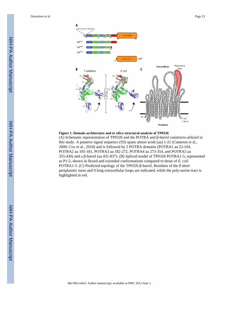
Figure 1. Domain architecture and in silico structural analysis of TP0326(A) Schematic representation of TP0326 and the POTRA and β-barrel constructs utilized inthis study. A putative signal sequence (SS) spans amino acids (aa) 1-21 (Cameron et al.,2000; Cox et al., 2010) and is followed by 5 POTRA domains (POTRA1 aa 22-104,POTRA2 aa 105-181, POTRA3 aa 182-272, POTRA4 aa 273-354, and POTRA5 aa355-430) and a β-barrel (aa 431-837). (B) Spliced model of TP0326 POTRA1-5, representedas P1-5, shown in flexed and extended conformations compared to those of E. coliPOTRA1-5. (C) Predicted topology of the TP0326 β-barrel. Residues of the 8 shortperiplasmic turns and 9 long extracellular loops are indicated, while the poly-serine tract ishighlighted in red.
Desrosiers et al. Page 23
Mol Microbiol. Author manuscript; available in PMC 2012 June 1.
NIH
-PA Author Manuscript
NIH
-PA Author Manuscript
NIH
-PA Author Manuscript
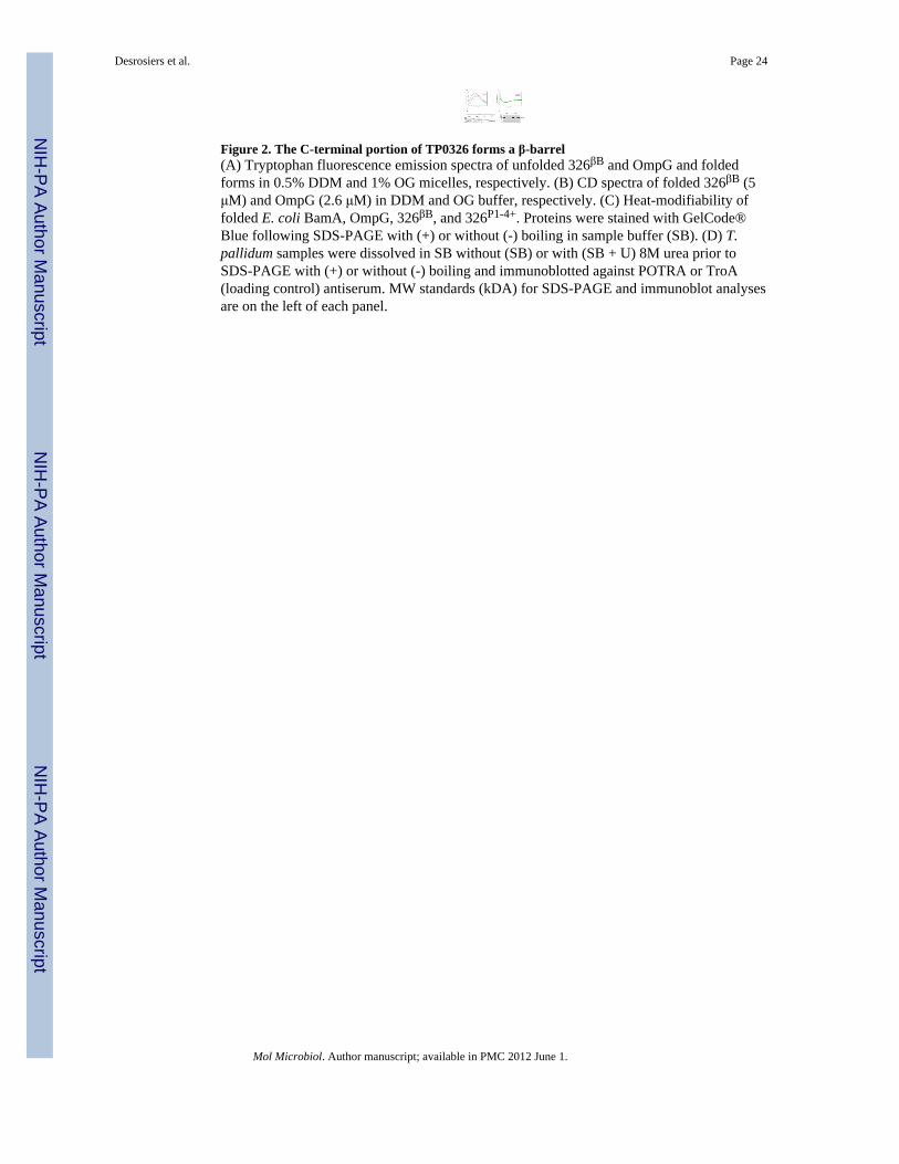
Figure 2. The C-terminal portion of TP0326 forms a β-barrel(A) Tryptophan fluorescence emission spectra of unfolded 326βB and OmpG and foldedforms in 0.5% DDM and 1% OG micelles, respectively. (B) CD spectra of folded 326βB (5μM) and OmpG (2.6 μM) in DDM and OG buffer, respectively. (C) Heat-modifiability offolded E. coli BamA, OmpG, 326βB, and 326P1-4+. Proteins were stained with GelCode®Blue following SDS-PAGE with (+) or without (-) boiling in sample buffer (SB). (D) T.pallidum samples were dissolved in SB without (SB) or with (SB + U) 8M urea prior toSDS-PAGE with (+) or without (-) boiling and immunoblotted against POTRA or TroA(loading control) antiserum. MW standards (kDA) for SDS-PAGE and immunoblot analysesare on the left of each panel.
Desrosiers et al. Page 24
Mol Microbiol. Author manuscript; available in PMC 2012 June 1.
NIH
-PA Author Manuscript
NIH
-PA Author Manuscript
NIH
-PA Author Manuscript
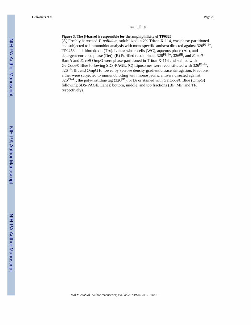
Figure 3. The β-barrel is responsible for the amphiphilicity of TP0326(A) Freshly harvested T. pallidum, solubilized in 2% Triton X-114, was phase-partitionedand subjected to immunoblot analysis with monospecific antisera directed against 326P1-4+,TP0453, and thioredoxin (Trx). Lanes: whole cells (WC), aqueous phase (Aq), anddetergent-enriched phase (Det). (B) Purified recombinant 326P1-4+, 326βB, and E. coliBamA and E. coli OmpG were phase-partitioned in Triton X-114 and stained withGelCode® Blue following SDS-PAGE. (C) Liposomes were reconstituted with 326P1-4+,326βB, Br, and OmpG followed by sucrose density gradient ultracentrifugation. Fractionseither were subjected to immunoblotting with monospecific antisera directed against326P1-4+, the poly-histidine tag (326βB), or Br or stained with GelCode® Blue (OmpG)following SDS-PAGE. Lanes: bottom, middle, and top fractions (BF, MF, and TF,respectively).
Desrosiers et al. Page 25
Mol Microbiol. Author manuscript; available in PMC 2012 June 1.
NIH
-PA Author Manuscript
NIH
-PA Author Manuscript
NIH
-PA Author Manuscript
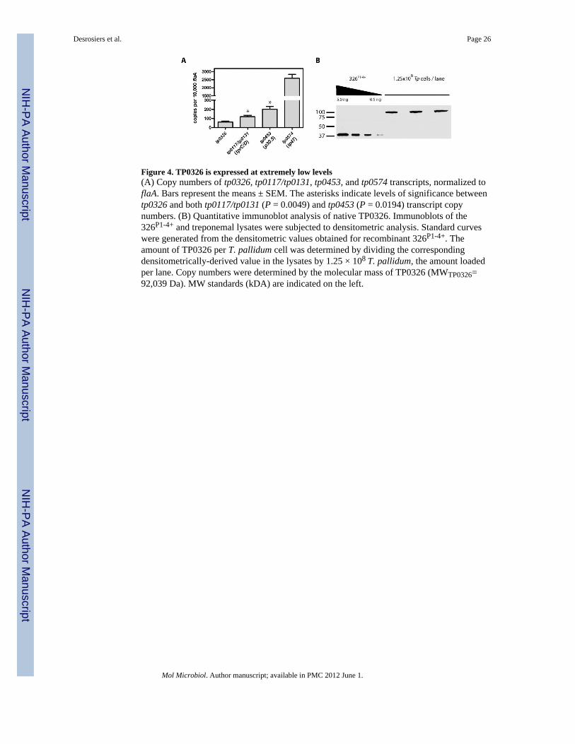
Figure 4. TP0326 is expressed at extremely low levels(A) Copy numbers of tp0326, tp0117/tp0131, tp0453, and tp0574 transcripts, normalized toflaA. Bars represent the means ± SEM. The asterisks indicate levels of significance betweentp0326 and both tp0117/tp0131 (P = 0.0049) and tp0453 (P = 0.0194) transcript copynumbers. (B) Quantitative immunoblot analysis of native TP0326. Immunoblots of the326P1-4+ and treponemal lysates were subjected to densitometric analysis. Standard curveswere generated from the densitometric values obtained for recombinant 326P1-4+. Theamount of TP0326 per T. pallidum cell was determined by dividing the correspondingdensitometrically-derived value in the lysates by 1.25 × 10⁸ T. pallidum, the amount loadedper lane. Copy numbers were determined by the molecular mass of TP0326 (MWTP0326=92,039 Da). MW standards (kDA) are indicated on the left.
Desrosiers et al. Page 26
Mol Microbiol. Author manuscript; available in PMC 2012 June 1.
NIH
-PA Author Manuscript
NIH
-PA Author Manuscript
NIH
-PA Author Manuscript
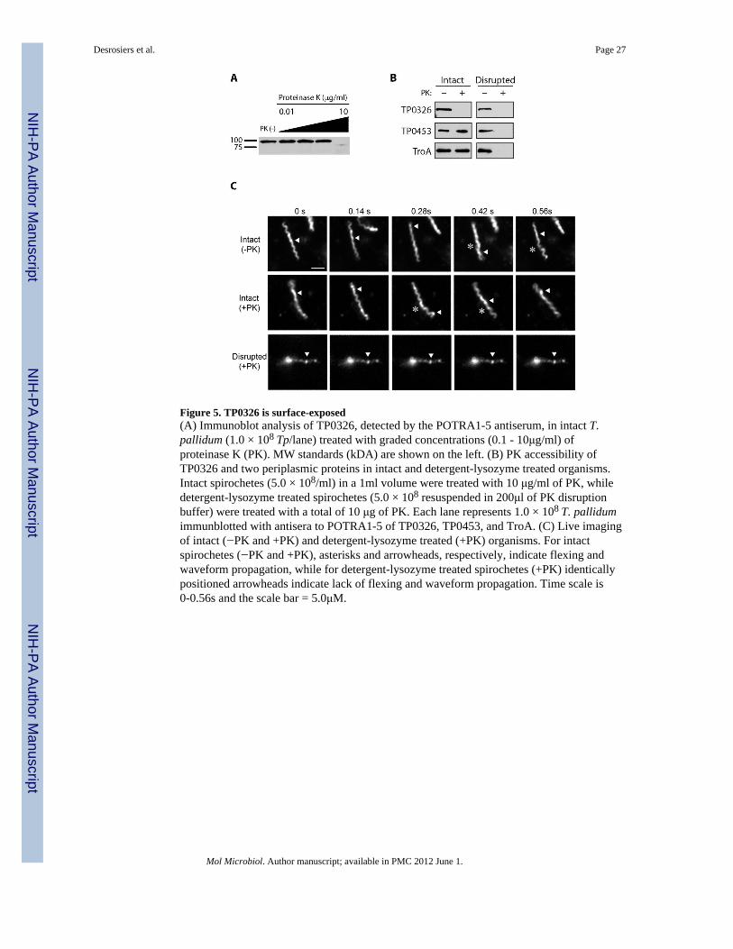
Figure 5. TP0326 is surface-exposed(A) Immunoblot analysis of TP0326, detected by the POTRA1-5 antiserum, in intact T.pallidum (1.0 × 10⁸ Tp/lane) treated with graded concentrations (0.1 - 10μg/ml) ofproteinase K (PK). MW standards (kDA) are shown on the left. (B) PK accessibility ofTP0326 and two periplasmic proteins in intact and detergent-lysozyme treated organisms.Intact spirochetes (5.0 × 10⁸/ml) in a 1ml volume were treated with 10 μg/ml of PK, whiledetergent-lysozyme treated spirochetes (5.0 × 10⁸ resuspended in 200μl of PK disruptionbuffer) were treated with a total of 10 μg of PK. Each lane represents 1.0 × 10⁸ T. pallidumimmunblotted with antisera to POTRA1-5 of TP0326, TP0453, and TroA. (C) Live imagingof intact (−PK and +PK) and detergent-lysozyme treated (+PK) organisms. For intactspirochetes (−PK and +PK), asterisks and arrowheads, respectively, indicate flexing andwaveform propagation, while for detergent-lysozyme treated spirochetes (+PK) identicallypositioned arrowheads indicate lack of flexing and waveform propagation. Time scale is0-0.56s and the scale bar = 5.0μM.
Desrosiers et al. Page 27
Mol Microbiol. Author manuscript; available in PMC 2012 June 1.
NIH
-PA Author Manuscript
NIH
-PA Author Manuscript
NIH
-PA Author Manuscript
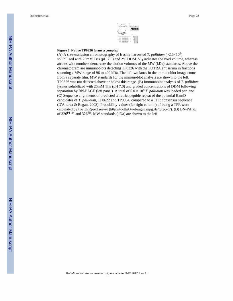
Figure 6. Native TP0326 forms a complex(A) A size-exclusion chromatography of freshly harvested T. pallidum (~2.5×10⁹)solubilized with 25mM Tris (pH 7.0) and 2% DDM. VO indicates the void volume, whereasarrows with numbers demarcate the elution volumes of the MW (kDa) standards. Above thechromatogram are immunoblots detecting TP0326 with the POTRA antiserum in fractionsspanning a MW range of 96 to 400 kDa. The left two lanes in the immunoblot image comefrom a separate film. MW standards for the immunoblot analysis are shown to the left.TP0326 was not detected above or below this range. (B) Immunoblot analysis of T. pallidumlysates solubilized with 25mM Tris (pH 7.0) and graded concentrations of DDM followingseparation by BN-PAGE (left panel). A total of 5.0 × 10⁸ T. pallidum was loaded per lane.(C) Sequence alignments of predicted tetratricopeptide repeat of the potential BamDcandidates of T. pallidum, TP0622 and TP0954, compared to a TPR consensus sequence(D'Andrea & Regan, 2003). Probability-values (far right column) of being a TPR werecalculated by the TPRpred server (http://toolkit.tuebingen.mpg.de/tprpred/). (D) BN-PAGEof 326P1-4+ and 326βB. MW standards (kDa) are shown to the left.
Desrosiers et al. Page 28
Mol Microbiol. Author manuscript; available in PMC 2012 June 1.
NIH
-PA Author Manuscript
NIH
-PA Author Manuscript
NIH
-PA Author Manuscript
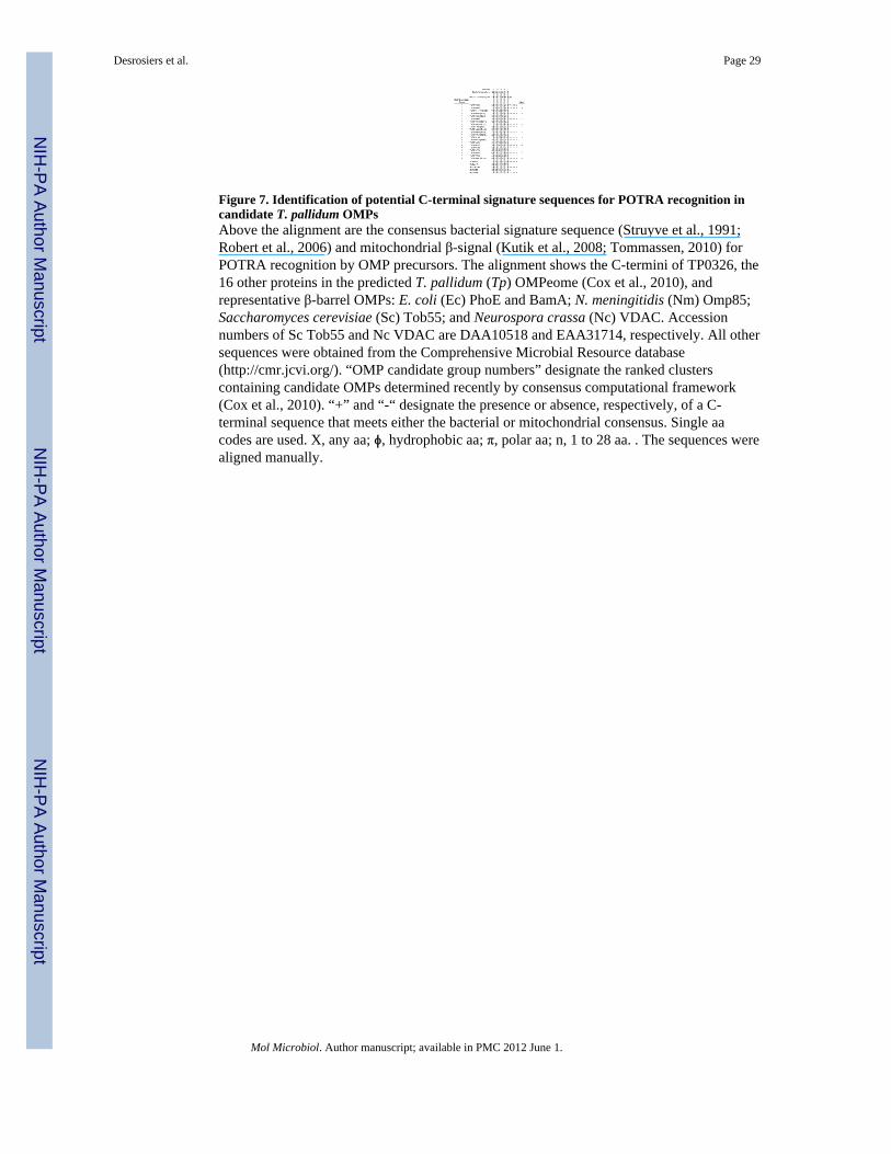
Figure 7. Identification of potential C-terminal signature sequences for POTRA recognition incandidate T. pallidum OMPsAbove the alignment are the consensus bacterial signature sequence (Struyve et al., 1991;Robert et al., 2006) and mitochondrial β-signal (Kutik et al., 2008; Tommassen, 2010) forPOTRA recognition by OMP precursors. The alignment shows the C-termini of TP0326, the16 other proteins in the predicted T. pallidum (Tp) OMPeome (Cox et al., 2010), andrepresentative β-barrel OMPs: E. coli (Ec) PhoE and BamA; N. meningitidis (Nm) Omp85;Saccharomyces cerevisiae (Sc) Tob55; and Neurospora crassa (Nc) VDAC. Accessionnumbers of Sc Tob55 and Nc VDAC are DAA10518 and EAA31714, respectively. All othersequences were obtained from the Comprehensive Microbial Resource database(http://cmr.jcvi.org/). “OMP candidate group numbers” designate the ranked clusterscontaining candidate OMPs determined recently by consensus computational framework(Cox et al., 2010). “+” and “-“ designate the presence or absence, respectively, of a C-terminal sequence that meets either the bacterial or mitochondrial consensus. Single aacodes are used. X, any aa; ϕ, hydrophobic aa; π, polar aa; n, 1 to 28 aa. . The sequences werealigned manually.
Desrosiers et al. Page 29
Mol Microbiol. Author manuscript; available in PMC 2012 June 1.
NIH
-PA Author Manuscript
NIH
-PA Author Manuscript
NIH
-PA Author Manuscript
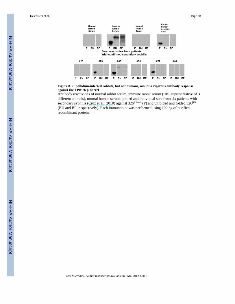
Figure 8. T. pallidum-infected rabbits, but not humans, mount a vigorous antibody responseagainst the TP0326 β-barrelAntibody reactivities of normal rabbit serum, immune rabbit serum (IRS, representative of 3different animals), normal human serum, pooled and individual sera from six patients withsecondary syphilis (Cruz et al., 2010) against 326P1-4+ (P) and unfolded and folded 326βB
(BU and BF, respectively). Each immunoblot was performed using 100 ng of purifiedrecombinant protein.
Desrosiers et al. Page 30
Mol Microbiol. Author manuscript; available in PMC 2012 June 1.
NIH
-PA Author Manuscript
NIH
-PA Author Manuscript
NIH
-PA Author Manuscript
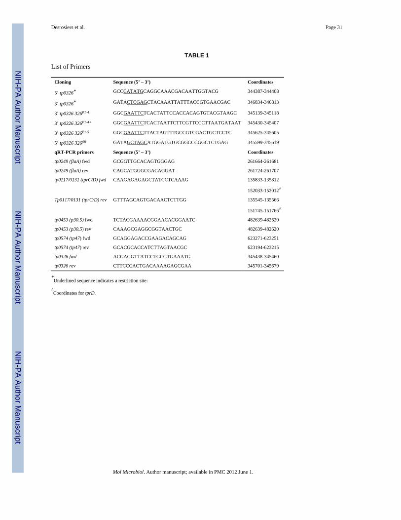
NIH
-PA Author Manuscript
NIH
-PA Author Manuscript
NIH
-PA Author Manuscript
Desrosiers et al. Page 31
TABLE 1
List of Primers
Cloning Sequence (5’ – 3’) Coordinates
5’ tp0326* GCCCATATGCAGGCAAACGACAATTGGTACG 344387-344408
3’ tp0326* GATACTCGAGCTACAAATTATTTACCGTGAACGAC 346834-346813
3’ tp0326 326P1-4 GGCGAATTCTCACTATTCCACCACAGTGTACGTAAGC 345139-345118
3’ tp0326 326P1-4+ GGCGAATTCTCACTAATTCTTCGTTCCCTTAATGATAAT 345430-345407
3’ tp0326 326P1-5 GGCGAATTCTTACTAGTTTGCCGTCGACTGCTCCTC 345625-345605
5’ tp0326 326βB GATAGCTAGCATGGATGTGCGGCCCGGCTCTGAG 345599-345619
qRT-PCR primers Sequence (5’ – 3’) Coordinates
tp0249 (flaA) fwd GCGGTTGCACAGTGGGAG 261664-261681
tp0249 (flaA) rev CAGCATGGGCGACAGGAT 261724-261707
tp0117/0131 (tprC/D) fwd CAAGAGAGAGCTATCCTCAAAG 135833-135812
152033-152012^
Tp0117/0131 (tprC/D) rev GTTTAGCAGTGACAACTCTTGG 135545-135566
151745-151766^
tp0453 (p30.5) fwd TCTACGAAAACGGAACACGGAATC 482639-482620
tp0453 (p30.5) rev CAAAGCGAGGCGGTAACTGC 482639-482620
tp0574 (tp47) fwd GCAGGAGACCGAAGACAGCAG 623271-623251
tp0574 (tp47) rev GCACGCACCATCTTAGTAACGC 623194-623215
tp0326 fwd ACGAGGTTATCCTGCGTGAAATG 345438-345460
tp0326 rev CTTCCCACTGACAAAAGAGCGAA 345701-345679
*Underlined sequence indicates a restriction site:
^Coordinates for tprD.
Mol Microbiol. Author manuscript; available in PMC 2012 June 1.
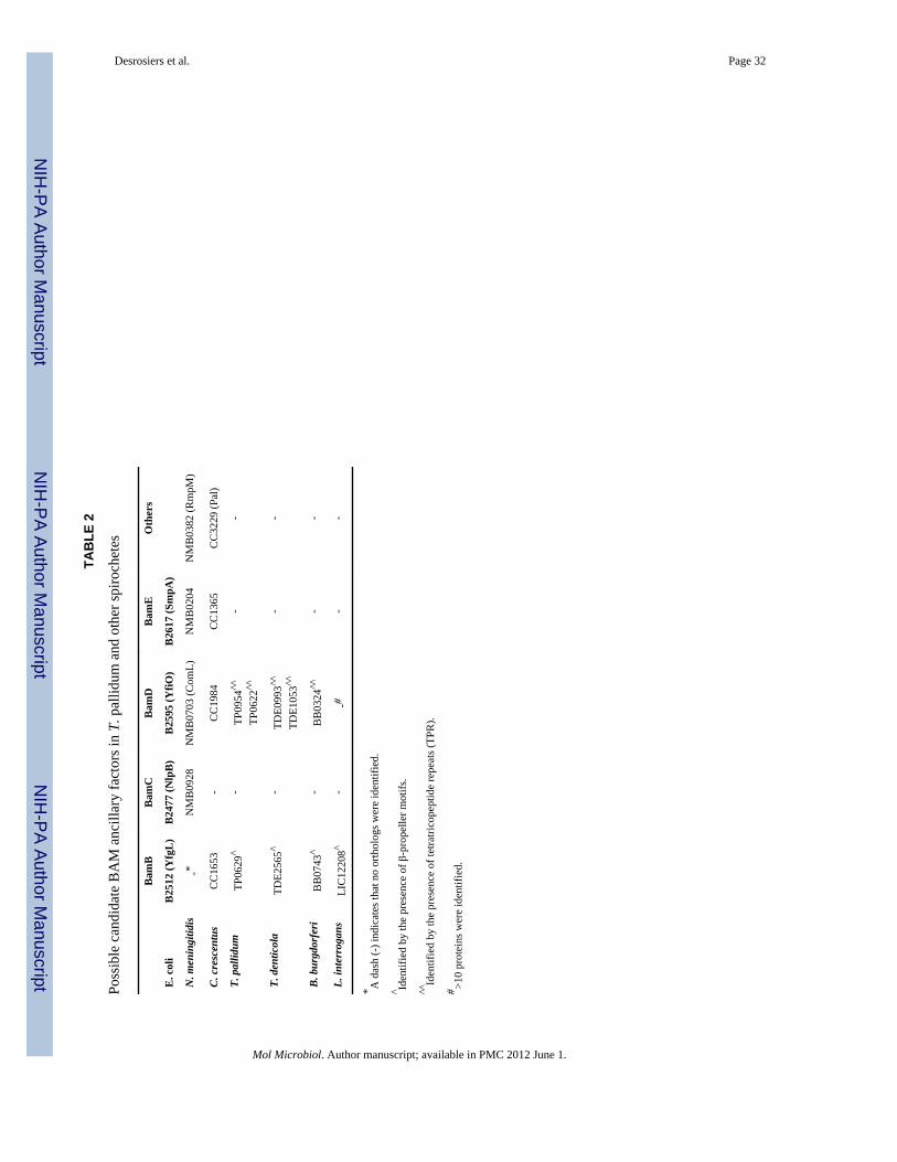
NIH
-PA Author Manuscript
NIH
-PA Author Manuscript
NIH
-PA Author Manuscript
Desrosiers et al. Page 32
TAB
LE 2
Poss
ible
can
dida
te B
AM
anc
illar
y fa
ctor
s in
T. p
allid
um a
nd o
ther
spiro
chet
es
Bam
BB
amC
Bam
DB
amE
Oth
ers
E. c
oli
B25
12 (Y
fgL
)B
2477
(Nlp
B)
B25
95 (Y
fiO)
B26
17 (S
mpA
)
N. m
enin
gitid
is-*
NM
B09
28N
MB
0703
(Com
L)N
MB
0204
NM
B03
82 (R
mpM
)
C. c
resc
entu
sC
C16
53-
CC
1984
CC
1365
CC
3229
(Pal
)
T. p
allid
umTP
0629
^-
TP09
54^^
TP06
22^^
--
T. d
entic
ola
TDE2
565^
-TD
E099
3^^
TDE1
053^
^-
-
B. b
urgd
orfe
riB
B07
43^
-B
B03
24^^
--
L. in
terr
ogan
sLI
C12
208^
--#
--
* A d
ash
(-) i
ndic
ates
that
no
orth
olog
s wer
e id
entif
ied.
^ Iden
tifie
d by
the
pres
ence
of β
-pro
pelle
r mot
ifs.
^^Id
entif
ied
by th
e pr
esen
ce o
f tet
ratri
cope
ptid
e re
peat
s (TP
R).
# >10
prot
eins
wer
e id
entif
ied.
Mol Microbiol. Author manuscript; available in PMC 2012 June 1.