Evaluation of multiplex real-time PCR for detection of Haemophilus ducreyi, Treponema pallidum,...
10
Evaluation of multiplex real-time PCR for detection of Haemophilus ducreyi, Treponema pallidum, herpes simplex virus type 1 and 2 in the diagnosis of genital ulcer disease in the Rakai District, Uganda T R Suntoke 1 , A Hardick 2 , A A R Tobian 2 , B Mpoza 4 , O Laeyendecker 1,2 , D Serwadda 3 , P Opendi 4 , C A Gaydos 2 , R H Gray 5 , M J Wawer 5 , T C Quinn 1,2 , and S J Reynolds 1,2 1 National Institute of Allergy and Infectious Diseases, National Institutes of Health, Bethesda, Maryland, USA 2 Division of Infectious Diseases, Johns Hopkins Medical Institutions, Baltimore, Maryland, USA 3 Institute of Public Health, Makerere University, Kampala, Uganda 4 Rakai Health Sciences Program, Rakai, Uganda 5 Department of Population and Family Health Sciences, Bloomberg School of Public Health, Johns Hopkins Medical Institutions, Baltimore, Maryland, USA Abstract Objective: To develop a real-time PCR assay that reliably and accurately detects the predominant sexually transmitted aetiological agents of genital ulcer disease (GUD) (Haemophilus ducreyi, Treponema pallidum and herpes simplex virus type 1 (HSV-1) and type 2 (HSV-2)) and to assess the use of real-time PCR diagnostic testing in a rural African field site. Methods: Two multiplex real-time PCR reactions were used to detect H ducreyi/and HSV-1/HSV-2 in ulcer swabs from 100 people with symptomatic genital ulcers in rural Rakai, Uganda. Results were compared with syphilis, HSV-1 and HSV-2 serology. Results: Of 100 GUD samples analysed from 43 HIV positive and 57 HIV negative individuals, 71% were positive for one or more sexually transmitted infection (STI) pathogens by real-time PCR (61% for HSV-2, 5% for T pallidum, 3% for HSV-1, 1% for H ducreyi and 1% for dual H ducreyi/ HSV-2). The frequency of HSV in genital ulcers was 56% (32/57) in HIV negative individuals and 77% (33/43) in HIV positive individuals (p=0.037). Assay reproducibility was evaluated by repeat PCR testing in the USA with 96% agreement (κ=0.85). Conclusions: STI pathogens were detected in the majority of GUD swab samples from symptomatic patients in Rakai, Uganda, by real-time PCR. HSV-2 was the predominant cause of genital ulcers. Real-time PCR technology can provide sensitive, rapid and reproducible evaluation of GUD aetiology in a resource-limited setting. Correspondence to: Dr Thomas C Quinn, Division of Infectious Diseases, Johns Hopkins Medical Institutions, 855 N. Wolfe Street, Room 531, Baltimore, Maryland 21205, USA; E-mail: [email protected]. Contributors: TRS, AH, AAT, BM, OL, CAG and SJR performed real-time PCR and serological testing and provided technical assistance on assay development. BM, PO and DS provided support for sample data and collection in Rakai, Uganda. TRS, AAT, OL, CAG, RHG, MJW, TCQ and SJR contributed to manuscript preparation and provided overall direction and support for the project. Competing interests: None. Ethics approval: The study was approved by the Science and Ethics Committee of the Uganda Virus Research Institute and the Western Institutional Review Board for Johns Hopkins University. NIH Public Access Author Manuscript Sex Transm Infect. Author manuscript; available in PMC 2009 June 8. Published in final edited form as: Sex Transm Infect. 2009 April ; 85(2): 97–101. doi:10.1136/sti.2008.034207. NIH-PA Author Manuscript NIH-PA Author Manuscript NIH-PA Author Manuscript
Transcript of Evaluation of multiplex real-time PCR for detection of Haemophilus ducreyi, Treponema pallidum,...
Evaluation of multiplex real-time PCR for detection ofHaemophilus ducreyi, Treponema pallidum, herpes simplex virustype 1 and 2 in the diagnosis of genital ulcer disease in the RakaiDistrict, Uganda
T R Suntoke1, A Hardick2, A A R Tobian2, B Mpoza4, O Laeyendecker1,2, D Serwadda3, POpendi4, C A Gaydos2, R H Gray5, M J Wawer5, T C Quinn1,2, and S J Reynolds1,2
1 National Institute of Allergy and Infectious Diseases, National Institutes of Health, Bethesda, Maryland,USA 2 Division of Infectious Diseases, Johns Hopkins Medical Institutions, Baltimore, Maryland, USA 3Institute of Public Health, Makerere University, Kampala, Uganda 4 Rakai Health Sciences Program, Rakai,Uganda 5 Department of Population and Family Health Sciences, Bloomberg School of Public Health, JohnsHopkins Medical Institutions, Baltimore, Maryland, USA
AbstractObjective: To develop a real-time PCR assay that reliably and accurately detects the predominantsexually transmitted aetiological agents of genital ulcer disease (GUD) (Haemophilus ducreyi,Treponema pallidum and herpes simplex virus type 1 (HSV-1) and type 2 (HSV-2)) and to assessthe use of real-time PCR diagnostic testing in a rural African field site.
Methods: Two multiplex real-time PCR reactions were used to detect H ducreyi/and HSV-1/HSV-2in ulcer swabs from 100 people with symptomatic genital ulcers in rural Rakai, Uganda. Results werecompared with syphilis, HSV-1 and HSV-2 serology.
Results: Of 100 GUD samples analysed from 43 HIV positive and 57 HIV negative individuals,71% were positive for one or more sexually transmitted infection (STI) pathogens by real-time PCR(61% for HSV-2, 5% for T pallidum, 3% for HSV-1, 1% for H ducreyi and 1% for dual H ducreyi/HSV-2). The frequency of HSV in genital ulcers was 56% (32/57) in HIV negative individuals and77% (33/43) in HIV positive individuals (p=0.037). Assay reproducibility was evaluated by repeatPCR testing in the USA with 96% agreement (κ=0.85).
Conclusions: STI pathogens were detected in the majority of GUD swab samples fromsymptomatic patients in Rakai, Uganda, by real-time PCR. HSV-2 was the predominant cause ofgenital ulcers. Real-time PCR technology can provide sensitive, rapid and reproducible evaluationof GUD aetiology in a resource-limited setting.
Correspondence to: Dr Thomas C Quinn, Division of Infectious Diseases, Johns Hopkins Medical Institutions, 855 N. Wolfe Street,Room 531, Baltimore, Maryland 21205, USA; E-mail: [email protected]:TRS, AH, AAT, BM, OL, CAG and SJR performed real-time PCR and serological testing and provided technical assistance on assaydevelopment. BM, PO and DS provided support for sample data and collection in Rakai, Uganda. TRS, AAT, OL, CAG, RHG, MJW,TCQ and SJR contributed to manuscript preparation and provided overall direction and support for the project.Competing interests:None.Ethics approval:The study was approved by the Science and Ethics Committee of the Uganda Virus Research Institute and the Western InstitutionalReview Board for Johns Hopkins University.
NIH Public AccessAuthor ManuscriptSex Transm Infect. Author manuscript; available in PMC 2009 June 8.
Published in final edited form as:Sex Transm Infect. 2009 April ; 85(2): 97–101. doi:10.1136/sti.2008.034207.
NIH
-PA Author Manuscript
NIH
-PA Author Manuscript
NIH
-PA Author Manuscript

It is estimated that over 340 million new cases of sexually transmitted infections (STIs) occurannually throughout the world, with the highest number in developing countries.1 These STIsfrequently present as genital ulcer disease (GUD) of which the predominant aetiological agentsare Haemophilus ducreyi (chancroid), Treponema pallidum (syphilis) and herpes simplex virustype 2 (HSV-2).
Genital herpes, mainly caused by HSV-2, is the leading cause of genital ulceration worldwide.2-5 Recent investigations indicate a rise in genital herpes caused by HSV-1.3 6 7 PrimaryHSV-1 and HSV-2 infections are clinically indistinguishable and require laboratory tests todifferentiate.6 8
GUD caused by HSV-2 is also a risk factor for HIV acquisition and transmission,2 9-11 andmodel estimates from sub-Saharan Africa suggest that the proportion of HIV incidenceattributable to HSV-2 infection may increase as the HIV epidemic matures.9 12
Standard methods used to detect the causative agents of GUD include in vitro culture, serologytests and microscopy;13-15 however, despite their potentially high sensitivity and specificity,these methods have limitations. In vitro culture of H ducreyi is difficult and often impracticalin clinical and research settings.13 Detection of HSV by culture varies greatly with durationand stage of the ulcerative lesion.13 Serological tests commonly used to diagnose syphilis andherpes do not detect infection during the early pre-seroconversion window or distinguishbetween active and latent infection.
Real-time PCR is a fast, sensitive and high-throughput method for pathogen detection and israpidly becoming a mainstay of research and clinical diagnostic applications.16-18 SeveralPCR-based techniques that require post-amplification processing have been used to detectGUD pathogens.13 19 20 The fluorogenic probes used in real-time PCR enable directmeasurement of product amplification as the reaction proceeds, minimising additional “hands-on” time and decreasing the risk of contamination. The probes permit sensitive and specificdetection of target nucleic acids over a wide dynamic range of input sample concentrations.21 22 Furthermore, the use of multiple probes per reaction, or multiplexing, allows severalpathogens to be detected in a single well.
We established two duplex real-time PCR reactions to detect H ducreyi, T pallidum, HSV-1and HSV-2 in an on-site laboratory in Rakai, Uganda. We report the validation of this duplexreal-time PCR assay and evaluate its use in this setting.
METHODSStudy population and clinical specimens
The population consisted of people enrolled in the Rakai Community Cohort who were seenduring annual cohort surveys or in mobile clinics and who reported genital ulceration at timeof interview. During the study period 2002–2006, approximately 120 consecutive clinicalepisodes of GUD were recorded among Rakai participants. Although a single swab was to becollected per episode, 100 swabs were delivered and stored in the laboratory during this periodand used in this evaluation. Samples were collected in 1 mL specimen transport medium (RocheAmplicor STM, Roche Diagnostics, Indianapolis, USA) or Herptran medium (Perkin Elmer,Waltham, Maryland, USA) and stored at −80°C. For most patients (98%), blood samples werealso collected at the same visit and sera was stored at −80°C.
Real-time multiplex PCRThe real-time PCR methods presented here are based on previously established PCR assaysthat have been used to detect GUD pathogens13 23 but with two major adaptations. First, a
Suntoke et al. Page 2
Sex Transm Infect. Author manuscript; available in PMC 2009 June 8.
NIH
-PA Author Manuscript
NIH
-PA Author Manuscript
NIH
-PA Author Manuscript

widely used two-step PCR assay (triplex PCR followed by colorimetric detection), whichsimultaneously detects H ducreyi, T pallidum and HSV,13 was optimised for use in the real-time PCR format. The original 5′ primer biotinylation was removed. A unique fluorophore(VIC for HSV, TET for syphilis and FAM for chancroid) and 3′ MGB quencher that enhancesprobe-target annealing were added to the probe. Tenfold dilutions of positive control DNA andvarying primer (250–900 nM) and probe (100–250 nM) concentrations were used to determineoptimal detection conditions. Using these modified primers, probes and Qiagen HotStar TaqDNA Polymerase kit (Qiagen, Valencia, California, USA), one or more pathogens (H ducreyi,T pallidum and HSV) were simultaneously detected in approximately 41% of swab specimensby real-time PCR (data not shown).
Second, to improve detection and distinguish between HSV-1 and HSV-2, HSV primers andprobe were removed from the original reaction and a second separate reaction for HSV-1 andHSV-2 was added. The H ducreyi/T pallidum duplex reaction was re-evaluated for optimalprimer (250 nM) and probe (100 nM) concentrations and used with ABI Master Mix (AppliedBiosystems, Valencia, California, USA). The HSV-1/HSV-2 typing reaction is the same aspreviously reported for real-time PCR detection of HSV-1 and HSV-2 using the ABI 7900HTthermocycler23 (Applied Biosystems) and ABI Master Mix. Thus, the real-time PCR assaypresented here consists of two duplex PCR reactions: one that detects H ducreyi and Tpallidum and the other that detects HSV-1 and HSV-2. Except for specific primers and probes,all PCR components and thermocycling parameters are identical for both reactions enhancingthe efficiency of reaction assembly.
DNA was extracted from swab samples using the QIAamp DNA Blood Mini kit following theblood and body fluids protocol as supplied by the manufacturer (Qiagen). Four positive controlswere diluted 1:2 in TE buffer and extracted: H ducreyi (ATCC # 51566 or 700724, Manassas,Virginia, USA), T pallidum (Lee Laboratories/Becton Dickinson # 210483, Grayson, Georgia,USA), HSV-1 strain GHSV-UL46 (ATCC # VR-1544) and HSV-2 strain MS (ATCC #VR-540). Two separate multiplex assays were performed for each sample: one for H ducreyi/T pallidum detection and the other for HSV-1/ HSV-2 detection. The primer and probesequences used for this study have been previously reported.13 23
The H ducreyi/T pallidum reaction contained ABI 2× Universal Master Mix, 250 nM of eachof four primers (K07A, K08A, K03A, K014) and 100 nM of each of two probes (K015, K017).The HSV-1/HSV-2 reaction contained ABI 2× Universal Master Mix, 800 nM of each of twoprimers (GbTypF, GbTypR) and 100 nM of each of two probes (HSVgBTyp1, HSVgBTyp2).Assays were performed in 96-well plate format using an ABI 7900 HT Fast real-time PCRinstrument with a 50 μL total reaction volume that included 10 μL of extracted sample orpositive control DNA. Thermocycle parameters were the same for both duplex assays: 50°Cfor 2 min, 95°C for 10 min and 40 cycles of 95°C for 15 sec, 60°C for 1 min. Samples wererun in duplicate for each duplex assay. To analyse results, the threshold was selected based onpositive and negative controls and used to determine a threshold-cycle value (CT, the PCRcycle at which amplification signal crosses the threshold). A cutoff CT value was notdesignated. Samples were counted as weakly positive if they had a CT value greater than 36and a fluorescence signal increase in the multicomponent view of at least 1000 units. Discrepantor weakly positive samples were re-evaluated in singleplex format using a single primer pairand probe corresponding to one pathogen.
Swab samples were re-extracted and analysed in the USA for independent confirmation of real-time PCR results. Sample processing procedures were identical to those reported above exceptthat DNA extraction was performed using the Roche MagNA Pure robot (Roche Diagnostics,Indianapolis, USA) with the DNA I Blood Cell High Performance kit and protocol. Sampleswere analysed with the same criteria used in the Rakai laboratory.
Suntoke et al. Page 3
Sex Transm Infect. Author manuscript; available in PMC 2009 June 8.
NIH
-PA Author Manuscript
NIH
-PA Author Manuscript
NIH
-PA Author Manuscript

SerologyBlood samples from 98 of 100 people with current GUD symptoms were collected at the samevisit. Sera were screened for the presence of antibodies to T pallidum using the nontreponemalToluidine Red Untreated Serum Test (TRUST, New Horizons Diagnostic Corporation,Columbia, Massachusetts, USA) assay. TRUST-positive samples were further evaluated bythe Treponema pallidum Particle Agglutination Assay (TPPA) (Serodia-TP PA kit, FujirebioInc, Tokyo, Japan). Samples reactive on both the TRUST and TPPA assays were classified asT pallidum antibody positive.
Sera from 94 GUD patients were tested for HSV-2 antibodies using the Kalon ELISA (KalonBiological Ltd, Guilford, UK) according to the manufacturer's protocol with minormodifications. Samples were run in duplicate and the mean index value was used to classifyHSV-2 status. People with an index value <0.9 were classified as HSV-2 negative asrecommended by the manufacturer. Based on prior evaluation of test performance in Ugandansera, HSV-2 positive individuals were defined as an index value of ⩾1.5.24 Samples withequivocal results (0.9–1.5) were repeated by Euroimmune Western blot (Euroimmune, Lubeck,Germany) and HSV-2 Western blot at the University of Washington (Seattle, Washington,USA).25 Confirmatory University of Washington HSV-2 Western blots were performed forsamples that were HSV-2 seronegative by Kalon and HSV-2 positive by real-time PCR. HSV-1serostatus was determined using the Euroimmune Western blot test according to manufacturerrecommendations.
For all patients, HIV infection status at the time of the GUD visit was established using twoHIV enzyme immunosorbent assays (Vironostika HIV-1, Organon Teknika, Charlotte, NorthCarolina, USA; Cambridge Biotech, Worcester, Massachusetts, USA) and Western blot(HIV-1 Western Blot, BioMerieux-Vitek, St Louis, Missouri, USA) for discordant results.
Statistical analysisData were analysed using Microsoft Excel 2003 and Intercooled STATA version 10.0(StataCorp, College Station, Texas, USA). To compare proportions of detectable swabs in thereal-time PCR assay versus triplex and other PCR assays and HSV-2 seropositive serumsamples, two-tailed z-tests were performed and the resulting p values are reported. To detectassociations between HSV by real-time PCR and HIV infection, both Fisher exact and χ2 testswere performed.
RESULTSUsing duplex real-time PCR reactions, one or more pathogens were detected in 71% of swabspecimens collected from 100 people with symptomatic genital ulcers (fig 1 and table 1).Altogether, 61% of samples were positive for HSV-2, 5% for T pallidum, 3% for HSV-1, 1%each for H ducreyi and dual H ducreyi/HSV-2, and the remaining 29% were undetectable forall four STI pathogens.
Of 100 patients, 43 (20 females and 23 males) were HIV seropositive (table 1). Altogether,77% (33/43) of HIV seropositive individuals had detectable HSV-1 or HSV-2 in their ulcerswabs compared with 56% (32/57) of HIV negative individuals (p=0.037).
To confirm results obtained in the Rakai Uganda laboratory, swab specimens were shipped tothe USA and re-tested using the same methodology. The overall agreement of real-time PCRresults was high: 100% for T pallidum, 98% for H ducreyi, 97% for HSV-1 and 89% for HSV-2(κ=0.85) (results not shown). Except for two samples, all discrepancies arose in HSV testing.Two samples were negative for H ducreyi when tested in the Rakai laboratory but positive inthe USA. All three discordant HSV-1 samples and ten out of eleven HSV-2 discordant samples
Suntoke et al. Page 4
Sex Transm Infect. Author manuscript; available in PMC 2009 June 8.
NIH
-PA Author Manuscript
NIH
-PA Author Manuscript
NIH
-PA Author Manuscript

were weakly positive by multiplex real-time PCR in Rakai. These samples were confirmed asPCR positive in Rakai by inspection of the fluorescent signal increase in the multicomponentplot as well as when repeated in singleplex format. However, all 14 samples tested negative inthe USA.
Of 94 specimens tested for serologic syphilis, 11 (12%) were TRUST positive and confirmedby TPPA (table 2). Five of these eleven samples (45%) were also T pallidum positive by real-time PCR. Of the six samples (55%) that were T pallidum PCR negative, five contained eitherHSV-1 or HSV-2 DNA by real-time PCR.
HSV-1 serology was performed using Western blot, with 88% (80/91) of serum samplesshowing reactivity. HSV-1 specific DNA was detectable in three of the ulcer swabs fromseropositive patients.
Altogether, 73 of 95 serum samples tested (77%) were HSV-2 seropositive by the Kalon ELISAassay. HSV-2 DNA was detected in 50 (68%) of the corresponding ulcer swabs by real-timePCR. Of the remaining 23 HSV-2 seropositive PCR negative samples, HSV-1 or T pallidumDNA was detected in four ulcer swabs. Conversely, 9 out of 22 HSV-2 seronegative samples(41%) contained HSV-2-specific DNA as determined by multiplex PCR, possibly representingrecent infections during the window period prior to development of HSV-2 antibodies. HSV-2serology for eight of these nine samples was repeated by Western blot, which can identifyseroconversions earlier than Kalon,26 and HSV-2 antibodies were detected in two samples.
Key messages
▶ Multiplex real-time PCR can rapidly and reproducibly detect the four predominantgenital ulcer disease (GUD) pathogens: Haemophilus ducreyi, Treponema pallidum,herpes simplex virus type 1 and 2.
▶ Using real-time PCR in a Ugandan laboratory, GUD pathogens were detected in themajority (71%) of samples collected from patients with symptomatic genital ulcers.
▶ The ability to detect recent infection and to diagnose the cause of current ulcerationregardless of prior infection are distinct advantages of real-time PCR methodology.
Of the 29 patients with undetectable STI pathogens by real-time PCR, 18 (62%) were HSV-2seropositive, 1 (3%) was T pallidum/HSV-2 dual seropositive, 8 (28%) had no detectableantibodies to HSV-2 or T pallidum and 2 (7%) were not tested (data not shown). There was nostatistically significant association between HSV-2 seropositivity and detection of pathogenby real-time PCR (p=0.346).
DISCUSSIONWe demonstrated the use of real-time PCR technology for the diagnosis of GUD aetiology ina resource-limited research setting. Among patients with current symptomatic genital ulcers,a STI pathogen was detected in 71% of swab samples by real-time PCR. This represents animprovement in detection over both the original triplex real-time PCR assay on the samespecimens (41%; p⩽0.001) (data not shown) and previous reports of PCR-based detection ofGUD pathogens in Uganda (50–55%; p⩽0.003)5 27 and other geographical regions.20 28 29
Results obtained by duplex real-time PCR were highly reproducible upon repeat testing in USlaboratories by independent technicians with an observed agreement of 96%. All discordantHSV results were positive in Rakai and negative upon repeat in the USA. While it is possiblethat the initial results were false positives, sample degradation (upon multiple freeze-thaw
Suntoke et al. Page 5
Sex Transm Infect. Author manuscript; available in PMC 2009 June 8.
NIH
-PA Author Manuscript
NIH
-PA Author Manuscript
NIH
-PA Author Manuscript

cycles before and after shipping), different DNA extraction procedures and variation in lasercalibration between the two instruments may have also contributed to these discrepancies. Themajority of discrepant samples were initially weakly positive in Uganda, even upon repeattesting in singleplex reactions, suggesting that initial pathogen titres in these specimens werelow.
In most resource-limited settings, GUD is syndromically managed. While this real-time PCRmethod does not currently play a role in clinical management, it can be used to rapidlydiscriminate between latent and active infection among seropositive individuals and thereforemore accurately detect the aetiological agent causing ulceration. Real-time PCR can alsoidentify early GUD infections in the pre-seroconversion window. As such, it can be a powerfultool for clinicians and researchers in sub-Saharan Africa to rapidly detect common GUDpathogens in their patient populations and map the changing epidemiology of the disease overtime.
AcknowledgmentsFunding:
This research was supported by The Division of Intramural Research, The National Institute of Allergy and InfectiousDiseases, National Institutes of Health.
REFERENCES1. World Health Organization. Global Strategy for the Prevention and Control of Sexually Transmitted
Infections: 2006–2015. Geneva: 2007.2. Ahmed HJ, Mbwana J, Gunnarsson E, et al. Etiology of genital ulcer disease and association with
human immunodeficiency virus infection in two Tanzanian cities. Sex Transm Dis 2003;30:114–19.[PubMed: 12567167]
3. Paz-Bailey G, Ramaswamy M, Hawkes SJ, et al. Herpes simplex virus type 2: epidemiology andmanagement options in developing countries. Sex Transm Infect 2007;83:16–22. [PubMed: 17098770]
4. Weiss H. Epidemiology of herpes simplex virus type 2 infection in the developing world. Herpes2004;11:24A–35A.
5. Pickering JM, Whitworth JAG, Hughes P, et al. Aetiology of sexually transmitted infections andresponse to syndromic treatment in southwest Uganda. Sex Transm Infect 2005;81:488–93. [PubMed:16326853]
6. Gupta R, Warren T, Wald A. Genital herpes. Lancet 2007;370:2127–37. [PubMed: 18156035]7. Kasubi MJ, Nilsen A, Marsden HS, et al. Prevalence of antibodies against herpes simplex virus types
1 and 2 in children and young people in an urban region in Tanzania. J Clin Microbiol 2006;44:2801–7. [PubMed: 16891495]
8. Centers for Disease Control and Prevention. Sexually Transmitted Diseases Treatment Guidelines2006;55:RR–11.
9. Freeman EE, Weiss HA, Glynn JR, et al. Herpes simplex virus 2 infection increases HIV acquisitionin men and women: systematic review and meta-analysis of longitudinal studies. Aids 2006;20:73–83. [PubMed: 16327322]
10. Gray RH, Wawer MJ, Sewankambo NK, et al. Relative risks and population attributable fraction ofincident HIV associated with symptoms of sexually transmitted diseases and treatable symptomaticsexually transmitted diseases in Rakai District, Uganda. Rakai Project Team. Aids 1999;13:2113–23. [PubMed: 10546865]
11. Sheffield J, Wendel JG, McIntire D, et al. Effect of genital ulcer disease on HIV–1 coreceptorexpression in the female genital tract. J Infect Dis 2007;196:1509–16. [PubMed: 18008231]
12. Freeman EE, Orroth KK, White RG, et al. Proportion of new HIV infections attributable to herpessimplex 2 increases over time: simulations of the changing role of sexually transmitted infections insub-Saharan African HIV epidemics. Sex Transm Infect 2007;83:17–24.
Suntoke et al. Page 6
Sex Transm Infect. Author manuscript; available in PMC 2009 June 8.
NIH
-PA Author Manuscript
NIH
-PA Author Manuscript
NIH
-PA Author Manuscript

13. Orle KA, Gates CA, Martin DH, et al. Simultaneous PCR detection of Haemophilus ducreyi,Treponema pallidum, and herpes simplex virus types 1 and 2 from genital ulcers. J Clin Microbiol1996;34:49–54. [PubMed: 8748271]
14. Morse SA. Chancroid and Haemophilus ducreyi. Clin Microbiol Rev 1989;2:137–57. [PubMed:2650859]
15. Cone RW, Hobson AC, Palmer J, et al. Extended duration of herpes simplex virus DNA in genitallesions detected by the polymerase chain reaction. J Infect Dis 1991;164:757–60. [PubMed: 1654360]
16. Burrows J, Nitsche A, Bayly B, et al. Detection and subtyping of herpes simplex virus in clinicalsamples by LightCycler PCR, enzyme immunoassay and cell culture. BMC Microbiol 2002;2:12.[PubMed: 12069697]
17. Cleary TJ, Roudel G, Casillas O, et al. Rapid and specific detection of Mycobacterium tuberculosisby using the Smart Cycler instrument and a specific fluorogenic probe. J Clin Microbiol2003;41:4783–6. [PubMed: 14532220]
18. Drago L, Lombardi A, De Vecchi E, et al. Comparison of nested PCR and real time PCR ofHerpesvirus infections of central nervous system in HIV patients. BMC Infect Dis 2004;4:55.[PubMed: 15571633]
19. Lai W, Chen CY, Morse SA, et al. Increasing relative prevalence of HSV-2 infection among menwith genital ulcers from a mining community in South Africa. Sex Transm Infect 2003;79:202–7.[PubMed: 12794202]
20. Mackay I, Harnett G, Jeoffreys N, et al. Detection and discrimination of herpes simplex viruses,Haemophilus ducreyi, Treponema pallidum, and calymmatobacterium (Klebsiella) granulomatisfrom genital ulcers. Clin Infect Dis 2006;42:1431–8. [PubMed: 16619156]
21. Bustin SA, Mueller R. Real-time reverse transcription PCR (qRT-PCR) and its potential use in clinicaldiagnosis. Clin Sci 2005;109:365–79. [PubMed: 16171460]
22. Watzinger F, Ebner K, Lion T. Detection and monitoring of virus infections by real-time PCR. MolAspects Med 2006;27:254–98. [PubMed: 16481036]
23. Corey L, Huang ML, Selke S, et al. Differentiation of herpes simplex virus types 1 and 2 in clinicalsamples by a real-time taqman PCR assay. J Med Virol 2005;76:350–5. [PubMed: 15902702]
24. Gamiel JL, Tobian AA, Laeyendecker OB, et al. Improved performance of enzyme-linkedimmunosorbent assays and the effect of human immunodeficiency virus coinfection on the serologicdetection of herpes simplex virus type 2 in Rakai, Uganda. Clin Vaccine Immunol 2008;15:888–90.[PubMed: 18321879]
25. Ashley RL, Militoni J, Lee F, et al. Comparison of Western blot (immunoblot) and glycoprotein G-specific immunodot enzyme assay for detecting antibodies to herpes simplex virus types 1 and 2 inhuman sera. J Clin Microbiol 1988;26:662–7. [PubMed: 2835389]
26. Morrow RA, Friedrich D, Krantz E. Performance of the focus and Kalon enzyme-linkedimmunosorbent assays for antibodies to herpes simplex virus type 2 glycoprotein G in culture-documented cases of genital herpes. J Clin Microbiol 2003;41:5212–4. [PubMed: 14605166]
27. Wawer MJ, Sewankambo NK, Serwadda D, et al. Control of sexually transmitted diseases for AIDSprevention in Uganda: a randomised community trial. Lancet 1999;353:525–35. [PubMed:10028980]
28. Hoyo C, Hoffman I, Moser BK, et al. Improving the accuracy of syndromic diagnosis of genital ulcerdisease in Malawi. Sex Transm Dis 2005;32:231–7. [PubMed: 15788921]
29. Risbud A, Chan-Tack K, Gadkari D, et al. The etiology of genital ulcer disease by multiplexpolymerase chain reaction and relationship to HIV infection among patients attending sexuallytransmitted disease clinics in Pune, India. Sex Transm Dis 1999;26:55–62. [PubMed: 9918324]
Suntoke et al. Page 7
Sex Transm Infect. Author manuscript; available in PMC 2009 June 8.
NIH
-PA Author Manuscript
NIH
-PA Author Manuscript
NIH
-PA Author Manuscript
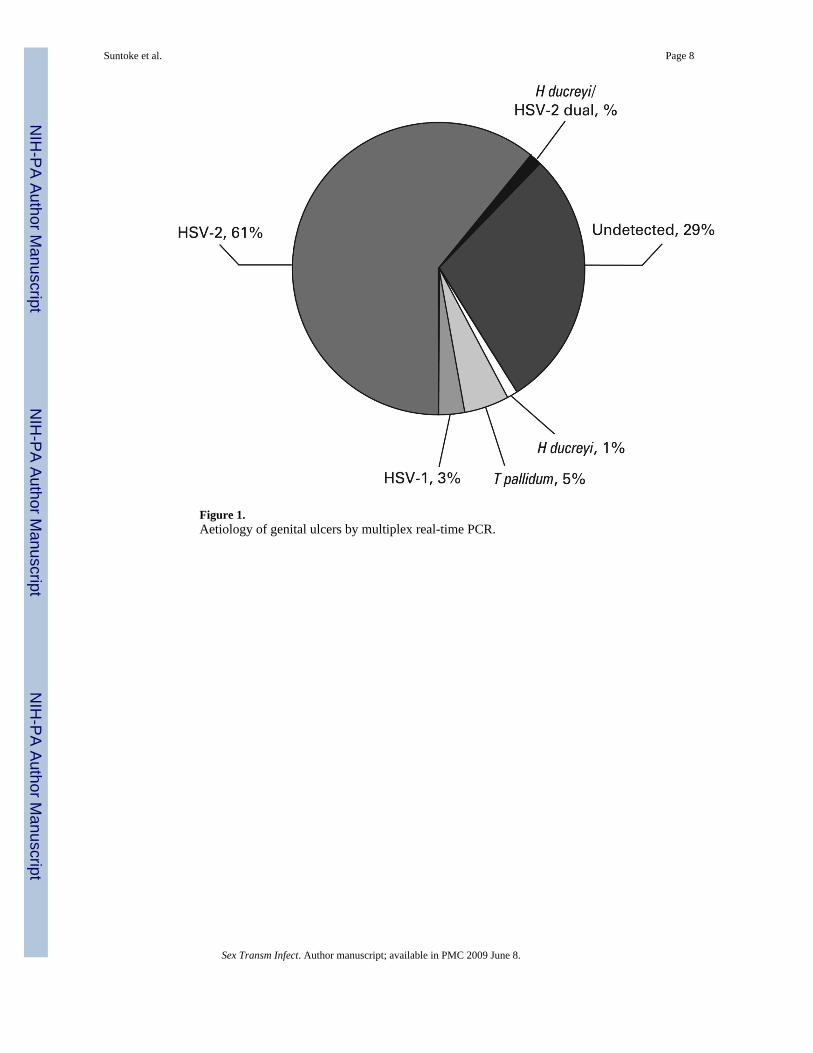
Figure 1.Aetiology of genital ulcers by multiplex real-time PCR.
Suntoke et al. Page 8
Sex Transm Infect. Author manuscript; available in PMC 2009 June 8.
NIH
-PA Author Manuscript
NIH
-PA Author Manuscript
NIH
-PA Author Manuscript

NIH
-PA Author Manuscript
NIH
-PA Author Manuscript
NIH
-PA Author Manuscript
Suntoke et al. Page 9Ta
ble
1Pa
tient
dem
ogra
phic
s an
d di
strib
utio
n of
Hae
mop
hilu
s duc
reyi
(HD
), Tr
epon
ema
palli
dum
(TP)
, her
pes
sim
plex
viru
s ty
pe 1
(HSV
-1)
and
HSV
-2 b
y m
ultip
lex
real
-tim
e PC
R
Gen
ital u
lcer
dis
ease
aet
iolo
gy b
y re
al-ti
me
PCR
HD
n (%
)T
Pn
(%)
HSV
-1n
(%)
HSV
-2n
(%)
HD
/H
SV-2
n (%
)N
Dn
(%)
Tota
l (n=
100)
1 (1
)5
(5)
3 (3
)61
(61)
1 (1
)29
(29)
Gen
der
Mal
e (n
=50)
1 (2
)2
(4)
3 (6
)28
(56)
1 (2
)15
(30)
Fem
ale
(n=5
0)0
(0)
3 (6
)0
(0)
33 (6
6)0
(0)
14 (2
8)
Age
(y)
19–
29 (n
=51)
0 (0
)3
(6)
0 (0
)33
(65)
1 (2
)14
(27)
30–
39 (n
=31)
0 (0
)1
(3)
0 (0
)20
(65)
0 (0
)10
(32)
40+
(n=1
5)0
(0)
0 (0
)3
(20)
7 (4
7)0
(0)
5 (3
3)
Not
kno
wn
(n=3
)1
(33)
1 (3
3)0
(0)
1 (3
3)0
(0)
0 (0
)
HIV
sero
stat
us
Neg
ativ
e (n
=57)
1 (2
)4
(7)
2 (4
)29
(51)
1 (2
)20
(35)
Pos
itive
(n=4
3)0
(0)
1 (2
)1
(2)
32 (7
4)0
(0)
9 (2
1)
ND
, pat
hoge
n no
t det
ecte
d by
real
-tim
e PC
R.
Sex Transm Infect. Author manuscript; available in PMC 2009 June 8.

NIH
-PA Author Manuscript
NIH
-PA Author Manuscript
NIH
-PA Author Manuscript
Suntoke et al. Page 10Ta
ble
2C
ompa
rison
of m
ultip
lex
real
-tim
e PC
R w
ith sy
phili
s, H
SV-1
and
HSV
-2 se
rolo
gy
Syph
ilis s
erol
ogy
HSV
-1 se
rolo
gyH
SV-2
sero
logy
TR
UST
/TPP
AE
uroi
mm
une
Kal
on
+ (%
)−
(%)
Tot
al (%
)+
(%)
− (%
)T
otal
(%)
+ (%
)−
(%)
Tot
al (%
)
PCR
+5
(45)
0 (0
)5
(5)
3 (4
)0
(0)
3 (3
)50
(68)
9 (4
1)59
(62)
PCR−
6 (5
5)83
(100
)89
(95)
77 (9
6)11
(100
)88
(97)
23 (3
2)13
(59)
36 (3
8)
Tota
l11
(100
)83
(100
)94
(100
)80
(100
)11
(100
)91
(100
)73
(100
)22
(100
)95
(100
)
HSV
, her
pes s
impl
ex v
irus;
TPP
A, T
repo
nem
a pa
llidu
m p
artic
le a
gglu
tinat
ion
assa
y.
Sex Transm Infect. Author manuscript; available in PMC 2009 June 8.