Evidence that the nucleus accumbens shell, ventral pallidum and lateral hypothalamus are components...
7
Behavioural Brain Research 226 (2012) 548–554 Contents lists available at SciVerse ScienceDirect Behavioural Brain Research jo u r n al hom epa ge: www.elsevier.com/locate/bbr Research report Evidence that the nucleus accumbens shell, ventral pallidum, and lateral hypothalamus are components of a lateralized feeding circuit Thomas R. Stratford ∗ , David Wirtshafter Laboratory of Integrative Neuroscience, Department of Psychology (m/c 285), University of Illinois at Chicago, 1007 West Harrison Street, Chicago, IL 60607-7137, United States a r t i c l e i n f o Article history: Received 10 September 2011 Received in revised form 6 October 2011 Accepted 10 October 2011 Available online 15 October 2011 Keywords: Food intake Eating Ingestive behavior Ventral striatum NeuN a b s t r a c t Pronounced feeding can be elicited by injections of the GABA A agonist muscimol into the medial shell region of the nucleus accumbens (AcbSh). This region of AcbSh has been shown to project to both the lateral hypothalamus (LH) and the medial ventral pallidum (VPm). The current study examined the effects of unilateral LH or VPm lesions on the ingestive responses induced by injections of muscimol into the AcbSh on either the same or the opposite side of the brain. We found that lesions of either of these structures drastically attenuated feeding induced from the ipsilateral, as compared to the contralateral, AcbSh. The “ipsilateral/contralateral disruption design” employed here virtually rules out the possibility that the suppressive effects of the lesions were nonspecific and suggests that the VPm and LH play essential roles in mediating the ingestive effects of inactivation of the AcbSh. © 2011 Elsevier B.V. All rights reserved. 1. Introduction Although the nucleus accumbens is often assumed to exert a generalized influence on motivational or reward mechanisms, studies have demonstrated that pharmacological inactivation of the shell subregion of the accumbens (AcbSh) with GABA agonists or glutamate receptor antagonists induces a large, and remarkably specific, increase in feeding behavior in satiated rats [1–4]. The active site in the ventral striatum at which these drugs elicit feed- ing has been localized to the rostral medial AcbSh [1,5–7] and the fact that large, behaviorally specific, increases in food intake can be elicited by blocking the actions of endogenous glutamate, or by increasing levels of endogenous GABA [1], indicates that the AcbSh is likely to play a role in the normal physiological control of feeding behavior. The importance of the AcbSh in the physiological control of feeding behavior is underscored by the fact that changes in food intake have also been observed after local injections of a variety of feeding related compounds including nociceptin, cocaine- and amphetamine-related transcript protein, melanin-concentrating hormone, orexin, opioids, amylin, and endocannabinoids (see [8] for review). An obviously important line of investigation involves the deter- mination of the functional circuitry through which the AcbSh mediates food intake. The AcbSh sends substantial projections to at least two brain regions known to play a role in the control of ∗ Corresponding author. Tel.: +1 3129965248; fax: +1 3124134122. E-mail address: [email protected] (T.R. Stratford). feeding; the lateral hypothalamus (LH) and medial ventral pallidum (VPm) [9–13]. While the role played by the VP in the expression of feeding induced by manipulations of amino acid-coded circuits in the AcbSh has never been investigated, the available data sug- gest the occurrence of a functional relationship between the AcbSh and the LH. Thus, feeding induced by inactivation of the AcbSh can be significantly attenuated by acute suppression of LH activity through local drug injections [2,14]. While these findings suggest that the feeding behavior elicited by inhibition of neurons in the AcbSh is dependent on activation of LH neurons, they are, unfor- tunately, open to alternative explanations. It is quite possible, for example, that the LH does not play a direct role in mediating effects obtained from the AcbSh, but that the LH manipulations performed in these studies may have suppressed feeding as a result of some “nonspecific” effect, such as sedation, malaise, alterations in hor- monal or autonomic reactivity, or changes in gustatory or olfactory processing. One way to circumvent these interpretive difficulties is through the use of what could be called an “ipsilateral/contralateral dis- ruption design” (ICD) which allows for an evaluation of whether lesion effects are due to a specific disruption of lateralized cir- cuitry activated by the AcbSh injections. This approach requires that the AcbSh on one side of the brain exerts a larger influence on the ipsilateral LH than on the contralateral LH. The available anatomical data supports this possibility with respect to direct pro- jections, since very few fibers originating in the AcbSh appear to cross the midline to terminate in the contralateral LH [13,15,16]. The uncrossed functional nature of these pathways is further sup- ported by the observation that unilateral injections of muscimol 0166-4328/$ – see front matter © 2011 Elsevier B.V. All rights reserved. doi:10.1016/j.bbr.2011.10.014
Transcript of Evidence that the nucleus accumbens shell, ventral pallidum and lateral hypothalamus are components...
R
Eh
TL
a
ARRAA
KFEIVN
1
astosaifbiiboioahf
mma
0d
Behavioural Brain Research 226 (2012) 548– 554
Contents lists available at SciVerse ScienceDirect
Behavioural Brain Research
jo u r n al hom epa ge: www.elsev ier .com/ locate /bbr
esearch report
vidence that the nucleus accumbens shell, ventral pallidum, and lateralypothalamus are components of a lateralized feeding circuit
homas R. Stratford ∗, David Wirtshafteraboratory of Integrative Neuroscience, Department of Psychology (m/c 285), University of Illinois at Chicago, 1007 West Harrison Street, Chicago, IL 60607-7137, United States
r t i c l e i n f o
rticle history:eceived 10 September 2011eceived in revised form 6 October 2011ccepted 10 October 2011vailable online 15 October 2011
a b s t r a c t
Pronounced feeding can be elicited by injections of the GABAA agonist muscimol into the medial shellregion of the nucleus accumbens (AcbSh). This region of AcbSh has been shown to project to both thelateral hypothalamus (LH) and the medial ventral pallidum (VPm). The current study examined the effectsof unilateral LH or VPm lesions on the ingestive responses induced by injections of muscimol into theAcbSh on either the same or the opposite side of the brain. We found that lesions of either of these
eywords:ood intakeatingngestive behaviorentral striatum
structures drastically attenuated feeding induced from the ipsilateral, as compared to the contralateral,AcbSh. The “ipsilateral/contralateral disruption design” employed here virtually rules out the possibilitythat the suppressive effects of the lesions were nonspecific and suggests that the VPm and LH playessential roles in mediating the ingestive effects of inactivation of the AcbSh.
© 2011 Elsevier B.V. All rights reserved.
euN. Introduction
Although the nucleus accumbens is often assumed to exert generalized influence on motivational or reward mechanisms,tudies have demonstrated that pharmacological inactivation ofhe shell subregion of the accumbens (AcbSh) with GABA agonistsr glutamate receptor antagonists induces a large, and remarkablypecific, increase in feeding behavior in satiated rats [1–4]. Thective site in the ventral striatum at which these drugs elicit feed-ng has been localized to the rostral medial AcbSh [1,5–7] and theact that large, behaviorally specific, increases in food intake cane elicited by blocking the actions of endogenous glutamate, or by
ncreasing levels of endogenous GABA [1], indicates that the AcbShs likely to play a role in the normal physiological control of feedingehavior. The importance of the AcbSh in the physiological controlf feeding behavior is underscored by the fact that changes in foodntake have also been observed after local injections of a varietyf feeding related compounds including nociceptin, cocaine- andmphetamine-related transcript protein, melanin-concentratingormone, orexin, opioids, amylin, and endocannabinoids (see [8]
or review).An obviously important line of investigation involves the deter-
ination of the functional circuitry through which the AcbShediates food intake. The AcbSh sends substantial projections to
t least two brain regions known to play a role in the control of
∗ Corresponding author. Tel.: +1 3129965248; fax: +1 3124134122.E-mail address: [email protected] (T.R. Stratford).
166-4328/$ – see front matter © 2011 Elsevier B.V. All rights reserved.oi:10.1016/j.bbr.2011.10.014
feeding; the lateral hypothalamus (LH) and medial ventral pallidum(VPm) [9–13]. While the role played by the VP in the expressionof feeding induced by manipulations of amino acid-coded circuitsin the AcbSh has never been investigated, the available data sug-gest the occurrence of a functional relationship between the AcbShand the LH. Thus, feeding induced by inactivation of the AcbShcan be significantly attenuated by acute suppression of LH activitythrough local drug injections [2,14]. While these findings suggestthat the feeding behavior elicited by inhibition of neurons in theAcbSh is dependent on activation of LH neurons, they are, unfor-tunately, open to alternative explanations. It is quite possible, forexample, that the LH does not play a direct role in mediating effectsobtained from the AcbSh, but that the LH manipulations performedin these studies may have suppressed feeding as a result of some“nonspecific” effect, such as sedation, malaise, alterations in hor-monal or autonomic reactivity, or changes in gustatory or olfactoryprocessing.
One way to circumvent these interpretive difficulties is throughthe use of what could be called an “ipsilateral/contralateral dis-ruption design” (ICD) which allows for an evaluation of whetherlesion effects are due to a specific disruption of lateralized cir-cuitry activated by the AcbSh injections. This approach requiresthat the AcbSh on one side of the brain exerts a larger influenceon the ipsilateral LH than on the contralateral LH. The availableanatomical data supports this possibility with respect to direct pro-
jections, since very few fibers originating in the AcbSh appear tocross the midline to terminate in the contralateral LH [13,15,16].The uncrossed functional nature of these pathways is further sup-ported by the observation that unilateral injections of muscimol
ioural
icimusdlfaelei
iiiiaitIri
maiucla
2
2
2u(nLC
2
nwfplmSihtuRaursps
2
rv
T.R. Stratford, D. Wirtshafter / Behav
nto the AcbSh produce a much larger increase in the number ofells expressing the nuclear protein Fos, a marker of neural activ-ty [17,18], in the ipsilateral LH than the contralateral LH [19]. If
uscimol-induced feeding is mediated through a predominantlynilateral activation of the LH, one would expect that lesions of thistructure on the same side as the muscimol injection would pro-uce a much greater disruption of the feeding response than would
esions on the contralateral side. Conversely, if AcbSh-mediatedood intake were suppressed because a particular lesion elicited
“nonspecific” response, such as sedation or malaise, one wouldxpect that the “nonspecific” response would be elicited in a simi-ar manner by lesions on either side of the brain and therefore, thatquivalent reductions in food intake would be observed after eitherpsilateral or contralateral lesions.
The AcbSh also projects heavily to the VPm and both lesion-ng and drug microinjection studies strongly implicate this regionn the control of feeding [12,20,21]. Considerably fewer Fos-mmunoreactive cells are seen in the VPm than the LH afterntra-AcbSh muscimol injections, but those cells that are presentre located primarily ipsilateral to the injection site [19]. Anatom-cal data also suggest that the direct projection from the AcbSh tohe VPm is strongly unilateral [16]. These findings suggest that theCD experimental design may again be useful for evaluating theole of the VPm in mediating the feeding induced by GABAergicnactivation of the AcbSh.
In light of these considerations, we conducted three experi-ents to evaluate the connections through which the AcbSh is
ble to influence food intake. First, we examined feeding follow-ng unilateral injections of muscimol into the AcbSh, to verify thatnilateral inactivation of the AcbSh is able to induce feeding in oururrent testing situation. We then examined the effects of unilateralesions of the LH and of the VPm using the ICD approach outlinedbove.
. Methods
.1. Subjects
Male Sprague-Dawley rats (Charles Rivers, Wilmington, MA) weighing between80 and 337 g at the time of surgery served as subjects. The rats were housed individ-ally in plastic cages on a 12 h light:12 h dark cycle at a constant room temperature∼21 ◦C) with food (Harlan Teklad) and tap water available ad libitum, except asoted below. All experiments conformed to the NIH Guide for the Care and Use ofaboratory Animals and were approved by the Institutional Animal Care and Useommittee.
.2. Surgery
Surgery was performed using standard, aseptic, flat-skull stereotaxic tech-iques. Subjects in Experiment 1 received only bilateral AcbSh cannula implants,hereas subjects in Experiments 2 and 3 received unilateral excitotoxic lesions
ollowed immediately by cannula implants. Rats were anesthetized with sodiumentobarbital (60 mg/kg). In order to produce excitotoxic lesions, a 28-gauge stain-
ess steel injector was lowered into either the VPm (AP: −0.1, LM: 1.8, DV: −8.8;m from bregma) or LH (AP: −2.5, LM: 1.8, DV: −9.1) and ibotenic acid (Sigma,
aint Louis, MO; 10 �g/�l) was infused at a rate of 0.25 �l/min. The total amount ofbotenic acid infused was 5.0 �g into the VPm and 15 �g into the LH. Approximatelyalf the animals were lesioned on the left and the remainder on the right. The injec-or was allowed to remain in place for 5 min after the infusion to minimize diffusionp the injector path. Bilateral 22-gauge stainless steel guide cannulae (Plastics One,oanoke, VA), aimed so as to terminate 2.0 mm dorsal to the AcbSh, were implantedt coordinates of AP: 1.6, LM: ±0.9, DV: −6.1. The guide cannulae were held in placesing stainless steel screws and denture lining material and a stainless steel obtu-ator was inserted into the lumen of each cannula to help maintain patency. Afterurgery, the rats received an injection of carprofen (5 mg/kg, sc) to help alleviateostoperative pain. Each rat was allowed to recover for at least 7 days before thetart of behavioral testing.
.3. Intracerebral injections
During the intracerebral injections, the rats were gently restrained, the obtu-ators removed, and 28-gauge injection cannulae, extending 2.0 mm beyond theentral tip of the guide, were inserted into the guide cannulae. All injections were
Brain Research 226 (2012) 548– 554 549
made in a volume of 0.5 �l at a rate of 0.33 �l/min using motor driven microsy-ringes attached to the injection cannulae through lengths of polyethylene tubing.Injections on the two sides were made simultaneously. After the infusion, the injec-tion cannulae were left in place for an additional 30 s in order to minimize leakageup the cannula track, at which time the obturators were replaced and the animalsplaced in the testing cages.
2.4. Feeding measurements
Test chambers (pelletometers) consisted of plastic shoebox cages (L 43 cm × W22 cm × H 21 cm) equipped with metal troughs into which individual 45 mg foodpellets (Precision Dustless Pellets, Bio-Serve, Frenchtown, NJ) could be depositedby automated dispensers (Med-Associates, St. Albans, VT) under computer control.Removal of the pellet unblocked an infrared photobeam; the time of these eventswas recorded to the nearest 0.1 s, after which another pellet was delivered. Testsessions lasted for 120 min after introduction of the animal to the pelletometer. Inorder to acclimate the rats to the apparatus and procedure, they were placed in thepelletometers and allowed to eat for 120 min on at least 5 days before the start oftesting. The rats were mildly food-deprived for the first one or two acclimation trialsand non-deprived for the remainder. In order to accustom them to the injection pro-cedure, each rat, on the final acclimation day, received bilateral 0.25 �l intracerebralinjections of sterile 0.15 M saline.
2.5. Experimental procedure
2.5.1. Experiment 1. Injections in unlesioned animalsSeven animals received two feeding tests that were presented in a counterbal-
anced order and separated from each other by at least 48 h. On one test day, animalsreceived simultaneous bilateral injections of sterile saline and on the other an injec-tion of saline into the AcbSh on one side of the brain and an injection of 100 ng ofmuscimol into the AcbSh on the other side.
2.5.2. Experiment 2. Unilateral muscimol injections in rats with unilateral lesionsof the LH
Eleven animals with unilateral LH lesions were studied. Each rat received thefollowing three combinations of intracerebral injections in counterbalanced order:(1) bilateral saline injections, (2) injections of 100 ng of muscimol on the intact sideand saline on the lesioned side, (3) injections of 100 ng of muscimol on the lesionedside and saline on the intact side. Tests were separated from each other by at least48 h.
2.5.3. Experiment 3. Unilateral muscimol injections in rats with unilateral lesionsof the VPm
Fifteen rats with unilateral VPm lesions were studied who received bilateralsaline and unilateral muscimol injections as described above under Experiment 2.
2.6. Histology
When behavioral testing was completed, each of the rats was deeply anes-thetized with sodium pentobarbital (100 mg/kg) and perfused transcardially with50 ml of a 0.15 M saline solution followed immediately by 200 ml of a 10% bufferedformalin solution at pH 6.5 and then 300 ml of a 10% buffered formalin solution atpH 9.0 [22]. The brains were removed, postfixed for 1 h and then stored in phos-phate buffered saline (PBS) with 20% sucrose at 4 ◦C for at least 2 days, after whichthey were frozen and 35-�m-thick coronal sections taken throughout the extentof the AcbSh and the lesion sites. Sections through the injection sites were stainedwith cresyl violet, and those through the lesions were processed for the immuno-histochemical detection of neuronal nuclear protein (NeuN), a sensitive marker ofundamaged neurons [23] which we have found to be particularly useful in evaluat-ing excitotoxic lesions. Briefly, the sections were rinsed in PBS and incubated on arotary shaker table for 24 h at 4 ◦C in a monoclonal mouse anti-NeuN primary anti-body (diluted 1:20,000 with 0.01 M PBS containing 0.2% Triton X-100 and 4% normalhorse serum; Oncogene Research Products, La Jolla, CA). The sections were rinsed inPBS and incubated in the biotinylated horse anti-mouse secondary antibody (VectorLaboratories, Burlingame, CA, diluted 1:200 with PBS containing 4% normal horseserum) for 60 min at room temperature. They were then rinsed in PBS (3 × 10 min),and incubated in the avidin–biotin complex solution (Vector) for 60 min. Follow-ing another series of rinses in PBS, the peroxidase was visualized by incubatingthe tissue for 5 min in a nickel-enhanced chromogen solution obtained from a Vec-tor 3,3′-diaminobenzidine tetrahydrochloride peroxidase substrate kit. The sectionswere mounted on chrome-alum coated slides, air-dried, cleared in xylene, and cov-
erslipped with Eukitt mounting medium. The lesion boundaries were mapped andthe AcbSh injection sites were examined for placement with reference to the atlasof Paxinos and Watson [24]. Two animals from Experiment 2 and four from Exper-iment 3 were removed from the studies due to misplacement of the lesions or toextensive spread of damage along the cannula tracks.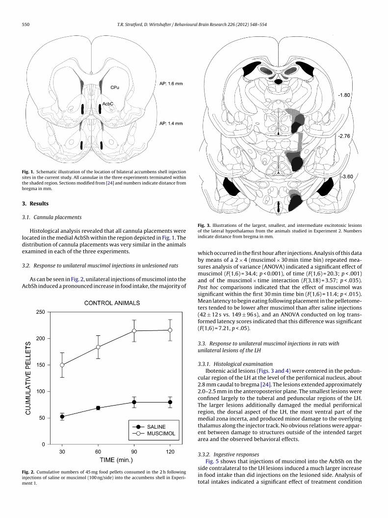
550 T.R. Stratford, D. Wirtshafter / Behavioural Brain Research 226 (2012) 548– 554
Fig. 1. Schematic illustration of the location of bilateral accumbens shell injectionstb
3
3
lde
3
A
Fim
ites in the current study. All cannulae in the three experiments terminated withinhe shaded region. Sections modified from [24] and numbers indicate distance fromregma in mm.
. Results
.1. Cannula placements
Histological analysis revealed that all cannula placements wereocated in the medial AcbSh within the region depicted in Fig. 1. Theistribution of cannula placements was very similar in the animalsxamined in each of the three experiments.
.2. Response to unilateral muscimol injections in unlesioned rats
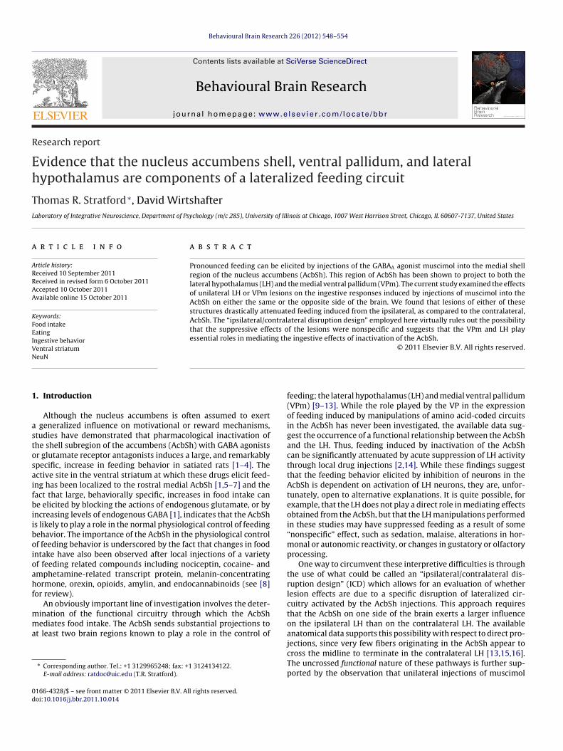
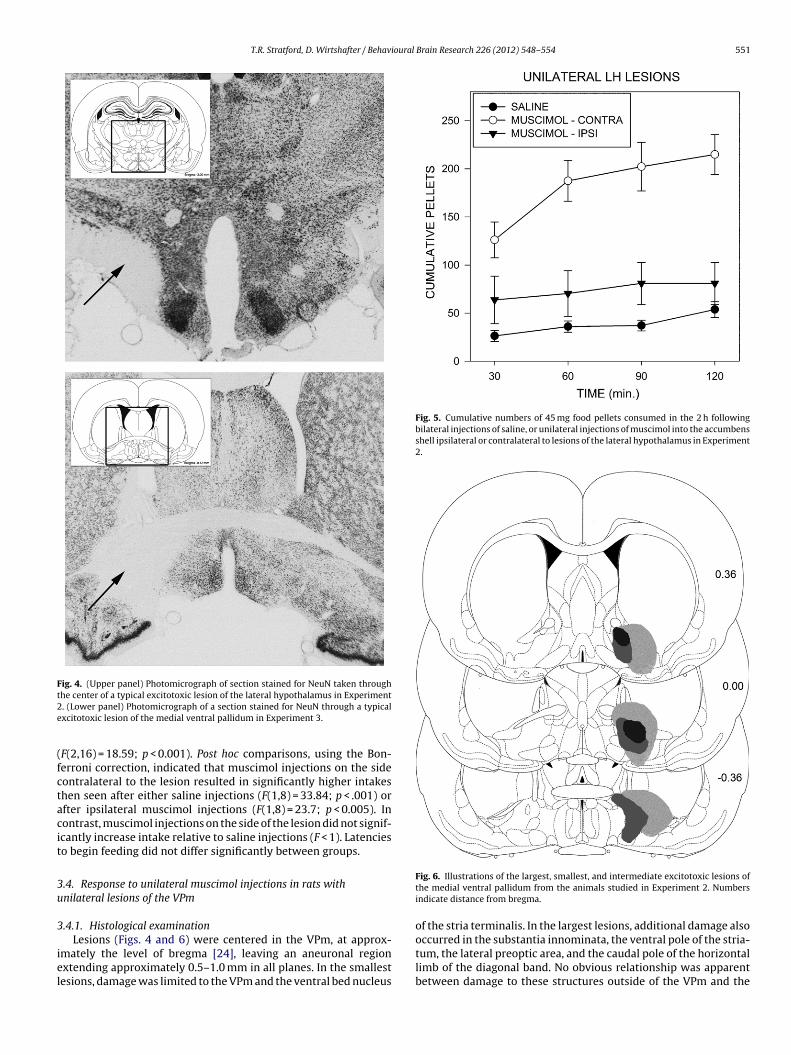
As can be seen in Fig. 2, unilateral injections of muscimol into thecbSh induced a pronounced increase in food intake, the majority of
ig. 2. Cumulative numbers of 45 mg food pellets consumed in the 2 h followingnjections of saline or muscimol (100 ng/side) into the accumbens shell in Experi-
ent 1.
Fig. 3. Illustrations of the largest, smallest, and intermediate excitotoxic lesionsof the lateral hypothalamus from the animals studied in Experiment 2. Numbersindicate distance from bregma in mm.
which occurred in the first hour after injections. Analysis of this databy means of a 2 × 4 (muscimol × 30 min time bin) repeated mea-sures analysis of variance (ANOVA) indicated a significant effect ofmuscimol (F(1,6) = 34.4; p < 0.001), of time (F(1,6) = 20.3; p < .001)and of the muscimol × time interaction (F(3,18) = 3.57; p < .035).Post hoc comparisons indicated that the effect of muscimol wassignificant within the first 30 min time bin (F(1,6) = 11.4; p < .015).Mean latency to begin eating following placement in the pelletome-ters tended to be lower after muscimol than after saline injections(42 ± 12 s vs. 149 ± 96 s), and an ANOVA conducted on log trans-formed latency scores indicated that this difference was significant(F(1,6) = 7.21, p < .05).
3.3. Response to unilateral muscimol injections in rats withunilateral lesions of the LH
3.3.1. Histological examinationIbotenic acid lesions (Figs. 3 and 4) were centered in the pedun-
cular region of the LH at the level of the perifornical nucleus, about2.8 mm caudal to bregma [24]. The lesions extended approximately2.0–2.5 mm in the anteroposterior plane. The smallest lesions wereconfined largely to the tuberal and peduncular regions of the LH.The larger lesions additionally damaged the medial perifornicalregion, the dorsal aspect of the LH, the most ventral part of themedial zona incerta, and produced minor damage to the overlyingthalamus along the injector track. No obvious relations were appar-ent between damage to structures outside of the intended targetarea and the observed behavioral effects.
3.3.2. Ingestive responses
Fig. 5 shows that injections of muscimol into the AcbSh on theside contralateral to the LH lesions induced a much larger increasein food intake than did injections on the lesioned side. Analysis oftotal intakes indicated a significant effect of treatment condition
T.R. Stratford, D. Wirtshafter / Behavioural Brain Research 226 (2012) 548– 554 551
Fig. 4. (Upper panel) Photomicrograph of section stained for NeuN taken throught2e
(fctacit
3u
3
iel
Fig. 5. Cumulative numbers of 45 mg food pellets consumed in the 2 h followingbilateral injections of saline, or unilateral injections of muscimol into the accumbensshell ipsilateral or contralateral to lesions of the lateral hypothalamus in Experiment2.
he center of a typical excitotoxic lesion of the lateral hypothalamus in Experiment. (Lower panel) Photomicrograph of a section stained for NeuN through a typicalxcitotoxic lesion of the medial ventral pallidum in Experiment 3.
F(2,16) = 18.59; p < 0.001). Post hoc comparisons, using the Bon-erroni correction, indicated that muscimol injections on the sideontralateral to the lesion resulted in significantly higher intakeshen seen after either saline injections (F(1,8) = 33.84; p < .001) orfter ipsilateral muscimol injections (F(1,8) = 23.7; p < 0.005). Inontrast, muscimol injections on the side of the lesion did not signif-cantly increase intake relative to saline injections (F < 1). Latencieso begin feeding did not differ significantly between groups.
.4. Response to unilateral muscimol injections in rats withnilateral lesions of the VPm
.4.1. Histological examination
Lesions (Figs. 4 and 6) were centered in the VPm, at approx-mately the level of bregma [24], leaving an aneuronal regionxtending approximately 0.5–1.0 mm in all planes. In the smallestesions, damage was limited to the VPm and the ventral bed nucleus
Fig. 6. Illustrations of the largest, smallest, and intermediate excitotoxic lesions ofthe medial ventral pallidum from the animals studied in Experiment 2. Numbersindicate distance from bregma.
of the stria terminalis. In the largest lesions, additional damage also
occurred in the substantia innominata, the ventral pole of the stria-tum, the lateral preoptic area, and the caudal pole of the horizontallimb of the diagonal band. No obvious relationship was apparentbetween damage to these structures outside of the VPm and the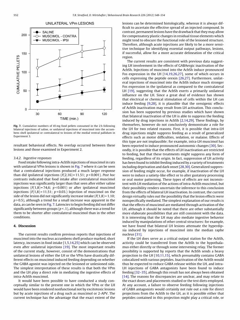
552 T.R. Stratford, D. Wirtshafter / Behavioural
Fig. 7. Cumulative numbers of 45 mg food pellets consumed in the 2 h followingbbE
rl
3
wttciiispdstc
4
mleouftTai
cwbc
ilateral injections of saline, or unilateral injections of muscimol into the accum-ens shell ipsilateral or contralateral to lesions of the medial ventral pallidum inxperiment 3.
esultant behavioral effects. No overlap occurred between theseesions and those examined in Experiment 2.
.4.2. Ingestive responsesFood intake following intra-AcbSh injections of muscimol in rats
ith unilateral VPm lesions is shown in Fig. 7 where it can be seenhat a contralateral injections produced a much larger responsehan did ipsilateral injections (F(2,16) = 11.51; p < 0.001). Post hocontrasts indicated that food intake after contralateral muscimolnjections was significantly larger than that seen after either salinenjections (F(1,8) = 74,6; p < 0.001) or after ipsilateral muscimolnjections (F(1,8) = 11.51; p < 0.03.) Injection of muscimol on theide of the lesion did not significantly increase intake (F(1,8) = 1.10;
> 0.5), although a trend for a small increase was apparent in theata, as can be seen in Fig. 7. Latencies to begin feeding did not differignificantly between groups (p < .1), although there was a trend forhem to be shorter after contralateral muscimol than in the otheronditions.
. Discussion
The current results confirm previous reports that injections ofuscimol into the nucleus accumbens shell produce marked, short
atency, increases in food intake [1,5,14,25] which can be observedven after unilateral injections [19]. The most important resultsf the current study, however, consist of the demonstrations thatnilateral lesions of either the LH or the VPm have drastically dif-erent effects on muscimol induced feeding depending on whetherhe GABA agonist was injected on the lesioned or unlesioned side.he simplest interpretation of these results is that both the VPmnd the LH play a direct role in mediating the ingestive effects ofntra-AcbSh muscimol.
It would have been possible to have conducted a study con-
eptually similar to the present one in which the VPm or the LHould have been rendered nonfunctional not by excitotoxic lesionsut by acute injections of a drug such as muscimol or 2-APV. Theurrent technique has the advantage that the exact extent of the
Brain Research 226 (2012) 548– 554
lesions can be determined histologically, whereas it is always dif-ficult to ascertain the effective spread of an injected compound. Incontrast, permanent lesions have the drawback that they may allowfor compensatory plastic changes in residual tissue elements whichmight tend to obscure the functional role of the lesioned structure.Therefore, although acute injections are likely to be a more sensi-tive technique for identifying essential output pathways, lesions,if successful, allow for a more accurate delineation of the criticalregion.
The current results are consistent with previous data suggest-ing LH involvement in the effects of GABAergic inactivation of theAcbSh. Injections of muscimol into the AcbSh induce pronouncedFos expression in the LH [14,19,26,27], some of which occurs incells expressing the peptide orexin [26,27]. Furthermore, unilat-eral injections of muscimol into the AcbSh induce much strongerFos expression in the ipsilateral as compared to the contralateralLH [19], suggesting that the AcbSh exerts a primarily unilateralinfluence on the LH. Since a great deal of evidence has shownthat electrical or chemical stimulation of cells within the LH caninduce feeding [9,28], it is plausible that the orexigenic effectsof AcbSh inactivation may result from LH activation. This conclu-sion has been supported by previous studies which have shownthat bilateral inactivation of the LH is able to suppress the feedinginduced by drug injections in AcbSh [2,14,29]. These findings, bythemselves, however do not conclusively demonstrate a role forthe LH for two related reasons. First, it is possible that intra-LHdrug injections might suppress feeding as a result of generalizedeffects such as motor difficulties, sedation, or malaise. Effects ofthis type are not implausible; for example, intra-LH muscimol hasbeen reported to induce pronounced autonomic changes [30]. Sec-ondly, it is possible that the effects of LH inactivation are restrictedto feeding, but that these treatments might suppress any form offeeding, regardless of its origin. In fact, suppression of LH activityhas been found to inhibit feeding induced by a variety of treatmentsincluding deprivation and dark onset [28,30]. Generalized suppres-sion of feeding might occur, for example, if inactivation of the LHwere to induce a satiety-like effect or to alter gustatory processingor oral motor patterning. These types of effects are not inconsis-tent with LH mediation of the actions of intra-AcbSh muscimol, buttheir possibility renders uncertain the inference to this conclusionfrom the effects of bilateral LH inactivation. In contrast, the currentdesign virtually rules out the possibility that the lesion effects werenonspecifically mediated. The simplest explanation of our results isthat the effects of muscimol are mediated through activation of theLH, although it should be noted that there are other substantiallymore elaborate possibilities that are still consistent with the data.It is interesting that the LH may also mediate ingestive behaviorinduced by manipulations of other central structures: for example,we have found that bilateral LH lesions attenuate the hyperdip-sia induced by injections of muscimol into the median raphenucleus [31].
If the LH does serve as a critical output station for the AcbSh,activity could be transferred from the AcbSh to the hypothala-mus either directly or through some intervening relay. The formerpossibility is supported by reports that the AcbSh sends a heavyprojection to the LH [10,11,15], which presumably contains GABAcolocalized with various peptides. Inactivation of the AcbSh wouldthus be expected to reduce GABA release within the LH, and intra-LH injections of GABA antagonists have been found to inducefeeding [32–35], although this result has not always been obtained[14]. The reasons for discrepancies are unclear, and may relate tothe exact doses and placements studied or the test diets employed.
At any account, a failure to observe feeding following injectionsof GABA antagonists would certainly not rule out a role for directprojections from the AcbSh to the LH, as it is possible either thatpeptides contained in this projection might play a critical role, or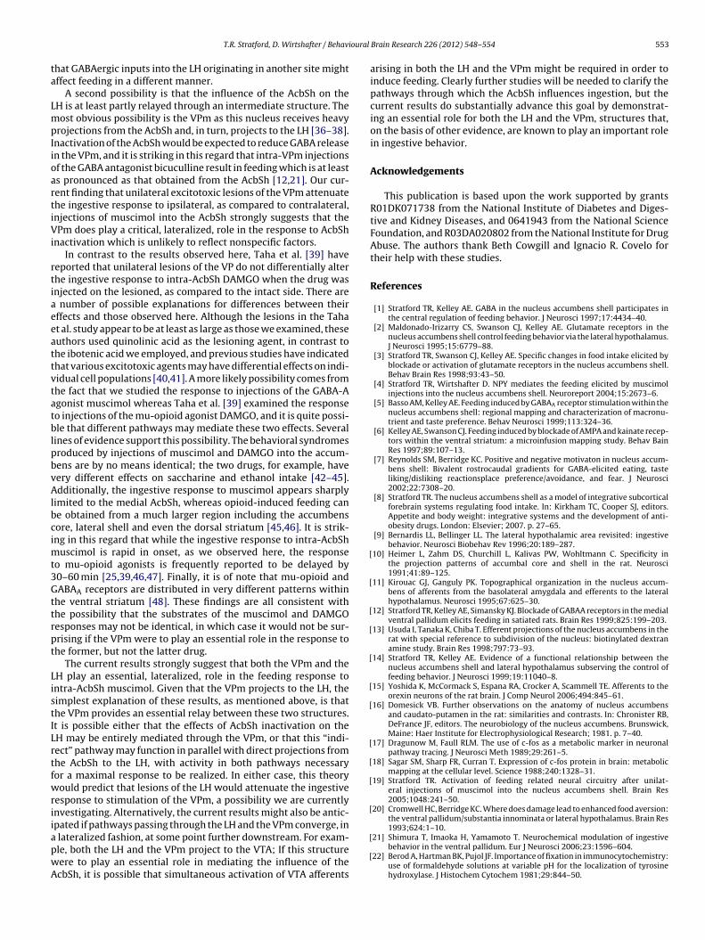
ioural
ta
LmpIioartiVi
rtiaeeattvtatblpbvAlbcimt3Gttrpt
ListILrtfwriiapwA
[
[
[
[
[
[
[
[
[
[
[
T.R. Stratford, D. Wirtshafter / Behav
hat GABAergic inputs into the LH originating in another site mightffect feeding in a different manner.
A second possibility is that the influence of the AcbSh on theH is at least partly relayed through an intermediate structure. Theost obvious possibility is the VPm as this nucleus receives heavy
rojections from the AcbSh and, in turn, projects to the LH [36–38].nactivation of the AcbSh would be expected to reduce GABA releasen the VPm, and it is striking in this regard that intra-VPm injectionsf the GABA antagonist bicuculline result in feeding which is at leasts pronounced as that obtained from the AcbSh [12,21]. Our cur-ent finding that unilateral excitotoxic lesions of the VPm attenuatehe ingestive response to ipsilateral, as compared to contralateral,njections of muscimol into the AcbSh strongly suggests that thePm does play a critical, lateralized, role in the response to AcbSh
nactivation which is unlikely to reflect nonspecific factors.In contrast to the results observed here, Taha et al. [39] have
eported that unilateral lesions of the VP do not differentially alterhe ingestive response to intra-AcbSh DAMGO when the drug wasnjected on the lesioned, as compared to the intact side. There are
number of possible explanations for differences between theirffects and those observed here. Although the lesions in the Tahat al. study appear to be at least as large as those we examined, theseuthors used quinolinic acid as the lesioning agent, in contrast tohe ibotenic acid we employed, and previous studies have indicatedhat various excitotoxic agents may have differential effects on indi-idual cell populations [40,41]. A more likely possibility comes fromhe fact that we studied the response to injections of the GABA-Agonist muscimol whereas Taha et al. [39] examined the responseo injections of the mu-opioid agonist DAMGO, and it is quite possi-le that different pathways may mediate these two effects. Several
ines of evidence support this possibility. The behavioral syndromesroduced by injections of muscimol and DAMGO into the accum-ens are by no means identical; the two drugs, for example, haveery different effects on saccharine and ethanol intake [42–45].dditionally, the ingestive response to muscimol appears sharply
imited to the medial AcbSh, whereas opioid-induced feeding cane obtained from a much larger region including the accumbensore, lateral shell and even the dorsal striatum [45,46]. It is strik-ng in this regard that while the ingestive response to intra-AcbSh
uscimol is rapid in onset, as we observed here, the responseo mu-opioid agonists is frequently reported to be delayed by0–60 min [25,39,46,47]. Finally, it is of note that mu-opioid andABAA receptors are distributed in very different patterns within
he ventral striatum [48]. These findings are all consistent withhe possibility that the substrates of the muscimol and DAMGOesponses may not be identical, in which case it would not be sur-rising if the VPm were to play an essential role in the response tohe former, but not the latter drug.
The current results strongly suggest that both the VPm and theH play an essential, lateralized, role in the feeding response tontra-AcbSh muscimol. Given that the VPm projects to the LH, theimplest explanation of these results, as mentioned above, is thathe VPm provides an essential relay between these two structures.t is possible either that the effects of AcbSh inactivation on theH may be entirely mediated through the VPm, or that this “indi-ect” pathway may function in parallel with direct projections fromhe AcbSh to the LH, with activity in both pathways necessaryor a maximal response to be realized. In either case, this theoryould predict that lesions of the LH would attenuate the ingestive
esponse to stimulation of the VPm, a possibility we are currentlynvestigating. Alternatively, the current results might also be antic-pated if pathways passing through the LH and the VPm converge, in
lateralized fashion, at some point further downstream. For exam-le, both the LH and the VPm project to the VTA; If this structureere to play an essential role in mediating the influence of thecbSh, it is possible that simultaneous activation of VTA afferents
[
[
Brain Research 226 (2012) 548– 554 553
arising in both the LH and the VPm might be required in order toinduce feeding. Clearly further studies will be needed to clarify thepathways through which the AcbSh influences ingestion, but thecurrent results do substantially advance this goal by demonstrat-ing an essential role for both the LH and the VPm, structures that,on the basis of other evidence, are known to play an important rolein ingestive behavior.
Acknowledgements
This publication is based upon the work supported by grantsR01DK071738 from the National Institute of Diabetes and Diges-tive and Kidney Diseases, and 0641943 from the National ScienceFoundation, and R03DA020802 from the National Institute for DrugAbuse. The authors thank Beth Cowgill and Ignacio R. Covelo fortheir help with these studies.
References
[1] Stratford TR, Kelley AE. GABA in the nucleus accumbens shell participates inthe central regulation of feeding behavior. J Neurosci 1997;17:4434–40.
[2] Maldonado-Irizarry CS, Swanson CJ, Kelley AE. Glutamate receptors in thenucleus accumbens shell control feeding behavior via the lateral hypothalamus.J Neurosci 1995;15:6779–88.
[3] Stratford TR, Swanson CJ, Kelley AE. Specific changes in food intake elicited byblockade or activation of glutamate receptors in the nucleus accumbens shell.Behav Brain Res 1998;93:43–50.
[4] Stratford TR, Wirtshafter D. NPY mediates the feeding elicited by muscimolinjections into the nucleus accumbens shell. Neuroreport 2004;15:2673–6.
[5] Basso AM, Kelley AE. Feeding induced by GABAA receptor stimulation within thenucleus accumbens shell: regional mapping and characterization of macronu-trient and taste preference. Behav Neurosci 1999;113:324–36.
[6] Kelley AE, Swanson CJ. Feeding induced by blockade of AMPA and kainate recep-tors within the ventral striatum: a microinfusion mapping study. Behav BainRes 1997;89:107–13.
[7] Reynolds SM, Berridge KC. Positive and negative motivaton in nucleus accum-bens shell: Bivalent rostrocaudal gradients for GABA-elicited eating, tasteliking/disliking reactionsplace preference/avoidance, and fear. J Neurosci2002;22:7308–20.
[8] Stratford TR. The nucleus accumbens shell as a model of integrative subcorticalforebrain systems regulating food intake. In: Kirkham TC, Cooper SJ, editors.Appetite and body weight: integrative systems and the development of anti-obesity drugs. London: Elsevier; 2007. p. 27–65.
[9] Bernardis LL, Bellinger LL. The lateral hypothalamic area revisited: ingestivebehavior. Neurosci Biobehav Rev 1996;20:189–287.
10] Heimer L, Zahm DS, Churchill L, Kalivas PW, Wohltmann C. Specificity inthe projection patterns of accumbal core and shell in the rat. Neurosci1991;41:89–125.
11] Kirouac GJ, Ganguly PK. Topographical organization in the nucleus accum-bens of afferents from the basolateral amygdala and efferents to the lateralhypothalamus. Neurosci 1995;67:625–30.
12] Stratford TR, Kelley AE, Simansky KJ. Blockade of GABAA receptors in the medialventral pallidum elicits feeding in satiated rats. Brain Res 1999;825:199–203.
13] Usuda I, Tanaka K, Chiba T. Efferent projections of the nucleus accumbens in therat with special reference to subdivision of the nucleus: biotinylated dextranamine study. Brain Res 1998;797:73–93.
14] Stratford TR, Kelley AE. Evidence of a functional relationship between thenucleus accumbens shell and lateral hypothalamus subserving the control offeeding behavior. J Neurosci 1999;19:11040–8.
15] Yoshida K, McCormack S, Espana RA, Crocker A, Scammell TE. Afferents to theorexin neurons of the rat brain. J Comp Neurol 2006;494:845–61.
16] Domesick VB. Further observations on the anatomy of nucleus accumbensand caudato-putamen in the rat: similarities and contrasts. In: Chronister RB,DeFrance JF, editors. The neurobiology of the nucleus accumbens. Brunswick,Maine: Haer Institute for Electrophysiological Research; 1981. p. 7–40.
17] Dragunow M, Faull RLM. The use of c-fos as a metabolic marker in neuronalpathway tracing. J Neurosci Meth 1989;29:261–5.
18] Sagar SM, Sharp FR, Curran T. Expression of c-fos protein in brain: metabolicmapping at the cellular level. Science 1988;240:1328–31.
19] Stratford TR. Activation of feeding related neural circuitry after unilat-eral injections of muscimol into the nucleus accumbens shell. Brain Res2005;1048:241–50.
20] Cromwell HC, Berridge KC. Where does damage lead to enhanced food aversion:the ventral pallidum/substantia innominata or lateral hypothalamus. Brain Res1993;624:1–10.
21] Shimura T, Imaoka H, Yamamoto T. Neurochemical modulation of ingestivebehavior in the ventral pallidum. Eur J Neurosci 2006;23:1596–604.
22] Berod A, Hartman BK, Pujol JF. Importance of fixation in immunocytochemistry:use of formaldehyde solutions at variable pH for the localization of tyrosinehydroxylase. J Histochem Cytochem 1981;29:844–50.

5 ioural
[
[
[
[
[
[
[
[
[
[
[
[
[
[
[
[
[
[
[
[
[
[
[
[
[
54 T.R. Stratford, D. Wirtshafter / Behav
23] Jongen-Relo AL, Feldon J. Specific neuronal protein: a new tool for histologicalevaluation of excitotoxic lesions. Physiol Behav 2002;76:449–56.
24] Paxinos G, Watson C. The rat brain in stereotaxic coordinates. 6th ed. New York:Academic Press; 2007.
25] Hanlon EC, Baldo BA, Sadeghian K, Kelley AE. Increases in food intake or food-seeking behavior induced by GABAergic, opioid, or dopaminergic stimulation ofthe nucleus accumbens: is it hunger? Psychopharmacology 2004;172:241–7.
26] Zheng H, Corkern M, Stoyanova I, Patterson LM, Tian R, Berthoud HR. Peptidesthat regulate food intake: appetite-inducing accumbens manipulation acti-vates hypothalamic neurons and inhibits POMC neurons. Am J Physiol RegulIntegr Comp Physiol 2003;284:R1436–44.
27] Baldo BA, Gual-Bonilla L, Sijapati K, Danies RA, Landry CF, Kelley AE. Activationof a subpopulation of orexin/hypocretin-containing hypothalamic neurons byGABAA receptor-mediated inhibiton of the nucleus accumbens shell, but notby exposure to a novel environment. Eur J Neurosci 2004;19:376–86.
28] Stanley BG, Willett VL, Donias HW, Dee MG, Duva MA. Lateral hypothalamicNMDA receptors and glutamate as physiological mediators of eating and weightcontrol. Am J Physiol Reg Integr Comp Physiol 1996;270:R443–9.
29] Will MJ, Franzblau EB, Kelley AE. Nucleus accumbens �-opioids regulate intakeof a high-fat diet via activation of a distributed brain network. J Neurosci2003;23:2882–8.
30] Kelly J, Rothstein J, Grossman SP. GABA and hypothalamic feeding systems.I. Topographic analysis of the effects of microinjections of muscimol. PhysiolBehav 1979;23:1123–34.
31] Stratford TR, Wirtshafter D. Forebrain lesions differentially affect drinkingelicited by dipsogenic challenges and injections of muscimol into the medianraphe nucleus. Behav Neurosci 2000;114:760–71.
32] Turenius CL, Htut MM, Prodon DA, Ebersole PL, Ngo PT, Lara RN, et al. GABAA
receptors in the lateral hypothalamus as mediators of satiety and body weightregulation. Brain Res 2009;1262:16–24.
33] Kelly J, Alheid GF, Newberg A, Grossman SP. GABA stimulation and blockadein the hypothalamus and midbrain: Effects on feeding and locomotor activity.Pharmacol Biochem Behav 1977;7:537–41.
34] Turenius CL, Charles JR, Tsai DH, Ebersole PL, Htut MH, Ngo PT, et al. The tuberallateral hypothalamus is a major target for GABAA- but not GABAB-mediatedcontrol of food intake. Brain Res 2009;1283:65–72.
35] Tsujii S, Bray GA. GABA-related feeding control in genetically obese rats. BrainRes 1991;540:48–54.
[
Brain Research 226 (2012) 548– 554
36] Groenewegen HJ, Berendse HW, Haber SN. Organization of the output of theventral striatopallidal system in the rat: ventral pallidal efferents. Neuroscience1993;57:113–42.
37] Zahm DS, Heimer L. Two transpallidal pathways orginating in the rat nucleusaccumbens. J Comp Neurol 2011;302:437–46.
38] Haber SN, Groenewegen HJ, Grove EA, Nauta WJ. Efferent connections of theventral pallidum: evidence of a dual striato pallidofugal pathway. J Comp Neu-rol 1985;235:322–35.
39] Taha SA, Katsuura Y, Noorvaash D, Seroussi A, Fields HL. Convergent, not serial,striatal and pallidal circuits regulate opioid-induced food intake. Neuroscience2009;161:718–33.
40] Inglis WL, Semba K. Discriminable excitotoxic effects of ibotenic acid, AMPA,NMDA and quinolinic acid in the rat laterodorsal tegmental nucleus. Brain Res2011;755:17–27.
41] Dunnett SB, Whishaw IQ, Jones GH, Bunch ST. Behavioural, biochemical andhistochemical effects of different neurotoxic amino acids injected into nucleusbasalis magnocellularis of rats. Neurosci 1987;20:653–69.
42] Stratford, TR, Wirtshafter, D. Activation of GABA-A receptors in the nucleusaccumbens shell elicits opposite effects on consumption of sucrose and sac-charin solutions. Neuroscience Meeting Planner 2007: Program No. 630.5.
43] Stratford TR, Wirtshafter D. Opposite effects on the ingestion of ethanol andsucrose solutions after injections of muscimol into the nucleus accumbensshell. Behav Brain Res 2011;216:514–8.
44] Zhang M, Kelley AE. Intake of saccharin, salt and ethanol solutions is increasedby infusion of a mu opioid agonist into the nucleus accumbens. Psychophar-macol 2002;159:415–23.
45] Zhang M, Kelley AE. Enhanced intake of high-fat food following striatal mu-opioid stimulation: microinjection mapping and fos expression. Neuroscience2000;99:267–77.
46] Bakshi VP, Kelley AE. Feeding induced by opioid stimulation of the ventralstriatum: role of opiate receptor subtypes. J Pharmacol Exp Ther 1993;265:1253–60.
47] Zhang M, Kelley AE. Opiate agonists microinjected into the nucleus accum-
bens enhance sucrose drinking in rats. Psychopharmacology 1997;132:350–60.48] Herkenham M, Moon Edley S, Stuart J. Cell clusters in the nucleus accumbens ofthe rat, and the mosaic relationship of opiate receptors, acetyhcholinesteraseand subcorftical afferent terminations. Neuroscience 1984;11:561–93.