Hippocampal, amygdala and nucleus accumbens volume in first-episode schizophrenia patients and...
7
Hippocampal, amygdala and nucleus accumbens volume in first-episode schizophrenia patients and individuals at high familial risk: A cross-sectional comparison Catherine Bois a, ⁎, Liat Levita b , Isabelle Ripp c , David C.G. Owens a , Eve C. Johnstone a , Heather C. Whalley a , Stephen S. Lawrie a a Division of Psychiatry, University of Edinburgh, Royal Edinburgh Hospital, Edinburgh, UK b Department of Psychology, University of Sheffield, Sheffield, UK c Department of Neuroscience, University of Cologne, Cologne, Germany abstract article info Article history: Received 28 August 2014 Received in revised form 21 March 2015 Accepted 22 March 2015 Available online xxxx Keywords: Schizophrenia Hippocampus Amygdala Nucleus accumbens High-risk Structural MRI It is unknown whether brain changes occur prior to onset of schizophrenia or after it develops. Prospective familial high risk studies provide a good method to investigate this. In the Edinburgh High Risk Study, structural MRI scans of 150 young individuals at familial high risk of schizophrenia, 34 patients with first-episode schizo- phrenia and 36 matched controls were obtained. Of the high risk participants with scans suitable for analysis, 17 developed schizophrenia after the scans were taken, whilst 57 experienced isolated or sub-clinical psychotic symptoms, and 70 remained well. We used Freesurfer to extract volumetric measurements of the hippocampus, amygdala and nucleus accumbens with the aim of assessing whether any alterations found were present in all those at high risk, or selectively in the high risk cohort based on future clinical outcome, or only in those experiencing their first-episode of psychosis. We found no significant differences in any examined regions between controls and those at high risk, or between those at high risk who later developed schizophrenia and those who remained well. However, patients with first-episode schizophrenia demonstrated significant volu- metric reductions in the bilateral hippocampus, left amygdala, and right nucleus accumbens compared to high risk individuals and healthy controls, which were not significantly associated with the intake of anti-psychotic medication or duration of illness. We found that patients had significantly smaller left amygdalae and bilateral hippocampus compared to HR[ill]. Our findings suggest that volumetric reductions of the hippocampus, amygda- la and nucleus accumbens occur early in the first-episode of psychosis. The apparent absence of high risk versus control differences we found using Freesurfer is at odds with our previous studies conducted on the same sample, and possible methodological reasons for these apparent discrepancies are discussed. © 2015 Elsevier B.V. All rights reserved. 1. Introduction Structural abnormalities of several sub-cortical regions have been reported in schizophrenia (Haijma et al., 2013; Ganzola et al., 2014), in- cluding volumetric reductions of the hippocampus (Herold et al., 2013; Arnold et al., 2015), the amygdala (Watson et al., 2012) and the nucleus accumbens (Lauer et al., 2001; Kreczmanski et al., 2007). The nucleus accumbens is involved in anticipatory behaviors and responses to re- wards or aversive events, behaviors that are abnormal in schizophrenia (Chau et al., 2004; Belujon et al., 2014; Greer et al., 2014, 2014; Womer et al., 2014). Both functional and structural abnormalities of the hippocampus, amygdala as well as other temporal lobe structures have been extensively documented in schizophrenia (Bogerts et al., 1993; Qui et al., 2013; Lappin et al., 2014). The hippocampus plays an integral part in mediating encoding and retrieval of multi-modal senso- ry information (Heckers and Konradi, 2002) whilst the amygdala is crucially implicated in emotional learning (Exner et al., 2004; Lawrie et al., 2006). Abnormalities in these structures have therefore been at- tributed key roles in the deficits of episodic memory and emotional learning seen in schizophrenia (Dere et al., 2010). Increasingly, it is being hypothesized that the brain deficit in schizo- phrenia is one of “dysconnectivity”, i.e., either increased or decreased connectivity between regions. As the hippocampus, amygdala and nucleus accumbens share several efferent and afferent anatomical connections, it has been suggested that brain circuits involving these regions are impaired in schizophrenia (for a review, see Grace, 2003). Indeed, both functional and structural studies of patients have found connectivity alterations, including these regions (Hall et al., 2010; Whalley et al., 2012). However, reported data on the extent of changes Schizophrenia Research xxx (2015) xxx–xxx ⁎ Corresponding author at: University of Edinburgh, Royal Edinburgh Hospital, Division of Psychiatry, Kennedy Tower, Edinburgh EH10 5HF, UK. E-mail address: [email protected] (C. Bois). SCHRES-06307; No of Pages 7 http://dx.doi.org/10.1016/j.schres.2015.03.024 0920-9964/© 2015 Elsevier B.V. All rights reserved. Contents lists available at ScienceDirect Schizophrenia Research journal homepage: www.elsevier.com/locate/schres Please cite this article as: Bois, C., et al., Hippocampal, amygdala and nucleus accumbens volume in first-episode schizophrenia patients and individuals at high familial risk: ..., Schizophr. Res. (2015), http://dx.doi.org/10.1016/j.schres.2015.03.024
Transcript of Hippocampal, amygdala and nucleus accumbens volume in first-episode schizophrenia patients and...

Schizophrenia Research xxx (2015) xxx–xxx
SCHRES-06307; No of Pages 7
Contents lists available at ScienceDirect
Schizophrenia Research
j ourna l homepage: www.e lsev ie r .com/ locate /schres
Hippocampal, amygdala and nucleus accumbens volume in first-episodeschizophrenia patients and individuals at high familial risk: Across-sectional comparison
Catherine Bois a,⁎, Liat Levita b, Isabelle Ripp c, David C.G. Owens a, Eve C. Johnstone a,Heather C. Whalley a, Stephen S. Lawrie a
a Division of Psychiatry, University of Edinburgh, Royal Edinburgh Hospital, Edinburgh, UKb Department of Psychology, University of Sheffield, Sheffield, UKc Department of Neuroscience, University of Cologne, Cologne, Germany
⁎ Corresponding author at: University of Edinburgh, Royof Psychiatry, Kennedy Tower, Edinburgh EH10 5HF, UK.
E-mail address: [email protected] (C. Bois).
http://dx.doi.org/10.1016/j.schres.2015.03.0240920-9964/© 2015 Elsevier B.V. All rights reserved.
Please cite this article as: Bois, C., et al., Hipindividuals at high familial risk: ..., Schizoph
a b s t r a c t
a r t i c l e i n f oArticle history:Received 28 August 2014Received in revised form 21 March 2015Accepted 22 March 2015Available online xxxx
Keywords:SchizophreniaHippocampusAmygdalaNucleus accumbensHigh-riskStructural MRI
It is unknown whether brain changes occur prior to onset of schizophrenia or after it develops. Prospectivefamilial high risk studies provide a goodmethod to investigate this. In the Edinburgh High Risk Study, structuralMRI scans of 150 young individuals at familial high risk of schizophrenia, 34 patients with first-episode schizo-phrenia and 36 matched controls were obtained. Of the high risk participants with scans suitable for analysis,17 developed schizophrenia after the scans were taken, whilst 57 experienced isolated or sub-clinical psychoticsymptoms, and 70 remained well. We used Freesurfer to extract volumetric measurements of the hippocampus,amygdala and nucleus accumbens with the aim of assessing whether any alterations found were present in allthose at high risk, or selectively in the high risk cohort based on future clinical outcome, or only in thoseexperiencing their first-episode of psychosis. We found no significant differences in any examined regionsbetween controls and those at high risk, or between those at high risk who later developed schizophrenia andthose who remained well. However, patients with first-episode schizophrenia demonstrated significant volu-metric reductions in the bilateral hippocampus, left amygdala, and right nucleus accumbens compared to highrisk individuals and healthy controls, which were not significantly associated with the intake of anti-psychoticmedication or duration of illness. We found that patients had significantly smaller left amygdalae and bilateralhippocampus compared to HR[ill]. Our findings suggest that volumetric reductions of the hippocampus, amygda-la and nucleus accumbens occur early in the first-episode of psychosis. The apparent absence of high risk versuscontrol differenceswe foundusing Freesurfer is at oddswith our previous studies conducted on the same sample,and possible methodological reasons for these apparent discrepancies are discussed.
© 2015 Elsevier B.V. All rights reserved.
1. Introduction
Structural abnormalities of several sub-cortical regions have beenreported in schizophrenia (Haijma et al., 2013; Ganzola et al., 2014), in-cluding volumetric reductions of the hippocampus (Herold et al., 2013;Arnold et al., 2015), the amygdala (Watson et al., 2012) and the nucleusaccumbens (Lauer et al., 2001; Kreczmanski et al., 2007). The nucleusaccumbens is involved in anticipatory behaviors and responses to re-wards or aversive events, behaviors that are abnormal in schizophrenia(Chau et al., 2004; Belujon et al., 2014; Greer et al., 2014, 2014; Womeret al., 2014). Both functional and structural abnormalities of thehippocampus, amygdala as well as other temporal lobe structures
al Edinburgh Hospital, Division
pocampal, amygdala and nucr. Res. (2015), http://dx.doi.or
have been extensively documented in schizophrenia (Bogerts et al.,1993; Qui et al., 2013; Lappin et al., 2014). The hippocampus plays anintegral part in mediating encoding and retrieval of multi-modal senso-ry information (Heckers and Konradi, 2002) whilst the amygdala iscrucially implicated in emotional learning (Exner et al., 2004; Lawrieet al., 2006). Abnormalities in these structures have therefore been at-tributed key roles in the deficits of episodic memory and emotionallearning seen in schizophrenia (Dere et al., 2010).
Increasingly, it is being hypothesized that the brain deficit in schizo-phrenia is one of “dysconnectivity”, i.e., either increased or decreasedconnectivity between regions. As the hippocampus, amygdala andnucleus accumbens share several efferent and afferent anatomicalconnections, it has been suggested that brain circuits involving theseregions are impaired in schizophrenia (for a review, see Grace, 2003).Indeed, both functional and structural studies of patients have foundconnectivity alterations, including these regions (Hall et al., 2010;Whalley et al., 2012). However, reported data on the extent of changes
leus accumbens volume in first-episode schizophrenia patients andg/10.1016/j.schres.2015.03.024

Table 1Demographics and clinical information of the study sample.
FES HC HR Statistic (p)
Number ofparticipants
34 36 144
Age in years (SD) 21.59 (3.63) 21.17 (2.37) 21.19 (3.01) F = .033 (0.56)Male 22 17 72 X2 = 2.75 (0.25)Right-handed 31 31 127Duration of illness 6 (2.5–18.75)
Antipsychotica medicationTypical 17Atypical 7Unmedicated 2
HC: healthy controls; HR: individuals at high familial risk for schizophrenia; FES;first-episode schizophrenia patients.
a Records of duration of illness were available for 29 patients and duration of illness isdescribed in median number of months and inter-quartile range. Record of medication atthe time of assessment was available for 26 patients.
2 C. Bois et al. / Schizophrenia Research xxx (2015) xxx–xxx
to these structures in patients are inconsistent, and no clear patterns ofalterations have yet been established (Stefanis et al., 1999; Tanskanenet al., 2005). Methodological differences such as sample size, chronicityof illness and medication use, are often reported as factors influencingthe generalizability across studies to date (Fusar-Poli et al., 2011;Smieksova et al., 2013; Cooper et al., 2014).
The ambiguity pertaining to the relationship between structuralabnormalities of these regions and schizophrenia may be avoided byexploring whether these alterations can be observed in individuals athigh risk of schizophrenia, or whether they are specifically associatedwith subsequent development of schizophrenia (Chakos et al., 2005).Indeed, some studies investigating these structures in high risk co-horts have found that alterations precede disorder onset, suggestingthat they are not caused by secondary confounding effects of the ill-ness itself (Sun et al., 2009; Cannon et al., 2015; Bois et al., 2015).Investigating people at high risk of developing schizophrenia mayserve to disentangle the extent to which alterations to sub-corticalstructures are associated with an early neurodevelopmental vulner-ability to develop schizophrenia, or whether they only occurwith themanifestation of frank psychosis (Whalley et al., 2004). Twin, familyand sibling studies have found that structural brain abnormalitiesare to some extent shared between affected and unaffected familymembers, leading researchers to suggest that subcortical abnormal-ities reflect a risk factor mediated by genetic components (Lawrieet al., 1999; Seidman et al., 1999; Staal et al., 2000; Cannon et al.,2002; Job et al., 2003). However, studies have also reported no differ-ences in hippocampal and amygdalar volumes in healthy relatives ofpatients (Arnold et al., 2015) or in individuals at high clinical risk ofthe disorder (Velakoulis et al., 2006). By focusing on the extent towhich brain alterations form predictive markers of the disorder, re-searchers aim to facilitate pre-onset/first-episode interventions(Pantelis et al., 2005), as early diagnosis has been associated withan improved clinical outcome.
Studies have also sought to investigate whether structural differ-ences exist between those at high risk that transition to schizophreniacompared to those at high risk that do not. Some studies have indicatedthat there are anatomical differences between those that transitioncompared to those that do not, but the localization of these changes isslightly inconsistent to date (Phillips and LeDoux, 1992; Pantelis et al.,2003, Job et al., 2005 Fusar-Poli et al., 2011; Bois et al., 2014). This hasprompted researchers to suggest that some, if not all, volumetric alter-ations in schizophrenia may develop as the disease progresses, oroccur very close to disorder onset (Lawrie et al., 2008). However,other researchers have suggested that different structural segmentationprocedures used across studies may contribute to the discrepanciesfound (Arnold et al., 2015).
Thus at present it remains unclear to what extent abnormalities ofthe hippocampus, amygdala and nucleus accumbens formpart of an un-derlying vulnerability to schizophrenia, or whether they are more spe-cifically associated with the illness itself. To address this question thepresent study compared gray matter volumes of the amygdala, hippo-campus and nucleus accumbens between patients with first-episodeschizophrenia, individuals at high familial risk for the disorder, andan age-matched healthy control group without a family history ofpsychiatric illness. Secondly, we tested whether these structures weredifferentially affected in individuals at high risk who subsequentlydeveloped schizophrenia compared to those who presented only withisolated psychotic symptoms, and those at high risk who remainedwell. In contrast to these studies, the present study utilizes an automat-ed ROI technique that enables us to examine the hippocampus andamygdala independently, rather than examining the hippocampal–amygdala complex as a whole. Furthermore, we also examine thenucleus accumbens, which previous studies published on this cohorthave not. Based on previous studies conducted in the same cohort, wepredicted that the patient group and to a lesser extent high risk individ-uals would show sub-cortical brain alterations in all three structures,
Please cite this article as: Bois, C., et al., Hippocampal, amygdala and nucindividuals at high familial risk: ..., Schizophr. Res. (2015), http://dx.doi.or
with the high risk sub-group that subsequently became ill showingthe largest volume alterations.
2. Methods
2.1. Participants
As part of the EdinburghHigh Risk Study, 150 individuals at high risk,36 healthy controls and 34 patients in their first episode of schizophreniaunderwent structuralMRI scanning between 1994 and 1999. The patientgroup was recruited at first admission to psychiatric hospitals inEdinburgh and the Lothians, Scotland. Case notes were reviewed usingthe Operational Criteria (OPCRIT) check-list (McGuffin et al., 1991) anddiagnosis was confirmed using the Present State Examination (PSE)(Wing et al., 1974). All patientsmet DSM-IV (American Psychiatric Asso-ciation, 2000) criteria for schizophrenia or schizophreniform disorder.
All participants volunteered to be a part of the EHRS and had theright to withdraw at any time. Informed consent was obtained fromall participants, as approved by the Psychiatry and Clinical Psychologysubcommittee of the Multi-Centre Research Ethics Committee forScotland. All applications for continuation and amendment to thisstudy have been filed appropriately with the Scotland Research EthicsCommittee.
High risk participants ranged from 16–25 years, without a personalhistory of psychiatric disorder when recruited for this study, but withtwo or more first- or second-degree relatives with a clinical diagnosisof schizophrenia. Healthy controls without a personal or family historyof major psychiatric disorder were recruited from the social networksof the high risk subjects and local youth groups. The patients withfirst-episode schizophrenia, healthy controls and high risk participantswere balanced for age, sex, education and ethnicity. See Table 1 formore detailed demographic and clinical information, a more in depthdetail of the EHRS sample can be found elsewhere (Hodges et al.,2000; Johnstone et al., 2000). The high risk group was thus subdividedinto three groups based on the presence of psychiatric symptomsbased on their PSE examinations. 21 high risk subjects were diagnosedwith schizophrenia according to DSM-IV criteria (HR[ill]) on average2.5 years after the initial assessment with 17 of them having usablescans. Fifty-seven high risk participants experienced isolated and/ormild psychotic symptoms (HR[symp]), whilst seventy high risk partici-pants HR[well] never experienced any psychotic symptoms. Demo-graphic details of the high risk sub-groups are summarized in Table 2.
2.2. Imaging parameters
Concurrently with the clinical assessment, participants underwentstructural MRI. The scans were done on a 42 SPE Siemens (Erlangen,Germany) Magnetom operating at 1.0 T. The scanning sequence was a
leus accumbens volume in first-episode schizophrenia patients andg/10.1016/j.schres.2015.03.024

Table 2Demographics of HR participants with MRI scans by clinical outcome.
HR[well] HR[symp] HR[ill] Statistic (p)
Number of participantsat scan
70 57 17
Age in years (SD)at scan
21.14 (3.11) 20.98 (2.83) 20.55 (3.02) F = 0.46 (0.50)
Male:female 37:33 24:33 11:6 X2= 3.12 (0.21)Handedness(right/left/mixed)
63/5/2 48/4/5 16/0/1 NA
HR[well]: individuals at high familial risk of schizophrenia who remained well duringfollow-up period; HR[symp]: individuals at high familial risk of schizophrenia who hadpsychotic symptoms during follow-up period; HR[ill]: individuals at high familial risk ofschizophrenia who developed schizophrenia during follow-up period.
3C. Bois et al. / Schizophrenia Research xxx (2015) xxx–xxx
three-dimensional magnetization prepared rapid acquisition gradientecho sequence consisting of a 180° inversion pulse followed by a fastlow angle shot collection (flip angle 12°, repetition time 10 ms, echotime 4 ms, inversion time 200 ms, relaxation delay time 500 ms, fieldof view 250 mm× 250 mm), giving 128 contiguous slices with a thick-ness of 1.88 mm. The sequence was selected in order to obtain optimalgray and white matter contrast.
2.3. Subcortical Freesurfer segmentations
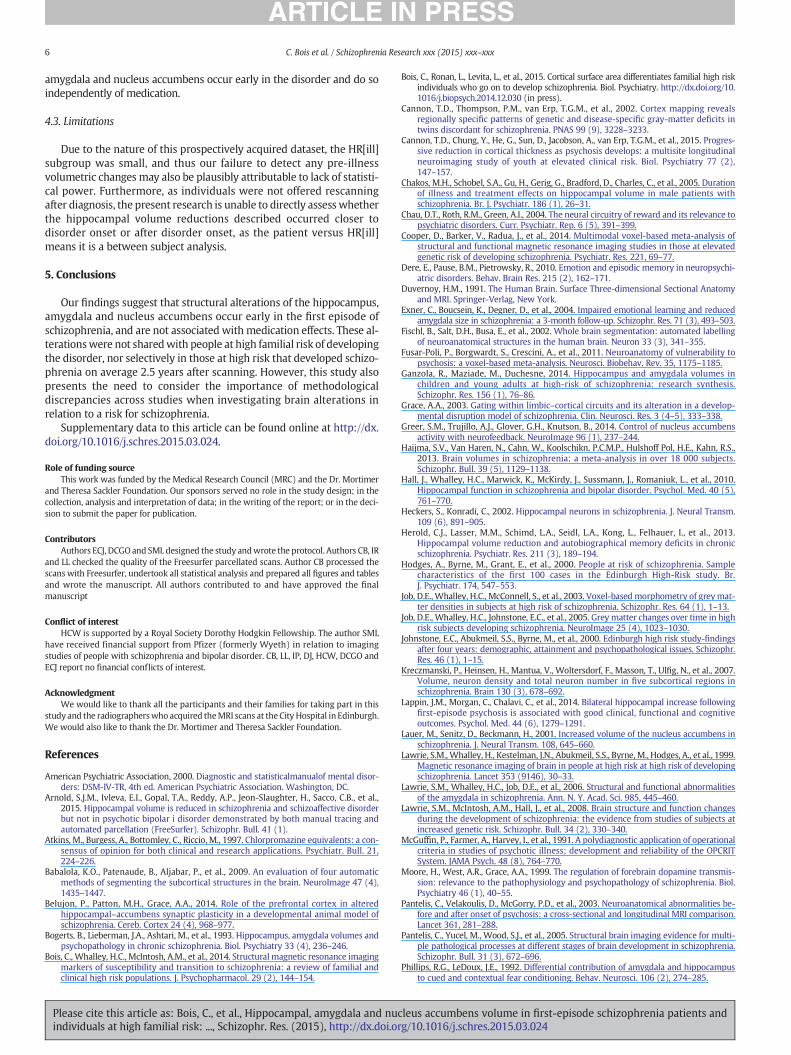
Automated subcortical segmentations were conducted usingFreesurfer 5.30 (http://surfer.nmr.mgh.harvard.edu). Parcellationof the subcortical anatomy, and calculations of the total intracranial vol-umewere performed by delineating anatomical divisions via automaticparcellation methods, in which the statistical knowledge base derivesfrom a training set incorporating the anatomical landmarks andconventions described by Duvernoy (1991). This procedure assigns aneuroanatomical label to each voxel in anMRI volume based on proba-bilistic information estimated from a manually labeled training set. Theclassification technique employs a non-linear registration procedurethat is robust to anatomical variability (Fischl et al., 2002). The segmen-tation uses three pieces of information to disambiguate labels: (1) theprior probability of a given tissue class occurring at a specific atlas loca-tion, (2) the likelihood of the image given what tissue class and (3) theprobability of the local spatial configuration of labels given the tissueclass. The gray matter volumes of the hippocampus, amygdala andnucleus accumbens were then extracted from Freesurfer. An exampleof Freesurfer extractions of these volumes is illustrated in Fig. 1.
2.4. Statistical analysis
The graymatter volumes of the left and right hippocampus, amygda-la andnucleus accumbenswere extracted fromFreesurfer. Two analyseswere performed on the extracted subcortical parcellations: one compar-ing the patients with first-episode schizophrenia, those at high risk andthe healthy controls, and a second comparing the high risk sub-groups totest for brain differences relating to clinical outcome. These comparisons
H
A
a) b)
Fig. 1. Example figure of a) = A = amygdala, H = hippocampus, an
Please cite this article as: Bois, C., et al., Hippocampal, amygdala and nucindividuals at high familial risk: ..., Schizophr. Res. (2015), http://dx.doi.or
were performed using separate Analyses of Covariance (ANCOVAs;adjusting for intracranial volume, age and sex).Where the ANCOVAs indi-cated significant between-group differences, pairwise comparisons wereperformed using package lsmeans (Version 2.10) implemented in R.Where gray matter volume was found to be non-normally distributed, aboxCox transform implemented in package geoR (Version 1.74)was con-ducted. Additionally, in the patients with first episode schizophrenia,chlorpromazine equivalents of current medication dose were calculatedaccording toWoods (2003) andAtkins et al. (1997). Spearman's rank cor-relation coefficients between the three subcortical regions and chlor-promazine equivalents and duration of illness (in months) were alsocalculated.
3. Results
3.1. Hippocampal volume in patients with a first-episode, high riskparticipants and healthy controls
For the left hippocampus, when all those at high risk were consid-ered together and compared against controls and patients there was asignificant main effect of group (F = 5.7, df = 2,204, p = .004). Subse-quent contrasts revealed that this was due to patients having signifi-cantly smaller (M = 3739, SE = 87.4) left hippocampi than controls(M = 4033, SE = 80.8) (t = 3.036, df = 204, p = .008), as well ashigh risk individuals (M = 3991 SE = 53.2) (t = −3.167, p = .005).No significant differences were found between controls and those athigh risk (t = 0.586, df = 204, p = 0.877). For the right hippocampus,when all those at high risk were considered together and comparedagainst controls and patients, there was a significant main effect ofgroup on volume (F= 5.55 df = 2,204 p= .004). Subsequent contrastsrevealed that this was due to patients having significantly smaller righthippocampi (M = 3698, SE = 86.5) than controls (M = 3952, SE =79.9) (t= 2.64, df= 204, p=0.0239), aswell as patients having signif-icantly smaller hippocampi compared to high risk individuals (M =3958, SE = 52.7; t = −3.29, df = 204, p = .003). These results areillustrated in Fig. 2. No significant differences emerged between highrisk individuals and controls (t=−0.090, df= 204, p= .996). Medica-tion had non-significant effects for all sub-cortical regions investigatedin the present study, as did length of illness (Supplementary data 1).
3.2. Amygdala volume in patients with a first-episode, high risk participantsand healthy controls
For the left amygdala, when all those high risk were considered to-gether and compared against controls and patients there was a signifi-cant main effect of group on volume (F = 5.2, df = 2,204, p = .006).Subsequent contrasts revealed that this was due to patients havingsignificantly smaller left amygdalae (M = 1181 SE = 43.9) comparedto controls (M = 1329 SE = 40.6; t = 3.026 df = 204 p = .007), aswell as compared to those at high risk (M = 1299 SE = 26.7)(t = −2.93 df = 204 p = 0.011) (Fig. 3). No significant differences
NA
d b) = NA = nucleus accumbens extractions using Freesurfer.
leus accumbens volume in first-episode schizophrenia patients andg/10.1016/j.schres.2015.03.024

Fig. 2. Scattergrams of a) the left hippocampus (mm3) and b) the right hippocampus(mm3) for CON = healthy controls, FEP = first episode patients and HR = high risk.
Fig. 3. Scattergrams of a) the left amygdala (mm3) and b) the right amygdala (mm3) forCON = healthy controls, FEP = first episode patients and HR = high risk.
4 C. Bois et al. / Schizophrenia Research xxx (2015) xxx–xxx
Please cite this article as: Bois, C., et al., Hippocampal, amygdala and nucindividuals at high familial risk: ..., Schizophr. Res. (2015), http://dx.doi.or
emerged between high risk individuals and controls (t = 0.840, df =204, p = 0.678). There was no significant main effect of group on thevolume of the right amygdala (F = 1.88, df = 2,204, p = 0.155).
3.3. Nucleus accumbens volume in patients with a first-episode, high riskparticipants and healthy controls
There was a significant main effect of group on the right accumbens(F = 0.017, df = 2,204, p = 0.017) which subsequent post-hoc testingrevealed was due to patients (M= 582, SE= 19.1) having significantlysmaller right nucleus accumbens volumes than controls (M = 638SE = 15.9; t = 2.65, df = 207, p = 0.023) and high risk individuals(M= 629 SE= 8.44; t =−2.766, df = 207, p = 0.017). No significantdifferences emerged between high risk individuals and controls (t =0.528, df = 207, p = 0.857, Fig. 4). There was no significant main effectof group on the volume of the left nucleus accumbens (F= 0.648, df =2,204, p = 0.524).
3.4. Hippocampal, amygdala and nucleus accumbens volume comparedbetween high risk participants based in subsequent clinical outcome
No significant group differences emerged for any of these structureswhen those at high risk were considered based on subsequent illnesstransition or not. Table 3 shows the statistics of these analyses, andTable 4 presents the means for these structures based on the high risksub-groupings.
3.5. Hippocampal, amygdala and nucleus accumbens volume comparedbetween HR[ill] and patients with first-episode psychosis
We also conducted an additional analyses for all structures compar-ing patients with first-episode schizophrenia specifically to the HR[ill]
Fig. 4. Scattergrams of a) the left nucleus accumbens (mm3) and b) the right nucleusaccumbens (mm3) for CON = healthy controls, FEP = first episode patients and HR =high risk.
leus accumbens volume in first-episode schizophrenia patients andg/10.1016/j.schres.2015.03.024

Table 3Table of results comparing all three structures between HR[well], HR[ill], & HR[symp].
F df p
Left Hippocampus 1.9 2,141 0.156Amygdala 0.915 2,141 0.405Nucleus accumbens 0.911 2,141 0.406
Right Hippocampus 0.814 2,141 0.369Amygdala 1.21 2,141 0.302Nucleus accumbens 0.1667 2,141 0.123
5C. Bois et al. / Schizophrenia Research xxx (2015) xxx–xxx
sub-group. We found that patients had significantly smaller left amyg-dalae (F = 7.15, df = 1,33, p = 0.016), and left (F = 6.34, df = 1,33,p= 0.017) and right (F= 5.40, df= 1,33, p=0.026, p=0.017) hippo-campus compared to HR[ill]. The right amygdala showed no significantgroup differences, nor did the left or right nucleus accumbens.
3.6. Correction for multiple comparisons
The results reported above are uncorrected for multiple compari-sons. However, we also conducted a false discovery rate (FDR) correc-tion for multiple comparisons, and all results for all significantlyreported structures remained significant, except for the right nucleusaccumbens comparison between high risk, patients and controls(Supplementary data 2).
4. Discussion
This study assessed hippocampal, amygdala and nucleus accumbensvolumes in a sample of individuals at high familial risk of developingschizophrenia, along with a group of patients experiencing their firstepisode of psychosis, and healthy controls. Our main aim was to inves-tigatewhether these structureswere altered in those at high risk or onlypresent in those in their first episode of psychosis. As a secondary aimwe wished to examine whether volume changes were selectively pres-ent in those at high risk that went on to develop schizophrenia on aver-age 2.5 years after the scan took place, compared to those at high riskthat remained well or developed only transient psychotic symptoms.The rationale behind this approachwas to assesswhether any structuralalterationswere present before illness onset in those thatwent on to de-velop the disorder, or whether any alterations were more specificallyassociated with the disorder onset or found in those that wereexperiencing their first episode of psychosis.
4.1. Implications of present findings
We found significant bilateral volumetric reductions of the hippo-campus in patients, compared to controls and high-risk participants.For the amygdala, this effect was lateralized to the left hemisphere,whilst the opposite effect was observed for the nucleus accumbens.Unexpectedly, we found no statistically significant abnormalities inhigh-risk patients when compared to controls, nor when the high-riskindividuals were separately considered based on subsequent clinicaloutcome. The findings of reduced volumes in these three structures inthose in their first episode of schizophrenia were not significantlyassociated with medication intake, or duration of illness. This suggeststhat these alterations may be more associated with pathological
Table 4Table of means for HR sub-groups.
HR[well] HR[symp] HR[ill]
Left Hippocampus 3955 (49.9) 4108 (53.9) 4046 (97)Amygdala 1228 (25.4) 1298 (27.2) 1328 (47.6)Nucleus accumbens 606 (13.7) 641 (14.7) 605 (24.7)
Right Hippocampus 3955 (49.9) 4108 (53.9) 4046 (97)Amygdala 1378 (29.4) 1427 (31.4) 1410 (53.7)Nucleus accumbens 680 (15.1) 680 (643) 619 (26.8)
Please cite this article as: Bois, C., et al., Hippocampal, amygdala and nucindividuals at high familial risk: ..., Schizophr. Res. (2015), http://dx.doi.or
mechanisms occurring early in the disease process (Moore et al.,1999; Grace, 2003), rather than the effects of medication. Most of thepatients used in the present study were scanned six months or lessafter diagnosis, thus also suggesting that the brain alterations demon-strated in the present study were not merely a by-product of durationof psychosis. It is interesting that these structural alterations occur inthe hippocampus, amygdala and nucleus accumbens, as these struc-tures are thought to be critically involved in reward and aversiveevent behavior, that is typically altered in schizophrenia (Grace,2003), and future studies may wish to examine how structural alter-ations in these regions relate to functional alterations.
4.2. Methodological considerations
The present study found no significant subcortical differences be-tween high risk individuals and controls. This is consistent with someprevious studies using Freesurfer (Arnold et al., 2015; Sprooten et al.,2013) but not our preceding studies using hand-traced (Lawrie et al.,1999) or voxel-based (Job et al., 2003) methods. A previous study con-ducted on the same sample as that of the present study measured thehippocampal–amygdalar complex using manual tracing methods andfound significant reductions in the high risk cohort compared to con-trols (Lawrie et al., 1999). However, the present study used Freesurfer,a semi-automated parcellation software that is able to separately delin-eate the hippocampus and amygdala, finding no significant differences.It remains possible therefore that by investigating these structures sep-arately and using a different sMRI technique, subtle structural changesoccurring in the high risk individuals may be lost, as evidence suggeststhat any alterations found in high risk individuals will be present to alesser extent than those with established schizophrenia (Lawrie et al.,2008). It is noteworthy that all subcortical structures investigated inthe present study were slightly smaller in those at high risk comparedto controls. It remains possible that use of an automated brain segmen-tationmethod such as Freesurfer, compared tomanual tracingmethod-ologies, typically considered the “gold standard” of ROI studies, are lessable to pick up the subtle structural differences hypothesized to existbetween those at high risk and controls. Indeed, a recent study conduct-ed by Arnold et al. (2015) found evidence for Freesurfer to produce sig-nificantly larger volumes than manual tracing methods, suggesting theneed to consider methodological discrepancies when interpreting theresults of the study. Furthermore, when investigating structures suchas the nucleus accumbens, specific limitations to the delineationprotocol may inherently arise to produce inconsistencies across studies,as this structure has previously shown low accuracy in automatedsegmentation studies (Babalola et al., 2009).
Another reason for the present study not finding any significant highrisk versus control differences in the investigated structures is because itremains possible that as theHR[ill] group did not develop schizophreniauntil on average 2.5 years after the scans were acquired, that furtherprogressive changes may have occurred closer to the time of disorderonset. A third possibility is that the results reported here are correct,and that any differences in familial high risk subjects are primarily in en-torhinal cortex bordering the hippocampal–amygdala complex; indeeda previous study conducted with Freesurfer found a trend for a signifi-cant difference between controls and those at high risk located inthe entorhinal cortex, as did a voxel-based study (Job et al., 2003;Sprooten et al., 2013). This is an imperative point to consider in futurehigh risk sMRI studies. However, another important point to considerhere is the low conversion rate in this study, of approximately 13%(Whalley et al., 2007), and hence the possibility that our sample reflectsa specific group of familial high risk participants with less vulnerabilityto develop schizophrenia. Our present results do highlight the need forfuture sMRI studies to incorporate standardizedmethods across studies,in order to increase the generalizability and interpretability of findings.However, our current findings also reiterate findings that the alterationsfound in schizophrenia in subcortical structure; namely the hippocampus,
leus accumbens volume in first-episode schizophrenia patients andg/10.1016/j.schres.2015.03.024
6 C. Bois et al. / Schizophrenia Research xxx (2015) xxx–xxx
amygdala and nucleus accumbens occur early in the disorder and do soindependently of medication.
4.3. Limitations
Due to the nature of this prospectively acquired dataset, the HR[ill]subgroup was small, and thus our failure to detect any pre-illnessvolumetric changes may also be plausibly attributable to lack of statisti-cal power. Furthermore, as individuals were not offered rescanningafter diagnosis, the present research is unable to directly assess whetherthe hippocampal volume reductions described occurred closer todisorder onset or after disorder onset, as the patient versus HR[ill]means it is a between subject analysis.
5. Conclusions
Our findings suggest that structural alterations of the hippocampus,amygdala and nucleus accumbens occur early in the first episode ofschizophrenia, and are not associated with medication effects. These al-terationswere not sharedwith people at high familial risk of developingthe disorder, nor selectively in those at high risk that developed schizo-phrenia on average 2.5 years after scanning. However, this study alsopresents the need to consider the importance of methodologicaldiscrepancies across studies when investigating brain alterations inrelation to a risk for schizophrenia.
Supplementary data to this article can be found online at http://dx.doi.org/10.1016/j.schres.2015.03.024.
Role of funding sourceThis work was funded by the Medical Research Council (MRC) and the Dr. Mortimer
and Theresa Sackler Foundation. Our sponsors served no role in the study design; in thecollection, analysis and interpretation of data; in the writing of the report; or in the deci-sion to submit the paper for publication.
ContributorsAuthors ECJ, DCGO and SML designed the study andwrote the protocol. Authors CB, IR
and LL checked the quality of the Freesurfer parcellated scans. Author CB processed thescans with Freesurfer, undertook all statistical analysis and prepared all figures and tablesand wrote the manuscript. All authors contributed to and have approved the finalmanuscript
Conflict of interestHCW is supported by a Royal Society Dorothy Hodgkin Fellowship. The author SML
have received financial support from Pfizer (formerly Wyeth) in relation to imagingstudies of people with schizophrenia and bipolar disorder. CB, LL, IP, DJ, HCW, DCGO andECJ report no financial conflicts of interest.
AcknowledgmentWe would like to thank all the participants and their families for taking part in this
study and the radiographerswho acquired theMRI scans at theCity Hospital in Edinburgh.We would also like to thank the Dr. Mortimer and Theresa Sackler Foundation.
References
American Psychiatric Association, 2000. Diagnostic and statisticalmanualof mental disor-ders: DSM-IV-TR, 4th ed. American Psychiatric Association. Washington, DC.
Arnold, S.J.M., Ivleva, E.I., Gopal, T.A., Reddy, A.P., Jeon-Slaughter, H., Sacco, C.B., et al.,2015. Hippocampal volume is reduced in schizophrenia and schizoaffective disorderbut not in psychotic bipolar i disorder demonstrated by both manual tracing andautomated parcellation (FreeSurfer). Schizophr. Bull. 41 (1).
Atkins, M., Burgess, A., Bottomley, C., Riccio, M., 1997. Chlorpromazine equivalents: a con-sensus of opinion for both clinical and research applications. Psychiatr. Bull. 21,224–226.
Babalola, K.O., Patenaude, B., Aljabar, P., et al., 2009. An evaluation of four automaticmethods of segmenting the subcortical structures in the brain. NeuroImage 47 (4),1435–1447.
Belujon, P., Patton, M.H., Grace, A.A., 2014. Role of the prefrontal cortex in alteredhippocampal–accumbens synaptic plasticity in a developmental animal model ofschizophrenia. Cereb. Cortex 24 (4), 968–977.
Bogerts, B., Lieberman, J.A., Ashtari, M., et al., 1993. Hippocampus, amygdala volumes andpsychopathology in chronic schizophrenia. Biol. Psychiatry 33 (4), 236–246.
Bois, C., Whalley, H.C., McIntosh, A.M., et al., 2014. Structural magnetic resonance imagingmarkers of susceptibility and transition to schizophrenia: a review of familial andclinical high risk populations. J. Psychopharmacol. 29 (2), 144–154.
Please cite this article as: Bois, C., et al., Hippocampal, amygdala and nucindividuals at high familial risk: ..., Schizophr. Res. (2015), http://dx.doi.or
Bois, C., Ronan, L., Levita, L., et al., 2015. Cortical surface area differentiates familial high riskindividuals who go on to develop schizophrenia. Biol. Psychiatry. http://dx.doi.org/10.1016/j.biopsych.2014.12.030 (in press).
Cannon, T.D., Thompson, P.M., van Erp, T.G.M., et al., 2002. Cortex mapping revealsregionally specific patterns of genetic and disease-specific gray-matter deficits intwins discordant for schizophrenia. PNAS 99 (9), 3228–3233.
Cannon, T.D., Chung, Y., He, G., Sun, D., Jacobson, A., van Erp, T.G.M., et al., 2015. Progres-sive reduction in cortical thickness as psychosis develops: a multisite longitudinalneuroimaging study of youth at elevated clinical risk. Biol. Psychiatry 77 (2),147–157.
Chakos, M.H., Schobel, S.A., Gu, H., Gerig, G., Bradford, D., Charles, C., et al., 2005. Durationof illness and treatment effects on hippocampal volume in male patients withschizophrenia. Br. J. Psychiatr. 186 (1), 26–31.
Chau, D.T., Roth, R.M., Green, A.I., 2004. The neural circuitry of reward and its relevance topsychiatric disorders. Curr. Psychiatr. Rep. 6 (5), 391–399.
Cooper, D., Barker, V., Radua, J., et al., 2014. Multimodal voxel-based meta-analysis ofstructural and functional magnetic resonance imaging studies in those at elevatedgenetic risk of developing schizophrenia. Psychiatr. Res. 221, 69–77.
Dere, E., Pause, B.M., Pietrowsky, R., 2010. Emotion and episodic memory in neuropsychi-atric disorders. Behav. Brain Res. 215 (2), 162–171.
Duvernoy, H.M., 1991. The Human Brain. Surface Three-dimensional Sectional Anatomyand MRI. Springer-Verlag, New York.
Exner, C., Boucsein, K., Degner, D., et al., 2004. Impaired emotional learning and reducedamygdala size in schizophrenia: a 3-month follow-up. Schizophr. Res. 71 (3), 493–503.
Fischl, B., Salt, D.H., Busa, E., et al., 2002. Whole brain segmentation: automated labellingof neuroanatomical structures in the human brain. Neuron 33 (3), 341–355.
Fusar-Poli, P., Borgwardt, S., Crescini, A., et al., 2011. Neuroanatomy of vulnerability topsychosis: a voxel-based meta-analysis. Neurosci. Biobehav. Rev. 35, 1175–1185.
Ganzola, R., Maziade, M., Duchesne, 2014. Hippocampus and amygdala volumes inchildren and young adults at high-risk of schizophrenia: research synthesis.Schizophr. Res. 156 (1), 76–86.
Grace, A.A., 2003. Gating within limbic–cortical circuits and its alteration in a develop-mental disruption model of schizophrenia. Clin. Neurosci. Res. 3 (4–5), 333–338.
Greer, S.M., Trujillo, A.J., Glover, G.H., Knutson, B., 2014. Control of nucleus accumbensactivity with neurofeedback. NeuroImage 96 (1), 237–244.
Haijma, S.V., Van Haren, N., Cahn, W., Koolschikn, P.C.M.P., Hulshoff Pol, H.E., Kahn, R.S.,2013. Brain volumes in schizophrenia: a meta-analysis in over 18 000 subjects.Schizophr. Bull. 39 (5), 1129–1138.
Hall, J., Whalley, H.C., Marwick, K., McKirdy, J., Sussmann, J., Romaniuk, L., et al., 2010.Hippocampal function in schizophrenia and bipolar disorder. Psychol. Med. 40 (5),761–770.
Heckers, S., Konradi, C., 2002. Hippocampal neurons in schizophrenia. J. Neural Transm.109 (6), 891–905.
Herold, C.J., Lasser, M.M., Schimd, L.A., Seidl, L.A., Kong, L., Felhauer, I., et al., 2013.Hippocampal volume reduction and autobiographical memory deficits in chronicschizophrenia. Psychiatr. Res. 211 (3), 189–194.
Hodges, A., Byrne, M., Grant, E., et al., 2000. People at risk of schizophrenia. Samplecharacteristics of the first 100 cases in the Edinburgh High-Risk study. Br.J. Psychiatr. 174, 547–553.
Job, D.E., Whalley, H.C., McConnell, S., et al., 2003. Voxel-based morphometry of greymat-ter densities in subjects at high risk of schizophrenia. Schizophr. Res. 64 (1), 1–13.
Job, D.E., Whalley, H.C., Johnstone, E.C., et al., 2005. Grey matter changes over time in highrisk subjects developing schizophrenia. NeuroImage 25 (4), 1023–1030.
Johnstone, E.C., Abukmeil, S.S., Byrne, M., et al., 2000. Edinburgh high risk study-findingsafter four years: demographic, attainment and psychopathological issues. Schizophr.Res. 46 (1), 1–15.
Kreczmanski, P., Heinsen, H., Mantua, V., Woltersdorf, F., Masson, T., Ulfig, N., et al., 2007.Volume, neuron density and total neuron number in five subcortical regions inschizophrenia. Brain 130 (3), 678–692.
Lappin, J.M., Morgan, C., Chalavi, C., et al., 2014. Bilateral hippocampal increase followingfirst-episode psychosis is associated with good clinical, functional and cognitiveoutcomes. Psychol. Med. 44 (6), 1279–1291.
Lauer, M., Senitz, D., Beckmann, H., 2001. Increased volume of the nucleus accumbens inschizophrenia. J. Neural Transm. 108, 645–660.
Lawrie, S.M., Whalley, H., Kestelman, J.N., Abukmeil, S.S., Byrne, M., Hodges, A., et al., 1999.Magnetic resonance imaging of brain in people at high risk at high risk of developingschizophrenia. Lancet 353 (9146), 30–33.
Lawrie, S.M., Whalley, H.C., Job, D.E., et al., 2006. Structural and functional abnormalitiesof the amygdala in schizophrenia. Ann. N. Y. Acad. Sci. 985, 445–460.
Lawrie, S.M., McIntosh, A.M., Hall, J., et al., 2008. Brain structure and function changesduring the development of schizophrenia: the evidence from studies of subjects atincreased genetic risk. Schizophr. Bull. 34 (2), 330–340.
McGuffin, P., Farmer, A., Harvey, I., et al., 1991. A polydiagnostic application of operationalcriteria in studies of psychotic illness; development and reliability of the OPCRITSystem. JAMA Psych. 48 (8), 764–770.
Moore, H., West, A.R., Grace, A.A., 1999. The regulation of forebrain dopamine transmis-sion: relevance to the pathophysiology and psychopathology of schizophrenia. Biol.Psychiatry 46 (1), 40–55.
Pantelis, C., Velakoulis, D., McGorry, P.D., et al., 2003. Neuroanatomical abnormalities be-fore and after onset of psychosis: a cross-sectional and longitudinal MRI comparison.Lancet 361, 281–288.
Pantelis, C., Yucel, M., Wood, S.J., et al., 2005. Structural brain imaging evidence for multi-ple pathological processes at different stages of brain development in schizophrenia.Schizophr. Bull. 31 (3), 672–696.
Phillips, R.G., LeDoux, J.E., 1992. Differential contribution of amygdala and hippocampusto cued and contextual fear conditioning. Behav. Neurosci. 106 (2), 274–285.
leus accumbens volume in first-episode schizophrenia patients andg/10.1016/j.schres.2015.03.024

7C. Bois et al. / Schizophrenia Research xxx (2015) xxx–xxx
Qui, A., Gan, S.C., Wang, Y., et al., 2013. Amygdala hippocampal shape and cortical thick-ness abnormalities in first-episode schizophrenia and mania. Psychol. Med. 43 (7),1353–1363.
Seidman, L.J., Faraone, S.V., Goldstein, J.M., et al., 1999. Thalamic and amygdala–hippocampal volume reductions in first-degree relatives of patients withschizophrenia: an MRI-based morphometric analysis. Biol. Psychiatry 46 (7),941–954.
Smieksova, R., Marmy, J., Schmidt, A., et al., 2013. Do subjects at clinical high risk forpsychosis differ from those with a genetic high risk? A systematic review of structuraland functional brain abnormalities. Curr. Med. Chem. 20, 467–481.
Sprooten, E., Papmeyer, M., Smyth, A.M., et al., 2013. Cortical thickness in first−episodeschizophrenia patients and induviduals at high familial risk: a cross−sectional com-parison. Schiz. Res. 151 (1–3), 259–264.
Staal, W.G., Hulshoff Pol, H.E., Schnack, H.G., Hoogendoorn, M.L.C., Jellema, K., Kahn, R.,2000. Structural brain abnormalities in patients with schizophrenia and their healthysiblings. Am. J. Psychiatry 157 (3), 416–421.
Stefanis, N., Frangou, S., Yakeley, J., Sharma, T., O'Connell, P., Morgan, K., et al., 1999.Hippocampal volume reduction in schizophrenia: effects of genetic risk and pregnan-cy and birth complications. Biol. Psychiatry 46 (5), 697–702.
Sun, D., Phillips, L., Velakoulis, D., Yung, A., McGorry, P.D., Wood, S.J., et al., 2009. Progres-sive brain structural changes mapped as psychosis develops in ‘at risk’ individuals.Schizophr. Res. 108 (1–3), 85–92.
Tanskanen, P., Veijola, J.M., Piippo, U.K., Haapea, M., Meittunen, J.A., Pyhtinen, J., et al.,2005. Hippocampus and amygdala volumes in schizophrenia and other psychosesin the northern Finland 1966 birth cohort. Schizophr. Res. 75 (2–3), 283–294.
Please cite this article as: Bois, C., et al., Hippocampal, amygdala and nucindividuals at high familial risk: ..., Schizophr. Res. (2015), http://dx.doi.or
Velakoulis, D., Wood, S.J., Wong, M.T.H., McGorry, P.D., Yung, A., Phillips, L., et al., 2006.Hippocampal and amygdala volumes according to psychosis stage and diagnosis. Amagnetic resonance imaging study of chronic schizophrenia, first-episode psychosisand ultra-high risk individuals. JAMA Psych. 63 (2), 139–149.
Watson, D.R., Bai, F., Barrett, S.L., Turkington, A., Rushe, T.M., Mulholland, C.C., et al., 2012.Structural changes in the hippocampus and amygdala at first episode of psychosis.Brain Imaging Behav. 6 (1), 49–60.
Whalley, H.C., Simonotto, E., Flett, S., Marshall, I., Ebmeier, K.P., Owens, D.G.C., 2004. fMRIcorrelates of state and trait effects in subjects at genetically enhanced risk ofschizophrenia. Brain 127 (3), 478–490.
Whalley, H.C., Harris, J.C., Lawrie, S.M., 2007. The neurobiological underpinnings of riskand conversion in relatives of patients with schizophrenia. Int. Rev. Psychiatry 19(4), 383–397.
Whalley, H.C., Papmeyer, M., Sprooten, E., Lawrie, S.M., Sussmann, J.E., McIntosh, A.M.,2012. Review of functional magnetic resonance imaging studies comparing bipolardisorder and schizophrenia. Bipolar Disord. 14 (4), 411–431.
Wing, J.K., Cooper, J.E., Sartorius, N., et al., 1974. Measurement and Classification ofPsychiatric Symptoms, an Instruction Manual for the PSE and CATEGO Programme.Cambridge University Press, Cambridge.
Womer, F.Y., Wang, L., Alpert, K.I., Smith, M.J., Csernansky, J.G., Barch, D.M., et al., 2014.Basal ganglia and thalamic morphology in schizophrenia and bipolar disorder.Psychiatr. Res. 223, 75–83.
Woods, S.W., 2003. Chlorpromazine equivilant doses for the newer atypical antipsy-chotics. J. Clin. Psychiatry. 64 (6), 663–667.
leus accumbens volume in first-episode schizophrenia patients andg/10.1016/j.schres.2015.03.024