The effect of tryptophan depletion on the action of haloperidol in MK-801-treated rats
A
opsS(iTTts©
K
1
bftibsTi
0d
Behavioural Brain Research 178 (2007) 190–199
Research report
Injections of the selective adenosine A2A antagonist MSX-3 intothe nucleus accumbens core attenuate the locomotor
suppression induced by haloperidol in rats
Keita Ishiwari a, Lisa J. Madson a, Andrew M. Farrar a, Susana M. Mingote a, John P. Valenta a,Michael D. DiGianvittorio a, Lauren E. Frank a, Merce Correa a,c,
Jorg Hockemeyer b, Christa Muller b, John D. Salamone a,∗a Department of Psychology, University of Connecticut, Storrs, CT 06269-1020, United States
b Universitat Bonn, Pharmazeutisches Institut, Pharmazeutische Chemie, Poppelsdorf, Bonn, Germanyc Area de Psicobiol., Department of Psicologia, Universitat de Jaume I, Castello, Spain
Received 25 September 2006; received in revised form 11 December 2006; accepted 14 December 2006Available online 21 December 2006
bstract
There is considerable evidence of interactions between adenosine A2A receptors and dopamine D2 receptors in striatal areas, and antagonistsf the A2A receptor have been shown to reverse the motor effects of DA antagonists in animal models. The D2 antagonist haloperidol producesarkinsonism in humans, and also induces motor effects in rats, such as suppression of locomotion. The present experiments were conducted totudy the ability of the adenosine A2A antagonist MSX-3 to reverse the locomotor effects of acute or subchronic administration of haloperidol in rats.ystemic (i.p.) injections of MSX-3 (2.5–10.0 mg/kg) were capable of attenuating the suppression of locomotion induced by either acute or repeatedi.e., 14 day) administration of 0.5 mg/kg haloperidol. Bilateral infusions of MSX-3 directly into the nucleus accumbens core (2.5 �g or 5.0 �gn 0.5 �l per side) produced a dose-related increase in locomotor activity in rats treated with 0.5 mg/kg haloperidol either acutely or repeatedly.
here were no overall significant effects of MSX-3 infused directly into the dorsomedial nucleus accumbens shell or the ventrolateral neostriatum.hese results indicate that antagonism of adenosine A2A receptors can attenuate the locomotor suppression produced by DA antagonism, and thathis effect may be at least partially mediated by A2A receptors in the nucleus accumbens core. These studies suggest that adenosine and dopamineystems interact to modulate the locomotor and behavioral activation functions of nucleus accumbens core.
2007 Elsevier B.V. All rights reserved.
hotic
DoirsWir
eywords: Dopamine; Basal ganglia; Neostriatum; Caudate putamen; Antipsyc
. Introduction
Interactions between diverse neurotransmitter systems in theasal ganglia are thought to regulate several aspects of motorunction [22,82]. Neostriatal depletions of dopamine (DA) arehe immediate cause of motor dysfunction in patients withdiopathic Parkinson’s disease [35], while pharmacologicallockade of DA transmission with DA receptor antagonists
uch as haloperidol leads to drug-induced parkinsonism [53].he most common treatments for Parkinson’s disease generallynvolve dopaminergic strategies, including the DA precursor L-
∗ Corresponding author. Tel.: +1 860 486 4302; fax: +1 860 486 2760.E-mail address: [email protected] (J.D. Salamone).
[astseD
166-4328/$ – see front matter © 2007 Elsevier B.V. All rights reserved.oi:10.1016/j.bbr.2006.12.020
; Parkinson’s disease; D2 receptor
OPA, as well as DA agonists such as bromocriptine, pergolide,r ropinirole [10,47,48]. Nevertheless, considerable research hasmplicated several other neurotransmitters in motor processeselated to the basal ganglia, including acetylcholine [67,68],erotonin [11], glutamate [58,60], and GABA [12,45,77,80,81].
ithin the last few years, evidence has begun to emergendicating that brain adenosine neurons play an importantole in regulating the motor functions of the basal ganglia24,25,28,76]. Although several subtypes of adenosine receptorsre involved in motor function, anatomical studies have demon-trated that the adenosine A2A receptor subtype is expressed
o a high degree in striatal regions [15,27,33,49,76,78]. Aden-onine A2A receptors in the striatum are largely expressed onnkephalin-positive striatopallidal neurons, which also containA D2 receptors [76]. Antagonism of adenosine A2A receptors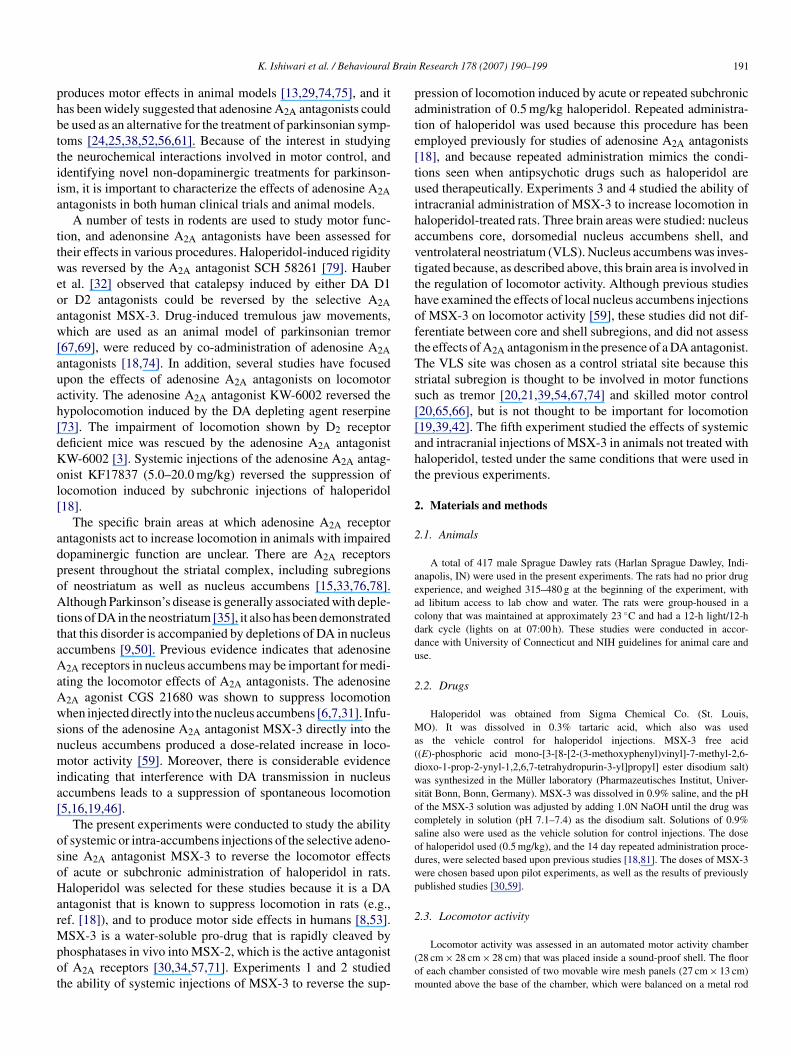
Brain
phbttiia
ttweoaw[auah[dKol[
adpoAttaAaAwsnmia[
osoHarMpot
pate[tuihavtthoftTss[[aht
2
2
aeacddu
2
Ma(dwsocsodwp
2
K. Ishiwari et al. / Behavioural
roduces motor effects in animal models [13,29,74,75], and itas been widely suggested that adenosine A2A antagonists coulde used as an alternative for the treatment of parkinsonian symp-oms [24,25,38,52,56,61]. Because of the interest in studyinghe neurochemical interactions involved in motor control, anddentifying novel non-dopaminergic treatments for parkinson-sm, it is important to characterize the effects of adenosine A2Antagonists in both human clinical trials and animal models.
A number of tests in rodents are used to study motor func-ion, and adenonsine A2A antagonists have been assessed forheir effects in various procedures. Haloperidol-induced rigidityas reversed by the A2A antagonist SCH 58261 [79]. Hauber
t al. [32] observed that catalepsy induced by either DA D1r D2 antagonists could be reversed by the selective A2Antagonist MSX-3. Drug-induced tremulous jaw movements,hich are used as an animal model of parkinsonian tremor
67,69], were reduced by co-administration of adenosine A2Antagonists [18,74]. In addition, several studies have focusedpon the effects of adenosine A2A antagonists on locomotorctivity. The adenosine A2A antagonist KW-6002 reversed theypolocomotion induced by the DA depleting agent reserpine73]. The impairment of locomotion shown by D2 receptoreficient mice was rescued by the adenosine A2A antagonistW-6002 [3]. Systemic injections of the adenosine A2A antag-nist KF17837 (5.0–20.0 mg/kg) reversed the suppression ofocomotion induced by subchronic injections of haloperidol18].
The specific brain areas at which adenosine A2A receptorntagonists act to increase locomotion in animals with impairedopaminergic function are unclear. There are A2A receptorsresent throughout the striatal complex, including subregionsf neostriatum as well as nucleus accumbens [15,33,76,78].lthough Parkinson’s disease is generally associated with deple-
ions of DA in the neostriatum [35], it also has been demonstratedhat this disorder is accompanied by depletions of DA in nucleusccumbens [9,50]. Previous evidence indicates that adenosine2A receptors in nucleus accumbens may be important for medi-
ting the locomotor effects of A2A antagonists. The adenosine2A agonist CGS 21680 was shown to suppress locomotionhen injected directly into the nucleus accumbens [6,7,31]. Infu-
ions of the adenosine A2A antagonist MSX-3 directly into theucleus accumbens produced a dose-related increase in loco-otor activity [59]. Moreover, there is considerable evidence
ndicating that interference with DA transmission in nucleusccumbens leads to a suppression of spontaneous locomotion5,16,19,46].
The present experiments were conducted to study the abilityf systemic or intra-accumbens injections of the selective adeno-ine A2A antagonist MSX-3 to reverse the locomotor effectsf acute or subchronic administration of haloperidol in rats.aloperidol was selected for these studies because it is a DA
ntagonist that is known to suppress locomotion in rats (e.g.,ef. [18]), and to produce motor side effects in humans [8,53].
SX-3 is a water-soluble pro-drug that is rapidly cleaved byhosphatases in vivo into MSX-2, which is the active antagonistf A2A receptors [30,34,57,71]. Experiments 1 and 2 studiedhe ability of systemic injections of MSX-3 to reverse the sup-
(om
Research 178 (2007) 190–199 191
ression of locomotion induced by acute or repeated subchronicdministration of 0.5 mg/kg haloperidol. Repeated administra-ion of haloperidol was used because this procedure has beenmployed previously for studies of adenosine A2A antagonists18], and because repeated administration mimics the condi-ions seen when antipsychotic drugs such as haloperidol aresed therapeutically. Experiments 3 and 4 studied the ability ofntracranial administration of MSX-3 to increase locomotion inaloperidol-treated rats. Three brain areas were studied: nucleusccumbens core, dorsomedial nucleus accumbens shell, andentrolateral neostriatum (VLS). Nucleus accumbens was inves-igated because, as described above, this brain area is involved inhe regulation of locomotor activity. Although previous studiesave examined the effects of local nucleus accumbens injectionsf MSX-3 on locomotor activity [59], these studies did not dif-erentiate between core and shell subregions, and did not assesshe effects of A2A antagonism in the presence of a DA antagonist.he VLS site was chosen as a control striatal site because thistriatal subregion is thought to be involved in motor functionsuch as tremor [20,21,39,54,67,74] and skilled motor control20,65,66], but is not thought to be important for locomotion19,39,42]. The fifth experiment studied the effects of systemicnd intracranial injections of MSX-3 in animals not treated withaloperidol, tested under the same conditions that were used inhe previous experiments.
. Materials and methods
.1. Animals
A total of 417 male Sprague Dawley rats (Harlan Sprague Dawley, Indi-napolis, IN) were used in the present experiments. The rats had no prior drugxperience, and weighed 315–480 g at the beginning of the experiment, withd libitum access to lab chow and water. The rats were group-housed in aolony that was maintained at approximately 23 ◦C and had a 12-h light/12-hark cycle (lights on at 07:00 h). These studies were conducted in accor-ance with University of Connecticut and NIH guidelines for animal care andse.
.2. Drugs
Haloperidol was obtained from Sigma Chemical Co. (St. Louis,O). It was dissolved in 0.3% tartaric acid, which also was used
s the vehicle control for haloperidol injections. MSX-3 free acid(E)-phosphoric acid mono-[3-[8-[2-(3-methoxyphenyl)vinyl]-7-methyl-2,6-ioxo-1-prop-2-ynyl-1,2,6,7-tetrahydropurin-3-yl]propyl] ester disodium salt)as synthesized in the Muller laboratory (Pharmazeutisches Institut, Univer-
itat Bonn, Bonn, Germany). MSX-3 was dissolved in 0.9% saline, and the pHf the MSX-3 solution was adjusted by adding 1.0N NaOH until the drug wasompletely in solution (pH 7.1–7.4) as the disodium salt. Solutions of 0.9%aline also were used as the vehicle solution for control injections. The dosef haloperidol used (0.5 mg/kg), and the 14 day repeated administration proce-ures, were selected based upon previous studies [18,81]. The doses of MSX-3ere chosen based upon pilot experiments, as well as the results of previouslyublished studies [30,59].
.3. Locomotor activity
Locomotor activity was assessed in an automated motor activity chamber28 cm × 28 cm × 28 cm) that was placed inside a sound-proof shell. The floorf each chamber consisted of two movable wire mesh panels (27 cm × 13 cm)ounted above the base of the chamber, which were balanced on a metal rod
1 Brain
tcotdtaftaa[
2
kosgfasMdahSOsaawd
2
2l
wi0hM1sht
2l
t0(h((ara
2s
t
s(scrao5a
2s
eeaad0itddt
2M
dtgsatgsibwdla3
2
mBsimtAscocspat
92 K. Ishiwari et al. / Behavioural
hat passed through the center and was attached at either end to the sides of thehamber; this allowed for slight vertical movement of the floor panels. Movementf the panels was detected by microswitches mounted outside the chamber athe ends of the panels. The floor was thus divided into four quadrants, and aepression of each quadrant (i.e., 1/2 of each panel) would close the circuit onhe microswitch attached to the panel. Each microswitch closure was counteds a single activity count, and activity counts were recorded by a computeror a 30 min period. Rats were not previously habituated, which was done sohat low levels of activity in haloperidol-treated rats could be attributed to DAntagonism and not to prior habituation. Previous studies have used the samepparatus and procedure for measuring drug-induced changes in locomotion17,77].
.4. Cannula implantations and intracranial injections
Rats were anesthetized with a solution (1.0 ml/kg, i.p.) that containedetamine (100 mg/ml) and xylazine (0.75 ml of a 20 mg/ml solution per 10.0 mlf ketamine solution). Bilateral guide cannulae (25 ga extra-thin wall stainlessteel tubing, Small Parts) were chronically implanted 1.0 mm dorsal to the tar-et structure. The coordinates for the different placements were as follows (AProm interaural line; ML lateral from midline; DV from skull surface): nucleusccumbens core (AP + 2.8 mm, ML ± 1.8 mm, DV − 6.8 mm), the dorsomedialhell (AP + 2.8 mm, ML ± 1.0 mm, DV − 6.8 mm), or the VLS (AP + 1.4 mm,L ± 4.0 mm, DV − 6.2 mm) (incisor bar 5 mm above the intraural line). The
orsal coordinate was modified slightly based upon weight, such that 1.0–2.0dditional mm were added for rats weighing over 350 g. All rats were single-oused after surgery, and were allowed 7–10 days recovery before testing.tainless steel stylets were kept in the guide cannulae to maintain their integrity.n the drug test day, the intracranial injections were made via 30-gauge stainless-
teel injectors extending 1.0 mm below the guide cannulae. The injectors werettached to 10 �l Hamilton syringes by PE-10 tubing. All injections were madet a volume of 0.5 �l per side (at a rate of 0.5 �l/min for 1 min). Injectorsere left in place for 1 min after the infusion to allow for diffusion of therug.
.5. Experiments
.5.1. Experiment 1. Effects of systemic MSX-3 on the suppression ofocomotion induced by acute injections of haloperidol
A total of 117 rats were used for this experiment. Separate groups of ratsere used to test each dose of MSX-3. On the test day, 101 rats received i.p.
njections of 0.5 mg/kg haloperidol, while one group of animals (n = 16) received.3% tartaric acid. Thirty min after the first injection, the rats that had been givenaloperidol also received i.p. injections of one of the following treatments withSX-3 or vehicle (n = 16–19 per dose): saline vehicle, 0.625, 1.25, 2.5, 5.0, or
0.0 mg/kg MSX-3. The group that had received tartaric acid was injected withaline vehicle. After MSX-3 or vehicle injection, animals were returned to theirome cage for 20 min. Rats were then placed in the motor activity chamber andested for 30 min.
.5.2. Experiment 2. Effects of systemic MSX-3 on the suppression ofocomotion induced by repeated injections of haloperidol
A total of 99 rats were used, with separate groups of rats being used toest each dose of MSX-3. Eighty-seven rats received daily i.p. injections of.5 mg/kg haloperidol for 14 consecutive days, while one group of animalsn = 12) received 0.3% tartaric acid for 14 days. On day 14, the rats that receivedaloperidol were given i.p. injections of one of the following doses of MSX-3n = 12–16 per dose): saline vehicle, 0.625, 1.25, 2.5, 5.0, or 10.0 mg/kg MSX-330 min after haloperidol injection). The group that had been receiving tartariccid was injected with vehicle. After MSX-3 or vehicle injection, animals wereeturned to their home cage for 20 min. Rats were then placed in the motorctivity chamber and tested for 30 min.
.5.3. Experiment 3. Effects of intracranial injections of MSX-3 on theuppression of locomotion induced by acute injections of haloperidol
For this experiment, 93 rats were used, and separate groups of rats were usedo test each dose of MSX-3 within each experiment. Rats were tested in three
2
g
Research 178 (2007) 190–199
eparate experiments, each of which involved different cannula placement sitesexperiment 3a: nucleus accumbens core; experiment 3b: nucleus accumbenshell; experiment 3c: VLS), and bilateral implantations with stainless steel guideannulae were conducted as described above. After 7–10 days of recovery, ratseceived i.p. injections of 0.5 mg/kg haloperidol 50 min before testing. Immedi-tely before testing, animals received bilateral intracranial injections (see above)f one of the following doses of MSX-3 or vehicle: saline vehicle, 2.5 �g, or.0 �g MSX-3 per side. Animals were then placed in the motor activity chambernd tested for 30 min.
.5.4. Experiment 4. Effects of intracranial injections of MSX-3 on theuppression of locomotion induced by repeated injections of haloperidol
Separate groups of rats were used to test each dose of MSX-3 within eachxperiment, and a total of 80 rats were used. Rats were tested in three separatexperiments involving different cannula placement sites (experiment 4a: nucleusccumbens core; experiment 4b: nucleus accumbens shell; experiment 4c: VLS),nd bilateral implantations with stainless steel guide cannulae were conducted asescribed above. After 7–10 days of recovery, rats received daily i.p. injections of.5 mg/kg haloperidol for 14 consecutive days. On day 14, the rats were given i.p.njections of 0.5 mg/kg haloperidol 50 min before testing. Immediately beforeesting, animals received bilateral intracranial injections of one of the followingoses of MSX-3 or saline: saline vehicle, 2.5 �g, or 5.0 �g MSX-3 per side, asescribed above. Animals were then placed in the motor activity chamber andested for 30 min.
.5.5. Experiment 5. Effects of systemic and intracranial injections ofSX-3 on locomotion in animals not treated with haloperidol
Experiment 5A assessed the effects of systemic administration of the highose (i.e., 10.0 mg/kg, i.p.) MSX-3 on locomotor activity, with rats tested underhe same behavioral conditions as those used in experiments 1–2. Separateroups of naive rats were used to assess the effects of i.p. administration ofaline (n = 7) and 10.0 mg/kg MSX-3 (n = 7). After MSX-3 or saline injection,nimals were returned to their home cage for 20 min. Rats were then placed inhe motor activity chamber and tested for 30 min. In experiment 5B, separateroups of rats were used to test the effects of acute intra-accumbens injections ofaline (n = 7) and 5.0 �g MSX-3 (n = 7). The nucleus accumbens core was stud-ed in this experiment based upon the results of experiments 3–4, and the sameehavioral methods were used as those described above. Rats were implantedith cannulae in the nucleus accumbens core as described above. After 7–10ays of recovery, rats received bilateral intracranial injections of one of the fol-owing doses of either 0.5 �l saline or 5.0 �g MSX-3 per side, as describedbove. Animals were then placed in the motor activity chamber and tested for0 min.
.6. Histology
After experiments 3, 4 and 5 were completed, all animals in these experi-ents were intracardially perfused with 0.9% saline, followed by 3.7% formalin.rains were then stored refrigerated in a formalin solution several days prior to
licing. The placements of the injectors were verified histologically by collect-ng consecutive 50 �m sections through the relevant brain areas. Sections were
ounted on slides and stained with cresyl violet to aid in detection of the injectorracts. Slides were viewed microscopically to assess accuracy of implantation.ny animal with improper placement in either hemisphere (i.e., not in the target
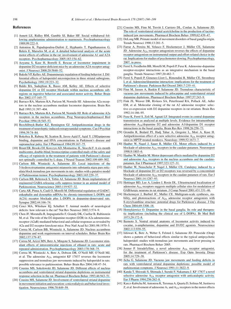
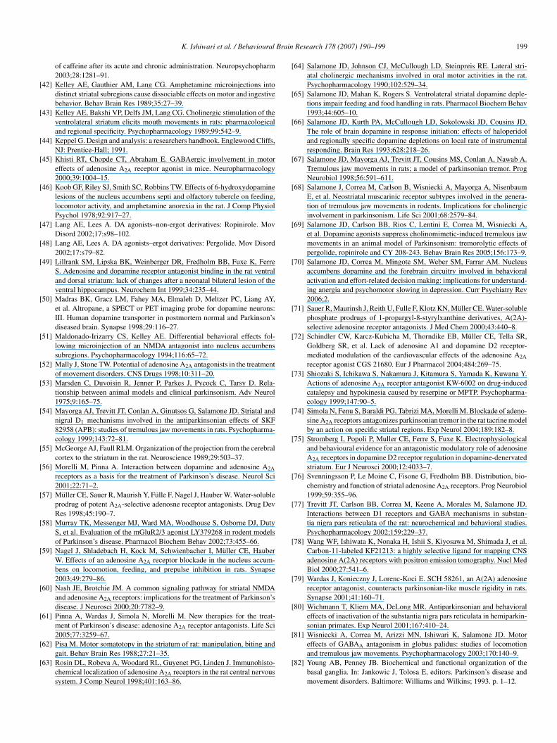
tructures, such as accumbens core, or shell, or VLS; asymmetrical), or signifi-ant damage around the injection site, was not included in the statistical analysesf behavioral data (41.2% of all implantations were rejected). For analyses ofore and shell placements, rats had to have bilateral and symmetrical placementsolely within the core or the shell in order to be included, with no ambiguouslacements in areas that separated the two subregions. See Figs. 1 and 2 fordrawings and photomicrographs of representative cannula placements in the
arget structures.
.7. Data analyses
Total number of locomotor activity counts were analyzed with between-roups analysis of variance (ANOVA). Non-orthogonal planned comparisons
K. Ishiwari et al. / Behavioural Brain Research 178 (2007) 190–199 193
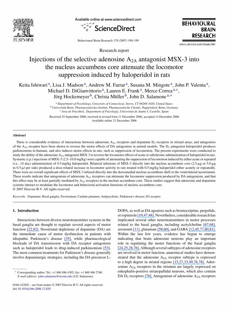
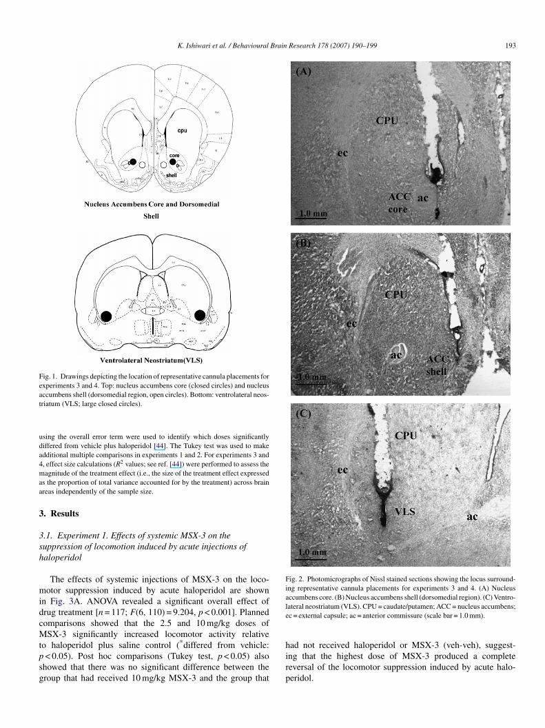
Fig. 1. Drawings depicting the location of representative cannula placements forexperiments 3 and 4. Top: nucleus accumbens core (closed circles) and nucleusat
uda4maa
3
3sh
midcMtpsg
Fig. 2. Photomicrographs of Nissl stained sections showing the locus surround-ing representative cannula placements for experiments 3 and 4. (A) Nucleusaccumbens core. (B) Nucleus accumbens shell (dorsomedial region). (C) Ventro-le
ccumbens shell (dorsomedial region, open circles). Bottom: ventrolateral neos-riatum (VLS; large closed circles).
sing the overall error term were used to identify which doses significantlyiffered from vehicle plus haloperidol [44]. The Tukey test was used to makedditional multiple comparisons in experiments 1 and 2. For experiments 3 and, effect size calculations (R2 values; see ref. [44]) were performed to assess theagnitude of the treatment effect (i.e., the size of the treatment effect expressed
s the proportion of total variance accounted for by the treatment) across brainreas independently of the sample size.
. Results
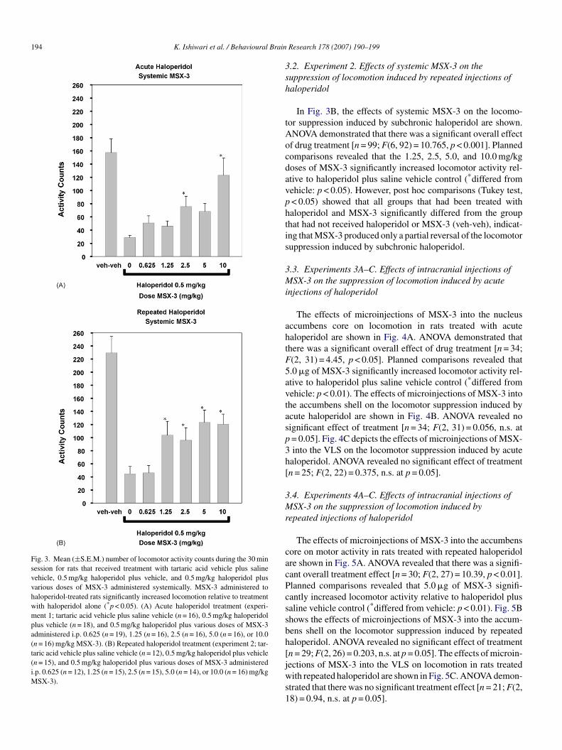
.1. Experiment 1. Effects of systemic MSX-3 on theuppression of locomotion induced by acute injections ofaloperidol
The effects of systemic injections of MSX-3 on the loco-otor suppression induced by acute haloperidol are shown
n Fig. 3A. ANOVA revealed a significant overall effect ofrug treatment [n = 117; F(6, 110) = 9.204, p < 0.001]. Plannedomparisons showed that the 2.5 and 10 mg/kg doses ofSX-3 significantly increased locomotor activity relative
o haloperidol plus saline control (*differed from vehicle:< 0.05). Post hoc comparisons (Tukey test, p < 0.05) also
howed that there was no significant difference between theroup that had received 10 mg/kg MSX-3 and the group that
hirp
ateral neostriatum (VLS). CPU = caudate/putamen; ACC = nucleus accumbens;c = external capsule; ac = anterior commissure (scale bar = 1.0 mm).
ad not received haloperidol or MSX-3 (veh-veh), suggest-ng that the highest dose of MSX-3 produced a completeeversal of the locomotor suppression induced by acute halo-eridol.
194 K. Ishiwari et al. / Behavioural Brain
Fig. 3. Mean (±S.E.M.) number of locomotor activity counts during the 30 minsession for rats that received treatment with tartaric acid vehicle plus salinevehicle, 0.5 mg/kg haloperidol plus vehicle, and 0.5 mg/kg haloperidol plusvarious doses of MSX-3 administered systemically. MSX-3 administered tohaloperidol-treated rats significantly increased locomotion relative to treatmentwith haloperidol alone (*p < 0.05). (A) Acute haloperidol treatment (experi-ment 1; tartaric acid vehicle plus saline vehicle (n = 16), 0.5 mg/kg haloperidolplus vehicle (n = 18), and 0.5 mg/kg haloperidol plus various doses of MSX-3administered i.p. 0.625 (n = 19), 1.25 (n = 16), 2.5 (n = 16), 5.0 (n = 16), or 10.0(n = 16) mg/kg MSX-3). (B) Repeated haloperidol treatment (experiment 2; tar-taric acid vehicle plus saline vehicle (n = 12), 0.5 mg/kg haloperidol plus vehicle(n = 15), and 0.5 mg/kg haloperidol plus various doses of MSX-3 administeredi.p. 0.625 (n = 12), 1.25 (n = 15), 2.5 (n = 15), 5.0 (n = 14), or 10.0 (n = 16) mg/kgMSX-3).
3sh
tAocdavphtis
3Mi
ahtF5avtasp3h[
3Mr
cacPcssbh[jws1
Research 178 (2007) 190–199
.2. Experiment 2. Effects of systemic MSX-3 on theuppression of locomotion induced by repeated injections ofaloperidol
In Fig. 3B, the effects of systemic MSX-3 on the locomo-or suppression induced by subchronic haloperidol are shown.NOVA demonstrated that there was a significant overall effectf drug treatment [n = 99; F(6, 92) = 10.765, p < 0.001]. Plannedomparisons revealed that the 1.25, 2.5, 5.0, and 10.0 mg/kgoses of MSX-3 significantly increased locomotor activity rel-tive to haloperidol plus saline vehicle control (*differed fromehicle: p < 0.05). However, post hoc comparisons (Tukey test,< 0.05) showed that all groups that had been treated withaloperidol and MSX-3 significantly differed from the grouphat had not received haloperidol or MSX-3 (veh-veh), indicat-ng that MSX-3 produced only a partial reversal of the locomotoruppression induced by subchronic haloperidol.
.3. Experiments 3A–C. Effects of intracranial injections ofSX-3 on the suppression of locomotion induced by acute
njections of haloperidol
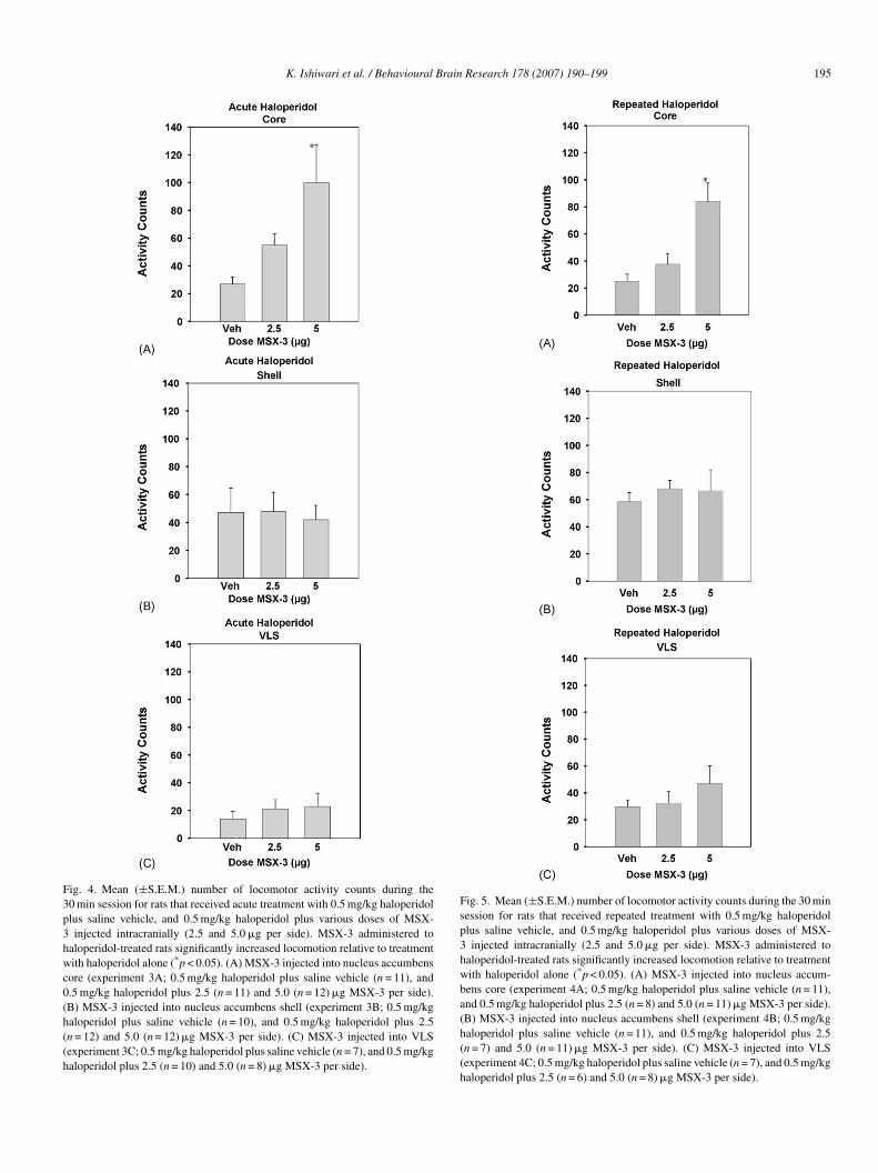
The effects of microinjections of MSX-3 into the nucleusccumbens core on locomotion in rats treated with acutealoperidol are shown in Fig. 4A. ANOVA demonstrated thathere was a significant overall effect of drug treatment [n = 34;(2, 31) = 4.45, p < 0.05]. Planned comparisons revealed that.0 �g of MSX-3 significantly increased locomotor activity rel-tive to haloperidol plus saline vehicle control (*differed fromehicle: p < 0.01). The effects of microinjections of MSX-3 intohe accumbens shell on the locomotor suppression induced bycute haloperidol are shown in Fig. 4B. ANOVA revealed noignificant effect of treatment [n = 34; F(2, 31) = 0.056, n.s. at= 0.05]. Fig. 4C depicts the effects of microinjections of MSX-into the VLS on the locomotor suppression induced by acute
aloperidol. ANOVA revealed no significant effect of treatmentn = 25; F(2, 22) = 0.375, n.s. at p = 0.05].
.4. Experiments 4A–C. Effects of intracranial injections ofSX-3 on the suppression of locomotion induced by
epeated injections of haloperidol
The effects of microinjections of MSX-3 into the accumbensore on motor activity in rats treated with repeated haloperidolre shown in Fig. 5A. ANOVA revealed that there was a signifi-ant overall treatment effect [n = 30; F(2, 27) = 10.39, p < 0.01].lanned comparisons revealed that 5.0 �g of MSX-3 signifi-antly increased locomotor activity relative to haloperidol plusaline vehicle control (*differed from vehicle: p < 0.01). Fig. 5Bhows the effects of microinjections of MSX-3 into the accum-ens shell on the locomotor suppression induced by repeatedaloperidol. ANOVA revealed no significant effect of treatmentn = 29; F(2, 26) = 0.203, n.s. at p = 0.05]. The effects of microin-
ections of MSX-3 into the VLS on locomotion in rats treatedith repeated haloperidol are shown in Fig. 5C. ANOVA demon-trated that there was no significant treatment effect [n = 21; F(2,8) = 0.94, n.s. at p = 0.05].
K. Ishiwari et al. / Behavioural Brain Research 178 (2007) 190–199 195
Fig. 4. Mean (±S.E.M.) number of locomotor activity counts during the30 min session for rats that received acute treatment with 0.5 mg/kg haloperidolplus saline vehicle, and 0.5 mg/kg haloperidol plus various doses of MSX-3 injected intracranially (2.5 and 5.0 �g per side). MSX-3 administered tohaloperidol-treated rats significantly increased locomotion relative to treatmentwith haloperidol alone (*p < 0.05). (A) MSX-3 injected into nucleus accumbenscore (experiment 3A; 0.5 mg/kg haloperidol plus saline vehicle (n = 11), and0.5 mg/kg haloperidol plus 2.5 (n = 11) and 5.0 (n = 12) �g MSX-3 per side).(B) MSX-3 injected into nucleus accumbens shell (experiment 3B; 0.5 mg/kghaloperidol plus saline vehicle (n = 10), and 0.5 mg/kg haloperidol plus 2.5(n = 12) and 5.0 (n = 12) �g MSX-3 per side). (C) MSX-3 injected into VLS(experiment 3C; 0.5 mg/kg haloperidol plus saline vehicle (n = 7), and 0.5 mg/kghaloperidol plus 2.5 (n = 10) and 5.0 (n = 8) �g MSX-3 per side).
Fig. 5. Mean (±S.E.M.) number of locomotor activity counts during the 30 minsession for rats that received repeated treatment with 0.5 mg/kg haloperidolplus saline vehicle, and 0.5 mg/kg haloperidol plus various doses of MSX-3 injected intracranially (2.5 and 5.0 �g per side). MSX-3 administered tohaloperidol-treated rats significantly increased locomotion relative to treatmentwith haloperidol alone (*p < 0.05). (A) MSX-3 injected into nucleus accum-bens core (experiment 4A; 0.5 mg/kg haloperidol plus saline vehicle (n = 11),and 0.5 mg/kg haloperidol plus 2.5 (n = 8) and 5.0 (n = 11) �g MSX-3 per side).(B) MSX-3 injected into nucleus accumbens shell (experiment 4B; 0.5 mg/kghaloperidol plus saline vehicle (n = 11), and 0.5 mg/kg haloperidol plus 2.5(n = 7) and 5.0 (n = 11) �g MSX-3 per side). (C) MSX-3 injected into VLS(experiment 4C; 0.5 mg/kg haloperidol plus saline vehicle (n = 7), and 0.5 mg/kghaloperidol plus 2.5 (n = 6) and 5.0 (n = 8) �g MSX-3 per side).
196 K. Ishiwari et al. / Behavioural Brain
Table 1Effect size calculations for the intracranial experiments (3A–C and 4A–C)
R2 values
Acute haloperidolNucleus accumbens core 0.223*
Nucleus accumbens shell 0.004Ventrolateral neostriatum 0.033
Repeated haloperidolNucleus accumbens core 0.435*
Nucleus accumbens shell 0.015Ventrolateral neostriatum 0.095
To
3e
uMsio
3ih
eeti1e3spi5
4
tei2laM0aoMs
sawalaTtr
sTatiahvatammrjsaowmasoddt
aartiaMd0diMnMab
his table shows the results of the effect size calculations (R2 values, ref. [44])btained with injections into each brain region studied.* Effect size significantly different from 0, p < 0.05.
.5. Comparisons of effect sizes across brain areas inxperiments 3 and 4
Table 1 shows the calculated effect magnitudes (i.e., R2 val-es; ref. [44]) for all six intracranial experiments. Injections ofSX-3 into nucleus accumbens core produced a moderate effect
ize in experiment 3A (0.223) and a large effect size in exper-ment 4A (0.435). All other effects sizes were small, and nonef them significantly differed from 0.
.6. Experiments 5A and B. Effects of systemic andntracranial injections of MSX-3 in rats not treated withaloperidol
Systemic administration of 10.0 mg/kg MSX-3, which wasffective at stimulating locomotion in experiments 1–2, had noffect on locomotor activity in rats that were not haloperidolreated in experiment 5A. Mean (±S.E.M.) locomotor activ-ty counts per 30 min were as follows: saline: 176.4 (±26.7),0.0 mg/kg MSX-3: 223.4 (±46.6); (t = 0.87, d.f. = 12, n.s.). Inxperiment 5B, accumbens core injections at the dose of MSX-that was effective in experiments 3A and 4A (i.e., 5.0 �g per
ide) had no significant effect on locomotion in rats that were notre-treated with haloperidol. Mean (±S.E.M.) locomotor activ-ty counts per 30 min were as follows: saline: 252.4 (±23.9),.0 �g MSX-3: 267.6 (±39.1); (t = 0.33, d.f. = 12, n.s.).
. Discussion
These experiments were conducted to study the ability ofhe adenosine A2A antagonist MSX-3 to reverse the locomotorffects of acute or subchronic administration of haloperidoln rats. Systemic injections of MSX-3 in a dose range of.5–10.0 mg/kg were capable of reversing the suppression ofocomotion induced by either acute or repeated (i.e., 14 day)dministration of 0.5 mg/kg haloperidol. Bilateral infusions ofSX-3 into the nucleus accumbens core (2.5 �g or 5.0 �g in
.5 �l per side) produced a dose-related increase in locomotor
ctivity in rats treated with 0.5 mg/kg haloperidol either acutelyr repeatedly. There was not an overall significant effect ofSX-3 infused into either the dorsomedial nucleus accumbenshell or the VLS. In addition, there were no significant effects of
stmo
Research 178 (2007) 190–199
ystemic or intra-accumbens injections of MSX-3 (10.0 mg/kgnd 5.0 �g per side, respectively) in rats that were not treatedith haloperidol. Taken together, these results indicate that
ntagonism of adenosine A2A receptors can reverse theocomotor suppression produced by DA antagonism, and that
critical site for this effect is the nucleus accumbens core.hese results have important implications for understanding
he mechanisms underlying the motor effects of adenosine A2Aeceptor antagonists.
In the first two experiments, MSX-3 attenuated the suppres-ion of locomotion induced by administration of haloperidol.his effect was evident whether the haloperidol was injectedcutely (experiment 1) or with a repeated 14 day administra-ion procedure (experiment 2). The stimulation of locomotionnduced by MSX-3 was comparable in both experiments,lthough the control levels of activity were higher in the repeatedaloperidol study. The present results are consistent with pre-ious studies demonstrating that systemic administration ofdenosine A2A antagonists can reverse the deficits in locomotionhat were induced by reserpine [73], D2 receptor deficiency [3],nd haloperidol [18]. In addition, these results involving loco-otion are consistent with those of studies that employed othereasures of motor dysfunction, including haloperidol-induced
igidity [79], catalepsy [30,32,40], and drug-induced tremulousaw movements, which are used as an animal model of parkin-onian tremor [18,74]. Taken together, these results providedditional support for the hypothesis that adenosine A2A antag-nism can reverse motor impairments induced by interferenceith DA transmission in animals. These findings with animalodels are widely used to support the idea that adenosine A2A
ntagonists could represent a novel non-dopaminergic treatmenttrategy for idiopathic Parkinson’s disease [37,38,76]. More-ver, the ability of adenosine A2A antagonists to reverse motorysfunctions induced by typical antipsychotics such as haloperi-ol suggests that adenosine A2A antagonists also could be usedo treat antipsychotic-induced parkinsonism in humans [18].
Although studies involving systemic administration of drugsre important, they do not provide specific information about thenatomical locus mediating the effects of those drugs. For thateason, experiments 3 and 4 were conducted in order to studyhe effects of local intracranial administration of MSX-3 directlynto distinct striatal subregions, including two sites in the nucleusccumbens and one in the neostriatum. Bilateral infusions ofSX-3 directly into the nucleus accumbens core produced a
ose-related increase in locomotor activity in rats treated with.5 mg/kg haloperidol. This effect occurred when the haloperi-ol was injected acutely, and also when the 14 day repeatednjection procedure was used. Effect size analyses indicated that
SX-3 produced moderate-to-large effects when injected intoucleus accumbens core. These positive effects obtained withSX-3 injections into the core clearly demonstrate that nucleus
ccumbens is an effective site for the stimulation of locomotiony MSX-3 in haloperidol-treated rats, an observation that is con-
istent with previous studies showing that adenosine A2A recep-ors in nucleus accumbens are involved in the regulation of loco-otion [6,7,31,59]. However, the robust and consistent effectsbserved after injections of MSX-3 into the nucleus accumbens
Brain
cTdTidiswpADatoforntp[iutaittolmiartoer
MoVntfteatDffttso
drialwntmriittbtp
risddaseasbe(ariactaascurtantbpww
A
K. Ishiwari et al. / Behavioural
ore were not mimicked by injections into the dorsomedial shell.here was not an overall significant effect of MSX-3 infusedirectly into the dorsomedial shell in either experiment 3 or 4.he involvement of specific subregions of nucleus accumbens
n locomotion appears to differ depending upon the particularrug class being studied [36,51,57]. The present results, indicat-ng that adenosine A2A antagonism in the core was effective attimulating locomotion in rats with impaired DA transmissionhile shell injections of MSX-3 were not, are consistent withrevious reports indicating that the concentration of adenosine2A receptors is higher in the core than in the shell [29,63].espite the fact that 5.0 �g MSX-3 injected into the nucleus
ccumbens core was able to stimulate locomotion in haloperidol-reated rats, this dose did not stimulate locomotion in the absencef haloperidol. This observation is consistent with the resultsrom experiment 5A with systemic MSX-3, and also with previ-us findings. A systemic dose of KF 17837 that was capable ofeversing the haloperidol-induced suppression of locomotion didot stimulate locomotion when administered alone [18]. In addi-ion, doses of MSX-3 that reversed haloperidol-induced leverressing had no effects when injected without the DA antagonist23]. Although recent work has indicated that intra-accumbensnjections of MSX-3 can increase locomotion [59], that papersed behavioral methods that were substantially different fromhose used in the present study (i.e., open-field activity in habitu-ted animals). Moreover, previous studies showing that systemicnjections of MSX-3 increased locomotion used younger ratshan the present study [2,41,72]. Additional studies should inves-igate the role of factors such as dose, placement, age, degreef habituation, and behavioral conditions in modulating theocomotor effects of MSX-3 when administered alone. Further-
ore, in view of data indicating that medial caudate-putamen isnvolved in the effects of adenosine A2A receptor agonists andntagonists on catalepsy [30–32], it is possible that this striatalegion also is involved in locomotor effects of these drugs. Forhat reason, future mapping studies should explore the effectsf drug injections into various placement sites within the regionxtending from the nucleus accumbens core to the anteromedialegions of caudate-putamen.
In contrast to the positive effects obtained from injections ofSX-3 into the nucleus accumbens core, no significant effects
n locomotion were obtained after injections of MSX-3 into theLS. Previous work has suggested minimal involvement of thiseostriatal subregion in locomotor function. For example, deple-ions of DA in the VLS by local infusions of 6-hydroxydopamineailed to suppress locomotion [19,39], and intracranial injec-ions of amphetamine into the VLS were reported to have noffect on locomotor activity [42]. Although the VLS does notppear to be an important site for the regulation of locomo-ion, it is important for other aspects of motor function. VLSA depletions have been shown to impair skilled use of the
orepaws for functions such as reaching, grasping, feeding,ood handling and lever pressing [19,20,39,65,66]. In addi-
ion, the VLS is an important region of the neostriatum forhe control of oral motor activity [43,62,64,67]. It has beenuggested that the lateral striatum of the rat is the homologuef the putamen of primates [67], and anatomical studies haveUB
Research 178 (2007) 190–199 197
emonstrated that the VLS of the rat receives input from head-elated areas of motor cortex [55]. Neurochemical mechanismsn the VLS involving several neurotransmitters, including DA,cetylcholine, and adenosine, have been implicated in tremu-ous jaw movements [21,26,39,54,64,67,74]. These movements,hich are induced by DA antagonists, DA depletions, and choli-omimetics, have been used as a rodent model of parkinsonianremor [14,37,67,69]. Recently, it was demonstrated that the jaw
ovements induced by the anticholinesterase tacrine could beeversed by infusions of an adenosine A2A antagonist directlynto the VLS [64]. Thus, despite the fact that the VLS has beenmplicated in several aspects of motor function and dysfunc-ion, including skilled control of the forepaws and oral tremor,he present results indicate that the stimulation of locomotiony an adenosine A2A antagonist in animals with impaired DAransmission is related to actions on the nucleus accumbens,articularly the core subregion, but not to actions on the VLS.
In conjunction with other published studies, the presentesults emphasize that different striatal subregions are involvedn distinct aspects of motor function. This principle is demon-trated clearly in the substantial literature showing that DAepletions or antagonism can have radically different effectsepending upon the striatal locus being affected [4,19,65,66]. Inddition, this principle has important implications for under-tanding the anatomical mechanisms underlying the motorffects of antiparkinsonian drugs, including adenosine A2Antagonists. Although antiparkinsonian drugs are typically givenystemically, and the therapeutic target would generally be aroad improvement across diverse motor symptoms, it is nev-rtheless reasonable to argue that different therapeutic effectsi.e., increases in locomotion, decreases in rigidity or tremor)re related to actions on distinct striatal subcircuits. The presentesults would suggest that the nucleus accumbens core is a crit-cal striatal site for the restoration of locomotion seen afterdministration of adenosine A2A antagonists. This observation isonsistent with previous studies involving the locomotor func-ions of nucleus accumbens in both rodents [6,7,16,19,31,59]nd primates [1]. Thus, it is possible that restoration of locomotorctivity in human patients also is related to actions of antiparkin-onian drugs on the nucleus accumbens, at least in part. Inontrast, motor dysfunctions such as tremor and skilled forelimbsage appear to involve other striatal subregions [67]. Additionalesearch should focus on how adenosine A2A antagonists exertheir effects by modifying distinct subcircuits within the over-ll striatal circuitry, including neostriatal areas adjacent to theucleus accumbens. Moreover, future studies should investigatehe extent to which adenosine A2A receptors in nucleus accum-ens core are involved in other behavioral processes, such assychomotor activation and energy-related functions [23,70],hich are related to the motivational deficits seen in patientsith Parkinson’s disease, depression, and other disorders.
cknowledgements
This research was supported by a grant to J.D.S. from thenited States NIH/NINDS. Many thanks to Joshua Orabone,rian Mirante and Jamie Bunce for their technical assistance.
1 Brain
R
[
[
[
[
[
[
[
[
[
[
[
[
[
[
[
[
[
[
[
[
[
[
[
[
[
[
[
[
[
[
98 K. Ishiwari et al. / Behavioural
eferences
[1] Annett LE, Ridley RM, Gamble SJ, Baker HF. Social withdrawal fol-lowing amphetamine administration to marmosets. Psychopharmacology1989;99:222–9.
[2] Antoniou K, Papadopoulou-Daifoti Z, Hyphantis T, Papathanasiou G,Bekris E, Marselos M, et al. A detailed behavioral analysis of the acutemotor effects of caffeine in the rat: involvement of adenosine A1 and A2Areceptors. Psychopharmacology 2005;183:154–62.
[3] Aoyama S, Kase H, Borrelli E. Rescue of locomotor impairment indopamine D2 receptor-deficient mice by an adenosine A2A receptor antag-onist. J Neurosci 2000;20:5848–52.
[4] Bakshi VP, Kelley AE. Dopaminergic regulation of feeding behavior: I. Dif-ferential effects of haloperidol microinjection in three striatal subregions.Psychobiology 1991;19:223–32.
[5] Baldo BA, Sadeghian K, Basso AM, Kelley AE. Effects of selectivedopamine D1 or D2 receptor blockade within nucleus accumbens sub-regions on ingestive behavior and associated motor activity. Behav BrainRes 2002;137:165–77.
[6] Barraco RA, Martens KA, Parizon M, Normile HJ. Adenosine A2a recep-tors in the nucleus accumbens mediate locomotor depression. Brain ResBull 1993;31:397–404.
[7] Barraco RA, Martens KA, Parizon M, Normile HJ. Role of adenosine A2areceptors in the nucleus accumbens. Prog Neuropsychopharmacol BiolPsychiat 1994;18:545–53.
[8] Bezchlibnyk-Butler KZ, Remington GJ. Antiparkinsonian drugs in thetreatment of neuroleptic-induced extrapyramidal symptoms. Can J Psychiat1994;39:74–84.
[9] Bokobza B, Ruberg M, Scatton B, Javoy-Agid F, Agid Y. [3H]spiperonebinding, dopamine and HVA concentrations in Parkinson’s disease andsupranuclear palsy. Eur J Pharmacol 1984;99:167–75.
10] Brunt ER, Brooks DJ, Korczyn AD, Montastruc JL, Stocchi F. A six-monthmulticentre, double-blind, bromocriptine-controlled study of the safety andefficacy of ropinirole in the treatment of patients with Parkinson’s diseasenot optimally controlled by L-dopa. J Neural Transm 2002;109:489–502.
11] Carlson BB, Wisniecki A, Salamone JD. Local injections of the5-hydroxytryptamine antagonist mianserin into substantia nigra pars retic-ulata block tremulous jaw movements in rats: studies with a putative modelof Parkinsonian tremor. Psychopharmacology 2003;165:229–37.
12] Carlson BB, Behrstock S, Tobin AJ, Salamone JD. Brain implantations ofengineered GABA-releasing cells suppress tremor in an animal model ofParkinsonism. Neuroscience 2003;119:927–32.
13] Carta AR, Pinna A, Cauli O, Morelli M. Differential regulation of GAD67,enkephalin and dynorphin mRNAs by chronic-intermittent L-DOPA andA(2A) receptor blockade plus L-DOPA in dopamine-denervated rats.Synapse 2002;44:166–74.
14] Cenci MA, Whishaw IQ, Schallert T. Animal models of neurologicaldeficits: how relevant is the rat? Nat Rev Neurosci 2002;3:574–9.
15] Chen JF, Moratalla R, Impagnatiello F, Grandy DK, Cuellar B, RubinsteinM, et al. The role of the D2 dopamine receptor (D2R) in A2a adenenosine-receptor (A2aR) mediated behavioral and cellular responses as revealed byA2a and D2 receptor knockout mice. Proc Natl Acad Sci 2001;98:1970–5.
16] Correa M, Carlson BB, Wisniecki A, Salamone JD. Nucleus accumbensdopamine and work requirements on interval schedules. Behav Brain Res2002;137:179–87.
17] Correa M, Arizzi MN, Betz A, Mingote S, Salamone JD. Locomotor stim-ulant effects of intraventricular injections of ethanol in rats: acute andrepeated administration. Psychopharmacology 2003;170:368–75.
18] Correa M, Wisniecki A, Betz A, Dobson DR, O’Neill MF, O’Neill MJ,et al. The adenosine A2A antagonist KF 17837 reverses the locomotorsuppression and tremulous jaw movements induced by haloperidol in rats:possible relevance to parkinsonism. Behav Brain Res 2004;148:47–54.
19] Cousins MS, Sokolowski JD, Salamone JD. Different effects of nucleus
accumbens and ventrolateral striatal dopamine depletions on instrumentalresponse selection in the rat. Pharmacol Biochem Behav 1993;46:943–51.20] Cousins MS, Salamone JD. Involvement of ventrolateral striatal dopaminein movement initiation and execution: a microdialysis and behavioral inves-tigation. Neuroscience 1996;70:849–59.
[
[
Research 178 (2007) 190–199
21] Cousins MS, Finn M, Trevitt J, Carriero DL, Conlan A, Salamone JD.The role of ventrolateral striatal acetylcholine in the production of tacrine-induced jaw movements. Pharmacol Biochem Behav 1999;62:439–47.
22] DeLong MR. Primate model of movement disorders of basal ganglia origin.Trends Neurosci 1990;13:281–5.
23] Farrar A, Pereira M, Velasco F, Hockemeyer J, Muller CE, SalamoneJD. Adenosine A2A receptor antagonism reverses the effects of dopaminereceptor antagonism on instrumental output and effort-related choice in therat. Implications for studies of psychomotor slowing. Psychopharmacology,2007, in press.
24] Ferre S, Freidholm BB, Morelli M, Popoli P, Fuxe K. Adenosine-dopaminereceptor-receptor interactions as an integrative mechanism in the basalganglia. Trends Neurosci 1997;20:482–7.
25] Ferre S, Popoli P, Gimenez-Llort L, Rimondini R, Muller CE, StrombergI, et al. Adenosine/dopamine interaction: implications for the treatment ofParkinson’s disease. Parkinson Rel Disord 2001;7:235–41.
26] Finn M, Jassen A, Baskin P, Salamone JD. Tremulous characteristic ofvacuous jaw movements induced by pilocarpine and ventrolateral striataldopamine depletions. Pharmacol Biochem Behav 1997;57:243–9.
27] Fink JS, Weaver DR, Rivkees SA, Peterfreund RA, Pollack AE, AdlerEM, et al. Molecular cloning of the rat A2 adenosine receptor: selec-tive co-expression with D2 dopamine receptors in rat striatum. Brain Res1992;14:186–95.
28] Fuxe K, Ferre S, Zoli M, Agnati LF. Integrated events in central dopaminetransmission as analyzed at multiple levels. Evidence for intramembraneadenosine A2A/dopamine D2 and adenosine A1/dopamine D1 receptorinteractions in the basal ganglia. Brain Res Rev 1998;26:258–73.
29] Grondin R, Bedard PJ, Hadj Tahar A, Gregoire L, Mori A, Kase H.Antiparkinsonian effect of a new selective adenosine A2A receptor antag-onist in MPTP-treated monkeys. Neurology 1999;52:1673–7.
30] Hauber W, Nagel J, Sauer R, Muller CE. Motor effects induced by ablockade of adenosine A2A receptors in the caudate-putamen. Neuroreport1998;9:1803–6.
31] Hauber W, Munkle M. Motor depressant effects mediated by dopamine D2and adenosine A2A receptors in the nucleus accumbens and the caudate-putamen. Eur J Pharmacol 1997;323:127–31.
32] Hauber W, Neuscheler P, Nagel J, Muller CE. Catalepsy induced by ablockade of dopamine D1 or D2 receptors was reversed by a concomitantblockade of adenosine A2A receptors in the caudate putamen of rats. Eur JNeurosci 2001;14:1287–93.
33] Hettinger BD, Lee A, Linden J, Rosin DL. Ultrastructural localization ofadenosine A2A receptors suggests multiple cellular sites for modulation ofGABAergic neurons in rat striatum. J Comp Neurol 2001;431:331–46.
34] Hockemeyer J, Burbiel JC, Muller CE. Multigram-scale syntheses, sta-bility, and photoreactions of A2A adenosine receptor antagonists with8-styrylxanthine structure: potential drugs for Parkinson’s disease. J OrgChem 2004;69:3308–18.
35] Hornykiewicz O. Dopamine in the basal ganglia. Its role and therapeu-tic implications (including the clinical use of L-DOPA). Br Med Bull1973;29:172–8.
36] Ikemoto S. Ventral striatal anatomy of locomotor activity induced bycocaine, d-amphetamine, dopamine and D1/D2 agonists. Neuroscience2002;113:939–55.
37] Ishiwari K, Betz A, Weber S, Felsted J, Salamone JD. Pimozide (Orap)shows a pattern of behavioral effects similar to the typical antipsychotichaloperidol: studies with tremulous jaw movements and lever pressing inrats. Pharmacol Biochem Behav 2005.
38] Jenner P. Istradefylline, a novel adenosine A2A receptor antagonist,for the treatment of Parkinson’s disease. Exp Opin Investig Drugs2005;14:729–38.
39] Jicha G, Salamone JD. Vacuous jaw movements and feeding deficits inrats with ventrolateral striatal dopamine depletions: possible model ofparkinsonian symptoms. J Neurosci 1991;11:3822–9.
40] Kanda T, Shiozaki S, Shimada J, Suzuki F, Nakamara J. KF 17837 a novelselective adenosine A2A receptor antagonist with anticataleptic activity.Eur J Pharm 1994;256:262–8.
41] Karcz-Kubicha M, Antoniou K, Tersmaa A, Quarta D, Solinas M, JustinovaZ, et al. Involvement of adenosine A1 and A2A receptors in the motor effects
Brain
[
[
[
[
[
[
[
[
[
[
[
[
[
[
[
[
[
[
[
[
[
[
[
[
[
[
[
[
[
[
[
[
[
[
[
[
[
[
[
[effects of GABAA antagonism in globus palidus: studies of locomotion
K. Ishiwari et al. / Behavioural
of caffeine after its acute and chronic administration. Neuropsychopharm2003;28:1281–91.
42] Kelley AE, Gauthier AM, Lang CG. Amphetamine microinjections intodistinct striatal subregions cause dissociable effects on motor and ingestivebehavior. Behav Brain Res 1989;35:27–39.
43] Kelley AE, Bakshi VP, Delfs JM, Lang CG. Cholinergic stimulation of theventrolateral striatum elicits mouth movements in rats: pharmacologicaland regional specificity. Psychopharmacology 1989;99:542–9.
44] Keppel G. Design and analysis: a researchers handbook. Englewood Cliffs,NJ: Prentice-Hall; 1991.
45] Khisti RT, Chopde CT, Abraham E. GABAergic involvement in motoreffects of adenosine A2A receptor agonist in mice. Neuropharmacology2000;39:1004–15.
46] Koob GF, Riley SJ, Smith SC, Robbins TW. Effects of 6-hydroxydopaminelesions of the nucleus accumbens septi and olfactory tubercle on feeding,locomotor activity, and amphetamine anorexia in the rat. J Comp PhysiolPsychol 1978;92:917–27.
47] Lang AE, Lees A. DA agonists–non-ergot derivatives: Ropinirole. MovDisord 2002;17:s98–102.
48] Lang AE, Lees A. DA agonists–ergot derivatives: Pergolide. Mov Disord2002;17:s79–82.
49] Lillrank SM, Lipska BK, Weinberger DR, Fredholm BB, Fuxe K, FerreS. Adenosine and dopamine receptor antagonist binding in the rat ventraland dorsal striatum: lack of changes after a neonatal bilateral lesion of theventral hippocampus. Neurochem Int 1999;34:235–44.
50] Madras BK, Gracz LM, Fahey MA, Elmaleh D, Meltzer PC, Liang AY,et al. Altropane, a SPECT or PET imaging probe for dopamine neurons:III. Human dopamine transporter in postmortem normal and Parkinson’sdiseased brain. Synapse 1998;29:116–27.
51] Maldonado-Irizarry CS, Kelley AE. Differential behavioral effects fol-lowing microinjection of an NMDA antagonist into nucleus accumbenssubregions. Psychopharmacology 1994;116:65–72.
52] Mally J, Stone TW. Potential of adenosine A2A antagonists in the treatmentof movement disorders. CNS Drugs 1998;10:311–20.
53] Marsden C, Duvoisin R, Jenner P, Parkes J, Pycock C, Tarsy D. Rela-tionship between animal models and clinical parkinsonism. Adv Neurol1975;9:165–75.
54] Mayorga AJ, Trevitt JT, Conlan A, Ginutsos G, Salamone JD. Striatal andnigral D1 mechanisms involved in the antiparkinsonian effects of SKF82958 (APB): studies of tremulous jaw movements in rats. Psychopharma-cology 1999;143:72–81.
55] McGeorge AJ, Faull RLM. Organization of the projection from the cerebralcortex to the striatum in the rat. Neuroscience 1989;29:503–37.
56] Morelli M, Pinna A. Interaction between dopamine and adenosine A2A
receptors as a basis for the treatment of Parkinson’s disease. Neurol Sci2001;22:71–2.
57] Muller CE, Sauer R, Maurish Y, Fulle F, Nagel J, Hauber W. Water-solubleprodrug of potent A2A-selective adenosne receptor antagonists. Drug DevRes 1998;45:190–7.
58] Murray TK, Messenger MJ, Ward MA, Woodhouse S, Osborne DJ, DutyS, et al. Evaluation of the mGluR2/3 agonist LY379268 in rodent modelsof Parkinson’s disease. Pharmacol Biochem Behav 2002;73:455–66.
59] Nagel J, Shladebach H, Kock M, Schwienbacher I, Muller CE, HauberW. Effects of an adenosine A2A receptor blockade in the nucleus accum-bens on locomotion, feeding, and prepulse inhibition in rats. Synapse2003;49:279–86.
60] Nash JE, Brotchie JM. A common signaling pathway for striatal NMDAand adenosine A2A receptors: implications for the treatment of Parkinson’sdisease. J Neurosci 2000;20:7782–9.
61] Pinna A, Wardas J, Simola N, Morelli M. New therapies for the treat-ment of Parkinson’s disease: adenosine A2A receptor antagonists. Life Sci2005;77:3259–67.
62] Pisa M. Motor somatotopy in the striatum of rat: manipulation, biting and
gait. Behav Brain Res 1988;27:21–35.63] Rosin DL, Robeva A, Woodard RL, Guyenet PG, Linden J. Immunohisto-chemical localization of adenosine A2A receptors in the rat central nervoussystem. J Comp Neurol 1998;401:163–86.
[
Research 178 (2007) 190–199 199
64] Salamone JD, Johnson CJ, McCullough LD, Steinpreis RE. Lateral stri-atal cholinergic mechanisms involved in oral motor activities in the rat.Psychopharmacology 1990;102:529–34.
65] Salamone JD, Mahan K, Rogers S. Ventrolateral striatal dopamine deple-tions impair feeding and food handling in rats. Pharmacol Biochem Behav1993;44:605–10.
66] Salamone JD, Kurth PA, McCullough LD, Sokolowski JD, Cousins JD.The role of brain dopamine in response initiation: effects of haloperidoland regionally specific dopamine depletions on local rate of instrumentalresponding. Brain Res 1993;628:218–26.
67] Salamone JD, Mayorga AJ, Trevitt JT, Cousins MS, Conlan A, Nawab A.Tremulous jaw movements in rats; a model of parkinsonian tremor. ProgNeurobiol 1998;56:591–611.
68] Salamone J, Correa M, Carlson B, Wisniecki A, Mayorga A, NisenbaumE, et al. Neostriatal muscarinic receptor subtypes involved in the genera-tion of tremulous jaw movements in rodents. Implications for cholinergicinvolvement in parkinsonism. Life Sci 2001;68:2579–84.
69] Salamone JD, Carlson BB, Rios C, Lentini E, Correa M, Wisniecki A,et al. Dopamine agonists suppress cholinomimetic-induced tremulous jawmovements in an animal model of Parkinsonism: tremorolytic effects ofpergolide, ropinirole and CY 208-243. Behav Brain Res 2005;156:173–9.
70] Salamone JD, Correa M, Mingote SM, Weber SM, Farrar AM. Nucleusaccumbens dopamine and the forebrain circuitry involved in behavioralactivation and effort-related decision making: implications for understand-ing anergia and psychomotor slowing in depression. Curr Psychiatry Rev2006;2.
71] Sauer R, Maurinsh J, Reith U, Fulle F, Klotz KN, Muller CE. Water-solublephosphate prodrugs of 1-propargyl-8-styrylxanthine derivatives, A(2A)-selective adenosine receptor antagonists. J Med Chem 2000;43:440–8.
72] Schindler CW, Karcz-Kubicha M, Thorndike EB, Muller CE, Tella SR,Goldberg SR, et al. Lack of adenosine A1 and dopamine D2 receptor-mediated modulation of the cardiovascular effects of the adenosine A2A
receptor agonist CGS 21680. Eur J Pharmacol 2004;484:269–75.73] Shiozaki S, Ichikawa S, Nakamura J, Kitamura S, Yamada K, Kuwana Y.
Actions of adenosine A2A receptor antagonist KW-6002 on drug-inducedcatalepsy and hypokinesia caused by reserpine or MPTP. Psychopharma-cology 1999;147:90–5.
74] Simola N, Fenu S, Baraldi PG, Tabrizi MA, Morelli M. Blockade of adeno-sine A2A receptors antagonizes parkinsonian tremor in the rat tacrine modelby an action on specific striatal regions. Exp Neurol 2004;189:182–8.
75] Stromberg I, Popoli P, Muller CE, Ferre S, Fuxe K. Electrophysiologicaland behavioural evidence for an antagonistic modulatory role of adenosineA2A receptors in dopamine D2 receptor regulation in dopamine-denervatedstriatum. Eur J Neurosci 2000;12:4033–7.
76] Svenningsson P, Le Moine C, Fisone G, Fredholm BB. Distribution, bio-chemistry and function of striatal adenosine A2A receptors. Prog Neurobiol1999;59:355–96.
77] Trevitt JT, Carlson BB, Correa M, Keene A, Morales M, Salamone JD.Interactions between D1 receptors and GABA mechanisms in substan-tia nigra pars reticulata of the rat: neurochemical and behavioral studies.Psychopharmacology 2002;159:229–37.
78] Wang WF, Ishiwata K, Nonaka H, Ishii S, Kiyosawa M, Shimada J, et al.Carbon-11-labeled KF21213: a highly selective ligand for mapping CNSadenosine A(2A) receptors with positron emission tomography. Nucl MedBiol 2000;27:541–6.
79] Wardas J, Konieczny J, Lorenc-Koci E. SCH 58261, an A(2A) adenosinereceptor antagonist, counteracts parkinsonian-like muscle rigidity in rats.Synapse 2001;41:160–71.
80] Wichmann T, Kliem MA, DeLong MR. Antiparkinsonian and behavioraleffects of inactivation of the substantia nigra pars reticulata in hemiparkin-sonian primates. Exp Neurol 2001;167:410–24.
81] Wisniecki A, Correa M, Arizzi MN, Ishiwari K, Salamone JD. Motor
and tremulous jaw movements. Psychopharmacology 2003;170:140–9.82] Young AB, Penney JB. Biochemical and functional organization of the
basal ganglia. In: Jankowic J, Tolosa E, editors. Parkinson’s disease andmovement disorders. Baltimore: Williams and Wilkins; 1993. p. 1–12.
Copyright © 2022 FDOKUMEN


![Regional distribution and kinetics of haloperidol binding in human brain: A pet study with [18F]haloperidol](https://static.fdokumen.com/doc/165x107/6344f2dd596bdb97a908b31a/regional-distribution-and-kinetics-of-haloperidol-binding-in-human-brain-a-pet.jpg)