Mortality risk associated with haloperidol use compared with ...

Journal of Neuroscience Research 33:605-616 (1992)
Haloperidol Prevents Induction of the hsp70 Heat Shock Gene in Neurons Injured by Phencyclidine (PCP), MKSOl, and Ketamine F.R. Sharp, M. Butman, S. Wang, J. Koistinaho, S.H. Graham, S.M. Sagar, L. Noble, P. Berger, and F.M. Longo Departments of Neurology (F.R.S., M.B., S. W., J.K., S.H.G., S.M.S., F.M.L.) and Psychiatry (P.B.), and Neurosurgery (L.N.), University of California at San Francisco, and the Department of Veterans Affairs Medical Center (F.R.S., M.B., S. W., J .K. , S.H.G., S.M.S., P.B., F.M.L.), San Francisco, California
The non-competitive NMDA receptor antagonists, PCP (phencyclidine), MK801, and ketamine produce psychosis in humans and abnormal vacuoles in pos- terior cingulate and retrosplenial rat cortical neu- rons. We show that PCP ( 2 5 mg/kg), MKS01 (20 .1 mg/kg), and ketamine (>20 mg/kg) induce hsp70 mRNA and HSP70 heat shock protein in these vacu- olated, injured neurons, and PCP also induces hsp70 in injured neocortical, piriform, and amygdala neu- rons. The PCP, MK801, and ketamine drug induced injury occurs in 30 day and older rats, but not in 0-20 day old rats, and is prevented by prior administration of the antipsychotic drugs haloperidol and rimcazole. Since haloperidol and rimcazole block dopamine and sigma receptors, and since MI muscarinic cholinergic receptor antagonists also prevent the injury produced by PCP, MKSO1, and ketamine, future studies will be needed to determine whether dopamine, sigma, M1, or other receptors mediate the injury. 0 1992 Wiley-Liss, Inc.
Key words: NMDA receptors, NMDA antagonists, glutamate, sigma receptors, dopamine receptors, phencyclidine (PCP), heat shock proteins, neuronal injury, ketamine, MK801, cingulate cortex, psycho- sis, schizophrenia
INTRODUCTION NMDA receptor antagonists decrease neuronal in-
jury produced by ischemia, hypoglycemia, and status epilepticus (Choi, 1988; Gill et al., 1987; Ozyurt et al., 1988; Rothman and Olney, 1986; Simon et al., 1984). Competitive NMDA receptor antagonists block access of glutamate to its receptor, whereas non-competitive NMDA receptor antagonists block entry of calcium through the NMDA-coupled calcium channel (Kemp et al., 1987; Watkins and Olvennan, 1987). The non-com- petitive NMDA antagonists, MK801, phencyclidine
(PCP), and ketamine (Watkins and Olvennan, 1987; Wong et al., 1986, 1988) have greater therapeutic po- tential because they cross the blood brain barrier much more effectively than competitive NMDA antagonists (Meldrum, 1985).
NMDA receptor antagonists, however, consis- tently cause behavioral abnormalities in animals (Koek et a]., 1988; Tricklebank et al., 1989) and can cause psy- chosis in humans (Domino and Luby, 1981). Olney et al. (1989) and then others (Allen and Iversen, 1990; Sharp et al., 1990, 1991a) showed that PCP, MK801, and ke- tamine produce vacuolar damage in posterior cingulate and retrosplenial cortical neurons and suggested that this could contribute to, and perhaps explain, the behavioral side effects of these drugs. It was then shown that the damaged, vacuolated posterior cingulate and retrosple- nial cortical neurons express a 70 kD heat shock protein (Sharp et al., 1990; Sharp et al., 1991a; Olney et al., 1991) transcribed from an inducible hsp70 gene which is believed to be homologous to the gene cloned by Hunt and Morimoto (1985), though it could be homologous to a closely related human hsp70 gene characterized by Leung et al. (1990).
The hsp70 gene is not expressed in most tissues, including brain, under normal conditions (Craig, 1985; Lindquist, 1986). hsp70 is induced in brain by a wide variety of insults, including heat, ischemia, and pro- longed seizures (Brown et al., 1989; Dienel et a]., 1986; Dwyer et al., 1989; Ferriero et al., 1990; Gonzalez et al., 1989, 1991; Lowenstein et al., 1990; Manzerra and Brown, 1990, 1992; Nishimura et al., 1989; Nowak, 1985, 1988; Nowak et al., 1990a,b; Sharp et al., 1990;
Received June 18, 1992; revised July 24, 1992; accepted July 30, 1992.
Address reprint requests to Dr. Frank Sharp, Neurology (V127), UCSF and Veterans Affairs Medical Center, 4150 Clement Street, San Francisco, CA 94121.
0 1992 Wiley-Liss, Inc.

606 Sharp et al.
Sharp et al., 1991a,b; Vass et al., 1988, 1989). The finding that NMDA antagonists also injure neurons, and that the hsp7O gene was induced in these injured neu- rons, has made it possible to visualize the injured neu- rons by staining them immunocytochemically for HSP70 protein (Sharp et al., 1990; Sharp et al., 1991a; Olney et al., 1991). We have shown that HSP70 immunoreactive neurons visualized by electron microscopy contain ab- normal vacuoles 8-1 2 hr following administration of MK801. However, the HSP70 immunostaining is easier to visualize and quantitate than the vacuoles, persists for a much longer time than the vacuoles, and makes it pos- sible to easily detect injury to one or many neurons. Therefore, the hsp70 gene induction provides a more convenient marker for the injured cells than the vacuoles.
Because of the therapeutic potential for the NMDA antagonists, it would be helpful to understand and to prevent the neuronal injury produced by these agents. Olney et al. (1990, 1991) recently reported that anti- cholinergic drugs prevented injury produced by PCP and MK801. This prompted us to determine whether anti- psychotic medications, which can decrease or prevent psychosis, could also prevent the neuronal injury pro- duced by PCP, MK801, and ketamine.
MATERIALS AND METHODS Animal Drug Studies
Adult Sprague Dawley rats (200-300 g, female) were injected intraperitoneally (i.p.) with PCP (phencyc- lidine) (n = 36) in doses of 0.5, 1.0, 5.0, 10.0, 20.0, 30.0, 40.0, 50.0, and 60.0 mgikg; MK801 (n = 20) in doses of 0.05, 0.10, 0.5, 1 .O, and 5.0 mg/kg; and with ketamine (n = 20) in doses of 20, 40, 60, 80, and 100 mg/kg. Subjects were allowed to survive 24 hr. Ten con- trol subjects were injected with isotonic (0.9% NaCl) saline i.p. and allowed to survive 24 hr.
The above experiments showed that 30-50 mg/kg PCP, 1 mg/kg MK801, and 80 mg/kg ketamine induced the most HSP70 immunoreactive neurons (see Results). These doses of drugs were used in all subsequent exper- iments in order to induce substantial numbers of HSP70 immunoreactive neurons. To investigate the effects of age on HSP70 induction, PCP (30 mg/kg) and MK801 (1 mg/kg) were given to 1 day, 20 day, 30 day, 45 day, and 90 day old rats (n = 20). The most neurons were induced in 45 and 90 day old rats (see Results). There- fore, 90 day old rats were used in the remainder of the experiments.
Ninety day old, female Sprague Dawley rats (n = 20, five per dose) were given haloperidol subcutaneously (s.c.) in doses of 0.0, 1.0, 5.0, and 10.0 mg/kg. One hour later subjects were given PCP (30 mg/kg) S.C. A
second series of rats (n = 28, four per dose) were given rimcazole, a drug that likely serves as a sigma receptor antagonist (Beart et al., 1989), in doses of 0.0, 10.0, 30.0, 40.0, 50.0, 60.0, 70.0, and 80.0 mg/kg S.C. One hour later all subjects in this group were given PCP, 30 mg/kg S.C. Rimcazole was chosen as the sigma antago- nist because it crosses the blood-brain-barrier and has been used in humans (Ferris et al., 1986).
Following a survival time of 24 hr, the time of maximal HSP70 protein induction following PCP and MK801 administration (Sharp et al., 1991), most sub- jects were anesthetized with isoflurane (metafane) and given heparin (100 unitslkg) i.p. Ten minutes later they were perfused via the ascending aorta with 4% parafor- maldehyde in pH 7.4, 0.1 M phosphate buffered saline (PBS). Brains were removed, post-fixed for 1-4 hr, and 50 pm thick coronal sections were cut on a vibratome and placed in PBS overnight.
An additional experiment was performed in which five subjects were given PCP (10 mg/kg) and five were given haloperidol (10 mg/kg) followed 1 hr later by PCP (10 mgikg). Four hours after administration of PCP all subjects were anesthetized, perfused, and their brains frozen. Ten-micron thick frozen sections were cut on a cryostat and mounted onto coverslips. After staining with cresyl echt violet, neurons in layer 3 of posterior cingulate and retrosplenial cortex were examined for ev- idence of vacuoles.
Immunocytochemistry Immunocytochemistry was performed as described
(Sharp et al., 1991a,b) using the avidin-biotidhorse- radish peroxidase technique (Elite Mouse, Vectastain, Burlingame, CA). Sections were placed in PBS contain- ing 2% horse serum, 0.2% Triton X-100, 0.1% bovine serum albumin (HS-PBS) and 2 hr at room temperature. They were then incubated for 48 hr at 5°C in the primary antibody to HSP70 (Amersham, Illinois) diluted 1 :4,000 in HS-PBS. The monoclonal antibody, raised by Welch and Suhan (1986), reacts mainly with HSP70 and either a breakdown product of HSP70 or another minor protein on Western blots of ischemic brain (Vass et al., 1988).
Sections were then washed in PBS, incubated for 2 hr in second antibody (biotinylated horse anti-mouse IgG adsorbed against rat serum) and incubated in the avidin- horseradish peroxidase solution prepared from the ABC kit for 3 hr. Sections were washed twice in PBS and reacted for HRP using diaminobenzidine (0.01 5% in PBS, Sigma) and 0.001% hydrogen peroxide. Sections were washed for 1 hr and mounted on gelatinized slides. Alternate sections which were incubated in the absence of primary antibody, as an immunocytochemical control, showed no staining.
Antipsychotics Prevent Brain Injury 607
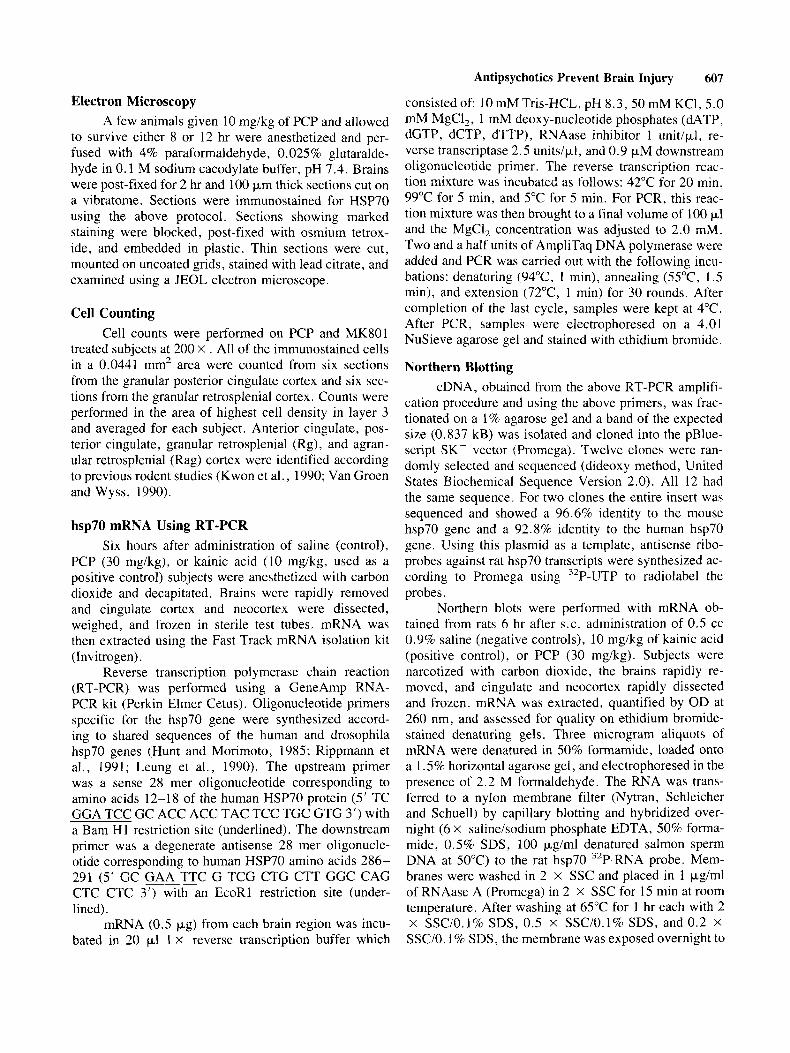
consisted of: 10 mM Tris-HCL, pH 8.3, 50 mM KCl, 5.0 mM MgCl,, 1 mM deoxy-nucleotide phosphates (dATP, dGTP, dCTP, dTTP), RNAase inhibitor 1 unit/pl, re- verse transcriptase 2.5 unitsipl, and 0.9 pM downstream oligonucleotide primer. The reverse transcription reac- tion mixture was incubated as follows: 42°C for 20 min, 99°C for 5 min, and 5°C for 5 min. For PCR, this reac- tion mixture was then brought to a final volume of 100 pl and the MgC1, concentration was adjusted to 2.0 mM. Two and a half units of AmpliTaq DNA polymerase were added and PCR was carried out with the following incu- bations: denaturing (94"C, 1 min), annealing (55"C, 1.5 min), and extension (72"C, 1 min) for 30 rounds. After completion of the last cycle, samples were kept at 4°C. After PCR, samples were electrophoresed on a 4.01 NuSieve agarose gel and stained with ethidium bromide.
Northern Blotting cDNA, obtained from the above RT-PCR amplifi-
cation procedure and using the above primers, was frac- tionated on a 1% agarose gel and a band of the expected size (0.837 kB) was isolated and cloned into the pBlue- script SK- vector (Promega). Twelve clones were ran- domly selected and sequenced (dideoxy method, United States Biochemical Sequence Version 2.0). All 12 had the same sequence. For two clones the entire insert was sequenced and showed a 96.6% identity to the mouse hsp70 gene and a 92.8% identity to the human hsp70 gene. Using this plasmid as a template, antisense ribo- probes against rat hsp70 transcripts were synthesized ac- cording to Promega using 32P-UTP to radiolabel the probes.
Northern blots were performed with mRNA ob- tained from rats 6 hr after S.C. administration of 0.5 cc 0.9% saline (negative controls), 10 mgikg of kainic acid (positive control), or PCP (30 mgikg). Subjects were narcotized with carbon dioxide, the brains rapidly re- moved, and cingulate and neocortex rapidly dissected and frozen. mRNA was extracted, quantified by OD at 260 nm, and assessed for quality on ethidium bromide- stained denaturing gels. Three microgram aliquots of mRNA were denatured in 50% formamide, loaded onto a 1.5% horizontal agarose gel, and electrophoresed in the presence of 2.2 M formaldehyde. The RNA was trans- ferred to a nylon membrane filter (Nytran, Schleicher and Schuell) by capillary blotting and hybridized over- night (6 X salineisodium phosphate EDTA, 50% forma- mide, 0.5% SDS, 100 pgiml denatured salmon sperm DNA at 50°C) to the rat hsp70 32P-RNA probe. Mem- branes were washed in 2 x SSC and placed in 1 pgiml of RNAase A (Promega) in 2 x SSC for 15 min at room temperature. After washing at 65°C for 1 hr each with 2 x SSCIO. 1% SDS, 0.5 x SSCIO. 1% SDS, and 0.2 X SSC/O. 1% SDS, the membrane was exposed overnight to
Electron Microscopy A few animals given 10 mg/kg of PCP and allowed
to survive either 8 or 12 hr were anesthetized and per- fused with 4% paraformaldehyde, 0.025% glutaralde- hyde in 0.1 M sodium cacodylate buffer, pH 7.4. Brains were post-fixed for 2 hr and 100 pm thick sections cut on a vibratome. Sections were immunostained for HSP70 using the above protocol. Sections showing marked staining were blocked, post-fixed with osmium tetrox- ide, and embedded in plastic. Thin sections were cut, mounted on uncoated grids, stained with lead citrate, and examined using a JEOL electron microscope.
Cell Counting Cell counts were performed on PCP and MK801
treated subjects at 200 X . All of the immunostained cells in a 0.0441 mm2 area were counted from six sections from the granular posterior cingulate cortex and six sec- tions from the granular retrosplenial cortex. Counts were performed in the area of highest cell density in layer 3 and averaged for each subject. Anterior cingulate, pos- terior cingulate, granular retrosplenial (Rg), and agran- ular retrosplenial (Rag) cortex were identified according to previous rodent studies (Kwon et al., 1990; Van Groen and Wyss, 1990).
hsp70 mRNA Using RT-PCR Six hours after administration of saline (control),
PCP (30 mg/kg), or kainic acid (10 mg/kg, used as a positive control) subjects were anesthetized with carbon dioxide and decapitated. Brains were rapidly removed and cingulate cortex and neocortex were dissected, weighed, and frozen in sterile test tubes. mRNA was then extracted using the Fast Track mRNA isolation kit (Invitrogen) .
Reverse transcription polymerase chain reaction (RT-PCR) was performed using a GeneAmp RNA- PCR kit (Perkin Elmer Cetus). Oligonucleotide primers specific for the hsp70 gene were synthesized accord- ing to shared sequences of the human and drosophila hsp70 genes (Hunt and Morimoto, 1985; Rippmann et al., 1991; Leung et al., 1990). The upstream primer was a sense 28 mer oligonucleotide corresponding to amino acids 12-18 of the human HSP70 protein (5' TC GGA TCC GC ACC ACC TAC TCC TGC GTG 3') with a Bam HI restriction site (underlined). The downstream primer was a degenerate antisense 28 mer oligonucle- otide corresponding to human HSP70 amino acids 286- 291 (5' GC GAA TTC G TCG CTG CTT GGC CAG CTC CTC 3') with an EcoRl restriction site (under- lined).
mRNA (0.5 pg) from each brain region was incu- bated in 20 pl 1 x reverse transcription buffer which

608 Sharp et al.
Kodak X-OMAT AR film with an intensifying screen at - 70°C. and ketamine.
ported (Sharp et al., 1991) in rats injected with MKSOI
Differences in the pattern and number of neurons
In Situ Hybridization Subjects given PCP (30 mg/kg), kainic acid (10
mg/kg), or 0.5 cc isotonic saline were narcotized with carbon dioxide 6 hr later and decapitated. Brains were rapidly removed and frozen in 2-methylbutane cooled with dry ice. Twenty-micron thick frozen sections were cut on a cryostat and picked up onto gelatinized Probe- on-Slides (Fisher). Sections were fixed in 4% parafor- maldehyde for 5 min, placed in 70%, 90%, and 100% ethanol for 1 min each, choroform for 5 min, and then air dried and frozen until used. One hundred fifty microliters of hybridization solution was used to cover three sections per slide and a second slide was then placed over the first to sandwich the sections. The hybridization solution was made as follows: 10 X lo6 dpm 35S labeled hsp70 ribo- probe in 20 pl DEP-H,O, 40 p l 5 M DTT, 50 pl salmon sperm DNA (10 mgiml), and 900 pL cocktail (50 ml formamide, 20 ml20 x SSC, 2 ml50 X Denhardt's, 10 m10.2 M NaP04 pH 7.4 buffer, 10 g dextran, 4 ml 25% sarcosyl). After hybridizing at 42°C for 20 hr, sections were rinsed two times for 5 sec in 1 X SSC at 55"C, four times for 15 min in 1 x SSC at 55"C, and for 60 min in 1 X SSC at room temperature. After two 5 min rinses in deionized water, sections were air dried and autoradio- graphed on Kodak SB5 x-ray film.
RESULTS Induction of HSP70 Protein by PCP, MK801, and Ketamine
HSP70-like immunoreactivity was never detected in any brain region, including the posterior cingulate and retrosplenial cortex, of saline-injected control Sprague Dawley rats (n = lo).
PCP induced HSP70 immunoreactivity in many layer 3 pyramidal and some layer 2 non-pyramidal neu- rons of granular posterior cingulate cortex (Fig. 1A). Some layer 3 and 5 neurons in adjacent agranular pos- terior cingulate cortex stained for HSP70 as well (Fig. 1A). PCP also induced HSP70 immunoreactivity in layer 3 and 5 neurons of granular (Fig. IB) and agranular ret- rosplenial cortex (not shown). Electron microscopy showed that HSP70 immunostained neurons that were visualized under EM had abnormal cytoplasmic vacuoles in the cell body (with MK801, Sharp et al., 1991a) and dendrites (Fig. 1H) when examined 8-12 hr after admin- istration of the PCP. PCP never induced HSP70 immu- noreactivity in anterior cingulate neurons rostra1 to the septum. This pattern of HSP70 induction in posterior cingulate and retrosplenial cortex is identical to that re-
induced by PCP compared to MK801 and ketamine were observed in neocortex, piriform cortex, and amygdala, however. PCP induced HSP70 immunoreactivity in many neocortical neurons (Figs. 1D-G) particularly at the 50-60 mg/kg doses in parietal cortex (Fig. ID) and temporal cortex. PCP induced HSP70 immunoreactivity in neocortex in pyramidal and non-pyramidal neurons in layers 2, 3 (Fig. 1 D,E), 5 (Fig. 1D,F), and 6 (Fig. 1G). The 40-50 mg/kg doses of PCP also induced HSP70 in a large number of neurons in the piriform cortex and in the central nucleus of the amygdala, which are not shown. These findings contrast to those found with MK801 (1 mgikg) and ketamine (80 mg/kg), which in- duced HSP70 in scattered, rare neocortical neurons (Sharp et a]., 1990; Sharp et al., 1991a), but not in other brain regions.
PCP induced HSP70 immunoreactivity over a wide range of doses (Fig. 2). The 5 mg/kg dose induced HSP70 in a number of neurons in each section in all animals. Counts of random 0.0441 mm2 fields yielded average counts near zero, however. Higher doses (10-60 mg/kg) induced HSP70 in hundreds of neurons in every 100 pm thick coronal section of every animal through the posterior cingulate and retrosplenial cortex. Though the 40-60 mg/kg doses induced the most HSP70 immu- nostained neurons (Fig. 2), the 30 mgikg dose was used for subsequent studies to decrease the behavioral effects of PCP and to make it possible to detect increases and decreases in the number of HSP70 stained neurons. MK801 and ketamine also induced HSP70 in a dose dependent manner, the greatest number of neurons being induced at the 1 mgikg and 80 mgikg doses, respec- tively.
HSP70 protein induction by PCP, MK801, and ke- tamine was also age dependent. This experiment was performed since it is believed that newborn and young humans may tolerate ketamine better than adults. Figure 3 shows that MK801 (1 mg/kg) does not induce HSP70 immunoreactivity in posterior cingulate of 1 or 20 day old rats. However, HSP70 is induced in 30 day old rats and reaches near adult levels of induction in 45 day old subjects. Similarly, PCP (10 mg/kg) and ketamine (60 mgikg) induced HSP70 in 30,45, and 90 day old but not in 1 and 20 day old rats (not shown).
PCP-hsp70 mRNA hsp70 mRNA, detected using RT-PCR, was in-
duced in cingulate cortex 6 hr following injection of PCP (30 mg/kg) and kainic acid (10 mg/kg, used as positive control) (Fig. 4). The bands shown in Figure 4 occurred between the 700 and 900 base pair molecular weight

Antipsychotics Prevent Brain Injury 609
studied. Rimcazole is believed to act as an antagonist at sigma receptors (Beart et al., 1989; Ferris et al., 1986), though it is unknown whether it has specific actions at dopamine or other receptors. The 30 mg/kg dose of rim- cazole, given 1 hr prior to PCP (30 mg/kg), decreased the number of HSP70 immunoreactive neurons in poste- rior cingulate by 40% (Fig. 7), and higher doses of rim- cazole completely blocked HSP70 induction by PCP in posterior cingulate cortex (Fig. 7), retrosplenial cortex, and neocortex (data not shown). Rimcazole (60 mg/kg) also blocked the HSP70 induced by 1 mg/kg doses of MK801 and 60 mg/kg doses of ketamine (not shown).
markers, indicating that the PCR product in both PCP and kainic acid treated subjects corresponded to the pre- dicted 837 base pair fragment of the rat hsp70 gene that would be homologous to the drosophila and human genes cloned by Hunt and Morimoto (1 985). This contrasted to saline-injected control subjects in which no hsp70 mRNA was detected (Fig. 4).
Northern blots (Fig. 5) also showed induction of hsp70 mRNA in the neocortex of 90 day old rats injected 6 hr earlier with PCP (50 mg/kg) and kainic acid (10 mgikg), but not in saline-injected control animals. It is notable that three definite bands were detected in PCP injected rats (Fig. 5 ) using our rat hsp70 riboprobe. It is difficult to say how many bands there are in the kainic acid lane because of overexposure of the lane, though it is clear that the relative abundance of the several sizes of hybridizing RNAs are different in the two preparations. The presence of three bands was repeated in two addi- tional PCP treated rats. The lowermost bands have a molecular weight appropriate for hsp70. The upper bands could represent alternative mRNA splicing of hsp70, which seems unlikely since the gene has no in- trons or alternative start sites, or the other bands could represent closely related genes of similar molecular weight such as hsc73, grp78, or others (Lindquist, 1986; Leung et al., 1990; Chang et al., 1987; Sorger and Pel- ham, 1987). In situ hybridization (Fig. 5 ) confirmed in- duction of hsp70 mRNA in cingulate of subjects given PCP (30 mg/kg) and in cortex and hippocampus of sub- jects given kainate (1 0 mg/kg) compared to absence of hsp70 mRNA saline-injected control subjects.
Haloperidol and Rimcazole Blockade of HSP70 Induction Following PCP
Haloperidol, when administered 1 hr prior to PCP (30 mg/kg, 90 day old rats), blocked induction of HSP70 protein in posterior cingulate neurons in a dose depen- dent fashion (Fig. 6). The 1 mg/kg dose of haloperidol decreased the number of HSP70 immunoreactive neu- rons by 40%, and the 5 mg/kg dose essentially blocked induction of all HSP70 immunoreactivity in posterior cingulate cortex (Figs. lC, 6), retrosplenial cortex, and neocortex (not shown). Haloperidol (5 mg/kg) also blocked the HSP70 protein induced by I mg/kg doses of MK801 and 60 mg/kg doses of ketamine (not shown).
Histological examination of posterior cingulate showed that all of the subjects ( 5 / 5 ) that received PCP (10 mg/kg) 4 hr earlier had vacuoles in layer 3 neurons. However, none (0/5) of the subjects that had received haloperidol (10 mg/kg) prior to administration of the PCP (10 mg/kg) had vacuolated layer 3 neurons.
Since haloperidol is known to act at both sigma and dopamine receptors (McCann and Su, 1990; Seeman, 1989), the novel antipsychotic drug, rimcazole, was
DISCUSSION
Olney et a]. (1989) originally reported that the non- competitive NMDA receptor antagonists PCP, MK8Ol , and ketamine injured posterior cingulate and retrosple- nial cortical neurons in rats. Though the injured layer 3 neurons had large intracytoplasmic vacuoles, most of the neurons appeared to survive with time. We have shown (Sharp et al., 1990; Sharp et al., 1991a) and Olney et al. (1991) confirmed that PCP, MK801, and ketamine in- duce the hsp7O heat shock or “stress” gene, including both hsp70 mRNA and its HSP70 protein, in the injured, vacuolated neurons in posterior cingulate and retrosple- nial cortex. It is proposed that the vacuoles produced by PCP and related drugs reflect the formation of denatured proteins, and that these denatured proteins activate the heat shock factors (Anathan et al., 1986; Wu et al., 1987) that bind to heat shock elements and stimulate hsp70 transcription (Craig, 1985; Lindquist, 1986; Xiao and Lis, 1988; Zimarino et al., 1990). This suggests that the presence of HSP70 protein provides a marker for neurons damaged by PCP, ketamine, and MK801 (Sharp et al., 1991; Olney et al., 1991).
Though PCP induces both hsp70 and vacuoles in neurons, the precise relationship between the two is not proven. We propose PCP produces vacuoles and dena- tured proteins in injured neurons that respond by induc- ing the hsp70 gene. This suggests that hsp70 should be induced in all vacuolated neurons. However, the data to date only show that 1) vacuolated and HSP7O immuno- reactive neurons occur in the same layers in posterior cingulate and retrosplenial cortex, 2) HSP70 immunore- active neurons have vacuoles when examined at the EM level in cingulate cortex, and 3 ) haloperidol (present study) and M1 antagonists (Olney et al., 1991) prevent induction of HSP70 and vacuoles in cingulate gyrus neu- rons. The data have not proven whether all HSP70 im- munoreactive neurons have vacuoles, whether HSP70 is induced in all vacuolated neurons, or whether HSP70 and vacuoles coexist in all injured neurons in all brain regions affected by PCP and MK801.

Fig. 1.

25 40 - v1 c g 35-- 2 20-- 2 0 2 30--
I ? 20--
E, 10-
z Z
5 25-- 0
c
15- n
z 5
I 0 4
Fig. 2 . The number (5 SEMl0.0441 mm2) of HSP70 immu- noreactive neurons in layer 3 of granular posterior cingulate cortex is plotted for adult rats given various doses of P c P (0.0 to 60.0 mg/kg) i.p. 24 hr previously.
Fig. 3, The number (t SEM/0.0441 mm2) of HSP70 immu- noreactive induced 1 day after administration of MK801 (1 mg/kg i.p.) in layer 3 of granular posterior cingulate cortex of 1, 20, 30, 45, and 90 day old rats.
- - MK 801 (1 rng/kg)
b
--
; v I
The precise role of the HSP7O heat shock protein induced in the neurons by PCP and MK801 is unknown. Pelham (1 986) hypothesized that the inducible hsp70 genes might play a role in preventing partially denatured proteins from becoming irreversibly denatured following cell injury. Some members of the hsp70 family facilitate protein import into mitochondria and other organelles (Deshaies et al., 1989). Beckmann et al. (1990) have proposed that some hsp70 family member(s) bind to newly synthesized proteins, and may ensure the proper
Fig. 1. One day following administration of PCP (50 mg/kg) HSP70 heat shock protein immunoreactivity was induced in layer 2 and 3 neurons in granular, posterior cingulate cortex (A), in layer 3 and 5 neurons of agranular posterior cingulate cortex (A), and in layer 2 , 3, and 5 neurons of granular (B) and agranular retrosplenial cortex (not shown). PCP (50 mglkg) also induced HSP70 immunoreactivity in neurons throughout - parietal (D-G) and temporal neocortex, piriform cortex, and amygdala (not shown). PCP induced HSP70 in the perikarya, dendrites, and axons of neocortical neurons in layers 2 and 3 (D,E), layer 5 (D,F), and layer 6 (G). Prior administration of haloperidol (10 mg/kg) prevented induction of HSP70 protein in retrosplenial cortex (C) and all other brain regions (not shown). HSP70 was not induced in the posterior cingulate
Fig. 4. hsp70 mRNA (white arrow) was detected using RT- PCR in posterior cingulate cortex of adult rats 6 hr after in- traperitoneal administration of 30 mg/kg of phencyclidine (P) and 10 mg/kg kainate (K), but was not detectable in saline injected controls (C). ox174 molecular weight marker (m) is shown.
cortex or in any other brain region of saline injected, control rats (not shown). 2, 3, 4, 5, are layers in posterior cingulate (A), retrosplenial @), and neocortex @). Calibration bar, 100
folding and assembly of newly forming proteins. What- ever the function of HSp70 protein, many authors have pm. Eight hours following administration of PCP (50 mg/kg)
HSP70 immunostained neurons in layer 3 of the posterior cin- gulate demonstrated the presence of cytoplasmic vacuoles in the cell body (not shown) and in the dendrites (arrow, H) in the electron microscope, which are similar to the vacuoles origi- nally reported by Olney et al. (1989, 1991) at the light micro- scope level.
suggested that the hsp70 family of genes has a protective function (Lowenstein et al., 1991; Barbe et al., 1988; Chopp et al. 9 1989; Li and Werby 1982; Riabowol et 1988; Rordorf et al., 1991).
Though the mechanism for the MK801, PCP, and ketamine induced neuronal injury is still unclear, the

612 Sharp et al.
28s
18s
Fig. 5. Northern blots revealed that hsp70 mRNA was induced in neocortex of adult rats 6 hr following administration of 10 mgikg kainic acid (K) and 50 mg/kg phencyclidine (P), but was not induced in saline-injected control (C) rats. In situ hybrid- ization for hsp70 mRNA was performed 6 hr after administra-
tion of 30 mgikg phencyclidine (P), 10 mgikg of kainic acid (K), or 0.5 cc of normal saline (C, control). hsp70 mRNA was induced in posterior cingulate cortex (white arrow) of P in- jected subjects and in neocortex and hippocampus of K injected subjects, as compared to saline-injected controls (C).
1
PCP 30rng/kg ; 12 z
n n .,, , , , , , I I , I
0 1 2 3 4 5 6 7 0 9 1 0
m c e W z 0 P.
cn I a
L 0 n
5 z
PCP 30rng/kg
6
4
2
--
--
--
0 10 20 30 40 50 60 70 80
PCP 30rng/kg
6
4
2
--
--
--
0 10 20 30 40 50 60 70 80
Haloperidol Dose (rng/kg) Rirncazole Dose (rng/kg)
Fig. 6. The number ( 5 SEM/O.0441 mm2) of HSP70 immu- noreactive neurons in layer 3 of granular posterior cingulate cortex is plotted for adult rats given PCP (30 mgikg) i.p. 24 hr previously. These subjects had had haloperidol administered i.p. in various doses (0.0 to 10 mgikg) 1 hr prior to adminis- tration of the PCP.
current study provides insights into the mechanism. First, MK801 (0.1-5.0 mgikg) is more potent at induc- ing injury than PCP (5-60 mgikg) or ketamine (40-120 mg/kg). Since MK801 acts as a non-competitive inhibi- tor of the calcium channel in the NMDA receptor com- plex, as do PCP and ketamine (Kemp et al., 1987; Wat-
Fig. 7 . The number (+ SEMi0.0441 mm2) of HSP70 immu- noreactive neurons in layer 3 of granular posterior cingulate cortex is plotted for adult rats given PCP (30 mgikg) i.p. 24 hr previously. These subjects had had rimcazole administered i.p. in various doses (0.0 to 80 mgikg) 1 hr prior to administration of the PCP.
kins and Olverman, 1987), and MK801 is much more potent than PCP and ketamine at this site, it is likely that MK801 produces injury in cingulate neurons by blocking the NMDA receptor complex. Moreover, since compet- itive as well as non-competitive NMDA receptor antag- onists produce the injury (Olney et al., 1991), this sug-

Antipsychotics Prevent Brain Injury 613
wicz, 1983; Castle et al., 1988; ffrench-Mullen and Ro- gawski, 1989). Mattson et al. (1992) recently proposed that PCP toxicity in cultured neurons was due to the blockade of the K + channels. This was based upon ob- servations that PCP and other potassium channel block- ers elevated intracellular calcium to lethal levels, the pathology of known potassium channel antagonists was similar to PCP, and that sigma and NMDA antagonists did not affect PCP induced neuronal death. It is difficult to understand how the K + channel blockade hypothesis of PCP-induced degeneration (Mattson et a]., 1992) could explain why so many types of receptor antagonists might prevent injury produced by PCP in vivo, however. It is conceivable that multiple receptors have indirect interactions with potassium channels (Hjorth et al., 1983; Hudkins and DeHaven-Hudkins, 1991; Mulder et al., 1985; Steinfels and Tam, 1989), and that this could explain why M I , dopamine, and sigma antagonists and GABA agonists prevent injury produced by PCP, keta- mine, and MK801.
However, it is also possible that haloperidol, rim- cazole, GABA agonists, and MI antagonists all block a single receptor. Haloperidol has affinity for DI-D5 do- pamine receptors, sigma receptors, alpha 1 adrenorecep- tor, and one serotonin (5HT2) receptor (Reynolds, 1991 ; Seeman, 1980; Tam and Cook, 1984; McLean and We- her, 1988; Snyder and Largent, 1989; Walker et al., 1988). Rimcazole acts primarily at sigma receptors though with a low affinity (Beart et al., 1989; Ferris et al., 1986). Most M1 antagonists (Bonner, 1989; Rich- ards, 1991) block sigma receptors (Hudkins and De- Haven-Hudkins, 199 1). Since haloperidol, rimcazole, and most MI antagonists all antagonize sigma receptors, it is possible that blockade of NMDA receptors activates sigma receptors which produce, via an unknown mech- anism, vacuolar injury to neurons. This could explain why PCP produces the widest distribution and largest number of injured neurons since PCP would both block NMDA receptors and stimulate sigma receptors, whereas MK801 and ketamine would only block NMDA recep- tors. Moreover, since one PCP receptor in brain may be part of a potassium channel (Sorenson and Blaustein, 1986), PCP effects on sigma receptors and PCP blockade of potassium channels could be linked to produce neu- ronal injury.
Sigma receptors have aroused interest because some antipsychotic agents, including haloperidol, act at sigma as well as dopamine receptors (McCann and SU, 1990; Tam and Cook, 1984; Largent et al., 1988; Snyder and Largent, 1989; Walker et al., 1988). Sigma agonists cause psychosis, and sigma antagonists have some an- tipsychotic properties (Koek et al., 1988; Domino and Luby, 1981; Ferris et al., 1986; Manallack et a]., 1986; Sonders et al., 1988; Snyder and Largent, 1989).
gests that blockade of the NMDA receptor in general, and not just one site on the receptor, can result in injury.
PCP, however, not only binds non-competitively to the calcium channel of the NMDA receptor (Itzhak, 1989a,b; Quirion et a]., 1987), but also binds competi- tively to high affinity, non-NMDA, non-opiate, halo- peridol-sensitive sites called sigma receptors (Gundlach et al., 1985, 1986; HeIIewell and Bowen, 1990; Itzhak, 1989a,b; Knight et al., 1991; Largent et al., 1987, 1988; Manallack et al., 1986; Sonders et al., 1988; Tam, 1983; Tam and Cook, 1984). It is possible, therefore, that since PCP binds to both sigma and NMDA receptors, PCP produces injury by direct effects on sigma as well as by blocking NMDA receptors (Quirion et al., 1987; Man- allack et a]., 1986; Sonders et al., 1988). However, MK801 and ketamine (unlike PCP) have little or no ef- fect on sigma receptors (Itzhak, 1989a,b). Therefore, it is likely that all three compounds, MK801, ketamine, and PCP, produce neuronal injury by first blocking NMDA receptors (Watkins and Olverman, 1987).
Though NMDA receptor blockade appears to h i - tiate the injury, recent results show that this is just the first step in the cascade of injury. Olney et al. (1990, 1991) first showed that blockade of the MI muscarinic cholinergic receptor prevented injury and stimulation of GAB A receptors partially prevented the neuronal injury produced by PCP, MK801, and ketamine. This led them to hypothesize that NMDA receptor blockade resulted in overactivation of MI receptors on cingulate gyrus neu- rons which mediated injury to those neurons. This hy- pothesis is difficult to reconcile with our current findings that show that haloperidol and rimcazole, drugs not cur- rently known to act at M1 receptors (Bonner, 1989; Rich- ards, 1991), also prevent injury to posterior cingulate and retrosplenial cortical neurons produced by NMDA receptor antagonists.
If blockade of NMDA receptors by PCP, MK80 1, and related compounds leads to activation of other ex- tracellular receptors which produce the injury, these other receptors would have to be blocked by haloperidol, rimcazole, M1 antagonists, and GABA agonists. Since so many agents prevent injury, it is possible that multiple extracellular receptors might participate in producing the injury. These could include the M1, sigma, and dopa- mine receptors, since haloperidol antagonizes dopamine and sigma receptors (McCann and Su, 1990; Seeman, 1980; Hellewell and Bowen, 1990; Largent et al., 1988; McLean and Weber, 1988; Snyder and Largent, 1989), rimcazole acts primarily at sigma receptors (Beart et a]. , 1989; Ferris et al., 1986), and MI antagonists act at M1 muscarinic receptors and sigma receptors (Bonner, 1989; Richards, 1991).
PCP is also known to block both transient and de- layed types of potassium channels (Blaustein and Icko-

614 Sharp et al.
The distribution of sigma receptors, however, is different than the pattern of regional injury produced by PCP, ketamine, and MK801. Though high densities of sigma receptors exist in cingulate, piriform, and neo- cortex, they are also found in substantia nigra and other regions not affected by PCP or MK801 (McLean and Weber, 1988; Jansen et al., 1991; Graybiel et al., 1989; Weisman et al., 1988). Another argument against sigma receptors mediating the injury includes the fact that at- ropine prevents vacuole formation by PCP (Olney et al., 1991), whereas atropine does not bind to sigma receptors in vitro (Hudkins and DeHaven-Hudkins, 199 1).
It is of interest that PCP and MK801 do not appear to produce injury in very young rats. It is possible that higher doses are required to produce injury in young animals, particularly since Mattson et al. (1992) have shown that PCP is directly toxic to cultured human fetal neurons. Alternatively, the delayed development of NMDA, dopamine, sigma, or M1 receptors on cingulate gyms and neocortical neurons could account for the in- creasing degree of injury in the maturing brain. This is of interest since the psychosis of schizophrenia often begins around puberty in humans and could correlate with a delayed expression of similar receptors. Since theories of schizophrenia suggest that either excess stimulation of dopamine, sigma, or other receptors produce psychosis (Su et al., 1986), it is possible that blockade of dopamine or sigma receptors with haloperidol and other antipsy- chotic drugs might prevent neuronal injury in neurons produced by excessive stimulation of these receptors. At the very least the data in the current study suggest that prior administration of haloperidol will prevent the neu- ronal injury and therefore might prevent the undesirable side effects of PCP, MK801, and ketamine.
ACKNOWLEDGMENTS We gratefully acknowledge the support of the Merit
Review Program of the Department of Veterans Affairs and NIH grants NS28167 and NS14.543 from the NINCDS to FRS and NS27448 to SMS. We also ac- knowledge the help of Pat Jasper, Katy Hicks, John Hall, and Matt Morton on various phases of this project.
REFERENCES Allen HL, Iversen LL (1990): Phencyclidine, dizocilipine, and cere-
brocortical neurons. Science 247:221. Anathan J , Goldberg AL, Voellmy R (1986): Abnormal proteins serve
as eukaryotic stress signals and trigger the activaton of heat shock genes. Science 232:522-524.
Barbe MF, Tytell M, Gower DJ, Welch WJ (1988): Hyperthermia protects against light damage in the rat retina. Science 241: 1817-1820.
Beart PM, O’Shea RD, Manallack DT (1989): Regulation of sigma- receptors: High- and low-affinity agonist states, GTP shifts, and up-regulation by rimcazole and 1,3-Di-(2-Tolyl) guani- dine. J Neurochem 53:779-788.
Beckmann RP, Mizzen LA, Welch WJ (1990): Interaction of HSP70 with newly synthesized proteins: Implications for protein fold- ing and assembly. Science 248:850-854.
Blaustein MP, Ickowicz RK (1983): Phencyclidine in nanomolar con- centrations binds to synaptosomes and blocks certain potassium channels. PNAS 80:3855-3859.
Bonner TI (1989): New subtypes of muscarinic acetylcholine recep- tors. Trends Pharmacol Sci 1O:ll-15.
Brown IR, Rush S, Ivy GO (1989): Induction of a heat shock gene at the site of tissue injury in the rat brain. Neuron 2:1559-1564.
Castle NA, Haylett DG, Jenkinson DH (1988): Toxins in the charac- terization of potassium channels. Trends Neurosci l2:59-65.
Chang SC, Wooden SK, Nakaki T, Kin YK, Lin AY, Kung L, At- tenello JW, Lee AS (1987): The rat gene encoding the 78-kD glucose-regulated protein (GRP 78): Its regulatory sequences and the effect of glycosylation on its expression. PNAS 84:
Choi DW (1988): Glutamate neurotoxicity and diseases of the nervous system. Neuron 1:623-634.
Chopp M, Chen H, Ho K-L, Dereski MO, Brown E, Hetzel FW, Welch KMA ( 1989): Transient hyperthermia protects against subsequent forebrain ischemic cell damage in the rat. Neurol-
Craig EA (1985): The heat shock response. CRC Crit Rev Biochem
Deshaies RJ, Koch BD, Schenkman R (1989): The role of stress proteins in membrane biogenesis. Trends Biol Sci 13:384-388.
Dienel GA, Kiessling M, Jacewicz M, Pulsinelli WA (1986): Synthe- sis of heat shock proteins in rat brain cortex after transient ischemia. J Cereb Blood Flow Metab 6:505-510.
Domino EF, Luby ED (1981): Abnormal mental states induced by phencyclidine as a model of schizophrenia. In Domino EF (ed): “PCP (Phencyclidine): Historical and Current Perspectives.” Ann Arbor: NPP Books, pp 401-418.
Dwyer BE, Nishimura RN, Brown IR (1989): Synthesis of the major inducible heat shock protein in rat hippocampus after neonatal hypoxia-ischemia. Exp Neurol 104:28-3 I .
Ferriero DM, Soberano HQ, Simon RP, Sharp FR (1990): Hypoxia- ischemia induces heat shock protein-like (HSP70) immunore- activity in neonatal rat brain. Dev Brain Res 53:145-150.
Ferns RM, Tang FL, Chang K-J, Russel A (1986): Evidence that the potential antipsychotic agent rimcazole (BW 234U) is a spe- cific, competitive antagonist of sigma sites in brain. Life Sci 3812329-2337.
ffrench-Mullen JMH, Rogawski MA (1989): Interaction of phencyc- lidine with voltage-dependent potassium channels in cultured rat hippocampal neurons: Comparison with block of NMDA receptor-ionophore complex. J Neurosci 9:405 1-4061,
Gill R , Foster AC, Woodruff GN (1987): Systemic administration of MK-801 protects against ischemia-induced hippocampal neu- rodegeneration in the gerbil. J Neurosci 7:3343-3349.
Gonzalez MF, Shiraishi K, Hisanaga K, Sagar SM, Mandabach M, Sharp FR (1989): Heat shock proteins as markers of neural injury. Mol Brain Res 6:93-100.
Gonzalez MF, Lowenstein D, Hisanaga K, Simon RP, Sagar SM, Sharp FR (1991): Induction of heat shock protein 72-like im- munoreactivity in the hippocampal formation following tran- sient global ischemia. Brain Res Bull 26:241-250.
Graybiel AM, Besson M-J, Weber E (1989): Neuroleptic-sensitive binding sites in the nigrostriatal system: Evidence for differen-
680-684.
ogy 39:1396-1398.
18:239-280.

Antipsychotics Prevent Brain Injury 615
Lowenstein DH, Simon RP, Sharp FR (1990): The pattern of 72-kDa heat shock protein-like immunoreactivity in the rat following flurothyl-induced status epilepticus. Brain Res 531:173-182.
Lowenstein DH, Chan PH, Miles MF (1991): The stress protein re- sponse in cultured neurons: Characterization and evidence for a protective role in excitotoxicity. Neuron 7: 1053-1060.
Manallack DT, Beart PB, Grundlach AL (1986): Psychomimetic sigma-opiates and PCP. Trends Pharmacol Sci 7:448-45 1.
Manzerra P, Brown IR (1990): Time course of induction of a heat shock gene (hsp70) in the rabbit cerebellum after LSD in vivo: Involvement of drug-induced hyperthermia. Neurochem Res
Manzerra P, Brown IR (1992): Expression of heat shock genes (hsp7O) in the rabbit spinal cord: Localization of constitutive and hy- perthermia-inducible mRNA species. J Neurosci Res 3 1 :606- 615.
Mattson MP, Rychlik B, Cheng B (1992): Degenerative and axon- altering effects of phencyclidine in human fetal cerebral corti- cal cells. Neuropharmacology 31:279-291.
McCann DJ, Su T-P (1990): Haloperidol competitively inhibits the binding of (+)-(3H) SKF-10,047 to sigma sites. Eur J Phar- macol 180:361-364.
McLean S, Weber E (1988): Autoradiographic visualization of halo- peridol-sensitive sigma receptors in guinea-pig brain. Neuro- science 25:259-269.
Meldrum B ( I 985): Possible therapeutic applications of antagonists of excitatory amino acid neurotransmitters. Clin Sci 68: 113-122.
Mulder AH, Draper R, Sminia P, Schofflemeer ANM, Stoof JC (1985): Agonist and antagonist effects of 3-PPP enantiomers on functional dopamine autoreceptors and postsynaptic dopamine receptors in vitro. Eur J Pham 197:291-297.
Nishimura RN, Dwyer BE, Cole R, de Vellis J , Picard K (1989): Induction of the major inducible 68-kDa heat shock protein after rapid changes in extracellular pH in cultured rat asfro- cytes. Exp Cell Res 180:276-280.
Nowak TS Jr (1985): Synthesis of a stress protein following transient ischemia in the gerbil. J Neurochem 45:1635-1641.
Nowak TS Jr (1988): MK-801 prevents 70 KDa stress protein induc- tion in gerbil brain after ischemia: The heat shock response as a marker for excitotoxic pathology. In Kriegstein J fed): “Phar- macology of Cerebral Ischemia.” Boca Raton: CRC Press, pp
Nowak TS Jr, Ikeda J , Nakajima T (1990a): 70-kDa heat shock protein and c-fos gene expression after transient ischemia. Stroke 21
Nowak TS Jr, Bond U, Schlesinger MJ (1990b): Heat shock RNA levels in brain and other tissues after hyperthermia and transient ischemia. J Neurochem 54:451-458.
Olney JW, Labruyere J, Price MT (1989): Pathological changes in- duced in cerebrocortical neurons by phencyclidine and related drugs. Science 244: 1360-1362.
Olney JW, Labruyere J , Wang GJ, Price MT (1990): Anticholinergics prevent neurotoxic side effects of NMDA antagonists. Neuro- sci Abstr 16:1122.
Olney JW, Labruyere J, Wang G, Wozniak DF, Price MT, Sesma MA (1 991): NMDA antagonist neurotoxicity: Mechanism and pre- vention. Science 254:1515-1518.
Ozyurt E, Graham DI, Woodruff GN, McCulloch J (1988): The pro- tective effect of the glutamate antagonist MK-801 in focal ce- rebral ischemia in the cat. J Cereb Blood Flow Metab 8:138- 143.
Pelham HRB (1986): Speculations on the functions of the major heat shock and glucose-regulated proteins. Cell 46:959-961.
Quirion R, Chicheportiche R, Coneras PC, Johnson KM, Lodge D,
15~53-59.
229-234.
[SUPPI III]:107-1Il.
tial distribution of sigma sites in the substantia nigra, pars compacta of the cat. J Neurosci 9:326-338.
Gundlach AL, Largent BL, Snyder SH (1985): Phencyclidine and sigma opiate receptors in brain: Biochemical and autoradio- graphic differentiation. Eur J Pharmacol 1 13:465-466.
Gundlach AL, Largent BL, Snyder SH (1986): Autoradiographic lo- calization of sigma receptor binding sites in guinea pig and rat central nervous system with (+ ) (’H)-3-PPP. J Neurosci 6: 175771770,
Hellewell SB, Bowen WD (1990): A sigma-like binding site in rat pheochromocytoma (PC12) cells: Decreased affinity for ( + )- benzomorphans and lower molecular weight suggest a different sigma receptor form from that of guinea pig brain. Brain Res
Hjorth S, Carlsson A, Clark D, Svensson K, Wikstrom H, Sanchez D, Lindberg P, Hacksell U , Arvidsson L-E, Johansson A, Nilsson JLG (1983): Central dopamine receptor agonist and antagonist actions of the enantiomers of 3-PPP. Psychopharmacology 8 1: 89-99.
Hudkins RL, DeHaven-Hudkins DL (1991): MI muscarinic antago- nists interact with sigma recognition sites. Life Sci 49:1229- 1235.
Hunt C, Morimoto RI (1985): Conserved features of eukaryotic hsp70 genes revealed by comparison to the nucleotide sequence of human hsp70. PNAS 82:6455-6459.
ltzhak Y (1989a): Multiple affinity binding states of the sigma recep- tor: Effect of GTP-binding protein-modifying agents. Mol Pharmacol 3 6 5 12-517.
Itzhak Y (1989b): Different modulation of the binding to two phen- cyclidine (PCP) receptor subtypes: Effects of N-methyl-D-as- partate agonists and antagonists. Neurosci Left 104:314-3 19.
Jansen KLR, Faull RLM, Dragunow M, Leslie RA (1991): Autora- diographic distribution of sigma receptors in human neocortex, hippocampus, basal ganglia, cerebellum, pineal and pituitary glands. Brain Res 559:172-177.
Kemp JA, Foster AC, Wong EHF (1987): Non-competitive antago- nists of excitatory amino acid receptors. Trends Neurosci 10: 294-298.
Koek W, Woods JH, Winger GD (1988): MK-801, a proposed non- competitive antagonist of excitatory amino acid neurotransmis- sion, produces phencyclidine-like behavioral effects in pi- geons, rats and rhesus monkeys. J Pharmacol Exp Ther 245:
Knight AR, Gillard J, Wong EHF, Middlemiss DN (1991): The hu- man sigma site, which resembles that in NCB20 cells, may correspond to a low-affinity site in guinea pig brain. Neurosci Lett 131:233-236.
Kwon SE, Nadeau SE, Heilman KM (1990): Retrosplenial cortex: Possible role in habituation of the orienting response. J Neu- rosci 10:3559-3663.
Largent BL, Wikstrom H, Gundlach AL, Snyder SH (1987): Struc- tural determinants of sigma receptor affinity. Mol Pharmacol 32:172-784.
Largent BL, Wikstrom H, Snowman AM, Snyder SH (1988): Novel antipsychotic drugs share high affinity for the sigma receptor. Eur J Pharmacol 155:345-347.
Leung KC, Rajendran MY, Monfries C , Hall C, Hall C, Lim L (1990): The human heat-shock protein family. Biochem J 267: 125-132.
Li GC, Werb Z (1982): Correlation between synthesis of heat shock proteins and development of thermotolerance in Chinese ham- ster fibroblasts. Proc Natl Acad Sci USA 79:3218-3222.
Lindquist S (1986): The heat shock response. Ann Rev Biochem 55:
527: 244 -253.
969-974.
1151-1 191.

616 Sharp et al.
Tam SW, Woods JH, Zukin SR (1987): Classification and no- menclature of phencyclidine and sigma receptor sites. Trends Neurosci I 1 :444-445.
Reynolds GP (1992): Developments in the drug treatment of schizo- phrenia. Trends Pharmacol Sci 13:116-121.
Riabowol KT, Mizzen LA, Welch WJ (1988): Heat shock is lethal to fibroblasts microinjected with antibodies against hsp70. Sci- ence 242:433-436.
Richards MH (1991): Pharmacology and second messenger interac- tions of cloned muscarinic receptors. Biochem Pharmacol 42: 1645-1 653.
Rippmann F, Taylor WR, Rothbard JB, Green NM (1991): A hypo- thetical model for the peptide binding domain of hsp70 based on the peptide binding domain of HLA. EMBO J 10:1053- 10.59.
Rordorf G, Koroshetz WJ, Bonventre JV (1991): Heat shock protects cultured neurons from glutamate toxicity. Neuron 7:1043- 1051.
Rothman SM, Olney JW (1986): Glutamate and the pathophysiology of hypoxic-ischemic brain damage. Ann Neurol 19:105-111.
Seeman P (1980): Brain dopamine receptors. Pharmacol Rev 32:229- 313.
Sharp FR, Jasper P, Hall J , Noble L, Sagar SM (1991a): MK-801 and ketamine induce heat shock protein HSP70 in injured neurons in posterior cingulate and retrosplenial cortex. Ann Neurol 30: 80 1-809.
Sharp FR, Lowenstein D, Simon RP, Hisanaga K (1991b): Heat shock protein HSP70 induction in cortical and striatal astrocytes and neurons following infarction. J Cereb Blood Flow Metab 1 1 : 621-627.
Sharp JW, Sagar SM, Sharp FR (1990): MK801 induces the 72kDa heat shock protein and Fos in the cingulate gyms. Neurosci Abstr 16:1122.
Simon RP, Swan JH, Griffiths T, Meldrum BS (1984): Blockade of N-methyl-D-aspartate receptors may protect against ischemic damage in the brain. Science 2262350-853.
Snyder SH, Largent BL (1989): Receptor mechanisms in antipsychotic drug action: Focus on sigma receptors. J Neuropsychiatr I: 7-15.
Sonders MS, Keana JFW, Weber E (1988): Phencyclidine and psy- chomimetic sigma opiates: recent insights into their biochem- ical and physiological site of action. Trends Neurosci 11:37- 40.
Sorensen RG, Blaustein MP (1986): The phencyclidine (PCP) receptor in rat brain is part of a K f channel. In Domino EF, Kamenka JM (eds): “Sigma and Phencyclidine-Like Compounds as Mo- lecular Probes in Biology.” Ann Arbor: NPP Books, pp 185- 198.
Sorger PK, Pelham HRB (1987): Cloning and expression of a gene encoding hsc73, the major hsp70-like protein in unstressed rat cells. EMBO J 6:993-998.
Steinfels GF, Tam WS (1989): Selective sigma receptor agonist and antagonist affect dopamine neuronal activity. Eur J Pharmacol
Su T-P, Weissman AD, Yeh S-Y (1986): Endogenous ligands for sigma opioid receptors in the brain (“sigmaphin”): Evidence from binding assays. Life Sci 38:2199-2210.
Tam SW (1983): Naloxone-inaccessible sigma receptor in rat central nervous system. PNAS 80:6703-6707.
Tam SW, Cook L (1984): Sigma opiates and certain antipsychotic drugs mutally inhibit (+ ) (’H) SKF 10047 and (3H) haloperidol binding in guinea pig brain membranes. PNAS 81:5618- 5621.
Tricklebank MD, Singh L, Oles RJ, Preston C, Ivcrsen SD (1989): The behavioral effects of MK-801: A comparison with antag- onists acting non-competitively and competitively at the NMDA receptor. Eur J Pharmacol 167:127-135.
Van Groen T, Wyss JM (1990): Connections of the retrosplenial gran- ular A cortex in the rat. J Comp Neurol 300:593-606.
Vass K, Welch WJ, Nowak TS Jr (1988): Localization of 70kD stress protein induction in gerbil brain after ischemia. Acta Neuro- pathol 77:128-135.
Vass K, Berger ML, Nowak TS Jr, Welch WJ, Lassmann H (1989): Induction of stress protein HSP70 in nerve cells after status epilepticus in the rat. Neurosci Lett 100:259-264.
Walker JM, Matsumoto RR, Bowen WD, Gans DL, Jones KD, Walker FO (1988): Evidence for a role of haloperidol-sensitive sigma “opiate” receptors in the motor effects of antipsychotic drugs. Neurology 38:961-965.
Watkins JC. Olverman HJ (1987): Agonists and antagonists for exci- tatory amino acid receptors. Trends Neurosci 10:265-272.
Weisman AD, Su TP, Hedreen JC, London ED (1988): Sigma recep- tors in post-mortem human brains. J Pharmacol Exp Ther 247: 29-33.
Welch WJ, Suhan JP (1986): Cellular and biochemical events in mam- malian cells during and after recovery from physiological stress. J Cell Biol 103:2035-2052.
Wong EHF. Kemp JA, Priestley T, Knight AR, Woodruff GN, Iversen LL (1986): The anticonvulsant MK-801 is a potent N-methyl-D-aspartate antagonist. Proc Natl Acad Sci USA 83:
Wong EHF, Knight AR, Woodruff GN (1988): (‘H) MK-801 labels a site on the N-methyl-D-aspartate receptor channel complex in rat brain membranes. J Neurochem 50:29-33.
WU C, Wilson S, Walker B, Dawid I, Paisley T, Zimarino V, Ueda H (1987): Purification and properties of Drosophila heat shock activator protein. Science 238: 124771253,
Xiao H, Lis JT (1988): Germline transformation used to define key features of heat-shock response elements. Science 239: I 139- 1142.
Zimarino V, Tsai C, Wu C (1990): Complex modes of heat shock factor activation. Mol Cell Biol 10:752-759.
163:167-170.
7 1 04-7 I 08.
Copyright © 2022 FDOKUMEN