
Wdpcp, a PCP protein required for ciliogenesis, regulates directional cell migration and cell...
17
Wdpcp, a PCP Protein Required for Ciliogenesis, Regulates Directional Cell Migration and Cell Polarity by Direct Modulation of the Actin Cytoskeleton Cheng Cui 1,2 , Bishwanath Chatterjee 1,2 , Thomas P. Lozito 3 , Zhen Zhang 1 , Richard J. Francis 1,2 , Hisato Yagi 1 , Lisa M. Swanhart 1¤a , Subramaniam Sanker 1 , Deanne Francis 2 , Qing Yu 2 , Jovenal T. San Agustin 4 , Chandrakala Puligilla 5¤b , Tania Chatterjee 2 , Terry Tansey 2 , Xiaoqin Liu 1 , Matthew W. Kelley 5 , Elias T. Spiliotis 6 , Adam V. Kwiatkowski 7 , Rocky Tuan 3 , Gregory J. Pazour 4 , Neil A. Hukriede 1 , Cecilia W. Lo 1,2 * 1 Department of Developmental Biology, University of Pittsburgh School of Medicine, Pittsburgh, Pennsylvania, United States of America, 2 Laboratory of Developmental Biology, National Heart Lung and Blood Institute, National Institutes of Health, Bethesda, Maryland, United States of America, 3 Center for Cellular and Molecular Engineering, Department of Orthopedic Surgery, University of Pittsburgh School of Medicine, Pittsburgh, Pennsylvania, United States of America, 4 Program in Molecular Medicine, University of Massachusetts Medical Center, Worcester, Massachusetts, United States of America, 5 Section on Developmental Neuroscience, National Institute on Deafness and Other Communication Disorders, National Institutes of Health, Bethesda, Maryland, United States of America, 6 Department of Biology, Drexel University, Philadelphia, Pennsylvania, United States of America, 7 Department of Cell Biology, University of Pittsburgh School of Medicine, Pittsburgh, Pennsylvania, United States of America Abstract Planar cell polarity (PCP) regulates cell alignment required for collective cell movement during embryonic development. This requires PCP/PCP effector proteins, some of which also play essential roles in ciliogenesis, highlighting the long- standing question of the role of the cilium in PCP. Wdpcp, a PCP effector, was recently shown to regulate both ciliogenesis and collective cell movement, but the underlying mechanism is unknown. Here we show Wdpcp can regulate PCP by direct modulation of the actin cytoskeleton. These studies were made possible by recovery of a Wdpcp mutant mouse model. Wdpcp-deficient mice exhibit phenotypes reminiscent of Bardet–Biedl/Meckel–Gruber ciliopathy syndromes, including cardiac outflow tract and cochlea defects associated with PCP perturbation. We observed Wdpcp is localized to the transition zone, and in Wdpcp-deficient cells, Sept2, Nphp1, and Mks1 were lost from the transition zone, indicating Wdpcp is required for recruitment of proteins essential for ciliogenesis. Wdpcp is also found in the cytoplasm, where it is localized in the actin cytoskeleton and in focal adhesions. Wdpcp interacts with Sept2 and is colocalized with Sept2 in actin filaments, but in Wdpcp-deficient cells, Sept2 was lost from the actin cytoskeleton, suggesting Wdpcp is required for Sept2 recruitment to actin filaments. Significantly, organization of the actin filaments and focal contacts were markedly changed in Wdpcp-deficient cells. This was associated with decreased membrane ruffling, failure to establish cell polarity, and loss of directional cell migration. These results suggest the PCP defects in Wdpcp mutants are not caused by loss of cilia, but by direct disruption of the actin cytoskeleton. Consistent with this, Wdpcp mutant cochlea has normal kinocilia and yet exhibits PCP defects. Together, these findings provide the first evidence, to our knowledge, that a PCP component required for ciliogenesis can directly modulate the actin cytoskeleton to regulate cell polarity and directional cell migration. Citation: Cui C, Chatterjee B, Lozito TP, Zhang Z, Francis RJ, et al. (2013) Wdpcp, a PCP Protein Required for Ciliogenesis, Regulates Directional Cell Migration and Cell Polarity by Direct Modulation of the Actin Cytoskeleton. PLoS Biol 11(11): e1001720. doi:10.1371/journal.pbio.1001720 Academic Editor: Matthew P. Scott, Stanford University, United States of America Received August 9, 2013; Accepted October 18, 2013; Published November 26, 2013 This is an open-access article, free of all copyright, and may be freely reproduced, distributed, transmitted, modified, built upon, or otherwise used by anyone for any lawful purpose. The work is made available under the Creative Commons CC0 public domain dedication. Funding: This work was supported by NIH grants HL098180 (CWL), GM060992 (GJP) and GM097664 (ETS) at http://www.nih.gov/. The funders had no role in study design, data collection and analysis, decision to publish, or preparation of the manuscript. Competing Interests: The authors have declared that no competing interests exist. Abbreviations: AA or aa, amino acid; AER, apical ectodermal ridge (limb); AVSD, atrioventricular septal defects (heart); BBS, Bardet–Biedl Syndromes; Co-IP, coimmunoprecipitation; DBA, Dolichos biflorus agglutinin; DL, distal early tubule (kidney); EFIC, episcopic fluorescence image capture; EM, electronic microscopy; ENU, ethylnitrosourea; ES cells, embryonic stem cells; IHC, inner hair cell (cochlea); KO, knockout; LV, left ventricle (heart); MA, major axis; MEF, mouse embryonic fibroblast; MFP, membrane fluctuation period; MI, minor axis; MKS, Meckel–Gruber Syndromes; MO, morpholino; NGS, normal goat serum; OFT, outer flow tract (heart); OHC, outer hair cell (cochlea); PAtr, pulmonary atresia (heart); PBT, phosphate buffered saline with Tween-20; PCP, planar cell polarity; PCR, polymerase chain reaction; PD, pronephric duct (kidney); PST, proximal straight (kidney); PTA, persistent truncus arteriosus (heart); Phal, Phalloidin; Ptch1, Patched1; SMA, smooth muscle actin; Sept2, septin 2; Shh, Sonic hedgehog; Smo, Smoothened; TEF, tracheoesophageal fistula. * E-mail: [email protected] ¤a Current address: Department of Biology, Canisius College, Buffalo, New York, United States of America. ¤b Current address: Department of Pathology and Laboratory Medicine, Medical University of South Carolina, Charleston, South Carolina, United States of America. PLOS Biology | www.plosbiology.org 1 November 2013 | Volume 11 | Issue 11 | e1001720
Transcript of Wdpcp, a PCP protein required for ciliogenesis, regulates directional cell migration and cell...

Wdpcp, a PCP Protein Required for Ciliogenesis,Regulates Directional Cell Migration and Cell Polarity byDirect Modulation of the Actin CytoskeletonCheng Cui1,2, Bishwanath Chatterjee1,2, Thomas P. Lozito3, Zhen Zhang1, Richard J. Francis1,2,
Hisato Yagi1, Lisa M. Swanhart1¤a, Subramaniam Sanker1, Deanne Francis2, Qing Yu2, Jovenal T. San
Agustin4, Chandrakala Puligilla5¤b, Tania Chatterjee2, Terry Tansey2, Xiaoqin Liu1, Matthew W. Kelley5,
Elias T. Spiliotis6, Adam V. Kwiatkowski7, Rocky Tuan3, Gregory J. Pazour4, Neil A. Hukriede1,
Cecilia W. Lo1,2*
1 Department of Developmental Biology, University of Pittsburgh School of Medicine, Pittsburgh, Pennsylvania, United States of America, 2 Laboratory of Developmental
Biology, National Heart Lung and Blood Institute, National Institutes of Health, Bethesda, Maryland, United States of America, 3 Center for Cellular and Molecular
Engineering, Department of Orthopedic Surgery, University of Pittsburgh School of Medicine, Pittsburgh, Pennsylvania, United States of America, 4 Program in Molecular
Medicine, University of Massachusetts Medical Center, Worcester, Massachusetts, United States of America, 5 Section on Developmental Neuroscience, National Institute
on Deafness and Other Communication Disorders, National Institutes of Health, Bethesda, Maryland, United States of America, 6 Department of Biology, Drexel University,
Philadelphia, Pennsylvania, United States of America, 7 Department of Cell Biology, University of Pittsburgh School of Medicine, Pittsburgh, Pennsylvania, United States of
America
Abstract
Planar cell polarity (PCP) regulates cell alignment required for collective cell movement during embryonic development.This requires PCP/PCP effector proteins, some of which also play essential roles in ciliogenesis, highlighting the long-standing question of the role of the cilium in PCP. Wdpcp, a PCP effector, was recently shown to regulate both ciliogenesisand collective cell movement, but the underlying mechanism is unknown. Here we show Wdpcp can regulate PCP by directmodulation of the actin cytoskeleton. These studies were made possible by recovery of a Wdpcp mutant mouse model.Wdpcp-deficient mice exhibit phenotypes reminiscent of Bardet–Biedl/Meckel–Gruber ciliopathy syndromes, includingcardiac outflow tract and cochlea defects associated with PCP perturbation. We observed Wdpcp is localized to thetransition zone, and in Wdpcp-deficient cells, Sept2, Nphp1, and Mks1 were lost from the transition zone, indicating Wdpcpis required for recruitment of proteins essential for ciliogenesis. Wdpcp is also found in the cytoplasm, where it is localized inthe actin cytoskeleton and in focal adhesions. Wdpcp interacts with Sept2 and is colocalized with Sept2 in actin filaments,but in Wdpcp-deficient cells, Sept2 was lost from the actin cytoskeleton, suggesting Wdpcp is required for Sept2recruitment to actin filaments. Significantly, organization of the actin filaments and focal contacts were markedly changed inWdpcp-deficient cells. This was associated with decreased membrane ruffling, failure to establish cell polarity, and loss ofdirectional cell migration. These results suggest the PCP defects in Wdpcp mutants are not caused by loss of cilia, but bydirect disruption of the actin cytoskeleton. Consistent with this, Wdpcp mutant cochlea has normal kinocilia and yet exhibitsPCP defects. Together, these findings provide the first evidence, to our knowledge, that a PCP component required forciliogenesis can directly modulate the actin cytoskeleton to regulate cell polarity and directional cell migration.
Citation: Cui C, Chatterjee B, Lozito TP, Zhang Z, Francis RJ, et al. (2013) Wdpcp, a PCP Protein Required for Ciliogenesis, Regulates Directional Cell Migration andCell Polarity by Direct Modulation of the Actin Cytoskeleton. PLoS Biol 11(11): e1001720. doi:10.1371/journal.pbio.1001720
Academic Editor: Matthew P. Scott, Stanford University, United States of America
Received August 9, 2013; Accepted October 18, 2013; Published November 26, 2013
This is an open-access article, free of all copyright, and may be freely reproduced, distributed, transmitted, modified, built upon, or otherwise used by anyone forany lawful purpose. The work is made available under the Creative Commons CC0 public domain dedication.
Funding: This work was supported by NIH grants HL098180 (CWL), GM060992 (GJP) and GM097664 (ETS) at http://www.nih.gov/. The funders had no role instudy design, data collection and analysis, decision to publish, or preparation of the manuscript.
Competing Interests: The authors have declared that no competing interests exist.
Abbreviations: AA or aa, amino acid; AER, apical ectodermal ridge (limb); AVSD, atrioventricular septal defects (heart); BBS, Bardet–Biedl Syndromes; Co-IP,coimmunoprecipitation; DBA, Dolichos biflorus agglutinin; DL, distal early tubule (kidney); EFIC, episcopic fluorescence image capture; EM, electronic microscopy;ENU, ethylnitrosourea; ES cells, embryonic stem cells; IHC, inner hair cell (cochlea); KO, knockout; LV, left ventricle (heart); MA, major axis; MEF, mouse embryonicfibroblast; MFP, membrane fluctuation period; MI, minor axis; MKS, Meckel–Gruber Syndromes; MO, morpholino; NGS, normal goat serum; OFT, outer flow tract(heart); OHC, outer hair cell (cochlea); PAtr, pulmonary atresia (heart); PBT, phosphate buffered saline with Tween-20; PCP, planar cell polarity; PCR, polymerasechain reaction; PD, pronephric duct (kidney); PST, proximal straight (kidney); PTA, persistent truncus arteriosus (heart); Phal, Phalloidin; Ptch1, Patched1; SMA,smooth muscle actin; Sept2, septin 2; Shh, Sonic hedgehog; Smo, Smoothened; TEF, tracheoesophageal fistula.
* E-mail: [email protected]
¤a Current address: Department of Biology, Canisius College, Buffalo, New York, United States of America.¤b Current address: Department of Pathology and Laboratory Medicine, Medical University of South Carolina, Charleston, South Carolina, United States ofAmerica.
PLOS Biology | www.plosbiology.org 1 November 2013 | Volume 11 | Issue 11 | e1001720

Introduction
The cilium is a microtubule-based organelle projecting from the
cell surface with a variety of signaling functions. It plays an
important role in development as indicated by the wide spectrum
of birth defects associated with ciliopathy syndromes such as
Bardet–Biedl (BBS) and Meckel–Gruber (MKS) syndromes [1].
Studies in mice showed some of the developmental anomalies
associated with BBS and MKS, such as limb polydactyly, arise
from the disruption of Sonic hedgehog (Shh) signaling, a process
well described to be cilia transduced [2–4]. This entails the
regulated trafficking of Shh receptors Patched-1 (Ptch1) [5] and
Smoothened (Smo) [6] into the cilia, and the cilium sequestration
and processing of Gli transcription factors [7,8].
Ciliopathy syndromes also exhibit developmental anomalies
arising from disruption of Wnt signaling. Noncanonical or b-
catenin–independent Wnt signaling specifies planar cell polarity
(PCP), a process whereby cells organize and align in a polarized
manner relative to the plane of the epithelium. PCP regulated
convergent extension cell movement is required for neural tube
closure [9–11], and patterning of stereocilia polarity in the cochlea
requires asymmetric localization of PCP core components [12–
14]. While the mechanistic link between the cilia and PCP
signaling [15–17] is not known [18,19], it is worth noting some
PCP components are cilia localized and are required for
ciliogenesis [20–22]. As cilia disruption can elevate canonical
Wnt signaling, the cilium is also proposed to play a role in
canonical or b-catenin–dependent Wnt signaling [23,24]. This
may entail maintaining the balance between canonical versus
noncanonical Wnt signaling, since cilia disruption can cause
opposing effects on canonical versus noncanonical Wnt signaling
[24].
In this study, we investigated the role of Wdpcp and the cilia in
regulating planar cell polarity using a Wdpcp mutant mouse model.
Wdpcp is the homolog of Drosophila Fritz, a PCP effector required
for patterning hair cell orientation in the Drosophila pupal wing
[25]. Studies in Xenopus embryos [26] showed Fritz regulates
ciliogenesis and PCP-dependent collective cell movement during
gastrulation. However, the role of the cilia and Wdpcp (Fritz) in the
regulation of cell polarity and polarized cell migration was
unknown. In this study, we show mice deficient for Wdpcp have
phenotypes consistent with MKS/BBS ciliopathy syndromes. This
included disruption of ciliogenesis together with developmental
anomalies consistent with PCP perturbations. We show Wdpcp is
localized in the ciliary transition zone, and also in the actin
cytoskeleton and focal adhesions. This involves Wdpcp interac-
tions with Sept2 (septin 2), both in the cilia transition zone and in
the actin cytoskeleton. Significantly, the Wdpcp-deficient cells not
only exhibited ciliogenesis defect, but also global disruption in the
actin cytoskeleton required for maintaining cell polarity and
polarized cell migration. To our knowledge, these studies provide
the first evidence that a PCP component required for ciliogenesis
can regulate planar cell polarity by directly modulating the actin
cytoskeleton.
Results
A mutant, named WdpcpCys40, was recovered from an ethylni-
trosourea mouse mutagenesis screen exhibiting a wide spectrum of
developmental anomalies consistent with MKS/BBS. Particularly
notable is their phenotypic similarity to Mks1 mutant mice that are
models of MKS [27,28]. This included anophthalmia (Figure 1A),
central polydactyly (Figure 1B,C), and cysts in the kidney and a
variety of other organs (Figure 1D,E). WdpcpCys40 mutants also
exhibited complex congenital heart defects, usually consisting of
persistent truncus arteriosus or pulmonary atresia (Figure 1F,G,J,K),
and atrioventricular septal defects (AVSDs; Figure 1L,M). Some
mutants had duplex kidney (Figure 1E) and facial clefts and/or cleft
palate (Figure 1A). Tracheoesophageal fistula (TEF) due to defects
in septation of the oropharynx (unpublished data) and cloacal
septation defects were also observed (Figure 1H,I).
We mapped the mutation to a 6.36 Mb interval delimited by
SNP rs26841005 and rs26856862 on mouse chromosome 11. RT-
PCR analysis was carried out using RNA extracted from E12.5
hearts of WdpcpCys40 mutant embryos to interrogate transcript
expression from the 36 genes in the mapped interval. This analysis
revealed an anomalous transcript from Wdpcp (NM_145425.3 and
NP_663400.2). Further sequencing analysis suggested this was
derived from a splicing defect mutation, which was confirmed with
genomic DNA sequencing. An A to G substitution was observed at
nucleotide 224 of the mRNA (Figure 2A), corresponding to the 8th
base before the splice donor site of exon 5 (Figure 2B). As a result,
a premature stop codon (S54X) is generated, causing protein
truncation after amino acid 54 (Figure 2C). Quantitative PCR
analysis with primers covering exons 5–6 showed only low trace
amount (0.6%) of normal transcripts, suggesting WdpcpCys40 is
essentially a null or strong hypomorphic Wdpcp mutant allele.
Wdpcp Required for Recruitment of Proteins forCiliogenesis
Analysis of WdpcpCys40 mutant embryos revealed ciliogenesis
defects. This was observed in the kidney-collecting duct
(Figure 2N,O,R) and in the neuroepithelium (Figure 2P,Q).
Analysis of mouse embryonic fibroblasts (MEFs) derived from
WdpcpCys40 mutant embryos, referred to as WdpcpCys40 mutant
MEFs, confirmed a defect in ciliogenesis (Figure 2R; Figure
S1A,B). Using an antibody raised to Wdpcp, we showed Wdpcp is
localized to the ciliary axoneme and in a ring-like structure at the
base of the cilia in IMCD3 cells (Figure 2D). A similar distribution
was observed in NIH-3T3 cells transfected with Wdpcp–FLAG, a
FLAG-tagged expression vector (Figure 2E–H). The Wdpcp ring-
like structure in the cilium showed colocalization with Sept2,
Author Summary
Cilia are microscopic cell surface hair-like protrusions thatcan act as antennae to mediate cell signaling. Mutationsdisrupting ciliogenesis can cause many developmentalanomalies associated with syndromes known as ‘‘ciliopa-thies.’’ Some developmental defects, such as limb poly-dactyly, arise from disruption of cilia-transduced sonichedgehog signaling, while other defects, such as aberrantpatterning of hair cells in the inner ear, arise fromdisrupted Wnt signaling resulting in modulation of planarcell polarity (PCP)—a process whereby cells are polarizedand aligned. While ciliopathy phenotypes would suggestthat cilia are involved in modulating PCP, the mechanisticlink between cilia and PCP has been elusive. Our studyusing a mouse model carrying a mutation in Wdpcp, agene required for both ciliogenesis and PCP, suggest thatWdpcp modulation of PCP involves interactions with theactin cytoskeleton separate from its function in ciliogen-esis. We observe Wdpcp localization in cilia, where it isrequired for recruitment of proteins essential for ciliogen-esis. Wdpcp interacts with Sept2, and is also found in actinfilaments, where it regulates actin dynamics essential forPCP. Together, these findings show that PCP regulation byWdpcp is distinct from its function in ciliogenesis andinvolves direct modulation of the actin cytoskeleton.
Wdpcp Regulates PCP via Cytoskeleton Modulation
PLOS Biology | www.plosbiology.org 2 November 2013 | Volume 11 | Issue 11 | e1001720

which is known to be present as a ring in the ciliary transition zone
(Figure 2I–M; see Movie S2) [26]. In WdpcpCys40 mutant MEFs, no
specific Wdpcp immunostaining was observed, consistent with
WdpcpCys40 being a loss-of-function allele (see below). Even in rare
WdpcpCys40 mutant MEFs that are ciliated, Sept2 was usually not
detected in the cilia (Figure 3I–L; 11 of 12 with no Sept2 staining;
one with very low Sept2 staining). In contrast, wild-type MEFs
typically showed strong Sept2 localization in the cilia (24 of 30 cilia).
To further interrogate the role of Wdpcp in ciliogenesis, we
examined WdpcpCys40 mutant MEFs for the distribution of Nphp1,
a protein also found in the ciliary transition zone [29], and Mks1, a
transition zone protein expressed only in the mother centriole and
required for basal body docking to the membrane [27]. Both
proteins are known to be required for ciliogenesis. Analysis of the
rare ciliated WdpcpCys40 mutant MEFs revealed Nphp1 was
mislocalized to adjacent regions, such as in the basal body
(Figure 3E–H; five of eight cilia), while Mks1 was absent
(Figure 3M–P; 9 of 12 cilia). In comparison, Nphp1 (25 out of
27) and Mks1 (17 out of 21) were found in the cilia transition zone
and basal body, respectively, of wild-type MEFs (p,0.01). These
results show Wdpcp is required for recruitment of Mks1 and
Nphp1 to the ciliary transition zone. However, Mks1 is not
required for Wdpcp recruitment to the cilia, as Mks1 mutant
MEFs showed normal distribution of Wdpcp in the ciliary
transition zone (Figure 3Q–T). In comparison, Ift88 distribution
in the ciliary axoneme and basal body was unchanged in
WdpcpCys40 mutant MEFs, indicating IFT transport is not disrupted
by Wdpcp deficiency (Figure 3A–D). Together, these observations
suggest the ciliogenesis defect in WdpcpCys40 mutant MEFs arises
from the combined loss of Sept2, Nphp1, and Mks1 from the
ciliary transition zone.
Wdpcp Required for Motile Cilia Function in ZebrafishBut Not Mouse Embryos
To examine whether Wdpcp also may play a role in motile cilia
function, we examined cilia in the mouse embryonic node and in
the trachea airway epithelium. The mouse embryonic node
(Figure S1C,D) exhibited a normal pattern of ciliation with motile
Figure 1. Developmental defects of neonatal WdpcpCys40 mutants. (A–I) Necropsy showed WdpcpCys40 neonatal mutants have a widespectrum of defects including facial cleft and anophthalmia (arrowhead in A), polydactyly (B, C), intestinal cysts (D), duplex kidney (black arrowheadsin E) with glomerular cysts (white arrows in E), outflow tract defect consisting of a single common trunk (asterisk in G), and cloaca septation defectwith abnormal connection between the intestine and bladder (box region in I). (J, K, L, M) Episcopic fluorescence image capture (EFIC) histopathologyshowed a WdpcpCys40 mutant with an incomplete septum unevenly dividing the outflow tract into one large and one small chamber, indicatingpulmonary atresia (PAtr; black arrow in K). Also observed was an atrioventricular septal defect (AVSD; asterisk in M). Shown in (J, L) are comparableviews of a control heart. Abbreviations: P, pancreas; K, kidney in (D); P, pulmonary trunk in (F) and (J); LV, left ventricle; PAtr, Pulmonary atresia; AVSD,Atrioventricular septal defect. Scale bars, 1 mm in (B, C, D, E, F, H). Magnifications are the same in (F, G, J–M; H, I).doi:10.1371/journal.pbio.1001720.g001
Wdpcp Regulates PCP via Cytoskeleton Modulation
PLOS Biology | www.plosbiology.org 3 November 2013 | Volume 11 | Issue 11 | e1001720

cilia (Movie S1) that generated effective flow (unpublished data).
This is consistent with the absence of laterality defects in the
WdpcpCys40 mutants. Similarly, the trachea airway epithelia from
E17.5 WdpcpCys40 mutant embryos were ciliated, and the cilia were
motile and exhibited normal cilia motility (Movie S3).
In zebrafish, wdpcp is expressed starting from 10 somite stage
(Figure S2A–O), and surprisingly, wdpcp morpholino (MO)
knockdown resulted in a constellation of defects indicative of
motile cilia defects. This included curved body axis (Figure 4B),
pericardial effusion and kidney cysts (Figure 4B), hydrocephalus
(Figure 4D), and increased number of otoliths (Figure 4D).
Consistent with motile cilia defects, wdpcp morphants exhibited a
20% incidence of heterotaxy (Figure S3). Wdpcp morphants also
exhibited kidney cysts (20%) and cilia disarray in the pronephric
tubule, which were more pronounced proximally (Figure 4G–L).
Videomicroscopy showed abnormal cilia motility throughout the
pronephric tubule (Movie S4). As observed in the Wdpcp mouse
mutants, some wdpcp zebrafish morphants (37%; N = 208) exhibited
Figure 2. Wdpcp mutation, Wdpcp cilia localization, and ciliary phenotypes in WdpcpCys40 mutants. (A) Wdpcp genomic and proteinstructure. (B) An A.G mutation in WdpcpCys40 mutant (red arrow) resulted in two abnormally spliced transcripts—one with exon 5 deleted and onewith exons 5 and 8 deleted. (C) Exon 5 deletion generates premature stop, causing protein truncation. (D) IMCD3 cells immunostained with Wdpcp(green) and acetylated a-tubulin (red) antibodies showed Wdpcp localization in the axoneme and ring-like structure (arrowhead) at the cilia base. (E–H) Immunostaining of NIH-3T3 cells transfected with Wdpcp–FLAG showed colocalization of Wdpcp (red) and FLAG (green) in the cilia, with strongstaining at the base of the cilium (acetylated a-tubulin, blue). (I–M) Immunostaining of MEFs showed Wdpcp (red, I: Wdpcp) localized in ring-likestructure at the base of the cilia (green, I: anti-cetylated a-tubulin). Sept2 (green) showed colocalization with Wdpcp (red, panels L and M). Thearrowheads (J–L) point to Wdpcp (J, L) and Sept2 staining (K, L). Note ring-like structure, better seen in 3D isosurface reconstruction (M) of sameconfocal images as in (J–L). (N, O) Acetylated a-tubulin antibody staining (red) of E15.5 wild-type kidney shows cilia (arrowheads in N), but few ciliawere seen in the WdpcpCys40 mutant collecting duct (stained green with Dolichos Biflorus Agglutinin; arrowheads in O) and they were much shorter.(P, Q) Scanning EM of E10.5 embryo floor plate showed WdpcpCys40 mutant neural epithelium (Q) are less ciliated (see arrow) and lack microvilli ascompared to control (P). (R) Percentage of ciliated cells is reduced in WdpcpCys40 mutant kidney epithelia and MEFs. Scale bars, 1 mm in (D), 2 mm in(E–L), 5 mm in (N, O), 2 mm in (P, Q).doi:10.1371/journal.pbio.1001720.g002
Wdpcp Regulates PCP via Cytoskeleton Modulation
PLOS Biology | www.plosbiology.org 4 November 2013 | Volume 11 | Issue 11 | e1001720

obstructed cloaca (Figure S3K–P). This was not correlated with the
pronephric cilia disarray, as cilia disarray was observed in 38% of
morphants with cloaca obstruction (n = 63) and 50% without cloaca
obstruction (n = 82). Wdpcp antibody staining showed punctate
Wdpcp localization in the ciliary axoneme in the pronephric tubule
(Figure 4F), which was lost with wdpcp MO knockdown (Figure 4E).
Western blotting confirmed Wdpcp protein expression is reduced
with wdpcp MO knockdown (Figure S2P,Q). Specificity of the MO
knockdown effects was confirmed with injection of wdpcp mRNA,
which showed complete rescue of the defect phentoypes seen with
wdpcp MO knockdown (Figure S4E,F).
These findings suggest that unlike mouse embryos, Wdpcp is
required for motile cilia function in zebrafish embryos. This raises
the question of whether the WdpcpCys40 allele is a null mutation or if
it might have residual function that could account for the
differences between the mouse and fish phenotypes. To examine
this question, we generated a Wdpcp knockout mouse model by
gene targeting (Figure S5A). Breeding of mice carrying the
knockout allele showed homozygous Wdpcp knockout mice died at
birth with the same spectrum of developmental anomalies seen in
WdpcpCys40 mutants, including anopthalmia, cleft palate, heart
outflow tract (OFT) septation defects, duplex kidney, limb
polydactyly (Figure S5B–E), and multiple organ cysts. As in
WdpcpCys40 mutants, no laterality defects were noted. These
findings demonstrate that the phenotypes observed in WdpcpCys40
mutants reflect the loss of Wdpcp function.
Disruption of Shh Signaling in WdpcpCys40 MutantsMany of the WdpcpCys40 mutant phenotypes, such as the limb
polydactyly, TEF, and cleft palate, are consistent with the
Figure 3. Wdpcp is required for cilia recruitment of Sept2, Mks1, and Nphp1. (A–D) Ift88 is localized correctly to the basal body and ciliarytip in the rare cilium formed in WdpcpCys40 mutant MEFs. (E–H) Nphp1 is normally found at the transition zone (E, and arrowhead in F), but inWdpcpCys40 mutant MEFs, Nphp1 is mislocalized in the basal body (G, H). (I–L) Sept2 is normally found in the transition zone and sometimes in theciliary axoneme in control MEFs (I, and arrowhead in J), but is absent from the cilium of WdpcpCys40 mutant MEF (K, L). (M–P) Mks1 is normally found inthe transition zone of the cilium (M, N), but it is absent from the cilium of WdpcpCys40 mutant MEF (O, P). (Q–T) Wdpcp is localized to the transitionzone in both control (Q, R) and Mks1 mutant MEFs (S, T). All panels are at the same scales, and the scale bar in (A) is 2 mm.doi:10.1371/journal.pbio.1001720.g003
Wdpcp Regulates PCP via Cytoskeleton Modulation
PLOS Biology | www.plosbiology.org 5 November 2013 | Volume 11 | Issue 11 | e1001720

disruption of Shh signaling, which is known to be cilia transduced.
Consistent with this, analysis of WdpcpCys40 mutant MEFs showed
little response to stimulation with SAG, a Shh agonist (Figure
S6B). In situ hybridization analysis of the developing limb buds in
WdpcpCys40 mutants showed expansion of Fgf4 and Gremlin
expression in the apical ectodermal ridge and digit forming
mesenchyme, respectively, while Ptch1 expression was diminished
(Figure 5A–F). These results confirmed the disruption of Shh
signaling in the limb bud, consistent with the polydactyly
phenotype. We also observed disruption of Shh signaling in the
neural tube, which was indicated by dorsalization of the neural
tube (Figure S6A). Further examination of WdpcpCys40/Cys40;
Smo2/2 (n = 3) and WdpcpCys40/Cys40;Ptch12/2 (n = 4) double
mutant embryos showed rescue of the severe Smo and Ptch1
Figure 4. Ciliary defects in wdpcp morpholino knockdown embryos. (A–D) wdpcp morpholino knockdown causes pericardial edema (blackarrow in B), pronephric tubule cyst (red arrows in B), severe hydrocephaly (arrow in D), and increased number of otoliths (red asterisks in D) ascompared to control MO injected embryo (A). (E–L) Wdpcp antibody staining of the zebrafish embryo pronephric tubule showed punctatelocalization (red; arrows in F) along the ciliary axoneme (stained green with acetylated a-tubulin). Such staining is absent in wdpcp morphants,showing efficacy of the wdpcp MO knockdown (E). Cilia in proximal straight/distal early tubule (PST/DL) of wdpcp morphants are disorganized (K), ascompared to that of control morphants (I) and uninjected embryos (G), while the distal late pronephric duct (DL/PD) showed little or no changecompared to uninjected (H) or control MO (J) injected embryos. Scale bars, 2 mm in (E) and 10 mm (G). (E,F) and (G–L) are at the same scale.doi:10.1371/journal.pbio.1001720.g004
Wdpcp Regulates PCP via Cytoskeleton Modulation
PLOS Biology | www.plosbiology.org 6 November 2013 | Volume 11 | Issue 11 | e1001720

knockout phenotypes (Figure 5G–J). This indicated Wdpcp
functions downstream of Smo and Ptch1. Abnormal Gli transcrip-
tion factor processing was revealed with Western blotting analysis
of isolated limb bud and whole embryo extracts. This is indicated
by alterations in the ratio of full-length activator versus shorter
cleaved inhibitor Gli3 protein (Figure 5K; Figure S6C). We also
observed more full-length Gli2 in the WdpcpCys40 mutant embryos
(Figure S6D).
PCP Defects in WdpcpCys40 MutantsWe investigated WdpcpCys40 mutants for PCP defects, given
Wdpcp plays an important role in PCP regulated convergent
extension cell movement in the Xenopus embryo [26]. We
examined patterning of hair cells in the cochlea, as it is well
described to be PCP dependent. The hair cells are normally
arrayed in repeating rows, all exhibiting the same polarized
orientation as defined by the actin-based stereocilia bundles. These
are organized in a stereotypical ‘‘chevron’’ configuration, each
with a single microtubule-based kinocilium protruding at the tip of
the chevron (for review see [30]) (Figure 6A,C,E). Examination of
the cochlea of WdpcpCys40 mutants showed the hair cells were
disarrayed, with some of the chevron misaligned (Figure 6B,F).
Although the kinocilia were present, they were mislocalized
(Figure 6D). We also observed expression of Vangl2, a membrane-
localized PCP core component normally asymmetric expressed in
the hair cells, was nearly extinguished in the WdpcpCys40 mutant
cochlea (Figure 6G,H).
WdpcpCys40 mutants also exhibit outflow tract septation defects,
another PCP-dependent developmental process. PCP is thought to
regulate outflow tract septation via its role in modulating
myocardialization of the outflow tract—a process in which
cardiomyocytes invade and migrate into the conotruncal region
of the heart to form the muscular outlet septum [31,32]. In E13.5
control hearts, cardiomyocytes can be seen invading into the
outflow tract at the base of the aorta and pulmonary trunk
(Figure 6I), with the invading cells exhibiting an elongate
morphology with long cell processes projecting into the outflow
cushion aligned with the direction of cell migration (Figure 6K).
However, in the WdpcpCys40 mutant heart, cardiomyocytes failed to
invade the OFT cushion (Figure 6J), and they did not exhibit the
polarized cell projections seen in the wild-type heart (Figure 6L).
These observations suggest polarized cell migration required for
formation of the outflow septum in the embryonic heart is
compromised in the WdpcpCys40 mutant.
Figure 5. Sonic hedgehog signaling defect in WdpcpCys40 mutant. (A–F) In-situ hybridization of E10.5 forelimbs shows WdpcpCys40 mutantswith expanded expression of Fgf4 in the AER (apical ectodermal ridge) (A, B) and Gremlin in the limb mesenchyme (C, D), but reduced expression ofPtch1 (E, F and asterisk in F). In (A) and (B), black arrowheads indicate the span of the AER, and white arrowheads are the anterior and posterior basesof the limb bud. (G–J) Wdpcp deficiency rescued the severe defect phenotypes of Smo2/2 (G) and Ptch12/2 (I) mutant embryos at E10.5 dpc. TheWdpcpCys40/Cys40;Smo2/2 (H) and WdpcpCys40/Cys40;Ptch12/2 (J) double homozygous mutant embryos collected at E10.5 dpc show more robust growthwith better axial development and also more normal head and heart development. (K) Western blotting of Gli3 in tissue extracts obtained from thelimb and neural tube shows a decrease of Gli3-R/Gli3-FL ratio in WdpcpCys40 mutant embryos. Scale bars, 200 mm in (A), 1 mm in (G). Scales are thesame in (A–F) and (G–J).doi:10.1371/journal.pbio.1001720.g005
Wdpcp Regulates PCP via Cytoskeleton Modulation
PLOS Biology | www.plosbiology.org 7 November 2013 | Volume 11 | Issue 11 | e1001720

Canonical Versus Noncanonical Wnt Signaling inWdpcpCys40 Mutants
Using real-time PCR analysis, we further examined the
expression of transcripts for components of the noncanonical
Wnt signaling pathway known to regulate PCP using RNA
obtained from the base of the OFT where the outlet septum forms
(Table S1). This analysis showed a reduction in the expression of
Wnt5a, a noncanonical Wnt ligand (Figure 6O). We also examined
expression of the canonical Wnt signaling components, as
opposing changes in noncanonical versus canonical Wnt signaling
have been observed in ciliopathy mutants [24]. Indeed, real-time
PCR analysis showed an increase in Axin2 and Dishevelled (Dvl1/2/
Figure 6. WdpcpCys40 mutants show PCP defects and disrupted canonical Wnt signaling. (A–D) Scanning EM of cochlear hair cells showedchevron-shaped stereocilia pointing laterally in control (A, C), but in WdpcpCys40 mutants, the stereocilia point in varying directions (B, D), withmisshappened stereocilia bundles. The kinocilia, normally positioned at the chevron tip, were mislocalized in WdpcpCys40 mutant. (E–H) Staining withphalloidin (Phal; E, F) and Vangl2 antibody (G, H) showed diminished Vangl2 expression in hair cells of WdpcpCys40 mutant cochlea, while Vangl2 incontrol exhibited the characteristic asymmetric expression pattern. (I, J) Transverse sections of E13.5 embryos showed base of the heartcounterstained with nuclear fast red and immunostained with anti a-SMA antibody (blue) to visualize migrating cardiomyocytes. In wild-type heart (I),cardiomyocytes were observed in the outflow tract cushion (arrow), but in WdpcpCys40 mutants, cardiomyocytes were mostly absent in the cushiontissue (asterisk in J). (K, L) Cardiomyocytes in outflow cushion of wild-type embryos (K) visualized with MF20 immunostaining showed polarized cellmorphology with distinct elongated finger-like projections (asterisks) aligned with direction of cell migration and projecting into forming outflowseptum (arrow in K). In contrast, in WdpcpCys40 mutant embryos (L), the cardiomyocytes exhibited rounded morphology without obvious cell polarity,nor the elongated cell projections seen in wild-type embryos. (M, N) The heart of control embryo at E13.5 showed very weak BAT–lacZ expression atthe base of the pulmonary trunk (M), while markedly elevated BAT–lacZ expression was observed in the same region in a WdpcpCys40 mutant embryo(N). (O) Quantitative real-time PCR analysis showed changes in expression level for noncanonical and canonical Wnt signaling pathway genes at thebase of the outflow tract of WdpcpCys40 mutant embryos. Blue bars indicate elevated versus orange bars, indicating reduced expression levels.Standard errors and p values are shown. Scale bars, 5 mm in (A, B), 2 mm in (C, D), 50 mm in (E–H), 100 mm in (I–L), 1 mm in (M, N).doi:10.1371/journal.pbio.1001720.g006
Wdpcp Regulates PCP via Cytoskeleton Modulation
PLOS Biology | www.plosbiology.org 8 November 2013 | Volume 11 | Issue 11 | e1001720

3) transcripts, while transcripts for the canonical Wnt inhibitors
Dkk1/2/3 were reduced (Figure 6O). Consistent with these real-
time PCR results, analysis of WdpcpCys40 mutants carrying the
BAT–lacZ canonical Wnt reporter [33] also showed marked
increase in lacZ expression at the base of the outflow tract
(Figure 6M,N). Together, these observations suggest that while
noncanonical Wnt signaling is reduced, canonical Wnt signaling is
upregulated in the WdpcpCys40 mutants.
Wdpcp Modulation of Actin Stress FibersTo investigate the mechanism by which Wdpcp may regulate
PCP, we further examined Wdpcp distribution in the cytoplasm,
in particular its interaction with Sept2, which is known to associate
with the actin cytoskeleton [34,35]. Interestingly, in wild-type
MEFs, Wdpcp showed extensive colocalization with actin filament
bundles or stress fibers delineated by phalloidin staining
(Figure 7A–H). Given the known role of Sept2 in associating with
and stabilizing actin filaments, we investigated whether Wdpcp
and Sept2 may be colocalized in actin filaments. Phalloidin
staining to visualize actin filaments together with double immu-
nostaining with Sept2 and Wdpcp antibodies indeed showed
regions of Wdpcp and Sept2 colocalization in actin stress fibers in
wild-type MEFs (Figure 7A–H). Similar analysis of the WdpcpCys40
mutant MEFs revealed the loss of Wdpcp immunostaining, and
interestingly, the actin cytoskeleton was markedly changed. None
of the aligned stress fibers comprising of thick actin filament
bundles were observed, but instead only thin actin filaments were
seen (Figure 7J,N). Sept2 exhibited a beaded arrangement that
were loosely aligned with but not colocalized with the actin
filaments (Figure 7M,N,P). A magnified view showed these beaded
structures were comprised of ‘‘o’’ and ‘‘c’’ shaped rings and circles
similar to those previously reported in cells treated with latrunculin
to disrupt the actin cytoskeleton [36]. Quantitation showed
phalloidin staining was reduced by 28% in the mutant (n = 144)
versus control (n = 111) MEFs (p = 0.011), consistent with the
observed reduction in actin stress fibers. These observations
suggest Wdpcp, through interactions with Sept2, may play an
essential role in modulating actin filaments and the formation of
stress fibers.
Wdpcp and Sept2 InteractionTo interrogate Wdpcp interaction with Sept2, we carried out
coimmunoprecipitation experiments to determine if Wdpcp and
Sept2 may be found in the same protein complex. For these
Figure 7. Wdpcp colocalizes with Sept2 and actin filaments. (A–D) Confocal imaging of control MEFs stained with phalloidin, and antibodiesto Wdpcp and Sept2 showed Sept2 (red) and Wdpcp (green) are colocalized in actin stress fiber (phalloidin stained, blue) (examples denoted byarrow). This is better visualized in the magnified image shown in (E–H). (E–H) Magnified view of the region indicated by the asterisk-denoted arrowfrom (A–D) show colocalization of Wdpcp (green) and Sept2 (red) with actin filaments (blue). (I–L) Confocal imaging of WdpcpCys40 mutant MEFsshowed only background fluorescence (green, panel J) with the Wdpcp antibody. However, Sept2 immunostaining remained robust (red, panel I), butthere was no colocalizaiton with actin filament visualized with phalloidin staining (blue, panel K). The region indicated by the asterisk-denoted arrowis magnified in (M–P). Inset shown is magnified view of a region from (M), showing ‘c’ and ‘o’ shaped Sept2 immunostained structures. (M–P)Magnified view of the region marked by the asterisk-denoted arrow in (I–L). While actin (blue, panel O) and Sept2 filaments (red, panel M) can beobserved, Sept2 is not colocalized with actin (P). Sept2 immunostaining delineated ‘c’ and ‘o’ shaped structures (M), which are better visualized in thefurther magnified view shown in the inset in (I). Scale bars, 20 mm in (A), 10 mm in (E), and 5 mm in inset image in (I). Scales are the same in (A–D), (I–L);(E–H), (M–P).doi:10.1371/journal.pbio.1001720.g007
Wdpcp Regulates PCP via Cytoskeleton Modulation
PLOS Biology | www.plosbiology.org 9 November 2013 | Volume 11 | Issue 11 | e1001720
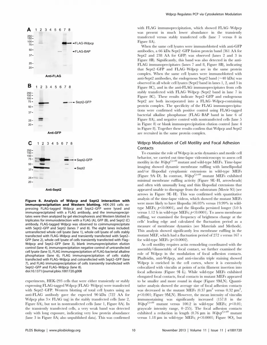
experiments, HEK-293 cells that were either transiently or stably
expressing FLAG-tagged Wdpcp (FLAG–Wdpcp) were transfected
with Sept2–GFP. Western blotting of total cell lysates using an
anti-FLAG antibody gave the expected 90 kDa (722 AA for
Wdpcp plus 36 FLAG tag) in the stably transfected cells (lane 2,
Figure 8A), but not in nontransfected cells (lane 1, Figure 8A). In
the transiently transfected cells, a very weak band was detected
only with long exposure, indicating very low protein abundance
(lane 3 in Figure 8A; also unpublished data). This was confirmed
with FLAG immunoprecipitation, which showed FLAG–Wdpcp
was present in much lower abundance in the transiently
transfected versus stably transfected cells (lane 7 versus 8 in
Figure 8A).
When the same cell lysates were immunoblotted with anti-GFP
antibodies, a 66 kDa Sept2–GFP fusion protein band (361 AA for
Sept2 and 238 AA for GFP) was observed (lanes 2 and 3 in
Figure 8B). Significantly, this band was also detected in the anti-
FLAG immunoprecipitates (lanes 7 and 8, Figure 8B), indicating
that Sept2–GFP and FLAG–Wdpcp are in the same protein
complex. When the same cell lysates were immunoblotted with
anti-Sept2 antibodies, the endogenous Sept2 band (,40 kDa) was
observed in all whole cell lysates (Sept2 band in lanes 1, 2, and 3 in
Figure 8C), and in the anti-FLAG immunoprecipitates from cells
stably transfected with FLAG–Wdpcp (Sept2 band in lane 7 in
Figure 8C). These results indicate Sept2–GFP and endogenous
Sept2 are both incorporated into a FLAG–Wdpcp-containing
protein complex. The specificity of the FLAG immunoprecipita-
tions were confirmed with positive control using FLAG-tagged
bacterial alkaline phosphatase (FLAG–BAP band in lane 6 of
Figure 8A), and negative control with nontransfected cells (lane 5
in Figure 8) or blank immunoprecipitation elution control (lane 4
in Figure 8). Together these results confirm that Wdpcp and Sept2
are recruited in the same protein complex.
Wdpcp Modulation of Cell Motility and Focal AdhesionContacts
To examine the role of Wdpcp in actin dynamics and motile cell
behavior, we carried out time-lapse videomicroscopy to assess cell
motility in the WdpcpCys40 mutant and wild-type MEFs. Time-lapse
imaging showed dynamic membrane ruffling with lamellipodial
and/or filopodial cytoplasmic extensions in wild-type MEFs
(Figure 9A–D). In contrast, WdpcpCys40 mutant MEFs exhibited
minimal membrane ruffling activity (Figure 9E–H, arrowheads)
and often with unusually long and thin filopodial extensions that
appeared unable to disengage from the substratum (Movie S5) (see
arrows in Figure 9E–H). This was confirmed with quantitative
analysis of the time-lapse videos, which showed the mutant MEFs
were more likely to have filopodia (46.03% versus 19.99% in wild-
type MEFs; p,0.0001), and the filopodia persisted longer (3.0 h
versus 1.12 h in wild-type MEFs; p,0.0001). To assess membrane
ruffling, we examined the frequency of brightness change at the
cells’ leading edge and calculated the fluctuation period as a
measure of membrane dynamics (see Materials and Methods).
This analysis showed significantly less membrane ruffling in the
mutant MEF, which had a fluctuation period of 272 s versus 167 s
for wild-type MEFs (p,0.0002).
As cell motility requires actin remodeling coordinated with the
assembly/disassembly of focal contact, we further examined the
role of Wdpcp in the modulation of focal adhesion contacts.
Phalloidin, anti-Wdpcp, and anti-vinculin triple staining showed
Wdpcp is enriched in the cell cortex, where it is extensively
colocalized with vinculin at points of actin filament insertion into
focal adhesions (Figure 9I–L). While wild-type MEFs exhibited
elongated focal contacts, focal contacts in mutant MEFs appeared
to be smaller and more round in shape (Figure 9M,N). Quanti-
tative analysis showed the average size of focal adhesion contacts
was decreased in the mutant MEFs (0.37 mm2 versus 0.32 mm2,
p,0.001; Figure 9M,N). However, the mean intensity of vinculin
immunostaining was significantly increased (157.8 in the
WdpcpCys40 mutant versus 100.2 in wild-type MEFs; p,0.01;
grayscale intensity range, 0–255). The focal adhesion contacts
exhibited a reduction in length (0.76 mm in WdpcpCys40 mutant
versus 1.18 mm in wild-type MEFs; p,0.0001; Figure 9O), but
Figure 8. Analysis of Wdpcp and Sept2 interaction withimmunoprecipitation and Western blotting. HEK-293 cells ex-pressing FLAG-tagged Wdpcp and Sept2–GFP were lysed andimmunoprecipitated with a FLAG antibody, and the immunoprecipi-tates were then analyzed by gel electrophoresis and Western blotted intriplicates for immunodetection with a FLAG (A), GFP (B), and Sept2 (C)antibody. FLAG-tagged Wdpcp was observed to coimmunoprecipitatewith Sept2–GFP and Sept2 (lanes 7 and 8). The eight lanes included:untransfected whole cell lysate (lane 1), whole cell lysate of cells stablytransfected with FLAG–Wdpcp and transiently transfected with Sept2–GFP (lane 2), whole cell lysate of cells transiently transfected with Flag–Wdpcp and Sept2–GFP (lane 3), blank immunoprecipitation elutioncontrol (lane 4), immunoprecipitation negative control of untransfectedcell lysate (lane 5), FLAG immunoprecipitation of FLAG-bacterial alkalinephosphatase (lane 6), FLAG immunoprecipitation of cells stablytransfected with FLAG–Wdpcp and cotransfected with Sept2–GFP (lane7), and FLAG immunoprecipitation of cells transiently transfected withSept2–GFP and FLAG–Wdpcp (lane 8).doi:10.1371/journal.pbio.1001720.g008
Wdpcp Regulates PCP via Cytoskeleton Modulation
PLOS Biology | www.plosbiology.org 10 November 2013 | Volume 11 | Issue 11 | e1001720
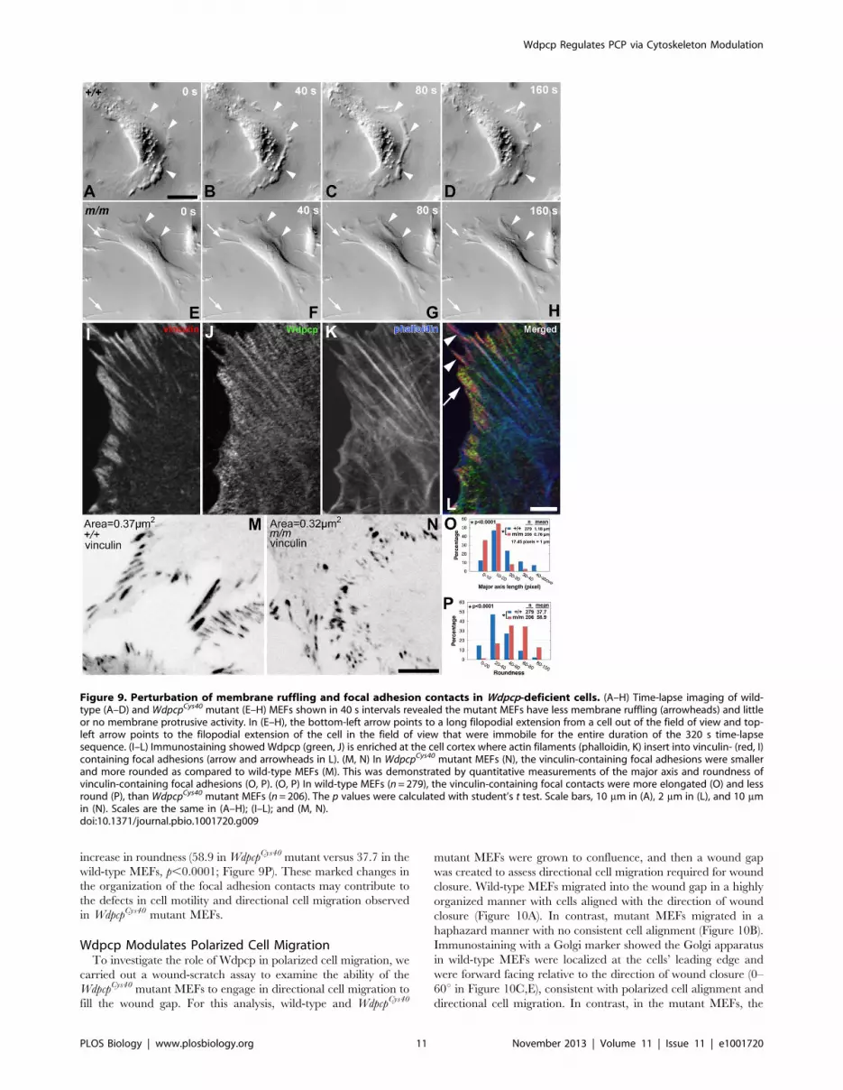
increase in roundness (58.9 in WdpcpCys40 mutant versus 37.7 in the
wild-type MEFs, p,0.0001; Figure 9P). These marked changes in
the organization of the focal adhesion contacts may contribute to
the defects in cell motility and directional cell migration observed
in WdpcpCys40 mutant MEFs.
Wdpcp Modulates Polarized Cell MigrationTo investigate the role of Wdpcp in polarized cell migration, we
carried out a wound-scratch assay to examine the ability of the
WdpcpCys40 mutant MEFs to engage in directional cell migration to
fill the wound gap. For this analysis, wild-type and WdpcpCys40
mutant MEFs were grown to confluence, and then a wound gap
was created to assess directional cell migration required for wound
closure. Wild-type MEFs migrated into the wound gap in a highly
organized manner with cells aligned with the direction of wound
closure (Figure 10A). In contrast, mutant MEFs migrated in a
haphazard manner with no consistent cell alignment (Figure 10B).
Immunostaining with a Golgi marker showed the Golgi apparatus
in wild-type MEFs were localized at the cells’ leading edge and
were forward facing relative to the direction of wound closure (0–
60u in Figure 10C,E), consistent with polarized cell alignment and
directional cell migration. In contrast, in the mutant MEFs, the
Figure 9. Perturbation of membrane ruffling and focal adhesion contacts in Wdpcp-deficient cells. (A–H) Time-lapse imaging of wild-type (A–D) and WdpcpCys40 mutant (E–H) MEFs shown in 40 s intervals revealed the mutant MEFs have less membrane ruffling (arrowheads) and littleor no membrane protrusive activity. In (E–H), the bottom-left arrow points to a long filopodial extension from a cell out of the field of view and top-left arrow points to the filopodial extension of the cell in the field of view that were immobile for the entire duration of the 320 s time-lapsesequence. (I–L) Immunostaining showed Wdpcp (green, J) is enriched at the cell cortex where actin filaments (phalloidin, K) insert into vinculin- (red, I)containing focal adhesions (arrow and arrowheads in L). (M, N) In WdpcpCys40 mutant MEFs (N), the vinculin-containing focal adhesions were smallerand more rounded as compared to wild-type MEFs (M). This was demonstrated by quantitative measurements of the major axis and roundness ofvinculin-containing focal adhesions (O, P). (O, P) In wild-type MEFs (n = 279), the vinculin-containing focal contacts were more elongated (O) and lessround (P), than WdpcpCys40 mutant MEFs (n = 206). The p values were calculated with student’s t test. Scale bars, 10 mm in (A), 2 mm in (L), and 10 mmin (N). Scales are the same in (A–H); (I–L); and (M, N).doi:10.1371/journal.pbio.1001720.g009
Wdpcp Regulates PCP via Cytoskeleton Modulation
PLOS Biology | www.plosbiology.org 11 November 2013 | Volume 11 | Issue 11 | e1001720

direction of Golgi orientation was randomized (Figure 10D,E).
These observations show Wdpcp is required for establishing the
planar cell polarity needed to engage in directional cell migration.
Discussion
We showed WdpcpCys40, a mouse mutant with a wide spectrum
of developmental defects consistent with MKS/BBS ciliopathy
syndromes, harbors a Wdpcp loss of function mutation. We
generated a Wdpcp knockout mouse model, which exhibited
identical phenotypes to those seen in the WdpcpCys40 mutants. A
role for Wdpcp in human disease is suggested by a previous finding
of a homozygous WDPCP mutation in a screen of MKS/BBS
patients [26]. We observed Wdpcp deficiency disrupted ciliogen-
esis and this was associated with the disruption of Shh signaling,
accounting for many of the defect phenotypes observed in the
WdpcpCys40 and Wdpcp knockout mice.
The WdpcpCys40 mutant mice exhibited phenotypes that are
remarkably similar to those observed in a MKS mutant mouse
model, Mks1del64-323 [27]. While heterotaxy was observed in the
Mks1 mutant, no laterality defects were found in the WdpcpCys40
mutants. Consistent with this, nodal cilia motility and nodal flow
were unaffected in WdpcpCys40 mutant embryos. Surprisingly, wdpcp
MO knockdown in zebrafish embryos did not disrupt ciliogenesis,
but perturbed motile cilia function. This was associated with a low
incidence of heterotaxy. It is interesting to note that wdpcp MO
knockdown in Xenopus embryos also perturbed motile cilia function
[26]. These species differences may reflect evolutionary divergence
in the function of Wdpcp and other cilia-related proteins.
Wdpcp Regulates Ciliogenesis via Recruitment of CiliaryProteins
Our findings indicate the ciliogenesis defect in WdpcpCys40
mutants arises from the failure of Wdpcp-deficient cells to recruit
proteins required for ciliogenesis to the ciliary transition zone,
including Nphp1, Sept2, and Mks1. Wdpcp is observed to form a
ring structure in the ciliary transition zone overlapping with Sept2.
Septins are well described to form ring structures both in vitro and
in vivo [37]. In the ciliary transition zone, the septin ring forms a
diffusion barrier regulating protein trafficking into the ciliary
compartment [38]. Thus, failure to recruit Sept2 to the transition
zone in Wdpcp-deficient cells is expected to disrupt ciliogenesis.
Wdpcp-deficient cells also failed to recruit Mks1, a Meckel
syndrome–associated protein localized to the mother centriole and
required for basal body docking to the membrane [27,28].
Figure 10. Wdpcp deficiency disrupts polarized cell migration. (A–D) In a wound healing assay, control MEFs (A) were well aligned with thedirection of wound closure (indicated by white arrow). In contrast, WdpcpCys40 mutant MEFs (B) showed a disorganized distribution and with manylong thin filopodial extensions. These differences in cell polarity were also reflected in their Golgi orientation—Golgi orientation is indicated by awhite line drawn from the cell nucleus through the center of the Golgi (C, D). In wild-type MEFs, the Golgi was mostly situated at the cell’s leadingedge (C), aligned with the direction of wound closure (white arrow). However, in WdpcpCys40 mutant MEFs, Golgi orientation was randomized (D). (E)Quantitative analysis of Golgi orientation showed wild-type MEFs (n = 117) with highly polarized arrangement of the Golgi well aligned with thedirection of cell migration (94 of 117 cells with Golgi apparatus in 0–60u sector), while WdpcpCys40 mutant MEFs (n = 161) showed random orientationof the Golgi apparatus (64 of 161 cells with Golgi in 0–60u sector) (see Materials and Methods). Scale bar, 20 mm in (B) and (D). Scales are the same in(A, B) and (C, D).doi:10.1371/journal.pbio.1001720.g010
Wdpcp Regulates PCP via Cytoskeleton Modulation
PLOS Biology | www.plosbiology.org 12 November 2013 | Volume 11 | Issue 11 | e1001720
Analysis of Mks1-deficient cells showed Wdpcp acts upstream of
Mks1. We also observed Wdpcp-deficient cells with mislocaliza-
tion of Nphp1, a cilia transition zone protein required for
ciliogenesis and associated with cystic kidney defects in Joubert
syndrome and other ciliopathies [39]. Together these findings
indicate Wdpcp may play an important role in recruiting proteins
essential for ciliogenesis. Consistent with this is the presence of two
WD repeats in the Wdpcp protein. The WD40 domain has been
identified in many protein interaction pairs [40], and many WD40
repeat-containing proteins have been shown to serve as scaffolds
for assembly of multiprotein complexes. Together these findings
suggest Wdpcp may serve as a scaffold in the cilia transition zone
to facilitate the assembly of multiprotein complexes required for
ciliogenesis.
Wdpcp Constrains Hedgehog and Canonical WntSignaling
While our studies with WdpcpCys40 mutant MEFs showed Wdpcp
deficiency disrupted Shh signaling, surprisingly the loss of Wdpcp
function partially rescued the severe defect phenotypes of the Ptch1
or Smo knockout embryos. This suggests Wdpcp may constrain Shh
signaling downstream of Smo/Ptch1. As ciliogenesis is disrupted
with Wdpcp deficiency, how Gli processing required for Shh
activation is regulated in the WdpcpCys40/Ptch1 or WdpcpCys40/Smo
knockout embryos is unknown. However, it is worth noting
Drosophila hedgehog signaling occurs in the absence of the cilium
[41].
Our studies also suggested Wdpcp may have a role in
constraining canonical Wnt signaling, as BAT–lacZ and the
expression of canonical Wnt transducers (Dvl1/2/3) are upregu-
lated, while canonical Wnt inhibitors were down-regulated (Dkk1/
2/3) in the WdpcpCys40 mutant OFT. While a role for the cilium in
constraining canonical Wnt signaling is well described, the
mechanism remains unclear [18]. We note Chibby, a basal body
protein that negatively regulates canonical Wnt signaling, can bind
b-catenin and prevent its entry into the nucleus [42]. One
possibility to consider is whether Wdpcp may function in the same
multiprotein complex with Chibby to regulate b-catenin traffick-
ing.
Wdpcp Modulates PCP and the Actin CytoskeletonWe showed WdpcpCys40 mutants exhibited PCP defects, such as
malpatterning of stereocilia in the cochlea and abnormal
myocardialization of the outflow tract in the heart. The
myocardialization defect was characterized by failure of the
invading cardiomyocytes to elongate and align their actin
cytoskeleton with the direction of cell migration. We note the
Loop-tail (Lp) mouse mutant harboring a mutation in the PCP
component, Vangl2, also exhibits outflow tract defects [31]. Similar
to our findings, in the Lp mutant cell protrusions into the outflow
cushion were absent, and the organization of the actin cytoskel-
eton was disrupted. Perturbation of the actin cytoskeleton also may
contribute to defects in the cochlea, as the stereocilia are actin-
based structures and in WdpcpCys40 mutants, formation of the
kinocilia was not affected. While cochlea expression of Vangl2 was
reduced in WdpcpCys40 mutants, we did not observe mislocalization
of PCP core components that would suggest a disruption in PCP
signaling.
Using Wdpcp-deficient MEFs, we investigated the role of
Wdpcp in establishing planar cell polarity. Our studies indicate
Wdpcp plays an essential role in PCP via regulation of the actin
cytoskeleton. We showed Wdpcp modulates the actin cytoskeleton
by mediating Sept2 interaction with actin. In wild-type cells,
Wdpcp is colocalized with Sept2 in actin filaments, but in Wdpcp-
deficient cells, Sept2 is no longer actin localized and actin stress
fibers are largely absent. Instead, Sept2 is observed in ‘‘o’’ and ‘‘c’’
configurations, structures also observed with inhibition of actin
polymerization [36]. These results suggest formation of a Wdpcp–
Sept2 complex may be required for Sept2 binding to actin
filaments and the stabilization of actin filaments. Coimmunopre-
cipitation and Western immunoblotting confirmed Wdpcp and
Sept2 localization in the same protein complex, but further
experiments are needed to examine Wdpcp and Sept2 interaction
with actin filaments.
A further indication of a role for Wdpcp in the modulation of
actin dynamics was the reduction in membrane ruffling in Wdpcp
mutant MEFs. We noted Wdpcp is enriched in the cell cortex
where actin filaments insert into vinculin-containing focal
adhesions. As focal contacts are also sites of actin polymerization,
the abundance of Wdpcp in the vicinity of focal contacts may
facilitate recruitment of Sept2 to enhance actin filament
stabilization and stress fiber formation. We observed Wdpcp-
deficient cells had smaller focal contacts, but with significantly
higher concentration of vinculin. This could account for the
failure of cell processes to disengage from the substratum in
Wdpcp-deficient cells.
Most significantly, Wdpcp-deficient cells showed defects in
motile cell behavior that was associated with defects in planar cell
polarization. Thus, Wdpcp-deficient cells were unable to establish
cell polarity in a wound scratch assay. This is indicated by failure
of the Golgi to reorient to the cell’s leading edge and align with the
direction of cell migration. As a result, migrating cells were unable
to engage in directional cell motility. It is significant to note that
Golgi orientation is specified by the microtubule-organizing center
or centrosome, which also templates formation of the cilium. This
defect in establishing cell polarity together with perturbation in
membrane and actin dynamics and the modulation of focal
adhesion contacts may underlie the PCP defects associated with
Wdpcp deficiency. Together, these findings suggest Wdpcp may
regulate planar cell polarity by modulating both the microfilament
and microtubule cytoskeleton.
Dual Functionality of Wdpcp in Ciliogenesis and PCPWe showed the PCP effector Wdpcp plays an essential role in
both ciliogenesis and PCP. This appears to involve separable
functions of Wdpcp in recruitment of proteins to the ciliary
transition zone required for ciliogenesis, and in the modulation of
actin dynamics in the cytoplasm. Our findings provide the first
evidence, to our knowledge, that a PCP component required for
ciliogenesis can directly modulate the actin cytoskeleton to
regulate planar cell polarity and directional cell migration. These
observations suggest Wdpcp regulation of PCP is independent of
its role in ciliogenesis. It is interesting to consider whether such
dual functionality may have evolved as a means to functionally
integrate pathways regulating ciliogenesis with those regulating
PCP and the cytoskeleton. Whether such dual functionality may
account for other PCP proteins known to be required for
ciliogenesis is an important question that needs to be addressed
in future studies.
Materials and Methods
Institutional Approval for Animal StudiesAll mouse experiments were carried out using protocols
approved by the Institutional Animal Care and Use Committee
at the National Heart Lung Blood Institute and at the University
of Pittsburgh. The zebrafish experiments were carried out with
approved protocols at the University of Pittsburgh.
Wdpcp Regulates PCP via Cytoskeleton Modulation
PLOS Biology | www.plosbiology.org 13 November 2013 | Volume 11 | Issue 11 | e1001720

Mapping and Recovery of the Wdpcp Mutation andMouse Breeding
C57BL/6J(B6)/C3H hybrid mutant offspring were used to map
the mutation using 48 microsatellite markers polymorphic between
B6/C3H [43]. Once the chromosome interval was identified,
additional polymorphic DNA markers were used to narrow the
interval to a 5–10 Mb region. Then cDNA sequencing was carried
out for each gene in the interval.
Smotm1Amc (strain 004288), Ptch1tm1Mps (strain 003081), and BAT–
lacZ (strain 005317) mouse lines were obtained from the Jackson
Laboratory and intercrossed with the WdpcpCys40 mouse line to
generate double heterozygous mice, which were further inter-
crossed to generate embryos double homozygous for the
WdpcpCys40 and Smotm1Amc, WdpcpCys40 and Ptch1tm1Mps, or homozy-
gous WdpcpCys40 carrying BAT–lacZ allele.
Construction of FLAG-Tagged Wdpcp ConstructTo make the mouse FLAG–Wdpcp construct, we isolated RNA
from mutant heart and amplified the full-length cDNA using one
step RT-PCR using Superscript III/HF Platinum Taq DNA
polymerase and cloned cDNA into pCR2.1 TOPO vector. We
then confirmed the clones by Sanger sequencing. We amplified
TOPO vector containing Wdpcp (NM_145425.3) cDNA using
primers (Table S1) with restriction site at the 59 end, cut with
appropriate restriction endonuclease, and cloned into expression
vectors containing FLAG at either the 59 end or 39 end (Sigma-
Aldrich). The subsequent experiments were performed by using
the FLAG at the 59 end of Wdpcp.
Zebrafish wdpcp Morpholino Knockdown and RescueZebrafish embryos were obtained from incrossing wild-type
adults and following manipulation, maintained at 28.5uC, and
staged according to [44]. A complementary MO (59-
AGCTCCGCCAGGCAGAACGACATCT-39) targeting the ini-
tiation codon of zebrafish wdpcp and standard control (59-
CCTCTTACCTCAGTTACAATTTATA-39) morpholino (MO)
were designed and obtained from GeneTools LLC. Embryos at
the one-cell stage were injected with 5 nl of morpholino at 2.0 ng/
nl in phenol red. Control embryos were either injected with the
standard control (2.0 ng/nl) MO or uninjected. Capped mRNAs
for rescue experiments were synthesized in vitro from linearized
pCS2+ mouse Wdpcp construct (NM_145425.3) using SP6
mMessage mMachine kit (Ambion). We microinjected 200 pg of
Wdpcp mRNA at 200 pg/1 nl into the blastomere at the one-cell
stage. After injections, the embryos were incubated in16E3
medium (5 mM NaCl, 0.17 mM KCl, 0.33 mM CaCl2, 0.33 mM
MgSO4, 0.01% methylene blue) at 28uC until the desired stage
and imaged using Leica MZ16 microscope fitted with Retiga 1300
camera. Statistical distribution of morphants and morphologically
normal embryos from three separate rescue experiments were
analyzed using Graphpad prism version 6.
Zebrafish in Situ Hybridization andImmunocytochemistry
For in-situ hybridization, zebrafish embryos were fixed in 4%
paraformaldehyde and processed as described [45]. Partial
zebrafish wdpcp cDNA clone obtained from OpenBiosystems
(#EDR1052-97951134, clone ID 7911360) was used to generate
sense and antisense in-situ probes for wdpcp. Plasmids for cmlc2 and
fabp10a in-situ probes were obtained from members of the
zebrafish community and were described previously [46,47]. For
immunocytochemistry, zebrafish embryos were fixed in Dent’s
fixative (80% Methanol and 20% DMSO) for 4 h at room
temperature, washed with methanol, and stored at 220uC.
Antibody staining for acetylated a-tubulin was performed as
described [48]. For Wdpcp and acetylated a-tubulin double
immunostaining, embryos were rehydrated in a series of metha-
nol/PBT washes and permeabilized with 0.02% trypsin for 5 min.
Following blocking with 10% normal goat serum (NGS, Sigma),
embryos were incubated overnight at 4uC in PBT/10% NGS with
Wdpcp (1:100; Aves, Inc) and acetylated a-tubulin (1:1,000;
Sigma) antibodies. Goat anti-mouse Alexa Fluor 488 and goat
anti-chicken Alexa Fluor 555 (both 1:1,000; Invitrogen) secondary
antibodies were used. Embryos were de-yolked, mounted in Aqua
Poly/Mount (Polysciences, Inc.), and visualized with a Zeiss LSM
510 Meta inverted laser scanning confocal microscope.
Production of Wdpcp KO MiceThe Wdpcp target or null allele was generated by gene targeting
in 129 ES cells, as shown in Figure S3A. Exon 5 was flanked by
two loxP/Flox sites and an FRT-flanked PGKneo cassette
(neomycin-resistant gene driven by the PGK promoter) was
inserted in intron 5 of Wdpcp by homologous recombination. ES
cell clones with correct homologous recombination were screened
by long-range PCR. Two independent ES cell clones were injected
into C57BL/6J blastocysts, and germ line transmission was
achieved with both clones. The PGKNeo cassette was removed
by FLP-FRT recombination to generate Wdpcp flox allele. The
Wdpcp global knockout allele was generated by CMV-Cre/Lox
recombination.
Immunocytochemistry and ImmunohistologyMEFs derived from E11.5–E12.5 embryos were serum starved
for 24 to 48 h to grow out the cilia, with staining of ciliary proteins
carried out using various antibodies including acetylated a-tubulin
antibody (Sigma T7451, 1:1,000), c-tubulin antibody (Sigma
T6557, 1:1,000), Wdpcp chicken polyclonal antibody (Aves, Inc.)
made against synthetic peptide (DTTILEYREPVSKYARR)
corresponding to Wdpcp amino residues 529–545, Wdpcp goat
antibody (Santa Cruz), Sept2 antibody (Millipore, 1:1,000), Mks1
antibody (1:2,000) [27], and antibodies to Ift88 and Nphp1 as
previously described [49,50]. FLAG antibody (Sigma F7425,
1:1,000) was used for detection of Wdpcp–FLAG fusion protein.
Kidney paraffin sections were stained with DBA (Dolichos biflorus
agglutinin, Sigma, 1:20) to delineate kidney tubules. Dissected
cochleae were stained with Phalloidin and Vangl2 antibody as
described previously [12]. Heart cryo-sections were stained with
antibodies to MF20 (Hybridoma Bank, 1:40), smooth muscle actin
(Sigma, 1:40). Neural tube cryo-sectioning and immunostaining
with FoxA2 (Hybridoma Bank, 1:1,000), Pax6 (Hybridoma Bank,
1:1,000), and Olig2 (gift from Dr. Bennett Novitch at UCLA,
1:1,000) was previously described [27].
Analysis of Shh SignalingDIG-labeled probes were made with DIG RNA reaction
mixture (Roche), and procedures for whole mount in-situ
hybridization were described previously [51] and gene expression
patterns were visualized with BM purple AP substrate (Roche).
RNA in-situ probe plasmids for Fgf4 (Lee Niswander lab), Gremlin
(Richard Harland lab), and Ptch1 were gifts from Dr. Susan
Mackem (NCI). A 600 bp nucleotide Gli3 in-situ probe was
generated by amplifying cDNA specific to Gli3 encompassing
exons 12–14.
For limb Gli3 Western blotting, left fore- and hindlimbs at
E10.5 (32–37 somite stages) were harvested and bisected into
anterior and posterior halves, whereas the right fore- and
hindlimbs were harvested intact. The remaining tissues were
Wdpcp Regulates PCP via Cytoskeleton Modulation
PLOS Biology | www.plosbiology.org 14 November 2013 | Volume 11 | Issue 11 | e1001720

further removed until the neural tube was left, which was then
used for Gli2 and Gli3 Western blotting. In addition, whole
mutant and wild-type E10.5 embryos were lysed and processed for
Gli3 Western blotting. The Western blotting procedures for Gli2
and Gli3 were carried out as described previously in [27]. Gli2 and
Gli3 antibodies were gifts from Dr. Baolin Wang at Cornell
University and Dr. Susan Mackem at NCI, respectively.
Quantitative Analysis of Vinculin-Containing FocalAdhesions
Focal adhesions were visualized using a vinculin antibody
(Sigma V9264, 1:1,000). The area of vinculin immunostaining was
traced using the image editor Gimp (www.gimp.org), and the area
and intensity of staining were measured in 8-bit images. To
quantify the length and roundness of vinculin-containing focal
adhesion sites, we measured the major axis (MA) and minor axes
(MI) of each focal adhesion. The mean MA length was determined
and was plotted as a histogram with bin size of 10 pixels
(equivalent to 0.57 mm). The roundness factor for each focal
adhesion contact was measured using MI/MA*100, with 100
being focal adhesions that were perfectly round with major and
minor axes of equal length.
Scanning EM and Videomicroscopy of Nodal Cilia andPronephric Duct
Scanning EM of the nodal cilia and neural tube epithelium was
carried out as described previously [27]. Analysis of nodal cilia
motility in E8.0 mouse embryos and pronephric duct of zebrafish
embryos were carried as described previously [52]. The videos
were converted into Quicktime movies.
Time-Lapse Videomicroscopy for Assessing Cell MotileBehavior
Time-lapse videomicroscopy was carried out to quantify the
motile behavior of wild-type and Wdpcp mutant MEFs using an
inverted microscope (Leica, DMIRE2) and a Hamamatsu ORCA-
ER camera. Video sequences encompassing an 8 h recording
period with images captured every 10 min were used to measured
the number of cells having one or more filopodia/total number of
cells (percentage of cells with filopodia), and we also measured the
number of frames in which the same filopodia was observed
(persistence of filopodia). To quantify membrane ruffling activity,
we recorded cultured cells at a shorter time-lapse interval
comprising 10 s for a total of 30 min. These time-lapse images
were then used to measure the frequency of brightness change at
the cells’ leading edge to quantitate the membrane ruffling activity.
This entailed selecting boxed regions from the leading edge of a
migrating cell, summing the brightness for each frame, and
performing Fourier transform to obtain the frequency of
brightness changes. The inverse of this frequency provided an
index of the ‘‘period’’ of membrane fluctuation (MFP, in seconds),
which is a measure of membrane dynamics, with greater
membrane ruffling indicated by lower MFP.
Wound Scratch Assay for Assaying Polarized CellMigration
Confluent MEF cultures were scratched using a 10 ml micro-
pipette tip to generate a gap. Then time-lapse imaging was carried
out using a 406 objective on an inverted microscope (Leica,
DMIRE2) with images captured every 10 s over a 30 min interval
using a Hamamatsu ORCA-ER camera. To examine cell polarity,
cells were immunostained with a Golgi antibody (Sigma
HPA021799, 1:1,000) followed by DAPI staining. Cells that are
polarized and aligned with the direction of migration have Golgi
situated in front of the nucleus (forward facing) and aligned with
the migration direction. Polarity was scored by overlaying a clock
face on each cell, and polarized cells are defined as those with
Golgi situated within a 60u sector centered along the direction of
wound closure [53,54].
Supporting Information
Figure S1 Ciliogenesis defect in WdpcpCys40 mutant. (A,
B) Immunostaining of cilium with acetylated a-tubulin, a-tub (red),
and c-tubulin, (g-tub, green) antibodies showing a shorter cilium in
WdpcpCys40 mutant MEF (B). Scanning EM images of embryonic
nodes of control (C) and mutant (D) embryos at E8.0 showing the
node cells are ciliated normally and cilia in mutant embryonic
node are of normal shape and length. Scale bars, 2 mm in (A) and
(C). Scales are the same in (A, B) and (C, D).
(JPG)
Figure S2 Wdpcp zebrafish in situ hybridization andmorphant at 48 h, Western blotting with Wdpcp chickenantibody. (A–O) Embryonic wdpcp mRNA localization (purple)
by whole mount in-situ hybridization with wdpcp antisense
riboprobe at the four-cell stage (A, B), eight-cell stage (C, D),
1,000-cell stage (E, F), and shield stage (G) showed absence of
maternal wdpcp transcripts. Embryonic wdpcp expression is
observed at the 10-somite stage (H, I) and at 24 hpf (J, K). At
48 hpf (L–O) wdpcp staining appears less specific, since faint
staining is observed with both the sense and antisense probes. (P)
Immunoblot using wdpcp antibody (green) with 24 hpf zebrafish
embryo lysate showed effective knockdown of wdpcp protein
expression. a-Tubulin (red) was used as a sample loading control.
Lane 1, protein molecular weight markers; lane 2, lysate from
embryos injected with 10 ng control morpholino (MO); and lane
3, lysate from embryos injected with 10 ng wdpcp morpholino. (Q)
The ratio of wdpcp (green) to a-tubulin (red) in the immunoblot
was quantified using Image studio version 2.0 from LI-COR
Biosciences (Lincoln, NE), which showed significant reduction in
the wdpcp protein with wdpcp MO knockdown. Scale bars,
200 mm in (A), (G), (I), (J), and (L) and 150 mm in (M). Scales are
the same in (A–F), (H, I), (J, K), (L, N), and (M, O).
(JPG)
Figure S3 Laterality defects in Wdpcp zebrafish mor-phants. (A–D) Ventral view of RNA in-situ hybridization staining
with cmlc2 probe delineating the heart tube in 54 hpf embryos in
wdpcp morphants revealed normal right-sided looping (B), no
looping (C), or reversed heart looping (D) orientation. (E–H)
Dorsal view of gut orientation as observed with LFABP in-situ
hybridization analysis delineating liver position in 54 hpf embryo.
Three types of gut orientation were observed: normal left-sided (F),
duplicated (G), and right-sided (H). (I, J) Distribution of heart (I)
and gut (J) looping orientation in Wdpcp morphants, with asterisk
indicating statistically significant differences between control
versus wdpcp morphants. (K–P) In-situ hybridization with an evx1
probe on 24 hpf embryos (K–M) delineated the normal cloaca in
uninjected (K) and control MO (L) injected embryos, while in the
wdpcp morphant (M), the cloaca is abnormally formed. Compar-
ison of the corresponding brightfield images (N–P) suggests the
cloaca in the wdpcp morphant may be obstructed. The arrowhead
denotes the obstructed cloaca, which was seen in 37% of the wdpcp
morphants (n = 208).
(JPG)
Figure S4 Rescue of wdpcp morpholino (MO)-inducedphenotype. (A) Representative images of 48 hpf embryos
Wdpcp Regulates PCP via Cytoskeleton Modulation
PLOS Biology | www.plosbiology.org 15 November 2013 | Volume 11 | Issue 11 | e1001720
injected at one-cell stage with 10 ng of scrambled control MO. (B)
Representative images of 48 hpf embryos injected with 10 ng of
wdpcp MO at one-cell stage showing pericardial edema (black
arrows) and a curved tail. (C) Representative images of 48 hpf
embryos injected at one-cell stage with 200 pg synthetic mouse
wdpcp mRNA. (D) Representative images of 48 hpf embryos co-
injected at one-cell stage with 10 ng of wdpcp MO and 200 pg
synthetic mouse wdpcp mRNA showing rescue of morphant
phenotype. (E, F) Morphant phenotypes (normal, mild, and
severe) obtained in the experiments examining wdpcp mRNA
rescue of wdpcp MO-injected embryos are summarized in the
graph shown in (F) and the table in (G).
(JPG)
Figure S5 Production and phenotype of Wdpcp knock-out mice. (A) Schema of the Wdpcp targeted mouse allele
generated by homologous recombination containing an FRT-
flanked PGKneo cassette bracketed with two loxp sites that would
allow the deletion of exon 5 to generate a Wdpcp knockout allele.
(B–E) Newborn homozygous Wdpcp knockout mouse exhibited
craniofacial defects (B), congenital heart defects (pulmonary
atresia) (C), limb polydactyly (D), and duplex kidney (arrows in
E), phenotypes identical to those seen in the WdpcpCys40 mutants.
Scales bars, 200 mm in (C–E).
(JPG)
Figure S6 Shh signaling is compromised in WdpcpCys40
mutants. (A) In WdpcpCys40 mutant embryos (E10.5 dpc), caudal
neural tube (between the forelimb and hindlimb) showed dimin-
ished FoxA2 in the ventral floorplate, and expansion of Olig2 and
Pax6 to the ventral most position. (B) Smoothened agonist SAG
treatment upregulated Gli1 expression in wild-type MEFs by 20-
fold, while WdpcpCys40 mutant MEFs were not responsive to SAG
stimulation. (C) Western blot of E10.5 whole embryo extract
showed WdpcpCys40 homozygous mutants had higher Gli3-FL/Gli3-
R ratio compared to wild-type controls, indicating impaired Gli3
processing. (D) Western blot of E10.5 neural tube extract showed
elevated Gli2-FL level in WdpcpCys40 mutant. Scales are the same for
images in (A), and the scale bar is 50 mm.
(JPG)
Movie S1 Nodal cilia show normal motility inWdpcpCys40 mutant embryos. DIC (Differential Interference
Contrast) imaging showed similar nodal cilia motility in wild-type
(left) and homozygous WdpcpCys40 mutant (right) E8.0 embryos.
Scale bar, 10 mm.
(MOV)
Movie S2 3D reconstruction showing Wdpcp and Sept2in ring-like structure. The confocal image stack used to
generate the images shown in Figure 2I–L was reconstructed in
three dimension (3D) to show ring-like structure comprising
Wdpcp (red) with Sept2 (green).
(MOV)
Movie S3 Motile cilia in the WdpcpCys40 mutant trachealrespiratory epithelium. The ciliated tracheal epithelia of a
near-term WdpcpCys40 mutant fetus showed normal coordinated
ciliary motion.
(MOV)
Movie S4 Motile cilia in the zebrafish pronephric ductshowed weak and uncoordinated beat after wdpcpmorpholino knockdown. The cilia in the pronephric duct
showed strong coordinated beat in zebrafish embryo injected with
control morpholino, but in wdpcp morpholino-injected embryos
(bottom five panels), cilia in the pronephric duct showed weak and
uncoordinated beat.
(MOV)
Movie S5 Time-lapse imaging shows decreased mem-brane ruffling in WdpcpCys40 mutant MEF. Time-lapse
imaging shows dynamic membrane ruffling in wild-type MEF (top
panel). In contrast, WdpcpCys40 mutant MEF (bottom panel)
showed much less membrane ruffling activity, with unusually long
thin filopodial extensions that were immotile. Images were
captured every 5 s over a 320 s interval.
(MOV)
Table S1 Primer sequences for cloning and qPCR. Rows
1 and 2, primers used to clone Wdpcp for making FLAG–Cys40
construct. Rows 3–34, primers used for heart outflow tract qPCR.
(DOCX)
Acknowledgments
We would like to thank Dr. Mathew P. Daniels and Patricia Connelly at
the NHLBI Electron Microscopy Core Facility for help with scanning
electron microscopy. We also would like to thank Dr. Baolin Wang at
Cornell University (Gli2), Dr. Susan Mackem at NCI (Gli3), and Dr.
Bennett Novitch at UCLA (Olig2) for antibody reagents.
Author Contributions
The author(s) have made the following declarations about their
contributions: Conceived and designed the experiments: CC CWL.
Performed the experiments: CC BC TPL ZZ RJF HY LMA SS DF QY
JTS CP TC TT XL. Analyzed the data: CC BC TPL ZZ RJF HY LMA
SS MWK ETS AVK GJP NAH CWL. Contributed reagents/materials/
analysis tools: MWK ETS AVK RT GJP NAH CWL. Wrote the paper:
CC CWL.
References
1. Badano JL, Mitsuma N, Beales PL, Katsanis N (2006) The ciliopathies: an
emerging class of human genetic disorders. Annu Rev Genomics Hum Genet 7:
125–148.
2. Caspary T, Larkins CE, Anderson KV (2007) The graded response to Sonic
Hedgehog depends on cilia architecture. Dev Cell 12: 767–778.
3. Huangfu D, Anderson KV (2005) Cilia and Hedgehog responsiveness in the
mouse. Proc Natl Acad Sci U S A 102: 11325–11330.
4. Huangfu D, Liu A, Rakeman AS, Murcia NS, Niswander L, et al. (2003)
Hedgehog signalling in the mouse requires intraflagellar transport proteins.
Nature 426: 83–87.
5. Rohatgi R, Milenkovic L, Scott MP (2007) Patched1 regulates hedgehog
signaling at the primary cilium. Science 317: 372–376.
6. Corbit KC, Aanstad P, Singla V, Norman AR, Stainier DY, et al. (2005)
Vertebrate Smoothened functions at the primary cilium. Nature 437: 1018–1021.
7. Haycraft CJ, Banizs B, Aydin-Son Y, Zhang Q, Michaud EJ, et al. (2005) Gli2 and
Gli3 localize to cilia and require the intraflagellar transport protein polaris for
processing and function. PLoS Genet 1(4): e53. doi:10.1371/journal.pgen.0010053
8. Wen X, Lai CK, Evangelista M, Hongo JA, de Sauvage FJ, et al. (2010) Kinetics
of hedgehog-dependent full-length Gli3 accumulation in primary cilia and
subsequent degradation. Mol Cell Biol 30: 1910–1922.
9. Hildebrandt F, Attanasio M, Otto E (2009) Nephronophthisis: disease
mechanisms of a ciliopathy. J Am Soc Nephrol 20: 23–35.
10. Wallingford JB, Harland RM (2002) Neural tube closure requires Dishevelled-
dependent convergent extension of the midline. Development 129: 5815–
5825.
11. Wang J, Hamblet NS, Mark S, Dickinson ME, Brinkman BC, et al. (2006)
Dishevelled genes mediate a conserved mammalian PCP pathway to regulate
convergent extension during neurulation. Development 133: 1767–1778.
12. Montcouquiol M, Sans N, Huss D, Kach J, Dickman JD, et al. (2006)
Asymmetric localization of Vangl2 and Fz3 indicate novel mechanisms for
planar cell polarity in mammals. J Neurosci 26: 5265–5275.
13. Wang J, Mark S, Zhang X, Qian D, Yoo SJ, et al. (2005) Regulation of polarized
extension and planar cell polarity in the cochlea by the vertebrate PCP pathway.
Nat Genet 37: 980–985.
Wdpcp Regulates PCP via Cytoskeleton Modulation
PLOS Biology | www.plosbiology.org 16 November 2013 | Volume 11 | Issue 11 | e1001720

14. Wang Y, Guo N, Nathans J (2006) The role of Frizzled3 and Frizzled6 in neural
tube closure and in the planar polarity of inner-ear sensory hair cells. J Neurosci
26: 2147–2156.
15. Jones C, Chen P (2008) Primary cilia in planar cell polarity regulation of the
inner ear. Curr Top Dev Biol 85: 197–224.
16. Jones C, Roper VC, Foucher I, Qian D, Banizs B, et al. (2008) Ciliary proteins
link basal body polarization to planar cell polarity regulation. Nat Genet 40: 69–
77.
17. Sipe CW, Lu X (2011) Kif3a regulates planar polarization of auditory hair cells
through both ciliary and non-ciliary mechanisms. Development 138: 3441–
3449.
18. Wallingford JB, Mitchell B (2011) Strange as it may seem: the many links
between Wnt signaling, planar cell polarity, and cilia. Genes Dev 25: 201–213.
19. Vladar EK, Antic D, Axelrod JD (2009) Planar cell polarity signaling: the
developing cell’s compass. Cold Spring Harb Perspect Biol 1: a002964.
20. Saburi S, Hester I, Fischer E, Pontoglio M, Eremina V, et al. (2008) Loss of Fat4
disrupts PCP signaling and oriented cell division and leads to cystic kidney
disease. Nat Genet 40: 1010–1015.
21. Ross AJ, May-Simera H, Eichers ER, Kai M, Hill J, et al. (2005) Disruption of
Bardet-Biedl syndrome ciliary proteins perturbs planar cell polarity in
vertebrates. Nat Genet 37: 1135–1140.
22. Park TJ, Haigo SL, Wallingford JB (2006) Ciliogenesis defects in embryos
lacking inturned or fuzzy function are associated with failure of planar cell
polarity and Hedgehog signaling. Nat Genet 38: 303–311.
23. Corbit KC, Shyer AE, Dowdle WE, Gaulden J, Singla V, et al. (2008) Kif3a
constrains beta-catenin-dependent Wnt signalling through dual ciliary and non-
ciliary mechanisms. Nat Cell Biol 10: 70–76.
24. Simons M, Gloy J, Ganner A, Bullerkotte A, Bashkurov M, et al. (2005) Inversin,
the gene product mutated in nephronophthisis type II, functions as a molecular
switch between Wnt signaling pathways. Nat Genet 37: 537–543.
25. Collier S, Lee H, Burgess R, Adler P (2005) The WD40 repeat protein fritz links
cytoskeletal planar polarity to frizzled subcellular localization in the Drosophila
epidermis. Genetics 169: 2035–2045.
26. Kim SK, Shindo A, Park TJ, Oh EC, Ghosh S, et al. (2010) Planar cell polarity
acts through septins to control collective cell movement and ciliogenesis. Science
329: 1337–1340.
27. Cui C, Chatterjee B, Francis D, Yu Q, SanAgustin JT, et al. (2011) Disruption of
Mks1 localization to the mother centriole causes cilia defects and developmental
malformations in Meckel-Gruber syndrome. Dis Model Mech 4: 43–56.
28. Weatherbee SD, Niswander LA, Anderson KV (2009) A mouse model for
Meckel syndrome reveals Mks1 is required for ciliogenesis and Hedgehog
signaling. Hum Mol Genet 18: 4565–4575.
29. Fliegauf M, Horvath J, von Schnakenburg C, Olbrich H, Muller D, et al. (2006)
Nephrocystin specifically localizes to the transition zone of renal and respiratory
cilia and photoreceptor connecting cilia. J Am Soc Nephrol 17: 2424–2433.
30. Kelly M, Chen P (2007) Shaping the mammalian auditory sensory organ by the
planar cell polarity pathway. Int J Dev Biol 51: 535–547.
31. Phillips HM, Murdoch JN, Chaudhry B, Copp AJ, Henderson DJ (2005) Vangl2
acts via RhoA signaling to regulate polarized cell movements during
development of the proximal outflow tract. Circ Res 96: 292–299.
32. Phillips HM, Rhee HJ, Murdoch JN, Hildreth V, Peat JD, et al. (2007)
Disruption of planar cell polarity signaling results in congenital heart defects and
cardiomyopathy attributable to early cardiomyocyte disorganization. Circ Res
101: 137–145.
33. Maretto S, Cordenonsi M, Dupont S, Braghetta P, Broccoli V, et al. (2003)
Mapping Wnt/beta-catenin signaling during mouse development and incolorectal tumors. Proc Natl Acad Sci U S A 100: 3299–3304.
34. Kinoshita M, Field CM, Coughlin ML, Straight AF, Mitchison TJ (2002) Self-
and actin-templated assembly of Mammalian septins. Dev Cell 3: 791–802.35. Joo E, Surka MC, Trimble WS (2007) Mammalian SEPT2 is required for
scaffolding nonmuscle myosin II and its kinases. Dev Cell 13: 677–690.36. Schmidt K, Nichols BJ (2004) Functional interdependence between septin and
actin cytoskeleton. BMC Cell Biol 5: 43.
37. Meseroll RA, Howard L, Gladfelter AS (2012) Septin ring size scaling anddynamics require the coiled-coil region of Shs1p. Mol Biol Cell 23: 3391–3406.
38. Hu Q, Milenkovic L, Jin H, Scott MP, Nachury MV, et al. (2010) A septindiffusion barrier at the base of the primary cilium maintains ciliary membrane
protein distribution. Science 329: 436–439.39. Castori M, Valente EM, Donati MA, Salvi S, Fazzi E, et al. (2005) NPHP1 gene
deletion is a rare cause of Joubert syndrome related disorders. J Med Genet 42:
e9.40. Stirnimann CU, Petsalaki E, Russell RB, Muller CW (2010) WD40 proteins
propel cellular networks. Trends Biochem Sci 35: 565–574.41. Yoder B (2008) Ciliary function in mammalian development. Academic Press.
464 p.
42. Takemaru K, Yamaguchi S, Lee YS, Zhang Y, Carthew RW, et al. (2003)Chibby, a nuclear beta-catenin-associated antagonist of the Wnt/Wingless
pathway. Nature 422: 905–909.43. Yu Q, Shen Y, Chatterjee B, Siegfried BH, Leatherbury L, et al. (2004) ENU
induced mutations causing congenital cardiovascular anomalies. Development131: 6211–6223.
44. Kimmel CB, Ballard WW, Kimmel SR, Ullmann B, Schilling TF (1995) Stages
of embryonic development of the zebrafish. Dev Dyn 203: 253–310.45. Kudoh T, Tsang M, Hukriede NA, Chen X, Dedekian M, et al. (2001) A gene
expression screen in zebrafish embryogenesis. Genome Res 11: 1979–1987.46. Yelon D, Horne SA, Stainier DY (1999) Restricted expression of cardiac myosin
genes reveals regulated aspects of heart tube assembly in zebrafish. Dev Biol 214:
23–37.47. Stuckenholz C, Lu L, Thakur PC, Choi TY, Shin D, et al. (2013) Sfrp5
modulates both Wnt and BMP signaling and regulates gastrointestinalorganogenesis in the zebrafish, Danio rerio. PLoS ONE 8(4): e62470.
doi:10.1371/journal.pone.006247048. Drummond I (2009) Studying cilia in zebrafish. Methods Cell Biol 93: 197–217.
49. Pazour GJ, Baker SA, Deane JA, Cole DG, Dickert BL, et al. (2002) The
intraflagellar transport protein, IFT88, is essential for vertebrate photoreceptorassembly and maintenance. J Cell Biol 157: 103–113.
50. Schermer B, Hopker K, Omran H, Ghenoiu C, Fliegauf M, et al. (2005)Phosphorylation by casein kinase 2 induces PACS-1 binding of nephrocystin and
targeting to cilia. EMBO J 24: 4415–4424.
51. Albrecht U, Eichele G, Helms J, Lu HC (1997) Visualization of gene expressionpatterns by in situ hybridization. In: Daston GP, editor. Molecular and cellular
methods in developmental toxicology. New York: CRC Press. pp. 23–48.52. Francis RJ, Chatterjee B, Loges NT, Zentgraf H, Omran H, et al. (2009)
Initiation and maturation of cilia-generated flow in newborn and postnatalmouse airway. Am J Physiol Lung Cell Mol Physiol 296: L1067–1075.
53. Magdalena J, Millard TH, Machesky LM (2003) Microtubule involvement in
NIH 3T3 Golgi and MTOC polarity establishment. J Cell Sci 116: 743–756.54. Nobes CD, Hall A (1999) Rho GTPases control polarity, protrusion, and
adhesion during cell movement. J Cell Biol 144: 1235–1244.
Wdpcp Regulates PCP via Cytoskeleton Modulation
PLOS Biology | www.plosbiology.org 17 November 2013 | Volume 11 | Issue 11 | e1001720


![Acute ethanol exposure induces [Ca 2+ ] i transients, cell swelling and transformation of actin cytoskeleton in astroglial primary cultures](https://static.fdokumen.com/doc/165x107/63221b7a61d7e169b00c78d0/acute-ethanol-exposure-induces-ca-2-i-transients-cell-swelling-and-transformation.jpg)


