Cytoskeleton--plasma membrane interactions
10
aa IM IL 5 I ,I - . l m .l .l .£ ,l _ _ 74. K. Parczyk, W. Haase, C. Kondor-Koch, J. Biol. Chem. 264, 16837 (1989); M. J. A. H. Van Zeijl and K. S. Matlin, Cell Regul. 1, 921 (1990). 75. C. Achler, D. Filmer, C. Merte, D. Drenckhahn, J. Cell Biol. 109, 179 (1989); P. P. Breitfeld, W. C. McKinnon, K. E. Mostov, ibid. 111, 2365 (1990). 76. P. van der Sluijs, M. K. Bennett, C. Antony, K. Simons, T. E. Kreis, J. Cell Sci. 95, 545 (1990). 77. M. Bomsel, R. Parton, S. E. Kuznetsov, T. A. Schroer, J. Gruenberg, Cell 62, 719 (1990). 78. T. P. Fleming and M. H. Johnson, Annu. Rev. Cell Biol. 4, 459 (1988); P. Ekblom, D. Vestweber, R. Kemler, ibid. 2, 27 (1986). 79. S. J. Roberts, D. S. Leaf, H.-P. Moore, J. C. Gerhart, J. Cell Biol. 118, 1359 (1992). 80. P. Doherty, S. V. Ashton, S. E. Moore, F. S. Walsh, Cell 67, 21 (1991); T. Frei, F. von Bohlen und Halbach, W. Wille, M. Schachner, J. Cell Blot. 1 18, 177 (1992). 81. G. L. Augustine, M. P. Chariton, S. J. Smith, Annu. Rev. Neurosci. 10, 633 (1987). 82. S. J. Shattil and J. S. Brugge, Curr. Opin. Cell Biol. 3, 869 (1991). 83. E. H. Fischer, H. Charbonneau, N. K. Tonks, Science 253, 401 (1991). 84. T. Volberg et al., EMBO J. 11, 1733 (1992). 85. W. J. Nelson and P. J. Veshnock, J. Cell Biol. 104, 1527 (1987); H. McNeill, M. Ozawa, R. Kemler, W. J. Nelson, Cell 62, 309 (1990). 86. D. Gravotta, M. Adesnik, D. D. Sabatini, J. Cell Biol. 111, 2893 (1990); A. Mayer, I. Ivanov, M. Adesnik, D. D. Sabatini, Mot. Cell Biol. 3, 307a (abstr.) (1992). 87. P. Chavrier, R. G. Parton, H.-P. Hauri, K. Simons, M. Zerial, Cell 62, 317 (1990); B. Goud and M. McCaffrey, Curr. Opin. Cell Biol. 3, 626 (1991). 88. L. A. Huber et al., Eur. J. Cell Biol. 36 (suppl.), 35 (1992). 89. M. McCloskey and M.-M. Po, tnt. Rev. Cytol. 87, 19 (1984). 90. D. E. Vega-Salas, P. J. I. Salas, D. Gundersen, E. Rodriguez-Boulan, J. Cell Biol. 104, 905 (1987). 91. G. van Meer and K. Simons, EMBO J. 5, 1455 (1986). 92. W. J. Nelson and P. J. Veshnock, Nature 328, 533 (1987); R. Koob, M. Zimmerman, W. Schoner, D. Drenckhahn, Eur. J. Cell Biol. 45, 230 (1987); J. S. Morrow, C. D. Cianci, T. Ardito, A. S. Mann, M. Kashgarian, J. Cell Biol. 108, 455 (1989). 93. J. Davis and V. Bennett, J. Biol. Chem. 265,17252 (1 990). 94. P. R. Smith, G. Saccomani, E.-h. Joe, K. J. An- gelides, D. J. Benos, Proc. Nati. Acad. Sci. U.S.A. 88, 6971 (1991); Y. Srinivasan, L. Elmer, J. Davis, V. Bennett, K. Angelides, Nature 333, 177 (1988). 95. W. J. Nelson, R. W. Hammerton, A. Z. Wang, E. M. Shore, Semin. Cell Biol. 1, 359 (1990). 96. W. J. Nelson and R. W. Hammerton, J. Cell Biol. 108, 893 (1989); R. W. Hammerton et al., Science 254, 847 (1991). 97. K. Mostov, Semin. Cell Biol. 2, 411 (1991). 98. thank D. Burgess, M. Edidin, I. Herskowitz, A. Hubbard, J. Pringle, D. Sabatini, and L. Shapiro for helpful discussions and 1. Nathke, L. Hinck, J. Marrs, L. Shapiro, S. Pfeffer, M. von Zastrow, R. Fuller, R. Kopito, H. Goodson, and 1. Herskowitz for comments on the manuscript. Supported by the American Heart Association, NIH, American Cancer Society, and March of Dimes Foundation. Cytoskeleton-Plasma Membrane Interactions Elizabeth J. Luna* and Anne L. Hitt Proteins at the boundary between the cytoskeleton and the plasma membrane control cell shape, delimit specialized membrane domains, and stabilize attachments to other cells and to the substrate. These proteins also regulate cell locomotion and cytoplasmic responses to growth factors and other external stimuli. This diversity of cellular functions is matched by the large number of biochemical mechanisms that mediate the connections between membrane proteins and the underlying cytoskeleton, the so-called membrane skeleton. General organizational themes are beginning to emerge from examination of this bio- chemical diversity. The first definition of a "membrane skele- ton" (1) originated with the observation that the nonionic detergent, Triton X-100, disrupts hydrophobic, but not polar, pro- tein-protein and protein-lipid interactions in the membrane of the human red blood cell (2). Interconnected cytoskeletal pro- teins, including actin and spectrin, and tightly associated integral membrane pro- teins co-pellet as Triton-insoluble struc- tures with the approximate size and shape of the unsolubilized cells. The possible gener- ality of this approach was suggested by the observation that most of the surface-labeled proteins in tissue culture cells also remain associated with a thin cytoplasmic layer after Triton extraction (3). However, Tri- ton insolubility is an insufficient criterion for cytoskeletal attachment because other interactions also confer resistance to nonionic detergents (4). Thus, an associa- tion with cytoskeletal elements must now be directly demonstrated in order to con- clusively identify a protein as a component of the membrane skeleton. In this review, we will focus on systems in which molecular information is available on the attachments between membrane and cytoskeletal proteins. Because this is a large SCIENCE * VOL. 258 * 6 NOVEMBER 1992 and fast-moving field summarized previous- ly (5-7), we will emphasize recent advances in understanding how the membrane skel- eton governs basic cell processes. Thus, we primarily describe specialized membrane domains that have been isolated and dis- sected biochemically. Most cells coordinate the functions of these domains so that they act in concert with each other and with other membrane and cytoskeletal proteins. Control of Cell Shape, Membrane Stability, and Domain Organization Erythrocytes. Because of its comparative simplicity and the relative ease with which large amounts of homogeneous membrane can be obtained, the best understood mem- brane skeleton is that of the human red blood cell (8). These highly specialized cells are biconcave disks that lack internal organelles and transcellular filament sys- tems. The membrane stability and deform- ability required during the erythrocyte's 120-day, tortuous journey through the bloodstream are maintained solely by the underlying meshwork of spectrin, actin, and associated proteins (Fig. 1) . This mesh- work is attached to the membrane by ankyrin and protein 4.1. Ankyrin links band 3 (the anion exchanger) to the P subunit of spectrin near the middle of the extended spectrin tetramer. Protein 4.1 binds to both spectrin subunits near the ends of the tetramer, enhancing the affinity of spectrin for actin. Protein 4.1 also binds in vitro to the transmembrane proteins, band 3 and glycophorin C. Binding be- tween protein 4.1 and band 3 may involve an interaction between the amino acid sequence, LEEDY, near the NH2-terminus of protein 4.1 and an oppositely charged motif (IRRRY or LRRRY) in the cytoplas- mic domain of band 3, although other sites on band 3 also have been implicated (9). A sequence (YRHKG) present in glycophorin C contains the same charge distribution and hydrophobicity as the motif in band 3. Finally, spectrin and band 4.1 bind with low affinity to negatively charged phospho- lipids (10), interactions that may help to stabilize the lipid bilayer. Proteins not bound directly to the mem- brane also add to the stability of the mem- brane skeleton (11). For instance, a het- erodimeric calmodulin-associated protein, adducin, enhances spectrin binding to ac- tin. Tropomyosin and dematin (protein 4.9) bind along the sides of actin filaments, and the tropomyosin-binding protein, tropomodulin, may control the lengths of the short actin filaments bound to the spectrin tetramers. Studies of mice and humans with hered- itary defects in erythrocyte shape and sta- bility confirm the role of the membrane 955 E. J. Luna and A. L. Hitt are with the Cell Biology Group, Worcester Foundation for Experimental Biolo- gy, Shrewsbury, MA 01545. *To whom correspondence should be addressed. --------------------- lingua l- on January 5, 2008 www.sciencemag.org Downloaded from
-
Upload
independent -
Category
Documents
-
view
0 -
download
0
Transcript of Cytoskeleton--plasma membrane interactions
aa IM IL 5I,I -. l m .l .l.£,l _ _
74. K. Parczyk, W. Haase, C. Kondor-Koch, J. Biol.Chem. 264, 16837 (1989); M. J. A. H. Van Zeijland K. S. Matlin, Cell Regul. 1, 921 (1990).
75. C. Achler, D. Filmer, C. Merte, D. Drenckhahn, J.Cell Biol. 109, 179 (1989); P. P. Breitfeld, W. C.McKinnon, K. E. Mostov, ibid. 111, 2365 (1990).
76. P. van der Sluijs, M. K. Bennett, C. Antony, K.Simons, T. E. Kreis, J. Cell Sci. 95, 545 (1990).
77. M. Bomsel, R. Parton, S. E. Kuznetsov, T. A.Schroer, J. Gruenberg, Cell 62, 719 (1990).
78. T. P. Fleming and M. H. Johnson, Annu. Rev. CellBiol. 4, 459 (1988); P. Ekblom, D. Vestweber, R.Kemler, ibid. 2, 27 (1986).
79. S. J. Roberts, D. S. Leaf, H.-P. Moore, J. C.Gerhart, J. Cell Biol. 118, 1359 (1992).
80. P. Doherty, S. V. Ashton, S. E. Moore, F. S. Walsh,Cell 67, 21 (1991); T. Frei, F. von Bohlen undHalbach, W. Wille, M. Schachner, J. Cell Blot. 1 18,177 (1992).
81. G. L. Augustine, M. P. Chariton, S. J. Smith, Annu.Rev. Neurosci. 10, 633 (1987).
82. S. J. Shattil and J. S. Brugge, Curr. Opin. Cell Biol.3, 869 (1991).
83. E. H. Fischer, H. Charbonneau, N. K. Tonks,Science 253, 401 (1991).
84. T. Volberg et al., EMBO J. 11, 1733 (1992).85. W. J. Nelson and P. J. Veshnock, J. Cell Biol. 104,
1527 (1987); H. McNeill, M. Ozawa, R. Kemler, W.J. Nelson, Cell 62, 309 (1990).
86. D. Gravotta, M. Adesnik, D. D. Sabatini, J. CellBiol. 111, 2893 (1990); A. Mayer, I. Ivanov, M.Adesnik, D. D. Sabatini, Mot. Cell Biol. 3, 307a(abstr.) (1992).
87. P. Chavrier, R. G. Parton, H.-P. Hauri, K. Simons,M. Zerial, Cell 62, 317 (1990); B. Goud and M.
McCaffrey, Curr. Opin. Cell Biol. 3, 626 (1991).88. L. A. Huber et al., Eur. J. Cell Biol. 36 (suppl.), 35
(1992).89. M. McCloskey and M.-M. Po, tnt. Rev. Cytol. 87,
19 (1984).90. D. E. Vega-Salas, P. J. I. Salas, D. Gundersen, E.
Rodriguez-Boulan, J. Cell Biol. 104, 905 (1987).91. G. van Meer and K. Simons, EMBO J. 5, 1455
(1986).92. W. J. Nelson and P. J. Veshnock, Nature 328,
533 (1987); R. Koob, M. Zimmerman, W.Schoner, D. Drenckhahn, Eur. J. Cell Biol. 45,230 (1987); J. S. Morrow, C. D. Cianci, T. Ardito,A. S. Mann, M. Kashgarian, J. Cell Biol. 108, 455(1989).
93. J. Davis and V. Bennett, J. Biol. Chem. 265,17252(1 990).
94. P. R. Smith, G. Saccomani, E.-h. Joe, K. J. An-gelides, D. J. Benos, Proc. Nati. Acad. Sci. U.S.A.88, 6971 (1991); Y. Srinivasan, L. Elmer, J. Davis,V. Bennett, K. Angelides, Nature 333, 177 (1988).
95. W. J. Nelson, R. W. Hammerton, A. Z. Wang, E. M.Shore, Semin. Cell Biol. 1, 359 (1990).
96. W. J. Nelson and R. W. Hammerton, J. Cell Biol.108, 893 (1989); R. W. Hammerton et al., Science254, 847 (1991).
97. K. Mostov, Semin. Cell Biol. 2, 411 (1991).98. thank D. Burgess, M. Edidin, I. Herskowitz, A.
Hubbard, J. Pringle, D. Sabatini, and L. Shapirofor helpful discussions and 1. Nathke, L. Hinck, J.Marrs, L. Shapiro, S. Pfeffer, M. von Zastrow, R.Fuller, R. Kopito, H. Goodson, and 1. Herskowitzfor comments on the manuscript. Supported bythe American Heart Association, NIH, AmericanCancer Society, and March of Dimes Foundation.
Cytoskeleton-Plasma MembraneInteractions
Elizabeth J. Luna* and Anne L. HittProteins at the boundary between the cytoskeleton and the plasma membrane control cellshape, delimit specialized membrane domains, and stabilize attachments to other cells andto the substrate. These proteins also regulate cell locomotion and cytoplasmic responsesto growth factors and other external stimuli. This diversity of cellular functions is matchedby the large number of biochemical mechanisms that mediate the connections betweenmembrane proteins and the underlying cytoskeleton, the so-called membrane skeleton.General organizational themes are beginning to emerge from examination of this bio-chemical diversity.
The first definition of a "membrane skele-ton" (1) originated with the observationthat the nonionic detergent, Triton X-100,disrupts hydrophobic, but not polar, pro-tein-protein and protein-lipid interactionsin the membrane of the human red bloodcell (2). Interconnected cytoskeletal pro-teins, including actin and spectrin, andtightly associated integral membrane pro-teins co-pellet as Triton-insoluble struc-tures with the approximate size and shape ofthe unsolubilized cells. The possible gener-
ality of this approach was suggested by theobservation that most of the surface-labeledproteins in tissue culture cells also remainassociated with a thin cytoplasmic layerafter Triton extraction (3). However, Tri-ton insolubility is an insufficient criterionfor cytoskeletal attachment because otherinteractions also confer resistance tononionic detergents (4). Thus, an associa-tion with cytoskeletal elements must nowbe directly demonstrated in order to con-clusively identify a protein as a componentof the membrane skeleton.
In this review, we will focus on systemsin which molecular information is availableon the attachments between membrane andcytoskeletal proteins. Because this is a largeSCIENCE * VOL. 258 * 6 NOVEMBER 1992
and fast-moving field summarized previous-ly (5-7), we will emphasize recent advancesin understanding how the membrane skel-eton governs basic cell processes. Thus, weprimarily describe specialized membranedomains that have been isolated and dis-sected biochemically. Most cells coordinatethe functions of these domains so that theyact in concert with each other and withother membrane and cytoskeletal proteins.
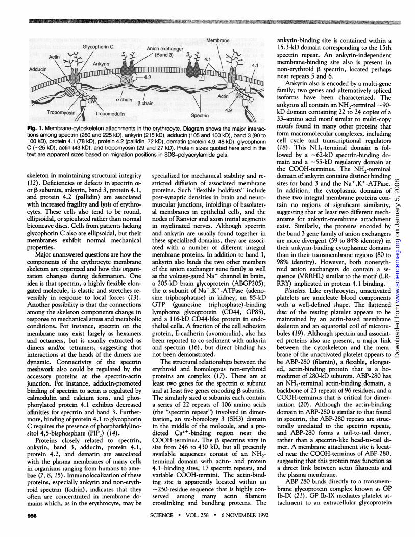
Control of Cell Shape, MembraneStability, and Domain OrganizationErythrocytes. Because of its comparativesimplicity and the relative ease with whichlarge amounts of homogeneous membranecan be obtained, the best understood mem-brane skeleton is that of the human redblood cell (8). These highly specializedcells are biconcave disks that lack internalorganelles and transcellular filament sys-tems. The membrane stability and deform-ability required during the erythrocyte's120-day, tortuous journey through thebloodstream are maintained solely by theunderlying meshwork of spectrin, actin,and associated proteins (Fig. 1) . This mesh-work is attached to the membrane byankyrin and protein 4.1. Ankyrin linksband 3 (the anion exchanger) to the Psubunit of spectrin near the middle of theextended spectrin tetramer. Protein 4.1binds to both spectrin subunits near theends of the tetramer, enhancing the affinityof spectrin for actin. Protein 4.1 also bindsin vitro to the transmembrane proteins,band 3 and glycophorin C. Binding be-tween protein 4.1 and band 3 may involvean interaction between the amino acidsequence, LEEDY, near the NH2-terminusof protein 4.1 and an oppositely chargedmotif (IRRRY or LRRRY) in the cytoplas-mic domain of band 3, although other siteson band 3 also have been implicated (9). Asequence (YRHKG) present in glycophorinC contains the same charge distributionand hydrophobicity as the motif in band 3.Finally, spectrin and band 4.1 bind withlow affinity to negatively charged phospho-lipids (10), interactions that may help tostabilize the lipid bilayer.
Proteins not bound directly to the mem-brane also add to the stability of the mem-brane skeleton (11). For instance, a het-erodimeric calmodulin-associated protein,adducin, enhances spectrin binding to ac-tin. Tropomyosin and dematin (protein4.9) bind along the sides of actin filaments,and the tropomyosin-binding protein,tropomodulin, may control the lengths ofthe short actin filaments bound to thespectrin tetramers.
Studies of mice and humans with hered-itary defects in erythrocyte shape and sta-bility confirm the role of the membrane
955
E. J. Luna and A. L. Hitt are with the Cell BiologyGroup, Worcester Foundation for Experimental Biolo-gy, Shrewsbury, MA 01545.*To whom correspondence should be addressed.
---------------------lingua l-
on
Janu
ary
5, 2
008
ww
w.s
cien
cem
ag.o
rgD
ownl
oade
d fr
om
in-in~fl~inin----M "
Membrane
FIR. 1. Membrane-cytoskeleton attachments in the erythrocyte. Diagram shows the major interac-tions among spectrin (260 and 225 kD), ankyrin (215 kD), adducin (105 and 100 kD), band 3 (90 to100 kD), protein 4.1 (78 kD), protein 4.2 (pallidin, 72 kD), dematin (protein 4.9, 48 kD), glycophorinC (-25 kD), actin (43 kD), and tropomyosin (29 and 27 kD). Protein sizes quoted here and in thetext are apparent sizes based on migration positions in SDS-polyacrylamide gels.
skeleton in maintaining structural integrity(12). Deficiencies or defects in spectrin a-
or subunits, ankyrin, band 3, protein 4.1,and protein 4.2 (pallidin) are associatedwith increased fragility and lysis of erythro-cytes. These cells also tend to be round,ellipsoidal, or spiculated rather than normalbiconcave discs. Cells from patients lackingglycophorin C also are ellipsoidal, but theirmembranes exhibit normal mechanicalproperties.
Major unanswered questions are how thecomponents of the erythrocyte membraneskeleton are organized and how this organi-zation changes during deformation. Oneidea is that spectrin, a highly flexible elon-gated molecule, is elastic and stretches re-
versibly in response to local forces (13).Another possibility is that the connectionsamong the skeleton components change inresponse to mechanical stress and metabolicconditions. For instance, spectrin on themembrane may exist largely as hexamersand octamers, but is usually extracted as
dimers and/or tetramers, suggesting thatinteractions at the heads of the dimers are
dynamic. Connectivity of the spectrinmeshwork also could be regulated by theaccessory proteins at the spectrin-actinjunction. For instance, adducin-promotedbinding of spectrin to actin is regulated bycalmodulin and calcium ions, and phos-phorylated protein 4.1 exhibits decreasedaffinities for spectrin and band 3. Further-more, binding of protein 4.1 to glycophorinC requires the presence of phosphatidylino-sitol 4,5-bisphosphate (PIP2) (14).
Proteins closely related to spectrin,ankyrin, band 3, adducin, protein 4.1,protein 4.2, and dematin are associatedwith the plasma membranes of many cellsin organisms ranging from humans to ame-
bae (7, 8, 15). Immunolocalization of theseproteins, especially ankyrin and non-eryth-roid spectrin (fodrin), indicates that theyoften are concentrated in membrane do-mains which, as in the erythrocyte, may be
956
specialized for mechanical stability and re-
stricted diffusion of associated membraneproteins. Such "flexible holdfasts" includepost-synaptic densities in brain and neuro-
muscular junctions, infoldings of basolater-al membranes in epithelial cells, and thenodes of Ranvier and axon initial segmentsin myelinated nerves. Although spectrinand ankyrin are usually found together inthese specialized domains, they are associ-ated with a number of different integralmembrane proteins. In addition to band 3,ankyrin also binds the two other membersof the anion exchanger gene family as wellas the voltage-gated Na+ channel in brain,a 205-kD brain glycoprotein (ABGP205),the a subunit of Na+,K+-ATPase (adeno-sine triphosphatase) in kidney, an 85-kDGTP (guanosine triphosphate)-bindinglymphoma glycoprotein (CD44, GP85),and a 116-kD CD44-like protein in endo-thelial cells. A fraction of the cell adhesionprotein, E-cadherin (uvomorulin), also hasbeen reported to co-sediment with ankyrinand spectrin (16), but direct binding hasnot been demonstrated.
The structural relationships between theerythroid and homologous non-erythroidproteins are complex (17). There are atleast two genes for the spectrin a subunitand at least five genes encoding 1B subunits.The similarly sized a subunits each containa series of 22 repeats of 106 amino acids(the "spectrin repeat") involved in dimer-ization, an src-homology 3 (SH3) domainin the middle of the molecule, and a pre-dicted Ca2+-binding region near theCOOH-terminus. The spectrins vary insize from 246 to 430 kD, but all presentlyavailable sequences consist of an NH2-terminal domain with actin- and protein4.1-binding sites, 17 spectrin repeats, andvariable COOH-termini. The actin-bind-ing site is apparently located within an
-250-residue sequence that is highly con-
served among many actin filamentcrosslinking and bundling proteins. The
SCIENCE * VOL. 258 * 6 NOVEMBER 1992
ankyrin-binding site is contained within a15.3-kD domain corresponding to the 15thspectrin repeat. An ankyrin-independentmembrane-binding site also is present innon-erythroid j spectrin, located perhapsnear repeats 5 and 6.
Ankyrin also is encoded by a multi-genefamily; two genes and alternatively splicedisoforms have been characterized. Theankyrins all contain an NH2-terminal -90-kD domain containing 22 to 24 copies of a33-amino acid motif similar to multi-copymotifs found in many other proteins thatform macromolecular complexes, includingcell cycle and transcriptional regulators(18). This NH2-terminal domain is fol-lowed by a -62-kD spectrin-binding do-main and a -55-kD regulatory domain atthe COOH-terminus. The NH2-terminaldomain of ankyrin contains distinct bindingsites for band 3 and the Na+,K+-ATPase.In addition, the cytoplasmic domains ofthese two integral membrane proteins con-tain no regions of significant similarity,suggesting that at least two different mech-anisms for ankyrin-membrane attachmentexist. Similarly, the proteins encoded bythe band 3 gene family of anion exchangersare more divergent (59 to 84% identity) intheir ankyrin-binding cytoplasmic domainsthan in their transmembrane regions (80 to98% identity). However, both noneryth-roid anion exchangers do contain a se-quence (VRRHL) similar to the motif (LR-RRY) implicated in protein 4.1 binding.
Platelets. Like erythrocytes, unactivatedplatelets are anucleate blood componentswith a well-defined shape. The flatteneddisc of the resting platelet appears to bemaintained by an actin-based membraneskeleton and an equatorial coil of microtu-bules (19). Although spectrin and associat-ed proteins also are present, a major linkbetween the cytoskeleton and the mem-brane of the unactivated platelet appears tobe ABP-280 (filamin), a flexible, elongat-ed, actin-binding protein that is a ho-modimer of 280-kD subunits. ABP-280 hasan NH2-terminal actin-binding domain, abackbone of 23 repeats of 96 residues, and aCOOH-terminus that is critical for dimer-ization (20). Although the actin-bindingdomain in ABP-280 is similar to that foundin spectrin, the ABP-280 repeats are struc-turally unrelated to the spectrin repeats,and ABP-280 forms a tail-to-tail dimer,rather than a spectrin-like head-to-tail di-mer. A membrane attachment site is locat-ed near the COOH-terminus of ABP-280,suggesting that this protein may function asa direct link between actin filaments andthe plasma membrane.
ABP-280 binds directly to a transmem-brane glycoprotein complex known as GPIb-IX (21). GP Ib-IX mediates platelet at-tachment to an extracellular glycoprotein
on
Janu
ary
5, 2
008
ww
w.s
cien
cem
ag.o
rgD
ownl
oade
d fr
om

- - - - - g m ILL IMRLA IA IMLM1 Im m Mm
(von Willebrand factor) immobilized ondamaged vascular walls. The GP Ib-IXcomplex consists of two disulfide-linkedsubunits, GP Iba (145 kD) and GP Ib (24kD); and a third, noncovalently boundsubunit called GP IX (17 kD). Whereasefficient expression of the complex requiresall three subunits, GP Iba contains boththe ligand-binding site for von Willebrandfactor and the ABP-280-binding site,which is located in the center of the cyto-plasmic domain (22).
The pathophysiology of Bernard Souliersyndrome, an inherited bleeding disorder, isconsistent with a role for the GP Ib-IXinteraction with ABP-280 in the determi-nation of platelet shape and function. Ber-nard Soulier patients have a defect or defi-ciency in the GP Ib-IX complex and bleedeasily with long clotting times. Their rela-tively few blood platelets are large, poly-morphic cells that exhibit decreased adhe-sion to vascular tissue because of an inabil-ity to bind von Willebrand factor.An essentially ubiquitous component of
cortical cytoplasm, ABP-280 also may beinvolved in stabilizing the membrane skel-etons of other cells. Melanoma cell lineslacking ABP-280 migrate poorly in re-sponse to external stimuli and exhibit bleb-bing of the plasma membrane around thecircumference of the cell. Restoration ofnormal ABP-280 levels by transfection withABP-280 cDNA restores cell locomotionand reduces blebbing (23). As GP Ib-IX isnot present in these cells, other membraneproteins also may bind ABP-280. One can-didate is the immunoglobulin G (IgG) Fcreceptor. This protein binds directly toABP-280 in vitro (24), but the nature ofthe binding interactions and the impor-tance of this binding in vivo are not yetknown.
Striated muscle. Membrane stability andflexibility also are important in the ex-tremely large (10 to 100 gxm wide, up to 50cm long) multinucleated cells of striatedmuscle. Stress-induced damage to the stri-ated muscle plasma membrane (the sarco-lemma) and leakiness to external calciumappear to be early hallmarks of Duchenneand Becker muscular dystrophy (25). An-other flexible, elongated membrane-associ-ated protein, called dystrophin, has beenidentified as the X-chromosome gene prod-uct that is characteristically absent (Duch-enne) or altered (Becker) in these diseases.One of the largest members of the spectrinsuperfamily (427 kD), dystrophin containsfour distinct domains: (i) an actin-bindingNH2-terminus homologous to the actin-binding domains of spectrin and ABP-280,(ii) a central rod-like domain with 24 spec-trin-like repeats of 109 amino acids and fourproline-rich "hinges," (iii) a cysteine-richsegment similar to the Ca2+-binding do-
main of the actin cross linking protein,a-actinin, and (iv) a unique COOH-termi-nal domain of 420 amino acids (26). Byanalogy to spectrin, the central rod-likedomain is thought to promote dimerizationof dystrophin. The COOH-terminal tailmay be the site of membrane attachment.The importance of the two domains nearthe COOH- terminus is indicated by thevery high sequence conservation (95%identity) of these domains between chickenand human dystrophin. By comparison, theactin-binding and rod-like domains are 80and 75% identical, respectively.
Dystrophin is localized at the cytoplas-mic surface of the sarcolemmal membraneand in the infoldings of myotendinous andneuromuscular junctions (specialized sitesof attachment to tendons and neurons)where it appears to link the plasma mem-brane to the sides of actin filament bundles(27). Dystrophin also may be enrichedalong with muscle ,B-spectrin in a subsar-colemmal latticework that contains bothperiodic transverse elements and strandsextending the length of the cell (28). Thislattice may stabilize the plasma membraneduring muscle contraction by inducing anordered folding of the membrane as themyofiber length decreases. Stress-inducedruptures of membrane undoubtedly contrib-ute to the decreased osmotic stability ofmuscle cells lacking dystrophin and to theincreased influx of calcium ions observed indystrophic muscle fibers (29).
In addition to skeletal muscle, dystro-phin is expressed in heart and smoothmuscle and at low levels in a number ofnon-muscle tissues, including kidney, lung,placenta, and brain. In brain, dystrophin isenriched at post-synaptic densities in neu-rons of the cerebral and cerebellar cortices(30). The major transcription product fromthe dystrophin gene in non-muscle tissuesencodes a 70- to 75-kD protein that con-tains the cysteine-rich and COOH-termi-nal domains of muscle dystrophin (31). Inthis protein, six amino acids encoded by analternatively spliced exon replace the actin-binding region and spectrin repeats of dys-trophin. Although the function of thissmall dystrophin isoform is unknown, itsexistence may help explain the extramus-cular symptoms, such as mental impair-ment, that sometimes accompany Duch-enne muscular dystrophy.An autosomal homolog of the dystro-
phin gene encodes a ~400-kD dystrophin-related protein (DMDL) that is found invarying amounts in most tissues (32). Al-though most of this gene is distinct fromdystrophin, DMDL is predicted to consist of490-amino acids that are 73% identical tothe dystrophin COOH-terminus. DMDL islocalized at neuromuscular junctions inboth normal and dystrophin-negative mus-
SCIENCE * VOL. 258 * 6 NOVEMBER 1992
cle. Although DMDL does not localize tothe subsarcolemmal latticework in normalmuscle, weak staining is observed at non-junctional plasma membrane in dystrophicmuscle, implying that DMDL may bind tounoccupied membrane attachment sites fordystrophin.
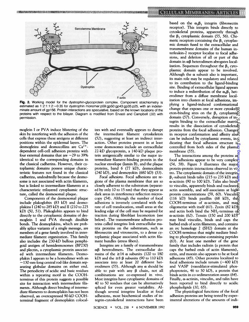
Dystrophin solubilized from sarcolemmawith 1% digitonin is part of a large complexcontaining six other proteins (33) (Fig. 2).A peripheral laminin-binding glycoprotein(156 kD) apparently provides a linkagebetween the complex and the extracellularmatrix (34). The core of the complex ap-pears to be a group of integral membraneproteins (50, 43, 35, and 25 kD) that reactwith a hydrophobic photolabel, remain as-sociated with membranes after alkaline ex-traction, and co-sediment in the absence ofperipheral proteins; the 156-, 50-, 43-, and35-kD proteins are glycosylated. The 43-kDglycoprotein is synthesized as part of theCOOH-terminus of a precursor polypeptide(dystroglycan) that is post-translationallyprocessed into both the 156- and 43-kDproteins. Analysis of the cDNA encodingdystroglycan predicts that the 156-kD gly-coprotein is extracellular and consists of-457 amino acids. The 43-kD glycoproteinis predicted to contain a single membrane-spanning region and a 121-residue cytoplas-mic domain. An unglycosylated 59-kD pro-tein that co-extracts with dystrophin at pH11 may help anchor dystrophin at the cy-toplasmic surface of the complex. Expres-sion of all the major dystrophin-associatedproteins is dramatically decreased in dystro-phic muscle (35), suggesting that dystro-phin is required for stability of the entirecomplex.
Analogs of the dystrophin-associatedproteins may be present in many cells (36).After overexpression in COS cells, dystro-phin predominantly localizes to the plasmamembrane, even though these cells are notof muscle origin and normally contain verylittle dystrophin. Furthermore, the electricray contains a dystrophin-associated 58-kDprotein that is involved in acetylcholinereceptor clustering at postsynaptic mem-branes in electric tissue (and which may behomologous to the 59-kD muscle protein).Thus, the dystrophin-associated proteinsfrom muscle may constitute just the firstexample of a general molecular mechanismfor anchoring dystrophin-like proteins atspecialized membrane domains.
Euglenozoa. Not all membrane skeletonsare based on actin filaments and elongateactin-binding proteins. Cell shape in tworelated, but quite different, flagellated eu-karyotes, Trypanosoma brucei and Euglenagracilis, is maintained solely by membraneskeletons that are based on microtubulesand articulins, respectively. In T. brucei,the plasma membrane is stabilized by a
- 11 - 90-1--..0-m1..-0m---mmLIREM-10
on
Janu
ary
5, 2
008
ww
w.s
cien
cem
ag.o
rgD
ownl
oade
d fr
om

uuinuwinuinmmuim~~~~~~~~~~~~~~~~~~~~~1...
corset of closely spaced microtubules encas-ing the entire cell. MARP-1 (microtubule-associated repetitive protein), a 320-kDprotein that contains >50 tandem micro-tubule-binding repeats of 38 amino acids, isone of several proteins that may link themicrotubules to each other or to the plasmamembrane (37). Studies with antibodiesagainst MARP-1 suggest that it abuts theplasma membrane, but the microtubule-membrane linkage is not known.
The membrane skeleton in E. gracilis isdivided into -40 strips that change theirlateral positions relative to one anotherduring cell movement (38). Although mi-crotubules are present at strip intersec-tions, the membrane skeleton consistsmostly of equimolar amounts of two pro-teins (80 and 86 kD) called articulins.These proteins form 17-nm filaments thatapparently assemble perpendicular to themembrane surface. Binding of the 86-kDarticulin to the membrane is dependent onthe presence of the 80-kD protein. Eacharticulin contains a core domain with >30repeats of a 12-amino acid motif. Anotherregion containing four repeats of sevenhydrophobic amino acids is found at boththe COOH-terminus of the 80-kD articu-lin and the NH2-terminus of 86-kD arti-culin. Since the 80-kD articulin binds to a39-kD integral membrane protein (IP39),IP39 may bind one end of filaments con-taining head-to-tail articulin het-erodimers. Additional articulin-associatedproteins apparently crosslink or otherwisestabilize the skeleton.
Although the membrane skeletons dis-cussed above are all quite different, thereare at least two recurrent themes. First, allcontain proteins with repetitive structuresthat may function both as spacers and aselastic elements to provide flexibility. Sec-ond, there appears to be more diversity inthe cytoplasmic domains of the integralmembrane proteins than in the cytoskeletalproteins that they anchor. In cells contain-ing more than one type of specialized mem-brane skeleton, the location of a particularmembrane domain thus may be determinedmostly by the locations of the integralproteins. On the other hand, the apparentmodularity of the actin-based cytoskeletalcomponents should facilitate interactionsbetween these specialized areas of mem-brane and the rest of the cytoskeleton.
Adhesion and the MembraneSkeleton
There are four major families of receptorsthat mediate cell adhesion (39): (i) cadher-ins; (ii) members of the Ig superfamily,including N-CAM and ICAM-1; (iii) car-bohydrate-binding proteins called selectins;and (iv) integrins. The first three families
primarily mediate cell-cell interactions,whereas most integrins are involved in at-tachments to the substratum. AlthoughICAM-1 recently was shown to bind a-ac-tinin in vitro (40), relatively little is knownabout the intracellular interactions of mem-bers of the immunoglobulin superfamily,and the selectins are thought not to beattached to the cytoskeleton. We will focushere on junctional assemblies in which thecadherins and the integrins are mixed andmatched with components of the actin andintermediate filament networks to form fourof the best understood junctions mediatingcell-cell and cell-substrate attachments.
CeU-to-ceU adherens junctions. The cad-herin superfamily is a group of proteins thatmediate Ca2+-dependent homophilic (like-with-like) adhesion between cells (41) . Dif-ferential expression of cadherins appears tobe involved in cell sorting because cellstransfected with a particular cadherin ad-here preferentially to other cells expressingthe same protein. The so-called "classical"cadherins include E- (epithelial, also calleduvomorulin), N- (neural, also calledA-CAM), and P- (placental) cadherin, butat least 12 different members of the familyare known. The classical cadherins are gly-coproteins with a common structure. Eachhas an extracellular domain containing fourmajor repeats (which have 37 to 46% iden-tity among classical cadherins), a singletransmembrane domain, and a highly con-served cytoplasmic domain (56 to 80%identity). The extracellular self-recognitionsequence, which differs among cadherins, islocated in the NH2-terminal 113-aminoacid residues. The cytoplasmic domains ofthe classical cadherins mediate interactionswith at least three cytosolic proteins, calleda-, 1B-, and y-catenin (102, 92, and 85 kD,respectively).
In the presence of millimolar extracellu-lar Ca2+, cells expressing both cadherinsand catenins form belt-like sites of cell-cellattachment (called the zonulae adherens oradherens junctions), which contain cyto-plasmic "undercoats" associated with actinfilaments (41). Cadherin-mediated cell ad-hesion is not observed in cells that lacka-catenin (42) or in cells expressing cad-herins lacking the cytoplasmic domain.The a-catenin is structurally similar to vin-culin (30% identity), an adherens junctionprotein that does not bind cadherin. Botha-catenin and vinculin have five domainsof which domains one, three, and five aresimilar. As the fifth domain of vinculin isinvolved in oligomerization, a similar func-tion has tentatively been assigned to thisregion of a-catenin. Oligomerization ofa-catenin may induce clustering of cadher-ins, which could foster junctional assembly.Of the three catenins, 1-catenin binds thetightest to cadherin. The 0-catenin from
SCIENCE * VOL. 258 * 6 NOVEMBER 1992
Xenopus is similar to the Drosophila proteinencoded by the segment polarity gene, ar-madillo (70% identity), and to human pla-koglobin (63% identity), a protein found atthe cytoplasmic surfaces of both adherensjunctions and desmosomes (43). Althoughplakoglobin and 0-catenin are structurallydistinct, plakoglobin appears to be the sameas y-catenin (44).
Although it is not known how cadherinsand catenins are linked to the cytoskeleton,actin is a principal component of the adhe-rens junction undercoat. Actin-associatedproteins in the undercoat include a-actinin(a 90-kD protein that crosslinks actin fila-ments), zyxin (an 82-kD protein that bindsthe NH2-terminus of a-actinin), vinculin(a 116-kD protein that binds the COOH-terminus of a-actinin), and radixin (an82-kD protein that caps the barbed ends ofactin filaments). Radixin is related to ezrinand moesin (-75% identity); all three pro-teins contain 234 amino acids near theirNH2-termini that are similar to a regionnear the NH2-terminus of protein 4.1(-30% identity) (45). However, the ami-no acid sequence (LEEDY) postulated tomediate protein 4.1 binding to band 3 isnot found in radixin, ezrin, or moesin.Although radixin has been postulated tolink barbed ends of actin filaments to theundercoat, interactions with proteins otherthan actin have not been characterized.Other proteins immunolocalized to the ad-herens junction undercoat include the rod-like protein tenuin (-400 kD) and a 220-kD globular protein that binds a-spectrin(46).
Junctional integrity appears to be regu-lated by protein tyrosine phosphorylation(47). Elevated levels of phosphotyrosinegenerated either by transformation withRous sarcoma virus or by inhibition ofphosphotyrosine phosphatases rapidly, butreversibly, induce junctional disassembly.Although the tyrosine kinases encoded byc-src, c-yes, and lyn have been localized atthe adherens junction, the correspondingprotein tyrosine phosphatases have not yetbeen identified.
Desnosomes. The desmosome (maculaadherentes) is a spot-like site of intercellularcontact that, like the adherens junction,mediates Ca2+-sensitive cell-cell adhesion(48). Intercellular attachment is mediatedby transmembrane proteins: desmoglein I(150 kD) and the closely related splicevariants, desmocollins I (120 kD) and II(110 kD). Desmoglein I and the desmocol-lins are each members of a gene familywithin the cadherin superfamily. The des-moglein family also includes the gene forthe 130-kD pemphigus vulgaris antigen(PVA), a protein recognized by autoim-mune sera in the blistering disease, pemphi-gus vulgaris (49). Antibodies against des-
on
Janu
ary
5, 2
008
ww
w.s
cien
cem
ag.o
rgD
ownl
oade
d fr
om
4
Dystrophin
F-actinFig. 2. Working model for the dystrophin-glycoprotein complex. Component stoichiometry isestimated as 11:2:1 2:0.35 for dystrophin monomer:p59:gp50:gp43:gp35:p25, with an indeter-minate amount of gpl 56. Protein interactions are speculative, based on the known locations of theproteins with respect to the bilayer. Diagram is modified from Ervasti and Campbell (33) withpermission.
moglein I or PVA induce blistering of theskin by interfering with the adhesion of thecells that express these antigens at differentpositions within the epidermal layers. Thedesmogleins and desmocollins are Ca2+_dependent cell-cell adhesion proteins withfour external domains that are -29 to 39%identical to the corresponding domains inthe classical cadherins. However, their cy-toplasmic domains possess unique charac-teristic features not found in the classicalcadherins, undoubtedly because the desmo-some is not associated with actin filaments,but is linked to intermediate filaments at acharacteristic trilayered cytoplasmic struc-ture, called the desmosomal plaque.
Components of the desmosomal plaqueinclude plakoglobin (83 kD) and desmo-plakins I (240 to 285 kD) and 11 (210 to 225kD) (50, 51). Plakoglobin appears to binddirectly to the cytoplasmic domains of des-moglein I and PVA through disulfidebonds. The desmoplakins, which are prob-ably splice variants of a single message, aremembers of a gene family involved in inter-mediate filament organization. This familyalso includes the 230-kD bullous pemphi-goid antigen of hemidesmosomes (BP230)and plectin, a cytoplasmic protein associat-ed with intermediate filaments. Desmo-plakin I appears to be a homodimer with an
- 130-nm-long central rod-like domain sep-arating globular domains on either end.The periodicity of acidic and basic residueswithin a repeating motif in the COOH-terminus of this protein suggests a possiblesite for interaction with intermediate fila-ments. Although direct binding of interme-diate filaments to desmoplakin has not beenobserved, an overexpressed 90-kD COOH-terminal fragment of desmoplakin colocal-
izes with and eventually appears to disruptthe intermediate filament cytoskeleton(52), suggesting at least an indirect inter-action. Other proteins present in at leastsome desmosomes include an extracellular22-kD glycoprotein, a 140-kD plaque pro-tein antigenically similar to the major in-termediate filament-binding protein in thenuclear envelope (lamin B), and the plaqueproteins, band 6 (77 kD), desmocalmin(240 kD), and desmoyokin (680 kD) (53).
Focal adhesinas. Focal adhesions are re-gions of the plasma membrane that are soclosely adherent to the substratum (separat-ed by only 10 to 15 nm) that they appear asdark areas in interference reflection micros-copy (54). Although the number of focaladhesions is inversely correlated with therate of cell translocation, small areas at theleading edge of the cell may be required fortraction during fibroblast locomotion (seebelow). The transmembrane adhesion pro-teins called integrins link extracellular ma-trix proteins on the substratum, such asfibronectin and vitronectin, to a dense cy-toplasmic plaque associated with actin fila-ment bundles (stress fibers).
Integrins are a family of transmembraneaLP heterodimers. The extracellular do-mains of the >14 a subunits (120 to 180kD) and the >8 P subunits (90 to 110 kD)associate into at least 20 different het-erodimers (55). Although any a should beable to pair with any P chain, not allcombinations are co-expressed in vivo.Most integrins have cytoplasmic domains of40 to 50 residues that can be alternativelyspliced for even greater variability. Al-though several integrins localize at focaladhesions, most biochemical studies of in-tegrin-cytoskeletal interactions have been
SCIENCE * VOL. 258 * 6 NOVEMBER 1992
based on the a501 integrin (fibronectinreceptor). This integrin binds directly tocytoskeletal proteins, apparently throughthe I31 cytoplasmic domain (55, 56). Chi-meric receptors containing the PI cytoplas-mic domain fused to the extracellular andtransmembrane domains of the human in-terleukin-2 receptor localize to focal adhe-sions, and deletion of all or part of thisdomain in a13 heterodimers abrogates local-ization. Sequences throughout the P, cyto-plasmic domain appear to be involved.Although the a subunit also is important,its main role may be regulatory and relatedto its contribution to the ligand-bindingsite. Binding of extracellular ligand appearsto induce a redistribution of the t5P13 het-erodimer from a diffuse membrane local-ization into clusters at focal adhesions, im-plying a ligand-induced conformationalchange that exposes one or more cytoskel-eton-binding sites on the , cytoplasmicdomain (57). Conversely, disruption of in-tegrin binding to the extracellular matrixresults in the dissociation of cytoskeletalproteins from the focal adhesion. Changesin receptor conformation and affinity alsocan be induced by cytoplasmic events, in-dicating that focal adhesion structure iscontrolled from both sides of the plasmamembrane (55).
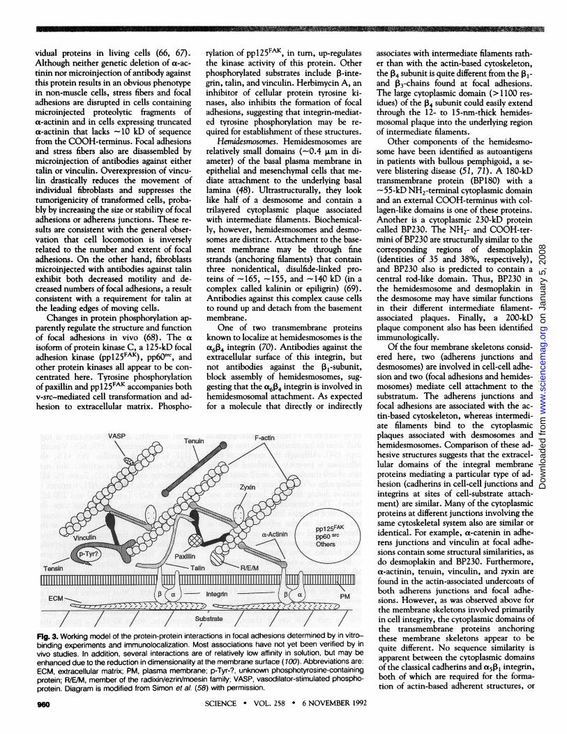
The interactions among the proteins atfocal adhesions appear to be very complex(54, 58). Figure 3 illustrates the majorprotein-protein interactions observed in vi-tro. The cytoplasmic domain of the integrinPI-subunit binds talin (215 to 235 kD) anda-actinin (90 to 100 kD). Talin also bindsto vinculin, apparently binds and nucleatesactin assembly, and self-associates at highprotein concentrations (59, 60). Vinculin(116 kD) binds paxillin (68 kD), theCOOH-terminus of a-actinin, and mayoligomerize with itself (61). Zyxin (82 kD)and actin both bind the NH2-terminus ofa-actinin (62). Tensin (150 and 200 kD)may bind vinculin, binds and caps thebarbed ends of actin filaments, and containsan src homology 2 (SH2) domain at theCOOH-terminus that might mediate bind-ing to phosphotyrosine-containing peptides(63). At least one member of the genefamily that includes radixin (a protein thatcaps the barbed ends of actin filaments),ezrin, and moesin also appears to be at focaladhesions (45). Other proteins localized tofocal adhesions include tenuin (-400 kD)and VASP (vasodilator-stimulated phos-phoprotein, 46 to 50 kD), a protein thatbinds actin in co-sedimentation assays (64).Finally, a-actinin, vinculin, and talin havebeen reported to bind directly to acidicphospholipids (10, 65).
The physiological functions of the focaladhesion proteins are being tested by exper-imental alterations of the amounts of indi-
959
no ilill liiiiiiiiIiiMi
------------------
(](](In(]------------V~ WELMMI..5~5
on
Janu
ary
5, 2
008
ww
w.s
cien
cem
ag.o
rgD
ownl
oade
d fr
om
vidual proteins in living cells (66, 67).Although neither genetic deletion of a-ac-tinin nor microinjection of antibody againstthis protein results in an obvious phenotypein non-muscle cells, stress fibers and focaladhesions are disrupted in cells containingmicroinjected proteolytic fragments ofa-actinin and in cells expressing truncateda-actinin that lacks ~ 10 kD of sequencefrom the COOH-terminus. Focal adhesionsand stress fibers also are disassembled bymicroinjection of antibodies against eithertalin or vinculin. Overexpression of vincu-lin drastically reduces the movement ofindividual fibroblasts and suppresses thetumorigenicity of transformed cells, proba-bly by increasing the size or stability of focaladhesions or adherens junctions. These re-sults are consistent with the general obser-vation that cell locomotion is inverselyrelated to the number and extent of focaladhesions. On the other hand, fibroblastsmicroinjected with antibodies against talinexhibit both decreased motility and de-creased numbers of focal adhesions, a resultconsistent with a requirement for talin atthe leading edges of moving cells.
Changes in protein phosphorylation ap-parently regulate the structure and functionof focal adhesions in vivo (68). The aisoform of protein kinase C, a 125-kD focaladhesion kinase (ppl25FAK), pp60STC, andother protein kinases all appear to be con-centrated here. Tyrosine phosphorylationof paxillin and ppl25FAK accompanies bothv-src-mediated cell transformation and ad-hesion to extracellular matrix. Phospho-
rylation of pp125FAK, in turn, up-regulatesthe kinase activity of this protein. Otherphosphorylated substrates include P-inte-grin, talin, and vinculin. Herbimycin A, aninhibitor of cellular protein tyrosine ki-nases, also inhibits the formation of focaladhesions, suggesting that integrin-mediat-ed tyrosine phosphorylation may be re-quired for establishment of these structures.
Hemidesmosomes. Hemidesmosomes arerelatively small domains (-0.4 gm in di-ameter) of the basal plasma membrane inepithelial and mesenchymal cells that me-diate attachment to the underlying basallamina (48). Ultrastructurally, they looklike half of a desmosome and contain atrilayered cytoplasmic plaque associatedwith intermediate filaments. Biochemical-ly, however, hemidesmosomes and desmo-somes are distinct. Attachment to the base-ment membrane may be through finestrands (anchoring filaments) that containthree nonidentical, disulfide-linked pro-teins of -165, -155, and -140 kD (in acomplex called kalinin or epiligrin) (69).Antibodies against this complex cause cellsto round up and detach from the basementmembrane.
One of two transmembrane proteinsknown to localize at hemidesmosomes is thea614 integrin (70). Antibodies against theextracellular surface of this integrin, butnot antibodies against the P1-subunit,block assembly of hemidesmosomes, sug-gesting that the a614 integrin is involved inhemidesmosomal attachment. As expectedfor a molecule that directly or indirectly
Fig. 3. Working model of the protein-protein interactions in focal adhesions determined by in vitro-binding experiments and immunolocalization. Most associations have not yet been verified by invivo studies. In addition, several interactions are of relatively low affinity in solution, but may beenhanced due to the reduction in dimensionality at the membrane surface (100). Abbreviations are:ECM, extracellular matrix; PM, plasma membrane; p-Tyr-?, unknown phosphotyrosine-containingprotein; R/E/M, member of the radixin/ezrin/moesin family; VASP, vasodilator-stimulated phospho-protein. Diagram is modified from Simon et al. (58) with permission.
associates with intermediate filaments rath-er than with the actin-based cytoskeleton,the 14 subunit is quite different from the 1PI-and 13-chains found at focal adhesions.The large cytoplasmic domain (>1100 res-idues) of the 14 subunit could easily extendthrough the 12- to 15-nm-thick hemides-mosomal plaque into the underlying regionof intermediate filaments.
Other components of the hemidesmo-some have been identified as autoantigensin patients with bullous pemphigoid, a se-vere blistering disease (51, 71). A 180-kDtransmembrane protein (BP180) with a-55-kD NH2-terminal cytoplasmic domainand an external COOH-terminus with col-lagen-like domains is one of these proteins.Another is a cytoplasmic 230-kD proteincalled BP230. The NH2- and COOH-ter-mini of BP230 are structurally similar to thecorresponding regions of desmoplakin(identities of 35 and 38%, respectively),and BP230 also is predicted to contain acentral rod-like domain. Thus, BP230 inthe hemidesmosome and desmoplakin inthe desmosome may have similar functionsin their different intermediate filament-associated plaques. Finally, a 200-kDplaque component also has been identifiedimmunologically.
Of the four membrane skeletons consid-ered here, two (adherens junctions anddesmosomes) are involved in cell-cell adhe-sion and two (focal adhesions and hemides-mosomes) mediate cell attachment to thesubstratum. The adherens junctions andfocal adhesions are associated with the ac-tin-based cytoskeleton, whereas intermedi-ate filaments bind to the cytoplasmicplaques associated with desmosomes andhemidesmosomes. Comparison of these ad-hesive structures suggests that the extracel-lular domains of the integral membraneproteins mediating a particular type of ad-hesion (cadherins in cell-cell junctions andintegrins at sites of cell-substrate attach-ment) are similar. Many of the cytoplasmicproteins at different junctions involving thesame cytoskeletal system also are similar oridentical. For example, a-catenin in adhe-rens junctions and vinculin at focal adhe-sions contain some structural similarities, asdo desmoplakin and BP230. Furthermore,at-actinin, tenuin, vinculin, and zyxin arefound in the actin-associated undercoats ofboth adherens junctions and focal adhe-sions. However, as was observed above forthe membrane skeletons involved primarilyin cell integrity, the cytoplasmic domains ofthe transmembrane proteins anchoringthese membrane skeletons appear to bequite different. No sequence similarity isapparent between the cytoplasmic domainsof the classical cadherins and a5 PI integrin,both of which are required for the forma-tion of actin-based adherent structures, or
SCIENCE * VOL. 258 * 6 NOVEMBER 1992
INN
9W
on
Janu
ary
5, 2
008
ww
w.s
cien
cem
ag.o
rgD
ownl
oade
d fr
om

between the cytoplasmic domains of x614integrin and the desmosomal cadherins,which both anchor plaques containing in-termediate filaments. Thus, it appears thatmembrane-cytoskeleton associations devel-oped independently many times during ev-olution.
Movement and Cell SurfaceProtrusions
Cell surface protrusions at the leading edgeappear to be involved in the movement ofmost cells (72). The names of these struc-tures vary with the morphology of theprotrusion: filopodia are thin and usuallylong, lamellipodia are broad, and pseudopo-dia are of moderate width with respect tothe dimensions of the cell. Constantlyforming, these protrusions either lift andfold back into the cell or spread and extendafter contacting the substratum or anothercell. While the mechanisms by which theseprotrusions are generated are not well un-derstood (73), both membrane-cytoskele-ton interactions and actin accessory pro-teins appear to be involved (Table 1). Thispart of the cell is hard to isolate becauseinteractions at the leading edge tend to betransient. Thus, most of the proteins inTable 1 were first identified by their inter-
actions with actin, the principal compo-nent of the protrusions, and then immuno-localized to this region of the cell.
Proteins apparently enriched at the lead-ing edge include particular isoforms of actinand proteins that nucleate actin filamentassembly, such as hisactophilin, ponticulin,and talin (60, 74). Hisactophilin is a histi-dine-rich peripheral protein that nucleatesactin polymerization in a low salt solution;interactions at the membrane have notbeen characterized yet. Ponticulin is atransmembrane protein required for thebinding and nucleation of actin filamentsby isolated Dictyostelium plasma mem-branes. Unlike most actin-nucleating pro-teins that cap one end of the filament, thecytoplasmic domain of ponticulin promotesnucleation of actin filaments with bothends free for actin monomer addition or forinteractions with other proteins. Thus,ponticulin apparently constitutes a directactin-membrane link that could be in-volved in generating actin filaments inpseudopodial meshworks (75, 76). Highconcentrations of talin also have been re-ported to promote actin assembly. Becausetalin binds integrin and other focal adhe-sion proteins, actin nucleation mediated bytalin or an associated protein could be animportant step in the assembly of the actin
filament bundles involved in cell-substrateattachments. Profilin, which also is con-centrated near the leading edge, may accel-erate filament elongation by promoting theformation of assembly-competent ATP-ac-tin monomers and may even promote addi-tion of these monomers to the fast-growingbarbed ends of elongating filaments (77).
Rapidly polymerizing actin becomes as-sembled either into bundles of filaments infilopodia or into crosslinked meshworks inpseudopodia and lamellipodia. Filament or-ganization at the leading edge is controlledby actin accessory proteins (5, 78), whichinclude filament crosslinking proteins[ABP-280, spectrin, ABP-120, a-actinin,and MARCKS (a myristoylated alanine-rich C kinase substrate)], filament bundlingproteins (caldesmon, fimbrin, p30a, andvillin), calcium-dependent filament sever-ing proteins (villin, gelsolin, and fragmin/severin), and proteins that stabilize fila-ments (caldesmon and tropomyosin). Someof these proteins also may help link actin tothe membrane. For instance, ABP-280,spectrin, and a-actinin bind integral mem-brane proteins, and dephosphorylatedMARCKS binds to an unknown membranesite (79). Although only the genetic dele-tion ofABP-280 has been shown to dramat-ically decrease cortical stability and cell
Table 1. Proteins at the leading edge.
Name (SkDe) Distribution
A- and y-actin 43 Filopodia, pseudopodia, and lamellipodia in retinal pericytes and endothelial cellsProteins controlling Actin Nucleation and PolymerizationHisactophilin 17 Pseudopodia in DictyosteliumPonticulin 17 Nascent pseudopodia in Dictyostelium; cell extensions in neutrophilsTalin 215 Platelets, smooth muscle, many non-neuronal tissuesProfilin 12-15 Isoforms in most cells; fibroblast lamellipodiaActin Filament Accessory ProteinsABP-280 (filamin) 250 Isoforms in most cells; leading edges of chick gizzard and skin fibroblastsSpectrin 240/235 Isoforms in most cells; ruffling membranes in EGF-stimulated A431 cellsABP-120 120 Pseudopodia and lamellipodia in Dictyosteliuma-actinin 90-100 Most cells, including lower eukaryotesMARCKS 68-87 Punctate localization at tips of filopodia and pseudopodia in macrophagesCaldesmon 70-80 Essentially all non-muscle cells; larger isoforms in smooth muscleFimbrin (plastin) 68 Microvilli and ruffling membranes in essentially all non-muscle cellsp30a 30 Filopodia and membrane ridges in Dictyostelium; ruffles and stress fibers in fibroblastsVillin 95 Microvilli in brush borders of intestine, kidney pancreatic acinar cells and visceral endodermGelsolin 90 Mammalian cellsFragmin/severin 42 Physarum, DictyosteliumTropomyosin 30-40 Most cells; at least some isoforms in peripheral ruffles of rat kidney cellsMotor ProteinsMyosin I 110-140 Called 1 1OK-calmodulin in brush border microvilli; other isoforms at neuronal growth cones
and pseudopods of Acanthamoeba and DictyosteliumMyosin II -200 Established protrusions in fibroblasts; neuronal growth conesOther Actin-Binding Proteinsp58 58 Peripheral cytoplasmic protein associated with a large glycoprotein complex in microvilliCoronin 55 Cell surface projections in DictyosteliumMAb 2E4 antigen 43 Platelets; fibroblasts lamellipodia; growth cones in PC12 cellsOther Potential Regulatory ProteinsEzrin 80 Microvilli and surface structures in many cells; marginal band of avian erythrocytesGAP-43 43-57 Neuronal growth cones and processes
SCIENCE * VOL. 258 * 6 NOVEMBER 1992
--------
-----------------------------------
----------- l! 11 il il il i'WERWOMI
uuuuu MXLMMIA 2AMMIZM kz 11
961
on
Janu
ary
5, 2
008
ww
w.s
cien
cem
ag.o
rgD
ownl
oade
d fr
om
locomotion, functional overlap among themembrane skeletal proteins may obscureindividual roles in the membrane skeleton(23, 66). Alternatively, the actin filament-crosslinking activities of these proteins maybe their primary contribution to corticalstability at the leading edge.
Isoforms of single-headed myosin I andthe myosin I kinase that activates them arealso enriched at membrane protrusions(80). These myosins contain an ATP-inde-pendent, as well as an ATP-dependent,binding site for actin and can slide filamentsrelative to each other in the presence ofATP. These proteins also bind to acidicphospholipids (and probably to uncharac-terized membrane proteins) and movemembrane vesicles toward the barbed endsof actin filaments. As these are the filamentends in actin bundles that are generallyassociated with filopodial tips, myosin Imight mediate the movement of freely-diffusing membrane receptors toward theleading edge (81). Similarly, myosin I an-chored to the substratum through trans-membrane linkages could pull actin fila-ments rearward. Both types of movementshave been reported (7, 82). By contrast,conventional dimeric myosin II appearsonly in established protrusions and is appar-ently not essential for pseudopod expansion(83). Myosin II, however, is required foroptimal cell movement and is probablyresponsible for cortical contractions thatpull the rear of the cell forward.
Several other proteins that bind actin(p58, coronin, and the MAb 2E4 antigen)or become phosphorylated on tyrosine(ezrin) or serine (GAP-43) also may con-tribute to the structure and function of theleading edge (5, 84). GAP-43, a proteinenriched in nerve growth cones, has beenshown to activate trimeric G proteins andto induce the formation of long, thin pro-cesses after transfection into non-neuronalcells (85).
Membrane-associated actin assembly hasoften been proposed as a driving forcebehind pseudopod extension because actinfilaments appear to assemble at the mem-brane of the leading edge (73, 86). Alter-natively, actin polymerization at the mem-brane may stabilize protrusions generated byosmotic forces (87). Myosin I attached tolarge "rafts" of membrane components (88)also could propel membrane into the tips ofextending protrusions. A better under-standing of the membrane skeleton at theleading edge may help us to distinguishamong these possibilities.
Regulation of MembraneSkeleton Assembly
The mechanisms by which the cytoskele-ton is assembled at the cell surface are at
962
least as varied as the number of differentmembrane skeletons. Regulated gene ex-pression, protein synthesis, protein degra-dation, and post-translational modifica-tions all are important (89). More recent-ly, changes in compartmentalization ofregulatory proteins have become increas-ingly appreciated (90). Lateral segregationat the cell surface appears to be involvedin processes as disparate as clustering ofacetylcholine receptors at synapses, adhe-sion-dependent cell signaling, and desen-sitization of a chemotactic receptor. Re-cruitment of components from a compart-ment near the Golgi apparently functionsin assembly of desmosomes and in spectrinrearrangements accompanying cell stimu-lation and development (91, 92). Regula-tory enzymes such as protein kinase C andthe tyrosine kinase, pp60csTc, also may befound in similarly located internal stores(92, 93).
Modular "src homology domains"known as SH2 and SH3 appear to beresponsible for at least some of the intra-cellular shuffling (94). For instance, theSH2 domain present in tensin may allowthis focal adhesion protein to bind phos-photyrosine-containing proteins as well asactin filaments. The SH3 domains foundin spectrin and myosin I may representpotential binding sites for GTPase-activat-ing proteins (GAPs) (95). Although high-ly speculative, this possibility is excitingbecause GAPs regulate the activities ofsmall GTP-binding proteins-such as Ras,Rac, and Rho-that stimulate actin reor-ganization in vivo (96). Thus, recruitmentof spectrin or myosin I to the site ofRas-mediated actin assembly at the lead-ing edge could affect the local rate of actinpolymerization as well as other aspects ofcortical structure. Agonist-regulated cap-ping proteins on actin filaments are otherpossible downstream targets in this signal-ing pathway (97).
Actin polymerization at the membranealso may be mediated by phosphoinosi-tides or their metabolites (98). For exam-ple, PIP2 can potentiate both the releaseof assembly-competent actin monomerfrom a complex with profilin and theuncovering of barbed filament endsblocked by gelsolin or other filament-capping proteins. The formation of newactin nucleation sites at the membrane isalso triggered by diacylglycerol, a secondmessenger produced by hydrolysis of PIP2and other phospholipids. Interplay be-tween Ras- and profilin-mediated actinassembly is suggested by the observationthat overexpression of profilin largely res-cues the morphological defects associatedwith the loss of a Ras-mediated signalpathway-a pathway that also is regulatedby diacylglycerol and phospholipids (99).
SCIENCE * VOL. 258 * 6 NOVEMBER 1992
Conclusion
The current challenge in the diverse field ofmembrane-cytoskeleton interactions is tointegrate the emerging information on thenumerous proteins and their localizationswith studies of protein function in vivo.Although these analyses are complicated bythe multiplicity of isoforms and overlappingfunctions ofmany membrane skeleton com-ponents, such studies are requisite for un-derstanding the large number of fundamen-tal cell processes controlled at the mem-brane-cytoskeleton interface.
REFERENCES AND NOTES
1. Citations are not comprehensive, but are intend-ed only to provide an entry into the literature.Single-letter abbreviations for amino acids areas follows: A, Ala; C, Cys; D, Asp; E, Glu; F, Phe;G, Gly; H, His; I, lie; K, Lys; L, Leu; M, Met; N,Asn; P, Pro; 0, Gin; R, Arg; S, Ser; T, Thr; V, Val;W, Trp; and Y, Tyr.
2. J. Yu, D. A. Fischman, T. L. Steck, J. Supramol.Struct. 1, 233 (1973).
3. A. Ben-Ze'ev, A. Duerr, F. Solomon, S. Penman,Cell 17, 859 (1979).
4. D. A. Brown and J. K. Rose, ibid. 68, 533 (1992);S. Coulter and M. Rodbell, Proc. Nati. Acad. Sci.U.S.A. 89, 5842 (1992); D. J. Moss and C. A.White, Eur. J. Cell Biol. 57, 59 (1992).
5. A. Bretscher, Annu. Rev. Cell Biot. 7, 337 (1991).6. V. Niggli and M. M. Burger, J. Membr. Biol. 100,
97 (1987); K. L. Carraway and C. A. C. Carr-away, Biochim. Biophys. Acta 988, 147 (1989);E. J. Luna, Curr. Opin. Cell Biol. 3,120 (1991).
7. L. Y. W. Bourguignon, in Encyclopedia of Immu-nology, I. M. Roitt and P. J. Delves, Eds. (Aca-demic Press, New York, 1992), pp. 1044-1046.
8. S. R. Goodman, K. E. Krebs, C. F. Whitfield, B.M. Riederer, I. S. Zagon, CRC Crit. Rev. Bio-chem. 23,171 (1988); V. Bennett, Biochim. Bio-phys. Acta 988, 107 (1989).
9. T. Jons and D. Drenckhahn, EMBO J. 11, 2863(1992); C. R. Lombardo, B. M. Willardson, P. S.Low, J. Biol. Chem. 267, 9540 (1992).
10. G. Isenberg, J. Muscle Res. Cell Motility 12, 136(1991).
11. S. M. Mische, M. S. Mooseker, J. S. Morrow, J.Cell Biol. 105, 2837 (1987); K. Gardner and V.Bennett, Nature 328, 359 (1987); A. Husain-Chishti, W. Faquin, C.-C. We, D. Branton, J. Biol.Chem. 264, 8985 (1989); V. M. Fowler, M. A.Sussman, P. A. Miller, B. E. Flucher, M. P.Daniels, J. Cell Biol., in press.
12. J. Palek and K. E. Sahr, Blood 80, 1 (1992).13. R. J. Bloch and D. W. Pumplin, Trends Cell
Biology 2, 186 (1992).14. C. M. Cohen and P. Gascard, Semin. Hematol.
29, 244 (1992).15. V. Bennett, J. Biol. Chem. 267, 8703 (1992); T. R.
Coleman, D. J. Fishkind, M. S. Mooseker, J. S.Morrow, Cell Motil. Cytoskel. 12, 225 (1989); V.B. Lokeshwar and L. Y. W. Bourguignon, J. Biol.Chem. 267, 22073 (1992); L. Y. W. Bourguignon,V. B. Lokeshwar, J. He, X. Chen, G. J. Bourgui-gnon, Mol. Cell. Biol. 12, 4464 (1992); R. R.Kopito, Int. Rev. Cytol. 123, 177 (1990); D.Drenckhahn, T. Jons, R. Koob, D. Kraemer, S.Wagner, Prog. Cell Res. 2, 263 (1992); A. Wa-seem and H. C. Palfrey, J. Cell Sci. 96, 93(1990); J. L. Christian, G. M. Kelly, R. T. Moon,Curr. Top. Membr. Transp. 38, 99 (1991); B.Friedrichs, R. Koob, D. Kraemer, D. Drenckhahn,Eur. J. Cell Biol. 48, 121 (1989); R. A. White etal.,Nature Genet. 2, 80 (1992).
16. W. J. Nelson, E. M. Shore, A. Z. Wang, R. W.Hammerton, J. Cell Biol. 110, 349 (1990).
17. V. Bennett, Physiol. Rev. 70, 1029 (1990); S. P.Kennedy, S. L. Warren, B. G. Forget, J. S.
11
on
Janu
ary
5, 2
008
ww
w.s
cien
cem
ag.o
rgD
ownl
oade
d fr
om

Morrow, J. Cell Biol. 115, 267 (1991); R.-J. Hu,M. Watanabe, V. Bennett, J. Biol. Chem. 267,18715 (1992).
18. P. Michaely and V. Bennett, Trends Cell Biol. 2,127 (1992); L. H. Davis, J. 0. Davis, V. Bennett,J. Biol. Chem. 267,18966 (1992).
19. J. E. B. Fox and J. K. Boyles, BioEssays 8, 14(1988); V. T. Nachmias and K. Yoshida, Adv. CellBiol. 2, 181 (1988); E. L. Bearer, Anat. Rec. 227,1 (1990); J. H. Hartwig and M. DeSisto, J. CellBiol. 112, 407 (1991).
20. R. S. Hock, G. Davis, D. W. Speicher, Biochem-istly 29, 9441 (1990); J. B. Gorlin et al., J. CellBiol. 111, 1089 (1990).
21. N. Kieffer and D. R. Phillips, Annu. Rev. Cell Biol.6,329 (1990); R. K. Andrews, and J. E. B. Fox, J.Biol. Chem. 266, 7144 (1991).
22. J. A. L6pez, B. Leung, C. C. Reynolds, C. 0. U,
J. E. B. Fox, J. Bibl. Chem. 267,12851 (1992); R.K. Andrews and J. E. B. Fox, ibid., p. 18605.
23. C. C. Cunningham et al., Science 255, 325(1992).
24. Y. Ohta, T. P. Stossel, J. H. Hartwig, Cell 67, 275(1991).
25. E. P. Hoffman and J. R. M. Gorospe, Curr. Top.Membr. Transp. 38, 113 (1991).
26. M. Koenig, A. P. Monaco, L. M. Kunkel, Cell 53,219 (1988); C. Lernaire, R. Heilig, J. L. Mandel,EMBO J. 7, 4157 (1988); M. Koenig and L. M.Kunkel, J. Biol. Chem. 265,4560 (1990); M. Way,B. Pope, R. A. Cross, J. Kendrick-Jones, A. G.Weeds, FEBS Lett. 301, 243 (1992).
27. J. G. Tidball and D. J. Law, Am. J. Pathol. 138,17 (1991); T. J. Byers, L. M. Kunkel, S. C.Watkins, J. Cell Biol. 1 15, 411 (1991).
28. G. A. Porter, G. M. Drmytrenko, J. C. Winkelmann,R. J. Bloch, J. Cell Biol. 117, 997 (1992).
29. A. Menke and H. Jockusch, Nature 349, 69(1991); P. R. Turner, P. Fong, W. F. Denetclaw,R. A. Steinhardt, J. Cell Biol. 115, 1701 (1991).
30. H. G. W. Udov, T. J. Byers, S. C. Watkins, L. M.Kunkel, Nature 348, 725 (1990).
31. D. Lederfein et al., Proc. Natt. Acad. Sci. U.S.A.89, 5346 (1992); J. P. Hugnot et al., ibid., p.7506.
32. D. R. Love et al., Nature 339, 55 (1989); S.Ishiura et al., J. Biochem. 107, 510 (1990); N. t.Man, J. Cell Biol. 115, 1695 (1991).
33. J. M. Ervasti and K. P. Campbell, Cell 66, 1121(1991).
34. 0. Ibraghimov-Beskrovnaya et al., Nature 355,696 (1992).
35. K. Ohlendieck and K. P. Campbell, J. Cell Biol.115, 1685 (1991).
36. C. C. Lee, J. A. Peariman, J. S. Chamberlain, C.T. Caskey, Nature 349, 334 (1991); M. H. Butleret al., J. Biol. Chem. 267, 6213 (1992).
37. A. Hemphill, T. Seebeck, D. Lawson, J. Struct.Biol. 107, 211 (1991); A. Hemphill, M. Affolter, T.Seebeck, J. Cell Biol. 117, 95 (1992).
38. R. R. Dubreuil, A. Marrs, G. B. Bouck, in TheCytoskeleton of the Atgae, D. Menzel, Ed. (CRCPress, Boca Raton, FL, 1992), pp. 59-78; J. A.Marrs and G. B. Bouck, J. Cell Biol. 118, 1465(1992); T. K. Rosiere, J. A. Marrs, G. B. Bouck,ibid. 110, 1077 (1990).
39. R. 0. Hynes and A. D. Lander, Cell 68, 303(1992).
40. 0. Carpen, P. Pallai, D. E. Staunton, T. A. Spring-er, J. Cell Biol. 118, 1223 (1992).
41. M. Takeichi, Science 251, 1451 (1991); A. I.
Magee and R. S. Buxton, Curr. Opin. Cell Biol. 3,854 (1991); S. Suzuki, K. Sano, H. Tanihara, CellRegul. 2, 261 (1991); S. Tsukita, S. Tsukita, A.Nagafuchi, S. Yonemura, Curr. Opin. Cell Biol.,in press.
42. S. Hirano, N. Kimoto, Y. Shimoyama, S. Hiro-hashi, M. Takeichi, Cell 70, 293 (1992).
43. P. D. McCrea, C. W. Turck, B. Gumbiner, Sci-ence 254, 1359 (1991).
44. M. Peifer, P. D. McCrea, K. J. Green, E. Wie-schaus, B. M. Gumbiner, J. Cell Biol. 118, 681(1992); K. A. Knudsen and M. J. Wheelock, ibid.,p. 671.
45. N. Sato et al., J. Cell Sci. 103, 131 (1992).46. M. Itoh, S. Yonemura, A. Nagafuchi, S. Tsukita,
S. Tsukita, J. Cell Biol. 115, 1449 (1991).47. T. Volberg, B. Geiger, R. Dror, Y. Zick, Cell
Regul. 2, 105 (1991); T. Volberg et al., EMBO J.11, 1733 (1992); S. Tsukita et at., J. Cell Biol.113, 867 (1991).
48. M. A. Schwarz, K. Owaribe, J. Kartenbeck, W. W.Franke, Annu. Rev. Cell Biol. 6, 461 (1990); J. C.R. Jones and K. J. Green, Curr. Opin. Cell Biol. 3,127 (1991).
49. M. Amagai, V. Klaus-Kovtun, J. R. Stanley, Cell67, 869 (1991); P. J. Koch, M. D. Goldschmidt,R. Zimbelmann, R. Troyanovsky, W. W. Franke,Proc. Natt. Acad. Sci. U.S.A. 89, 353 (1992).
50. N. J. Norman, R. W. Eyre, V. Klaus-Kovtun, J. R.Stanley, N. Engl. J. Med. 321, 631 (1992); M. L.A. Virata, R. M. Wagner, D. A. D. Parry, K. J.Green, Proc. Natt. Acad. Sci. U.S.A. 89, 544(1992).
51. K. J. Green, M. L. A. Virata, G. W. Elgart, J. R.Stanley, D. A. D. Parry, Int. J. Biol. Macromol. 14,145 (1992).
52. T. S. Stappenbeck and K. J. Green, J. Cell Biol.116, 1197 (1992).
53. Y. Hieda, S. Tsukita, S. Tsukita, ibid. 109, 1511(1989); A. Cartaud, M. A. Ludosky, J. C. Cour-valin, J. Cartaud, ibid. 111, 581 (1990).
54. B. Geiger and D. Ginsberg, Cell Motil. Cytoskel.20, 1 (1991); C. E. Turner and K. Burridge, Curr.Opin. Cell Biol. 3, 849 (1991).
55. R. 0. Hynes, Cell 69, 11 (1992); M. A. Schwartz,Trends Cell Biol. 2, 304 (1992).
56. S. E. LaFlamme, S. K. Akiyama, K. M. Yamada, J.Cell Bibl. 117, 437 (1992); A. A. Reszka, Y.Hayashi, A. F. Horwitz, ibid., p. 1321.
57. K. M. Yamada, S. Aota, S. K. Akiyama, S. E.LaFlamme, Cold Spring Harbor Symp. Quant.BioL., in press.
58. K. 0. Simon, C. A. Otey, M. Pavalko, K. Burridge,Curr. Top. Membr. Transp. 38, 57 (1991).
59. M. C. Beckerle and R. K. Yeh, Cell Moti. Cy-toskel. 16, 7 (1990); K. Burridge and L. Molony,Adv. Cell Biol. 3,95 (1990); W. H. Goldmann andG. Isenberg, Biochem. Biophys. Res. Comm.178, 718 (1991).
60. S. Kaufmann, T. Piekenbrock, W. H. Goldmann,M. BUrmann, G. Isenberg, FEBS Lett. 284, 187(1991).
61. J. J. Otto, Cell Motit. Cytoskel. 16, 1 (1990).62. A. W. Crawford, J. W. Michelsen, M. C. Beckerle,
J. Cell Biol. 1 1 6, 1381 (1992).63. J. A. Butler and S. Lin, ibid. 109, 172a (1989); S.
Davis et al., Science 252, 712 (1991).64. M. Reinhard et al., EMBO J. 11, 2063 (1992).65. K. Fukami et al., Nature 359, 150 (1992).66. W. Witke, M. Schleicher, A. A. Noegel, Cell 68,
53 (1992); E. M. Roulier, C. Fyrberg, E. Fyrberg,J. Cell Biol. 116, 911 (1992).
67. B. M. Jockusch, B. Zurek, R. Zahn, A. West-meyer, A. Fuchtbauer, J. Cell Sci. Suppl. 14, 41(1991); F. M. Pavalko and K. Burridge, J. CellBiol. 114, 481 (1991); T. Schultheiss et al., Proc.Natt. Acad. Sci. U.S.A. 89, 9282 (1992); J. L.Rodriguez Fernandez, B. Geiger, D. Salomon, A.Ben-Ze'ev, Cell Motit. Cytoskel. 22, 127 (1992);J. L. Rodriguez FemAndez et al., J. Cell Biol.119, 427 (1992); G. H. Nuckolls, L. H. Romer, K.Burridge, J. Cell Sci. 102, 753 (1992).
68. M. D. Schaller et al., Proc. Natt. Acad. Sci.U.S.A. 89, 5192 (1992); J.-L Guan and D.Shalloway, Nature 358, 690 (1992); K. Burr-idge, C. E. Turner, L. H. Romer, J. Cell Biol., inpress; S. K. Hanks, M. B. Calalb, M. C. Harper,S. K. Patel, Proc. Natl. Acad. Sci. U.S.A. 89,8487 (1992).
69. P. Rousselle, G. P. Lunstrum, D. R. Keene, R. E.Burgeson, J. Cell Biol. 114, 567 (1991); W. G.Carter, M. C. Ryan, P. J. Gahr, Cell 65, 599(1991).
70. V. Quaranta and J. C. R. Jones, Trends Cell Biol.1, 2 (1991).
71. G. J. Giudice, H. L. Squiquera, P. M. Elias, L. A.Diaz, J. Clin. Invest. 87, 734 (1991); S. B. Hop-kinson, K. S. Riddelle, J. C. R. Jones, J. Invest.Dermatol., in press; M. A. Kurpakus and J. C. R.Jones, Exp. Cell Res. 194, 139 (1991).
72. L. A. Amos and W. B. Amos, Motecules of the
SCIENCE * VOL. 258 * 6 NOVEMBER 1992
Cytoskeleton (Guilford, New York, 1991); D.Bray, Cell Movements (Garland, New York,1992).
73. J. A. Cooper, Annu. Rev. Physiol. 53, 585 (1991);Y. Fukui, tnt. Rev. Cytol., in press.
74. T. C. Hoock, P. M. Newcomb, I. M. Herman, J.Cell Biol. 112, 653 (1991); J. Scheel etal, J. Blo.Chem. 264, 2832 (1989); E. J. Luna et al., Dev.Genet. 11, 354 (1990); J. A. DePasquale and C.S. Izzard, J. Cell Biol. 113, 1351 (1991).
75. J. A. Theriot and T. J. Mitchison, Trends Cell Biol.2, 219 (1992).
76. J. L. Podolski and T. L. Steck, J. Biol. Chem. 265,1312 (1990); A. K. Lewis and P. C. Bridgman, J.Cell Biol., in press.
77. F. BuB, C. Temm-Grove, S. Henning, B. M.Jockusch, Cell Motit. Cytoskel. 22, 51 (1992); P.J. Goldschmidt-Clermont et al., Mol. Biol. Cell 3,1015 (1992).
78. F. M. Pavalko, C. A. Otey, K. Burridge, J. CellSci. 94, 109 (1989); S. Ogihara, J. Carboni, J.Condeelis, Dev. Genet. 9, 505 (1988); K. Sobueand J. R. Sellers, J. Biol. Chem. 266, 12115(1991); A. Rosen, K. F. Keenan, M. Thelen, A. C.Naim, A. Aderem, J. Exp. Med. 172, 1211(1990); M. Fechheimer, J. Cell Biol. 104, 1539(1987); J. A. Johns, A. M. Brock, J. D. Pardee,Cell Motil. Cytoskel. 9, 205 (1988); E. Friederich,E. Pringault, M. Arpin, D. Louvard, BoEssays 12,403 (1990); J. H. Harhiwg, K. A. Chambers, T. P.Stossel, J. Cell Biol. 108, 467 (1989); H. L. Yin, P.A. Janmey, M. Schleicher, FEBS Lett. 264, 78(1990); R. H. Warren, E. Gordon, R. Azarnia, Eur.J. Cell Bibl. 38, 245 (1985).
79. M. Thelen, A. Rosen, A. C. Naim, A. Aderem,Nature 351, 320 (1991).
80. R. E. Cheney and M. S. Mooseker, Curr. Opin.Cell Biol. 4, 27 (1992); M. C. Wagner, B. Barylko,J. P. Albanesi, J. Cell Biot. 119, 163 (1992).
81. J. V. Small, G. Rinnerthaler, H. Hinssen, ColdSpring Harbor Symp. Quant. Biol. 46, 599(1982); M. P. Sheetz, D. B. Wayne, A. L. Pearl-man, Cell Motit. Cytoskel. 22, 160 (1992).
82. B. F. Holifield, A. Ishihara, K. Jacobson, J. CellBiol. 111, 2499 (1990); M. P. Sheetz, N. L.Baumrind, D. B. Wayne, A. L. PearIman, Cell 61,231 (1990).
83. D. Wessels et al., Dev. Biol. 128, 164 (1988); M.Miller, E. Bower, P. Levitt, D. U, P. D. Chantler,Neuron 8, 25 (1992).
84. Y. Uu, K. L. Carraway, C. A. C. Carraway, J. Biol.Chem. 264, 1208 (1989); E. L. de Hostos, B.Bradtke, F. Lottspeich, R. Guggenheim, G.Gerisch, EMBO J. 10, 4097 (1991); E. L. Bearer,J. Neurosci. 12, 750 (1992).
85. S. M. Strittmatter et al., J. Cell Sci. 15, 27 (1991).86. L. G. Tilney and S. Inou6, J. Cell Biol. 93, 820
(1982); J. Condeelis, Cell Motif. Cytoskel. 22, 1(1992).
87. G. Oster, Cell Motit. Cytoskel. 10, 164 (1988).88. M. Edidin, Trends Cell Biol., in press.89. E. Lazarides and C. Woods, Annu. Rev. Cell Biol.
5, 427 (1989); M. B. Heintzelman and M. S.Mooseker, Curr. Topics Dev. Biol. 26, 93 (1992).
90. S. C. Froehner, J. Cell Biol. 114, 1 (1991); D. S.Hartman, N. S. Millar, T. Claudio, ibid. 115, 165(1991); S.J. Singer, Science 255, 1671 (1992);A.J. Jesaitis, Comments Mot. Cell. Biophys. 8,97(1992).
91. M. Pasdar, K. A. Krzeminski, W. J. Nelson, J. CellBiol. 113, 645 (1991); D. P. Kiehart, Semin. CellBiol. 1, 325 (1990); D. J. Fishkind, E. M. Bonder,D. A. Begg, Dev. Biol. 142, 453 (1990).
92. C. C. Gregorio, R. T. Kubo, R. B. Bankert, E. A.Repasky, Proc. Natt. Acad. Sci. U.S.A. 89, 4947(1992).
93. M. D. Resh, Oncogene 5, 1437 (1990).94. C. A. Koch, D. Anderson, M. F. Moran, C. Ellis, T.
Pawson, Science 252, 668 (1991); A. Musac-chio, T. Gibson, V.-P. Lehto, M. Saraste, FEBSLett. 307, 55 (1992).
95. P. Cicchetti, B. J. Mayer, G. Thiel, D. Baltimore,Science 257, 803 (1992).
96. D. Bar-Sagi and J. R. Feramisco, ibid. 233, 1061(1986); A. J. Ridley and A. Hall, Cell 70, 389(1992); A. J. Ridley, H. F. Paterson, C. L.
963
-----------------------------------
----------- li &
-":a .. A
on
Janu
ary
5, 2
008
ww
w.s
cien
cem
ag.o
rgD
ownl
oade
d fr
om

pp. 198-215; C. DeLisi, Mol. Immun. 18, 507(1981).
101. We thank A. Werme for the graphics in Figs. 1and 3. We also thank B. Bouck, K. Burridge, C.Cohen, K. Campbell, D. Fishkind, V. Fowler, J.Fox, K. Green, J. C. R. Jones, K. Knudsen, Y.-LWang, and M. Wheelock for their comments on
the manuscript and all the other members of themembrane-cytoskeleton community who helpedwith preprints and advice. Work cited from thislaboratory was supported by the National Insti-tutes of Health.
Selectins: Interpreters ofCell-Specific Carbohydrate
Information During Inflammation
Laurence A. LaskyAlthough a bewildering array of cell surface carbohydrate structures have been described,the physiological relevance of any of these complex molecules has often eluded biologists.A family of cell surface glycoproteins, the "selectins," has a characteristic ability to use
some of these carbohydrate structures in adhesive mechanisms that help localize leuko-cytes to regions of inflammation. This article will review the biology of these carbohydrate-binding adhesive proteins and discuss the potential for developing anti-inflammatory an-tagonists that could inhibit binding events that are selectin-mediated.
Leukocytes are the major purveyors of hostdefense during a pathogenic onslaught.Neutrophils provide for rapid, relativelynonspecific defense mechanisms, afterwhich a more long-lived, antigen-specificresponse is determined by macrophage-monocytes and B and T lymphocytes.These circulating cells are constantly scru-
tinizing the organism for potentially threat-ening situations, and it is this surveillancethat prevents a rapid and wholesale destruc-tion of the individual by the plethora ofpathogens that are routinely encountered.Unfortunately, this highly regulated systemcan show an overzealousness that results inan attack on the host itself. The resultanttissue damage can range from mild psoriasisto multi-organ failure, asthma, or arthritis.An understanding of the various processesthat mediate normal inflammatory responsesmay lead to novel drugs that could be effec-tive in the treatment of diseases induced byabnormal inflammation.
One of the most important aspects of theinflammatory process involves cell adhesionevents (1-5). Leukocytes must combine a
high degree of vascular mobility with theability to specifically adhere in a temporallyrelevant manner to endothelial sites thatare adjacent to tissues destined to be invad-ed by the appropriate type of inflammatory
964
cell. This type of endothelial adherenceand tissue extravasation is encountered on a
routine basis in the lymph nodes, wherelymphocytes of diverse antigenic specifici-ties constantly pass through so that theymay encounter sequestered antigens appro-
priately presented by resident antigen-pre-senting cells (6). This category of lympho-cyte-endothelial cell adherence is part of a
chronic, normal inflammatory event that iscritical for the rapid analysis of potentiallydeleterious antigens that have been en-
countered in the periphery.The importance of the adhesive interac-
tions between neutrophils and the vascularendothelium is underscored by a rare syn-drome termed the leukocyte adhesion defi-ciency, or LAD, syndrome (7). Individualssuffering from this genetic disease have an
abnormal degree of life-threatening bacte-rial infections because their leukocytes can-
not adhere properly to endothelial cells.This lack of adhesion to the endotheliumtherefore results in a deficiency in the abil-ity of neutrophils to extravasate to tissuesthat are threatened by bacterial invasion.Lymphocyte recognition of the vasculatureand neutrophil extravasation toward infect-ed tissues are two examples of the criticalinvolvement of cell adhesive events in theimmune-surveillance of leukocytes. Adhe-sive mechanisms similar to those utilizedduring these normal inflammatory responsesare probably used during pathogenic inflam-
SCIENCE * VOL. 258 * 6 NOVEMBER 1992
matory episodes as well.Although lectin, or carbohydrate recog-
nition, domains of proteins have beenshown to be involved in a number ofbiological events (8), the relationship be-tween carbohydrate-mediated cell adhesionand leukocyte inflammation was not alwaysclear. The discovery of the selectins, a
family of three cell surface glycoproteinsthat contain lectin domains that mediateregional inflammatory responses by recogni-tion of cell-specific carbohydrates, has uni-fied these divergent fields and has given riseto some observations that may ultimatelyprove to be clinically significant.
Leukocyte Trafficking
Early work by Gowans and colleagues (6)gave the initial indications that tissue-spe-cific adhesive interactions could influenceregional leukocyte trafficking. These inves-tigators showed that lymphocyte populationsthat were derived from regional lymphoidsites, when reinjected into an animal, tend-ed to migrate, or "home," back to the sitesfrom which they were initially derived. Oneinterpretation of these data was that tissue-specific adhesive interactions between lym-
phocytes and the endothelium directed thesecells to different lymphoid sites (9). Thesefindings were expanded upon when an invitro system derived from frozen tissue sec-
tions was produced that enabled an exami-nation of the adhesive interactions betweenlymphocytes and the endothelium in variouslymphoid sites (10) .
These in vitro assays supported previousin vivo data on tissue-specific mechanismsof lymphocyte homing and allowed for theproduction of monoclonal antibodies(MAbs) to lymphocyte surface antigensthat specifically blocked these adhesive in-teractions. One such MAb, termed the Mel14 antibody, blocked the binding of lym-phocytes to post-capillary high endothelialvenules (HEVs) of the peripheral lymphnode (PLN) and recognized a -90- to 100-kD antigen that was confined to leukocytecell surfaces (1 1). This MAb was inefficientat inhibiting adhesion of lymphocytes toanother lymphoid tissue, the Peyer's patch-es, which suggested a tissue-specific distri-bution of the ligands recognized by the Mel14 antigen. These data were consistentwith the -90- to 100-kD leukocyte surfaceantigen being a cell adhesion molecule thatinteracted with a ligand on the HEVs anddirected lymphocytes to migrate primarilyto PLNs. Because of its apparent involve-ment in the homing of lymphocytes toPLNs, the antigen recognized by Mel 14was termed the "homing receptor," al-though subsequent publications used a di-versity of other names.
A similar frozen section binding assay was
Johnston, D. Diekmann, A. Hall, ibid., p. 401.97. R. A. Sauterer, R. J. Eddy, A. L. Hall, J. S.
Condeelis, J. Biol. Chem. 266, 24533 (1991).98. P. J. Gokdschmidt-Clermont and P. A. Janmey, Cell
66, 419 (1991); J. H. Hartwig and D. J. Kwiat-kowski, Curr. Opin. Cell Biol. 3, 87 (1991); A. Shariffand E. J. Luna, Science 256, 245 (1992).
99. A. Vojtek et al., Cell 66, 497 (1991); M.-H. Tsai,C.-L. Yu, D. W. Stacey, Science 250, 982 (1990).
100. G. Adam and M. Delbruck, in Structural Chem-istryi and Molecular Biology, A. Rich and N.Davidson, Eds. (Freeman, San Francisco, 1968),
The author is in the Department of Immunology, Ge-nentech, Inc., South San Francisco, CA 94080.
on
Janu
ary
5, 2
008
ww
w.s
cien
cem
ag.o
rgD
ownl
oade
d fr
om