Characteristics of a presynaptic plasma membrane Ca2+ATPase activity from electric organ
8
Biochimica et Biophysica Acta, 1030 (1990) 195-202 195 Elsevier BBAMEM 75066 Characteristics of a presynaptic plasma membrane Ca2+-ATPase activity from electric organ Claudia Donnet 1, Silvia Medrano 2,., Marcela Otero 2, Enrique L.M. Ochoa 2 and Juan Pablo F.C. Rossi 1 1 Instituto de Quimica y Fisicoqulmica Bioltgicas (IQUIFIB, UBA-CONICET), Facultad de Farmacia y Bioqulmica, Universidad de Buenos Aires, and 2 Instituto de Biologla Celular, Facultad de Medicina, Universidad de Buenos Aires, Buenos Aires (Argentina) (Received 1 June 1990) Key words: ATPase, Ca2+-; Synaptosome;Electricorgan; Presynapticreceptor Ca2+-ATPase activity was measured in electric organ synaptosomal homogenates and their derived presynaptic plasma membranes using a low ionic strength medium, low in Ca2÷ and Mg 2÷, and devoid of K +. The enzyme activity showed a high apparent affinity for Ca 2+ (Kca:0.5 pM) and was: (1) 5-fold stimulated by 120 nM calmodulin, (2) highly sensitive to LaC! 3 inhibition, and (3) not affected by 20 mM NaN 3 or 0.1 mM ouahain. The addition of Mg 2+ promoted the disappearance of Ca2+-ATPase activity. Incubation of synaptosomal homogenates in the above-mentioned assay medium with h,-32p]ATP resulted in the appearance of a 140 kDa band as revealed by SDS-gel electrophoresis. Labeling of this band with 32p was inhibited by 1 mM EGTA or 10 mM NH2OI-I, indicating that the isotope incorporation required the presence of Ca2+ and the formation of an acyl-phosphate derivative. The results indicate that the Ca2+-ATPase activity from synaptosomal homogenates had characteristics corresponding to those of the enzyme that catalyzes an outward transport of Ca 2÷ in nerve terminals. Preincubation of synaptosomes in Ca2+ plus K ÷, a depolarizing procedure, induced a large and rapid decrease in the Ca2+-ATPase activity, possibly mediated via Ca2÷ entry through voltage-gated Ca z+ channels. Furthermore, the muscarinic cholinergic agonist oxotremorine (at 15/~M concentration) did not significantly affect either the enzyme activity or the intensity of the Ca2+-dependent 32p incorporation into the 140 kDa band, suggesting that the enzyme is not coupled to muscarinic binding sites. Introduction Cholinergic nerve endings maintain a 10000-fold Ca 2+ concentration gradient between the axoplasm and the extraceUular fluid. The invasion of the presynaptic nerve membrane by an action potential effects the open- ing of voltage-gated Ca 2+ channels allowing an influx of Ca 2÷ into the axoplasm down the divalent cation * Present address: Department of Molecular Genetics, Biochemistry, and Microbiology, Universityof Cincinnati, Cineinnali, OH, U.S.A. Abbreviations: a-BTX; a-bungarotoxin; QNB: quinuclidinyl- bermylate ACHE, acetylcholinesterase (EC 3.1.1.7); SDS-PAGE: sodium dodecyl sulphate-polyaerylamide gel dectroplioresis. Correspondence (present address): E.L.M. Ochoa, Department of Biochemistryand Biophysics, University of California,Davis, Davis, CA, 95616, U.S.A. electrochemical potential gradient. The axoplasmic free Ca 2÷ concentration suddenly increases initiating a series of events which culminate with the liberation of the neurotransmitter acetylcholine [1-5]. The concentration of axoplasmic free Ca 2÷ returns to its original steady-state level (circa 0.1 /~M) due to several mechanisms: (a) an ATP-dependent Ca 2÷ sequestration inside membranes other than the presyn- aptic nerve ending membrane (e.g., axoplasmic reticu- him [6-8], (b) a plasma membrane Na+-Ca 2÷ ex- changer [9,10], (c) binding to axoplasmic and presyn- aptic plasma membrane proteins [11], and most im- portant of all, (d) active extrusion of Ca 2÷ from the axoplasm by the action of a presynaptic membrane bound Ca2+-ATPase (for review see Ref. 12). Due to the normal operation of the plasma membrane Ca 2÷- ATPase, cells maintain a low level of cytoplasmic free Ca 2÷. This is essential to many cellular functions regu- lated by transient modifications in this divalent ion concentration [13,14]. This paper describes the characteristics of the Ca 2+- ATPase activity of synaptosomal homogenates from the 0005-2736/90/$03.50 © 1990 ElsevierSciencePublishers B.V. (BiomedicalDivision)
-
Upload
independent -
Category
Documents
-
view
0 -
download
0
Transcript of Characteristics of a presynaptic plasma membrane Ca2+ATPase activity from electric organ

Biochimica et Biophysica Acta, 1030 (1990) 195-202 195 Elsevier
BBAMEM 75066
Characteristics of a presynaptic plasma membrane Ca2+-ATPase activity from electric organ
C l a u d i a D o n n e t 1, Si lv ia M e d r a n o 2 , . , M a r c e l a O t e r o 2, E n r i q u e L . M . O c h o a 2
a n d J u a n P a b l o F . C . R o s s i 1
1 Instituto de Quimica y Fisicoqulmica Bioltgicas (IQUIFIB, UBA-CONICET), Facultad de Farmacia y Bioqulmica, Universidad de Buenos Aires, and 2 Instituto de Biologla Celular, Facultad de Medicina, Universidad de Buenos Aires, Buenos Aires (Argentina)
(Received 1 June 1990)
Key words: ATPase, Ca2+-; Synaptosome; Electric organ; Presynaptic receptor
Ca 2 +-ATPase activity was measured in electric organ synaptosomal homogenates and their derived presynaptic plasma membranes using a low ionic strength medium, low in Ca 2÷ and Mg 2÷, and devoid of K +. The enzyme activity showed a high apparent affinity for Ca 2+ (Kca:0.5 pM) and was: (1) 5-fold stimulated by 120 nM calmodulin, (2) highly sensitive to LaC! 3 inhibition, and (3) not affected by 20 mM NaN 3 or 0.1 mM ouahain. The addition of Mg 2+ promoted the disappearance of Ca2+-ATPase activity. Incubation of synaptosomal homogenates in the above-mentioned assay medium with h,-32p]ATP resulted in the appearance of a 140 kDa band as revealed by SDS-gel electrophoresis. Labeling of this band with 32p was inhibited by 1 mM EGTA or 10 mM NH2OI-I, indicating that the isotope incorporation required the presence of Ca 2+ and the formation of an acyl-phosphate derivative. The results indicate that the Ca2+-ATPase activity from synaptosomal homogenates had characteristics corresponding to those of the enzyme that catalyzes an outward transport of Ca 2÷ in nerve terminals. Preincubation of synaptosomes in Ca 2+ plus K ÷, a depolarizing procedure, induced a large and rapid decrease in the Ca2+-ATPase activity, possibly mediated via Ca 2÷ entry through voltage-gated Ca z+ channels. Furthermore, the muscarinic cholinergic agonist oxotremorine (at 15/~M concentration) did not significantly affect either the enzyme activity or the intensity of the Ca2+-dependent 32p incorporation into the 140 kDa band, suggesting that the enzyme is not coupled to muscarinic binding sites.
Introduction
Cholinergic nerve endings maintain a 10000-fold Ca 2+ concentration gradient between the axoplasm and the extraceUular fluid. The invasion of the presynaptic nerve membrane by an action potential effects the open- ing of voltage-gated Ca 2+ channels allowing an influx of Ca 2÷ into the axoplasm down the divalent cation
* Present address: Department of Molecular Genetics, Biochemistry, and Microbiology, University of Cincinnati, Cineinnali, OH, U.S.A. Abbreviations: a-BTX; a-bungarotoxin; QNB: quinuclidinyl- bermylate ACHE, acetylcholinesterase (EC 3.1.1.7); SDS-PAGE: sodium dodecyl sulphate-polyaerylamide gel dectroplioresis.
Correspondence (present address): E.L.M. Ochoa, Department of Biochemistry and Biophysics, University of California, Davis, Davis, CA, 95616, U.S.A.
electrochemical potential gradient. The axoplasmic free Ca 2÷ concentration suddenly increases initiating a series of events which culminate with the liberation of the neurotransmitter acetylcholine [1-5].
The concentration of axoplasmic free Ca 2÷ returns to its original steady-state level (circa 0.1 /~M) due to several mechanisms: (a) an ATP-dependent Ca 2÷ sequestration inside membranes other than the presyn- aptic nerve ending membrane (e.g., axoplasmic reticu- him [6-8], (b) a plasma membrane Na+-Ca 2÷ ex- changer [9,10], (c) binding to axoplasmic and presyn- aptic plasma membrane proteins [11], and most im- portant of all, (d) active extrusion of Ca 2÷ from the axoplasm by the action of a presynaptic membrane bound Ca2+-ATPase (for review see Ref. 12). Due to the normal operation of the plasma membrane Ca 2÷- ATPase, cells maintain a low level of cytoplasmic free Ca 2÷. This is essential to many cellular functions regu- lated by transient modifications in this divalent ion concentration [13,14].
This paper describes the characteristics of the Ca 2+- ATPase activity of synaptosomal homogenates from the
0005-2736/90/$03.50 © 1990 Elsevier Science Publishers B.V. (Biomedical Division)

196
electric organ of Discopyge tschudii in a low ionic strength medium with low concentrations of both Ca z ÷ and Mg z÷ and devoid of K +, a technique which was used to characterize the Ca2+-ATPase activity of a crude homogenate of Langerhans islets from pancreas [15]. The effects of depolarization and the muscarinic cholinergic agonist oxotremorine on enzyme activity were also determined.
Materials and Methods
Electric fish. Live male or female Discopyge tschudii electric fish pertaining to the order Torpediniformes [16-18] were captured near the port of Mar del Plata and transported by air in sealed polyethylene bags containing oxygen and sea water. On arrival, the fish were killed by pithing and the electric organs were dissected, weighed, and stored under liquid N 2 until further use.
Preparation of synaptosomes and synaptic plasma membranes. The procedure for preparing synaptosomes was similar to that described by Dowdall and Zimmer- mann [19] and carried out at 0 - 4 ° C . 50 g of tissue was cut into smaller pieces with scissors and homogenized in 200 ml 0.8 M glycine (pH 7.0) using a Waring blendor by four 30 s bursts at maximal setting. The homogenate was centrifuged using a Sorvall rotor at 1500 x g for 10 min. The resulting supernatant was filtered through four layers of cheesecloth and centrifuged at 17 000 × g for 1 h. This procedure yielded a pellet which was resuspended in 0.8 M glycine (pH 7.0) at 1 m l / g original tissue weight, layered onto a discontinuous sucrose gradient consisting of 10.3 ml of 0.8 M sucrose and 10.3 ml of 1.2 M sucrose, and centrifuged at 75 000 x gav using an SW2s rotor for 2 h. Such a procedure yielded a band (synaptosomes, S) on top of the 0.8 M sucrose layer, an intermediate band at the 0.8 M-1.2 M sucrose interphase (I, unidentified membrane frag- ments), and a pellet (P) containing mainly mito- chondria. The bands were removed by careful suction using a Pasteur pipette, twice diluted using 0.8 M glycine (pH 7.0), and centrifuged at 80000 x g for 1 h. The resulting pellets were resuspended in two volumes of 25 mM Tris-HC1 (pH 7.5), or in 5 mM Tris (pH 8.0; osmotic lysis buffer) and stored under liquid N 2. The procedure was controlled by electron microscopy ob- servations of the different fractions (courtesy of Profes- sor A. Pellegrino de Iraldi).
Presynaptic plasma membranes were prepared from the synaptosomal fraction S using osmotic lysis of the structures at alkaline pH, a freeze-thawing step, and a sucrose gradient centrifugation, as described by Morel et al. [20]. Synaptosomes in lysis buffer were thawed and centrifuged at 80000 X g for 1 h. The resulting
pellet was resuspended in 4.5 ml of lysis buffer at about 4 m g / m l in Lowry protein [21], placed on top of a sucrose gradient consisting of 8 ml of 0.6 M sucrose, 6.5 ml of 0.8 M sucrose, 6.5 ml of 1.0 M sucrose, and 6.5 ml of 1.2 M sucrose, and centrifuged at 75 000 x g using a SW25 rotor for 4 h. This procedure yielded three net bands (A, B, and C), a fourth band (D) which could be resolved into two components, and a pellet (P). The bands were diluted with two volumes of lysis buffer and centrifuged at 12000 x g for 90 min. The final pellets were resuspended in 25 mM Tris-HCl (pH 7.5) and stored under liquid N 2.
Enzyme preparation. Ca2+-ATPase activity was mea- sured as described by Rossi et al. [15]. Synaptosomes (except for those used in the experiments of Table IV, see Results and the text of Table IV) or presynaptic plasma membrane fractions were homogenized in bidis- tilled water using a Teflon Potter-Elvehjem (size 18, rod o.d. 310 mm, 0.5 ml capacity, Kontes Scientific Glass- ware Instruments) with 200 excursions of the plunger. Ca 2 +-ATPase activity was measured in 50 mM Tris-HC1 (pH 7.4 at 37°C), 4 - 6 / t M Ca 2÷ (Ca2+-EGTA buffer) without the addition of MgC12, at 0.1 to 0.2 m g / m l of Lowry protein. Enzyme activity was expressed as the difference obtained between the activity measured in the described assay medium and the activity obtained in the assay medium without Ca 2÷. When Ca2÷-ATPase activity was assayed in the absence of endogenous calmodulin, the homogenate was incubated with 200 /~M EGTA for 5 min at 37°C. Mg2+-ATPase activity was expressed as the difference between the activity in the Ca2+-free assay medium and the spontaneous hy- drolysis obtained in the same medium when protein was not added. After a 60 min incubation at 37°C, the tubes were transferred to an ice-water bath. After 1 min, 0.6 ml isobutanol followed by 0.75 ml 0.5% (w/v) ammonium molybdate in 5% perchloric acid, was added to each tube. Each sample was then vortexed for 20 s and centrifuged at 1700 X gay for 30 min. The radioac- tivity was measured in an aliquot of the organic phase by liquid scintillation counting, and the amount of Pi liberated from ATP was calculated. Under these experi- mental conditions, no more than 4% of the ATP in the reaction mixture underwent enzymatic hydrolysis, and the rate of appearance of [32P]P i remained constant up to 90 min incubation.
Endogenous calmodulin was removed by incubating the synaptosomal homogenates three times for 5 min at 37 ° C with 1 mM EGTA-Tris buffer (pH 7.4) and by centrifugation at 50 000 X g for 5 min at 4 o C.
Phosphorylation procedure. This was carried out at 4 ° C in a medium containing 50 mM Tris-HC1 (pH 7.4) and several reagents as described in the legend to Fig. 2. The reaction was started by the addition of [3,_32p] ATP (20 /~M final concentration) under vigorous stirring conditions, and stopped by adding an ice-cold solution

197
of 50 mM Pi in 50% trichloroacetic acid. The tubes were then centrifuged at 3 000 × g for 5 min and the resulting precipitates were thrice washed in the same solution and processed for SDS-PAGE.
SDS-PAGE of the phosphorylated samples. Each pre- cipitate was dissolved in 50 mM Tris-HC1 (pH 7.0), 5% SDS, 5% DTT, 10% glycerol, and Bromophenol blue and incubated at 37°C for 15 min. Immediately after this procedure, the samples were transferred to an ice- cold bath until they were transferred to the stacking gel wells. Electrophoresis was performed at pH 6.5 using discontinuous 1.5 mm thick slab gels. They were casted from a 5.6% acrylamide/0.2% bisacrylamide solution containing 0.1 M sodium phosphate, 0.2% SDS, 0.05% TEMED, and 0.15 ammonium persulfate. The stacking gel had a similar composition, except that the acryl- amide concentration was 4.6%. The reservoir buffer contained 0.1 M sodium phosphate (pH 6.5) and 0.2% SDS. Migration of the sample components took place at 12 ° C, using a current of 40 mA, and was stopped when the tracking dye reached a distance of about 8 cm from the gel top. Gels were stained overnight in the cold room using 0.05% Amido black 10 B in methanol/acetic acid/water (4.5:1:4.5, v /v) and partially destained with methanol/acetic acid/water (1.5 : 1:7.5, v /v) and dried under a vacuum at room temperature. Radioactiv- ity was visualized using Kodak XAR-5 X-ray films, each backed by an intensifying screen, and exposed at approx. 80 °C for 12 days. Protein standards for molec- ular weight estimations were soybean trypsin inhibitor, carbonic anhydrase, ovalbumin, bovine serum albumin, and phosphorylase b (all from Bio-Rad Laboratories). In addition, bovine serum albumin dimer cross-linked with dimethylsuberimidate was used.
Other methods. The concentration of free Ca 2+ in the incubation media was measured according to the method of Kratje et al. [21]. Lowry protein refers to material determined according to the original Lowry procedure [22]. The equilibrium binding of a-125I-bungarotoxin (a-BTX) was measured as in [23] using the DEAE cellulose filter assay of Schmidt and Raftery [24]. 3H- Quinuclidinylbenzylate (QNB) binding was kindly de- termined for us by Dr. J.S. Aguilar according to Yamamura and Snyder [25] Acetylcholinesterase (ACHE) activity was measured as in [26] using the spectrophotometric method of Ellman et al. [27].
Materials. [7-32p]ATP was prepared according to Glynn and Chappell [28] but unlabelled orthophosphate was not added to the incubation mixture. 32p-Ortho- phosphate was from CONEA (Argentina) and a-125I- bungarotoxin and 3H-quinuclydinylbenzylate were from New England Nuclear. Calmodulin was purified from bovine brain as described by Kakiuchi et al. [29]. Ouabain, ATP, oxotremorine, electrophoresis reagents, enzymes, and cofactors used for synthesizing [v32p]ATP, were from Sigma Chemical Co.
Results
Ca2+-ATPase activity and muscarinic agonist binding sites in synaptosomes and presynaptic plasma membranes from D. tschudii electric organ
Purely cholinergic synaptosomes devoid of post- synaptic contacts can be prepared from the electric organ using subcellular fractionation techniques (Ref. 19; the present paper; see Table I). When compared to fractions I and P, the fraction on top of 0.8 mM sucrose (fraction S, corresponding to synaptosomes) showed the highest binding of 3H-QNB, a typical marker of cholinergic receptors of the muscarinic type [25]. Table I also shows that Fraction S was comparatively less enriched in two predominantly postsynaptic membrane markers: the enzyme ACHE, and a-BTX, a quasi irre- versible ligand that labels the nicotinic acetylchohne receptor [30,31]. Fraction I is enriched in Ca2+-ATPase activity as compared to fractions S and P (Table III).
The synaptosomal fraction S was subjected to the freeze-thaw/osmotic lysis procedure of Morel et al. [20] to get rid of cytoplasmic membranes containing Ca 2+- ATPase activities (i.e., synaptic vesicles, Golgi, and axoplasmic reticulum membranes) and fractioned on sucrose gradients to obtain presynaptic plasma mem- brane fractions. Application of this procedure gave rise to three well demarcated bands (termed A, B, and C), a fourth band (D) which could be consistently resolved into two poorly demarcated bands (see Experimental Procedures), and a pellet (P). Fraction A had very low ATPase activities (not shown) whereas fraction B showed the highest Ca 2 +-ATPase activity (Table III).
Characterization of the Ca 2 +-A TPase activity ATPase activity was measured in a low Ca 2+ and
Mg 2+, K+-free, low ionic strength medium to ensure that only the plasma membrane Ca/+-ATPase activity was expressed. Fig. 1 shows that the enzyme activity of homogenized synaptosomes decreased as the total Mg 2÷
TABLE I
Pre- and postsynaptic markers in subcellular fractions from D. tschudii electric organ
Fractions S, L, and P (as defined in Methods) were assayed for AchE and for a-bungarotoxin and QNB binding as markers of nicotinic and muscarinic binding sites, respectively. The toxin and AChE data are means+ S.D. of three independent experiments performed in dupli- cate The QNB data are means from one experiment performed in duplicate. N.D., non determined.
Fraction a-125I-BTX binding aH-QNB binding AChE activity (pmol. mg -1 ) (fmol-mg -1 ) (/tmol.min- 1.mg
protein- 1)
S 74.3 + 0.85 113 2.94+0.09 I 742.5 + 26.20 14 5.78 + 0.01 P 135.7 + 34.40 N.D. 1.78 + 0.01
198
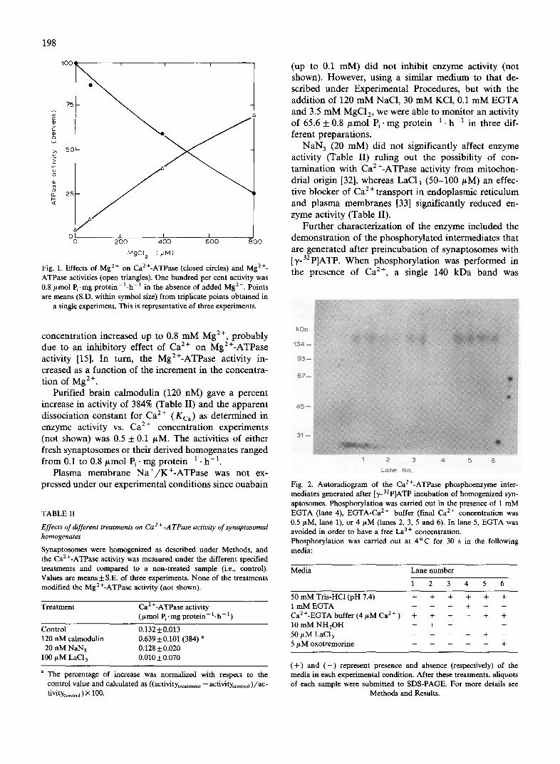
1 0 0 ~ ~ 1 I i
T,
OI I I I 0 200 400 600 800
tvlgc 12 (utvl)
Fig. 1. Effects of Mg 2+ on Ca2+-ATPase (closed circles) and Mg 2+- ATPase activities (open triangles). One hundred per cent activity was 0.8 lamol Pi .mg p r o t e i n - l . h - t in the absence of added Mg 2+. Points are means (S.D. within symbol size) from triplicate points obtained in
a single experiment. This is representative of three experiments.
concentration increased up to 0.8 mM Mg 2+, probably due to an inhibitory effect of Ca 2÷ on Mg2+-ATPase activity [15]. In turn, the Mg2+-ATPase activity in- creased as a function of the increment in the concentra- tion of Mg 2+.
Purified brain calmodulin (120 nM) gave a percent increase in activity of 384% (Table II) and the apparent dissociation constant for Ca 2÷ (Kca) as determined in enzyme activity vs. Ca 2÷ concentration experiments (not shown) was 0.5 + 0.1 #M. The activities of either fresh synaptosomes or their derived homogenates ranged from 0.1 to 0.8 #mol Pi" mg protein -1 • h -a.
Plasma membrane Na+/K+-ATPase was not ex- pressed under our experimental conditions since ouabain
TABLE II
2+ Effects of different treatments on Ca -ATPase actioity of synaptosomal homogenates
Synaptosomes were homogenized as described under Methods, and the Ca2÷-ATPase activity was measured under the different specified treatments and compared to a non-treated sample (i.e., control). Values are means + S.E. of three experiments. None of the treatments modified the Mg2+-ATPase activity (not shown).
Treatment Ca 2 +-ATPase activity (/~ mol Pi' mg prote in- 1. h - 1)
Control 0.132 + 0.013 120 nM calmodulin 0.639 + 0.101 (384) a
20 nM NaN 3 0.128 + 0.020 100/~M LaC13 0.010+0.070
a The percentage of increase was normalized with respect to the control value and calculated as ((activitytreatmen t -- activitycontrol)/ac" tivitycontrol ) × 100.
(up to 0.1 mM) did not inhibit enzyme activity (not shown). However, using a similar medium to that de- scribed under Experimental Procedures, but with the addition of 120 mM NaC1, 30 mM KC1, 0.1 mM EGTA and 3.5 mM MgC12, we were able to monitor an activity of 65.6 + 0.8 /~mol Pi" mg protein -1 • h -1 in three dif- ferent preparations.
NaN 3 (20 mM) did not significantly affect enzyme activity (Table II) ruling out the possibility of con- tamination with Ca2+-ATPase activity from mitochon- drial origin [32], whereas LaC13 (50-100 #M) an effec- tive blocker of Ca 2 ÷ transport in endoplasmic reticulum and plasma membranes [33] significantly reduced en- zyme activity (Table II).
Further characterization of the enzyme included the demonstration of the phosphorylated intermediates that are generated after preincubation of synaptosomes with [-/-32p]ATP. When phosphorylation was performed in the presence of Ca 2÷, a single 140 kDa band was
kDa
134 - -
9 3 - -
6 7 - -
4 5 -
3 1 -
1 2 3 4 5 6
L a n e No.
Fig. 2. Autoradiogram of the Ca2+-ATPase phosphoenzyme inter- mediates generated after ['r-32 P]ATP incubation of homogenized syn- aptosomes. Phosphorylation was carried out in the presence of 1 m M EGTA (lane 4), EGTA-Ca 2+ buffer (final Ca 2÷ concentration was 0.5/xM, lane 1), or 4 /~M (lanes 2, 3, 5 and 6). In lane 5, EGTA was avoided in order to have a free La 3 + concentration. Phosphorylation was carried out at 4 ° C for 30 s in the following media:
Media Lane number
1 2 3 4 5 6
50 m M Tris-HC1 (pH 7.4) + + + + + + 1 m M EGTA - - - + Ca2+-EGTA buffer (4/~M Ca 2+ ) + + + - + + 10 m M NH2OH - + - 50 # M LaC13 . . . . + - 5/~M oxotremorine - +
( + ) and ( - ) represent presence and absence (respectively) of the media in each experimental condition. After these treatments, aliquots of each sample were submitted to SDS-PAGE. For more details see
Methods and Results.

observed (Fig. 2,lane 1). There was n o 32p incorporation in the absence of Ca 2÷ (Fig. 2, lane 4) or in the presence of Ca 2÷ plus hydroxylamine (Fig 2, lane 2). The latter finding suggests that an acyl phosphate groups exists as the labelled species [15]. The level of phospho- rylation was increased by 50 /.tM LaC13 (50 ttM) (cf lane 5), a characteristic reactivity of some plasma mem- brane Ca2+-ATPases [33].
The protein profile after electrophoresis of the syn- aptosomal homogenate as revealed by Coomassie blue staining, showed several bands of different M r exhibit- ing no differences between them using the treatments described above. When the homogenates were in- cubated with [3'-32p]ATP using different conditions but keeping always a low ionic strength medium, a single 140 kDa band could be demonstrated. The addition of 2 mM MgC12 during phosphorylation of the enzyme (not shown) did not affect the appearance of the phospho- rylated intermediate.
Effects of K + depolarization and of the cholinergic muscarinic agent oxotremorine on enzyme activity
In another series of experiments depicted in Table IV, intact synaptosomes were preincubated in the media described in the text of Table IV. After this, they were homogenized, and the Ca2÷-ATPase and Mg2+-ATPase activities measured as described in Materials and Meth- ods. Preincubation with 250 mM KC1 caused a signifi- cant decrease of Ca2+-ATPase activity even when Ca 2 + was included in the preincubation medium. The Mg 2÷- ATPase activity remained unchanged throughout these experimental procedures (Table IV).
Preincubation with oxotremorine at 15 #M con- centration, did not significantly modify the activities of Ca2÷-ATPase or Mg2+-ATPase in intact synaptosomes
TABLE III
Effects of oxotremorine on the Ca24--ATPase activity of subcellular fractions and presynaptic plasma membrane homogenates from electric tissue.
D. tschudii electric organ was subjected to subcellular fractionation obtaining the S, I, and P fractions described in Methods. Subsequent fractionation of lysed synaptosomes yielded bands B, C, and D (see Methods). The Ca 2 +-ATPase activity (in #tool Pi .mg protein-1, h - 1 ) was determined in duplicate in these fractions in the absence and in the presence of 15 / tM oxotremorine. Values are means + S.E. of three experiments. The Ca 2+ concentration of the incubation media was adjusted to 4.8 + 0 .5 / tM using EGTA.
Fraction Oxotremorine (~tM)
0 15
S 0.148 + 0.058 0.163 + 0.010 I 0.568 + 0.090 0.572 + 0.070 P 0.301 + 0.020 0.288 + 0.010 B 0.295 + 0.015 0.304 + 0.024 C 0.157 + 0.054 0.134 -I- 0.033 D 0.177 -I- 0.089 0.154 + 0.030
199
TABLE IV
Effect of different incubation media on Ca2+-ATPase and Mg 24-- A TPase activities of intact synaptosomes
Intact synaptosomes were preincubated in media previously gassed with 02 and containing 3 m M MgC12, 250 m M sucrose, 10 m M glucose, Hepes-Tris (pH 7.4 at 22°C) , and 250 m M NaC1 or KC1, plus EGTA, Ca 2÷ a n d / o r oxotremorine as indicated above. After this the synaptosomes were subjected to homogenization, and the Ca2+-ATPase and Mg2+-ATPase activities were determined using 100 /~1 aliquots of the homogenates as described under Materials and Methods. Values are given as means + S.E. of three different experi- ments.
Medium /tmol P i 'mg prote in- l"h -1
Mg2 +-ATPase Ca2 +-ATPase
Ringer-Hepes-Na ÷ (1 m M 2.58 -/- 0.15 EGTA)
Ringer-Hepes-K + (1 m M EGTA) 2.65 + 0.20 Ringer-Hepes K + + 1 m M Ca 24- 2.63 + 0.09 Ringer-Hepes K + + 1 m M Ca 24- 2.65 +0.08
+ 15/~M oxotremorine
0.76 + 0.08
0.65+0.11 (15) a 0.36 + 0.04 (53) 0.38 + 0.03 (50)
a The percentage of decrease in activity was normalized with respect to the control value and calculated as ((activitycontro I --activi-
tYtreatmen t )/activitycontro I ) X 100.
(Table IV), or the Ca2+-ATPase activities in homo- genates of fractions I and P, or their synaptosomally-de- rived plasma membrane fractions (Table III). The muscarinic agonist neither affected the decrease in en- zyme activity which follows K ÷ depolarization of intact synaptosomes (Table IV). In line with this experimental evidence, oxotremorine did not modify the level of Ca2+-induced phosphorylation of the 140 kDa band (Fig. 2, lane 6).
Discussion
Cae+-ATPase activity and muscarinic agonist binding sites in synaptosomes and presynaptic plasma membranes from D. tschudii electric organ.
Pinched-off nerve endings (i.e., synaptosomes) are suited for studying the several steps involved in synaptic transmission [3,5,12]. The results presented in this paper were obtained using synaptosomes prepared from the electric organ of Discopyge tschudii, a ray of the order Torpediniformes that has an electric organ biochemi- cally [23,26,34-36] and structurally [37] similar to those of the most studied species T. californica and T. marmorata [30].
When analyzing chohnergic functions, electric fish present several advantages over other preparations such as brain synaptosomes usually derived from a mixture of nerve endings which in turn use different neurotrans- mitters. Firstly, electric organ nerve endings are exclu- sively cholinergic [38]. Secondly, synaptosomes can be prepared from the electric organ almost devoid of any post synaptic attachments [19] and plasma membrane
200
fractions can be obtained from those synaptosomes [20]. Thirdly, presynaptic muscarinic autoreceptors are well documented in these structures [39,40].
To our knowledge, there are no reports describing a Ca 2+- ATPase activity in electric organ presynaptic plasma membranes. Nevertheless, numerous experimen- tal evidences show that Ca2+-ATPase is presynaptically located (e.g.: at the frog neuromuscular junction [42], or at central nervous system synapses [43]). Biochemically, Ca 2 +-ATPase has been described in brain synaptosomal plasma membranes [7,8,44-47], Electrophorus electricus microsomal fractions [48] and Torpedo synaptic vesicles [49-51].
The synaptosomal fraction S was characterized using three separate biochemical markers (Table I) and Ca2+-ATPase activity and muscarinic agonist binding sites were simultaneously demonstrated in the same preparation (Fig. 1, Tables I and II). The enzyme was assayed in a low Ca 2÷ and Mg 2÷, low ionic strength medium devoid of K ÷, conditions under which the Na+/K+-ATPase activity is not detected.
The Ca2+-ATPase activity of fraction I was higher than that of fractions S or P (Table III). This may be due to the fact that fraction I contains unidentified membrane fragments [19] possibly derived from axo- plasmic reticulum and other membrane bound organelles containing Ca2+-ATPase activities.
It is worthwhile pointing out that both Ca 2 +-ATPase and Mg2+-ATPase were activated by Ca 2+, even without the addition of Mg 2÷ (data not shown). Since the K0. 5 for Mg 2÷ during ATP hydrolysis by Ca2+-ATPase is within the #molar range [52], supposedly Mg2+-free solutions could contain enough Mg 2÷ to activate ATP hydrolysis.
The synaptosomal fraction S was processed by the procedure of Morel et al. [20]. Synaptosomes were first freeze-thawed and osmotically disrupted to get rid of synaptic vesicles, and subsequently fractioned on sucrose gradients to obtain pure presynaptic membrane frac- tions. The original data obtained on T. marmorata elec- tric tissue reported five bands (F1 to F5), and a pellet (F6). Mg2+-ATPase activity (the only ATPase activity reported in the Morel et al. paper) was enriched in fractions F2 and F3.
Our procedure yielded three net bands (A, B, and C), a fourth band (D) which could be resolved into two poorly demarcated bands (see Experimental Proce- dures), and a pellet (P). For comparative purposes we assigned our bands A, B,C, to bands F1 to F3, and our band D to bands F4 plus F5 of Morel et al. [20]. This discrepancy in band number is attributed to species differences in electric organ subcellular fractionation.
Characterization of the Ca2 +-ATPase activity The physiological Ca 2+ pump with Ca2+-ATPase
activity from the plasma membrane has been charac-
terized as a single 140 kDa band using SDS gel electro- phoresis criteria [53] (for reviews see Refs. 13 and 14). Coincidentally, the Ca2+ATPase from brain synapto- somal membranes has also a molecular weight of 140 kDa and shows a Ca2+-dependent, calmodulin-stimu- lated, hydroxylamine-sensitive phosphorylation [7].
The enzyme activity from D. tschudii electric organ reported in this paper qualifies as a true presynaptic plasma membrane Ca2+-ATPase. Catalysis was en- hanced by 120 nM calmodulin (Table II), and synapto- somes incubated with [y-32p]ATP and in the presence of Ca 2÷ gave a 140 kDa phosphorylated band which was: (a) sensitive to hydroxylamine attack and (b) en- hanced by preincubation with 50/xM LaC13 (Fig. 2; see Ref. 15).
Contamination from other sources could also be ruled out. The Ca2+-ATPase from Torpedo synaptic vesicles [49-51] has rather similar Ca 2÷ and Mg 2÷ require- ments when compared to the Ca2+-ATPase, but is not stimulated by calmodulin [54] and has an apparent M r of 210000 [51]. Furthermore, the freeze-thaw/osmotic lysis procedure employed in our experiments yields a presynaptic plasma membrane preparation devoid of synaptic vesicles [20]. Finally, mitochondrial or endo- plasmic reticulum ATPases could also be excluded as possible contaminants since the former is known to be inhibited by NaN 3 [32] and the latter is not stimulated by calmodulin [14].
Despite the above-mentioned characteristics which point to the described Ca2+-ATPase as the same en- zyme that is involved in Ca 2+ transport across the synaptosomal membrane [7,8,44-48], the possibility still remains that the present activity may be similar to the one described by Enyedi et al. [55] in rat myometrium, which does not participate in Ca 2+ transport.
Possible coupfing between muscarinic autoreceptors and Ca 2 +-A TPase
Muscarinic autoreceptors have been implied in the feed-back inhibition of acetylcholine release [56-58] and are well documented in brain [59,60], Torpedo [39,40] and Electrophorus electric organ [48], and Aplysia [61]. The occupation of such receptor sites by muscarinic agonists is known to reduce the amount of acetylcholine released either spontaneously or under depolarizing conditions [60,62,63]
An involvement of muscarinic autoreceptors in the regulation of acetylcholine release in Torpedo electric organ was shown by Michaelson et al. [41]. These authors demonstrated that K + depolarization of Torpedo synaptosomes in the presence of Ca 2 + induces acetylcholine release, and the phosphorylation of a specific 100 kDa synaptosomal protein. The muscarinic agonist oxotremorine had no effect on Ca 2+ uptake, but inhibited transmitter release and phosphorylation. These
muscarinic agonist-induced effects were blocked by at- ropine.
These data were particularly relevant since the fine control of the axoplasmic free Ca 2÷ concentration is known to be associated to the phosphorylation of specific proteins [64-66]. Ca2+-ATPase activity was not measured by Michaelson et al. [41], and it seems un- likely that their synaptosomal 100 kDa protein is analo- gous to our 140 kDa band. Interestingly enough, the Ca2+-ATPase from brain synaptosomes also undergoes a Ca 2 +-dependent phosphorylation [7,46].
On the basis of experimental data, and taking into account the aforementioned observations, a linkage be- tween presynaptic muscarinic autoreceptors and a Ca 2 + / M g 2 +-ATPase from brain synaptosomes was pro- posed [67]. Brain synaptosomal membranes exposed to micromolar concentrations of muscarine or oxotremor- ine show inhibited Ca2+-ATPase and Ca 2÷ transport activities (but not Na+-Ca 2+ exchange or Ca 2÷ move- ments through voltage dependent Ca 2+ channels). The authors suggested [67] that muscarinic autoreceptor oc- cupation (specially at cholinergic nerve terminals) re- duces acetylcholine release by preventing the mainte- nance of an optimal axoplasmic free Ca 2+ concentra- tion. A comparison of our data and those of Ross et al. [67] seems to be difficult since the enzyme activities studied appear to be different, and their brain synapto- somal preparations are heterogeneous, consisting of pinched-off nerve endings corresponding to synapses served by different neurotransmitters.
The present experiments, conducted in a pure cholinergic system, show that the muscafinic agonist oxotremorine did not block Ca2+-ATPase activity (Ta- bles I I I and IV, Fig. 2) although the enzyme activity was sensitive to depolarization, a procedure by which Ca 2+ is expected to enter the nerve ending axoplasm through voltage-gated Ca 2÷ channels and inhibit Ca 2+- ATPase activity (Table IV).
The possibility that other Ca2+-ATPase activities (distinct from the physiological Ca 2+ pump) may be instrumental in the control of transmitter release and therefore be effectively coupled to the operation of muscarinic autoreceptors merits further exploration. Within this context, Gandhi and Ross [68] described a Ca2+-ATPase from rat brain synaptosomal membranes which is totally independent from Mg 2+, is sensitive to c G M P (see also Ref. 69), uses GTP as a substrate, and is not inhibited by La 3+. It is then probable that autore- ceptors may be regulated by Ca 2 +-ATPase(s) activit(ies) coupled to G proteins and special signal transduction mechanisms [69], although these usually slow (i.e., sec- ond to minute) mechanisms will have to account for the rapidity of events in synaptic transmission (occurring within the ms time range).
The signalling function of Ca 2 ÷ demands a very low ionic concentration of the cation within cells. The elec-
201
troplaque, the electric fish counterpart of mammalian skeletal muscle [30], is expected to possess a Ca 2+- ATPase with similar characteristics to those found in other vertebrate tissues. In fact, the enzyme activity described in this paper shares common features with other ATPases from cardiac and skeletal muscle sarco- lemma and the red cell plasma membrane [13,70].
Future experimental efforts will be directed towards characterizing the Ca2+-ATPase activities of Torpe- dinidae synaptosomes as well as establishing the rela- tionship between these activities and the transport of the divalent cation across the membrane. These studies will help to establish the precise role that these enzymes play within the complex metabolic machinery engaged in the release of acetylcholine.
Acknowledgements
The authors thank Professor M.B. Cousseau from I N I D E P (Mar del Plata, Pcia. de Buenos Aires) for providing the electric fish, Professor A.P. de Iraldi for the electron microscope observations, and Dr. J.S.O. Aguilar for 3H-QNB binding data. This work was sup- ported by a P I D - C O N I C E T grant to E.L.M.O. J.P.F.C.R. and E.L.M.O. are Career Investigators from C O N I C E T (Argentina).
References
1 Katz, B. and Miledi, R. (1967) Proc. Roy. SOC. Lond. B Biol. Sci. 167, 23-38.
2 Kelly, R.B., Deutsch, J.W., Cadson, S.S. and Wagner J.A. (1979) Annu. Rev. Neurosci. 2, 399-420.
3 Israel, M. and Manaranche, R. (1985) Biol. Cell 55, 1-14. 4 Augustine, G., Charleton, M.P. and Smith, S.J. (1987) Annu. Rev.
Neurosci. 10, 633-693. 5 Smith, S.J. and Augustine, G.J. (1988) Trends Neurosci. 11,458-
464. 6 Blaustein, M.P., Ratzlaff, R.W. and Kendrick, N.K. (1978) Annu.
N.Y. Acad. Sci. 307, 195-212. 7 Goldin, S.M., Chan, S.Y., Papazian, D.M., Hess, E.J. and
Rahamimoff, H. (1983) Cold Spring Harbor Symp. Quant. Biol. 48, 287-295.
8 Michaelis, M.L., Kitos, T.E., Nunley, E.W., Lecluyse, E. and Michaelis, E.K. (1987) J. Biol. Chem. 262, 4182-4189.
9 Michaelis, M.L. and Michaelis, E.K. (1981) Life Sci. 28, 37-45. 10 Gill, D.M., Grollman, E.F. and Kohn, L.D. (1981) J. Biol. Chem.
266, 184-192. 11 Teo, T.S. and Wang, J.H. (1973) J. Biol. Chem. 248, 5950-5958. 12 Blaustein, M. (1988) Trends Neurosci. 11,438-443. 13 Rega, A.F. and Garrahan, P. (1986) The Ca 2+ Pump of Plasma
Membranes, pp. 173, CRC, Boca Raton, FL. 14 Schatzmann, H.J. (1989) Annu. Rev. Physiol. 51, 473-485. 15 Rossi, J.P., Gronda, C.M., Fern~ndez, H. and Gagliardino, J.J.
(1988) Biochim. Biophys. Acta 943, 175-182. 16 Norman, J.R. (1937) in Discovery Reports 16, pp. 3-152, Cam-
bridge University Press, Cambridge. 17 Ringuelet, R.A. and Aramburu, R.H. (1960) Agro 2, 1-141. 18 Menni, R., Ringuelet, R.A. and Aramburu, R.A. (1984) Peces
marinos de la Argentina y Uruguay, pp. 91-92, Editorial Hemis- ferio Sur S.A.

202
19 Dowdall, M.J. and Zimmermann, H. (1977) Neuroscience 2, 405- 421.
20 Morel, N., Manaranche, M., Israel, M. and Gulik-Krzywicki T. (1982) J. Cell Biol. 93, 349-356.
21 Kratje, R.B., Garrahan, P.J., and Rega, A.F. (1983) Biochim. Biophys. Acta 731, 40-46.
22 Lowry, O.H., Rosebrough, N.J., Farr, A.L. and Randall, R.J. (1951) J. Biol. Chem. 193, 265-275.
23 Ochoa, E.L.M., Dalziel, A.W. and McNamee, M. (1983a) Biochim. Biophys. Acta 727, 151-162.
24 Schmidt, J. and Raftery, M.A. (1973) Anal. Biochem. 52, 349-354. 25 Yamamura, H.I. and Snyder, S.H. (1974) Proc. Natl. Acad. Sci.
USA 71, 1725-1729. 26 Ochoa, E.L.M. (1980b) Eur. J. Biochem. 107, 415-421. 27 Ellman, G.L., Courtney, K.D., Andres, V. and Featherstone, R.M.
(1961) Biochem. Pharmacol. 7, 88-9. 28 Glyrm, I.M and Chappell, B.J. (1964) Biochem. J. 90, 147-149. 29 Kakiuchi, S., Sobue, K., Yamazaki, R., Kambayashi, J., Sakon, M.
and Kosaki, G. (1981) FEBS Lett. 126, 203-207. 30 Changeux, J.P. (1981) in The Harvey Lectures, Academic Press,
Vol. 75, pp. 85-254, New York. 31 Ochoa, E.L.M., Chattopadhyay, A. and McNamee, M.G. (1989)
Cell. Mol. Neurobiol. 9, 141-178. 32 Kemmler, W. and l_25ffler, G. (1977) Diabetologia 13, 235-238. 33 Enyedi, A., Sarkadi, B., FSldes-Papp, Z., Monostory, S. and
Gardos, G. (1986) J. Biol. Chem. 261, 9558-9563. 34 Ochoa, E.L.M. (1978) FEBS Lett. 89, 317-320. 35 Ochoa, E.L.M. (1980) Comp. Biochem. Physiol. 66C, 99-103. 36 Ochoa, E.L.M., Biscoglio de Jimenez Bonino, M., Cascone, O.,
Medrano, S. and Cousseau, M.B. (1983) Comp. Biochem. Physiol. 76C, 313-317.
37 Iraldi, C., Pellegrino de Iraldi, A. and Ochoa, E.L.M. (1984) Comp. Biol. 2, 293-301.
38 Feldberg, W. and Fessard, A. (1942) J. Physiol. 101,200-216. 39 Kloog, Y., Michaelson, D.M. and Sokolovsky, M. (1978) FEBS
Lett. 95, 331-334. 40 Pickard, M.R. and Strange, P.G. (1978) Biochem. Soc. Trans. 6,
129-131. 41 Michaelson, D.M., Avissar, S., Kloog, Y. and Sokolovsky, M.
(1979) Proc. Natl. Acad. Sci. USA 76, 6336-6340. 42 Pappas, G.D. and Kriho, V. (1988) J. Neur0cytol. 17, 417-423. 43 Mata, M. and Fink, DJ. (1989) J. Histochem. Cytochem. 37,
971-980. 44 Sobue, K., Ichida, S., Yoshida, H., Yamazaki, R. and Kakiuchi, S.
(1979) FEBS Lett. 99, 199-202.
45 Sorensen, R.G. and Mahler, H.R. (1981) J. Neurochem. 37, 1407- 1418.
46 Javors, M.A., Bowden, C.L. and Ross, D.H. (1981) J. Neurochem. 37, 381-387.
47 Ross, D.H. and Cardenas, H.L. (1983) J. Neurochem. 41, 161-171. 48 Amende, L.M., Chock, S.P. and Albers, R.W. (1983) J. Neure~
chem. 40, 1040-1047. 49 Breer, H., Morris, S.J. and Whittaker V.P. (1977) Eur. J. Biochem.
80, 313-318. 50 Rothlein, J.E. and Parsons, S.M. (1980) Biochem. Biophys. Res.
Commun. 95, 1869-1874. 51 Yamagata, S.K. and Parsons, S.M. (1989) J. Neurochem. 52,
168-173. 52 Caride, A.J., Rega, A.F. and Garrahan, P.J. (1986) Biochim.
Biophys. Acta 863, 165-177. 53 Brandl, C.J., Green, M., Korczak, B. and MacLennan, D.H. (1986)
Cell 44, 597-607. 54 Raphaeh, A. and Parsons, S.M. (1982) Proc. Natl. Acad. Sci. USA
79, 5783-5787. 55 Enyedi, A., Minami, J., Caride, A.J., and Permiston, J.T. (1988)
Biochem. J. 252, 215-220. 56 Szerb, J.C. and Somogyi, C.T. (1973) Nature 241, 121-122. 57 Szerb, J.C. (1978) in Cholinergic Mechanisms and Psychophar-
macology (Jenden D.J., ed.), pp. 49-60, Plenum Press, New York. 58 Starke, K., Gt~thert, M. and Kitbinger, H. (1989) Physiol. Rev. 69,
864-989. 59 Meyer, E.M., Otero, D.H., Morgan, E., Marchand, S. and Baker
S.P. (1987) J. Neurochem. 48, 477-482. 60 Suzuki, T., Fujimoto, K., Oohata, H. and Kawashima, K. (1988)
Neurosci. Lett. 84, 209-212. 61 Baux, G. and Tauc, L. (1987) J. Physiol. 388, 665-680. 62 Meyer, E.M. and Otero, D.H. (1985) J. Neurosci. 5, 1202-1207. 63 Luz, S., Pinchasi, I. and Michaelson, D.M. (1985) J. Neurochem.
45, 43-50. 64 De Lorenzo, R.J. (1976) Biochem. Biophys. Res. Commun. 71,
590-597. 65 Krueger, B.K., Forn, J. and Greengard, P. (1977) J. Biol. Chem.
252, 2764-2773. 66 Hershkowitz, M. (1978) Biochim. Biophys. Acta 542, 274-283. 67 Ross, D.H., Shreeve, S.M. and Hamilton, M.G. (1985) Brain Res.
329, 39-47. 68 Gandhi, C.R and Ross, D.H. (1988) J. Neurochem. 50, 248-256. 69 Stauderman, K.A., Jones, D.J. and Ross, D.H. (1985) J. Neuro-
chem. 45, 970-972. 70 Carafoh, E. (1987) Annu. Rev. Biochem. 56, 395-433.


![Near-Membrane [Ca2+] Transients Resolved Using the Ca2+ Indicator FFP18](https://static.fdokumen.com/doc/165x107/631286873ed465f0570a4533/near-membrane-ca2-transients-resolved-using-the-ca2-indicator-ffp18.jpg)