Functional analysis of archaeal MBF1 by complementation studies in yeast
Upload
khangminh22Category
view
3download
0
Published online 31 December 2019 Nucleic Acids Research, 2020, Vol. 48, No. 5 2457–2472doi: 10.1093/nar/gkz1228
Adjacent mutations in the archaeal Rad50 ABCATPase D-loop disrupt allosteric regulation of ATPhydrolysis through different mechanismsZachary K. Boswell, Marella D. Canny , Tanner A. Buschmann, Julie Sang and MichaelP. Latham *
Department of Chemistry and Biochemistry, Texas Tech University, Lubbock, TX 79409-1061, USA
Received June 21, 2019; Revised November 25, 2019; Editorial Decision December 18, 2019; Accepted December 20, 2019
ABSTRACT
DNA damage is the driving force for mutation and ge-nomic instability, which can both lead to cell deathor carcinogenesis. DNA double strand breaks are de-tected and processed in part by the Mre11–Rad50–Nbs1 protein complex. Although the Mre11–Rad50–Nbs1 complex is essential, several spontaneous mu-tations have been noted in various cancers. One ofthese mutations, within a conserved motif of Rad50,resulted in an outlier curative response in a clini-cal trial. We show through biochemical and biophys-ical characterization that this cancer-associated mu-tation and a second mutation to the adjacent residue,previously described in a breast cancer patient, bothhave gain-of-function Rad50 ATP hydrolysis activitythat results not from faster association of the ATP-bound form but faster dissociation leading to lessstable Rad50 dimer. This disruption impairs the reg-ulatory functions of the protein complex leading toa loss of exonuclease activity from Mre11. Interest-ingly, these two mutations affect Rad50 structure anddynamics quite differently. These studies describethe relationship between function, structure, andmolecular motions in improperly regulated Rad50,which reveal the underlying biophysical mechanismfor how these two cancer-associated mutations af-fect the cell.
INTRODUCTION
The integrity and stability of a genome is under constant as-sault from a number of internal and external stresses, suchas reactive oxygen species, replicative stress, UV light, andgenotoxic chemicals. Failure to detect and accurately repairthe various DNA lesions that result from these assaults canlead to simple mutations and/or large scale chromosomalrearrangements, which could drive the development of can-
cer in humans. Fortunately, the cell has developed sophisti-cated systems to find and repair each distinct form of DNAdamage. DNA double strand breaks, where both strands ofthe DNA double helix are broken, are a particularly danger-ous form of DNA damage, as a template for accurate repairis not readily available. Thus, the detection and repair ofDNA double strand breaks are critical for cellular survivaland cancer prevention (1).
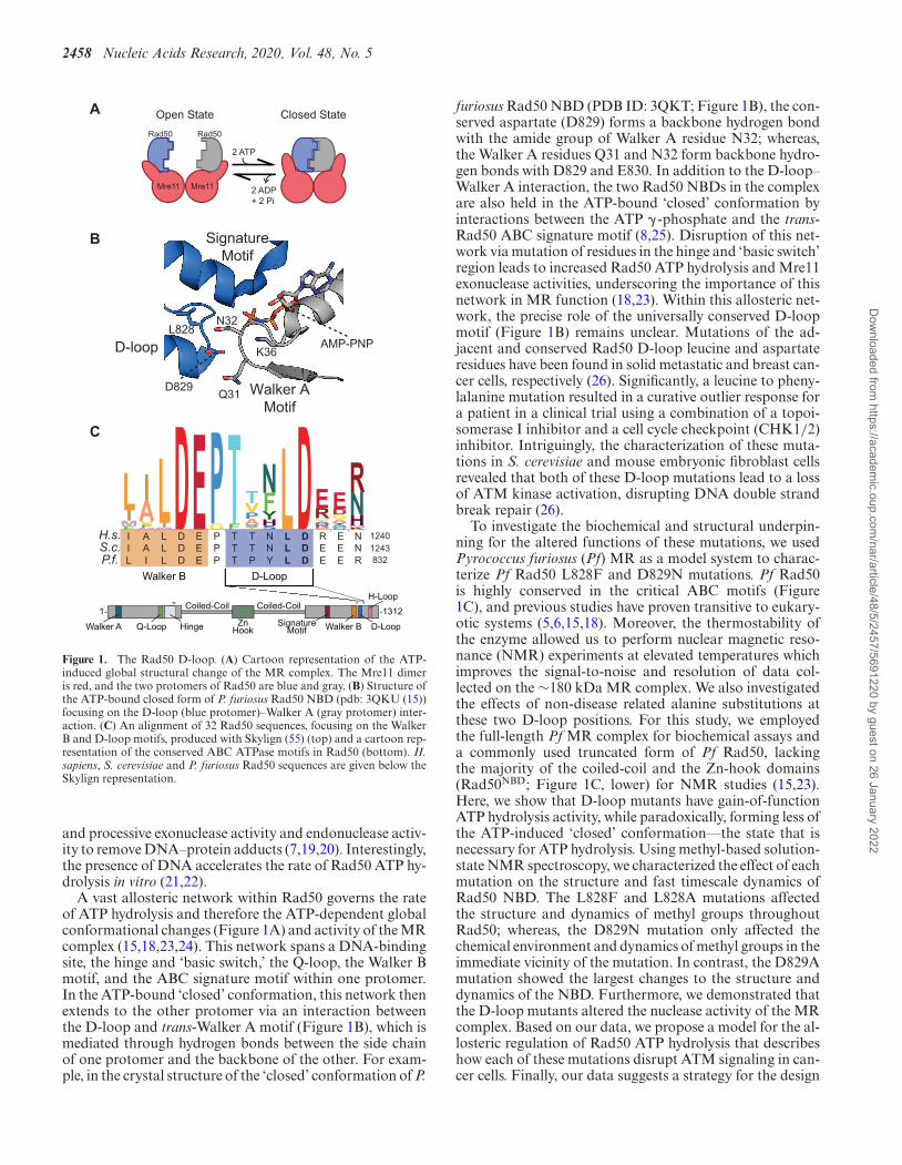
Mre11–Rad50–Nbs1 (MRN) is an essential protein com-plex that is a primary responder to DNA double strandbreaks, hairpins, and other anomalous terminal DNAstructures. MRN participates in tethering damaged DNAstrands and preparing the broken ends for repair by down-stream homologous recombination or non-homologousend joining pathways (2,3). The MRN complex utilizes avariety of functions in the process of detecting and initi-ating DNA double strand break repair. Mre11 functionsas a dimer and has Mn2+-dependent exo- and endonucle-ase activities (4–6). The endonuclease activity in particularis important for the removal of DNA-protein adducts thatare formed from failed topoisomerase reactions (7). OneRad50, a member of the ATP-Binding Cassette (ABC) AT-Pase super-family, binds to each of the Mre11 protomersand regulates the conformational and functional states ofMRN via ATP-induced association of the two Rad50 nu-cleotide binding domains (NBDs) and subsequent ATP hy-drolysis (2,3,8). The eukaryotic Nbs1 (or Xrs1 in buddingyeast) is a signaling hub associated with Mre11 that recruitsdownstream effectors to the site of damage (9–11). In theuniversally conserved Mre112–Rad502 (MR) core complex,Rad50 undergoes dramatic conformational changes in re-sponse to ATP binding and hydrolysis (12–15). In the ATP-free ‘open’ conformation, Mre11 is an active nuclease (Fig-ure 1A; left); whereas, in the ATP-bound ‘closed’ confor-mation (Figure 1A; right), Mre11 active sites are occluded.Additionally, Rad50 binds DNA in the ATP-bound ‘closed’conformation, a function that is important for telomeremaintenance (16–18). Rad50 ATP hydrolysis returns thecomplex to the ‘open’ conformation and leads to increased
*To whom correspondence should be addressed. Tel: +1 806 834 2564; Email: [email protected]
C© The Author(s) 2019. Published by Oxford University Press on behalf of Nucleic Acids Research.This is an Open Access article distributed under the terms of the Creative Commons Attribution License (http://creativecommons.org/licenses/by/4.0/), whichpermits unrestricted reuse, distribution, and reproduction in any medium, provided the original work is properly cited.
Dow
nloaded from https://academ
ic.oup.com/nar/article/48/5/2457/5691220 by guest on 26 January 2022
2458 Nucleic Acids Research, 2020, Vol. 48, No. 5
A
2 ATP
2 ADP+ 2 Pi
Mre11 Mre11
Open State Closed State
Rad50Rad50
B
H.s.S.c.P.f. 832
12401243
Walker B D-Loop
I A L D E P T T N L D R E NI A L D E P T T N L D E E NL I L D E P T P Y L D E E R
LIMF
IAVFM
LIMDEPGTF TVPASIGNFYHSLDERGTSQNKDAEDGAYSQPNRNHMK
ZnHook
Coiled-Coil
Walker A Walker B D-LoopQ-Loop Hinge
H-Loop
SignatureMotif
1- -1312Coiled-Coil
C
AMP-PNP
D829
L828
K36
Walker AMotif
D-loop
SignatureMotif
Q31
N32
Figure 1. The Rad50 D-loop. (A) Cartoon representation of the ATP-induced global structural change of the MR complex. The Mre11 dimeris red, and the two protomers of Rad50 are blue and gray. (B) Structure ofthe ATP-bound closed form of P. furiosus Rad50 NBD (pdb: 3QKU (15))focusing on the D-loop (blue protomer)–Walker A (gray protomer) inter-action. (C) An alignment of 32 Rad50 sequences, focusing on the WalkerB and D-loop motifs, produced with Skylign (55) (top) and a cartoon rep-resentation of the conserved ABC ATPase motifs in Rad50 (bottom). H.sapiens, S. cerevisiae and P. furiosus Rad50 sequences are given below theSkylign representation.
and processive exonuclease activity and endonuclease activ-ity to remove DNA–protein adducts (7,19,20). Interestingly,the presence of DNA accelerates the rate of Rad50 ATP hy-drolysis in vitro (21,22).
A vast allosteric network within Rad50 governs the rateof ATP hydrolysis and therefore the ATP-dependent globalconformational changes (Figure 1A) and activity of the MRcomplex (15,18,23,24). This network spans a DNA-bindingsite, the hinge and ‘basic switch,’ the Q-loop, the Walker Bmotif, and the ABC signature motif within one protomer.In the ATP-bound ‘closed’ conformation, this network thenextends to the other protomer via an interaction betweenthe D-loop and trans-Walker A motif (Figure 1B), which ismediated through hydrogen bonds between the side chainof one protomer and the backbone of the other. For exam-ple, in the crystal structure of the ‘closed’ conformation of P.
furiosus Rad50 NBD (PDB ID: 3QKT; Figure 1B), the con-served aspartate (D829) forms a backbone hydrogen bondwith the amide group of Walker A residue N32; whereas,the Walker A residues Q31 and N32 form backbone hydro-gen bonds with D829 and E830. In addition to the D-loop–Walker A interaction, the two Rad50 NBDs in the complexare also held in the ATP-bound ‘closed’ conformation byinteractions between the ATP � -phosphate and the trans-Rad50 ABC signature motif (8,25). Disruption of this net-work via mutation of residues in the hinge and ‘basic switch’region leads to increased Rad50 ATP hydrolysis and Mre11exonuclease activities, underscoring the importance of thisnetwork in MR function (18,23). Within this allosteric net-work, the precise role of the universally conserved D-loopmotif (Figure 1B) remains unclear. Mutations of the ad-jacent and conserved Rad50 D-loop leucine and aspartateresidues have been found in solid metastatic and breast can-cer cells, respectively (26). Significantly, a leucine to pheny-lalanine mutation resulted in a curative outlier response fora patient in a clinical trial using a combination of a topoi-somerase I inhibitor and a cell cycle checkpoint (CHK1/2)inhibitor. Intriguingly, the characterization of these muta-tions in S. cerevisiae and mouse embryonic fibroblast cellsrevealed that both of these D-loop mutations lead to a lossof ATM kinase activation, disrupting DNA double strandbreak repair (26).
To investigate the biochemical and structural underpin-ning for the altered functions of these mutations, we usedPyrococcus furiosus (Pf) MR as a model system to charac-terize Pf Rad50 L828F and D829N mutations. Pf Rad50is highly conserved in the critical ABC motifs (Figure1C), and previous studies have proven transitive to eukary-otic systems (5,6,15,18). Moreover, the thermostability ofthe enzyme allowed us to perform nuclear magnetic reso-nance (NMR) experiments at elevated temperatures whichimproves the signal-to-noise and resolution of data col-lected on the ∼180 kDa MR complex. We also investigatedthe effects of non-disease related alanine substitutions atthese two D-loop positions. For this study, we employedthe full-length Pf MR complex for biochemical assays anda commonly used truncated form of Pf Rad50, lackingthe majority of the coiled-coil and the Zn-hook domains(Rad50NBD; Figure 1C, lower) for NMR studies (15,23).Here, we show that D-loop mutants have gain-of-functionATP hydrolysis activity, while paradoxically, forming less ofthe ATP-induced ‘closed’ conformation––the state that isnecessary for ATP hydrolysis. Using methyl-based solution-state NMR spectroscopy, we characterized the effect of eachmutation on the structure and fast timescale dynamics ofRad50 NBD. The L828F and L828A mutations affectedthe structure and dynamics of methyl groups throughoutRad50; whereas, the D829N mutation only affected thechemical environment and dynamics of methyl groups in theimmediate vicinity of the mutation. In contrast, the D829Amutation showed the largest changes to the structure anddynamics of the NBD. Furthermore, we demonstrated thatthe D-loop mutants altered the nuclease activity of the MRcomplex. Based on our data, we propose a model for the al-losteric regulation of Rad50 ATP hydrolysis that describeshow each of these mutations disrupt ATM signaling in can-cer cells. Finally, our data suggests a strategy for the design
Dow
nloaded from https://academ
ic.oup.com/nar/article/48/5/2457/5691220 by guest on 26 January 2022
Nucleic Acids Research, 2020, Vol. 48, No. 5 2459
of novel small molecules that can modulate the activity ofRad50 and interrupt ATM signaling, possibly providing an-other approach for synthetic lethality in anti-cancer thera-pies.
MATERIALS AND METHODS
Protein expression, purification, and Mre112–Rad502 (MR)complex formation
Unlabeled and U-2H, Ile�1-[13CH3], Leu�/Val� -[13CH3,12CD3], Metε-[13CH3] (ILVM)-labeled (27) P.furiosus (Pf) Rad50 nucleotide binding domain (Rad50NBD;amino acids 1–195––GGAGGAGG [linker]––709–882)was expressed and purified as previously described (23,28).The gene for full-length Pf Rad50 was cloned out ofP. furiosus genomic DNA (ATCC) and inserted into amodified pET-29 vector (Novagen) with an N-terminalTEV protease-cleavable 6x-His tag. Full-length Rad50 wasexpressed overnight at 18◦C in Rosetta (DE3) competentcells (Novagen). Cells were lysed via sonication in 25 mMHEPES, 300 mM KCl, 25 mM imidazole, 10% glycerol,20 mM �ME, pH 7 buffer and after heat denaturationand centrifugation to pellet the denatured proteins, thefull-length Rad50 protein was purified on a HisTrap (GEHealthcare) column and eluted in 300 mM imidazole.After TEV-cleavage of the 6xHis tag, full-length Rad50was re-purified over the HisTrap column. At this point,the sample was diluted to bring the concentration of KCldown to 75 mM, was loaded onto a HiTrapQ HP (GEHealthcare) column, and eluted in a linear gradient into25 mM Tris, 1 M NaCl, 10% glycerol, 1 mM DTT, pH 8.0.2% Tween-20 was added to the pooled Q peak fractionsbefore concentrating (Vivaspin, Sartorius) the proteindown to 1 ml to load on a Superdex 200 HILoad 16/600(GE Healthcare) gel filtration column equilibrated in 25mM Tris, 100 mM NaCl, 10% glycerol, 1 mM TCEP, pH8. Point mutations to Rad50 were generated via a modifiedStratagene Quikchange protocol and were verified viaSanger sequencing. All L51C mutant purification bufferscontained either 10 mM �-mercaptoethanol or 1 mMTCEP (tris(2-carboxyethyl)phosphine) to keep the cysteinereduced.
The Pf Mre11 gene, codon optimized for expression inEscherichia coli, was synthesized by Life Technologies andexpressed from a modified pET-29 vector (Novagen) withan N-terminal TEV protease-cleavable 6x-His tag in E. coliBL21 (DE3) C41 cells (Sigma). Mre11 protein was purifiedto homogeneity by heat denaturation, HisTrap (GE Health-care) nickel affinity, HiTrapQ HP (GE Healthcare) anionexchange, and Superdex 200 HiLoad 16/600 (GE Health-care) gel filtration chromatography. Mre11HLH was purifiedas described (23).
MR complexes for activity and binding assays wereformed by mixing purified Mre11 and full-length Rad50 orRad50NBD in a 2:2 molar ratio and heating at 65◦C for 10min before cooling to room temperature.
ATP hydrolysis
The steady-state kinetics of Pf MR complex ATP hydrolysiswere measured via the BIOMOL Green (Enzo Lifesciences)
colorimetric assay as previously described (23) with somemodifications. 60 �l reactions containing either full-lengthMR or truncated MRNBD and 0–300 �M ATP were in-cubated for 60 min at 65◦C. For full-length MR, hydroly-sis reactions contained 2.5 �M MR complexes in the ab-sence of DNA or 1.25 �M MR complexes in the presenceof DNA. For truncated MRNBD, 2.5 �M wild type, 1.25�M L828F, 625 nM D829N, 2.5 �M L828A or 1.25 �MD829A were present for the hydrolysis reactions in the ab-sence of DNA, and 2.5 �M wild type, 1.25 �M L828F, 625nM D829N, 1.25 �M L828A or 0.625 �M D829A wereused in the presence of DNA. Buffer contained 50 mM Tris,80 mM NaCl, 1% glycerol and 5 mM MgCl2, pH 7.5. Reac-tions with DNA contained 25 nM of ∼5 kb DNA (pRS416,Addgene) that was linearized with EcoR1 (New EnglandBioLabs), ethanol precipitated, and resuspended in water.The phosphate concentration-dependent absorbance of theBIOMOL reagent for each condition (i.e., no protein blank,with MR, and with MR and DNA) was converted to �MPO4 �M MR−1 h−1 via individual standard curves. Initialvelocities (v) were fit to the Hill equation
v = Vmax[ATP]n
KnM + [ATP]n
where Vmax is the maximal velocity, [ATP] is the concentra-tion of ATP, KM is the Michaelis constant and n is the Hillcoefficient. Plots are the average of three replicate experi-ments using standard deviations as the reported errors.
LRET
Purified Pf Rad50NBD L51C and L51C/D-loop mutantproteins were mixed with a 2-fold molar excess of ei-ther the thiol-reactive Tb3+ chelate DTPA-cs124-EMPH(donor, Lanthascreen, Life Technologies, Inc.) or BodipyFL maleimide (acceptor, Invitrogen) in the presence of 1mM TCEP. Labeling reactions were incubated at room tem-perature for 1–2 h in the dark. Excess label was removedvia size exclusion chromatography on a Superdex 200 In-crease 10/300 GL column (GE Healthcare) equilibratedwith 25 mM HEPES, 200 mM NaCl, 0.1 mM EDTA,pH 7.0. LRET labeling of full-length Pf Rad50 L51C andL51C/D-loop mutants was essentially the same, except theTb3+ chelate and Bodipy FL labels were added together atthe same time and the S200 column to remove excess labelwas equilibrated in 50 mM HEPES, 100 mM NaCl, 10%glycerol, 1 mM TCEP, pH 7.5.
LRET emission spectra in the absence or presence of 2mM ATP were recorded with a Photon Technology Inter-national spectrometer (QM3SS) using a 3-mm pathlengthquartz cuvette, with 0.5 �M wild type, L828A, and D829AMRNBD complexes (i.e. 0.5 �M Tb3+-labeled Rad50NBD,0.5 �M Bodipy FL-labeled Rad50NBD and 1.0 �M Mre11)or 2.0 �M L828F and D829N MRNBD complexes, in 150�l of 50 mM Tris, 200 mM NaCl, 5 mM MgCl2, pH 7.5 at50◦C. LRET experiments on full-length Rad50 constructsall contained 0.5 �M MR complex where dual-labeled full-length Rad50 was mixed 1:1 with Mre11. The luminescentTb3+ chelate was excited with a ∼1-�s pulse from a Xenonflash lamp at 337 nm, and emission scans were measured
Dow
nloaded from https://academ
ic.oup.com/nar/article/48/5/2457/5691220 by guest on 26 January 2022
2460 Nucleic Acids Research, 2020, Vol. 48, No. 5
from 470–570 nm after a 200 �s delay. The spectra were nor-malized to the Tb3+ emission peak at 549 nm as its emissionremains constant in this experiment. The signal at ∼515 nmcorresponds to donor-sensitized Bodipy FL emission.
Tb3+ and Bodipy FL lifetimes were calculated from inten-sity decays measured with an Optical Building Blocks phos-phorescence lifetime photometer (EasyLife L) in the sameconditions as above. After excitation at 335 nm through anarrow band filter (Semrock FF01-335/7) and a 200 �s de-lay, donor emission intensity was collected for Tb3+ at 50Hz through a 490/10 nm band-pass filter (Omega Opti-cal) while donor-sensitized acceptor emission of Bodipy FLwas collected at 100 Hz through a 520/10 nm band-passfilter (Omega Optical). Bodipy FL emission decay curveswere used to calculate donor-sensitized lifetime distribu-tions using the exponential series method (ESM) (29) in thePTI FeliX32 software package. To derive the distributions,the ESM method used 200 logarithmically-spaced lifetimebins ranging from 100 to 1500 �s. Bodipy FL emission de-cay curves were also fit to a three-exponential function inPTI FeliX32. In both these analyses, lifetime distributionsshorter of ∼100 �s were discarded as these are largely afunction of the instrument response time (30–32). The dis-tances between donor and acceptor molecules (R) were sub-sequently calculated from these lifetimes with
E = 1 − τDA/τD
and
R = R0(E−1 − 1
)1/6
where E is the efficiency of energy transfer, τDA is the donor-sensitized lifetime of the acceptor (i.e. Bodipy FL), τD is thelifetime of the donor (i.e. Tb3+), and R0 is the Forster dis-tance between Tb3+ and Bodipy FL (44.9 A). Errors are thestandard deviations of at least three measurements.
The equilibration rate constant (kex = kassoc + kdissoc)for the approach of the ATP-induced MR complex
openkassoc�
kdissoc
closed to equilibrium was calculated from
the increase in donor-sensitized Bodipy FL emission overtime after the addition of 2 mM ATP in the same condi-tions as described above. Bodipy FL emission was contin-uously recorded, as described above, until the signal fullyplateaued: ∼10, ∼3, ∼2 and ∼3 min for wild type, L828F,D829N, L828A MRNBD complexes respectively. kex werecalculated from fits of the recorded intensities (F) by
F = (Fmax − F0)(1 − e−kext) + F0
where Fmax is the maximum intensity, F0 is the initial inten-sity, and t is time. Errors are from the covariance in the fit.
Chemical shift perturbations
NMR spectra of wild type, L828F, D829N, L828A andD829A Pf MRNBD complexes were recorded on U-2H,Ile�1-[13CH3], Leu�/Val� -[13CH3,12CD3], Metε-[13CH3]ILVM-labeled Rad50 in complex with unlabeled Mre11 indeuterated 25 mM HEPES, 150 mM ammonium sulfate,100 mM sodium acetate, pH 7 (uncorrected). 2D 13C,1H
methyl-Transverse Relaxation Optimized Spectroscopy(TROSY) Heteronuclear Multiple Quantum Correlation(HMQC) spectra (33,34) were recorded at 60◦C on a 600MHz (14.1 T) Agilent DD2 NMR spectrometer equippedwith a room temperature z-axis gradient probe; datawere processed with NMRPipe and analysed with CCPNanalysis (35,36).
Chemical shift perturbations (CSPs) were determinedby calculating distances between the 13C (�C) and 1H (�H)chemical shifts for wild type (wt) and mutant (mut) protein:
CSPs =√(
δC,wt − δC,mut
wC
)2
+(
δH,wt − δH,mut
wH
)2
where wC = (1.65, 1.6, 1.4 and 1.54) and wH = (0.29, 0.28,0.27 and 0.41) are the standard deviations for the side chainmethyl group Ile�1, Leu�, Val� and Metε chemical shifts(�C and �H) from the Biological Magnetic Resonance databank. CSP outliers, which were values above the mean, arereported (i.e. CSPs ≥ 20 ppb; parts per billion). We assumethat peaks that do not change position or have small CSPs(< 20 ppb) are not affected from mutation.
Nano- to picosecond timescale protein dynamics
Side chain methyl group 1H ‘forbidden’ Triple Quantumcoherence relaxation rates (37) were measured at 60◦C ona 600 MHz (14.1 T) Agilent DD2 NMR spectrometerequipped with a room temperature z-axis gradient probe.Relaxation delays (T) of 2, 4, 6, 8, 10, 15, 20, 25 and 30ms were recorded for the ‘allowed’ and ‘forbidden’ transi-tions in an interleaved manner. Intra-methyl 1H–1H dipolarcross-correlated relaxation rates (η) were calculated by fit-ting ratios of ‘forbidden’ and ‘allowed’ intensities as a func-tion of T,
∣∣∣∣ Iforbid
Iallow
∣∣∣∣ =C η tanh
(√η2 + δ2T
)√
η2 + δ2 − δ tanh(√
η2 + δ2T)
where Iforbid and Iallow are the intensities of the 1H triple-(‘forbidden’) and single- (‘allowed’) quantum coherences,respectively, C = 0.75, and δ accounts for the relaxationfrom external protons. The reported errors were calculatedfrom the covariance of the fit. Changes in dynamics, whichwere above the mean change, (i.e. |�η| > 16 s−1) are re-ported.
Mre11 nuclease activity assays
Pf MR complex exonuclease activity was determined us-ing a 29-nucleotide substrate with a 2-aminopurine at thesecond nucleotide from the 3′-end (Exo2) as previously de-scribed (23). MRNBD reactions containing 0.5 �M MRNBD
and 1 �M Exo2 dsDNA substrate in 50 mM Tris, 150 mMNaCl, 0.1% PEG-6000 and 2.5% glycerol, pH 7.5 bufferwere incubated at 60◦C for 45 min in the presence of 1 mMMnCl2/5 mM MgCl2 or 1 mM MnCl2/5 mM MgCl2/1mM ATP or 1 mM MnCl2/5 mM MgCl2/1 mM AMP-PNP. For full-length MR, the exonuclease assay was es-sentially the same, except the reaction buffer was 50 mM
Dow
nloaded from https://academ
ic.oup.com/nar/article/48/5/2457/5691220 by guest on 26 January 2022
Nucleic Acids Research, 2020, Vol. 48, No. 5 2461
HEPES, 150 mM NaCl, 0.1% PEG-6000, 2.5% glycerol, 1mM DTT, pH 7. Reactions contained 0.5 �M full-lengthMR complex and 1 �M of either the Exo2 dsDNA substrateor the Exo11 dsDNA substrate (5′-GGCGTGCCTTGGGCGCGC[2AmPur]GCGGGCGGAG-3′ annealed to 5′-CTCCGCCCGCTGCGCGCCCAAGGCACGCC-3′) andwere incubated at 60◦C for 30 min. 2-Aminopurine fluores-cence (ex310/em375) was measured with a Synergy Neo2plate reader. The reported fluorescence values are the aver-age and standard deviation (error bars) of three replicates.
Endonuclease reactions were performed using 1 �g of�X174 single stranded virion circular DNA (New EnglandBiolabs) as the substrate in 30 �l reactions. 0.5 �M MRNBD
complex in 50 mM Tris, 150 mM NaCl, 2.5% glycerol, 0.1%PEG-6000, 5 mM MgCl2, 1 mM DTT, pH 7.5 (without orwith 1 mM MnCl2 and 1 mM ATP) was incubated at 60◦C.For full-length MR, 1 �M complex was incubated in thesame conditions, except with 50 mM HEPES pH 7 as thebuffer instead of the Tris. 6 �l time points were removed at5, 10, 20 and 30 min and were stopped by adding 1% (w/v)SDS, 10 mM EDTA, 0.3 mg/ml Proteinase K. Endonucle-ase products were resolved on 1% agarose gels (in 1× TAE)run at 150 V for 50 min, stained with GelRed nucleic acidstain (Biotium), and imaged with UV light.
DNA end tethering
The phagemid pRS413 plasmid (Addgene) was linearizedwith EcoRI (New England Biolabs), to generate 4 nu-cleotide 5′ overhangs, incubated for 30 min at 60◦C, ethanolprecipitated, and resuspended in MQ water. 10 �l reactionsof 100 ng linearized DNA and 1 �M full-length MR orMRNBD in 50 mM Tris pH, 80 mM NaCl, 5 mM MgCl2,1% glycerol, 2 mM ATP, pH 7.5 were incubated for 10 minat 65◦C. Reactions were then cooled to room temperature,and 1 unit of T4 DNA ligase (New England Biolabs) with1× ligase buffer was added. Ligation reactions proceededfor 30 min at 16◦C. Reactions were quenched with 0.2%SDS and 10 mM EDTA and were incubated with 4 �g ofProteinase K at 37◦C for one hour. Linear DNA and multi-meric tethering products were resolved via 0.5% agarose gelelectrophoresis (in 1× TAE) at 150 V for 2 h, stained withGel Red nucleic acid stain (Biotium), and imaged with UVlight.
ATP binding
Pf MR complex ATP binding affinity was measured aspreviously described (23) with slight modification. Briefly,5 nM Bodipy FL ATP was titrated with either 0–25 �MMRNBD (i.e. 0–50 �M Rad50 ATP binding sites) or 0–22.5�M full-length MR. Fluorescence polarization of BodipyFL ATP (Life Technologies) was measured at room tem-perature, to limit ATP hydrolysis, in a Synergy Neo2 multi-mode reader (BioTek) using a FP 485/530 filter. Bindingaffinities were calculated by fitting polarization (F) versusprotein concentration ([P]) to the two-state quadratic bind-ing function,
F = F0 + (Fmax − F0)
×(KD + [ATP] + [P]) −
√(KD + [ATP] + [P])2 − 4 [ATP] [P]
2 [ATP]
where F0 and Fmax are the initial and final polarization val-ues, [ATP] is the concentration of fluorescent ligand, andKD is the calculated dissociation constant. Errors are thestandard deviation of at least three experiments.
Size exclusion chromatography
Mre11HLH-Rad50NBD dimerization in the absence and pres-ence of ATP (5 mM) was measured as previously described(23) via size exclusion chromatography (Superdex 200 In-crease 10/300 GL column; GE Healthcare). Dimerizationassays were performed at 4◦C to limit ATP hydrolysis andmaintain the dimer state. The area under the elution peakswere determined by box summation, and the fraction ofdimerized Mre11HLH-Rad50NBD was calculated by dividingthe area for the dimer peak by the sum of the areas for themonomer and dimer peaks.
RESULTS
Rad50 D-loop cancer-associated mutants have increasedATP hydrolysis activity
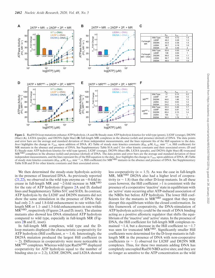
The ATP hydrolysis activity for wild type, L828F, andD829N D-loop mutant Pf Rad50 was measured in both full-length MR (Figure 2A, solid lines) and MRNBD complexes(Figure 2D, solid lines) by the colorimetric detection ofthe inorganic phosphate product. Although the KM for themutants was ∼2.3-fold larger than wild type full-length PfMR, the L828F and D829N mutations surprisingly showedfaster ATP hydrolysis rates than wild type Rad50 (Figure2C and Supplementary Table S1A). The catalytic efficien-cies (i.e. kcat/KM) of L828F and D829N were similar- and∼1.3-fold greater than wild type MR, respectively (Supple-mentary Table S1A). The effect of the disease-associated D-loop mutants was more pronounced for Pf MRNBD com-plexes. Again, ∼2.5-fold increases were observed for theKM values of both mutants, but significantly larger 6.5- and12.8-fold increases were found for the ATP hydrolysis rates,which produced 2.2- and 4.7-fold greater catalytic efficiencyfor L282F and D829N, respectively (Figures 2D and F andSupplementary Table S1B). For both forms of the complex,the differences were not due to changes in the affinity forATP as the KD for substrate binding was similar for wildtype and the mutants (Supplementary Figures S1A and Band Supplementary Table S1); instead, the differences inkcat/KM were driven by the greater kcat.
We also examined the effect on ATP hydrolysis of ala-nine substitutions at each of the D-loop mutation sites (Fig-ures 2B and E, solid lines). Generally, the L828A mutationyielded steady state ATP hydrolysis kinetics intermediate towild type and L828F for both full-length MR and MRNBD
complexes (Figure 2 and Supplementary Tables S1A andS1B). In contrast, the D829A mutation caused significantlyhigher catalytic rates for both full-length (3.8-fold; Figure2B and C and Supplementary Table S1A) and truncatedRad50s (8.7-fold; Figure 2E and F and Supplementary Ta-ble S1B), which resulted in the largest catalytic efficienciesof the D-loop mutants tested. Thus, these alanine mutationssuggest that the universally conserved leucine and aspartatein the ABC ATP D-loop are critical for proper ATP hydrol-ysis activity in Rad50.
Dow
nloaded from https://academ
ic.oup.com/nar/article/48/5/2457/5691220 by guest on 26 January 2022
2462 Nucleic Acids Research, 2020, Vol. 48, No. 5
L828A (--DNA)D829A (--DNA)
100
log[ATP] (μM)101 102
AT
Pas
e A
ctiv
ity(P
O4
MR
-1 h
r-1)
50
40
30
20
10
WT (--DNA)L828F (--DNA)D829N (--DNA)
A
2ATP + MRNBD → 2ADP + 2Pi + MRNBD
100
log[ATP] (μM)101 102
AT
Pas
e A
ctiv
ity(P
O4
MR
-1 h
r-1)
50
40
30
20
10
B
100
log[ATP] (μM)101 102
AT
Pas
e A
ctiv
ity
120
100
80
60
40
20
WT (--DNA)L828F (--DNA)D829N (--DNA)
WT L828F D829N
Vm
ax F
old
Cha
nge
8.0
6.0
4.0
2.0
D E
100
log[ATP] (μM)101 102
AT
Pas
e A
ctiv
ity
120
100
80
60
40
20
L828A (--DNA)D829A (--DNA)
WT L828F D829N
Vm
ax F
old
Cha
nge
8.0
6.0
4.0
2.0
L828A D829A
Vm
ax F
old
Cha
nge
8.0
6.0
4.0
2.0
L828A D829A
Vm
ax F
old
Cha
nge
8.0
6.0
4.0
2.0
(PO
4 M
RN
BD
-1
hr -1
)
(PO
4 M
RN
BD
-1
hr -1
)
KM kcat nWild Type 19.8 0.12 1.4
L828F 49.2 0.26 1.3D829N 43.9 0.36 1.4L828A 36.7 0.18 1.6D829A 39 0.46 2
Wild Type 63.6 0.93 1.4L828F 138 0.64 1D829N 99.6 0.64 1.1L828A 56.7 0.23 1.3D829A 46.6 0.91 1.6
Full length MR
Full length MR + DNA
KM kcat nWild Type 17 0.13 2.2
L828F 47 0.84 1.5D829N 44.6 1.66 1.5L828A 53.2 0.63 1.4D829A 24.5 1.13 1.8
Wild Type 33.3 0.26 1.7L828F 48.8 0.91 1.5D829N 66.4 2.15 1.4L828A 100 1.2 1D829A 30.3 1.79 1.1
MRNBD
MRNBD + DNA
2ATP + MR → 2ADP + 2Pi + MR 2ATP + MR → 2ADP + 2P
i + MR
2ATP + MRNBD → 2ADP + 2Pi + MRNBD
C
F
Figure 2. Rad50 D-loop mutations enhance ATP hydrolysis. (A and B) Steady-state ATP hydrolysis kinetics for wild type (green), L828F (orange), D829N(blue) (A), L828A (purple), and D829A (light blue) (B) full-length MR complexes in the absence (solid) and presence (dotted) of DNA. The data pointsand error bars are the average and standard deviation of three independent measurements, and the lines represent fits of the Hill equation to the data.Inset highlights the change in Vmax upon addition of DNA. (C) Table of steady state kinetics constants (KM, �M; kcat, min−1; n, Hill coefficient) forMR mutants in the absence and presence of DNA. See Supplementary Table S1A and C for other kinetic constants and their associated errors. (D andE) Steady-state ATP hydrolysis kinetics for wild type (green), L828F (orange), D829N (blue) (D), L828A (purple), and D829A (light blue) (E) truncatedMRNBD complexes in the absence (solid) and presence (dotted) of DNA. The data points and error bars are the average and standard deviation of threeindependent measurements, and the lines represent fits of the Hill equation to the data. Inset highlights the change in Vmax upon addition of DNA. (F) Tableof steady state kinetics constants (KM, �M; kcat, min−1; n, Hill coefficient) for MRNBD mutants in the absence and presence of DNA. See SupplementaryTable S1B and D for other kinetic constants and their associated errors.
We then determined the steady-state hydrolysis activityin the presence of linearized DNA. As previously reported(21,22), we observed in the wild type enzyme an ∼8-fold in-crease in full-length MR and ∼2-fold increase in MRNBD
for the rate of ATP hydrolysis (Figures 2A and D, dashedlines and Supplementary Tables S1C and S1D). In contrast,ATP hydrolysis by the L828F and D829N mutants did notshow the same stimulation in the presence of DNA: theyhad only 2.5- and 1.8-fold enhancement in rate within full-length MR or 1.1- and 1.3-fold enhancements in rate withinMRNBD, respectively (Figures 2A and D, inset). The alaninemutants also showed less DNA stimulated ATP hydrolysiscompared to wild type, especially in full-length MR (Fig-ures 2B and E, inset).
In full-length MR complexes, wild type and the D-loop mutants displayed the characteristic cooperativity forATP hydrolysis (Hill coefficient, n ∼ 1.4). Interestingly, theD829A mutation produced the largest Hill coefficient (n∼ 2). Differences in cooperativity were more noticeable inMRNBD complexes. Whereas wild type Rad50NBD displayedcooperativity for ATP hydrolysis equal to the number ofbinding sites (n = 2.2), L828F, D829N, and L828A showed
less cooperativity (n = 1.5). As was the case in full-lengthMR, MRNBD D829A also had a higher level of coopera-tivity (n ∼ 1.8) than the other D-loop mutants. In all thesecases however, the Hill coefficient >1 is consistent with thepresence of a cooperative ‘inactive’ state in equilibrium withan ‘active’ state occurring after ATP-induced association ofthe NBDs but before ATP hydrolysis. The lower Hill coef-ficients for the mutants in MRNBD suggest that they maydisrupt this equilibrium within the closed conformation. Inthis framework of cooperativity, the DNA-stimulation ofATP hydrolysis activity could be the result of DNA bindingacting as a positive allosteric regulator that shifts the equi-librium of the ‘inactive’ and ‘active’ states. In the presence ofDNA, the Hill coefficient for full-length MR complexes re-mained ∼1.4, but a decrease in the Hill coefficient to ∼1.7was seen for truncated MRNBD. Significantly smaller Hillcoefficients were determined for the D-loop mutants in full-length MR in the presence of DNA with non-cooperativecoefficients (n ∼ 1) observed for L828F and D829N MRcomplexes. Thus, for these two mutants adding DNA hascompletely uncoupled Rad50 NBD active sites; and they areno longer as sensitive to the ATP concentration as the wild
Dow
nloaded from https://academ
ic.oup.com/nar/article/48/5/2457/5691220 by guest on 26 January 2022
Nucleic Acids Research, 2020, Vol. 48, No. 5 2463
type. Furthermore, the D-loop MRNBD mutants showedno change or decreases in cooperativity in the presenceof DNA, suggesting that the disease-associated mutationshave already bypassed this step in allosteric regulation in thetruncated construct. Together, these data provided strongprimary evidence for the deregulation of Rad50 ATP hy-drolysis in the D-loop mutants.
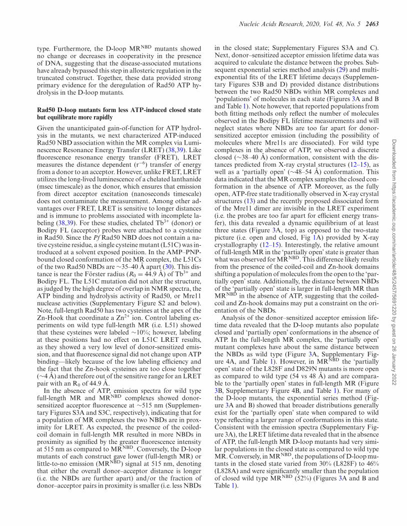
Rad50 D-loop mutants form less ATP-induced closed statebut equilibrate more rapidly
Given the unanticipated gain-of-function for ATP hydrol-ysis in the mutants, we next characterized ATP-inducedRad50 NBD association within the MR complex via Lumi-nescence Resonance Energy Transfer (LRET) (38,39). Likefluorescence resonance energy transfer (FRET), LRETmeasures the distance dependent (r−6) transfer of energyfrom a donor to an acceptor. However, unlike FRET, LRETutilizes the long-lived luminescence of a chelated lanthanide(msec timescale) as the donor, which ensures that emissionfrom direct acceptor excitation (nanoseconds timescale)does not contaminate the measurement. Among other ad-vantages over FRET, LRET is sensitive to longer distancesand is immune to problems associated with incomplete la-beling (38,39). For these studies, chelated Tb3+ (donor) orBodipy FL (acceptor) probes were attached to a cysteinein Rad50. Since the Pf Rad50 NBD does not contain a na-tive cysteine residue, a single cysteine mutant (L51C) was in-troduced at a solvent exposed position. In the AMP–PNP-bound closed conformation of the MR complex, the L51Csof the two Rad50 NBDs are ∼35–40 A apart (30). This dis-tance is near the Forster radius (R0 = 44.9 A) of Tb3+ andBodipy FL. The L51C mutation did not alter the structure,as judged by the high degree of overlap in NMR spectra, theATP binding and hydrolysis activity of Rad50, or Mre11nuclease activities (Supplementary Figure S2 and below).Note, full-length Rad50 has two cysteines at the apex of theZn-Hook that coordinate a Zn2+ ion. Control labeling ex-periments on wild type full-length MR (i.e. L51) showedthat these cysteines were labeled ∼10%; however, labelingat these positions had no effect on L51C LRET results,as they showed a very low level of donor-sensitized emis-sion, and that fluorescence signal did not change upon ATPbinding––likely because of the low labeling efficiency andthe fact that the Zn-hook cysteines are too close together(∼4 A) and therefore out of the sensitive range for an LRETpair with an R0 of 44.9 A.
In the absence of ATP, emission spectra for wild typefull-length MR and MRNBD complexes showed donor-sensitized acceptor fluorescence at ∼515 nm (Supplemen-tary Figures S3A and S3C, respectively), indicating that fora population of MR complexes the two NBDs are in prox-imity for LRET. As expected, the presence of the coiled-coil domain in full-length MR resulted in more NBDs inproximity as signified by the greater fluorescence intensityat 515 nm as compared to MRNBD. Conversely, the D-loopmutants of each construct gave lower (full-length MR) orlittle-to-no emission (MRNBD) signal at 515 nm, denotingthat either the overall donor–acceptor distance is longer(i.e. the NBDs are further apart) and/or the fraction ofdonor–acceptor pairs in proximity is smaller (i.e. less NBDs
in the closed state; Supplementary Figures S3A and C).Next, donor–sensitized acceptor emission lifetime data wasacquired to calculate the distance between the probes. Sub-sequent exponential series method analysis (29) and multi-exponential fits of the LRET lifetime decays (Supplemen-tary Figures S3B and D) provided distance distributionsbetween the two Rad50 NBDs within MR complexes and‘populations’ of molecules in each state (Figures 3A and Band Table 1). Note however, that reported populations fromboth fitting methods only reflect the number of moleculesobserved in the Bodipy FL lifetime measurements and willneglect states where NBDs are too far apart for donor-sensitized acceptor emission (including the possibility ofmolecules where Mre11s are dissociated). For wild typecomplexes in the absence of ATP, we observed a discreteclosed (∼38–40 A) conformation, consistent with the dis-tances predicted from X-ray crystal structures (12–15), aswell as a ‘partially open’ (∼48–54 A) conformation. Thisdata indicated that the MR complex samples the closed con-formation in the absence of ATP. Moreover, as the fullyopen, ATP-free state traditionally observed in X-ray crystalstructures (13) and the recently proposed dissociated formof the Mre11 dimer are invisible in the LRET experiment(i.e. the probes are too far apart for efficient energy trans-fer), this data revealed a dynamic equilibrium of at leastthree states (Figure 3A, top) as opposed to the two-statepicture (i.e. open and closed, Fig 1A) provided by X-raycrystallography (12–15). Interestingly, the relative amountof full-length MR in the ‘partially open’ state is greater thanwhat was observed for MRNBD. This difference likely resultsfrom the presence of the coiled-coil and Zn-hook domainsshifting a population of molecules from the open to the ‘par-tially open’ state. Additionally, the distance between NBDsof the ‘partially open’ state is larger in full-length MR thanMRNBD in the absence of ATP, suggesting that the coiled-coil and Zn-hook domains may put a constraint on the ori-entation of the NBDs.
Analysis of the donor–sensitized acceptor emission life-time data revealed that the D-loop mutants also populateclosed and ‘partially open’ conformations in the absence ofATP. In the full-length MR complex, the ‘partially open’mutant complexes have about the same distance betweenthe NBDs as wild type (Figure 3A, Supplementary Fig-ure 4A, and Table 1). However, in MRNBD the ‘partiallyopen’ state of the L828F and D829N mutants is more openas compared to wild type (54 vs 48 A) and are compara-ble to the ‘partially open’ states in full-length MR (Figure3B, Supplementary Figure 4B, and Table 1). For many ofthe D-loop mutants, the exponential series method (Fig-ure 3A and B) showed that broader distributions generallyexist for the ‘partially open’ state when compared to wildtype reflecting a larger range of conformations in this state.Consistent with the emission spectra (Supplementary Fig-ure 3A), the LRET lifetime data revealed that in the absenceof ATP, the full-length MR D-loop mutants had very simi-lar populations in the closed state as compared to wild typeMR. Conversely, in MRNBD, the populations of D-loop mu-tants in the closed state varied from 30% (L828F) to 46%(L828A) and were significantly smaller than the populationof closed wild type MRNBD (52%) (Figures 3A and B andTable 1).
Dow
nloaded from https://academ
ic.oup.com/nar/article/48/5/2457/5691220 by guest on 26 January 2022
2464 Nucleic Acids Research, 2020, Vol. 48, No. 5
100 200 300 400 500 600Time (sec)
Rel
ativ
e In
tens
ity
1.0
0.8
0.6
0.4
0.2
WT MRNBD
L828F MRNBD
D829N MRNBD
+ ATP
Less Closed More Closed
2 ATP
C
L828A MRNBD 8
4
WT LF DN LA
Relative
Equilibration R
ate
WT MR (--ATP)
L828F MR (--ATP)
D829N MR (--ATP)
A Partially OpenClosed
Tb3+ Tb3+
Bo Bo
Open(Invisible)
Tb3+
Bo
Dis
trib
utio
n In
tens
ity
55504540Distance (Å)
35 60
L828A MR (--ATP)
D829A MR (--ATP)
B Partially OpenClosed
Tb3+ Tb3+
Bo Bo
55504540Distance (Å)
35 60
Open(Invisible)
Tb3+
Bo
WT MRNBD (--ATP)
L828F MRNBD (--ATP)
D829N MRNBD (--ATP)
L828A MRNBD (--ATP)
D829A MRNBD (--ATP)
Dis
trib
utio
n In
tens
ity
Figure 3. Rad50 D-loop mutations form less ‘closed’ state. (A and B) Distance distributions derived from exponential series method analysis of the LRETacceptor intensity decays. Distributions of the apo and ATP-bound states are shown for distances between Tb3+ (donor) and Bodipy FL (Bo; acceptor)labeled wild type (green), L828F (orange), D829N (blue), L828A (purple), and D829A (light blue) Rad50 NBDs in full-length MR (A) or truncated MRNBD
(B) complexes. Vertical dashed lines represent the average distance between donor and acceptor (from three measurements; see Table 1) of the closed and‘partially open’ conformations for ATP-bound wild type MR. The schematic on top highlights that the closed (∼38 A) and ‘partially open’ (∼48–55 A)conformations are within the sensitive range of the LRET probes, while NBDs farther apart than ∼60 A (i.e. open conformations) are invisible. (C) LRETtime-based data where donor–sensitized acceptor intensity was measured after the addition of ATP to the indicated concentrations of MRNBD complexes.The intensities for mutant curves are scaled by a factor of 1.5 for graphical purposes. Inset compares the equilibration rates relative to wild type MR.
As expected, the addition of saturating ATP to wild typeMR shifted more molecules into the closed conformationas seen by the increase in the distribution intensity of theclosed state (Figures 3A and B, dashed lines, and Table 1). Incontrast, of the full-length D-loop mutants, only L828F andD829A showed moderate increases (∼50% of the changeseen for wild type) in the population of the closed stateupon ATP binding. In MRNBD, L828A showed a moderateincrease, but adding ATP to D829A changed the popula-tion of the closed to more than wild type. We suggest thatthe changes in populations for MRNBD are complicated bymolecules coming from the ‘open’ state (as seen in SAXS)into the ‘partially open’ state. Not surprisingly, the distancebetween NBDs in the closed state remained constant in allof the ATP-bound complexes. Consistent with this data,size exclusion chromatography on isolated Rad50 samplesrevealed that <5% of D-loop mutant Rad50 NBDs con-verted to a stable dimer upon addition of ATP as comparedto ∼30% for wild type (Supplementary Figure S1B). Thus,these results reveal a paradox for the D-loop mutations: in-creased ATP hydrolysis activity as compared to wild type,but a smaller relative population of the stable closed statefrom which ATP hydrolysis occurs.
To gain insight into the kinetics of ATP-induced Rad50NBD association, we monitored the increase in LRET sig-nal intensity over time after the addition of saturating ATP.These experiments provide kinetic information on Rad50NBD association and dissociation by measuring the timerequired to reach a new open-to-closed equilibrium in re-sponse to Mg·ATP binding (Figure 3C, top). As expectedfrom the population and lifetime measurements, the LRETintensity for the wild type MRNBD complex plateaued at ahigher value than that of the L828F, L828A and D829Nmutants, reflecting the greater population of ATP-inducedclosed state. The equilibration rate constants for ATP-induced open-to-closed equilibrium of full-length (80.3 s−1)and truncated MR (74.1 s−1) were similar. Unfortunately,
we were only able to measure equilibrium kinetics in full-length MR for the wild type complex, as the mutants didnot have sufficient change into the closed conformation orreached the new ATP-bound equilibrium too quickly tomeasure. Remarkably, in MRNBD we observed an increasedequilibration rate in the mutants, where L828F, D829N, andL828A had ∼4-, ∼8- and ∼4-times faster approach to equi-librium than wild type, respectively (Figure 3C, inset). Thisrate was even faster for D829A MRNBD, and it reached thenew equilibrium too quickly to accurately determine a rate.Under the conditions used here, the equilibration rate is thesum of the Mg·ATP-bound Rad50 association and dissocia-tion rates; therefore, the faster approach to equilibrium thatwe observe could be due to an increase in either rate or inboth rates. If only the association rate increased for the D-loop mutants, we would have observed a larger populationof the closed state. Therefore, given the lower populationsof closed MR in the mutants (Figure 3), these increasedequilibration rates are likely the result of faster Rad50 NBDdissociation. A strong correlation between Rad50 equilibra-tion rates in MRNBD and ATPase maximum velocities (R2
= 0.975; Supplementary Figure S4C) suggests that Rad50NBD dissociation is directly linked to the observed rate ofATP hydrolysis.
Mutations to the D-loop have disparate effects on Rad50structure and dynamics
To obtain higher resolution structural information, methyl-based NMR spectroscopy (34,40) was employed on the∼180 kDa MRNBD complexes. We used uniformly deuter-ated, side chain methyl group Ile�1-[13CH3], Leu�/Val� -[13CH3,12CD3], Metε-[13CH3] (ILVM)-labeled wild type,L828F, D829N, L828A and D829A Pf Rad50NBD in com-plex with unlabeled Mre11. The ILVM methyl group as-signments we previously determined on isolated Rad50(23) readily transferred to the MRNBD complex. Spec-
Dow
nloaded from https://academ
ic.oup.com/nar/article/48/5/2457/5691220 by guest on 26 January 2022
Nucleic Acids Research, 2020, Vol. 48, No. 5 2465
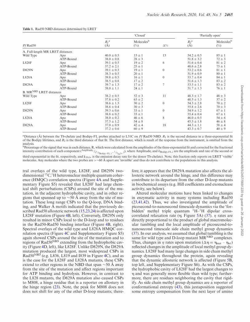
Table 1. Rad50 NBD distances determined by LRET
‘Closed’ ‘Partially open’
R1a Moleculesb R2
a Moleculesb
Pf Rad50 (A) (%) �% (A) (%)
A. Full-length MR LRET distancesWild Type Apo 40.0 ± 0.5 15 ± 1 13 54.2 ± 0.5 85 ± 1
ATP-Bound 38.0 ± 0.8 28 ± 3 51.8 ± 3.2 72 ± 3L828F Apo 39.1 ± 0.5 19 ± 2 6 51.6 ± 0.4 81 ± 2
ATP-Bound 37.2 ± 2.1 25 ± 1 48.6 ± 2.8 75 ± 1D829N Apo 39.1 ± 0.3 19 ± 1 0 53.2 ± 0.6 81 ± 1
ATP-Bound 38.3 ± 0.5 20 ± 1 51.9 ± 0.9 80 ± 1L828A Apo 39.8 ± 0.5 16 ± 1 0 53.7 ± 0.4 84 ± 1
ATP-Bound 38.5 ± 0.8 17 ± 2 51.6 ± 1.3 83 ± 2D829A Apo 39.7 ± 1.3 17 ± 2 7 53.5 ± 1.1 83 ± 2
ATP-Bound 38.0 ± 1.1 24 ± 1 51.7 ± 1.3 76 ± 1B. MRNBD LRET distancesWild Type Apo 38.2 ± 0.5 52 ± 3 11 48.3 ± 1.7 48 ± 3
ATP-Bound 37.8 ± 0.2 63 ± 2 48.5 ± 1.3 37 ± 2L828F Apo 38.6 ± 1.3 30 ± 2 0 54.3 ± 2.8 70 ± 2
ATP-Bound 38.6 ± 0.4 30 ± 3 55.8 ± 2.6 70 ± 3D829N Apo 39.3 ± 0.6 33 ± 1 0 54.9 ± 1.2 67 ± 1
ATP-Bound 38.8 ± 0.2 33 ± 1 55.4 ± 0.4 67 ± 1L828A Apo 38.0 ± 0.2 46 ± 6 8 46.0 ± 0.5 54 ± 6
ATP-Bound 37.5 ± 1.2 54 ± 8 45.3 ± 1.8 46 ± 8D829A Apo 37.0 ± 0.9 42 ± 4 18 44.3 ± 1.1 58 ± 4
ATP-Bound 37.2 ± 0.6 60 ± 9 43.3 ± 0.7 40 ± 9
aDistance (A) between the Tb-chelate and Bodipy-FL probes attached to L51C on Pf Rad50 NBD. R1 is the second distance in a three-exponential fitof the Bodipy lifetimes, and R2 is the third distance of that fit. The first distance, which is result of the response from the instrument, is omitted from theanalysis.bPercentage of the signal that was in each distance, R, which were calculated from the amplitudes of the three-exponential fit and corrected for the fractionalintensity contribution of each component (Amplitudei /(1/kBodipy−FL, i − 1/k
Tb3+ )), where Amplitudei and kBodipy-FL,i are the amplitude and rate of the second or
third exponential in the fit, respectively, and kTb3+ is the emission decay rate for the donor Tb-chelate). Note, this fraction only reports on LRET ‘visible’molecules. Any molecules where the two probes are >∼60 A apart are ‘invisible’ and thus do not contribute to the populations in this analysis.
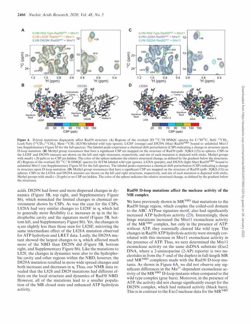
tral overlays of the wild type, L828F, and D829N two-dimensional 13C,1H heteronuclear multiple quantum coher-ence (HMQC) correlation spectra (Figure 4A and Supple-mentary Figure S5) revealed that L828F had large chem-ical shift perturbations (CSPs) around the site of the mu-tation, in the adjacent hydrophobic cavity, and in other re-gions that spanned up to ∼50 A away from the site of mu-tation. These long range CSPs to the Q-loop, DNA bind-ing, and Walker A motifs indicated that the previously de-scribed Rad50 allosteric network (15,23,24) is affected uponL828F mutation (Figure 4B, left). Conversely, D829N onlyresulted in minor CSPs local to the D-loop and to residuesin the Rad50-Rad50 binding interface (Figure 4B, right).Spectral overlays of the wild type and L828A HMQC cor-relation spectra (Figure 4C and Supplementary Figure S5)again showed CSPs around the site of the mutation and toregions of Rad50NBD extending from the hydrophobic cav-ity (Figure 4D, left), like L828F. Unlike D829N, the D829Amutation produced the largest, most widespread CSPs inRad50NBD (e.g. L836, L819 and I839 in Figure 4C), and asis the case for the L828F and L828A mutants, these CSPsextend to other regions in the NBD that span ∼50 A awayfrom the site of the mutation and affect regions importantfor ATP binding and hydrolysis. However, in contrast tothe L828 mutants, the D829A mutation also caused CSPsto M808, a hinge residue that is a reporter on allostery inthe hinge region (23). Note, the peak for M808 does notdoes not shift for any of the other D-loop mutants; there-
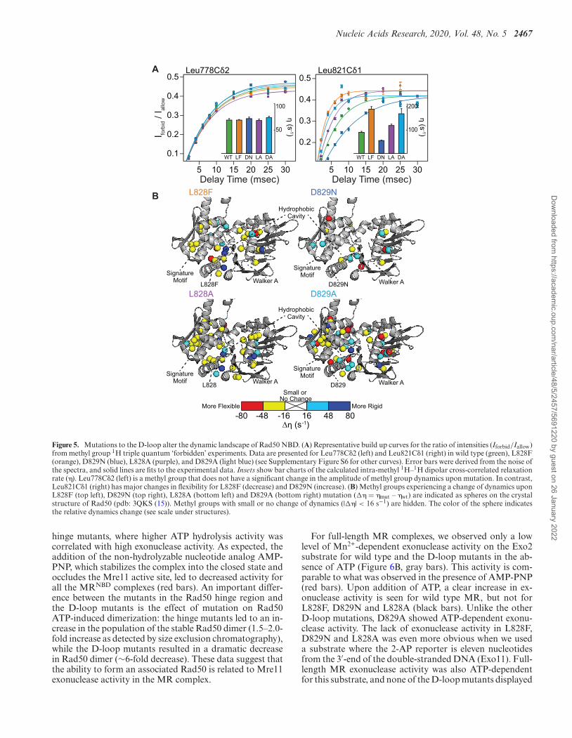
fore, it appears that the D829A mutation also affects the al-losteric network around the hinge, and this difference maycontribute to its departure from the other D-loop mutantsin biochemical assays (e.g. Hill coefficients and exonucleaseactivity, see below).
Altered molecular motions have been linked to changesin enzymatic activity in many systems including Rad50(23,41,42). Thus, we also investigated the amplitude ofpicosecond-to-nanosecond timescale dynamics via the ‘for-bidden’ methyl triple quantum 1H–1H dipolar cross-correlated relaxation rate (�; Figure 5A) (37). η rates aredirectly proportional to the product of global macromolec-ular tumbling and the local amplitude of picosecond-to-nanosecond timescale side chain methyl group dynamics(37). In our analysis, we assumed that global tumbling is thesame for wild type and D-loop mutant MRNBD complexes.Thus, changes in η rates upon mutation (�� = �mut – �wt)reflected changes in the amplitude of local methyl group dy-namics. L828F had many large changes in side chain methylgroup dynamics throughout the protein, again revealingthat the dynamic allosteric network is affected (Figure 5B,top left, and Supplementary Figure S6). As with the CSPs,the hydrophobic cavity of L828F had the largest changes to� and was generally more flexible than wild type; further-more, there are residues neighboring the cavity that rigid-ify. As side chain methyl group dynamics are a reporter ofconformational entropy (43), this juxtaposition suggestedsome entropy compensation from the next layer of amino
Dow
nloaded from https://academ
ic.oup.com/nar/article/48/5/2457/5691220 by guest on 26 January 2022
2466 Nucleic Acids Research, 2020, Vol. 48, No. 5
D
C
20 50 70 90 110 130 150CSP (ppb)
L828 D829
HydrophobicCavity
L828A D829A
SignatureMotif Walker A
SignatureMotif
Walker A
ILVM Wild Type Rad50NBD + Mre11ILVM L828A Rad50NBD + Mre11ILVM D829A Rad50NBD + Mre11
13C
(pp
m)
1H (ppm)1.2 1.0 0.8 0.6 0.8 0.6
23
24 14
13
12
1H (ppm)
L819
L836
L51
L859
L802
L806
I138
I816
I29
V850
0.4
I839
B
A
20 50 70 90 110 130 150CSP (ppb)
L828F D829N
HydrophobicCavity
L828F D829N
SignatureMotif Walker A
SignatureMotif
Walker A
ILVM Wild Type Rad50NBD + Mre11ILVM L828F Rad50NBD + Mre11ILVM D829N Rad50NBD + Mre11
13C
(pp
m)
1H (ppm)1.2 1.0 0.8 0.6 0.8 0.6
23
24L819
L836
L51
L859
L802
L806
14
13
12
V850
1H (ppm)0.4
I138
I816
I29
I839
Figure 4. D-loop mutations disparately affect Rad50 structure. (A) Regions of the overlaid 2D 13C,1H HMQC spectra for U-2H12C, Ile�1–13CH3,Leu�/Val� -[12CD3/
13CH3], Metε-13CH3 (ILVM)-labeled wild type (green), L828F (orange) and D829N (blue) Rad50NBD bound to unlabeled Mre11(see Supplementary Figure S5 for the full spectra). The labeled peaks experience a chemical shift perturbation (CSP) indicating a change in structure uponD-loop mutation. (B) Methyl group resonances that have a significant CSP are mapped on the structure of Rad50 (pdb: 3QKS (15)) as spheres. CSPs inthe L828F and D829N mutants are shown on the left and right structures, respectively, and site of each mutation is depicted with sticks. Methyl groupswith small (<20 ppb) or no CSP are hidden. The color of the sphere indicates the relative structural change, as defined by the gradient below the structures.(C) Regions of the overlaid 2D 13C,1H HMQC spectra for ILVM-labeled wild type (green), L828A (purple), and D829A (light blue) Rad50NBD bound tounlabeled Mre11 (see Supplementary Figure S5 for the full spectra). The labeled peaks experience a chemical shift perturbation (CSP) indicating a changein structure upon D-loop mutation. (D) Methyl group resonances that have a significant CSP are mapped on the structure of Rad50 (pdb: 3QKS (15)) asspheres. CSPs in the L828A and D829A mutants are shown on the left and right structures, respectively, and site of each mutation is depicted with sticks.Methyl groups with small (<20 ppb) or no CSP are hidden. The color of the sphere indicates the relative structural change, as defined by the gradient belowthe structures.
acids. D829N had fewer and more dispersed changes in dy-namics (Figure 5B, top right, and Supplementary FigureS6), which mimicked the limited changes in chemical en-vironment shown by CSPs. As was the case for the CSPs,L828A had very similar changes to L828F in �, which ledto generally more flexibility (i.e. increases in �) in the hy-drophobic cavity and the signature motif (Figure 5B, bot-tom left, and Supplementary Figure S6). Yet, the changes in� are slightly less than those seen for L828F, mirroring thesame intermediate effect of the L828A mutation observedfor ATP hydrolysis and LRET data. Lastly, the D829A mu-tant showed the largest changes to �, which affected muchmore of the NBD than D829N did (Figure 5B, bottomright, and Supplementary Figure S6). Like the mutations toL828, the changes in dynamics were also to the hydropho-bic cavity and other regions within the NBD; however, theD829A mutation resulted in more wide spread changes andboth increases and decreases in �. Thus, our NMR data re-vealed that the L828 and D829 mutations had different ef-fects on the local structure and dynamics of Rad50 NBD.However, all of the mutations lead to a smaller popula-tion of the MR closed state and enhanced ATP hydrolysisactivity.
Rad50 D-loop mutations affect the nuclease activity of theMR complex
We have previously shown in MRNBD that mutations to theRad50 hinge region, which couples the coiled-coil domainto the ABC ATPase signature motif, also had significantlyincreased ATP hydrolysis activity (23). Interestingly, thesehinge mutations increased the Mre11 exonuclease activityof the MRNBD complex, but only in the presence of ATP:without ATP, they essentially cleaved like wild type. Thechanges in Rad50 ATP hydrolysis activity were strongly cor-related with this increase in Mre11 exonuclease activity inthe presence of ATP. Thus, we next determined the Mre11exonuclease activity on the same dsDNA substrate (Exo2DNA, where a 2-aminopurine (2-AP) reporter is two nu-cleotides in from the 3′-end of the duplex) in full-length MRand MRNBD complexes made with the Rad50 D-loop mu-tants. As shown in Figure 6A, we did not observe any sig-nificant differences in the Mn2+-dependent exonuclease ac-tivity of the MRNBD D-loop mutants when compared to thewild type complex (gray bars). Moreover, in the presence ofATP, the activity did not change significantly except for theD829N complex, which had reduced activity (black bars).This is in contrast to the Exo2 nuclease data for the MRNBD
Dow
nloaded from https://academ
ic.oup.com/nar/article/48/5/2457/5691220 by guest on 26 January 2022
Nucleic Acids Research, 2020, Vol. 48, No. 5 2467
Leu778Cδ2 Leu821Cδ1
5 10 15 20 25 30
0.5
0.4
0.3
0.2
0.1
5 10 15 20 25 30Delay Time (msec) Delay Time (msec)
I forb
id /
I allo
w
B
A
-80 -48 -16 16 48 80∆η (s-1)
More RigidMore Flexible
Small or No Change
L828F D829N
HydrophobicCavity
L828F D829N
SignatureMotif Walker A
SignatureMotif
Walker A
L828A D829A
η (s-1)
100
50
WT LF DN LA DA
η (s-1)
200
100
WT LF DN LA DA
0.5
0.4
0.3
0.2
L828 D829
HydrophobicCavity
SignatureMotif Walker A
SignatureMotif
Walker A
Figure 5. Mutations to the D-loop alter the dynamic landscape of Rad50 NBD. (A) Representative build up curves for the ratio of intensities (Iforbid/Iallow)from methyl group 1H triple quantum ‘forbidden’ experiments. Data are presented for Leu778C�2 (left) and Leu821C�1 (right) in wild type (green), L828F(orange), D829N (blue), L828A (purple), and D829A (light blue) (see Supplementary Figure S6 for other curves). Error bars were derived from the noise ofthe spectra, and solid lines are fits to the experimental data. Insets show bar charts of the calculated intra-methyl 1H–1H dipolar cross-correlated relaxationrate (�). Leu778C�2 (left) is a methyl group that does not have a significant change in the amplitude of methyl group dynamics upon mutation. In contrast,Leu821C�1 (right) has major changes in flexibility for L828F (decrease) and D829N (increase). (B) Methyl groups experiencing a change of dynamics uponL828F (top left), D829N (top right), L828A (bottom left) and D829A (bottom right) mutation (�� = �mut – �wt) are indicated as spheres on the crystalstructure of Rad50 (pdb: 3QKS (15)). Methyl groups with small or no change of dynamics (|��| < 16 s−1) are hidden. The color of the sphere indicatesthe relative dynamics change (see scale under structures).
hinge mutants, where higher ATP hydrolysis activity wascorrelated with high exonuclease activity. As expected, theaddition of the non-hydrolyzable nucleotide analog AMP-PNP, which stabilizes the complex into the closed state andoccludes the Mre11 active site, led to decreased activity forall the MRNBD complexes (red bars). An important differ-ence between the mutants in the Rad50 hinge region andthe D-loop mutants is the effect of mutation on Rad50ATP-induced dimerization: the hinge mutants led to an in-crease in the population of the stable Rad50 dimer (1.5–2.0-fold increase as detected by size exclusion chromatography),while the D-loop mutants resulted in a dramatic decreasein Rad50 dimer (∼6-fold decrease). These data suggest thatthe ability to form an associated Rad50 is related to Mre11exonuclease activity in the MR complex.
For full-length MR complexes, we observed only a lowlevel of Mn2+-dependent exonuclease activity on the Exo2substrate for wild type and the D-loop mutants in the ab-sence of ATP (Figure 6B, gray bars). This activity is com-parable to what was observed in the presence of AMP-PNP(red bars). Upon addition of ATP, a clear increase in ex-onuclease activity is seen for wild type MR, but not forL828F, D829N and L828A (black bars). Unlike the otherD-loop mutations, D829A showed ATP-dependent exonu-clease activity. The lack of exonuclease activity in L828F,D829N and L828A was even more obvious when we useda substrate where the 2-AP reporter is eleven nucleotidesfrom the 3′-end of the double-stranded DNA (Exo11). Full-length MR exonuclease activity was also ATP-dependentfor this substrate, and none of the D-loop mutants displayed
Dow
nloaded from https://academ
ic.oup.com/nar/article/48/5/2457/5691220 by guest on 26 January 2022
2468 Nucleic Acids Research, 2020, Vol. 48, No. 5
F
BA C
0
500
1000
1500
2000No Mn Mn ATP AMP-PNP
WTMR
L828FMR
D829NMR
L828AMR
D829AMR
Re
lativ
e F
luo
resc
en
ce U
nits
0
500
1000
1500
2000
2500No Mn Mn ATP AMP-PNP
Re
lativ
e F
luo
resc
en
ce U
nits
WTMR
L828FMR
D829NMR
L828AMR
D829AMR
Re
lativ
e F
luo
resc
en
ce U
nits
0
500
1000
1500
2000
2500
3000
WTMRNBD
L828FMRNBD
D829NMRNBD
L828AMRNBD
D829AMRNBD
No Mn Mn ATP AMP-PNP
11-AP3’
5’
5’
3’
2-AP3’
5’
5’
3’
2-AP3’
5’
5’
3’
D WTMR
L51CMR
L828FMR
D829NMR
L828AMR
D829AMR
5 10 20 30 5 10 20 30 5 10 20 30 5 10 20 30 5 10 20 30 5 10 20 30 minNo
Mn2+
No
Mn2+
No
Mn2+
No
Mn2+
No
Mn2+
No
Mn2+
E WTMRNBD
L828FMRNBD
L828AMRNBD
D829NMRNBD
D829AMRNBD
L51CMRNBD
5 10 20 30 5 10 20 30 5 10 20 30 5 10 20 30 5 10 20 30 5 10 20 30 minNo
Mn2+
No
Mn2+
No
Mn2+
No
Mn2+
No
Mn2+
No
Mn2+
10 kb
Linear
Multimer
DN
A
Mre
11
Rad
50
WT
MR
L828
F M
R
D82
9N M
R
L828
A M
R
D82
9A M
R
L51C
MR
G
10 kb
Linear
Multimer
DN
A
Mre
11
Rad
50N
BD
WT
MR
NB
D
L828
F M
RN
BD
D82
9N M
RN
BD
L828
A M
RN
BD
D82
9A M
RN
BD
L51C
MR
NB
D
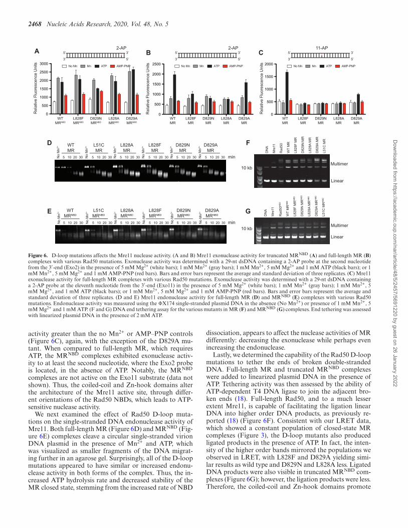
Figure 6. D-loop mutations affects the Mre11 nuclease activity. (A and B) Mre11 exonuclease activity for truncated MRNBD (A) and full-length MR (B)complexes with various Rad50 mutations. Exonuclease activity was determined with a 29-nt dsDNA containing a 2-AP probe at the second nucleotidefrom the 3′-end (Exo2) in the presence of 5 mM Mg2+ (white bars); 1 mM Mn2+ (gray bars); 1 mM Mn2+, 5 mM Mg2+ and 1 mM ATP (black bars); or 1mM Mn2+, 5 mM Mg2+ and 1 mM AMP-PNP (red bars). Bars and error bars represent the average and standard deviation of three replicates. (C) Mre11exonuclease activity for full-length MR complexes with various Rad50 mutations. Exonuclease activity was determined with a 29-nt dsDNA containinga 2-AP probe at the eleventh nucleotide from the 3′-end (Exo11) in the presence of 5 mM Mg2+ (white bars); 1 mM Mn2+ (gray bars); 1 mM Mn2+, 5mM Mg2+, and 1 mM ATP (black bars); or 1 mM Mn2+, 5 mM Mg2+ and 1 mM AMP-PNP (red bars). Bars and error bars represent the average andstandard deviation of three replicates. (D and E) Mre11 endonuclease activity for full-length MR (D) and MRNBD (E) complexes with various Rad50mutations. Endonuclease activity was measured using the �X174 single-stranded plasmid DNA in the absence (No Mn2+) or presence of 1 mM Mn2+, 5mM Mg2+ and 1 mM ATP. (F and G) DNA end tethering assay for the various mutants in MR (F) and MRNBD (G) complexes. End tethering was assessedwith linearized plasmid DNA in the presence of 2 mM ATP.
activity greater than the no Mn2+ or AMP–PNP controls(Figure 6C), again, with the exception of the D829A mu-tant. When compared to full-length MR, which requiresATP, the MRNBD complexes exhibited exonuclease activ-ity to at least the second nucleotide, where the Exo2 probeis located, in the absence of ATP. Notably, the MRNBD
complexes are not active on the Exo11 substrate (data notshown). Thus, the coiled-coil and Zn-hook domains alterthe architecture of the Mre11 active site, through differ-ent orientations of the Rad50 NBDs, which leads to ATP-sensitive nuclease activity.
We next examined the effect of Rad50 D-loop muta-tions on the single-stranded DNA endonuclease activity ofMre11. Both full-length MR (Figure 6D) and MRNBD (Fig-ure 6E) complexes cleave a circular single-stranded virionDNA plasmid in the presence of Mn2+ and ATP, whichwas visualized as smaller fragments of the DNA migrat-ing further in an agarose gel. Surprisingly, all of the D-loopmutations appeared to have similar or increased endonu-clease activity in both forms of the complex. Thus, the in-creased ATP hydrolysis rate and decreased stability of theMR closed state, stemming from the increased rate of NBD
dissociation, appears to affect the nuclease activities of MRdifferently: decreasing the exonuclease while perhaps evenincreasing the endonuclease.
Lastly, we determined the capability of the Rad50 D-loopmutations to tether the ends of broken double-strandedDNA. Full-length MR and truncated MRNBD complexeswere added to linearized plasmid DNA in the presence ofATP. Tethering activity was then assessed by the ability ofATP-dependent T4 DNA ligase to join the adjacent bro-ken ends (18). Full-length Rad50, and to a much lesserextent Mre11, is capable of facilitating the ligation linearDNA into higher order DNA products, as previously re-ported (18) (Figure 6F). Consistent with our LRET data,which showed a constant population of closed-state MRcomplexes (Figure 3), the D-loop mutants also producedligated products in the presence of ATP. In fact, the inten-sity of the higher order bands mirrored the populations weobserved in LRET, with L828F and D829A yielding simi-lar results as wild type and D829N and L828A less. LigatedDNA products were also visible in truncated MRNBD com-plexes (Figure 6G); however, the ligation products were less.Therefore, the coiled-coil and Zn-hook domains promote
Dow
nloaded from https://academ
ic.oup.com/nar/article/48/5/2457/5691220 by guest on 26 January 2022
Nucleic Acids Research, 2020, Vol. 48, No. 5 2469
end-tethering activities, which occurs through their DNAbinding ability (18).
DISCUSSION
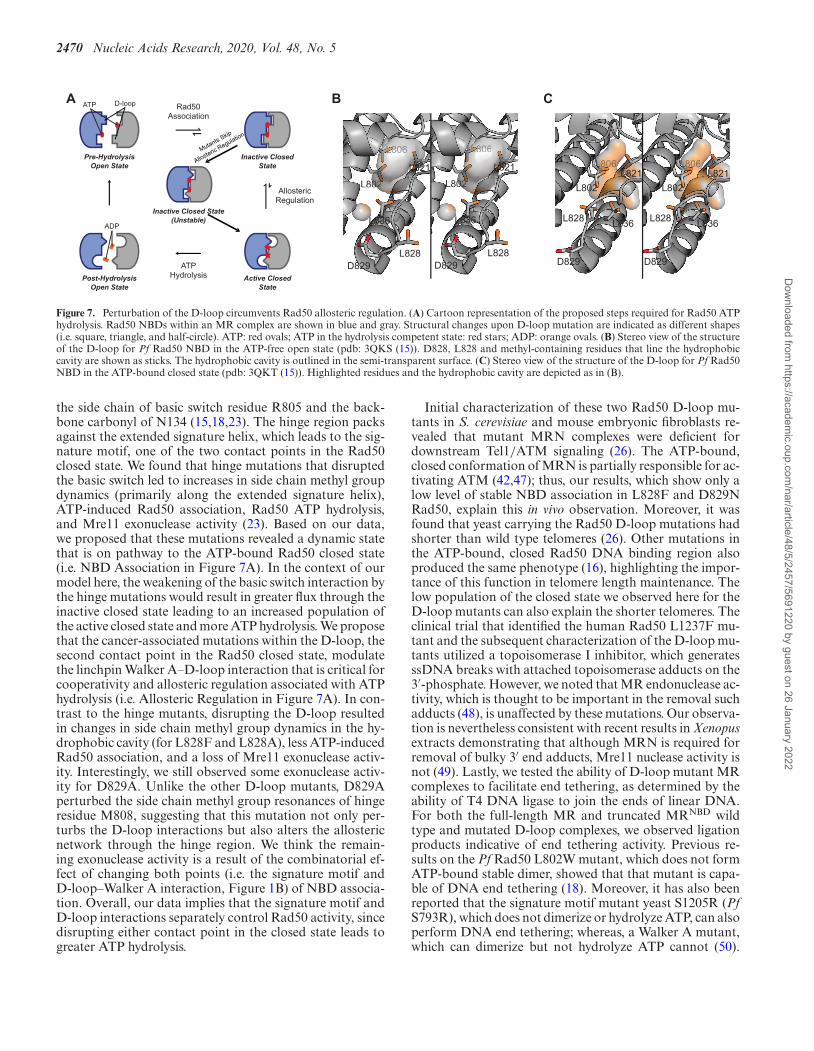
Figure 7A describes our model for the structural and dy-namic mechanism of ATP hydrolysis in wild type Rad50:(i) ATP binding, (ii) Rad50 NBD association, (iii) cooper-ative structure change subject to allosteric regulation and(iv) ATP hydrolysis and Rad50 NBD and product (ADP +Pi) dissociation. With a KD for ATP of ∼3–5 �M, cellularconcentrations of ATP (∼1–10 mM) produce fully boundRad50·Mg·ATP, therein promoting Rad50 NBD associa-tion into a population of closed and ‘partially open’ states.Based on crystal structures of AMP-PNP-bound closed PfRad50 NBD (15), D829 (human D1237) in the D-loopforms an interaction with the Walker A motif of the trans-Rad50 NBD (Figures 1 and 7A). We hypothesize this linch-pin interaction is required to form the stable closed stateand transmit the cooperative structural changes required toallosterically regulate MR ATP hydrolysis. Consistent withthis idea, recent molecular dynamics simulations revealed astable ATP-bound, closed state of MR from which alloster-ically modulated cooperative hydrolysis could occur (42).Furthermore, it has been found that the D-loop – WalkerA interaction of the ABC transporter associated with anti-gen processing, TAP, was necessary to couple unidirectionalpeptide substrate transport with ATP hydrolysis (44). Thus,we propose that the Rad50 D-loop–Walker A interaction inMR increases the population of an ‘inactive’ closed state, re-sulting in slow ATP hydrolysis. Rad50 is a notoriously slowATPase (18,45), and wild type Pf MR only performs ATPhydrolysis once every ∼8.3 min in vitro; however, we andothers show that the rate of hydrolysis increases in the pres-ence of DNA (21, 22). Thus, DNA binding likely acts as anallosteric activator increasing the population of an ‘active’closed state. ATP hydrolysis and Rad50 NBD dissociationthen occur from this ‘active’ closed state.
The enhanced ATP hydrolysis rates, decreases in coop-erativity, lower populations of the ATP-bound MR stableclosed state, and the likely faster Rad50 NBD dissociationall suggest that the rate limiting step of Rad50 coopera-tive structural changes is being circumvented in these twoD-loop mutants. We show that L828F reorients the struc-tural and dynamic landscape within a Rad50 protomer andtherefore hypothesize that L828F shifts the ‘inactive’ to‘active’ closed state equilibrium toward the ATP hydrol-ysis competent ‘active’ form. Ile138, Ile143, Leu802 andLeu836 methyl groups, which line the hydrophobic cav-ity (Figure 7B), experience the largest CSPs; therefore, wethink that the mutated F828 side chain alters the orienta-tion of the D-loop resulting in a conformation of the hy-drophobic cavity and D-loop that mimics the ATP-bound‘active’ form, pre-emptively orienting the Rad50 active sitesfor ATP hydrolysis, and prohibiting the formation of thestable ‘closed’ Rad50 NBDs (Figure 7A). The importanceof the hydrophobic cavity in Rad50 was previously shownwith structural and functional studies of a L802W (PfRad50) mutation. This mutation, which puts a bulky in-dole side chain into the top of the hydrophobic cavity, alsodisplayed an increase in kcat for ATP hydrolysis and de-
crease in ATP-induced Rad50 association (18). Moreover,the L802W mutation resulted in increased mobility of theD-loop, as judged by crystallographic temperature-factors,in line with our NMR data connecting the D-loop andhydrophobic cavity in L828F and our model for the D-loop forming a linchpin interaction. Finally, cooperativityin L828F is reduced between active sites because each arealready primed for catalysis. As such, the mutant is notas sensitive to allosteric modulation by DNA. In contrastto L828F, D829N does not show significant structural ordynamics changes, suggesting a different mechanism forcircumventing allosteric regulation. As the D-loop holdsRad50 in the ‘inactive’ state, we propose that the D829Nmutation only weakly forms the Walker A–D-loop interac-tion resulting in unstable ‘inactive’ closed Rad50. This ideais consistent with an aspartate to alanine mutation in theTAP ABC ATPase: there, the D-loop mutation uncoupledunidirectional transport of substrate peptide from ATP hy-drolysis, and the ATP hydrolysis activity of the mutationwas not stimulated by peptide (44), similar to what was ob-served here. Thus, the weak D-loop interaction in Rad50D829N allows the enzyme to readily proceed to ATP hy-drolysis bypassing the cooperative structural change (Fig-ure 7A). Since the ‘inactive’ closed Rad50 is destabilized,cooperativity is also reduced between active sites of D892N,and the mutant is also not sensitive to stimulation by DNA.
Generally, compared to L828F, the L828A mutation wasnot as active in ATP hydrolysis, not as stimulated by DNA,and had similar response to ATP-induced dimerization(i.e. did not appreciably shift the population to the closedstate). L828A produced changes in structure and dynam-ics of the NBD to the same regions as the L828F mutation.Thus, this mutation also alters the conformation of the D-loop and nearby hydrophobic cavity. We propose thereforethat the alanine mutation acts in the same way as the L828Fmutation: changing the conformation of the D-loop suchthat the stable ‘closed’ of the enzyme cannot form. Finally,the D829A mutation had the highest level of ATP hydrolysisin the absence of DNA and forms the broadest distributionof ‘partially open’ state. Moreover, the D829A mutation hadthe largest and most widespread changes to the Rad50 NBDstructure and dynamics. In the wild type structure, D829might acts as a helical N-capping residue (Figures 1 and7B). Mutation to asparagine does not significantly alter freeenergy for N-capping, but mutation to alanine does (46).Altered stabilization of this alpha helix could lead to thewide spread changes in structure and dynamics that we seefor this mutant. Further, the alanine mutation completelyremoves all hydrogen bond accepting groups from the con-served aspartate, in contrast to the asparagine mutant thatcan still accept a hydrogen bond on the carbonyl group,which would disrupt the interaction with the trans-WalkerA motif. Thus, we propose that the changes to alpha he-lix capping leads to an even weaker D-loop interaction inD829A bypassing the rate-limiting cooperative structuralchange in Rad50.
We have previously characterized the structure, dynam-ics, Rad50 ATP hydrolysis, and Mre11 exonuclease activ-ity for a number of mutations to the hinge region of PfRad50 (R805E, V156M and V160M) – a motif that in theATP-free state is stabilized by an ionic interaction between
Dow
nloaded from https://academ
ic.oup.com/nar/article/48/5/2457/5691220 by guest on 26 January 2022
2470 Nucleic Acids Research, 2020, Vol. 48, No. 5
ATPHydrolysis
AllostericRegulation
ADP
Post-HydrolysisOpen State
D-loopATP
Pre-HydrolysisOpen State
Inactive ClosedState
Active ClosedState
Rad50Association
Mutants Skip
Allosteric Regulation
Inactive Closed State(Unstable)
A B
L802
L806L806
L802 L8000000000L800000000L 0L 000000L 0222222222222222222222222222222
LLLLLL8L8L8L80LLL80LLLLLLL8L80LLLLLLL8L8L80LLLLLLLLL8LLL8LLLLL8L80LLLLLL8880LL88080LLLLLLLLLLLLLLLLLLLLLL 66666666L8LL8L80L80LLL8L88LLLLL8L80L80LLLLL8L8L8LLLLL8L8LLLLL8L80LLLL88LLLLLL88L80LLL88L8L80LL8L8L8LLLLLL8L80LLLLL8LLLLLLLLLLLLLLLLLLLLLL 66666666
L800000000000000000000000000000222222222222222222222222
L828D829
L836
L821
L828D829
L836
L821
C
L806 L806LLLLLLLLLLLLLL8880LLL8LLL80LLLLLLLLLLL8LLLLLL8LLLLLLLLLLLL8LLLLLL8LLLLLLL8LLLLLLLLLLLL88LLLLLL88008000088800006666666 LLLLLLLLLLL888880LLLL880LLL8LLLL8LLL8LLL8LLLL8LLL88LLLLL8LLLLLL88LLLLLLL8LLLLLLLL8LLLLLLL88LLLLLLL8800000808888000666666
L802
L828
D829
L836
L821
L802
L828
D829
L836
L821
Figure 7. Perturbation of the D-loop circumvents Rad50 allosteric regulation. (A) Cartoon representation of the proposed steps required for Rad50 ATPhydrolysis. Rad50 NBDs within an MR complex are shown in blue and gray. Structural changes upon D-loop mutation are indicated as different shapes(i.e. square, triangle, and half-circle). ATP: red ovals; ATP in the hydrolysis competent state: red stars; ADP: orange ovals. (B) Stereo view of the structureof the D-loop for Pf Rad50 NBD in the ATP-free open state (pdb: 3QKS (15)). D828, L828 and methyl-containing residues that line the hydrophobiccavity are shown as sticks. The hydrophobic cavity is outlined in the semi-transparent surface. (C) Stereo view of the structure of the D-loop for Pf Rad50NBD in the ATP-bound closed state (pdb: 3QKT (15)). Highlighted residues and the hydrophobic cavity are depicted as in (B).
the side chain of basic switch residue R805 and the back-bone carbonyl of N134 (15,18,23). The hinge region packsagainst the extended signature helix, which leads to the sig-nature motif, one of the two contact points in the Rad50closed state. We found that hinge mutations that disruptedthe basic switch led to increases in side chain methyl groupdynamics (primarily along the extended signature helix),ATP-induced Rad50 association, Rad50 ATP hydrolysis,and Mre11 exonuclease activity (23). Based on our data,we proposed that these mutations revealed a dynamic statethat is on pathway to the ATP-bound Rad50 closed state(i.e. NBD Association in Figure 7A). In the context of ourmodel here, the weakening of the basic switch interaction bythe hinge mutations would result in greater flux through theinactive closed state leading to an increased population ofthe active closed state and more ATP hydrolysis. We proposethat the cancer-associated mutations within the D-loop, thesecond contact point in the Rad50 closed state, modulatethe linchpin Walker A–D-loop interaction that is critical forcooperativity and allosteric regulation associated with ATPhydrolysis (i.e. Allosteric Regulation in Figure 7A). In con-trast to the hinge mutants, disrupting the D-loop resultedin changes in side chain methyl group dynamics in the hy-drophobic cavity (for L828F and L828A), less ATP-inducedRad50 association, and a loss of Mre11 exonuclease activ-ity. Interestingly, we still observed some exonuclease activ-ity for D829A. Unlike the other D-loop mutants, D829Aperturbed the side chain methyl group resonances of hingeresidue M808, suggesting that this mutation not only per-turbs the D-loop interactions but also alters the allostericnetwork through the hinge region. We think the remain-ing exonuclease activity is a result of the combinatorial ef-fect of changing both points (i.e. the signature motif andD-loop–Walker A interaction, Figure 1B) of NBD associa-tion. Overall, our data implies that the signature motif andD-loop interactions separately control Rad50 activity, sincedisrupting either contact point in the closed state leads togreater ATP hydrolysis.
Initial characterization of these two Rad50 D-loop mu-tants in S. cerevisiae and mouse embryonic fibroblasts re-vealed that mutant MRN complexes were deficient fordownstream Tel1/ATM signaling (26). The ATP-bound,closed conformation of MRN is partially responsible for ac-tivating ATM (42,47); thus, our results, which show only alow level of stable NBD association in L828F and D829NRad50, explain this in vivo observation. Moreover, it wasfound that yeast carrying the Rad50 D-loop mutations hadshorter than wild type telomeres (26). Other mutations inthe ATP-bound, closed Rad50 DNA binding region alsoproduced the same phenotype (16), highlighting the impor-tance of this function in telomere length maintenance. Thelow population of the closed state we observed here for theD-loop mutants can also explain the shorter telomeres. Theclinical trial that identified the human Rad50 L1237F mu-tant and the subsequent characterization of the D-loop mu-tants utilized a topoisomerase I inhibitor, which generatesssDNA breaks with attached topoisomerase adducts on the3′-phosphate. However, we noted that MR endonuclease ac-tivity, which is thought to be important in the removal suchadducts (48), is unaffected by these mutations. Our observa-tion is nevertheless consistent with recent results in Xenopusextracts demonstrating that although MRN is required forremoval of bulky 3′ end adducts, Mre11 nuclease activity isnot (49). Lastly, we tested the ability of D-loop mutant MRcomplexes to facilitate end tethering, as determined by theability of T4 DNA ligase to join the ends of linear DNA.For both the full-length MR and truncated MRNBD wildtype and mutated D-loop complexes, we observed ligationproducts indicative of end tethering activity. Previous re-sults on the Pf Rad50 L802W mutant, which does not formATP-bound stable dimer, showed that that mutant is capa-ble of DNA end tethering (18). Moreover, it has also beenreported that the signature motif mutant yeast S1205R (PfS793R), which does not dimerize or hydrolyze ATP, can alsoperform DNA end tethering; whereas, a Walker A mutant,which can dimerize but not hydrolyze ATP cannot (50).
Dow
nloaded from https://academ
ic.oup.com/nar/article/48/5/2457/5691220 by guest on 26 January 2022
Nucleic Acids Research, 2020, Vol. 48, No. 5 2471
Thus, Rad50 ATP-induced dimerization (or ATP hydrolysisfor that matter) is not required for end tethering activity.
It was also shown in yeast that D-loop leucine to alanineor arginine mutations did not produce the same magnitudeof response as the phenylalanine substitution in vivo (26),consistent with our results: in that study, the L828A ho-molog also had an intermediate effect in biochemical andbiophysical assays when compared to the leucine mutation.Lastly, it was demonstrated in yeast that the aspartate toalanine mutation produced an attenuated response in vivo:for example, the aspartate to alanine mutation was muchless sensitive to genotoxic agents than the asparagine mu-tant (26). Based on our side chain methyl group CSPs, thePf D829A mutation affects the hinge region allosteric net-work possibly leading to a mixed phenotype between theMre11 exonuclease dead and ATP-dependent exonucleaseactive which could give rise to the less severe defects of thismutation in yeast. As a side note, we believe this agreementin the nuanced results between our model system Pf mu-tants and in vivo yeast mutants gives further credence to us-ing archaebacteria to study MR.
MRN expression has been linked to reduced progression-free survival in patients with colorectal cancers (51), and theknockdown of Rad50 sensitizes colorectal cancer cell linesto ionizing irradiation (52); additionally, a Rad50 mutantlacking the NBDs sensitizes nasopharyngeal carcinoma toirradiation (53) highlighting the potential for allosteric in-hibitors of Rad50. Interestingly, previous modeling of smallmolecule activators that bind to the NBDs of the relatedCystic Fibrosis Transmembrane Conductance Regulator(CFTR) revealed a series of molecules that bind in the re-gion of the hydrophobic cavity (54), which suggests that asimilar molecules could be found for Rad50 (18). Based onthe results reported here, we anticipate that pharmacologi-cally mimicking the L828F structural/dynamic state with asmall molecule could result in synthetic lethality in cancercells when combined with DNA damage inducing agents,such as topoisomerase I inhibitors, and a checkpoint kinase(CHK1/2) inhibitor, as MRN would be improperly regu-lated and downstream ATM signaling would be disrupted.
DATA AVAILABILITY
Side chain methyl chemical shifts for wild type (BMRBID: 27955), L828F (27956), D892N (27957), L828A(28049) and D829A (28051) are available from the Biolog-ical Magnetic Resonance Bank. All other data used in thisstudy are available within the article, the Supplementaryfiles, or from the authors upon request.
SUPPLEMENTARY DATA
Supplementary Data are available at NAR Online.
ACKNOWLEDGEMENTS
We thank Prof. Guillermo Altenberg and Dr Mariana Fiori(Texas Tech University Health Sciences Center, Lubbock,TX, USA) for access to LRET spectrometers, assistance indata analysis, and comments to the manuscript.
FUNDING
Welch Foundation [D-1876 to M.P.L.]; Cancer Preventionand Research Institute of Texas [RP180553 to M.P.L.];Welch Summer Scholars Program [to J.S.]. Funding foropen access charge: Cancer Prevention and Research Insti-tute of Texas.Conflict of interest statement. None declared.
REFERENCES1. Negrini,S., Gorgoulis,V.G. and Halazonetis,T.D. (2010) Genomic
instability–an evolving hallmark of cancer. Nat. Rev. Mol. Cell Biol.,11, 220–228.
2. Syed,A. and Tainer,J.A. (2018) The MRE11–RAD50–NBS1 complexconducts the orchestration of damage signaling and outcomes tostress in DNA replication and repair. Annu. Rev. Biochem., 87,263–294.
3. Rupnik,A., Lowndes,N.F. and Grenon,M. (2010) MRN and the raceto the break. Chromosoma, 119, 115–135.
4. Paull,T.T. and Gellert,M. (1998) The 3′ to 5′ exonuclease activity ofMre11 facilitates repair of DNA double-strand breaks. Mol. Cell, 1,969–979.
5. Hopfner,K.-P., Karcher,A., Craig,L., Woo,T.T., Carney,J.P. andTainer,J.A. (2001) Structural biochemistry and interactionarchitecture of the DNA double-strand break repair Mre11 nucleaseand Rad50-ATPase. Cell, 105, 473–485.
6. Williams,R.S., Moncalian,G., Williams,J.S., Yamada,Y., Limbo,O.,Shin,D.S., Groocock,L.M., Cahill,D., Hitomi,C., Guenther,G. et al.(2008) Mre11 dimers coordinate DNA end bridging and nucleaseprocessing in double-strand-break repair. Cell, 135, 97–109.
7. Hoa,N.N., Shimizu,T., Zhou,Z.W., Wang,Z.Q., Deshpande,R.A.,Paull,T.T., Akter,S., Tsuda,M., Furuta,R., Tsusui,K. et al. (2016)Mre11 is essential for the removal of lethal topoisomerase 2 covalentcleavage complexes. Mol. Cell, 64, 580–592.
8. Hopfner,K.-P. (2016) Invited review: architectures and mechanismsof ATP binding cassette proteins. Biopolymers, 105, 492–504.
9. Lloyd,J., Chapman,J.R., Clapperton,J.A., Haire,L.F., Hartsuiker,E.,Li,J., Carr,A.M., Jackson,S.P. and Smerdon,S.J. (2009) Asupramodular FHA/BRCT-repeat architecture mediates Nbs1adaptor function in response to DNA damage. Cell, 139, 100–111.
10. Carney,J.P., Maser,R.S., Olivares,H., Davis,E.M., Le Beau,M.M.,Yates,J.R., Hays,L., Morgan,W.F. and Petrini,J.H.J. (1998) ThehMre11/hRad50 protein complex and Nijmegen breakage syndrome:linkage of double-strand break repair to the cellular DNA damageresponse. Cell, 93, 477–486.
11. Williams,R.S., Dodson,G.E., Limbo,O., Yamada,Y., Williams,J.S.,Guenther,G., Classen,S., Glover,J.N.M., Iwasaki,H., Russell,P. et al.(2009) Nbs1 flexibly tethers Ctp1 and Mre11-Rad50 to coordinateDNA double-strand break processing and repair. Cell, 139, 87–99.
12. Lim,H.S., Kim,J.S., Park,Y.B., Gwon,G.H. and Cho,Y. (2011) Crystalstructure of the Mre11-Rad50-ATP�S complex: understanding theinterplay between Mre11 and Rad50. Genes Dev., 25, 1091–1104.
13. Lammens,K., Bemeleit,D.J., Mockel,C., Clausing,E., Schele,A.,Hartung,S., Schiller,C.B., Lucas,M., Angermuller,C., Soding,J. et al.(2011) The Mre11:Rad50 structure shows an ATP-dependentmolecular clamp in DNA double-strand break repair. Cell, 145,54–66.
14. Mockel,C., Lammens,K., Schele,A., Hopfner,K.-P. and Pemberton,T.(2012) ATP driven structural changes of the bacterial Mre11:Rad50catalytic head complex. Nucleic Acids Res., 40, 914–927.
15. Williams,G.J., Williams,R.S., Williams,J.S., Moncalian,G.,Arvai,A.S., Limbo,O., Guenther,G., SilDas,S., Hammel,M.,Russell,P. et al. (2011) ABC ATPase signature helices in Rad50 linknucleotide state to Mre11 interface for DNA repair. Nat. Struct. Mol.Biol., 18, 423–431.
16. Rojowska,A., Lammens,K., Seifert,F.U., Direnberger,C.,Feldmann,H. and Hopfner,K.-P. (2014) Structure of the Rad50 DNAdouble-strand break repair protein in complex with DNA. EMBO J.,33, 2847–2859.
17. Liu,Y., Sung,S., Kim,Y., Li,F., Gwon,G., Jo,A., Kim,T., Song,O.-K.,Lee,S.E., Cho,Y. et al. (2016) ATP-dependent DNA binding,
Dow
nloaded from https://academ
ic.oup.com/nar/article/48/5/2457/5691220 by guest on 26 January 2022
2472 Nucleic Acids Research, 2020, Vol. 48, No. 5
unwinding, and resection by the Mre11/Rad50 complex. EMBO J.,35, 743–758.
18. Deshpande,R.A., Williams,G.J., Limbo,O., Williams,R.S.,Kuhnlein,J., Lee,J.-H., Classen,S., Guenther,G., Russell,P.,Tainer,J.A. et al. (2014) ATP-driven Rad50 conformations regulateDNA tethering, end resection, and ATM checkpoint signaling.EMBO J., 33, 482–500.
19. Hopkins,B.B. and Paull,T.T. (2008) The P. furiosus mre11/rad50complex promotes 5′ strand resection at a DNA double-strand break.Cell, 135, 250–260.
20. Albrecht,D.W., Herdendorf,T.J. and Nelson,S.W. (2012) Disruptionof the bacteriophage T4 Mre11 dimer interface reveals a two-statemechanism for exonuclease activity. J. Biol. Chem., 287, 31371–31381.
21. Deshpande,R.A., Lee,J.-H. and Paull,T.T. (2017) Rad50 ATPaseactivity is regulated by DNA ends and requires coordination of bothactive sites. Nucleic Acids Res., 45, 5255–5268.
22. Saathoff,J.-H., Kashammer,L., Lammens,K., Byrne,R.T. andHopfner,K.-P. (2018) The bacterial Mre11–Rad50 homolog SbcCDcleaves opposing strands of DNA by two chemically distinct nucleasereactions. Nucleic Acids Res., 46, 11303–11314.
23. Boswell,Z.K., Rahman,S., Canny,M.D. and Latham,M.P. (2018) Adynamic allosteric pathway underlies Rad50 ABC ATPase function inDNA repair. Sci. Rep., 8, 1639.
24. Gao,Y., Meyer,J.R. and Nelson,S.W. (2016) A network ofallosterically coupled residues in the bacteriophage T4 Mre11-Rad50complex. Protein Sci., 25, 2054–2065.
25. De La Rosa,M.B. and Nelson,S.W. (2011) An interaction between thewalker A and D-loop motifs is critical to ATP hydrolysis andcooperativity in bacteriophage T4 Rad50. J. Biol. Chem., 286,26258–26266.
26. Al-Ahmadie,H., Iyer,G., Hohl,M., Asthana,S., Inagaki,A.,Schultz,N., Hanrahan,A.J., Scott,S.N., Brannon,A.R.,McDermott,G.C. et al. (2014) Synthetic Lethality in ATM-DeficientRAD50-mutant tumors underlies outlier response to cancer therapy.Cancer Discov., 4, 1014–1021.
27. Tugarinov,V., Kanelis,V. and Kay,L.E. (2006) Isotope labelingstrategies for the study of high-molecular-weight proteins by solutionNMR spectroscopy. Nat. Protoc., 1, 749–754.
28. Azatian,S.B., Kaur,N. and Latham,M.P. (2019) Increasing thebuffering capacity of minimal media leads to higher protein yield. J.Biomol. NMR, 73, 11–17.
29. Siemiarczuk,A., Wagner,B.D. and Ware,W.R. (1990) Comparison ofthe maximum entropy and exponential series methods for therecovery of distributions of lifetimes from fluorescence lifetime data.J. Phys. Chem., 94, 1661–1666.
30. Zoghbi,M.E., Mok,L., Swartz,D.J., Singh,A., Fendley,G.A.,Urbatsch,I.L. and Altenberg,G.A. (2017) Substrate-inducedconformational changes in the nucleotide-binding domains of lipidbilayer–associated P-glycoprotein during ATP hydrolysis. J. Biol.Chem., 292, 20412–20424.
31. Zoghbi,M.E., Krishnan,S. and Altenberg,G.A. (2012) Dissociation ofATP-binding cassette nucleotide-binding domain dimers intomonomers during the hydrolysis cycle. J. Biol. Chem., 287,14994–15000.
32. Cooper,R.S. and Altenberg,G.A. (2013) Association/dissociation ofthe nucleotide-binding domains of the atp-binding cassette proteinMSBA measured during continuous hydrolysis. J. Biol. Chem., 288,20785–20796.
33. Pervushin,K., Riek,R., Wider,G., Wuthrich,K. and Wuthrich,K.(1997) Attenuated T2 relaxation by mutual cancellation ofdipole-dipole coupling and chemical shift anisotropy indicates anavenue to NMR structures of very large biological macromolecules insolution. Proc. Natl. Acad. Sci. U.S.A., 94, 12366–12371.
34. Tugarinov,V., Hwang,P.M., Ollerenshaw,J.E. and Kay,L.E. (2003)Cross-correlated relaxation enhanced 1 H− 13 C NMR spectroscopyof methyl groups in very high molecular weight proteins and proteincomplexes. J. Am. Chem. Soc., 125, 10420–10428.
35. Delaglio,F., Grzesiek,S., Vuister,G.W., Zhu,G., Pfeifer,J. and Bax,A.(1995) A multidimensional spectral processing system based on pipes.J. Biomol. NMR, 6, 277–293.
36. Vranken,W.F., Boucher,W., Stevens,T.J., Fogh,R.H., Pajon,A.,Llinas,M., Ulrich,E.L., Markley,J.L., Ionides,J. and Laue,E.D. (2005)The CCPN data model for NMR spectroscopy: development of asoftware pipeline. Proteins, 59, 687–696.
37. Sun,H., Kay,L.E. and Tugarinov,V. (2011) An OptimizedRelaxation-Based coherence transfer NMR experiment for themeasurement of side-chain order in methyl-protonated, highlydeuterated proteins. J. Phys. Chem. B, 115, 14878–14884.
38. Selvin,P.R. (2002) Principles and biophysical applications oflanthanide-Based Probes. Annu. Rev. Biophys. Biomol. Struct., 31,275–302.
39. Allen,K.N. and Imperiali,B. (2010) Lanthanide-tagged proteins–anilluminating partnership. Curr. Opin. Chem. Biol., 14, 247–254.
40. Rosenzweig,R. and Kay,L.E. (2014) Bringing dynamic molecularmachines into focus by methyl-TROSY NMR. Annu. Rev. Biochem.,83, 291–315.
41. Sekhar,A. and Kay,L.E. (2019) An NMR view of protein dynamicsin health and disease. Annu. Rev. Biophys., 48, 297–319.
42. Cassani,C., Vertemara,J., Bassani,M., Marsella,A., Tisi,R.,Zampella,G. and Longhese,M.P. (2019) The ATP-boundconformation of the Mre11–Rad50 complex is essential forTel1/ATM activation. Nucleic Acids Res., 47, 3550–3567.
43. Igumenova,T.I., Frederick,K.K. and Wand,A.J. (2006)Characterization of the fast dynamics of protein amino acid sidechains using nmr relaxation in solution. Chem. Rev., 106, 1672–1699.
44. Grossmann,N., Vakkasoglu,A.S., Hulpke,S., Abele,R., Gaudet,R.and Tampe,R. (2014) Mechanistic determinants of the directionalityand energetics of active export by a heterodimeric ABC transporter.Nat. Commun., 5, 5419–5427.
45. Hopfner,K.-P., Karcher,A., Shin,D.S., Craig,L., Arthur,L.M.,Carney,J.P. and Tainer,J.A. (2000) Structural biology of Rad50ATPase: ATP-driven conformational control in DNA double-strandbreak repair and the ABC-ATPase superfamily. Cell, 101, 789–800.
46. Doig,A.J. and Baldwin,R.L. (1995) N- and C-capping preferences forall 20 amino acids in �-helical peptides. Protein Sci., 4, 1325–1336.
47. Lee,J.-H., Mand,M.R., Deshpande,R.A., Kinoshita,E., Yang,S.-H.,Wyman,C. and Paull,T.T. (2013) Ataxia Telangiectasia-Mutated(ATM) kinase activity is regulated by ATP-driven conformationalchanges in the Mre11/Rad50/Nbs1 (MRN) complex. J. Biol. Chem.,288, 12840–12851.
48. Hartsuiker,E., Neale,M.J. and Carr,A.M. (2009) Distinctrequirements for the Rad32Mre11 nuclease and Ctp1CtIP in theremoval of covalently bound topoisomerase I and II from DNA. Mol.Cell, 33, 117–123.
49. Liao,S., Tammaro,M. and Yan,H. (2016) The structure of endsdetermines the pathway choice and Mre11 nuclease dependency ofDNA double-strand break repair. Nucleic Acids Res., 44, 5689–5701.
50. Bhaskara,V., Dupre,A., Lengsfeld,B., Hopkins,B.B., Chan,A.,Lee,J.-H., Zhang,X., Gautier,J., Zakian,V. and Paull,T.T. (2007)Rad50 adenylate kinase activity regulates DNA tethering byMre11/Rad50 complexes. Mol. Cell, 25, 647–661.
51. Situ,Y., Chung,L., Lee,C. and Ho,V. (2019) MRN(MRE11-RAD50-NBS1) complex in human cancer and prognosticimplications in colorectal cancer. Int. J. Mol. Sci., 20, 816.
52. Chen,C., Wang,Y., Mei,J.F., Li,S.S., Xu,H.X., Xiong,H.P.,Wang,X.H. and He,X. (2018) Targeting RAD50 increases sensitivityto radiotherapy in colorectal cancer cells. Neoplasma, 65, 75–80.
53. Chang,L., Huang,J., Wang,K., Li,J., Yan,R., Zhu,L., Ye,J., Wu,X.,Zhuang,S., Li,D. et al. (2016) Targeting Rad50 sensitizes humannasopharyngeal carcinoma cells to radiotherapy. BMC Cancer, 16,1–12.
54. Moran,O., Galietta,L.J.V. and Zegarra-Moran,O. (2005) Binding siteof activators of the cystic fibrosis transmembrane conductanceregulator in the nucleotide binding domains. Cell. Mol. Life Sci., 62,446–460.
55. Wheeler,T.J., Clements,J. and Finn,R.D. (2014) Skylign: a tool forcreating informative, interactive logos representing sequencealignments and profile hidden Markov models. BMC Bioinformatics,15, 7.
Dow
nloaded from https://academ
ic.oup.com/nar/article/48/5/2457/5691220 by guest on 26 January 2022
Copyright © 2022 FDOKUMEN