Effects of water regime on archaeal community composition in Arctic soils

Published online 23 December 2007 Nucleic Acids Research, 2008, Vol. 36, No. 4 1187–1199doi:10.1093/nar/gkm1122
Structural insights into RNA-dependent eukaryaland archaeal selenocysteine formationYuhei Araiso1, Sotiria Palioura2, Ryuichiro Ishitani1, R. Lynn Sherrer2,
Patrick O’Donoghue2, Jing Yuan2, Hiroyuki Oshikane1, Naoshi Domae3,
Julian DeFranco2, Dieter Soll2,* and Osamu Nureki1,4,*
1Department of Biological Information, Graduate School of Bioscience and Biotechnology, Tokyo Institute ofTechnology, 4259 Nagatsuta-cho, Midori-ku, Yokohama-shi, Kanagawa 226-8501, Japan, 2Department ofMolecular Biophysics and Biochemistry, Yale University, New Haven, Connecticut 06520-8114, USA,3Biomolecular Characterization, RIKEN, 2-1 Hirosawa, Wako-shi, Saitama 351-0198 and 4SORST, JST, Honcho,Kawaguchi-shi, Saitama 332-0012, Japan
Received October 25, 2007; Revised November 29, 2007; Accepted November 30, 2007
ABSTRACT
The micronutrient selenium is present in proteinsas selenocysteine (Sec). In eukaryotes and archaea,Sec is formed in a tRNA-dependent conversionof O-phosphoserine (Sep) by O-phosphoseryl-tRNA:selenocysteinyl-tRNA synthase (SepSecS).Here, we present the crystal structure ofMethanococcus maripaludis SepSecS complexedwith PLP at 2.5 A resolution. SepSecS, a memberof the Fold Type I PLP enzyme family, forms an (a2)2homotetramer through its N-terminal extension.The active site lies on the dimer interface witheach monomer contributing essential residues. Incontrast to other Fold Type I PLP enzymes, Asn247in SepSecS replaces the conserved Asp in bindingthe pyridinium nitrogen of PLP. A structural com-parison with Escherichia coli selenocysteine lyaseallowed construction of a model of Sep bindingto the SepSecS catalytic site. Mutations of threeconserved active site arginines (Arg72, Arg94,Arg307), protruding from the neighboring subunit,led to loss of in vivo and in vitro activity. The lackof active site cysteines demonstrates that aperselenide is not involved in SepSecS-catalyzedSec formation; instead, the conserved argininesmay facilitate the selenation reaction. Structuralphylogeny shows that SepSecS evolved early inthe history of PLP enzymes, and indicates thattRNA-dependent Sec formation is a primordialprocess.
INTRODUCTION
The indirect tRNA-dependent pathways of aminoacyl-tRNA formation, in which a noncognate amino acidbound to tRNA is converted to the cognate one, arewidely distributed in nature. In fact, the tRNA-dependentpathways for Gln and Asn formation are evolutionarilyolder than the corresponding direct aminoacylationroute catalyzed by the aminoacyl-tRNA synthetases (1).Although selenocysteine occurs in organisms from allthree domains of life (2,3), Sec-tRNA is synthesized solelyby the indirect route; actually it is the only natural aminoacid found in proteins for which a cognate aminoacyl-tRNA synthetase did not evolve. Seryl-tRNA synthetase(SerRS) forms Ser-tRNASec in bacteria (4), archaea (5,6)and eukaryotes (7). Using the selenium donor selenopho-sphate bacteria convert this misacylated aminoacyl-tRNAspecies to Sec-tRNASec by the action of the SelA protein, aPLP-dependent selenocysteine synthase (3). Some metha-nogenic archaea harbor a gene that was thought to encodea SelA homolog (e.g. MJ0158), but its product is unable tosynthesize Sec-tRNASec in vitro (6).On the other hand, eukaryotes and archaea require an
additional phosphorylation step catalyzed by O-phospho-seryl-tRNASec kinase (PSTK) (8–10) and convert theresulting Sep-tRNASec to Sec-tRNASec by Sep-tRNA:Sec-tRNA synthase (SepSecS) (11,12). An unexpected andimportant property of the human SepSecS protein is thefact that it is the target antigen for soluble liver antigen/liver–pancreas (SLA/LP) autoantibodies (13–15) that arefound in about a quarter of the patients with autoimmunehepatitis (16). The reactions catalyzed by PSTK andSepSecS are reminiscent of the indirect pathway of
Correspondence may also be addressed to Osamu Nureki. Tel: +81 45 924 5711; Fax: +81 45 924 5831; Email: [email protected]*To whom correspondence should be addressed. Tel: +1 203 432 6200; Fax: +1 203 432 6202; Email: [email protected]
The authors wish it to be known that, in their opinion, the first two authors should be regarded as joint First Authors.
� 2007 The Author(s)
This is an Open Access article distributed under the terms of the Creative Commons Attribution Non-Commercial License (http://creativecommons.org/licenses/
by-nc/2.0/uk/) which permits unrestricted non-commercial use, distribution, and reproduction in any medium, provided the original work is properly cited.
by guest on July 27, 2015http://nar.oxfordjournals.org/
Dow
nloaded from

Cys-tRNACys synthesis in archaeal methanogens (17)where Sep-tRNACys is converted to Cys-tRNACys bySep-tRNA:Cys-tRNA synthase (SepCysS), a PLP-depen-dent enzyme carrying out a b-replacement on tRNA-bound Sep. The crystal structure of Archaeoglobusfuldgidus SepCysS has been reported (18).The initial characterization of SepSecS revealed that
this protein is a PLP-dependent enzyme (11,12). In naturesuch enzymes are abundant; in some microbial genomesthey represent as much as 1.5% of all genes (19). Theyhave many diverse functions and are often involved inamino acid biosynthesis (20). The structures of three otherPLP-dependent enzymes that use substrates (selenocys-teine, cysteine) chemically similar to those of SepSecShave been solved. One is the A. fulgidus SepCysS(AFSepCysS) (18), while another one is Escherichia coliselenocysteine lyase (ECCsdB) (21), an enzyme thatconverts selenocysteine to alanine and elemental selenium(22). The last enzyme is the E. coli cysteine desulfurase(ECIscS) that catalyzes the desulfuration of cysteine (23).The reactions catalyzed by these enzymes are illustrated inScheme 1; SepSecS and SepCysS carry out b-replacementson tRNA-bound Sep, while selenocysteine lyase removesthe b-substituent of Sec to form elemental selenium,and cysteine desulfurase catalyzes the fragmentation ofcysteine to alanine and elemental sulfur. Biochemical dataon SepSecS and SepCysS currently do not exist.Here, we report the crystal structure of Methanococcus
maripaludis SepSecS (MMPSepSecS) at 2.5 A resolutionand perform a structural comparison with ECCsdB andAFSepCysS. We propose active site residues important forthe enzymatic function of MMPSepSecS by employing acombination of mutational in vivo and in vitro activityanalyses. Finally, we present a structural phylogeny of the
Fold Type I family of PLP-dependent enzymes thatdocuments the evolutionary history of SepSecS.
MATERIALS AND METHODS
General
Oligonucleotide synthesis and DNA sequencing wasperformed by the Keck Foundation BiotechnologyResource Laboratory at Yale University. [14C]Serine(163mCi/mmol) and [a-32P]ATP (10mmol/mCi) wereobtained from Amersham Pharmacia Biosciences(Piscataway, NJ, USA). The E. coli BL21-CodonPlus(DE3)-RIL strain and the pUC18 vector were fromStratagene (LaJolla, CA, USA). The pET15b and thepACYC184 vectors were from Novagen (San Diego, CA,USA). Nickel-nitriloacetic acid agarose was from Qiagen(Valencia, CA, USA). Nickel-sepharose and ResourcePHE were from GE Healthcare Bio-Sciences KK (Tokyo,Japan).
Bacterial strains and plasmids
Construction of the E. coli DselA deletion strain JS1(DE3), cloning of the M. maripaludis SepSecS gene(MMP0595) and the E. coli SelD gene into the pET15bvector, of the M. maripaludis tRNASec gene into thepUC18 vector, and of the M. jannaschii PSTK gene intothe pACYC184 vector were described previously (11). TheM. maripaludis SepSecS mutants R72A, R72Q, R72K,R94A, R94Q, H166A, H166F, H166Q, R307A, R307Q,R307K, Q102A, K278A, N247A, D277A, and K278Awere generated using the QuikChange site-directed muta-genesis kit (Stratagene) and cloned into the pET15b vectorwith an N-terminal His-tag. An N-terminal �1–34SepSecSdeletion mutant was constructed by PCR using theprimers 50-CCGCTCGAGCATCGGAAAATTCCTGAAAACGGAATTGATGACG-30 and 50-GCTAGTTATTGCTCAGCGGTGGCAGC-30 and the pET15b-sepsecS plasmid DNA as the template. The resultingDNA fragment was digested with BamHI and XhoI andre-inserted into the pET15b vector.
Protein expression and purification
Expression and purification of the M. maripaludis wild-type and mutant SepSecS proteins and the E. coli SelD(used for biochemical experiments) was done as describedpreviously using Ni-NTA column chromatography (11).Purification of the wild-type SepSecS used for crystal-lization involved two chromatographic steps. Briefly,pET15b-sepsecS was transformed into the E. coli BL21-CodonPlus (DE3)-RIL strain, cells were grown toA600=0.6 and gene expression was induced with0.4mM IPTG. After growth for 17 h at 208C, cells wereharvested, resuspended in 50mM Tris–HCl (pH 7.0),300mM NaCl, 10 mM PLP, 5mM 2-mercaptoethanol,10% glycerol, 1mM PMSF and gently sonicated. Aftercentrifugation at 14 000g for 30min, the supernatant wascollected and purified by sequential passage through aNi-Sepharose and a Resource PHE chromatographycolumn.
HOPO3
2HSe
CO2
NH3
HβCO
2
NH3
CO2
NH3
Hβ
HSe
HSCO
2
NH3
HβCO
2
NH3
CO2
NH3
Hβ
HS
CO2
NH3
Hβ
HSe+ Se°
CO2
NH3
H
HOPO3
2
HOPO3
2
HOPO3
2
O3PO
O3PO
2
2
A
B
C
CO2
NH3
Hβ
HS+ S°
CO2
NH3
HD
Scheme 1. Graphic representation of the reaction schemes. (A)MMPSepSecS catalyzes the conversion of tRNASec-bound Sep to Sec.(B) AFSepCysS mediates the tRNACys-dependent transformation ofSep to Cys. (C) ECCsdB converts selenocysteine to alanine andelemental selenium. (D) ECIscS converts cysteine to alanine andelemental sulfur.
1188 Nucleic Acids Research, 2008, Vol. 36, No. 4
by guest on July 27, 2015http://nar.oxfordjournals.org/
Dow
nloaded from

Gel filtration ofMMPSepSecS
A 0.5ml sample of a 1.5mg/ml purified solution ofselenomethionine-labeled MMPSepSecS was loaded ontoa HiPrep 16/60 Sephacryl S-300 HR column (GEHealthcare). The column was run at 0.5ml/min in thesame buffer as crystallization, containing 20mM HEPES(pH 7.0), 300mM NaCl, 10 mM PLP and 5mM DTT. Theelution volume of MMPSepSecS was compared to theelution volumes of other oligomeric proteins according tothe GE healthcare web site (http://www.gelifesciences.co.jp/catalog/pdf_attach/18106088AC.pdf).
Crystallization, structure determination and refinement
The purified SepSecS was dialyzed against crystallizationbuffer, containing 20mM HEPES–NaOH (pH 7.0), 10 mMPLP and 5mMDTT, 300mMNaCl and was concentrated.Crystals of M. maripaludis SepSecS were grown within aday at 208C by the sitting-drop vapor diffusion method.Drops were prepared by mixing equal volumes of the 6mg/ml SepSecS solution and the reservoir solution, containing45mM HEPES–NaOH (pH 7.0), 10mM MES–HCl (pH6.5), 90mM KCl, 9mM CaCl2, 160mM MgSO4 and 10%PEG550MME. Selenomethionine-labeled SepSecS wasprepared by the conventional method and was purified inthe same manner as the wild-type.
The SepSecS crystals were flash-cooled in a nitrogenstream at 100K. All diffraction data sets were collectedat the BL41XU at SPring-8 (Harima, Japan), and wereprocessed with the HKL2000 suite. The crystals belong tothe primitive monoclinic space group P21, with unit-cellparameters a=75.7, b=108.1, c=110.4 A, b=978.There are four SepSecS molecules in the asymmetricunit. A multiwavelength anomalous dispersion (MAD)data set of the selenomethionine-substituted crystals wascollected, and was used to search for the locations ofthe selenium atoms by using the program SnB (24).
Subsequent phase refinements were performed with theprogram SHARP (25), and the model was manually builtinto the electron density maps by using the program O(26). The model was refined against reflections up to 2.5 Aresolution by using the program CNS (27). The backbonesof all residues were clearly defined in the final 2Fo�Fc
electron density maps. Graphic representations wereprepared with CueMol (http://www.cuemol.org).Statistics on data collection, phasing and refinement are
shown in Tables 1 and 2.
In vivo SepSecS assay
The M. maripaludis wild-type and mutant SepSecS geneswere transformed into the DselA E. coli JS1 strain withor without the M. jannaschii PSTK gene. Aerobic over-night cultures were streaked in aerobic conditions onLB-agar plates supplemented with 0.01mM IPTG, 1 mMNa2MoO4, 1 mM Na2SeO3 and 50mM sodium formate.The plates were placed in an anaerobic incubation jar thatwas flushed with an N2: CO2: H2 (90 : 5 : 5) gas mix threetimes to give an anaerobic atmosphere and then grown for16 h at 378C and 36 h at 308C. The plates were then overlaidwith agar containing 1mg/ml benzyl viologen (BV), 0.25Msodium formate and 25mM KH2PO4 adjusted to pH 7.0.The appearance of a blue/purple color is the indication ofactive formate dehydrogenase H (FDHH).
Preparation and purification of tRNA gene transcripts
The M. maripaludis tRNASec used as substrate in thein vitro assays was synthesized by in vitro T7 RNApolymerase run-off transcription as described (28).The tRNASec gene together with the T7 promoter wasconstructed from overlapping chemically synthesizedoligonucleotides, cloned into the pUC18 plasmid andpurified from E.coli DH5a transformants using aMaxiPrep plasmid purification kit (Qiagen). The purified
Table 1. Data collection and phasing statistics
X-ray source SPring-8 BL41XU
Peak Edge Reml Remh
Data collection statistics SeMetWavelength (A) 0.9791 0.9793 0.9820 0.9770Resolution (A) 50–2.5 (2.54–2.5) 50–2.5 (2.54–2.5) 50–2.5 (2.54–2.5) 50–2.5 (2.54–2.5)Unique reflections 59 938 59 274 59 463 59 123Redundancy 5.0 (2.4) 4.9 (2.3) 4.8 (2.2) 4.8 (2.0)Completeness (%) 97.9 (86.1) 97.6 (86.1) 97.0 (81.2) 96.6 (79.3)I/s(I) 17.0 (2.9) 16.4 (2.7) 16.3 (2.6) 15.3 (2.4)Rsym 0.135 (0.258) 0.127 (0.272) 0.125 (0.261) 0.127 (0.272)
Phasing statisticsNo. of Se sites 31 31 31 31Phasing power
Iso (cen./acen.) 0.633/0.587 – 1.006/0.879 0.706/0.653Ano 1.399 0.79 0.107 0.811
Rcullis
Iso (cen./acen.) 0.896/0.833 – 0.713/0.767 0.749/0.794Ano 0.754 0.89 0.987 0.89
Mean FOMCen./Acen. 0.406/0.461
The numbers in parentheses are for the last shell.Rsym ¼
P
i
�I� Ii��
��=P
i
Ii: Rcullis ¼P
jFPH þ FPj � FcalcH
��
��=P
FPHj j.
Nucleic Acids Research, 2008, Vol. 36, No. 4 1189
by guest on July 27, 2015http://nar.oxfordjournals.org/
Dow
nloaded from

plasmid was digested with BstN1at 558C for 16 h.The in vitro transcription reaction was performed at378C for 5 h in buffer containing 40mM Tris–HCl (pH 8),22mM MgCl2, 25mM DTT, 2mM spermidine, 50 mg/mlBSA, 0.1mg/ml pyrophosphatase, 4mM of eachnucleoside triphosphate, BstNI-digested vector containingthe tRNASec gene (60mg/ml) and 1mM T7 RNApolymerase. The tRNASec transcript was purified byelectrophoresis on a 12% denaturing polyacrylamide gel.Full-length tRNA was eluted and desalted on SephadexG25 Microspin columns (Amersham). The tRNAtranscripts were refolded by heating for 5min at 708Cin buffer containing 10mM Tris–HCl (pH 7.0), followedby addition of 5mM MgCl2 and immediate coolingon ice (5).
Preparation of 32P-labeled Sep-tRNASec
Refolded tRNASec transcript was 32P-labeled on the30 terminus by using the E. coli CCA-adding enzyme and[a-32P]AMP (Amersham) as previously described withsome modifications (29). Briefly, 6mg of tRNASec trans-cript was incubated with the CCA-adding enzyme and[a-32P]ATP (50mCi) for 1 h at room temperature in buffercontaining 50mM Tris–HCl (pH 8.0), 20mM MgCl2,5mM DTT and 50 mM sodium pyrophosphate. Afterphenol/chloroform extraction the sample was passed overa Sephadex G25 Microspin column (Amersham) toremove excess ATP (30).The recovered [32P]-labeled tRNASec was serylated and
phosphorylated by M. maripaludis SerRS (5 mM) andM. jannaschii PSTK (1 mM) for 75min at 378C in buffercontaining 50mM HEPES (pH 7.5), 10mM MgCl2,20mM KCl, 1mM DTT, 1mM serine and 10mM ATP.After phenol/chloroform extraction aminoacylated
Sep-tRNASec was ethanol precipitated at �208C for45min and collected as a pellet by centrifugation at10 000 g at 48C for 30min. After washing the pellet with70% ethanol, it was allowed to dry on ice in order to avoiddeacylation.
To check levels of serylation and Ser!Sep conversion1 ml aliquots at the start and end of the reaction werequenched on ice with 3 ml of 100mM sodium citrate(pH 4.75) and 0.66mg/ml of nuclease P1 (Sigma).Following nuclease P1 digestion at room temperaturefor 1 h, 1.5 ml of the sample was spotted onto polyethyl-eneimine (PEI) cellulose 20 cm� 20 cm thin layer chroma-tography (TLC) plates (Merck). To separate theSep-[32P]AMP spot from [32P]AMP and any remainingSer-[32P]AMP the plates were developed for 75min inbuffer containing 100mM ammonium acetate, 5% aceticacid. The plates were exposed on an imaging plate(FujiFilms) for 14 h, scanned using a MolecularDynamics Storm 860 scanner and quantified using theImageQuant densitometry software. The amount ofSep-tRNASec formed can be calculated by dividing theintensity of the Sep-[32P]AMP spot by the sum of theintensities of all spots (Sep-[32P]AMP, [32P]AMP andSer-[32P]AMP).
In vitro conversion of Sep-tRNASec
to Cys-tRNASec
Purified wild-type or mutant MMPSepSecS (1 mM) wasincubated with 1 mM 32P-labeled Sep-tRNASec in buffercontaining 50mM HEPES (pH 7.0), 20mM KCl, 10mMMgCl2, 5mM DTT, 2 mM PLP and 500 mM sodiumthiophosphate. Reactions were carried out anaerobicallyat 378C over 40min. At each time point taken, 1 mlreaction aliquots were quenched on ice with 3 ml of100mM sodium citrate (pH 4.75) and 0.66mg/ml ofnuclease P1 (Sigma). Following nuclease P1 digestion,1.5 ml of the sample was spotted onto PEI cellulose TLCplates that were developed, scanned and quantified asdescribed above. To separate the Cys-[32P]AMP spot fromthe Sep-[32P]AMP, the [32P]AMP and any remainingSer-[32P]AMP spots the plates were developed for 75minin buffer 100mM ammonium acetate, 5% acetic acid. Theplates were exposed on an imaging plate (FujiFilms) for14 h, scanned using a Molecular Dynamics Storm 860scanner and quantified using the ImageQuant densitome-try software. The amount of Cys-[32P]AMP formed wascalculated by dividing the intensity of the Cys-[32P]AMPspot by the sum of the intensities of all spots(Cys-[32P]AMP, Sep-[32P]AMP, [32P]AMP and Ser-[32P]AMP). The added elevated concentration of PLPwas used to assure that the mutant enzymes weresaturated with the cofactor.
In vitro conversion of Sep-tRNASec to Sec-tRNASec
The Sep-to-Sec conversion reaction was carried out asdescribed before (11). Briefly, purified tRNASec (10 mM)was incubated with M. maripaludis SerRS (6mM) andM. jannaschii PSTK (3mM) in reaction buffer containing100 mM [14C]Ser, 100mM HEPES (pH 7.0), 10mM KCl,10mM magnesium acetate, 1mM DTT and 0.1mg/mlBSA at 378C for 1 h. The aminoacylated Sep-tRNASec
Table 2. Structure refinement statistics
Refinement statistics Se-Met
Resolution (A) 50–2.5No. of atomsProtein 13 552Water 146PLP 160SO4 10
Luzzati coordinate error (A) 0.3Cross-validated Luzzati coordinate error (A) 0.39RMSD ofBond length (A) 0.007Bond angle (8) 1.39Dihedral angle (8) 22.2Improper angle (8) 0.89
Average B factor (A2) 38.5Ramachandran plotCore region (%) 88.2Additionally allowed region (%) 11.3Generously allowed region (%) 0.3Disallowed region (%) 0.2Rwork/Rfree 0.208/0.269
Rwork=�|Fo�Fc|/�Fo for reflections of work set.Rfree=�|Fo�Fc|/�Fo for reflections of test set (10% of totalreflections).
1190 Nucleic Acids Research, 2008, Vol. 36, No. 4
by guest on July 27, 2015http://nar.oxfordjournals.org/
Dow
nloaded from

products were purified by phenol extraction followedby passage over a Sephadex G25 Microspin column(Amersham) and ethanol precipitation. Purified wild-typeSepSecS or the R72Q mutant was incubated with 10 mMSep-tRNASec and 100mM purified E. coli SelD in reactionbuffer containing 100mM HEPES pH 7.0, 300mM KCl,10mM MgCl2, 1mM DTT and 250 mM Na2SeO3. Allbuffers were prepared anaerobically and the reaction wascarried out in an anaerobic chamber at 378C. After 30minincubation the reaction was stopped by phenol extractionand the tRNAs were purified by application on aSephadex G25 Microspin column (Amersham) andethanol precipitation. Purified tRNA products weredeacylated in 20mM NaOH at room temperature for10min. The released amino acids were oxidized withperformic acid and spotted onto silica gel 60 TLCaluminium sheets (Merck) that were subsequently devel-oped in 85% ethanol.
Alignment and phylogeny
The STAMP (31) structural superposition algorithm inthe Multiseq 2.0 module of VMD 1.8.6 (32) was used toestablish a structure-based alignment between SepSecSand the other members of the fold type I PLP-dependentfamily. The structural similarity measure QH (33) wasused to determine evolutionary distances betweenmembers of the fold type I group for the structuralphylogeny shown in Figure 8. The tree was drawn usingthe programs NEIGHBOR and DRAWTREE in thePhylip 3.66 package (34). A similar structure-basedalignment was used for the structure-based sequencealignment shown in Figure 2. First, SepSecS wasstructurally aligned to SepCysS, IscS and CsdB. Thisstructure-based alignment was then supplemented withthree additional SepSecS sequences, which had beenpreviously aligned to MMPSepSecS with CLUSTAL(35). Some alignment ambiguities were corrected bymanual adjustment to the structure-based sequencealignment.
Accession numbers
The Protein Data Bank (http://www.rscb.org/pdb) acces-sion number for the coordinates of MMPSepSecSconjugated with PLP is 2Z67.
RESULTS
Experimental outline
The MMPSepSecS protein was overproduced in E. coli,then purified by two column chromatographic steps, andcrystallized. Using selenomethionine-labeled protein thestructure was solved by the MADmethod. The complex ofSepSecS with covalently-bound PLP was refined to anRfree of 26.9% at 2.5 A resolution.
In this first characterization of M. maripaludis SepSecS(MMPSepSecS) activity, we used the genetic complemen-tation of an E. coli DselA deletion strain as an in vivo test.When grown anaerobically, E. coli produces the selenium-dependent formate dehydrogenase FDHH. Its activity
enables the cells to reduce benzyl viologen (BV) in thepresence of formate; this is usually observed by a blue/purple color in agar overlay plates under anaerobicconditions (36). Furthermore, we employed two in vitrotests. The first detects the enzyme’s final reaction product,Sec-tRNASec, as determined by TLC of Sec releasedfrom tRNASec (11). Given the difficulty in working withselenophosphate (availability and oxygen sensitivity) weused thiophosphate as a surrogate substrate to measurethe time course of Cys-tRNASec formation by SepSecS.In this assay, 32P-labeled Sep-tRNASec was incubatedanaerobically with wild-type or mutant SepSecS proteinsand thiophosphate. After nuclease P1 digestion of thetRNA in the reaction mixture, the product Cys-[32P]AMPwas separated from Sep-[32P]AMP by TLC andquantitated.
Overall structure ofM.maripaludis SepSecS
The MMPSepSecS protein adopts an L-shaped structureconsisting of the N-terminal extension domain (1–130),a catalytic domain (131–309) and a C-terminal domain(353–436) (Figure 1A). Long, kinked helices (310–352)connect the catalytic and C-terminal domains. The PLPmolecule is covalently bound to the conserved Lys residueK278 (Figure 2) at the active site. As can be seen inFigure 1, the overall architecture of MMPSepSecS issimilar to the Fold Type I (20) PLP enzymes AFSepCysS(18) and ECCsdB (21) (Figure 1). These enzymes consistof a catalytic domain similar to that of MMPSepSecS, anda ‘small domain’, formed by the N-terminal polypeptideand C-terminal polypeptide, which also resembles theMMPSepSecS C-terminal domain (Figure 1B and C)(18,21). We constructed a structure-based sequencealignment (Figure 2) in order to accurately compareMMPSepSecS to three PLP enzymes, AFSepCysS,ECCsdB and ECIscS, that act upon chemically similarsubstrates. According to structural similarity measuresSepCysS, CsdB and IscS are significantly more closelyrelated to each other than they are to SepSecS (Figure 3).Sequence relationships show a similar trend with SepCysS,CsdB and IscS sharing 24 identical residues while theseproteins have only 8 residues in common with SepSecS(Figure 2). The alignment also shows that the archaealSepSecS proteins lack the C-terminal extension thathas been identified as the major antigenic region for theSLA/LP autoantibodies (37).In solution, SepSecS is a tetramer as revealed by gel
filtration (Figure 4E). The asymmetric unit of the crystalcontains four SepSecS molecules (chains A, B, C and D)related by non-crystallographic symmetry, suggesting thatSepSecS forms a homotetramer (dimer of homodimers)(Figure 4A). The four molecules are mostly identical, withroot mean square (RMS) deviations of about 0.5 Abetween the subunits. The N-terminal extension domainof each subunit plays a pivotal role in the tetramericorganization of SepSecS. These domains interact witheach other to form a hydrophobic core (chain A withchain D, and chain B with chain C), which involvesinward-facing hydrophobic residues protruding from thehelices, a1, a2 and a4 (Figure 4B). Thus, the N-terminal
Nucleic Acids Research, 2008, Vol. 36, No. 4 1191
by guest on July 27, 2015http://nar.oxfordjournals.org/
Dow
nloaded from

extension facilitates tetramer formation and its deletion ispredicted to produce a dimeric SepSecS. Interestingly,AFSepCysS (18) and ECCsdB (21) lack the N-terminalextension domain and form dimers (Figure 4C and D).We investigated whether SepSecS would be active in ahomodimeric form by assaying the enzymatic activity ofan MMPSepSecS protein that lacks the N-terminalextension residues 1–34. The �1–34SepSecS enzyme didnot form Sec-tRNASec in vivo (Figure 5). It was not able torescue selenoprotein biosynthesis in our in vivo comple-mentation test of the E.coli DselA deletion strain. Since theactive site of SepSecS, like other PLP enzymes, is formedat the dimer interface it is clear that SepSecS would notfunction as a monomer. Why the tetrameric organizationof SepSecS is critical for function remains unclear, but it ispossible that the quaternary structure of SepSecS isimportant for tRNA recognition. In such a scenario,when a tRNA acceptor stem is bound to the active site of
one dimer, the other dimer in the tetramer could beinteracting with other regions of the tRNA.
Recognition of PLP
As in other Fold Type I PLP enzymes the active sites ofMMPSepSecS lie on the dimer interface with eachmonomer contributing essential residues (20). The activesite of chain A is formed by chains A and B that bothrecognize the PLP molecule (Figure 6). The catalyticdomain harbors a seven-stranded b–sheet, with only thesixth b–strand being antiparallel; this is a common featureof Fold Type I PLP enzymes (20,38) includingAFSepCysS (18) and ECCsdB (21). In the MMPSepSecSstructure, PLP is covalently bound via a Schiff base to thestrictly conserved Lys278 (chain A) (Figure 2), which islocated between the sixth and seventh b strand in theactive site (Figure 6A). Indeed, a Lys278Ala mutationabolishes MMPSepSecS catalytic activity as shown bothin vivo by the lack of BV reduction by FDHH (Figure 5),and in vitro by the inability of the Lys278Ala mutant toform Cys-tRNASec (Figure 7).
All known Fold Type I PLP enzymes possess a criticalaspartate that contacts the N1 atom of the pyridine ring(39), which is thought to further increase the electron sinkcharacter of the PLP cofactor. SepSecS is an exceptionto this paradigm, Asn247 is found in the correspond-ing position, and it forms a hydrogen bond with thepyridinium nitrogen (Figure 6). The other ‘noncon-forming’ enzyme is SepCysS where the structure alsoreveals a similar Asn contact with PLP (18). Thereplacement of Asn247 with the uncharged Ala247 yieldsa partially active enzyme (Figures 5 and 7). The phosphatemoiety of Sep appears to be such a good leaving groupthat the presence of an Asp or Asn residue is not requiredfor the catalytic activity of SepSecS.
The aromatic pyridine ring of PLP is sandwichedbetween His166 (chain A) and Ala249 (chain A) throughhydrophobic interactions at the bottom of the catalyticsite (Figure 6). This recognition mode is commonlyobserved in the structures of Fold Type I PLP enzymeswhere the His residue increases the electron sink characterof PLP’s pyridine ring through the stacking interactionswith it and may also play a role in substrate activation andacid base catalysis. In the latter case, the His residuemakes a direct hydrogen bond with the critical aspartatethat contacts the N1 atom of the coenzyme pyridine ringin all Fold Type I PLP enzymes (see above discussion) andis thus thought to assist in the dissipation of the negativecharge generated around PLP’s ring during catalysis (40).Furthermore, the phosphate moiety hydrogen bonds withthe main-chain amide and carbonyl groups of Gly140 andSer168, respectively (Figure 6B). The MMPSepSecSmutant His166Ala was partially active in forming Sec-tRNASec in vivo (Figure 5). In vitro, the His166AlaMMPSepSecS mutant was partially active in formingCys-tRNASec as can be seen by the increase in intensity ofthe Cys-[32P]AMP spot and the concomitant decrease ofthe Sep-[32P]AMP spot during the course of the reaction(Figure 7B). Obviously, the mutant enzyme with Ala166has PLP still in a partially functional position. A mutation
Figure 1. The overall architectures of SepSecS and related enzymes.(A) Stereo view of the MMPSepSecS structure. The N-terminal domain(residues 1–130), the catalytic domain (residues 131–309), the linkerhelix (310–352) and the C-terminal domain (residues 353–434) arecolored yellow, green, blue and pink, respectively. The carboxy-terminal two residues are disordered. The PLP molecule bound to theactive site is shown as a ball and stick model. (B) Stereo view of theAFSepCysS structure in the same orientation as (A) (18). The smalldomain, the linker helices and the catalytic domain are colored pink,blue and green, respectively. (C) Stereo view of the ECCsdB structurein the same orientation as (A) (21). The coloring scheme is the sameas in (B).
1192 Nucleic Acids Research, 2008, Vol. 36, No. 4
by guest on July 27, 2015http://nar.oxfordjournals.org/
Dow
nloaded from

Figure 2. Structure-based amino acid sequence alignment. The alignment includes representative SepSecS sequences from two archaea(M. maripaludis and M. kandleri) and two eukaryotes (H. sapiens and T. thermophila) as well as A. fulgidus SepCysS, E. coli IscS and E. coliCsdB. Amino acids are colored according to residue types, and strictly conserved residues are marked (�). The secondary structure of MMPSepSecSis illustrated above the alignment, and the active site arginines are highlighted in blue. The autoimmune antigenic region of human SepSecS (37) anda eukaryotic specific insertion are shown in gray boxes.
Nucleic Acids Research, 2008, Vol. 36, No. 4 1193
by guest on July 27, 2015http://nar.oxfordjournals.org/
Dow
nloaded from
of the corresponding His residue (position 143) in E. coliaspartate aminotransferase also led to a functional enzyme(40). In contrast, both the His166Gln and His166Phemutants were inactive in vivo (Figure 5). The lack ofenzymatic activity of the His166Phe mutant implicatesHis166 in the catalytic mechanism and not only instructural ring stacking interactions with the pyridinering of PLP. It is interesting to note that with theexception of Plasmodium all known eukaryotic SepSecSproteins contain Gln at a homologous position to 166 intheir sequences (Figure 2 and data not shown).Since the catalytic pocket is formed in the dimerization
interface, chain B also contributes residues that participatein PLP recognition. In particular, the guanidinium groupof Arg72 and the main-chain amide group of Arg307(from chain B) hydrogen bond to the phosphate moiety ofPLP (Figure 6B). Mutations of Arg72 and Arg307 to Ala,Gln or Lys resulted in MMPSepSecS mutants that weresignificantly less active in Sec-tRNASec formation in vivo
(Figure 5) and Cys-tRNASec formation in vitro(Figure 7C). We also show that the Arg72Gln mutantenzyme is unable to form Sec-tRNASec in vitro(Figure 7A).
Such PLP recognition differs from that of AFSepCysS(18) and ECCsdB (21) (Figure 8). Unlike in theMMPSepSecS structure, the adjacent subunit ofECCsdB does not come close to the active site. InECCsdB, an additional bab structural motif covers theactive site, presumably to stabilize the PLP and Secsubstrates inside the pocket. This motif also precludesinteraction between residues from the neighboring subunitand the active site (Figure 8D). Although the active site ofAFSepCysS is formed by residues from chains A and B,the chain B amino acids are located too distant for PLPrecognition (Figure 8E). The active sites of MMPSepSecSand AFSepCysS are spacious enough to accommodate theO-phosphoseryl-CCA end of the tRNASec or tRNACys
species, respectively.
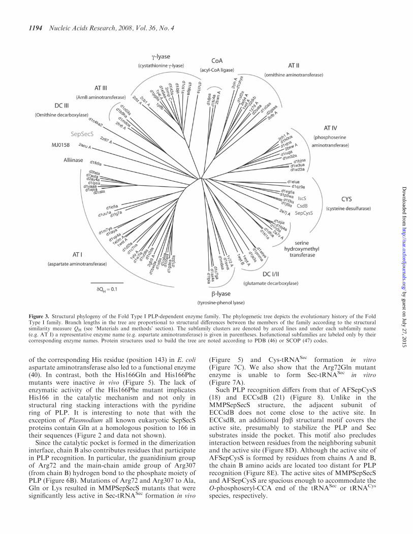
Figure 3. Structural phylogeny of the Fold Type I PLP-dependent enzyme family. The phylogenetic tree depicts the evolutionary history of the FoldType I family. Branch lengths in the tree are proportional to structural differences between the members of the family according to the structuralsimilarity measure QH (see ‘Materials and methods’ section). The subfamily clusters are denoted by arced lines and under each subfamily name(e.g. AT I) a representative enzyme name (e.g. aspartate aminotransferase) is given in parentheses. Isofunctional subfamilies are labeled only by theircorresponding enzyme names. Protein structures used to build the tree are noted according to PDB (46) or SCOP (47) codes.
1194 Nucleic Acids Research, 2008, Vol. 36, No. 4
by guest on July 27, 2015http://nar.oxfordjournals.org/
Dow
nloaded from

Phosphoserine binding model
Our crystallization solution contained 10mM magnesiumsulfate. In the present structure, a strong electron density(4.5s), presumably corresponding to a sulfate ion, wasobserved adjacent to the PLP molecule of chains A and B(Figure 8A). The sulfate ion is recognized by Arg94,Ser95, Gln102 and Arg307 of chain B (Figure 8A). Thestructure of ECCsdB with Sec bound to the active site(Figure 8D) was reported (21). Using the CE program (41)we superposed the active site structure of MMPSepSecSonto that of ECCsdB. This allowed modeling of Sec fromCsdB into the MMPSepSecS active site (Figure 8B). Thedistance between the selenium atom of the modeled Secand the sulfur atom of the bound sulfate is 3.74 A. Weoverlaid a Sep molecule on the position of the modeled Secin the active site. The phosphate moiety of this modeledSep overlapped with the bound sulfate, suggesting thatthis sulfate mimics the phosphate moiety of Sep attachedto tRNASec. This allowed us to construct the Sep-bindingmodel of MMPSepSecS (Figure 8C). Mutations of Arg94,Gln102 and Arg307 residues that according to ourSep-binding model recognize the phosphate moiety ofSep significantly decreased the catalytic activity ofMMPSepSecS both in vivo (Figure 5) and in vitro(Figure 7). While the distance of the Schiff base linkagebetween the C4a atom of PLP and the N� atom of Lys278is 1.74 A, the PLP C4a atom is 2.19 A away from theamino group of the modeled Sep; thus, the amino group ofSep forms a Schiff base with PLP as a reactionintermediate.
The active site
We should first note that the active site of MMPSepSecSlacks a Cys residue. In contrast, the crystal structures ofECCsdB (21) and of AFSepCysS (18) each possess acysteine-active site residue; Cys364 in ECCsdB andCys247 in AFSepCysS (Figure 8). As revealed by astructural study (21), Cys364 of ECCsdB recognizes Secand withdraws its selenium to form perselenide as anintermediate. Cys364 of ECCsdB resides in the babstructural motif which does not exist in MMPSepSecS.On the other hand, Cys247 (from the neighboring subunit)of AFSepCysS is the candidate residue that forms a
Figure 4. The oligomeric states of SepSecS and related enzymes.(A) The overall architecture of the MMPSepSecS tetramer. Chains Aand B, and chains C and D form dimers, respectively. The PLPmolecules bound to each subunit are shown as ball and stick models.Chain A, chain B, chain C and chain D are colored pink, blue, greenand yellow, respectively. (B) The N-terminal extension domains ofchains B and C form a hydrophobic core that stabilizes the tetramericstate of MMPSepSecS. Three a-helices (a1,a2,a4) are labeled. Chains Band C are colored blue and green, respectively. (C) Stereo view of theAFSepCysS dimer in the same orientation as (A) (18). Chains A and Bare colored pink and blue, respectively. The PLP molecules are shownas ball and stick models. (D) Stereo view of the ECCsdB dimer in thesame orientation as (A) (21). The coloring scheme and the PLP repre-sentation are the same as in (C). (E) Gel filtration of selenomethionine-labeled SepSecS on Sephacryl S-300. Absorbance at 280 nm is shown asa blue line. The elution volumes of other oligomeric proteins areindicated in the chromatogram. The molecular weight of theMMPSepSecS monomer is 50 kDa. MMPSepSecS eluted at the sizeexpected for a tetrameric species.
Figure 5. In vivo assays of SepSecS mutants. Formation of Sec-tRNASec in vivo is assayed by the ability of the wild-type MMPSepSecS and itsmutant variants (N-terminal deletion �1-34, R72A, R72Q, R72K, R94A, R94Q, H166A, H166F, H166Q, R307A, R307Q, R307K, Q102A, K278A,N247A, D277A, K278A) to restore the BV reducing activity of the selenoprotein FDHH in the E.coli selA deletion strain JS1. Cotransformation ofthe PSTK gene (indicated with +) from M. jannaschii is required for the formation of the Sep-tRNASec intermediate.
Nucleic Acids Research, 2008, Vol. 36, No. 4 1195
by guest on July 27, 2015http://nar.oxfordjournals.org/
Dow
nloaded from

persulfide that is the sulfur source for enzyme-catalyzedCys-tRNACys formation (18). The absence of such a Cysactive site residue indicates that the reaction mechanismsand chemistries of SepSecS are fundamentally differentfrom those of CsdB and SepCysS. Interestingly, SepSecS
has an active site arginine (Arg307) in a homologousposition to Cys247 in SepCysS (Figure 2).
The MMPSepSecS structure reveals three conservedarginines (Arg72, Arg94 and Arg307) that are located inproximity of each other and close to the active site; theyrecognize the phosphate groups of PLP and presumably ofSep acylated to tRNASec (Figures 2 and 8A–C). Mutationsof Arg72, Arg94 and Arg307 to Ala, Gln or Lys yieldedMMPSepSecS enzymes that were unable to form Sec-tRNASec in vivo (Figure 5). Asp277 interacts electrostati-cally with Arg72, and the Asp277Ala enzyme was alsoinactive in vivo (Figure 5). The Arg72Gln, Arg94Gln andArg307Gln MMPSepSecS mutants were inactive informing Cys-tRNASec in vitro (Figure 7). In addition tothe PLP-conjugated Lys278, Sep-binding arginines mayalso facilitate general acid/base catalysis as was shown forthe tRNA modification enzyme TrmH where an arginineactivated by a phosphate group acts as a general base (42).
DISCUSSION
The mechanism
SepSecS is a PLP enzyme catalyzing a b-replacement(Scheme 1), leading to the exchange of a phosphate groupfor a selenol moiety. We were interested to see if SepSecSmight employ a perselenide intermediate during catalysis,which would require the presence of an active site cysteine.There are four moderately-to-highly conserved cysteineresidues in the SepSecS sequences, which are found in35–97% of the SepSecSs. Three of these cysteines (Cys146,Cys214 and Cys237 in MMPSepSecS) are distant from theactive site and have no chance to contribute to catalysis.
Figure 7. In vitro conversion of Sep-tRNASec to Sec-tRNASec or Cys-tRNASec. (A) Phosphorimages of TLC separation of [14C]Sep and [14C]Secrecovered from the aa-tRNAs of the SepSecS activity assay (see ‘Materials and methods’ section). Sec was analyzed in its oxidized form asselenocysteic acid (Secya). Lane 1, Ser marker; lane 2, Sep marker; lane 3, Sep-tRNASec with wild-type MMPSepSecS; lane 4, Sep-tRNASec with theR72Q MMPSepSecS mutant. (B) Representative phosphorimage for the H166A SepSecS mutant of the separation of Ser-[32P]AMP, Cys-[32P]AMP,[32P]AMP and Sep-[32P]AMP. At the indicated time points aliquots of the SepSecS reaction were quenched, digested with nuclease P1 and spottedonto PEI-cellulose TLC plates as described in the ‘Materials and methods’ section. (C) Plot of Cys-tRNASec formed versus time with 1 mM of wild-type and mutant SepSecS enzymes using Sep-tRNASec (1 mM) and thiophosphate (500 mM) as substrates. Following quantification of the intensities ofSer-[32P]AMP, Cys-[32P]AMP, [32P]AMP and Sep-[32P]AMP using ImageQuant, the concentration of Cys-tRNASec formed at each time point wascalculated by dividing the intensity of the Cys-[32P]AMP spot by the total intensity. The experiment was carried out in duplicate.
Figure 6. PLP recognition. Stereo views of (A) Ribbon representationof the active site of MMPSepSecS in the dimer interface between chainsA and B that are colored pink and blue, respectively. The PLPmolecule is covalently bound to Lys278 of chain A. The Fo�Fc omitmap of the PLP molecule, contoured at 3.5 s, is shown. (B) The aminoacid residues that recognize the PLP molecule. The residues of chainsA and B are colored pink and blue, respectively. The Fo�Fc omit map(contoured at 3.5 s) of all of the residues and PLP is shown.
1196 Nucleic Acids Research, 2008, Vol. 36, No. 4
by guest on July 27, 2015http://nar.oxfordjournals.org/
Dow
nloaded from

The least conserved of these residues is a strictly conservedproline (Pro169) in the archaeal sequences, while theresidue is conserved as a cysteine in 78% of the eukaryoticsequences. Interestingly, Pro169 in MMPSepSecS is in theactive site and in close contact with the PLP moiety(3.77 A at closest approach), which would then mean thatmost eukaryotic SepSecSs have a cysteine adjacent tothe PLP. It is unclear whether the eukaryotic sequencesmake use of this cysteine in catalysis; nevertheless, thisposition is an intriguing candidate for mutagenesis in theeukaryotic context.Concerning the archaeal SepSecSs, there are no cysteine
residues that could be involved in the formation of acatalytically important perselenide intermediate. In con-trast, SepCysS is proposed to use a persulfide mechanism(18). Thus, SepSecS and SepCysS, two related PLPenzymes that perform chemically analogous tRNA-dependent transformations of Sep to Sec or Cys,respectively, proceed with different selenium and sulfurtransfer mechanisms. Based on the observed active siteresidues we propose that the arginine residues, that bindthe phosphate groups of PLP and of Sep, also recruit theselenium donor selenophosphate. In this context, Arg307in the present structure resides at a similar position to thatof Cys364 in ECCsdB and of Cys247 in AFSepCysS.
Structural phylogeny of Fold Type I PLP enzymes
SepSecS belongs to the largest and most diverse family ofPLP-dependent enzymes found in nature. The evolution-ary history of the Fold Type I PLP-dependent enzymes,also referred to as the a-family, has been studied indetail with sequence-based phylogenetic methods (43). Byapplying the family profile analysis (FPA) technique (44),the authors were able to use sequence-based phylogeneticsto partially capture the distant evolutionary eventsrecorded in the sequences of the members of this family,some of whom share only 5% sequence identity. However,the low level of sequence similarity may lead to artifacts inthe tree reconstruction process. Since crystal structures formost members of this protein family exist, we appliedthe technique of structural phylogeny (45) to explore thehistory of the Fold Type I family. This technique derivesphylogenetic information directly from three-dimensionalstructures and thus allows an accurate reconstructionof the most distantly detectable evolutionary events. Ourstructural phylogeny (Figure 3) is largely in agreementwith the previous sequence-based work, but somesignificant rearrangements in the tree can be seen. Forexample, the DC I and DC II subfamilies coalesce into asingle subfamily according to structural similarity, and theg-lyase and CYS groups are not one but two evolutionarydistinct subfamilies.There are two kinds of subfamilies in the Fold Type I
group. The first type includes several proteins with distinctbut related functions, which may have descended from aprogenitor with a promiscuous enzymatic function.Examples of these progenitor enzymes include the fourdistinct aminotransferases (AT I–AT IV), an amino aciddecarboxylase (DC I/II), a persulfide forming cysteinedesulfurase/sulfhydrylase and finally separate b- and
Figure 8. The active site. Close-up stereo views of the active site regionsin MMPSepSecS, ECCsdB, and AFSepCysS. Chains A and B arecolored pink and blue, respectively. The PLP molecule (shown as a balland stick model) is covalently bound to chain A. (A) The recognitionmanner of the sulfate ion in the active site of MMPSepSecS. Thesulfate ion is shown as a ball-and-stick model. The Fo�Fc omit map ofthe sulfate ion, contoured at 4.5 s, is shown. (B) Sec docking modelof MMPSepSecS, based on the structural comparison with ECCsdB.(C) Sep binding model of MMPSepSecS, based on the locations of thesulfate ion and Sep. (D) Close-up view of the active site region inECCsdB. The PLP molecule is covalently bound to Lys226 of chain A.The orientation of the PLP molecule is the same as that in (A).The chain A residues that recognize PLP are shown. The PLP moleculeis covered by the bab structural motif. (E) Close-up view of theactive site region in AFSepCysS. The PLP molecule is covalentlybound to Lys209 of chain A. The orientation of PLP is the same asthat in (A). The chain A residues that recognize PLP and sulfate areshown.
Nucleic Acids Research, 2008, Vol. 36, No. 4 1197
by guest on July 27, 2015http://nar.oxfordjournals.org/
Dow
nloaded from

g-amino acid lyases. Subsequent evolution in each of thesesubfamilies ultimately produced the specific enzymaticfunctions observed in modern PLP-dependent enzymes.The second kind of subfamilies are those that include a
set of isofunctional enzymes. The modern enzymes inthese subfamilies trace back to ancestors that had alreadyevolved their modern enzymatic specificity in the initialevolutionary radiation of the Fold Type I family. Theseisofunctional subfamilies include serine hydroxymethyltransferase, alliinase and an archaeal SelA-like protein(MJ0158), which is of unknown function.Importantly, the structural phylogeny allows the
accurate placement of SepSecS within its family tree andreveals how this enzyme came into being. SepSecS is also afounding member of the Fold Type I family. It shows nospecific relationship to any of the other subfamilies andemerges near the root of its family tree. While SepCysS,CsdB and IscS are part of a multifunctional subfamily(CYS in Figure 8) and these proteins have chemicallysimilar substrates to SepSecS, there is no special relation-ship between SepSecS and the CYS subfamily. This resultindicates that SepSecS is truly an ancient enzyme, and thustRNA-dependent selenocysteine biosynthesis is a primor-dial process.
ACKNOWLEDGEMENTS
We thank the beam-line staff at BL41XU of SPring-8(Harima, Japan) for technical help in data collection. DanSu, Markus Englert, Kelly Sheppard, Michael Hohn,Hee-Sung Park, Juan Salazar, Motoyuki Hattori, KotaroNakanishi and Tomoyuki Numata participated in manydiscussions on this topic.This work was supported by a SORST Program grantfrom Japan Science and Technology (to O.N.), by grantsfrom the Ministry of Education, Culture, Sports, Scienceand Technology (to R.I. and O.N.), by a National Projecton Protein Structural and Functional Analyses grant fromthe Ministry of Education, Culture, Sports, Science andTechnology (to O.N.) and by grants from the Departmentof Energy, the National Institute of General MedicalSciences and the National Science Foundation (to D.S.).S.P. holds a fellowship of the Yale University School ofMedicine MD/PhD Program. R.L.S. is the recipient ofa Ruth L. Kirschstein National Research Service Awardfrom the National Institute of General Medical Sciences.P.O’D. holds a National Science Foundation PostdoctoralFellowship in Biological Informatics. Funding to pay theOpen Access publication charges for this article wasprovided by GM22854 (to D.S.).
Conflict of interest statement. None declared.
REFERENCES
1. Ibba,M. and Soll,D. (2004) Aminoacyl-tRNAs: setting the limits ofthe genetic code. Genes Dev., 18, 731–738.
2. Hatfield,D.L. and Gladyshev,V.N. (2002) How selenium has alteredour understanding of the genetic code. Mol. Cell Biol., 22,3565–3576.
3. Bock,A., Thanbichler,M., Rother,M. and Resch,A. (2005)Selenocysteine. In Ibba,M., Francklyn,C.S. and Cusack,S. (eds),
Aminoacyl-tRNA Synthetases. Landes Bioscience, Georgetown, TX,pp. 320–327.
4. Leinfelder,W., Zehelein,E., Mandrand-Berthelot,M.A. and Bock,A.(1988) Gene for a novel tRNA species that accepts L-serine andcotranslationally inserts selenocysteine. Nature, 331, 723–725.
5. Bilokapic,S., Korencic,D., Soll,D. and Weygand-Durasevic,I. (2004)The unusual methanogenic seryl-tRNA synthetase recognizestRNASer species from all three kingdoms of life. Eur. J. Biochem.,271, 694–702.
6. Kaiser,J.T., Gromadski,K., Rother,M., Engelhardt,H.,Rodnina,M.V. and Wahl,M.C. (2005) Structural and functionalinvestigation of a putative archaeal selenocysteine synthase.Biochemistry, 44, 13315–13327.
7. Ohama,T., Yang,D.C.H. and Hatfield,D.L. (1994) SelenocysteinetRNA and serine tRNA are aminoacylated by the same synthetase,but may manifest different identities with respect to the long extraarm. Arch. Biochem. Biophys., 315, 293–301.
8. Maenpaa,P.H. and Bernfield,M.R. (1970) A specific hepatictransfer RNA for phosphoserine. Proc. Natl Acad. Sci. USA, 67,688–695.
9. Sharp,S.J. and Stewart,T.S. (1977) The characterization ofphosphoseryl tRNA from lactating bovine mammary gland.Nucleic Acids Res., 4, 2123–2136.
10. Carlson,B.A., Xu,X.M., Kryukov,G.V., Rao,M., Berry,M.J.,Gladyshev,V.N. and Hatfield,D.L. (2004) Identification and char-acterization of phosphoseryl-tRNA[Ser]Sec kinase. Proc. Natl Acad.Sci. USA, 101, 12848–12853.
11. Yuan,J., Palioura,S., Salazar,J.C., Su,D., O’Donoghue,P.,Hohn,M.J., Cardoso,A.M., Whitman,W.B. and Soll,D. (2006)RNA-dependent conversion of phosphoserine forms selenocysteinein eukaryotes and archaea. Proc. Natl Acad. Sci. USA, 103,18923–18927.
12. Xu,X.M., Carlson,B.A., Mix,H., Zhang,Y., Saira,K., Glass,R.S.,Berry,M.J., Gladyshev,V.N. and Hatfield,D.L. (2007) Biosynthesisof selenocysteine on its tRNA in eukaryotes. PLoS Biol., 5, e4.doi:10.1371/journal.pbio.0050004.
13. Gelpi,C., Sontheimer,E.J. and Rodriguez-Sanchez,J.L. (1992)Autoantibodies against a serine tRNA-protein complex implicatedin cotranslational selenocysteine insertion. Proc. Natl Acad. Sci.USA, 89, 9739–9743.
14. Costa,M., Rodriguez-Sanchez,J.L., Czaja,A.J. and Gelpi,C. (2000)Isolation and characterization of cDNA encoding the antigenicprotein of the human tRNP(Ser)Sec complex recognized by auto-antibodies from patients with type-1 autoimmune hepatitis. Clin.Exp. Immunol., 121, 364–374.
15. Wies,I., Brunner,S., Henninger,J., Herkel,J., Kanzler,S., Meyer zumBuschenfelde,K.H. and Lohse,A.W. (2000) Identification of targetantigen for SLA/LP autoantibodies in autoimmune hepatitis.Lancet, 355, 1510–1515.
16. Herkel,J., Manns,M.P. and Lohse,A.W. (2007) Selenocysteine,soluble liver antigen/liver-pancreas, and autoimmune hepatitis.Hepatology, 46, 275–277.
17. Sauerwald,A., Zhu,W., Major,T.A., Roy,H., Palioura,S., Jahn,D.,Whitman,W.B., Yates,J.R.III, Ibba,M. et al. (2005) RNA-dependent cysteine biosynthesis in archaea. Science, 307,1969–1972.
18. Fukunaga,R. and Yokoyama,S. (2007) Structural insights into thesecond step of RNA-dependent cysteine biosynthesis in archaea:crystal structure of Sep-tRNA:Cys-tRNA synthase fromArchaeoglobus fulgidus. J. Mol. Biol., 370, 128–141.
19. Percudani,R. and Peracchi,A. (2003) A genomic overview ofpyridoxal-phosphate-dependent enzymes. EMBO Rep., 4, 850–854.
20. Eliot,A.C. and Kirsch,J.F. (2004) Pyridoxal phosphate enzymes:mechanistic, structural, and evolutionary considerations. Annu. Rev.Biochem., 73, 383–415.
21. Lima,C.D. (2002) Analysis of the E. coli NifS CsdB protein at 2.0Areveals the structural basis for perselenide and persulfide inter-mediate formation. J. Mol. Biol., 315, 1199–1208.
22. Mihara,H., Maeda,M., Fujii,T., Kurihara,T., Hata,Y. and Esaki,N.(1999) A nifS-like gene, csdB, encodes an Escherichia coli counter-part of mammalian selenocysteine lyase. Gene cloning, purification,characterization and preliminary x-ray crystallographic studies.J. Biol. Chem., 274, 14768–14772.
1198 Nucleic Acids Research, 2008, Vol. 36, No. 4
by guest on July 27, 2015http://nar.oxfordjournals.org/
Dow
nloaded from

23. Cupp-Vickery,J.R., Urbina,H. and Vickery,L.E. (2003) Crystalstructure of IscS, a cysteine desulfurase from Escherichia coli.J. Mol. Biol., 330, 1049–1059.
24. Weeks,C.M. and Miller,R. (1999) The design and implementationof SnB version 2.0. J. Appl. Cryst., 32, 120–124.
25. de La Fortelle,E. and Bricogne,G. (1997) Maximum-likelihoodheavy-atom parameter refinement for multiple isomorphousreplacement and multiwavelength anomalous diffraction methods.Methods Enzymol., 276, 472–494.
26. Jones,T.A., Zou,J.Y., Cowan,S.W. and Kjeldgaard,M. (1991)Improved methods for building protein models in electrondensity maps and the location of errors in these models. ActaCrystallogr. A., 47(Pt 2), 110–119.
27. Brunger,A.T., Adams,P.D., Clore,G.M., DeLano,W.L., Gros,P.,Grosse-Kunstleve,R.W., Jiang,J.S., Kuszewski,J., Nilges,M. et al.(1998) Crystallography & NMR system: a new software suite formacromolecular structure determination. Acta Crystallogr. D. Biol.Crystallogr., 54, 905–921.
28. Milligan,J.F., Groebe,D.R., Witherell,G.W. and Uhlenbeck,O.C.(1987) Oligoribonucleotide synthesis using T7 RNA polymerase andsynthetic DNA templates. Nucleic Acids Res., 15, 8783–8798.
29. Oshikane,H., Sheppard,K., Fukai,S., Nakamura,Y., Ishitani,R.,Numata,T., Sherrer,R.L., Feng,L., Schmitt,E. et al. (2006)Structural basis of RNA-dependent recruitment of glutamine to thegenetic code. Science, 312, 1950–1954.
30. Bullock,T.L., Uter,N., Nissan,T.A. and Perona,J.J. (2003)Amino acid discrimination by a class I aminoacyl-tRNAsynthetase specified by negative determinants. J. Mol. Biol., 328,395–408.
31. Russell,R.B. and Barton,G.J. (1992) Multiple protein sequencealignment from tertiary structure comparison: assignment of globaland residue confidence levels. Proteins, 14, 309–323.
32. Roberts,E., Eargle,J., Wright,D. and Luthey-Schulten,Z. (2006)MultiSeq: unifying sequence and structure data for evolutionaryanalysis. BMC Bioinformatics, 7, 382.
33. O’Donoghue,P. and Luthey-Schulten,Z. (2005) Evolutionaryprofiles derived from the QR factorization of multiple structuralalignments gives an economy of information. J. Mol. Biol., 346,875–894.
34. Felsenstein,J. (1989) PHYLIP - Phylogeny Interference Package(Version 3.2). Cladistics, 5, 164–166.
35. Chenna,R., Sugawara,H., Koike,T., Lopez,R., Gibson,T.J.,Higgins,D.G. and Thompson,J.D. (2003) Multiple sequence align-ment with the Clustal series of programs. Nucleic Acids Res., 31,3497–3500.
36. Lacourciere,G.M., Levine,R.L. and Stadtman,T.C. (2002) Directdetection of potential selenium delivery proteins by using anEscherichia coli strain unable to incorporate selenium from seleniteinto proteins. Proc. Natl Acad. Sci. USA, 99, 9150–9153.
37. Herkel,J., Heidrich,B., Nieraad,N., Wies,I., Rother,M. andLohse,A.W. (2002) Fine specificity of autoantibodies to soluble liverantigen and liver/pancreas. Hepatology, 35, 403–408.
38. Jansonius,J.N. (1998) Structure, evolution and action of vitaminB6-dependent enzymes. Curr. Opin. Struct. Biol., 8, 759–769.
39. Schneider,G., Kack,H. and Lindqvist,Y. (2000) The manifold ofvitamin B6 dependent enzymes. Structure, 8, R1–R6.
40. Yano,T., Kuramitsu,S., Tanase,S., Morino,Y., Hiromi,K. andKagamiyama,H. (1991) The role of His143 in the catalyticmechanism of Escherichia coli aspartate aminotransferase. J. Biol.Chem., 266, 6079–6085.
41. Shindyalov,I.N. and Bourne,P.E. (1998) Protein structure alignmentby incremental combinatorial extension (CE) of the optimal path.Protein Eng., 11, 739–747.
42. Nureki,O., Watanabe,K., Fukai,S., Ishii,R., Endo,Y., Hori,H. andYokoyama,S. (2004) Deep knot structure for construction of activesite and cofactor binding site of tRNA modification enzyme.Structure, 12, 593–602.
43. Christen,P. and Mehta,P.K. (2001) From cofactor to enzymes. Themolecular evolution of pyridoxal- 5’-phosphate-dependent enzymes.Chem. Rec., 1, 436–447.
44. Mehta,P.K., Argos,P., Barbour,A.D. and Christen,P. (1999)Recognizing very distant sequence relationships among proteins byfamily profile analysis. Proteins, 35, 387–400.
45. O’Donoghue,P. and Luthey-Schulten,Z. (2003) On the evolution ofstructure in aminoacyl-tRNA synthetases. Microbiol. Mol. Biol.Rev., 67, 550–573.
46. Berman,H.M., Westbrook,J., Feng,Z., Gilliland,G., Bhat,T.N.,Weissig,H., Shindyalov,I.N. and Bourne,P.E. (2000) The ProteinData Bank. Nucleic Acids Res., 28, 235–242.
47. Andreeva,A., Howorth,D., Brenner,S.E., Hubbard,T.J., Chothia,C.and Murzin,A.G. (2004) SCOP database in 2004: refinementsintegrate structure and sequence family data. Nucleic Acids Res., 32,D226–D229.
Nucleic Acids Research, 2008, Vol. 36, No. 4 1199
by guest on July 27, 2015http://nar.oxfordjournals.org/
Dow
nloaded from
Copyright © 2022 FDOKUMEN