RNA recognition by a Staufen double-stranded RNA-binding domain
13
The EMBO Journal Vol.19 No.5 pp.997–1009, 2000 RNA recognition by a Staufen double-stranded RNA-binding domain Andres Ramos 1,2 , Stefan Gru ¨ nert 3,4 , Jan Adams 3 , David R.Micklem 3 , Mark R.Proctor 5 , Stefan Freund 5 , Mark Bycroft 5 , Daniel St Johnston 3 and Gabriele Varani 1,6 1 MRC Laboratory of Molecular Biology and 5 Cambridge Centre for Protein Engineering, Hills Road, Cambridge CB2 2QH and 3 Wellcome/CRC Institute and the Department of Genetics, University of Cambridge, Tennis Court Rd, Cambridge CB2 1QR, UK 2 Present address: Molecular Structure Division, NIMR, The Ridgeway, Mill Hill, London NW7 1AA, UK 4 Present address: IMP, Dr, Bohrgasse 7, 1030 Wien, Austria 6 Corresponding author e.mail: [email protected] A.Ramos and S.Gru ¨nert contributed equally to this work The double-stranded RNA-binding domain (dsRBD) is a common RNA-binding motif found in many proteins involved in RNA maturation and localization. To deter- mine how this domain recognizes RNA, we have studied the third dsRBD from Drosophila Staufen. The domain binds optimally to RNA stem–loops containing 12 uninterrupted base pairs, and we have identified the amino acids required for this interaction. By mutating these residues in a staufen transgene, we show that the RNA-binding activity of dsRBD3 is required in vivo for Staufen-dependent localization of bicoid and oskar mRNAs. Using high-resolution NMR, we have deter- mined the structure of the complex between dsRBD3 and an RNA stem–loop. The dsRBD recognizes the shape of A-form dsRNA through interactions between conserved residues within loop 2 and the minor groove, and between loop 4 and the phosphodiester backbone across the adjacent major groove. In addition, helix α1 interacts with the single-stranded loop that caps the RNA helix. Interactions between helix α1 and single-stranded RNA may be important determinants of the specificity of dsRBD proteins. Keywords: dsRBD/NMR/RNA localization/RNA– protein/Staufen Introduction The double-stranded RNA-binding domain (dsRBD) is among the most common RNA-binding motifs, and is found in single or multiple copies in many eukaryotic and prokaryotic proteins involved in RNA processing, maturation and localization (Green and Matthews, 1992; St Johnston et al., 1992). Three-dimensional structures of dsRBDs from several proteins have shown that the domain folds into a compact αβββα structure (Bycroft et al., 1995a; Kharrat et al., 1995; Nanduri et al., 1998). As in © European Molecular Biology Organization 997 the other two major eukaryotic RNA-binding protein domains (Varani, 1997), the α-helical surface of the dsRBD structure packs through a conserved hydrophobic core against an antiparallel β-sheet. In vitro studies have shown that dsRBD proteins bind to dsRNA, but not to single-stranded RNA or DNA, nor dsDNA (St Johnston et al., 1992; Bass et al., 1994; Clarke and Matthews, 1995; Bevilacqua and Cech, 1996). These studies have shown that dsRBDs bind any dsRNA of sufficient length, regardless of its base composition, and therefore they represent general dsRNA-binding modules. The dsRBD was first identified in the Drosophila protein Staufen, which contains five copies of this motif (St Johnston et al., 1992). Staufen plays an essential role in the formation of the anterior–posterior axis in Drosophila and represented the first protein factor to be identified as critical for mRNA localization (St Johnston, 1995). Staufen protein associates with oskar mRNA during oogenesis and is required for its transport to the posterior pole of the oocyte, where it defines where the abdomen and germline will develop (Ephrussi et al., 1991; Kim-Ha et al., 1991; St Johnston et al., 1991). After the egg has been laid, Staufen accumulates at the anterior pole of the egg, and anchors the anterior determinant bicoid mRNA (St Johnston et al., 1989; Ferrandon et al., 1994). Staufen plays a role in RNA localization in somatic cells as well, by associating with prospero mRNA during the asymmetric divisions of the embryonic neuroblasts, and by mediating its segregation to the smaller daughter cell produced by this division (Broadus et al., 1998; Schuldt et al., 1998). In common with most other systems where mRNA localization has been studied, the cis-acting signals required for oskar , bicoid and prospero localization all reside within the 3-untranslated regions (3-UTR) of these mRNAs (MacDonald and Struhl, 1988; Kim-Ha et al., 1993). Staufen protein associates in vivo with the 3-UTRs of bicoid and prospero mRNAs to form ribonucleoprotein particles (Ferrandon et al., 1994; Schuldt et al., 1998). The bicoid RNA sequences required for this interaction have been mapped to three largely double- stranded regions (Schuldt et al., 1998). However, it remains to be proven whether Staufen interacts directly with these RNAs in vivo and, if so, how Staufen recognizes these specific transcripts. The binding of the dsRBD to dsRNA represents an example of protein–nucleic acid recognition distinct from the other common RNA-binding motifs characterized so far (Varani, 1997). To determine the nature of the dsRBD– dsRNA interaction, we have conducted extensive mutagen- esis on the third dsRBD from Staufen (dsRBD3) and have used nuclear magnetic resonance (NMR) to determine the structure of the complex between this domain and an RNA stem–loop containing an optimal Staufen-binding site. We have mutated five critical interfacial residues
-
Upload
independent -
Category
Documents
-
view
4 -
download
0
Transcript of RNA recognition by a Staufen double-stranded RNA-binding domain
The EMBO Journal Vol.19 No.5 pp.997–1009, 2000
RNA recognition by a Staufen double-strandedRNA-binding domain
Andres Ramos1,2, Stefan Grunert3,4,Jan Adams3, David R.Micklem3,Mark R.Proctor5, Stefan Freund5,Mark Bycroft5, Daniel St Johnston3 andGabriele Varani1,6
1MRC Laboratory of Molecular Biology and 5Cambridge Centre forProtein Engineering, Hills Road, Cambridge CB2 2QH and3Wellcome/CRC Institute and the Department of Genetics, Universityof Cambridge, Tennis Court Rd, Cambridge CB2 1QR, UK2Present address: Molecular Structure Division, NIMR, The Ridgeway,Mill Hill, London NW7 1AA, UK4Present address: IMP, Dr, Bohrgasse 7, 1030 Wien, Austria6Corresponding authore.mail: [email protected]
A.Ramos and S.Grunert contributed equally to this work
The double-stranded RNA-binding domain (dsRBD) isa common RNA-binding motif found in many proteinsinvolved in RNA maturation and localization. To deter-mine how this domain recognizes RNA, we have studiedthe third dsRBD from Drosophila Staufen. The domainbinds optimally to RNA stem–loops containing 12uninterrupted base pairs, and we have identified theamino acids required for this interaction. By mutatingthese residues in a staufen transgene, we show that theRNA-binding activity of dsRBD3 is required in vivofor Staufen-dependent localization of bicoid and oskarmRNAs. Using high-resolution NMR, we have deter-mined the structure of the complex between dsRBD3and an RNA stem–loop. The dsRBD recognizes theshape of A-form dsRNA through interactions betweenconserved residues within loop 2 and the minor groove,and between loop 4 and the phosphodiester backboneacross the adjacent major groove. In addition, helixα1 interacts with the single-stranded loop that capsthe RNA helix. Interactions between helix α1 andsingle-stranded RNA may be important determinantsof the specificity of dsRBD proteins.Keywords: dsRBD/NMR/RNA localization/RNA–protein/Staufen
Introduction
The double-stranded RNA-binding domain (dsRBD) isamong the most common RNA-binding motifs, and isfound in single or multiple copies in many eukaryoticand prokaryotic proteins involved in RNA processing,maturation and localization (Green and Matthews, 1992;St Johnston et al., 1992). Three-dimensional structures ofdsRBDs from several proteins have shown that the domainfolds into a compact αβββα structure (Bycroft et al.,1995a; Kharrat et al., 1995; Nanduri et al., 1998). As in
© European Molecular Biology Organization 997
the other two major eukaryotic RNA-binding proteindomains (Varani, 1997), the α-helical surface of thedsRBD structure packs through a conserved hydrophobiccore against an antiparallel β-sheet. In vitro studies haveshown that dsRBD proteins bind to dsRNA, but not tosingle-stranded RNA or DNA, nor dsDNA (St Johnstonet al., 1992; Bass et al., 1994; Clarke and Matthews,1995; Bevilacqua and Cech, 1996). These studies haveshown that dsRBDs bind any dsRNA of sufficient length,regardless of its base composition, and therefore theyrepresent general dsRNA-binding modules.
The dsRBD was first identified in the Drosophilaprotein Staufen, which contains five copies of this motif(St Johnston et al., 1992). Staufen plays an essentialrole in the formation of the anterior–posterior axis inDrosophila and represented the first protein factor to beidentified as critical for mRNA localization (St Johnston,1995). Staufen protein associates with oskar mRNA duringoogenesis and is required for its transport to the posteriorpole of the oocyte, where it defines where the abdomenand germline will develop (Ephrussi et al., 1991;Kim-Ha et al., 1991; St Johnston et al., 1991). After theegg has been laid, Staufen accumulates at the anteriorpole of the egg, and anchors the anterior determinantbicoid mRNA (St Johnston et al., 1989; Ferrandon et al.,1994). Staufen plays a role in RNA localization in somaticcells as well, by associating with prospero mRNA duringthe asymmetric divisions of the embryonic neuroblasts,and by mediating its segregation to the smaller daughtercell produced by this division (Broadus et al., 1998;Schuldt et al., 1998). In common with most other systemswhere mRNA localization has been studied, the cis-actingsignals required for oskar, bicoid and prospero localizationall reside within the 3�-untranslated regions (3�-UTR) ofthese mRNAs (MacDonald and Struhl, 1988; Kim-Haet al., 1993). Staufen protein associates in vivo withthe 3�-UTRs of bicoid and prospero mRNAs to formribonucleoprotein particles (Ferrandon et al., 1994; Schuldtet al., 1998). The bicoid RNA sequences required for thisinteraction have been mapped to three largely double-stranded regions (Schuldt et al., 1998). However, it remainsto be proven whether Staufen interacts directly with theseRNAs in vivo and, if so, how Staufen recognizes thesespecific transcripts.
The binding of the dsRBD to dsRNA represents anexample of protein–nucleic acid recognition distinct fromthe other common RNA-binding motifs characterized sofar (Varani, 1997). To determine the nature of the dsRBD–dsRNA interaction, we have conducted extensive mutagen-esis on the third dsRBD from Staufen (dsRBD3) and haveused nuclear magnetic resonance (NMR) to determine thestructure of the complex between this domain and anRNA stem–loop containing an optimal Staufen-bindingsite. We have mutated five critical interfacial residues
A.Ramos et al.
located at the RNA–protein interface into full-lengthStaufen protein. These mutations abolish the RNA-bindingactivity of dsRBD3 in vitro and prevent Staufen-dependentRNA localization in vivo. The present results provide adescription at the atomic level of the interactions betweenthe dsRBD and RNA and demonstrate their physiologicalsignificance for Staufen-dependent RNA localization.
Results
RNA binding by Staufen dsRBD3
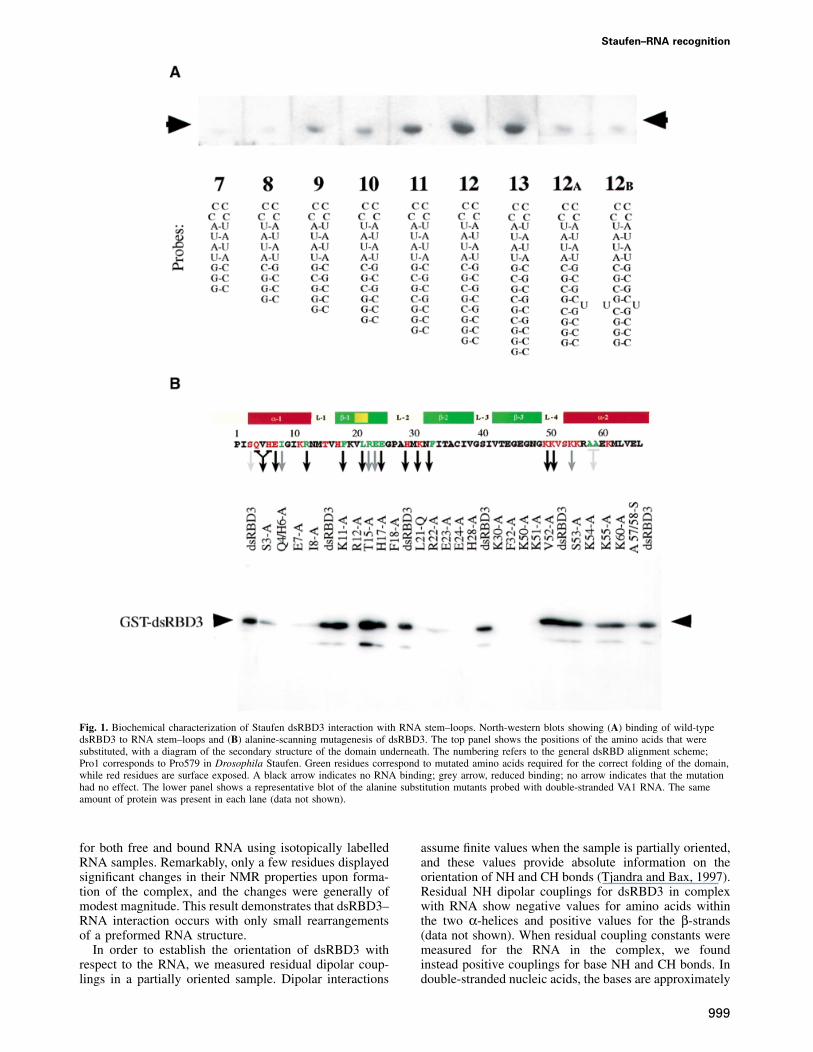
The third dsRBD from Staufen (dsRBD3) binds dsRNAwith micromolar affinity, and conforms particularly wellto the consensus sequence of the dsRBD motif (Gibsonand Thompson, 1994). We therefore chose this domainto analyse the structural basis of dsRBD–RNA inter-action. As a first step, we determined the minimal andoptimal length of dsRNA required for binding by adsRBD by performing North-western blots with RNAhairpin substrates containing double-helical stems ofincreasing length. RNAs containing �8 bp of dsRNAbind to dsRBD3, but optimal binding is observed withRNAs of 12 bp or longer (Figure 1A). Since furtherincreases in the length of the double-helical region donot improve binding, we conclude that dsRBD3 bindsoptimally to stem–loops containing 12 bp. Disruptionof the helical structure of the RNA by the introductionof unpaired bases significantly reduces binding. Theseresults are consistent with studies on polypeptidesderived from the two dsRBDs of RNA-activated proteinkinase (PKR) (Schmedt et al., 1995; Bevilacqua andCech, 1996). The full-length polypeptide binds to RNAsthat contain at least 16 bp, but each dsRBD was foundto cover ~11 bp of RNA.
The identity of amino acids within dsRBD3 involvedin RNA recognition was established by systematic alanine-scanning mutagenesis using the same North-western assay(Figure 1B). Several mutations involved amino acidswhose identity is crucial for the structure of the dsRBD.Ile8, Phe18, Ala57 and Ala58 form part of the hydrophobiccore of the domain. Mutations in Leu21, Arg22, Glu23and Glu24 were introduced to disrupt the β-bulge withinthe first strand of the β-sheet, a highly conserved featurethat is also present in ribosomal protein S5, a protein thatis very similar in both sequence and structure to thedsRBD (Bycroft et al., 1995). As expected, mutation ofeach of these amino acids strongly reduces or abolishesRNA binding. Mutations in Arg12 and Phe32 are alsolikely to fall into this class, even though these amino acidsare partially exposed on the surface of the domain. Arg12caps the N-terminal α-helix, and its replacement withalanine might disrupt RNA binding by extending the helixinto the following tight turn. Evidence presented belowindicates that Phe32 anchors loop 2 and loop 4.
The most informative mutations were changes in surfaceresidues that affect RNA binding without altering theconformation of the domain, as demonstrated by circulardichroism. Mutations of this type cluster in three regionsof the domain: Ser3, Gln4 plus His6, and Glu7 within theN-terminal helix α1; His28 and Lys30 within loop 2; andLys50, Lys51 and Lys54 within loop 4 and the beginningof helix α2. It is notable that mutations in Lys50, Lys51or Lys54 abolish RNA binding, whereas two non-basic
998
amino acids in this loop, Val52 and Ser53, can be mutatedto alanine without loss of binding. This suggests thatelectrostatic interactions mediated by basic residues playan important role in dsRBD–RNA recognition.
Staufen dsRBD3 binds RNA using a highly
conserved surface and without altering the RNA
conformation
Having established the biochemical properties of dsRBD3–RNA recognition, we used high-resolution NMR spectro-scopy to characterize this interaction in structural detail.A stem–loop of 12 bp capped by an exceptionally stableC(UUCG)G loop was chosen to represent an optimalsubstrate, as defined by the experiments reported inFigure 1A. Since the dsRBD–dsRNA interaction is notsequence specific, the double-helical region was madefully symmetrical to simplify the NMR spectral analysis.
Many protein resonances broadened considerably atsubsaturating ratios of RNA when dsRBD3 was titratedwith RNA, then sharpened up again when the RNA wasadded in stoichiometric amounts (Figure 2). This behaviouris found when the interconversion between free and boundforms occurs with intermediate exchange kinetics. Thisresult strongly suggests that the off rate of binding is inthe millisecond time scale, consistent with a micromolardissociation constant and with the on rate being diffusionlimited. Essentially complete spectral assignments wereobtained for the bound form of dsRBD3 in the presenceof RNA by applying standard procedures utilizing 15N- and13C-15N-labelled dsRBD3 samples mixed with unlabelledRNA. Changes in chemical shift upon RNA binding definethe footprint of the RNA on dsRBD3. The regions of theprotein where large changes in the NMR spectrum occurupon RNA binding cluster at the N-terminus of the protein,in loop 2 and loop 4 and in the region where α1 packsagainst α2 and the β-sheet. No significant changes wereobserved for β2 and β3 or in the C-terminal region of theprotein. Two conserved lysine residues within loop 4(Lys50 and Lys51) are particularly interesting. The back-bone amide resonances of Lys50 and Lys51 are invisiblein the free protein spectra, presumably due to the accessib-ility of solvent molecules to this exposed region of thestructure. However, the same resonances become visibleupon complex formation. These residues are protectedfrom exchange with solvent by the RNA, confirming theirrole in RNA recognition revealed by the alanine-scanningexperiment.
The NMR data demonstrate that the folding of thedomain does not change significantly upon RNA binding.Residues that display significant changes in the NMRspectrum of dsRBD3 upon RNA binding were thereforemapped on the structure of the free protein domain (Bycroftet al., 1995). The results unambiguously demonstrate thatthe face of the dsRBD formed by the N-termini of bothhelices, and by loops 2 and 4 along the edge of the firststrand of the β-sheet, represents the RNA-binding surfaceof dsRBD3. Thus, the results of the alanine-scanningmutagenesis and the NMR footprint identify the sameface of Staufen dsRBD3 as the surface where RNArecognition occurs. This protein surface contains exposedresidues that are almost completely conserved amongStaufen proteins from Drosophila to humans (Figure 3).
Essentially complete spectral assignments were obtained
Staufen–RNA recognition
Fig. 1. Biochemical characterization of Staufen dsRBD3 interaction with RNA stem–loops. North-western blots showing (A) binding of wild-typedsRBD3 to RNA stem–loops and (B) alanine-scanning mutagenesis of dsRBD3. The top panel shows the positions of the amino acids that weresubstituted, with a diagram of the secondary structure of the domain underneath. The numbering refers to the general dsRBD alignment scheme;Pro1 corresponds to Pro579 in Drosophila Staufen. Green residues correspond to mutated amino acids required for the correct folding of the domain,while red residues are surface exposed. A black arrow indicates no RNA binding; grey arrow, reduced binding; no arrow indicates that the mutationhad no effect. The lower panel shows a representative blot of the alanine substitution mutants probed with double-stranded VA1 RNA. The sameamount of protein was present in each lane (data not shown).
for both free and bound RNA using isotopically labelledRNA samples. Remarkably, only a few residues displayedsignificant changes in their NMR properties upon forma-tion of the complex, and the changes were generally ofmodest magnitude. This result demonstrates that dsRBD3–RNA interaction occurs with only small rearrangementsof a preformed RNA structure.
In order to establish the orientation of dsRBD3 withrespect to the RNA, we measured residual dipolar coup-lings in a partially oriented sample. Dipolar interactions
999
assume finite values when the sample is partially oriented,and these values provide absolute information on theorientation of NH and CH bonds (Tjandra and Bax, 1997).Residual NH dipolar couplings for dsRBD3 in complexwith RNA show negative values for amino acids withinthe two α-helices and positive values for the β-strands(data not shown). When residual coupling constants weremeasured for the RNA in the complex, we foundinstead positive couplings for base NH and CH bonds. Indouble-stranded nucleic acids, the bases are approximately
A.Ramos et al.
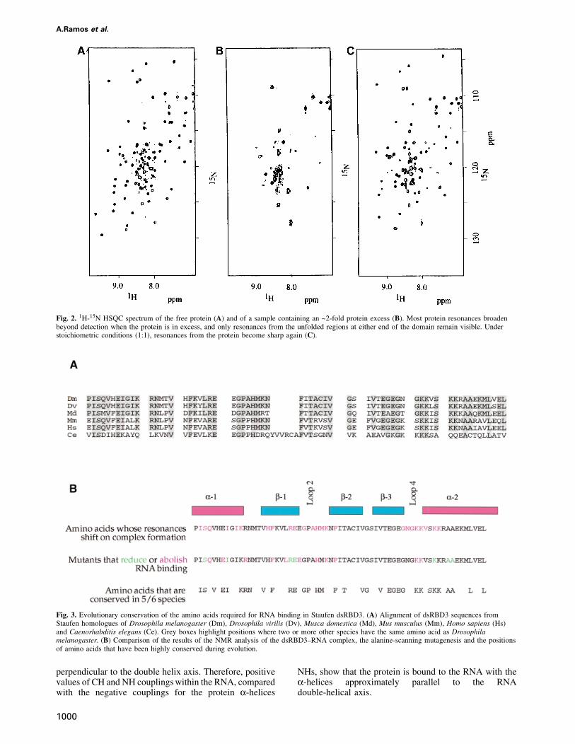
Fig. 2. 1H-15N HSQC spectrum of the free protein (A) and of a sample containing an ~2-fold protein excess (B). Most protein resonances broadenbeyond detection when the protein is in excess, and only resonances from the unfolded regions at either end of the domain remain visible. Understoichiometric conditions (1:1), resonances from the protein become sharp again (C).
Fig. 3. Evolutionary conservation of the amino acids required for RNA binding in Staufen dsRBD3. (A) Alignment of dsRBD3 sequences fromStaufen homologues of Drosophila melanogaster (Dm), Drosophila virilis (Dv), Musca domestica (Md), Mus musculus (Mm), Homo sapiens (Hs)and Caenorhabditis elegans (Ce). Grey boxes highlight positions where two or more other species have the same amino acid as Drosophilamelanogaster. (B) Comparison of the results of the NMR analysis of the dsRBD3–RNA complex, the alanine-scanning mutagenesis and the positionsof amino acids that have been highly conserved during evolution.
perpendicular to the double helix axis. Therefore, positivevalues of CH and NH couplings within the RNA, comparedwith the negative couplings for the protein α-helices
1000
NHs, show that the protein is bound to the RNA with theα-helices approximately parallel to the RNAdouble-helical axis.
Staufen–RNA recognition
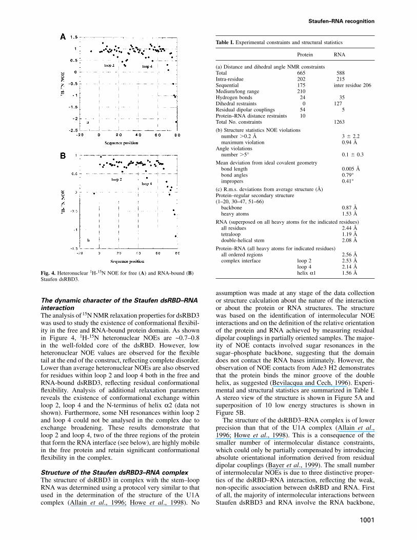
Fig. 4. Heteronuclear 1H-15N NOE for free (A) and RNA-bound (B)Staufen dsRBD3.
The dynamic character of the Staufen dsRBD–RNA
interaction
The analysis of 15N NMR relaxation properties for dsRBD3was used to study the existence of conformational flexibil-ity in the free and RNA-bound protein domain. As shownin Figure 4, 1H-15N heteronuclear NOEs are ~0.7–0.8in the well-folded core of the dsRBD. However, lowheteronuclear NOE values are observed for the flexibletail at the end of the construct, reflecting complete disorder.Lower than average heteronuclear NOEs are also observedfor residues within loop 2 and loop 4 both in the free andRNA-bound dsRBD3, reflecting residual conformationalflexibility. Analysis of additional relaxation parametersreveals the existence of conformational exchange withinloop 2, loop 4 and the N-terminus of helix α2 (data notshown). Furthermore, some NH resonances within loop 2and loop 4 could not be analysed in the complex due toexchange broadening. These results demonstrate thatloop 2 and loop 4, two of the three regions of the proteinthat form the RNA interface (see below), are highly mobilein the free protein and retain significant conformationalflexibility in the complex.
Structure of the Staufen dsRBD3–RNA complex
The structure of dsRBD3 in complex with the stem–loopRNA was determined using a protocol very similar to thatused in the determination of the structure of the U1Acomplex (Allain et al., 1996; Howe et al., 1998). No
1001
Table I. Experimental constraints and structural statistics
Protein RNA
(a) Distance and dihedral angle NMR constraintsTotal 665 588Intra-residue 202 215Sequential 175 inter residue 206Medium/long range 210Hydrogen bonds 24 35Dihedral restraints 0 127Residual dipolar couplings 54 5Protein–RNA distance restraints 10Total No. constraints 1263
(b) Structure statistics NOE violationsnumber �0.2 Å 3 � 2.2maximum violation 0.94 Å
Angle violationsnumber �5° 0.1 � 0.3
Mean deviation from ideal covalent geometrybond length 0.005 Åbond angles 0.79°impropers 0.41°
(c) R.m.s. deviations from average structure (Å)Protein–regular secondary structure(1–20, 30–47, 51–66)
backbone 0.87 Åheavy atoms 1.53 Å
RNA (superposed on all heavy atoms for the indicated residues)all residues 2.44 Åtetraloop 1.19 Ådouble-helical stem 2.08 Å
Protein–RNA (all heavy atoms for indicated residues)all ordered regions 2.56 Åcomplex interface loop 2 2.53 Å
loop 4 2.14 Åhelix α1 1.56 Å
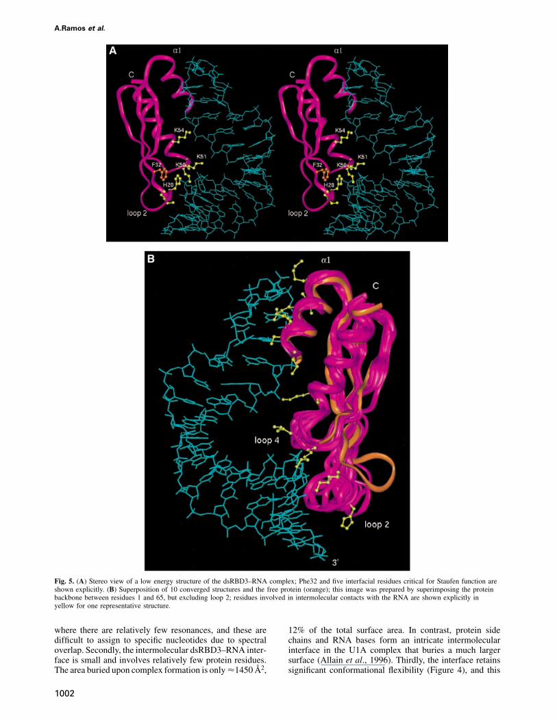
assumption was made at any stage of the data collectionor structure calculation about the nature of the interactionor about the protein or RNA structures. The structurewas based on the identification of intermolecular NOEinteractions and on the definition of the relative orientationof the protein and RNA achieved by measuring residualdipolar couplings in partially oriented samples. The major-ity of NOE contacts involved sugar resonances in thesugar–phosphate backbone, suggesting that the domaindoes not contact the RNA bases intimately. However, theobservation of NOE contacts from Ade3 H2 demonstratesthat the protein binds the minor groove of the doublehelix, as suggested (Bevilacqua and Cech, 1996). Experi-mental and structural statistics are summarized in Table I.A stereo view of the structure is shown in Figure 5A andsuperposition of 10 low energy structures is shown inFigure 5B.
The structure of the dsRBD3–RNA complex is of lowerprecision than that of the U1A complex (Allain et al.,1996; Howe et al., 1998). This is a consequence of thesmaller number of intermolecular distance constraints,which could only be partially compensated by introducingabsolute orientational information derived from residualdipolar couplings (Bayer et al., 1999). The small numberof intermolecular NOEs is due to three distinctive proper-ties of the dsRBD–RNA interaction, reflecting the weak,non-specific association between dsRBD and RNA. Firstof all, the majority of intermolecular interactions betweenStaufen dsRBD3 and RNA involve the RNA backbone,
A.Ramos et al.
Fig. 5. (A) Stereo view of a low energy structure of the dsRBD3–RNA complex; Phe32 and five interfacial residues critical for Staufen function areshown explicitly. (B) Superposition of 10 converged structures and the free protein (orange); this image was prepared by superimposing the proteinbackbone between residues 1 and 65, but excluding loop 2; residues involved in intermolecular contacts with the RNA are shown explicitly inyellow for one representative structure.
where there are relatively few resonances, and these aredifficult to assign to specific nucleotides due to spectraloverlap. Secondly, the intermolecular dsRBD3–RNA inter-face is small and involves relatively few protein residues.The area buried upon complex formation is onlyµ1450 Å2,
1002
12% of the total surface area. In contrast, protein sidechains and RNA bases form an intricate intermolecularinterface in the U1A complex that buries a much largersurface (Allain et al., 1996). Thirdly, the interface retainssignificant conformational flexibility (Figure 4), and this
Staufen–RNA recognition
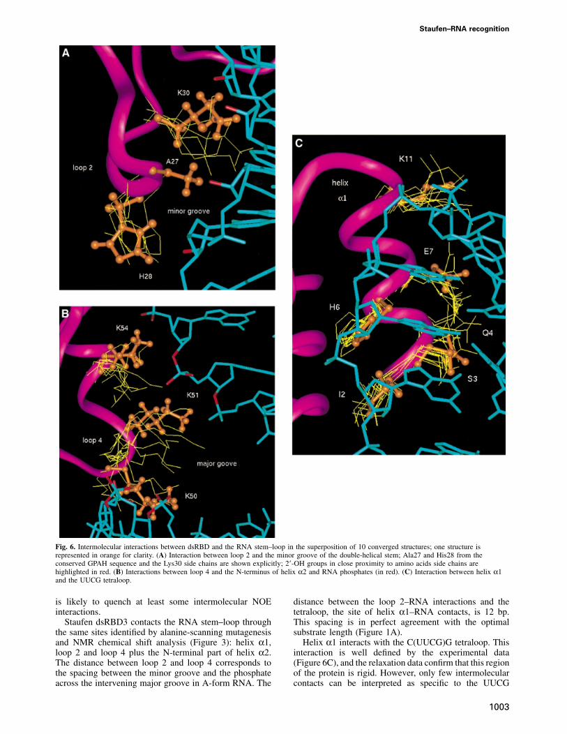
Fig. 6. Intermolecular interactions between dsRBD and the RNA stem–loop in the superposition of 10 converged structures; one structure isrepresented in orange for clarity. (A) Interaction between loop 2 and the minor groove of the double-helical stem; Ala27 and His28 from theconserved GPAH sequence and the Lys30 side chains are shown explicitly; 2�-OH groups in close proximity to amino acids side chains arehighlighted in red. (B) Interactions between loop 4 and the N-terminus of helix α2 and RNA phosphates (in red). (C) Interaction between helix α1and the UUCG tetraloop.
is likely to quench at least some intermolecular NOEinteractions.
Staufen dsRBD3 contacts the RNA stem–loop throughthe same sites identified by alanine-scanning mutagenesisand NMR chemical shift analysis (Figure 3): helix α1,loop 2 and loop 4 plus the N-terminal part of helix α2.The distance between loop 2 and loop 4 corresponds tothe spacing between the minor groove and the phosphateacross the intervening major groove in A-form RNA. The
1003
distance between the loop 2–RNA interactions and thetetraloop, the site of helix α1–RNA contacts, is 12 bp.This spacing is in perfect agreement with the optimalsubstrate length (Figure 1A).
Helix α1 interacts with the C(UUCG)G tetraloop. Thisinteraction is well defined by the experimental data(Figure 6C), and the relaxation data confirm that this regionof the protein is rigid. However, only few intermolecularcontacts can be interpreted as specific to the UUCG
A.Ramos et al.
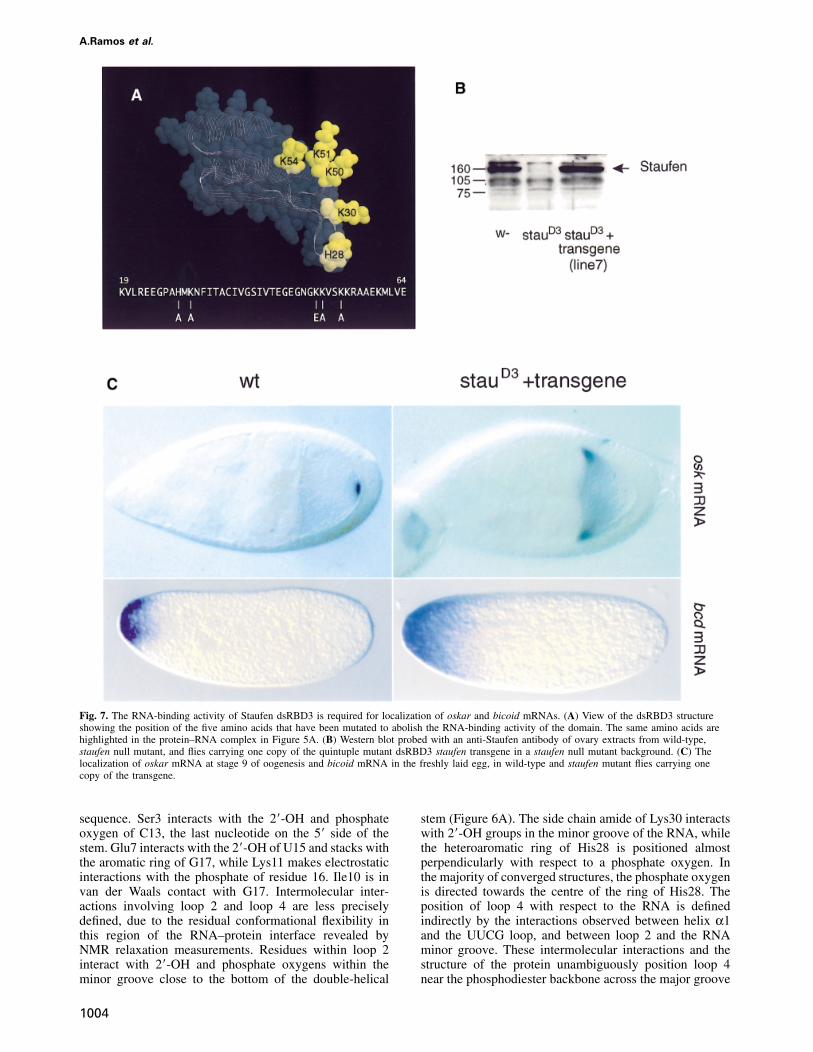
Fig. 7. The RNA-binding activity of Staufen dsRBD3 is required for localization of oskar and bicoid mRNAs. (A) View of the dsRBD3 structureshowing the position of the five amino acids that have been mutated to abolish the RNA-binding activity of the domain. The same amino acids arehighlighted in the protein–RNA complex in Figure 5A. (B) Western blot probed with an anti-Staufen antibody of ovary extracts from wild-type,staufen null mutant, and flies carrying one copy of the quintuple mutant dsRBD3 staufen transgene in a staufen null mutant background. (C) Thelocalization of oskar mRNA at stage 9 of oogenesis and bicoid mRNA in the freshly laid egg, in wild-type and staufen mutant flies carrying onecopy of the transgene.
sequence. Ser3 interacts with the 2�-OH and phosphateoxygen of C13, the last nucleotide on the 5� side of thestem. Glu7 interacts with the 2�-OH of U15 and stacks withthe aromatic ring of G17, while Lys11 makes electrostaticinteractions with the phosphate of residue 16. Ile10 is invan der Waals contact with G17. Intermolecular inter-actions involving loop 2 and loop 4 are less preciselydefined, due to the residual conformational flexibility inthis region of the RNA–protein interface revealed byNMR relaxation measurements. Residues within loop 2interact with 2�-OH and phosphate oxygens within theminor groove close to the bottom of the double-helical
1004
stem (Figure 6A). The side chain amide of Lys30 interactswith 2�-OH groups in the minor groove of the RNA, whilethe heteroaromatic ring of His28 is positioned almostperpendicularly with respect to a phosphate oxygen. Inthe majority of converged structures, the phosphate oxygenis directed towards the centre of the ring of His28. Theposition of loop 4 with respect to the RNA is definedindirectly by the interactions observed between helix α1and the UUCG loop, and between loop 2 and the RNAminor groove. These intermolecular interactions and thestructure of the protein unambiguously position loop 4near the phosphodiester backbone across the major groove
Staufen–RNA recognition
from the site of loop 2 interactions with the minor groove.Three critical lysine residues within loop 4 and theN-terminus of helix α2, Lys50, Lys51 and Lys54, interactwith phosphate oxygens and one 2�-OH group (Figure 6B)across the major groove from the sites of loop 2–minorgroove interaction. The side chains of Lys50 and Lys51bridge the major groove by interacting with RNA phos-phates across the major groove from each other, whileLys54 reinforces these contacts by interacting with thephosphate immediately following the site of interactionof Lys51.
Comparison of the structure of dsRBD3 free and in theRNA complex confirms that the structure of the proteindoes not change significantly on RNA binding, with theexception of loop 2. The rotation of loop 2 (towards theRNA in Figure 5B) is necessary to allow interactionsbetween this region of the protein and the RNA. TheRNA double-helical region preserves the A-form structurethroughout the double-helical stem, and the UUCG tetra-loop is in its well characterized conformation in thepresence or absence of the protein. The only significantchange in RNA structure upon protein binding is a kinkat the stem–loop junction, resulting in the bent appearanceof the RNA in the complex (Figure 5A). The presence ofthis distortion is supported indirectly by the observationof significantly shifted resonances in this region of theRNA. The bend allows the interaction between helix α1and the tetraloop to occur at the same time as the contactsbetween loop 2 and the RNA minor groove.
dsRBD mutagenesis in vivo
The biochemical and structural data on dsRBD3 describedabove provide a framework to analyse whether the RNA-binding activity of this domain is required for Staufenfunction. Five highly conserved basic amino acids withinloop 2 and loop 4 (His28, Lys30, Lys50, Lys51 and Lys54)are required for RNA binding in vitro, and lie at theRNA–protein interface where they interact with the RNA(Figure 5A). To generate a form of domain 3 that iscompletely null for RNA binding, we replaced all five ofthese amino acids with uncharged or negatively chargedresidues. 1H-15N HSQC spectra of the bacterially expressedquintuple mutant dsRBD3 are very similar to that of thewild-type protein (data not shown), demonstrating thatmutant and wild-type proteins adopt the same conforma-tion. Consistent with this observation, the domain dis-played normal solubility and stability when expressed inEscherichia coli, but its in vitro RNA-binding activitywas abolished. The DNA encoding this mutant domainwas inserted into a staufen cDNA in place of the wild-type domain, and then transformed into the Drosophilagermline in a vector that directs expression of the transgenein the female ovary (Micklem et al., 1997). In controlflies, a single copy of the wild-type staufen transgenecompletely rescues the maternal effect of a staufen nullmutation, and restores the wild-type localization of bothoskar and bicoid mRNAs. In contrast, 10 independentinsertions of the dsRBD3 mutant construct give no rescueof the staufen phenotype. In one line that was examinedin detail, the mutant Staufen protein is expressed in thefemale germline at the same level as the wild-type protein(Figure 7B). Nevertheless, the mutant protein does notrescue the localization of oskar mRNA to the posterior of
1005
the oocyte, nor the anchoring of bicoid mRNA at theanterior of the egg (Figure 7C), and 100% of embryos diewith head defects and no abdomen or pole cells (data notshown). Thus, in the transgenic flies, oskar mRNA is nottransported to the posterior of the oocyte, and bicoidmRNA fails to be anchored at the anterior of the egg,showing that the transgene does not rescue the staufenmutant phenotype. Thus, the amino acids in dsRBD3 thatinteract with dsRNA in vitro are required for the in vivofunction of Staufen. These results demonstrate for the firsttime that the dsRNA-binding activity of dsRBD3 isessential for the interaction of Staufen protein with bicoidand oskar mRNAs, strongly suggesting that Staufen bindsdirectly to these transcripts in vivo.
Discussion
We have studied how the third dsRBD from DrosophilaStaufen protein recognizes RNA and have describedfeatures of the dsRBD–RNA interaction that are verylikely to be of general relevance to RNA recognition byall dsRBD-containing proteins. We have also shown forthe first time that direct interactions between individualdsRBDs and RNA are essential for Staufen function inRNA localization and early development. These resultssuggest very strongly that Staufen binds directly to theoskar, bicoid and prospero 3�-UTRs to mediate the localiz-ation of these mRNAs in vivo, and describe the molecularinteractions that are necessary for this to occur.
Molecular basis of the interaction of dsRBDs with
dsRNA
The biochemical and structural data presented here identifythe three regions of the dsRBD that mediate the bindingof the domain to RNA: helix α1, loop 2 and loop 4.Mutations of amino acids in each of these regions abolishor reduce RNA binding significantly, whereas mutationsin surface residues in other regions of the protein have noeffect. Furthermore, the amino acids in these regions arehighly conserved in Staufen homologues from Drosophilato humans, indicating that they play an essential role inthe function of the domain. These results are likely tobe applicable to other dsRBD-containing proteins, sincemutagenesis studies have shown that analogous regionsof other dsRBDs are required for interaction with RNA. Forexample, the RNA-binding activity of PKR is significantlyreduced by mutations in the first α-helix or in the lysine-rich loop 4 of the first dsRBD of this protein (Green andMatthews, 1992; Green et al., 1995). Similarly, theRNA-binding activity of dsRBD2 of Xlrbpa is severelycompromised by mutation of a histidine in loop 2 that isequivalent to His28 in Staufen dsRBD3 (Krovat andJantsch, 1996).
The crystallographic structure of the complex betweendsRBD2 of Xlrbpa and dsRNA (Ryter and Schultz, 1998)identified the same three regions of the domain that contactRNA as our NMR structure. Both structures show that loop 2interacts with the minor groove of the RNA (Figure 6A), andloop 4 interacts with the phosphodiester backbone across themajor groove from the sites of loop 2 contacts (Figure 6B).However, the two structures provide very different descrip-tions of the interaction between helix α1 and RNA. In thecrystal structure, helix α1 interacts with the minor groove
A.Ramos et al.
of a second RNA duplex that abuts the first RNA moleculeto form a pseudo-continuous double helix. As a con-sequence, the Xlrbpa dsRBD covers 16 bp across the junc-tion between the two RNA molecules. In contrast, helix α1of Staufen dsRBD3 binds to a tetraloop that caps a 12 bpstem of perfect A-form RNA, but this interaction requiresbending of the RNA at the stem–loop junction. This struc-tural difference is significant. As shown in Figure 1, StaufendsRBD3 would not bind the 10 bp RNA duplex used inthe crystallographic studies, but binds optimally to RNAhairpins with a stem of 12 bp. This length requirement islikely to be important because RNA duplexes of 16 bp donot exist within the bicoid 3�-UTR.
Within loop 2 and loop 4, most differences betweenthe NMR and crystallographic structures are attributableto the lower precision of the NMR structure and thedifferent dynamic behaviour of the protein–RNA com-plexes in the two systems, as discussed below. However,one important difference concerns His28. In the crystalstructure of the Xlrbpa–dsRNA complex, the side chainof His141 stacks on Phe145 (corresponding to His28 andPhe32 in the present numbering system) and interacts witha 2�-OH group (Ryter and Schultz, 1998). However, thisinteraction requires a backbone conformation inconsistentwith the NMR data. In the present structure, the histidine–phenylalanine stacking interaction is not present, andthe histidine–phosphate interaction we observe is insteadsimilar to a contact reported between a phenylalanine sidechain and a DNA phosphate, as observed in the structureof the P22 Arc repressor–DNA complex (Schildbacket al., 1999). The phenylalanine–DNA interaction plays aprominent role in determining the specificity of recognitionby modulating the structure of the protein–DNA interface.Mutation of His28 to alanine in Staufen dsRBD3 abolishesRNA binding (Figure 1B), suggesting that interactionsbetween heteroaromatic side chains and the phosphatescould play important roles in RNA recognition as well.
Specificity of the dsRBD for dsRNA
Since NMR and X-ray structures reveal different inter-actions between helix α1 and RNA, it seems very likelythat the common interactions involving loop 2 and loop 4account for the specificity of both domains for dsRNA.Further support for this view comes from the analysis ofthe N-terminal domain of bacterial ribosomal protein S5,which has a very similar fold to the dsRBD and containsmany of the conserved residues that form the hydrophobiccore of the domain but lacks the N-terminal α-helix foundin the dsRBD (Bycroft et al., 1995). S5 interacts withhelix 34 in 16S rRNA (Heilik and Noller, 1996; Davieset al., 1998). The present results suggest that S5 binds torRNA through loops 2 and 4 alone. The ability of thesetwo loops to discriminate between dsRNA and DNA canbe attributed firstly to interactions with 2�-OH groups inthe RNA minor groove, as originally described in theXlrbpa crystal structure (Ryter and Schultz, 1998).In addition, the spacing between loop 2 and loop 4corresponds well with the spacing and groove distancesfound in the A-type helix formed by dsRNA, but wouldnot fit the B-type helix of dsDNA. Consistent with thisinterpretation, mutation of Phe32 abolishes RNA bindingcompletely. Phe32 is buried between loop 2 and loop 4(Figure 5A); the present structure suggests very strongly
1006
that its identity is essential to position these loops withrespect to the RNA.
Dynamic nature of the dsRBD–dsRNA interaction
An important aspect of dsRBD interaction revealed bythe NMR analysis is the dynamic character of the interface.Broadening of side chain and backbone resonances, theresults reported in Figure 4 and other relaxation parametersdetermined in the course of this study all demonstrate thatloop 2, loop 4 and the N-terminus of helix α1 retainsignificant conformational flexibility in the protein–RNAcomplex. When the NMR structures are compared, aminoacid side chains from loop 2 and loop 4 are foundto interact with different acidic groups on the RNA.Electrostatic interactions similar to those observed in thecrystal structure can be observed in all structures thatsatisfy the NMR data (Figure 6A and B), but the sameprotein side chain sometimes interacts with different 2�-OHor phosphate groups. This description of the intermolecularinterface provided by the NMR data is entirely consistentwith the observation of a disordered loop 2 interface inthe second of the two Xlrbpa dsRBD2 molecules inthe crystallographic asymmetric unit. This observationsuggests that interactions mediated by loop 2 are dynamicin the crystal as well.
The absence in the dsRBD–RNA structure of significantreorganizations of the RNA or protein structures was asurprise, since induced fit has so far been a nearly universalfeature of RNA recognition by proteins and small molecules(Varani, 1997). Furthermore, the dsRBD–RNA complexdoes not contain a tightly packed intermolecular interface.In both respects, dsRBD–RNA recognition differs substan-tially from the paradigm for RNA recognition establishedby human U1A protein (Oubridge et al., 1994; Allain et al.,1996). Formation of the U1A–RNA complex requires signi-ficant rearrangements in RNA and protein structures,resulting in a tightly packed intermolecular interface.Staufen dsRBD3 sits instead on the edge of the RNA doublehelix and interacts with the RNA sugar–phosphate back-bone, without making intimate contacts with the bases. Theabsence of direct contacts with the RNA bases, the lack of arequirement for distortion in RNA structure and the residualconformational flexibility present at the RNA–protein inter-face all contribute to the lack of sequence specificity inrecognition of dsRNA.
The significance of the interaction between helix
α1 and the single-stranded loop
The unexpected observation of interactions between helixα1 and the single-stranded loop raises the question ofwhether these are physiologically significant. The alanine-scanning data demonstrate that surface-exposed residueswithin helix α1 make essential contributions to RNAbinding. Mutation of Gln4, Glu7 and Arg12 abolishesbinding, while substitution of Ser3 reduces binding signi-ficantly. Furthermore, Ser3, Gln4, Glu7, Lys11 and Arg12have been conserved during the evolution of StaufendsRBD3, suggesting that these exposed amino acids playan important role in Staufen function. This hypothesiscould be addressed by mutating these residues in full-length Staufen. The observation in our structure of welldefined intermolecular interactions mediated by these
Staufen–RNA recognition
residues (Figure 6C) raises the possibility that these aminoacids play a critical role in RNA recognition.
Intriguing clues as to the diverse functional role ofhelix α1 compared with loops 2 and 4 are provided bythe extension of the phylogenetic comparison with otherdsRBDs. The key residues within loops 2 and 4 (His28,Lys/Arg30, Lys50, Lys51 and Lys54) are highly conservedacross species in Staufen dsRBD3 (Figure 3), as well asStaufen dsRBD1, a second domain of the protein thatbinds dsRNA (J.Adams, S.Grunert and D.St Johnston,unpublished results). In contrast, interfacial residues fromhelix α1 are highly conserved for each domain whendifferent species are examined, but are significantlydivergent when the two domains are compared witheach other, even within the same species. dsRBD1 containsthe conserved Glu7, but has serine–cysteine substitutionat position 3, a conserved leucine instead of Glu4, phenyl-alanine or tyrosine in place of Lys11, and a conservedglutamine in place of Arg12. Similarly, Xlrbpa dsRBD2contains the same key residues in loops 2 and 4 as foundin Staufen dsRBD1 and dsRBD3, but differs from bothStaufen domains in four out of five of the exposedpositions in helix α1.
The preceding observations indicate that the identity ofresidues within helix α1 is conserved and domain specific,raising the possibility that different dsRBD domains cancontribute to specificity in Staufen–RNA recognition byforming different helix α1–loop interactions. It is verylikely that multiple domains from Staufen bind bicoid3�-UTR, but this does not rule out at all the possibilitythat helix α1 contributes to specificity. The data presentedin Figure 7 demonstrate that dsRBD3–RNA interactionsare critical for Staufen function. However, the RNA usedin the present study was optimized for affinity in order tofacilitate structural studies, and does not correspond toany of the stem–loops within bicoid 3�-UTR. Therefore,we cannot yet identify the binding site for Staufen dsRBD3in bicoid 3�-UTR.
Support for the importance of helix α1–loop interactionsin dsRBD proteins is provided by yeast Rnt1 protein, theeukaryotic RNase III. This dsRBD-containing enzymecleaves pre-rRNA and a set of snoRNA precursors at sitesdefined by a conserved hairpin loop (Elela et al., 1996;Chanfreau et al., 1998). Mutation of the AG sequencewithin the loop reduces Rnt1 binding and severely affectsRNA processing. The site of cleavage is always separatedfrom the apical hairpin loop by 14–16 bp, often interruptedby internal loops or bulges (Chanfreau et al., 1998). Thisseparation is just a few base pairs longer than the footprintof Staufen dsRBD3 on our stem–loop structure (12 bp).Although it is not yet possible to exclude a role forthe catalytic domain in Rnt1–substrate recognition, it istempting to suggest that interactions between helix α1 ofthe Rnt1 dsRBD and the tetraloop, analogous to thosedescribed here, could be important in defining the substratespecificity of Rnt1.
The RNA-binding activity of dsRBD proteins hasbeen defined by in vitro studies as non-sequence-specificrecognition of perfect dsRNA substrates. However, someof the most extensively investigated dsRBD-containingproteins, such as Staufen, Rnt1 and PKR, regulate theactivity of very specific RNAs in vivo. The structural andphylogenetic analysis presented here suggest that domain-
1007
specific interactions between helix α1 and single-strandedRNA loops could modulate the specificity of individualdsRBD domains and provide selectivity in the recognitionof cellular RNAs.
Materials and methods
Protein and RNA preparationThe dsRBD3 fragment (residues 579–646 of Drosophila Staufen protein)and the various mutant proteins described in the present report wereexpressed in E.coli using appropriate isotope-labelled nutrients andpurified as described (Bycroft et al., 1995). The RNA used in NMRstudies was: 5�-GGACAGCUGUCC(CUUCGG)GGACAGCUGUCC-3�(the tetraloop sequence and flanking base pair is within parentheses).Unlabelled and isotopically labelled RNA oligonucleotides were synthe-sized in vitro using T7 RNA polymerase and synthetic DNA templates(Price et al., 1998).
Mutagenesis of dsRBD3Mutagenesis was performed using a two-step PCR mutagenesis protocol.Briefly, we used two primers (sequences available upon request) flankingthe dsRBD3 with BamHI and EcoRI restriction sites, which enabledcloning of the dsRBD3-derived PCR fragments in-frame with GSTinto pGEX2-T (Promega). For each mutation, we synthesized a thirdmutagenic primer of the desired sequence, including silent mutations tocreate novel restriction sites in the mutated PCR product. PCRs wereperformed as described (Bycroft et al., 1995) using Pfu polymerase(Stratagene). The resulting PCR products were subcloned into pGEX2-Tand minipreparations were screened for the newly introduced restrictionsites. The presence of the desired mutations was verified by sequencing.
RNA-binding assayNorth-western binding assays were performed as described (St Johnstonet al., 1992). Radiolabelled short hairpins were synthesized byin vitro transcription of synthetic oligonucleotides. Approximately100 000 c.p.m./ml were used for each probe.
Identification of staufen homologues in other speciesstaufen homologues from Drosophila virilis and Musca domestica wereidentified by screening genomic libraries at low stringency with probesderived from the five dsRBDs of the Drosophila melanogaster gene;positive clones were sequenced using the µ transposon strategy(Strathmann et al., 1991). A human staufen homologue was identifiedfrom the homology of EST HFBDQ83 (DDBJ/EMBL/GenBank acces-sion Nos T06248 and T06429) to Drosophila staufen; the correspondingcDNA clone was sequenced in its entirety. Four cDNAs encoding amouse homologue of staufen were identified by screening a 7.7 dayspost-conception mouse embryonic cDNA library with the humancDNA HFBDQ83. The analysis of the sequence of these homologueswill be reported in detail elsewhere (D.R.Micklem, J.Adams, S.Grunertand D.St Johnston, in preparation).
Generation of mutant staufen transgenic linesFive mutations were introduced into dsRBD3 in a full-length staufencDNA clone by performing two consecutive rounds of mutagenesis asdescribed in the supplementary material (available in The EMBO JournalOnline). An XhoI–MluI fragment containing the mutated dsRBD3 wascloned into the wild-type staufen cDNA, and this was then inserted intotransformation vector D277 as described (Schuldt et al., 1998). Thisproduced a construct in which the female germline-specific α4-tubulinpromoter drives the expression of a fusion protein that contains aminoacids 1–9 of α4-tubulin, a 16 amino acid myc epitope and amino acids18–1026 of Staufen. This construct was introduced into the germline ofw–; cn stauD3 sp/Cy O flies by P element-mediated transformation (Rubinand Spradling, 1982). Multiple independent insertions of the transgenewere tested for their ability to rescue the staufen maternal effectphenotype. This was done by performing cuticle preparations on theprogeny of stauD3 homozygous females that carry one copy of thetransgene, and by examining the localization of bicoid and oskar mRNAsin the ovaries and eggs of these females by in situ hybridization. Theexpression of the mutant protein was monitored by performing Westernblots on ovary extracts of females of the same genotype, and by stainingwith a rabbit anti-Staufen antibody (St Johnston et al., 1991).
A.Ramos et al.
NMRNMR spectra were recorded on a Bruker DMX-600 spectrometer.Several RNA–protein samples were prepared: [15N]dsRBD3 and[15N-13C]dsRBD3 bound to unlabelled RNA and 15N-13C-labelled RNAwith unlabelled dsRBD3. Samples were ~1 mM in each component; theRNA to protein ratio was adjusted by integrating the intensity of wellresolved protein and RNA resonances. An extensive set of two- andthree-dimensional NMR spectra was recorded on free protein and RNAcomponents and on the different preparations of RNA–protein complexes.A thorough description of methodological aspects of this work will bepresented elsewhere. In total, 98% of protein backbone resonances, 70%of side chain resonances and �95% of all RNA resonances were assignedunambiguously in the complex. Assignments of free and bound RNAspectra were obtained by analysing an extensive set of two- and three-dimensional NMR spectra, as described previously (Varani et al., 1996).Heteronuclear 1H-15N NOE, T1 and T2 relaxation times for dsRBD3backbone amide resonances were recorded and analysed essentially asreported (Farrow et al., 1994). Partially oriented samples for theextraction of NH and CH bond orientations were prepared by mixingsamples of the complex with appropriately labelled phospholipid solu-tions, as described previously (Tjandra and Bax, 1997; Bayer et al.,1999). Residual dipolar couplings and the orientation tensor wereobtained by a variational procedure (Bayer et al., 1999).
Structure determinationDistance constraints for the RNA and protein components of the complexwere obtained by analysing three-dimensional 13C- and 15N-editedNOESY spectra recorded at 100 ms mixing time and two-dimensionalNOESY spectra recorded at mixing times of 50 and 100 ms. Theexperimental constraints were generated and interpreted exactly as inthe determination of the U1A protein–RNA complex (Howe et al.,1998). Intermolecular NOE interactions were identified in 1/2� filteredNOESY spectra (Otting and Wuthrich, 1990). Ten NOEs could beidentified unambiguously and were used in the structure calculation;these are listed in the supplementary material online. Additional NOEcross-peaks involving protein side chains and RNA sugar resonancesare observable and could often be assigned to specific residue types (e.g.H2�, H3� or H4�), but could not be assigned unambiguously to individualRNA nucleotides or protein side chains due to spectral overlap.
The structure of the protein–RNA complex was calculated using anX-PLOR-based simulated annealing protocol optimized for the calcula-tion of the structure of the U1A complex (Howe et al., 1998). The useof residual dipolar couplings in the refinement step necessitated theintroduction of a modified refinement protocol (Tjandra et al., 1997;Bayer et al., 1999) (see supplementary material online). Thirty-sixconverged and were clearly identified from a significant difference intotal energy from other structures (Howe et al., 1998), and were energyminimized by introducing the electrostatic component of the potential.Differences between structures calculated before and after minimizationwere smaller than the uncertainty in the structures themselves. Statisticsfor the experimental constraints and structural statistics are reportedin Table I.
Supplementary data
Supplementary data to this paper are available in The EMBO JournalOnline.
Acknowledgements
It is a pleasure to thank Dr Peter Bayer for help in measurement ofresidual dipolar couplings and their incorporation in the structuredetermination protocol, and Lisa Elphick for help in generating thetransgenic Drosophila lines. J.A. was supported by a BoehringerIngelheim studentship; D.R.M. by a Wellcome Trust Prize studentship;D.StJ. and S.G. by the Wellcome Trust; and A.R. by an EU trainingfellowship and by the MRC.
References
Allain,F.H.-T., Gubser,C.C., Howe,P.W.A., Nagai,K., Neuhaus,D. andVarani,G. (1996)Specificityof ribonucleoprotein interactiondeterminedby RNA folding during complex formation. Nature, 380, 646–650.
Bass,B.L., Hurst,S.R. and Singer,J.D. (1994) Binding properties of newly
1008
identified Xenopus proteins containing dsRNA-binding motifs. Curr.Biol., 4, 301–314.
Bayer,P., Varani,L. and Varani,G. (1999) Refinement of the structure ofprotein–RNA complexes by residual dipolar coupling analysis.J. Biomol. NMR, 14, 149–155.
Bevilacqua,P.C. and Cech,T.R. (1996) Minor-groove recognition of thedouble-stranded RNA-binding domain from the RNA-activated proteinkinase PKR. Biochemistry, 35, 9983–9994.
Broadus,J., Fuestenberg,S. and Doe,C.Q. (1998) Staufen-dependentlocalization of prospero mRNA contributes to neuroblast daughter-cellfate. Nature, 391, 792–795.
Bycroft,M., Grunert,S., Murzin,A.G., Proctor,M. and St Johnston,D.(1995) NMR solution structure of a dsRNA binding domain fromDrosophila Staufen protein reveals homology to the N-terminal domainof ribosomal protein S5. EMBO J., 14, 3563–3571.
Chanfreau,G., Legrain,P. and Jacquier,A. (1998) Yeast RNase III as a keyprocessing enzyme in small nucleolar RNAs metabolism. J. Mol. Biol.,284, 975–988.
Clarke,P.A. and Matthews,M.B. (1995) Interactions between the double-stranded RNA binding motif and RNA: definition of the binding site forthe interferon-induced protein kinase DAI (PKR) on adenovirus VARNA. RNA, 1, 7–20.
Davies,C., Bussiere,D.E., Golden,B.L., Ramakrishnan,V. and White,S.W.(1998) Ribosomal proteins S5 and L6; high-resolution crystal structuresand roles in protein synthesis and antibiotic resistance. J. Mol. Biol.,279, 873–888.
Elela,S.A., Igel,H. and Ares,M.J. (1996) RNase III cleaves eukaryoticpreribosomal RNA at a U3 snoRNP-dependent site. Cell, 85, 115–124.
Ephrussi,A., Dickinson,L.K. and Lehmann,R. (1991) oskar organizes thegerm plasm and directs localization of the posterior determinant nanos.Cell, 66, 37–50.
Farrow,N.A. et al. (1994) Backbone dynamics of a free and aphosphopeptide complexed Src homology 2 domain studied by 15Nrelaxation. Biochemistry, 33, 5984–6003.
Ferrandon,D., Elphick,L., Nusslein-Volhard,C. and St Johnston,D. (1994)Staufen protein associates with the 3�UTR of bicoid mRNA to formparticles that move in a microtubule-dependent manner. Cell, 79,1221–1232.
Gibson,T.J. and Thompson,J. (1994) Detection of dsRBA-binding domainsin the RNA helicase A and Drosophila Maleless: implications formonomeric RNA helicases. Nucleic Acids Res., 22, 2552–2556.
Green,S.R. and Matthews,M.B. (1992) Two RNA-binding motifs in thedouble-stranded RNA-activated protein kinase, DAI. Genes Dev., 6,2478–2490.
Green,S.R., Manche,L. and Mathews,M.B. (1995) Two functionallydistinct RNA-binding motifs in the regulatory domain of the proteinkinase DAI. Mol. Cell. Biol., 15, 358–364.
Heilik,G.M. and Noller,H.F. (1996) Site-directed hydroxyl radical probingof the rRNA neighborhood of ribosomal protein S5. Science, 272,1659–1662.
Howe,P.W.A., Allain,F.H.-T., Varani,G. and Neuhaus,D. (1998)Determination of the NMR structure of the complex between U1Aprotein and its RNA polyadenylation inhibition element. J. Biomol.NMR, 11, 59–84.
Kharrat,A., Macias,M.J., Gibson,T.J., Nilges,M. and Pastore,A. (1995)Structure of the dsRNA binding domain of E.coli RNase III. EMBO J.,14, 3572–3584.
Kim-Ha,J., Smith,J.L. and Macdonald,P.M. (1991) oskar mRNA islocalized to the posterior pole of the Drosophila oocyte. Cell, 66, 23–35.
Kim-Ha,J., Webster,P., Smith,J. and MacDonald,P. (1993) Multiple RNAregulatory elements mediate distinct steps in localization of oskarmRNA. Development, 119, 169–178.
Krovat,B.C. and Jantsch,M.F. (1996) Comparative mutational analysis ofthe double-stranded RNA binding domains of Xenopus laevis RNA-binding protein A. J. Biol. Chem., 271, 28112–28119.
MacDonald,P.M. and Struhl,G. (1988) Cis-acting sequences responsiblefor anterior localization of bicoid mRNA in Drosophila embryos.Nature, 336, 595–598.
Micklem,D.R., Dasgupta,R., Elliott,H., Gergely,F., Davidson,C.,Brand,A., Gonzalez-Reyes,A. and St Johnston,D. (1997) The magonashi gene is required for the polarisation of the oocyte and the formationof perpendicular axes in Drosophila. Curr. Biol., 7, 468–478.
Nanduri,S., Carpick,B.W., Yang,Y., Williams,B.R.G. and Qin,J. (1998)Structure of the double stranded RNA-binding domain of the proteinkinase PKR reveals the molecular basis of its dsRNA-mediatedactivation. EMBO J., 17, 5458–5465.
Otting,G. and Wuthrich,K. (1990) Heteronuclear filters in two-dimensional
Staufen–RNA recognition
[1H-1H]-NMR spectroscopy: combined use with isotope labeling forstudies of macromolecular conformation and intermolecularinteractions. Q. Rev. Biophys., 23, 39–96.
Oubridge,C., Ito,N., Evans,P.R., Teo,C.-H. and Nagai,K. (1994) Crystalstructure at 1.92 Å resolution of the RNA-binding domain of the U1Aspliceosomal protein complexed with an RNA hairpin. Nature, 372,432–438.
Price,S.R., Oubridge,C., Varani,G. and Nagai,K. (1998) Preparation ofRNA–protein complexes for X-ray crystallography and NMR. InSmith,C. (ed.), RNA–Protein Interactions: A Practical Approach.Oxford University Press, Oxford, UK, pp. 48–74.
Rubin,G. and Spradling,A.C. (1982) Genetic transformation of Drosophilawith transposable elements vectors. Science, 218, 348–353.
Ryter,J.M. and Schultz,S.C. (1998) Molecular basis of double-strandedRNA–protein interactions: structure of a dsRNA-binding domaincomplexed with dsRNA. EMBO J., 17, 7505–7513.
Schildback,J.F., Karzai,A.W., Raumann,B.E. and Sauer,R.T. (1999)Origins of DNA-binding specificity: role of protein contacts with theDNA backbone. Proc. Natl Acad. Sci. USA, 96, 811–817.
Schmedt,C., Green,S.R., Manche,L., Taylor,D.R., Ma,Y. andMathews,M.B. (1995) Functional characterization of the RNA-bindingdomain and motif of the double-stranded RNA-dependent protein kinaseDAI (PKR) J. Mol. Biol., 249, 29–44.
Schuldt,A.J., Adams,J.H.J., Davidson,C.M., Micklem,D.R., Haseloff,J.,St Johnston,D. and Brand,A.H. (1998) Miranda mediates asymmetricprotein and RNA localization in the developing nervous system. GenesDev., 12, 1847–1857.
St Johnston,D. (1995) The intracellular localization of messenger RNAs.Cell, 81, 161–170.
St Johnston,D., Driever,W., Berleth,T., Richstein,S. and Nusslein-Volhard,C. (1989) Multiple steps in the localization of bicoid RNA tothe anterior pole of the Drosophila oocyte. Development Suppl., 107,13–19.
St Johnston,D., Beuchle,D. and Nusslein-Volhard,C. (1991) Staufen, agene required to localize maternal RNAs in the Drosophila egg. Cell,66, 51–63.
St Johnston,D., Brown,N.H., Gall,J.G. and Jantsch,M. (1992) A conserveddouble-stranded RNA binding domain. Proc. Natl Acad. Sci. USA, 89,10979–10983.
Strathmann,M., Hamilton,B.A., Mayeda,C.A., Simon,M.I.,Meyerowitz,E.M. and Palazzolo,A. (1991) Transposon-facilitatedDNA sequencing. Proc. Natl Acad. Sci. USA, 88, 1247–1250.
Tjandra,N. and Bax,A. (1997) Direct measurement of distances and anglesin biomolecules by NMR in a dilute liquid crystalline medium. Science,278, 1111–1114.
Varani,G. (1997) RNA–protein intermolecular recognition. Acc. Chem.Res., 30, 189–195.
Varani,G., Aboul-ela,F. and Allain,F.H.-T. (1996) NMR investigations ofRNA structure. Prog. NMR Spectrosc., 29, 51–127.
Received November 25, 1999; revised December 21, 1999;accepted January 12, 2000
1009