Sequenzspezifische Erkennung und Modifikation von Doppelhelix-DNA durch Oligonucleotide
Upload
mondodomaniCategory
view
2download
0
Using Triplex-Forming Oligonucleotide Probes for theReagentless, Electrochemical Detection of Double-StrandedDNA
Adriana Patterson†, Felice Caprio‡, Alexis Vallée-Bélisle†, Danila Moscone‡, Kevin W.Plaxco†,§, Giuseppe Palleschi‡, and Francesco Ricci*,‡Interdepartmental Program in Biomolecular Science and Engineering, and Department ofChemistry and Biochemistry, University of California–Santa Barbara, Santa Barbara, California93106, United States, and Dipartimento di Scienze e Tecnologie Chimiche, Università di RomaTor Vergata, Via della Ricerca Scientifica, 00133, Rome, Italy
AbstractWe report a reagentless, electrochemical sensor for the detection of double-stranded DNA targetsthat employs triplex-forming oligonucleotides (TFOs) as its recognition element. These sensorsare based on redox-tagged TFO probes strongly chemisorbed onto an interrogating gold electrode.Upon the addition of the relevant double-stranded DNA target, the probe forms a rigid triplexstructure via reverse Hoogsteen base pairing in the major groove. The formation of the tripleximpedes contact between the probe’s redox moiety and the interrogating electrode, thus signalingthe presence of the target. We first demonstrated the proof of principle of this approach by using awell-characterized 22-base polypurine TFO sequence that readily detects a synthetic, double-stranded DNA target. We then confirmed the generalizability of our platform with a second probe,a 19-base polypyrimidine TFO sequence that targets a polypurine tract (PPT) sequence conservedin all HIV-1 strains. Both sensors rapidly and specifically detect their double-stranded DNAtargets at concentrations as low as ~10 nM and are selective enough to be employed directly incomplex sample matrices such as blood serum. Moreover, to demonstrate real-world applicabilityof this new sensor platform, we have successfully detected unpurified, double-stranded PCRamplicons containing the relevant conserved HIV-1 sequence.
Many DNA detection methods are based on hybridization and thus require the generation ofsingle-stranded DNA prior to analysis. For example, to properly generate a signal, methodssuch as DNA microarrays, Southern blotting, and in situ hybridization require denaturationof the double-stranded DNA of interest into single strands, followed by their subsequentrenaturation with specific probes. In response, a number of approaches have been developedin which double-stranded targets are detected directly, such as the use of intercalating dyes.1–3 As these approaches lack sequence specificity, however, they are prone to false positivesarising due to, for example, spurious amplification.4 The development of assays that areboth sequence specific and avoid the cumbersome need to generate single-stranded DNAtargets would thus significantly simplify DNA detection.
Several approaches have been recently proposed for the direct, sequence-specific detectionof double-stranded DNA. Most of these employ non-DNA recognition probes and opticalread-outs. Such probes include low molecular weight polyamides, which bind to specific
*To whom correspondence should be addressed. [email protected].†Interdepartmental Program in Biomolecular Science and Engineering, University of California–Santa Barbara.‡Università di Roma Tor Vergata.§Department of Chemistry and Biochemistry, University of California–Santa Barbara.
NIH Public AccessAuthor ManuscriptAnal Chem. Author manuscript; available in PMC 2012 April 11.
NIH
-PA Author Manuscript
NIH
-PA Author Manuscript
NIH
-PA Author Manuscript

minor groove sequences,5,6 or DNA binding proteins, which target specific duplexsequences.7–9 However, while several of these methods have been reduced to practice in thelaboratory, their use in “real-world” settings is beset by drawbacks. Protein-based methods,for example, showed promising features of selectivity and sensitivity toward double-stranded targets,10 but they require cumbersome selection processes for the production ofthe relevant affinity reagents, and the use of proteins as recognition elements reduces thestability of the platform. Polyamide probes are likewise limited by the length of the targetedDNA sequence, which is generally between four to six base pairs, although, with recognitionfootprints of 10–12 base pairs, head-to-head bis-hairpin minor groove binders (MGBs) are arecent exception.11,12 The utility of these approaches to the direct, sequence-specificdetection of double-stranded DNA has thus proven limited to date.
A potential route toward the direct, sequence-specific detection of double-stranded DNA isthe observation that many such sequences support the formation of triplex structures inwhich a third strand of DNA runs along the major groove of the double helix, where it formsHoogsteen base pairs.13–15 Specifically, triplex-forming oligonucleotides (TFOs), which arehomopurine or homopyrimidine oligonucleotides that bind in the major groove ofhomopurine–homopyrimidine duplexes,16 exhibit high affinity and specificity, suggestingthat they might serve as viable probes for the detection of double-stranded DNA targetswithout invoking the prior generation of single-stranded DNA. Motivated by thesearguments, several groups have reported the use of TFOs in the optical detection of double-stranded DNA.17–20 Here, we expand this approach into a reagentless, electrochemicalplatform for the direct detection of double-stranded DNA targets.
MATERIALS AND METHODSProbe and Target DNA Sequences
Polypurine TFO Probes and Targets—The modified polypurine 22-base TFO wasobtained from Biosearch Technologies (Novato, CA) and employed as the probe DNAwithout further purification.
The polypurine probe sequence is as follows: 5′-HS-(CH2)6-CGTTC-GAAGG-AGGAA-GGAGG-GA-(CH2)τNH2-MB-3′.
The probe is modified at the 5′-end with a mercaptohexanol moiety and at the 3′-end with amethylene blue (MB) redox label. The MB redox moiety conjugation has been performed atthe 3′-end of the oligonucleotide via succinimide ester coupling to a 3′-amino modification.The 15 internal bases of this sequence (underlined above) target the duplex obtained byhybridizing (before the injection in the working solution) oligos T1-R and T2-R. Thecomplementary 15-base single-stranded target was also tested. Sequences of theseoligonucleotides are reported below:
T1-R (15 bases, 5′-GGAGG-AAGGA-GGAAG-3′).
T2-R (15 bases, 5′-CTTCC-TCCTT-CCTCC-3′).
ss-DNA target complementary to the polypurine TFO probe (15 bases, 5′-CCTCC-TTCCT-CCTTC-3′).
Polypyrimidine TFO Probes and Targets—The modified polypyrimidine 19-baseTFO was obtained from Biosearch Technologies (Novato, CA) and employed as the probeDNA without further purification. The polypyrimidine probe sequence is as follows:
Patterson et al. Page 2
Anal Chem. Author manuscript; available in PMC 2012 April 11.
NIH
-PA Author Manuscript
NIH
-PA Author Manuscript
NIH
-PA Author Manuscript

5′-HS-(CH2)6-TATTT-TTCTT-TTCCC-CCCT-(CH2)τ-NH2-MB-3′.
The probe is modified at the 5′-end with a mercaptohexanol moiety and at the 3′-end with amethylene blue (MB) redox label. The 15 internal bases of this sequence (underlined above)target the duplex obtained by hybridizing (before the injection in the working solution)oligos T1-Y and T2-Y or by directly injecting the hairpin self-complementary target (T3-Y)whose sequences are reported below:
T1-Y, oligopurine target strand (15 bases, 5′-AAAAG-AAAAG-GGGGG-3′).
T2-Y, oligopyrimidine target strand (15 bases, 5′-CCCCC-CTTTT-CTTTT-3′).
T3-Y, oligopurine–oligopyrimidine hairpin target (34 bases, 5′-AAAAG-AAAAG-GGGGG-TTTT-CCCCC-CTTTT-CTTTT-3′).
The polypyrimidine TFO probe was also tested for specificity by challenging with a hairpintarget sequence mutated at four positions. The hairpin is still able to form fullycomplementary double-stranded DNA, but four base pairs (underlined in the sequencebelow) are not complementary to the TFO probe:
Mutated oligopurine–oligopyrimidine hairpin for specificity tests (34 bases, 5′-AAACG-CAAAG-GTGGT-TTTT-ACCAC-CTTTG-CGTTT-3′).
The complementary 15-base single-stranded target forming a double-strand DNA with theTFO was also tested. Sequences of this oligonucleotide are reported below:
ss-DNA target complementary to the polypyrimidine TFO probe (15 bases, 5′-GGGGG-GAAAA-GAAAA-3′).
The polypyrimidine TFO was also tested with PCR amplification products. In order todemonstrate the feasibility of detecting the PCR products with this sensor, a synthetic 63base-pair PCR duplex sequence, which mimics the actual HIV1 double-stranded PCRproduct, was preliminarily tested (target region is underlined):
plus strand, 5′-GTAGATCTTAGCCACTTTTTAAAAGAAAA-GGGGGGACTGGAAGGGCTAATTCACTCCCAAAGAA-3′.
minus strand, 5′-TCTTTGGGAGTGAATTAGCCCTTCCAGTC-CCCCCTTTTCTTTTAAAAAGTGGCTAAGATCTAC-3′.
Reagents—Reagent grade chemicals, including 6-mercapto-1-hexanol (C6-OH), sulfuricacid, potassium phosphate monobasic and dibasic, and sodium chloride (all from Sigma-Aldrich, St. Louis, MO), were used without further purification.
Sensor Fabrication—The sensors were fabricated by depositing the relevant TFO probeon gold rod electrodes (3.0 mm diameter) as previously described.21 Prior to use, theelectrodes were cleaned using a series of oxidation and reduction cycles in 0.5 M H2SO4,0.01 M KCl/0.1 M H2SO4, and 0.05 M H2SO4.21 The thiol-containing oligonucleotide wehave employed is supplied as a mixed disulfide of 6-mercaptohexanol in order to minimizethe risk of oxidation. The first step in sensor fabrication is the reduction of the DNA probes(100 μM) for 1 h in a solution of 0.4 mM tris(2-carboxyethyl)phosphine hydrochloride(TCEP) in 1 M NaCl/10 mM potassium phosphate, pH 7. This solution was then diluted to50 nM with 1 M NaCl/10 mM potassium phosphate buffer, pH 7.
Patterson et al. Page 3
Anal Chem. Author manuscript; available in PMC 2012 April 11.
NIH
-PA Author Manuscript
NIH
-PA Author Manuscript
NIH
-PA Author Manuscript

Electrodes (thoroughly rinsed with DI water) were incubated in 250 μL of this DNA probesolution for 60 min. Electrodes were rinsed with DI water and incubated in 2 mMmercaptohexanol in 1 M NaCl/10 mM potassium phosphate buffer (pH 7) for 2 h to displacenonspecifically adsorbed DNA and passivate the remaining electrode area. After thoroughlyrinsing with DI water, electrodes were stored in the working buffer solution for 30 minbefore use. Polypurine TFO probes were tested in 0.1 M pH 7 Tris buffer containing 10 mMMgCl2, while polypyrimidine TFO probes were tested in 0.1 M pH 6.5 Tris buffer solutionalso containing 10 mM MgCl2. For the PCR experiments, the PCR product solution wasdiluted 1:5 prior to measurement using a highly acidic, pH 2.3 Tris buffer (0.2 M)containing 11 mM of MgCl2 to achieve a final, postdilution pH of 6.5.
PCR of HIV-1 RNA—HIV-1 RNA was obtained by extraction from inactivated, intactviral particles (Optiqual HIV-1 RNA Positive Controls, Acrometrix, Benecia, CA) using aQIAamp Viral RNA kit (Qiagen, Valencia, CA). To improve the yield of target DNA forelectrochemical detection, we amplified this starting material using a nested, reversetranscriptase-PCR (RT-PCR) protocol. This protocol requires two sets of primers, heretermed outer and inner primers. All primers were obtained from IDT (Coralville, IA).
The outer primers, which flank the targeted duplex region, contain the following sequences:
Outer_F, 5′-CACAAGAGGAGGAGGAGGTG-3′.
Outer_R, 5′-TGGCCCTGGTGTGTAGTTCT-3′.
The sequences for the inner primers are as follows:
Inner_F, 5′-GTAGATCTTAGCCACTTTTTAAAAG-3′.
Inner_R, 5′-TCTTTGGGAGTGAATTAGCCCTTCCA-3′.
The following procedure was adopted for the nested RT-PCR. One microliter of a 1 μg/μLsolution of extracted viral RNA was added to a RT-PCR tube containing the followingreaction mixture (all from Qiagen, Valencia, CA): 1× OneStep RT-PCR buffer mix [Tris-Cl/KCl/(NH4)2SO4/1.25 mM MgCl2/DTT/pH 8.7]/2.5 mM MgCl2/400 μM of each dNTP/0.6μM of each outer primer/2 units of OneStep RT-PCR enzyme mix. The reaction tubes werethen placed in a standard thermal cycler programmed with the following settings: reversetranscription at 50 °C for 30 min; polymerase activation at 95 °C for 15 min; then 30 cyclesof denaturation at 94 °C for 30 s, annealing at 55 °C for 30 s, and elongation at 72 °C for 1min. Finally, a 10 min elongation step at 72 °C was used to complete the polymerization ofany less than full length products. Immediately following RT-PCR, 1 μL of the amplifiedproducts was added to a new tube containing a standard PCR mixture (all products fromQiagen, Valencia, CA) composed of the following: 1× Taq polymerase buffer [Tris-Cl/KCl/(NH4)2SO4/1.5 mM MgCl2/pH 8.7]/2.5 mM of MgCl2/400 μM of each dNTP/0.5 μM ofeach inner primer/2.5 units of HotStar Taq polymerase. The reaction tubes were placed in astandard thermal cycler programmed with the following settings: initial polymeraseactivation step at 95 °C for 15 min; 35 cycles of denaturation at 94 °C for 30 s, annealing at55 °C for 30 s, and elongation at 72 °C for 1 min. A final elongation step of 10 min at 72 °Cwas applied to complete polymerization of any less than full-length products. Visualizationof the products was performed using gel analysis with 4–20% TBE polyacrylimide gels andstained with SYBR gold (Invitrogen, Carlsbad, CA).
Electrochemical Measurements—The sensor response was measured by incubating theelectrodes in a solution containing the appropriate target DNA. The sensors wereinterrogated at different intervals in the same target solution until a stable current peak was
Patterson et al. Page 4
Anal Chem. Author manuscript; available in PMC 2012 April 11.
NIH
-PA Author Manuscript
NIH
-PA Author Manuscript
NIH
-PA Author Manuscript

obtained (typically after 20 min). The ratio between the stabilized current peak in thepresence of target DNA and the current peak in the absence of target DNA gives themeasure of the signal suppression caused by the target.
Between target detection experiments, the electrodes were rinsed with an 8 M guanidinehydrochloride and subsequently interrogated in target-free buffer. This method provides ameasure of the extent to which each sensor can be regenerated. All measurements wereperformed at room temperature using an Autolab potentiostat (EcoChemie, Utrecht, TheNetherlands). Square wave voltammetry (SWV) was recorded at 60 Hz, 50 mV amplitude,and with an increment potential of 1 mV over a potential range from −0.1 to −0.45 V in astandard cell with a platinum counter electrode and a Ag/AgCl (3 M NaCl) referenceelectrode.21
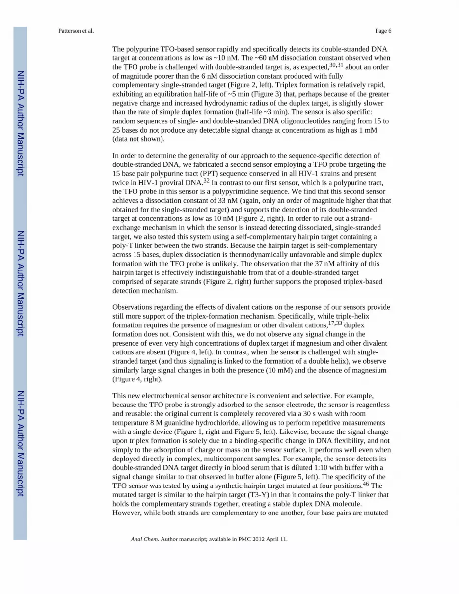
RESULTSWe have employed TFOs as recognition elements for the direct detection of specific double-stranded DNA targets in an electrochemical sensor format analogous to the previouslydescribed E-DNA platform.21 Our sensors are comprised of a redox-tagged TFO probe thatis strongly chemisorbed to an interrogating gold electrode (Figure 1). In the absence of itsdouble-stranded DNA target, the probe is flexible, allowing an attached methylene blue toapproach the electrode and exchange electrons. Upon the addition of its specific double-stranded target, the probe forms a rigid triplex structure that impedes contact between themethylene blue and the interrogating electrode. Thus, the TFO probe serves as an E-DNAsensor23 and maintains the various positive features of this class of devices.21,23–29 Forexample, we show below that this new class of sensor is reagentless, reusable, and suitablefor deployment directly in complex matrices.
To test the principle of a triplex-forming E-DNA platform, we first fabricated a sensor usinga well-characterized 22-base polypurine TFO probe designed to bind a specific double-stranded target DNA via reverse Hoogsteen base pairing in the major groove18 (Figure 1).The sensor was fabricated by the formation of a mixed self-assembled monolayer (SAM)comprised of the TFO probe and 6-mercaptohexanol on the gold electrode, which gives riseto a sharp, well-defined peak at ~260 mV (vs Ag/AgCl), consistent with the formal potentialof the methylene blue redox moiety (Figure 1, right). This TFO probe readily binds to itsspecific double-stranded DNA target (formed by the previous hybridization ofoligonucleotides T1-R and T2-R) via triplex formation,18 producing a readily measurabledecrease in Faradaic current (Figure 1, right, Figure 2, left). Support for the proposedformation of triplex is provided by the observation that the 65% signal suppression observedin the presence of the double-stranded target is somewhat greater than the 55% suppressionobtained when the TFO probe is instead hybridized to its fully complementary, single-stranded DNA target to form a simple duplex (Figure 2, left). This presumably occursbecause the greater bulk and charge of triplex DNA is more effective at reducing theefficiency with which the reporting redox tag approaches the electrode. As controlexperiments we also investigated the effects of adding the two single-strandedoligonucleotides (T1-R) employed in the double-stranded target in isolation. We find thatthe first of these, T1-R, does not lead to any detectable change in current, even at highconcentrations (data not shown). In contrast the addition of the other, T2-R, leads to anontrivial signal change, presumably because an 11-base portion of this strand is fullycomplementary to the TFO probe. Despite this, the signal suppression observed in thepresence of this strand is much less than that obtained in presence of the duplex target (datanot shown), further suggesting that the signal change produced by the duplex target arisesdue to triplex formation and not due to the dissociation of the duplex target and theconsequent formation of a duplex with the probe DNA.
Patterson et al. Page 5
Anal Chem. Author manuscript; available in PMC 2012 April 11.
NIH
-PA Author Manuscript
NIH
-PA Author Manuscript
NIH
-PA Author Manuscript
The polypurine TFO-based sensor rapidly and specifically detects its double-stranded DNAtarget at concentrations as low as ~10 nM. The ~60 nM dissociation constant observed whenthe TFO probe is challenged with double-stranded target is, as expected,30,31 about an orderof magnitude poorer than the 6 nM dissociation constant produced with fullycomplementary single-stranded target (Figure 2, left). Triplex formation is relatively rapid,exhibiting an equilibration half-life of ~5 min (Figure 3) that, perhaps because of the greaternegative charge and increased hydrodynamic radius of the duplex target, is slightly slowerthan the rate of simple duplex formation (half-life ~3 min). The sensor is also specific:random sequences of single- and double-stranded DNA oligonucleotides ranging from 15 to25 bases do not produce any detectable signal change at concentrations as high as 1 mM(data not shown).
In order to determine the generality of our approach to the sequence-specific detection ofdouble-stranded DNA, we fabricated a second sensor employing a TFO probe targeting the15 base pair polypurine tract (PPT) sequence conserved in all HIV-1 strains and presenttwice in HIV-1 proviral DNA.32 In contrast to our first sensor, which is a polypurine tract,the TFO probe in this sensor is a polypyrimidine sequence. We find that this second sensorachieves a dissociation constant of 33 nM (again, only an order of magnitude higher that thatobtained for the single-stranded target) and supports the detection of its double-strandedtarget at concentrations as low as 10 nM (Figure 2, right). In order to rule out a strand-exchange mechanism in which the sensor is instead detecting dissociated, single-strandedtarget, we also tested this system using a self-complementary hairpin target containing apoly-T linker between the two strands. Because the hairpin target is self-complementaryacross 15 bases, duplex dissociation is thermodynamically unfavorable and simple duplexformation with the TFO probe is unlikely. The observation that the 37 nM affinity of thishairpin target is effectively indistinguishable from that of a double-stranded targetcomprised of separate strands (Figure 2, right) further supports the proposed triplex-baseddetection mechanism.
Observations regarding the effects of divalent cations on the response of our sensors providestill more support of the triplex-formation mechanism. Specifically, while triple-helixformation requires the presence of magnesium or other divalent cations,17,33 duplexformation does not. Consistent with this, we do not observe any signal change in thepresence of even very high concentrations of duplex target if magnesium and other divalentcations are absent (Figure 4, left). In contrast, when the sensor is challenged with single-stranded target (and thus signaling is linked to the formation of a double helix), we observesimilarly large signal changes in both the presence (10 mM) and the absence of magnesium(Figure 4, right).
This new electrochemical sensor architecture is convenient and selective. For example,because the TFO probe is strongly adsorbed to the sensor electrode, the sensor is reagentlessand reusable: the original current is completely recovered via a 30 s wash with roomtemperature 8 M guanidine hydrochloride, allowing us to perform repetitive measurementswith a single device (Figure 1, right and Figure 5, left). Likewise, because the signal changeupon triplex formation is solely due to a binding-specific change in DNA flexibility, and notsimply to the adsorption of charge or mass on the sensor surface, it performs well even whendeployed directly in complex, multicomponent samples. For example, the sensor detects itsdouble-stranded DNA target directly in blood serum that is diluted 1:10 with buffer with asignal change similar to that observed in buffer alone (Figure 5, left). The specificity of theTFO sensor was tested by using a synthetic hairpin target mutated at four positions.46 Themutated target is similar to the hairpin target (T3-Y) in that it contains the poly-T linker thatholds the complementary strands together, creating a stable duplex DNA molecule.However, while both strands are complementary to one another, four base pairs are mutated
Patterson et al. Page 6
Anal Chem. Author manuscript; available in PMC 2012 April 11.
NIH
-PA Author Manuscript
NIH
-PA Author Manuscript
NIH
-PA Author Manuscript

such that reverse Hoogsteen interactions with the TFO probe are compromised. Whenchallenging the sensor with high concentrations (200 nM) of this mutated duplex, the sensordoes not produce any measurable signal change, thus demonstrating the high specificity ofthe TFO E-DNA platform (Figure 5, right).
The above proof-of-principle studies employed fully synthetic targets that would be of littleif any interest in a clinical setting. Thus motivated, we have also explored the utility ofemploying this new sensor architecture in the detection of authentic, unpurified PCRamplicons generated from HIV-1 samples. We amplified a region of the HIV-1 genomicRNA containing the PPT sequence recognized by the polypyrimidine TFO probe employedabove. As a first step toward this goal, we determined the ease with which we can use oursensor to quantify synthetic, 63-base pair oligonucleotides equivalent to the PCR amplicon(Figure 6, left). We find that these produce signal changes similar to the large, easilymeasurable signal changes associated with the short hairpin targets described above. Thedissociation constants for these larger targets, however, are slightly poorer than thoseobtained for the short hairpin target. This is presumably due to steric and/or electrostaticeffects associated with the larger targets.
Moving forward, we find that our new sensor platform readily detects double-stranded DNAproduced via reverse transcriptase polymerase chain reaction amplification of HIV-1genomic RNA (Figure 6, right). To demonstrate this, we employed a conventional nested,amplification protocol to produce a high concentration of amplified products. To inhibit anypotential hybridization of the primers–or primer-dimer or other primer-sequence-containingamplification artifacts–to the TFO probes, we placed the sensor’s recognition footprint 20bases internal to the end of the amplicon. Starting from 1 μg of HIV-1 genomic RNA, weobtain about 100 nM of the appropriate, double-stranded DNA amplification product(nucleic acid concentrations obtained using Nanodrop instrument). After incubation of theE-DNA sensors with the PCR amplicon solution, we observe about a 40% drop in the sensorcurrent signal, which is indicative of the presence of the expected target sequence (Figure 6,right). Negative controls performed exclusively with the primers sets and the TFO probeproduced no change in signal (data not shown), and a control experiment using nontemplatenegative control RT-PCR did not show any significant signal change over the same timeperiod (Figure 6, right).
DISCUSSIONAmong the numerous methods recently proposed for the sequence-specific detection ofDNA, E-DNA sensors, the electrochemical analog of optical molecular beacons,22,29,34–38
present several advantages over other optical or electrochemical hybridization detectionmethods.21 E-DNA sensors are based on the hybridization-induced folding of an electrode-bound, redox-tagged DNA probe. The E-DNA platform is reagentless, electronic(electrochemical), and highly selective (they perform well even when challenged directly incomplex, multicomponent samples such as blood serum or soil) and can discriminatebetween a perfect match and a single mismatch target.21,23 For all these reasons, they appearto be a promising and appealing approach for the sequence-specific detection of DNA andRNA.39,40 But like other hybridization-based methods, traditional E-DNA sensors requirethe generation of single-stranded DNA targets prior to detection. Here, in contrast, wedescribe an E-DNA sensor that overcomes this limitation by hybridizing directly to double-stranded DNA targets. And while this approach is limited to the detection of homopurine orhomopyrmidine tracks, these triplex-forming sequences are nevertheless common enoughthat it is straightforward to generate 16–20 base probes with sufficient specificity to targetunique sites in human or pathogen genomes.41,42
Patterson et al. Page 7
Anal Chem. Author manuscript; available in PMC 2012 April 11.
NIH
-PA Author Manuscript
NIH
-PA Author Manuscript
NIH
-PA Author Manuscript

Our approach is not the first to employ triplex formation in the detection of double-strandedDNA. Several groups, for example, have previously reported the use of TFOs in the opticaldetection of double-stranded DNA.16–19 Similarly, two electrochemical methods have beenproposed to date where triplex formation is monitored via a direct electrochemical signalfrom guanine43 or via HPLC separation of the triplex coupled with electrochemicaldetection.44 While these latter approaches are interesting and achieve remarkable detectionlimits, their detection principles are limited by several drawbacks: the direct oxidation ofguanine appears impractical for realistic applications due to the high overpotential required,the possibility of electrochemical interferences in complex samples (from, for example,exogenous, nontarget DNA), and a high background currents.45 Likewise, the use withHPLC renders the second platform complicated and less suitable for PCR coupling or rapidmeasurements. For these reasons, the E-DNA sensor described here would appear to offerpotentially important advantages for the rapid, electrochemical detection of specific double-stranded DNA sequences.
Like earlier E-DNA counterparts,21 sensors based on triplex-forming oligonucleotides arelabel-free, reusable, and selective enough to employ directly in complex sample matricessuch as blood serum. Directly measuring double-stranded DNA targets makes these TFOsensors an optimal candidate for use with the PCR amplification process. We havedemonstrated this coupling here by performing a nested, reverse-transcriptase PCR of HIV-1genomic RNA followed by the measurement of the amplified target with the TFO-based E-DNA sensors. As E-DNA sensors have proved suitable for low-cost sensors and portableinstrumentations,26 our TFO sensors provide many optimal features that demonstrate thepossibility of adopting these sensors in real-world applications. All these attributes suggestthat E-DNA sensors may be better suited for clinical applications than the previous, mostlyoptical methods supporting the direct detection of double-stranded DNA, includingapproaches based on non-DNA probes.
References1. Persil O, Hud NV. Trends Biotechnol. 2007; 25(10):433–436. [PubMed: 17825446]2. Fang TH, Ramalingam N, Xian-Dui D, Ngin TS, Xianting Z, Kuan ATL, Huat EYP, Hai-Qing
Gong. Biosens Bioelectron. 2009; 24(7):2131–2136. [PubMed: 19162460]3. Ihmels H, Otto D. Top Curr Chem. 2005; 258:161–204.4. Markoulatos P, Saifakas N, Moncany M. J Clin Lab Anal. 2002; 16(1):47–51. [PubMed: 11835531]5. Ghosh I, Stains CI, Ooi AT, Segal D. J Mol Biosyst. 2006; 2(11):551–560.6. White S, Szewczyk JW, Turner JM, Baird EE, Dervan PB. Nature. 1998; 391:468–471. [PubMed:
9461213]7. Ooi AT, Stains CI, Ghosh I, Segal DJ. Biochemistry. 2006; 45:3620–3625. [PubMed: 16533044]8. Stains CI, Furman JL, Segal DJ, Ghosh I. J Am Chem Soc. 2006; 128:9761–9765. [PubMed:
16866532]9. Stains CI, Porter JR, Ooi AT, Segal DJ, Ghosh I. J Am Chem Soc. 2005; 127:10782–10783.
[PubMed: 16076155]10. Paleček E, Masařiḱ M, Kizek R, Kuhlmeier D, Hassmann J, Schülein J. Anal Chem. 2004; 76(19):
5930–5936. [PubMed: 15456317]11. Halby L, Ryabinin VA, Sinyakov AN, Boutorine AS. Bioorg Med Chem Lett. 2005; 15:3720–
3724. [PubMed: 16005219]12. Buchmann W, Boutorine A, Halby L, Tortajada J, De Pauw E. J Mass Spectrom. 2009; 44(8):
1171–81. [PubMed: 19408249]13. Thuong TN, Hélène C. Angew Chem, Int Ed Engl. 1993; 32:666–690.14. Helene C. Anti-Cancer Drug Des. 1991; 6(6):569–584.15. Helene C, Toulme JJ. Biochim Biophys Acta Gene Struct Expression. 1990; 1049(2):99–125.
Patterson et al. Page 8
Anal Chem. Author manuscript; available in PMC 2012 April 11.
NIH
-PA Author Manuscript
NIH
-PA Author Manuscript
NIH
-PA Author Manuscript

16. Dervan PB. Bioorg Med Chem. 2001; 9:2215–2235. [PubMed: 11553460]17. Johnson MD. Chromosoma. 1999; 108:181–189. [PubMed: 10398847]18. Escude C, Garestier T, Helene C. Proc Natl Acad Sci USA. 1999; 96:10603–10607. [PubMed:
10485872]19. Geron-Landre B, Roulon T, Desbiolles P, Escude C. Nucleic Acids Res. 2003; 31:e125. [PubMed:
14530458]20. Geron-Landre B, Roulon T, Escude C. FEBS J. 2005; 272:5343–5352. [PubMed: 16218964]21. Xiao Y, Lai RY, Plaxco KW. Nature Prot. 2007; 2:2875–2880.22. Ricci F, Plaxco KW. Mikrochim Acta. 2008; 163(3–4):149–155.23. Lubin AA, Plaxco KW. Acc Chem Res. 2010; 43(4):496–505. [PubMed: 20201486]24. Lai RY, Lagally ET, Lee SH, Soh HT, Plaxco KW, Heeger AJ. Proc Natl Acad Sci USA. 2006;
103(11):4017–4021. [PubMed: 16537478]25. Pavlovic E, Lai RY, Wu TT, Ferguson BS, Sun R, Plaxco KW, et al. Langmuir. 2008; 24:1102–
1107. [PubMed: 18181654]26. Ricci F, Zari N, Caprio F, Recine S, Amine A, Moscone D, et al. Bioelectrochem. 2009; 76(1–2):
208–213.27. Ricci F, Lai RY, Heeger AJ, Plaxco KW, Sumner JJ. Langmuir. 2007; 23(12):6827–6834.
[PubMed: 17488132]28. Ricci F, Lai RY, Plaxco KW. Chem Commun. 2007; 36:3768–3770.29. Xiao Y, Lubin AA, Baker BR, Plaxco KW, Heeger AJ. Proc Natl Acad Sci USA. 2006; 103(45):
16677–16680. [PubMed: 17065320]30. Shafer RH. Prog Nucleic Acid Res Mol Biol. 1998; 59:55–94. [PubMed: 9427840]31. Antony T, Thomas T, Sigal LH, Shirahata A, Thomas TJ. Biochemistry. 2001; 40(31):9387–9395.
[PubMed: 11478908]32. Giovannangeli C, Perrouault L, Escude C, Thuong N, Helene C. Biochemistry. 1996; 35:10539–
10548. [PubMed: 8756710]33. Malkov VA, Voloshin ON, Soyfer VN, Frank-Kamenetskii MD. Nucleic Acids Res. 1993;
21:585–591. [PubMed: 8382800]34. Immoos CE, Lee SJ, Grinstaff MW. Chem Bio Chem. 2004; 5:1100–1104.35. Immoos CE, Lee SJ, Grinstaff MW. J Am Chem Soc. 2004; 126:10814–10815. [PubMed:
15339145]36. Mao T, Luo C, Ouyang Q. Nucleic Acids Res. 2003; 31:108–172.37. Phares N, White RJ, Plaxco KW. Anal Chem. 2009; 81:1095–1100. [PubMed: 19133790]38. Lubin AA, Vander Stoep Hunt B, White RJ, Plaxco KW. Anal Chem. 2009; 81:2150–2158.
[PubMed: 19215066]39. Paleček E. Trends Biotechnol. 2004; 22(2):55–58. [PubMed: 14757035]40. Thorp HH. Trends Biotechnol. 2003; 21:522–524. [PubMed: 14624859]41. Goñi JR, de la Cruz X, Orozco M. Nucleic Acids Res. 2004; 32(1):354–360. [PubMed: 14726484]42. Duca M, Vekhoff P, Oussedik K, Halby L, Arimondo PB. Nucleic Acids Res. 2008; 36(16):5123–
5138. [PubMed: 18676453]43. Cai X, Rivas G, Shirashi H, Farias P, Wang J, Tomschik M, Jelen F, Palecek E. Anal Chim Acta.
1997; 344:65–76.44. Ihara T, Maruo Y, Takenaka S, Takagi M. Nucleic Acids Res. 1996; 24:4273–4280. [PubMed:
8932383]45. Drummond TG, Hill MG, Barton JK. Nat Biotechnol. 2003; 21:1192–1199. [PubMed: 14520405]46. Giovannangeli C, Diviacco S, Labrousse V, Gryaznov S, Charneau P, Helene C. Proc Natl Acad
Sci USA. 1997; 94(1):79–84. [PubMed: 8990164]
Patterson et al. Page 9
Anal Chem. Author manuscript; available in PMC 2012 April 11.
NIH
-PA Author Manuscript
NIH
-PA Author Manuscript
NIH
-PA Author Manuscript
Figure 1.An E-DNA sensor employing a triplex-forming oligonucleotide (TFO) probe readily detectsdouble-stranded DNA targets. (Left) The sensor consists of a polypurine or polypyrimidineTFO probe modified at its 3′-terminus with a methylene blue redox tag and at its 5′-terminuswith a mercaptohexanol moiety for attachment on a gold electrode. (Right) The Faradaiccurrent arising from the flexible TFO probe is significantly reduced in the presence of thedouble-stranded DNA target, presumably because triplex formation reduces the efficiencywith which the terminal redox tag collides with the electrode surface and transfers electrons.
Patterson et al. Page 10
Anal Chem. Author manuscript; available in PMC 2012 April 11.
NIH
-PA Author Manuscript
NIH
-PA Author Manuscript
NIH
-PA Author Manuscript

Figure 2.The polypurine (left) and polypyrimidine (right) TFO based E-DNA sensors respond well totheir specific double-stranded DNA targets (triplex formation). As expected, they alsorespond to fully complementary, single-stranded targets (via the formation of duplex DNA).As previously reported, the affinity of TFO probes for their double-stranded DNA targets isapproximately an order of magnitude poorer than that for their single-stranded targets.30,31
Patterson et al. Page 11
Anal Chem. Author manuscript; available in PMC 2012 April 11.
NIH
-PA Author Manuscript
NIH
-PA Author Manuscript
NIH
-PA Author Manuscript

Figure 3.The TFO-based E-DNA sensor is rapid. We observe an equilibration half-life (time requiredfor half of the total signal change to occur) of ~5 min for the double-stranded (triplex-forming) DNA target and ~3 min for a fully complementary single-stranded target. Theresults shown here were obtained with the polypurine TFO probe.
Patterson et al. Page 12
Anal Chem. Author manuscript; available in PMC 2012 April 11.
NIH
-PA Author Manuscript
NIH
-PA Author Manuscript
NIH
-PA Author Manuscript

Figure 4.The proposed triplex formation mechanism, shown here with our polypyrimidine TFOprobe, is supported by experiments in which the concentration of magnesium ions is altered.As triplex formation is dependent on magnesium ion concentration, the absence ofmagnesium produces no signal, even at high concentrations of double-stranded target (left).A duplex-forming target, in contrast, is readily detected in both the presence and absence ofmagnesium ions (right). The magnesium ion concentration was 10 mM in both experiments.
Patterson et al. Page 13
Anal Chem. Author manuscript; available in PMC 2012 April 11.
NIH
-PA Author Manuscript
NIH
-PA Author Manuscript
NIH
-PA Author Manuscript

Figure 5.The TFO-based E-DNA sensors are selective, reusable, and specific. Because signal changeupon triplex formation is solely due to a DNA binding-specific event, the TFO-based E-DNA sensor performs well, even when deployed directly in complex, multicomponentsamples, including 1:10 diluted blood serum (left). The original current is completelyrecovered via a 30 s rinse in room temperature 8 M guanidine hydrochloride solution, thusallowing us to perform repetitive measurements with a single sensor (left). Moreover, theTFO E-DNA probe displays specificity when challenged with mutated double-strandedDNA (right). Here are shown voltammograms obtained when challenging thepolypyrimidine TFO probe with 200 nM of double-stranded target and 200 nM of mutatedtarget (right).
Patterson et al. Page 14
Anal Chem. Author manuscript; available in PMC 2012 April 11.
NIH
-PA Author Manuscript
NIH
-PA Author Manuscript
NIH
-PA Author Manuscript

Figure 6.TFO sensor responds to unpurified PCR amplicons containing a triplex-forming element inthe HIV-1 genome. (left) Specifically, test results using a synthetic, duplex oligonucleotideequivalent to the relevant HIV-1 sequence binds to the probe, albeit with a poorerdissociation constant and somewhat poorer signaling than those seen with a short hairpintarget containing the same recognition footprint. These effects are presumably due to thelarger size of the amplicon. (right) The sensor also responds rapidly and robustly toauthentic PCR amplified target molecules (at 100 nM), while producing no signal change inresponse to negative control PCR samples. Polyacrylimide gel (right, inset) containingamplicons from HIV-specific PCR, a negative control (no template added to the PCR), andsynthetic HIV duplex show the specificity of the PCR reaction. The size of our HIV targetDNA is 63 bp.
Patterson et al. Page 15
Anal Chem. Author manuscript; available in PMC 2012 April 11.
NIH
-PA Author Manuscript
NIH
-PA Author Manuscript
NIH
-PA Author Manuscript
Copyright © 2022 FDOKUMEN