Bacterial and Archaeal α-Amylases - CORE
21
REVIEW published: 28 July 2016 doi: 10.3389/fmicb.2016.01129 Frontiers in Microbiology | www.frontiersin.org 1 July 2016 | Volume 7 | Article 1129 Edited by: Peter Neubauer, Technical University of Berlin, Germany Reviewed by: Gotthard Kunze, Institute of Plant Genetics and Crop Plant Research (LG), Germany David B. Wilson, Cornell University, USA *Correspondence: Tulasi Satyanarayana [email protected] Specialty section: This article was submitted to Microbiotechnology, Ecotoxicology and Bioremediation, a section of the journal Frontiers in Microbiology Received: 17 May 2016 Accepted: 06 July 2016 Published: 28 July 2016 Citation: Mehta D and Satyanarayana T (2016) Bacterial and Archaeal α-Amylases: Diversity and Amelioration of the Desirable Characteristics for Industrial Applications. Front. Microbiol. 7:1129. doi: 10.3389/fmicb.2016.01129 Bacterial and Archaeal α-Amylases: Diversity and Amelioration of the Desirable Characteristics for Industrial Applications Deepika Mehta and Tulasi Satyanarayana* Department of Microbiology, University of Delhi, New Delhi, India Industrial enzyme market has been projected to reach US$ 6.2 billion by 2020. Major reasons for continuous rise in the global sales of microbial enzymes are because of increase in the demand for consumer goods and biofuels. Among major industrial enzymes that find applications in baking, alcohol, detergent, and textile industries are α-amylases. These are produced by a variety of microbes, which randomly cleave α-1,4-glycosidic linkages in starch leading to the formation of limit dextrins. α-Amylases from different microbial sources vary in their properties, thus, suit specific applications. This review focuses on the native and recombinant α-amylases from bacteria and archaea, their production and the advancements in the molecular biology, protein engineering and structural studies, which aid in ameliorating their properties to suit the targeted industrial applications. Keywords: archaea, bacteria, dextrinizing α-amylases, saccharogenic α-amylases, protein engineering, native and recombinant α-amylases INTRODUCTION Starch is a glucose polymer, which is synthesized by a wide array of plant species. Starch granules contain of two types of α-glucans, amylose and amylopectin, overall representing 98–99% of its total dry weight. Amylose is a linear water insoluble polymer of glucose joined by α-1, 4 glycosidic bonds (99%), while amylopectin is branched water soluble polysaccharide with short α-1, 4 linked linear chains of 10–60 glucose units and α-1, 6 linked side chains with 15–45 glucose units that form the volume of starch molecule (Buleon et al., 1998; Tester et al., 2004). The ratio of the two polysaccharides depends on the botanical origin of the starch, but the representative levels of amylose to amylopectin are 25–28 and 72–75%, respectively. Based on the mode of action, starch hydrolyzing enzymes may be endo-acting or exo-acting. α-Amylases (EC 3.2.1.1), categorized in GH-13 family of glyosyl hydrolases, are the extracellular endo-acting enzymes that hydrolyse α-1,4 glycosidic linkages of starch randomly, while bypassing the branch points and liberating α-limit dextrins as products (Antranikian, 1992; Gupta et al., 2003; Sivaramakrishnan et al., 2006). Amylolytic enzymes have been elegantly reviewed by several workers earlier (Vihinen and Mantasala, 1989; Henrissat, 1991; Antranikian, 1992; Svensson, 1994; Davies and Henrissat, 1995; Guzman-Maldonadao and Paredes-Lopez, 1995; Nigam and Singh, 1995; Crabb and Mitchinson, 1997; Janecek, 1997; Janecek et al., 1997, 1999; Sunna et al., 1997; Bisgaad-Frantzen et al., 1999; Pandey et al., 2000; Vieille and Zeikus, 2001; van der Maarel et al., 2002; Gupta et al., 2003; Rubin-Pitel and Zhao, 2006; Sivaramakrishnan et al., 2006). brought to you by CORE View metadata, citation and similar papers at core.ac.uk provided by Frontiers - Publisher Connector
-
Upload
khangminh22 -
Category
Documents
-
view
0 -
download
0
Transcript of Bacterial and Archaeal α-Amylases - CORE

REVIEWpublished: 28 July 2016
doi: 10.3389/fmicb.2016.01129
Frontiers in Microbiology | www.frontiersin.org 1 July 2016 | Volume 7 | Article 1129
Edited by:
Peter Neubauer,
Technical University of Berlin,
Germany
Reviewed by:
Gotthard Kunze,
Institute of Plant Genetics and Crop
Plant Research (LG), Germany
David B. Wilson,
Cornell University, USA
*Correspondence:
Tulasi Satyanarayana
Specialty section:
This article was submitted to
Microbiotechnology, Ecotoxicology
and Bioremediation,
a section of the journal
Frontiers in Microbiology
Received: 17 May 2016
Accepted: 06 July 2016
Published: 28 July 2016
Citation:
Mehta D and Satyanarayana T (2016)
Bacterial and Archaeal α-Amylases:
Diversity and Amelioration of the
Desirable Characteristics for Industrial
Applications. Front. Microbiol. 7:1129.
doi: 10.3389/fmicb.2016.01129
Bacterial and Archaeal α-Amylases:Diversity and Amelioration of theDesirable Characteristics forIndustrial ApplicationsDeepika Mehta and Tulasi Satyanarayana*
Department of Microbiology, University of Delhi, New Delhi, India
Industrial enzyme market has been projected to reach US$ 6.2 billion by 2020. Major
reasons for continuous rise in the global sales of microbial enzymes are because of
increase in the demand for consumer goods and biofuels. Among major industrial
enzymes that find applications in baking, alcohol, detergent, and textile industries are
α-amylases. These are produced by a variety of microbes, which randomly cleave
α-1,4-glycosidic linkages in starch leading to the formation of limit dextrins. α-Amylases
from different microbial sources vary in their properties, thus, suit specific applications.
This review focuses on the native and recombinant α-amylases from bacteria and
archaea, their production and the advancements in the molecular biology, protein
engineering and structural studies, which aid in ameliorating their properties to suit the
targeted industrial applications.
Keywords: archaea, bacteria, dextrinizing α-amylases, saccharogenic α-amylases, protein engineering, native and
recombinant α-amylases
INTRODUCTION
Starch is a glucose polymer, which is synthesized by a wide array of plant species. Starch granulescontain of two types of α-glucans, amylose and amylopectin, overall representing 98–99% of itstotal dry weight. Amylose is a linear water insoluble polymer of glucose joined by α-1, 4 glycosidicbonds (99%), while amylopectin is branched water soluble polysaccharide with short α-1, 4 linkedlinear chains of 10–60 glucose units and α-1, 6 linked side chains with 15–45 glucose units thatform the volume of starch molecule (Buleon et al., 1998; Tester et al., 2004). The ratio of thetwo polysaccharides depends on the botanical origin of the starch, but the representative levelsof amylose to amylopectin are 25–28 and 72–75%, respectively.
Based on the mode of action, starch hydrolyzing enzymes may be endo-acting or exo-acting.α-Amylases (EC 3.2.1.1), categorized in GH-13 family of glyosyl hydrolases, are the extracellularendo-acting enzymes that hydrolyse α-1,4 glycosidic linkages of starch randomly, while bypassingthe branch points and liberating α-limit dextrins as products (Antranikian, 1992; Gupta et al.,2003; Sivaramakrishnan et al., 2006). Amylolytic enzymes have been elegantly reviewed by severalworkers earlier (Vihinen and Mantasala, 1989; Henrissat, 1991; Antranikian, 1992; Svensson,1994; Davies and Henrissat, 1995; Guzman-Maldonadao and Paredes-Lopez, 1995; Nigam andSingh, 1995; Crabb and Mitchinson, 1997; Janecek, 1997; Janecek et al., 1997, 1999; Sunna et al.,1997; Bisgaad-Frantzen et al., 1999; Pandey et al., 2000; Vieille and Zeikus, 2001; van der Maarelet al., 2002; Gupta et al., 2003; Rubin-Pitel and Zhao, 2006; Sivaramakrishnan et al., 2006).
brought to you by COREView metadata, citation and similar papers at core.ac.uk
provided by Frontiers - Publisher Connector
Mehta and Satyanarayana Bacterial and Archaeal α-Amylases
Another category belongs to carbohydrases that hydrolyzecyclodextrins. They are called maltogenic amylases (EC3.2.1.133), which are catalytically versatile and can hydrolyzeα-(1,4)- as well as α-(1,6)- linkages of the substrate moleculeand transglycosylate the hydrolytic products. They possess anadditional 130 residues at the N-terminus that are absent in thetypical α-amylases. They can also hydrolyze acarbose, a potentinhibitor of α-amylases, to produce glucose and acarviosine-glucose [pseudotrisaccharide (PTS); (Li et al., 2011)]. β-Amylase(E.C.3.2.1.2) is an exoenzyme that hydrolyzes every alternateα-1,4 linkage from the non-reducing end, causing inversion ofthe anomeric configuration of the liberated maltose to its β-form,and hence, they are called β-amylases. This enzyme bypassesα–1,6-linkages of branched substrates producing maltose andhigh molecular weight β-limit dextrins (Vihinen and Mantasala,1989). In comparison, glucoamylase (E.C.3.2.1.3), also known asamyloglucosidase or γ–amylase, slowly acts on α–1,4 linkages ofα-glucans from the non-reducing ends and also α–1,6 linkages.It preferentially degrades polysaccharides with a high molecularweight. These enzymes hydrolyze starch to yield glucose intheoretically 100% yield (Kumar and Satyanarayana, 2004).CGTase (E.C.2.4.1.19) produces a series of α, β, γ cyclodextrinsfrom starch, amylose, and other polysaccharides. In addition,CGTases also catalyze coupling reaction by which the rings areopened and transferred to co-substrates like glucose, maltose, orsucrose. The enzyme also catalyzes disproportionation reactionin which one or more glycosyl units are transferred betweenlinear oligosaccharides. A hydrolysis reaction is also catalyzed bythe enzyme that produces dextrins. Another starch hydrolyzingenzyme α–glucosidase (E.C.3.2.1.20) acts on α–1,4-and/orα–1,6-linkages of oligosaccharides from non-reducing end,which are formed by the action of other amylolytic enzymes,liberating α–D glucose units. Most enzymes show high affinitytoward maltose and so they are called as maltases (Vihinen andMantasala, 1989; Gupta et al., 2003; Sivaramakrishnan et al.,2006).
According to international starch market index, the totalutilization of starch in the world in 2008 was 66 million tons.This increased to 75 million tons by 2012, which suggests anannual growth rate of 2–3%. Fructose syrups represent 72% ofstarch output, while ethanol alone 40% (http://starch.dk/isi/market/index.asp). The industrial enzyme market was valued atUSD 4.2 billion in 2014 and is estimated to reach USD 6.2 billionby 2020. The food and beverage segment of industrial enzymesmarket is estimated to reach US$ 2.0 billion by 2020 (http://www.marketsandmarkets.com/PressReleases/industrial-enzymes.asp).Among industrial enzymes, starch hydrolyzing enzymes findapplications in the production of ethanol, HFCS (high fructosecorn syrup), and detergent and baking industries. Alcohol andstarch enzyme market is estimated to be worth $2.24 billionby 2018 (http://www.marketsandmarkets.com/PressReleases/alcohol-starch-sugar-enzyme.asp).
DIVERSITY OF α-AMYLASES
α-Amylases are divided into two categories according to thedegree of hydrolysis. Saccharifying α-amylases produce freesugars and reduce the viscosity less rapidly in comparison with
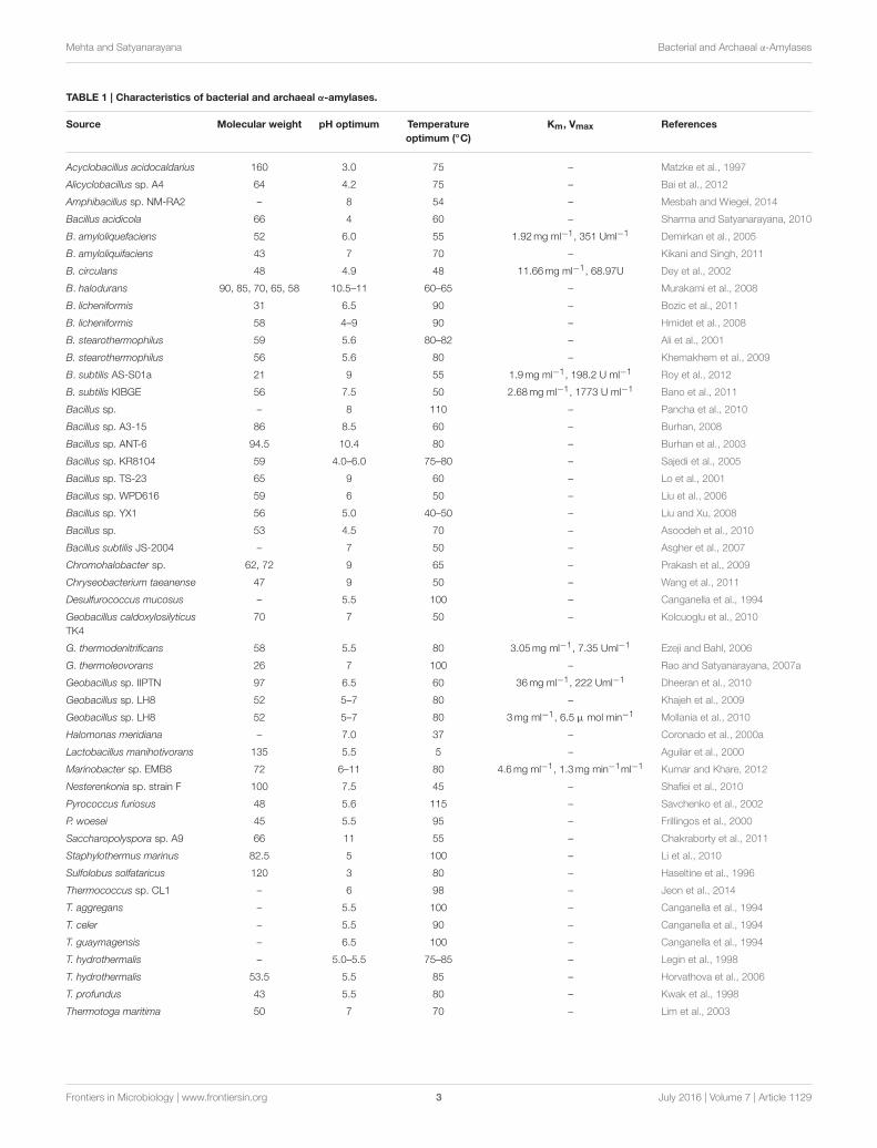
the amount of reducing sugars released. While liquefying α-amylases, on the other hand, break down the starch polymer, butdo not produce free sugars and cause more rapid reduction inviscosity of starch pastes. α-Amylases have bacterial, archaeal,and eukaryotic origin. The bacterial α-amylases are quitedistant from the eukaryotic enzymes evolutionarily. Amongall α-amylases, the bacterial enzymes are the most diverse(Pandey et al., 2000). The temperature-activity optima forbacterial α-amylases range from ∼25◦C (Feller et al., 1992;AHA) to around 100◦C (Rao and Satyanarayana, 2007b;Geobacillus thermoleovorans α-amylase). The pH optima ofbacterial α-amylases vary from 1 to 11.5 (Vihinen andMantasala,1989; Pandey et al., 2000). α-Amylase from Bacillus sp. andAlicyclobacillus acidocaldarius showed an acidic pH optima of1 and 3, respectively (Uchino, 1982; Schwermann et al., 1994).In contrast, alkaline amylase with optima of pH 9–10.5 hadbeen reported from many alkaliphilic Bacillus spp. (Saito, 1973;Krishnan and Chandra, 1983; Lee et al., 1994; Shinke et al., 1996).An extremely alkaline α-amylase with pH optima of 11.5 wasreported from Bacillus sp. GM8901 (Kim et al., 1995). Archaealα-amylases, in general, are thermostable and acidic (pH 5–6) innature. For example the highest temperature optimum has beenreported as 100 and 130◦C from archaea, Pyrococcus furiosusand P. woesei, respectively (Koch et al., 1991; Laderman et al.,1993). Molecular weight of α-amylases vary from about 10 to210 kDa, 10 kDa enzyme from Bacillus caldolyticus (Grootegoedet al., 1973) and the highest molecular weight (210 kDa) fromChloroflexus aurantiacus has been reported (Ratanakhanokchaiet al., 1992). Table 1 presents temperature and pH optima andmolecular weights of α-amylases from bacteria and archaea.
Increased demands for α-amylases with desired propertiesfor industrial applications also encouraged exploration frommetagenomes from different habitats. The screening of clonesfrom a soil metagenomic library constructed in pUC 19 vectorled to find a putative amylase gene (amyM), which was overexpressed and purified. This enzyme was optimally active at 42◦Cand pH 9.0 with transglycosylation activity and requirement forCa2+ for its stability (Yun et al., 2004). In another attempt, agene (pAMY) of 909 bp encoding an α-amylase was found whena soil-derived metagenomic library was screened. Phylogeneticanalysis revealed that pAMY was closely related to amylasesfrom uncultured bacteria. The enzyme with a molecular massof 38 kDa exhibited optimum activity at 40◦C and neutralpH (Sharma et al., 2010). A function-based screening of anacid mine drainage (AMD) derived metagenomic library ledto the discovery two endo-acting amylases which shared nosequence similarity with any known amylase. They didn’t haveknown amylolytic domains or could be assigned to any GH-family (Delavat et al., 2012). A thermostable α-amylase genewas isolated from a metagenomic library constructed frommetagenome constructed from a pilot-plant biogas reactor.When the gene (1461 bp) was cloned in Escherichia coli, avery high titre was attained. Further characterization revealedthat it has optimal activity at 80◦C and highly salt tolerant[25% (w/v) NaCl]. This novel enzyme Amy13A showed 75%sequence identity to α-amylases from Petrotoga mobilis andHalothermothrix orenii (Jabbour et al., 2013). Vester et al. (2015)identified a cold-active α-amylase by a functional metagenomics
Frontiers in Microbiology | www.frontiersin.org 2 July 2016 | Volume 7 | Article 1129
Mehta and Satyanarayana Bacterial and Archaeal α-Amylases
TABLE 1 | Characteristics of bacterial and archaeal α-amylases.
Source Molecular weight pH optimum Temperature
optimum (◦C)
Km, Vmax References
Acyclobacillus acidocaldarius 160 3.0 75 – Matzke et al., 1997
Alicyclobacillus sp. A4 64 4.2 75 – Bai et al., 2012
Amphibacillus sp. NM-RA2 – 8 54 – Mesbah and Wiegel, 2014
Bacillus acidicola 66 4 60 – Sharma and Satyanarayana, 2010
B. amyloliquefaciens 52 6.0 55 1.92mg ml−1, 351 Uml−1 Demirkan et al., 2005
B. amyloliquifaciens 43 7 70 – Kikani and Singh, 2011
B. circulans 48 4.9 48 11.66mg ml−1, 68.97U Dey et al., 2002
B. halodurans 90, 85, 70, 65, 58 10.5–11 60–65 – Murakami et al., 2008
B. licheniformis 31 6.5 90 – Bozic et al., 2011
B. licheniformis 58 4–9 90 – Hmidet et al., 2008
B. stearothermophilus 59 5.6 80–82 – Ali et al., 2001
B. stearothermophilus 56 5.6 80 – Khemakhem et al., 2009
B. subtilis AS-S01a 21 9 55 1.9mg ml−1, 198.2 U ml−1 Roy et al., 2012
B. subtilis KIBGE 56 7.5 50 2.68mg ml−1, 1773 U ml−1 Bano et al., 2011
Bacillus sp. – 8 110 – Pancha et al., 2010
Bacillus sp. A3-15 86 8.5 60 – Burhan, 2008
Bacillus sp. ANT-6 94.5 10.4 80 – Burhan et al., 2003
Bacillus sp. KR8104 59 4.0–6.0 75–80 – Sajedi et al., 2005
Bacillus sp. TS-23 65 9 60 – Lo et al., 2001
Bacillus sp. WPD616 59 6 50 – Liu et al., 2006
Bacillus sp. YX1 56 5.0 40–50 – Liu and Xu, 2008
Bacillus sp. 53 4.5 70 – Asoodeh et al., 2010
Bacillus subtilis JS-2004 – 7 50 – Asgher et al., 2007
Chromohalobacter sp. 62, 72 9 65 – Prakash et al., 2009
Chryseobacterium taeanense 47 9 50 – Wang et al., 2011
Desulfurococcus mucosus – 5.5 100 – Canganella et al., 1994
Geobacillus caldoxylosilyticus
TK4
70 7 50 – Kolcuoglu et al., 2010
G. thermodenitrificans 58 5.5 80 3.05mg ml−1, 7.35 Uml−1 Ezeji and Bahl, 2006
G. thermoleovorans 26 7 100 – Rao and Satyanarayana, 2007a
Geobacillus sp. IIPTN 97 6.5 60 36mg ml−1, 222 Uml−1 Dheeran et al., 2010
Geobacillus sp. LH8 52 5–7 80 – Khajeh et al., 2009
Geobacillus sp. LH8 52 5–7 80 3mg ml−1, 6.5 µ mol min−1 Mollania et al., 2010
Halomonas meridiana – 7.0 37 – Coronado et al., 2000a
Lactobacillus manihotivorans 135 5.5 5 – Aguilar et al., 2000
Marinobacter sp. EMB8 72 6–11 80 4.6mg ml−1, 1.3mg min−1ml−1 Kumar and Khare, 2012
Nesterenkonia sp. strain F 100 7.5 45 – Shafiei et al., 2010
Pyrococcus furiosus 48 5.6 115 – Savchenko et al., 2002
P. woesei 45 5.5 95 – Frillingos et al., 2000
Saccharopolyspora sp. A9 66 11 55 – Chakraborty et al., 2011
Staphylothermus marinus 82.5 5 100 – Li et al., 2010
Sulfolobus solfataricus 120 3 80 – Haseltine et al., 1996
Thermococcus sp. CL1 – 6 98 – Jeon et al., 2014
T. aggregans – 5.5 100 – Canganella et al., 1994
T. celer – 5.5 90 – Canganella et al., 1994
T. guaymagensis – 6.5 100 – Canganella et al., 1994
T. hydrothermalis – 5.0–5.5 75–85 – Legin et al., 1998
T. hydrothermalis 53.5 5.5 85 – Horvathova et al., 2006
T. profundus 43 5.5 80 – Kwak et al., 1998
Thermotoga maritima 50 7 70 – Lim et al., 2003
Frontiers in Microbiology | www.frontiersin.org 3 July 2016 | Volume 7 | Article 1129
Mehta and Satyanarayana Bacterial and Archaeal α-Amylases
approach. Sequence analysis revealed that the enzyme was similarto α-amylase from the class Clostridia. Its temperature andpH optima were 10–15◦C and pH 8–9. α-Amylases have alsobeen reported from metagenomic libraries from pygmy loris(Nycticebus pygmaeus) and cow dung (Xu et al., 2014; Sharmaet al., 2015).
PRODUCTION AND CHARACTERIZATIONOF α-AMYLASES
Production of Bacterial and Archaealα-AmylasesThe production of α-amylases has been studied extensivelyin submerged and/or solid state fermentations. In general,extracellular α-amylase production is growth associated(Murthy et al., 2009; Asoodeh et al., 2010; Abou Dobaraet al., 2011). Among the physical parameters, the temperatureand pH of the medium play an important role in α-amylaseproduction. The influence of temperature on α-amylaseproduction is related to the growth of the organism. Amylaseproduction in bacteria has been studied in a wide range oftemperatures. α-Amylase production has been reported fromthermophilic and hyperthermophilic bacteria and archaea likePyrococcus, Thermococcus, and Sulfolobus species (Leuschnerand Antranikian, 1995; Sunna et al., 1997), G. thermoleovorans(Rao and Satyanarayana, 2007a), B. acidocaldarius (Buonocoreet al., 1976), and Alicyclobacillus sp. A4 (Bai et al., 2012), frommesophiles B. amyloliquifaciens (Gangadharan et al., 2008),Halomonas meridiana (Coronado et al., 2000a) B. subtilis(Ravindar and Elangovan, 2013), Lactobacillus plantarum(Kanpiengjai et al., 2015) as well as from psychrotolerantsand psychrophiles Microbacterium foliorum GA2 (Roohi andKuddus, 2014) and Aeromonas veronii NS07 (Samie et al., 2012).Among different carbon sources used, starch, fructose, glucose,and rice flour, are known to support high enzyme production(Ezeji et al., 2005; Prakash et al., 2009). Carbon sources likeglucose and maltose have been used for the production ofα-amylase, but the use of starch remains ubiquitous (Mamo andGessesse, 1999; Sajedi et al., 2005; Liu and Xu, 2008; Sharmaand Satyanarayana, 2010). Industrially important enzymes havetraditionally been produced in submerged fermentation, butthese enzymes are also produced by solid state fermentation.Sodhi et al. (2005) and Hashemi et al. (2010) explained the use ofwheat bran for the production of α-amylase. Agro-residues wereused for cold-active α-amylase production from M. foliorum(Roohi and Kuddus, 2014). In general α-amylase productionis inducible in nature (Aiyer, 2004; Ryan et al., 2006; Asoodehet al., 2010; Abou Dobara et al., 2011), but in few cases α-amylaseproduction is also constitutive (Rao and Satyanarayana, 2007a).Like most other inducible enzymes, α-amylase production issubjected to catabolite repression by maltose and glucose, starchhydrolytic products (Bhella and Altosaar, 1988; Morkeberg et al.,1995) with the exception of some Bacillus strains (Kalishwaralalet al., 2010). Nitrogen source is a major factor that affectsα-amylase production. Many investigators had confirmed thatorganic nitrogen sources support maximum α-amylase yields
(Saxena et al., 2007; Aqeel and Umar, 2010; Abou Dobara et al.,2011).
The combination of low molecular weight dextran withTween-80 increased 27-fold higher α-amylase production(Arnesen et al., 1998). Various metal ions like Ca2+, Fe2+,Mg2+, and K+ are added to the α-amylase production medium(Sajedi et al., 2005; Liu and Xu, 2008). Phosphate is a vitalrequirement for microbes as it regulates the synthesis of primaryand secondary metabolites. Lower and higher levels of phosphatein the medium significantly affect the growth and enzymeproduction (Hillier et al., 1997; Sharma and Satyanarayana,2010).
The production of amylases by microbes is affectedconsiderably by physical and chemical parameters of themedium (Gigras et al., 2002; Rao and Satyanarayana, 2007a;Roohi and Kuddus, 2014; Sen et al., 2014). Traditionally “one-variable-at-a-time” approach has been used (Gokhale et al.,1991; Pham et al., 1998), but it is time consuming and doesnot permit understanding interactions among the processparameters. The statistical Plackett and Burman design, onthe other hand, allows screening of critical culture variables(Sharma and Satyanarayana, 2006; Rao and Satyanarayana,2007a; Kumar and Satyanarayana, 2008; Roohi and Kuddus,2014), and response surface methodology (RSM) providesinformation about the optimum levels of each variable,interactions among them and their effects on the product yield(Gu et al., 2005; Rao and Satyanarayana, 2007a). The statisticalapproaches have been proved to be useful in optimizingmedium components and cultural variables for maximizingamylase titres from many organisms like B. acidicola (Sharmaand Satyanarayana, 2011), Bacillus sp. KR 8104 (Hashemiet al., 2010), and G. thermoleovorans (Rao and Satyanarayana,2007a), Alcaligenes faecalis (Sen et al., 2014), and M. foliorum(Roohi and Kuddus, 2014).
Characterization of Bacterial and Archaealα-AmylasesOnce produced and purified, an enzyme is biochemicallycharacterized. α-Amylases show a wide range of substratedegradation. They degrade amylose, amylopectin, cyclodextrins,glycogen, and dextrins, but they possess highest specificity towardstarch (Antranikian, 1992). Various metal ions influence activityof the enzymes (Vihinen and Mantasala, 1989). α-Amylase isa metal activated enzyme and has high affinity for Ca2+. In,general, Ca2+ enhances the activity and thermal stability of mostof the α-amylases (Khajeh et al., 2001). The number of boundCa2+ varies from 1 to 10. Dialysis against EDTA can remove thebound Ca2+. The Ca2+-free amylase can be reactivated by addingCa2+. Although most of the α-amylases are Ca2+-dependent,there are reports of Ca2+-independent α-amylases too (Sajediet al., 2005; Rao and Satyanarayana, 2007b; Hmidet et al., 2008;Asoodeh et al., 2010; Sharma and Satyanarayana, 2010). BesidesCa2+-independent α-amylases, there are also a few α-amylases,which are inhibited by Ca2+ ions (Babu and Satyanarayana,1993; Tanaka and Hoshino, 2003; Mehta and Satyanarayana,2013a). The metal ions, which inhibit α-amylase activity, includeHg2+ ions (Mamo and Gessesse, 1999; Asoodeh et al., 2010).Inhibition of α-amylase by Hg2+ ions indicates the presence
Frontiers in Microbiology | www.frontiersin.org 4 July 2016 | Volume 7 | Article 1129

Mehta and Satyanarayana Bacterial and Archaeal α-Amylases
of carboxyl groups in enzyme molecule (Dey et al., 2002).Furthermore, Hg2+ is also known to oxidize indole ring and tointeract with aromatic ring present in tryptophan (Zhang et al.,2007; Liu et al., 2010). Various inhibitors such as dithiothreitol,β-mercaptoethanol, N-bromosuccinimide (NBS), p-hydroxymercuribenzoic acid, iodoacetate, PMSF (phenylmethylsuphonylfluoride), Woodward’s reagent K, EDTA, and EGTA have beenshown to inhibit α-amylases (Hamilton et al., 1999). Inhibitionof enzyme activity by NBS demonstrates the role of tryptophan inmaintaining the conformational stability of the enzyme (Rao andSatyanarayana, 2007b). Dithiothreitol and β-mercaptoethanolare the reducing agents, the effects of which suggest the role of—SH groups in the catalytic activity of enzyme. DTTmay stimulateor inhibit the α-amylase (Ballschmiter et al., 2006; Rao andSatyanarayana, 2007b). In maltogenic α-amylase of Bacillus sp.WPD616, DTT had no effect on the α-amylase activity indicatingthat—SH groups have no role to play in the catalytic activity orthese enzymes have no free and accessible—SH groups (Liu et al.,2006). The inhibition of α-amylase by PMSF indicates the roleof seryl hydroxyl group in enzyme activity. Woodward’s reagentK (WRK) has a role in the chemical modification of asparticand glutamic acid residues (Paoli et al., 1997). The inactivationof amylase by WRK also indicates the involvement of acidicamino acids in the active site of the enzyme (Chauthaiwale andRao, 1994; Komissarov et al., 1995; Sharma and Satyanarayana,2010).
GENE CLONING AND OVER EXPRESSIONOF RECOMBINANT α-AMYLASES
Several attempts have been made on cloning α-amylase encodinggenes from several bacteria and archaea in heterologous hostssuch as E. coli. A thermostable α-amylase gene of 1203 bpencoding a 401-amino acid protein of Thermococcus profundus,was cloned and expressed in E. coli. Recombinant α-amylaseproduction was 155.5-fold higher than that in the wild strain(Lee et al., 1996). Another α-amylase gene (1383 bp encoding461 amino acid residues) from a hyperthermophilic archaeon,Pyrococcus sp. KODl, was cloned and expressed in E. coli.The molecular mass of this mature enzyme is 49.456 kDawith 435 amino acid residues which displays <40% homologyto other amylases. The optimum temperature and pH forthe enzyme activity are 90◦C and pH 6.5. Ca2+ (2.0 mM)enhanced the thermostability of this enzyme (Tachibana et al.,1996). The recombinant α-amylase from P. furiosus is Ca2+-independent. This α-amylase is a liquefying enzyme with aspecific activity of 3900 U mg−1 at 98◦C. It was optimallyactive at pH 5.5–6.0 and 100◦C with a half-life of 13 h at 98◦C(Dong et al., 1997). Another α-amylase encoding gene from theextremely thermophilic archaeon, T. hydrothermalis was clonedand expressed in E. coli. The recombinant α-amylase possessesmolecular characteristics similar to Pyrococcus species encodinga protein of 457 amino acids with a 22 amino acid putative signalpeptide and a 435 amino acid mature protein (molecular mass49.236 kDa). This recombinant α-amylase is optimally activeat 75–85◦C and pH 5.0–5.5 (Lévêque et al., 2000; Horvathova
et al., 2006). Hyperthermophilic archaeon, Pyrococcus woeseialso produces hyperthermophilic α-amylase, the gene encodingwhich has been cloned and expressed in the moderate halophileHalomonas elongata. The 14 kb protein-coding sequence, andbiochemical properties of the expressed protein are identical tothe α-amylase gene of the closely related P. furiosus (Frillingoset al., 2000). A maltogenic α-amylase encoding gene with 588amino acids from thermophilic Bacillus sp. WPD 616 was clonedand expressed in E. coli, which showed optimum activity at pH6.0 and 50◦C (Liu et al., 2006). Sajedi et al. (2005) reportedcloning and expression of α-amylase gene (1328 bp) fromBacillus sp. KR-8104 that encodes 440 amino acids without 20amino acids of N and C termini in E. coli. A gene encoding athermostable α-amylase with the temperature optima of 75◦Cfrom G. thermoleovorans YN cloned in Bluescript R© II KS(+)vector in E. coli has been sequenced. The corresponding aminoacid sequence showed 99% sequence similarity with the knownα-amylases from different Bacilli and Geobacilli (Berekaa et al.,2007). Another α-amylase gene, Amy N, from B. licheniformisNH1 was also cloned and expressed in E. coli using pDEST17expression system. This recombinant α-amylase exhibited higherthermostability at 85◦C (T1/2 60 min) than the native amylase(8min; Hmidet et al., 2008). In another study, the gene encodingα-amylase from B. subtilis PY22 was amplified by PCR andcloned into Pichia pastoris KM71H using the vector pPICZα,which allows methanol induced expression and secretion of theprotein. The recombinant expression resulted in high levels ofextracellular amylase production, as high as 22 mg/L in theshake flask culture supernatant (Karakas et al., 2010). A genecorresponding to thermo- and pH-stable maltogenic α-amylaseof G. caldoxylosilyticus TK4 has been cloned into pET28a (+)vector and expressed in E. coli with 6xHis-tag at the N-terminus(Kolcuoglu et al., 2010). A gene encoding a hyperthermostablemaltogenic α-amylase of Staphylothermus marinus (SMMA) wascloned and over expressed in E. coli. SMMA consisted of 696amino acids with a predicted molecular weight of 82.5 kDa.The enzyme was active in acidic conditions (pH 3.5–5.0) withan optimal pH of 5.0, and was extremely thermostable with atemperature optimum of 100◦C and a melting temperature of109◦C; both these favored starch conversion process (Li et al.,2010). The α-amylase encoding gene of an acidophile B. acidicolawith N and C terminal truncation has been cloned in pET28a(+)and expressed in E. coli (Sharma and Satyanarayana, 2012). The62 kDa recombinant α-amylase was optimally active at pH 4.0and 60◦C. Another raw starch digesting α-amylase gene (amyBS-I) with its signal peptide, from B. subtilis strain AS01a wascloned and expressed in E. coli. This recombinant enzyme wassecreted extracellularly. The production was increased seven-fold by response surface optimization of culture conditions.This enzyme shows optimum activity at 70◦C and pH 6.0. Itis Ca2+ independent and was supplemented in bread doughfor the amelioration of the bread quality as compared to thebread supplemented with commercial α-amylase (Roy et al.,2013). A gene encoding acidic, thermostable, and raw starchhydrolysing α-amylase was cloned from an extreme thermophileG. thermoleovorans and expressed (Mehta and Satyanarayana,2013a). The ORF of 1650 bp encodes a 515 amino acid
Frontiers in Microbiology | www.frontiersin.org 5 July 2016 | Volume 7 | Article 1129
Mehta and Satyanarayana Bacterial and Archaeal α-Amylases
protein (Gt-amy) with a signal peptide of 34 amino acids atthe N-terminus. The specific enzyme activity of recombinantGt-amy is 1723 U mg−1 protein with a molecular weightof 59 kDa. It is optimally active at pH 5.0 and 80◦C. Twoother amylases with distinct properties from G. thermoleovoranshave been cloned and expressed. One dimeric cyclodextrin-degrading maltogenic amylase with optimum temperature 80
◦C
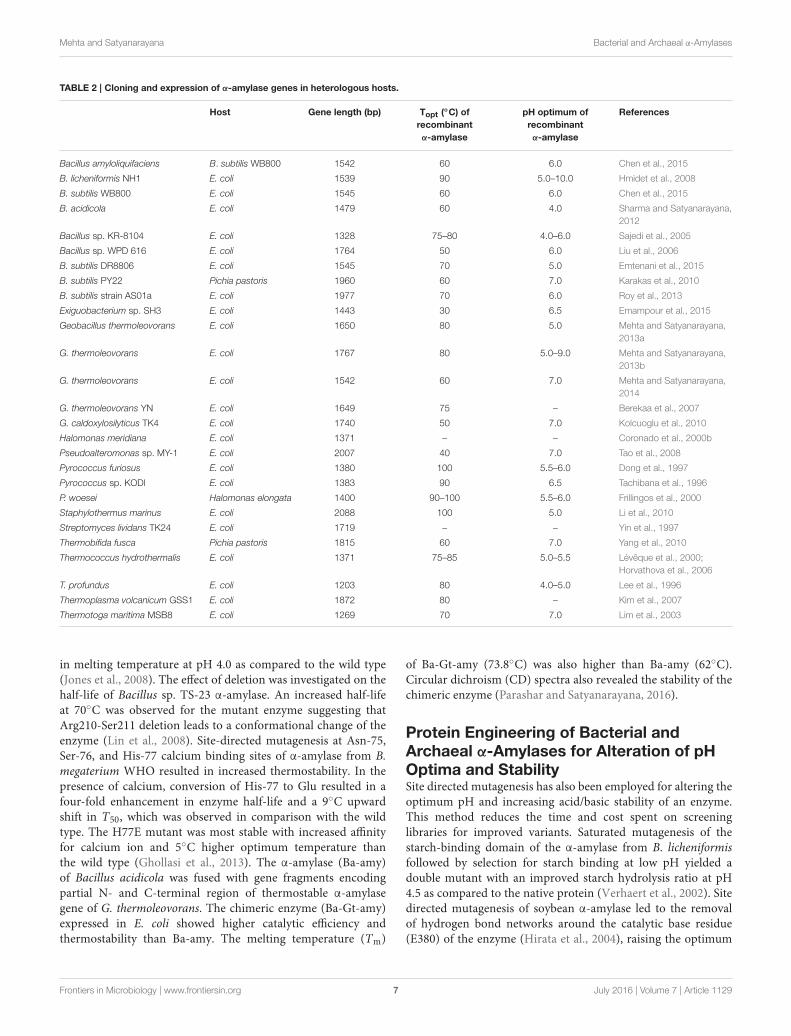
and pH activity between 5 and 9 (Mehta and Satyanarayana,2013b), and the second raw starch hydrolysing thermostableα-amylase of 56 kDa with optimum activity at 60◦C and pH7.0 (Mehta and Satyanarayana, 2014). A gene encoding an α-amylase (Amy-E) from Exiguobacterium sp. SH3 was expressedin E. coli as a functional His-tagged protein of about 53 kDawith maximum activity at 30◦C and pH 6.5. This enzymeretains 41% of its maximum activity at 0◦C. This enzymewas biochemically characterized. It was halotolerant, and itsactivity was stimulated at high salt concentrations in the rangeof 1–5 M (Emampour et al., 2015). Another α-amylase genefrom B. amyloliquifaciens was cloned into pMD18-T shuttlevector which was reconstructed to obtain vector pP43X for itsheterologous expression in B. subtilis WB800. The recombinantpurified enzyme had a specific activity of 5566 U/mg. Itsproductivity is 1.48-fold higher than the wild strain. The α-amylase gene encodes a protein of 514 amino acid residueswith a predicted molecular weight of 58.4 kDa. The optimalconditions for its activity are pH 6.0 and 60◦C. It was alsobiochemically characterized (Chen et al., 2015). The details ofcloning and expression of α-amylase encoding genes are listed inTable 2.
PROTEIN ENGINEERING OF α-AMYLASESAND MOLECULAR METHODS FOR THEIRAMELIORATION
Since several decades, a wide array of techniques have beendeveloped allowing engineering of the enzyme properties.Directed evolution and site-directed mutagenesis are powerfultools for engineering enzymes, to improve their functionsand to alter their properties like activity, selectivity, substratespecificity, stability, and solubility (Rubin-Pitel and Zhao,2006). Thermostability, activity in acidic range and Ca2+-independence of α-amylases are desirable for their use in thestarch saccharification process, and activity in alkaline rangeand oxidative stability is a prerequisite for its applicability indetergent industry. These and several other properties of α-amylases have been improved by various methods such as site-directed mutagenesis or directed evolution approach, which aredescribed below.
Protein Engineering of Bacterial andArchaeal α-Amylases to IncreaseThermostabilityMany factors contribute to the stability of thermostableproteins including the presence of hydrogen bonds, electrostaticinteractions, salt bridges, hydrophobic interactions, disulfidebonds, reduced entropy of unfolding, oligomerization, increased
occurrence of proline residues, and others (Salminen et al., 1996;Russell et al., 1997; Vogt et al., 1997; Mehta and Satyanarayana,2013b, 2015). Any of these properties of an enzyme can bemodified by using methods such as site-directed mutagenesis,directed evolution, deletion mutation, and others for increasingits thermostability. Some examples of increasing thermostabilityof α-amylases are explained as follows. The first determinantsof thermostability in bacterial α-amylases were identified bycomparison of amino acid sequences of α-amylases of B.licheniformis (BLA) and B. amyloliquifaciens (BAA). Threestabilizing mutations in BAA were proposed, which caused asignificant and additive thermostabilization of BAA; deletionof R176 and G177, and the substitution of K269A (Suzukiet al., 1989). This R176-G177 deletion showed similar effectson the thermostability of other α-amylases as well (Igarashiet al., 1998; Bisgaad-Frantzen et al., 1999). Conrad et al. (1995)separately determined four regions of particular importance forthe thermostability of BLA, namely 34–76, 112–142, 174–179,and 263–276. Two of these coincide with the mutations identifiedearlier by Suzuki et al. (1989). BLA has been further mutated towithstand high temperature and acidic pH. Two positions 133and 209 have been identified as important for its thermostability(Declerck et al., 1990; Joyet et al., 1992). Twelve different aminoacids substitutions were made at H133 by a tRNA suppressormethod, and H133Y substitution improved the stability themost (Declerck et al., 1990). Random mutagenesis and screeningapproach was used to create A209V substitution. The stabilizingeffect of this mutation was an additive with the H133Y mutation,and the half-life of the double mutant was 10-fold higher at90◦C (Joyet et al., 1992). All naturally occurring amino acidswere introduced in each of these two positions (Declerck et al.,1995). Of these, H133I substitution had stronger effect onthermostabilization than the previous substitution. Randommutagenesis and screening have also been used to identify M15Tand N188S as stabilizing mutations of BLA (Shaw et al., 1999).A 23-fold enhancement in stability was achieved at pH 5.0and 83◦C in the presence of 5mM CaCl2 by combining thesemutations with H133Y/A209V. Shaw et al. (1999) also predicteda stabilizing effect for additional substitutions: A33S, A52S,N96Q, S148N, and A379S. Four rounds of DNA shuffling andsubsequent recombination produced a variant from Thermus sp.IM6051 maltogenic amylase with a 15◦C increase in optimumtemperature as compared to wild-type (Kim et al., 2003). Thehalf-life of this variant was 172min at 80◦C as compared to thewild type, which was completely inactivated at this temperature.After three rounds of DNA shuffling of B. thermalkalophilusET2 maltogenic amylase, followed by recombination of selectedmutations, variants with optimal reaction temperatures 10◦Chigher than the native enzyme was achieved. One of the variantscarrying mutations displayed a 20-fold longer half-life than wild-type at 78◦C (Tang et al., 2006). To improve the performanceof a maltogenic amylase (Novamyl) as an antistaling agentfor breads made at low pH, two epPCR libraries of Novamylwere constructed. These libraries were screened for improvedthermal stability at 80◦C and activity at pH 4.3 for 25 min.A triple mutant was better than the wild type for antistalingactivity in bread made at pH 4.3 and exhibited a 10◦C increase
Frontiers in Microbiology | www.frontiersin.org 6 July 2016 | Volume 7 | Article 1129
Mehta and Satyanarayana Bacterial and Archaeal α-Amylases
TABLE 2 | Cloning and expression of α-amylase genes in heterologous hosts.
Host Gene length (bp) Topt (◦C) of
recombinant
α-amylase
pH optimum of
recombinant
α-amylase
References
Bacillus amyloliquifaciens B. subtilis WB800 1542 60 6.0 Chen et al., 2015
B. licheniformis NH1 E. coli 1539 90 5.0–10.0 Hmidet et al., 2008
B. subtilis WB800 E. coli 1545 60 6.0 Chen et al., 2015
B. acidicola E. coli 1479 60 4.0 Sharma and Satyanarayana,
2012
Bacillus sp. KR-8104 E. coli 1328 75–80 4.0–6.0 Sajedi et al., 2005
Bacillus sp. WPD 616 E. coli 1764 50 6.0 Liu et al., 2006
B. subtilis DR8806 E. coli 1545 70 5.0 Emtenani et al., 2015
B. subtilis PY22 Pichia pastoris 1960 60 7.0 Karakas et al., 2010
B. subtilis strain AS01a E. coli 1977 70 6.0 Roy et al., 2013
Exiguobacterium sp. SH3 E. coli 1443 30 6.5 Emampour et al., 2015
Geobacillus thermoleovorans E. coli 1650 80 5.0 Mehta and Satyanarayana,
2013a
G. thermoleovorans E. coli 1767 80 5.0–9.0 Mehta and Satyanarayana,
2013b
G. thermoleovorans E. coli 1542 60 7.0 Mehta and Satyanarayana,
2014
G. thermoleovorans YN E. coli 1649 75 – Berekaa et al., 2007
G. caldoxylosilyticus TK4 E. coli 1740 50 7.0 Kolcuoglu et al., 2010
Halomonas meridiana E. coli 1371 – – Coronado et al., 2000b
Pseudoalteromonas sp. MY-1 E. coli 2007 40 7.0 Tao et al., 2008
Pyrococcus furiosus E. coli 1380 100 5.5–6.0 Dong et al., 1997
Pyrococcus sp. KODl E. coli 1383 90 6.5 Tachibana et al., 1996
P. woesei Halomonas elongata 1400 90–100 5.5–6.0 Frillingos et al., 2000
Staphylothermus marinus E. coli 2088 100 5.0 Li et al., 2010
Streptomyces lividans TK24 E. coli 1719 – – Yin et al., 1997
Thermobifida fusca Pichia pastoris 1815 60 7.0 Yang et al., 2010
Thermococcus hydrothermalis E. coli 1371 75–85 5.0–5.5 Lévêque et al., 2000;
Horvathova et al., 2006
T. profundus E. coli 1203 80 4.0–5.0 Lee et al., 1996
Thermoplasma volcanicum GSS1 E. coli 1872 80 – Kim et al., 2007
Thermotoga maritima MSB8 E. coli 1269 70 7.0 Lim et al., 2003
in melting temperature at pH 4.0 as compared to the wild type(Jones et al., 2008). The effect of deletion was investigated on thehalf-life of Bacillus sp. TS-23 α-amylase. An increased half-lifeat 70◦C was observed for the mutant enzyme suggesting thatArg210-Ser211 deletion leads to a conformational change of theenzyme (Lin et al., 2008). Site-directed mutagenesis at Asn-75,Ser-76, and His-77 calcium binding sites of α-amylase from B.megaterium WHO resulted in increased thermostability. In thepresence of calcium, conversion of His-77 to Glu resulted in afour-fold enhancement in enzyme half-life and a 9◦C upwardshift in T50, which was observed in comparison with the wildtype. The H77E mutant was most stable with increased affinityfor calcium ion and 5◦C higher optimum temperature thanthe wild type (Ghollasi et al., 2013). The α-amylase (Ba-amy)of Bacillus acidicola was fused with gene fragments encodingpartial N- and C-terminal region of thermostable α-amylasegene of G. thermoleovorans. The chimeric enzyme (Ba-Gt-amy)expressed in E. coli showed higher catalytic efficiency andthermostability than Ba-amy. The melting temperature (Tm)
of Ba-Gt-amy (73.8◦C) was also higher than Ba-amy (62◦C).Circular dichroism (CD) spectra also revealed the stability of thechimeric enzyme (Parashar and Satyanarayana, 2016).
Protein Engineering of Bacterial andArchaeal α-Amylases for Alteration of pHOptima and StabilitySite directed mutagenesis has also been employed for altering theoptimum pH and increasing acid/basic stability of an enzyme.This method reduces the time and cost spent on screeninglibraries for improved variants. Saturated mutagenesis of thestarch-binding domain of the α-amylase from B. licheniformisfollowed by selection for starch binding at low pH yielded adouble mutant with an improved starch hydrolysis ratio at pH4.5 as compared to the native protein (Verhaert et al., 2002). Sitedirected mutagenesis of soybean α-amylase led to the removalof hydrogen bond networks around the catalytic base residue(E380) of the enzyme (Hirata et al., 2004), raising the optimum
Frontiers in Microbiology | www.frontiersin.org 7 July 2016 | Volume 7 | Article 1129
Mehta and Satyanarayana Bacterial and Archaeal α-Amylases
from pH 5.4 to a more neutral pH range between 6.0 and6.6. Richardson et al. (2002) used activity and sequence basedscreening to single and multi-organism DNA libraries fromvarious environments to identify three different α-amylases withone or more aspects of the necessary phenotype: temperaturestability, pH optimum, and lowered requirement of Ca2+. Thegenes encoding amylases with these properties were used asparental sequences for DNA shuffling in order to combine thebest aspects of the three enzyme phenotypes. The two bestchimeric α-amylases found by high throughput screening had40-fold longer half-life for activity in the absence of Ca2+ ionsat 90◦C and pH 4.5 as compared to the most stable wild-typeparent. The α-amylase from B. amyloliquefaciens with optimalactivity at pH 6.0 was engineered by error prone PCR (epPCR)to produce a variant having optimal activity at alkaline pH forthe detergent industry. The screening of an epPCR library foramylase activity at pH 7.0 and pH 10.0 yielded 26 variantswith a very high activity. Sixteen of the selected variants werethen randomly recombined by DNA shuffling and screened forincreased activity at pH 10. The best mutant displayed a five-fold higher activity at pH 10 than the wild type (Bessler et al.,2003). Liu et al. (2008) observed that the mutations at two crucialpositions Leu134 and Ser320 together affected the acid resistance ofthe α-amylase of B. licheniformisCICC 10181. Directed evolutionwas used to increase acid stability. In another example, proteinstability and catalytic efficiency of α-amylase from B. subtilis wasincreased under acidic conditions by site-directed mutagenesis.Based on the three dimensional structure model analysis, fourbasic histidine (His) residues His222, His275, His293, and His310
in the catalytic domain were found to be important and single,double as well as triple mutants were constructed at these sites.The acidic stability of enzyme was significantly enhanced aftermutation, and 45–92% of initial activity of mutants was retainedafter incubation at pH 4.5 and 25◦C for 24 h as compared to thewild-type (39.5%). The catalytic efficiency for each active mutantwas also much higher than that of wild-type at low pH. Due toincrease in the hydrogen bonds and salt bridges after mutation,an obvious shift of the basic limb toward acidity was observed.These changes around the catalytic domain contributed to thesignificantly improved protein stability and catalytic efficiency atlow pH (Yang et al., 2013).
Protein Engineering of Bacterial andArchaeal α-Amylases for IncreasingOxidative StabilityOxidation also has a demonstrated negative effect on the stabilityof α-amylases. Cysteine 362 was identified as the oxidationprone residue in BStA (Tomazic and Klibanov, 1988; Brosnanet al., 1992), and methionine 197, which is situated close tothe active site, has been shown to be responsible for theinactivation of BLA (Borchert et al., 1995). The introductionof any non-sulfur-containing amino acid at position 197 wasshown to greatly reduce the oxidation sensitivity of BLA. Thespecific activity was, however, very much dependent on theintroduced side chain, with an apparent preference for thesmaller side chains (Borchert et al., 1995). Bacillus sp. TS-23
α-amylase was truncated at both N and C termini, and furthermutated to increase thermal and oxidative stability. Met231was mutated to leucine and 483th codon of this enzyme wasmutated to stop codon i.e., TAA by site-directed mutagenesis.The resultant engineered enzyme showed higher T1/2 at 70◦Cand showed compatibility to detergents (oxidative stability; Chiet al., 2010). In another example, oxidative stability of α-amylase(AmyC) from Thermotoga maritima was improved by site-directed mutagenesis. In this, methionine residues at positions 43and 44, 55, and 62 were mutated to alanine, which is oxidative-resistant. The M55A mutant showed 50% residual activity in thepresence of H2O2, while the wild-type α-amylase was inactive(Ozturk et al., 2013).
Protein Engineering of Bacterial andArchaeal α-Amylases for the Alteration ofTransglycosylation/Hydrolysis RatioProduct re-hydrolyzation is a problem duringmaltooligosaccharide synthesis using maltogenic amylase.Maltogenic amylase (MAG1) from Bacillus lehensis G1 wasmutatedusinga structure-guidedprotein engineeringapproach todecrease the hydrolysis activity of the enzyme.W359F, Y377F, andM375I mutations reduced steric interference, altered the subsiteoccupation and increased the internal flexibility to accommodatelonger donor/acceptor molecules for transglycosylation, whichresulted in an increase in the transglycosylation to hydrolysisratio of up to 4.0-fold (Manas et al., 2015). Maltogenic amylasesalso use donor and acceptor sugar molecules to form α-1,4or 1,6 glycosidic linkages between them. This process, calledtransglycosylation, is responsible for formation of undesirablemaltotriose in the high maltose syrups. Maltogenic amylaseswith high hydrolytic activity than transglycosylation activityare used to decrease the maltotriose content of high maltosesyrups. Maltogenic amylase from B. stearothermophilus wasengineered by site-directed mutagenesis of W177 to decrease itstransglycosylation activity. The mutant W177S exhibited notableincreased maltose production with a minimum amount ofmaltotriose under industrial conditions (Sun et al., 2016). Role ofHis and Glu in the catalytic activity of B. licheniformis α-amylase(BLA) was determined by performing site-directed mutagenesisat His235. It was observed that the mutant enzyme (H235E)displayed high substrate transglycosylation activity, while thewild-type BLA exhibited high hydrolysis activity exclusively.This shows that Glu235 located on a wide open groove nearsubsite +1 is most probably involved in transglycosylation andmay recognize and stabilize the non-reducing end glucose of theacceptor molecule (Tran et al., 2014)
SEQUENCE AND STRUCTURALDIVERSITY OF α-AMYLASES
Conserved Regions of GH-13 FamilyAccording to the Carbohydrate-Active enZYmes (CAZY)database, the α-amylases are grouped with different kindsof glycosyl hydrolases in GH-13 family, also known as α-amylase family (Henrissat, 1991). This family contains the
Frontiers in Microbiology | www.frontiersin.org 8 July 2016 | Volume 7 | Article 1129

Mehta and Satyanarayana Bacterial and Archaeal α-Amylases
enzymes with the following characteristics. They act on α-glycosidic bonds and hydrolyze it to produce α-anomeric mono-or oligosaccharides (hydrolysis) or form α-1,4 or 1,6 glycosidiclinkages (transglycosylation), or show both activities. Theypossess a (β/α)8 or TIMbarrel structure that contains the catalyticsite residues (Figure 1). Their primary sequences have sevenhighly conserved regions with catalytic amino acids and someamino acids that are essential for the stability of the topologyof conserved TIM barrel (Kuriki and Imanaka, 1999). The firstfour conserved regions (I to IV) are found in the TIM-barrel onβ-strands 3, 4, and 5 and in the loop connecting β-strand 7 to α-helix 7. Amino acid residues corresponding to region I are foundin the C-terminal end of the third β-strand of the TIM barrel (β3).This region includes the three conserved amino acids Asp 100,Asn104, and His105 (BLA numbering). Asp100 is essential forthe active site integrity because it forms hydrogen bonding withArg229. Arg229 is a completely conserved residue. This is presentwithin hydrogen bond distance of the catalytic nucleophileAsp231 and the proton donor Glu26. Asn104 coordinates theconserved calcium ion between the A and B domains and doesnot directly stabilize the active site (Boel et al., 1990; MacHiuset al., 1995, 1998). His105 stabilizes the interaction betweenthe C-terminal of β3 and the rest of TIM barrel by hydrogenbonding to Asn104 and to the backbone oxygen of Tyr56 (BLAnumbering), which is situated in the loop connecting β2 to α2.Region II is positioned in β4 and has the catalytic nucleophileAsp231 and the invariant residue Arg229. These two residues areindispensable for catalytic activity and are found in all α-amylases(Svensson, 1994). Lys234 and His235 are also present in thisregion, which form part of subsite +2. These amino acids bindthe reducing end of the glucose chain in the substrate-binding
FIGURE 1 | Schematic representation of the (β/α)8 barrel (TIM barrel).
cleft (Svensson, 1994). Region III includes the conserved residueand catalytic proton donor Glu261, which lies in the C-terminalpart of the fifth L-strand of the TIM-barrel. The residues formingregion IV are situated in the loop connecting L7 to K7 and protectthe active site from the solvent. This region has fully conservedresidue Asp328, which is postulated to be involved in substratebinding, substrate distortion and in elevating the pKa of Glu261.There is a strong preference for His, Phe, Val and Asn are oftenobserved at positions 327, 323, 324, and 326, respectively (Kleinet al., 1992; Knegtel et al., 1995; Strokopytov et al., 1995). Thenumber of residues between the conserved Gly and Pro of theregion VI differentiates between an α-amylase (7 residues) anda CGTase (8 residues; Janecek et al., 1995). Region VII does notcontain conserved residues, but it usually starts with Gly followedby proline at i+ 2 position.
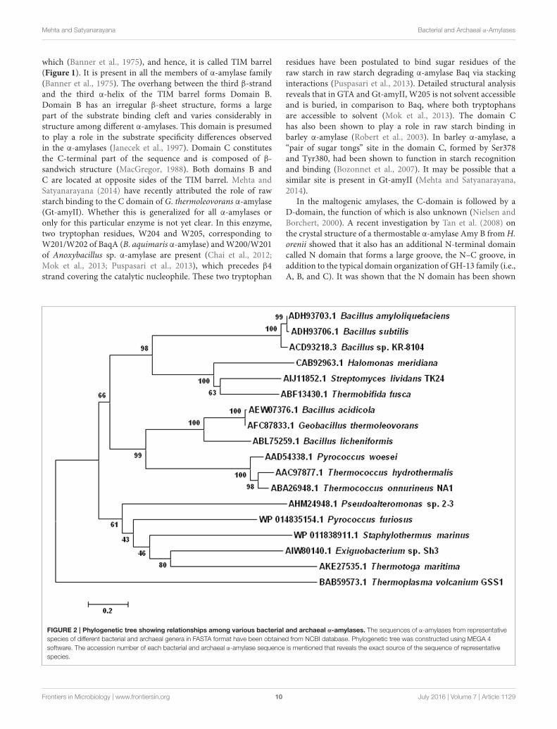
GH-13 vs. GH-57 Family: A ComparisonWith Respect to Archaeal α-AmylasesComparison of the T. hydrothermalis α-amylase sequence withthose of 21 other bacterial and archaeal α-amylases, which wererepresentatives of more than 100 known α-amylase sequences,revealed that this enzyme as well as the other Thermococcalessecreted α-amylases, contain most of the β-strands and thepentapeptide stretch located near the C-terminus of loop β3→α3that are present in family GH-13 (Janecek, 1997; Janecek et al.,1999). This indicates that α-amylase secreted by the members ofThermococcales belong to family GH-13. However, α-amylasefrom P. furiosus does not contain these conserved regions, soit belongs to GH-57 family instead. It was also showed thatthe α-amylase from the methanogenic archaeon Methanococcusjannaschii contains features of both glycosl hydrolase families,which indicates that these two families are either derived froma distant common ancestor or that one evolved from the other(Janecek, 1998). Also, the conserved regions of Thermococcaleα-amylases show some characteristic sequence features (Janeceket al., 1999). A tryptophan is present in all the archaeal enzymesin the β4 strand. Moreover, a histidine, which has been suggestedto be responsible for substrate binding, is replaced by glycinein archaeal enzymes (Svensson, 1994). It was suggested that thetryptophan found in β4 strand might be involved in catalysisinstead of the missing histidine (Lee et al., 1996). Phylogeneticanalysis revealed that archaeal α-amylases are closely related toplant α-amylases (Janecek et al., 1999). It also suggested thatarchaeal α-amylases are closer to the liquefying α-amylases, thanto the saccharifying ones, which also agrees with the nature of theproducts of starch hydrolysis by the archaeal enzymes (Janeceket al., 1999). The phylogenetic analysis of various archaeal andbacterial α-amylases is shown in Figure 2.
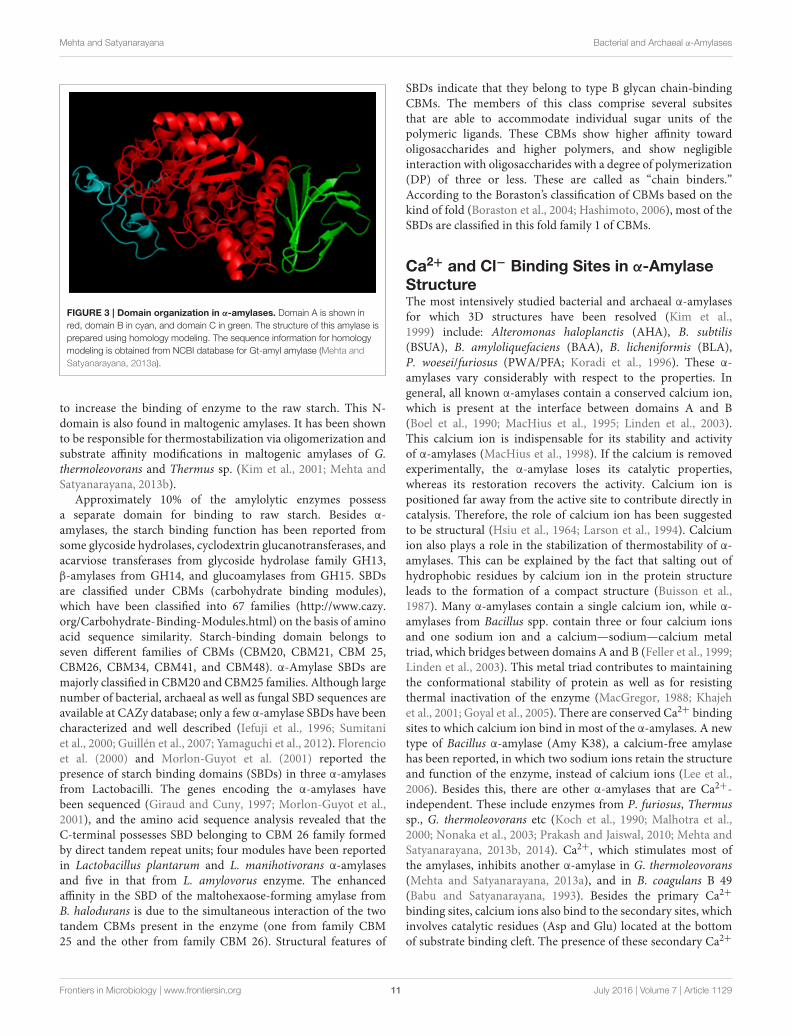
Domain Organization in α-AmylasesThe structure of α-amylase, in general, consists of a polypeptidechain folded into three domains called A, B, and C (Figure 3).Domain A is the catalytic domain, which consists of N-terminal (β/α)8 barrel. This consists of a highly symmetricalfold which includes eight parallel β-strands organized in a barrelwith a border of eight α-helices. This (β/α)8 barrel was firstobserved in chicken muscle triosephosphate isomerase (TIM),
Frontiers in Microbiology | www.frontiersin.org 9 July 2016 | Volume 7 | Article 1129
Mehta and Satyanarayana Bacterial and Archaeal α-Amylases
which (Banner et al., 1975), and hence, it is called TIM barrel(Figure 1). It is present in all the members of α-amylase family(Banner et al., 1975). The overhang between the third β-strandand the third α-helix of the TIM barrel forms Domain B.Domain B has an irregular β-sheet structure, forms a largepart of the substrate binding cleft and varies considerably instructure among different α-amylases. This domain is presumedto play a role in the substrate specificity differences observedin the α-amylases (Janecek et al., 1997). Domain C constitutesthe C-terminal part of the sequence and is composed of β-sandwich structure (MacGregor, 1988). Both domains B andC are located at opposite sides of the TIM barrel. Mehta andSatyanarayana (2014) have recently attributed the role of rawstarch binding to the C domain of G. thermoleovorans α-amylase(Gt-amyII). Whether this is generalized for all α-amylases oronly for this particular enzyme is not yet clear. In this enzyme,two tryptophan residues, W204 and W205, corresponding toW201/W202 of BaqA (B. aquimaris α-amylase) andW200/W201of Anoxybacillus sp. α-amylase are present (Chai et al., 2012;Mok et al., 2013; Puspasari et al., 2013), which precedes β4strand covering the catalytic nucleophile. These two tryptophan
residues have been postulated to bind sugar residues of theraw starch in raw starch degrading α-amylase Baq via stackinginteractions (Puspasari et al., 2013). Detailed structural analysisreveals that in GTA and Gt-amyII, W205 is not solvent accessibleand is buried, in comparison to Baq, where both tryptophansare accessible to solvent (Mok et al., 2013). The domain Chas also been shown to play a role in raw starch binding inbarley α-amylase (Robert et al., 2003). In barley α-amylase, a“pair of sugar tongs” site in the domain C, formed by Ser378and Tyr380, had been shown to function in starch recognitionand binding (Bozonnet et al., 2007). It may be possible that asimilar site is present in Gt-amyII (Mehta and Satyanarayana,2014).
In the maltogenic amylases, the C-domain is followed by aD-domain, the function of which is also unknown (Nielsen andBorchert, 2000). A recent investigation by Tan et al. (2008) onthe crystal structure of a thermostable α-amylase Amy B from H.orenii showed that it also has an additional N-terminal domaincalled N domain that forms a large groove, the N–C groove, inaddition to the typical domain organization of GH-13 family (i.e.,A, B, and C). It was shown that the N domain has been shown
FIGURE 2 | Phylogenetic tree showing relationships among various bacterial and archaeal α-amylases. The sequences of α-amylases from representative
species of different bacterial and archaeal genera in FASTA format have been obtained from NCBI database. Phylogenetic tree was constructed using MEGA 4
software. The accession number of each bacterial and archaeal α-amylase sequence is mentioned that reveals the exact source of the sequence of representative
species.
Frontiers in Microbiology | www.frontiersin.org 10 July 2016 | Volume 7 | Article 1129
Mehta and Satyanarayana Bacterial and Archaeal α-Amylases
FIGURE 3 | Domain organization in α-amylases. Domain A is shown in
red, domain B in cyan, and domain C in green. The structure of this amylase is
prepared using homology modeling. The sequence information for homology
modeling is obtained from NCBI database for Gt-amyI amylase (Mehta and
Satyanarayana, 2013a).
to increase the binding of enzyme to the raw starch. This N-domain is also found in maltogenic amylases. It has been shownto be responsible for thermostabilization via oligomerization andsubstrate affinity modifications in maltogenic amylases of G.thermoleovorans and Thermus sp. (Kim et al., 2001; Mehta andSatyanarayana, 2013b).
Approximately 10% of the amylolytic enzymes possessa separate domain for binding to raw starch. Besides α-amylases, the starch binding function has been reported fromsome glycoside hydrolases, cyclodextrin glucanotransferases, andacarviose transferases from glycoside hydrolase family GH13,β-amylases from GH14, and glucoamylases from GH15. SBDsare classified under CBMs (carbohydrate binding modules),which have been classified into 67 families (http://www.cazy.org/Carbohydrate-Binding-Modules.html) on the basis of aminoacid sequence similarity. Starch-binding domain belongs toseven different families of CBMs (CBM20, CBM21, CBM 25,CBM26, CBM34, CBM41, and CBM48). α-Amylase SBDs aremajorly classified in CBM20 and CBM25 families. Although largenumber of bacterial, archaeal as well as fungal SBD sequences areavailable at CAZy database; only a few α-amylase SBDs have beencharacterized and well described (Iefuji et al., 1996; Sumitaniet al., 2000; Guillén et al., 2007; Yamaguchi et al., 2012). Florencioet al. (2000) and Morlon-Guyot et al. (2001) reported thepresence of starch binding domains (SBDs) in three α-amylasesfrom Lactobacilli. The genes encoding the α-amylases havebeen sequenced (Giraud and Cuny, 1997; Morlon-Guyot et al.,2001), and the amino acid sequence analysis revealed that theC-terminal possesses SBD belonging to CBM 26 family formedby direct tandem repeat units; four modules have been reportedin Lactobacillus plantarum and L. manihotivorans α-amylasesand five in that from L. amylovorus enzyme. The enhancedaffinity in the SBD of the maltohexaose-forming amylase fromB. halodurans is due to the simultaneous interaction of the twotandem CBMs present in the enzyme (one from family CBM25 and the other from family CBM 26). Structural features of
SBDs indicate that they belong to type B glycan chain-bindingCBMs. The members of this class comprise several subsitesthat are able to accommodate individual sugar units of thepolymeric ligands. These CBMs show higher affinity towardoligosaccharides and higher polymers, and show negligibleinteraction with oligosaccharides with a degree of polymerization(DP) of three or less. These are called as “chain binders.”According to the Boraston’s classification of CBMs based on thekind of fold (Boraston et al., 2004; Hashimoto, 2006), most of theSBDs are classified in this fold family 1 of CBMs.
Ca2+ and Cl− Binding Sites in α-AmylaseStructureThe most intensively studied bacterial and archaeal α-amylasesfor which 3D structures have been resolved (Kim et al.,1999) include: Alteromonas haloplanctis (AHA), B. subtilis(BSUA), B. amyloliquefaciens (BAA), B. licheniformis (BLA),P. woesei/furiosus (PWA/PFA; Koradi et al., 1996). These α-amylases vary considerably with respect to the properties. Ingeneral, all known α-amylases contain a conserved calcium ion,which is present at the interface between domains A and B(Boel et al., 1990; MacHius et al., 1995; Linden et al., 2003).This calcium ion is indispensable for its stability and activityof α-amylases (MacHius et al., 1998). If the calcium is removedexperimentally, the α-amylase loses its catalytic properties,whereas its restoration recovers the activity. Calcium ion ispositioned far away from the active site to contribute directly incatalysis. Therefore, the role of calcium ion has been suggestedto be structural (Hsiu et al., 1964; Larson et al., 1994). Calciumion also plays a role in the stabilization of thermostability of α-amylases. This can be explained by the fact that salting out ofhydrophobic residues by calcium ion in the protein structureleads to the formation of a compact structure (Buisson et al.,1987). Many α-amylases contain a single calcium ion, while α-amylases from Bacillus spp. contain three or four calcium ionsand one sodium ion and a calcium—sodium—calcium metaltriad, which bridges between domains A and B (Feller et al., 1999;Linden et al., 2003). This metal triad contributes to maintainingthe conformational stability of protein as well as for resistingthermal inactivation of the enzyme (MacGregor, 1988; Khajehet al., 2001; Goyal et al., 2005). There are conserved Ca2+ bindingsites to which calcium ion bind in most of the α-amylases. A newtype of Bacillus α-amylase (Amy K38), a calcium-free amylasehas been reported, in which two sodium ions retain the structureand function of the enzyme, instead of calcium ions (Lee et al.,2006). Besides this, there are other α-amylases that are Ca2+-independent. These include enzymes from P. furiosus, Thermussp., G. thermoleovorans etc (Koch et al., 1990; Malhotra et al.,2000; Nonaka et al., 2003; Prakash and Jaiswal, 2010; Mehta andSatyanarayana, 2013b, 2014). Ca2+, which stimulates most ofthe amylases, inhibits another α-amylase in G. thermoleovorans(Mehta and Satyanarayana, 2013a), and in B. coagulans B 49(Babu and Satyanarayana, 1993). Besides the primary Ca2+
binding sites, calcium ions also bind to the secondary sites, whichinvolves catalytic residues (Asp and Glu) located at the bottomof substrate binding cleft. The presence of these secondary Ca2+
Frontiers in Microbiology | www.frontiersin.org 11 July 2016 | Volume 7 | Article 1129
Mehta and Satyanarayana Bacterial and Archaeal α-Amylases
binding sites at catalytic sites explains inhibition of α-amylase byCa2+ at higher concentration (MacHius et al., 1995; Mehta andSatyanarayana, 2013a). Many α-amylases also contain a chlorideion in the active site, which enhances the catalytic efficiencyof the enzyme (Levitsky and Steer, 1974; Feller et al., 1996).Althoughmajorly found inmammalian α-amylases (Larson et al.,1994; Brayer et al., 1995; Ramasubbu et al., 1996), a chlorideion has also been reported in a α-amylase (AHA) from thepsychrophilic bacterium, A. haloplanctis (Aghajari et al., 1998).Chloride binding increases the affinity of the enzyme for thecalcium ion, therefore it is much likely that binding of chlorideion also induces conformational changes around the active site(Levitsky and Steer, 1974).
Active Site Cleft of α-AmylasesIn total, three steps are involved in the catalytic mechanismfor retaining glycosyl hydrolases (Davies and Henrissat, 1995).Firstly, the glycosidic oxygen is protonated by the protondonor (Glu261). This is followed by a nucleophilic attack onthe C1 of the sugar residue in subsite-1 by Asp231 (BLAnumbering; Nielsen et al., 1999). Once the glycon part of thesubstrate leaves, a water molecule is activated presumably bythe deprotonated Glu261. This water molecule hydrolyses thecovalent bond between the nucleophilic oxygen and the Cl of thesugar residue in subsite-1, thereby completing the catalytic cycle(Nielsen et al., 1999).
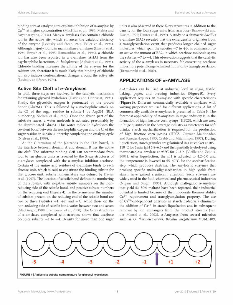
At the C-terminus of the β-strands in the TIM barrel, inthe interface between domain A and domain B lies the activesite cleft. The substrate binding cleft can accommodate fromfour to ten glucose units as revealed by the X-ray structures ofα-amylases complexed with the α-amylase inhibitor acarbose.Certain of the amino acid residues of α-amylase binds to eachglucose unit, which is said to constitute the binding subsite forthat glucose unit. Subsite nomenclature was defined by Davieset al. (1997). The location of scissile bond defines the numberingof the subsites, with negative subsite numbers on the non-reducing side of the scissile bond, and positive subsite numberson the reducing end (Figure 4). In the α-amylases the numberof subsites present on the reducing end of the scissile bond aretwo or three (subsites +1, +2, and +3), while those on thenon-reducing side of scissile bond varies between two and seven(MacGregor, 1988; Brzozowski et al., 2000). The X-ray structuresof α-amylases complexed with acarbose shows that acarboseoccupies subsites −1 to +4. Density for more than one sugar
units is also observed in these X-ray structures in addition to thedensity for the four sugar units from acarbose (Brzozowski andDavies, 1997; Dauter et al., 1999). A study on a chimaeric Bacillusα-amylase (BA2) revealed that the extra density originates froma transglycosylation event that produces longer chained sugarmolecules, which span the subsites −7 to +3, in comparison toan active site mutant of BA2, in which acarbose molecule spansthe subsites−7 to−4. This observation suggests that the catalyticactivity of the α-amylases is necessary for converting acarboseinto amore potent longer-chained inhibitor by transglycosylation(Brzozowski et al., 2000).
APPLICATIONS OF α-AMYLASE
α-Amylases can be used at industrial level in sugar, textile,baking, paper, and brewing industries (Figure 5). Everyapplication requires an α-amylase with specific characteristics(Figure 6). Different commercially available α-amylases withvarying properties are used for different applications. A list ofcommercially available α-amylases is presented in Table 3. Theforemost applicability of α-amylases in sugar industry is in theformation of high fructose corn syrups (HFCS), which are usedin huge quantities in the beverage industry as sweeteners for softdrinks. Starch saccharification is required for the productionof high fructose corn syrups (HFCS; Guzman-Maldonadaoand Paredes-Lopez, 1995; Crabb and Mitchinson, 1997). Duringliquefaction, starch granules are gelatinized in a jet cooker at 105–110◦C for 5 min (pH 5.8–6.5) and then partially hydrolyzed usingthermostable α-amylase at 95◦C for 2–3 h (Vieille and Zeikus,2001). After liquefaction, the pH is adjusted to 4.2–5.0 andthe temperature is lowered to 55–60◦C for the saccharificationstep, which produces dextrins. The amylolytic enzymes thatproduce specific malto-oligosaccharides in high yields fromstarch have gained significant attention. Such enzymes arewidely used in the food, chemical and pharmaceutical industries(Nigam and Singh, 1995). Although maltogenic α-amylasesthat yield 53–80% maltose have been reported, their industrialpotential is limited because of their moderate thermostability,Ca2+ requirement and transglycosylation property. The useof Ca2+-independent enzymes in starch hydrolysis eliminatesthe addition of Ca2+ in starch liquefaction and its subsequentremoval by ion exchangers from the product streams (vander Maarel et al., 2002). α-Amylases from several microbessuch as G. thermoleovorans, Bacillus megaterium VUMB109,
FIGURE 4 | Active site subsite nomenclature for glycosyl hydrolases.
Frontiers in Microbiology | www.frontiersin.org 12 July 2016 | Volume 7 | Article 1129

Mehta and Satyanarayana Bacterial and Archaeal α-Amylases
FIGURE 5 | Applications of α-amylases.
P. furiosus, and others have been reported to possess most of thedesired properties, which enable them to produce high yieldsof maltooligosaccharides and maltose (Rao and Satyanarayana,2007b; Jana et al., 2013; Cuong et al., 2016). Some raw starchdegrading α-amylases have also been reported which can act atthe native pH of starch, thereby avoiding the pH adjustmentstep in the starch saccharification process. Hydrolysis rates ofcorn and wheat starches at 60◦C were 65.5 and 70.3% with 10.0%starch slurry (Mehta and Satyanarayana, 2013a). Hydrolysisrates of 60 and 37% of 1% raw wheat and corn starches in 4 hhas been recorded with 0.07 U mg−1 starch of B. licheniformisATCC 9945a α-amylase (Bozic et al., 2011). The α-amylase ofAlicyclobacillus at 0.5 U mg−1 starch hydrolyzed 52.0% of 1.0%corn starch in 2 h (Bai et al., 2012). While G. thermoleovoranssubsp. stromboliensis α- amylase at 500 U g−1 starch, hydrolyzed50.0% of the corn starch (20%) in 12 h (Finore et al., 2011).
Another application of α-amylases is in textile desizing.During the weaving process the warp (chain) threads are exposedto considerable mechanical strain. In order to prevent breakage,they are usually reinforced by coating (sizing) with a gelatinoussubstance (size) like starch. Small amounts of fats or oilsmay be also added to the size, with the aim of lubricating
the warp coat surface. As a consequence of the sizing, thewarp threads of the fabric are not able to absorb water orfinishing agents to a sufficient degree. For that, size must beremoved (desizing) before finishing. The complete removal ofstarch containing size without fiber damage is best obtainedby using enzymatic desizing agents. The enzymatic desizingprocess has three stages. First stage is impregnation. In thisstage, enzyme solution is absorbed by the fabric. This stageinvolves thorough wetting of fabric with enzyme solution at70◦C or higher. An amylase enzyme for desizing must beactive at 70◦C or higher and optimum pH 5.5–6.5, althoughefficient desizing have been reported at lower temperatures aswell (Cavaco-Paulo and Gübitz, 2003; Chand et al., 2012, 2014).Maximal textile desizing was achieved at 45◦C (Chand et al.,2014) and at 60◦C (Chand et al., 2012), respectively at pH 4–5. During this stage, gelatinization of the size (starch) is tothe highest possible extent. After this, the cloth is incubatedunder optimum conditions so that the size is broken downby the enzyme. Then, an after wash is performed in whichthe breakdown products from the size are removed fromthe fabric. This is best obtained by a subsequent detergentwash (with NaOH) at the highest possible temperature. The
Frontiers in Microbiology | www.frontiersin.org 13 July 2016 | Volume 7 | Article 1129

Mehta and Satyanarayana Bacterial and Archaeal α-Amylases
FIGURE 6 | Characteristics of α-amylases for specific applications. A bar graph is plotted using the range of temperature and pH requirements of α-amylases
to be used for different industrial applications. The temperature/pH range of α-amylases to be used in detergent industry, feed, baking, desizing, brewing, paper
industry, and starch saccharification are 30–45◦C/10.0–11.5, 30–45◦C/4.5–7.0, 30–50◦C/4.5–5.5, 60–80◦C/5.5–6.5, 60–70◦C/5.5–6.0, 60–70◦C/4.5–5.5,
95–100◦C/4.5–7.0, respectively.
TABLE 3 | Commercially available bacterial α-amylases.
Commercial name of α-amylase Manufacturer Producer microorganism Application
Amzyme TX Parchem7 Bacillus amyloliquifaciens Foods and feeds
Aquazym 120l Novo Nordisk, Denmark5 – Desizing of textiles
Aquazym Ultra 250l Novo Nordisk, Denmark5 – Desizing of textiles
BANTM Novozymes B. amyloliquifaciens Foods and feeds, paper industry
Enzymex (Cocktail), Exotic Biosolutions Pvt. Ltd.4 B. amyloliquifaciens Foods and feeds
Fructamyl® FHT ERBSLOEH3 – Starch saccharification
Liquozyme® SC DC Novozymes6 Genetically engineered from B. licheniformis Starch saccharification
Natalase® Novozymes6 – Detergent industry
Stainzyme® plus Novozymes6 Genetically engineered Detergent industry
Thermamyl®, Takaterm Novo Nordisk, Denmark5 B. licheniformis Detergent industry, paper industry
Validase BAA DSM Valley Research2 B. amyloliquifaciens Food industry
VERON® XTENDER AB enzymes1 – Baking industry
1http://www.abenzymes.com2www.dsm.com3www.erbsloeh.com4www.exoticbiosolutions.com5www.novonordisk.com6www.novozymes.com7www.parchem.com
enzymes Aquazym 120l, Aquazym Ultra 250l, and Termamyl60l are available commercially for desizing. The enzymes arecommercially available from Novo Nordisk. The advantage of
using enzymes in this process is that they are specific for starch,removing it without damage to the support fabric (http://www.novonordisk.com).
Frontiers in Microbiology | www.frontiersin.org 14 July 2016 | Volume 7 | Article 1129
Mehta and Satyanarayana Bacterial and Archaeal α-Amylases
Another major application of these enzymes is in bakingindustry. To prevent the staling of bread and other bakedgoods, and to improve its texture and shelf-life, the doughis supplemented with various additives (Pritchard, 1992).The bacterial maltogenic α-amylases with intermediatethermostability are known to act as antistaling agents, therebyreducing the crumb firmness during storage (Kumar andSatyanarayana, 2008) by production of malto-oligosaccharides(DP 2–12) and allowing the yeast to act continuously duringdough fermentation and early stages of baking. Several studieshave proved that supplementation of α-amylases to the doughimproves the crumb grain, volume, texture, flavor, and shelf-lifeof the bread (Van Dam and Hille, 1992; Rao and Satyanarayana,2007b; Sharma and Satyanarayana, 2010; Amigo et al., 2016).Wild type α-amylase from G. thermoleovorans is a high maltoseforming amylase which improved crumb structure, texture, andshelf life (Rao and Satyanarayana, 2007b). The bread preparedby supplementing the dough with α-amylase of B. acidicolahad a higher moisture content, reducing sugars and solubleprotein than the bread made by using commercial enzyme.It also had high shelf-life of 3 days at room temperature andshowed amelioration in the texture and softness (Sharma andSatyanarayana, 2010). Besides these applications, α-amylasesare also used for the clarification of haze formed in beer orfruit juices, in the animal feeds for improving the digestibility,in the fields of laundry and dish washing detergents (vander Maarel et al., 2002; Roy et al., 2012; Roy and Mukherjee,2013).
α-Amylases are also being investigated currently for severalother applications such as biodegradation of n-alkanes, synthesisof nanoparticles and others. α-Amylase from Bacillus subtilis TB1was studied for its applicability in the biodegradation of alkanes.The efficiency of biodegradation was better in the presence ofstarch and the obtained residual hydrocarbons in the systemwere 53% less than the samples without starch. In silico docking
of α-amylase with different molecular weight n-alkanes alsosupported this (Karimi and Biria, 2016). In a study, bioreductivepotential of Micrococcus luteus for the synthesis of goldnanoparticles (GNPs) was investigated. Extracellular α-amylase
and cell wall teichuronic acid (TUA) of M. luteus was used inthe synthesis of gold nanoparticles. The synthesized GNPs werecharacterized by UV–VIS spectrometry, transmission electronmicroscopy (TEM), Fourier transform infrared spectroscopy(FTIR), and dynamic light scattering (DLS; Arunkumar et al.,2013).
FUTURE PERSPECTIVES ANDCONCLUSIONS
There has been a marked increase in the research anddevelopment in the fields pertaining to the enzyme applicationsin industrial and medical sectors. α-Amylases are being usedin several industries for a variety of applications. Cloning,expression, structural studies, and protein engineering of α-amylases from different bacterial and archaeal sources have beencarried out for evolving enzymes with the desired characteristicsfor specific industrial applications. Using newer technologies andapproaches, exploration of α-amylases from novel sources mustbe continued. It is also essential to bring down the cost of enzymeproduction and meeting consumer demands in all the sectorswhere this enzyme finds application.
AUTHOR CONTRIBUTIONS
Both the authors DM and TS have contributed to the datacollection and writing this review in its final form.
FUNDING
Waiver request has been accepted.
REFERENCES
Abou Dobara M. I., El-Sayed, A. K., El-Fallal, A. A., and Omar, N. F. (2011).
Production and partial characterization of high molecular weight extracellular
α-amylase from Thermoactinomyces vulgaris isolated from Egyptian soil. Pol. J.
Microbiol. 60, 65–71.
Aghajari, N., Feller, G., Gerday, C., and Haser, R. (1998). Crystal structures
of the psychrophilic alpha-amylase from Alteromonas haloplanctis in its
native form and complexed with an inhibitor. Protein Sci. 7, 564–572. doi:
10.1002/pro.5560070304
Aguilar, G. J., Morlon-Guyot, B., Trejo-Aguilar, C., and Guyot, J. P. (2000).
Purification and characterization of an extracellular α-amylase produced
by Lactobacillus manihotivorans LMG 18010T an amylolytic lactic acid
bacterium. Enzyme Microb. Technol. 27, 406–413. doi: 10.1016/S0141-0229(00)
00230-1
Aiyer, P. (2004). Effect of C: N ratio on alpha amylase production by Bacillus
licheniformis SPT 27. Afr. J. Biotechnol. 3, 519–522.
Ali, M. B., Mhiri, S., Mezghani, M., and Bejar, S. (2001). Purification and
sequence analysis of the atypical maltohexaose-forming α-amylase of the
B. stearothermophilus US100. Enzyme Microb. Technol. 28, 537–542. doi:
10.1016/S0141-0229(01)00294-0
Amigo, J. M., Del Olmo Alvarez, A., Engelsen, M. M., Lundkvist, H., and Engelsen,
S. B. (2016). Staling of white wheat bread crumb and effect of maltogenic
alpha-amylases. Part 1: spatial distribution and kinetic modeling of hardness
and resilience. Food Chem. 208, 318–325. doi: 10.1016/j.foodchem.2016.02.162
Antranikian, G. (1992). “Microbial degradation of starch,” in Microbial
Degradation of Natural Products, ed A. Winkelmann (Germany: Weinheim
VCH), 27–56.
Aqeel, B., andUmar, D. (2010). Effect of alternative carbon and nitrogen sources on
production of α-amylase by Bacillus megaterium. World Appl. Sci. J. 8, 85–90.
doi: 10.1016/j.jbiotec.2008.07.1864
Arnesen, S., Eriksen, S. H., Olsen, J., and Jensen, B. (1998). Increased production of
α-amylase fromThermomyces lanuginosus by the addition of Tween 80. Enzyme
Microb. Technol. 23, 249–252. doi: 10.1016/S0141-0229(98)00040-4
Arunkumar, P., Thanalakshmi, M., Kumar, P., and Premkumar, K. (2013).
Micrococcus luteus mediated dual mode synthesis of gold nanoparticles:
involvement of extracellular alpha-amylase and cell wall teichuronic acid.
Colloids Surf. B Biointerfaces 103, 517–522. doi: 10.1016/j.colsurfb.2012.10.051
Asgher, M., Asad, M. J., Rahman, S. U., and Legge, R. L. (2007). A thermostable
α-amylase from a moderately thermophilic Bacillus subtilis strain for starch
processing. J. Food Eng. 79, 950–995. doi: 10.1016/j.jfoodeng.2005.12.053
Asoodeh, A., Chamanic, J., and Lagzian, M. (2010). A novel thermostable,
acidophilic α-amylase from a new thermophilic “Bacillus sp. Ferdowsicous”
isolated from Ferdows hot mineral spring in Iran: Purification and
biochemical characterization. Int. J. Biol. Macromol. 46, 289–297. doi:
10.1016/j.ijbiomac.2010.01.013
Frontiers in Microbiology | www.frontiersin.org 15 July 2016 | Volume 7 | Article 1129

Mehta and Satyanarayana Bacterial and Archaeal α-Amylases
Babu, K. R., and Satyanarayana, T. (1993). Extracellular calcium-inhibited α-
amylase of Bacillus coagulans B 49. Enzyme Microb. Technol. 15, 1066–1069.
doi: 10.1016/0141-0229(93)90056-8
Bai, Y., Huang, H., Meng, K., Shi, P., Yang, P., Luo, H., et al. (2012). Identification
of an acidic α-amylase from Alicyclobacillus sp. A4 and assessment of
its application in the starch industry. Food Chem. 131, 1473–1478. doi:
10.1016/j.foodchem.2011.10.036
Ballschmiter, M., Fütterer, O., and Liebl, W. (2006). Identification and
characterization of a novel intracellular alkaline α-amylase from the
hyperthermophilic bacterium Thermotoga maritima MSB8. Appl. Environ.
Microbiol. 72, 2206–2211. doi: 10.1128/AEM.72.3.2206-2211.2006
Banner, D. W., Bloomer, A. C., Petsko, G. A., Phillips, D. C., Pogson, C. I., Wilson,
I. A., et al. (1975). Structure of chicken muscle triose phosphate isomerase
determined crystallographically at 2.5 angstrom resolution using amino acid
sequence data. Nature 255, 609–614.
Bano, S., Qader, S. A. U., Aman, A., Syed, M. N., and Azhar, A. (2011).
Purification and characterization of Novel α-Amylase from Bacillus subtilis.
AAPS PharmSciTech 12, 2–22. doi: 10.1208/s12249-011-9586-1
Berekaa, M. M., Soliman, N. A., and Fattah, A. (2007). Production,
partial characterization and cloning of thermostable α-amylase of a
thermophiles Geobacillus thermoleovorans. Biotechnology 6, 175–183. doi:
10.3923/biotech.2007.175.183
Bessler, C., Schmitt, J., Maurer, K. H., and Schmid, R. D. (2003). Directed evolution
of a bacterial alpha-amylase: toward enhanced pH-performance and higher
specific activity. Protein Sci. 12, 2141–2149. doi: 10.1110/ps.0384403
Bhella, R. S., and Altosaar, I. (1988). Role of CAMP in the mediation of glucose
catabolite repression of glucoamylase synthesis in Aspergillus awamori. Curr.
Genet. 14, 247–252. doi: 10.1007/BF00376745
Bisgaad-Frantzen, H., Svendsen, A., Norman, B., Pedersen, S., Kjruli, S., Outtrup,
H., et al. (1999). Development of industrially important α-amylases. J. Appl.
Glycosci. 46, 199–206. doi: 10.5458/jag.46.199
Boel, E., Brady, L., Brzozowski, A. M., Derewenda, Z., Dodson, G. G., Jensen, V. J.,
et al. (1990). Calcium binding in alpha-amylases: an X-ray diffraction study at
2.1-A resolution of two enzymes from Aspergillus. Biochemistry 29, 6244–6249.
Boraston, A. B., Bolam, D. N., Gilbert, H. J., andDavies, G. J. (2004). Carbohydrate-
binding modules: fine-tuning polysaccharide recognition. Biochem. J. 382,
769–781. doi: 10.1042/BJ20040892
Borchert, T. V., Lassen, S. F., Svendsen, A., and Frantzen, H. B. (1995). “Oxidation
stable amylases for detergents,” in Carbohydrate Bioengineering, eds S. B.
Petersen, B. Svensson, and S. Pedersen (Amsterdam: Elsevier Science), 175–179.
Bozic, N., Ruiz, J., Lopez-Santin, J., and Vujcic, V. (2011). Production and
properties of the highly efficient raw starch digesting α-amylase from a
Bacillus licheniformis ATCC 9945a. Biochem. Eng. J. 53, 203–209. doi:
10.1016/j.bej.2010.10.014
Bozonnet, S., Jensen, M. T., Nielsen, M. M., Aghajari, N., Jensen, M. H., Kramhoft,
B., et al. (2007). The “pair of sugar tongs” site on the non-catalytic domain C of
barley alpha-amylase participates in substrate binding and activity. FEBS J. 274,
5055–5067. doi: 10.1111/j.1742-4658.2007.06024.x
Brayer, G. D., Luo, Y., andWithers, S. G. (1995). The structure of human pancreatic
alpha-amylase at 1.8 A resolution and comparisons with related enzymes.
Protein Sci. 4, 1730–1742. doi: 10.1002/pro.5560040908
Brosnan, M. P., Kelly, C. T., and Fogarty, W. M. (1992). Investigation of the
mechanisms of irreversible thermoinactivation of Bacillus stearothermophilus
alpha-amylase. Eur. J. Biochem. 203, 225–231.
Brzozowski, A. M., and Davies, G. J. (1997). Structure of the Aspergillus oryzae
alpha-amylase complexed with the inhibitor acarbose at 2.0 A resolution.
Biochemistry 36, 10837–10845. doi: 10.1021/bi970539i
Brzozowski, A. M., Lawson, D. M., Turkenburg, J. P., Bisgaard-Frantzen,
H., Svendsen, A., Borchert, T. V., et al. (2000). Structural analysis
of a chimeric bacterial alpha-amylase. High-resolution analysis of
native and ligand complexes. Biochemistry 39, 9099–9107. doi: 10.1021/
bi0000317
Buisson, G., Duee, E., Haser, R., and Payan, F. (1987). Three dimensional structure
of porcine pancreatic alpha-amylase at 2.9 A◦ resolution. Role of calcium in
structure and activity. EMBO J. 6, 3909–3916.
Buleon, A., Colonna, P., Planchot, V., and Ball, S. (1998). Starch granules: structure
and biosynthesis. Int. J. Biol. Macromol. 23, 85–112. doi: 10.1016/S0141-
8130(98)00040-3
Buonocore, V., Caporale, C., De Rosa, M., and Gambacorta, A. (1976).
Stable, inducible thermoacidophilic α-amylase from Bacillus acidocaldarius. J.
Bacteriol. 128, 515–521.
Burhan, A. (2008). Highly thermostable, thermophilic, alkaline, SDS and chelator
resistant amylase from a thermophilic Bacillus sp. isolate A3-15. Bioresour.
Technol. 99, 3071–3076. doi: 10.1016/j.biortech.2007.06.019
Burhan, A., Nisa, U., Gokhan, C., Omer, C., Ashabil, A., and Osman, G. (2003).
Enzymatic properties of a novel thermostable, thermophilic, alkaline and
chelator resistant amylase from an alkaliphilic Bacillus sp. isolate ANT-6.
Process Biochem. 38, 1397–1403. doi: 10.1016/S0032-9592(03)00037-2
Canganella, F., Andrade, C. M., and Antranikian, G. (1994). Characterization
of amylolytic and pullulytic enzymes from thermophilic archaea and from a
new Fervidobacterium species. Appl. Microbiol. Biotechnol. 42, 239–245. doi:
10.1007/BF00902723
Cavaco-Paulo, A., and Gübitz, G. M. (2003). Textile Processing with Enzymes.
Sawston; Cambridge: Woodhead Publishing Ltd.
Chai, Y. Y., Rahman, R. N., Illias, R. M., and Goh, K. M. (2012). Cloning and
characterization of two new thermostable and alkalitolerant alpha-amylases
from the Anoxybacillus species that produce high levels of maltose. J. Ind.
Microbiol. Biotechnol. 39, 731–741. doi: 10.1007/s10295-011-1074-9
Chakraborty, S., Khopade, A., Biao, R., Jian, W., Liu, X. Y., et al. (2011).
Characterization and stability studies on surfactant, detergent and oxidant
stable α-amylase frommarine haloalkaliphilic Saccharopolyspora sp. A9. J. Mol.
Catal. B Enzym 68, 52–58. doi: 10.1016/j.molcatb.2010.09.009
Chand, N., Nateri, A. S., Sajedi, R. H., Mahdavi, A., and Rassa, M.
(2012). Enzymatic desizing of cotton fabric using a Ca2+-independent α-
amylase with acidic pH profile. J. Mol. Catal. B Enzym. 83, 46–50. doi:
10.1016/j.molcatb.2012.07.003
Chand, N., Sajedi, R. H., Nateri, A. S., Khajeh, K., and Rassa, M. (2014).
Fermentative desizing of cotton fabric using an α-amylase-producing Bacillus
strain: optimization of simultaneous enzyme production and desizing. Process
Biochem. 49, 1884–1888. doi: 10.1016/j.procbio.2014.07.007
Chauthaiwale, J., and Rao, M. (1994). Chemical modification of xylanase from
alkalothermophilic Bacillus species: evidence for essential carboxyl group.
Biochem. Biophys. Acta 1204, 164–168. doi: 10.1016/0167-4838(94)90004-3
Chen, J., Chen, X., Dai, J., Xie, G., Yan, L., Lu, L., et al. (2015). Cloning,
enhanced expression and characterization of an alpha-amylase gene from a
wild strain in B. subtilis WB800. Int. J. Biol. Macromol. 80, 200–207. doi:
10.1016/j.ijbiomac.2015.06.018
Chi, M. C., Chen, Y. H., Wu, T. J., Lo, H. F., and Lin, L. L. (2010). Engineering
of a truncated alpha-amylase of Bacillus sp. strain TS-23 for the simultaneous
improvement of thermal and oxidative stabilities. J. Biosci. Bioeng. 109,
531–538. doi: 10.1016/j.jbiosc.2009.11.012
Conrad, B., Hoang, V., Polley, A., and Hofemeister, J. (1995). Hybrid
Bacillus amyloliquefaciens and Bacillus licheniformis α-amylases. Construction,
properties and sequence determinants. Eur. J. Biochem. 230, 481–490. doi:
10.1111/j.1432-1033.1995.0481h.x
Coronado, M. J., Vargas, C., Hofemeister, J., Ventosa, A., and Nieto, J. J.
(2000a). Production and biochemical characterization of an α-amylase from the
moderate halophile Halomonas meridiana. FEMS Microbiol. Lett. 183, 67–71.
doi: 10.1016/S0378-1097(99)00628-X
Coronado, M. J., Vargas, C., Mellado, E., Tegos, G., Drainas, C., Nieto, J. J., et al.
(2000b). The alpha-amylase gene amyH of the moderate halophile Halomonas
meridiana: cloning and molecular characterization.Microbiology 146, 861–868.
doi: 10.1099/00221287-146-4-861
Crabb, W. D., and Mitchinson, C. (1997). Enzymes involved in the processing of
starch to sugars. Trends Biotech. 15, 349–352.
Cuong, N. P., Lee, W. H., Oh, I. N., Thuy, N. M., Kim, D. G., Park, J. T., et al.
(2016). Continuous production of pure maltodextrin from cyclodextrin using
immobilized Pyrococcus furiosus thermostable amylase. Process Biochem. 51,
282–287. doi: 10.1016/j.procbio.2015.11.022
Dauter, Z., Dauter, M., Brzozowski, A. M., Christensen, S., Borchert, T. V., Beier,
L., et al. (1999). X-ray structure of Novamyl, the five-domain “maltogenic”
alpha-amylase from Bacillus stearothermophilus: maltose and acarbose
complexes at 1.7A resolution. Biochemistry 38, 8385–8392. doi: 10.1021/
bi990256l
Davies, G., and Henrissat, B. (1995). Structures and mechanisms of glycosyl
hydrolases. Structure 3, 853–859. doi: 10.1016/S0969-2126(01)00220-9
Frontiers in Microbiology | www.frontiersin.org 16 July 2016 | Volume 7 | Article 1129

Mehta and Satyanarayana Bacterial and Archaeal α-Amylases
Davies, G. J., Wilson, K. S., and Henrissat, B. (1997). Nomenclature for sugar-
binding subsites in glycosyl hydrolases. Biochem. J. 321, 557–559.
Declerck, N., Joyet, P., Gaillardin, C., and Masson, J. M. (1990). Use of amber
suppressors to investigate the thermostability of Bacillus licheniformis alpha-
amylase. Amino acid replacements at 6 histidine residues reveal a critical
position at His-133. J. Biol. Chem. 265, 15481–15488.
Declerck, N., Joyet, P., Trosset, J. Y., Garnier, J., and Gaillardin, C. (1995).
Hyperthermostable mutants of Bacillus licheniformis alpha-amylase: multiple
amino acid replacements and molecular modelling. Protein Eng. 8, 1029–1037.
Delavat, F., Phalip, V., Forster, A., Plewniak, F., Lett, M. C., and Lievremont,
D. (2012). Amylases without known homologues discovered in an acid mine
drainage: significance and impact. Sci. Rep. 2:354. doi: 10.1038/srep00354
Demirkan, E. S., Mikami, B., Adachi, M., Higasa, T., and Shigeru, U. (2005). α-
Amylase from B. amyloliquefaciens: purification, characterization, raw starch
degradation and expression in E. coli. Process Biochem. 40, 2629–2636. doi:
10.1016/j.procbio.2004.08.015
Dey, G., Palit, S., Banerjee, R., and Maiti, B. R. (2002). Purification and
characterization of maltooligosaccharide-forming amylase from Bacillus
circulans GRS 313. J. Ind. Microbiol. Biotechnol. 28, 193–200. doi:
10.1038/sj/jim/7000220
Dheeran, P., Kumar, S., Jaiswal, Y. K., and Adhikari, D. K. (2010). Characterization
of hyperthermostable α-amylase from Geobacillus sp. IIPTN. Appl. Microbiol.
Biotechnol. 86, 1857–1866. doi: 10.1007/s00253-009-2430-9
Dong, G., Vieille, C., Savchenko, A., and Zeikus, J. G. (1997). Cloning, sequencing,
and expression of the gene encoding extracellular α-amylase from Pyrococcus
furiosus and biochemical characterization of the recombinant enzyme. Appl.
Environ. Microbiol. 63, 3569–3576.
Emampour, M., Noghabi, K. A., and Zahiri, H. S. (2015). Molecular cloning
and biochemical characterization of a novel cold-adapted α-amylase with
multiple extremozyme characteristics. J. Mol. Catal. B Enzym. 111, 79–86. doi:
10.1016/j.molcatb.2014.10.012
Emtenani, S., Asoodeh, A., and Emtenani, S. (2015). Gene cloning and
characterization of a thermostable organic-tolerant alpha-amylase from
Bacillus subtilis DR8806. Int. J. Biol. Macromol. 72, 290–298. doi:
10.1016/j.ijbiomac.2014.08.023
Ezeji, T. C., and Bahl, H. (2006). Purification, characterization, and
synergistic action of phytate-resistant α-amylase and α-glucosidase from
Geobacillus thermodenitrificans HRO10. J. Biotechnol. 125, 27–38. doi:
10.1016/j.jbiotec.2006.02.006
Ezeji, T. C., Wolf, A., and Bahl, H. (2005). Isolation, characterization, and
identification of Geobacillus thermodenitrificans HRO10, an α-amylase and
α-glucosidase producing thermophile. Can. J. Microbiol. 51, 685–693. doi:
10.1139/w05-054
Feller, G., Bussy, O., Houssier, C., and Gerday, C. (1996). Structural and functional
aspects of chloride binding to Alteromonas haloplanctis alpha-amylase. J. Biol.
Chem. 271, 23836–23841.
Feller, G., d’Amico, D., and Gerday, C. (1999). Thermodynamic stability of a cold-
active alpha-amylase from the Antarctic bacterium Alteromonas haloplanctis.
Biochemistry 38, 4613–4619.
Feller, G., Lonhienne, T., Deroanne, C., Libioulle, C., Van Beeumen, J., and
Gerday, C. (1992). Purification, characterization, and nucleotide sequence
of the thermolabile α-amylase from the antarctic psychrotroph Alteromonas
haloplanctis A23. J. Biol. Chem. 267, 5217–5221.
Finore, I., Kasavi, C., Poli, A., Romano, I., and Oner, E. T. (2011). Purification,
biochemical characterization and gene sequencing of a thermostable raw starch
digesting α-amylase from Geobacillus thermoleovorans subsp. stromboliensis
subsp. nov.World J. Microbiol. Biotechnol. 27, 2425–2433. doi: 10.1007/s11274-
011-0715-5
Florencio, J. A., Eiras-Stofella, D. R., Soccol, C. R., Raimbault, M., Guyot, J. P., and
Fontana, J. D. (2000). Lactobacillus plantarum amylase acting on crude starch
granules. Native isoforms and activity changes after limited proteolysis. Appl.
Biochem. Biotechnol. 84–86, 721–730. doi: 10.1385/ABAB:84-86:1-9:721
Frillingos, S., Linden, A., Niehaus, F., Vargas, C., Nieto, J. J., Ventosa, A. et al.
(2000). Cloning and expression of α-amylase from the hyperthermophilic
archaeon Pyrococcus woesei in the moderately halophilic bacteriumHalomonas
elongata. J. Appl. Microbiol. 88, 495–503. doi: 10.1046/j.1365-2672.2000.
00988.x
Gangadharan, D., Sivaramakrishnan, S., Nampoothiri, K. M., Sukumaran, R. K.,
and Pandey, A. (2008). Response surface methodology for the optimization of
alpha amylase production by Bacillus amyloliquefaciens. Bioresour. Technol. 99,
4597–4602. doi: 10.1016/j.biortech.2007.07.028
Ghollasi, M., Ghanbari-Safari, M., and Khajeh, K. (2013). Improvement
of thermal stability of a mutagenised alpha-amylase by manipulation
of the calcium-binding site. Enzyme Microb. Technol. 53, 406–413. doi:
10.1016/j.enzmictec.2013.09.001
Gigras, P., Sahai, V., and Gupta, R. (2002). Statistical media optimization and
production of its ITS α-amylase from Aspergillus oryzae in a bioreactor. Curr.
Microbiol. 45, 203–208. doi: 10.1007/s00284-001-0107-4
Giraud, E., and Cuny, G. (1997). Molecular characterization of the alpha-amylase
genes of Lactobacillus plantarum A6 and Lactobacillus amylovorus reveals an
unusual 3′ end structure with direct tandem repeats and suggests a common
evolutionary origin. Gene 198, 149–157.
Gokhale, D. V., Patil, S. G., and Bastawde, K. B. (1991). Optimization of cellulase
production by Aspergillus niger NCIM 1207. Appl. Biochem. Biotechnol. 30,
99–109.
Goyal, N., Gupta, J. K., and Soni, S. K. (2005). A novel raw starch digesting
thermostable α-amylase from Bacillus sp. I-3 and its use in the direct
hydrolysis of raw potato starch. Enzyme Microb. Technol. 37, 723–734. doi:
10.1016/j.enzmictec.2005.04.017
Grootegoed, J. A., Lauwers, A. M., and Heinen, W. (1973). Separation and partial
purification of extracellular amylase and protease from Bacillus caldolyticus.
Arch. Microbiol. 90, 223.
Gu, X.-B., Zheng, Z.-M., Yu, H.-Q., Wang, J., Liang, F.-L., and Liu, R.-L. (2005).
Optimization of medium constituents for a novel lipopeptide production by
Bacillus subtilis MO-01 by a response surface method. Process Biochem. 40,
3196–3201. doi: 10.1016/j.procbio.2005.02.011
Guillén, D., Santiago, M., Linares, L., Perez, R., Morlon, J., Ruiz, B., et al. (2007).
Alpha-amylase starch binding domains: cooperative effects of binding to starch
granules of multiple tandemly arranged domains. Appl. Environ. Microbiol. 73,
3833–3837. doi: 10.1128/AEM.02628-06
Gupta, R., Gigras, P., Mohapatra, H., Goswami, V. K., and Chauhan, B. (2003).
Microbial α-amylases: a biotechnological perspective. Process Biochem. 38,
1599–1616. doi: 10.1016/S0032-9592(03)00053-0
Guzman-Maldonadao, H., and Paredes-Lopez, O. (1995). Amylolytic enzymes and
products derived from starch: a review. Crit. Rev. Food Sci. Nutr. 35, 373–403.
Hamilton, L. M., Kelly, C. T., and Fogarty, W. M. (1999). Purification and
properties of the raw starch-degrading α-amylase of Bacillus sp. IMD 434.
Biotechnol. Lett. 21, 111–115.
Haseltine, C., Rolfsmeier, M., and Blum, P. (1996). The glucose effect and
regulation of α-amylase synthesis in the hyperthermophilic archaeaon
Sulfolobus solfataricus. J. Bacteriol. 178, 945–950.
Hashemi, M., Razavi, S. H., Shojaosadati, S. A., Mousavi, S. M., Khajeh, K.,
and Safari, M. (2010). Development of a solid-state fermentation process for
production of an alpha amylase with potentially interesting properties. J. Biosci.
Bioeng. 110, 333–337. doi: 10.1016/j.jbiosc.2010.03.005
Hashimoto, H. (2006). Recent structural studies of carbohydrate-binding modules.
Cell. Mol. Life Sci. 63, 2954–2967. doi: 10.1007/s00018-006-6195-3
Henrissat, B. (1991). A classification of glycosyl hydrolases based on amino acid
sequence similarities. Biochem. J. 280, 309–316.
Hillier, P., Wase, D. A. J., Emery, A. N., and Solomons, G. L. (1997). Instability
of α-amylase production and morphological variation in continuous culture of
Bacillus amyloliquefaciens is associated with plasmid loss. Process Biochem. 32,
51–59.
Hirata, A., Adachi, M., Sekine, A., Kang, Y. N., Utsumi, S., and Mikami,
B. (2004). Structural and enzymatic analysis of soybean beta-amylase
mutants with increased pH optimum. J. Biol. Chem. 279, 7287–7295. doi:
10.1074/jbc.M309411200
Hmidet, N., Bayoudh, A., Berrin, J. G., Kanoun, S., Juge, N., and Nasri,
M. (2008). Purification and biochemical characterization of a novel α-
amylase from Bacillus licheniformis NH1 Cloning, nucleotide sequence and
expression of amyN gene in Escherichia coli. Process Biochem. 43, 499–510. doi:
10.1016/j.procbio.2008.01.017
Horvathova, V., Godany, A., Sturdik, E., and Janecek, S. (2006). α-Amylase from
Thermococcus hydrothermalis: re-cloning aimed at the improved expression
and hydrolysis of corn starch. Enzyme Microb. Technol. 39, 1300–1305. doi:
10.1016/j.enzmictec.2006.03.016
Hsiu, J., Fischer, E. H., and Stein, E. A. (1964). Alpha-amylases as calcium-
metalloenzymes. II. Calcium and the catalytic activity. Biochemistry 3, 61–66.
Frontiers in Microbiology | www.frontiersin.org 17 July 2016 | Volume 7 | Article 1129
Mehta and Satyanarayana Bacterial and Archaeal α-Amylases
Iefuji, H., Chino, M., Kato, M., and Iimura, Y. (1996). Raw-starch-digesting and
thermostable alpha-amylase from the yeast Cryptococcus sp. S-2: purification,
characterization, cloning and sequencing. Biochem. J. 318, 989–996.
Igarashi, K., Hatada, Y., Ikawa, K., Araki, H., Ozawa, T., Kobayashi, T., et al.
(1998). Improved thermostability of a Bacillus alpha-amylase by deletion of
an arginine-glycine residue is caused by enhanced calcium binding. Biochem.
Biophys. Res. Commun. 248, 372–377. doi: 10.1006/bbrc.1998.8970
Jabbour, D., Sorger, A., Sahm, K., and Antranikian, G. (2013). A highly
thermoactive and salt-tolerant alpha-amylase isolated from a pilot-plant biogas
reactor. Appl. Microbiol. Biotechnol. 97, 2971–2978. doi: 10.1007/s00253-012-
4194-x
Jana, M., Maity, C., Samanta, S., Pati, B. R., Islam, S. S., Mohapatra, P. K. D., et al.
(2013). Salt-independent thermophilic α-amylase from Bacillus megaterium
VUMB109: an efficacy testing for preparation of maltooligosaccharides. Ind.
Crop Prod. 41, 386–391.
Janecek, S. (1997). alpha-Amylase family: molecular biology and evolution. Prog.
Biophys. Mol. Biol. 67, 67–97.
Janecek, S. (1998). Sequence of archaeal Methanococcus jannaschii alpha-amylase
contains features of families 13 and 57 of glycosyl hydrolases: a trace of their
common ancestor? Folia Microbiol. (Praha) 43, 123–128.
Janecek, S., Leveque, E., Belarbi, A., and Haye, B. (1999). Close evolutionary
relatedness of alpha-amylases from Archaea and plants. J. Mol. Evol. 48,
421–426.
Janecek, S., MacGregor, E. A., and Svensson, B. (1995). Characteristic differences in
the primary structure allow discrimination of cyclodextrin glucanotransferases
from α-amylases. Biochem. J. 305, 685–686. doi: 10.1042/bj3050685
Janecek, S., Svensson, B., and Henrissat, B. (1997). Domain evolution in the
alpha-amylase family. J. Mol. Evol. 45, 322–331.
Jeon, E. J., Jung, J. H., Seo, D. H., Jung, D. H., Holden, J. F., and Park, C. S.
(2014). Bioinformatic and biochemical analysis of a novel maltose-forming α-
amylase of the GH57 family in the hyperthermophilic archaeon Thermococcus
sp. CL1. Enzyme Microb. Technol. 60, 9–15. doi: 10.1016/j.enzmictec.2014.
03.009
Jones, A., Lamsa, M., Frandsen, T. P., Spendler, T., Harris, P., Sloma, A., et al.
(2008). Directed evolution of a maltogenic alpha-amylase from Bacillus sp.
TS-25. J. Biotechnol. 134, 325–333. doi: 10.1016/j.jbiotec.2008.01.016
Joyet, P., Declerck, N., and Gaillardin, C. (1992). Hyperthermostable variants of a
highly thermostable alpha-amylase. Biotechnology 10, 1579–1583.
Kalishwaralal, K., Gopalram, S., Vaidyanathan, R., Deepak, V., Pandian, S., and
Gurunathan, S. (2010). Optimization of α-amylase production for the green
synthesis of gold nanoparticles. Colloids Surf. B Biointerfaces 77, 174–180. doi:
10.1016/j.colsurfb.2010.01.018
Kanpiengjai, A., Lumyong, S., Nguyen, T.-H., Haltrich, D., and Khanongnuch, C.
(2015). Characterization of a maltose-forming α-amylase from an amylolytic
lactic acid bacterium Lactobacillus plantarum S21. J. Mol. Catal. B Enzym. 120,
1–8. doi: 10.1016/j.molcatb.2015.06.010
Karakas, B., Inana, M., and Certel, M. (2010). Expression and characterization of
Bacillus subtilis PY22 α-amylase in Pichia pastoris. J. Mol. Catal. B Enzym. 64,
129–134. doi: 10.1016/j.molcatb.2009.07.006
Karimi, M., and Biria, D. (2016). The synergetic effect of starch and alpha
amylase on the biodegradation of n-alkanes. Chemosphere 152, 166–172. doi:
10.1016/j.chemosphere.2016.02.120
Khajeh, K., Hosseinkhani, S., and Mollania, N. (2009). Purification, biochemical
characterization and sequence analysis of a phytate-resistant and thermostable
α-amylase from Geobacillus spp. LH8. New Biotechnol. 25, S87.
Khajeh, K., Ranjbar, B., Naderi-Manesh, H., Habibi, A. E., and Nemat-Goegani,
M. (2001). Chemical modification of lysine residues in Bacillus α-amylases:
effect on activity and stability. Biochim. Biophys. Acta 1548, 229–237. doi:
10.1016/S0141-0229(01)00296-4
Khemakhem, B., Ali, M. B., Aghajari, N., Juy, M., Haser, R., and Bejar, S.
(2009). Engineering of the α-amylase from Geobacillus stearothermophilus
US100 for detergent incorporation. Biotechnol. Bioeng. 102, 380–390. doi:
10.1002/bit.22083
Kikani, B. A., and Singh, S. P. (2011). Single step purification and characterization
of a thermostable and calcium independent α-amylase from Bacillus
amyloliquifaciens TSWK1-1 isolated from Tulsi Shyam hot spring
reservoir, Gujarat (India). Int. J. Biol. Macromol. 48, 676–681. doi:
10.1016/j.ijbiomac.2011.02.010
Kim, J. W., Kim, Y. H., Lee, H. S., Yang, S. J., Kim, Y. W., Lee, M. H.,
et al. (2007). Molecular cloning and biochemical characterization of the
first archaeal maltogenic amylase from the hyperthermophilic archaeon
Thermoplasma volcanium GSS1. Biochim. Biophys. Acta 1774, 661–669. doi:
10.1016/j.bbapap.2007.03.010
Kim, J.-S., Cha, S.-S., Kim, H.-J., Kim, T.-J., and Ha, N.-C. (1999). Crystal structure
of a maltogenic amylase provides insights into a catalytic versatility. J. Biol.
Chem. 274, 26279–26286.
Kim, T. J., Nguyen, V. D., Lee, H. S., Kim, M. J., Cho, H. Y., Kim, Y. W., et al.
(2001). Modulation of the multisubstrate specificity of Thermus maltogenic
amylase by truncation of the N-terminal domain and by a salt-induced
shift of the monomer/dimer equilibrium. Biochemistry 40, 14182–14190. doi:
10.1021/bi015531u
Kim, T. U., Gu, B. G., Jeong, J. Y., Byun, S. M., and Shin, Y. C. (1995).
Purification and characterization of a maltotetraose forming alkaline aamylase
from an alkalophilic Bacillus sp. GM8901. Appl. Environ. Microbiol. 61,
3105–3112.
Kim, Y. W., Choi, J. H., Kim, J. W., Park, C., Kim, J. W., Cha, H., et al. (2003).
Directed evolution of Thermus maltogenic amylase toward enhanced thermal
resistance. Appl. Environ. Microbiol. 69, 4866–4874.
Klein, C., Hollender, J., Bender, H., and Schulz, G. E. (1992). Catalytic center
of cyclodextrin glycosyltransferase derived from X-ray structure analysis
combined with site-directed mutagenesis. Biochemistry 31, 8740–8746.
Knegtel, R. M., Strokopytov, B., Penninga, D., Faber, O. G., Rozeboom, H. J., Kalk,
K. H., et al. (1995). Crystallographic studies of the interaction of cyclodextrin
glycosyltransferase from Bacillus circulans strain 251 with natural substrates
and products. J. Biol. Chem. 270, 29256–29264.
Koch, R., Spreinat, A., Lemke, K., and Antranikian, G. (1991). Purification
and properties of a hyperthermoactive α-amylase from the archaeobacterium
Pyrococcus woesei. Arch. Microbiol. 155, 572–578.
Koch, R., Zablowski, P., Sprienat, A., and Antranikian, G. (1990). Extremely
thermostable amylolytic enzyme from the archaebacterium Pyrococcus furiosus.
FEMS Microbiol. Lett. 71, 21–26.
Kolcuoglu, Y., Colak, A., Faiz, O., and Ali, O. B. (2010). Cloning, expression and
characterization of highly thermo- and pH-stable maltogenic amylase from a
thermophilic bacterium Geobacillus caldoxylosilyticus TK4. Process Biochem.
45, 821–828. doi: 10.1016/j.procbio.2010.02.001
Komissarov, A. A., Romanova, D. V., and Debabov, V. G. (1995). Complete
inactivation of Escherichia coli uridine phosphorylase by modification of Asp
with Woodward’s reagent K. J. Biol. Chem. 270, 10050–10055.
Koradi, R., Billeter, M., andWuthrich, K. (1996). MOLMOL: a program for display
and analysis of macromolecular structures. J. Mol. Graph. 14, 51–55, 29–32.
Krishnan, T., and Chandra, A. K. (1983). Purification and characterization of α-
amylase from Bacillus licheniformis CUMC 305. Appl. Environ. Microbiol. 46,
430–437.
Kumar, P., and Satyanarayana, T. (2004). “Biotechnological aspects of thermophilic
fungal glucoamylases,” in Emerging Trends in Mycology, Plant Pathology and
Microbial Biotechnology, eds G. Bagyanarayana, B. Bhadraiah, and I. K. Kunwar
(Hyderabad: B.S. Publications), 539–563.
Kumar, P., and Satyanarayana, T. (2008). Optimization of culture variables
for improving glucoamylase production by alginate-trapped Thermomucor
indicae-seudaticae using statistical methods. Bioresour. Technol. 98, 1252–1259.
doi: 10.1016/j.biortech.2006.05.019
Kumar, S., and Khare, S. K. (2012). Purification and characterization of
maltooligosaccharide-forming α-amylase from moderately halophilic
Marinobacter sp. EMB8. Bioresour. Technol. 116, 247–251. doi:
10.1016/j.biortech.2011.11.109
Kuriki, T., and Imanaka, T. (1999). The concept of the alpha-amylase family:
structural similarity and common catalytic mechanism. J. Biosci. Bioeng. 87,
557–565.
Kwak, Y. S., Akiba, T., and Kudo, T. (1998). Purification and characterization of
α-amylase from hyperthermophilic archaeon Thermococcus profundus, which
hydrolyzes both α-1,4 and α-1,6 glucosidic linkages. J. Fermen. Bioeng. 86,
363–367.
Laderman, K. A., Davis, B. R., Krutzsch, H. C., Lewis, M. S., Griko, Y. V., Privalov,
P. L., et. al. (1993). The purification and characterization of an extremely
thermostable α-amylase from hypothermophilic archaebacterium Pyrococcus
furiosus. J. Biol. Chem. 268, 24394–24401.
Frontiers in Microbiology | www.frontiersin.org 18 July 2016 | Volume 7 | Article 1129

Mehta and Satyanarayana Bacterial and Archaeal α-Amylases
Larson, S. B., Greenwood, A., Cascio, D., Day, J., and McPherson, A. (1994).
Refinedmolecular structure of pig pancreatic alpha-amylase at 2.1 A resolution.
J. Mol. Biol. 235, 1560–1584. doi: 10.1006/jmbi.1994.1107
Lee, J. T., Kanai, H., Kobayashi, T., Akiba, T., and Kudo, T. (1996). Cloning,
nucleotide sequence, and hyperexpression of α-amylase gene from an archaeon
Thermococcus profundus. J. Fermen. Bioeng. 82, 432–438.
Lee, S., Mouri, Y., Minoda, M., Oneda, H., and Inouye, K. (2006). Comparison of
the wild-type α-amylase and its variants enzymes in Bacillus amyloliquefaciens
in activity and thermal stability, and insights into engineering the
thermal stability of Bacillus α-amylase. J. Biochem. 139, 1007–1015. doi:
10.1093/jb/mvj107
Lee, S. P., Morikawa, M., Takagi, M., and Imanaka, T. (1994). Cloning of the
aapT gene and characterization of its product, α-amylase pullulanase (AapT),
from thermophilic and alkaliphilic Bacillus sp. Strain XAL601. Appl. Environ.
Microbiol. 60, 3764–3773.
Legin, E., Copinet, A., and Duchiron, F. (1998). Production of thermostable
amylolytic enzymes by Thermococcus hydrothermalis. Biotech. Lett. 20,
363–367.
Leuschner, C., and Antranikian, G. (1995). Heat-stable enzymes from extremely
thermophilic and hyperthermophilic microorganisms. World J. Microbiol.
Biotechnol. 11, 95–114.
Lévêque, E., Haye, B., and Belarbi, A. (2000). Cloning and expression of an
α-amylase encoding gene from the hyperthermophilic archaebacterium
Thermococcus hydrothermalis and biochemical characterisation of
the recombinant enzyme. FEMS Microbiol. Lett. 186, 67–71. doi:
10.1111/j.1574-6968.2000.tb09083.x
Levitsky, A., and Steer, M. L. (1974). The allosteric activation of mammalian
α-amylase by chloride. Eur. J. Biochem. 41, 171–180.
Li, D., Park, J. T., Li, X., Kim, S., Lee, S., Shim, J. H., et al. (2010).
Overexpression and characterization of an extremely thermostable maltogenic
amylase, with an optimal temperature of 100 degrees C, from the
hyperthermophilic archaeon Staphylothermus marinus. N. Biotechnol. 27,
300–307. doi: 10.1016/j.nbt.2010.04.001
Li, F., Zhu, X., Li, Y., Cao, H., and Zhang, Y. (2011). Functional characterization
of a special thermophilic multifunctional amylase OPMA-N and its N-terminal
domain. Acta Biochim. Biophys. Sin. 43, 324–334. doi: 10.1093/abbs/gmr013
Lim, W. J., Park, S. R., An, C. L., Lee, J. Y., Hong, S. Y., Shin, E. C., et al. (2003).
Cloning and characterization of a thermostable intracellular alpha-amylase
gene from the hyperthermophilic bacterium Thermotoga maritimaMSB8. Res.
Microbiol. 154, 681–687. doi: 10.1016/j.resmic.2003.09.005
Lin, L.-L., Huang, C.-C., and Lo, H.-F. (2008). Impact of Arg210-Ser211 deletion
on thermostability of a truncated Bacillus sp. strain TS-23 α-amylase. Process
Biochem. 43, 559–565. doi: 10.1016/j.procbio.2008.01.020
Linden, A., Mayans, O., Meyer-Claucke, W., Antranikian, G., and Wilmanns,
M. (2003). Differential regulation of a hyperthermophilic α-amylase with a
novel (Ca, Zn) two metal center by zinc. J. Biol. Chem. 278, 9875–9884. doi:
10.1074/jbc.M211339200
Liu, B., Wang, Y., and Zhang, X. (2006). Characterization of a recombinant
maltogenic amylase from deep sea thermophilic Bacillus sp. WPD616. Enzyme
Microb. Technol. 39, 805–810. doi: 10.1016/j.enzmictec.2006.01.003
Liu, W., Shi, P., Chen, Q., Yang, P., Wang, G., Wang, Y., et al. (2010).
Gene cloning, overexpression, and characterization of a xylanase from
Penicillium sp. CGMCC 1699. Appl. Biochem. Biotechnol. 162, 1–12. doi:
10.1016/j.enzmictec.2006.01.003
Liu, X. D., and Xu, Y. (2008). A novel raw starch digesting α-amylase from a newly
isolated Bacillus sp. YX-1: Purification and characterization. Bioresour. Technol.
99, 4315–4320. doi: 10.1016/j.biortech.2007.08.040
Liu, Y.-H., Lu, F.-P., Li, Y., Wang, J. L., and Gao, C. (2008). Acid stabilization of
Bacillus licheniformis alpha amylase through introduction of mutations. Appl.
Microbiol. Biotechnol. 80, 795–803. doi: 10.1007/s00253-008-1580-5
Lo, H.-F., Lin, L.-L., Chen, H.-L., Hsu, W.-H., and Chang, C.-T. (2001).
Enzymic properties of a SDS-resistant Bacillus sp. TS-23 α-amylase
produced by recombinant Escherichia coli. Process Biochem. 36, 743–750. doi:
10.1016/S0032-9592(00)00273-9
MacGregor, E. A. (1988). Alpha-amylase structure and activity. J. Protein Chem. 7,
399–415.
MacHius, M., Declerck, N., Huber, R., and Wiegand, G. (1998). Activation of
Bacillus licheniformis alpha-amylase through a disorder–>order transition of
the substrate-binding site mediated by a calcium-sodium-calcium metal triad.
Structure 6, 281–292.
MacHius, M., Wiegand, G., and Huber, R. (1995). Crystal structure of calcium-
depleted Bacillus licheniformis alpha-amylase at 2.2 A resolution. J. Mol. Biol.
246, 545–559. doi: 10.1006/jmbi.1994.0106
Malhotra, R., Noorwez, S.M., and Satyanarayana, T. (2000). Production and partial
characterization of thermostable and calcium-independent α-amylase of an
extreme thermophile Bacillus thermooleovoransNP54. Lett. Appl. Microbiol. 31,
378–384. doi: 10.1046/j.1472-765x.2000.00830.x
Mamo, G., and Gessesse, A. (1999). Purification and characterization of two raw-
starch–digesting thermostable α-amylase from a thermophilic Bacillus. Enzyme
Microb. Technol. 25, 433–438.
Manas, N. H. A., Jonet, M. A., Murad, A. M. A., Mahadi, N. M., and Illias, R. M.
(2015). Modulation of transglycosylation and improved malto-oligosaccharide
synthesis by protein engineering of maltogenic amylase from Bacillus lehensis
G1. Process Biochem. 50, 1572–1580. doi: 10.1016/j.procbio.2015.06.005
Matzke, J., Schwermann, B., and Baker, E. P. (1997). Acidostable and acidophilic
proteins: the example of the α-amylase from Alicyclobacillus acidocaldarius.
Comp. Biochem. Physiol. A Physiol. 118, 475–479.
Mehta, D., and Satyanarayana, T. (2013a). Biochemical and molecular
characterization of recombinant acidic and thermostable raw starch
hydrolysing α-amylase from an extreme thermophile Geobacillus
thermoleovorans. J. Mol. Catal. B Enzym. 85–86, 229–238. doi:
10.1016/j.molcatb.2012.08.017
Mehta, D., and Satyanarayana, T. (2013b). Dimerization mediates thermo-
adaptation, substrate affinity and transglycosylation in a highly thermostable
maltogenic amylase of Geobacillus thermoleovorans. PLoS ONE 8:e73612. doi:
10.1371/journal.pone.0073612
Mehta, D., and Satyanarayana, T. (2014). Domain C of thermostable alpha-
amylase of Geobacillus thermoleovorans mediates raw starch adsorption. Appl.
Microbiol. Biotechnol. 98, 4503–4519. doi: 10.1007/s00253-013-5459-8
Mehta, D., and Satyanarayana, T. (2015). Structural elements of thermostability in
the maltogenic amylase of Geobacillus thermoleovorans. Int. J. Biol. Macromol.
79, 570–576. doi: 10.1016/j.ijbiomac.2015.04.011
Mesbah, N. M., and Wiegel, J. (2014). Halophilic alkali- and thermostable
amylase from a novel polyextremophilic Amphibacillus sp. NM-Ra2. Int. J. Biol.
Macromol. 70, 222–229. doi: 10.1016/j.enzmictec.2013.03.009
Mok, S. C., Teh, A. H., Saito, J. A., Najimudin, N., and Alam, M. (2013). Crystal
structure of a compact alpha-amylase from Geobacillus thermoleovorans.
Enzyme Microb. Technol. 53, 46–54. doi: 10.1016/j.enzmictec.2013.03.009
Mollania, N., Khajeh, K., Hosseinkhani, S., and Dabirmanesh, B. (2010).
Purification and characterization of a thermostable phytate resistant α-
amylase from Geobacillus sp. LH8. Int. J. Biol. Macromol. 46, 27–36. doi:
10.1016/j.ijbiomac.2009.10.010
Morkeberg, R., Carlsen, M., and Nielsen, J. (1995). Induction and repression of
α-amylase production in batch and continuous cultures of Aspergillus oryzae.
Microbiology 141, 2449–2454.
Morlon-Guyot, J., Mucciolo-Roux, F., Rodriguez Sanoja, R., and Guyot, J. P.
(2001). Characterization of the L. manihotivorans alpha-amylase gene. DNA
Seq. 12, 27–37. doi: 10.3109/10425170109042048
Murakami, S., Nagasaki, K., Nishimoto, H., Shigematu, R., Umesaki, J., et al.
(2008). Purification and characterization of five alkaline, thermotolerant, and
maltotetraose-producing α-amylases from Bacillus halodurans MS-2-5, and
production of recombinant enzymes in Escherichia coli. Enzyme Microb.
Technol. 43, 321–328. doi: 10.1016/j.enzmictec.2008.05.006
Murthy, P., Naidu, M., and Srinivas, P. (2009). Production of α-amylase under
solid-state fermentation utilizing coffee waste. J. Chem. Technol. Biotechnol. 84,
1246–1249. doi: 10.1002/jctb.2142
Nielsen, J. E., and Borchert, T. V. (2000). Protein engineering of bacterial α-
amylases. Biochim. Biophys. Acta 1543, 253–274. doi: 10.1016/S0167-4838(00)
00240-5
Nielsen, J. E., Beier, L., Otzen, D., Borchert, T. V., Frantzen, H. B., Andersen, K. V.,
et al. (1999). Electrostatics in the active site of an alpha-amylase. Eur. J. Biochem.
264, 816–824.
Nigam, P., and Singh, D. (1995). Enzyme and microbial systems involved in starch
processing. Enzyme Microb. Technol. 17, 770–778.
Nonaka, T., Fujihashi, M., Kita, A., Hagihara, H., Ozaki, K., et al. (2003).
Crystal structure of calcium-free α -amylase from Bacillus sp. strain KSM-K38
Frontiers in Microbiology | www.frontiersin.org 19 July 2016 | Volume 7 | Article 1129
Mehta and Satyanarayana Bacterial and Archaeal α-Amylases
(AmyK38) and its sodium ion binding sites. J. Biol. Chem. 278, 24818–24824.
doi: 10.1074/jbc.M212763200
Ozturk, H., Ece, S., Gundeger, E., and Evran, S. (2013). Site-directed mutagenesis
of methionine residues for improving the oxidative stability of alpha-
amylase from Thermotoga maritima. J. Biosci. Bioeng. 116, 449–451. doi:
10.1016/j.jbiosc.2013.04.018
Pancha, I., Jain, D., Shrivastav, A., Mishra, S. K., Shethia, B., et al. (2010). A
thermoactive α-amylase from a Bacillus sp. isolated from CSMCRI salt farm.
Int. J. Biol. Macromol. 47, 288–291. doi: 10.1016/j.ijbiomac.2010.04.006
Pandey, A., Nigam, P., Soccol, C. R., Soccol, V. T., Singh, D., andMohan, R. (2000).
Advances in microbial amylases. Appl. Biochem. Biotechnol. 31, 135–152.
Paoli, P., Fiaschi, T., Cirri, P., Camici, G., Manao, G., Cappugi, G., et al.
(1997). Mechanism of acylphosphatase inactivation by Woodward’s reagent K.
Biochem. J. 328, 855–861.
Parashar, D., and Satyanarayana, T. (2016). A chimeric alpha-amylase engineered
from Bacillus acidicola and Geobacillus thermoleovorans with improved
thermostability and catalytic efficiency. J. Ind. Microbiol. Biotechnol. 43,
473–484. doi: 10.1007/s10295-015-1721-7
Pham, P. L., Taillandier, P., Delmas, M., and Strehaiano, P. (1998). Optimization
of the culture medium for xylanase production by Bacillus sp. using statistical
experimental designs.World J. Microbiol. Biotechnol. 14, 185–190.
Prakash, B., Vidyasagar, M., Madhukumar, M. S., Muralikrishna, G., and
Sreeramulu, K. (2009). Production, purification, and characterization of
two extremely halotolerant, thermostable, and alkali-stable α-amylases from
Chromohalobacter sp. TVSP 101. Process Biochem. 44, 210–215. doi:
10.1016/j.procbio.2008.10.013
Prakash, O., and Jaiswal, N. (2010). α-Amylase: an ideal representative of
thermostable enzymes. Appl. Biochem. Biotechnol. 160, 2401–2414. doi:
10.1007/s12010-009-8735-8744
Pritchard, P. A. (1992). Studies on the bread improving mechanisms of fungal
α-amylase. J. Biol. Educ. 26, 12–18.
Puspasari, F., Radjasa, O. K., Noer, A. S., Nurachman, Z., Syah, Y. M., van der
Maarel, M., et al. (2013). Raw starch-degrading alpha-amylase from Bacillus
aquimaris MKSC 6.2: isolation and expression of the gene, bioinformatics and
biochemical characterization of the recombinant enzyme. J. Appl. Microbiol.
114, 108–120. doi: 10.1111/jam.12025
Ramasubbu, N., Paloth, V., Luo, Y., Brayer, G. D., and Levine, M. J. (1996).
Structure of human salivary alpha-amylase at 1.6 A resolution: implications for
its role in the oral cavity. Acta Crystallogr. D Biol. Crystallogr. 52, 435–446. doi:
10.1107/S0907444995014119
Rao, J. L. U. M., and Satyanarayana, T. (2007a). Improving production
of hyperthermostable and high maltose-forming α-amylase by an
extreme thermophile Geobacillus thermoleovorans using response surface
methodology and its applications. Bioresour. Technol. 98, 345–352. doi:
10.1016/j.biortech.2005.12.022
Rao, J. L. U.M., and Satyanarayana, T. (2007b). Purification and characterization of
a hyperthermostable and highmaltogenic α-amylase of an extreme thermophile
Geobacillus thermoleovorans. Appl. Biochem. Biotechnol. 142, 179–193. doi:
10.1007/s12010-007-0017-4
Ratanakhanokchai, K., Kaneko, J., Kamio, Y., and Izaki, K. (1992). Purification
and properties of a maltotetraose and maltotriose producing amylase from
Chloroflexus aurantiacus. Appl. Environ. Microbiol. 58, 2490–2494.
Ravindar, D. J., and Elangovan, N. (2013). Molecular identification of amylase
producing Bacillus subtilis and detection of optimal conditions. J. Pharm. Res.
64, 426–430. doi: 10.1016/j.jopr.2013.04.001
Richardson, T. H., Tan, X., Frey, G., Callen, W., Cabell, M., Lam, D., et al.
(2002). A novel, high performance enzyme for starch liquefaction. Discovery
and optimization of a low pH, thermostable alpha-amylase. J. Biol. Chem. 277,
26501–26507. doi: 10.1074/jbc.M203183200
Robert, X., Haser, R., Gottschalk, T. E., Ratajczak, F., Driguez, H., Svensson, B.,
et al. (2003). The structure of barley alpha-amylase isozyme 1 reveals a novel
role of domain C in substrate recognition and binding: a pair of sugar tongs.
Structure 11, 973–984. doi: 10.1016/S0969-2126(03)00151-5
Roohi and Kuddus, M. (2014). Bio-statistical approach for optimization of
cold-active α-amylase production by novel psychrotolerant M. foliorum GA2
in solid state fermentation. Biocatal. Agric. Biotechnol. 3, 175–181. doi:
10.1016/j.bcab.2013.09.007
Roy, J. K., Borah, A., Mahanta, C. L., and Mukherjee, A. K. (2013). Cloning and
overexpression of raw starch digesting α-amylase gene from Bacillus subtilis
strain AS01a in Escherichia coli and application of the purified recombinant α-
amylase (AmyBS-I) in raw starch digestion and baking industry. J. Mol. Catal.
B Enzym. 97, 118–129. doi: 10.1016/j.molcatb.2013.07.019
Roy, J. K., and Mukherjee, A. K. (2013). Applications of a high maltose
forming, thermo-stable α-amylase from an extremely alkalophilic Bacillus
licheniformisstrain AS08E in food and laundry detergent industries. Biochem.
Eng. J. 77, 220–230. doi: 10.1016/j.bej.2013.06.012
Roy, J. K., Rai, S. K., andMukherjee, A. K. (2012). Characterization and application
of a detergent-stable alkaline alpha-amylase from Bacillus subtilis strain AS-
S01a. Int. J. Biol. Macromol. 50, 219–229. doi: 10.1016/j.ijbiomac.2011.10.026
Rubin-Pitel, S. B., and Zhao, H. (2006). Recent advances in biocatalysis by directed
enzyme evolution. Comb. Chem. High Throughput Screen. 9, 247–257.
Russell, R. J. M., Ferguson, J. M., Hough, D. W., Danson, M. J., and Taylor, G. L.
(1997). The crystal structure of citrate synthase from the hyperthermophilic
Archaeon Pyrococcus furiosus at 19 angstrom resolution. Biochemistry 36,
9983–9994.
Ryan, S. M., Fitzgerald, G. F., and Van Sinderen, D. (2006). Screening for
and identification of starch-, amylopectin-, and pullulan-degrading activities
in Bifidobacterial strains. Appl. Environ. Microbiol. 72, 5289–5296. doi:
10.1128/AEM.00257-06
Saito, N. A. (1973). Thermophilic extracellular α-amylase from Bacillus
licheniformis. Arch. Biochem. Biophys. 155, 290–298.
Sajedi, R. H., Naderi-Mahesh, H., Khajeh, K., Ahmadvand, R., Ranjbar, B. A.,
Asoodeh, A., et al. (2005). A calcium independent α-amylase that is active and
stable at low pH from the Bacillus sp. KR-8104. Enzyme Microb. Technol. 36,
666–671. doi: 10.1016/j.enzmictec.2004.11.003
Salminen, T., Teplyakov, A., Kankare, J., Cooperman, B. S., Lahti, R., et al. (1996).
An unusual route to thermostability disclosed by the comparison of Thermus
thermophilus and Escherichia coli inorganic pyrophosphatases. Protein Sci. 5,
1014–1125.
Samie, N., Noghabi, K. A., Gharegozloo, Z., Zahiri, H. S., Ahmadian, G.,
Sharafi, H., et al. (2012). Psychrophilic α-amylase from Aeromonas veronii
NS07 isolated from farm soils. Process Biochem. 47, 1381–1387. doi:
10.1016/j.procbio.2012.05.007
Savchenko, A., Vieille, C., Kang, S., and Zeikus, G. (2002). Pyrococcus furiosus
α-amylase is stabilized by calcium and zinc. Biochemistry 41, 6193–6201. doi:
10.1021/bi012106s
Saxena, R. K., Dutt, K., Agarwal, L., and Nayyar, P. (2007). A highly thermostable
and alkaline amylase from a Bacillus sp. PNS. Bioresour. Technol. 98, 260–265.
doi: 10.1016/j.biortech.2006.01.016
Schwermann, B., Pfau, K., Liliensiek, B., Schleyer, M., Fischer, T., and Bakker, E. P.
(1994). Purification, properties and structural aspects of a thermoacidophilic
α-amylase from Alicyclobacillus acidocaldarius ATCC 27009: insight into
acidostability of proteins. Eur. J. Biochem. 226, 981–991.
Sen, S. K., Dora, T. K., Bandyopadhyay, B., Mohapatra, P. K. D., and Raut,
S. (2014). Thermostable alphα-amylase enzyme production from hot spring
isolates Alcaligenes faecalis SSB17 – statistical optimization. Biocatal. Agric.
Biotechnol. 3, 218–226. doi: 10.1016/j.bcab.2014.03.005
Shafiei, M., Ziaee, A. A., and Amoozegar, M. A. (2010). Purification and
biochemical characterization of a novel SDS and surfactant stable, raw
starch digesting, and halophilic α-amylase from a moderately halophilic
bacterium, Nesterenkonia sp. strain F. Process Biochem. 45, 694–699. doi:
10.1016/j.procbio.2010.01.003
Sharma, A., and Satyanarayana, T. (2010). High maltose-forming, Ca2+-
independent and acid stable α-amylase from a novel acidophilic bacterium,
Bacillus acidicola. Biotechnol. Lett. 32, 1503–1507. doi: 10.1007/s10529-010-
0322-9
Sharma, A., and Satyanarayana, T. (2011). Optimization of medium components
and cultural variables for enhanced production of acidic high maltose-forming
and Ca2+-independent α-amylase by Bacillus acidicola. J. Biosci. Bioeng. 111,
550–553. doi: 10.1016/j.jbiosc.2011.01.004
Sharma, A., and Satyanarayana, T. (2012). Cloning and expression of acidstable,
high maltose-forming, Ca2+-independent a-amylase from an acidophile
Bacillus acidicola and its applicability in starch hydrolysis. Extremophiles 16,
515–522. doi: 10.1007/s00792-012-0451-2
Sharma, D. C., and Satyanarayana, T. (2006). A marked enhancement in the
production of a highly alkaline and thermostable pectinase by Bacillus pumilus
dcsr1 in submerged fermentation by using statistical methods. Bioresour.
Technol. 97, 727–733. doi: 10.1016/j.biortech.2005.04.012
Frontiers in Microbiology | www.frontiersin.org 20 July 2016 | Volume 7 | Article 1129

Mehta and Satyanarayana Bacterial and Archaeal α-Amylases
Sharma, P., Pushpanathan, M., Jayashree, S., Gunasekaran, P., and Rajendhran, J.
(2015). Identification of periplasmic α-amlyase from cow dung metagenome by
product induced gene expression profiling (pigex). Ind. J. Microbiol. 55, 57–65.
doi: 10.1007/s12088-014-0487-3
Sharma, S., Khan, F. G., and Qazi, G. N. (2010). Molecular cloning and
characterization of amylase from soil metagenomic library derived from
Northwestern Himalayas. Appl. Microbiol. Biotechnol. 86, 1821–1828. doi:
10.1007/s00253-009-2404-y
Shaw, A., Bott, R., and Day, A. G. (1999). Protein engineering of alpha-amylase for
low pH performance. Curr. Opin. Biotechnol. 10, 349–352. doi: 10.1016/S0958-
1669(99)80063-9
Shinke, R., Aoki, K., Murokam, S., Inoue, T., and Babat, T. (1996). “Alkaline and
thermophilic amylases of industrial use,” in Enzyme Engineering XIII, Vol. 799,
eds J. S. Dordick and A. J. Russell (New York, NY: Annals of the New York
Academy of Sciences), 332–340.
Sivaramakrishnan, S., Gangadharan, D., Nampoothiri, K. M., Soccol, C. R., and
Pandey, A. (2006). α-amylases from microbial sources – an overview on recent
developments. Food Technol. Biotechnol. 44, 173–184.
Sodhi, H. K., Sharma, K., Gupta, J. K., and Soni, S. K. (2005). Production of
a thermostable α-amylase from Bacillus sp. PS-7 by solid state fermentation
and its synergistic use in the hydrolysis of malt starch for alcohol production.
Process Biochem. 40, 525–534. doi: 10.1016/j.procbio.2003.10.008
Strokopytov, B., Penninga, D., Rozeboom, H. J., Kalk, K. H., Dijkhuizen, L.,
and Dijkstra, B. W. (1995). X-ray structure of cyclodextrin glycosyltransferase
complexed with acarbose. Implications for the catalytic mechanism of
glycosidases. Biochemistry 34, 2234–2240.
Sumitani, J., Tottori, T., Kawaguchi, T., and Arai, M. (2000). New type of starch-
binding domain: the direct repeat motif in the C-terminal region of Bacillus sp.
no. 195 alpha-amylase contributes to starch binding and raw starch degrading.
Biochem. J. 350, 477–484. doi: 10.1042/bj3500477
Sun, Y., Duan, X., Wang, L., and Wu, J. (2016). Enhanced maltose
production through mutagenesis of acceptor binding subsite +2 in Bacillus
stearothermophilus maltogenic amylase. J. Biotechnol. 217, 53–61. doi:
10.1016/j.jbiotec.2015.11.007
Sunna, A., Moracci, M., Rossi, M., and Antranikian, G. (1997). Glycosyl hydrolases
from hyperthermophiles. Extremophiles 1, 2–13.
Suzuki, Y., Ito, N., Yuuki, T., Yamagata, H., and Udaka, S. (1989). Amino
acid residues stabilizing a Bacillus alpha-amylase against irreversible
thermoinactivation. J. Biol. Chem. 264, 18933–18938.
Svensson, B. (1994). Protein engineering in the alpha-amylase family: catalytic
mechanism, substrate specificity, and stability. Plant Mol. Biol. 25, 141–157.
Tachibana, Y., Leclere, M. M., Fujiwara, S., Takag, M., and Imanaka, T. (1996).
Cloning and Expression of the α-Amylase Gene from the Hyperthermophilic
archaeon Pyrococcus sp. KOD 1, and characterization of the enzyme. J. Fermen.
Bioeng. 82, 224–232.
Tan, T. C., Mijts, B. N., Swaminathan, K., Patel, B. K., and Divne, C.
(2008). Crystal structure of the polyextremophilic alpha-amylase AmyB from
Halothermothrix orenii: details of a productive enzyme-substrate complex and
an N domain with a role in binding raw starch. J. Mol. Biol. 378, 852–870. doi:
10.1016/j.jmb.2008.02.041
Tanaka, A., and Hoshino, E. (2003). Secondary calcium binding parameter of
Bacillus amyloliquifaciens α-amylase obtained from inhibition studies. J. Biosci.
Bioeng. 96, 262–267. doi: 10.1016/S1389-1723(03)80191-3
Tang, S. Y., Le, Q. T., Shim, J. H., Yang, S. J., Auh, J. H., Park, C., et al.
(2006). Enhancing thermostability of maltogenic amylase from Bacillus
thermoalkalophilus ET2 by DNA shuffling. FEBS J. 273, 3335–3345. doi:
10.1111/j.1742-4658.2006.05337.x
Tao, X., Jang, M. S., Kim, K. S., Yu, Z., and Lee, Y. C. (2008). Molecular
cloning, expression and characterization of alpha-amylase gene from a marine
bacterium Pseudoalteromonas sp. MY-1. Indian J. Biochem. Biophys. 45,
305–309.
Tester, R. F., Karkalas, J., and Qi, X. (2004). Starch—composition, fine structure
and architecture. J. Cereal Sci. 39, 151–165. doi: 10.1016/j.jcs.2003.12.001
Tomazic, S. J., and Klibanov, A. M. (1988). Mechanisms of irreversible thermal
inactivation of Bacillus α-amylases. J. Biol. Chem. 263, 3086–3091.
Tran, P. L., Cha, H. J., Lee, J. S., Park, S. H., Woo, E. J., and Park, K. H. (2014).
Introducing transglycosylation activity in Bacillus licheniformis alpha-amylase
by replacement of His235 with Glu. Biochem. Biophys. Res. Commun. 451,
541–547. doi: 10.1016/j.bbrc.2014.08.019
Uchino, F. (1982). A thermophilic and unusually acidophilic amylase produced by
a thermophilic acidophilic Bacillus sp. Agric. Biol. Chem. 46, 7–13.
Van Dam, H. W., and Hille, J. D. R. (1992). Yeast and enzymes in bread making.
Cereal Foods World 37, 245–252.
van der Maarel, M. J., van der Veen, B., Uitdehaag, J. C., Leemhuis, H.,
and Dijkhuizen, L. (2002). Properties and applications of starch-converting
enzymes of the alpha-amylase family. J. Biotechnol. 94, 137–155. doi:
10.1016/S0168-1656(01)00407-2
Verhaert, R. M., Beekwilder, J., Olsthoorn, R., van Duin, J., and Quax, W. J. (2002).
Phage display selects for amylases with improved low pH starch-binding. J.
Biotechnol. 96, 103–118. doi: 10.1016/S0168-1656(02)00041-X
Vester, J. K., Glaring, M. A., and Stougaard, P. (2015). An exceptionally cold-
adapted alpha-amylase from a metagenomic library of a cold and alkaline
environment. Appl. Microbiol. Biotechnol. 99, 717–727. doi: 10.1007/s00253-
014-5931-0
Vieille, C., and Zeikus, G. J. (2001). Hyperthermophilic enzymes: sources, uses, and
molecular mechanisms for thermostability.Microbiol. Mol. Biol. Rev. 65, 1–43.
doi: 10.1128/MMBR.65.1.1-43.2001
Vihinen, M., and Mantasala, P. (1989). Microbial amylolytic enzymes. Crit. Rev.
Biochem. Mol. Biol. 24, 329–418.
Vogt, G., Woell, S., and Argos, P. (1997). Protein thermal stability, hydrogen
bonds, and ion pairs. J. Mol. Biol. 269, 631–643.
Wang, S. L., Liang, Y. C., and Liang, T.W. (2011). Purification and characterization
of a novel alkali-stable α-amylase from Chryseobacterium taeanense TKU001,
and application in antioxidant and prebiotic. Process Biochem. 46, 745–750. doi:
10.1016/j.procbio.2010.11.022
Xu, B., Yang, F., Xiong, C., Li, J., Tang, X., Zhou, J., et al. (2014). Cloning and
characterization of a novel alpha-amylase from a fecal microbial metagenome.
J. Microbiol. Biotechnol. 24, 447–452. doi: 10.4014/jmb.1310.10121
Yamaguchi, R., Inoue, Y., Tokunaga, H., Ishibashi, M., Arakawa, T., Sumitani,
J., et al. (2012). Halophilic characterization of starch-binding domain from
Kocuria varians alpha-amylase. Int. J. Biol. Macromol. 50, 95–102. doi:
10.1016/j.ijbiomac.2011.10.007
Yang, C. H., Huang, Y. C., Chen, C. Y., and Wen, C. Y. (2010). Expression
of Thermobifida fusca thermostable raw starch digesting alpha-amylase
in Pichia pastoris and its application in raw sago starch hydrolysis.
J. Ind. Microbiol. Biotechnol. 37, 401–406. doi: 10.1007/s10295-009-
0686-9
Yang, H., Liu, L., Shin, H. D., Chen, R. R., Li, J., Du, G., et al. (2013). Structure-
based engineering of histidine residues in the catalytic domain of alpha-amylase
from Bacillus subtilis for improved protein stability and catalytic efficiency
under acidic conditions. J. Biotechnol. 164, 59–66. doi: 10.1016/j.jbiotec.2012.
12.007
Yin, X. H., Gagnat, J., Gerbaud, C., Guerineau, M., and Virolle, M. J. (1997).
Cloning and characterization of a new alpha-amylase gene from Streptomyces
lividans TK24. Gene 197, 37–45.
Yun, J., Kang, S., Park, S., Yoon, H., Kim, M. J., Heu, S., et al. (2004).
Characterization of a novel amylolytic enzyme encoded by a gene from a soil-
derived metagenomic library. Appl. Environ. Microbiol. 70, 7229–7235. doi:
10.1128/AEM.70.12.7229-7235.2004
Zhang, G. M., Huang, J., Huang, G. R., Ma, L. X., and Zhang, X. E. (2007).
Molecular cloning and heterologous expression of a new xylanase gene from
Plectosphaerella cucumerina. Appl. Microbiol. Biotechnol. 74, 339–346. doi:
10.1007/s00253-006-0648-3
Conflict of Interest Statement: The authors declare that the research was
conducted in the absence of any commercial or financial relationships that could
be construed as a potential conflict of interest.
Copyright © 2016 Mehta and Satyanarayana. This is an open-access article
distributed under the terms of the Creative Commons Attribution License (CC BY).
The use, distribution or reproduction in other forums is permitted, provided the
original author(s) or licensor are credited and that the original publication in this
journal is cited, in accordance with accepted academic practice. No use, distribution
or reproduction is permitted which does not comply with these terms.
Frontiers in Microbiology | www.frontiersin.org 21 July 2016 | Volume 7 | Article 1129


![Temperature-dependent infrared spectroscopic and crystallographic study of α,α[sup ʹ]-dimethyl oligothiophenes in neutral form](https://static.fdokumen.com/doc/165x107/633cbc71b29f243d4600ec33/temperature-dependent-infrared-spectroscopic-and-crystallographic-study-of-sup.jpg)