Synthesis, characterization, in-vitro biocidal and nuclease activity of some coordination compounds
Cell, Vol. 105, 473–485, May 18, 2001, Copyright 2001 by Cell Press
Structural Biochemistry and InteractionArchitecture of the DNA Double-Strand BreakRepair Mre11 Nuclease and Rad50-ATPase
a template for DNA resynthesis and rejoining (Roth andWilson, 1988; Sun et al., 1991b; Kadyk and Hartwell,1992; Phillips and Morgan, 1994; Leach, 1996).
The Mre11/Rad50 (MR) complex plays a key role inDSB repair. Homologs of Mre11 and Rad50 are found
Karl-Peter Hopfner,1,5 Annette Karcher,1
Lisa Craig,1 Tammy T. Woo,1 James P. Carney,3
and John A. Tainer1,2,4
1 Department of Molecular Biology and SkaggsInstitute for Chemical Biology
in all kingdoms of life (the E. coli MR homolog is denotedThe Scripps Research InstituteSbcCD) and are essential for genome integrity (AravindLa Jolla, California 92037et al., 1999). Eukaryotic MR can contain a third compo-2 Life Sciences Divisionnent, Nbs1 or yeast XRS2, that links the MR complexLawrence Berkeley National Laboratoryto damage-induced cell cycle checkpoints (Carney etBerkeley, California 94720al., 1998; Varon et al., 1998).3 The Radiation Oncology Research Laboratory
In vivo, the MR complex is primarily involved in homol-Department of Radiation Oncologyogous recombination by participating in the generationMarlene and Stewart Greenebaum Cancer Centerof the 39 ssDNA tails and possibly by bridging DNA endsand Molecular and Cell Biologyor sister chromatids (Dolganov et al., 1996; Bressan et al.,Graduate Program1999; Yamaguchi-Iwai et al., 1999). The MR complex isUniversity of Maryland School of Medicinealso required for meiotic DSB processing and is involvedBaltimore, Maryland 21201in telomere maintenance, DNA damage detection, yeastnonhomologous end joining, and checkpoint signaling(Haber, 1998). The underlying molecular mechanisms ofSummarythe MR complex are unclear, but its multiple functionssuggest both structural and DNA processing roles.To clarify functions of the Mre11/Rad50 (MR) complex
In vitro, Mre11 orthologs have three major activities,in DNA double-strand break repair, we report Pyrococ-ssDNA endonuclease, dsDNA 39→59 exonuclease, andcus furiosus Mre11 crystal structures, revealing a pro-DNA hairpin opening, all of which require Mn21 ionstein phosphatase-like, dimanganese binding domain(Connelly et al., 1998; Furuse et al., 1998; Paull andcapped by a unique domain controlling active site ac-Gellert, 1998; Trujillo et al., 1998; Usui et al., 1998; Hop-cess. These structures unify Mre11’s multiple nucleasefner et al., 2000a). dsDNA exonuclease and hairpin open-activities in a single endo/exonuclease mechanisming additionally require Rad50 and ATP. In vivo, Mre11and reveal eukaryotic macromolecular interactionfacilitates generation of 39 tails for strand invasion insites by mapping human and yeast Mre11 mutations.homologous recombination, likely by acting in concertFurthermore, the structure of the P. furiosus Rad50with a 59→39 exonuclease (Tsubouchi and Ogawa, 2000).ABC-ATPase with its adjacent coiled-coil defines a com-However, the molecular basis for these diverse nucleasepact Mre11/Rad50-ATPase complex and suggestsactivities of Mre11 and their control by the Rad50-ATPasethat Rad50-ATP-driven conformational switching di-is poorly understood.rectly controls the Mre11 exonuclease. Electron mi-
Rad50 resembles the structural maintenance of chro-croscopy, small angle X-ray scattering, and ultracen-mosome (SMC) proteins, which are involved in chromo-
trifugation data of human and P. furiosus MR revealsome cohesion and chromatin condensation (Connelly
a dual functional complex consisting of a (Mre11)2/ et al., 1998). Rad50 consists of bipartite N- and C-termi-(Rad50)2 heterotetrameric DNA processing head and nal ATPase segments at the ends of a 600–900 aminoa double coiled-coil linker. acid long heptad repeat insertion. The N- and C-terminal
ATPase segments assemble into a single ABC typeIntroduction ATPase domain at the end of a predicted antiparallel
coiled-coil (Melby et al., 1998; Hopfner et al., 2000b). InDNA double-strand breaks (DSBs) are highly cytotoxic vivo, ATP is essential for Rad50 function, and disruptionand mutagenic (Game, 1993; Zdzienicka, 1996; Petrini of the ATP binding Walker motifs in the Rad50-ATPaseet al., 1997; Luo et al., 1999; Stewart et al., 1999). DSBs domain leads to a Rad50 null phenotype (Alani et al.,can arise during replication and as products of ionizing 1990). In vitro, Rad50 evidently binds DNA in an ATP-radiation and genotoxic chemicals, but are also endo- dependent manner, and ATP binding to the Walker mo-nucleolytically generated intermediates in meiosis, mat- tifs engaged two Rad50 ABC domains and induced aing type switching, and V(D)J recombination (Ward, conformational switch (Raymond and Kleckner, 1993;1988; Sun et al., 1991a; Bierne et al., 1997; Gellert, 1997; Hopfner et al., 2000b). The engaged ATP bound ABCMichel et al., 1997). DSBs are predominantly repaired domain dimer binds DNA more tightly, suggesting ATPby two pathways. Nonhomologous end joining directly controls DNA binding by conformational switching andrejoins DSBs, whereas homologous recombination uti- an engagement/disengagement cycle of the Rad50 ABClizes a sister chromatid or homologous chromosome as domain dimer.
Thus, to better understand the ATP-dependent molec-ular mechanism of the MR complex in DSB repair, we4 Correspondence: [email protected] crystal structures of Pyrococcus furiosus5 Present address: Gene Center and Institute of Biochemistry, Uni-
versity of Munich, 81377 Munich, Germany. Mre11 and Rad50 catalytic domains and examined the

Cell474
interaction architecture and stoichiometry by electron architectures and are mechanistically distinct (Figure1C; see next section). For instance, Mre11 tightly coordi-microscopy, ultracentrifugation, and small angle X-ray
scattering of the human and Pyrococcus furiosus MR nates two Mn21 ions by seven conserved residueswhereas APE1 binds a single Mg21 ion primarily via acomplexes. The structure of the P. furiosus Mre11 in
complex with dAMP and Mn21 reveals a molecular single conserved glutamic acid residue (Mol et al., 2000).In contrast, the structure of the phosphodiesterase mo-mechanism consistent with the in vitro 39→59 nuclease
activity and indicates that Mre11’s active site geometry tifs of Mre11 closely resembles the di-metal (Fe/Zn)binding motifs of Ser/Thr phosphatases. This resem-prohibits endonucleolytic cleavage of dsDNA, but allows
endonucleolytic cleavage of ssDNA and partially un- blance suggests that the di-metal nuclease mechanismof Mre11 is similar to the di-metal protein phosphatasewound dsDNA ends or hairpins. Mapping of eukaryotic
mutations onto P. furiosus Mre11 structure indicates mechanism of Ser/Thr phosphatases (Griffith et al.,1995). However, differences in the active site geometrythat Nbs1/Xrs2 binds to eukaryotic Mre11 remote from
the Mre11 active site. We furthermore show that Mre11 surrounding the immediate metal coordinating motifsreflect the distinct substrate specificities of the Ser/Thrbinds to the Rad50 coiled-coil region adjacent to the
ABC domain. The specific location of a hydrophobic protein phosphatases (phosphorylated proteins) andMre11 (DNA).surface patch on this coiled-coil region indicates adja-
cent Mre11 and DNA binding sites on Rad50 and sug- Domain II, which consists of a 5-stranded b sheet(b12, b13, b16–b18) and two a helices, partially capsgests a mechanism for ATP-dependent control of the
Mre11 exonuclease by Rad50, by unwinding and/or re- the active site phosphodiesterase motifs of Domain I,suggesting that Domain II plays a role in DNA substratepositioning DNA ends into the Mre11 active site. The
electron microscopy, small angle X-ray scattering, and specificity (Figure 1C). This Domain II cap appears tobe a unique Mre11 feature as no equivalent domain orultracentrifugation data reveal a M2R2 heterotetrameric
architecture of the MR complex, consisting of a single fold is found in the protein phosphatases, APE1, or ina DALI search of the Protein Data Bank. Some rotationalDNA binding/processing head at one end of a double
coiled-coil linker, which suggests a dual function of the flexibility between Domains I and II (experimentally ob-served by comparing the two molecules in the asymmet-MR complex in DNA processing and sister chromatid
interaction. The heterotetrameric M2R2 complex reveals ric unit cell) may facilitate Mre11’s binding to differentDNA substrates including hairpins, ssDNA, and dsDNAsurprising similarity to the structural and functional ar-
chitecture of DNA mismatch repair enzymes and ABC ends. Despite this small rotational flexibility, the rmsdeviation in Ca atoms between both molecules in thetransporters.asymmetric unit is only 0.7 A. Since Domain II is attachedto Domain I by three peptide linker chains and a 1850 A2Results and Discussionlarge aromatic interface, the overall orientation of Do-main II with respect to Domain I appears rigid.Crystal Structure of the P. furiosus
Mre11 NucleaseTo help reveal the molecular basis for the diverse Mre11 Active Site and Nuclease Mechanism
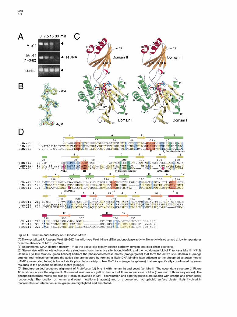
In Mn21-containing crystals, we observed electron den-nucleolytic activities of Mre11, we determined the struc-ture of P. furiosus Mre11 by multiple anomalous diffrac- sity for two Mn21 ions, resembling the di-metal active
site found in Ser/Thr protein phosphatases (Figure 2A).tion phasing to 2.6 A (Table 1). This Mre11 structure(residues 1–342) lacks 82 C-terminal residues, which The two Mn21 ions are octahedrally coordinated by
seven conserved residues of the phosphodiesterasedoes not affect endonucleolytic activity (Figure 1A), butfacilitated crystallization. The resulting high quality ex- motifs (Asp8, His10, Asp49, Asn84, His173, His206,
His208) and by a bridging water molecule. The four histi-perimental electron density (Figure 1B) revealed thepresence of two equivalent Mre11 molecules in the dine nitrogen ligands are consistent with a preference
of Mre11 for Mn21 over Mg21, which is typically coordi-asymmetric unit that are related by a noncrystallo-graphic 2-fold symmetry axis. nated by all oxygen ligands (Glusker, 1991). In fact, in
a crystal form containing Mg21 instead of Mn21, we ob-P. furiosus Mre11 comprises two domains that inter-act at the active site (Figure 1C). Domain I is composed serve only electron density corresponding to one Mg21
ion bound to the active site, which is not sufficient forof two parallel mixed b sheets (b15, b14, b1–b3 and b5,b6, b4, b7–b9, b11, b10, respectively) that are flanked catalysis, suggesting why Mre11 is inactive in the pres-
ence of Mg21 (K.P.H., unpublished data).by seven a helices (A–D, D’, E, E’; Figure 1C). DomainI contains five conserved phosphodiesterase motifs, To reveal the phosphodiesterase mechanism of
Mre11, we soaked P. furiosus Mn21-Mre11 with deoxy-which form the nuclease active site (Bressan et al., 1998;Tsubouchi and Ogawa, 1998). The phosphodiesterase adenosine monophosphate (dAMP represents a product
of an exonucleolytically cleaved DNA end) and deter-motifs are situated in loops connecting the core bstrands with their flanking a helices and place the active mined the dAMP-Mn-Mre11 complex structure to 2.2 A
resolution (Figure 2A; Table 1). The Mre11 active sitesite at the center of a shallow surface depression onDomain I (Figures 1C and 1D). Domain I resembles in binds dAMP mainly via the phosphate moiety, which is
liganded to Asn84 and His85 and to both Mn21 ions. Thefold and active site location the catalytic domain of cal-cineurin like Ser/Thr phosphatases and the DNA base double coordination of the dAMP phosphate by both
active site metals resembles the binding of phosphory-excision repair enzyme apurinic endonuclease 1 (APE1).However, although Mre11 and APE1 are both DNA repair lated protein residues in Ser/Thr phosphatases, further
supporting a common phosphoesterase mechanism be-nucleases, they evidently possess different active site

Mre11 and Rad50 Crystal Structures475
Table 1. Crystallographic Data Collection and Analysis
MAD Data Collection (SeMet-Mre11)
Space group: P212121 (2 molecules/asymmetric unit)Unit cell dimensions (A): a 5 73.1, b 5 87.0, c 5 146.3Data set SeMet l1 SeMet l2 SeMet l3 SeMet l4Wavelength (A) 0.96112 0.97951 0.97969 0.99987Data range (A) 30.0–2.6 30.0–2.4 30.0–2.6 30.0–2.8Observations (unique) 442,453 (29,437) 578,934 (39,228) 442,289 (29,321) 217,379 (23,713)Completeness (%) (last shell) 82.4 (36.9)a 92.4 (66.8)a 94.0 (60.3)a 98.4 (98.8)Rsym
b (last shell) 0.055 (0.268) 0.044 (0.216) 0.049 (0.249) 0.048 (0.384)
Refinement Mre11 1 Mn21 1 dAMP Rad50-ATPase
Space group P212121 P3121Unit cell dimensions (A) a 5 74.3, b 5 88.7, c 5 145.1 a 5 b 5 99.5, c 5 116.4Resolution range (A) 20–2.2 20–3.0Observations (unique) 431,781 (48,885) 119,376 (13,527)Completeness (%) (last shell) 98.1 (86.1) 83.0 (47.3)Rsym
a (last shell) 0.046 (0.326) 0.057 (0.348)Reflections F . 0 (cross validation) 47,867 (2,397) 11,886 (605)Non-hydrogen atoms (solvent molecules) 5,806 (274) 3,002 (64)Rcryst
c (Rfreed) 0.222 (0.269) 0.256 (0.291)
Rms bond length (A)/bond angles (8) 0.012/1.3 0.009/1.5% core (disallowed) in Ramachandran plot 88 (0.0) 78 (0.0)
a Anomalous completeness.b Rsym is the unweighted R value on I between symmetry mates.c Rcryst 5 Shkl||Fobs(hkl)| 2 |Fcalc(hkl)||/Shkl|Fobs(hkl)|.d Rfree 5 the cross validation R factor for 5% of reflections against which the model was not refined.
tween Mre11 and Ser/Thr phosphatases (Griffith et al., droxide would result in the crystallographically observedproduct with both Mn21 ions coordinating the dAMP1995).
The dAMP sugar moiety is situated in a groove formed phosphate (Figure 2A).The structure of the P. furiosus dAMP-Mre11 complexby the Domain I–Domain II interface (Figure 2A). The
sugar 39O forms a hydrogen bond to the His208 back- suggests that Mre11 cleaves DNA ends 39→59, liberatinga nucleotide with the phosphate bound to the 59 sugarbone N, suggesting that the dAMP may represent the
39 terminal nucleotide in exonuclease activity. The hy- oxygen. These 59-phosphorylated nucleotides are bio-chemically observed as main products of the Mre11drogen bond to the Mre11 backbone, however, could
also form to an internal 39 oxygen in bent single stranded dsDNA exonuclease activity, and no significant 59→39activity has been biochemically observed for Mre11or hairpin DNA, in endonuclease or hairpin nicking activ-
ity (see below). The sugar binding groove extends from (Trujillo et al., 1998; Connelly et al., 1998). Our structuretherefore supports the biochemically observed 39→59both sides of the bound dAMP and possesses several
positively charged residues at the 39 side of dAMP. The nuclease direction as the main dsDNA exonuclease ac-tivity of Mre11. These results indicate that the generationpositively charged residues likely bind the adjacent, prob-
ably bent ssDNA backbone in endonuclease activity. of 39 tails in homologous recombination in vivo requiresan additional 59→39 nuclease, such as EXO1 suggestedThe adenine base of dAMP has no specific interac-
tions with Mre11, as expected for a nonspecific endo/ by genetic data (Tsubouchi and Ogawa, 2000), or thenuclease direction of Mre11 is modulated in vivo by yetexonuclease. Mre11 binds dAMP primarily via the phos-
phate which exposes the Watson-Crick face of the ade- uncharacterized factors.nine moiety, suggesting that Mre11 recognizes the ter-minal base intrahelically. Tyr187 likely stacks against Mre11 Functional Motifs
To identify functional motifs of Mre11, we mapped eukary-the base of the terminal nucleotide in DNA exonucleasereactions (Figure 2A). otic Mre11 mutations that are connected with human
disease and yeast DSB repair/meiosis defects onto thedAMP is bound to the active site of Mre11 in an orien-tation suggesting that Mre11 cleaves the sugar-39-O- structure of P. furiosus Mre11 (Figure 3A). Interestingly,
several mutations map outside the phosphodiesterasephosphate bond of DNA. The conserved phosphodies-terase residue His85, which is essential for the nuclease motifs and may disrupt important protein interactions
with Rad50, Nbs1/Xrs2, and/or DNA. The N117S muta-activity of Mre11 (Bressan et al., 1998), arguably actsin transition state stabilization (Figure 2A). His85 likely tion in human Mre11 leads to an ataxia telangiectasia-
like disorder (ATLD), with cellular features similar todonates a proton to the leaving DNA 39-OH (Figure 2B).The protonated state of His85 is probably stabilized ataxia telangiectasia and Nijmegen breakage syndrome,
and the analogous yeast mutation N113S leads to sensi-through a charge relay system with the adjacent andconserved His52. In the enzyme-DNA complex, Mn21 tivity to ionizing radiation (Stewart et al., 1999). N117
of human Mre11 maps to a surface loop between theion (1) likely binds the attacking hydroxide ion as pro-posed for protein phosphatases (Rusnack, 2000). Collin- conserved secondary structure elements aB and b3, at
a position where human and yeast Mre11 possess a 33ear nucleophilic attack of the phosphoester by this hy-
Cell476
Figure 1. Structure and Activity of P. furiosus Mre11
(A) The crystallized P. furiosus Mre11(1–342) has wild-type Mre11-like ssDNA endonuclease activity. No activity is observed at low temperaturesor in the absence of Mn21 (control).(B) Experimental MAD electron density (1s) at the active site clearly defines carbonyl oxygen and side chain positions.(C) Stereo view with annotated secondary structure shows the active site, bound dAMP, and the two domain fold of P. furiosus Mre11(1–342).Domain I (yellow strands, green helices) harbors the phosphodiesterase motifs (orange/green) that form the active site. Domain II (orangestrands, red helices) completes the active site architecture by forming a likely DNA binding face adjacent to the phosphodiesterase motifs.dAMP (color-coded tubes) is bound via its phosphate moiety to two Mn21 ions (magenta spheres) that are specifically coordinated by sevenresidues in the phosphodiesterase motifs (orange).(D) Structure-guided sequence alignment of P. furiosus (pf) Mre11 with human (h) and yeast (sc) Mre11. The secondary structure of Figure1C is shown above the alignment. Conserved residues are yellow (two out of three sequences) or blue (three out of three sequences). Thephosphodiesterase motifs are orange. Residues involved in Mn21 coordination and ester hydrolysis are labeled with orange and green stars,respectively. The location of human and yeast mutations (magenta) and of a conserved hydrophobic surface cluster likely involved inmacromolecular interaction sites (green) are highlighted and annotated.

Mre11 and Rad50 Crystal Structures477
Figure 2. Mre11 Active Site and ImpliedNuclease Mechanism
(A) P. furiosus Mre11 local active site geome-try with simulated annealed Fo-Fc omitdensity (1.8s) of the bound dAMP (blue andcolor-coded tubes) and Mn21 ions (magentaspheres). The unbiased omit density showsthe specific octahedral Mn21 coordination byseven protein residues, a water molecule, andtwo phosphate oxygens. Only the phosphateand the sugar moieties of dAMP are specifi-cally recognized by the protein, consistentwith the nonsequence specific endo/exo-nuclease activities of Mre11.(B) Proposed structure-based nuclease mech-anism. Mn21 ion (1) binds the attacking hy-droxide ion (red). Collinear nucleophilic at-tack of this hydroxide leads to double Mn21
ion coordination by the phosphate in theproduct state. His85/His52 are positioned toform a charge relay to protonate the leaving39O of the penultimate sugar.
residue insertion relative to P. furiosus Mre11 (Figure site. Distortion of the yeast Mre11 DNA binding sitewould evidently affect processing of meiotic DSBs that1D). Since human Mre11-N117S interacts with Rad50
but fails to interact with Nbs1, this human and yeast require Mre11 nuclease activity.Like the eukaryotic enzymes, P. furiosus Mre11 (MWinsertion likely represents a unique eukaryotic binding
site for Nbs1/Xrs2. Notably, this proposed Nbs1 binding 50 kDa) elutes from gel filtration chromatography withan apparent MW of a dimer (z120 kDa). A sequenceinsertion is located on Domain I opposite of the phos-
phodiesterase motifs, arguing that Nbs1 does not inter- conserved hydrophobic surface patch in aB and aCforms a symmetric Mre11 dimerization interface for theact directly with the Mre11 active site. Experiments in
which Nbs1 was found to stimulate Mre11’s nuclease two molecules in the asymmetric unit and indicatesa potential self-dimerization site of Mre11 (Figure 3B).activity (Paull and Gellert, 1999) suggest that Nbs1 could
act in trans within a proposed Mre11 homodimer (Joh- The crystallized C-terminal truncated P. furiosus Mre11(1–342), however, elutes with an apparent MW of a mono-zuka and Ogawa, 1995; Furuse et al., 1998; Chamankhah
et al., 2000; Paull and Gellert, 2000). mer (z60 kDa), suggesting the noncrystallographic di-mer in the asymmetric unit probably forms during crystalIn yeast Mre11, P162S leads to temperature sensitivity
of DSB repair (Mre11(ts)), and the Mre11 mutant fails to packing. Therefore, the sequence conserved hydropho-bic surface cluster and could equally be a Rad50 bindingtightly interact with Rad50 and Xrs2 (Chamankhah et
al., 2000). Yeast P162 maps to the b strand extension site. In any case, the conservation of a hydrophobicsurface cluster in both archaeal and eukaryotic Mre11(b4–b6) of the core b sheet of Domain I, indicating that
this region is involved in Mre11/Rad50/Xrs2 complex plus the clustering of the hydrophobic, ATLD, andMre11(ts) surface patches on Domain I identifies a likelyformation. Although the surface loops containing the
ATLD and Mre11(ts) mutations are distant in sequence macromolecular interaction site on Mre11 to form theMre11/Rad50/Nbs1 complex (Figure 3A).(Figure 1D), the mutation sites are only z15 A apart on
the three-dimensional structure of P. furiosus Mre11(Figure 3A). This adjacent mapped location suggests DNA Binding Mechanism and Nuclease Direction
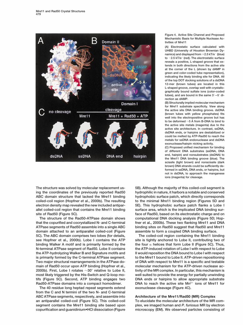
The Mre11 structure reveals that the nuclease activethat both Nbs1 and Xrs2 bind to equivalent locationson human and yeast Mre11, respectively. site is situated in an L-shaped groove between Domains
I and II that possesses the only significant positive sur-The yeast mre11S (separation of function) allele con-tains yeast P84S and T188I and leads to defective mei- face potential for DNA binding (Figure 4A). To objectively
and comprehensively evaluate all possible Mre11-DNAotic DSB processing, but normal mitotic DSB repair(Nairz and Klein, 1997). Yeast P84 is located in the pro- complexes, we used the complete systematic search
program DOT to dock a dsDNA 12-mer to Mre11 (Rob-posed eukaryotic Nbs1/Xrs2 binding loop insertion andcould disrupt the interaction of Mre11 with Xrs2 (Figure erts and Pique, 1999). In all 30 of the analyzed highest
scoring solutions, DNA binds to the proposed L-shaped1D). However, yeast T188 is located in a surface loopclose to the active site (Figures 1D and 3A). This surface DNA binding groove (Figure 4A). The DNA binding orien-
tation of the scissile strand in the docking solutionsloop contains conserved positively charged residuesand might participate in DNA binding by Mre11. T188I is consistent with the experimentally observed 59→39
binding orientation of dAMP, validating the computa-may therefore weaken or disrupt the interaction of Mre11with DNA by distorting the surface next to the active tional approach. Furthermore, the docked DNA binds

Cell478
suggesting a mechanism for the inability of Mre11 tocleave dsDNA endonucleolytically. A mechanism inwhich a DNA duplex is bound by Mre11 but stericallyexcluded from cleavage is supported by biochemicalexperiments, showing that Mre11 can bind circular DNAand that Mn21 ions are not required for DNA binding(Usui et al., 1998; de Jager et al., 2001). On the contrary,ssDNA, which possesses considerable conformationalflexibility, could both bind to the electropositive grooveand to the active site metals. Thus, ssDNA is likelynot sterically excluded, suggesting a mechanism forMre11’s ssDNA endonuclease activity. Finally, DNAends and hairpins could be partially unwound by Rad50and ATP to approach the active site metals for nucleo-lytic attack, as indicated by biochemical data (Paull andGellert, 1999). The structural and computational dockingresults therefore support a unified molecular mechanismfor Mre11’s broad but characteristic substrate specific-ity pattern by endonucleolytically cleaving both ssDNAand dsDNA ends/hairpins, the latter of which are par-tially unwound by ATP-Rad50 to expose ssDNA (Fig-ure 4C).
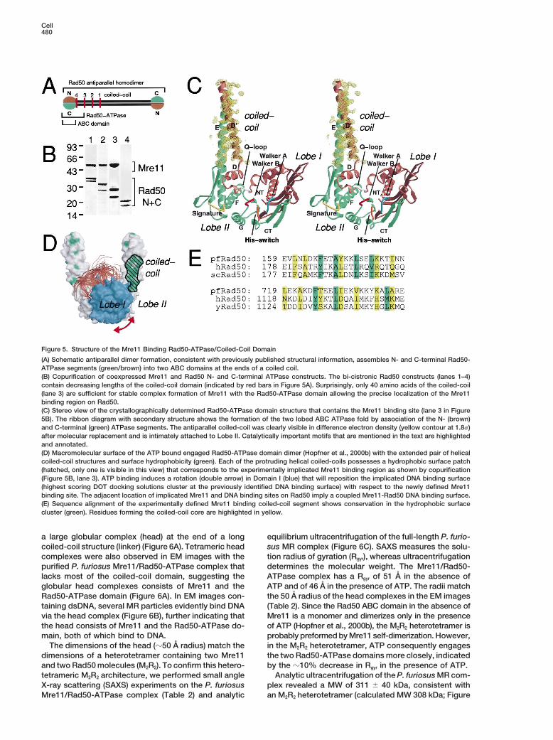
Physical Link between Mre11and the Rad50-ATPase DomainIn all organisms studied so far, Mre11 and Rad50 forma stable complex and are copurified together. To deter-mine the Mre11 binding site on Rad50, we coexpressedand copurified P. furiosus Mre11 with P. furiosus Rad50-ATPase domain constructs that contained increasinglengths of the coiled-coil domain (Figures 5A and 5B).To ensure stable complex formation, copurification in-Figure 3. Mre11 Macromolecular Interaction Surfacecluded a heat denaturation step plus three chromatogra-(A) A surface cluster of mutations connected with human diseasephy steps. We found that the ABC domain of Rad50and yeast defects identifies an important surface area that is remotealone without the coiled-coil does not form a stablefrom the active site and evidently acts in macromolecular interac-
tions of Mre11. complex with Mre11, suggesting the coiled-coil contains(B) The ribbon representation of the noncrystallographic Mre11 di- the Mre11 binding site. In fact, the ABC domain plusmer (yellow/orange and cyan/blue with magenta manganese ions) the adjacent 40 amino acid long coiled-coil segmentidentifies a conserved hydrophobic surface cluster (green side
(denoted Rad50-ATPase) firmly bound Mre11 (Figureschains) which is a likely macromolecular interaction site in the MR5A and 5B). To estimate relative binding affinities ofcomplex formation.Mre11 to different Rad50 fragments, we dissociated im-mobilized Rad50/Mre11 complexes in a gradient of thedenaturant guanidinium•HCl (see Experimental Proce-similar in location and orientation as the DNA bound todures; data not shown). The full-length P. furiosus MRthe fold-related DNA repair enzyme APE1 (Mol et al.,complex was stable up to 6.0 M guanidinium•HCl,2000). This similar DNA binding orientation suggestswhereas the truncated Mre11/Rad50-ATPase complexthat APE1 and Mre11 recognize DNA in analogous ways,containing only a 40 residue long coiled-coil was stabledespite their evidently different nuclease mechanisms.up to 5.8 M guanidinium•HCl. This practically wild-typeAlthough the dsDNA otherwise fits very well into thestability of the truncated Mre11/Rad50-ATPase complexelectropositive groove on both sides of dAMP, the shapesuggests that the Mre11 binding site of Rad50 is locatedof the active site depression at the Mn21 ions requireson an only 40 residue long coiled-coil region adjacenta DNA backbone deformation by at least 5 A from ato the Rad50 ABC domain. This close linkage of theB-DNA duplex across the DNA binding groove, in orderRad50-ATPase to Mre11 provides a testable mechanis-to appropriately approach the catalytic metals for pro-tic basis for the requirement of Rad50-ATP hydrolysisductive cleavage (Figure 4B). This 5 A distance is notfor the Mre11 exonuclease activity.likely to be overcome by the observed rotational flexibil-
ity of Domain II with respect to Domain I; Domain IIrotates along the DNA binding groove instead of across Structure of the Mre11 Binding
Rad50-ATPase Domainthe DNA binding groove, suggesting rather a functionin DNA translocation or conformational adaptability to To identify the structural nature and location of the
Mre11 binding site on the Rad50 coiled-coil region, weaccommodate different DNA end substrates. Since de-formation or unwinding of a dsDNA duplex to approach crystallized and determined the structure of the Rad50-
ATPase, containing the ATP binding cassette (ABC) do-the catalytic metals while bound to the DNA bindinggroove is energetically unfavorable, a dsDNA duplex main and the biochemically identified 40 residue long
Mre11 binding coiled-coil segment, to 3.0 A resolution.might be sterically excluded from the active site metals,
Mre11 and Rad50 Crystal Structures479
Figure 4. Active Site Channel and ProposedMechanistic Basis for Multiple Nuclease Ac-tivities of Mre11
(A) Electrostatic surface calculated withUHBD (University of Houston Brownian Dy-namics) and displayed from 12.5 kT/e2 (blue)to 22.5 kT/e2 (red). The electrostatic surfacereveals a positive, L-shaped groove that ex-tends in both directions from the active siteat the corner of the L (shown by dAMP ingreen and color-coded tube representation),indicating the likely binding site for DNA. Allof the top DOT docking solutions of a dsDNA12-mer (brown tubes) are located in thisL-shaped groove, overlap well with crystallo-graphically bound sulfate ions (color-codedtubes), and are bound in the same 39→59 di-rection as dAMP.(B) Structurally implied molecular mechanismfor Mre11 substrate specificity. View alongthe active site DNA binding groove. dsDNA(brown tubes with yellow phosphates) fitswell into the electropositive groove but hasto be deformed z5 A from B-DNA to bind tothe active site metals (magenta) due to theactive site architecture. In contrast, ssDNA,dsDNA ends, or hairpins are destabilized orcould be melted by ATP-Rad50 to reach themetals for ssDNA endonuclease and dsDNAexonuclease/hairpin nicking activity.(C) Proposed unified mechanism for bindingof different DNA substrates (ssDNA, DNAend, hairpin) and nonsubstrates (dsDNA) tothe Mre11 DNA binding groove (blue). Thescissile (light brown) and nonscissile (darkbrown) DNA strands could be sufficiently de-formed in ssDNA, DNA ends, or hairpins, butnot in dsDNA, to approach the manganeseions (magenta) for cleavage.
The structure was solved by molecular replacement us- 5B). Although the majority of this coiled-coil segment ishydrophilic in nature, it harbors a notable and conserveding the coordinates of the previously reported Rad50
ABC domain structure that lacked the Mre11 binding hydrophobic surface patch, which directly correspondsto the minimal Mre11 binding region (Figures 5D andcoiled-coil region (Hopfner et al., 2000b). The resulting
electron density map revealed the new included antipar- 5E). This hydrophobic surface patch flanks a Lobe Isurface area, which is the implicated DNA binding sur-allel coiled-coil region that contains the Mre11 binding
site of Rad50 (Figure 5C). face of Rad50, based on its electrostatic charge and oncomputational DNA docking analysis (Figure 5D; Hop-The structure of the Rad50-ATPase domain shows
that the copurified and cocrystallized N- and C-terminal fner et al., 2000b). These two flanking Mre11 and DNAbinding sites on Rad50 suggest that Rad50 and Mre11ATPase segments of Rad50 assemble into a single ABC
domain attached to an antiparallel coiled-coil (Figure assemble to form a coupled DNA binding surface.The coiled-coil region containing the Mre11 binding5C). The ABC domain comprises two lobes (for details,
see Hopfner et al., 2000b). Lobe I contains the ATP site is tightly anchored to Lobe II, contributing two ofthe four a helices that form Lobe II (Figure 5C). Thus,binding Walker A motif and is primarily formed by the
N-terminal ATPase segment of Rad50. Lobe II contains the ATP-induced rotation of Lobe I with respect to LobeII would reposition the DNA bound to Lobe I with respectthe ATP-hydrolyzing Walker B and Signature motifs and
is primarily formed by the C-terminal ATPase segment. to the Mre11 bound to Lobe II. ATP-driven repositioningof DNA with respect to Mre11 is a specific and testableTwo major structural rearrangements in the ATPase do-
main of Rad50 occur upon ATP binding (Hopfner et al., molecular mechanism for the ATP-driven nuclease ac-tivity of the MR complex. In particular, this mechanism is2000b). First, Lobe I rotates z308 relative to Lobe II,
most likely triggered by the His-Switch and Q-loop mo- well suited to provide the energy for partially unwindingDNA ends or hairpins to allow appropriate substratetifs (Figure 5C). Second, ATP binding engages two
Rad50-ATPase domains into a compact homodimer. DNA to reach the active site Mn21 ions of Mre11 forexonuclease cleavage (Figure 4C).The 40 residue long heptad repeat segments extend
from the C and N termini of the two N- and C-terminalABC ATPase segments, respectively, and assemble into Architecture of the Mre11/Rad50 (MR) Complex
To elucidate the molecular architecture of the MR com-an antiparallel coiled-coil (Figure 5C). This coiled-coilsegment contains the Mre11 binding site based upon plex, we imaged human and P. furiosus MR by electron
microscopy (EM). We observed particles consisting ofcopurification and guanidinium•HCl dissociation (Figure
Cell480
Figure 5. Structure of the Mre11 Binding Rad50-ATPase/Coiled-Coil Domain
(A) Schematic antiparallel dimer formation, consistent with previously published structural information, assembles N- and C-terminal Rad50-ATPase segments (green/brown) into two ABC domains at the ends of a coiled coil.(B) Copurification of coexpressed Mre11 and Rad50 N- and C-terminal ATPase constructs. The bi-cistronic Rad50 constructs (lanes 1–4)contain decreasing lengths of the coiled-coil domain (indicated by red bars in Figure 5A). Surprisingly, only 40 amino acids of the coiled-coil(lane 3) are sufficient for stable complex formation of Mre11 with the Rad50-ATPase domain allowing the precise localization of the Mre11binding region on Rad50.(C) Stereo view of the crystallographically determined Rad50-ATPase domain structure that contains the Mre11 binding site (lane 3 in Figure5B). The ribbon diagram with secondary structure shows the formation of the two lobed ABC ATPase fold by association of the N- (brown)and C-terminal (green) ATPase segments. The antiparallel coiled-coil was clearly visible in difference electron density (yellow contour at 1.8s)after molecular replacement and is intimately attached to Lobe II. Catalytically important motifs that are mentioned in the text are highlightedand annotated.(D) Macromolecular surface of the ATP bound engaged Rad50-ATPase domain dimer (Hopfner et al., 2000b) with the extended pair of helicalcoiled-coil structures and surface hydrophobicity (green). Each of the protruding helical coiled-coils possesses a hydrophobic surface patch(hatched, only one is visible in this view) that corresponds to the experimentally implicated Mre11 binding region as shown by copurification(Figure 5B, lane 3). ATP binding induces a rotation (double arrow) in Domain I (blue) that will reposition the implicated DNA binding surface(highest scoring DOT docking solutions cluster at the previously identified DNA binding surface) with respect to the newly defined Mre11binding site. The adjacent location of implicated Mre11 and DNA binding sites on Rad50 imply a coupled Mre11-Rad50 DNA binding surface.(E) Sequence alignment of the experimentally defined Mre11 binding coiled-coil segment shows conservation in the hydrophobic surfacecluster (green). Residues forming the coiled-coil core are highlighted in yellow.
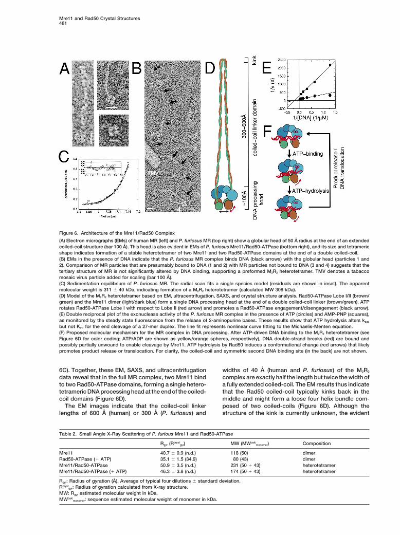
a large globular complex (head) at the end of a long equilibrium ultracentrifugation of the full-length P. furio-sus MR complex (Figure 6C). SAXS measures the solu-coiled-coil structure (linker) (Figure 6A). Tetrameric head
complexes were also observed in EM images with the tion radius of gyration (Rgyr), whereas ultracentrifugationdetermines the molecular weight. The Mre11/Rad50-purified P. furiosus Mre11/Rad50-ATPase complex that
lacks most of the coiled-coil domain, suggesting the ATPase complex has a Rgyr of 51 A in the absence ofATP and of 46 A in the presence of ATP. The radii matchglobular head complexes consists of Mre11 and the
Rad50-ATPase domain (Figure 6A). In EM images con- the 50 A radius of the head complexes in the EM images(Table 2). Since the Rad50 ABC domain in the absence oftaining dsDNA, several MR particles evidently bind DNA
via the head complex (Figure 6B), further indicating that Mre11 is a monomer and dimerizes only in the presenceof ATP (Hopfner et al., 2000b), the M2R2 heterotetramer isthe head consists of Mre11 and the Rad50-ATPase do-
main, both of which bind to DNA. probably preformed by Mre11 self-dimerization. However,in the M2R2 heterotetramer, ATP consequently engagesThe dimensions of the head (z50 A radius) match the
dimensions of a heterotetramer containing two Mre11 the two Rad50-ATPase domains more closely, indicatedby the z10% decrease in Rgyr in the presence of ATP.and two Rad50 molecules (M2R2). To confirm this hetero-
tetrameric M2R2 architecture, we performed small angle Analytic ultracentrifugation of the P. furiosus MR com-plex revealed a MW of 311 6 40 kDa, consistent withX-ray scattering (SAXS) experiments on the P. furiosus
Mre11/Rad50-ATPase complex (Table 2) and analytic an M2R2 heterotetramer (calculated MW 308 kDa; Figure
Mre11 and Rad50 Crystal Structures481
Figure 6. Architecture of the Mre11/Rad50 Complex
(A) Electron micrographs (EMs) of human MR (left) and P. furiosus MR (top right) show a globular head of 50 A radius at the end of an extendedcoiled-coil structure (bar 100 A). This head is also evident in EMs of P. furiosus Mre11/Rad50-ATPase (bottom right), and its size and tetramericshape indicates formation of a stable heterotetramer of two Mre11 and two Rad50-ATPase domains at the end of a double coiled-coil.(B) EMs in the presence of DNA indicate that the P. furiosus MR complex binds DNA (black arrows) with the globular head (particles 1 and2). Comparison of MR particles that are presumably bound to DNA (1 and 2) with MR particles not bound to DNA (3 and 4) suggests that thetertiary structure of MR is not significantly altered by DNA binding, supporting a preformed M2R2 heterotetramer. TMV denotes a tabaccomosaic virus particle added for scaling (bar 100 A).(C) Sedimentation equilibrium of P. furiosus MR. The radial scan fits a single species model (residuals are shown in inset). The apparentmolecular weight is 311 6 40 kDa, indicating formation of a M2R2 heterotetramer (calculated MW 308 kDa).(D) Model of the M2R2 heterotetramer based on EM, ultracentrifugation, SAXS, and crystal structure analysis. Rad50-ATPase Lobe I/II (brown/green) and the Mre11 dimer (light/dark blue) form a single DNA processing head at the end of a double coiled-coil linker (brown/green). ATProtates Rad50-ATPase Lobe I with respect to Lobe II (red arrow) and promotes a Rad50-ATPase engagement/disengagement (black arrow).(E) Double reciprocal plot of the exonuclease activity of the P. furiosus MR complex in the presence of ATP (circles) and AMP-PNP (squares),as monitored by the steady state fluorescence from the release of 2-aminopurine bases. These results show that ATP hydrolysis alters kcat,
but not Km, for the end cleavage of a 27-mer duplex. The line fit represents nonlinear curve fitting to the Michaelis-Menten equation.(F) Proposed molecular mechanism for the MR complex in DNA processing. After ATP-driven DNA binding to the M2R2 heterotetramer (seeFigure 6D for color coding; ATP/ADP are shown as yellow/orange spheres, respectively), DNA double-strand breaks (red) are bound andpossibly partially unwound to enable cleavage by Mre11. ATP hydrolysis by Rad50 induces a conformational change (red arrows) that likelypromotes product release or translocation. For clarity, the coiled-coil and symmetric second DNA binding site (in the back) are not shown.
6C). Together, these EM, SAXS, and ultracentrifugation widths of 40 A (human and P. furiosus) of the M2R2
complex are exactly half the length but twice the width ofdata reveal that in the full MR complex, two Mre11 bindto two Rad50-ATPase domains, forming a single hetero- a fully extended coiled-coil. The EM results thus indicate
that the Rad50 coiled-coil typically kinks back in thetetrameric DNA processing head at the end of the coiled-coil domains (Figure 6D). middle and might form a loose four helix bundle com-
posed of two coiled-coils (Figure 6D). Although theThe EM images indicate that the coiled-coil linkerlengths of 600 A (human) or 300 A (P. furiosus) and structure of the kink is currently unknown, the evident
Table 2. Small Angle X-Ray Scattering of P. furious Mre11 and Rad50-ATPase
Rgyr (Rcrystgyr) MW (MWcalc
monomer) Composition
Mre11 40.7 6 0.9 (n.d.) 118 (50) dimerRad50-ATPase (1 ATP) 35.1 6 1.5 (34.9) 80 (43) dimerMre11/Rad50-ATPase 50.9 6 3.5 (n.d.) 231 (50 1 43) heterotetramerMre11/Rad50-ATPase (1 ATP) 46.3 6 3.8 (n.d.) 174 (50 1 43) heterotetramer
Rgyr: Radius of gyration (A). Average of typical four dilutions 6 standard deviation.Rcryst
gyr: Radius of gyration calculated from X-ray structure.MW: Rgyr estimated molecular weight in kDa.MWcalc
monomer: sequence estimated molecular weight of monomer in kDa.

Cell482
similarity of this Rad50 coiled-coil structure with the tidomain enzymes all contain an ABC-ATPase dimerplus a substrate/function-specific dimer as revealedstructure of SMC proteins (that are involved in chromatid
interaction) suggests that the M2R2 coiled-coil linker here for the M2R2 heterotetramer (Fishel and Wilson,1997; Kolodner and Marsischky, 1999; Holland andcould play a role in sister chromatid interactions or in
the interaction of two broken DNA ends (Melby et al., Blight, 1999). The crystal structures of the ATP boundRad50 ABC domain (Hopfner et al., 2000b) and ADP1998; Bressan et al., 1999).bound or nucleotide free MutS (Lamers et al., 2000;Obmolova et al., 2000) revealed that the ABC dimerStructural and Functional Model for the Mre11/Rad50orientation in both complexes is surprisingly similar. InDNA Processing Complexfact, the location of the MutS domains connecting theTaken together, our data suggest a dual role for the MRMutS ABC and DNA binding domains corresponds di-complex in DSB repair. The (Mre11)2/(Rad50-ATPase)2
rectly to the identified coiled-coil segment linking thehead likely functions in ATP-dependent DNA processingRad50 ABC domain to Mre11. Likewise, the most fre-in meiosis and mitotic DSB repair, while the extendedquent cystic fibrosis causing mutation in the cystic fibro-coiled-coil domain could serve as linker in sister chro-sis transmembrane transporter (DF508), which resultsmatid interaction (Bressan et al., 1999). Mechanistically,in subunit detachment between the transmembranethe conformational changes of the Rad50-ATPase do-channel domains and the ABC domains, maps essen-main upon ATP binding are well suited to prepare DNAtially to the Mre11 binding coiled-coil surface patch onends/hairpins for nucleolytic cleavage by Mre11. ThisRad50 (Hung et al., 1998; Hopfner et al., 2000b). There-model is supported by results from a steady-state exo-fore, our results suggest that the substrate-specific do-nuclease assay that measures the release of the fluores-mains in ABC transporter, MutS mismatch repair en-cent adenine analog 2-aminopurine from dsDNA (Figurezymes, and Mre11/Rad50 are attached to the respective6E). In the presence of the nonhydrolyzable ATP analogsABC dimer in a similar location. This striking architec-AMP-PNP or ATPgS, the exonuclease activity of the P.tural similarity suggests that the ABC dimer controlsfuriosus MR complex is only z3–5 times slower than inthe corresponding substrate-specific domains in ABCthe presence of ATP, while no activity was found in thetransporter and DNA mismatch repair enzymes in a simi-presence of ADP, consistent with data from the E. colilar way as we proposed for Mre11/Rad50.homologs (Connelly et al., 1997). The kcat and Km values
for dsDNA cleavage by the MR complex in the presenceof ATP (Km 5 4.7 mM (6 0.7); kcat 0.0062 s21 (6 0.0004)) Experimental Proceduresor AMP-PNP (Km 5 5.0 mM (6 0.6) and kcat 0.0016 s21
Protein Expression and Purification(6 0.0008)) suggest that ATP hydrolysis mostly acceler-All P. furiosus proteins were synthesized in E. coli BL21-RIL (DE3)ates kcat without altering Km. The similar Km values sug-(Stratagene) and purified and characterized as described (Hopfnergest that both the ATP and AMP-PNP bound forms ofet al., 2000a, 2000b). Selenomethionine-Mre11 was synthesized in
Rad50 bind DNA with similar affinities, in agreement E. coli B834 (Novagen) in minimal medium supplemented with sele-with the structural data, where ATP and AMP-PNP have nomethionine. The human MR complex was produced from recom-
binant baculovirus expression in insect cells and purified by Ni21equivalent conformational effects on the Rad50-ATPaseaffinity and gel filtration chromatography.domain (Hopfner et al., 2000b). As ATP hydrolysis merely
accelerates the kcat of the Mre11 exonuclease, but is notrequired for initial exonucleolytic cleavage per se, ATP Crystallization, Crystallographic Data Collection, MAD
Phasing, and Structure Refinement of P. furiosus Mre11hydrolysis appears to accelerate product release afterP. furiosus Mre11(1–342) (12 mg/ml in 20 mM phosphate [pH 7.5],the Mre11 cleavage reaction. Taken together, our results200 mM NaCl, 0.1 mM EDTA, 5% glycerol) was crystallized in spacesupport the following working model for DNA processinggroup P212121 with cell dimensions a 5 72.9, b 5 87.0, c 5 146.4 A
by the MR complex (Figure 6F): ATP binding to Rad50 and two molecules in the asymmetric unit by sitting drop vaporprepares DNA substrates for cleavage by Mre11, likely diffusion at 48C after mixing 2 ml protein solution with 1.5 ml precipi-via engagement of two Rad50-ATPase domains plus tant solution (100 mM Ches [pH 9.5], 400 mM ammonium sulfate,
35% PEG 600). Four wavelength multiple anomalous diffractionrotation of Lobe I with respect to Lobe II (Figure 5D).(MAD) data to 2.6 A were recorded at 100 K with a Quantum 232After nucleolytic cleavage of the prepared ends byCCD detector at BL 5.0.2 (Advanced Light Source) and processedMre11, ATP hydrolysis accelerates product release forwith the HKL suite (Otwinowski, 1993) (Table 1).
subsequent cleavage reaction. A likely mechanism for Six selenium sites were located with SOLVE (Terwilliger and Be-enhancing release of product would be disengagement rendzen, 1999) and used to calculate initial phases with MLPHAREof the ATPase domain dimer to disrupt the interaction (CCP4, 1994). The remaining four selenium sites were located in
difference Fourier maps calculated from the initial phases. Phases toof the MR complex with DNA (Hopfner et al., 2000b).2.8 A were calculated with MLPHARE and improved with SOLOMONNotably, the M2R2 heterotetramer has the ability to simul-(CCP4, 1994). The resulting high quality electron density (Figure 1B)taneously process two DNA ends, which might be impor-was used to trace both molecules in the asymmetric unit with MAIN
tant in the initial search for microhomologies and/or (Turk, 1992). After overall anisotropic B-value and bulk solvent cor-annealing of two broken ends. rection, the structure was refined to 2.6 A by cycles of positional
and individual B-value refinement and manual model building withCNS (Brunger et al., 1998) and MAIN (Table 1). Five percent of theImplications for ABC-ATPase Machines in DNAdata were excluded from refinement to calculate the free R valueRepair and Membrane Transportfor cross validation. Initial NCS restraints were gradually removedThe heterotetrameric M2R2 DNA processing head is sur-in later stages of refinement. Mn21 1 dAMP containing P. furiosus
prisingly similar in its overall architecture to other ABC Mre11 was prepared by incubation of the crystal in mother liquorATPase machines, such as the ABC transporter and containing 1 mM MnCl2 and 10 mM dAMP for 3–5 hr at 48C. Data
to 2.1 A were collected at BL9-2 at SSRL using a Quantum 232MutS family DNA mismatch repair enzymes. These mul-

Mre11 and Rad50 Crystal Structures483
CCD detector. The models were rebuilt with MAIN and refined with prepared using GRASP (Nicholls et al., 1991). Figure 4A was madewith AVS and UHBD.CNS. Refinement and model statistics are shown in Table 1.
Crystallization, Crystallographic Data Collection, Phasing, Acknowledgmentsand Structure Refinement of the P. furiosusRad50-ATPase Domain We thank S. Parikh for help with data collection, the staff of beam-The Rad50-ATPase domain was crystallized in space group P3121 lines 5.0.2 (ALS) and 9–2 (SSRL) for assistance, H. Tsuruta (beamlineby mixing 2 ml of protein solution (8 mg/ml in 20 mM phosphate, 4-1, SSRL) and C. Putnam for help with SAXS data collection and[pH 7.5], 200 mM NaCl, 0.1 mM EDTA, 5% glycerol) with 2 ml precipi- processing, D. Millar and M. Bailey for help with fluorescencetant (100 mM Na-Acetate [pH 6.2], 8% Peg 6K, 10 mM Ca-Acetate) assays, J.-L. Pellequer for help with computational docking simula-using sitting drop vapor diffusion. Data to 3.0 A resolution were tions, and J. Kelly and S. Deechongkit for help with analytic ultracen-recorded at 100 K with a Quantum 232 CCD detector at BL 5.0.2 trifugation. We thank C. Putnam, D. Shin, and C. Mol for comments(Advanced Light Source) and processed with the HKL suite (Table on the manuscript. This work was supported by DOE (DE-AC03-1). Initial phases were calculated from a single AMoRe (Navaza, 76SF00098) and NSF (DBI 9904559) and by a research project grant1994) molecular replacement solution by using the previously pub- (CCE-99424) from the American Cancer Society (to J.P.C.). L.C. islished Rad50 ABC domain structure (lacking the coiled-coil) as supported by a grant from the Canadian Institutes of Health re-search model. The additional coiled-coil domain was clearly appar- search, and K.P.H. by The Skaggs Institute for Chemical Biologyent in 2Fo-Fc and Fo-Fc density maps (Figure 5C) and traced with and a BASF Fellowship from the Studienstiftung des deutschenMAIN. After overall anisotropic B-value and bulk solvent correction, Volkes e.V.the structure was refined by cycles of positional and restrainedindividual B-value refinement and by manual model building with Received February 27, 2001; revised April 13, 2001.CNS and MAIN (Table 1).
ReferencesNuclease AssaysEndonuclease assays on single-stranded φX174 DNA were per- Alani, E., Padmore, R., and Kleckner, N. (1990). Analysis of wild-formed as described (Hopfner et al., 2000a). Steady state 2-AP fluo- type and rad50 mutants of yeast suggests an intimate relationshiprescence assays were performed in a SLM8100 spectrofluorimeter between meiotic chromosome synapsis and recombination. Cell 61,essentially as described (Lam et al., 1999), by using 310 nm excita- 419–436.tion and 375 emission settings. Assays were performed at 508C in
Aravind, L., Walker, D.R., and Koonin, E.V. (1999). Conserved do-50 mM Tris, (pH 7.8), 150 mM NaCl, 0.1% Peg 6K, 2.5% glycerol, 1mains in DNA repair proteins and evolution of repair systems. Nu-mM MnCl2, 5 mM MgCl2 using 1 mM of the indicated nucleotide,cleic Acids Res. 27, 1223–1242.0.2 mM protein, and 1–12.5 mM dsDNA oligonucleotide (59-GGCGBierne, H., Ehrlich, S.D., and Michel, B. (1997). Deletions at stalledTGCCTTGGGCGCGCTGCGGGCGG(2-AP)G; six 2-fold dilutions).replication forks occur by two different pathways. EMBO J. 16,Linear rates were followed for 30–300 s and analyzed with SigmaPlot3332–3340.(Jandel). To rule out contaminating E. coli exonuclease activity, we
did a temperature profile of the exonuclease reaction. The activity Bressan, D.A., Olivares, H.A., Nelms, B.E., and Petrini, J.H. (1998).dramatically increases at high temperatures for both ATP and ATPgS Alteration of N-terminal phosphoesterase signature motifs inacti-bound P. furiosus MR, with the optimum beyond 658C. This activity vates Saccharomyces cerevisiae Mre11. Genetics 150, 591–600.profile is a signature of hyperthermophilic proteins such as Mre11 Bressan, D.A., Baxter, B.K., and Petrini, J.H. (1999). The Mre11-and Rad50 from P. furiosus and rules out contaminating activity. No Rad50-Xrs2 protein complex facilitates homologous recombination-significant activity was found at 378C where an E. coli contamination based double-strand break repair in Saccharomyces cerevisiae.would have its optimum activity. Mol. Cell. Biol. 19, 7681–7687.
Brunger, A.T., Adams, P.D., Clore, G.M., DeLano, W.L., Gros, P.,Dissociation of Mre11 and Rad50 in Guanidinium•HClGrosse-Kunstleve, R.W., Jiang, J.S., Kuszewski, J., Nilges, M.,Copurified P. furiosus 63His-Mre11/Rad50 and Mre11/Rad50-Pannu, N.S., et al. (1998). Crystallography & NMR system: A newATPase-6xHis was bound to 1 ml Ni-NTA sepharose column (Qiagen)software suite for macromolecular structure determination. Actaand eluted in 20 mM phosphate (pH 8.0), in gradient from 0 to 6 MCrystallogr. D 54, 905–921.guanidium•HCl (in 120 mM NaCl, 5% glycerol, 4 mM imidazol).Carney, J.P., Maser, R.S., Olivares, H., Davis, E.M., Le Beau, M.,Eluted subunits were detected with SDS-PAGE.Yates, J.R., 3rd, Hays, L., Morgan, W.F., and Petrini, J.H. (1998). ThehMre11/hRad50 protein complex and Nijmegen breakage syn-Electron Microscopydrome: linkage of double-strand break repair to the cellular DNACarbon-coated copper grids were glow-discharged for 1 min in thedamage response. Cell 93, 477–486.presence of amyl amine and floated on top of 5 ml drops containingChamankhah, M., Fontanie, T., and Xiao, W. (2000). The Saccharo-20–50 mg/ml protein for 2 min. The grids were stained in 3% uranylmyces cerevisiae mre11(ts) allele confers a separation of DNA repairacetate and viewed on a Philips CM120 microscope at 100 kV inand telomere maintenance functions. Genetics 155, 569–576.low dose mode.
CCP4 (1994). The CCP4 suite: programs for protein crystallography.Small Angle X-Ray Scattering Acta Crystallogr. D 50, 760–763.Data were recorded at SSRL beamline 4–2 typically using four pro- Connelly, J.C., de Leau, E.S., Okely, E.A., and Leach, D.R. (1997).tein concentrations (1, 2, 4, 8 mg/ml) in 20 mM Tris (pH 7.5), 200 Overexpression, purification, and characterization of the SbcCDmM NaCl, 5% glycerol, 5 mM MgCl2, 6 0.5 mM ATP). Rgyr was derived protein from Escherichia coli. J. Biol. Chem. 272, 19819–19826.from experimental data with GNOM (Svergun, 1993).
Connelly, J.C., Kirkham, L.A., and Leach, D.R. (1998). The SbcCDnuclease of Escherichia coli is a structural maintenance of chromo-Analytical Ultracentrifugationsomes (SMC) family protein that cleaves hairpin DNA. Proc. Natl.Equilibrium sedimentation was done in a Beckman XL-1 ultracentri-Acad. Sci. USA 95, 7969–7974.fuge with a An60Ti rotor. Sedimentation equilibrium was recordedde Jager, M., Dronkert, M.L., Modesti, M., Beerens, C.E., Kanaar,after 20 hr at 7000 rpm at 208C, using 0.4 mg/ml protein (in 20 mMR., and van Gent, D.C. (2001). DNA-binding and strand-annealingTris, [pH 7.2], 150 mM NaCl, 1 mM EDTA). Data were analyzed byactivities of human Mre11: implications for its roles in DNA double-Origin (Microcal).strand break repair pathways. Nucleic Acids Res. 15, 1317–1325.
Dolganov, G.M., Maser, R.S., Novikov, A., Tosto, L., Chong, S., Bres-Figure PreparationStructure figures were made with MOLSCRIPT (Avatar Software AB) san, D.A., and Petrini, J.H.J. (1996). Human Rad50 is physically
associated with hMre11: identification of a conserved multiproteinand Raster3D (Merritt and Bacon, 1997). Figures 3A and 5D were
Cell484
complex implicated in recombinational DNA repair. Mol. Cell. Biol. structures and mutants reveal abasic DNA binding by APE1 andDNA repair coordination. Nature 403, 451–456.16, 4832–4841.
Fishel, R., and Wilson, T. (1997). MutS homologs in mammalian cells. Nairz, K., and Klein, F. (1997). Mre11S-a yeast mutation that blocksCurr. Opin. Genet. Dev. 7, 105–113. double-strand-break processing and permits nonhomologous syn-
apsis in meiosis. Genes Dev. 11, 2272–2290.Furuse, M., Nagase, Y., Tsubouchi, H., Murakami-Murofushi, K.,Shibata, T., and Ohta, K. (1998). Distinct roles of two separable in Navaza, J. (1994). AMoRe: an automated package for molecularvitro activities of yeast Mre11 in mitotic and meiotic recombination. replacement. Acta Cryst. A 50, 157–163.EMBO J. 17, 6412–6425. Nicholls, A., Sharp, K., and Honig, B. (1991). Protein folding andGame, J.C. (1993). DNA double strand breaks and the RAD50-RAD57 association: insights from the interfacial and thermodynamic prop-genes in Saccharomyces. Cancer Biol. 4, 73–83. erties of hydrocarbons. Proteins 11, 281–296.
Gellert, M. (1997). Recent advances in understanding V(D)J recombi- Obmolova, G., Ban, C., Hsieh, P., and Yang, W. (2000). Crystal struc-nation. Adv. Immunol. 64, 39–64. tures of mismatch repair protein MutS and its complex with a sub-
strate DNA. Nature 407, 703–710.Glusker, J.P. (1991). Structural aspects of metal liganding to func-tional groups in proteins. Adv. Prot. Chem. 42, 1–76. Otwinowski, Z. (1993). Data Collection and Processing, L. Sawyer, N.
Isaacs, and S. Bailey, eds. (Warrington, UK: Science and EngineeringGriffith, J.P., Kim, J.L., Kim, E.E., Sintchak, M.D., Thomson, J.A.,Research Council).Fitzgibbon, M.J., Fleming, M.A., Caron, P.R., Hsiao, K., and Navia,
M.A. (1995). X-ray structure of calcineurin inhibited by the immu- Paull, T.T., and Gellert, M. (1998). The 39 to 59 exonuclease activitynophilin-immunosuppressant FKBP12–FK506 complex. Cell 82, of Mre 11 facilitates repair of DNA double-strand breaks. Mol. Cell.507–522. 1, 969–979.
Haber, J.E. (1998). The many interfaces of Mre11. Cell 95, 583–586. Paull, T.T., and Gellert, M. (1999). Nbs1 potentiates ATP-driven DNAunwinding and endonuclease cleavage by the Mre11/Rad50 com-Holland, I.B., and Blight, M.A. (1999). ABC-ATPases, adaptable en-plex. Genes Dev. 13, 1276–1288.ergy generators fueling transmembrane movement of a variety of
molecules in organisms from bacteria to humans. J. Mol. Biol. 293, Paull, T.T., and Gellert, M. (2000). A mechanistic basis for Mre11-381–399. directed DNA joining at microhomologies. Proc. Natl. Acad. Sci.
USA 97, 6409–6414.Hopfner, K.P., Karcher, A., Shin, D., Fairley, C., Tainer, J.A., andCarney, J.P. (2000a). Mre11 and Rad50 from Pyrococcus furiosus: Petrini, J.H., Bressan, D.A., and Yao, M.S. (1997). The RAD52 epista-cloning and biochemical characterization reveal an evolutionarily sis group in mammalian double strand break repair. Semin. Immunol.conserved multiprotein machine. J. Bacteriol. 182, 6036–6041. 9, 181–188.
Hopfner, K.P., Karcher, A., Shin, D.S., Craig, L., Arthur, L.M., Carney, Phillips, J.W., and Morgan, W.F. (1994). Illegitimate recombinationJ.P., and Tainer, J.A. (2000b). Structural biology of Rad50-ATPase: induced by DNA double-strand breaks in a mammalian chromo-ATP-driven conformational control in DNA double-strand break re- some. Mol. Cell. Biol. 14, 5794–5803.pair and the ABC-ATPase superfamily. Cell 101, 789–800. Raymond, W.E., and Kleckner, N. (1993). RAD50 protein of S. cere-Hung, L.W., Wang, I.X., Nikaido, K., Liu, P.Q., Ames, G.F.-L., and visiae exhibits ATP-dependent DNA binding. Nucleic Acids Res. 21,Kim, S.H. (1998). Crystal structure of the ATP-binding subunit of an 3851–3856.ABC transporter. Nature 396, 703–707. Roberts, V.A., and Pique, M.E. (1999). Definition of the interactionJohzuka, K., and Ogawa, H. (1995). Interaction of Mre11 and Rad50: domain for cytochrome c on cytochrome c oxidase: III. Predictiontwo proteins required for DNA repair and meiosis-specific double- of the docked complex by a complete, systematic search. J. Biol.strand break formation in Saccharomyces cerevisiae. Genetics 139, Chem. 274, 38051–38060.1521–1532. Roth, D.B., and Wilson, J.H. (1988). Illegitimate recombination inKadyk, L.C., and Hartwell, L.H. (1992). Sister chromatids are pre- mammalian cells. In Genetic Recombination, R. Kucherlapati andferred over homologs as substrates for recombinational repair in G.R. Smith, eds. (Washington, D.C.: American Society for Microbiol-Saccharomyces cerevisiae. Genetics 132, 387–402. ogy), pp. 621–654.Kolodner, R.D., and Marsischky, G.T. (1999). Eukaryotic DNA mis- Rusnack, F. (2000). Manganese activated Phosphatases. In Metalmatch repair. Curr. Opin. Genet. Dev. 9, 89–96. Ions in Biological Systems, A. Sigel and H. Sigel, eds. (Basel, Swit-
zerland: Marcel Dekker).Lam, W.C., Van der Schans, E.J., Sowers, L.C., and Millar, D.P.(1999). Interaction of DNA polymerase I (Klenow fragment) with DNA Stewart, G.S., Maser, R.S., Stankovic, T., Bressan, D.A., Kaplan,substrates containing extrahelical bases: implications for proof- M.I., Jaspers, N.G.J., Raams, A., Byrd, P.J., Petrini, J.H.J., and Tay-reading of frameshift errors during DNA synthesis. Biochemistry 38, lor, A.M.R. (1999). The DNA double-strand break repair gene2661–2668. hMRE11 is mutated in individuals with an ataxia-telangiectasia-like
disorder. Cell 99, 577–587.Lamers, M.H., Perrakis, A., Enzlin, J.H., Winterwerp, H.H., de Wind,N., and Sixma, T.K. (2000). The crystal structure of DNA mismatch Sun, H., Treco, D., Schultes, N.P., and Szostak, J.W. (1991a). Double-repair protein MutS binding to a G x T mismatch. Nature 407, strand breaks at an initiation site for meiotic gene conversion. Nature711–717. 338, 87–90.Leach, D.R.F. (1996). Genetic Recombination (Oxford: Blackwell Sun, H., Treco, D., and Szostak, J.W. (1991b). Extensive 39-over-Science). hanging, single-stranded DNA associated with the meiosis-specific
double-strand breaks at the ARG4 recombination initiation site. CellLuo, G., Yao, M.S., Bender, C.F., Mills, M., Bladl, A.R., Bradley, A.,64, 1155–1161.and Petrini, J.H. (1999). Disruption of mRad50 causes embryonic
stem cell lethality, abnormal embryonic development, and sensitivity Svergun, D.I. (1993). A direct indirect method of small-angle scatter-to ionizing radiation. Proc. Natl. Acad. Sci. USA 96, 7376–7381. ing data treatment. J. Appl. Cryst. 26, 258–267.Melby, T.E., Ciampaglio, C.N., Briscoe, G., and Erickson, H.P. (1998). Terwilliger, T.C., and Berendzen, J. (1999). Automated MAD andThe symmetrical structure of structural maintenance of chromo- MIR structure solution. Acta Crystallogr. D. 55, 849–861.somes (SMC) and MukB proteins: long, antiparallel coiled coils,
Trujillo, K.M., Yuan, S.S., Lee, E.Y., and Sung, P. (1998). Nucleasefolded at a flexible hinge. J. Cell. Biol. 142, 1595–1604.
activities in a complex of human recombination and DNA repairMerritt, E.A., and Bacon, D.J. (1997). Raster3D: Photorealistic molec- factors Rad50, Mre11, and p95. J. Biol. Chem. 273, 21447–21450.ular graphics. Methods Enzymol. 277, 505–524.
Turk, D. (1992). Weiterentwicklung eines Programms fur Molekul-Michel, B., Ehrlich, S.D., and Uzest, M. (1997). DNA double-strand graphik und Elektrondichte-Manipulation und seine Anwendung aufbreaks caused by replication arrest. EMBO J. 16, 430–438. verschiedene Protein-Strukturaufklarungen. Ph.D. Thesis, Tech-
nische Universitat, Munchen.Mol, C.D., Izumi, T., and Mitra, S. and Tainer, J.A. (2000). DNA-bound

Mre11 and Rad50 Crystal Structures485
Tsubouchi, H., and Ogawa, H. (1998). A novel mre11 mutation im-pairs processing of double-strand breaks of DNA during both mito-sis and meiosis. Mol. Cell. Biol. 18, 260–268.
Tsubouchi, H., and Ogawa, H. (2000). Exo1 roles for repair of DNAdouble-strand breaks and meiotic crossing over in Saccharomycescerevisiae. Mol. Cell. Biol. 11, 2221–2233.
Usui, T., Ohta, T., Oshiumi, H., Tomizawa, J., Ogawa, H., and Ogawa,T. (1998). Complex formation and functional versatility of Mre11 ofbudding yeast in recombination. Cell 95, 705–716.
Varon, R., Vissinga, C., Platzer, M., Cerosaletti, K.M., Chrzanowska,K.H., Saar, K., Beckmann, G., Seemanova, E., Cooper, P.R., Nowak,N.J., et al. (1998). Nibrin, a novel DNA double-strand break repairprotein, is mutated in Nijmegen breakage syndrome. Cell 93,467–476.
Ward, J.F. (1988). DNA damage produced by ionizing radiation inmammalian cells: identities, mechanisms of formation, and repair-ability. Prog. Nucleic Acid Res. Mol. Biol. 35, 95–125.
Yamaguchi-Iwai, Y., Sonoda, E., Sasaki, M.S., Morrison, C., Hara-guchi, T., Hiraoka, Y., Yamashita, Y.M., Yagi, T., Takata, M., Price,C., et al. (1999). Mre11 is essential for the maintenance of chromo-somal DNA in vertebrate cells. EMBO J. 18, 6619–6629.
Zdzienicka, M.Z. (1996). Mammalian X ray sensitive mutants: a toolfor the elucidation of the cellular response to ionizing radiation.Cancer Surv. 28, 281–293.
Accession Numbers
Coordinates for Mre11 (accession number 1II7) and the Rad50-ATPase (accession number 1II8) have been deposited in the ProteinData Bank.
Copyright © 2022 FDOKUMEN