p53 Amino-Terminus Region (1-125) Stabilizes and Restores Heat Denatured p53 Wild Phenotype
Upload
independentCategory
view
4download
0
The carboxy terminus of NBS1 is required for induction ofapoptosis by the MRE11 complex
Travis H. Stracker1, Monica Morales1, Suzana S. Couto2, Hussein Hussein1, and John H. J.Petrini1,3
1 Molecular Biology and Genetics, Sloan-Kettering Institute, New York, New York 10021, USA2 Pathology and Laboratory Medicine, Memorial Sloan-Kettering Cancer Center, New York, NewYork 10021, USA3 Weill-Cornell Graduate School of Medical Science, New York, New York 10021, USA
AbstractThe MRE11 complex (MRE11, RAD50 and NBS1) and the ataxia-telangiectasia mutated (ATM)kinase function in the same DNA damage response pathway to effect cell cycle checkpointactivation and apoptosis1–3. The functional interaction between the MRE11 complex and ATMhas been proposed to require a conserved C-terminal domain of NBS1 for recruitment of ATM tosites of DNA damage4,5. Human Nijmegen breakage syndrome (NBS) cells and those derivedfrom multiple mouse models of NBS express a hypomorphic NBS1 allele that exhibits impairedATM activity despite having an intact C-terminal domain3,6–11. This indicates that the NBS1 Cterminus is not sufficient for ATM function. We derived Nbs1ΔC/ΔC mice in which the C-terminalATM interaction domain is deleted. Nbs1ΔC/ΔC cells exhibit intra-S-phase checkpoint defects, butare otherwise indistinguishable from wild-type cells with respect to other checkpoint functions,ionizing radiation sensitivity and chromosome stability. However, multiple tissues of Nbs1ΔC/ΔC
mice showed a severe apoptotic defect, comparable to that of ATM- or CHK2-deficient animals.Analysis of p53 transcriptional targets and ATM substrates showed that, in contrast to thephenotype of Chk2−/− mice, NBS1ΔC does not impair the induction of proapoptotic genes. Instead,the defects observed in Nbs1ΔC/ΔC result from impaired phosphorylation of ATM targets includingSMC1 and the proapoptotic factor, BID.
To address the role of the conserved C-terminal domain of NBS1, homologousrecombination was used to delete exon 15 of the Nbs1 (also known as Nbn) gene(Supplementary Fig. 1). Splicing from exon 14 to 16 in the ensuing allele, hereafterdesignated Nbs1ΔC, results in a nonsense mutation. The messenger RNA transcribed fromthe targeted allele encodes an NBS1 protein that terminates after a non-native isoleucine andlacks the C-terminal 24 amino acids, which include the ATM binding domain (Fig. 1a, b).Western blotting confirmed that a smaller NBS1 protein was produced (Fig. 1c) andimmunoprecipitation with NBS1 antisera demonstrated that the MRE11 complex was intactand present at normal levels in Nbs1ΔC/ΔC cells (Fig. 1d). In contrast to cells from NBS
Correspondence and requests for materials should be addressed to J.H.P. ([email protected]).Full Methods and any associated references are available in the online version of the paper at www.nature.com/nature.Supplementary Information is linked to the online version of the paper at www.nature.com/nature.Author Contributions T.H.S. and J.H.P. conceived the experiments and wrote the paper. T.H.S., M.M., S.S.C., and H.H. performedthe experiments.Author Information Reprints and permissions information is available at www.nature.com/reprints. The authors declare nocompeting financial interests.
NIH Public AccessAuthor ManuscriptNature. Author manuscript; available in PMC 2011 May 9.
Published in final edited form as:Nature. 2007 May 10; 447(7141): 218–221. doi:10.1038/nature05740.
NIH
-PA Author Manuscript
NIH
-PA Author Manuscript
NIH
-PA Author Manuscript

patients and Nbs1ΔB/ΔB mice6,9, the MRE11 complex exhibited normal nuclear localizationand ionizing-radiation-induced foci (IRIF)-formation in Nbs1ΔC/ΔC cells (SupplementaryFig. 2a). Nbs1ΔC/ΔC mice were viable and born in normal mendelian ratios, and they did notexhibit overt developmental defects.
Unlike Atm−/−cells, Nbs1ΔC/ΔC mouse embryo fibroblasts (MEFs) did not senesceprematurely, did not exhibit increased spontaneous chromosome aberrations and were notsensitive to γ-irradiation (Supplementary Fig. 2b, c, and data not shown)12–14. Atm−/− miceuniformly develop thymic lymphoma from 2 to 8 months of age12,14. Whereas 90% ofAtm−/−mice in our colony present with lymphoma by 8 months, none has been observed inNbs1ΔC/ΔC mice of the same age (Supplementary Fig. 3a).
Atm−/− mice and ATM-deficient cells from ataxia telangiectasia patients are defective in theactivation of DNA-damage-dependent checkpoints at the G1/S and G2/M transitions, andwithin S phase3,12–14. Cells established from NBS patients, and from mice that model theNbs1657Δ5 allele, have normal G1/S checkpoints15,16, but are defective in the imposition ofintra-S-phase and G2/M DNA-damage-dependent checkpoints9,11,17. Neither the G1/S northe G2/M DNA-damage-dependent cell cycle checkpoints were altered in early passageNbs1ΔC/ΔC MEFs, indicating that these ATM-dependent checkpoints did not require theNBS1 C terminus (Fig. 2a, b). In contrast, Nbs1ΔC/ΔC cells exhibited an intra-S-phasecheckpoint defect comparable to Nbs1ΔB/ΔB, suppressing DNA synthesis after 10 Gy ofionizing radiation by 36% compared with 51% in wild type (Fig. 2c).
In response to ionizing radiation, ATM phosphorylates SMC1; this event is required forimposition of the intra-S-phase checkpoint18. Consistent with the defect observed, SMC1phosphorylation on ionizing radiation exposure was reduced in Nbs1ΔC/ΔC cells (Fig. 2d).This did not seem to reflect impaired ATM activation because ATM autophosphorylation (atSer 1981), an index of ATM activation19, was unaffected in Nbs1ΔC/ΔC (Fig. 2e). These datasuggest that the NBS1 C-terminal domain governs the access of activated ATM to SMC1,and that impairing this event contributes to the checkpoint defect of Nbs1ΔC/ΔC cells.
In contrast to the relatively minor impact on cell cycle checkpoint functions, Nbs1ΔC exerteda profound influence on apoptosis. Rad50S/S mice, which express the hypermorphic Rad50S
allele, exhibit ATM-dependent apoptotic attrition of haematopoietic cells, resulting in deathfrom anaemia at 2–3 months of age2,20. Rad50S/S mice thus provide a uniquely sensitivecontext to assess ATM function. The onset of age-dependent anaemia in Rad50S/S mice wasmarkedly reduced by the presence of even a single Nbs1ΔC allele2. Rad50S/S Nbs1+/ΔC andRad50S/S Nbs1ΔC/ΔC mice did not exhibit pathology at 8 months, an age at which 97.5% ofRad50S/S mice succumbed to anaemia (Supplementary Fig. 3b)2,20. Flow cytometryconfirmed that the attrition of B-, T- and myeloid-cell lineages was mitigated in Rad50S/S
Nbs1ΔC/ΔC mice (Supplementary Fig. 4). Rad50S/S mice also exhibit apoptosis in thesemeniferous tubules20 and the gut epithelium (Fig. 3a). Apoptosis in Rad50S/S Nbs1ΔC/ΔC
testes and gut was substantially mitigated, demonstrating that the effect of Nbs1ΔC onapoptosis was not confined to haematopoietic cells (Fig. 3a, and Supplementary Figs 5 and6).
Having established that Nbs1ΔC impaired apoptotic cellular attrition induced by the Rad50S
allele, we examined the induction of apoptosis by ionizing radiation. Nbs1ΔC/ΔC mice wereirradiated and thymi were examined by immunohistochemical staining for cleavedcaspase-3. Similar to Atm−/−, thymi from Nbs1ΔC/ΔC mice showed reduced caspase stainingafter 10 Gy, indicating an attenuated apoptotic response to ionizing radiation in vivo (Fig.3b).
Stracker et al. Page 2
Nature. Author manuscript; available in PMC 2011 May 9.
NIH
-PA Author Manuscript
NIH
-PA Author Manuscript
NIH
-PA Author Manuscript
To obtain a more quantitative view of the apoptotic defect, ionizing-radiation-inducedapoptosis was assessed in Nbs1ΔC/ΔC thymocytes ex vivo. Cultured thymocytes were γ-irradiated with 0.5, 1, 2, 4, 6 and 8 Gy, and annexinV-positive cells, indicative of apoptosis,were scored by flow cytometry. At each ionizing radiation dose, apoptosis of Nbs1ΔC/ΔC
thymocytes was reduced (Fig. 3c); the reduction was comparable in magnitude to Atm−/− orChk2−/− (also known as Chek2−/−) thymocytes at 5 Gy (Fig. 3d). This analysis also revealedthat the distribution of CD4, CD8 and double-positive thymocytes in Nbs1ΔC/ΔC wasindistinguishable from wild type (Supplementary Fig. 4b); hence, Nbs1ΔC/ΔC does notphenocopy Atm−/− in which thymic differentiation is impaired14.
If the apoptotic function of ATM were dependent on the NBS1 C terminus, Nbs1ΔC/ΔC
would be epistatic to Atm deficiency with respect to its apoptotic defect4,5. To test this, weinterbred Nbs1+/ΔC and Atm+/− mice. Homozygous double mutants were viable and born atthe expected mendelian ratios (data not shown). Thymocyte apoptosis in double mutants wascomparable to Atm−/− or Nbs1ΔC/ΔC, consistent with the interpretation that the apoptoticfunctions of ATM are largely dependent on the C-terminal domain of NBS1 (Fig. 3d). Todetermine if CHK2 functioned in the same signalling pathway as ATM and NBS1 in thethymus, we generated Atm−/− Chk2−/− double-mutant mice. Apoptosis in response toionizing radiation was as deficient as in cells lacking p53 (Fig. 3d). Hence, p53-dependentapoptosis is regulated in parallel, with CHK2 on one arm and the MRE11 complex andATM on the other.
p53’s influence on apoptosis in the thymus is mediated in part through transcriptionalregulation of proapoptotic genes. This aspect of p53 function is dependent on CHK2, andonly partially impaired by ATM deficiency21,22. To address the mechanistic basis of theNbs1ΔC/ΔC apoptotic defect, changes in the levels of Bax and Puma (also known as Bbc3)mRNA were assessed at 8 h post 5 Gy of ionizing radiation using quantitative PCR. Thelevels of Bax and Puma mRNA were similar in both Nbs1ΔC/ΔC and wild-type thymocytes(Fig. 4a). In contrast, cells lacking CHK2 or p53 were almost completely deficient in theirinduction (Fig. 4a). These data support the view that MRE11-complex-dependent apoptoticinduction is largely CHK2-independent, consistent with previous data indicating that NBS1and CHK2 exert parallel influences on the intra-S-phase checkpoint23.
Having established that transcriptional regulation was unaffected in Nbs1ΔC/ΔC mice, weexamined ATM substrates. The levels and phosphorylation status of the ATM substratesp53, CHK2 and the apoptotic effector BID were examined after ionizing radiation treatment.The ATM-dependent phosphorylation of BID was markedly reduced in Nbs1ΔC/ΔC cells(Fig. 4b). This finding is particularly compelling in light of the fact that Nbs1ΔC/ΔC
phenocopies BID deficiency with respect to apoptotic and intra-S-phase checkpointdefects24,25. In contrast, no defects in the ATM-dependent and MRE11-complex-dependenthyperphosphorylation of CHK2 was observed in Nbs1ΔC/ΔC cells (Fig. 4c). Similarly, thephosphorylation and stabilization of p53 after ionizing radiation, which is defective inAtm−/− and Chk2−/− cells21,22,26, was normal in Nbs1ΔC/ΔC (Fig. 4b). These data support amodel wherein the MRE11 complex, through the C terminus of NBS1, facilitates access ofATM to substrates that include effectors of apoptosis, and, in which the MRE11 complexand ATM act in parallel to CHK2. An implicit prediction of this model is met: the apoptoticdefects of ATM- and CHK2-deficiency are additive (Fig. 3d).
Loss of the NBS1 C terminus exerts a relatively circumscribed effect: NBS1ΔC does notimpair p53 phosphorylation, stabilization or transcriptional responses but reduces the abilityof ATM to phosphorylate SMC1 as well as the proapoptotic BID protein. The phenotypicsimilarities between Nbs1ΔC/ΔC and Bid−/− are consistent with the view that BID is amongthe major ATM-dependent apoptotic effectors impaired in Nbs1ΔC/ΔC (refs 24, 25). The
Stracker et al. Page 3
Nature. Author manuscript; available in PMC 2011 May 9.
NIH
-PA Author Manuscript
NIH
-PA Author Manuscript
NIH
-PA Author Manuscript
precise role of BID phosphorylation in apoptosis remains unclear, but our data are consistentwith the view that dynamic modification of BID influences apoptosis.
The findings presented support the functional significance of the NBS1 C terminus for ATMactivity in vivo; however, the specificity of the Nbs1ΔC/ΔC phenotype clearly demonstratesthat ATM recruitment is not mediated solely by the NBS1 C terminus. In vitro analysesusing purified proteins27, as well as the phenotypic differences between Nbs1ΔB/ΔB (lackingthe N terminus)9 and Nbs1ΔC/ΔC mice, illustrate that ATM makes multiple contacts withmembers of the MRE11 complex. We propose that ATM signalling (and presumablyrecruitment) may be mediated by distinct molecular determinants within the MRE11complex, as well as in other DNA damage sensors and response mediators, and that differentoutcomes of the ATM–MRE11-complex DNA damage response may be governed bydistinct molecular interactions.
METHODS SUMMARYCellular assays
Ionizing radiation sensitivity, analysis of chromosomal aberrations, G1/S, G2/M, and intra-S-phase checkpoint assays were performed as described28.
Immunoblotting, immunoprecipitation and immunofluorescenceImmunoblotting and immunoprecipitations were carried out as described previously2,29. Foranalysis of NBS1 localization, MEFs were fixed in 4% formaldehyde and permeabilizedwith 0.25% TX-100. For IRIF analysis cells were fixed in 1:1 methanol:acetone 8 h posttreatment with 10 Gy of ionizing radiation. Images were captured on a Zeiss Axiovert andimaged with a CCD camera using Volocity (Improvision) and cropped in Photoshop(Adobe).
Apoptotic analysisApoptosis in thymocytes was assessed as previously described29 at 20 h post ionizingradiation treatment, with the indicated dose. Haematopoietic cell preparation and analysiswas performed as described20.
Supplementary MaterialRefer to Web version on PubMed Central for supplementary material.
AcknowledgmentsWe thank N. Copeland, N. Jenkins, and C. Adelman for assistance with recombineering and ES cell culture, J.Theunissen for assistance with checkpoint and apoptotic analysis, G. Oltz and E. Rhuley for AC1 ES cells, Y.Shiloh for anti-ATM (MAT3) antibodies, and Petrini laboratory members for helpful suggestions. T.H.S. wassupported by an NRSA fellowship and this work was supported by NIH grants awarded to J.H.P. and the Joel andJoan Smilow Initiative.
References1. Stracker TH, Theunissen JW, Morales M, Petrini JH. The Mre11 complex and the metabolism of
chromosome breaks: the importance of communicating and holding things together. DNA Repair.2004; 3:845–854. [PubMed: 15279769]
2. Morales M, et al. The Rad50S allele promotes ATM-dependent DNA damage responses andsuppresses ATM deficiency: implications for the Mre11 complex as a DNA damage sensor. GenesDev. 2005; 19:3043–3054. [PubMed: 16357220]
Stracker et al. Page 4
Nature. Author manuscript; available in PMC 2011 May 9.
NIH
-PA Author Manuscript
NIH
-PA Author Manuscript
NIH
-PA Author Manuscript
3. Shiloh Y. ATM and related protein kinases: safeguarding genome integrity. Nature Rev Cancer.2003; 3:155–168. [PubMed: 12612651]
4. You Z, Chahwan C, Bailis J, Hunter T, Russell P. ATM activation and its recruitment to damagedDNA require binding to the C terminus of Nbs1. Mol Cell Biol. 2005; 25:5363–5379. [PubMed:15964794]
5. Falck J, Coates J, Jackson SP. Conserved modes of recruitment of ATM, ATR and DNA-PKcs tosites of DNA damage. Nature. 2005; 434:605–611. [PubMed: 15758953]
6. Carney JP, et al. The hMre11/hRad50 protein complex and Nijmegen breakage syndrome: linkageof double-strand break repair to the cellular DNA damage response. Cell. 1998; 93:477–486.[PubMed: 9590181]
7. Maser RS, Zinkel R, Petrini JHJ. An alternative mode of translation permits production of a variantNBS1 protein from the common Nijmegen breakage syndrome allele. Nature Genet. 2001; 27:417–421. [PubMed: 11279524]
8. Maser RS, et al. The MRE11 complex and DNA replication: linkage to E2F and sites of DNAsynthesis. Mol Cell Biol. 2001; 21:6006–6016. [PubMed: 11486038]
9. Williams BR, et al. A murine model of Nijmegen breakage syndrome. Curr Biol. 2002; 12:648–653.[PubMed: 11967151]
10. Kang J, Bronson RT, Xu Y. Targeted disruption of NBS1 reveals its roles in mouse developmentand DNA repair. EMBO J. 2002; 21:1447–1455. [PubMed: 11889050]
11. Difilippantonio S, et al. Role of Nbs1 in the activation of the Atm kinase revealed in humanizedmouse models. Nature Cell Biol. 2005; 7:675–685. [PubMed: 15965469]
12. Barlow C, et al. Atm-deficient mice: a paradigm of ataxia telangiectasia. Cell. 1996; 86:159–171.[PubMed: 8689683]
13. Xu Y, Baltimore D. Dual roles of ATM in the cellular response to radiation and in cell growthcontrol. Genes Dev. 1996; 10:2401–2410. [PubMed: 8843193]
14. Xu Y, et al. Targeted disruption of ATM leads to growth retardation, chromosomal fragmentationduring meiosis, immune defects, and thymic lymphoma. Genes Dev. 1996; 10:2411–2422.[PubMed: 8843194]
15. Yamazaki V, Wegner RD, Kirchgessner CU. Characterization of cell cycle checkpoint responsesafter ionizing radiation in Nijmegen breakage syndrome cells. Cancer Res. 1998; 58:2316–2322.[PubMed: 9622065]
16. Kang J, et al. Functional interaction of H2AX, NBS1, and p53 in ATM-dependent DNA damageresponses and tumor suppression. Mol Cell Biol. 2005; 25:661–670. [PubMed: 15632067]
17. Kang J, Bronson R, Xu Y. Targeted disruption of NBS1 reveals its roles in mouse developmentand DNA repair. EMBO J. 2002; 21:1447–1455. [PubMed: 11889050]
18. Kitagawa R, Bakkenist CJ, McKinnon PJ, Kastan MB. Phosphorylation of SMC1 is a criticaldownstream event in the ATM–NBS1–BRCA1 pathway. Genes Dev. 2004; 18:1423–1438.[PubMed: 15175241]
19. Bakkenist CJ, Kastan MB. DNA damage activates ATM through intermolecularautophosphorylation and dimer dissociation. Nature. 2003; 421:499–506. [PubMed: 12556884]
20. Bender CF, et al. Cancer predisposition and hematopoietic failure in Rad50S/S mice. Genes Dev.2002; 16:2237–2251. [PubMed: 12208847]
21. Takai H, et al. Chk2-deficient mice exhibit radioresistance and defective p53-mediatedtranscription. EMBO J. 2002; 21:5195–5205. [PubMed: 12356735]
22. Hirao A, et al. Chk2 is a tumor suppressor that regulates apoptosis in both an ataxia telangiectasiamutated (ATM)-dependent and an ATM-independent manner. Mol Cell Biol. 2002; 22:6521–6532. [PubMed: 12192050]
23. Falck J, Petrini JH, Williams BR, Lukas J, Bartek J. The DNA damage-dependent intra–S phasecheckpoint is regulated by parallel pathways. Nature Genet. 2002; 30:290–294. [PubMed:11850621]
24. Kamer I, et al. Proapoptotic BID is an ATM effector in the DNA-damage response. Cell. 2005;122:593–603. [PubMed: 16122426]
Stracker et al. Page 5
Nature. Author manuscript; available in PMC 2011 May 9.
NIH
-PA Author Manuscript
NIH
-PA Author Manuscript
NIH
-PA Author Manuscript
25. Zinkel SS, et al. A role for proapoptotic BID in the DNA-damage response. Cell. 2005; 122:579–591. [PubMed: 16122425]
26. Kastan MB, et al. A mammalian cell cycle checkpoint pathway utilizing p53 and GADD45 isdefective in ataxia-telangiectasia. Cell. 1992; 71:587–597. [PubMed: 1423616]
27. Lee JH, Paull TT. ATM activation by DNA double-strand breaks through the Mre11–Rad50–Nbs1complex. Science. 2005; 308:551–554. [PubMed: 15790808]
28. Theunissen JW, Petrini JH. Methods for studying the cellular response to DNA damage: influenceof the Mre11 complex on chromosome metabolism. Methods Enzymol. 2006; 409:251–284.[PubMed: 16793406]
29. Theunissen JW, et al. Checkpoint failure and chromosomal instability without lymphomagenesis inMre11ATLD1/ATLD1 mice. Mol Cell. 2003; 12:1511–1523. [PubMed: 14690604]
30. Liu P, Jenkins NA, Copeland NG. A highly efficient recombineering-based method for generatingconditional knockout mutations. Genome Res. 2003; 13:476–484. [PubMed: 12618378]
Stracker et al. Page 6
Nature. Author manuscript; available in PMC 2011 May 9.
NIH
-PA Author Manuscript
NIH
-PA Author Manuscript
NIH
-PA Author Manuscript
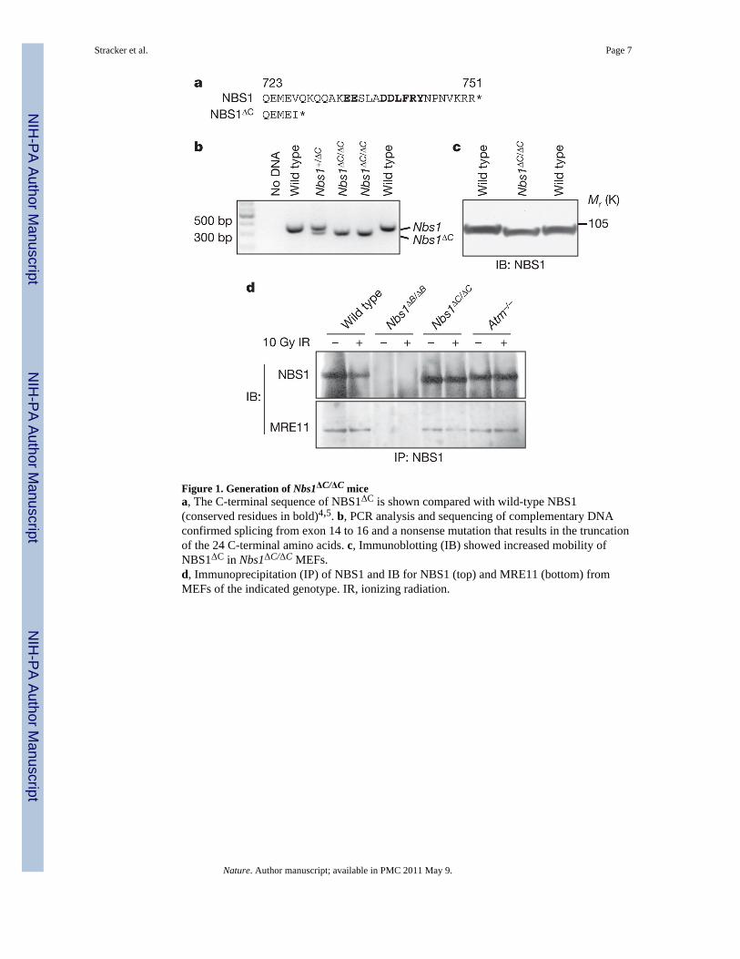
Figure 1. Generation of Nbs1ΔC/ΔC micea, The C-terminal sequence of NBS1ΔC is shown compared with wild-type NBS1(conserved residues in bold)4,5. b, PCR analysis and sequencing of complementary DNAconfirmed splicing from exon 14 to 16 and a nonsense mutation that results in the truncationof the 24 C-terminal amino acids. c, Immunoblotting (IB) showed increased mobility ofNBS1ΔC in Nbs1ΔC/ΔC MEFs.d, Immunoprecipitation (IP) of NBS1 and IB for NBS1 (top) and MRE11 (bottom) fromMEFs of the indicated genotype. IR, ionizing radiation.
Stracker et al. Page 7
Nature. Author manuscript; available in PMC 2011 May 9.
NIH
-PA Author Manuscript
NIH
-PA Author Manuscript
NIH
-PA Author Manuscript
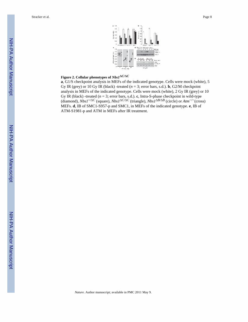
Figure 2. Cellular phenotypes of Nbs1ΔC/ΔC
a, G1/S checkpoint analysis in MEFs of the indicated genotype. Cells were mock (white), 5Gy IR (grey) or 10 Gy IR (black) -treated (n = 3; error bars, s.d.). b, G2/M checkpointanalysis in MEFs of the indicated genotype. Cells were mock (white), 2 Gy IR (grey) or 10Gy IR (black) -treated (n = 3; error bars, s.d.). c, Intra-S-phase checkpoint in wild-type(diamond), Nbs1+/ΔC (square), Nbs1ΔC/ΔC (triangle), Nbs1ΔB/ΔB (circle) or Atm−/−(cross)MEFs. d, IB of SMC1-S957-p and SMC1, in MEFs of the indicated genotype. e, IB ofATM-S1981-p and ATM in MEFs after IR treatment.
Stracker et al. Page 8
Nature. Author manuscript; available in PMC 2011 May 9.
NIH
-PA Author Manuscript
NIH
-PA Author Manuscript
NIH
-PA Author Manuscript
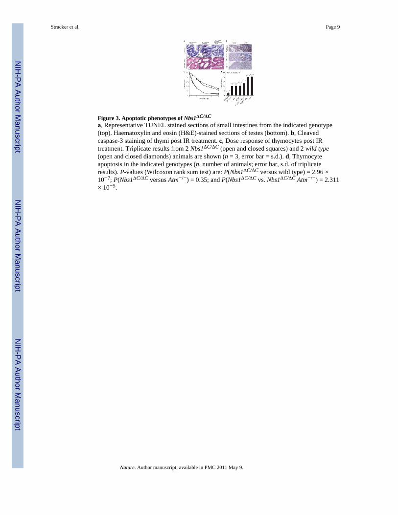
Figure 3. Apoptotic phenotypes of Nbs1ΔC/ΔC
a, Representative TUNEL stained sections of small intestines from the indicated genotype(top). Haematoxylin and eosin (H&E)-stained sections of testes (bottom). b, Cleavedcaspase-3 staining of thymi post IR treatment. c, Dose response of thymocytes post IRtreatment. Triplicate results from 2 Nbs1ΔC/ΔC (open and closed squares) and 2 wild type(open and closed diamonds) animals are shown (n = 3, error bar = s.d.). d, Thymocyteapoptosis in the indicated genotypes (n, number of animals; error bar, s.d. of triplicateresults). P-values (Wilcoxon rank sum test) are: P(Nbs1ΔC/ΔC versus wild type) = 2.96 ×10⁻⁷; P(Nbs1ΔC/ΔC versus Atm−/−) = 0.35; and P(Nbs1ΔC/ΔC vs. Nbs1ΔC/ΔC Atm−/−) = 2.311× 10⁻⁵.
Stracker et al. Page 9
Nature. Author manuscript; available in PMC 2011 May 9.
NIH
-PA Author Manuscript
NIH
-PA Author Manuscript
NIH
-PA Author Manuscript
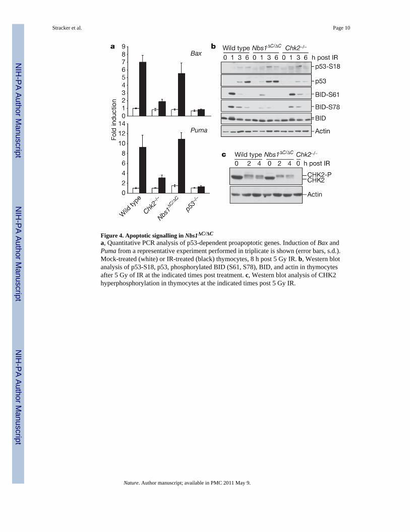
Figure 4. Apoptotic signalling in Nbs1ΔC/ΔC
a, Quantitative PCR analysis of p53-dependent proapoptotic genes. Induction of Bax andPuma from a representative experiment performed in triplicate is shown (error bars, s.d.).Mock-treated (white) or IR-treated (black) thymocytes, 8 h post 5 Gy IR. b, Western blotanalysis of p53-S18, p53, phosphorylated BID (S61, S78), BID, and actin in thymocytesafter 5 Gy of IR at the indicated times post treatment. c, Western blot analysis of CHK2hyperphosphorylation in thymocytes at the indicated times post 5 Gy IR.
Stracker et al. Page 10
Nature. Author manuscript; available in PMC 2011 May 9.
NIH
-PA Author Manuscript
NIH
-PA Author Manuscript
NIH
-PA Author Manuscript
Copyright © 2022 FDOKUMEN