Tetrapyrrole binding affinity of the murine and human p22HBP heme-binding proteins

11724 Biochemistry 1994,33, 11724-1 1733
Evidence for Involvement of the Carboxy Terminus of Helix 1 of Growth Hormone in Receptor Binding: Use of Charge Reversal Mutagenesis To Account for
Calcium Dependence of Binding and for Design of Higher Affinity Analogues? Scott W. Rowlinson,'J Ross Barnard,: Stan Basthas,* Allan J. Robins,$ Carol Senn,g Julian R. E. Wells,$
Ross Brinkworth,ll and Michael J. Waterst
Department of Physiology and Pharmacology and Centre for Molecular Biology and Biotechnology, The University of Queensland, Brisbane, Queensland 4072, Australia, Department of Biochemistry, University of Adelaide,
and Bresatec Limited, P.O. Box 1 1 , Rundle Mall, Adelaide, South Australia 5001, Australia, and Centre for Drug Design and Development, The University of Queensland, Brisbane, Queensland 4072, Australia
Received December 29, 1993; Revised Manuscript Received May 10, 1994'
ABSTRACT: In this study we have demonstrated that the C-terminus of helix 1 of porcine G H (pGH) is a receptor-interactive region, thus extending the current binding site model of GH. This was achieved by introducing charge reversal mutations into this region of pGH, which influenced receptor affinity and Ca2+ dependence of binding. The first mutant (R34E pGH, conversion of Arg 34 to Glu) introduced a putative Ca2+ binding site which is present in human G H (hGH) [Barnard et al. (1989) J . Theor. Biol. 140,355-3671 and sits opposite E220 of receptor subunit 1. This mutant exhibited increased Ca2+ dependence of receptor binding but even at optimal Ca2+ did not display higher than wild-type affinity. Introduction of a second Ca2+ binding site adjacent to the first by a second charge reversal (K30E R34E pGH) further increased Ca2+ dependence of binding and also increased affinity for the rabbit G H receptor (2.4 f 0.4)-fold relative to wild-type pGH at optimal Ca2+. Equilibrium dialysis and Scatchard analysis of binding of 45Ca2+ to pGH and K30E R34E pGH revealed two Ca2+ binding sites on wild-type pGH and an additional two Ca2+ binding sites on the K30E R34E pGH mutant ( K d 0.5-0.8 mM), as predicted. A third partial charge reversal mutant in the fourth helix (H170D) also led to enhanced Ca2+ dependence of binding, supporting our proposal that E34 and D170 are responsible for the Ca2+ dependence of hGH binding to the rabbit G H receptor. Examination of the crystal structure shows that E34 and D170 are in close proximity and would interact repulsively with a cluster of acidic residues on the receptor consisting of E126, E127, and E220 unless neutralized by Ca2+ or an introduced basic residue. Accordingly, charge reversal at the adjacent pGH residue E33 (E33K pGH) led to a Ca2+ independent (3.0 f 0.4)-fold increase in affinity of binding. As well as extending the binding site model of GH, these studies provide a mechanistic explanation for the unique Ca2+ dependence of hGH binding to the rabbit G H receptor. They also indicate that charge reversal can be used to design higher affinity G H analogues and could assist in the mapping of interactive regions in ligand-receptor complexes generally.
Porcine growth hormone (pGH) has 19 1 residues and the four antiparallel a-helical bundle structure (Abdel-Meguid et al., 1987) common tomanycytokines (Bazan, 1990). While chemical modification studies (Fukushima et al., 1983,1987; Teh & Chapman, 1987) have been of some value in defining residues of the hormone involved in binding to the GH receptor, it was the cloning of the receptor (Leung et al . , 1987) and the
This work was supported by an Australian Research Council grant to M.J.W. S.W.R. was supported by an Australian Pig Research and Development Council Scholarship. R. Barnard is supported by an NH MRC R. Douglas Wright Fellowship.
* To whom correspondence should be addressed. Department of Physiology and Pharmacology and Centre for
Molecular Biology and Biotechnology, The University of Queensland. 5 Department of Biochemistry, University of Adelaide, and Bresatec
Ltd. 11 Center for Drug Design and Development, The University of
Queensland. e Abstract published in Advance ACS Absrracts, September 1, 1994.
Abbreviations: Ca2+, calcium ion; GH, growth hormone; hGH, human growth hormone; MAC, monoclonal antibody; M2+, divalent metal ion; oGH, ovine growth hormone; pGH, porcine growth hormone. Single mutants are designated by the wild-type residue (in single-letter code) followed by its position and the mutant residue. Multiple mutants are indicated by a series of single mutants separated by a space; i.e., K30E R34E denotes a double mutant in which Lys 30 and Arg 34 are both converted to Glu.
0006-2960/94/0433- 1 1724$04.50/0
subsequent use of site-directed mutagenesis on both hormone and receptor that have enabled delineation of the interactive surfaces of these two proteins (Cunningham et ai., 1989; Cunningham & Wells, 1989; Bass et ai., 1991; Gobius et al., 1992). Recently, crystallographic and mutagenesis studies have demonstrated that in the hormone-receptor complex the same surface of two human GH receptor molecules interacts with two separate sites on the one human GH (hGH) molecule (De Vos et al., 1992; Cunningham et al., 1991). These mutagenesis studies defined hormone site 1 to include part of the center of helix 1, the latter two-thirds of helix 4, a short helical segment comprising residues 38-47, and the unstruc- tured loop region between residues 55 and 64 (Cunningham et al., 1989; Cunningham & Wells, 1989). Hormone site 2 was shown to include the amino terminus and a portion of helix 3 (Cunningham et al., 1991).
Contrary to the model of Cunningham and Wells (1989), we had predicted that the carboxy terminus of helix 1 would be part of an interactive domain in what is now known as site 1 of the hormone (Barnard & Waters, 1988; Barnard et al., 1989). This conclusion stemmed from the observation that binding of human but not ovine GH (oGH) to rabbit GH receptors requires Ca2+ or Mg2+. We proposed that an acidic
0 1994 American Chemical Society
Extension of the Binding Site Model of Growth Hormone
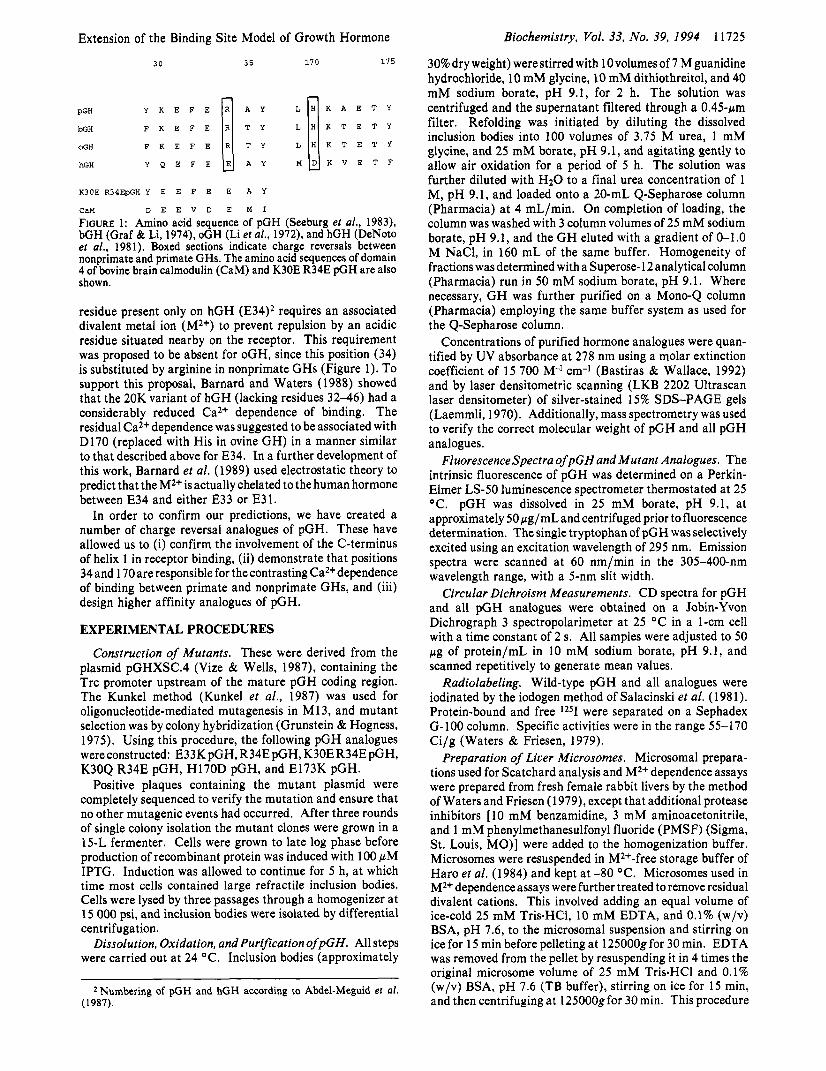
30 35 1 7 0 175
Y K E F E L H K A E T Y
pGH W H F K E F E I : ; L H K T E T Y 1 OGH F K E F E L H K T E T Y
hGH Y Q E F E M D K V E T F
K30E R34EpGH Y E E F E E A Y
CaM D E E V D E M I
FIGURE 1: Amino acid sequence of pGH (Seeburg e? al., 1983), bGH (Graf & Li, 1974), oGH (Li et al., 1972), and hGH (DeNoto e? al., 1981). Boxed sections indicate charge reversals between nonprimate and primate GHs. The amino acid sequences of domain 4 of bovine brain calmodulin (CaM) and K30E R34E pGH are also shown.
residue present only on hGH (E34)2 requires an associated divalent metal ion (M2+) to prevent repulsion by an acidic residue situated nearby on the receptor. This requirement was proposed to be absent for oGH, since this position (34) is substituted by arginine in nonprimate GHs (Figure 1). To support this proposal, Barnard and Waters (1988) showed that the 20K variant of hGH (lacking residues 32-46) had a considerably reduced Ca2+ dependence of binding. The residual Ca2+ dependence was suggested to be associated with D170 (replaced with His in ovine GH) in a manner similar to that described above for E34. In a further development of this work, Barnard et al. (1989) used electrostatic theory to predict that the M2+ is actually chelated to the human hormone between E34 and either E33 or E31.
In order to confirm our predictions, we have created a number of charge reversal analogues of pGH. These have allowed us to (i) confirm the involvement of the C-terminus of helix 1 in receptor binding, (ii) demonstrate that positions 34 and 170 are responsible for the contrasting Ca2+ dependence of binding between primate and nonprimate GHs, and (iii) design higher affinity analogues of pGH.
EXPERIMENTAL PROCEDURES
Construction of Mutants. These were derived from the plasmid pGHXSC.4 (Vize & Wells, 1987), containing the Trc promoter upstream of the mature pGH coding region. The Kunkel method (Kunkel et al . , 1987) was used for oligonucleotide-mediated mutagenesis in M 13, and mutant selection was by colony hybridization (Grunstein & Hogness, 1975). Using this procedure, the following pGH analogues were constructed: E33K pGH, R34E pGH, K30E R34E pGH, K30Q R34E pGH, H170D pGH, and E173K pGH.
Positive plaques containing the mutant plasmid were completely sequenced to verify the mutation and ensure that no other mutagenic events had occurred. After three rounds of single colony isolation the mutant clones were grown in a 15-L fermenter. Cells were grown to late log phase before production of recombinant protein was induced with 100 pM IPTG. Induction was allowed to continue for 5 h, at which time most cells contained large refractile inclusion bodies. Cells were lysed by three passages through a homogenizer at 15 000 psi, and inclusion bodies were isolated by differential centrifugation.
Dissolution, Oxidation, and Purification ofpGH. All steps were carried out at 24 "C. Inclusion bodies (approximately
* Numbering of pGH and hGH according to Abdel-Meguid et al. (1987).
Biochemistry, Vol. 33, No. 39, 1994 11725
30% dry weight) were stirred with 10 volumes of 7 M guanidine hydrochloride, 10 mM glycine, 10 mM dithiothreitol, and 40 mM sodium borate, pH 9.1, for 2 h. The solution was centrifuged and the supernatant filtered through a 0.45-pm filter. Refolding was initiated by diluting the dissolved inclusion bodies into 100 volumes of 3.75 M urea, 1 mM glycine, and 25 mM borate, pH 9.1, and agitating gently to allow air oxidation for a period of 5 h. The solution was further diluted with H2O to a final urea concentration of 1 M, pH 9.1, and loaded onto a 20-mL Q-Sepharose column (Pharmacia) at 4 mL/min. On completion of loading, the column was washed with 3 column volumes of 25 mM sodium borate, pH 9.1, and the G H eluted with a gradient of 0-1.0 M NaC1, in 160 mL of the same buffer. Homogeneity of fractions was determined with a Superose- 12 analytical column (Pharmacia) run in 50 mM sodium borate, pH 9.1. Where necessary, G H was further purified on a Mono-Q column (Pharmacia) employing the same buffer system as used for the Q-Sepharose column.
Concentrations of purified hormone analogues were quan- tified by UV absorbance at 278 nm using a molar extinction coefficient of 15 700 M-* cm-' (Bastiras & Wallace, 1992) and by laser densitometric scanning (LKB 2202 Ultrascan laser densitometer) of silver-stained 15% SDS-PAGE gels (Laemmli, 1970). Additionally, mass spectrometry was used to verify the correct molecular weight of pGH and all pGH analogues.
FluorescenceSpectra ofpGH and Mutant Analogues. The intrinsic fluorescence of pGH was determined on a Perkin- Elmer LS-50 luminescence spectrometer thermostated at 25 OC. pGH was dissolved in 25 mM borate, pH 9.1, at approximately 50 pg/mL and centrifuged prior to fluorescence determination. The single tryptophan of pGH was selectively excited using an excitation wavelength of 295 nm. Emission spectra were scanned at 60 nm/min in the 305-400-nm wavelength range, with a 5-nm slit width.
Circular Dichroism Measurements. CD spectra for pGH and all pGH analogues were obtained on a Jobin-Yvon Dichrograph 3 spectropolarimeter at 25 "C in a 1-cm cell with a time constant of 2 s. All samples were adjusted to 50 pg of protein/mL in 10 mM sodium borate, pH 9.1, and scanned repetitively to generate mean values.
Radiolabeling. Wild-type pGH and all analogues were iodinated by the iodogen method of Salacinski et al. (1981). Protein-bound and free 1251 were separated on a Sephadex G-100 column. Specific activities were in the range 55-170 Ci/g (Waters & Friesen, 1979).
Preparation of Liver Microsomes. Microsomal prepara- tions used for Scatchard analysis and M2+ dependence assays were prepared from fresh female rabbit livers by the method of Waters and Friesen (1979), except that additional protease inhibitors [ 10 mM benzamidine, 3 mM aminoacetonitrile, and 1 mM phenylmethanesulfonyl fluoride (PMSF) (Sigma, St. Louis, MO)] were added to the homogenization buffer. Microsomes were resuspended in M2+-free storage buffer of Haro et al. (1984) and kept at -80 OC. Microsomes used in M2+ dependence assays were further treated to remove residual divalent cations. This involved adding an equal volume of ice-cold 25 mM Tris.HC1, 10 mM EDTA, and 0.1% (w/v) BSA, pH 7.6, to the microsomal suspension and stirring on ice for 15 min before pelleting at 125000gfor 30 min. EDTA was removed from the pellet by resuspending it in 4 times the original microsome volume of 25 mM Tris.HC1 and 0.1% (w/v) BSA, pH 7.6 (TB buffer), stirring on ice for 15 min, and then centrifuging at 125000gfor 30 min. This procedure
11726 Biochemistry, Vol. 33, No. 39, 1994
was repeated twice before resuspension of microsomes to their original volume in cation-free buffer (Haro et al., 1984) and storage at -80 "C.
Ca2+ Dependence Assays. Determination of Ca2+ depen- dence of binding of pGH analogues was carried out as previously described (Barnard & Waters, 1988), except that TB buffer was used for all hormone and microsome dilutions. Nonspecific binding of lZ51-labeled hormones was determined in the presence of pGH at a final concentration of 1.66 pg/ mL, with buffer alone added to total binding tubes to ensure the same final assay volume. Tubes were incubated in triplicate for 18 h at 20 "C with shaking. Assays were terminated by the addition of 1 mL of cold 0.1% (w/v) bovine y-globulins followed by 1 mL of cold 30% (w/v) poly(ethy1ene glycol) 6000 (PEG) with vortexing and centrifugation. Pellets were counted in an LKB 1274 y spectrometer.
Although microsomal membranes containing a minor percentage of prolactin receptors were used for these experi- ments, nonprimate GHs do not bind to prolactin receptors (Waters & Friesen, 1979). Thiswasverified by total abolition of lZ51-pGH-specific binding to these membranes with MAb 7 (AGEN Ltd., Acacia Ridge, Australia), a GH-receptor- specific monoclonal antibody (Barnard et al., 1984).
p H Dependence of Ca2+ Enhancement of Binding. EDTA- washed rabbit liver microsomes were diluted appropriately in TB buffer, and 100 pL of diluted microsomes was added to 200pL of 25 mM Bis-Tris and 0.1% (w/v) BSA (BTB buffer) with either 30 mM CaCl2 or 10 mM EDTA. Solution pH was then set over the range from 5.0 to 7.5. One hundred microliters of 1251-labeled hormone (approximately lo5 cpm in BTB buffer containing either 30 mM CaC12 or 10 mM EDTA) was then added to the assay tubes. Nonspecific binding was determined simultaneously by addition of 100 pL of pGH (in BTB buffer with 30 mM CaCl2 or 10 mM EDTA) to give a final concentration of 2 pg/mL in a parallel series of tubes. At each pH value an extra tube was used to evaluate the final pH. Assays were left for 18 h at 20 OC with shaking and were terminated with PEG and bovine y-globulins as for Ca2+ dependence assays.
Zn2+ Dependence Assays. The Zn2+ dependence of binding of native pGH and of the K30Q R34E pGH mutant was determined in a manner similar to that of the Ca2+ dependence assays but using the buffer conditions described by Cunning- ham et al. (1990). EDTA-washed rabbit liver microsomes were diluted appropriately into 20 mM Tris.HC1 (f10 mM MgClz), 140 mM NaCl, and 0.1% BSA, pH 7.6 (TMSB buffer). Diluted microsomes (100 pL) were incubated with approximately 1 O5 cpm of 1251-labeled native or analogue pGH (diluted in TMSB buffer) plus 300 pL of TMSB buffer with zinc acetate at twice the final assay Concentration. Specific binding was determined in parallel by addition of 100 pL of unlabeled pGH (in TMSB buffer) to givea final concentration of 1.66 pg/mL. Assays were incubated and terminated as for CaZ+ dependence assays. To ensure that BSA was Zn2+ free, BSA (fraction V, Sigma) was dialyzed against 5 mM Tris.HC1 and 10 mM EDTA, pH 7.5, for 18 h, followed by dialysis against 5 L of deionized water (three changes) over 36 h.
As a positive control, the Zn2+ dependence of hGH binding to purified rabbit PRL receptor [prepared by the method of Waters et al . (1 990)] was assayed using the protocol described above. To determine if pGH binding was occurring to GH receptor alone, the Zn2+ dependence of IZ51-pGH binding was tested using microsomes preincubated with MAb 7, a mono- clonal antibody able to block hormone binding to the G H receptor (Barnard et ai., 1984).
Rowlinson et al.
Determination of Affinity Constants. The affinity of pGH analogues for the G H receptor was determined by Scatchard (1949) analysis of IZ51-labeled pGH analogue binding to rabbit liver microsomes in the presence of increasing concentrations of unlabeled hormone competitor. Incubations (final volume 0.5 mL, microsomes added last) were carried out in TB buffer with 20 mM MgC12,20 mM CaC12, or 10 mM EDTA, or in TMSB buffer f 250 pM zinc acetate, at 20 "C with shaking for 18 h. Assays were terminated by addition of 2 mL of cold TB buffer or PEG/bovine y-globulins, followed by centrifu- gation at 16OOg for 25 min at 4 OC. Pellets were counted in an LKB 1274 y spectrometer. Data analysis and curve fitting were as previously described (Barnard & Waters, 1988) using the EBDA and LIGAND programs adapted by McPherson (1985) for IBM personal computers. Determinations were carried out a minimum of three times.
Equilibrium Dialysis and Scatchard Analysis of 4sCa2+ Binding to pGH, HI 70DpGH, and K30E R34E pGH. Prior to equilibrium dialysis, M2+ present in each hormone preparation was removed by dialysis (one 2000 volume exchange with 2 mM EDTA and 10 mM Tris.HC1, pH 10.5, at 4 "C for 12 h and two 5000 volume exchanges with 10 mM Tris-HC1, pH 10.5, at 4 "C for 24 h). Dialysis membranes (Hoefer Scientific Instruments, San Francisco, CA), 12-14- kDa cutoff, were boiled in 5% (w/v) NaHCO3/ 1 mM EDTA and washed thoroughly in three changes of deionized HzO for 1 h before use. Equilibrium dialysis was carried out by the addition of 150 pL of pGH or an analogue (concentration approximately 130 pM) to one side of the membrane and 75 pL (approximately 1.5 X lo6 dpm) of 45Ca2+ (specific activity 21 Ci/g; Amersham Ltd., Buckinghamshire, England) and 75 pL of unlabeled CaC12 (1-20 mM) to the other side. All components were diluted in 10 mM Tris.HC1, pH 10.5. Binding assays were carried out at pH 10.5 because pGH analogues (at 130 pM) were poorly soluble below this pH, and calcium hydroxide precipitates appeared above pH 11.0 in tubes with higher CaC12 concentrations. Components were rotated slowly for 8 h at 20 "C, after which 25-pL aliquots of each compartment were analyzed for 45Ca2+ content by addition of 5 mL of scintillation fluid (FSE Laboratory Supplies, Loughborough, England) and use of a Packard 1900CA p spectrometer with quench correction. Scatchard analysis with regression analysis was used to derive binding parameters.
Statistical comparisons were by Student's t test or by analysis of variance with Neuman-Keuls' multiple range test.
RESULTS
Fluorescence Emission Spectra. These were determined for pGH and pGH analogues over the range 310-400 nm. Spectra were identical between wild-type and analogue GHs, with a broad emission maximum around 340 nm. This indicates that the polarity of the environment around the single tryptophan present in each protein is similar, with no gross changes in folding as a result of mutation. Emission spectra are not shown but are identical to those reported by Bastiras and Wallace (1992).
CD Spectra. Far-UV CD spectra were obtained for pGH and all pGH analogues. No significant difference was seen between the spectra of the native and analogue pGHs, indicating no measurable changes in a-helical content. Addition of 20 mM Ca2+ to the pGH and K30E R34E pGH mutant did not significantly influence the CD spectrum. CD spectra are not shown but were identical to those reported by Bastiras and Wallace (1992).
Extension of the Binding Site Model of Growth Hormone Biochemistry, Vol. 33, No. 39, 1994 11727 ~~
Table 1' ratio of ratio of ratio of
(Ka in 20 mM Ca2+)/ (% SB in 20 mM Ca2+)/ (Ka of analogue pGH)/ Ca2+ EDSO UGH (% SB in 10 mM EDTA) (Ka in 10 mM EDTA) (Ka of wild-type pGH) for receptor binding
wild type R34E K30Q R34E K30E R34E H170D wild typee E33K E33Kf E173K
1.6 f 0.1 (A) 2.8 f 0.2 (B) 4.2 f 0.4 (C) 7.24 f 0.6 (D) 2.9 f 0.5 (E) ND ND ND ND
1.4 f 0.1 ND 3.3 f 0.1c 8.8 f 0.2b.S 2.2 f O.2b 0.7 f 0.2b ND ND ND
1 .o 1.0 f 0.2 0.9 f 0.2 2.4 f 0.4b 0.8 f 0.2 ND 2.1 f 0.3b 3.0 f 0.4c 1.4 & 0.3
ND 5.2 f 2Sd 4.1 f 0.4d 3.8 f 0.6d ND ND ND ND ND
0 A vs B, A vs C, and A vs D, p < 0.001, A vs E and C vs D, p < 0.05, B vs C, p < 0.02, and B vs D, p < 0.01, represent the significant difference in the binding between the corresponding groups, ANOVA, with multiple range testing. SB = specific binding. Ka = affinity. ND = not determined. All values in the table are the mean of three or more determinations with SEM indicated. EDSO refers to the Ca2+ concentration producing 50% of total Ca2+ dependent specific binding. p < 0.05 indicates significant change relative to pGH. c p < 0.01 indicates significant change relative to pGH. d No significant difference between values. Assay performed in TMSB buffer plus 250 gM Zn2+ instead of 20 mM CaZ+. f Assay performed as per Experimental Procedures except M2+-free TB buffer was used. 8 Binding of K30E R34E pGH in 10 mM EDTA was too low to obtain Scatchard results using '25I-K30E R34E pGH; thus IZ5I-pGH was used.
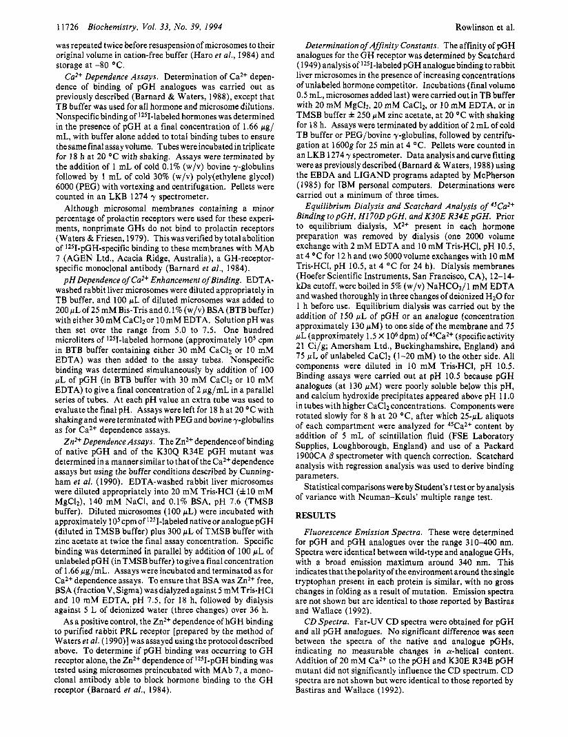
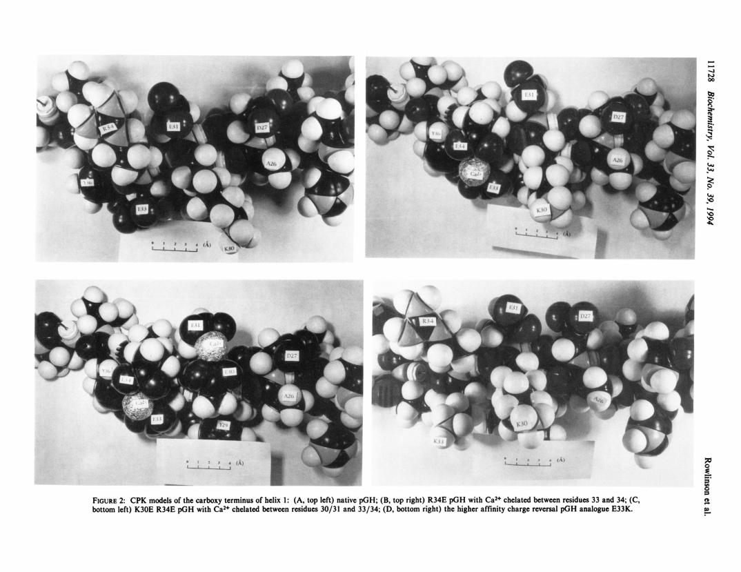
Ca2+ Dependence of Binding to Rabbit Liver GH Receptor. We have created four analogues, R34E pGH, K30Q R34E pGH, K30E R34E pGH, and H170D pGH, with an increased Ca2+ dependence of receptor binding compared with the native hormone (Table 1). Of the three analogues in the carboxy- terminal part of the first helix, significant differences exist in the degree of increase in binding seen when the Ca2+ concentration is raised to 20 mM (Figure 3 and Table 1). However, the EDSO for Ca2+ of each of these analogues is similar (Table 1). In agreement with this finding, Scatchard analysis of 45Ca2+ binding to the hormone shows that the engineered Ca2+ binding sites are similar in affinity to those of wild-type pGH (Figure 5).
In addition to the Ca2+ dependence induced by modifying residues in the first helix, the H170D pGH analogue shows a 2.9-fold increase in specific binding to the receptor when the Ca2+ concentration is increased to 20 mM (Table l), significantly more than the 1.6-fold increment seen with wild- type pGH. Enhancement of binding on addition of Ca2+ to CaZ+-dependent analogues was associated in all cases with significant increases in affinity for the receptor (Table 1). With the Ca2+-dependent analogue K30E R34E pGH, the reduction in affinity in the absence of Ca2+ was so great that Scatchard analysis of the 12sI-labeled analogue could not be performed. Accordingly, it was necessary to use 12SI-~ild- type pGH with displacement by K30E R34E pGH to obtain affinity constants.
Affinity of Analogues f o r Receptor. Scatchard analysis of binding data for pGH and pGH analogues showed that two of the analogues, K30E R34E pGH and E33K pGH, had a significant increase in receptor binding affinity relative to native pGH (Table 1). In the case of K30E R34E pGH, this increase was seen only when the assay was carried out in buffer containing 20 mM Ca2+. In contrast, the higher affinity of the charge reversal mutant E33K pGH could be further enhanced by assaying it in a cation-free buffer to minimize electrostatic screening3 (Table 1).
Zn2+ Dependence of Binding. Human G H binding to the prolactin (PRL) receptor is Zn2+-dependent (Cunningham et al., 1990), and rabbit liver microsomes contain prolactin receptors (Waters & Friesen, 1979). To demonstrate that the Ca2+ dependence we observed with our analogues is not
An increase in sensitivity of the charge reversal strategy can be achieved at low ionic strength when counterion screening is minimized (Barnard, 1993).
a result of Zn2+ contamination, or of specificity crossover (Waters & Friesen, 1979), the Zn2+ dependence experiments of Cunningham et al . (1990) were replicated with native pGH and a strongly Ca2+-dependent analogue (K30Q R34E pGH) using rabbit liver microsomes. Figure 6A shows that with increasing concentration of ZnZ+ there is an inhibition of binding of both pGH and K30Q R34E pGH to rabbit liver microsomes. This is a result of decreased affinity for the receptor (Table 1). Virtually identical results were obtained for these two hormones when the assay was carried out in 20 mM Tris and 0.1% BSA, pH 7.6, with zinc acetate only (no Mg2+or CaZ+) and when a higher concentration of microsomes was used. In contrast, hGH binding to PRL receptors is positively Zn2+-dependent (Figure 6B). 1251-pGH binding to rabbit liver microsomes in the presence of Zn2+ (9.9% specific binding) was abolished by the GH-receptor-specific inhibitory MAb 7, indicating that all receptor binding is of the somatogenic type (data not shown).
p H Dependence of Ca2+ Enhancement of Binding. If CaZ+ is bound to carboxyl groups involved in hormone-receptor interaction, then the Ca2+ enhancement of binding should correlate with ionization of these groups. The Ca2+ depen- dence of binding of K30E R34E pGH to receptor over the pH range 5.0-7.5 shows that Ca2+-dependent binding is evident above pH 6.0 and reaches a maximum above pH 7.0 (Figure 4). A similar result was obtained with the K30Q R34E pGH analogue (data not shown). The observed pH dependence of Ca2+ enhancement is consistent with the ionizations calculated for liganding carboxylate groups with an average pK, of 6.3 (Figure 4).
Equilibrium Dialysis and Scatchard Analysis of ' T a 2 + Binding to pGH, Hl7OD pGH, and K30E R34E pGH. Scatchard analysis of 4sCa2+ binding to pGH and two of the more strongly CaZ+-dependent analogues in the first and fourth helices (K30E R34E pGH and H170D pGH) reveals that all have a similar affinity for Ca2+, although a difference is seen in the stoichiometry of Ca2+ binding (Figure 5 ) . pGH and H170D pGH bind 1.8 f 0.1 and 1.8 f 0.2 mol of Ca2+/mol of hormone, respectively, while K30E R34E pGH binds 4.1 f 0.2 mol of Ca2+/mol of hormone, indicating that in this case the engineered Ca2+ binding sites are functional in the free hormone.
DISCUSSION
In the course of identifying the residues responsible for the contrasting Ca2+ dependence of binding of primate and
- I - I-
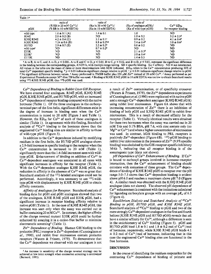
s FIGURE 2: CPK models of the carboxy terminus of helix 1: (A, top left) native pGH; (B, top right) R34E pGH with Ca2+ chelated between residues 33 and 34; (C, bottom left) K30E R34E pGH with Ca2+ chelated between residues 30/31 and 33/34; (D, bottom right) the higher affinity charge reversal pGH analogue E33K.
Extension of the Binding Site Model of Growth Hormone Biochemistry, Vol. 33, No. 39, 1994 11729
'6 $ 0 E 20__1 0 5 10 15 20 25 30 35 40 45
[Ca2+] (mM) FIGURE 3: Ca2+ dependence of pGH (O) , R34E pGH (a), and K30E R34E pGH (+) binding to rabbit GH receptors. The assay was performed as described in Experimental Procedures using 200 p g of EDTA-washed rabbit liver microsomes and radiolabeled pGH or an analogue (self-displaced with excess unlabeled hormone for non- specific binding values). Maximum specific binding for pGH and R34E and K30E R34E pGH was 12%, 6.3%, and 6.6%, respectively. Each point represents the mean of a triplicate determination with SEM indicated.
nonprimate GHs, we have added the C-terminus of helix 1 to the receptor-interactive region mapped on hGH by Cunning- ham and Wells (1989) and Cunningham et al. (1989, 1990, 199 1). Despite an extensive mutational analysis these workers did not include this region of the first helix as a binding determinant, even though in the crystal structure of De Vos et al. (1992) this region appears likely to interact. This is probably because their initial homolog-scanning mutagenesis strategy selected regions of hGH which conferred specificity of binding to the human GH receptor but in doing so missed hormone regions which bind equally well to both the human and nonprimate G H receptors. Exchange of regions containing these interactive determinants [as in the case of the homolog- scanning mutagenesis of Cunningham et al. (1989)] would produce no obvious changes in the binding of the mutant hormones to the human receptor, and so these segments would be incorrectly deemed as noninteractive. This reasoning could also explain why Cunningham et al. (1989) did not find histidines 20 and 22 to be binding determinants despite earlier studies by Fukushima et al. (1983, 1987) implicating these residues. Similarly, thedeleted segment in 20K hGH (residues 32-46) was not reported to be important in receptor binding, despite good literature evidence showing a difference in its binding characteristics compared with 22K hGH (Hughes et al., 1983; Smalet al., 1986; Barnard & Waters, 1988; Barnard et al., 1990).
To provide evidence that the residues responsible for the Ca2+ dependence of hGH binding are located in the proposed helix 1 and 4 regions, several charge reversals were made, the first being in the terminal portion of the first helix. These reversals resulted in increases in Ca2+ dependence of binding to the rabbit G H receptor (Table 1). Molecular modeling of the first helix (Figure 2) revealed E31 and E33 as candidate residues in hGH which could combine with E34 to chelate Ca2+. These models also indicated that K30 could adversely affect the liganding of Ca2+ to either the E34/E33 or E34/ E3 1 chelation sites, since its amine group can rotate to within 3 8, of these carboxyl pairs. To examine this possibility, K30 was replaced with a long uncharged side chain (Gln). This analogue, K30Q R34E pGH, is identical to hGH in this region and displayed a greater extent of Ca2+ enhancement of affinity (3.3-fold) than did R34E pGH alone (Table 1 and Figure 3). Thus, it appears that K30 does hinder interaction of E34 with Ca2+, although it may facilitate the interaction between hormone and receptor in the absence of Ca2+. Further
0-1 4.5 5 5.5 6 6.5 7 7.5 8 e
PH
FIGURE 4: Enhancement of Ca2+ dependence of pGH and K30E R34E pGH binding over the pH range 5-7.5. Measured values for pGH (0) and K30E R34E pGH (+) and predicted (0) pH dependence of Ca2+ enhancement of binding of K30E R34E were generated using pKa values of 6.3 for liganding carboxyl groups. Approximately 200 pg of EDTA-washed female rabbit liver mi- crosomes was assayed in Bis-Tris buffer, as described in Experimental Procedures. Maximum specific binding of pGH and K30E R34E pGH was 10.1% and 5.6%, respectively. Each point represents the mean of a triplicate determination with SEM indicated. Virtually identical Ca2+-dependent enhancement was seen in three other assays carried out using the same conditions as described.
examination of molecular models revealed that a second Ca2+ chelation site could be engineered into this region of the first helix by substitution of lysine 30 with glutamic acid (Le., K30E R34E pGH), introducing the additional Ca2+ chelation site between residues 30 and 31 or 27 (Figure 2, see below). The resulting analogue has four carboxyl groups in close association at positions 30, 3 1, 33, and 34, plus an aspartate 0-carboxyl at position 27 which can rotate close to residues 30 and 31. This K30E R34E pGH analogue was the most strongly Ca2+-dependent mutant (Table 1 and Figure 3), presumably because it possesses an additional negative charge at position 30, only 4.1 8, from E220 on the G H receptor. This analogue also had a 2.4-fold higher affinity than wild-type pGH in the presence of optimal Ca2+ (Table l), in accord with the predictions of the electrostatic model of Barnard et al. (1989). Changes in Ca2+ dependence and affinity of the pGH analogues were not due to conformational changes, since all mutants exhibited correct folding as determined by CD and fluorescence spectra. Moreover, pH dependence data support the hypothesis that the Ca2+ dependence seen with pGH analogues is due to unprotonated carboxyl side chains. Below pH 6 Ca2+ dependent binding is not observed, while above pH 6 there is an increase in the Ca2+ dependence of binding. This pH dependence of Ca2+ enhancement of binding is consistent with pKa values between 6.0 and 6.4 for the liganding carboxyl groups4 (see Figure 4).
To confirm the proposal that Ca2+ binds directly to the Ca2+-dependent mutants (Barnard et al., 1989), equilibrium dialysis and Scatchard analysis were carried out on pGH and K30E R34E pGH (Figure 5). This revealed that these two hormones had identical affinity for Ca2+ but differ in their stoichiometry of binding. Two additional Ca2+ binding sites were observed with K30E R34E pGH as a result of chelation within the cluster of five negative charges on residues 27, 30, 31, 33, and 34 (Figure 2C). In this analogue there are no
The high pKa value reflects the proximity of liganding carboxyl groups within a cluster of charged residues from the hormone and the GH receptor (E30, E33, E34, E126, E127 + R42). Using distances from the crystal coordinates, calculations were made using Coulomb's law with a distance- dependent dielectric (Mehler, 1990) corrected for ionic strength (Mehler & Eichele, 1984; Hill, 1956). These calculations generate pK, values equal to or greater than 6.3 for liganding carboxyl groups.

11730 Biochemistry, Vol. 33, No. 39, 1994 Rowlinson et al.
0.4 1 0 . 3 5 i
e, 0.25
c 0.2 . '0
0 50 100 150 200 250 300 Bound (pM)
FIGURE 5 : Scatchard plots of 4sCaZ+ binding to pGH (0), H170D pGH (O) , and K30E R34E pGH (A) derived from equilibrium dialysis. Binding assays were performed as described in Experimental Procedures. Final concentrations of pGH, H170D pGH, and K30E R34E pGH were 69, 80.5, and 70.5 pM, respectively. The specific activity of 4sCa2+ was 21 Ci/g. The line of best fit was generated by linear regression. The mean f SEM (n 2 three determinations) for the number of moles of CaZ+ per mole of hormone was 1.8 * 0.1 for pGH, 1.8 f 0.2 for H170D pGH, and 4.1 f 0.2 for K30E R34E pGH. The mean f SEM for the affinity of CaZ+ for each analogue was 0.5 * 0.1 mM-I for pGH, 0.8 0.2 mM-l for H170D pGH, and 0.8 f 0.1 mM-I K30E R34E pGH.
positively charged side chains in close proximity which could disrupt chelation of Ca2+ (cf. K30 of pGH). This is very similar to that of the auxillary Ca2+ binding site of bovine brain CaM (shown in Figure 1) or the chelation sites of chromogranin A (Yo0 & Albanesi, 1991) and calsequestrin (Fliegel et al., 1987). The 2 mol of Ca2+ bound/mol of wild- type pGH may be a result of binding to two potential sites within the third helix, at positions D115, E117, E l l 8 and E126, E128, D129. This is the region shown by Cunningham et al. (1991) to be the site 2 binding region for the hGH binding protein-receptor and could indicate that binding of the second receptor subunit to the hGH-GHR complex may be Ca2+ dependent. The two other potential Ca2+ binding sitesinpGH (D65/E66and D152/D153) presumablycannot bind Ca2+ because of interference by nearby K64 and R150. In the study of Barnard and Waters (1988) it was observed that there was residual Ca2+ dependence of binding present with 20K hGH which lacks E34. This led to the proposal that position 170 was responsible for residual Ca2+ dependence since it was the only other positive (His) to negative (Asp) charge reversal in a comparison of ovine G H and hGH. Site- directed mutagenesis at this position supports our hypothesis, since a 2.9-fold increase in specific binding was observed with this analogue in the presence of Ca2+ (H170D pGH) (Table 1). Despite this increased Ca2+ dependence, we could not detect Ca2+ binding to the D 170/E173 pair of carboxyl residues by equilibrium dialysis (Figure 5 ) . This could be explained by an analysis of the crystal structure of the GH/GH receptor (see below).
We find that the Ca2+ effect is not a Zn2+ effect analogous to that reported for hGH binding to PRL receptors by Cunningham et al. (1990). Indeed, Zn2+ inhibited binding of both wild-type pGH and K30Q R34E pGH (Table 1 and Figure 6), indicating that the Ca2+ dependence phenomenon cannot be a result of Zn2+ contamination and that we are in fact measuring somatotrophic sites. This observation is confirmed by means of receptor blockade with MAb 7, a GH- receptor-specific monoclonal binding antagonist. We do, however, find a Zn2+dependenceof hGH binding to the native prolactin receptor, in agreement with the study of Cunningham et a / . (1990).
0 50 100 150 200 250 0 50 100 150 200 250
[2n2+1 (PM) [Zn2'1 (IW FIGURE 6: (A) Zn2+ dependence of L2sI-pGH (0) and IZ5I-K30Q R34E pGH (0) binding to rabbit GH receptors. Approximately 200 pg of EDTA-washed female rabbit liver microsomes was added to reaction mixtures, and assays were carried out as described in Experimental Procedures. Maximum specific binding for pGH and K30Q R34E pGH in TMSB buffer was 17.4% and 8.8%, respectively. (B) Zn2+ dependence of lz5I-hGH (e) binding to purified PRL receptors. EDTA-dialyzed affinity-purified PRL receptors were added at a 1:20 dilution, and assays were carried out as described in Experimental Procedures. Maximum specific binding was 6.4%. Each point is the mean of triplicate assays with SEM indicated.
Glu 33 9 PGH
0
hGH Mutant pGH (E33K)
FIGURE 7: Configuration of key charges in wild-type pGH and hGH and a charge reversal mutant of pGH (E33K pGH). Such a strategy can provide an increase in the affinity of up to the square of the changeinaffinityseen with the neutralsubstitution (Barnard, 1993). This procedure, therefore, provides a more sensitive detection system for determining interactive residues than alanine scanning mutagenesis as well as an opportunity to construct higher affinity mutants.
On the basis of these charge reversal studies, we propose that the C-terminus of helix 1 and the central region of helix 4 are interactive with key negative charges on the receptor. Accordingly, these regions were targeted for further charge reversal mutagenesis to create higher affinity G H analogues. Changing one of the single carboxyls in wild-type pGH to a positive group will produce a pair of positive residues on the hormone. Given no other interferences, such a hormone should display higher affinity for the receptor when interacting with the juxtaposed negatively charged receptor side chain (Figure 7). To test this strategy, we constructed two analogues, E33K pGH and E173K pGH. The E33K pGH analogue exhibited a 3-fold greater affinity than wild-type pGH as predicted. However, no significant alteration in affinity of the E173K mutant was observed (Table 1).
Using the crystal structure of the GH/GH receptor complex, it is now possible to rationalize the results of the charge reversal technique and to validate the hypotheses derived from the Ca2+ dependence and equilibrium dialysis data. From the
Extension of the Binding Site Model of Growth Hormone Biochemistry, Vol. 33, No. 39, I994 1173 1
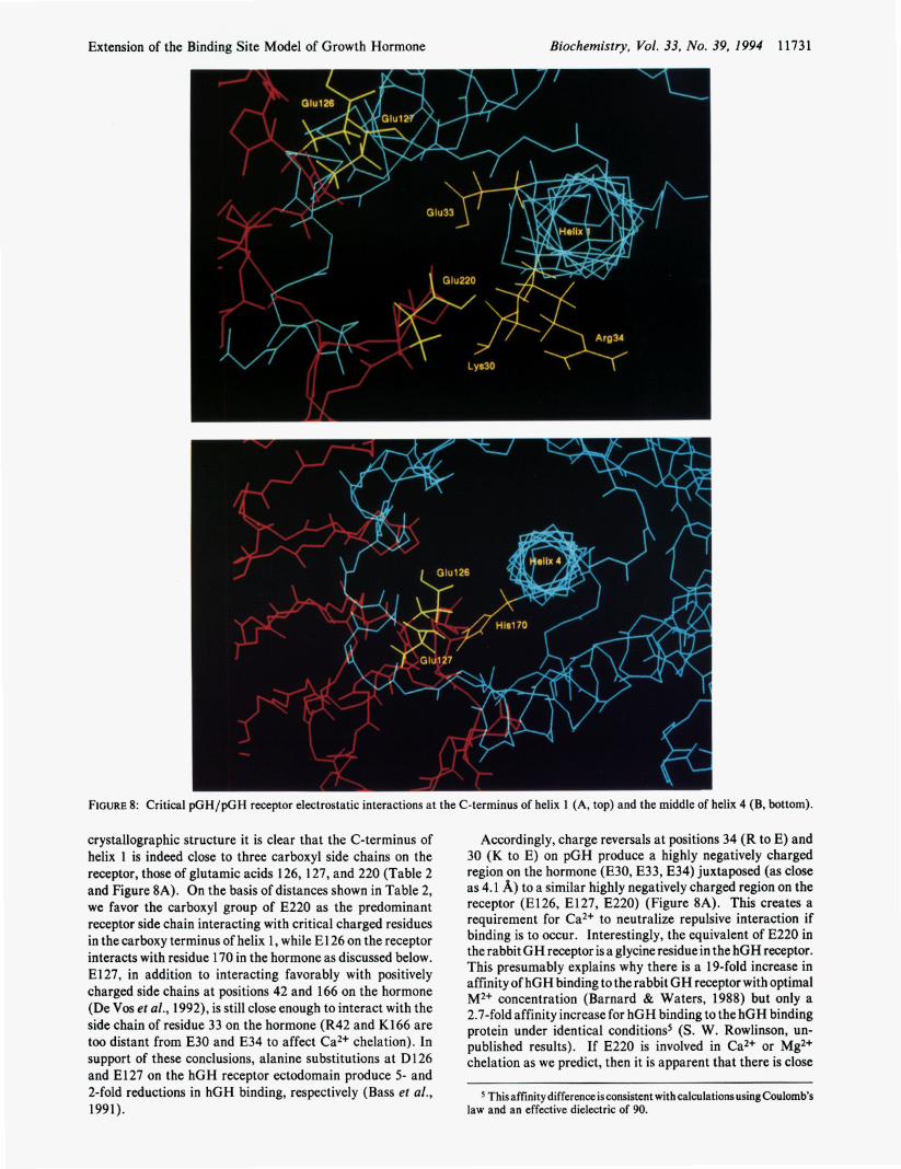
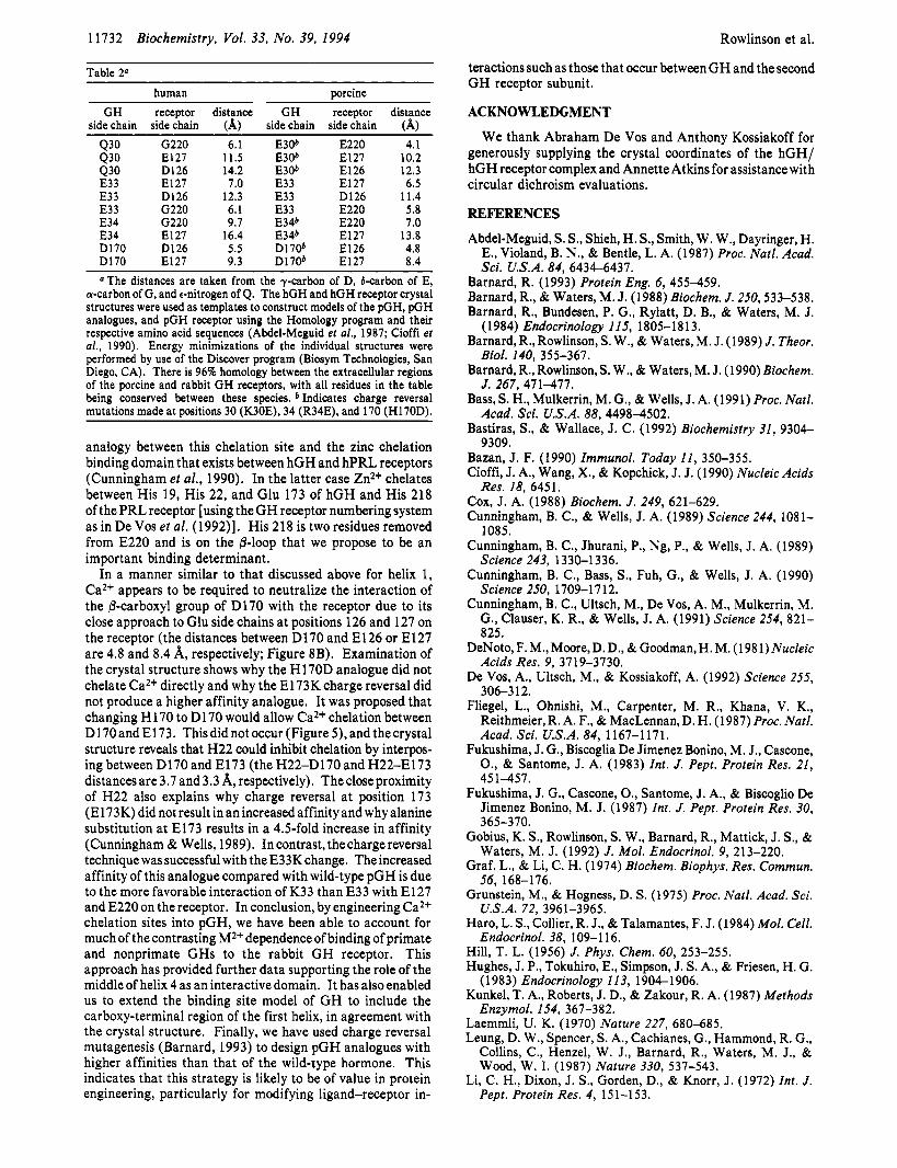
FIGURE 8: Critical pGH/pGH receptor electrostatic interactions at the C-terminus of helix 1 (A, top) and the middle of helix 4 (B, bottom).
crystallographic structure it is clear that the C-terminus of helix 1 is indeed close to three carboxyl side chains on the receptor, those of glutamic acids 126, 127, and 220 (Table 2 and Figure 8A). On the basis of distances shown in Table 2, we favor the carboxyl group of E220 as the predominant receptor side chain interacting with critical charged residues in the carboxy terminus of helix 1, while E l 26 on the receptor interacts with residue 170 in the hormone as discussed below. E l 27, in addition to interacting favorably with positively charged side chains at positions 42 and 166 on the hormone (De Vos et ai., 1992), is still close enough to interact with the side chain of residue 33 on the hormone (R42 and K166 are too distant from E30 and E34 to affect Ca2+ chelation). In support of these conclusions, alanine substitutions at D126 and E127 on the hGH receptor ectodomain produce 5- and 2-fold reductions in hGH binding, respectively (Bass et al., 1991).
Accordingly, charge reversals at positions 34 (R to E) and 30 (K to E) on pGH produce a highly negatively charged region on the hormone (E30, E33, E34) juxtaposed (as close as 4.1 A) to a similar highly negatively charged region on the receptor (E126, E127, E220) (Figure 8A). This creates a requirement for Ca2+ to neutralize repulsive interaction if binding is to occur. Interestingly, the equivalent of E220 in the rabbit GH receptor is a glycine residue in the hGH receptor. This presumably explains why there is a 19-fold increase in affinity of hGH binding to the rabbit GH receptor with optimal M2+ concentration (Barnard & Waters, 1988) but only a 2.7-fold affinity increase for hGH binding to the hGH binding protein under identical conditions5 (S. W. Rowlinson, un- published results). If E220 is involved in Ca2+ or Mg2+ chelation as we predict, then it is apparent that there is close
This affinity difference is consistent with calculations using Coulomb’s law and an effective dielectric of 90.
11732 Biochemistry, Vol. 33, No. 39, 1994
Table 2" human porcine
GH receptor distance GH receptor distance side chain side chain (A) side chain side chain (A)
Rowlinson et al.
teractions such as those that occur between G H and the second G H receptor subunit.
ACKNOWLEDGMENT
We thank Abraham De Vos and Anthony Kossiakoff for generously supplying the crystal coordinates of the hGH/ hGH receptor complex and Annette Atkins for assistance with circular dichroism evaluations.
REFERENCES
Abdel-Meguid, S. S., Shieh, H. S., Smith, W. W., Dayringer, H. E., Violand, B. N., & Bentle, L. A. (1987) Proc. Natl. Acad. Sci. U.S.A. 84, 6434-6431.
Barnard, R. (1993) Protein Eng. 6, 455-459. Barnard, R., & Waters, M. J. (1988) Biochem. J. 250,533-538. Barnard, R., Bundesen, P. G., Rylatt, D. B., & Waters, M. J.
Barnard, R., Rowlinson, S. W., & Waters, M. J. (1989) J . Theor.
Barnard, R., Rowlinson, S. W., & Waters, M. J . (1990) Biochem.
Bass, S. H., Mulkerrin, M. G., & Wells, J. A. (1991) Proc. Natl.
Bastiras, S., & Wallace, J. C. (1992) Biochemistry 31, 9304-
Bazan, J. F. (1990) Zmmunol. Today 11, 350-355. Cioffi, J. A., Wang, X., & Kopchick, J. J. (1990) Nucleic Acids
Cox, J. A. (1988) Biochem. J. 249, 621-629. Cunningham, B. C., & Wells, J. A. (1989) Science 244, 1081-
1085. Cunningham, B. C., Jhurani, P., Ng, P., & Wells, J. A. (1989)
Science 243, 1330-1336. Cunningham, B. C., Bass, S., Fuh, G., & Wells, J. A. (1990)
Science 250, 1709-1712. Cunningham, B. C., Ultsch, M., De Vos, A. M., Mulkerrin, M.
G., Clauser, K. R., & Wells, J. A. (1991) Science 254, 821- 825.
DeNoto, F. M., Moore, D. D., &Goodman, H. M. (1981) Nucleic Acids Res. 9, 3719-3730.
De Vos, A., Ultsch, M., & Kossiakoff, A. (1992) Science 255, 306-3 12.
Fliegel, L., Ohnishi, M., Carpenter, M. R., Khana, V. K., Reithmeier, R. A. F., & MacLennan, D. H. (1987) Proc. Natl. Acad. Sci. U.S.A. 84, 1167-1171.
Fukushima, J . G., Biscoglia De Jimenez Bonino, M. J., Cascone, O., & Santome, J. A. (1983) Znt. J . Pept. Protein Res. 21, 451-457.
Fukushima, J. G., Cascone, O., Santome, J. A., & Biscoglio De Jimenez Bonino, M. J. (1987) Znt. J . Pept. Protein Res. 30,
Gobius, K. S., Rowlinson, S. W., Barnard, R., Mattick, J. S., &
Graf, L., & Li, C. H. (1974) Biochem. Biophys. Res. Commun.
Grunstein, M., & Hogness, D. S. (1975) Proc. Natl. Acad. Sci.
Haro, L. S. , Collier, R. J., & Talamantes, F. J. (1984) Mol. Cell.
Hill, T. L. (1956) J. Phys. Chem. 60, 253-255. Hughes, J. P., Tokuhiro, E., Simpson, J . S. A., & Friesen, H. G.
Kunkel, T. A., Roberts, J. D., & Zakour, R. A. (1987) Methods
Laemmli, U. K. (1970) Nature 227, 680-685. Leung, D. W., Spencer, S. A,, Cachianes, G., Hammond, R. G.,
Collins, C., Henzel, W. J., Barnard, R., Waters, M. J., & Wood, W. I . (1987) Nature 330, 537-543.
Li, C . H., Dixon, J. S., Gorden, D., & Knorr, J. (1972) Int. J . Pept. Protein Res. 4, 151-153.
(1984) Endocrinology 115, 1805-1813.
Biol. 140, 355-367.
J . 267, 471-477.
Acad. Sci. U.S.A. 88, 4498-4502.
9309.
Res. 18, 6451.
365-370.
Waters, M. J. (1992) J . Mol. Endocrinol. 9, 213-220.
56, 168-176.
U.S.A. 72, 3961-3965.
Endocrinol. 38, 109-1 16.
(1983) Endocrinology 113, 1904-1906.
Enzymol. 154, 367-382.
430 G220 6.1 E30b E220 4.1 E127 11.5 E30b E127 10.2 D126 14.2 E306 E126 12.3 E127 7.0 E33 E127 6.5 E33
E33 D126 12.3 E33 D126 11.4 E33 G220 6.1 E33 E220 5.8 E34 G220 9.7 E346 E220 7.0 E34 E127 16.4 E346 E127 13.8 D170 D126 5 .5 D1706 E126 4.8 D170 E127 9.3 D1706 E127 8.4
430 430
"The distances are taken from the y-carbon of D, &carbon of E, a-carbon of G, and c-nitrogen of Q. The hGH and hGH receptor crystal structures were used as templates to construct models of the pGH, pGH analogues, and pGH receptor using the Homology program and their respective amino acid sequences (Abdel-Meguid et al., 1987; Cioffi et al., 1990). Energy minimizations of the individual structures were performed by use of the Discover program (Biosym Technologies, San Diego, CA). There is 96% homology between the extracellular regions of the porcine and rabbit GH receptors, with all residues in the table being conserved between these species. b Indicates charge reversal mutations made at positions 30 (K30E), 34 (R34E), and 170 (H170D).
analogy between this chelation site and the zinc chelation binding domain that exists between hGH and hPRL receptors (Cunningham et al., 1990). In the latter case Zn2+ chelates between His 19, His 22, and Glu 173 of hGH and His 218 of the PRL receptor [using the G H receptor numbering system as in De Vos et al. (1992)l. His 218 is two residues removed from E220 and is on the 0-loop that we propose to be an important binding determinant.
In a manner similar to that discussed above for helix 1, Ca2+ appears to be required to neutralize the interaction of the @-carboxyl group of D170 with the receptor due to its close approach to Glu side chains at positions 126 and 127 on the receptor (the distances between D170 and E126 or El27 are 4.8 and 8.4 A, respectively; Figure 8B). Examination of the crystal structure shows why the H170D analogue did not chelate Ca2+ directly and why the E173K charge reversal did not produce a higher affinity analogue. It was proposed that changing H 170 to D 170 would allow Ca2+ chelation between D170andEl73. Thisdidnotoccur(Figure S),andthecrystal structure reveals that H22 could inhibit chelation by interpos- ing between D170 and E173 (the H22-D170 and H22-E173 distances are 3.7 and 3.3 A, respectively). The close proximity of H22 also explains why charge reversal at position 173 (E173K) did not result in anincreasedaffinityand whyalanine substitution at E173 results in a 4.5-fold increase in affinity (Cunningham & Wells, 1989). In contrast, thecharge reversal technique was successful with the E33K change. The increased affinity of this analogue compared with wild-type pGH is due to the more favorable interaction of K33 than E33 with E127 and E220 on the receptor. In conclusion, by engineering Ca2+ chelation sites into pGH, we have been able to account for much of the contrasting M2+ dependence of binding of primate and nonprimate GHs to the rabbit G H receptor. This approach has provided further data supporting the role of the middle of helix 4 as an interactive domain. It has also enabled us to extend the binding site model of GH to include the carboxy-terminal region of the first helix, in agreement with the crystal structure. Finally, we have used charge reversal mutagenesis (Barnard, 1993) to design pGH analogues with higher affinities than that of the wild-type hormone. This indicates that this strategy is likely to be of value in protein engineering, particularly for modifying ligand-receptor in-
Extension of the Binding Site Model of Growth Hormone
McPherson, G. A. (1985) J. Pharmacol. Methods 14,213-228. Mehler, E. L. (1990) Protein Eng. 3, 415-417. Mehler, E. L., & Eichele, G. (1984) Biochemistry 23, 3887-
3891. Salacinski, P. R. P., McLean, C., Sykes, J. E. C., Clement-Jones,
V. V., & Lowry,P. J. (1981) Anal. Biochem. 117, 136-146. Scatchard, G. (1949) Ann. N.Y. Acad. Sci. 51, 660-672. Seeburg, P. H., Sias, S., Adelman, J., de Boer, H. A,, Hayflick,
J., Jhurani, P., Goeddel, D. V., & Hayneker, H. L. (1983) DNA 2, 37-45.
Smal, J., Closset, J., Hennen, G., & De Meyts, P. (1986) Biochem. Biophys. Res. Commun. 134, 159-165.
Biochemistry, Vol. 33, No. 39, I994 11733
Teh, L.-C., & Chapman, G. E. (1987) Biochem. Biophys. Res.
Vize,P. D., &Wells, J. R. D. (1987) FEBSLett. 213,155-158.
Waters, M. J., & Friesen, H. G. (1979) J. Biol. Chem. 254,
Waters, M. J., Spencer, S. A., Hamlin, G., Henzel, W. J., &
Yoo, S. H., & Albanesi, J. P. (1991) J. Biol. Chem. 266,7740-
Commun. 150, 391-398.
68 15-6825.
Wood, W. I. (1990) Znt. J . Biochem. 22, 1089-1095.
7745.
Copyright © 2022 FDOKUMEN