Non-DNA-binding cofactors enhance DNA-binding specificity of a transcriptional regulatory complex
14
Non-DNA-binding cofactors enhance DNA-binding specificity of a transcriptional regulatory complex Trevor Siggers 1 , Michael H Duyzend 1,2 , Jessica Reddy 1 , Sidra Khan 1,3 and Martha L Bulyk 1,2,4, * 1 Division of Genetics, Department of Medicine, Brigham and Women’s Hospital and Harvard Medical School, Boston, MA, USA, 2 Harvard-MIT Division of Health Sciences and Technology (HST), Harvard Medical School, Boston, MA, USA, 3 Department of Chemical-Biological Engineering, Massachusetts Institute of Technology, Cambridge, MA, USA and 4 Department of Pathology, Brigham and Women’s Hospital and Harvard Medical School, Boston, MA, USA * Corresponding author. Division of Genetics, Department of Medicine, Harvard University, Harvard Medical School New Research Building, Room 466D, 7 Avenue Louis Pasteur, Boston, MA 02115, USA. Tel.: þ 1 617 525 4725; Fax: þ 1 617 525 4705; E-mail: [email protected] Received 5.11.10; accepted 28.10.11 Recruitment of cofactors to specific DNA sites is integral for specificity in gene regulation. As a model system, we examined how targeting and transcriptional control of the sulfur metabolism genes in Saccharomyces cerevisiae is governed by recruitment of the transcriptional co-activator Met4. We developed genome-scale approaches to measure transcription factor (TF) DNA-binding affinities and cofactor recruitment to 41300 genomic binding site sequences. We report that genes responding to the TF Cbf1 and cofactor Met28 contain a novel ‘recruitment motif’ (RYAAT), adjacent to Cbf1 binding sites, which enhances the binding of a Met4–Met28–Cbf1 regulatory complex, and that abrogation of this motif significantly reduces gene induction under low-sulfur conditions. Furthermore, we show that correct recognition of this composite motif requires both non-DNA- binding cofactors Met4 and Met28. Finally, we demonstrate that the presence of an RYAAT motif next to a Cbf1 site, rather than Cbf1 binding affinity, specifies Cbf1-dependent sulfur metabolism genes. Our results highlight the need to examine TF/cofactor complexes, as novel specificity can result from cofactors that lack intrinsic DNA-binding specificity. Molecular Systems Biology 7: 555; published online6 December2011; doi:10.1038/msb.2011.89 Subject Categories: functional genomics; chromatin & transcription Keywords: cofactor; DNA-binding affinities; DNA binding site; sulfur metabolism; transcription factor Introduction Individual transcription factors (TFs) typically bind to a relatively broad set of DNA binding site sequences (Badis et al, 2009), yet must coordinate the exquisitely specific gene expression responses fundamental to cellular function. Therefore, a variety of mechanisms exist to differentiate the binding of a TF at different genomic loci, such as TF binding site affinity (Jiang and Levine, 1993; Gaudet and Mango, 2002; Rowan et al, 2010), TF binding site clustering (Berman et al, 2002; Frith et al, 2002; Markstein et al, 2002; Pramila et al, 2002; Giorgetti et al, 2010), cooperative interactions between TFs (Stein and Baldwin, 1993; Joshi et al, 2007; Mann et al, 2009), and synergistic recruitment of cofactors by TFs (Carey, 1998; Merika and Thanos, 2001). However, despite the known functions of many TFs in recruiting non-DNA-binding transcriptional cofactors to target sites in the genome (Dilworth and Chambon, 2001; Struhl, 2005), the sequence dependence of cofactor recruitment has remained largely unexplored. To address this issue, we examined the roles of TF binding site affinity and differential cofactor recruitment in regulating a set of target genes. As a model system, we selected the Met4- dependent genes that control sulfur metabolism in the yeast S. cerevisiae as both the recruited cofactors (Met4 and Met28) and the sequence-specific DNA-binding TFs (Cbf1, Met31, and Met32) had been characterized. Met4 is the sole transcrip- tional activator of the sulfur metabolism genes but exhibits no intrinsic DNA-binding activity (Lee et al, 2010). To promote transcription, Met4 is recruited to target gene promoters by the TFs Cbf1, Met31, or Met32 (Kuras et al, 1997; Blaiseau and Thomas, 1998). Cbf1 is a basic helix-loop-helix (bHLH)- containing TF that binds as a homodimer to a palindromic E-box site with a consensus CACGTG core, while Met31 and Met32 are paralogous C2H2 zinc finger-containing TFs that bind to sites with a TGTGGC core (Kuras et al, 1996, 1997; Blaiseau et al, 1997; Blaiseau and Thomas, 1998; Badis et al, 2008; Zhu et al, 2009). An additional transcriptional cofactor, Met28, has been shown to bind with Met4 to these TFs in DNA- bound, multi-protein complexes (Blaiseau et al, 1997; Kuras et al, 1997; Blaiseau and Thomas, 1998). Like Met4, Met28 does not exhibit intrinsic DNA-binding activity, but binding of Met28 has been shown to stabilize DNA-bound Met4–Met28– Cbf1 complexes (Kuras et al, 1997). In a recent comprehensive analysis of the Met4 transcrip- tional system, examining gene expression and TF promoter occupancy in multiple yeast strains deficient for key regulators of sulfur metabolism genes, Lee et al (2010) described a set of 45 sulfur metabolism genes that are induced under two different Met4-related conditions: Met4 hyperactivation and sulfur limitation. This gene set, referred to as the Met4 core regulon, comprises a comprehensive set of genes regulated by Molecular Systems Biology 7; Article number 555; doi:10.1038/msb.2011.89 Citation: Molecular Systems Biology 7: 555 & 2011 EMBO and Macmillan Publishers Limited All rights reserved 1744-4292/11 www.molecularsystemsbiology.com & 2011 EMBO and Macmillan Publishers Limited Molecular Systems Biology 2011 1
-
Upload
iqrauniversity -
Category
Documents
-
view
3 -
download
0
Transcript of Non-DNA-binding cofactors enhance DNA-binding specificity of a transcriptional regulatory complex

Non-DNA-binding cofactors enhance DNA-bindingspecificity of a transcriptional regulatory complex
Trevor Siggers1, Michael H Duyzend1,2, Jessica Reddy1, Sidra Khan1,3 and Martha L Bulyk1,2,4,*
1 Division of Genetics, Department of Medicine, Brigham and Women’s Hospital and Harvard Medical School, Boston, MA, USA, 2 Harvard-MIT Division of HealthSciences and Technology (HST), Harvard Medical School, Boston, MA, USA, 3 Department of Chemical-Biological Engineering, Massachusetts Institute of Technology,Cambridge, MA, USA and 4 Department of Pathology, Brigham and Women’s Hospital and Harvard Medical School, Boston, MA, USA* Corresponding author. Division of Genetics, Department of Medicine, Harvard University, Harvard Medical School New Research Building, Room 466D, 7 AvenueLouis Pasteur, Boston, MA 02115, USA. Tel.: þ 1 617 525 4725; Fax: þ 1 617 525 4705; E-mail: [email protected]
Received 5.11.10; accepted 28.10.11
Recruitment of cofactors to specific DNA sites is integral for specificity in gene regulation. As amodel system, we examined how targeting and transcriptional control of the sulfur metabolismgenes in Saccharomyces cerevisiae is governed by recruitment of the transcriptional co-activatorMet4. We developed genome-scale approaches to measure transcription factor (TF) DNA-bindingaffinities and cofactor recruitment to 41300 genomic binding site sequences. We report that genesresponding to the TF Cbf1 and cofactor Met28 contain a novel ‘recruitment motif’ (RYAAT), adjacentto Cbf1 binding sites, which enhances the binding of a Met4–Met28–Cbf1 regulatory complex, andthat abrogation of this motif significantly reduces gene induction under low-sulfur conditions.Furthermore, we show that correct recognition of this composite motif requires both non-DNA-binding cofactors Met4 and Met28. Finally, we demonstrate that the presence of an RYAAT motif nextto a Cbf1 site, rather than Cbf1 binding affinity, specifies Cbf1-dependent sulfur metabolism genes.Our results highlight the need to examine TF/cofactor complexes, as novel specificity can resultfrom cofactors that lack intrinsic DNA-binding specificity.Molecular Systems Biology 7: 555; published online 6 December 2011; doi:10.1038/msb.2011.89Subject Categories: functional genomics; chromatin & transcriptionKeywords: cofactor; DNA-binding affinities; DNA binding site; sulfur metabolism; transcription factor
Introduction
Individual transcription factors (TFs) typically bind to arelatively broad set of DNA binding site sequences(Badis et al, 2009), yet must coordinate the exquisitely specificgene expression responses fundamental to cellular function.Therefore, a variety of mechanisms exist to differentiatethe binding of a TF at different genomic loci, such as TFbinding site affinity (Jiang and Levine, 1993; Gaudet andMango, 2002; Rowan et al, 2010), TF binding site clustering(Berman et al, 2002; Frith et al, 2002; Markstein et al, 2002;Pramila et al, 2002; Giorgetti et al, 2010), cooperativeinteractions between TFs (Stein and Baldwin, 1993; Joshiet al, 2007; Mann et al, 2009), and synergistic recruitment ofcofactors by TFs (Carey, 1998; Merika and Thanos, 2001).However, despite the known functions of many TFs inrecruiting non-DNA-binding transcriptional cofactors to targetsites in the genome (Dilworth and Chambon, 2001; Struhl,2005), the sequence dependence of cofactor recruitment hasremained largely unexplored.
To address this issue, we examined the roles of TF bindingsite affinity and differential cofactor recruitment in regulating aset of target genes. As a model system, we selected the Met4-dependent genes that control sulfur metabolism in the yeastS. cerevisiae as both the recruited cofactors (Met4 and Met28)and the sequence-specific DNA-binding TFs (Cbf1, Met31, and
Met32) had been characterized. Met4 is the sole transcrip-tional activator of the sulfur metabolism genes but exhibits nointrinsic DNA-binding activity (Lee et al, 2010). To promotetranscription, Met4 is recruited to target gene promoters by theTFs Cbf1, Met31, or Met32 (Kuras et al, 1997; Blaiseau andThomas, 1998). Cbf1 is a basic helix-loop-helix (bHLH)-containing TF that binds as a homodimer to a palindromicE-box site with a consensus CACGTG core, while Met31 andMet32 are paralogous C2H2 zinc finger-containing TFs thatbind to sites with a TGTGGC core (Kuras et al, 1996, 1997;Blaiseau et al, 1997; Blaiseau and Thomas, 1998; Badis et al,2008; Zhu et al, 2009). An additional transcriptional cofactor,Met28, has been shown to bind with Met4 to these TFs in DNA-bound, multi-protein complexes (Blaiseau et al, 1997; Kuraset al, 1997; Blaiseau and Thomas, 1998). Like Met4, Met28does not exhibit intrinsic DNA-binding activity, but binding ofMet28 has been shown to stabilize DNA-bound Met4–Met28–Cbf1 complexes (Kuras et al, 1997).
In a recent comprehensive analysis of the Met4 transcrip-tional system, examining gene expression and TF promoteroccupancy in multiple yeast strains deficient for key regulatorsof sulfur metabolism genes, Lee et al (2010) described a set of45 sulfur metabolism genes that are induced under twodifferent Met4-related conditions: Met4 hyperactivation andsulfur limitation. This gene set, referred to as the Met4 coreregulon, comprises a comprehensive set of genes regulated by
Molecular Systems Biology 7; Article number 555; doi:10.1038/msb.2011.89Citation: Molecular Systems Biology 7: 555& 2011 EMBO and Macmillan Publishers Limited All rights reserved 1744-4292/11www.molecularsystemsbiology.com
& 2011 EMBO and Macmillan Publishers Limited Molecular Systems Biology 2011 1

the cofactor Met4 under both of these conditions. It was furtherdemonstrated that induction of every Met4 core regulon geneis abrogated in both a met4D strain and met31Dmet32D doubleknockout strain, while induction is affected for only a subset ofgenes in cbf1D or met28D strains. Based on their comprehen-sive gene expression analysis, the Met4 regulon was sub-divided into three classes: genes whose transcription is strictlydependent on Cbf1 and Met28 in both conditions (Class 1);genes with intermediate dependency on Cbf1 and Met28 (Class2); genes whose expression is independent of Cbf1 and Met28(Class 3) (Lee et al, 2010).
Here, we have examined the contributions of TF bindingsite affinity and cofactor recruitment to the cis-regulatorylogic governing the expression of the Met4 core regulon genes.We developed genome-scale approaches to measure bothprotein-DNA binding affinities (Kds) and sequence specificityin Met4 recruitment using the protein-binding microarray(PBM) technology (Bulyk et al, 2001; Mukherjee et al,2004; Berger et al, 2006b). Our results suggest that twodifferent modes of Met4 recruitment are used to target theMet4 regulon genes: (1) recruitment of Met4 by Met31 orMet32 to high-affinity Met31/Met32 DNA binding sitesspecifies the Class 2 and 3 subsets of the regulon genes;(2) recruitment of Met4 by Cbf1 and Met28 to variant Met4‘recruitment sites’ specifies the Class 1, Cbf1-dependent subsetof the Met4 regulon genes.
Examining the site-specific recruitment of Met4 by Cbf1 andMet28, we identified a strict requirement for a composite DNAbinding site composed of the Cbf1 E-box sequence (CACGTG)flanked by a newly discovered Met4 ‘recruitment motif’(RYAAT), separated by a 2-bp spacer. Reporter assaysconfirmed the importance of this recruitment motif in vivo;mutation of this RYAAT motif significantly reduces inductionof Cbf1-dependent (Class 1) regulon genes in low-sulfurconditions. The identification of this motif was unexpectedas Cbf1 binding is not affected by the presence of therecruitment motif, and neither Met4 nor Met28 exhibitany specific DNA binding either individually or together.Instead, selective binding to the composite DNA binding siteoccurs only with the full trimeric complex. Therefore, the non-DNA-binding cofactors Met4 and Met28 operate synergisticallyto direct their own recruitment to specific DNA sites, andthereby discriminate between Cbf1 bound at different sites.These results reveal an under-appreciated and powerfulmechanism for enhancing DNA sequence specificity intranscriptional cofactor recruitment that is distinct fromtraditional allosteric mechanisms. Our work highlights theneed to examine the DNA binding of cofactor/TF complexes,since novel specificity can arise even when cofactors do notbind DNA on their own. Furthermore, we demonstrate how thePBM technology can be used to examine these phenomena atgenome scale.
Results
Determining protein-DNA binding affinities (Kds)using PBMs
To perform a comprehensive, genome-scale biophysicalcharacterization of the roles exhibited by Cbf1, Met31, and
Met32 in regulation of the Met4 core regulon, we sought anaccurate characterization of the binding affinities (Kds) ofthese TFs to all predicted DNA binding sites in the S. cerevisiaegenome (a description of how these sites were identified isprovided below in the section ‘Genome-wide characterizationof Cbf1 and Met32 DNA-binding affinities’). Furthermore, toaccount for any potential dependence on the sequencesflanking the individual DNA binding sites, we chose tomeasure these TFs’ binding affinities to each DNA bindingsite within the context of its native genomic flanking sequence.The number of such unique binding sites (thousands)precluded the use of conventional approaches for determiningaffinities (e.g., electromobility shift assay or surface plasmonresonance (SPR)). Therefore, we utilized the PBM technology(Bulyk et al, 2001; Mukherjee et al, 2004; Berger and Bulyk,2006a) to determine protein-DNA binding affinities in ahigh-throughput manner.
PBMs are an in-vitro, double-stranded DNA (dsDNA)microarray technology that allows the simultaneous charac-terization of a protein’s DNA-binding preference to tens ofthousands of unique DNA sequences in a single experiment(Bulyk et al, 2001; Mukherjee et al, 2004; Berger et al, 2006b).PBM fluorescence signal intensities and derived scores forindividual DNA binding site sequences have been shown tocorrelate with prior protein-DNA binding affinity measure-ments (i.e., Kd values; Bulyk et al, 2001; Berger and Bulyk,2006a; Badis et al, 2009). To account for the proteinconcentration dependence of binding to DNA, we performedPBM experiments using purified Cbf1 or Met32 at eightdifferent protein concentrations, ranging from B10 nM to30 mM (Supplementary Table S1; Supplementary Figure S1),and we fit saturation binding curves to the eight fluorescencemeasurements for each probe on the microarray (Figure 1A;Materials and methods). This follows an approach usedsuccessfully by Jones et al (2006) to measure the affinities ofphosphopeptides binding to protein domains immobilized ona protein microarray. We identified Cbf1 and Met32 bindingsites in the S. cerevisiae genome using previously publisheduniversal PBM data (Zhu et al, 2009) (see details below), andwe incorporated those binding sites into DNA probe sequenceson custom arrays that we designed for this study. Thiscustomized Cbf1/Met32 PBM design allowed us to bettercontrol for the effects of binding site sequence context byputting the Cbf1 and Met32 binding sites at a constant positionrelative to the surface of the glass slide and within constantflanking sequences.
To assess the accuracy of the PBM-determined affinitymeasurements, we measured equilibrium binding affinities fora subset of the PBM probe sequences by SPR (Materials andmethods) and compared them with the PBM data. Weobserved excellent linear agreement between the natural logvalues of our PBM-determined Kds (i.e., the binding energies)and the SPR-determined Kds (R2¼0.96) over an affinity rangeof 10-fold (Met32; Figure 1B) to 20-fold (Cbf1; Figure 1C). OurPBM-determined values are also in excellent agreement(R2¼0.97) with data obtained using a high-throughputmicrofluidic approach (MITOMI) for Cbf1 binding to 64variant sites (Maerkl and Quake, 2007) over an B300-foldrange in Kd (Figure 1D). Despite the strong linear correlationwith independent measurements, the absolute Kds derived
Recruited cofactors enhance DNA-binding specificityT Siggers et al
2 Molecular Systems Biology 2011 & 2011 EMBO and Macmillan Publishers Limited

solely from the PBM data are consistently higher (i.e., weakeraffinity) than Kds determined by SPR or MITOMI (Figure 1;see Supplementary information for extended discussion).Therefore, we implemented a hybrid strategy whereby a lineartransformation is applied to the PBM-determined energiesbased on a set of SPR measurements. We assessed the accuracyof this approach using a standard cross-validation analysiswhere the linear transformation of the PBM data is performedusing n�1 of the SPR measurements and the accuracy isassessed on the remaining measurement. Using the ratio of theSPR affinity to the transformed PBM affinity as an indicator ofaccuracy, we observed mean values of 1.05 (±0.24) for Met32and 1.08 (±0.34) for Cbf1. Thus, the majority of thetransformed PBM affinity measurements (i.e., Kd values) arewithin B30% of the SPR-determined absolute Kd values.Therefore, the hybrid SPR-PBM approach provides a practicalapproach to accurately measure the absolute binding affinity(Kd) of a protein (or protein complex) to thousands of uniqueDNA sites simultaneously.
Genome-wide characterization of Cbf1 and Met32DNA-binding affinities
To characterize the DNA-binding affinity landscape of Cbf1and Met32 across the yeast genome, we used the hybrid SPR-PBM approach to measure the in-vitro DNA-binding affinities(absolute Kds) of Cbf1 and Met32 to predicted DNA bindingsites (673 and 685, respectively), identified in B4900intergenic regions of the S. cerevisiae genome (Materials andmethods). This set of intergenic regions contains the upstreamand downstream intergenic regions surrounding the 45 Met4regulon genes described in Lee et al (2010), and all intergenicregions identified as ‘bound’ (Po0.005) by any of 203S. cerevisiae TFs examined in a chromatin immunoprecipita-tion (ChIP) survey of in-vivo TF binding by Harbison et al(2004). We reasoned that the ChIP-‘bound’ regions from thislarge data set represented a reasonable estimate of these TFs’potential gene regulatory regions in the genome. We measuredthe binding affinities for Cbf1 and Met32, separately, to all
Cbf1 (PBM versus MITOMI)
MITOMI-determined Kd values [nM]
Met32 (PBM versus SPR)
PB
M-d
eter
min
ed K
d va
lues
[nM
]
SPR-determined Kd values [nM]
PB
M-d
eter
min
ed K
d va
lues
[μM
]
R2= 0.96
1000
450
725
Cbf1 (PBM versus SPR)
80
20
5
10
160
40
5 10 20 40 80 1 10 100 1000
640
40
10
160
2.5
C
A
D
B
Ln (
PB
M fl
uor.
inte
nsity
)
Concentration of Cbf1 [μM]
R2= 0.96 R2= 0.97
SPR-determined Kd values [nM]
PB
M-d
eter
min
ed K
d va
lues
[μM
]
8
10
12
14
16
0 0.04 0.2 1 5 25 10 20 40 80
1.4 nM10 nM40 nM100 nM
Kd
Figure 1 PBM-determined protein-DNA binding affinities (Kd’s) and comparison with SPR- and MITOMI-determined values. (A) Fitted saturation binding curves toPBM fluorescence values at eight concentrations of applied Cbf1 protein are shown for four representative PBM probes (i.e., sequences) with varying SPR-PBM-determined affinities. (B, C) Comparison of PBM- and SPR-determined Kd values for Met32 and Cbf1, respectively. Error bars indicate the standard deviation calculatedover replicate measurements (n¼4 for PBM, n¼2 for SPR). (D) Comparison of PBM- and MITOMI-determined (Maerkl and Quake, 2007) Kd values for Cbf1 to 64binding site variants of the form GTCACNNN. Plots are shown on a log-log scale. See Supplementary information for linear regression parameters.
Recruited cofactors enhance DNA-binding specificityT Siggers et al
& 2011 EMBO and Macmillan Publishers Limited Molecular Systems Biology 2011 3

1358 DNA binding sites in the context of their native genomicflanking sequences (Figure 2A and B; Materials and methods).
Our data are in excellent agreement with previouslypublished data for both Cbf1 and Met32. DNA binding sitemotifs constructed from the top 20 highest affinity sites agreewell with both ChIP-chip-derived (Harbison et al, 2004;MacIsaac et al, 2006) and other PBM-determined (Bergeret al, 2006b; Badis et al, 2009; Zhu et al, 2009) motifs(Figure 2C). For Cbf1, consistent with prior MITOMI data(Maerkl and Quake, 2007), we also identified many high-affinity sequences that deviated from the consensus sequence(G/A)TCACGTG. For example, many sequences with variantE-box sequences (CACATG, not consensus G), or variantflanking bases (GCACGTG, not consensus T) had Kd valueswithin five-fold of the highest affinity site.
For Met32, the in-vitro binding data suggested a longerbinding site than the TGTGGCG core previously defined byuniversal PBM experiments (Badis et al, 2008; Zhu et al, 2009;
Figure 2C). The A-rich sequence preference observed 50 to thecore agrees well with the ChIP-chip-derived motif (Harbisonet al, 2004; MacIsaac et al, 2006; Figure 2C), demonstratingthat the ChIP-identified sequence preferences are in factconsistent with affinity differences in Met32 monomerbinding. These results also demonstrate that the previouslydescribed AAACTGTGGC consensus (Lee et al, 2010), whichhad been motivated by identification of AAACTGTGGsequences upstream of many Met genes (Blaiseau et al,1997), is consistent with high-affinity Met32 binding. How-ever, our motif analysis identified additional sequencepreferences 30 to the consensus site (positions 11–13,Figure 2C); in fact, the affinity distribution of the 17 genomicsequences containing the consensus AAACTGTGGC (e.g.,NNAAACTGTGGCNNNNNNNNN) ranges from 9.0 to64.4 nM (46-fold range), demonstrating that flanking basesbeyond this high-affinity consensus sequence can have aconsiderable effect on Met32 binding affinity.
1 2 3 4 5 6 7 8 9 10 11 12 13
0.0
1.0
2.0
Bits
5′ 3′
1 2 3 4 5 6 7 8
5′ 3′
1 2 3 4 5 6 7 8 9 105′ 3′
1 2 3 4 5 6 7 8 9
5′ 3′
1 2 3 4 5 6 7 8 9
5′ 3′
UPBM(Zhu et al, 2009)
ChIP-chip(MacIsaac et al, 2004)
1 2 3 4 5 6 7 8 9 10 11 12
0.0
1.0
2.0
Bits
5′ 3′
Top 20 highaffinity sites
Cbf1 Met32
0.0
1.0
2.0
Bits
0.0
1.0
2.0
Bits
0.0
1.0
2.0
Bits
0.0
1.0
2.0
Bits
Probe
Genome
ConstantFlank
ConstantFlank
24 ntprimer
Met32 site (CTGTGGCGTT) with 5 bp genomic flanking sequence
Probe (RC)
Probe
Genome
ConstantFlank
ConstantFlank
24 ntprimer
Cbf1 site (GTCACGTGAA) with 5 bp genomic flanking sequence
Probe (RC)
A
B
C
CTACTATAGTTAAGTCACGTGGTTCATAATAGCTTCGTCTTGAT...
...ACTGTTGTTAAGTCACGTGGTTCATATCCTGTT...
GTCACGTGCbf1 8-mer
GAAGCTATTATGAACCACGTGACTTAACTATAGTAGGTCTTGAT...
CTACTATAGAAAACTGTGGCGTTTTTCCATAGCTTCGTCTTGAT...
...AGCTTGAAAACTGTGGCGTTTTTCCACCGAT...
CTGTGGCGMet32 8-mer
GAAGCTATGGAAAAACGCCACAGTTTTCTATAGTAGGTCTTGAT...
Figure 2 Genome-wide binding affinity analysis: PBM probe design and sequence specificity of high-affinity sites. (A, B) Schematic illustrating the design of PBMprobe sequences from binding site sequences identified in the yeast genome. Boxed are high-scoring 8-mers described for Cbf1 or Met32 (Zhu et al, 2009), 8-mers foreach factor are put in a common register based on binding motifs from Zhu et al and the 20-bp genomic sequence is incorporated into the probe within constant flankingsequence (see Materials and methods for details). All 20 bp binding sites are present on the PBM in duplicate and in their reverse complement (RC) orientation (fourprobes in total). (C) Logos for Cbf1 and Met32 constructed by aligning the top 20 highest affinity sites (top); determined by MacIsaac et al (2006) by analyzing ChIP-chipdata for each factor (middle); and determined by Zhu et al (2009) using a universal PBM approach (bottom). Palindromic Cbf1 sites were randomly oriented inconstructing the logo, Met32 sites were oriented with respect to the motif from Zhu et al.
Recruited cofactors enhance DNA-binding specificityT Siggers et al
4 Molecular Systems Biology 2011 & 2011 EMBO and Macmillan Publishers Limited

High-affinity Met31/Met32 sites specify the Met4regulon genes with Cbf1-independent expression
To explore the relative contributions of Cbf1 and Met31/Met32to the transcriptional regulation of the Met4 regulon genes, weconstructed a simple biophysical model of gene regulationbased on the binding of each factor to gene promoter regions.Cbf1, Met31, and Met32 have all been shown to recruit Met4 toDNA (Kuras et al, 1997; Blaiseau and Thomas, 1998);therefore, we used the predicted probability of finding a factorbound to at least one site in the gene promoter as a directmeasure for the strength of Met4 recruitment to each promoter,and consequently for the level of gene regulation. The bindingof proteins to sites was treated using an equilibrium thermo-dynamic model parameterized with our genome-scale bindingaffinity data (see Supplementary information). Here, and forthe rest of this analysis, we have used Met32 binding data tomodel binding of both Met31 and Met32. Universal PBMexperiments for these factors identified no detectable differ-ences in their DNA-binding specificities (Badis et al, 2008).
We generated models using either Met31/Met32 or Cbf1binding (i.e., single-TF models). We scored the promoterregions of the Met4 regulon genes as well as 4824 additionalintergenic regions from the Harbison et al (2004) ChIP-chip
data set as described above. For analysis, we divided the Met4regulon genes into the three classes described by Lee et al(2010) based on the Cbf1 dependence of their expression: Cbf1dependent (Class 1), partially Cbf1 dependent (Class 2), andCbf1 independent (Class 3). Scores for Met4 regulon geneswere compared with the 500 top-scoring background genes toprovide a stricter assessment of specificity and to better resolvedifferences among the regulon gene classes (Figure 3A and B).Receiver-operating characteristic (ROC) curve analyses wereused to assess the sensitivity and specificity of the modelpredictions (Figure 3C and D).
We found that the Met31/Met32-specific model of bindingwas strongly predictive of Class 3 (area under ROC curve(AUC)¼0.86) and Class 2 (AUC¼0.84) regulon genes, but apoor predictor for Class 1 genes (AUC¼0.51) (Figure 3A andC). These results were robust to the concentration of Met31/Met32 (the single free parameter) used in our modeling(Supplementary Table S9). Therefore, the Met31/Met32 bind-ing affinity provides a highly predictive measure for two geneclasses of the Met4 regulon.
In contrast to the results from the Met31/Met32-specificmodel, the Cbf1-specific model yielded moderate predictionsfor Class 1 (AUC¼0.66) and Class 2 (AUC¼0.65), butpoor predictions for the Cbf1-independent Class 3 genes
Met31/Met32A B
Cla
ss 1
Cla
ss 2
Cla
ss 3
Top
1–25
Top
25–5
0
Top
50–7
5
Top
500
All
482
4
Met4 regulonclasses
Top-scoringnon-regulon genes
C D
Tru
e po
sitiv
e ra
te
Class 2; AUC=0.65 (P = 1.8 × 10⁻²) Class 1; AUC=0.66 (P = 3.0 × 10⁻²)
Class 3; AUC=0.41 (P = 0.90)
Bin
ding
pro
babi
lity
0.0 0.2 0.4 0.6 0.8 1.0
False positive rate
Cla
ss 1
Cla
ss 2
Cla
ss 3
Top
1–25
Top
25–5
0
Top
50–7
5
Top
500
All
482
4
Met4 regulonclasses
Top-scoringnon-regulon genes
0
0.2
0.4
0.6
Class 2; AUC=0.84 (P = 9.2 × 10⁻⁷)Class 1; AUC=0.51 (P = 0.42)
Class 3; AUC=0.86 (P = 2.1 × 10⁻⁶)
Bin
ding
pro
babi
lity
Cbf1
Tru
e po
sitiv
e ra
te
0.0 0.2 0.4 0.6 0.8 1.0
False positive rate
Met31/Met32 Cbf1
0
0.2
0.4
0.6
0
0.2
0.4
1.0
0.6
0.8
0
0.2
0.4
1.0
0.6
0.8
Figure 3 Specificity of Met32- and Cbf1-specific binding models for Met4 regulon genes. (A, B) Probability distribution for Met32 and Cbf1 binding to gene promotersfrom different gene sets. Met4 regulon genes are grouped according to Met4 regulon class designation of Lee et al (2010) and non-regulon genes are grouped into thetop-scoring gene sets of 25 genes (3 sets) and top-scoring 500 genes (1 set). (C, D) ROC curve analysis for the prediction of each regulon gene class using the 500 top-scoring background (i.e., non-regulon) genes as false positives. Genes are scored and ranked according to the Met32-specific and Cbf1-specific binding probabilitiescalculated for each gene promoter (Materials and methods). Wilcoxon–Mann–Whitney U-test was applied to each regulon gene set to calculate significance of theAUC values.
Recruited cofactors enhance DNA-binding specificityT Siggers et al
& 2011 EMBO and Macmillan Publishers Limited Molecular Systems Biology 2011 5

(AUC¼0.41). These results were robust for nuclear Cbf1concentrations modeled from 0.5 to 5 nM; however, at muchhigher concentrations, we found that predictions for Class 2genes improved (AUC¼0.79, [Cbf1]¼250 nM, see Supplemen-tary Table S9), suggesting the existence of lower affinity Cbf1sites in Class 2 gene promoters that become important inregulating Class 2 genes at higher Cbf1 concentrations.Paradoxically, however, the Cbf1-specific model is onlymoderately predictive for the most Cbf1-dependent class ofregulon genes (Class 1). Therefore, we hypothesized that someadditional cis-regulatory feature must specify this class ofgenes and explain their observed Cbf1 dependence.
Met4 is recruited equally to all Met32-bound sites
An assumption in our affinity-dependent binding models wasthat the Met4 cofactor was recruited equally well to any DNA-bound Met31/Met32 or Cbf1 protein (Supplementary informa-tion). However, it has been demonstrated, using purifiedrecombinant proteins, that the multi-protein Met4–Met28–Cbf1 complex can assemble on the MET16 UAS element, butnot on the MET28 UAS element, despite both of these elementshaving a Cbf1 binding site (Kuras et al, 1997). Therefore, weexamined the possibility of DNA sequence requirements forthe assembly of Met4-containing protein complexes. Weperformed a genome-scale analysis of sequence specificity inMet4 recruitment by Met32, Cbf1, and Met28. To do this, weadapted the standard PBM experimental approach to examinethe recruitment of Met4 to the B1300 Cbf1 or Met32 sites onour custom, genomic microarray; specifically, we examinedthe DNA binding of Met4 by PBM experiments performed inthe presence or absence of Met32, Cbf1, and Met28 (Materialsand methods).
We observed that in the absence of Met32 (Figure 4B), Met4binds weakly and non-specifically to all 685 Met32 sites in thePBM experiments, consistent with the reported absence of anintrinsic DNA-binding ability (Lee et al, 2010). However, in thepresence of Met32, binding by Met4 scales with the bindingaffinity (Kd) of Met32 to each site (Figure 4A). Therefore, it isthe concentration of Met32 bound to each PBM spot thatdetermines the concentration of bound (recruited) Met4.Addition of Met28 had no effect on Met4 recruitment byMet32 (Supplementary Figure S2A and B). These resultsdemonstrate that DNA-bound Met32 recruits Met4 equally toall sites in a Met28-independent manner.
Met4–Met28–Cbf1 complex enhances Met4recruitment to specific DNA sites
In striking contrast to the results for Met32 recruitment ofMet4, we found that the Cbf1–Met28–Met4 complex assemblespreferentially in a sequence-dependent manner (Figure 4E).Cbf1 recruits Met4 weakly to the 673 Cbf1 binding sites(Figure 4C), and we observed that the weak Met4 recruitmentcorrelates with Cbf1 binding affinity (Kd). Met28 does notrecruit Met4 to DNA (Figure 4D), nor does Met4 bindspecifically to Cbf1 sites on its own (Supplementary FigureS2E), consistent with the reported absence of intrinsic DNA-binding activity for Met4 or Met28. However, when Met4
recruitment was examined in combination with both Cbf1 andMet28, we observed both (1) a stabilization of Met4 at all Cbf1sites (bottom ‘cloud’ in Figure 4E that correlates with Cbf1 Kd
values) and (2) an even stronger stabilization at a distinct setof Cbf1 sites with Kd values ranging from high (2 nM) tomoderate (10 nM) affinity. Normalizing the PBM fluorescencevalues from the full Met4/Met28/Cbf1 experiment (Figure 4E)by the non-specific signal from the Met4/Cbf1 experiment(Figure 4C) makes it apparent that addition of the Met28cofactor enhances binding of Met4 to all 673 Cbf1 sites by B2-to 3-fold, but to a small subset of B35 sites by 5- to 22-fold(Figure 4G), hereafter referred to as Met4 ‘recruitment’ sites.
Sequence specificity of Met4–Met28–Cbf1 complexbinding requires all three factors
Selective binding of the Met4–Met28–Cbf1 complex to a smallsubset of Cbf1 sites (Met4 recruitment sites) does not correlatewith binding affinity of Cbf1. In fact, many of the sites had Kd
values 5- to 10-fold lower than the highest affinity Cbf1 sites(Figure 4C). Preferred binding to the Met4 recruitment siteswas similarly not observed in the Met4/Cbf1 (Figure 4C) orMet4/Met28 (Figure 4D; Supplementary Figure S2D) experi-ments. It was previously shown that in-vitro Met28 couldstabilize Cbf1 binding to DNA (Kuras et al, 1997). Therefore,we examined whether specification of Met4 recruitment sitescould be due to a Met28–Cbf1 complex. PBM experimentswith Met28 and Cbf1, however, demonstrated no enhancedspecificity for these sites (Supplementary Figure S2C). Theseresults demonstrate that selectivity for Met4 recruitment sitesrequires the full Met4–Met28–Cbf1 complex.
Promoters of Cbf1-dependent regulon genesare enriched for binding sites with enhancedMet4–Met28–Cbf1 complex binding
We examined whether the Met4 recruitment sites that enhancethe binding of the Met4–Met28–Cbf1 complex are found in thepromoters of the Met4 regulon genes, and therefore might havea role in their regulation. We found that many Cbf1 sites foundin Class 1 and Class 2 genes’ upstream regions are Met4recruitment sites (Figure 4F and G; Supplementary Table S2).We assessed the statistical significance of the overlap betweenpromoter Cbf1 sites and Met4 recruitment sites using Fisher’sone-tailed exact test (i.e., using a hypergeometric distribution)(Figure 4H) and found that Cbf1 sites in Class 1 and Class 2genes are highly enriched for Met4 recruitment sites; 8/14(P¼6.8�10�7) and 6/19 (P¼8.6�10�4), respectively. Theserecruitment sites occur in the promoters of 8/12 Class 1 genes(67%) and 5/19 Class 2 genes (26%). We note that while bothClass 1 and Class 2 gene promoters are enriched for Met4recruitment sites, the enhanced binding of the Met4–Met28–Cbf1 complex is stronger to the sites in Class 1 gene promoters(Figure 4F and G), which correlates with the increased Cbf1dependency of the expression for this gene class. Our analysisreveals that the promoters of Met4 regulon genes that exhibitCbf1-dependent expression are highly enriched for specializedMet4 recruitment sites that enhance the binding of theMet4–Met28–Cbf1 complex.
Recruited cofactors enhance DNA-binding specificityT Siggers et al
6 Molecular Systems Biology 2011 & 2011 EMBO and Macmillan Publishers Limited

A B
C DMet32 Kd [nM]
Cbf1 Kd [nM]
Regulon Class 2; P = 8.6 × 10⁻⁴Regulon Class 1; P = 6.8 × 10⁻⁷
Regulon Class 3; P = 0.50
125025010 50
PB
M fl
uor.
inte
nsity
0
5e+4
1e+5
102 50
Met32 Kd [nM]
Met32
Met4
Cbf1 Kd [nM]
Cbf1 Kd [nM]
E F
Cbf1 Kd [nM]
Cbf1 Kd [nM]
Rat
io o
f PB
M fl
uor.
5
10
20
0
15
Regulon Class 2 Regulon Class 1
Regulon Class 3
PB
M fl
uor.
inte
nsity
0
5e+4
1e+5
Regulon Class 2 Regulon Class 1
Regulon Class 3
G H
Met4 Met28
Cbf
1C
bf1
Met4
Cbf
1C
bf1
Met4
Met28Met4
Overlap of Cbf1 sites in Met4 regulon Gene promoters with Met4 recruitment sites
Met4 “Recruitment” Sites
8
6
6
131 10
24
125025010 50
PB
M fl
uor.
inte
nsity
0
5e+4
1e+5
102 50
01e
+4
102 50
PB
M fl
uor.
inte
nsity
0
5e+4
1e+5
102 50
PB
M fl
uor.
inte
nsity
0
5e+4
1e+5
102 50
PB
M fl
uor.
inte
nsity
0
5e+4
1e+5
102 50
Figure 4 Sequence dependence of Met4 recruitment. (A–E) The median PBM probe fluorescence intensities for GST-tagged Met4 binding to 685 Met32 sites (A, B)and 673 Cbf1 sites (C–E) in the presence of different 6xHis-tagged proteins are shown: (A) Met4 binding to Met32 sites assayed in the presence of Met32; (B) Met4binding to Met32 sites by itself; (C) Met4 binding to Cbf1 sites assayed in the presence of Cbf1; (D) Met4 binding to Cbf1 sites in the presence of Met28; (E) Met4 bindingto Cbf1 sites in the presence of Met28 and Cbf1. X-axis coordinates are the PBM/SPR-determined Kd values for Met32 and Cbf1 binding to the respective sites. Cartoonsin each panel represent the hypothesis being tested. (F) The plot from (E) with Cbf1 sites identified in the promoters of Met4 regulon genes highlighted according to Met4regulon Class designations of Lee et al (2010) is shown. (G) Ratio of PBM fluorescence values for the Met4/Met28/Cbf1 experiment (E) over the Met4/Cbf1 experiment(C). Individual sites are colored as in (F). Met4 ‘recruitment sites’ are indicated as sites having a ratio 45.0. (H) Overlap of Cbf1 sites identified in upstream promoterregion of Met4 regulon genes and Met4 recruitment sites in (G). Promoter regions are defined as 1500 bp upstream of TSS or until next coding region. Significance ofobserved overlap is calculated using Fisher’s one-tail exact test (hypergeometric distribution).
Recruited cofactors enhance DNA-binding specificityT Siggers et al
& 2011 EMBO and Macmillan Publishers Limited Molecular Systems Biology 2011 7
RYAAT sequence motif located 50 to the Cbf1E-box enhances Met4–Met28–Cbf1 complex binding
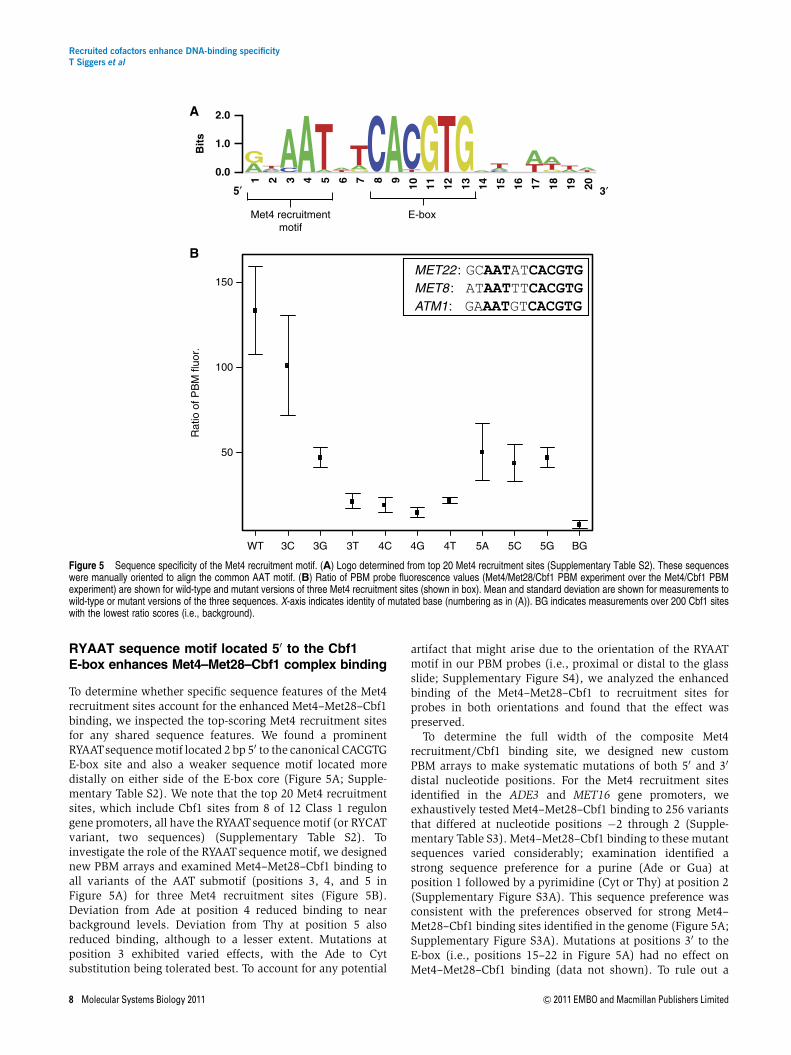
To determine whether specific sequence features of the Met4recruitment sites account for the enhanced Met4–Met28–Cbf1binding, we inspected the top-scoring Met4 recruitment sitesfor any shared sequence features. We found a prominentRYAATsequence motif located 2 bp 50 to the canonical CACGTGE-box site and also a weaker sequence motif located moredistally on either side of the E-box core (Figure 5A; Supple-mentary Table S2). We note that the top 20 Met4 recruitmentsites, which include Cbf1 sites from 8 of 12 Class 1 regulongene promoters, all have the RYAATsequence motif (or RYCATvariant, two sequences) (Supplementary Table S2). Toinvestigate the role of the RYAATsequence motif, we designednew PBM arrays and examined Met4–Met28–Cbf1 binding toall variants of the AAT submotif (positions 3, 4, and 5 inFigure 5A) for three Met4 recruitment sites (Figure 5B).Deviation from Ade at position 4 reduced binding to nearbackground levels. Deviation from Thy at position 5 alsoreduced binding, although to a lesser extent. Mutations atposition 3 exhibited varied effects, with the Ade to Cytsubstitution being tolerated best. To account for any potential
artifact that might arise due to the orientation of the RYAATmotif in our PBM probes (i.e., proximal or distal to the glassslide; Supplementary Figure S4), we analyzed the enhancedbinding of the Met4–Met28–Cbf1 to recruitment sites forprobes in both orientations and found that the effect waspreserved.
To determine the full width of the composite Met4recruitment/Cbf1 binding site, we designed new customPBM arrays to make systematic mutations of both 50 and 30
distal nucleotide positions. For the Met4 recruitment sitesidentified in the ADE3 and MET16 gene promoters, weexhaustively tested Met4–Met28–Cbf1 binding to 256 variantsthat differed at nucleotide positions �2 through 2 (Supple-mentary Table S3). Met4–Met28–Cbf1 binding to these mutantsequences varied considerably; examination identified astrong sequence preference for a purine (Ade or Gua) atposition 1 followed by a pyrimidine (Cyt or Thy) at position 2(Supplementary Figure S3A). This sequence preference wasconsistent with the preferences observed for strong Met4–Met28–Cbf1 binding sites identified in the genome (Figure 5A;Supplementary Figure S3A). Mutations at positions 30 to theE-box (i.e., positions 15–22 in Figure 5A) had no effect onMet4–Met28–Cbf1 binding (data not shown). To rule out a
A
B1 2 3 4 5 6 7 8 9
10 11 12 13 14 15 16 17 18 19 20
0.0
1.0
2.0
Bits
5′ 3′
Met4 recruitmentmotif
E-box
MET22 : GCAATATCACGTGMET8 : ATAATTTCACGTGATM1: GAAATGTCACGTG
Rat
io o
f PB
M fl
uor.
3GWT 3C 3T 4C 4G 4T 5A 5C 5G BG
50
100
150
Figure 5 Sequence specificity of the Met4 recruitment motif. (A) Logo determined from top 20 Met4 recruitment sites (Supplementary Table S2). These sequenceswere manually oriented to align the common AAT motif. (B) Ratio of PBM probe fluorescence values (Met4/Met28/Cbf1 PBM experiment over the Met4/Cbf1 PBMexperiment) are shown for wild-type and mutant versions of three Met4 recruitment sites (shown in box). Mean and standard deviation are shown for measurements towild-type or mutant versions of the three sequences. X-axis indicates identity of mutated base (numbering as in (A)). BG indicates measurements over 200 Cbf1 siteswith the lowest ratio scores (i.e., background).
Recruited cofactors enhance DNA-binding specificityT Siggers et al
8 Molecular Systems Biology 2011 & 2011 EMBO and Macmillan Publishers Limited

sequence preference at more distal positions, we re-examinedMet4–Met28–Cbf1 binding to the 673 Cbf1 sites in the presenceof an additional 5 bp of the genomic flanking sequence oneither site (positions �5 to 25 in Figure 5A). We observed nosignificant difference in Met4–Met28–Cbf1 binding (data notshown) and a binding motif constructed from the top 20‘extended flank’ recruitment sites showed no additionalsequence preference beyond the RYAAT motif (SupplementaryFigure S3A). These results demonstrate that enhanced Met4recruitment in vitro by the Met4–Met28–Cbf1 complex isdependent on the 5-bp Met4 recruitment motif RYAAT(positions 1–5 in Figure 5A) located 50 to the E-box motif.
The RYAAT sequence motif must occur at a fixeddistance from the Cbf1 E-box to enhanceMet4–Met28–Cbf1 complex binding
Given the conserved spacing of the Met4 recruitment motifrelative to the E-box in the genomic sequences, we tested theimportance of the spacing between these two motifs forenhanced Met4–Met28–Cbf1 binding. For the Met4 recruit-ment sites in the ADE3 and MET16 promoters, we system-atically varied the spacing of the Met4 recruitment motifrelative to the E-box from 0 bp (i.e., ACAATCACGTG) to 2 bp(i.e., ACAATNNCACGTG, 16 variants) and examined the effecton Met4–Met28–Cbf1 binding (Supplementary Table S3;Supplementary Figure S3B). Binding was reduced to nearbackground levels for all spacing variants except for the native2-bp spacing, suggesting a strict requirement for exact 2 bpspacing between the AATof the Met4 recruitment motif and theCbf1 E-box motif for enhanced Met4–Met28–Cbf1 binding.Therefore, the Met4 recruitment motif is a highly specificcomposite binding motif with strong spacing and sequencerequirements for functionality.
A second RYAAT sequence motif can furtherenhance Met4–Met28–Cbf1 binding
Motivated by the observation that Cbf1 binds the E-box as ahomodimer (Kuras et al, 1996), we asked whether adding asecond Met4 recruitment motif on the opposite (30) side of theE-box would result in a binding site with even stronger Met4–Met28–Cbf1 binding. We observed that adding a second,symmetrically positioned Met4 recruitment motif significantlyimproves Met4–Met28–Cbf1 binding (Supplementary FigureS3B). Furthermore, as Met28 concentration increases, the PBMsignal is enhanced more greatly for sites with a secondrecruitment motif than to sites with a single recruitment motif.These results demonstrate that the increased Met4 binding (i.e.,PBM signal) is due to additional Met28 binding (or recruitment)to the second recruitment site and suggests a direct role for Met28in the recognition of the Met4 recruitment motif.
Mutations to the RYAAT motif compromiseinduction of genes in low-sulfur conditions
We examined the contribution of the RYAATrecruitment motifto gene induction under conditions of low-sulfur growth. Yeaststrains were constructed in which wild-type or RYAAT-mutant
versions of the promoter regions from two Class 1 genes,YHR112C and MET14, were inserted upstream of LYS2, whichwe employed here as a reporter gene (Materials and methods;Figure 6A, Supplementary Figure S5). Both YHR112C andMET14 contained high-scoring Met4 recruitment sites (Sup-plementary Table S2). The ability of the wild-type and mutantpromoters to drive LYS2 gene expression was examined underlow-sulfur growth conditions. Mutations to the promoterregions were limited to the RYAAT motif (i.e., RYAAT toRYTTA; see Figure 6A) so as not to perturb Cbf1 binding itself.We observed significant reduction in the promoter activity forRYAAT-mutant versions of the promoters: YHR112C (B2-foldreduced; P¼3.2�10�6) and MET14 (B3-fold reduced;P¼6.6�10�6) (Figure 6B). Many Class 1 gene promoterscontain a moderate affinity Met31/Met32 binding site inaddition to a composite Met4 recruitment site. To examinethe potential dependence on the proximity of Met31/Met32sites, we chose MET14 and YHR112C as examples of promotersin which these sites are proximal to each other (MET14,30 bp; Supplementary Figure S5) or distal (YHR112C, 186 bp;Supplementary Figure S5). While both mutant promotersexhibited considerably reduced activity, some activityremained, which might have resulted from Met4 recruitmentto these moderate affinity Met31/Met32 sites. Our resultsdemonstrate that the RYAAT motif is a bone fide cis-regulatoryelement necessary for the full induction of Class 1 target genesof the Met4–Met28–Cbf1 complex under conditions oflow-sulfur growth.
Met4 recruitment sites specify Cbf1-dependentMet4 regulon genes
The presence of the RYAAT motif next to the Cbf1 binding site,in addition to enhancing Met4–Met28–Cbf1 complex binding,provides a means to functionally distinguish Cbf1 sites withinthe genome. This suggested that Met4 recruitment ability of aCbf1 site (i.e., the presence of an adjacent RYAAT motif) ratherthan Cbf1 binding site affinity may specify the Class 1 geneswithin the genome. To investigate this, we scored genes by theMet4 recruitment strength of Cbf1 sites present in theirpromoters, and compared the regulon genes with the top 500scoring non-regulon genes as was done previously (Figure 3Cand D). Met4 recruitment strength of Cbf1 sites was scoredas in Figure 4G. We found that the Class 1 regulon genes arepredicted strongly by Met4 recruitment strength alone(AUC¼0.84) (Figure 6C). While Class 2 genes do containMet4 recruitment sites (Figure 4G and H), the class as a wholeis not predicted well (AUC¼0.52). Scoring genes based on thepresence of an RYAAT motif adjacent to Cbf1 sites, as a proxyfor Met4 recruitment strength, performed identically (data notshown). These results demonstrate that Met4 recruitmentstrength of Cbf1 sites, rather than Cbf1 binding site affinity, iswhat distinguishes Class 1 regulon genes within the genome.
Discussion
Achieving specificity in transcriptional regulation requiresthat TFs are able to identify specific genomic loci. However,in eukaryotes the degenerate binding of TFs and large
Recruited cofactors enhance DNA-binding specificityT Siggers et al
& 2011 EMBO and Macmillan Publishers Limited Molecular Systems Biology 2011 9

genome sizes means that single binding sites occur toooften to explain the specificity observed for gene transcription(Wunderlich and Mirny, 2009). As a model system, we haveexamined the role of TF binding site affinity and sequence-specific cofactor recruitment in specifying the previouslydescribed Met4 regulon genes (Lee et al, 2010). Our resultssuggest that at least two distinct mechanisms are used toachieve specific recruitment of the Met4 transcriptionalactivator to Met4 regulon gene promoters. For Class 2 andClass 3 Met4 regulon genes (those with expression onlyweakly dependent or independent of Cbf1, respectively),the presence of high-affinity Met31/Met32 binding sites(which represent binding by either Met31 or Met32) providesspecificity and distinguishes these Met4 regulon genesfrom other genes in the genome. Consistent with this, wefound that Met32 can recruit Met4 equally well to any bindingsite; therefore, it is the binding of Met31 or Met32 itselfthat provides the specificity. In contrast, for the stronglyCbf1-dependent (Class 1) regulon genes, the presence ofnovel Met4 recruitment sites that enhance binding by theMet4–Met28–Cbf1 complex provides specificity. We find thatthe ability of Cbf1 sites to be bound by the Met4–Met28–Cbf1complex is considerably more predictive of this gene classthan is Cbf1 binding affinity alone (AUC¼0.84 versus 0.65,Figures 3 and 6, respectively). Furthermore, our demonstra-tion that the recognition of the Met4 recruitment sitesrequires the full trimeric complex provides an explanationfor the observed Cbf1 and Met28 dependence of the Class 1
subset of the Met4 regulon genes: deletions of either Cbf1or Met28 will abrogate the trimeric complex required torecognize the Met4 recruitment sites present in Class 1 genepromoters. These results demonstrate that TF targetingspecificity (Met4 targeting in this system) can be achievedby different mechanisms even within a tightly co-expressed setof genes.
Previous work has described still additional mechanisms forachieving specificity, such as stabilized binding of Met4–Met28–Met32 by proximally bound Cbf1 (Blaiseau andThomas, 1998) and differential reporter gene expression basedon altered spacing of Met31/Met32 and Cbf1 binding sites(Chiang et al, 2006). Future work examining these additionalmechanisms of specificity should lead to an even morecomplete model of transcriptional regulatory control for theMet4 regulon genes.
To investigate the role of DNA-binding affinity, we devel-oped a hybrid SPR-PBM methodology that readily allows themeasurement of absolute binding affinities (Kds) of a TF or TFcomplex to thousands of individual DNA binding sequences.With currently available array densities (e.g., Agilent 1�1 Marray format), this approach could be extended readily tohundreds of thousands of sites. In this study, we applied thisapproach to measure the binding affinities of Met32 and Cbf1to 41300 unique DNA binding sites from the S. cerevisiaegenome. We demonstrate that this approach can provideaccurate affinity measurements, which are in excellentagreement with other published methods (Figure 1D).
A
C
Class 1; AUC=0.84 (P=3.3 ×10⁻⁵)Class 1; AUC=0.52 (P=0.38)Class 3; AUC=0.27 (P=0.98)
Tru
e po
sitiv
e ra
te
0.0 0.2 0.4 0.6 0.8 1.0
0
0.2
0.4
1.0
0.6
0.8
False positive rate
0
2
4
6
8
10
12
14
Exp
ress
ion
fold
cha
nge
P=3.2 ×10⁻⁶
P=6.6 ×10⁻⁶
YHR112C
pYHR11
2C
Mut
. pYHR11
2C
MET14
pM
ET14
Mut
. pM
ET14
B
pYHR112C pYHR112C RYAAT-mutant
ATAATCACACGTGATATTTACACACGTGAT
pMET14pMET14 RYAAT-mutant
CTAATTTCACGTGATCTTTATTCACGTGAT
RYAAT Cbf1 E-box
Figure 6 RYAAT motif is critical to promoter activity and specification of Class 1 regulon genes. (A) Schematic of wild-type and mutant versions of the composite Met4recruitment sites from YHR112C and MET14 gene promoters (pYHR112C and pMET14) cloned upstream of LYS2 reporter gene. (B) Expression fold change, underswitch to low-sulfur growth conditions, for endogenous genes (YHR112C and MET14) and the LYS2 reporter gene driven by wild-type (pYHR112C and pMET14) andRYAAT-mutant (Mut. pYHR112C and Mut. pMET14) gene promoters. (C) ROC analysis for the prediction (identification) of the Met4 regulon genes using the Met4recruitment ‘strength’ of the 673 Cbf1 sites used in our genome-wide affinity analysis. Met4 recruitment strength is the ratio of PBM fluorescence intensities shown inFigure 3G (i.e., ratio of PBM fluorescence intensities for the Met4/Met28/Cbf1 and Met4/Cbf1 PBM experiments). ROC analysis was performed using 500 top-scoringnon-regulon genes as false positives. Wilcoxon–Mann–Whitney U-test was applied to each regulon gene set to calculate significance of the AUC value. Source data isavailable for this figure in the Supplementary information.
Recruited cofactors enhance DNA-binding specificityT Siggers et al
10 Molecular Systems Biology 2011 & 2011 EMBO and Macmillan Publishers Limited

The cooperative assembly of the Met4–Met28–Cbf1 complexon DNA that we report is consistent with results of Kuraset al (1997). Moreover, our results provide an explanationfor the differential binding that they observed in vitro forthe Met4–Met28–Cbf1 complex on E-box sites from theMET16 (ATCATTTCACGTG) and the MET28 (TAAGTCACGTGCACTCAG) gene promoters: the E-box (shown in bold)from the MET16 gene promoter has a Met4 recruitmentmotif adjacent to it (underlined), while the site from theMET28 promoter does not. However, in contrast to theirobservation that Met4–Met28–Cbf1 would not assemble onthe MET28 E-box sequence, we find that there is weaknon-specific stabilization of the Met4–Met28–Cbf1 complexesto all E-box sequences, and that this stabilization correlateswith the DNA-binding affinity of the Cbf1 site (compareFigure 4C and D with Figure 4E). This inconsistencymay be due to the different protein concentrations orexperimental approaches that were employed in our studyversus theirs, or may be due to the different Cbf1 proteinconstructs that were used; we used GST-tagged, full-lengthCbf1, whereas Kuras et al used a 6xHis-tagged, N-terminallytruncated version of Cbf1.
The ability of the Met4 recruitment motif RYAAT (Figure 5A)to enhance the assembly of the Met4–Met28–Cbf1 complexeson E-box sites was unexpected. Cbf1 does not preferentiallybind to sites adjacent to the Met4 recruitment motif, nor do thepairwise complexes of Met4–Cbf1, Met28–Cbf1, or Met28–Met4 (Figure 4B and D; Supplementary Figure S2). Therefore,specific recognition of the Met4 recruitment motif requires allthree proteins to be present in the bound complex. While itremains unclear what part of the Met4–Met28–Cbf1 complexrecognizes the Met4 recruitment motif, we find it unlikely thatsome unknown portion of Cbf1 protein confers the specificrecognition of the RYAAT motif. First, it was previously shownthat the region of Cbf1 N-terminal to the bHLH DNA-bindingdomain (amino acids 1–209) was unnecessary for differentialrecognition of the MET28 and MET16 UAS elements by a Met4–Met28–Cbf1DN complex (Kuras et al, 1997). Second, the Cbf1bHLH DNA-binding domain is itself unlikely to make strongDNA contacts 7 bp from the E-box core, and the B80 amino-
acid long region C-terminal to the bHLH domain does notcontain any known DNA-binding domains.
In contrast, despite exhibiting no intrinsic DNA-bindingability, both Met28 and Met4 contain a bZIP DNA-bindingmotif (Blaiseau and Thomas, 1998). Based on the considera-tions of protein sequence and structure, we propose a model inwhich the Met28 subunit of the Met4–Met28–Cbf1 complexmakes base-specific contacts to select for the Met4 recruitmentmotif. Sequence analysis identified a weak homology betweenthe bZIP regions of Met28 and C/EBPa from mouse (BLASTPE-value¼0.15, see Materials and methods), and a strikingsimilarity between amino-acid residues of Met28 and those ofthe C/EBPa paralog C/EBPb (Figure 7B) that make base-specific contacts with a GCAAT binding sequence in an X-rayco-crystal structure (Tahirov et al, 2002). Furthermore, theGCAAT half-site from the C/EBPb crystal structure itself is aperfect match to the RYAAT Met4 recruitment motif(Figure 7C). We favor a model where the Met28 subunit ofthe Met4–Met28–Cbf1 complex makes base-specific contactsto select for the Met4 recruitment motif. We propose that aplausible configuration for the trimeric complex is one inwhich a Met4/Met28 bZIP heterodimer, dimerizing via leucinezippers, is positioned adjacent to the Cbf1 homodimer(Figure 7A); this configuration would allow for Met28 toadopt a binding orientation analogous to the C/EBPb subunitthat similarly recognizes a GCAAT half-site.
Selective binding of the Met4–Met28–Cbf1 complex to thecomposite (RYAATNNCACGTG) Met4 recruitment site isstrikingly similar to the situation described for the Oct-1–HCF-1–VP16 complex that recognizes the consensus siteTAATGARAT (Babb et al, 2001). In both situations, non-DNA-binding transcriptional activators (Met4 and VP16) arerecruited to DNA by sequence-specific binding TFs (Cbf1 andOct-1, respectively), and this recruitment is facilitated by non-DNA-binding cofactors (Met28 and HCF-1, respectively).Furthermore, in both situations the multi-protein complexselects for binding sites where a ‘recruitment motif’ (RYAATand GARAT, respectively) occurs adjacent to the TF bindingsite motif (CACGTG for Cbf1 and TAAT for Oct-1). The extent towhich this shared mechanism exists beyond these two systems
1 5 10 15 | | | |
Met28 RRRKNTEASQRFRIRKKQKNC/EBPb RRERNNIAVRKSRDKAKMRNC/EBPa RRERNNIAVRKSRDKAKQRN ** -* * - * - * -*
Basic regions
Amino-acid residues making base-specificcontacts in C/EBPb structure
* Identically conserved amino acid
- Conserved as basic amino acid
A BModel: Met4–Met28–Cbf1 bound to Met4 recruitment site
C
GCAATNNCACGTG
Met4recruitment
motif
E-box
Met4
Met28
Cbf1
GTGGCGCAAT
1/2 site 1/2 site
C/EBPb
Figure 7 Proposed model of Met4 recruitment complexes. (A) Diagram of proposed orientation for DNA-binding domains of Met4 (bZIP domain), Met28 (bZIPdomain), and Cbf1 (bHLH domain) in the Met4–Met28–Cbf1 complex bound to a Met4 recruitment site. Met4 recruitment motif is in bold. Direct physical contacts thatexist between the Met4 and/or Met28 subunits and the Cbf1 homodimer are not explicitly indicated. (B) Sequence alignment of basic region from Met28, human C/EBPb,and human C/EBPa. (C) Diagram of C/EBPb bZIP region homodimer bound to DNA. Binding site sequence for C/EBPb complex is from X-ray crystal structure ofC/EBPb complex (PDB code 1H8A) (Tahirov et al, 2002). GCAAT half-site motif is in bold.
Recruited cofactors enhance DNA-binding specificityT Siggers et al
& 2011 EMBO and Macmillan Publishers Limited Molecular Systems Biology 2011 11

remains to be discovered; however, they highlight the need toexamine the DNA-binding specificity of multi-protein com-plexes even when the recruited cofactors are not known tointeract with DNA. A direct role for non-DNA-bindingcofactors in refining the gene targeting of regulatory complexesmight represent a widespread mechanism to achieve greatercomplexity in eukaryotic gene regulation.
Materials and methods
TF cloning and preparation of protein samples
Full-length CBF1, MET32, MET28, and MET4 open reading frames werecloned into Gateway pDEST15 (N-terminal GST tag) and pDEST17 (N-terminal 6xHis tag) expression vectors. GST-Met32, GST-Cbf1, and GST-Met4 were overexpressed in E. coli BL21 (DE3) cells (New EnglandBioLabs) and purified by FPLC (AKTAprime plus) using 1 ml GSTraptFF affinity columns (GE Healthcare). Samples were then concentratedby centrifugation using Amicon Ultra (10 K) filter devices (Millipore)and stored in 10% glycerol at �801C. Protein concentrations werequantified by standard Bradford assay using Coomassie Plus ProteinAssay reagent (Thermo Scientific); stock concentrations of the purifiedproteins were as follows: GST-Met32 (45mM), GST-Cbf1 (43mM), GST-Met4 (5mM). All 6xHis-tagged proteins produced by in-vitro transcrip-tion and translation (IVT) were made using the PURExpress kit (NewEngland BioLabs) from purified plasmids. Western blots wereperformed for each protein to assess quality and to approximateprotein concentration relative to a dilution series of recombinant GSTstandard (Sigma). See Supplementary information for further details.
Genomic binding site identification and PBMprobe construction
We identified potential DNA binding sites in yeast intergenic regions byscanning their sequence with universal PBM data for Cbf1 and Met32(Zhu et al, 2009). We identified all high-scoring ungapped 8-mers(PBM enrichment score 40.48) in the genome and aligned them to10 bp position weight matrices (PWMs) defining the core binding sitemotifs for Cbf1 (GTCACGTGAC) or Met31/Met32 (CTGTGGCGCT) todetermine a common sequence register. The identified genomicsequences constituting the core 10 bp motif plus 5 bp of flankingsequence on each side were incorporated into 60 bp probe sequenceson a new, custom-designed DNA microarray (Figure 2A and B;Supplementary information).
PBM experiments and analysis
PBM experiments were performed using custom-designed oligonucleo-tide arrays (Agilent Technologies, Inc., 8�15 K array platform; seeSupplementary information). Two different custom PBMs were designedand used for this work: design #1 (Agilent Technologies Inc., AMADID#024623) had genomic Cbf1 and Met32 binding sites (Figures 1, 2 and 4;Supplementary Figures S1, S2 and S4); design #2 (Agilent TechnologiesInc., AMADID #028293) had mutant versions of Met4 recruitment sites(Figure 5; Supplementary Figure S3; Supplementary Table S3). For PBMexperiments used in the hybrid SPR-PBM approach to determine bindingaffinities, GST-tagged protein (Met32 or Cbf1) was applied at eightdifferent concentrations on a single design #1 array (SupplementaryTable S1). For PBM experiments assessing Met4 recruitment (Figure 4),protein samples were applied at the concentrations indicated inSupplementary Table S4. PBM DNA probe sequences are provided inSupplementary File 1. Full PBM data and hybrid SPR-PBM determined Kd
values are provided (Supplementary Tables S6 and S7).
SPR experiments
SPR was performed on a Biacore 3000 instrument. Biotinylatedoligonucleotides were immobilized onto a Sensor Chip SA (Biacore).
Serial concentrations of protein sample were diluted into a runningbuffer (10 mM Tris–HCl, pH 7.4; 3 mM dithiothreitol (DTT); 0.2 mMEDTA, 0.02% Triton X-100; 120 mM NaCl; 10% glycerol; 0.2 mm filteredand de-gassed) and applied to the Sensor Chip at 25ml/min (KINJECToption: 250ml samples/150 s dissociation phase). Binding constants(Kd values) were determined using Scrubber2 software (BioLogicSoftware). Probes sequences and Kd values are provided (Supplemen-tary Table S5).
Generating yeast strains
Wild-type and RYAAT-mutant promoter constructs were insertedupstream of the native LYS2 gene in the S. cerevisiae genome (yMT-2450 strain; Lee et al, 2010) (Supplementary Figure S5A; Supplemen-tary information). The inserted promoter constructs displace thenative LYS2 promoter (i.e., in the 50 direction relative to the gene) anddo not remove it. Wild-type and mutant promoter regions for YHR112Cand MET14 (Figure 6A; Supplementary Figure S5B) were constructedby gene synthesis (GenScript). The high-efficiency transformationprotocol of Gietz and Woods (2002) was used for all transformations.
Gene expression experiments
Gene expression was examined under conditions of low-sulfur growthas described in Lee et al (2010) (see Supplementary information).Expression was measured in log-phase growth in minimal B-mediawith 0.5 mM methionine as sole sulfur source (t¼0) and 2 h afterswitching to minimal B-media lacking a sulfur source (t¼2 h).Expression was monitored by quantitative PCR (qPCR) for bothwild-type and RYAAT-mutant promoter strains. All measurementswere performed in biological triplicate (i.e., three independentinduction experiments) and technical triplicate (i.e., three indepen-dent PCRs).
Biophysical modeling
Met4 recruitment was modeled using an equilibrium thermodynamicmodel (Bintu et al, 2005; Rowan et al, 2010). Gene activation ismodeled as the probability of Met4 being bound at a promoter region.The model was parameterized using our PBM-determined protein-DNA binding affinities (Cbf1 and Met32) and site-specific Met4recruitment data. The model was implemented in Perl. See Supple-mentary information for full details.
Sequence analysis
Protein similarity searches for Met28 and Met4 bZIP regions (Met28a.a. 91–160; Met4 a.a. 581–640) were performed by blastp search fromthe NCBI BLAST website (http://blast.ncbi.nlm.nih.gov/Blast.cgi)against the non-redundant protein database.
Supplementary information
Supplementary information is available at the Molecular SystemsBiology website (www.nature.com/msb).
AcknowledgementsWe thank Mike Berger for technical assistance and advice with PBMsand data analysis; Traci Lee for technical assistance and generouslyproviding the yMT-2450 S. cerevisiae strain; Dan Spatt for technicalassistance; Fred Winston for the generous gift of pRS plasmids; SteveGisselbrecht, Leila Shokri, Raluca Gordan, and Jaime Chapoy forhelpful discussions; Kevin Struhl for helpful comments on themanuscript. This work was funded by grant # R01 HG003985 fromthe National Institutes of Health/National Human Genome ResearchInstitute to MLB, the i2b2/HST Summer Institute in Bioinformatics andIntegrative Genomics NIH grant #U54 LM008748, and in part by an
Recruited cofactors enhance DNA-binding specificityT Siggers et al
12 Molecular Systems Biology 2011 & 2011 EMBO and Macmillan Publishers Limited

NSF Postdoctoral Fellowship in Biological Informatics (#630639) to TS.Author contributions: TS designed all experiments and performed all
analyses, generated expression plasmids for Met4, expressed andpurified proteins, performed the PBM and SPR experiments, generatedreporter yeast strains and performed gene expression experiments.MHD and SK assisted with protein expression and purification. JRgenerated expression plasmids for Met28, Cbf1, and Met32. MLBsupervised the research. TS and MLB wrote the paper.
Conflict of interestThe authors declare that they have no conflict of interest.
References
Babb R, Huang CC, Aufiero DJ, Herr W (2001) DNA recognition by theherpes simplex virus transactivator VP16: a novel DNA-bindingstructure. Mol Cell Biol 21: 4700–4712
Badis G, Berger MF, Philippakis AA, Talukder S, Gehrke AR, Jaeger SA,Chan ET, Metzler G, Vedenko A, Chen X, Kuznetsov H, Wang CF,Coburn D, Newburger DE, Morris Q, Hughes TR, Bulyk ML (2009)Diversity and complexity in DNA recognition by transcriptionfactors. Science 324: 1720–1723
Badis G, Chan ET, van Bakel H, Pena-Castillo L, Tillo D, Tsui K, CarlsonCD, Gossett AJ, Hasinoff MJ, Warren CL, Gebbia M, Talukder S,Yang A, Mnaimneh S, Terterov D, Coburn D, Li Yeo A, Yeo ZX,Clarke ND, Lieb JD et al (2008) A library of yeast transcriptionfactor motifs reveals a widespread function for Rsc3 in targetingnucleosome exclusion at promoters. Mol Cell 32: 878–887
Berger MF, Bulyk ML (2006a) Protein binding microarrays (PBMs)for rapid, high-throughput characterization of the sequencespecificities of DNA binding proteins. Methods Mol Biol 338:245–260
Berger MF, Philippakis AA, Qureshi AM, He FS, Estep III PW, Bulyk ML(2006b) Compact, universal DNA microarrays to comprehensivelydetermine transcription-factor binding site specificities. NatBiotechnol 24: 1429–1435
Berman BP, Nibu Y, Pfeiffer BD, Tomancak P, Celniker SE, Levine M,Rubin GM, Eisen MB (2002) Exploiting transcription factor bindingsite clustering to identify cis-regulatory modules involved inpattern formation in the Drosophila genome. Proc Natl Acad SciUSA 99: 757–762
Bintu L, Buchler NE, Garcia HG, Gerland U, Hwa T, Kondev J, Phillips R(2005) Transcriptional regulation by the numbers: models. CurrOpin Genet Dev 15: 116–124
Blaiseau PL, Isnard AD, Surdin-Kerjan Y, Thomas D (1997) Met31p andMet32p, two related zinc finger proteins, are involved intranscriptional regulation of yeast sulfur amino acid metabolism.Mol Cell Biol 17: 3640–3648
Blaiseau PL, Thomas D (1998) Multiple transcriptional activationcomplexes tether the yeast activator Met4 to DNA. EMBO J 17:6327–6336
Bulyk ML, Huang X, Choo Y, Church GM (2001) Exploring the DNA-binding specificities of zinc fingers with DNA microarrays. ProcNatl Acad Sci USA 98: 7158–7163
Carey M (1998) The enhanceosome and transcriptional synergy. Cell92: 5–8
Chiang DY, Nix DA, Shultzaberger RK, Gasch AP, Eisen MB (2006)Flexible promoter architecture requirements for coactivatorrecruitment. BMC Mol Biol 7: 16
Dilworth FJ, Chambon P (2001) Nuclear receptors coordinate theactivities of chromatin remodeling complexes and coactivators tofacilitate initiation of transcription. Oncogene 20: 3047–3054
Frith MC, Spouge JL, Hansen U, Weng Z (2002) Statistical significanceof clusters of motifs represented by position specific scoringmatrices in nucleotide sequences. Nucleic Acids Res 30: 3214–3224
Gaudet J, Mango SE (2002) Regulation of organogenesis by theCaenorhabditis elegans FoxA protein PHA-4. Science 295: 821–825
Gietz RD, Woods RA (2002) Transformation of yeast by lithiumacetate/single-stranded carrier DNA/polyethylene glycol method.Methods Enzymol 350: 87–96
Giorgetti L, Siggers T, Tiana G, Caprara G, Notarbartolo S, Corona T,Pasparakis M, Milani P, Bulyk ML, Natoli G (2010) Noncooperativeinteractions between transcription factors and clustered DNAbinding sites enable graded transcriptional responses toenvironmental inputs. Mol Cell 37: 418–428
Harbison CT, Gordon DB, Lee TI, Rinaldi NJ, MacIsaac KD, DanfordTW, Hannett NM, Tagne JB, Reynolds DB, Yoo J, Jennings EG,Zeitlinger J, Pokholok DK, Kellis M, Rolfe PA, Takusagawa KT,Lander ES, Gifford DK, Fraenkel E, Young RA (2004)Transcriptional regulatory code of a eukaryotic genome. Nature431: 99–104
Jiang J, Levine M (1993) Binding affinities and cooperativeinteractions with bHLH activators delimit threshold responses tothe dorsal gradient morphogen. Cell 72: 741–752
Jones RB, Gordus A, Krall JA, MacBeath G (2006) A quantitativeprotein interaction network for the ErbB receptors using proteinmicroarrays. Nature 439: 168–174
Joshi R, Passner JM, Rohs R, Jain R, Sosinsky A, Crickmore MA, JacobV, Aggarwal AK, Honig B, Mann RS (2007) Functional specificity ofa Hox protein mediated by the recognition of minor groovestructure. Cell 131: 530–543
Kuras L, Barbey R, Thomas D (1997) Assembly of a bZIP-bHLHtranscription activation complex: formation of the yeast Cbf1-Met4-Met28 complex is regulated through Met28 stimulation of Cbf1DNA binding. EMBO J 16: 2441–2451
Kuras L, Cherest H, Surdin-Kerjan Y, Thomas D (1996) A heteromericcomplex containing the centromere binding factor 1 and two basicleucine zipper factors, Met4 and Met28, mediates the transcriptionactivation of yeast sulfur metabolism. EMBO J 15: 2519–2529
Lee TA, Jorgensen P, Bognar AL, Peyraud C, Thomas D, Tyers M (2010)Dissection of combinatorial control by the Met4 transcriptionalcomplex. Mol Biol Cell 21: 456–469
MacIsaac KD, Gordon DB, Nekludova L, Odom DT, Schreiber J, GiffordDK, Young RA, Fraenkel E (2006) A hypothesis-based approach foridentifying the binding specificity of regulatory proteinsfrom chromatin immunoprecipitation data. Bioinformatics 22:423–429
Maerkl SJ, Quake SR (2007) A systems approach to measuring thebinding energy landscapes of transcription factors. Science 315:233–237
Mann RS, Lelli KM, Joshi R (2009) Hox specificity unique roles forcofactors and collaborators. Curr Top Dev Biol 88: 63–101
Markstein M, Markstein P, Markstein V, Levine MS (2002) Genome-wide analysis of clustered Dorsal binding sites identifies putativetarget genes in the Drosophila embryo. Proc Natl Acad Sci USA 99:763–768
Merika M, Thanos D (2001) Enhanceosomes. Curr Opin Genet Dev 11:205–208
Mukherjee S, Berger MF, Jona G, Wang XS, Muzzey D, Snyder M,Young RA, Bulyk ML (2004) Rapid analysis of the DNA-bindingspecificities of transcription factors with DNA microarrays. NatGenet 36: 1331–1339
Pramila T, Miles S, GuhaThakurta D, Jemiolo D, Breeden LL (2002)Conserved homeodomain proteins interact with MADS box proteinMcm1 to restrict ECB-dependent transcription to the M/G1 phase ofthe cell cycle. Genes Dev 16: 3034–3045
Rowan S, Siggers T, Lachke SA, Yue Y, Bulyk ML, Maas RL (2010)Precise temporal control of the eye regulatory gene Pax6 viaenhancer-binding site affinity. Genes Dev 24: 980–985
Stein B, Baldwin Jr AS (1993) Distinct mechanisms for regulation ofthe interleukin-8 gene involve synergism and cooperativitybetween C/EBP and NF-kappa B. Mol Cell Biol 13: 7191–7198
Struhl K (2005) Transcriptional activation: mediator can act afterpreinitiation complex formation. Mol Cell 17: 752–754
Recruited cofactors enhance DNA-binding specificityT Siggers et al
& 2011 EMBO and Macmillan Publishers Limited Molecular Systems Biology 2011 13
Tahirov TH, Sato K, Ichikawa-Iwata E, Sasaki M, Inoue-Bungo T,Shiina M, Kimura K, Takata S, Fujikawa A, Morii H, Kumasaka T,Yamamoto M, Ishii S, Ogata K (2002) Mechanism of c-Myb-C/EBPbeta cooperation from separated sites on a promoter. Cell 108: 57–70
Wunderlich Z, Mirny LA (2009) Different gene regulation strategiesrevealed by analysis of binding motifs. Trends Genet 25: 434–440
Zhu C, Byers KJ, McCord RP, Shi Z, Berger MF, Newburger DE,Saulrieta K, Smith Z, Shah MV, Radhakrishnan M, Philippakis AA,Hu Y, De Masi F, Pacek M, Rolfs A, Murthy T, Labaer J, Bulyk ML
(2009) High-resolution DNA-binding specificity analysis of yeasttranscription factors. Genome Res 19: 556–566
Molecular Systems Biology is an open-access journalpublished by European Molecular Biology Organiza-
tion and Nature Publishing Group. This work is licensed under aCreative Commons Attribution-Noncommercial-No DerivativeWorks 3.0 Unported License.
Recruited cofactors enhance DNA-binding specificityT Siggers et al
14 Molecular Systems Biology 2011 & 2011 EMBO and Macmillan Publishers Limited