Regulation of the DNA Damage Response by p53 Cofactors
10
Regulation of the DNA Damage Response by p53 Cofactors Xiao-Peng Zhang, Feng Liu,* and Wei Wang* National Laboratory of Solid State Microstructures and Department of Physics, Nanjing University, Nanjing, China ABSTRACT The selective expression of p53-targeted genes is central to the p53-mediated DNA damage response. It is affected by multiple factors including posttranslational modifications and cofactors of p53. Here, we proposed an integrated model of the p53 network to characterize how the cellular response is regulated by key cofactors of p53, Hzf and ASPP. We found that the sequential induction of Hzf and ASPP is crucial to a reliable cell-fate decision between survival and death. After DNA damage, activated p53 first induces Hzf, which promotes the expression of p21 to arrest the cell cycle and facilitate DNA repair. The cell recovers to normal proliferation after the damage is repaired. If the damage is beyond repair, Hzf is effectively degraded, and activated E2F1 induces ASPP, which promotes the expression of Bax to trigger apoptosis. Furthermore, inter- rupting the induction of Hzf or ASPP remarkably impairs the cellular function. We also proposed two schemes for the production of the unknown E3 ubiquitin ligase for Hzf degradation: it is induced by either E2F1 or p53. In both schemes, the sufficient degra- dation of Hzf is required for apoptosis induction. These results are in good agreement with experimental observations or are experimentally testable. INTRODUCTION The tumor suppressor p53 has a major role in the cellular response to various stresses including DNA damage (1). In unstressed cells, p53 is kept at a low level by its negative regulator Mdm2 (2). Upon DNA damage, p53 is stabilized and activated to transactivate a large number of genes involved in cell cycle arrest or apoptosis (3). Among the p53-targeted genes, p21 can induce cell cycle arrest, which allows time for DNA repair and promotes cell survival (4), whereas Bax is crucial to p53-dependent apoptosis (5). Although how a cell makes a decision between life and death has been a focus of intensive research, its underlying mechanism is still not completely understood. The selective expression of target genes by p53 controls the cell fate (6). This selectivity is influenced by multiple factors including the protein level, posttranslational modifi- cations, subcellular localization, and cofactors of p53 (7). It was proposed that low levels of p53 can transactivate genes with high-affinity promoters associated with cell cycle arrest, whereas high levels of p53 are able to activate genes with low-affinity promoters involved in apoptosis (8). The posttranslational modifications of p53 such as phosphoryla- tion and acetylation remarkably affect its promoter selec- tivity (9). For example, p53 phosphorylation at Ser 46 stimulates the induction of the proapoptotic gene p53AIP1 in response to genotoxic stress (10). The cofactors of p53 enhance its binding to specific promoters. Among them, ASPP1 and ASPP2, which are collectively referred to as ASPP thereafter, promote apoptosis by recruiting p53 to the promoters of proapoptotic genes such as Bax (11). By contrast, Hzf preferentially promotes the binding of p53 to the promoters of proarrest genes like p21 (12). Interestingly, upon sustained or severe DNA damage, Hzf should be suffi- ciently degraded to allow the activation of proapoptotic genes (12). That is, ASPP and Hzf exert opposite effects on cellular outcome. It is intriguing to explore how they interact in the DNA damage response. A series of theoretical models has been constructed to clarify the mechanism for p53-mediated cell-fate decision after DNA damage. Previously, it was suggested that the decision between life and death is determined by the p53 level in a switchlike manner (13). In that model, activated p53 only triggers apoptosis, and thus it is impossible to distinguish cell cycle arrest from apoptosis, both mediated by p53. Recently, it was proposed that the cell fate is governed by the number of p53 pulses and apoptosis is induced only when the number of p53 pulses exceeds some threshold (14,15). Most recently, we further suggested that whereas the cell fate is determined by p53 pulses, apoptosis is evoked by high levels of p53 (16). This two- phase behavior of p53 may provide a flexible and efficient control mode. The above models focus on the influence of p53 phosphorylation (14–16), whereas little is known about the role of p53 cofactors in the decision-making process. Motivated by the above considerations, we developed an integrated model of the p53 signaling network to explore how the dynamics of Hzf and ASPP affect the DNA damage response. The model simulations indicate that the network can make a reliable decision between survival and death de- pending on the extent of DNA damage. For repairable damage, p53 is primarily activated and shows a moderate level, inducing Hzf to promote the expression of p21. Consequently, the cell undergoes transient cell cycle arrest before the damage is fixed. For irreparable damage, p53 exhibits a two-phase behavior: a moderate level of p53 induces cell cycle arrest in the first phase, whereas a high level of p53 evokes apoptosis in the second phase. The Submitted January 24, 2012, and accepted for publication April 2, 2012. *Correspondence: fl[email protected] or [email protected] Editor: H. Wiley. Ó 2012 by the Biophysical Society 0006-3495/12/05/2251/10 $2.00 doi: 10.1016/j.bpj.2012.04.002 Biophysical Journal Volume 102 May 2012 2251–2260 2251
-
Upload
independent -
Category
Documents
-
view
0 -
download
0
Transcript of Regulation of the DNA Damage Response by p53 Cofactors
Biophysical Journal Volume 102 May 2012 2251–2260 2251
Regulation of the DNA Damage Response by p53 Cofactors
Xiao-Peng Zhang, Feng Liu,* and Wei Wang*National Laboratory of Solid State Microstructures and Department of Physics, Nanjing University, Nanjing, China
ABSTRACT The selective expression of p53-targeted genes is central to the p53-mediated DNA damage response. It isaffected by multiple factors including posttranslational modifications and cofactors of p53. Here, we proposed an integratedmodel of the p53 network to characterize how the cellular response is regulated by key cofactors of p53, Hzf and ASPP. Wefound that the sequential induction of Hzf and ASPP is crucial to a reliable cell-fate decision between survival and death. AfterDNA damage, activated p53 first induces Hzf, which promotes the expression of p21 to arrest the cell cycle and facilitate DNArepair. The cell recovers to normal proliferation after the damage is repaired. If the damage is beyond repair, Hzf is effectivelydegraded, and activated E2F1 induces ASPP, which promotes the expression of Bax to trigger apoptosis. Furthermore, inter-rupting the induction of Hzf or ASPP remarkably impairs the cellular function. We also proposed two schemes for the productionof the unknown E3 ubiquitin ligase for Hzf degradation: it is induced by either E2F1 or p53. In both schemes, the sufficient degra-dation of Hzf is required for apoptosis induction. These results are in good agreement with experimental observations or areexperimentally testable.
INTRODUCTION
The tumor suppressor p53 has a major role in the cellularresponse to various stresses including DNA damage (1). Inunstressed cells, p53 is kept at a low level by its negativeregulator Mdm2 (2). Upon DNA damage, p53 is stabilizedand activated to transactivate a large number of genesinvolved in cell cycle arrest or apoptosis (3). Among thep53-targeted genes, p21 can induce cell cycle arrest, whichallows time for DNA repair and promotes cell survival (4),whereas Bax is crucial to p53-dependent apoptosis (5).Although how a cell makes a decision between life anddeath has been a focus of intensive research, its underlyingmechanism is still not completely understood.
The selective expression of target genes by p53 controlsthe cell fate (6). This selectivity is influenced by multiplefactors including the protein level, posttranslational modifi-cations, subcellular localization, and cofactors of p53 (7). Itwas proposed that low levels of p53 can transactivate geneswith high-affinity promoters associated with cell cyclearrest, whereas high levels of p53 are able to activate geneswith low-affinity promoters involved in apoptosis (8). Theposttranslational modifications of p53 such as phosphoryla-tion and acetylation remarkably affect its promoter selec-tivity (9). For example, p53 phosphorylation at Ser46
stimulates the induction of the proapoptotic gene p53AIP1in response to genotoxic stress (10). The cofactors of p53enhance its binding to specific promoters. Among them,ASPP1 and ASPP2, which are collectively referred to asASPP thereafter, promote apoptosis by recruiting p53 tothe promoters of proapoptotic genes such as Bax (11). Bycontrast, Hzf preferentially promotes the binding of p53 tothe promoters of proarrest genes like p21 (12). Interestingly,
Submitted January 24, 2012, and accepted for publication April 2, 2012.
*Correspondence: [email protected] or [email protected]
Editor: H. Wiley.
� 2012 by the Biophysical Society
0006-3495/12/05/2251/10 $2.00
upon sustained or severe DNA damage, Hzf should be suffi-ciently degraded to allow the activation of proapoptoticgenes (12). That is, ASPP and Hzf exert opposite effectson cellular outcome. It is intriguing to explore how theyinteract in the DNA damage response.
A series of theoretical models has been constructed toclarify the mechanism for p53-mediated cell-fate decisionafter DNA damage. Previously, it was suggested that thedecision between life and death is determined by the p53level in a switchlike manner (13). In that model, activatedp53 only triggers apoptosis, and thus it is impossible todistinguish cell cycle arrest from apoptosis, both mediatedby p53. Recently, it was proposed that the cell fate isgoverned by the number of p53 pulses and apoptosis isinduced only when the number of p53 pulses exceedssome threshold (14,15). Most recently, we further suggestedthat whereas the cell fate is determined by p53 pulses,apoptosis is evoked by high levels of p53 (16). This two-phase behavior of p53 may provide a flexible and efficientcontrol mode. The above models focus on the influence ofp53 phosphorylation (14–16), whereas little is known aboutthe role of p53 cofactors in the decision-making process.
Motivated by the above considerations, we developed anintegrated model of the p53 signaling network to explorehow the dynamics of Hzf and ASPP affect the DNA damageresponse. The model simulations indicate that the networkcan make a reliable decision between survival and death de-pending on the extent of DNA damage. For repairabledamage, p53 is primarily activated and shows a moderatelevel, inducing Hzf to promote the expression of p21.Consequently, the cell undergoes transient cell cycle arrestbefore the damage is fixed. For irreparable damage, p53exhibits a two-phase behavior: a moderate level of p53induces cell cycle arrest in the first phase, whereas a highlevel of p53 evokes apoptosis in the second phase. The
doi: 10.1016/j.bpj.2012.04.002
2252 Zhang et al.
sufficient degradation of Hzf is required for the activation ofE2F1 and ASPP, which cooperates with p53 to induce Bax.Specifically, we proposed two schemes guaranteeing thatHzf is degraded in the presence of severe DNA damage.These results clarify how the cofactors of p53 are orches-trated to regulate the cell-fate decision.
MODEL AND METHODS
Model
We constructed an integrated model to explore the response of the p53
network to DNA damage caused by ionizing radiation (IR) (see Fig. 1).
Compared with our previous models (15,16), the model network presented
in this article is still composed of four modules: a DNA repair module,
a sensor of DNA damage, a p53-centered feedback control module, and a
cell-fate decision module. But there are four large differences: 1), activated
p53p
p21
Hzf
Ligase
E2F1 ASPP
Bax
G1 arrest Apoptosis
PTEN
PIP2
PIP3
Aktp
Akt
Mdm2c
Mdm2cpMdm2
n
p53
ATMp
ATMm
ATMd
IR DNA Damage
CytoC
Casp3
"2"
"1"
CycE Rb
HIPK2
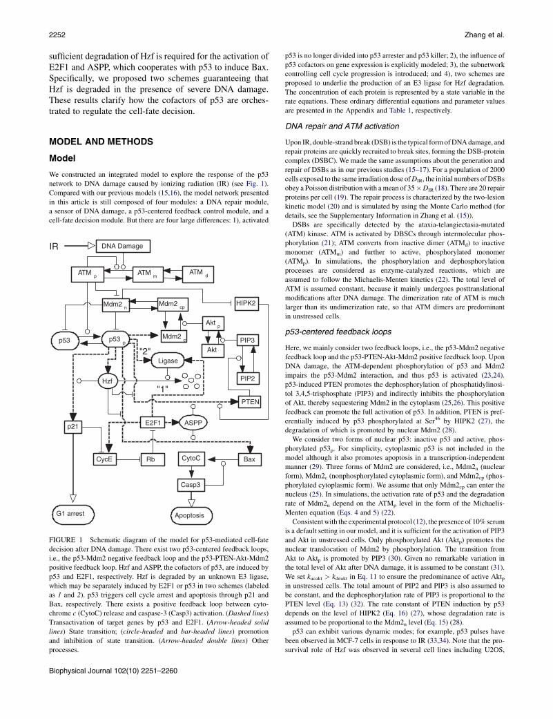
FIGURE 1 Schematic diagram of the model for p53-mediated cell-fate
decision after DNA damage. There exist two p53-centered feedback loops,
i.e., the p53-Mdm2 negative feedback loop and the p53-PTEN-Akt-Mdm2
positive feedback loop. Hzf and ASPP, the cofactors of p53, are induced by
p53 and E2F1, respectively. Hzf is degraded by an unknown E3 ligase,
which may be separately induced by E2F1 or p53 in two schemes (labeled
as 1 and 2). p53 triggers cell cycle arrest and apoptosis through p21 and
Bax, respectively. There exists a positive feedback loop between cyto-
chrome c (CytoC) release and caspase-3 (Casp3) activation. (Dashed lines)
Transactivation of target genes by p53 and E2F1. (Arrow-headed solid
lines) State transition; (circle-headed and bar-headed lines) promotion
and inhibition of state transition. (Arrow-headed double lines) Other
processes.
Biophysical Journal 102(10) 2251–2260
p53 is no longer divided into p53 arrester and p53 killer; 2), the influence of
p53 cofactors on gene expression is explicitly modeled; 3), the subnetwork
controlling cell cycle progression is introduced; and 4), two schemes are
proposed to underlie the production of an E3 ligase for Hzf degradation.
The concentration of each protein is represented by a state variable in the
rate equations. These ordinary differential equations and parameter values
are presented in the Appendix and Table 1, respectively.
DNA repair and ATM activation
Upon IR, double-strand break (DSB) is the typical formofDNAdamage, and
repair proteins are quickly recruited to break sites, forming the DSB-protein
complex (DSBC). We made the same assumptions about the generation and
repair of DSBs as in our previous studies (15–17). For a population of 2000
cells exposed to the same irradiation dose ofDIR, the initial numbers ofDSBs
obey a Poisson distributionwith amean of 35�DIR (18). There are 20 repair
proteins per cell (19). The repair process is characterized by the two-lesion
kinetic model (20) and is simulated by using the Monte Carlo method (for
details, see the Supplementary Information in Zhang et al. (15)).
DSBs are specifically detected by the ataxia-telangiectasia-mutated
(ATM) kinase. ATM is activated by DBSCs through intermolecular phos-
phorylation (21); ATM converts from inactive dimer (ATMd) to inactive
monomer (ATMm) and further to active, phosphorylated monomer
(ATMp). In simulations, the phosphorylation and dephosphorylation
processes are considered as enzyme-catalyzed reactions, which are
assumed to follow the Michaelis-Menten kinetics (22). The total level of
ATM is assumed constant, because it mainly undergoes posttranslational
modifications after DNA damage. The dimerization rate of ATM is much
larger than its undimerization rate, so that ATM dimers are predominant
in unstressed cells.
p53-centered feedback loops
Here, we mainly consider two feedback loops, i.e., the p53-Mdm2 negative
feedback loop and the p53-PTEN-Akt-Mdm2 positive feedback loop. Upon
DNA damage, the ATM-dependent phosphorylation of p53 and Mdm2
impairs the p53-Mdm2 interaction, and thus p53 is activated (23,24).
p53-induced PTEN promotes the dephosphorylation of phosphatidylinosi-
tol 3,4,5-trisphosphate (PIP3) and indirectly inhibits the phosphorylation
of Akt, thereby sequestering Mdm2 in the cytoplasm (25,26). This positive
feedback can promote the full activation of p53. In addition, PTEN is pref-
erentially induced by p53 phosphorylated at Ser46 by HIPK2 (27), the
degradation of which is promoted by nuclear Mdm2 (28).
We consider two forms of nuclear p53: inactive p53 and active, phos-
phorylated p53p. For simplicity, cytoplasmic p53 is not included in the
model although it also promotes apoptosis in a transcription-independent
manner (29). Three forms of Mdm2 are considered, i.e., Mdm2n (nuclear
form), Mdm2c (nonphosphorylated cytoplasmic form), and Mdm2cp (phos-
phorylated cytoplasmic form). We assume that only Mdm2cp can enter the
nucleus (25). In simulations, the activation rate of p53 and the degradation
rate of Mdm2n depend on the ATMp level in the form of the Michaelis-
Menten equation (Eqs. 4 and 5) (22).
Consistentwith the experimental protocol (12), the presence of 10%serum
is a default setting in our model, and it is sufficient for the activation of PIP3
and Akt in unstressed cells. Only phosphorylated Akt (Aktp) promotes the
nuclear translocation of Mdm2 by phosphorylation. The transition from
Akt to Aktp is promoted by PIP3 (30). Given no remarkable variation in
the total level of Akt after DNA damage, it is assumed to be constant (31).
We set kacakt > kdeakt in Eq. 11 to ensure the predominance of active Aktpin unstressed cells. The total amount of PIP2 and PIP3 is also assumed to
be constant, and the dephosphorylation rate of PIP3 is proportional to the
PTEN level (Eq. 13) (32). The rate constant of PTEN induction by p53
depends on the level of HIPK2 (Eq. 16) (27), whose degradation rate is
assumed to be proportional to the Mdm2n level (Eq. 15) (28).
p53 can exhibit various dynamic modes; for example, p53 pulses have
been observed in MCF-7 cells in response to IR (33,34). Note that the pro-
survival role of Hzf was observed in several cell lines including U2OS,
A B
p53 Cofactors Regulate the Cellular Response 2253
LNCaP, Saos2, and HCT116 (12), and no pulses in the p53 level were
reported in most of these cell lines (7). Thus, here we did not mimic oscil-
latory behavior of p53 and ignored the p53-Wip1-ATM negative feedback
loop, which is required for the initiation of p53 pulses (34).
Cell fate decision
The cofactors of p53 have important roles in the choice of cell fate between
survival and death. The prosurvival cofactor Hzf, induced by p53, promotes
the recruitment of p53 to proarrest genes like p21 (12). p21 inhibits the
activity of CDK2 by interaction with Cyclin E (CycE), thereby preventing
the phosphorylation of Rb and activation of E2F1 (4). On the other hand, the
proapoptotic cofactor ASPP, induced by E2F1, contributes to apoptosis
induction (35). Thus, the delicate regulation of expression of Hzf and
ASPP is crucial to the cell-fate decision.
Because the E3 ubiquitin ligase for Hzf degradation (which is denoted by
the term ‘‘Ligase’’ thereafter) is unknown, we propose two schemes
ensuring that the Ligase accumulates remarkably only in the presence of
severe DNA damage (12). In the first scheme, the Ligase is induced by
E2F1. In the second scheme, the p53-induced Ligase accumulates slowly
and becomes marked after a long time. Note that p53 and E2F1 are two
important transcription factors involved in cell-cycle control and cell fate
decision; very possibly, the Ligase is induced by p53 or E2F1, although
other possibilities cannot be excluded.
Here, we assume that the expression of Bax is enhanced by ASPP (11).
Bax promotes the release of cytochrome c (CytoC) from mitochondria
(36). Released CytoC results in the activation of caspase-3 (Casp3), and
activated Casp3 further promotes CytoC release by cleaving its inhibitors
(37). Consequently, apoptosis ensues (38). For simplicity, we did not explic-
itly consider the inhibitors of apoptosis like Bcl-xL (39) and the interme-
diate steps between CytoC release and Casp3 activation, including the
formation of apoptosome and activation of caspase-9 (40).
In simulations, p21 induction is promoted by Hzf, which is characterized
by a scaling of the rate constant of production (Eq. 18). The expression of
p53-mediated proapoptotic genes is inhibited by Hzf but promoted by
ASPP (Eqs. 16 and 29). Thus, PTEN and Bax are induced by p53 only
when Hzf drops to a low level and ASPP accumulates remarkably. Note
that the transactivation of genes by p53 or E2F1 is characterized by the
Hill function, and the Hill coefficient is set to four and two, respectively.
The degradation of Hzf is very slow with a basal rate of kdhzf0, but it is
improved with a large rate of kdhzf (i.e., kdhzf0 << kdhzf in Eq. 17) in the
presence of the Ligase (12). The nonphosphorylated Rb (Rb), rather
than the phosphorylated Rb, inhibits E2F1 activity by association with it
(Eq. 24) (41). Only free CycE, rather than p21-bound CycE, promotes
the phosphorylation of Rb (Eq. 26) (4). The total levels of E2F1 and Rb
are assumed to be constant, respectively (42).
Methods
The ordinary differential equations were numerically solved by using the
Runge-Kutta algorithm with a time step of 0.01 min. Whereas there are
two schemes for the regulation of the E3 ligase for Hzf degradation, most
simulation results were based on the first scheme unless specified other-
wise. The bifurcation diagrams were plotted by using the tool suite Oscill8
(http://oscill8.sourceforge.net/). The units of time and irradiation dose are
minutes and Gray (Gy), respectively, and the units of other parameters
ensure that the concentrations of proteins are dimensionless.
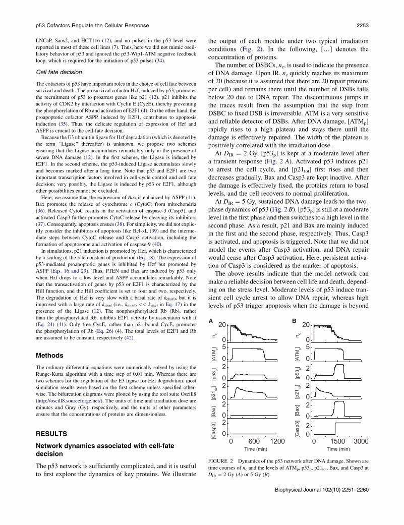
FIGURE 2 Dynamics of the p53 network after DNA damage. Shown are
time courses of nc and the levels of ATMp, p53p, p21tot, Bax, and Casp3 at
DIR ¼ 2 Gy (A) or 5 Gy (B).
RESULTS
Network dynamics associated with cell-fatedecision
The p53 network is sufficiently complicated, and it is usefulto first explore the dynamics of key proteins. We illustrate
the output of each module under two typical irradiationconditions (Fig. 2). In the following, [.] denotes theconcentration of proteins.
The number of DSBCs, nc, is used to indicate the presenceof DNA damage. Upon IR, nc quickly reaches its maximumof 20 (because it is assumed that there are 20 repair proteinsper cell) and remains there until the number of DSBs fallsbelow 20 due to DNA repair. The discontinuous jumps inthe traces result from the assumption that the step fromDSBC to fixed DSB is irreversible. ATM is a very sensitiveand reliable detector of DSBs. After DNA damage, [ATMp]rapidly rises to a high plateau and stays there until thedamage is effectively repaired. The width of the plateau ispositively correlated with the irradiation dose.
At DIR ¼ 2 Gy, [p53p] is kept at a moderate level aftera transient response (Fig. 2 A). Activated p53 induces p21to arrest the cell cycle, and [p21tot] first rises and thendecreases gradually. Bax and Casp3 are kept inactive. Afterthe damage is effectively fixed, the proteins return to basallevels, and the cell recovers to normal proliferation.
At DIR ¼ 5 Gy, sustained DNA damage leads to the two-phase dynamics of p53 (Fig. 2 B). [p53p] is still at a moderatelevel in the first phase and then switches to a high level in thesecond phase. As a result, p21 and Bax are mainly inducedin the first and the second phase, respectively. Thus, Casp3is activated, and apoptosis is triggered. Note that we did notmodel the events after Casp3 activation, and DNA repairwould cease after Casp3 activation. Here, persistent activa-tion of Casp3 is considered as the marker of apoptosis.
The above results indicate that the model network canmake a reliable decision between cell life and death, depend-ing on the stress level. Moderate levels of p53 induce tran-sient cell cycle arrest to allow DNA repair, whereas highlevels of p53 trigger apoptosis when the damage is beyond
Biophysical Journal 102(10) 2251–2260
2254 Zhang et al.
repair. That is, p53 is activated in a progressive manner, andthe cell-fate decision is closely associated with the dynamicsof p53. Essentially, this is an analog mode, in contrast to thedigital mode based on p53 pulses (15,17).
Two-phase dynamics of p53
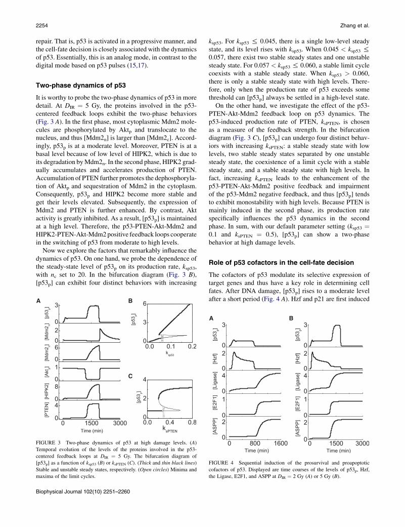
It is worthy to probe the two-phase dynamics of p53 in moredetail. At DIR ¼ 5 Gy, the proteins involved in the p53-centered feedback loops exhibit the two-phase behaviors(Fig. 3 A). In the first phase, most cytoplasmic Mdm2 mole-cules are phosphorylated by Aktp and translocate to thenucleus, and thus [Mdm2n] is larger than [Mdm2c]. Accord-ingly, p53p is at a moderate level. Moreover, PTEN is at abasal level because of low level of HIPK2, which is due toits degradation byMdm2n. In the second phase, HIPK2 grad-ually accumulates and accelerates production of PTEN.Accumulation of PTEN further promotes the dephosphoryla-tion of Aktp and sequestration of Mdm2 in the cytoplasm.Consequently, p53p and HIPK2 become more stable andget their levels elevated. Subsequently, the expression ofMdm2 and PTEN is further enhanced. By contrast, Aktactivity is greatly inhibited. As a result, [p53p] is maintainedat a high level. Therefore, the p53-PTEN-Akt-Mdm2 andHIPK2-PTEN-Akt-Mdm2positive feedback loops cooperatein the switching of p53 from moderate to high levels.
Now we explore the factors that remarkably influence thedynamics of p53. On one hand, we probe the dependence ofthe steady-state level of p53p on its production rate, ksp53,with nc set to 20. In the bifurcation diagram (Fig. 3 B),[p53p] can exhibit four distinct behaviors with increasing
A B
C
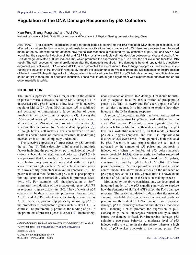
FIGURE 3 Two-phase dynamics of p53 at high damage levels. (A)
Temporal evolution of the levels of the proteins involved in the p53-
centered feedback loops at DIR ¼ 5 Gy. The bifurcation diagram of
[p53p] as a function of ksp53 (B) or ksPTEN (C). (Thick and thin black lines)
Stable and unstable steady states, respectively. (Open circles) Minima and
maxima of the limit cycles.
Biophysical Journal 102(10) 2251–2260
ksp53. For ksp53 % 0.045, there is a single low-level steadystate, and its level rises with ksp53. When 0.045 < ksp53 %0.057, there exist two stable steady states and one unstablesteady state. For 0.057 < ksp53 % 0.060, a stable limit cyclecoexists with a stable steady state. When ksp53 > 0.060,there is only a stable steady state with high levels. There-fore, only when the production rate of p53 exceeds somethreshold can [p53p] always be settled in a high-level state.
On the other hand, we investigate the effect of the p53-PTEN-Akt-Mdm2 feedback loop on p53 dynamics. Thep53-induced production rate of PTEN, ksPTEN, is chosenas a measure of the feedback strength. In the bifurcationdiagram (Fig. 3 C), [p53p] can undergo four distinct behav-iors with increasing ksPTEN: a stable steady state with lowlevels, two stable steady states separated by one unstablesteady state, the coexistence of a limit cycle with a stablesteady state, and a stable steady state with high levels. Infact, increasing ksPTEN leads to the enhancement of thep53-PTEN-Akt-Mdm2 positive feedback and impairmentof the p53-Mdm2 negative feedback, and thus [p53p] tendsto exhibit monostability with high levels. Because PTEN ismainly induced in the second phase, its production ratespecifically influences the p53 dynamics in the secondphase. In sum, with our default parameter setting (ksp53 ¼0.1 and ksPTEN ¼ 0.5), [p53p] can show a two-phasebehavior at high damage levels.
Role of p53 cofactors in the cell-fate decision
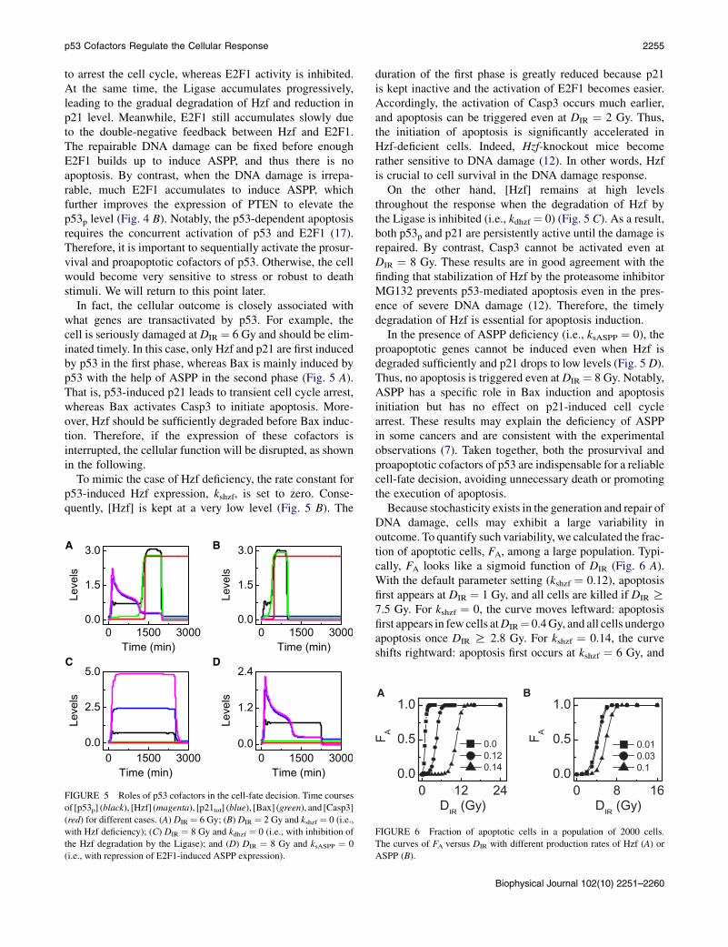
The cofactors of p53 modulate its selective expression oftarget genes and thus have a key role in determining cellfates. After DNA damage, [p53p] rises to a moderate levelafter a short period (Fig. 4 A). Hzf and p21 are first induced
A B
FIGURE 4 Sequential induction of the prosurvival and proapoptotic
cofactors of p53. Displayed are time courses of the levels of p53p, Hzf,
the Ligase, E2F1, and ASPP at DIR ¼ 2 Gy (A) or 5 Gy (B).
p53 Cofactors Regulate the Cellular Response 2255
to arrest the cell cycle, whereas E2F1 activity is inhibited.At the same time, the Ligase accumulates progressively,leading to the gradual degradation of Hzf and reduction inp21 level. Meanwhile, E2F1 still accumulates slowly dueto the double-negative feedback between Hzf and E2F1.The repairable DNA damage can be fixed before enoughE2F1 builds up to induce ASPP, and thus there is noapoptosis. By contrast, when the DNA damage is irrepa-rable, much E2F1 accumulates to induce ASPP, whichfurther improves the expression of PTEN to elevate thep53p level (Fig. 4 B). Notably, the p53-dependent apoptosisrequires the concurrent activation of p53 and E2F1 (17).Therefore, it is important to sequentially activate the prosur-vival and proapoptotic cofactors of p53. Otherwise, the cellwould become very sensitive to stress or robust to deathstimuli. We will return to this point later.
In fact, the cellular outcome is closely associated withwhat genes are transactivated by p53. For example, thecell is seriously damaged at DIR ¼ 6 Gy and should be elim-inated timely. In this case, only Hzf and p21 are first inducedby p53 in the first phase, whereas Bax is mainly induced byp53 with the help of ASPP in the second phase (Fig. 5 A).That is, p53-induced p21 leads to transient cell cycle arrest,whereas Bax activates Casp3 to initiate apoptosis. More-over, Hzf should be sufficiently degraded before Bax induc-tion. Therefore, if the expression of these cofactors isinterrupted, the cellular function will be disrupted, as shownin the following.
To mimic the case of Hzf deficiency, the rate constant forp53-induced Hzf expression, kshzf, is set to zero. Conse-quently, [Hzf] is kept at a very low level (Fig. 5 B). The
A B
C D
FIGURE 5 Roles of p53 cofactors in the cell-fate decision. Time courses
of [p53p] (black), [Hzf] (magenta), [p21tot] (blue), [Bax] (green), and [Casp3]
(red) for different cases. (A) DIR ¼ 6 Gy; (B) DIR ¼ 2 Gy and kshzf ¼ 0 (i.e.,
with Hzf deficiency); (C) DIR ¼ 8 Gy and kdhzf ¼ 0 (i.e., with inhibition of
the Hzf degradation by the Ligase); and (D) DIR ¼ 8 Gy and ksASPP ¼ 0
(i.e., with repression of E2F1-induced ASPP expression).
duration of the first phase is greatly reduced because p21is kept inactive and the activation of E2F1 becomes easier.Accordingly, the activation of Casp3 occurs much earlier,and apoptosis can be triggered even at DIR ¼ 2 Gy. Thus,the initiation of apoptosis is significantly accelerated inHzf-deficient cells. Indeed, Hzf-knockout mice becomerather sensitive to DNA damage (12). In other words, Hzfis crucial to cell survival in the DNA damage response.
On the other hand, [Hzf] remains at high levelsthroughout the response when the degradation of Hzf bythe Ligase is inhibited (i.e., kdhzf ¼ 0) (Fig. 5 C). As a result,both p53p and p21 are persistently active until the damage isrepaired. By contrast, Casp3 cannot be activated even atDIR ¼ 8 Gy. These results are in good agreement with thefinding that stabilization of Hzf by the proteasome inhibitorMG132 prevents p53-mediated apoptosis even in the pres-ence of severe DNA damage (12). Therefore, the timelydegradation of Hzf is essential for apoptosis induction.
In the presence of ASPP deficiency (i.e., ksASPP ¼ 0), theproapoptotic genes cannot be induced even when Hzf isdegraded sufficiently and p21 drops to low levels (Fig. 5 D).Thus, no apoptosis is triggered even at DIR ¼ 8 Gy. Notably,ASPP has a specific role in Bax induction and apoptosisinitiation but has no effect on p21-induced cell cyclearrest. These results may explain the deficiency of ASPPin some cancers and are consistent with the experimentalobservations (7). Taken together, both the prosurvival andproapoptotic cofactors of p53 are indispensable for a reliablecell-fate decision, avoiding unnecessary death or promotingthe execution of apoptosis.
Because stochasticity exists in the generation and repair ofDNA damage, cells may exhibit a large variability inoutcome. To quantify such variability, we calculated the frac-tion of apoptotic cells, FA, among a large population. Typi-cally, FA looks like a sigmoid function of DIR (Fig. 6 A).With the default parameter setting (kshzf ¼ 0.12), apoptosisfirst appears at DIR ¼ 1 Gy, and all cells are killed if DIR R7.5 Gy. For kshzf ¼ 0, the curve moves leftward: apoptosisfirst appears in few cells atDIR¼ 0.4Gy, and all cells undergoapoptosis once DIR R 2.8 Gy. For kshzf ¼ 0.14, the curveshifts rightward: apoptosis first occurs at kshzf ¼ 6 Gy, and
A B
FIGURE 6 Fraction of apoptotic cells in a population of 2000 cells.
The curves of FA versus DIR with different production rates of Hzf (A) or
ASPP (B).
Biophysical Journal 102(10) 2251–2260
2256 Zhang et al.
all cells die when kshzf R 15 Gy. That is, Hzf-deficient cellsbecome rather sensitive to death signals, whereas Hzf-profi-cient cells are resistant to stress. ASPP also affects thecellular outcome evidently. ASPP promotes the inductionof apoptosis, and thus when increasing or decreasing theproduction rate of ASPP, ksASPP, the curve shifts leftwardand rightward, respectively (Fig. 6B). Therefore, controllingthe levels of Hzf and ASPP within appropriate ranges is vitalto a balance between cell life and death.
Robustness of p53 dynamics to parameterfluctuations
We have demonstrated that the degradation of Hzf is criticalto p53-dependent apoptosis under the condition that theLigase for Hzf degradation is induced by E2F1. Here, wetake the second scheme where the Ligase is induced byp53 so slowly that its level becomes high enough to degradeHzf markedly only when the DNA repair is not completedafter a long time.
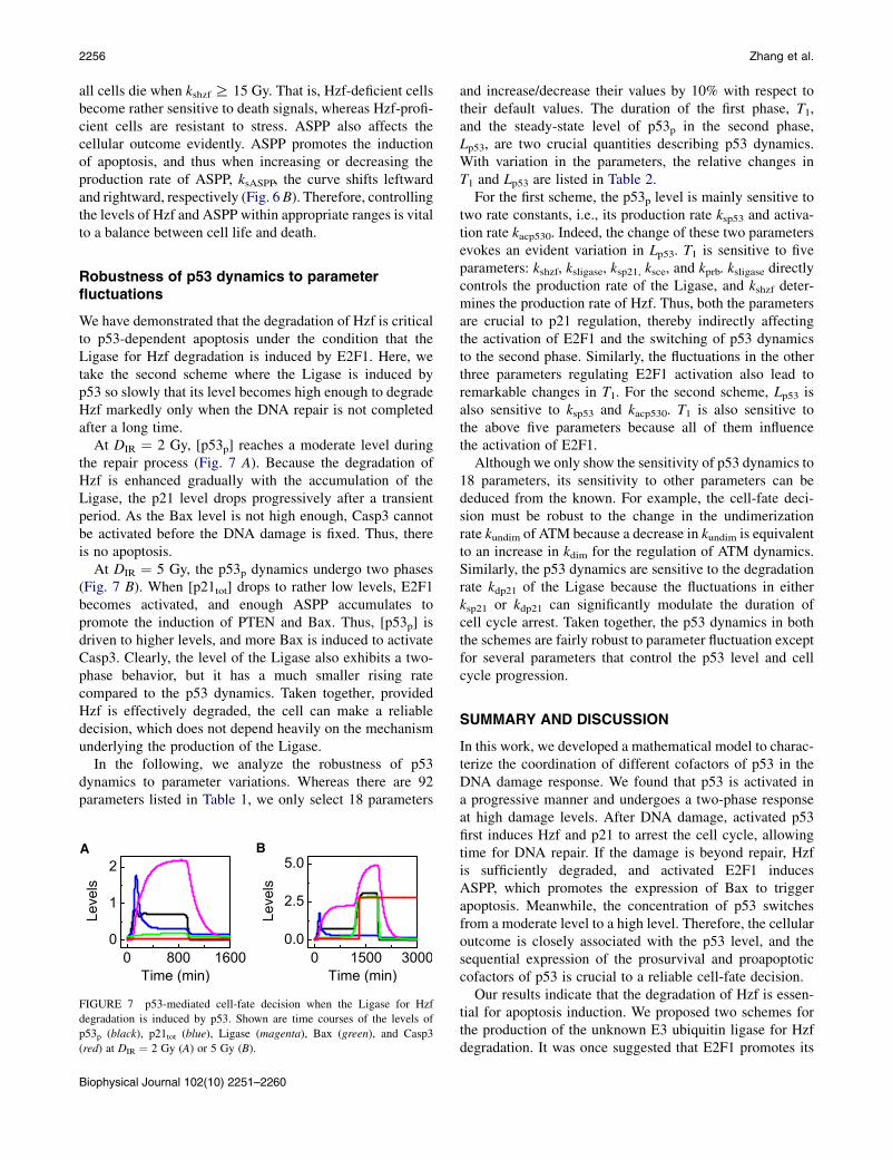
At DIR ¼ 2 Gy, [p53p] reaches a moderate level duringthe repair process (Fig. 7 A). Because the degradation ofHzf is enhanced gradually with the accumulation of theLigase, the p21 level drops progressively after a transientperiod. As the Bax level is not high enough, Casp3 cannotbe activated before the DNA damage is fixed. Thus, thereis no apoptosis.
At DIR ¼ 5 Gy, the p53p dynamics undergo two phases(Fig. 7 B). When [p21tot] drops to rather low levels, E2F1becomes activated, and enough ASPP accumulates topromote the induction of PTEN and Bax. Thus, [p53p] isdriven to higher levels, and more Bax is induced to activateCasp3. Clearly, the level of the Ligase also exhibits a two-phase behavior, but it has a much smaller rising ratecompared to the p53 dynamics. Taken together, providedHzf is effectively degraded, the cell can make a reliabledecision, which does not depend heavily on the mechanismunderlying the production of the Ligase.
In the following, we analyze the robustness of p53dynamics to parameter variations. Whereas there are 92parameters listed in Table 1, we only select 18 parameters
A B
FIGURE 7 p53-mediated cell-fate decision when the Ligase for Hzf
degradation is induced by p53. Shown are time courses of the levels of
p53p (black), p21tot (blue), Ligase (magenta), Bax (green), and Casp3
(red) at DIR ¼ 2 Gy (A) or 5 Gy (B).
Biophysical Journal 102(10) 2251–2260
and increase/decrease their values by 10% with respect totheir default values. The duration of the first phase, T1,and the steady-state level of p53p in the second phase,Lp53, are two crucial quantities describing p53 dynamics.With variation in the parameters, the relative changes inT1 and Lp53 are listed in Table 2.
For the first scheme, the p53p level is mainly sensitive totwo rate constants, i.e., its production rate ksp53 and activa-tion rate kacp530. Indeed, the change of these two parametersevokes an evident variation in Lp53. T1 is sensitive to fiveparameters: kshzf, ksligase, ksp21, ksce, and kprb. ksligase directlycontrols the production rate of the Ligase, and kshzf deter-mines the production rate of Hzf. Thus, both the parametersare crucial to p21 regulation, thereby indirectly affectingthe activation of E2F1 and the switching of p53 dynamicsto the second phase. Similarly, the fluctuations in the otherthree parameters regulating E2F1 activation also lead toremarkable changes in T1. For the second scheme, Lp53 isalso sensitive to ksp53 and kacp530. T1 is also sensitive tothe above five parameters because all of them influencethe activation of E2F1.
Although we only show the sensitivity of p53 dynamics to18 parameters, its sensitivity to other parameters can bededuced from the known. For example, the cell-fate deci-sion must be robust to the change in the undimerizationrate kundim of ATM because a decrease in kundim is equivalentto an increase in kdim for the regulation of ATM dynamics.Similarly, the p53 dynamics are sensitive to the degradationrate kdp21 of the Ligase because the fluctuations in eitherksp21 or kdp21 can significantly modulate the duration ofcell cycle arrest. Taken together, the p53 dynamics in boththe schemes are fairly robust to parameter fluctuation exceptfor several parameters that control the p53 level and cellcycle progression.
SUMMARY AND DISCUSSION
In this work, we developed a mathematical model to charac-terize the coordination of different cofactors of p53 in theDNA damage response. We found that p53 is activated ina progressive manner and undergoes a two-phase responseat high damage levels. After DNA damage, activated p53first induces Hzf and p21 to arrest the cell cycle, allowingtime for DNA repair. If the damage is beyond repair, Hzfis sufficiently degraded, and activated E2F1 inducesASPP, which promotes the expression of Bax to triggerapoptosis. Meanwhile, the concentration of p53 switchesfrom a moderate level to a high level. Therefore, the cellularoutcome is closely associated with the p53 level, and thesequential expression of the prosurvival and proapoptoticcofactors of p53 is crucial to a reliable cell-fate decision.
Our results indicate that the degradation of Hzf is essen-tial for apoptosis induction. We proposed two schemes forthe production of the unknown E3 ubiquitin ligase for Hzfdegradation. It was once suggested that E2F1 promotes its
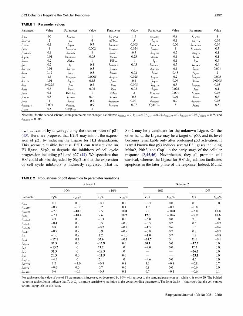
TABLE 1 Parameter values
Parameter Value Parameter Value Parameter Value Parameter Value Parameter Value
kdim 10 kundim 1 kacATM 1.5 kdeATM 0.8 jacATM 1
jdeATM 2 jnc 4 ATMtot 5 ksp53 0.1 kdp53n 0.05
j1p53n 0.1 kdp53 0.7 kdmdm2 0.003 kmdm2in 0.06 kmdm2out 0.09
jATM 1 ksmdm20 0.002 ksmdm2 0.024 jsmdm2 1 k1mdm2s 0.3
j1mdm2s 0.1 kmdm2s 8 jmdm2s 0.3 kacp531 0.2 kdep53 0.1
kdp53p 0.01 kdmdm2n1 0.05 kacakt 0.25 kdeakt 0.1 jacakt 0.1
jdeakt 0.2 Akttot 1 PIPtot 1 kp2 0.1 kp3 0.5
jp2 0.2 jp3 0.4 ksHIPK2 0.05 kdHIPK2 0.5 jHIPK2 0.6
ksPTEN0 0.01 ksPTEN 0.5 jsPTEN 1 kdPTEN 0.1 kshzf0 0.001
kshzf 0.12 jshzf 0.5 kdhzf0 0.02 kdhzf 0.45 jligase 2
jhzf 1.5 ksligase0 0.0005 ksligase 0.025 jsligase 0.2 kdligase 0.005
ksp210 0.01 ksp21 0.15 jsp21 0.1 kdp21 0.06 ksce0 0.0005
ksce 0.0275 jsce 0.2 kdce 0.005 kasp21e 0.5 kdsp21e 0.05
kasre 0.5 kdsre 0.05 kprb 0.05 kdprb 0.025 jprb 0.1
jdprb 0.1 E2F1tot 1 Rbtot 2 ksASPP0 0.001 ksASPP 0.03
jsASPP 0.5 kdASPP 0.01 jASPP 0.5 ksbax0 0.01 ksbax 0.3
jsbax 1 kdbax 0.1 kaccytco0 0.001 kaccytoc 0.9 kdecytoc 0.05
kaccasp30 0.001 kaccasp3 0.9 kdecasp3 0.07 CytoCtot 3 jcytoc 0.5
jcasp3 0.5 Casp3tot 3
Note that, for the second scheme, some parameters are changed as follows: kmdm2s¼ 7, ksce ¼ 0.02, jsce ¼ 0.25, ksligase0¼ 0, ksligase¼ 0.03, jsligase¼ 0.75, and
kdligase ¼ 0.006.
p53 Cofactors Regulate the Cellular Response 2257
own activation by downregulating the transcription of p21(43). Here, we proposed that E2F1 may inhibit the expres-sion of p21 by inducing the Ligase for Hzf degradation.This seems plausible because E2F1 can transactivate anE3 ligase, Skp2, to degrade the inhibitors of cell cycleprogression including p21 and p27 (44). We speculate thatHzf could also be degraded by Skp2 so that the expressionof cell cycle inhibitors is indirectly repressed. That is,
TABLE 2 Robustness of p53 dynamics to parameter variations
Parameter
Scheme 1
�10% þ10%
T1% Lp53% T1% Lp5
kdim 0.1 0.0 �0.1 0.
kacATM �0.7 �0.2 0.2 0.
kacp530 �2.6 �10.0 2.5 10
ksp53 �7.1 �10.7 7.6 10
ksmdm2 4.3 0.0 �3.3 0.
kmdm2s �0.4 0.8 0.5 �0
kmdm2in 0.8 0.7 �0.7 �0
kacakt �0.7 0.9 0.9 �0
kp2 �1.0 0.9 1.2 �1
kshzf �17.1 0.1 33.6 �0
ksligase 55.3 0.0 �17.9 0.
ksp21 �13.2 0 21.2 0
ksce 52.3 0 �18.5 0
kprb 20.3 0.0 �11.5 0.
kasp21e �4.9 0 5.1 0
ksPTEN 1.2 �1.0 �0.8 0.
ksHIPK2 �0.6 0.0 0.7 0.
ksASPP 0.6 �0.1 �0.5 0.
For each case, the value of one of 18 parameters is increased or decreased by 10%
values in each column indicate that T1 or Lp53 is more sensitive to variation in the
commit apoptosis in this case.
Skp2 may be a candidate for the unknown Ligase. On theother hand, the Ligase may be a target of p53, and its levelbecomes remarkable only after prolonged p53 activation. Itis well known that p53 induces several E3 ligases includingMdm2, Pirh2, and Cop1 in the early stage of the cellularresponse (2,45,46). Nevertheless, they all promote cellsurvival, whereas the Ligase for Hzf degradation facilitatesapoptosis in the later phase of the response. Indeed, Mdm2
Scheme 2
�10% þ10%
3% T1% Lp53% T1% Lp53%
0 �0.3 0.0 0.3 0.0
1 1.9 �0.2 �0.8 0.1
.0 5.2 �10.0 �3.8 10.0
.7 17.3 �10.6 �8.9 10.6
0 �6.0 0.0 7.3 0.0
.9 �0.5 0.7 0.5 �0.7
.7 �1.5 0.6 1.3 �0.6
.9 �0.8 0.7 0.8 �0.7
.0 �1.0 0.7 1.2 �0.8
.1 �14.7 0.1 31.0 �0.1
0 30.1 0.0 �12.2 0.0
�9.0 0.0 12.5 0.0
— — �26.2 0.0
0 — — �23.1 0.0
�4.6 0.0 4.6 0.0
8 1.1 �0.8 �0.8 0.7
0 0.8 0.0 �0.6 0.0
1 0.7 �0.1 �0.6 0.1
with respect to the standard parameter set, while nc is set to 20. The bolded
corresponding parameters. The long dash (—) indicates that the cell cannot
Biophysical Journal 102(10) 2251–2260
2258 Zhang et al.
is not the E3 ligase for Hzf degradation (12). Therefore, weprefer to expect that the Ligase is related to E2F1, whichawaits experimental identification.
It is necessary to compare our model with the previousmodels about p53 dynamics: 1), Although a series of modelshas focused on oscillatory dynamics of p53 in the DNAdamage response (14–17,19), we did not formulate p53oscillations here. Typical p53 oscillations were observedin MCF-7 cells in response to IR (33,34). Here, we aimedat revealing the mechanism for cell-fate decision in severalother cell lines that were used in the experiment by Das et al.(12), where no evidence supports oscillations in p53 levels.2), Previously, active p53 was often distinguished betweenp53 arrester and p53 killer, which promote cell cycle arrestand apoptosis, respectively, based on its phosphorylationstatus. Here, we found that cooperating with its cofactors,active p53 automatically fulfills its distinct functions duringdifferent stages of the response. 3), p53 undergoes a two-phase response to severe DNA damage, switching froma low level to a high level. In our previous work, p53 alsoexhibit a two-phase behavior, switching from periodic oscil-lations to high constant levels (16). In both cases, theswitching results from the predominance of the p53-PTEN-Akt-Mdm2 positive feedback. Such a two-phaseresponse mode provides a robust mechanism for the choiceof cell fate between survival and death. In sum, the p53dynamics are diverse, depending on the cell- and stress-type. Nevertheless, some generic mechanisms may be inoperation, controlling the cell fate after DNA damage.
Our work revealed how Hzf and ASPP are coordinated toaffect the cell fate. Hzf is first induced to promote cell cyclearrest by facilitating the expression of p21. Only when DNAdamage is severe or prolonged, Hzf is degraded and E2F1-induced ASPP promotes the expression of p53-targetedproapoptotic genes. Therefore, it is important that the pro-survival and proapoptotic cofactors of p53 are separatelyinduced. Moreover, the concurrent activation of p53 andE2F1 is essential for p53-mediated apoptosis, consistentwith Zhang et al. (17). We also simulated the effects ofknockout of Hzf gene or inhibition of Hzf degradation onthe cellular outcome, and our results show good agreementwith the experimental observations by Das et al. (12).
APPENDIX: RATE EQUATIONS
The rate equations of the model are written as
d½ATMd�dt
¼ 0:5kdim½ATMm�2�kundim½ATMd�; (1)
d�ATMp
�nc � � ½ATMm�
dt¼ kacATM
nc þ jncATMp ½ATMm� þ jacATM
� kdeATM
�ATMp
��ATMp
�þ jdeATM;
(2)
Biophysical Journal 102(10) 2251–2260
½ATMm� ¼ ATMtot � 2½ATMd� ��ATMp
�; (3)
�ATMp
�
kdmdm2n ¼ kdmdm2 þ kdmdm2n1�ATMp
�þ jATM; (4)
�ATMp
�
kacp53 ¼ kacp531�ATMp
�þ jATM; (5)
d�p53p
� � �
dt¼ kacp53½p53� � kdep53 p53p
� kdp53p½Mdm2n��p53p
��p53p
�þ j1p53n;
(6)
d½p53� ½p53�
dt¼ ksp53 � kdp53n½p53� � kdp53½Mdm2n� ½p53� þ j1p53n
� kacp53½p53� þ kdep53�p53p
�; (7)
d½Mdm2 ��p53
�4
cdt¼ ksmdm20 þ ksmdm2
p�p53p
�4þj4smdm2
� kdmdm2½Mdm2c�
þ k1mdm2s
�Mdm2cp
��Mdm2cp
�þ j1mdm2s
� kmdm2s
�Aktp
� ½Mdm2c�½Mdm2c� þ jmdm2s
;
(8)
d�Mdm2cp
� � � ½Mdm2c�
dt¼ kmdm2s Aktp ½Mdm2c� þ jmdm2s
� k1mdm2s
�Mdm2cp
��Mdm2cp
�þ j1mdm2s
� kmdm2in
�Mdm2cp
�þ kmdm2out½Mdm2n�� kdmdm2
�Mdm2cp
�;
(9)
d½Mdm2n� � �
dt¼ kmdm2in Mdm2cp � kmdm2out½Mdm2n�� kdmdm2n½Mdm2n�;
(10)
d�Aktp
�½Akt�
�Aktp
�
dt¼ kacakt½PIP3� ½Akt� þ jacakt� kdeakt�
Aktp�þ jdeakt
;
(11)
½Akt� ¼ Akttot ��Aktp
�; (12)
d½PIP3� ½PIP2� ½PIP3�
dt¼ kp2½PIP2� þ jp2� kp3½PTEN� ½PIP3� þ jp3
; (13)
p53 Cofactors Regulate the Cellular Response 2259
½PIP2� ¼ PIPtot � ½PIP3�; (14)
d½HIPK2�
dt¼ ksHIPK2 � kdHIPK2½Mdm2n�½HIPK2�; (15)
d½PTEN� ½HIPK2�2 j2
dt¼ ksPTEN½HIPK2�2 þ j2HIPK2
hzf
½Hzf�2 þ j2hzf
� ½ASPP�2½ASPP�2 þ j2ASPP
�p53p
�4�p53p
�4 þ j4sPTEN
þ ksPTEN0 � kdPTEN½PTEN�;
(16)
d½Hzf��p53p
�4
dt¼ kshzf0 þ kshzf�p53p
�4 þ j4shzf
� kdhzf0 þ kdhzf
½Ligase�2½Ligase�2 þ j2ligase
!½Hzf�;
(17)
2� �4
d½p21tot�dt
¼ ksp210 þ ksp21½Hzf�
½Hzf�2 þ j2hzf
p53p�p53p
�4 þ j4sp21
� kdp21½p21tot�;(18)
d½Ligase� ½E2F1�2
dt¼ ksligase0 þ ksligase½E2F1�2 þ j2sligase
� kdligase½Ligase�;(19)
½p21� ¼ ½p21tot� � ½p21CE�; (20)
d½CycEtot�dt
¼ ksce0 þ ksce½E2F1�2
½E2F1�2 þ j2sce� kdce½CycEtot�; (21)
½CycE� ¼ ½CycEtot� � ½p21CE�; (22)
d½ASPP� ½E2F1�2
dt¼ ksASPP0 þ ksASPP½E2F1�2 þ j2sASPP� kdASPP½ASPP�;
(23)
d½E2F1�
dt¼ �kasre½Rb�½E2F1� þ kdsre½RE�; (24)
½RE� ¼ E2F1tot � ½E2F1�; (25)
d�Rbp�
dt¼ kprb½CycE� ½Rb�
½Rb� þ jprb� kdprb
�Rbp��
Rbp�þ jdprb
; (26)
½Rb� ¼ Rbtot ��Rbp�� ½RE�; (27)
d½p21CE�
dt¼ kasp21e½p21�½CycE� � kdsp21e½p21CE�; (28)
d½Bax�dt
¼ ksbax0 þ ksbaxj2hzf
½Hzf�2 þ j2hzf
½ASPP�2½ASPP�2 þ j2ASPP
��p53p
�4�p53p
�4 þ j4sbax� kdbax½Bax�;
(29)
4
!
d½CytoC�dt¼ kaccytoc0 þ kaccytoc½Bax� ½Casp3�
½Casp3�4 þ j4casp3
� ðCytoCtot � ½CytoC�Þ � kdecytoc½CytoC�;(30)
4
!
d½Casp3�dt¼ kaccasp30 þ kaccasp3
½Cytc�½Cytc�4 þ j4cytoc
� ðCasp3tot � ½Casp3�Þ � kdecasp3½Casp3�:(31)
Note: For the second scheme, Eq. 19 should be replaced by the equation
d ½Ligase�dt
¼ ksligase0 þ ksligase
�p53p
�4�p53p
�4 þ j4sligase
� kdligase ½Ligase�:
This work was supported by the National Basic Research Program of China
(grant No. 2007CB814806), the National Natural Science Foundation of
China (grant No. 11175084), the Natural Science Foundation of Jiangsu
Province (grant No. BK2009008), the Program for New Century Excellent
Talents in Universities (grant No. NCET-08-0269), and the Priority
Academic Program Development of Jiangsu Higher Education Institutions.
REFERENCES
1. Meek, D. W. 2009. Tumor suppression by p53: a role for the DNAdamage response? Nat. Rev. Cancer. 9:714–723.
2. Wu, X., J. H. Bayle,., A. J. Levine. 1993. The p53-Mdm-2 autoregu-latory feedback loop. Genes Dev. 7:1126–1132.
3. Aylon, Y., and M. Oren. 2007. Living with p53, dying of p53. Cell.130:597–600.
4. Abbas, T., and A. Dutta. 2009. p21 in cancer: intricate networks andmultiple activities. Nat. Rev. Cancer. 9:400–414.
5. Miyashita, T., and J. C. Reed. 1995. Tumor suppressor p53 is a directtranscriptional activator of the human Bax gene. Cell. 80:293–299.
6. Vousden, K. H. 2006. Outcomes of p53 activation—spoilt for choice.J. Cell Sci. 119:5015–5020.
Biophysical Journal 102(10) 2251–2260

2260 Zhang et al.
7. Murray-Zmijewski, F., E. A. Slee, and X. Lu. 2008. A complex barcodeunderlies the heterogeneous response of p53 to stress. Nat. Rev. Mol.Cell Biol. 9:702–712.
8. Vousden, K. H., and D. P. Lane. 2007. p53 in health and disease. Nat.Rev. Mol. Cell Biol. 8:275–283.
9. Olsson, A., C. Manzl,., A. Villunger. 2007. How important are post-translational modifications in p53 for selectivity in target-gene tran-scription and tumor suppression? Cell Death Differ. 14:1561–1575.
10. Oda, K., H. Arakawa,., Y. Taya. 2000. p53AIP1, a potential mediatorof p53-dependent apoptosis, and its regulation by Ser-46-phosphory-lated p53. Cell. 102:849–862.
11. Samuels-Lev, Y., D. J. O’Connor, ., X. Lu. 2001. ASPP proteinsspecifically stimulate the apoptotic function of p53. Mol. Cell.8:781–794.
12. Das, S., L. Raj, ., S. W. Lee. 2007. Hzf determines cell survivalupon genotoxic stress by modulating p53 transactivation. Cell. 130:624–637.
13. Wee, K. B., and B. D. Aguda. 2006. Akt versus p53 in a network ofoncogenes and tumor suppressor genes regulating cell survival anddeath. Biophys. J. 91:857–865.
14. Zhang, T., P. Brazhnik, and J. J. Tyson. 2007. Exploring mechanisms ofthe DNA-damage response: p53 pulses and their possible relevance toapoptosis. Cell Cycle. 6:85–94.
15. Zhang, X. P., F. Liu,., W. Wang. 2009. Cell fate decision mediated byp53 pulses. Proc. Natl. Acad. Sci. USA. 106:12245–12250.
16. Zhang, X. P., F. Liu, and W. Wang. 2011. Two-phase dynamics of p53in the DNA damage response. Proc. Natl. Acad. Sci. USA. 108:8990–8995.
17. Zhang, X. P., F. Liu, and W. Wang. 2010. Coordination between cellcycle progression and cell fate decision by the p53 and E2F1 pathwaysin response to DNA damage. J. Biol. Chem. 285:31571–31580.
18. Rothkamm, K., and M. Lobrich. 2003. Evidence for a lack of DNAdouble-strand break repair in human cells exposed to very low x-raydoses. Proc. Natl. Acad. Sci. USA. 100:5057–5062.
19. Ma, L., J. Wagner, ., G. A. Stolovitzky. 2005. A plausible model forthe digital response of p53 to DNA damage. Proc. Natl. Acad. Sci. USA.102:14266–14271.
20. Stewart, R. D. 2001. Two-lesion kinetic model of double-strand breakrejoining and cell killing. Radiat. Res. 156:365–378.
21. Bakkenist, C. J., and M. B. Kastan. 2003. DNA damage activates ATMthrough intermolecular autophosphorylation and dimer dissociation.Nature. 421:499–506.
22. Kholodenko, B. N. 2006. Cell-signaling dynamics in time and space.Nat. Rev. Mol. Cell Biol. 7:165–176.
23. Canman, C. E., D. S. Lim, ., J. D. Siliciano. 1998. Activation ofthe ATM kinase by ionizing radiation and phosphorylation of p53.Science. 281:1677–1679.
24. Stommel, J. M., and G. M. Wahl. 2004. Accelerated MDM2 auto-degradation induced by DNA-damage kinases is required for p53 acti-vation. EMBO J. 23:1547–1556.
25. Mayo, L. D., and D. B. Donner. 2001. A phosphatidylinositol 3-kinase/Akt pathway promotes translocation of Mdm2 from the cytoplasm tothe nucleus. Proc. Natl. Acad. Sci. USA. 98:11598–11603.
Biophysical Journal 102(10) 2251–2260
26. Mayo, L. D., J. E. Dixon, ., D. B. Donner. 2002. PTEN protects p53fromMdm2 and sensitizes cancer cells to chemotherapy. J. Biol. Chem.277:5484–5489.
27. Mayo, L. D., Y. R. Seo, ., D. B. Donner. 2005. Phosphorylation ofhuman p53 at serine 46 determines promoter selection and whetherapoptosis is attenuated or amplified. J. Biol. Chem. 280:25953–25959.
28. Rinaldo, C., A. Prodosmo,., S. Soddu. 2007. Mdm2-regulated degra-dation of HIPK2 prevents p53Ser46 phosphorylation and DNA damage-induced apoptosis. Mol. Cell. 25:739–750.
29. Green, D. R., and G. Kroemer. 2009. Cytoplasmic functions of thetumor suppressor p53. Nature. 458:1127–1130.
30. Manning, B. D., and L. C. Cantley. 2007. AKT/PKB signaling: navi-gating downstream. Cell. 129:1261–1274.
31. Gottlieb, T. M., J. F. Leal, ., M. Oren. 2002. Cross-talk between Akt,p53 and Mdm2: possible implications for the regulation of apoptosis.Oncogene. 21:1299–1303.
32. Cantley, L. C., and B. G. Neel. 1999. New insights into tumorsuppression: PTEN suppresses tumor formation by restraining thephosphoinositide 3-kinase/AKT pathway. Proc. Natl. Acad. Sci. USA.96:4240–4245.
33. Lahav, G., N. Rosenfeld, ., U. Alon. 2004. Dynamics of the p53-Mdm2 feedback loop in individual cells. Nat. Genet. 36:147–150.
34. Batchelor, E., C. S. Mock, ., G. Lahav. 2008. Recurrent initiation:a mechanism for triggering p53 pulses in response to DNA damage.Mol. Cell. 30:277–289.
35. Fogal, V., N. N. Kartasheva, ., X. Lu. 2005. ASPP1 and ASPP2 arenew transcriptional targets of E2F. Cell Death Differ. 12:369–376.
36. Cory, S., and J. M. Adams. 2002. The Bcl2 family: regulators of thecellular life-or-death switch. Nat. Rev. Cancer. 2:647–656.
37. Kirsch, D. G., A. Doseff, ., J. M. Hardwick. 1999. Caspase-3-depen-dent cleavage of Bcl-2 promotes release of cytochrome c. J. Biol.Chem. 274:21155–21161.
38. Ow, Y. P., D. R. Green, ., T. W. Mak. 2008. Cytochrome c: functionsbeyond respiration. Nat. Rev. Mol. Cell Biol. 9:532–542.
39. Youle, R. J., and A. Strasser. 2008. The BCL-2 protein family:opposing activities that mediate cell death. Nat. Rev. Mol. Cell Biol.9:47–59.
40. Riedl, S. J., and G. S. Salvesen. 2007. The apoptosome: signaling plat-form of cell death. Nat. Rev. Mol. Cell Biol. 8:405–413.
41. Yao, G., T. J. Lee, ., L. You. 2008. A bistable Rb-E2F switch under-lies the restriction point. Nat. Cell Biol. 10:476–482.
42. Zhang, T., P. Brazhnik, and J. J. Tyson. 2009. Computational analysisof dynamical responses to the intrinsic pathway of programmed celldeath. Biophys. J. 97:415–434.
43. Wu, L., C. Timmers, ., G. Leone. 2001. The E2F1-3 transcriptionfactors are essential for cellular proliferation. Nature. 414:457–462.
44. Zhang, L., and C. Wang. 2006. F-box protein Skp2: a novel transcrip-tional target of E2F. Oncogene. 25:2615–2627.
45. Leng, R. P., Y. Lin,., S. Benchimol. 2003. Pirh2, a p53-induced ubiq-uitin-protein ligase, promotes p53 degradation. Cell. 112:779–791.
46. Dornan, D., I. Wertz,., V. M. Dixit. 2004. The ubiquitin ligase COP1is a critical negative regulator of p53. Nature. 429:86–92.