Lavender oil suppresses indoleamine 2,3-dioxygenase activity in human PBMC
Upload
independentCategory
view
0download
0
Chk1 Suppresses a Caspase-2 ApoptoticResponse to DNA Damage that Bypassesp53, Bcl-2, and Caspase-3Samuel Sidi,1 Takaomi Sanda,1 Richard D. Kennedy,2 Andreas T. Hagen,1 Cicely A. Jette,1 Raymond Hoffmans,1
Jennifer Pascual,1 Shintaro Imamura,4 Shuji Kishi,4 James F. Amatruda,5 John P. Kanki,1 Douglas R. Green,6
Alan A. D’Andrea,2 and A. Thomas Look1,3,*1Department of Pediatric Oncology2Division of Genomic Stability and DNA Repair, Department of Radiation OncologyDana-Farber Cancer Institute, Harvard Medical School, Boston, MA 02115, USA
3Division of Hematology/Oncology, Department of Pediatrics, Children’s Hospital Boston, Harvard Medical School, Boston, MA 02115, USA4Department of Ophthalmology, Schepens Eye Research Institute, Harvard Medical School, Boston, MA 02114, USA5Department of Pediatrics, Molecular Biology and Internal Medicine, University of Texas Southwestern Medical Center, Dallas, TX 75390, USA6Department of Immunology, St. Jude Children’s Research Hospital, Memphis, TN 38105, USA
*Correspondence: [email protected]
DOI 10.1016/j.cell.2008.03.037
SUMMARY
Evasion of DNA damage-induced cell death, via mu-tation of the p53 tumor suppressor or overexpressionof prosurvival Bcl-2 family proteins, is a key step to-ward malignant transformation and therapeutic resis-tance. We report that depletion or acute inhibition ofcheckpoint kinase 1 (Chk1) is sufficient to restore g-radiation-induced apoptosis in p53 mutant zebrafishembryos. Surprisingly, caspase-3 is not activatedprior to DNA fragmentation, in contrast to classical in-trinsic or extrinsic apoptosis. Rather, an alternativeapoptotic program is engaged that cell autono-mously requires atm (ataxia telangiectasia mutated),atr (ATM and Rad3-related) and caspase-2, and is notaffected by p53 loss or overexpression of bcl-2/xl.Similarly, Chk1 inhibitor-treated human tumor cellshyperactivate ATM, ATR, and caspase-2 after g-radi-ation and trigger a caspase-2-dependent apoptoticprogram that bypasses p53 deficiency and excessBcl-2. The evolutionarily conserved ‘‘Chk1-sup-pressed’’ pathway defines a novel apoptotic process,whose responsiveness to Chk1 inhibitors and insen-sitivity to p53 and BCL2 alterations have importantimplications for cancer therapy.
INTRODUCTION
The stress-inducible p53 protein acts as a central signal trans-
duction node in the apoptotic response to DNA damage, mainly
through its ability to transactivate intrinsic (mitochondrial) and
extrinsic (death-receptor) pathway genes (Vousden and Lu,
2002). However, ample evidence supports the existence of
864 Cell 133, 864–877, May 30, 2008 ª2008 Elsevier Inc.
p53-independent apoptotic responses to DNA damage. In Dro-
sophila and mouse p53 null embryos, for example, several cell
types undergo apoptosis in response to irradiation (IR), but
with slower kinetics than p53+/+ cells (Frenkel et al., 1999; Wich-
mann et al., 2006).
Candidate p53-independent apoptotic pathways have sur-
faced from in vitro studies. ATM/ATR-activated ABL, Chk1,
and Chk2 can upregulate p73 protein levels in genotoxically
challenged p53-deficient cells, restoring transactivation of
PUMA and other proapoptotic p53 targets (Gong et al., 1999;
Roos and Kaina, 2006; Urist et al., 2004; Yuan et al., 1999).
p53-independent coupling of DNA damage to mitochondria
can also occur through translocation of the nuclear orphan pro-
tein Nur77 into the cytosol, activation of nuclear and/or cytosolic
caspase-2, or de novo ceramide synthesis by mitochondrial ce-
ramide synthase, all converging on caspase-3 activation (Koles-
nick and Fuks, 2003; Li et al., 2000; Lin et al., 2004; Zhivotovsky
and Orrenius, 2005). Other p53-independent processes, involv-
ing MAPKs (e.g., SAPK/JNKs, p38) and the transcription factors
E2F1, NF-kB, and FOXO1 couple DNA damage to caspase-3
activation by upregulating extrinsic pathway genes including
CASP8, whose product activates caspase-3 in a mitochondria-
dependent (Bcl-2-inhibitable) or -independent manner (Afshar
et al., 2006; Huang et al., 2006; Kasibhatla et al., 1998; Yount
et al., 2001). Whether the p53-independent pathways identified
in vitro operate in vivo remains an active field of investigation.
Radio/chemoresistant p53 mutant human cancer cell lines can
be induced to die after genotoxic stress by pharmacologic or
RNAi targeting of DNA damage-response (DDR) kinases involved
in intra-S and/or G2/M checkpoint control, including ATM, ATR,
Chk1, Chk2, Polo-like kinases (Plks) (reviewed in Castedo et al.,
2004a), and most recently, the p38/MAPK-activated kinase
MAPKAPK2 (MK-2) (Reinhardt et al., 2007). Such treatments
might spare cells endowed with wild-type p53, presumably
because their intact G1 checkpoint enables them to repair and
thus survive DNA damage (Zhou and Bartek, 2004). Although
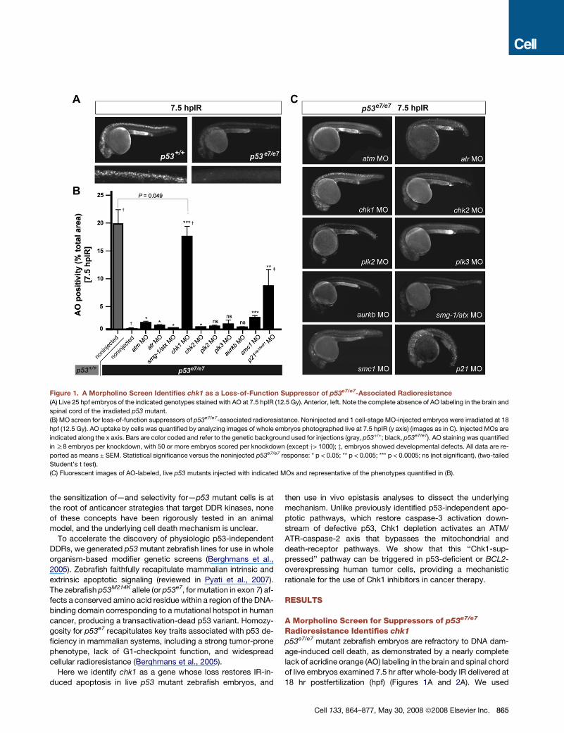
Figure 1. A Morpholino Screen Identifies chk1 as a Loss-of-Function Suppressor of p53e7/e7-Associated Radioresistance
(A) Live 25 hpf embryos of the indicated genotypes stained with AO at 7.5 hpIR (12.5 Gy). Anterior, left. Note the complete absence of AO labeling in the brain and
spinal cord of the irradiated p53 mutant.
(B) MO screen for loss-of-function suppressors of p53e7/e7-associated radioresistance. Noninjected and 1 cell-stage MO-injected embryos were irradiated at 18
hpf (12.5 Gy). AO uptake by cells was quantified by analyzing images of whole embryos photographed live at 7.5 hpIR (y axis) (images as in C). Injected MOs are
indicated along the x axis. Bars are color coded and refer to the genetic background used for injections (gray, p53+/+; black, p53e7/e7). AO staining was quantified
in R8 embryos per knockdown, with 50 or more embryos scored per knockdown (except y> 1000); z, embryos showed developmental defects. All data are re-
ported as means ± SEM. Statistical significance versus the noninjected p53e7/e7 response: * p < 0.05; ** p < 0.005; *** p < 0.0005; ns (not significant), (two-tailed
Student’s t test).
(C) Fluorescent images of AO-labeled, live p53 mutants injected with indicated MOs and representative of the phenotypes quantified in (B).
the sensitization of—and selectivity for—p53 mutant cells is at
the root of anticancer strategies that target DDR kinases, none
of these concepts have been rigorously tested in an animal
model, and the underlying cell death mechanism is unclear.
To accelerate the discovery of physiologic p53-independent
DDRs, we generated p53 mutant zebrafish lines for use in whole
organism-based modifier genetic screens (Berghmans et al.,
2005). Zebrafish faithfully recapitulate mammalian intrinsic and
extrinsic apoptotic signaling (reviewed in Pyati et al., 2007).
The zebrafish p53M214K allele (or p53e7, for mutation in exon 7) af-
fects a conserved amino acid residue within a region of the DNA-
binding domain corresponding to a mutational hotspot in human
cancer, producing a transactivation-dead p53 variant. Homozy-
gosity for p53e7 recapitulates key traits associated with p53 de-
ficiency in mammalian systems, including a strong tumor-prone
phenotype, lack of G1-checkpoint function, and widespread
cellular radioresistance (Berghmans et al., 2005).
Here we identify chk1 as a gene whose loss restores IR-in-
duced apoptosis in live p53 mutant zebrafish embryos, and
then use in vivo epistasis analyses to dissect the underlying
mechanism. Unlike previously identified p53-independent apo-
ptotic pathways, which restore caspase-3 activation down-
stream of defective p53, Chk1 depletion activates an ATM/
ATR-caspase-2 axis that bypasses the mitochondrial and
death-receptor pathways. We show that this ‘‘Chk1-sup-
pressed’’ pathway can be triggered in p53-deficient or BCL2-
overexpressing human tumor cells, providing a mechanistic
rationale for the use of Chk1 inhibitors in cancer therapy.
RESULTS
A Morpholino Screen for Suppressors of p53e7/e7
Radioresistance Identifies chk1
p53e7/e7 mutant zebrafish embryos are refractory to DNA dam-
age-induced cell death, as demonstrated by a nearly complete
lack of acridine orange (AO) labeling in the brain and spinal chord
of live embryos examined 7.5 hr after whole-body IR delivered at
18 hr postfertilization (hpf) (Figures 1A and 2A). We used
Cell 133, 864–877, May 30, 2008 ª2008 Elsevier Inc. 865
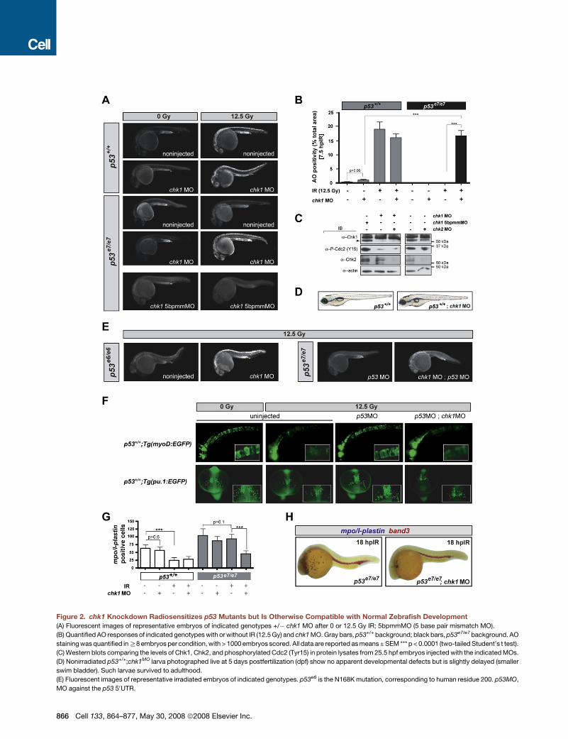
Figure 2. chk1 Knockdown Radiosensitizes p53 Mutants but Is Otherwise Compatible with Normal Zebrafish Development
(A) Fluorescent images of representative embryos of indicated genotypes +/� chk1 MO after 0 or 12.5 Gy IR; 5bpmmMO (5 base pair mismatch MO).
(B) Quantified AO responses of indicated genotypes with or without IR (12.5 Gy) and chk1 MO. Gray bars, p53+/+ background; black bars, p53e7/e7 background. AO
staining was quantified in R8 embryos per condition, with > 1000 embryos scored. All data are reported as means ± SEM *** p < 0.0001 (two-tailed Student’s t test).
(C) Western blots comparing the levels of Chk1, Chk2, and phosphorylated Cdc2 (Tyr15) in protein lysates from 25.5 hpf embryos injected with the indicated MOs.
(D) Nonirradiated p53+/+;chk1MO larva photographed live at 5 days postfertilization (dpf) show no apparent developmental defects but is slightly delayed (smaller
swim bladder). Such larvae survived to adulthood.
(E) Fluorescent images of representative irradiated embryos of indicated genotypes. p53e6 is the N168K mutation, corresponding to human residue 200. p53MO,
MO against the p53 50UTR.
866 Cell 133, 864–877, May 30, 2008 ª2008 Elsevier Inc.
morpholino antisense oligonucleotides (MOs) to knock down
eight zebrafish S- and G2-checkpoint kinases and two nonki-
nase checkpoint regulators (p21waf1/cip1 and smc1) in p53e7/e7
mutant embryos. We assessed the ability of each knockdown
to restore cell death (AO reactivity) at 7.5 hr post-IR (hpIR).
Single knockdowns of all genes tested, excluding plk2, plk3,
and aurkb, radiosensitized p53 mutants with variable efficiency
(Figures 1B and 1C). Whereas atm, atr, smg-1/atx, and chk2 de-
ficiencies restored only minor AO reactivity averaging 1%–5% of
the p53+/+ response, chk1 knockdown resulted in a staining pat-
tern that closely resembled wild-type (87.7% of the p53+/+ re-
sponse, p < 0.0001; see also Figures 2A and 2B). Enhanced
IR-induced cytotoxicity resulted specifically from chk1 knock-
down because (1) injections of a chk1 mismatch MO failed to ra-
diosensitize p53 mutants (Figure 2A, bottom panels); (2) the chk1
MO resulted in a robust reduction of the endogenous Chk1 pro-
tein pool, correlating with impaired Chk1 activity (Figure 2C); and
(3) a specific inhibitor of human Chk1, but not inhibitors of ATM or
Chk2, phenocopied the effects of chk1 MO (see Figure 7). As
would be expected from Chk1 loss, p53e7/e7;chk1MO embryos
lacked the IR-induced G2/M checkpoint (Figures S1A–S1D).
chk1 MO also fully radiosensitized p53e6 (p53N168K) homozy-
gotes (Berghmans et al., 2005) and p53 morphants (Langheinrich
et al., 2002) lacking p53 protein (Figure 2E), including in meso-
dermal derivatives (Figures 2F–2H). Together, these results pro-
vide in vivo evidence that Chk1 depletion is sufficient to restore
IR sensitivity to p53 mutant cells.
Transient Chk1 Depletion Is Viable in the Absence of IRChk1 is essential for fly and mouse development, with homozy-
gous null mutants succumbing to major cell cycle defects (Fo-
garty et al., 1997; Liu et al., 2000). We therefore tested whether
the cytotoxicity of chk1 knockdown in zebrafish p53 mutants
was strictly IR dependent. Indeed, chk1 depletion had no appar-
ent effect on normal zebrafish development and viability, in either
the p53+/+ or p53e7/e7 background (Figures 2A and 2D; compare
bars 1 and 2 in Figure 2B). Western blots performed with an anti-
zebrafish Chk1 antibody revealed a substantial knockdown of
the protein (Figure 2C). Yet chk1 morphants harbored residual
levels of Chk1 activity, as shown by weak but persistent levels
of phosphorylated Cdc2 (Figure 2C). These results demonstrate
that transient depletion, as opposed to persistent total loss (Liu
et al., 2000), of Chk1 function, is tolerable by vertebrate cells
in vivo and compatible with long-term organismal viability. Cru-
cially, however (as already shown above), such transient down-
regulation is sufficient to restore the IR-induced cell-death
response in p53 mutants (Figures 1B, 1C, 2A, and 2B).
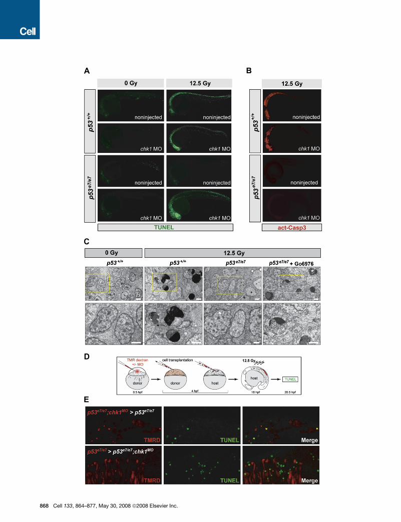
Irradiated p53e7/e7;chk1MO Embryos Undergo Caspase-3-Independent Cell-Autonomous ApoptosisChk1 knockdown might restore a wild-type response to IR (that is,
classical intrinsic apoptosis) (Kratz etal., 2006) or trigger a different
cell-death program in p53 mutants. To distinguish between these
possibilities,wefirstanalyzed two hallmarks ofapoptosis:TUNEL-
positive DNA fragmentation and cleaved caspase-3 (as well as
electron micrographs) in embryos fixed at 7.5 hpIR. AO labeling
of irradiated p53e7/e7;chk1MO embryos (Figures 1C and 2A) corre-
lated with high levels of TUNEL labeling throughout the CNS, sim-
ilar to findings in irradiated p53+/+ embryos (Figure 3A). Multiple
cells in the irradiated CNS of p53+/+ and Chk1-depleted p53e7/e7
embryos also showed similar ultrastructural manifestations of ap-
optosis (e.g., chromatin compaction/segregation and cytoplas-
mic condensation; Figures 3C and S2). Surprisingly, however,
while irradiated p53+/+ embryos exhibited strong immunostaining
for active caspase-3, irradiated p53e7/e7;chk1MO embryos did not
and showed no increase in active caspase-3 levels compared to
p53 single mutants, which were devoid of both TUNEL and active
caspase-3 (Figures 3A and 3B). Thus, the p53-independent cell
death-inducing DDR triggered by Chk1 depletion is a caspase-
3-independent apoptotic pathway (see also Figure S2F).
To determine the cell autonomy of the Chk1-antagonized
pathway, we generated genetic chimeras (Figures 3D and S3).
While p53e7/e7;chk1MO cells grafted into p53e7/e7 hosts often
stained TUNEL-positive after IR (39%, n = 102), neighboring
host cells did not (Figure 3E, upper panels). In the reciprocal ex-
periment, p53e7/e7 cells transplanted into p53e7/e7;chk1MO hosts
remained TUNEL negative within an otherwise TUNEL-positive
environment (Figure 3E, lower panels). Therefore, IR-induced
TUNEL reactivity of transplanted cells strictly depends on
Chk1 dosage, occurs irrespective of the cellular environment,
and has very little (if any) influence on neighboring cells. The
Chk1-suppressed apoptotic DDR pathway thus functions in
a cell-autonomous manner.
Chk1 Blocks a Mitochondria and DeathReceptor-Independent Apoptotic PathwayInvolving ATM, ATR, and Caspase-2To molecularly characterize the newly identified apoptotic
pathway, we capitalized on the unique advantages of zebrafish
embryos for in vivo epistasis analyses. Specifically, we knocked
down or forced the expression of candidate pathway contribu-
tors in p53e7/e7;chk1MO embryos and assessed the effects on
IR-induced cell death using the AO assay.
atm and atr single knockdowns severely impaired chk1 knock-
down-mediated radiosensitization of zebrafish p53 mutants,
(F) Fluorescent images of live transgenic embryos injected with the indicated MOs at the 1-cell stage and expressing EGFP in the notochord (top row, embryos
photographed at 24 hpf) or in myeloid progenitors (bottom row, embryos photographed at 16.5 hpf). Tg(myoD:EGFP) and Tg(pu.1:EGFP) embryos were treated
with or without IR (12.5 Gy) at 18 hpf and 10 hpf, respectively. Insets, higher magnification views of GFP-expressing cells. Top row, lateral views, anterior to the
left. Bottom row, dorsal views, anterior facing down.
(G) Quantification of myeloid cells in 28 hpf embryos generated as indicated (x axis) and processed as in (H). Gray bars, p53+/+ background; black bars, p53e7/e7
background. mpo/l-plastin staining was quantified in R15 embryos per condition. Data are reported as means ± SD ** p < 0.001, *** p < 0.0001 (two-tailed Student’s
t test). Note that while the numbers of mpo/l-plastin-positive cells are reduced�3-fold in IR-treated versus untreated p53+/+ embryos; they are unchanged in treated
versus untreated p53e7/e7 embryos. Also note that chk1 knockdown induces an average 2-fold reduction in myeloid cell numbers in the p53e7/e7 background
after IR.
(H) Images of representative 28 hpf embryos of indicated genotypes processed for in situ hybridization of mpo and l-plastin riboprobes (blue, differentiated gran-
ulocytes and monocytes) and band 3 (red, erythrocytes). Note the specific reduction in number of granulocytes/monocytes.
Cell 133, 864–877, May 30, 2008 ª2008 Elsevier Inc. 867
86
8 Cell 133, 864–877, May 30, 2008 ª2008 Elsevier Inc.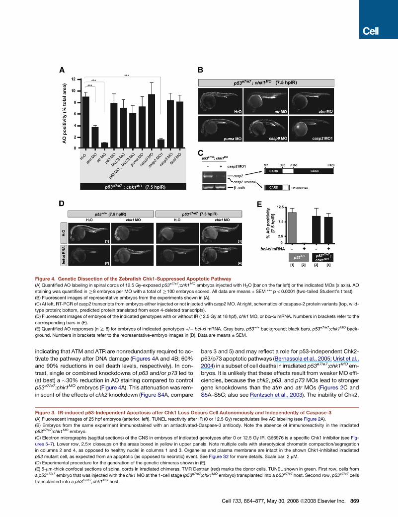
Figure 4. Genetic Dissection of the Zebrafish Chk1-Suppressed Apoptotic Pathway
(A) Quantified AO labeling in spinal cords of 12.5 Gy-exposed p53e7/e7;chk1MO embryos injected with H2O (bar on the far left) or the indicated MOs (x axis). AO
staining was quantified in R8 embryos per MO with a total of R100 embryos scored. All data are means ± SEM *** p < 0.0001 (two-tailed Student’s t test).
(B) Fluorescent images of representative embryos from the experiments shown in (A).
(C) At left, RT-PCR of casp2 transcripts from embryos either injected or not injected with casp2 MO. At right, schematics of caspase-2 protein variants (top, wild-
type protein; bottom, predicted protein translated from exon 4-deleted transcripts).
(D) Fluorescent images of embryos of the indicated genotypes with or without IR (12.5 Gy at 18 hpf), chk1 MO, or bcl-xl mRNA. Numbers in brackets refer to the
corresponding bars in (E).
(E) Quantified AO responses (n R 8) for embryos of indicated genotypes +/� bcl-xl mRNA. Gray bars, p53+/+ background; black bars, p53e7/e7;chk1MO back-
ground. Numbers in brackets refer to the representative-embryo images in (D). Data are means ± SEM.
indicating that ATM and ATR are nonredundantly required to ac-
tivate the pathway after DNA damage (Figures 4A and 4B; 60%
and 90% reductions in cell death levels, respectively). In con-
trast, single or combined knockdowns of p63 and/or p73 led to
(at best) a �30% reduction in AO staining compared to control
p53e7/e7;chk1MO embryos (Figure 4A). This attenuation was rem-
iniscent of the effects of chk2 knockdown (Figure S4A, compare
bars 3 and 5) and may reflect a role for p53-independent Chk2-
p63/p73 apoptotic pathways (Bernassola et al., 2005; Urist et al.,
2004) in a subset of cell deaths in irradiated p53e7/e7;chk1MO em-
bryos. It is unlikely that these effects result from weaker MO effi-
ciencies, because the chk2, p63, and p73 MOs lead to stronger
gene knockdowns than the atm and atr MOs (Figures 2C and
S5A–S5C; also see Rentzsch et al., 2003). The inability of Chk2,
Figure 3. IR-induced p53-Independent Apoptosis after Chk1 Loss Occurs Cell Autonomously and Independently of Caspase-3
(A) Fluorescent images of 25 hpf embryos (anterior, left). TUNEL reactivity after IR (0 or 12.5 Gy) recapitulates live AO labeling (see Figure 2A).
(B) Embryos from the same experiment immunostained with an antiactivated-Caspase-3 antibody. Note the absence of immunoreactivity in the irradiated
p53e7/e7;chk1MO embryo.
(C) Electron micrographs (sagittal sections) of the CNS in embryos of indicated genotypes after 0 or 12.5 Gy IR. Go6976 is a specific Chk1 inhibitor (see Fig-
ures 5–7). Lower row, 2.53 closeups on the areas boxed in yellow in upper panels. Note multiple cells with stereotypical chromatin compaction/segregation
in columns 2 and 4, as opposed to healthy nuclei in columns 1 and 3. Organelles and plasma membrane are intact in the shown Chk1-inhibited irradiated
p53 mutant cell, as expected from an apoptotic (as opposed to necrotic) event. See Figure S2 for more details. Scale bar, 2 mM.
(D) Experimental procedure for the generation of the genetic chimeras shown in (E).
(E) 5-mm-thick confocal sections of spinal cords in irradiated chimeras. TMR Dextran (red) marks the donor cells. TUNEL shown in green. First row, cells from
a p53e7/e7 embryo that was injected with the chk1 MO at the 1-cell stage (p53e7/e7;chk1MO embryo) transplanted into a p53e7/e7 host. Second row, p53e7/e7 cells
transplanted into a p53e7/e7;chk1MO host.
Cell 133, 864–877, May 30, 2008 ª2008 Elsevier Inc. 869
870 Cell 133, 864–877, May 30, 2008 ª2008 Elsevier Inc.
p63, and p73 to account for the majority of cell death events in ir-
radiated p53e7/e7;chk1MO embryos implies that ATM and ATR op-
erate predominantly within a novel apoptotic pathway, which we
have designated ‘‘Chk1-suppressed pathway’’ (CS in Figure 7D).
To test whether the mitochondrial apoptotic axis contributes
to the Chk1-suppressed pathway, we first knocked down the
proapoptotic BH3-only family member Puma. puma depletion
did not significantly affect AO labeling of irradiated p53e7/e7;
chk1MO embryos (Figures 4A and 4B) at a puma MO concentra-
tion that is otherwise sufficient to completely block IR-induced
apoptosis in p53+/+ zebrafish embryos (Figure S6) (Kratz et al.,
2006). Similarly, a dose of bcl-xl mRNA that completely blocked
cell death 7.5 hpIR in wild-type embryos failed to affect the AO
reactivity of irradiated p53e7/e7;chk1MO embryos (Figures 4D
and 4E; p53+/+ + bcl-xl, 0.035% of the mean p53+/+ response;
p53e7/e7;chk1MO + bcl-xl,�95% of the mean p53e7/e7;chk1MO re-
sponse). casp9 knockdown also lacked an effect (Figures 4A,
4B, and S5E). Thus, two major regulators of mitochondrial mem-
brane permeabilization (Puma and Bcl-xL), as well as the main
initiator and executioner caspases acting downstream of mito-
chondria (caspase-9 and caspase-3, see Figure 3), are dispens-
able for the Chk1-suppressed apoptotic pathway.
The death-receptor axis bypasses the requirement for mito-
chondria and caspase-9, suggesting that it could contribute to
the Chk1-suppressed pathway. In addition, a link between
Chk1 loss and caspase-8 activation has recently been observed
(Xiao et al., 2005). Even so, the death-receptor pathway con-
verges on caspase-3 activation via caspase-8 (Hengartner,
2000). This caspase-3 recruitment contrasts with the caspase-
3 independence of the pathway we identified, which, together
with the established cell autonomy of the new pathway
(Figure 3E), argues against a role for DNA damage-induced ex-
trinsic signaling downstream of chk1 depletion. Indeed, the AO
reactivity of p53e7/e7;chk1MO;casp8MO zebrafish embryos did
not differ from that of p53e7/e7;chk1MO specimens (Figures 4A
and S5D). Blocking death-receptor signaling with a fadd (Fas As-
sociated protein with Death Domain) MO (Eimon et al., 2006) also
failed to affect AO staining (Figure 4A). Thus, extrinsic signal-
ing—like mitochondrial signaling—does not appear to play an
important role downstream of chk1 loss.
The sole caspase whose depletion blocked the Chk1-sup-
pressed pathway was caspase-2, a poorly characterized yet
highly conserved caspase with features of both initiator and
executioner caspases (Zhivotovsky and Orrenius, 2005). In three
separate experiments, p53e7/e7;chk1MO;casp2MO1 embryos con-
sistently showed a mean 6-fold decrease in AO labeling com-
pared with p53e7/e7;chk1MO embryos (�16% of the mean
p53e7/e7;chk1MO response, p < 0.0001; Figures 4A and 4B).
casp2 MO1, which targets the splice donor site of intron 4, led
to marked reductions in casp2 mRNA levels and to aberrant
residual transcripts lacking exon 4 (Figure 4C). A second casp2
MO reduced IR-induced death in p53e7/e7;chk1MO embryos
(Figure S5F and S5G), and a mismatch version of casp2 MO1
had no effect (data not shown). Altogether, these epistasis anal-
yses identify a novel atm/atr-casp2 apoptotic program as a key
mechanism through which Chk1 depletion radiosensitizes p53
mutant zebrafish embryos without recruiting the classical mito-
chondrial and death-receptor pathways (Figure 7D).
The Chk1-Suppressed Apoptotic Pathway Is ConservedIn Human Cancer CellsWe next investigated whether the DNA damage-induced apo-
ptotic pathway suppressed by Chk1 in zebrafish is conserved
in human cancer cells defective in p53 signaling. To inhibit
Chk1 in these cells, we used the indolocarbazole small molecule
Go6976 (Kohn et al., 2003), which has greater specificity than the
commonly used Chk1 inhibitor UCN-01 (reviewed in Kawabe,
2004 and see below). In HeLa cells (in which the p53 protein
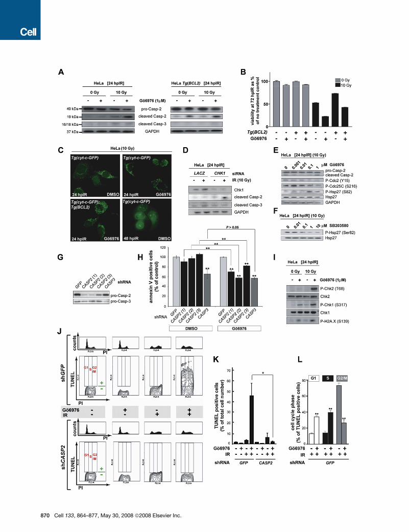
Figure 5. The Chk1-Suppressed Pathway Is Conserved in HeLa Cells
(A) Western blots comparing the levels of caspase-2 (pro and cleaved forms) and cleaved caspase-3 at 24 hpIR in lysates from HeLa cells carrying or not carrying
a BCL2 transgene (Tg[BCL2]) and treated with or without IR (10 Gy) or Chk1 inhibitor (Go6976, 1 mM).
(B) Analysis of HeLa cell survival at 72 hpIR (0 Gy versus 10 Gy) in the presence or absence of Go6976 and/or BCL2. Go6976 radiosensitizes the cells �2-fold
regardless of the BCL2 transgene (compare bars 5 and 6, and bars 7 and 8). Note that BCL2 is functional (i.e., radioprotective) in these experiments (compare
lanes 5 and 7). Data are means ± SEM.
(C) Fluorescent images of HeLa Tg(Cyt-c-GFP) cells with or without Tg(BCL2) or Go6976 at 24 or 48 hpIR (10 Gy). Note the punctate GFP patterns in all 24 hpIR
samples and the diffuse GFP pattern in the 48 hpIR sample.
(D) Levels of cleaved caspase-2 and caspase-3 at 24 hpIR (10 Gy) in HeLa cells transfected with LACZ or CHK1 siRNAs at 72 hr before IR.
(E) Western blots comparing the activities of Chk1 (Cdc2 phosphorylation at Tyr15 and CDC25C phosphorylation at Ser216) and MK-2 (Hsp-27 phosphorylation
at Ser82) following exposure to IR and increasing concentrations of Go6976.
(F) MK-2 phosphorylates Hsp-27 in HeLa cells. Western blot of lysates from irradiated HeLa cells exposed to increasing concentrations of the p38MAPK specific
inhibitor SB203580 (Reinhardt et al., 2007), showing a dose-dependent reduction in phosphorylated Hsp-27.
(G) Knockdown efficiencies of the indicated shRNAs as measured by western blots with anticaspase-2 and anticaspase-3 antibodies.
(H) Effects of GFP, CASP2, and CASP3 shRNAs on apoptotic cell numbers at 48 hpIR as measured by AnnexinV (+) / PI (�) staining of HeLa cells treated with 10
Gy with or without Go6976 (1 mM). For each shRNA, the average apoptotic cell number (given as % of GFP shRNA control) is shown. All data are means ± SD ** p <
0.01 (two-tailed Student’s t test). Asterisks on top of bars refer to comparisons with GFP shRNA.
(I) Synergistic activation of ATM and ATR by Go6976 and IR. Western blots comparing the activities of ATM (Chk2 phosphorylation at Thr68) and ATR (Chk1 phos-
phorylation at Ser317) after 0 or 10 Gy IR with or without Go6976 (1 mM). Levels of DNA damage were detected with an antiphospho-H2A.X antibody.
(J) Cell-cycle distribution of HeLa cells undergoing Chk1-suppressed apoptosis. HeLa cells harboring GFP or CASP2 shRNAs and treated with or without 10 Gy IR
with or without Go6976 (1 mM), as indicated, were fixed at 48 hpIR and stained for TUNEL and PI. For each shRNA line, upper panels show PI-single histograms
and lower panels show PI/TUNEL double-staining images. Cell-cycle phases and threshold for TUNEL positivity are indicated in red and green, respectively, in
each no-treatment control images.
(K) Quantification of the TUNEL stains shown in (J). Data are means ± SEM * p < 0.05 (two-tailed Student’s t test).
(L) Quantified data from experiment in (J) expressed as means ± SEM ** p < 0.002 (two-tailed Student’s t test). White bars indicate cells dying in G1 phase. Black
bars indicate cells dying in S phase. Grey bars, cells dying in G2 phase.
Cell 133, 864–877, May 30, 2008 ª2008 Elsevier Inc. 871
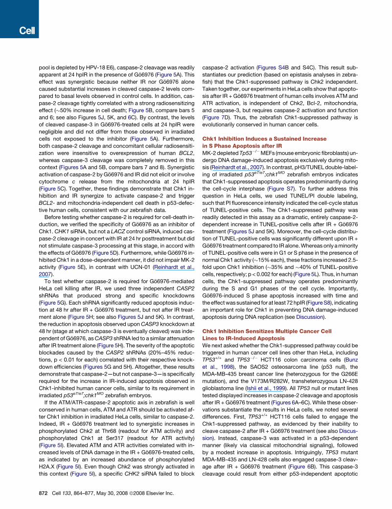
pool is depleted by HPV-18 E6), caspase-2 cleavage was readily
apparent at 24 hpIR in the presence of Go6976 (Figure 5A). This
effect was synergistic because neither IR nor Go6976 alone
caused substantial increases in cleaved caspase-2 levels com-
pared to basal levels observed in control cells. In addition, cas-
pase-2 cleavage tightly correlated with a strong radiosensitizing
effect (�50% increase in cell death; Figure 5B, compare bars 5
and 6; see also Figures 5J, 5K, and 6C). By contrast, the levels
of cleaved caspase-3 in Go6976-treated cells at 24 hpIR were
negligible and did not differ from those observed in irradiated
cells not exposed to the inhibitor (Figure 5A). Furthermore,
both caspase-2 cleavage and concomitant cellular radiosensiti-
zation were insensitive to overexpression of human BCL2,
whereas caspase-3 cleavage was completely removed in this
context (Figures 5A and 5B, compare bars 7 and 8). Synergistic
activation of caspase-2 by Go6976 and IR did not elicit or involve
cytochrome c release from the mitochondria at 24 hpIR
(Figure 5C). Together, these findings demonstrate that Chk1 in-
hibition and IR synergize to activate caspase-2 and trigger
BCL2- and mitochondria-independent cell death in p53-defec-
tive human cells, consistent with our zebrafish data.
Before testing whether caspase-2 is required for cell-death in-
duction, we verified the specificity of Go6976 as an inhibitor of
Chk1. CHK1 siRNA, but not a LACZ control siRNA, induced cas-
pase-2 cleavage in concert with IR at 24 hr posttreatment but did
not stimulate caspase-3 processing at this stage, in accord with
the effects of Go6976 (Figure 5D). Furthermore, while Go6976 in-
hibited Chk1 in a dose-dependent manner, it did not impair MK-2
activity (Figure 5E), in contrast with UCN-01 (Reinhardt et al.,
2007).
To test whether caspase-2 is required for Go6976-mediated
HeLa cell killing after IR, we used three independent CASP2
shRNAs that produced strong and specific knockdowns
(Figure 5G). Each shRNA significantly reduced apoptosis induc-
tion at 48 hr after IR + Go6976 treatment, but not after IR treat-
ment alone (Figure 5H; see also Figures 5J and 5K). In contrast,
the reduction in apoptosis observed upon CASP3 knockdown at
48 hr (stage at which caspase-3 is eventually cleaved) was inde-
pendent of Go6976, as CASP3 shRNA led to a similar attenuation
after IR treatment alone (Figure 5H). The severity of the apoptotic
blockades caused by the CASP2 shRNAs (20%–45% reduc-
tions, p < 0.01 for each) correlated with their respective knock-
down efficiencies (Figures 5G and 5H). Altogether, these results
demonstrate that caspase-2—but not caspase-3—is specifically
required for the increase in IR-induced apoptosis observed in
Chk1-inhibited human cancer cells, similar to its requirement in
irradiated p53e7/e7;chk1MO zebrafish embryos.
If the ATM/ATR-caspase-2 apoptotic axis in zebrafish is well
conserved in human cells, ATM and ATR should be activated af-
ter Chk1 inhibition in irradiated HeLa cells, similar to caspase-2.
Indeed, IR + Go6976 treatment led to synergistic increases in
phosphorylated Chk2 at Thr68 (readout for ATM activity) and
phosphorylated Chk1 at Ser317 (readout for ATR activity)
(Figure 5I). Elevated ATM and ATR activities correlated with in-
creased levels of DNA damage in the IR + Go6976-treated cells,
as indicated by an increased abundance of phosphorylated
H2A.X (Figure 5I). Even though Chk2 was strongly activated in
this context (Figure 5I), a specific CHK2 siRNA failed to block
872 Cell 133, 864–877, May 30, 2008 ª2008 Elsevier Inc.
caspase-2 activation (Figures S4B and S4C). This result sub-
stantiates our prediction (based on epistasis analyses in zebra-
fish) that the Chk1-suppressed pathway is Chk2 independent.
Taken together, our experiments in HeLa cells show that apopto-
sis after IR + Go6976 treatment of human cells involves ATM and
ATR activation, is independent of Chk2, Bcl-2, mitochondria,
and caspase-3, but requires caspase-2 activation and function
(Figure 7D). Thus, the zebrafish Chk1-suppressed pathway is
evolutionarily conserved in human cancer cells.
Chk1 Inhibition Induces a Sustained Increasein S Phase Apoptosis after IRMK-2 depleted Tp53�/�MEFs (mouse embryonic fibroblasts) un-
dergo DNA damage-induced apoptosis exclusively during mito-
sis (Reinhardt et al., 2007). In contrast, pH3/TUNEL double-label-
ing of irradiated p53e7/e7;chk1MO zebrafish embryos indicates
that Chk1-suppressed apoptosis operates predominantly during
the cell-cycle interphase (Figure S7). To further address this
question in HeLa cells, we used TUNEL/PI double labeling,
such that PI fluorescence intensity indicated the cell-cycle status
of TUNEL-positive cells. The Chk1-suppressed pathway was
readily detected in this assay as a dramatic, entirely caspase-2-
dependent increase in TUNEL-positive cells after IR + Go6976
treatment (Figures 5J and 5K). Moreover, the cell-cycle distribu-
tion of TUNEL-positive cells was significantly different upon IR +
Go6976 treatment compared to IR alone. Whereas only a minority
of TUNEL-positive cells were in G1 or S phase in the presence of
normal Chk1 activity (�15% each), these fractions increased 2.5-
fold upon Chk1 inhibition (�35% and �40% of TUNEL-positive
cells, respectively; p < 0.002 for each) (Figure 5L). Thus, in human
cells, the Chk1-suppressed pathway operates predominantly
during the S and G1 phases of the cell cycle. Importantly,
Go6976-induced S phase apoptosis increased with time and
the effect was sustained for at least 72 hpIR (Figure S8), indicating
an important role for Chk1 in preventing DNA damage-induced
apoptosis during DNA replication (see Discussion).
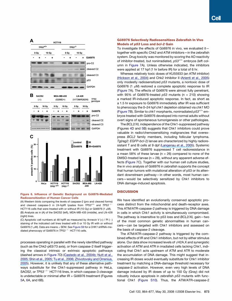
Chk1 Inhibition Sensitizes Multiple Cancer CellLines to IR-Induced ApoptosisWe next asked whether the Chk1-suppressed pathway could be
triggered in human cancer cell lines other than HeLa, including
TP53+/+ and TP53�/� HCT116 colon carcinoma cells (Bunz
et al., 1998), the SAOS2 osteosarcoma line (p53 null), the
MDA-MB-435 breast cancer line (heterozygous for the G266E
mutation), and the V173M/R282W, transheterozygous LN-428
glioblastoma line (Ishii et al., 1999). All TP53 null or mutant lines
tested displayed increases in caspase-2 cleavage and apoptosis
after IR + Go6976 treatment (Figures 6A–6C). While these obser-
vations substantiate the results in HeLa cells, we noted several
differences. First, TP53+/+ HCT116 cells failed to engage the
Chk1-suppressed pathway, as evidenced by their inability to
cleave caspase-2 after IR + Go6976 treatment (see also Discus-
sion). Instead, caspase-3 was activated in a p53-dependent
manner (likely via classical mitochondrial signaling), followed
by a modest increase in apoptosis. Intriguingly, TP53 mutant
MDA-MB-435 and LN-428 cells also engaged caspase-3 cleav-
age after IR + Go6976 treatment (Figure 6B). This caspase-3
cleavage could result from either p53-independent apoptotic
processes operating in parallel with the newly identified pathway
(such as the Chk2-p63/73 axis), or from caspase-2 itself trigger-
ing the classical intrinsic or extrinsic apoptotic pathways
(dashed arrows in Figure 7D) (Castedo et al., 2004b; Nutt et al.,
2005; Shin et al., 2005; Tu et al., 2006; Zhivotovsky and Orrenius,
2005). However, it is unlikely that any of these alternative path-
ways substitute for the Chk1-suppressed pathway in HeLa,
SAOS2, or TP53�/� HCT116 lines, in which caspase-3 cleavage
is undetectable or minimal after IR + Go6976 treatment (Figures
5A, 6A, and 6B).
Figure 6. Influence of Genetic Background on Go6976-Mediated
Radiosensitization of Human Cancer Cells
(A) Western blots comparing the levels of caspase-2 (pro and cleaved forms)
and cleaved caspase-3 in 24-hpIR lysates from TP53+/+ and TP53�/�
HCT116 cells that were treated with or without IR (10 Gy) or Go6976 (1 mM).
(B) Analysis as in (A) of the SAOS2 (left), MDA-MB-435 (middle), and LN-428
(right) lines.
(C) Apoptotic cell numbers at 48 hpIR as measured by Annexin V (+) / PI (�)
staining of the indicated cell lines treated with 0 or 10 Gy IR with or without
Go6976 (1 mM). Data are means ± SEM. See Figure S9 for a CHK1 shRNA-me-
diated phenocopy of Go6976 in TP53�/� HCT116 cells.
Go6976 Selectively Radiosensitizes Zebrafish In VivoModels of p53 Loss and bcl-2 GainTo investigate the effects of Go6976 in vivo, we evaluated it—
together with specific Chk2 and ATM inhibitors—in the zebrafish
system. Drug toxicity was monitored by scoring the AO reactivity
of inhibitor-treated, but nonirradiated, p53+/+ embryos (left col-
umn in Figure 7A). Unless otherwise indicated, the inhibitors
were applied at 17 hpf (1 hr before IR) for a total of 6 hr.
Whereas relatively toxic doses of KU55933 (an ATM inhibitor)
(Hickson et al., 2004) and Chk2 Inhibitor II (Arienti et al., 2005)
only modestly radiosensitized p53 mutants, a nontoxic dose of
Go6976 (1 mM) restored a complete apoptotic response to IR
(Figure 7A). The effects of Go6976 were almost fully penetrant,
with 95% of Go6976-treated p53 mutants (n = 210) showing
a marked IR-induced apoptotic response. In fact, as short as
a 1.5 hr exposure to Go6976 immediately after IR was sufficient
to phenocopy the 0–24 hpf chk1 depletion obtained via chk1 MO
(Figure 7B). Similar to chk1 morphants, nonirradiated p53+/+ em-
bryos treated with Go6976 developed into normal adults without
overt signs of spontaneous tumorigenesis or other pathologies.
The BCL2/XL independence of the Chk1-suppressed pathway
(Figures 4D and 5B) suggests that Chk1 inhibitors could prove
valuable in radio/chemosensitizing malignancies that overex-
press BCL2 family members, including follicular lymphoma.
Tg(rag2: EGFP-bcl-2) larvae are characterized by highly radiore-
sistant T and B cells at 9 dpf (Langenau et al., 2005). Systemic
treatment with Go6976 suppressed T cell radioresistance in
a mean 58% of these larvae (n = 26) compared to none of the
DMSO-treated larvae (n = 28), without any apparent adverse ef-
fects (Figure 7C). Together with our human cell culture studies,
the in vivo analysis of Go6976 in zebrafish supports the concept
that human tumors with mutational alteration of p53 or its atten-
dant downstream pathway—in other words, most human can-
cers—would be selectively sensitized by Chk1 inhibitors to
DNA damage-induced apoptosis.
DISCUSSION
We have identified an evolutionarily conserved apoptotic pro-
cess distinct from the mitochondrial and death-receptor axes.
This ATM/ATR-caspase-2 pathway is triggered by DNA damage
in cells in which Chk1 activity is simultaneously compromised.
The pathway is insensitive to p53 loss and BCL2/XL gain—two
of the most common genetic abnormalities in human can-
cers—can be targeted with Chk1 inhibitors and assessed on
the basis of caspase-2 cleavage.
The ATM/ATR-caspase-2 pathway is triggered by the com-
bined effects of IR and Chk1 inhibition, but not by either stimulus
alone. Our data show increased levels of gH2A.X and synergistic
activation of ATM and ATR in irradiated cells lacking Chk1, indi-
cating that Chk1 acts upstream of ATM and ATR to moderate
the accumulation of DNA damage. This might suggest that in-
creasing IR doses would eventually substitute for Chk1 inhibitor
treatment by matching a DNA-damage threshold necessary for
caspase-2 activation. However, even very high levels of DNA
damage induced by IR doses of up to 150 Gy (Gray) did not
robustly induce apoptosis in zebrafish p53 mutants with func-
tional Chk1 (Figure S10). Thus, the ATM/ATR-caspase-2
Cell 133, 864–877, May 30, 2008 ª2008 Elsevier Inc. 873
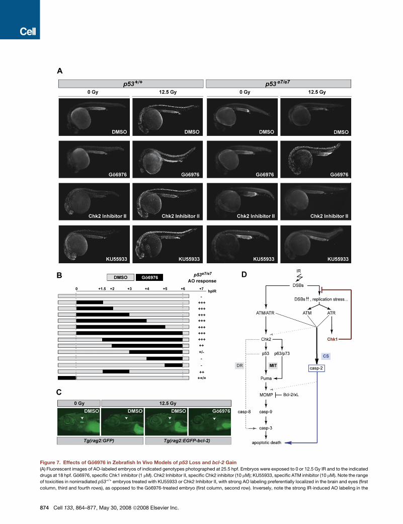
Figure 7. Effects of Go6976 in Zebrafish In Vivo Models of p53 Loss and bcl-2 Gain
(A) Fluorescent images of AO-labeled embryos of indicated genotypes photographed at 25.5 hpf. Embryos were exposed to 0 or 12.5 Gy IR and to the indicated
drugs at 18 hpf. Go6976, specific Chk1 inhibitor (1 mM). Chk2 Inhibitor II, specific Chk2 inhibitor (10 mM); KU55933, specific ATM inhibitor (10 mM). Note the range
of toxicities in nonirradiated p53+/+ embryos treated with KU55933 or Chk2 Inhibitor II, with strong AO labeling preferentially localized in the brain and eyes (first
column, third and fourth rows), as opposed to the Go6976-treated embryo (first column, second row). Inversely, note the strong IR-induced AO labeling in the
874 Cell 133, 864–877, May 30, 2008 ª2008 Elsevier Inc.
pathway cannot mount a nonspecific response to excess dam-
age, but rather is obligatorily tied to Chk1 activity. An involvement
of Chk1’s essential or damage-dependent checkpoint functions
during DNA replication (Bartek et al., 2004; Lam et al., 2004; Sor-
ensen et al., 2003; Syljuasen et al., 2005) seems likely given the
sustained rise in S phase apoptosis observed in IR + Chk1 inhib-
itor-treated HeLa cells. A role for replication stress in triggering
the ATM/ATR-caspase-2 pathway gains support from observa-
tions that Chk1-depleted cells exposed to replication inhibitors
undergo p53- and Chk2-independent apoptosis during S phase
(Rodrıguez and Meuth, 2006). Also, caspase-2 is the sole cas-
pase whose proform resides in the nucleus (Zhivotovsky et al.,
1999), where it is stabilized by cyclin D3, a positive regulator of
the G1/S transition (Mendelsohn et al., 2002). We propose that
tight control of the ATM/ATR-caspase-2 pathway by Chk1 con-
tributes to the decision to live (arrest and repair) or die in replicat-
ing cells suffering DNA damage.
ATM and ATR, while both necessary for activation of the Chk1-
suppressed pathway, are individually insufficient for this function
(Figure 4A). ATM and ATR might phosphorylate different sub-
strates, each being essential for caspase-2 activation and sus-
ceptible to Chk1 regulation. However, neither caspase-2 nor its
proposed activators, including PIDDosome components PIDD
(p53-induced protein with death domain) and RAIDD (RIP-asso-
ciated ICH-1/CED-3 homologous protein with a death domain)
(Tinel and Tschopp, 2004), belong to the list of 700 potential
ATM/ATR substrates (Matsuoka et al., 2007). A more likely inter-
pretation is that ATM and ATR serve different sensory functions,
with ATM responding primarily to IR-induced double-strand
breaks while ATR predominantly senses signals resulting from
reduced Chk1 activity, such as replication stress (Cuadrado
et al., 2006; Syljuasen et al., 2005).
The ATM/ATR-caspase-2 pathway may serve as a mechanism
that ensures the demise of cells carrying potentially harmful DNA
lesions in the absence of proper genome-surveillance activity (as
provided by Chk1) (Lam et al., 2004). Such a function might help
explain why CHK1 mutations, despite fueling genomic instability
(Lam et al., 2004), are paradoxically rare in human cancers (Bartek
and Lukas, 2003). Our demonstration that the Chk1-suppressed
pathway can operate in both the absence and presence of p53, as
revealed in irradiated p53+/+;chk1MO;bcl-xl embryos and in irradi-
ated p53+/+;Tg(rag2:EGFP-bcl-2) larvae treated with Go6976,
disqualifies it as a ‘‘backup’’ program (Roos and Kaina, 2006) op-
erating only in cells that lack p53. Rather, we propose that it con-
stitutes an alternative, perhaps primitive, response to DNA injury
that evolved independently of the p53 network. Intriguingly, how-
ever, TP53+/+ and TP53�/� HCT116 cells differed in their re-
sponse to IR + Go6976 treatment, in that caspase-2 but not cas-
pase-3 cleavage was actively inhibited in the TP53+/+ cells, via an
apparent downregulation of procaspase-2 levels (Figure 6A; see
also Baptiste-Okoh et al., 2008). Thus, a form of crosstalk might
have evolved to link these p53-dependent and -independent ap-
optotic pathways, similar to that described for caspase-depen-
dent and -independent pathways (Colell et al., 2007).
Chk1 inhibitors can radio/chemosensitize p53-deficient hu-
man tumor cells in vitro, leading to clinical trials of their activity
in cancer patients (Kawabe, 2004; Tse et al., 2007; Zhou and Bar-
tek, 2004). Because of the embryonic lethality of Chk1�/� mice,
however, it has remained unclear whether the potency and selec-
tivity of radio/chemosensitization observed in vitro will apply in
vivo. Our findings in zebrafish using the Chk1 inhibitor Go6976
and chk1 morphants, which retain residual levels of Chk1 activity,
indicate that levels of Chk1 inhibition not toxic to normal cells are
sufficient to sensitize p53 mutant cells to IR-induced apoptosis
within a living vertebrate. Our results also identify cleavage of
caspase-2 as a candidate biomarker for Chk1-targeting treat-
ments. The isolation of such specific biomarkers remains a press-
ing challenge in the development and optimal use of targeted
cancer therapeutics (Cully et al., 2006; Tse et al., 2007). Finally,
our results unexpectedly predict that in addition to tumors with
altered p53 activity, those with other types of prosurvival alter-
ations that block mitochondrial signaling downstream of p53,
such as BCL2-expressing follicular lymphomas, would respond
favorably to combination therapy with Chk1 inhibitors.
EXPERIMENTAL PROCEDURES
Zebrafish Stocks
The homozygous viable p53M214K and p53N168K mutant lines (p53e7 and p53e6,
respectively, in this article) (Berghmans et al., 2005), and the Tg(rag2:GFP)
(Langenau et al., 2003), Tg(rag2:EGFP-bcl-2) (Langenau et al., 2005),
Tg(pu.1:GFP) (Hsu et al., 2004), and Tg(myoD:EGFP) (Yang et al., 2004) trans-
genic lines were used and maintained at 28.5�C by standard methods. For ex-
perimental purposes, irradiated p53e6/e6 embryos were incubated for 6 hr at
37�C (restrictive temperature for N168K).
Morpholino Screen and Epistasis Analyses Using the Live AO Assay
MOs were obtained from Gene Tools, LLC. MO sequences, target sites, work-
ing concentrations, knockdown efficiencies, selected references, and injection
procedures, as well as detailed protocols for AO staining of live embryos and
the ImageJ-based quantification method, are listed in Table S1, Figure S5, and
the Supplemental Experimental Procedures.
Human Cancer Cell Lines, siRNAs, and shRNAs
The HeLa, SAOS2, MDA-MB-435, and LN-428 cell lines, the TP53+/+ and
TP53�/� HCT116 isogenic pair (Bunz et al., 1998), and the Cyt-c-GFP
Go6976-treated p53 mutant (last column, second row), but the lack of staining in the mutants treated with KU55933 or Chk2 Inhibitor II (last column, third and
fourth rows).
(B) Temporal requirement for Chk1 loss with respect to IR. p53 mutant embryos were exposed to Go6976 for the indicated times. AO staining was quantified on
a scale from ‘‘�’’ to ‘‘+++’’with ‘‘�’’ representing the p53 mutant response and ‘‘+++’’ the response of sibling mutants treated with Go6976 for 6 hr (�500-fold
greater response).
(C) Fluorescent images of 9 dpf zebrafish larvae carrying the indicated transgene. Larvae were treated with 0 Gy or 15 Gy IR at 5 dpf and were exposed to Go6976
(or DMSO as control) for a total of 5 days starting at 4 dpf. White arrowhead indicates the position of the thymus. Note the absence of detectable GFP in the
Go6976-treated Tg(rag2:EGFP-bcl-2) irradiated larva.
(D) Simplified model for the vertebrate apoptotic response to DNA damage, highlighting the p53-independent pathway normally blocked by IR-activated Chk1
(CS, for Chk1-suppressed pathway), which is distinct from the classical intrinsic (mitochondrial, MIT) and extrinsic (death-receptor, DR) pathways. See text for
details.
Cell 133, 864–877, May 30, 2008 ª2008 Elsevier Inc. 875
transgenic, 2H18 HeLa-derived lines (Goldstein et al., 2000), carrying or not
carrying a BCL2 transgene, were cultured in DMEM medium (GIBCO) supple-
mented with 15% fetal bovine serum (FBS). siRNAs were transfected in HeLa
cells using Hiperfect (QIAGEN) according to the manufacturer’s instructions.
Cells were exposed to IR +/� Go6976 at 48 or 72 hr posttransfection. shRNA
knockdown analyses were performed as previously described (Moffat et al.,
2006). See Supplemental Data for more details, siRNA and shRNA sequences,
and all other experimental procedures.
SUPPLEMENTAL DATA
Supplemental Data include ten figures, one table, Supplemental Experimental
Procedures, and Supplemental References and can be found online at http://
www.cell.com/cgi/content/full/133/5/864/DC1/.
ACKNOWLEDGMENTS
We thank John R. Gilbert for editorial review. We gratefully acknowledge
George Kourkoulis and Barry Baker for zebrafish care, Bert Vogelstein and Ro-
salind Segal for the HCT116 and LN-428 cell lines; William Hahn for the
CASP2(1), CASP3, and GFP shRNAs; Thomas Diefenbach and Louise Traki-
mas for assistance with confocal and electron microscopy; and Yebin Ahn
and Peter Schow for assistance with FCM analysis. This work was supported
by NIH grants HL-88664 (S.S., A.T.L.) and AI47891 (D.R.G.), the Susan G. Ko-
men Breast Cancer Foundation (R.D.K.), and the A-T Children’s Project (S.K.).
Received: March 2, 2007
Revised: January 25, 2008
Accepted: March 24, 2008
Published: May 29, 2008
REFERENCES
Afshar, G., Jelluma, N., Yang, X., Basila, D., Arvold, N.D., Karlsson, A., Yount,
G.L., Dansen, T.B., Koller, E., and Haas-Kogan, D.A. (2006). Radiation-in-
duced caspase-8 mediates p53-independent apoptosis in glioma cells. Can-
cer Res. 66, 4223–4232.
Arienti, K.L., Brunmark, A., Axe, F.U., McClure, K., Lee, A., Blevitt, J., Neff,
D.K., Huang, L., Crawford, S., Pandit, C.R., et al. (2005). Checkpoint kinase in-
hibitors: SAR and radioprotective properties of a series of 2-arylbenzimida-
zoles. J. Med. Chem. 48, 1873–1885.
Baptiste-Okoh, N., Barsotti, A.M., and Prives, C. (2008). Caspase 2 is both
required for p53-mediated apoptosis and downregulated by p53 in a p21-
dependent manner. Cell Cycle 7. Published online February 19, 2008.
Bartek, J., and Lukas, J. (2003). Chk1 and Chk2 kinases in checkpoint control
and cancer. Cancer Cell 3, 421–429.
Bartek, J., Lukas, C., and Lukas, J. (2004). Checking on DNA damage in S
phase. Nat. Rev. Mol. Cell Biol. 5, 792–804.
Berghmans, S., Murphey, R.D., Wienholds, E., Neuberg, D., Kutok, J.L.,
Fletcher, C.D., Morris, J.P., Liu, T.X., Schulte-Merker, S., Kanki, J.P., et al.
(2005). tp53 mutant zebrafish develop malignant peripheral nerve sheath tu-
mors. Proc. Natl. Acad. Sci. USA 102, 407–412.
Bernassola, F., Oberst, A., Melino, G., and Pandolfi, P.P. (2005). The promye-
locytic leukaemia protein tumour suppressor functions as a transcriptional
regulator of p63. Oncogene 24, 6982–6986.
Bunz, F., Dutriaux, A., Lengauer, C., Waldman, T., Zhou, S., Brown, J.P.,
Sedivy, J.M., Kinzler, K.W., and Vogelstein, B. (1998). Requirement for p53
and p21 to sustain G2 arrest after DNA damage. Science 282, 1497–1501.
Castedo, M., Perfettini, J.L., Roumier, T., Andreau, K., Medema, R., and
Kroemer, G. (2004a). Cell death by mitotic catastrophe: a molecular definition.
Oncogene 23, 2825–2837.
Castedo, M., Perfettini, J.L., Roumier, T., Yakushijin, K., Horne, D., Medema,
R., and Kroemer, G. (2004b). The cell cycle checkpoint kinase Chk2 is a nega-
tive regulator of mitotic catastrophe. Oncogene 23, 4353–4361.
876 Cell 133, 864–877, May 30, 2008 ª2008 Elsevier Inc.
Colell, A., Ricci, J.E., Tait, S., Milasta, S., Maurer, U., Bouchier-Hayes, L., Fitz-
gerald, P., Guio-Carrion, A., Waterhouse, N.J., Li, C.W., et al. (2007). GAPDH
and autophagy preserve survival after apoptotic cytochrome c release in the
absence of caspase activation. Cell 129, 983–997.
Cuadrado, M., Martinez-Pastor, B., and Fernandez-Capetillo, O. (2006). ‘‘ATR
activation in response to ionizing radiation: still ATM territory.’’ Cell Div. 1, 7.
Cully, M., You, H., Levine, A.J., and Mak, T.W. (2006). Beyond PTEN muta-
tions: the PI3K pathway as an integrator of multiple inputs during tumorigene-
sis. Nat. Rev. Cancer 6, 184–192.
Eimon, P.M., Kratz, E., Varfolomeev, E., Hymowitz, S.G., Stern, H., Zha, J., and
Ashkenazi, A. (2006). Delineation of the cell-extrinsic apoptosis pathway in the
zebrafish. Cell Death Differ. 13, 1619–1630.
Fogarty, P., Campbell, S.D., Abu-Shumays, R., Phalle, B.S., Yu, K.R., Uy, G.L.,
Goldberg, M.L., and Sullivan, W. (1997). The Drosophila grapes gene is related
to checkpoint gene chk1/rad27 and is required for late syncytial division fidel-
ity. Curr. Biol. 7, 418–426.
Frenkel, J., Sherman, D., Fein, A., Schwartz, D., Almog, N., Kapon, A., Goldfin-
ger, N., and Rotter, V. (1999). Accentuated apoptosis in normally developing
p53 knockout mouse embryos following genotoxic stress. Oncogene 18,
2901–2907.
Goldstein, J.C., Waterhouse, N.J., Juin, P., Evan, G.I., and Green, D.R. (2000).
The coordinate release of cytochrome c during apoptosis is rapid, complete
and kinetically invariant. Nat. Cell Biol. 2, 156–162.
Gong, J.G., Costanzo, A., Yang, H.Q., Melino, G., Kaelin, W.G., Jr., Levrero,
M., and Wang, J.Y. (1999). The tyrosine kinase c-Abl regulates p73 in apoptotic
response to cisplatin-induced DNA damage. Nature 399, 806–809.
Hengartner, M.O. (2000). The biochemistry of apoptosis. Nature 407, 770–776.
Hickson, I., Zhao, Y., Richardson, C.J., Green, S.J., Martin, N.M., Orr, A.I.,
Reaper, P.M., Jackson, S.P., Curtin, N.J., and Smith, G.C. (2004). Identifica-
tion and characterization of a novel and specific inhibitor of the ataxia-telangi-
ectasia mutated kinase ATM. Cancer Res. 64, 9152–9159.
Hsu, K., Traver, D., Kutok, J.L., Hagen, A., Liu, T.X., Paw, B.H., Rhodes, J.,
Berman, J.N., Zon, L.I., Kanki, J.P., et al. (2004). The pu.1 promoter drives
myeloid gene expression in zebrafish. Blood 104, 1291–1297.
Huang, H., Regan, K.M., Lou, Z., Chen, J., and Tindall, D.J. (2006). CDK2-de-
pendent phosphorylation of FOXO1 as an apoptotic response to DNA damage.
Science 314, 294–297.
Ishii, N., Maier, D., Merlo, A., Tada, M., Sawamura, Y., Diserens, A.C., and Van
Meir, E.G. (1999). Frequent co-alterations of TP53, p16/CDKN2A, p14ARF,
PTEN tumor suppressor genes in human glioma cell lines. Brain Pathol. 9,
469–479.
Kasibhatla, S., Brunner, T., Genestier, L., Echeverri, F., Mahboubi, A., and
Green, D.R. (1998). DNA damaging agents induce expression of Fas ligand
and subsequent apoptosis in T lymphocytes via the activation of NF-kappa
B and AP-1. Mol. Cell 1, 543–551.
Kawabe, T. (2004). G2 checkpoint abrogators as anticancer drugs. Mol. Can-
cer Ther. 3, 513–519.
Kohn, E.A., Yoo, C.J., and Eastman, A. (2003). The protein kinase C inhibitor
Go6976 is a potent inhibitor of DNA damage-induced S and G2 cell cycle
checkpoints. Cancer Res. 63, 31–35.
Kolesnick, R., and Fuks, Z. (2003). Radiation and ceramide-induced apopto-
sis. Oncogene 22, 5897–5906.
Kratz, E., Eimon, P.M., Mukhyala, K., Stern, H., Zha, J., Strasser, A., Hart, R.,
and Ashkenazi, A. (2006). Functional characterization of the Bcl-2 gene family
in the zebrafish. Cell Death Differ. 13, 1631–1640.
Lam, M.H., Liu, Q., Elledge, S.J., and Rosen, J.M. (2004). Chk1 is haploinsuf-
ficient for multiple functions critical to tumor suppression. Cancer Cell 6,
45–59.
Langenau, D.M., Traver, D., Ferrando, A.A., Kutok, J.L., Aster, J.C., Kanki,
J.P., Lin, S., Prochownik, E., Trede, N.S., Zon, L.I., et al. (2003). Myc-induced
T cell leukemia in transgenic zebrafish. Science 299, 887–890.
Langenau, D.M., Jette, C., Berghmans, S., Palomero, T., Kanki, J.P., Kutok,
J.L., and Look, A.T. (2005). Suppression of apoptosis by bcl-2 overexpression
in lymphoid cells of transgenic zebrafish. Blood 105, 3278–3285.
Langheinrich, U., Hennen, E., Stott, G., and Vacun, G. (2002). Zebrafish as
a model organism for the identification and characterization of drugs and
genes affecting p53 signaling. Curr. Biol. 12, 2023–2028.
Li, H., Kolluri, S.K., Gu, J., Dawson, M.I., Cao, X., Hobbs, P.D., Lin, B., Chen,
G., Lu, J., Lin, F., et al. (2000). Cytochrome c release and apoptosis induced by
mitochondrial targeting of nuclear orphan receptor TR3. Science 289, 1159–
1164.
Lin, B., Kolluri, S.K., Lin, F., Liu, W., Han, Y.H., Cao, X., Dawson, M.I., Reed,
J.C., and Zhang, X.K. (2004). Conversion of Bcl-2 from protector to killer by
interaction with nuclear orphan receptor Nur77/TR3. Cell 116, 527–540.
Liu, Q., Guntuku, S., Cui, X.S., Matsuoka, S., Cortez, D., Tamai, K., Luo, G.,
Carattini-Rivera, S., DeMayo, F., Bradley, A., et al. (2000). Chk1 is an essential
kinase that is regulated by Atr and required for the G(2)/M DNA damage check-
point. Genes Dev. 14, 1448–1459.
Matsuoka, S., Ballif, B.A., Smogorzewska, A., McDonald, E.R., 3rd, Hurov,
K.E., Luo, J., Bakalarski, C.E., Zhao, Z., Solimini, N., Lerenthal, Y., et al.
(2007). ATM and ATR substrate analysis reveals extensive protein networks
responsive to DNA damage. Science 316, 1160–1166.
Mendelsohn, A.R., Hamer, J.D., Wang, Z.B., and Brent, R. (2002). Cyclin D3
activates Caspase 2, connecting cell proliferation with cell death. Proc. Natl.
Acad. Sci. USA 99, 6871–6876.
Moffat, J., Grueneberg, D.A., Yang, X., Kim, S.Y., Kloepfer, A.M., Hinkle, G.,
Piqani, B., Eisenhaure, T.M., Luo, B., Grenier, J.K., et al. (2006). A lentiviral
RNAi library for human and mouse genes applied to an arrayed viral high-con-
tent screen. Cell 124, 1283–1298.
Nutt, L.K., Margolis, S.S., Jensen, M., Herman, C.E., Dunphy, W.G., Rathmell,
J.C., and Kornbluth, S. (2005). Metabolic regulation of oocyte cell death
through the CaMKII-mediated phosphorylation of caspase-2. Cell 123,
89–103.
Pyati, U.J., Look, A.T., and Hammerschmidt, M. (2007). Zebrafish as a powerful
vertebrate model system for in vivo studies of cell death. Semin. Cancer Biol.
17, 154–165.
Reinhardt, H.C., Aslanian, A.S., Lees, J.A., and Yaffe, M.B. (2007). p53-defi-
cient cells rely on ATM- and ATR-mediated checkpoint signaling through the
p38MAPK/MK2 pathway for survival after DNA damage. Cancer Cell 11,
175–189.
Rentzsch, F., Kramer, C., and Hammerschmidt, M. (2003). Specific and con-
served roles of TAp73 during zebrafish development. Gene 323, 19–30.
Rodrıguez, R., and Meuth, M. (2006). Chk1 and p21 cooperate to prevent
apoptosis during DNA replication fork stress. Mol. Biol. Cell 17, 402–412.
Roos, W.P., and Kaina, B. (2006). DNA damage-induced cell death by apopto-
sis. Trends Mol. Med. 12, 440–450.
Shin, S., Lee, Y., Kim, W., Ko, H., Choi, H., and Kim, K. (2005). Caspase-2
primes cancer cells for TRAIL-mediated apoptosis by processing procas-
pase-8. EMBO J. 24, 3532–3542.
Sorensen, C.S., Syljuasen, R.G., Falck, J., Schroeder, T., Ronnstrand, L.,
Khanna, K.K., Zhou, B.B., Bartek, J., and Lukas, J. (2003). Chk1 regulates
the S phase checkpoint by coupling the physiological turnover and ionizing ra-
diation-induced accelerated proteolysis of Cdc25A. Cancer Cell 3, 247–258.
Syljuasen, R.G., Sorensen, C.S., Hansen, L.T., Fugger, K., Lundin, C., Johans-
son, F., Helleday, T., Sehested, M., Lukas, J., and Bartek, J. (2005). Inhibition
of human Chk1 causes increased initiation of DNA replication, phosphorylation
of ATR targets, and DNA breakage. Mol. Cell. Biol. 25, 3553–3562.
Tinel, A., and Tschopp, J. (2004). The PIDDosome, a protein complex impli-
cated in activation of caspase-2 in response to genotoxic stress. Science
304, 843–846.
Tse, A.N., Carvajal, R., and Schwartz, G.K. (2007). Targeting checkpoint kinase
1 in cancer therapeutics. Clin. Cancer Res. 13, 1955–1960.
Tu, S., McStay, G.P., Boucher, L.M., Mak, T., Beere, H.M., and Green, D.R.
(2006). In situ trapping of activated initiator caspases reveals a role for cas-
pase-2 in heat shock-induced apoptosis. Nat. Cell Biol. 8, 72–77.
Urist, M., Tanaka, T., Poyurovsky, M.V., and Prives, C. (2004). p73 induction
after DNA damage is regulated by checkpoint kinases Chk1 and Chk2. Genes
Dev. 18, 3041–3054.
Vousden, K.H., and Lu, X. (2002). Live or let die: the cell’s response to p53. Nat.
Rev. Cancer 2, 594–604.
Wichmann, A., Jaklevic, B., and Su, T.T. (2006). Ionizing radiation induces cas-
pase-dependent but Chk2- and p53-independent cell death in Drosophila
melanogaster. Proc. Natl. Acad. Sci. USA 103, 9952–9957.
Xiao, Z., Xue, J., Sowin, T.J., Rosenberg, S.H., and Zhang, H. (2005). A novel
mechanism of checkpoint abrogation conferred by Chk1 downregulation. On-
cogene 24, 1403–1411.
Yang, H.W., Kutok, J.L., Lee, N.H., Piao, H.Y., Fletcher, C.D., Kanki, J.P., and
Look, A.T. (2004). Targeted expression of human MYCN selectively causes
pancreatic neuroendocrine tumors in transgenic zebrafish. Cancer Res. 64,
7256–7262.
Yount, G.L., Afshar, G., Ries, S., Korn, M., Shalev, N., Basila, D., McCormick,
F., and Haas-Kogan, D.A. (2001). Transcriptional activation of TRADD medi-
ates p53-independent radiation-induced apoptosis of glioma cells. Oncogene
20, 2826–2835.
Yuan, Z.M., Shioya, H., Ishiko, T., Sun, X., Gu, J., Huang, Y.Y., Lu, H., Khar-
banda, S., Weichselbaum, R., and Kufe, D. (1999). p73 is regulated by tyrosine
kinase c-Abl in the apoptotic response to DNA damage. Nature 399, 814–817.
Zhivotovsky, B., and Orrenius, S. (2005). Caspase-2 function in response to
DNA damage. Biochem. Biophys. Res. Commun. 331, 859–867.
Zhivotovsky, B., Samali, A., Gahm, A., and Orrenius, S. (1999). Caspases: their
intracellular localization and translocation during apoptosis. Cell Death Differ.
6, 644–651.
Zhou, B.B., and Bartek, J. (2004). Targeting the checkpoint kinases: chemo-
sensitization versus chemoprotection. Nat. Rev. Cancer 4, 216–225.
Cell 133, 864–877, May 30, 2008 ª2008 Elsevier Inc. 877
Copyright © 2022 FDOKUMEN