The Caspase3 Precursor Has a Cytosolic and Mitochondrial Distribution: Implications for Apoptotic...
11
The Rockefeller University Press, 0021-9525/98/03/1485/11 $2.00 The Journal of Cell Biology, Volume 140, Number 6, March 23, 1998 1485–1495 http://www.jcb.org 1485 The Caspase-3 Precursor Has a Cytosolic and Mitochondrial Distribution: Implications for Apoptotic Signaling Marie Mancini,* Donald W. Nicholson, ¶ Sophie Roy, ¶ Nancy A. Thornberry,** Erin P. Peterson,** Livia A. Casciola-Rosen, ‡§ and Antony Rosen* §i *Department of Medicine, ‡ Department of Dermatology, § Department of Cell Biology and Anatomy, i Department of Pathology, Johns Hopkins University School of Medicine, Baltimore, Maryland 21205; ¶ Department of Biochemistry and Molecular Biology, Merck Frosst Center for Therapeutic Research, Pointe Claire-Dorval, Quebec, H9R 4P8, Canada; and **Department of Biochemistry, Merck Research Laboratories, Rahway, New Jersey 07065 Abstract. Caspase-3–mediated proteolysis is a critical element of the apoptotic process. Recent studies have demonstrated a central role for mitochondrial proteins (e.g., Bcl-2 and cytochrome c) in the activation of caspase-3, by a process that involves interaction of sev- eral protein molecules. Using antibodies that specifi- cally recognize the precursor form of caspase-3, we demonstrate that the caspase-3 proenzyme has a mito- chondrial and cytosolic distribution in nonapoptotic cells. The mitochondrial caspase-3 precursor is con- tained in the intermembrane space. Delivery of a vari- ety of apoptotic stimuli is accompanied by loss of mito- chondrial caspase-3 precursor staining and appearance of caspase-3 proteolytic activity. We propose that the mitochondrial subpopulation of caspase-3 precursor molecules is coupled to a distinct subset of apoptotic signaling pathways that are Bcl-2 sensitive and that are transduced through multiple mitochondrion-specific protein interactions. A poptosis is a controlled form of cell suicide that can be triggered by a variety of internal or external signals (for reviews see Thompson, 1995; White, 1996; Jacobson et al., 1997). Recent studies have demon- strated that specific protease activation is a critical ele- ment of the apoptotic process. In all systems described to date, the implicated proteases are members of a novel cys- teine protease family which share an absolute requirement for aspartic acid in the P 1 position of proteolytic substrates (Martin and Green, 1995; Chinnaiyan and Dixit, 1996; Nicholson and Thornberry, 1997; Salvesen and Dixit, 1997). The 12 described members of the mammalian fam- ily of these proteases, which are all present as inactive pre- cursors in resting cells, have been termed caspases (for cysteinyl aspartate-specific proteinase [Alnemri et al., 1996]). During apoptosis, these precursors are cleaved at Asp-X sites, generating a large and small subunit, which together constitute the active protease. Numerous obser- vations, including identification of the individual substrate specificities of the caspases, suggest that this protease fam- ily forms a multitiered proteolytic system that collectively responds to apoptotic stimuli (activator caspases) and manifests the apoptotic phenotype (effector caspases) (Liu et al., 1996; Orth et al., 1996; Muzio et al., 1997; Thornberry et al., 1997). Activation of effector caspases, such as caspase-3, is the common event initiated by the multiple different stimuli that induce apoptosis. Signals are transduced by at least two pathways, which are differentially sensitive to Bcl-2: (a) The signals originating from the tumor necrosis factor receptor family (which are transduced through caspase- activating adaptor proteins such as FADD/MORT1 [Bol- din et al., 1995; Chinnaiyan et al., 1995]), are largely Bcl-2 insensitive (for examples see Strasser et al., 1995; Chin- naiyan et al., 1996b; Erhardt and Cooper, 1996). These sig- nals activate upstream caspases (e.g., caspases-6, -8, and -9), which catalyze the direct activation of pro-caspase-3 (Bol- din et al., 1996; Liu et al., 1996; Muzio et al., 1996, 1997; Orth et al., 1996). Similar direct activation of the effector caspase pathway is accomplished by granzyme B (Darmon et al., 1995; Chinnaiyan et al., 1996a; Duan et al., 1996; Gu et al., 1996; Martin et al., 1996; Quan et al., 1996; Shi et al., 1996). (b) Signals originating from numerous other stim- uli, including UVB irradiation, staurosporine treatment, growth factor deprivation, and p53 upregulation, are po- tently inhibited by Bcl-2 (Reed, 1994; Yin et al., 1994; Vaux and Strasser, 1996). The mechanism of activation of effec- tor caspases in the Bcl-2–sensitive pathway has been elu- sive (Reed, 1997b), but recent evidence has underscored Address all correspondence to Antony Rosen, Johns Hopkins University School of Medicine, 720 Rutland Ave., Room 1055, Baltimore, MD 21205. Tel.: (410) 955-0139. Fax: (410) 955-0964. E-mail: arosen@welchlink. welch.jhu.edu on May 19, 2014 jcb.rupress.org Downloaded from Published March 23, 1998
-
Upload
independent -
Category
Documents
-
view
2 -
download
0
Transcript of The Caspase3 Precursor Has a Cytosolic and Mitochondrial Distribution: Implications for Apoptotic...
The Rockefeller University Press, 0021-9525/98/03/1485/11 $2.00The Journal of Cell Biology, Volume 140, Number 6, March 23, 1998 1485–1495http://www.jcb.org 1485
The Caspase-3 Precursor Has a Cytosolic and MitochondrialDistribution: Implications for Apoptotic Signaling
Marie Mancini,* Donald W. Nicholson,
¶
Sophie Roy,
¶
Nancy A. Thornberry,** Erin P. Peterson,**Livia A. Casciola-Rosen,
‡§
and Antony Rosen*
§
i
*Department of Medicine,
‡
Department of Dermatology,
§
Department of Cell Biology and Anatomy,
i
Department of Pathology, Johns Hopkins University School of Medicine, Baltimore, Maryland 21205;
¶
Department of Biochemistry and Molecular Biology, Merck Frosst Center for Therapeutic Research, Pointe Claire-Dorval, Quebec, H9R 4P8, Canada; and **Department of Biochemistry, Merck Research Laboratories, Rahway, New Jersey 07065
Abstract.
Caspase-3–mediated proteolysis is a critical element of the apoptotic process. Recent studies have demonstrated a central role for mitochondrial proteins (e.g., Bcl-2 and cytochrome
c
) in the activation of caspase-3, by a process that involves interaction of sev-eral protein molecules. Using antibodies that specifi-cally recognize the precursor form of caspase-3, we demonstrate that the caspase-3 proenzyme has a mito-chondrial and cytosolic distribution in nonapoptotic cells. The mitochondrial caspase-3 precursor is con-
tained in the intermembrane space. Delivery of a vari-ety of apoptotic stimuli is accompanied by loss of mito-chondrial caspase-3 precursor staining and appearance of caspase-3 proteolytic activity. We propose that the mitochondrial subpopulation of caspase-3 precursor molecules is coupled to a distinct subset of apoptotic signaling pathways that are Bcl-2 sensitive and that are transduced through multiple mitochondrion-specific protein interactions.
A
poptosis
is a controlled form of cell suicide that canbe triggered by a variety of internal or externalsignals (for reviews see Thompson, 1995; White,
1996; Jacobson et al., 1997). Recent studies have demon-strated that specific protease activation is a critical ele-ment of the apoptotic process. In all systems described todate, the implicated proteases are members of a novel cys-teine protease family which share an absolute requirementfor aspartic acid in the P
1
position of proteolytic substrates(Martin and Green, 1995; Chinnaiyan and Dixit, 1996;Nicholson and Thornberry, 1997; Salvesen and Dixit,1997). The 12 described members of the mammalian fam-ily of these proteases, which are all present as inactive pre-cursors in resting cells, have been termed caspases (for
c
ysteinyl
asp
artate-specific protein
ase
[Alnemri et al.,1996]). During apoptosis, these precursors are cleaved atAsp-X sites, generating a large and small subunit, whichtogether constitute the active protease. Numerous obser-vations, including identification of the individual substratespecificities of the caspases, suggest that this protease fam-ily forms a multitiered proteolytic system that collectivelyresponds to apoptotic stimuli (activator caspases) and
manifests the apoptotic phenotype (effector caspases)(Liu et al., 1996; Orth et al., 1996; Muzio et al., 1997;Thornberry et al., 1997).
Activation of effector caspases, such as caspase-3, is thecommon event initiated by the multiple different stimulithat induce apoptosis. Signals are transduced by at leasttwo pathways, which are differentially sensitive to Bcl-2:(
a
) The signals originating from the tumor necrosis factorreceptor family (which are transduced through caspase-activating adaptor proteins such as FADD/MORT1 [Bol-din et al., 1995; Chinnaiyan et al., 1995]), are largely Bcl-2insensitive (for examples see Strasser et al., 1995; Chin-naiyan et al., 1996
b
; Erhardt and Cooper, 1996). These sig-nals activate upstream caspases (e.g., caspases-6, -8, and -9),which catalyze the direct activation of pro-caspase-3 (Bol-din et al., 1996; Liu et al., 1996; Muzio et al., 1996, 1997;Orth et al., 1996). Similar direct activation of the effectorcaspase pathway is accomplished by granzyme B (Darmonet al., 1995; Chinnaiyan et al., 1996
a
; Duan et al., 1996; Guet al., 1996; Martin et al., 1996; Quan et al., 1996; Shi et al.,1996). (
b
) Signals originating from numerous other stim-uli, including UVB irradiation, staurosporine treatment,growth factor deprivation, and p53 upregulation, are po-tently inhibited by Bcl-2 (Reed, 1994; Yin et al., 1994; Vauxand Strasser, 1996). The mechanism of activation of effec-tor caspases in the Bcl-2–sensitive pathway has been elu-sive (Reed, 1997
b
), but recent evidence has underscored
Address all correspondence to Antony Rosen, Johns Hopkins UniversitySchool of Medicine, 720 Rutland Ave., Room 1055, Baltimore, MD 21205.Tel.: (410) 955-0139. Fax: (410) 955-0964. E-mail: [email protected]
on May 19, 2014
jcb.rupress.orgD
ownloaded from
Published March 23, 1998
The Journal of Cell Biology, Volume 140, 1998 1486
the potential role of mitochondria and novel adaptor mol-ecules in this pathway.
Significant data that strongly indicate a role for mito-chondria in the initiation of apoptosis has now accumu-lated (for recent reviews see Henkart and Grinstein, 1996;Kroemer et al., 1997; Reed, 1997
a
,
b
; Vaux, 1997). Recon-stitution experiments in a cell-free system using extracts of
Xenopus laevis
oocytes initially demonstrated that only cy-toplasmic fractions enriched in mitochondria are able toinduce nuclear apoptosis events (Newmeyer et al., 1994).In several cell types, changes in mitochondrial transmem-brane potential consistent with permeability transition oc-cur before the onset of nuclear apoptosis (Deckwerth andJohnson, 1993; Vayssiere et al., 1994; Castedo et al., 1995;Cossarizza et al., 1995; Macho et al., 1995; Petit et al., 1995;Zamzami et al., 1995
a
,
b
). In isolated mitochondria, agentsthat induce mitochondrial permeability transition releasea novel “apoptosis-inducing factor” (AIF)
1
that is capableof initiating features of nuclear apoptosis in a cell-free sys-tem (Susin et al., 1996; Zamzami et al., 1996). During apop-tosis, cytochrome
c
, which is normally located in the mito-chondrial intermembrane space, is released. Together withother cytosolic factors, cytochrome
c
can trigger the acti-vation of caspase-3 in a cell-free system (Liu et al., 1996;Kluck et al., 1997; Yang et al., 1997; Zou et al., 1997). Re-cent studies have suggested several mechanisms that maybe responsible for cytochrome
c
release from mitochon-dria during apoptosis: (
a
) physical disruption of the outermitochondrial membrane early in apoptosis occurring as aresult of inner mitochondrial membrane hyperpolarizationand matrix swelling, before permeability transition (VanderHeiden et al., 1997); (
b
) outer mitochondrial membranerupture occurring as a secondary consequence of mito-chondrial permeability transition (Susin et al., 1997). In allcases, changes in mitochondrial potential, volume homeo-stasis, and cytochrome
c
or AIF release are prevented byoverexpression of Bcl-2 and similar family members (Zam-zami et al., 1995
a
, 1996; Kluck et al., 1997; Vander Heidenet al., 1997; Yang et al., 1997).
While the release of cytochrome
c
from mitochondria isable to initiate activation of caspase-3 in cytosolic extractsin vitro, there is substantial evidence that the interactionof multiple proteins is essential for this to occur (Liu et al.,1996; Zou et al., 1997). Apaf-1, which shares homologywith Ced-4, binds to cytochrome
c
and is required to ini-tiate caspase-3 processing; caspase-9 is another integralcomponent of the complex that initiates the processing ofcaspase-3 (Li et al., 1997; Zou et al., 1997). The subcellularsite at which such multiprotein complexes might form dur-ing the initiation of Bcl-2–sensitive apoptosis has not yetbeen determined, but several observations highlight themitochondrial outer membrane as a potential candidate.These include the observations that (
a
) Bcl-2 is enriched inthe outer mitochondrial membrane (Krajewski et al.,1993); (
b
) CED-4 has the capacity to bind to both CED-9
and -3 (Chinnaiyan et al., 1997; Wu et al., 1997); and (
c
)coexpression of CED-9 alters the subcellular distributionof CED-4 from cytosol to a membrane fraction enriched inmitochondria (Wu et al., 1997).
To determine the subcellular distribution of the caspase-3precursor, we generated antibodies that exclusively recog-nize the precursor form of caspase-3 and used these tostudy the distribution of caspase-3 in several different celltypes before and during apoptosis. These studies show thatin addition to its diffuse cytoplasmic location, the caspase-3precursor also has a mitochondrial distribution. In mor-phologically apoptotic cells, mitochondrial staining of thecaspase-3 precursor is absent. The mitochondrial enrich-ment of a central apoptotic effector molecule, with severalmolecules that regulate its activation, and the loss of thismitochondrial caspase-3 precursor pool during apoptosissuggest a mechanism whereby the apoptotic stimulus ema-nating from mitochondria is efficiently transduced.
Materials and Methods
Antibodies Recognizing the PrecursorForm of Caspase-3
Generation of Antibody R280.
The large subunit of caspase-3 was engi-neered as a MetSer29-Asp175 construct, expressed from an IPTG-induc-ible vector (pET11d; Novagen, Inc., Madison, WI) in
Escherichia coli
BL21(DE3)pLysS cells (Novagen, Inc.) and recovered by purification ofinclusion bodies. The resulting recombinant protein (
.
98% pure asjudged by SDS-PAGE) was denatured and resuspended in 6 M guanidineHCl, 25 mM Tris-HCl, pH 7.4, and used directly to immunize rabbits. An-tibody MF393 was generated similarly and had identical characteristics(data not shown).
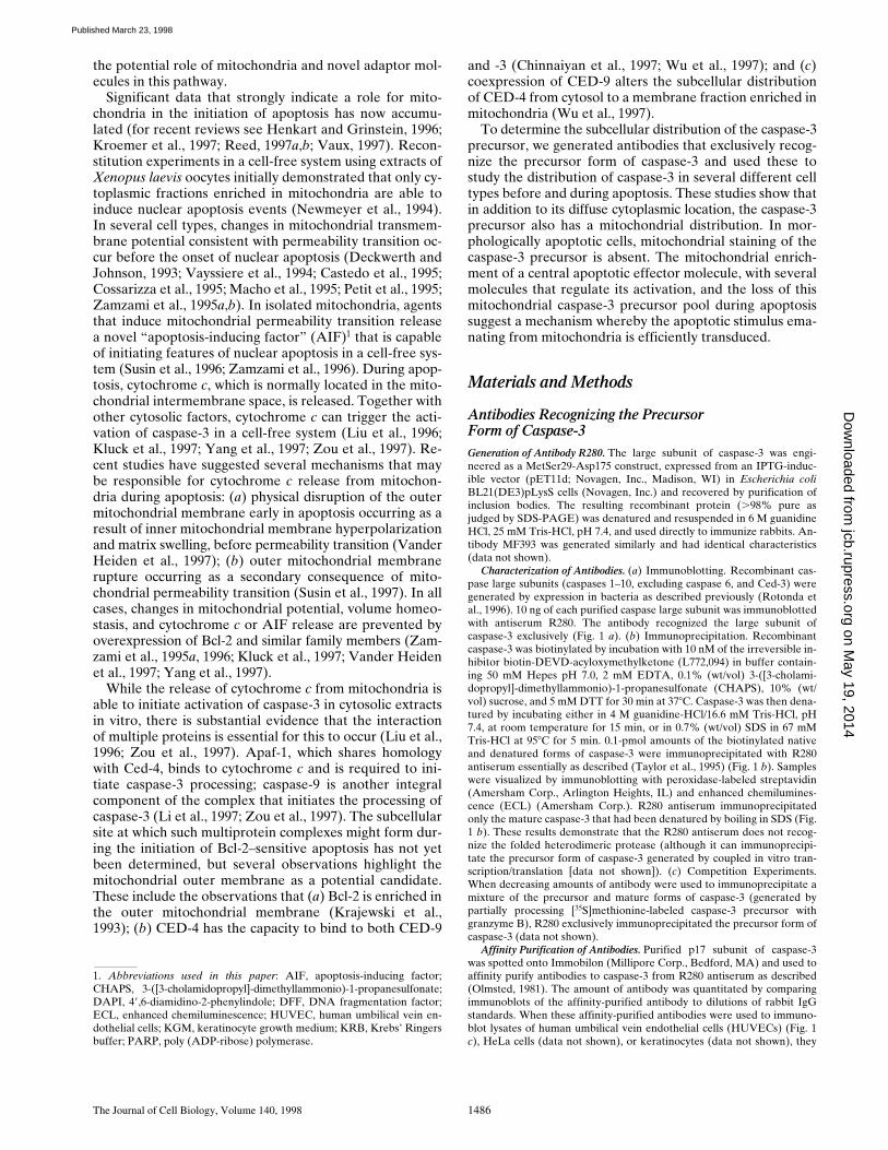
Characterization of Antibodies.
(
a
) Immunoblotting. Recombinant cas-pase large subunits (caspases 1–10, excluding caspase 6, and Ced-3) weregenerated by expression in bacteria as described previously (Rotonda etal., 1996). 10 ng of each purified caspase large subunit was immunoblottedwith antiserum R280. The antibody recognized the large subunit ofcaspase-3 exclusively (Fig. 1
a
). (
b
) Immunoprecipitation. Recombinantcaspase-3 was biotinylated by incubation with 10 nM of the irreversible in-hibitor biotin-DEVD-acyloxymethylketone (L772,094) in buffer contain-ing 50 mM Hepes pH 7.0, 2 mM EDTA, 0.1% (wt/vol) 3-([3-cholami-dopropyl]-dimethyllammonio)-1-propanesulfonate (CHAPS), 10% (wt/vol) sucrose, and 5 mM DTT for 30 min at 37
8
C. Caspase-3 was then dena-tured by incubating either in 4 M guanidine-HCl/16.6 mM Tris-HCl, pH7.4, at room temperature for 15 min, or in 0.7% (wt/vol) SDS in 67 mMTris-HCl at 95
8
C for 5 min. 0.1-pmol amounts of the biotinylated nativeand denatured forms of caspase-3 were immunoprecipitated with R280antiserum essentially as described (Taylor et al., 1995) (Fig. 1
b
). Sampleswere visualized by immunoblotting with peroxidase-labeled streptavidin(Amersham Corp., Arlington Heights, IL) and enhanced chemilumines-cence (ECL) (Amersham Corp.). R280 antiserum immunoprecipitatedonly the mature caspase-3 that had been denatured by boiling in SDS (Fig.1
b
). These results demonstrate that the R280 antiserum does not recog-nize the folded heterodimeric protease (although it can immunoprecipi-tate the precursor form of caspase-3 generated by coupled in vitro tran-scription/translation [data not shown]). (
c
) Competition Experiments.When decreasing amounts of antibody were used to immunoprecipitate amixture of the precursor and mature forms of caspase-3 (generated bypartially processing [
35
S]methionine-labeled caspase-3 precursor withgranzyme B), R280 exclusively immunoprecipitated the precursor form ofcaspase-3 (data not shown).
Affinity Purification of Antibodies.
Purified p17 subunit of caspase-3was spotted onto Immobilon (Millipore Corp., Bedford, MA) and used toaffinity purify antibodies to caspase-3 from R280 antiserum as described(Olmsted, 1981). The amount of antibody was quantitated by comparingimmunoblots of the affinity-purified antibody to dilutions of rabbit IgGstandards. When these affinity-purified antibodies were used to immuno-blot lysates of human umbilical vein endothelial cells (HUVECs) (Fig. 1
c
), HeLa cells (data not shown), or keratinocytes (data not shown), they
1.
Abbreviations used in this paper
: AIF, apoptosis-inducing factor;CHAPS, 3-([3-cholamidopropyl]-dimethyllammonio)-1-propanesulfonate;DAPI, 4
9
,6-diamidino-2-phenylindole; DFF, DNA fragmentation factor;ECL, enhanced chemiluminescence; HUVEC, human umbilical vein en-dothelial cells; KGM, keratinocyte growth medium; KRB, Krebs’ Ringersbuffer; PARP, poly (ADP-ribose) polymerase.
on May 19, 2014
jcb.rupress.orgD
ownloaded from
Published March 23, 1998
Mancini et al.
Mitochondrial Distribution of Caspase-3
1487
recognized only the caspase-3 precursor. A very minor band (
,
2% of thecaspase-3 precursor intensity) of 120 kD was variably seen.
Cell Culture and Induction of Apoptosis
Human keratinocytes were obtained from neonatal foreskins and culturedin keratinocyte growth medium (KGM) (Clonetics Corp., San Diego, CA) asdescribed (Casciola-Rosen et al., 1994
a
). Confluent secondary monolayerswere used for experiments. HUVECs were obtained from Clonetics Corp.,grown in endothelial cell growth medium (Clonetics Corp.), and used atthe third or fourth passage. HeLa and WIF-B cells were cultured as described(Ihrke et al., 1993; Casciola-Rosen et al., 1994
b
). Apoptosis was induced byirradiation with UVB (Casciola-Rosen et al., 1994
a
), C2-ceramide (Haimo-witz-Friedman et al., 1994), or staurosporine (Jacobson et al., 1996).
Visualization of Mitochondria and the Caspase-3 Precursor by Immunofluorescence Microscopy
Immunofluorescence was performed on cells that had been grown on No. 1glass coverslips.
Labeling of Caspase-3.
Cells were fixed with 4% paraformaldehyde for5 min at 4
8
C before being permeabilized with acetone (30 s at 4
8
C). To vi-sualize the precursor form of caspase-3, cells were incubated for 30 min onice with 10
m
g/ml affinity-purified anti–caspase-3 antibody (R280) andthen washed in PBS before incubating with fluorescein-conjugated goatanti–rabbit IgG (Jackson ImmunoResearch Laboratories, Inc., WestGrove, PA) for 20 min at room temperature.
Labeling of Mitochondria.
Mitochondria were labeled in intact cells withMitoTracker™ CMXRos-H
2
(Molecular Probes, Inc., Eugene, OR) ac-cording to the manufacturer’s directions.
Briefly, MitoTracker was dilutedto a final concentration of 1
m
M in serum-free medium (KGM for kerati-nocytes and DME for HUVECs). Coverslips containing viable kerati-nocytes and HUVECs were incubated in the appropriate MitoTracker-containing medium for 30–45 min in a 5% CO
2
humidified incubator. Thecells were then fixed and permeabilized and either mounted immediately(for single-labeled mitochondria) or were double-labeled with anti–caspase-3 antibody R280 as described above. When specified, nuclei were
visualized by labeling with 4
9
,6-diamidino-2-phenylindole (DAPI) (Mo-lecular Probes, Inc.). In all cases, coverslips were mounted on glass slideswith PermaFluor (Lipshaw, Pittsburgh, PA), and confocal microscopy wasperformed on a scanning confocal microscope system (model LSM 410;Carl Zeiss, Inc., Thornwood, NY).
Immunoelectron Microscopy
Immuno-EM was performed essentially as described (Berryman et al.,1992). Briefly, WIF-B cells (Ihrke et al., 1993) were fixed at room temper-ature for 10 min (3% paraformaldehyde, 0.05% glutaraldehyde in Hank’ssalt solution) and scraped, and the cell pellet was fixed for an additional 45min in this fixative. Cell pellets were washed with 3.5% sucrose in 0.1 Mphosphate buffer, pH 7.4 (SPB), and postfixed with 0.25% tannic acidin SPB for 60 min at 0
8
C before quenching free aldehydes for 60 min with50 mM ammonium chloride in SPB. After washing in sucrose-maleatebuffer, pH 6.5, containing 4% sucrose, cells were stained en bloc with 2%uranyl acetate in sucrose-maleate buffer, pH 6.0, dehydrated, and embed-ded in LR gold resin at
2
20
8
C. Silver-gold sections were mounted onformvar-coated 200-mesh nickel grids, blocked in 1% BSA/TBST (10 mMTris, pH 7.2, 500 mM NaCl, and 0.05% Tween 20), and stained for 2.5 hwith affinity-purified R280 antibodies (15
m
g/ml). After rinsing, grids werestained for 60 min with 12-nm colloidal gold-labeled donkey anti–rabbitIgG (Jackson ImmunoResearch Laboratories, Inc.; OD
520
5
0.05 [1:40 di-lution]) followed by 2% aqueous osmium tetroxide (15 min) and thencounterstained for 3 min with Reynold’s lead citrate. Sections wereviewed and photographed in an electron microscope (TEM type 1A; CarlZeizz, Inc.), and prints were made to a final enlargement of 21,000 magni-fication. For quantitation, the area of mitochondria and extramitochon-drial structures was measured, and the number of gold particles/cm
2
asso-ciated with mitochondria or other organelles was calculated (
.
200 goldparticles in 10 separate prints were quantitated).
Western Blot Analysis of Caspase-3 in Mitochondria Isolated from Human Liver
Mitochondria were isolated from human liver recovered at autopsy of a17-yr-old male (cause of death: accidental) or rat liver as previously de-scribed (Nicholson and McMurray, 1984). All steps were performed at4
8
C. The tissue was rinsed in SME buffer (0.25 M sucrose, 10 mM MOPS,2 mM EDTA, pH 7.4), rid of blood vessels, and dispersed in SME buffer.The suspension was centrifuged twice at 480
g
to sediment out nuclei andlarge cellular debris, and the mitochondria were recovered by centrifuga-tion at 4,300
g
for 10 min followed by a 2-min centrifugation at 10,000
g
.After washing the mitochondrial pellet four times in SME buffer to mini-mize microsomal contamination, the mitochondria were resuspended inSME buffer to a protein concentration of 16 mg/ml and stored at
2
80
8
C.To perform the experiments shown in Fig. 3, mitochondria (200
m
g)were diluted in SME buffer with or without NP-40 (final concentration1%). After a 1-h incubation at 37
8
C in the presence or absence of 0.2
m
ggranzyme B, the mitochondrial suspension was analyzed by SDS-PAGEand immunoblotted with the R280 anti–caspase-3 antibody essentially asdescribed (Scoggan et al., 1996) except that the primary antibody was usedat a dilution of 1:5,000. Identical results were obtained when mitochondriawere disrupted by repeated freeze-thaw (data not shown). Progressivedigitonin extraction and enzyme assays to measure adenylate kinase andfumarase were performed exactly as described on freshly isolated mito-chondria (Nicholson et al., 1987).
Subcellular Fractionation of HeLa Cells
Mitochondrial and cytosolic fractions were obtained by differential cen-trifugation of HeLa cell homogenates as described (Vander Heiden et al.,1997). Briefly, cells were washed twice with PBS, scraped, pelleted, andresuspended in 2.5 ml of ice-cold buffer A (250 mM sucrose, 20 mMHepes, pH 7.4, 10 mM KCl, 1.5 mM MgCl
2
, 1 mM EGTA, 1 mM EDTA,1 mM DTT, and the protease inhibitors PMSF, leupeptin, chymostatin,pepstatin, and antipain). Cells were homogenized with a Balch/Rothmanhomogenizer (Balch and Rothman, 1985), with ball bearings that allowedcalculated clearances of 0.0019 in or 0.0011 in. A postnuclear supernatantwas prepared and centrifuged at 10,000
g
for 25 min to obtain a pellethighly enriched in mitochondria. The pellet was resuspended in a volumeof buffer A equal to that of the postmitochondrial supernatant. The post-mitochondrial supernatant was further centrifuged at 100,000
g
to obtaincytosol. Equivalent volume amounts of mitochondrial and cytosolic frac-
Figure 1. Characterization of anti–caspase-3 antiserum (R280).(a) 10-ng amounts of the purified large subunits (caspases 1–10,excluding caspase-6) and Ced-3 were immunoblotted with R280antiserum. (b) Biotinylated recombinant mature caspase-3 wasdenatured by treatment with guanidium-HCl or SDS, as describedin the Materials and Methods section. Native and denatured bi-otinylated caspase-3 were subsequently immunoprecipitated us-ing R280, and the p17 subunit was detected by immunoblotting.(c) 46 mg of HUVEC lysate was immunoblotted with affinity-puri-fied R280 antiserum. Similar results were obtained when kerati-nocyte lysates were immunoblotted with affinity-purified R280antiserum (data not shown).
on May 19, 2014
jcb.rupress.orgD
ownloaded from
Published March 23, 1998
The Journal of Cell Biology, Volume 140, 1998 1488
tions were electrophoresed on 13.5% SDS–polyacrylamide gels, trans-ferred to polyvinyl difluoride, and immunoblotted with R280 (1:5,000) ormonoclonal antibodies to human cytochrome
c
(mAb 7H8.2C12 at 1.5
m
g/ml;Pharmingen, San Diego, CA) and cytochrome oxidase (subunit IV) (mAb20E8-C12 at 0.1
m
g/ml; Molecular Probes, Eugene, OR). Relative distri-butions of proteins in the two fractions were assessed by densitometry asdescribed below. While
.
95% of cytochrome
c
was associated with themitochondrial fraction in cells homogenized with a ball bearing clearanceof 0.0019 in,
.
95% of cytochrome
c
was found in the cytosol when cellswere homogenized with a ball bearing clearance of 0.0011 in (Fig. 3
c
anddata not shown; seven separate experiments). In contrast,
.
95% of cyto-chrome oxidase was located in the mitochondrial fraction regardless of theball bearing clearance (Fig. 3
c
and data not shown).
Purification of Human Granzyme B
Granzyme B was purified from granules isolated from cultured humannatural killer leukemia YT cells essentially as described (Hanna et al.,1993; Quan et al., 1995). The granule contents were extracted with NaCl(the calcium was omitted), diluted, and then loaded onto a Mono-S cat-ion-exchange column (0.5
3
0.5 cm; Pharmacia Biotech, Inc., Piscataway,NJ) that had been pre-equilibrated in 50 mM MES, pH 6.1, 25 mM NaCl.After washing with 12 vol of equilibration buffer, proteins were elutedwith a linear gradient up to 1 M NaCl (in 50 mM MES, pH 6.1) over 40column volumes. Granzyme B eluted at
z
0.6 M NaCl and was homoge-nous as judged by SDS-PAGE.
Cleavage Assays to Measure EndogenousCaspase-3 Activity
Generation of Cell Lysates.
Control or apoptotic keratinocytes (incubatedfor the indicated times after UVB irradiation) were washed in KrebsRingers buffer (KRB) containing 20 mM Hepes/NaOH, pH 7.4, 10 mMdextrose, 127 mM NaCl, 5.5 mM KCl, 1 mM CaCl
2
, and 2 mM MgSO
4
, andthen harvested by scraping into KRB, followed by centrifugation at 300
g
.The harvested cells were lysed in buffer containing 10 mM Hepes/KOH,pH 7.4, 2 mM EDTA, 1 mM DTT, 1% NP-40 (vol/vol), and the proteaseinhibitors PMSF, antipain, leupeptin, and pepstatin A to obtain whole celllysates (protein concentration
5
2.5 mg/ml). Aliquots of lysates were
taken for protein assays, and DTT was then added to samples to obtain afinal concentration of 5 mM DTT.
In Vitro Cleavage of Endogenous Poly (ADP-Ribose) Polymerase.
Wholecell lysates were incubated for 0 or 2 h at 37
8
C, as indicated in Fig. 7, be-fore the addition of SDS electrophoresis sample buffer. Samples wereelectrophoresed on 10% SDS–polyacrylamide gels containing 0.087%bisacrylamide, and the proteins were transferred to nitrocellulose. Endog-enous poly (ADP-ribose) polymerase (PARP) was detected by immuno-blotting with a patient serum monospecific for PARP as described (Casci-ola-Rosen et al., 1995). The densities of bands on autoradiograms weredetermined on a PDI Discovery densitometry system (Protein Databases,Inc., Huntington Station, NY) with Quantity One software (Protein Data-bases, Inc.).
Cleavage of Ac-DEVD-Aminomethylcoumarin by Cell Lysates.
To mea-sure Ac-DEVD-aminomethylcoumarin (Ac-DEVD-AMC) cleavage ac-tivity, lysates were added to reaction mixtures containing 10 or 100
m
MAc-DEVD-AMC, 100 mM Hepes, 10% sucrose, 0.1% CHAPS, 0.1 mMEDTA, and 1 mM DTT, pH 7.5, in a total volume of 100
m
l. Production ofAMC was monitored continuously at ambient temperature in an SLT Flu-ostar 96-well plate reader (Tecan U.S. Inc., Research Triangle Park, N.C.)using an excitation wavelength of 380 nm and an emission wavelength of460 nm. To confirm that the observed activity was due to caspase-3 ora closely related homologue, each sample was tested for inhibition by
N
-ethylmaleimide, Ac-DEVD-CHO (
K
i
caspase-3
5
0.35 nM), and Ac-YVAD-CHO (
K
i
caspase-3 5 10,000 nM). Activities that were inhibitedby N-ethylmaleimide (5 mM) and Ac-DEVD-CHO (500 nM) but not byAc-YVAD-CHO (500 nM) were attributed to caspase-3 or a closely re-lated homologue. Activity is expressed in U/mg, where a unit is defined asthe amount of enzyme required to produce 1 pmol of AMC per min using100 mM Ac-DEVD-AMC.
Results
The Caspase-3 Precursor Has a Cytosolic and Mitochondrial Distribution
The subcellular location of the caspase-3 precursor in hu-man foreskin keratinocytes was first determined with con-focal fluorescence microscopy (Fig. 2). Using affinity-puri-
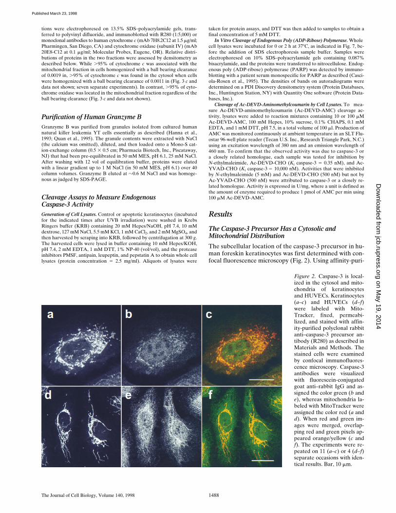
Figure 2. Caspase-3 is local-ized in the cytosol and mito-chondria of keratinocytesand HUVECs. Keratinocytes(a–c) and HUVECs (d–f)were labeled with Mito-Tracker, fixed, permeabi-lized, and stained with affin-ity-purified polyclonal rabbitanti–caspase-3 precursor an-tibody (R280) as described inMaterials and Methods. Thestained cells were examinedby confocal immunofluores-cence microscopy. Caspase-3antibodies were visualizedwith fluorescein-conjugatedgoat anti–rabbit IgG and as-signed the color green (b ande), whereas mitochondria la-beled with MitoTracker wereassigned the color red (a andd). When red and green im-ages were merged, overlap-ping red and green pixels ap-peared orange/yellow (c andf). The experiments were re-peated on 11 (a–c) or 4 (d–f)separate occasions with iden-tical results. Bar, 10 mm.
on May 19, 2014
jcb.rupress.orgD
ownloaded from
Published March 23, 1998
Mancini et al. Mitochondrial Distribution of Caspase-3 1489
fied polyclonal antibody R280, which exclusively recognizesthe caspase-3 precursor (see Materials and Methods), adiffuse cytosolic staining pattern with a superimposedpunctate perinuclear component was obtained; low levelsof diffuse nuclear staining were also observed (Fig. 2 b).The punctate component appeared similar to that seen formitochondria, prompting us to double-stain keratinocyteswith a mitochondrion-selective vital dye (MitoTracker)and R280. These studies confirmed that mitochondriawere distributed in a punctate, perinuclear pattern in kera-tinocytes (Fig. 2 a), and that the punctate fraction ofcaspase-3 colocalized with mitochondria (Fig 2 c; overlap-ping red and green pixels seen as yellow).
The specificity of the caspase-3 precursor staining wasconfirmed in the following ways: (a) Competition experi-ments were performed by preincubating affinity-purifiedanti–caspase-3 (R280) antibody with a 100-fold excess ofits p17 cognate antigen before use in immunofluorescence.In cells stained with these preincubated antibodies, thepunctate pattern (mitochondria) was abolished, and thediffuse cytoplasmic staining was greatly diminished (datanot shown). Since nuclear staining was minimally affected,this most likely represents nonspecific binding (data notshown). In contrast, when the anti–caspase-3 antibody wassimilarly preincubated with the p20 subunit of caspase-1,the punctate staining and diffuse cytoplasmic stainingwere not affected (data not shown). (b) No staining wasobserved when keratinocytes were incubated with second-ary antibody alone, nor when an affinity-purified anti–caspase-1 antibody was used (data not shown). (c) Whencells were single-stained with anti–caspase-3 antibodies,
an identical punctate staining pattern was seen, confirmingthat this pattern was directly due to antibody binding,rather than bleedthrough between channels in the double-stained samples (data not shown). (d) Perinuclear punc-tate and diffuse cytoplasmic staining patterns were ob-served when cells were fixed with 2% paraformaldehyde/0.5% glutaraldehyde; the level of cytoplasmic staining wasincreased and nuclear staining was decreased using thisfixation technique (data not shown). (e) An identical stain-ing pattern was observed when a different rabbit anti–caspase-3 precursor serum (antibody MF393; raised againstthe unfolded p17 subunit of caspase-3) was used to stainkeratinocytes (data not shown). Taken together, these datashow that the caspase-3 precursor has a cytosolic and mi-tochondrial distribution in keratinocytes.
To examine whether this distribution was unique to ke-ratinocytes, similar immunofluorescence experiments wereperformed using several other cell types. In HUVECs, mi-tochondria stained with a filamentous pattern that ex-tended through the cytoplasm to the cell periphery (Fig. 2d). The caspase-3 precursor shared an identical filamen-tous pattern but was also present diffusely in the cyto-plasm (Fig. 2 e). Double staining confirmed colocalizationof caspase-3 precursor and mitochondria (Fig. 2 f; overlap-ping red and green pixels seen as yellow). A similar mito-chondrial and cytosolic distribution of the caspase-3 pre-cursor was also observed in several other cells, includingprimary human myoblasts and myotubules, primary differ-entiated human neuronal cell cultures, MDCK cells, HeLacells, and WIF-B cells (data not shown).
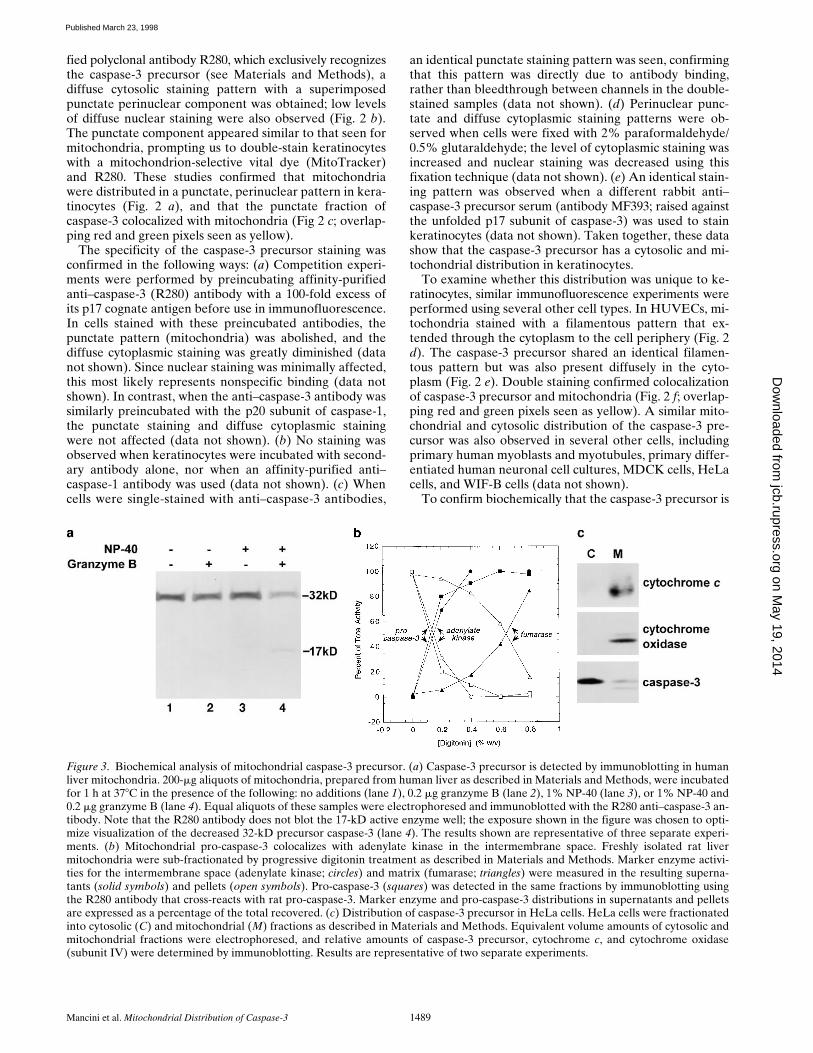
To confirm biochemically that the caspase-3 precursor is
Figure 3. Biochemical analysis of mitochondrial caspase-3 precursor. (a) Caspase-3 precursor is detected by immunoblotting in humanliver mitochondria. 200-mg aliquots of mitochondria, prepared from human liver as described in Materials and Methods, were incubatedfor 1 h at 378C in the presence of the following: no additions (lane 1), 0.2 mg granzyme B (lane 2), 1% NP-40 (lane 3), or 1% NP-40 and0.2 mg granzyme B (lane 4). Equal aliquots of these samples were electrophoresed and immunoblotted with the R280 anti–caspase-3 an-tibody. Note that the R280 antibody does not blot the 17-kD active enzyme well; the exposure shown in the figure was chosen to opti-mize visualization of the decreased 32-kD precursor caspase-3 (lane 4). The results shown are representative of three separate experi-ments. (b) Mitochondrial pro-caspase-3 colocalizes with adenylate kinase in the intermembrane space. Freshly isolated rat livermitochondria were sub-fractionated by progressive digitonin treatment as described in Materials and Methods. Marker enzyme activi-ties for the intermembrane space (adenylate kinase; circles) and matrix (fumarase; triangles) were measured in the resulting superna-tants (solid symbols) and pellets (open symbols). Pro-caspase-3 (squares) was detected in the same fractions by immunoblotting usingthe R280 antibody that cross-reacts with rat pro-caspase-3. Marker enzyme and pro-caspase-3 distributions in supernatants and pelletsare expressed as a percentage of the total recovered. (c) Distribution of caspase-3 precursor in HeLa cells. HeLa cells were fractionatedinto cytosolic (C) and mitochondrial (M) fractions as described in Materials and Methods. Equivalent volume amounts of cytosolic andmitochondrial fractions were electrophoresed, and relative amounts of caspase-3 precursor, cytochrome c, and cytochrome oxidase(subunit IV) were determined by immunoblotting. Results are representative of two separate experiments.
on May 19, 2014
jcb.rupress.orgD
ownloaded from
Published March 23, 1998
The Journal of Cell Biology, Volume 140, 1998 1490
localized in mitochondria, intact mitochondria were pre-pared and immunoblotted with R280 antibodies. The 32-kDcaspase-3 precursor alone was detected in these samples(Fig. 3 a, lane 1). To address whether caspase-3 was con-tained within these organelles or was associated with theouter surface, we incubated mitochondria in the presenceof granzyme B, a cytolytic lymphocyte granule proteaseknown to induce the processing and activation of caspase-3(Darmon et al., 1995; Quan et al., 1996). In NP-40–perme-abilized mitochondria (Fig. 3 a, lane 4), but not in mito-chondria incubated in the absence of detergent (Fig. 3 a,lane 2), addition of granzyme B produced an 80% de-crease in the amount of immunoblotted caspase-3 precur-sor, with concomitant appearance of the p17 subunit, aspreviously described (Martin et al., 1996; Quan et al.,1996). Similar results were obtained when repeated freeze-thawing was used to permeabilize mitochondria (data notshown). These biochemical data confirm the morphologi-cal finding that the caspase-3 precursor has a mitochon-drial distribution and indicate that it is contained withinthe organelle.
To investigate the submitochondrial location of thecaspase-3 precursor biochemically, purified mitochondriawere treated with increasing concentrations of digitoninbefore recentrifugation. The relative amounts of the cas-pase-3 precursor, adenylate kinase (a mitochondrial inter-membrane space marker enzyme), and fumarase (a mito-chondrial matrix marker) were assayed in the resultingpellets and supernatants. The release of caspase-3 precur-sor from mitochondria at progressively higher digitoninconcentrations coincided with adenylate kinase, with 50%of the total appearing in the supernatant at 0.15% (adeny-late kinase) and 0.125% (caspase-3 precursor) digitonin(Fig. 3 b). In contrast, 50% fumarase release into the su-pernatant only occurred at 0.65% digitonin (Fig. 3 b).
Since intermembrane space contents frequently leakfrom mitochondria during subcellular fractionation proce-dures, establishing an accurate stoichiometry of distribu-tion of caspase-3 precursor in mitochondrial versus post-mitochondrial fractions required a fractionation procedurethat maintained the outer mitochondrial membrane integ-rity (see Materials and Methods). Cytochrome c and cyto-chrome oxidase (subunit IV) were used as markers of theintermembrane space and inner membrane, respectively.Under homogenization conditions where .95% of cyto-chrome c was retained in mitochondria, the amount ofcaspase-3 precursor in mitochondria was z10% that foundin cytosol (equal cell equivalents assayed; Fig. 3 c). Inseveral separate experiments, caspase-3 immunoblotted asboth the intact 32-kD form and a 29-kD form, most likelyrepresenting the protease without its prodomain (Fig. 3 c).In contrast, very low levels (,1%) of caspase-3 precursorwere retained in mitochondria under conditions where.90% of cytochrome c was present in the cytosol (datanot shown). Taken together, these data demonstrate that asignificant proportion of the caspase-3 precursor is mito-chondrial in distribution. Furthermore, since caspase-3precursor is released from mitochondria at digitonin con-centrations almost identical to those required to releaseadenylate kinase (Fig. 3 b) and retained in mitochondriaonly during homogenization conditions that maintain cy-tochrome c in the intermembrane space (Fig. 3 c), we
conclude that caspase-3 is most likely located in the mito-chondrial intermembrane space. These data are also con-sistent with the inaccessibility of the caspase-3 precursorto granzyme B in nonpermeabilized mitochondria (Fig. 3a). It is also possible that the caspase-3 precursor is tightlyassociated with the outer surface of mitochondria.
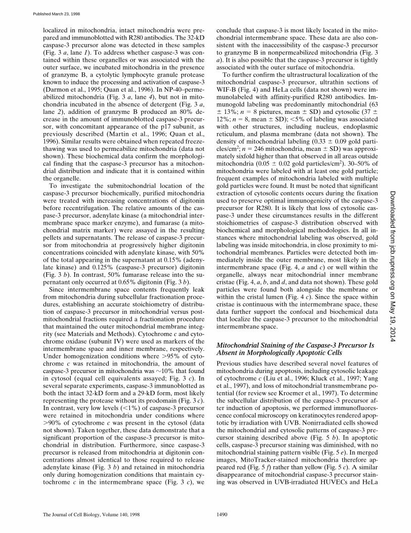
To further confirm the ultrastructural localization of themitochondrial caspase-3 precursor, ultrathin sections ofWIF-B (Fig. 4) and HeLa cells (data not shown) were im-munolabeled with affinity-purified R280 antibodies. Im-munogold labeling was predominantly mitochondrial (636 13%; n 5 8 pictures, mean 6 SD) and cytosolic (37 612%; n 5 8, mean 6 SD); ,5% of labeling was associatedwith other structures, including nucleus, endoplasmicreticulum, and plasma membrane (data not shown). Thedensity of mitochondrial labeling (0.33 6 0.09 gold parti-cles/cm2; n 5 246 mitochondria, mean 6 SD) was approxi-mately sixfold higher than that observed in all areas outsidemitochondria (0.05 6 0.02 gold particles/cm2). 30–50% ofmitochondria were labeled with at least one gold particle;frequent examples of mitochondria labeled with multiplegold particles were found. It must be noted that significantextraction of cytosolic contents occurs during the fixationused to preserve optimal immunogenicity of the caspase-3precursor for R280. It is likely that loss of cytosolic cas-pase-3 under these circumstances results in the differentstoichiometries of caspase-3 distribution observed withbiochemical and morphological methodologies. In all in-stances where mitochondrial labeling was observed, goldlabeling was inside mitochondria, in close proximity to mi-tochondrial membranes. Particles were detected both im-mediately inside the outer membrane, most likely in theintermembrane space (Fig. 4, a and c) or well within theorganelle, always near mitochondrial inner membranecristae (Fig. 4, a, b, and d, and data not shown). These goldparticles were found both alongside the membrane orwithin the cristal lumen (Fig. 4 c). Since the space withincristae is continuous with the intermembrane space, thesedata further support the confocal and biochemical datathat localize the caspase-3 precursor to the mitochondrialintermembrane space.
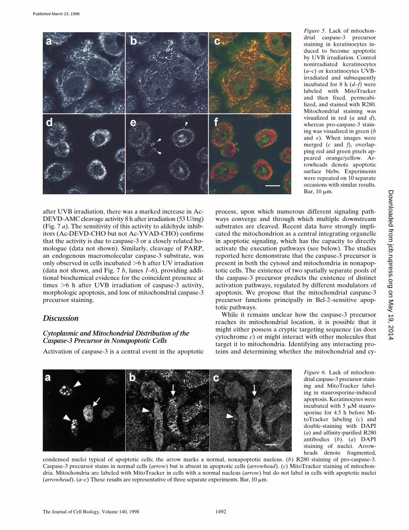
Mitochondrial Staining of the Caspase-3 Precursor Is Absent in Morphologically Apoptotic Cells
Previous studies have described several novel features ofmitochondria during apoptosis, including cytosolic leakageof cytochrome c (Liu et al., 1996; Kluck et al., 1997; Yanget al., 1997), and loss of mitochondrial transmembrane po-tential (for review see Kroemer et al., 1997). To determinethe subcellular distribution of the caspase-3 precursor af-ter induction of apoptosis, we performed immunofluores-cence confocal microscopy on keratinocytes rendered apop-totic by irradiation with UVB. Nonirradiated cells showedthe mitochondrial and cytosolic patterns of caspase-3 pre-cursor staining described above (Fig. 5 b). In apoptoticcells, caspase-3 precursor staining was diminished, with nomitochondrial staining pattern visible (Fig. 5 e). In mergedimages, MitoTracker-stained mitochondria therefore ap-peared red (Fig. 5 f) rather than yellow (Fig. 5 c). A similardisappearance of mitochondrial caspase-3 precursor stain-ing was observed in UVB-irradiated HUVECs and HeLa
on May 19, 2014
jcb.rupress.orgD
ownloaded from
Published March 23, 1998

Mancini et al. Mitochondrial Distribution of Caspase-3 1491
cells, as well as in keratinocytes and HeLa cells in whichapoptosis was induced with C2-ceramide (data notshown). Interestingly, no enrichment of caspase-3 precur-sor staining was observed in apoptotic surface blebs (Fig. 5e, arrowheads), although we have detected the mature ac-tive enzyme in these structures (data not shown).
We also addressed the effects of staurosporine treat-ment on the mitochondrial staining of the caspase-3 pre-cursor in keratinocytes (Fig. 6) and HeLa cells (data notshown). This agent does not induce the characteristic sur-face blebbing and cytosolic shrinkage of apoptosis butdoes induce classic nuclear condensation and fragmenta-tion (detected by DAPI staining in Fig. 6 a), caspase-medi-ated substrate cleavage, and internucleosomal DNA deg-radation (Jacobson et al., 1996; and data not shown). Sinceapoptosis occurs asynchronously in cell populations, adja-cent nonapoptotic and apoptotic cells can be visualized si-multaneously. In cells with the condensed, fragmented nu-cleus characteristic of apoptotic cells (Fig. 6 a, arrowheads),mitochondrial caspase-3 staining was absent (Fig. 6 b, ar-rowhead). In this field, the adjacent cell with a nonapop-totic chromatin pattern (Fig. 6 a, arrow) had normal mito-chondrial staining of the caspase-3 precursor (Fig. 6 b,
arrow). Interestingly, unlike cells in which apoptosis wasinduced by UV irradiation, mitochondria in staurosporine-treated apoptotic cells that lacked caspase-3 staining alsofailed to label with MitoTracker, indicating a markedly re-duced mitochondrial transmembrane potential, possiblydue to permeability transition.
Appearance of Caspase-3 Activity in Apoptotic Cells
Since antibody R280 recognizes only the caspase-3 precur-sor, decreased R280 staining in apoptotic cells is consistentwith activation of caspase-3. To address biochemicallywhether the caspase-3 activity was present in cell popula-tions containing morphologically apoptotic cells that lackedmitochondrial staining with antibody R280, we measuredcleavage of an exogenous peptide substrate (Ac-DEVD-AMC) in keratinocyte or HeLa cell lysates known to sup-port the activity of caspase-3 (Nicholson et al., 1995; Casci-ola-Rosen et al., 1996). In nonirradiated cells, backgroundlevels of Ac-DEVD-AMC cleavage activity were observed(14 U/mg); these did not increase up to 4 h after irradia-tion of parallel cultures (Fig. 7 a). Consistent with thepresence of numerous apoptotic cells at time points .6 h
Figure 4. Immunolocalization ofthe caspase-3 precursor. Ultrathinsections of cultured WIF-B cellswere labeled with affinity-puri-fied R280 antibodies and 12-nmcolloidal gold conjugated to don-key anti–rabbit IgG. a–d showcaspase-3 precursor stainingwithin mitochondria. Gold parti-cles were localized to just insidethe outer membrane (a and c),as well as to inner membranecristae (a, b, and d). Omission ofprimary antibody resulted inthe complete abolition of bothcytosolic and mitochondrial stain-ing (data not shown). Similar re-sults were obtained in HeLa cells(data not shown). Bar, 300 nm.
on May 19, 2014
jcb.rupress.orgD
ownloaded from
Published March 23, 1998
The Journal of Cell Biology, Volume 140, 1998 1492
after UVB irradiation, there was a marked increase in Ac-DEVD-AMC cleavage activity 8 h after irradiation (53 U/mg)(Fig. 7 a). The sensitivity of this activity to aldehyde inhib-itors (Ac-DEVD-CHO but not Ac-YVAD-CHO) confirmsthat the activity is due to caspase-3 or a closely related ho-mologue (data not shown). Similarly, cleavage of PARP,an endogenous macromolecular caspase-3 substrate, wasonly observed in cells incubated .6 h after UV irradiation(data not shown, and Fig. 7 b, lanes 1–6), providing addi-tional biochemical evidence for the coincident presence attimes .6 h after UVB irradiation of caspase-3 activity,morphologic apoptosis, and loss of mitochondrial caspase-3precursor staining.
Discussion
Cytoplasmic and Mitochondrial Distribution of the Caspase-3 Precursor in Nonapoptotic Cells
Activation of caspase-3 is a central event in the apoptotic
process, upon which numerous different signaling path-ways converge and through which multiple downstreamsubstrates are cleaved. Recent data have strongly impli-cated the mitochondrion as a central integrating organellein apoptotic signaling, which has the capacity to directlyactivate the execution pathways (see below). The studiesreported here demonstrate that the caspase-3 precursor ispresent in both the cytosol and mitochondria in nonapop-totic cells. The existence of two spatially separate pools ofthe caspase-3 precursor predicts the existence of distinctactivation pathways, regulated by different modulators ofapoptosis. We propose that the mitochondrial caspase-3precursor functions principally in Bcl-2–sensitive apop-totic pathways.
While it remains unclear how the caspase-3 precursorreaches its mitochondrial location, it is possible that itmight either possess a cryptic targeting sequence (as doescytochrome c) or might interact with other molecules thattarget it to mitochondria. Identifying any interacting pro-teins and determining whether the mitochondrial and cy-
Figure 5. Lack of mitochon-drial caspase-3 precursorstaining in keratinocytes in-duced to become apoptoticby UVB irradiation. Controlnonirradiated keratinocytes(a–c) or keratinocytes UVB-irradiated and subsequentlyincubated for 8 h (d–f) werelabeled with MitoTrackerand then fixed, permeabi-lized, and stained with R280.Mitochondrial staining wasvisualized in red (a and d),whereas pro-caspase-3 stain-ing was visualized in green (band e). When images weremerged (c and f), overlap-ping red and green pixels ap-peared orange/yellow. Ar-rowheads denote apoptoticsurface blebs. Experimentswere repeated on 10 separateoccasions with similar results.Bar, 10 mm.
Figure 6. Lack of mitochon-drial caspase-3 precursor stain-ing and MitoTracker label-ing in staurosporine-inducedapoptosis. Keratinocytes wereincubated with 5 mM stauro-sporine for 4.5 h before Mi-toTracker labeling (c) anddouble-staining with DAPI(a) and affinity-purified R280antibodies (b). (a) DAPIstaining of nuclei. Arrow-heads denote fragmented,
condensed nuclei typical of apoptotic cells; the arrow marks a normal, nonapoptotic nucleus. (b) R280 staining of pro-caspase-3.Caspase-3 precursor stains in normal cells (arrow) but is absent in apoptotic cells (arrowhead). (c) MitoTracker staining of mitochon-dria. Mitochondria are labeled with MitoTracker in cells with a normal nucleus (arrow) but do not label in cells with apoptotic nuclei(arrowhead). (a–c) These results are representative of three separate experiments. Bar, 10 mm.
on May 19, 2014
jcb.rupress.orgD
ownloaded from
Published March 23, 1998

Mancini et al. Mitochondrial Distribution of Caspase-3 1493
tosolic caspase-3 precursors are equivalent in terms of ac-tivation mechanism remains a high priority.
Mitochondrial Caspase-3 Precursor Staining Is Absent in Apoptotic Cells
Our studies demonstrate that the caspase-3 precursor lo-calizes to mitochondria in healthy cells but is absent fromthis site in apoptotic cells. This loss of caspase-3 precursorstaining in mitochondria is induced in a variety of celltypes by diverse apoptotic stimuli including UV irradia-
tion, staurosporine, or C2-ceramide. Caspase-3 activity isonly detectable in cell populations containing morphologi-cally apoptotic cells, indicating that the loss of mitochon-drial caspase-3 staining and caspase-3 activation are tem-porally linked. The inability of R280 to recognize themature form of caspase-3, as well as the asynchronousnature of apoptosis in cell populations, prevents us fromdistinguishing whether caspase-3 activation occurs in mi-tochondria, or whether the caspase-3 precursor and cy-tochrome c are redistributed from mitochondria before ac-tivation.
Interestingly, the mitochondrial pool of caspase-3 colo-calizes with cytochrome c, which has also been shown toleak from mitochondria into the cytosol early during apop-tosis (before alteration of mitochondrial transmembranepotential). Accumulating evidence suggests that the re-lease of cytochrome c from mitochondria plays a criticalrole in initiating the apoptotic process: (a) The anti-apop-totic proteins Bcl-2 or Bcl-XL (which localize to the outermitochondrial membrane) prevent cytochrome c releasefrom mitochondria in vitro and in vivo and prevent activa-tion of caspase-3 (Kluck et al., 1997; Vander Heiden et al.,1997; Yang et al., 1997). (b) Cytochrome c, in conjunctionwith two protein cofactors (Apaf-1 and caspase-9/Apaf-3)and dATP/ATP, stimulate the conversion of the caspase-3precursor to its active form in cell-free systems (Li et al.,1997; Zou et al., 1997). These studies strongly support thehypothesis that spatial separation of interacting proteins iskey to modulation of the apoptotic process. This hypothe-sis predicts that cofactors that participate in cytochromec–mediated processing of caspase-3 (e.g., Apaf-1 andcaspase-9) are segregated from the caspase-3 precursorand cytochrome c in healthy cells, and that proapoptoticsignals permit the efficient formation of an activation com-plex, with ensuing apoptosis.
The release of cytochrome c from mitochondria duringapoptosis precedes mitochondrial membrane depolariza-tion (Kluck et al., 1997; Yang et al., 1997), whereas releaseof AIF requires permeability transition (Susin et al., 1996).These differences might reflect the existence of multiplemechanisms through which mitochondria can influencecell death pathways. The observations that mitochondriain keratinocytes in which apoptosis is induced by UV irra-diation still label with MitoTracker but fail to label whenapoptosis is induced by staurosporine suggest that thesedifferent pathways might be operating to different extentsin these settings. Whether active caspase-3 can affect mito-chondrial membrane potential remains unknown but is ofsignificant interest.
In conclusion, the coenrichment of caspase-3 at a sitecontaining several molecules that regulate its activationsuggests a mechanism whereby the apoptotic stimulus em-anating from mitochondria is efficiently transduced. Whileall apoptotic stimuli appear to converge on a common setof “executioner” proteases, the upstream mechanisms thatactivate this final common pathway appear to vary for dif-ferent stimuli. We propose that the mitochondrial subpop-ulation of caspase-3 precursor molecules is spatially andbiochemically distinct and is coupled to a unique subset ofapoptotic signaling pathways, which are Bcl-2 sensitive.These apoptotic signals are transduced by bringing to-gether locally enriched but spatially separated molecules
Figure 7. Time course of appearance of cleavage activity in kera-tinocyte lysates after UVB irradiation. (a) Whole cell lysates pre-pared at various times after UVB irradiation were assayed forcleavage of the fluorogenic substrate Ac-DEVD-AMC as de-scribed in the Materials and Methods section. Cleavage activity isexpressed in U/mg, where a unit is defined as the amount of en-zyme needed to produce 1 pmol per min using 100 mM Ac-DEVD-AMC. Four separate experiments yielded similar results.(b) PARP cleavage is observed in keratinocytes 6 h after UV ir-radiation. Irradiated and control nonirradiated keratinocyteswere lysed at the indicated times as described in Materials andMethods. Whole cell lysates (lanes 1–3), as well as whole cell ly-sates incubated in vitro for 2 h at 378C (lanes 4–6), were immuno-blotted with a serum recognizing PARP (Casciola-Rosen et al.,1995). The migration positions of intact PARP (113 kD) and itscleavage fragment (89 kD) are marked. 60 mg of protein wasloaded in each lane. This experiment was repeated on three sepa-rate occasions with similar results. Identical results were obtainedusing HeLa cells.
on May 19, 2014
jcb.rupress.orgD
ownloaded from
Published March 23, 1998
The Journal of Cell Biology, Volume 140, 1998 1494
(e.g., cytochrome c, caspase-3 precursor, Apaf-1, caspase-9), thus permitting the formation of a multiprotein com-plex that catalyzes efficient activation of caspase-3. Alter-natively, the mitochondrial caspase-3 precursor may havea unique upstream function in some forms of apoptosis,perhaps becoming activated in mitochondria before alter-ation of mitochondrial physiology. Determining the effectsof Bcl-2 family members on the function of this mitochon-drial caspase-3 pool is a major priority.
We thank Ann Hubbard for helpful discussions, Greg Martin for assis-tance with immuno-EM, and Chip Montrose (all three from Johns Hop-kins University) for assistance with confocal microscopy.
This work was supported by National Institutes of Health grants 5T32-AI07247 and K12DK01298, a Career Development Award form the Der-matology Foundation/Lever Brothers Co. (LCR), AR44684 (LCR), thePeggy Meyerhoff-Pearlstone Foundation, the Howard Bank MemorialFund, and an Arthritis Foundation (Maryland Chapter) InstitutionalGrant. A. Rosen is a Pew Scholar in the Biomedical Sciences.
Received for publication 18 September 1997 and in revised form 22 Janu-ary 1998.
References
Alnemri, E.S., D.J. Livingston, D.W. Nicholson, G. Salvesen, N.A. Thornberry,W.W. Wong, and J.Y. Yuan. 1996. Human ICE/CED-3 protease nomencla-ture. Cell. 87:171.
Balch, W.E., and J.E. Rothman. 1985. Characterization of protein transport be-tween successive compartments of the Golgi: asymmetric properties of do-nor and acceptor activities in a cell-free system. Arch. Biochem. Biophys.240:413–425.
Berryman, M.A., W.R. Porter, R.D. Rodewald, and A.L. Hubbard. 1992. Ef-fects of tannic acid on antigenicity and membrane contrast in ultrastructuralimmunocytochemistry. J. Histochem. Cytochem. 40:845–857.
Boldin, M.P., E.E. Varfolomeev, Z. Pancer, I.L. Mett, J.H. Camonis, and D.Wallach. 1995. A novel protein that interacts with the death domain of Fas/APO-1 contains a sequence motif related to the death domain. J. Biol.Chem. 270:7795–7798.
Boldin, M.P., T.M. Goncharov, Y.V. Goltsev, and D. Wallach. 1996. Involve-ment of MACH, a novel MORT1/FADD-interacting protease, in Fas/APO-1-and TNF receptor-induced cell death. Cell. 85:803–815.
Casciola-Rosen, L.A., G. Anhalt, and A. Rosen. 1994a. Autoantigens targetedin systemic lupus erythematosus are clustered in two populations of surfacestructures on apoptotic keratinocytes. J. Exp. Med. 179:1317–1330.
Casciola-Rosen, L.A., D.K. Miller, G.J. Anhalt, and A. Rosen. 1994b. Specificcleavage of the 70-kD protein component of the U1 small nuclear ribonucle-oprotein is a characteristic biochemical feature of apoptotic cell death. J.Biol. Chem. 269:30757–30760.
Casciola-Rosen, L.A., G.J. Anhalt, and A. Rosen. 1995. DNA-dependent pro-tein kinase is one of a subset of autoantigens specifically cleaved early duringapoptosis. J. Exp. Med. 182:1625–1634.
Casciola-Rosen, L.A., D.W. Nicholson, T. Chong, K.R. Rowan, N.A. Thorn-berry, D.K. Miller, and A. Rosen. 1996. Apopain/CPP32 cleaves proteinsthat are essential for cellular repair: a fundamental principle of apoptoticdeath. J. Exp. Med. 183:1957–1964.
Castedo, M., A. Macho, N. Zamzami, T. Hirsch, P. Marchetti, J. Uriel, and G.Kroemer. 1995. Mitochondrial perturbations define lymphocytes undergoingapoptotic depletion in vivo. Eur. J. Immunol. 25:3277–3284.
Chinnaiyan, A.M., and V.M. Dixit. 1996. The cell-death machine. Curr. Biol.6:555–562.
Chinnaiyan, A.M., K. O’Rourke, M. Tewari, and V.M. Dixit. 1995. FADD, anovel death domain-containing protein, interacts with the death domain ofFas and initiates apoptosis. Cell. 81:505–512.
Chinnaiyan, A.M., W.L. Hanna, K. Orth, H.J. Duan, G.G. Poirier, C.J.Froelich, and V.M. Dixit. 1996a. Cytotoxic T-cell-derived granzyme B acti-vates the apoptotic protease ICE-LAP3. Curr. Biol. 6:897–899.
Chinnaiyan, A.M., K. Orth, K. O’Rourke, H.J. Duan, G.G. Poirier, and V.M.Dixit. 1996b. Molecular ordering of the cell death pathway: Bcl-2 and Bcl-xLfunction upstream of the Ced-3-like apoptotic proteases. J. Biol. Chem. 271:4573–4576.
Chinnaiyan, A.M., K. O’Rourke, B.R. Lane, and V.M. Dixit. 1997. Interactionof CED-4 with CED-3 and CED-9: a molecular framework for cell death.Science. 275:1122–1126.
Cossarizza, A., C. Franceschi, D. Monti, S. Salvioli, E. Bellesia, R. Rivabene, L.Biondo, G. Rainaldi, A. Tinari, and W. Malorni. 1995. Protective effect ofN-acetyl-cysteine in tumor necrosis factor-a-induced apoptosis in U937 cells:the role of mitochondria. Exp. Cell Res. 220:232–240.
Darmon, A.J., D.W. Nicholson, and R.C. Bleackley. 1995. Activation of the apop-
totic protease CPP32 by cytotoxic T-cell-derived granzyme B. Nature. 377:446–448.
Deckwerth, T.L., and E.M. Johnson. 1993. Temporal analysis of events associ-ated with programmed cell death (apoptosis) of sympathetic neurons de-prived of nerve growth factor. J. Cell Biol. 123:1207–1222.
Duan, H., K. Orth, A.M. Chinnaiyan, G.G. Poirier, C.J. Froelich, W.-W. He,and V.M. Dixit. 1996. ICE-LAP6, a novel member of the ICE/Ced-3 genefamily, is activated by the cytotoxic T cell protease granzyme B. J. Biol.Chem. 271:16720–16724.
Erhardt, P., and G.M. Cooper. 1996. Activation of the CPP32 apoptotic pro-tease by distinct signaling pathways with differential sensitivity to Bcl-xL. J.Biol. Chem. 271:17601–17604.
Gu, Y., C. Sarnecki, M.A. Fleming, J.A. Lippke, R.C. Bleakley, and M.S.S. Su.1996. Processing and activation of CMH-1 by granzyme B. J. Biol. Chem.271:10816–10820.
Haimowitz-Friedman, A., C.-C. Kan, D. Ehleiter, R.S. Persaud, M. McLough-lin, Z. Fuks, and R.N. Kolesnick. 1994. Ionizing radiation acts on cellularmembranes to generate ceramide and initiate apoptosis. J. Exp. Med. 180:525–535.
Hanna, W.L., X. Zhang, J. Turbov, U. Winkler, D. Hudig, and C.J. Froelich.1993. Rapid purification of cationic granule proteases: application to humangranzymes. Prot. Expr. Purif. 3:398–404.
Henkart, P.A., and S. Grinstein. 1996. Apoptosis: mitochondria resurrected? J.Exp. Med. 183:1293–1295.
Ihrke, G., E.B. Neufeld, T. Meads, M.R. Shanks, D. Cassio, M. Laurent, T.A.Schroer, R.E. Pagano, and A.L. Hubbard. 1993. WIF-B cells: an in vitromodel for studies of hepatocyte polarity. J. Cell Biol. 123:1761–1775.
Jacobson, M.D., M. Weil, and M.C. Raff. 1996. Role of Ced3/ICE-family pro-teases in staurosporine-induced programmed cell death. J. Cell Biol. 133:1041–1051.
Jacobson, M.D., M. Weil, and M.C. Raff. 1997. Programmed cell death in ani-mal development. Cell. 88:347–354.
Kluck, R.M., E. Bossy-Wetzel, D.R. Green, and D.D. Newmeyer. 1997. The re-lease of cytochrome c from mitochondria: a primary site for Bcl-2 regulationof apoptosis. Science. 275:1132–1136.
Krajewski, S., S. Tanaka, S. Takayama, M.J. Schibler, W. Fenton, and J.C.Reed. 1993. Investigation of the subcellular distribution of the Bcl-2 onco-protein: residence in the nuclear envelope, endoplasmic reticulum, and outermitochondrial membranes. Cancer Res. 53:4701–4714.
Kroemer, G., N. Zamzami, and S.A. Susin. 1997. Mitochondrial control of apop-tosis. Immunol. Today. 44:44–51.
Li, P., D. Nijhawan, I. Budihardjo, S.M. Srinivasula, M. Ahmad, E.S. Alnemri,and X. Wang. 1997. Cytochrome c and dATP-dependent formation of Apaf-1/caspase-9 complex initiates an apoptotic protease cascade. Cell. 91:479–489.
Liu, X., C.N. Kim, J. Yang, R. Jemmerson, and X. Wang. 1996. Induction of apop-totic program in cell-free extracts: requirement for dATP and cytochrome c.Cell. 86:147–157.
Macho, A., M. Castedo, P. Marchetti, J.J. Aguilar, D. Decaudin, N. Zamzami,P.M. Girard, J. Uriel, and G. Kroemer. 1995. Mitochondrial dysfunctions incirculating T lymphocytes from human immunodeficiency virus-1 carriers.Blood. 86:2481–2487.
Martin, S., G. Amarante-Mendes, L. Shi, T.-H. Chuang, C. Casiano, G.O’Brian, P. Fitzgerald, E. Tan, G. Bokoch, A. Greenberg, and D. Green.1996. The cytotoxic cell protease granzyme B initiates apoptosis in a cell-freesystem by proteolytic processing and activation of the ICE/CED-3 familyprotease, CPP32, via a novel two-step mechanism. EMBO (Eur. Mol. Biol.Organ.) J. 15:2407–2416.
Martin, S.J., and D.R. Green. 1995. Protease activation during apoptosis: deathby a thousand cuts? Cell. 82:349–352.
Muzio, M., A.M. Chinnaiyan, F.C. Kischkel, K. O’Rourke, A. Shevchenko, J.Ni, C. Scaffidi, J.D. Bretz, M. Zhang, R. Gentz, et al. 1996. FLICE, a novelFADD-homologous ICE/CED-3-like protease, is recruited to the CD95(Fas/APO-1) death-inducing signaling complex. Cell. 85:817–827.
Muzio, M., G.S. Salvesen, and V.M. Dixit. 1997. FLICE induced apoptosis in acell-free system—cleavage of caspase zymogens. J. Biol. Chem. 272:2952–2956.
Newmeyer, D.D., D.M. Farschon, and J.C. Reed. 1994. Cell-free apoptosis inXenopus egg extracts: inhibition by Bcl-2 and requirement for an organellefraction enriched in mitochondria. Cell. 79:353–364.
Nicholson, D.W., A. Ali, N.A. Thornberry, J.P. Vaillancourt, C.K. Ding, M.Gallant, Y. Gareau, P.R. Griffin, M. Labele, Y.A. Lazebnik, et al. 1995.Identification and inhibition of the ICE/CED-3 protease necessary for mam-malian apoptosis. Nature. 376:37–43.
Nicholson, D.W., and W.C. McMurray. 1984. CDP-diglyceride hydrolase frompig liver mitochondria. Can. J. Biochem. Cell Biol. 62:1205–1216.
Nicholson, D.W., and N.A. Thornberry. 1997. Caspases: killer proteases.Trends Biochem. Sci. 22:299–306.
Nicholson, D.W., H. Kohler, and W. Neupert. 1987. Import of cytochrome cinto mitochondria. Cytochrome c heme lyase. Eur. J. Biochem. 164:147–157.
Olmsted, J.B. 1981. Affinity purification of antibodies from diazotized paperblots of heterogeneous protein samples. J. Biol. Chem. 256:11955–11957.
Orth, K., K. O’Rourke, G.S. Salvesen, and V.M. Dixit. 1996. Molecular order-ing of apoptotic mammalian CED-3/ICE-like proteases. J. Biol. Chem. 271:20977–20980.
Petit, P.X., H. Lecoeur, E. Zorn, C. Dauguet, B. Mignotte, and M.-L. Gougeon.
on May 19, 2014
jcb.rupress.orgD
ownloaded from
Published March 23, 1998
Mancini et al. Mitochondrial Distribution of Caspase-3 1495
1995. Alterations in mitochondrial structure and function are early events ofdexamethasone-induced thymocyte apoptosis. J. Cell Biol. 130:157–167.
Quan, L.T., A. Caputo, R.C. Bleakley, D.J. Pickup, and G.S. Salveson. 1995.Granzyme B is inhibited by the cowpox virus serpin cytokine response mod-ifier A. J. Biol. Chem. 270:10377–10379.
Quan, L.T., M. Tewari, K. O’Rourke, V. Dixit, S.J. Snipas, G.G. Poirier, C.Ray, D.J. Pickup, and G.S. Salveson. 1996. Proteolytic activation of the celldeath protease Yama/CPP32 by granzyme B. Proc. Natl. Acad. Sci. USA. 93:1972–1976.
Reed, J.C. 1994. Bcl-2 and the regulation of programmed cell death. J. CellBiol. 124:1–6.
Reed, J.C. 1997a. Cytochrome c: can't live with it—can't live without it. Cell. 91:559–562.
Reed, J.C. 1997b. Double identity for proteins of the Bcl-2 family. Nature. 387:773–776.
Rotonda, J., D.W. Nicholson, K.M. Fazil, M. Gallant, Y. Gareau, M. Labele,E.P. Peterson, D.M. Rasper, R. Ruel, J.P. Vaillancourt, et al. 1996. Thethree-dimensional structure of apopain/CPP32, a key mediator of apoptosis.Nat. Struct. Biol. 3:619–625.
Salvesen, G., and V.M. Dixit. 1997. Caspases: intracellular signaling by proteol-ysis. Cell. 91:443–446.
Scoggan, K.A., D.W. Nicholson, and A.W. Ford-Hutchinson. 1996. Regulationof leukotriene-biosynthetic enzymes during differentiation of myelocyticHL-60 cells to eosinophilic or neutrophilic cells. Eur. J. Biochem. 230:572–578.
Shi, L., G. Chen, G. MacDonald, L. Bergeron, H. Li, M. Miura, R.J. Rotello,D.K. Miller, T. Seshadri, J. Yuan, and A.H. Greenberg. 1996. Activation ofan interleukin 1b converting enzyme-dependent apoptosis pathway bygranzyme B. Proc. Natl. Acad. Sci. USA. 93:11002–11007.
Strasser, A., A.W. Harris, D.C.S. Huang, P.H. Krammer, and S. Cory. 1995.Bcl-2 and Fas/APO-1 regulate distinct pathways to lymphocyte apoptosis.EMBO (Eur. Mol. Biol. Organ.) J. 14:6136–6147.
Susin, S.A., N. Zamzami, M. Castedo, E. Daugas, H.G. Wang, S. Geley, F.Fassy, J.C. Reed, and G. Kroemer. 1997. The central executioner of apopto-sis: multiple connections between protease activation and mitochondria inFas/APO-1/CD95- and ceramide-induced apoptosis. J. Exp. Med. 186:25–37.
Susin, S.A., N. Zamzami, M. Castedo, T. Hirsch, P. Marchetti, A. Macho, E.Daugas, M. Geuskens, and G. Kroemer. 1996. Bcl-2 inhibits the mitochon-drial release of an apoptogenic protease. J. Exp. Med. 184:1331–1341.
Taylor, I.C.A., S. Roy, P. Yaswen, M.R. Stampfer, and H.E. Varmus. 1995.Mouse mammary tumors express elevated levels of RNA encoding the murinehomolog of SKY, a putative receptor kinase. J. Biol. Chem. 270:6872–6880.
Thompson, C.B. 1995. Apoptosis in the pathogenesis and treatment of disease.Science. 267:1456–1462.
Thornberry, N.A., T.A. Ranon, E.P. Peterson, D.M. Rasper, T. Timkey, M.Garcia-Calvo, V.M. Houtzager, P.A. Nordstrom, S. Roy, J.P. Vaillancourt,et al. 1997. A combinatorial approach defines specificities of members of thecaspase family and granzyme B. Functional relationships established for keymediators of apoptosis. J. Biol. Chem. 272:17907–17911.
Vander Heiden, M.G., N.S. Chandel, E.K. Williamson, P.T. Schumacker, andC.B. Thompson. 1997. Bcl-XL regulates the membrane potential and volumehomeostasis of mitochondria. Cell. 91:627–637.
Vaux, D.L. 1997. CED-4: the third horseman of apoptosis. Cell. 90:389–390.Vaux, D.L., and A. Strasser. 1996. The molecular biology of apoptosis. Proc.
Natl. Acad. Sci. USA. 93:2239–2244.Vayssiere, J.-L., P.X. Petit, Y. Risler, and B. Mignotte. 1994. Commitment to
apoptosis is associated with changes in mitochondrial biogenesis and activityin cell lines conditionally immortalized with simian virus 40. Proc. Natl.Acad. Sci. USA. 91:11752–11756.
White, E. 1996. Life, death, and the pursuit of apoptosis. Genes Dev. 10:1–15.Wu, D., H.D. Wallen, and G. Nunez. 1997. Interaction and regulation of subcel-
lular localization of CED-4 by CED-9. Science. 275:1126–1129.Yang, J., X. Liu, K. Bhalla, C.N. Kim, A.M. Ibrado, J. Cai, T.-I. Peng, D.P.
Jones, X. Wang, X.S. Liu, et al. 1997. Prevention of apoptosis by Bcl-2: re-lease of cytochrome c from mitochondria blocked. Science. 275:1129–1132.
Yin, X.-M., Z.N. Oltvai, D.J. Veis-Novack, G.P. Linette, and S.J. Korsmeyer.1994. Bcl-2 gene family and the regulation of programmed cell death. ColdSpring Harbor Symp. Quant. Biol. 59:387–394.
Zamzami, N., P. Marchetti, M. Castedo, D. Decaudin, A. Macho, T. Hirsch, S.Susin, P.X. Petit, G.B. Mignotte, and G. Kroemer. 1995a. Sequential reduc-tion of mitochondrial transmembrane potential and generation of reactiveoxygen species in early programmed cell death. J. Exp. Med. 182:367–377.
Zamzami, N., P. Marchetti, M. Castedo, C. Zanin, J.-L. Vayssiere, P.X. Petit,and G. Kroemer. 1995b. Reduction in mitochondrial potential constitutes anearly irreversible step of programmed lymphocyte death in vivo. J. Exp.Med. 181:1661–1672.
Zamzami, N., S.A. Susin, P. Marchetti, T. Hirsch, I. Gomez-Monterrey, M.Castedo, and G. Kroemer. 1996. Mitochondrial control of nuclear apoptosis.J. Exp. Med. 183:1533–1544.
Zou, H., W.J. Henzel, X. Liu, A. Lutschg, and X. Wang. 1997. Apaf-1, a humanprotein homologous to C. elegans CED-4, participates in cytochrome c-depen-dent activation of caspase-3. Cell. 90:405–413.
on May 19, 2014
jcb.rupress.orgD
ownloaded from
Published March 23, 1998