Z-scan fluorescence profile deconvolution of cytosolic and membrane-associated protein populations.
Biochimica et Biophysica Acta 1844 (2014) 576–584
Contents lists available at ScienceDirect
Biochimica et Biophysica Acta
j ourna l homepage: www.e lsev ie r .com/ locate /bbapap
FAD binding properties of a cytosolic version of Escherichia coliNADH dehydrogenase-2
Josefina M. Villegas a, Lorena Valle b, Faustino E. Morán Vieyra b, María R. Rintoul a,Claudio D. Borsarelli b,⁎, Viviana A. Rapisarda a,⁎a Instituto de Química Biológica “Dr Bernabé Bloj”, Facultad de Bioquímica, Química y Farmacia (UNT) and Instituto Superior de Investigaciones Biológicas, INSIBIO, CCT-Tucumán (CONICET-UNT),Chacabuco 461, T4000ILI Tucumán, Argentinab Laboratorio de Cinética y Fotoquímica, Centro de Investigaciones y Transferencia de Santiago del Estero (CITSE-CONICET), Universidad Nacional de Santiago del Estero (UNSE), RN9 Km 1125,Villa el Zanjón, 4206 Santiago del Estero, Argentina
⁎ Corresponding authors.E-mail addresses: [email protected] (C.D. Borsar
(V.A. Rapisarda).
1570-9639/$ – see front matter © 2014 Elsevier B.V. All rhttp://dx.doi.org/10.1016/j.bbapap.2013.12.021
a b s t r a c t
a r t i c l e i n f oArticle history:Received 21 October 2013Received in revised form 27 December 2013Accepted 31 December 2013Available online 10 January 2014
Keywords:NADH dehydrogenaseFADFlavoproteinFluorescenceFlavin binding
Respiratory NADH dehydrogenase-2 (NDH-2) of Escherichia coli is a peripheral membrane-bound flavoprotein.By eliminating its C-terminal region, a water soluble truncated version was obtained in our laboratory. Overallconformation of the mutant version resembles the wild-type protein. Considering these data and the fact thatthe mutant was obtained as an apo-protein, the truncated version is an ideal model to study the interaction be-tween the enzyme and its cofactor. Here, the FAD binding properties of this version were characterizedusing far-UV circular dichroism (CD), differential scanning calorimetry (DSC), limited proteolysis, and steady-state and dynamic fluorescence spectroscopy. CD spectra, thermal unfolding and DSC profiles did not revealanymajor difference in secondary structure between apo- and holo-protein. In addition, digestion site accessibil-ity and tertiary conformation were similar for both proteins, as seen by comparable chymotryptic cleavage pat-terns. FAD binding to the apo-protein produced a parallel increment of both FAD fluorescence quantumyield andsteady-state emission anisotropy. On the other hand, addition of FAD quenched the intrinsic fluorescence emis-sion of the truncated protein, indicating that the flavin cofactor should be closely located to the protein Trp res-idues. Analysis of the steady-state and dynamic fluorescence data confirms the formation of the holo-proteinwith a 1:1 binding stoichiometry and an association constant KA = 7.0(±0.8) × 104 M−1. Taken together, theFAD–protein interaction is energetically favorable and the addition of FAD is not necessary to induce the enzymefolded state. For the first time, a detailed characterization of the flavin:protein interactionwas performed amongalternative NADH dehydrogenases.
© 2014 Elsevier B.V. All rights reserved.
1. Introduction
Many proteins require the binding of a non-covalently bound ligandto be functional. The ligand, or cofactor, can vary from a simple metalion to an organicmolecule. Sometimes, the presence of a cofactor is nec-essary to acquire the correct folding of a protein [1,2]. On the otherhand, it is also possible that the cofactor binds to the protein when itis already folded, without major structural changes [3].
Flavoprotein family embraces an enormous amount of enzymescatalyzing a variety of reactions utilizing flavin mononucleotide (FMN)or flavin adenine dinucleotide (FAD) as noncovalently or covalentlybound cofactors [4–6]. They have a central role in aerobic metabolismthrough their ability to catalyze both one- and two-electron transfer re-actions. Type II NADHdehydrogenase (NDH-2), amembrane-bound fla-voprotein, belongs to the pyridine nucleotide disulfide reductase
elli), [email protected]
ights reserved.
(PNDR) protein family and catalyzes the electron transfer from NADHto quinones without energy transduction [7,8]. NDH-2s are found in abroad range of organisms including plants, fungi, protozoa, and bacteria,but crucially, not in mammalian mitochondria [9–12]. Due to thisfact, they were studied as potential therapeutic targets against humanpathogens such as Plasmodium falciparum [13–15] and Mycobacteriumtuberculosis [16,17]. On the other hand, Saccharomyces cerevisiae homo-logue Ndi1 has been proposed as substitute in gene therapy since itcomplements deficiencies in respiratory complex I of human cell cul-tures [18–21].
Respiratory NDH-2 of Escherichia coli has been biochemically charac-terized [22,23]. It consists of a single polypeptidic chain of 433 residues(47.2 kDa), containing a non-covalently bound FAD as redox cofactor[23]. Even though the crystal structure of S. cerevisiae Ndi1 has been re-cently reported [24,25], E. coliNDH-2 structural featureswere restrictedto bioinformatics information [26,27] and experimental data obtainedwith low-resolution techniques [28,29].
In earlier works, we demonstrated that NDH-2 is a peripheral mem-brane protein with its C-terminal region as responsible for membrane

577J.M. Villegas et al. / Biochimica et Biophysica Acta 1844 (2014) 576–584
binding [28]. A cytosolic variant of NDH-2 (named as Trun-3), whichwas truncated in the last 43 amino acids, was purified and characterized[28]. Trun-3 lacked flavin cofactor, even though it contains the con-served FAD binding domain and motifs [27,30]. Thus, apo-Trun-3 wasinactive but after cofactor addition immediately restored its enzymaticactivity, with the same substrate affinity as the wild-type enzyme [28].In addition, the elimination of the C-terminal region did not affectNDH-2 globularity, since no significant differences in the overall topolo-gy of NDH-2 and reconstituted holo-Trun-3 were observed by far-UVcircular dichroism, Fourier transform infrared spectroscopy and limitedproteolysis analysis [29].
Trun-3 was proposed as a suitable testing model to study unknownstructural/functional aspects of NDH-2 [28,29]. The usage of this solubleversion avoided the several difficulties related to handle the nativemembrane protein [31]. Till date, no information about achievementof apo-Type II NADH dehydrogenase is available, indicating the com-plexity of this approach. Several attempts to deflavonize E. coli NDH-2failed, as reconstitution with the FAD cofactor resulted in an inactiveprotein [23; Rapisarda et al., unpublished data]. Thus, Trun-3 versionoffers a good opportunity to characterize the cofactor–protein interac-tion, since it is possible to prepare large amounts of a stable andreconstitutable apo-protein with nearly the same structural propertiesto those of native enzyme.
In the present study, structural properties of apo- and holo-forms ofTrun-3 were investigated by far-UV circular dichroism and limited pro-teolysis analysis. The FAD binding equilibrium constant of type-2 NADHdehydrogenase was determined by steady-state and time-resolvedfluorescence measurements. This is the first time that the flavin–apoprotein interaction was characterized for any alternative NADHdehydrogenase.
2. Materials and methods
2.1. Chemicals and media
All chemicals and growth medium were purchased from Sigma-Aldrich (St. Louis, MO, USA).
2.2. Protein expression and purification
Expression of His-tagged Trun-3 protein was induced with 0.1 mMIPTG in early log phase cultures of BLTrun-3 strain [28]. Briefly, theprotein was purified by affinity chromatography using a Ni-NTAcolumn following the protocol adapted by Villegas et al. [28]. Purifiedprotein that lacked FAD cofactor was named as apo-Trun-3 and thereconstituted holo-form, after the addition of FAD excess, was namedas holo-Trun-3. All assays were performed in 50 mM potassium phos-phate buffer, pH 7.5, containing 0.5 M NaCl. Protein concentration wasdetermined by the method of Lowry et al. [32].
2.3. Limited proteolysis
Samples (0.5 mg ml−1) were incubated with bovine pancreas chy-motrypsin at 30 °C (protease/protein ratio 1:30). Reaction was stoppedafter 5, 15 and 60 min by adding SDS-PAGE loading buffer and boiling at100 °C for 5 min. Proteolysis reaction products were analyzed by10% SDS-PAGE [33], and stained with Coomassie Blue. When indicated,4-fold molar excess of FAD was added to the protein sample.
2.4. Circular dichroism
Far-UV circular dichroism (CD) spectra were performedwith a Jasco810 spectropolarimeter under constant N2 flush, equippedwith aHaaketemperature control unit. Scans were carried out in a 2 mmpath lengthquartz cuvette at a speed of 100 nm min−1, a band width of 1 nm, adata pitch of 0.2 nm s−1 and a response time of 1 s. When indicated,
4-fold molar excess of FAD was added to the protein sample. Bufferscans were subtracted from the protein spectra. The results wereexpressed as mean residual ellipticity [θ], given in deg cm2 dmol−1,and calculated by Eq. (1),
θ½ � ¼ θ� 100�MC � l� n
ð1Þ
where θ is the observed ellipticity,M is themolecularmass, C is the sam-ple concentration inmg ml−1, l the optic length in cm, and n is the num-ber of residues in the protein [34].
2.5. Differential scanning calorimetry
Calorimetric experiments were obtained using a MicroCal VPDSCcalorimeter from MicroCal Llc. (Northampton, MA, USA). The proteinconcentration was 0.5 mg ml−1. All the solutions were degassed byvacuum. The reference cell was filled with buffer and a pressure of26 psi was applied to both cells. A scan rate of 60 °C/h was used in allexperiments. Buffer–buffer scan was subtracted to the crude samplescan and subsequently normalized for total protein concentration. Theexperimental data were deconvoluted using Origin software providedby the manufacturer. A non-two-state transition analysis was applied,previous baseline subtraction, to obtain the thermal transition mid-point (Tm).
2.6. Steady-state UV–vis absorption and fluorescence
Absorption UV–vis spectra were registered with a HewlettPackard 8453 UV–visible spectrophotometer (Palo Alto, CA, USA). Fluo-rescence excitation and emission spectra were recorded with a HitachiF-2500 spectrofluorimeter (Kyoto, Japan) equipped with an R-928photomultiplier, in a 100 μl fluorescence quartz cell of 3 mm of opticalpath cells (Hellma, Germany), using excitation and emission slits of5 nm bandwidth. Fluorescence titration of FAD binding to the apo-protein was determined by excitation of the flavin at 450 nm, and therespective FAD quantum yield (ΦF) was calculated using, as reference,the fluorescence of the cofactor in buffer solution (ΦF = 0.033) [35],by comparing the integrated fluorescence intensity of the sample andreference solutions matched in absorbance at the excitation wave-length. Correction by the difference of the media refractive index wasperformed and the absorbance of both sample and reference solutionsat 450 nm was kept ≤0.05 to avoid inner filter effects [35].
The steady-state quenching of Trun-3 intrinsic fluorescence by addi-tion of FADwas studied by excitation at 295 ± 5 nm, and the emissionspectra were recorded between 300 and 450 nm. Since FAD absorbslight both at the excitation and emission spectral regions of Trun-3,the observed emission spectra, F(λ)obs, were corrected by the primaryand secondary inner filter effects using Eq. (9) [36],
F λemð Þcorr ¼ F λemð Þobsanti logA λexð Þ þ A λemð Þ
2
� �ð2Þ
where A(λex) and A(λem) are the absorbance of the solution upon addi-tion of different FAD concentrations at the excitation and emissionranges, respectively.
Steady-state fluorescence anisotropy r was determined using theclassical L-format and calculated with Eq. (3), where IVV and IVH arethe fluorescence intensities with different orientations of the excitationand emission polarizers, indicating the position by the subscripts V(vertical) and H (horizontal), respectively. The G factor represents thesensitivity ratio of the detection system for vertically and horizontallypolarized light calculated as IHV/IHH.
r ¼ IVV−GIVHIVV þ 2GIVH
ð3Þ
578 J.M. Villegas et al. / Biochimica et Biophysica Acta 1844 (2014) 576–584
Changes of the steady-state anisotropy of FAD, rFAD, as a function ofthe Trun-3 concentration were calculated using the red-edge fluores-cence intensity collected between 560 and 620 nm with excitation at450 nm, in order to avoid light scattering effects that are compoundedat the blue edge of the spectrum at increasing concentrations of theapo-protein. In turn, the anisotropy of 10 μMapo-protein in buffer solu-tion, rApo, was calculated by excitation at 295 nm and collecting theemission between 300 and 450 nm. In all cases, the average value of an-isotropy in the studied range is reported.
2.7. Time-resolved fluorescence measurements
Fluorescence emission decays of the apo-protein were obtained witha Tempro-01 apparatus of Horiba (Glasgow, UK), using as excitationsource 1 MHz pulsed LEDs (Nanoled® from Horiba) emitting at277 ± 11 nm. The emission wavelength was selected at 340 ± 16 nm,through an f/4 monochromator. The fluorescence intensity decaywas fitted with the Fluorescence Decay Analysis Software DAS6®from Horiba by deconvolution of the pulse function using the multi-exponential model function (Eq. (4)),
I tð Þ ¼Xni¼1
αi exp −t=τið Þ ð4Þ
where n is the number of single exponential decays, τi and αi are thedecay time and the fluorescence intensity amplitude at t = 0 of eachdecay, respectively. The average lifetime (τav) was calculated withEq. (5), where fi is the fractional contribution of each decay time to thesteady-state intensity [36].
τav ¼Xni¼1
f iτi ¼
Xni¼1
αiτ2i
Xni¼1
αiτi
ð5Þ
All spectroscopic measurements were performed under air-saturated conditions and controlled temperature at 30 ± 0.1 °C usingan external thermostat (Haake F3, Germany).
3. Results and discussion
3.1. Apo- and holo-Trun-3 structural features
Based on spectroscopic experiments, it was previously shown thatholo-Trun-3 retained a compact fold with a high degree of native-likesecondary structure. Furthermore, limited proteolysis experimentsdemonstrated a comparable tertiary structure, indicating that theoverall conformation of Trun-3 resembles the wild-type protein[29]. Also, the similar Km values obtained showed that the substratebinding site(s) of both enzymes remain unchanged [28]. These data,complemented with the fact that Trun-3 was obtained as an apo-protein (apo-Trun-3), allowed us to propose this variant as an idealmodel for studying the binding between the protein and the cofactor,overcoming the difficulties related to the native NDH-2 handle. Here,in order to elucidate if structural rearrangements could be inducedexclusively by the cofactor addition, comparative studies betweenapo- and holo-Trun-3 were performed. Far-UV circular dichroism spec-troscopy (λ b 250 nm) was used to estimate possible modifications inthe secondary structure of Trun-3 by addition of FAD. At 30 °C, theCD-spectra of both apo- and holo-protein were very similar, showinga characteristic α/β pattern [37], as indicated by the broad negativeband with maxima centered at 208 and 222 nm, together with a posi-tive band slightly red-shifted at 201 nm(Fig. 1A). As observed in the dif-ference spectrum between the holo- and apo-protein (inset of Fig. 1A),only a very small signal incrementwas observed at 204 nm, probably by
a slight increased contribution of type I β-turn in the holo-protein [37].These results indicate that the conformation adopted by the apo-proteinis not strongly influenced by the binding of the flavin cofactor, and rein-force the assertion that the structure of both apo and holo-forms ofthe mutant enzyme is overall very similar to that of wild-type protein.In addition, both apo- and holo-proteins showed the same thermal sta-bility of the secondary structure, as monitored by the changes of thenormalized ellipticity at 222 nm (Fig. 1B). No significant changes wereobserved up to 41 °C. An apparent midpoint transition temperature(Tm) of 48.8 ± 1 °C was obtained by fitting with a Boltzmann model[38]. To further characterize the thermal transitions of apo and holo-Trun-3, high-sensitivity differential scanning calorimetry (DSC) mea-surements were carried out. Both calorimetric scans consisted of threeentities melting at different temperatures. The lower transition peakfor the apo-protein had a Tm of 41.5 ± 0.5 °C (Fig. 1C), while theholo- form showed a Tm of 43.5 ± 0.7 °C (Fig. 1D). The other peakswere located at 50 and 53 °C for both proteins, being the last one thehigher transition peak. Considering the CD and DSC results, it can beconcluded that both proteins show similar thermal unfolding profilesand therefore the binding of FAD does not significantly modify the sta-bility of Trun-3 structure. However, slight structural modificationsupon flavin addition cannot be discarded upon formation of the holo-protein. The effect of FAD binding on possible changes of the proteintertiary structure was investigated by chymotryptic limited digestion.As shown in Fig. 2, comparable cleavage patterns for apo- and holo-Trun-3were obtained, indicating both similar digestion site accessibilityand conformation, in agreement with results obtained by CD spectros-copy and DSC. However, a differential time-course of digestion was ob-served, as part of the holo-protein remained intact even after a one-hour treatment, while the apo-protein suffered a relatively faster prote-olysis (Fig. 2A and B). The absence of the cofactor seems to introducesome plasticity on the tertiary structure, although does not negativelyimpact the overall protein fold. This flexibility is a likely prerequisiteto enable the flavin to enter the interior of the apoprotein. Our data isconsistent with studies on other flavoenzymes in which a more flexiblestructure is observed for deflavo enzymes giving rise, for example, to in-crease susceptibility to proteolysis in time [39].
Taken together, the present results indicate that apo-protein has adefined conformation, since the addition of FAD is not necessary to in-duce the conformational state of Trun-3. Apparently, the folded apo-enzyme is flexible enough to allow the entrance of the cofactor to thepre-organized binding pocket resulting in the active holo-enzyme. Thismechanism for FAD binding was described in other flavoproteins [40].In the case of dihydrolipoamide dehydrogenase E3 subunit from E. coli,the presence of FADpromotes subtle conformational rearrangements, al-though its initial presence appears not to be required to promote the cor-rect folding [41]. There are other cases where the protein is able toautonomously adopt a folded native conformation, and as soon asenough cofactor becomes available, the apo-protein binds the flavinand forms the functional complex, as was observed with the long-chain Anabaena PCC7119 [42] and Azotobacter vinelandii [43] apo-flavodoxins. When the wild-type Anabaena protein is overexpressedand the cells are quickly harvested (in FMN limiting conditions), orwhen FMN binding defective apo-flavodoxin variants are expressed,large amounts of well-folded apo-protein were recovered [44].
3.2. Characterization of FAD binding to the apo-protein Trun-3
Information about flavin–protein interaction was scarce amongNDH-2s. The binding of FAD to apo-Trun-3 was analyzed by fluores-cence spectroscopy, either by keeping constant the flavin concentrationand changing protein concentration or conversely.
Fig. 3A shows the changes elicited in thefluorescence emission spec-trum of 10 μM FAD with increasing concentration of apo-Trun-3. Theprogressive increment of the fluorescence intensity is also accompaniedby a blue-shifting of the emissionmaximum λemmax. In turn, Fig. 3B shows

A C
B D
Fig. 1. FAD binding does not change apo-Trun-3 structure. (A) Far-UV CD spectra at 30 °C of apo-Trun-3 (solid line) and holo-Trun-3 (dotted line) in 50 mM potassium phosphate buffer,pH 7.5, containing 0.5 MNaCl. Data are expressed asmeanmolar residue ellipticity [θ], Eq. (1). Each CD spectrumwas the average of five scans. Inset: differential CD spectrumbetween theholo- and apo-protein. (B) Thermal transition curves following ellipticity at 222 nm for apo- (■) and holo-Trun-3 (○). Values were normalized to the total change observed between 20and 60 °C. Experiments were repeated three times and the average values are reported. The solid line represents the data fitting with the Boltzmann equation: θ ¼ θmin þ θmax−θminð Þ
1þe Tm−Tð Þ=p . Dif-ferential scanning calorimetry thermogramsof apo- (C) and holo-Trun-3 (D). DSC experimental data points (circles), bestfit curves (solid line) and transitions obtained from thenon-two-state transition model analyses (dotted lines) are shown. Crude thermograms were normalized by total protein concentration. Experiments were repeated three times and the averageresults are shown.
0 20 40 600
20
40
60
80
100
Digestion time (min)
Tru
n-3
inte
gri
ty (
%)
B
A
t (min)
apo-Trun-3 holo-Trun-3
0 5 15 60 0 5 15 6050 kDa
37 kDa
29 kDa
MW
Fig. 2. FADdoes not strongly impact the overall protein folding. Limited proteolysiswith chymotrypsin experiments: (A) 10% SDS-PAGE of the apo- and holo-Trun-3 samples before digestionand digested fragments after 5 min, 15 min or 60 min. BIO-RAD low rangepre-stained SDS-PAGE standards,MW50, 37, and29 kDa. Gelwas stainedwith Coomassie Blue. (B)Quantificationby gel densitometric analysis of apo- (□) and holo-Trun-3 (■) degradation during incubation with chymotrypsin. Representative results from three sets of experiments are shown.
579J.M. Villegas et al. / Biochimica et Biophysica Acta 1844 (2014) 576–584

500 550 600 650 7000
100
200
300
0.01 0.1 1 10
510
515
520
525
300 350 400 450 5000
50
100
70
20
10
1
Flu
ore
scen
ce (
a.u
.)
0
5
[Apo] (µM)
B
Xm= 1.04±0.07
max
em (
nm
)X = [Apo]/[FAD]
A F
luo
resc
ence
(a.
u.)
FAD + 70 μM Apo
FAD
λem (nm)
λex(nm)
λ
Fig. 3. FAD binding to apo-Trun-3 changes the surrounding environment of the flavin.Changes on the emission (A) and excitation (B) fluorescence spectra of FAD, elicitedby the apo-protein Trun-3 in 50 mM potassium phosphate buffer, pH 7.5, containing0.5 M NaCl. Inset of (A) represents the shifting of the emission maximumwavelength ofFAD, λemmax, with the protein/flavin molar ratio X = [Apo]/[FAD]. The solid line is the datafitting with the Boltzmann equation: λ ¼ λmin þ λmax−λminð Þ
1þe Xm−Xð Þ=p .
Fig. 4. FAD efficiently binds to apo-Trun-3. (A) Variation of the fluorescence quantum yield(ΦF
FAD) and steady-state anisotropy (rFAD) of FADwith the concentration of the apo-proteinTrun-3. (B) Calculation of the association constant KA and n of FAD to apo-Trun-3, Eq. (9),using steady-state fluorescence intensity ( ) and anisotropy (●) values of FAD.
580 J.M. Villegas et al. / Biochimica et Biophysica Acta 1844 (2014) 576–584
the variation of the fluorescence excitation spectrum of FAD,monitoredwith the emission wavelength fixed at 520 nm, by the addition of70 μM of the apo-protein. Besides the almost twice fluorescence incre-ment of the flavin upon binding, a modification in the intensity bandratio corresponding to the transitions from the ground state (S0) tothe lowest-lying excited states of the singlet manifold S1 (λab
max ≈442–450 nm) and S2 (λabmax ≈ 360–375 nm) of the isoalloxazine chro-mophore was produced. All these spectral changes suggest a modifica-tion of the surrounding microenvironment sensed by FAD due to theinteraction with the protein. This type of behavior for flavin derivativesis produced by the lack or displacement of hydrogen bonds formed be-tween the isoalloxazine ring with solvent water molecules, as observedin the core of reversed micelles at very low water/surfactant molar ra-tios, where water molecules are highly immobilized [35], or in proteincavities, such as the blue-light receptor YtvA of Bacillus subtilis, inwhich the cofactor FMN is highly stabilized and immobilized by a hy-drogen bond network involving several amino acids of the active site[45].
The inset of Fig. 3A shows the sigmoid-like behavior of the λemmax
shifting with the protein/cofactor concentration ratio X = [Apo]/[FAD], which midpoint value at Xm ≈ 1 suggests a 1:1 binding stoichi-ometry upon formation of the holo-protein, according with the follow-ing association equilibrium, Eq. (6).
FADþ Apo⇄Holo KA ¼ Holo½ �FAD½ � Apo½ � ð6Þ
The binding of FAD to apo-Trun-3 produced a parallel increment ofboth the fluorescence quantum yield (ΦF
FAD) and the steady-state emis-sion anisotropy (rFAD) of FAD (Fig. 4A). It is worth to mention that thesaturation value of rFAD = 0.207, obtained by addition of high concen-tration of Trun-3, is close to that obtained for apo-protein alone(rApo = 0.208), calculated by measuring its intrinsic fluorescence emis-sion at λem
max = 335 ± 5 nm (data not shown). Assuming similar life-times for both the excited state of bound FAD and for the apo-Trun-3,this result suggests that the cofactor is tightly located into the protein,and the fluorescence depolarization of the flavin is mainly governedby the slower rotational diffusion of the whole protein backbone [46].
The increment of both ΦFFAD and rFAD was recently reported for FAD
dissolved in the core of sodium docusate (AOT) reverse micelles atlowwater content, e.g.w0 = [H2O] / [AOT] ≤ 6,where the local viscos-ity is high enough to consider the flavin derivative almost immobilizedinside the core of the reverse micelle [35]. A similar increase of fluores-cence anisotropy of FAD was observed for the apo-protein PheA2, aflavin reductase component of phenol hydroxylase from Bacillusthermoglucosidasius A7 [47], indicating a strong interaction. Moreover,the ΦF
FAD increment could be also the result of the preferential locationof FAD in the binding site with an “open” conformation, as predictedby the reported PDB ID: 1OZK structure of the native E. coli NDH-2[26] (Fig. 5), in which the intramolecular electron-transfer quenchingof the excited state of the isoalloxazine ring by the adenine moiety isavoided [48].
The elicited changes of both fluorescence intensity and anisotropycan be used to estimate the association constant KA of the equilibriumof Eq. (6). In the simplest scenario, only a single binding site or identical
Trp46
Trp271
16.4Å9.7 Å
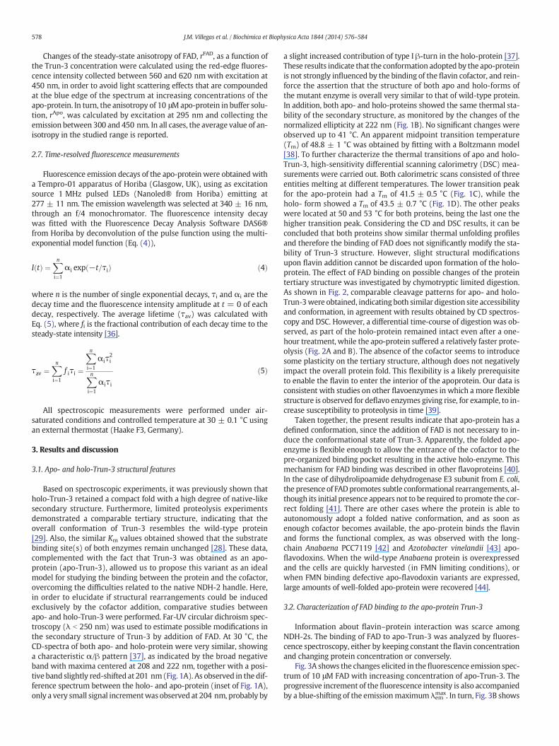
Fig. 5. FAD binds close to Trp residues of Trun-3. Ribbon representation of a portion of the NDH-2 three-dimensional model: FAD and tryptophan residues are colored in orange and blue,respectively. The diagram was created using the PDB file of NDH-2 theoretical model [24], with the program Visual Molecular Dynamics (VMD).
581J.M. Villegas et al. / Biochimica et Biophysica Acta 1844 (2014) 576–584
ideal sites in the proteinwill be considered. In this case, the overall fluo-rescence intensity (F)must be considered as the sumof thefluorescenceintensity of the population of FAD molecules free in the aqueous pool[FAD]F and of that coming from the cofactor molecules bound to theprotein [FAD]B [49]. Thus, the fraction of bound FAD (fB) is defined asthe ratio between [FAD]B (i.e. equivalent to the holo-protein concentra-tion) and the total flavin concentrations [FAD]T, Eq. (7), where F and F0are the measured fluorescence intensity of FAD in the presence and ab-sence of apo-protein, and F∞ is the fluorescence intensity of totallybounded FAD.
f B ¼ FAD½ �BFAD½ �F þ FAD½ �B
¼ Holo½ �FAD½ �T
¼ F−F0F∞−F0
ð7Þ
Because the fluorescence intensity of FAD increases upon binding,the calculation of fB using the steady state anisotropy r is given byEq. (8), where rF and rB are the anisotropies of the free and boundFAD, respectively, and R = F∞ / F0 is the fluorescence intensity correc-tion factor under the same monochromator settings as the anisotropymeasurement [46,50].
f B ¼ r−r FR rB−rð Þ þ r−r Fð Þ ð8Þ
Eqs. (7) and (8) assume the existence of only two states forthe solute, e.g. bound and free, interacting with a single binding site N,irrespective of the protein concentration [49]. Therefore, the calculationof this parameter from both fluorescence intensity and anisotropydata allows the estimation of the average number of FAD moleculesbound per protein molecule, n = [FAD]B / [Trun-3]T. As [Trun-3]T =
[Apo] + [Holo], combining with KA (Eq. (6)) and [FAD]F = (1 − fB)[FAD]T, the hyperbolic function (Eq. (9)) for n is obtained.
n ¼ nmax � FAD½ �FK−1A þ FAD½ � F
ð9Þ
In this case, the factor nmax was introduced as fitting variable to ac-count themaximumvalue of cofactormolecules boundper proteinmol-ecule. According with the two pseudophase model proposed, a value ofnmax = 1must be expected [50]. Fig. 4B shows the variation of the spe-cific binding ratio ν with [FAD]F, and the non-linear fitting with Eq. (9)yields KA = 7.7 × 10⁴ M−1 and nmax ≈ 1, confirming the bindingmodel proposed with a 1:1 stoichiometry upon formation of the holo-protein, as previously obtained from the sigmoidal fitting of the emis-sion maximum shifting (inset of Fig. 3A).
Excitation at 295 nm of apo-Trun-3 shows the typical tryptophan-like emissionwith amaximum at 335 nm (Fig. 6A). This result indicatesthat the Trp residues are sensing a less polar environment than in aque-ous solution [36], in agreement with the solvent-accessible surface area(SASA) analysis of the native E. coli NDH-2 computational model (PDBID: 1OZK), which predicts that both tryptophan residues (Trp46 andTrp271) are buried in the protein [26,29]. The addition of FAD stronglyquenched the protein fluorescence intensity, and after inner filter cor-rections with Eq. (2), it can be observed that no major spectral shift ofthe emission maximum was observed (Fig. 6A). Furthermore, theStern–Volmer (SV) plot of (F0/F)corr vs [FAD]T was perfectly linear(Fig. 6B). These results indicate that both Trp residues are accessible tothe quencher as, in the case of a non-accessible Trp residue, the SVplot should show a downward curvature (saturation-like curve). Thus,the intrinsic fluorescence quenching of Trun-3 by FAD suggests that

Fig. 6. FAD quenches almost statically the fluorescence of apo-Trun-3. (A) Fluorescenceemission spectra of Trun-3 corrected by inner filter effects upon addition of FAD withEq. (2). Inset: variation of the average fluorescence lifetime τav and fractional contributionfi with the molar ratio [FAD]/[Apo]. (B) Stern–Volmer (SV) plots for the corrected fluores-cence intensity (F0/F)corr (■) and average lifetime (τ0/τ)av (●) ratios, respectively, and forthe corrected static quenching contribution Y = [(F0/F)corr] / [(τ0/τ)av] ( ), Eq. (10).
582 J.M. Villegas et al. / Biochimica et Biophysica Acta 1844 (2014) 576–584
theflavin cofactor can interactwith both Trp residues in the binding siteof the protein. In fact, estimation of the distance between the isoalloxa-zine ring of FAD to Trp46 and Trp271 from the 3D model of NDH-2shows a closer location of FAD to both Trp residues (b17 Å) (Fig. 5).Thus, assuming similar protein conformation in solution, the proximityof FAD to both Trp residues of Trun-3 can be extrapolated.
In order to confirm this assumption, thefluorescence decay behaviorof apo-Trun-3 was analyzed as a function of increasing FAD concentra-tion by TCSPC (Time-Correlated Single Photon Counting) experimentswith excitation at 280 nm and emission monitored at 340 nm. In theabsence of FAD, the best deconvolutionfitting of the decaywas obtainedusing a three-exponential decay function (Eq. (4)), yielding individuallifetimes τi of 0.85, 3.32, and 7.53 nswith fluorescence fractional contri-butions fi of 0.30, 0.42, and 0.28, respectively. From this lifetime distri-bution, an average lifetime of τav = 3.75 ns was calculated withEq. (5). Multi-exponential decay is a common feature either in singleor multi-tryptophan containing proteins, mainly due to the conforma-tional complexity and/or specific interaction of some amino acid resi-dues that can act as quenchers [51]. Upon addition of FAD, the proteinfluorescence decay was also well-fitted with the three-exponentialdecay function (data not shown). However, as all individual lifetimeswere reducedwith the flavin concentration, the fractional contributionsf1 and f2 corresponding to the shorter and middle lifetimes were re-duced up to a molar ratio [FAD]/[Apo] ≈ 1, while a parallel increasewas observed for the f3 value associated with the longest lifetime ofthe protein (inset of Fig. 6A). This saturation behavior of the fluores-cence fractional contributions reached at [FAD]/[Apo] ≥ 1 can be associ-ated with the 1:1 stoichiometry for the binding as described above.
Thus, the combined variation of both τi and fi resulted in a slight andprogressive reduction of the average lifetime τav value. Once more, thedynamic quenching of all Trp fluorescence-like decay components ofTrun-3 by FAD addition supports our previous assumption of closer lo-cation of the cofactor to the two Trp residues of the truncated protein.
Finally, both steady-state and dynamic fluorescence quenching ofTrun-3 by FADwere analyzed, according to the classical SV relationship(F0/F) or (τ0/τ) = 1 + KSV[FAD]T, respectively, with KSV (M−1) as theSV quenching constant. For both (F0/F)corr and (τ0/τ)av ratios, satisfacto-ry linear correlations with the total FAD concentration were fulfilled(Fig. 6B), and by comparing the slope difference between both SV-plots, it can be concluded that the quenching mechanism of Trun-3by FAD is mainly static (see [36]). In fact, from the apparent dynamiccomponent KSV = KD = kqτ0,av = 1.3 × 10⁴ M−1, the bimolecularquenching constant kq = 3.5 × 10¹² M−1 s−1 was estimated. This kqvalue exceeds, by almost three-order of magnitude, the rate constantvalue for bimolecular diffusion-controlled processes in water atroom temperature (≈6 × 10⁹ M−1 s−1), suggesting that the dynamicquenching contribution (τ0/τ)av is produced by transient and/orenergy-transfer effects due to the close vicinity of FAD to the Trp resi-dues into the binding site of apo-Trun-3 [51].
Usually, in the analysis of a solute binding efficiency by intrinsic fluo-rescence quenching of a protein, most articles consider a 100% staticquenching mechanism, since the dynamic quenching component is ig-nored. This is based on the fact that its occurrence would require aquenching rate constant higher than that expected for diffusion con-trolled processes in homogeneous fluid solutions (i.e. ≈1010 M−1 s−1)(see review [52]). However, the SV-plot of Fig. 6B, obtained from fluores-cence lifetime's analysis, clearly illustrates the occurrence of aminor con-tribution of dynamic quenching that affects the total fluorescenceintensity ratio (F0/F)cor. Therefore, the (F0/F)corr ratio obtained from in-tensity fluorescencemeasurements can be considered as the product be-tween “pure” static and “apparent” dynamic quenching contributions,Eq. (10), where the association constant KA (as defined in Eq. (6)) isonly included in the static quenching contribution:
F0F
� �corr
¼ 1þ KA FAD½ �T� �� 1þ KD FAD½ �T
� �
¼ 1þ KA FAD½ �T� �� τ0
τ
� �av: ð10Þ
Thus, the dynamic quenching component can be easily correctedfrom (F0/F)corr by plotting Y = (F0/F)corr / (τ0/τ)av vs. [FAD]T. A linearplot was obtained, according with a 1:1 stoichiometry of the complexformation, and a slope KA = 6.2 (±0.2) × 104 M−1. This KA value isvery close to that of 7.7 × 10⁴ M−1 obtained by the fluorescence inten-sity and anisotropy enhancement of FAD as a function of Trun-3 concen-tration (Fig. 4), confirming the validity of the two approaches used.
4. Conclusions
For the first time, FAD binding properties to type II NADH dehydro-genase were characterized. Our strategy involved the use of the solubletruncated mutant Trun-3, which represents an excellent model forstudying the protein/cofactor interactions, since eventual difficulties re-lated to the use of the native membrane enzyme were avoided. As wedescribed before, Trun-3 was obtained as a cytosolic apo-protein andthe overall structural conformation of holo-Trun-3 resembles theE. coli NDH-2 wild-type protein [29]. The binding of the FAD cofactorto apo-Trun-3 did not induce significant changes in secondary and ter-tiary structures, suggesting that FAD was not required to promote thecorrect folding of NDH-2. The fact that Trun-3 structurewas kept almostintact in the absence of the cofactor is in agreement with the fast reac-tivation process after FAD binding [28]. The FAD cofactor binding toapo-Trun-3 was determined, obtaining an average association constantvalue of KA = 7.0(±0.8) × 104 M−1, which corresponds to a Gibbs free

583J.M. Villegas et al. / Biochimica et Biophysica Acta 1844 (2014) 576–584
energy ΔG° ≈ −28 kJ/mol, a driven force consistent with a highly fa-vorable protein–cofactor interaction. The incorporation of FAD into thesoluble protein occurs with a 1:1 stoichiometry, in agreement withthe results previously reported for native E. coli NDH-2 by Jaworowskiet al. [23].
Acknowledgements
We specially thank Dr G.D. Fidelio and Dr M.I. Burgos for receivingJ.M.V. at their laboratory and for helpful contribution in far UV-CD andDSCmeasurements. This researchwas supported by Argentinean grantsof the Consejo Nacional de Investigaciones Científicas y Técnicas(CONICET-PIP's 6399/09 and 0374/12), Agencia de Promoción Científicay Tecnológica (ANPCyT-PICT 2012-2666), Universidad Nacional deTucumán (UNT-CIUNT 26/D443), and Universidad Nacional de Santiagodel Estero (UNSE-CICyT 23A/162). J.M.V. thanks CONICET for doctoralfellowship. F.E.M.V., C.D.B. and V.A.R. are researchmembers of CONICET.
References
[1] E. Steensma, C.P. van Mierlo, Structural characterisation of apo-flavodoxin showsthat the location of the stable nucleus differs among proteins with a flavodoxin-like topology, J. Mol. Biol. 282 (1998) 653–666.
[2] W.R. Fisher, H. Taniuchi, C.B. Anfinsen, On the role of heme in the formation of thestructure of cytochrome c, J. Biol. Chem. 248 (1973) 3188–3195.
[3] E. Steensma, M.J. Nijman, Y.J. Bollen, P.A. de Jager, W.A. van den Berg, W.M. vanDongen, C.P. van Mierlo, Apparent local stability of the secondary structure ofAzotobacter vinelandii holoflavodoxin II as probed by hydrogen exchange: implicationsfor redox potential regulation and flavodoxin folding, Protein Sci. 7 (1998) 306–317.
[4] V. Joosten, W.J.H. van Berkel, Flavoenzymes, Curr. Opin. Chem. Biol. 11 (2007)195–202.
[5] M.W. Fraaije, A. Mattevi, Flavoenzymes: diverse catalysts with recurrent features,Trends Biochem. Sci. 25 (2000) 126–132.
[6] S. Bornemann, Flavoenzymes that catalyse reactions with no net redox change, Nat.Prod. Rep. 19 (2002) 761–772.
[7] C.L. Ávila, V.A. Rapisarda, R.N. Farías, J. De Las Rivas, R. Chehín, Linear array of con-served sequence motifs to discriminate protein subfamilies: study on pyridinenucleotide-disulfide reductases, BMC Bioinforma. 8 (2007) 96.
[8] A.M. Melo, T.M. Bandeiras, M. Teixeira, New insights into type II NAD(P)H:quinoneoxidoreductases, Microbiol. Mol. Biol. Rev. 68 (2004) 603–616.
[9] J. Fang, D.S. Beattie, Novel FMN-containing rotenone-insensitive NADH dehydroge-nase from Trypanosoma brucei mitochondria: isolation and characterization, Bio-chemistry 41 (2002) 3065–3072.
[10] A.M. Michalecka, A.S. Svensson, F.I. Johansson, S.C. Agius, U. Johanson, A. Brennicke,S. Binder, A.G. Rasmusson, Arabidopsis genes encodingmitochondrial type II NAD(P)H dehydrogenases have different evolutionary origin and showdistinct responses tolight, Plant Physiol. 133 (2003) 642–652.
[11] P.R. Rich, A. Marechal, The mitochondrial respiratory chain, Essays Biochem. 47(2010) 1–23.
[12] M.G. Matus-Ortega, K.G. Salmerón-Santiago, O. Flores-Herrera, G. Guerra-Sánchez, F.Martínez, J.L. Rendón, J.P. Pardo, The alternativeNADHdehydrogenase is present inmi-tochondria of some animal taxa, Comp. Biochem. Physiol. Part D Genomics Proteomics6 (2011) 256–263.
[13] N. Fisher, P.G. Bray, S.A.Ward, G.A. Biagini, Themalaria parasite type II NADH:quinoneoxidoreductase: an alternative enzyme for an alternative lifestyle, Trends Parasitol. 23(2007) 305–310.
[14] C.K. Dong, V. Patel, J.C. Yang, J.D. Dvorin, M.T. Duraisingh, J. Clardy, D.F. Wirth, Type IINADH dehydrogenase of the respiratory chain of Plasmodium falciparum and its in-hibitors, Bioorg. Med. Chem. Lett. 19 (2009) 972–975.
[15] G.A. Biagini, N. Fisher, A.E. Shone,M.A. Mubaraki, A. Srivastava, A. Hill, T. Antoine, A.J.Warman, J. Davies, C. Pidathala, R.K. Amewu, S.C. Leung, R. Sharma, P. Gibbons, D.W.Hong, B. Pacorel, A.S. Lawrenson, S. Charoensutthivarakul, L. Taylor, O. Berger, A.Mbekeani, P.A. Stocks, G.L. Nixon, J. Chadwick, J. Hemingway, M.J. Delves, R.E.Sinden, A.M. Zeeman, C.H. Kocken, N.G. Berry, P.M. O'Neill, S.A. Ward, Generationof quinolone antimalarials targeting the Plasmodium falciparummitochondrial respira-tory chain for the treatment and prophylaxis of malaria, Proc. Natl. Acad. Sci. U. S. A.109 (2012) 8298–8303.
[16] T. Yano, L.S. Li, E. Weinstein, J.S. Teh, H. Rubin, Steady-state kinetics and inhibitory ac-tion of antitubercular phenothiazines on Mycobacterium tuberculosis type-II NADH-menaquinone oxidoreductase (NDH-2), J. Biol. Chem. 281 (2006) 11456–11463.
[17] J.S. Teh, T. Yano, H. Rubin, Type II NADH: menaquinone oxidoreductase ofMycobac-terium tuberculosis, Infect. Disord. Drug Targets 7 (2007) 169–181.
[18] B.B. Seo, J. Wang, T.R. Flotte, T. Yagi, A. Matsuno-Yagi, Use of the NADH-quinone ox-idoreductase (NDI1) gene of Saccharomyces cerevisiae as a possible cure for complexI defects in human cells, J. Biol. Chem. 275 (2000) 37774–37778.
[19] M.F. Maas, C.H. Sellem, F. Krause, N.A. Dencher, A. Sainsard-Chanet, Molecular genetherapy: overexpression of the alternative NADH dehydrogenase NDI1 restoresoverall physiology in a fungal model of respiratory complex I deficiency, J. Mol.Biol. 399 (2010) 31–40.
[20] M. Marella, B.B. Seo, T.R. Flotte, A. Matsuno-Yagi, T. Yagi, No immune responses bythe expression of the yeast Ndi1 protein in rats, PLoS One 6 (2011) 25910.
[21] M. Marella, B.B. Seo, T. Yagi, A. Matsuno-Yagi, Parkinson's disease and mitochondrialcomplex I: a perspective on the Ndi1 therapy, J. Bioenerg. Biomembr. 41 (2009)493–497.
[22] I.G. Young, B.L. Rogers, H.D. Campbell, A. Jaworowski, D.C. Shaw, Nucleotide se-quence coding for the respiratory NADH dehydrogenase of Escherichia coli: UUG ini-tiation codon, Eur. J. Biochem. 116 (1981) 165–170.
[23] A. Jaworowski, G.Mayo, D.C. Shaw, H.D. Campbell, I.G. Young, Characterization of therespiratory NADH dehydrogenase of Escherichia coli and reconstitution of NADH ox-idase in ndh mutant membrane vesicles, Biochemistry 20 (1981) 3621–3628.
[24] Y. Feng, W. Li, J. Li, J. Wang, J. Ge, D. Xu, Y. Liu, K. Wu, Q. Zeng, J.W. Wu, C. Tian, B.Zhou, M. Yang, Structural insight into the type-II mitochondrial NADH dehydroge-nases, Nature 491 (2012) 478–482.
[25] M. Iwata, Y. Lee, T. Yamashita, T. Yagi, S. Iwata, A.D. Cameron, M.J. Maher, The struc-ture of the yeast NADH dehydrogenase (Ndi1) reveals overlapping binding sites forwater- and lipid-soluble substrates, Proc. Natl. Acad. Sci. U. S. A. 109 (2012)15247–15252.
[26] R. Schmid, D.L. Gerloff, Functional properties of the alternative NADH: ubiquinoneoxidoreductase from E. coli through comparative 3-D modeling, FEBS Lett. 578(2004) 163–168.
[27] V.A. Rapisarda, R.N. Chehín, J. De Las Rivas, L. Rodríguez-Montelongo, R.N. Farías,E.M. Massa, Evidence for Cu(I)-thiolate ligation and prediction of a putativecopper-binding site in the Escherichia coli NADH dehydrogenase-2, Arch. Biochem.Biophys. 405 (2002) 87–94.
[28] J.M. Villegas, S.I. Volentini, M.R. Rintoul, V.A. Rapisarda, Amphipathic C-terminal re-gion of Escherichia coli NADH dehydrogenase-2 mediates membrane localization,Arch. Biochem. Biophys. 505 (2011) 155–159.
[29] J.M. Villegas, C.M. Torres-Bugeau, R. Chehín, M.I. Burgos, G.D. Fidelio, M.R. Rintoul,V.A. Rapisarda, Maintenance and thermal stabilization of NADH dehydrogenase-2conformation upon elimination of its C-terminal region, Biochimie 95 (2013)382–387.
[30] G. Eggink, H. Engel, G. Vriend, P. Terpstra, B. Witholt, Rubredoxin reductase of Pseu-domonas oleovorans. Structural relationship to other flavoprotein oxidoreductasesbased on one NAD and two FAD fingerprints, J. Mol. Biol. 212 (1990) 135–142.
[31] V.A. Rapisarda, L.R. Montelongo, R.N. Farías, E.M. Massa, Characterization of anNADH-linked cupric reductase activity from the Escherichia coli respiratory chain,Arch. Biochem. Biophys. 370 (1999) 143–150.
[32] O.H. Lowry, N.J. Rosebrough, A.L. Farr, R.J. Randall, Protein measurement with theFolin phenol reagent, J. Biol. Chem. 193 (1951) 265–275.
[33] U.K. Laemmli, Cleavage of structural proteins during the assembly of the head ofbacteriophage T4, Nature 227 (1970) 680–685.
[34] J.B. Bertoldo, G. Razzera, J. Vernal, F.C. Brod, A.C. Arisi, H. Terenzi, Structural stabilityof Staphylococcus xylosus lipase is modulated by Zn(2+) ions, Biochim. Biophys. Acta1814 (2011) 1120–1126.
[35] L. Valle, F.E.M. Vieyra, C.D. Borsarelli, Hydrogen-bondingmodulation of excited-stateproperties of flavins in a model of aqueous confined environment, Photochem.Photobiol. Sci. 11 (2012) 1051–1061.
[36] J.R. Lakowicz, Principles of Fluorescence Spectroscopy, 3st edn Springer Science +Business Media, LLC, Singapore, 2006.
[37] S.M. Kelly, T.J. Jess, N.C. Price, How to study proteins by circular dichroism, Biochim.Biophys. Acta 1751 (2005) 119–139.
[38] U.B. Ericsson, B.M. Hallberg, G.T. DeTitta, N. Dekker, P. Nordlund, Thermofluor-basedhigh-throughput stability optimization of proteins for structural studies, Anal.Biochem. 357 (2006) 289–298.
[39] G.T. Tarelli, M.A. Vanoni, A. Negri, B. Curti, Characterization of a fully activeN-terminal 37-kDa polypeptide obtained by limited tryptic cleavage of pig kidneyD-amino acid oxidase, J. Biol. Chem. 265 (1990) 21242–21246.
[40] M.W. Fraaije, R.H. van Den Heuvel, W.J. van Berkel, A. Mattevi, Structural analysis offlavinylation in vanillyl-alcohol oxidase, J. Biol. Chem. 275 (2000) 38654–38658.
[41] H. Lindsay, E. Beaumont, S.D. Richards, S.M. Kelly, S.J. Sanderson, N.C. Price, J.G.Lindsay, FAD insertion is essential for attaining the assembly competence of thedihydrolipoamide dehydrogenase (E3) monomer from Escherichia coli, J. Biol.Chem. 275 (2000) 36665–36670.
[42] J. Fernández-Recio, C.G. Genzor, J. Sancho, Apoflavodoxin folding mechanism: analpha/beta protein with an essentially off-pathway intermediate, Biochemistry 40(2001) 15234–15245.
[43] Y.J. Bollen, I.E. Sánchez, C.P. van Mierlo, Formation of on- and off-pathway interme-diates in the folding kinetics of Azotobacter vinelandii apoflavodoxin, Biochemistry43 (2004) 10475–10489.
[44] J. López-Llano, S. Maldonado, S. Jain, A. Lostao, R. Godoy-Ruiz, J.M. Sanchez-Ruiz, M.Cortijo, J. Fernández-Recio, J. Sancho, The long and short flavodoxins: II. The role ofthe differentiating loop in apoflavodoxin stability and folding mechanism, J. Biol.Chem. 279 (2004) 47184–47191.
[45] S. Raffelberg, M. Mansurova, W. Gärtner, A. Losi, Modulation of the photocycle of aLOV domain photoreceptor by hydrogen-bonding network, J. Am. Chem. Soc. 113(2011) 5346–5356.
[46] C.M. Ingersoll, C.M. Strollo, Steady-state fluorescence anisotropy to investigate fla-vonoids binding to proteins, J. Chem. Educ. 84 (2007) 1313–1315.
[47] R.H. van den Heuvel, A.H.Westphal, A.J. Heck, M.A. Walsh, S. Rovida,W.J. van Berkel,A. Mattevi, Structural studies on flavin reductase PheA2 reveal binding of NAD in anunusual folded conformation and support novel mechanism of action, J. Biol. Chem.279 (2004) 12860–12867.
[48] G.F. Li, K.D. Glusac, The role of adenine in fast excited-state deactivation of FAD: afemtosecond mid-IR transient absorption study, J. Phys. Chem. B 113 (2009)9059–9061.

584 J.M. Villegas et al. / Biochimica et Biophysica Acta 1844 (2014) 576–584
[49] E. Alarcón, A. Aspée, E.B. Abuin, E.A. Lissi, Evaluation of solute binding to proteinsand intra-protein distances from steady state fluorescence measurements,J. Photochem. Photobiol. B Biol. 106 (2012) 1–17.
[50] D.A. Malencik, S.R. Anderson, Peptide binding by calmodulin and its proteolytic frag-ments and by troponin-C, Biochemistry 23 (1984) 2420–2428.
[51] Y. Engelborghs, The analysis of time resolved protein fluorescence inmulti-tryptophan proteins, Spectrochim. Acta A Mol. Biomol. Spectrosc. 57 (2001)2255–2270.
[52] M. van deWeert, L. Stella, Fluorescence quenching and ligand binding: a critical dis-cussion of a popular methodology, J. Mol. Struct. 998 (2011) 144–150.
Copyright © 2022 FDOKUMEN