![Separate [3H]-nitrendipine binding sites in mitochondria and plasma membranes of bovine adrenal medulla](https://static.fdokumen.com/doc/165x107/63443fe2df19c083b10781df/separate-3h-nitrendipine-binding-sites-in-mitochondria-and-plasma-membranes-of.jpg)
Nicotine-evoked cytosolic Ca2+ increase and cell depolarization in capillary endothelial cells of...
10
Biol Res 42: 111-119, 2009 BR Nicotine-evoked cytosolic Ca 2+ increase and cell depolarization in capillary endothelial cells of the bovine adrenal medulla RAÚL VINET 1, 2 , MAGDALENA CORTÉS 1 , MARIO LUXORO 3 and MARCO A. DELPIANO 4, 5 1 Facultad de Farmacia, Universidad de Valparaíso, Gran Bretaña 1093, Valparaíso, Chile. 2 Centro Regional de Estudios en Alimentos Saludables (CREAS), Valparaíso, Chile. 3 Departamento de Biología, Facultad de Ciencias, Universidad de Chile, Las Palmeras 3425, Santiago, Chile. 4 Departamento de Fisiología, Universidad de Valparaíso, Gran Bretaña 1111, Valparaíso, Chile. 5 Max-Planck-Institute for Molecular Physiology, Otto-Hahn-Str. 11, 44227 Dortmund, Germany. ABSTRACT Endothelial cells are directly involved in many functions of the cardiovascular system by regulating blood flow and blood pressure through Ca 2+ dependent exocitosis of vasoactive compounds. Using the Ca 2+ indicator Fluo-3 and the patch-clamp technique, we show that bovine adrenal medulla capillary endothelial cells (BAMCECs) respond to acetylcholine (ACh) with a cytosolic Ca 2+ increase and depolarization of the membrane potential (20.3±0.9 mV; n=23). The increase in cytosolic Ca 2+ induced by 10μM ACh was mimicked by the same concentration of nicotine but not by muscarine and was blocked by 100 μM of hexamethonium. On the other hand, the increase in cytosolic Ca 2+ could be depressed by nifedipine (0.01 - 100 μM) or withdrawal of extracellular Ca 2+ . Taken together, these results give evidence for functional nicotinic receptors (nAChRs) in capillary endothelial cells of the adrenal medulla. It suggests that nAChRs in BAMCECs may be involved in the regulation of the adrenal gland’s microcirculation by depolarizing the membrane potential, leading to the opening of voltage-activated Ca 2+ channels, influx of external Ca 2+ and liberation of vasoactive compounds. Key terms: capillary, endothelial cells, intracellular Ca 2+ , nicotinic receptor, and voltage-activated Ca 2+ channels. Correspondence to: Dr. Raúl Vinet Huerta, Facultad de Farmacia, Universidad de Valparaíso, Gran Bretaña 1093, Valparaíso, Chile. Phone: +56-32-2508108. Fax: +56-32-2508111. e-mail: [email protected] Received: July 25, 2008. In Revised form: December 19, 2008. Accepted: January 26, 2009 INTRODUCTION Endothelial cells (ECs) participate in the regulation of blood flow and blood pressure through the secretion of vasoactive compounds such as prostacyclin (PGI 2 ) (Lückhoff et al., 1988), endothelium-derived relaxing factor (EDRF) (Lückhoff et al., 1988; Furchgott and Zawadzki 1980), identified as nitric oxide (NO) (Palmer et al., 1987) and endothelium-derived hyperpolarizing factor (EDHF) (Taylor and Weston, 1988). The signal transduction for the secretion of these compounds is an increase in cytosolic Ca 2+ when agonists like ACh, bradykinin, ATP, histamine and thrombin bind to specific receptors (Himmel et al., 1993). One of the mechanisms regulating the increase in cytosolic Ca 2+ besides its liberation from intracellular storage is the influx of Ca 2+ ions through Ca 2+ channels. In this context, it is well established, that voltage-activated Ca 2+ channels (VACCs) are present in ECs of capillaries (endothelium without smooth muscle cells) but not in large blood vessels (Bossu et al., 1989; Delpiano and Altura, 1996; Vinet and Vargas, 1999; Delpiano, 2000); they are involved in Ca 2+ influx. On the other hand, it is known that ECs express functional acetylcholine receptors of a
Transcript of Nicotine-evoked cytosolic Ca2+ increase and cell depolarization in capillary endothelial cells of...

111VINET ET AL. Biol Res 42, 2009, 111-119Biol Res 42: 111-119, 2009 BRNicotine-evoked cytosolic Ca2+ increase and celldepolarization in capillary endothelial cells of the bovineadrenal medulla
RAÚL VINET1, 2, MAGDALENA CORTÉS1, MARIO LUXORO3 andMARCO A. DELPIANO4, 5
1 Facultad de Farmacia, Universidad de Valparaíso, Gran Bretaña 1093, Valparaíso, Chile.2 Centro Regional de Estudios en Alimentos Saludables (CREAS), Valparaíso, Chile.3 Departamento de Biología, Facultad de Ciencias, Universidad de Chile, Las Palmeras 3425, Santiago, Chile.4 Departamento de Fisiología, Universidad de Valparaíso, Gran Bretaña 1111, Valparaíso, Chile.5 Max-Planck-Institute for Molecular Physiology, Otto-Hahn-Str. 11, 44227 Dortmund, Germany.
ABSTRACT
Endothelial cells are directly involved in many functions of the cardiovascular system by regulating bloodflow and blood pressure through Ca2+ dependent exocitosis of vasoactive compounds. Using the Ca2+
indicator Fluo-3 and the patch-clamp technique, we show that bovine adrenal medulla capillary endothelialcells (BAMCECs) respond to acetylcholine (ACh) with a cytosolic Ca2+ increase and depolarization of themembrane potential (20.3±0.9 mV; n=23). The increase in cytosolic Ca2+ induced by 10μM ACh wasmimicked by the same concentration of nicotine but not by muscarine and was blocked by 100 μM ofhexamethonium. On the other hand, the increase in cytosolic Ca2+ could be depressed by nifedipine (0.01 -100 μM) or withdrawal of extracellular Ca2+. Taken together, these results give evidence for functionalnicotinic receptors (nAChRs) in capillary endothelial cells of the adrenal medulla. It suggests that nAChRs inBAMCECs may be involved in the regulation of the adrenal gland’s microcirculation by depolarizing themembrane potential, leading to the opening of voltage-activated Ca2+ channels, influx of external Ca2+ andliberation of vasoactive compounds.
Key terms: capillary, endothelial cells, intracellular Ca2+, nicotinic receptor, and voltage-activated Ca2+
channels.
Correspondence to: Dr. Raúl Vinet Huerta, Facultad de Farmacia, Universidad de Valparaíso, Gran Bretaña 1093,Valparaíso, Chile. Phone: +56-32-2508108. Fax: +56-32-2508111. e-mail: [email protected]
Received: July 25, 2008. In Revised form: December 19, 2008. Accepted: January 26, 2009
INTRODUCTION
Endothelial cells (ECs) participate in theregulat ion of blood f low and bloodpressure through the secret ion ofvasoactive compounds such asprostacyclin (PGI2) (Lückhoff et al . ,1988), endothelium-derived relaxing factor(EDRF) (Lückhoff et al., 1988; Furchgottand Zawadzki 1980), identified as nitricoxide (NO) (Palmer et al., 1987) andendothelium-derived hyperpolarizingfactor (EDHF) (Taylor and Weston, 1988).The signal transduction for the secretion ofthese compounds is an increase incytosolic Ca2+ when agonists like ACh,
bradykinin, ATP, histamine and thrombinbind to specific receptors (Himmel et al.,1993). One of the mechanisms regulatingthe increase in cytosolic Ca2+ besides itsliberation from intracellular storage is theinflux of Ca2+ ions through Ca2+ channels.In this context, it is well established, thatvoltage-activated Ca2+ channels (VACCs)are present in ECs of capil lar ies(endothelium without smooth muscle cells)but not in large blood vessels (Bossu et al.,1989; Delpiano and Altura, 1996; Vinetand Vargas, 1999; Delpiano, 2000); theyare involved in Ca2+ influx. On the otherhand, i t is known that ECs expressfunctional acetylcholine receptors of a

VINET ET AL. Biol Res 42, 2009, 111-119112
muscarinic and nicotinic nature (Olesen etal., 1988; Macklin et al., 1998). The actionof ACh on blood vessels associated with arise in cytosolic Ca2+ and vasorelaxation isa well-validated endothelium-dependentmechanism (Furchgott and Zawadski, 1980;Furchgott et al., 1987; Emerson and Segal,2000). Muscarinic receptors (mAChRs),when stimulated by ACh inducevasodilatation mediated by the release ofnitric oxide (Furchgott and Zawadski,1980). Although nAChRs have already beenreported in human ECs of large bloodvessels (Haberberger et al., 2000; Macklinet al., 1998) their functional presence inECs of capillaries has not been reported,besides the hyperpolarizing effect of AChon capillaries of hamster cheeks (McGahrenet al., 1998). The aim of this study was toinvestigate whether BAMCECs exhibitfunctional nAChRs by monitoring changesin the cytosolic Ca2+ and the membranepotential, and to explore whether VACCsmay be involved in the Ca2+ response.
MATERIAL AND METHODS
Cell culture
BAMCECs were obtained according toVinet et al. (2000). Briefly, glands wereperfused through their veins with amodified Locke solution containing 0.25%collagenase (Böhringer Mannheim,Germany). Dissociated cells weresuspended in Percoll (Pharmacia, USA) andcentrifuged at 20,000 g for 20 min. Theband containing the highest density of ECswas seeded on glass cover slips and platedinto 35 mm petri dishes with nutrientmedium mixture F12 Ham (SigmaChemical, USA) supplemented with 10%fetal calf serum (Biofluids, Rockville,USA), 50 U/mL penicillin, 50 μg/mlstreptomycin, at pH 7.2 and incubated at37ºC in a humidity atmosphere of 5% CO2.BAMCECs were identified as ECs by theirpolygonal shape and the fluorescenceinduced by the specific uptake of anacetylated low-density lipoprotein (Dil Ac-LDL; Molecular Probes, USA) (Voyta etal., 1984).
Cytosolic Ca2+ measurements
As described elsewhere (Luxoro et al.,1997), BAMCECs were loaded for 20 minwith 7 μM Fluo-3 AM (Molecular Probes,USA) and 0.094% pluronic acid in modifiedKrebs solution (in mM: 140 NaCl, 5 KCl,2.6 CaCl2, 1 MgCl2, 2.5 NaHCO3, 10glucose, 5 Na-HEPES, at pH 7.4). Lightemitted by Fluo-3 emerging from changesin cytosolic Ca2+ ([Ca2+]i) of a single cell,placed in a superfusion chamber (volume:200 ml) on a microscope stage (Leitz,Wetzlar, Germany) with appropriate filters(excitation 490 nm; emission 530 nm) wasmeasured continuously with aphotomultiplier (Hamamatsu, Japan). Datawere digitized at 3 Hz by an analogueconverter and analyzed by using the GlobalLab software (Data Translation, USA). Themagnitude of the fluorescent signal waspresented as a ratio according to theequation ΔF/Fb = (Ft - Fb) / Fb, where Ft isthe fluorescence value of Fluo-3 at time “t”and Fb is i t basal fluorescence, aspreviously described (Kao et al., 1989).
Membrane potential
The membrane potential was measured insingle cells at room temperature by usingthe classical patch-clamp technique (Hamillet al., 1981) with an EPC-7 patch-amplifier(Darmstadt-Eberstadt, Germany) in thewhole-cell configuration and current-clampmode. Soft glass pipettes, heat polished andpulled with a vertical puller (David Kopf,USA) were filled with intracellular solution(in mM: 130 KCl, 1 CaCl2, 2 MgCl2, 20HEPES/KOH, 5 EGTA/KOH, at pH 7.2)and had a resistance of 3 to 5 MΩ. Singlecells were recorded on the stage of aninverted microscope (Nikon, Diaphot,Japan) and superfused with an extracellularsolution (in mM: 140 NaCl, 5 KCl, 2.6CaCl2, 2 MgCl2, 2.5 NaHCO3, 5 HEPES/NaOH, 10 Glucose, at pH 7.4). Cells withmembrane potentials of at least -40 mV andable to depolarize with a 60 mMextracellular K+ concentration (high K+)were selected. A similar criterion was usedfor Ca2+ experiments. Data werecontinuously monitored and analyzed by a

113VINET ET AL. Biol Res 42, 2009, 111-119
12-bit analogue converter board (AxonInstruments, USA).
Drugs
Acetylcholine, hexamethonium andmuscarine were obtained from Sigma(USA). Nicotine and nifedipine wereobtained from Research BiochemicalsInternational (RBI, USA).
Statistical analysis
Where appropriate, the results wereexpressed as the mean ± standard error(S.E.M.). Statistical differences were
assessed with Student’s t-test, considering aP value of ≤ 0.05 as significant.
RESULTS
Single freshly dissociated BAMCECsresponded to high K+, applied into themedium, with membrane depolarization andincrease in [Ca2+]i. Figure 1A illustrates theeffect of high K+. The inset shows stepsdepolarizations produced by additiveconcentrations of high K+. The increase in[Ca2+]i induced by high K+ exhibits a fastonset with a rapid recovery followed by adelayed decrease. Application of 10 μM
Figure 1: Effect of high K+ and ACh on cytosolic Ca2+ in fresh dissociated BAMCECs versus time.(A) Typical response of [Ca2+]i induced by 60 mM K+ (n=37). Inset: depolarizing effect of 60 mMK+ on membrane potential. (B) Representative recording of cytosolic Ca2+ increase induced by 10μM ACh (n=8). Inset: dose-response curve for ACh effect on the Ca2+ increase.

VINET ET AL. Biol Res 42, 2009, 111-119114
ACh, as illustrated in Figure 1B, elicited arapid and sustained rise in [Ca2+]i. In 8different experiments we observed a meanΔF/Fb change in Ca2+ of about 0.325±0.088units. After a wash out of ACh from themedium the Ca2+ signal declined to thebaseline in a time-span of about 1 to 2 min.The inset of Figure 1B illustrates the dose-dependent Ca2+ increase induced by AChwith an EC50 of 3.98±0.04 μM, that reachessaturation by about 100 μM ACh (n=8).
Nicotine receptors mediate ACh-induced[Ca2+]i increase
Using nicotine and muscarine and thenicotinic antagonist hexamethonium weapproach the nature of the nAChRs. Figure2A illustrates the effect of muscarine andnicotine on [Ca2+]i. Figure 2A shows thatwhen muscarine and nicotine were added(both at 10 μM) to the medium, 10 minutesafter the test stimulus with high K+, only
Figure 2: Effect of high K+, nicotine, muscarine and hexamethonium on [Ca2+]i (A) Typical [Ca2+]i
change in a cell exposed subsequently to 60 mM K+, 10 μM muscarine and 10 μM nicotine (n=9).Inset: dose-response curve of different nicotine concentration versus cytosolic Ca2+. (B) 100 μMhexamethonium abolished the [Ca2+]i increase induced nicotine (n=5) but restored afterhexamethonium washout.

115VINET ET AL. Biol Res 42, 2009, 111-119
nicotine was able to induce increase in[Ca2+]i
but in 6 other experiments no effectwas observed with muscarine. The [Ca2+]ielevation induced by 10 μM nicotinereached 57.0±2.3% (n=9) of the responseinduced by high K+. The inset of Figure 2Aillustrates a dose-response curve fornicotine with an EC50 of 1.51±0.05 mM(n=4) that reached saturation at 100 mM.To further investigate the nature of AChreceptor, hexamethonium (100 μM) wasapplied to the medium. As illustrated inFigure 2B, this nicotinic antagonistmarkedly depressed the increase in [Ca2+]i(n=5). After the wash out of
hexamethonium, a second stimulation withnicotine restored the cytosolic Ca2+
response.
Participation of extracellular Ca2+ in theCa2+ increase
In order to investigate the source of thecytosolic Ca2+ changes induced by nicotine,BAMCECs were superfused with a Ca2+-free medium containign 0.5 mM EGTA. Asshown in Figure 3A, under these conditionsin all , nicotine sensitive BAMCECsmeasured under Ca2+-free medium (n=6)nicotine, failed to induce a [Ca2+]i increase.
Figure 3: The nicotine-induced [Ca2+]i increase in BAMCECs is dependent on extracellular Ca2+
concentration. (A) Representative recording of [Ca2+]i changes induced by 10 μM nicotine in anominal 0 Ca2+ concentration and when 2.6 mM Ca2+ was restored in the medium (n=6). (B) Dose-response curve of 10 μM nicotine at different external Ca2+ concentrations (0.01-10 mM).
VINET ET AL. Biol Res 42, 2009, 111-119116
Figure 3B illustrates the dose-responsecurve for BAMCECs stimulated with 10μM nicotine that were superfused underdifferent extracellular Ca2+ concentrationsbetween 0.01 to 10 mM (n=4). It can beseen that the magnitude of the nicotin-induced increase in [Ca2+]i
was dependenton the extracellular Ca2+ concentration.
Organic and inorganic compoundsaffecting changes in cytosolic Ca2+
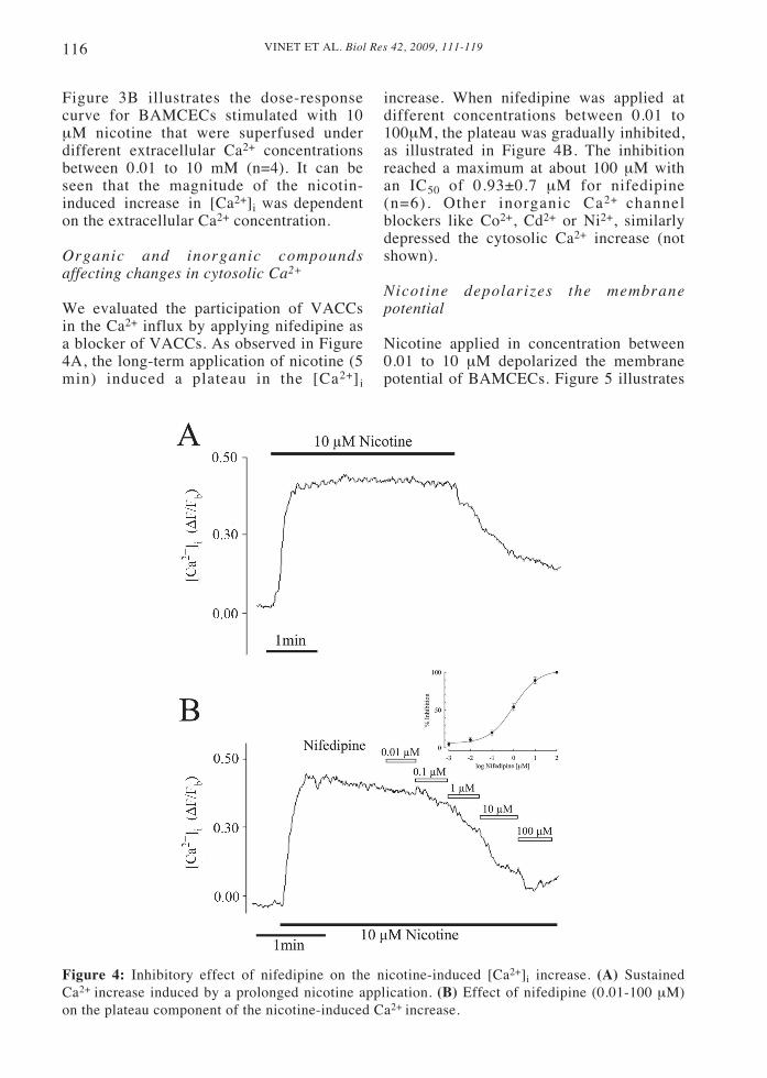
We evaluated the participation of VACCsin the Ca2+ influx by applying nifedipine asa blocker of VACCs. As observed in Figure4A, the long-term application of nicotine (5min) induced a plateau in the [Ca2+]i
Figure 4: Inhibitory effect of nifedipine on the nicotine-induced [Ca2+]i increase. (A) SustainedCa2+ increase induced by a prolonged nicotine application. (B) Effect of nifedipine (0.01-100 μM)on the plateau component of the nicotine-induced Ca2+ increase.
increase. When nifedipine was applied atdifferent concentrations between 0.01 to100μM, the plateau was gradually inhibited,as illustrated in Figure 4B. The inhibitionreached a maximum at about 100 μM withan IC50 of 0.93±0.7 μM for nifedipine(n=6). Other inorganic Ca2+ channelblockers like Co2+, Cd2+ or Ni2+, similarlydepressed the cytosolic Ca2+ increase (notshown).
Nicotine depolarizes the membranepotential
Nicotine applied in concentration between0.01 to 10 μM depolarized the membranepotential of BAMCECs. Figure 5 illustrates

117VINET ET AL. Biol Res 42, 2009, 111-119
two typical depolarizations induced by aconcentration of nicotine of 10 μM (n=6).The mean amplitude of the depolarizationamounted to 20.3±0.9 mV (n=23) (notshown).
Vargas, 1999; Delpiano, 2000) but no ECsof large blood vessels (Colden-Stanfield etal., 1987; Johns et al., 1987; Olesen et al.,1988; Bregestovski et al., 1988; Vargas etal., 1994). From these findings we couldhypothesize that activation of nAChRs inBAMCECs could induce a chain of eventswith a primary influx of divalent cations, asit is known that nAChRs have a broadpermeability to them and some a-sub-unitsallow relatively more influx of Ca2+ overNa+ ions (Liu and Berg, 1999), leading to aCa2+ release from intracellular storage andconcomitantly membrane depolarizationthat would activate VACCs and allow asecondary influx of external Ca2+
reinforcing the initial cytosolic increase,e.g., a mechanism already known as Ca2+-induced Ca2+ release (CICR) (Sharma andVijayaraghavan, 2001). Our hypothesis issupported by the fact that removal ofexternal Ca2+ abolished the whole [Ca2+]iincrease (Fig. 3), where nifedipine, thatblocks influx of Ca2+ through VACCs,mainly block the plateau phase of the[Ca2+]i increase (Fig. 4B).
Our results concerning the nicotineeffect on [Ca2+]i disagree with similarstudies in BAMCECs reported by Castro etal. (1994). The latter did not find anychanges in [Ca2+]i when endothelial cellswere stimulated by nicotine. These authorsused co-cultures of chromaffin cellstogether with endothelial cells and appliednicotine as a test to identify endothelialcells. They argued that only chromaffincells respond to nicotine, in agreement withprevious authors (Nassar-Gentina et al.,1997). In this study we used culturedendothelial cells identified previously intheir specificity by acetylated LDL (Voytaet al., 1984). We excluded chromaffin cellsfrom culture and selected BAMCECs forexperiments with stable membrane potentialof about -40 or more mV that were able todepolarize with high K+ as criteria for celldamage or inability to respond to nicotine.The cell morphology itself was not acriterion for cell viability. The non-responding ECs to nicotine in the study ofCastro et al. (1994) might be due to celldamage or desensitization. The effect ofnicotine observed in BAMCECs has not
Figure 5: Depolarizing effect of nicotine (10μM) on the membrane potential of BAMCECs.The application of nicotine induced in all 23recorded BAMCECs depolarization of about 20to 30 mV.
DISCUSSION
In this study we bring evidence for thefunctional presence of nAChRs but nomAChRs in BAMCECs. We found thatnicotine and ACh application stimulatedchanges in Fluo-3 fluorescence intensity.Though these changes were not calibrated,they correspond to qualitative changes in[Ca2+]i. The fact that the [Ca2+]i increaseand the membrane depolarization inducedby ACh was mimicked by nicotine, but notby muscarine, and was blocked byhexamethonium (Fig. 2), strengthens ourassumption that nAChRs but not mAChRs,may be present in BAMCECs. Theinhibition of the increase in [Ca2+]i bynifedipine, a blocker of VACCs (Fig. 4B),as well as by inorganic Ca2+ blockers (notshown), lead us to assume that we aredealing with ECs of capillaries deprived ofsmooth muscle cells and supports the ideathat VACCs of the L-type may be involved.To date it is well established that only ECsof capillaries exhibit VACCs of the L- andT-type (Bossou et al., 1989; Vinet and

VINET ET AL. Biol Res 42, 2009, 111-119118
been reported so far. It is interesting thatBAMCECs and chromaffin cells arephysiologically related in the adrenal gland,the latter also having nicotinic andmuscarinic receptors (Nassar-Gentina et al.,1997). In this regard, it can be postulatedthat BAMCECs interact with chromaffincells during stress-induced catecholaminerelease (Luxoro et al., 1997). The presenceof nAChRs has also been reported in otherECs like pulmonary arterial (Haberberger etal., 2000), carotid artery (Kawashima et al.,1990), and cerebral microvessels (Ikeda etal., 1994). Additionally, ECs synthesizeACh that could be liberated as an autocoid(Kawashima et al., 1990). Therefore wesuggest that stimulation of nAChRs inBAMCECs releases vasoactive substances(Lückhoff et al., 1988; Furchgott andZawadzki 1980; Palmer et al., 1987), whichby acting on neighboring sphinctercapillaries produce vasodilatation and couldcounteract the vasoconstriction ofcatecholamine released by chromaffin cells.Such vasodilatation would provide anappropriate transport and redistribution ofcathecholamines outside the gland. Inconclusion, we reported that functionalnAChRs are present in BAMCECs becauseACh stimulates changes in [Ca2+]i thatcould be mimicked by nicotine but notmuscarine (Fig. 2A). Moreover, thenicotinic effect was strongly dependent ofthe presence of Ca2+ in the medium (Fig. 3)and VACCs (Fig. 4B). Further experimentswith electrophysiological methods would benecessary for a better understanding of theinteraction between BAMCECs andchromaffin cells in the adrenal glandmicrocirculation.
ACKNOWLEDGMENTS
This work was supported by Grant #1960302 from FONDECYT, Chile. Wewish to thank Dr. Maritza Rebolledo forproviding the adrenal glands.
REFERENCES
1. BOSSU JL, FELTZ A, RODEAU JL, TANZI F (1989)Voltage-dependent transient calcium currents in freshly
dissociated capillary endothelial cells. FEBS Lett. 255:377-380
2. BREGESTOVSKI P, BAKHRAMOV A, DANILOV S,MOLDOBAEVA A, TAKEDA K (1988) Histamine-induced inward currents in cultured endothelial cellsfrom human umbilical vein. Br J Pharmacol 95: 429-436
3. CASTRO E, TOMÉ AR, MIRAS-PORTUGAL MT,ROSÁRIO LM (1994) Single-cell fura-2microfluorometry reveals different purinoceptorsubtypes coupled to Ca2+ influx and intracellular Ca2+
release in bovine adrenal chromaffin and endothelialcells. Pflügers Archiv 426: 524-533
4. COLDEN-STANFIELD M, SCHILLING W, RITCHIEA, ESKIN S, NAVARRO L, KUNZE D (1987)Bradykinin-induced increases in cytosolic calcium andionic currents in cultured bovine aortic cells. Cir Res61: 632-640
5. DELPIANO MA, ALTURA B (1996) Modulatoryeffect of extracellular Mg2+ ions on K+ and Ca2+
currents of capillary endothelial cell from rat brain.FEBS Lett 394: 335-339
6. DELPIANO MA (2000) ATP-dependent K+ andvoltage-gated Ca2+ channels in endothelial cells ofbrain capillaries. Adv Exp Med Biol 475: 435-440
7. EMERSON GG, SEGAL SS (2000) Endothelial cellpathway for conduction of hyperpolarization andvasodilatation along hamster feed artery. Circ Res 86:94-100
8. FURCHGOTT RF, ZAWADZKI JV (1980) Theobligatory role of endothelial cells in the relaxation ofarterial smooth muscle by acetylcholine. Nature 288:373-376
9. FURCHGOTT RF, CARVALHO MH, MATSUNAGAK (1987) Evidence for endothelium-dependentvasodilatation of resistance vessels by acetylcholine.Blood Vessels 24: 145-149
10. HABERBERGER RV, BODENBENNER M,KUMMER W (2000) Expression of the cholinergicgene locus in pulmonary arterial endothelial cells.Histochem Cell Biol 113: 379-387
11. HAMILL OP, MARTY A, NEHER E, SAKMANN B,SIGWORTH FJ (1981) Improved patch-clamptechniques for high-resolution current recording fromcells and cell-free membrane patches. Pflügers Arch391: 85-100
12. HIMMEL HM, WHORTON AR, STRAUSS HC (1993)Intracellular calcium, currents, and stimulus-responsecoupling in endothelial cells. Hypertension 21: 112-127
13. IKEDA C, MORITA I, MORI A, FUJIMOTO K,SUZUKI T, KAWASHIMA K, MUROTA, S (1994)Phorbol ester stimulates acetylcholine synthesis incultured endothelial cells isolated from porcinecerebral microvessels. Brain Res 29: 147-152
14. JOHNS A, LATEGAN T, LODGE N, RYAN U, VANBREEMEN C, ADAMS D (1987) Calcium entrythrough receptor-operated channels in bovinepulmonary artery endothelial cells. Tissue Cell 19:733-745
15. KAO JP, HAROOTUNIAN AT, TSIEN RY (1989)Photochemically generated cytosolic calcium pulsesand their detection by fluo-3. J Biol Chem 264: 8171-8178
16. Kawashima K, Watanabe N, Oohata H, Fujimoto K,Suzuki T, Ishizaka Y, Morita I, Murota S (1990)Synthesis and release of acetylcholine by culturedbovine arterial endothelial cells. Neurosci Lett 119:156-158
17. LIU Q-S, BERG DK (1999) Extracellular calciumregulates response of both a3 and a7-containing
119VINET ET AL. Biol Res 42, 2009, 111-119
nicotinic receptors on chick ciliary ganglion neurons. JNeurophysiol 82: 1124-1132
18. LUXORO M, NASSAR-GENTINA V, ROJAS E(1997) Deprivation of Na+, Ca2+ and Mg2+ from theextracellular solution increases cytosolic Ca2+ andstimulates catecholamines secretion from culturedbovine adrenal chromaffin cells. Mol Cell Biochem170: 65-74
19. LÜCKHOFF A, POHL U, MÜLSCH A, BUSSE R(1988) Differential role of extra- and intracellularcalcium in the release of EDRF and prostacyclin fromcultured endothelial cells. Br J Pharmacol 95: 189-196
20. MACKLIN KD, MAUS ADJ, PEREIRA EFR,ALBUQUERQUE EX, CONTI-FINE BM (1998)Human vascular endothelial cells express functionalnicotinic acetylcholine receptors. J Pharmaco ExpTher. 287: 435-439
21. MC GAHREN ED, BEACH JM, DULING BR (1998)Capillaries demonstrate changes in membrane potentialin response to pharmacological stimuli. Am J Physiol274: H60-H65
22. NASSAR-GENTINA V, CATALÁN L, LUXORO M(1997) Nicotinic and muscarinic components inacetylcholine stimulation of porcine adrenal medullarycells. Mol Cell Biochem 169: 107-113
23. OLESEN SP, DAVIES P, CLAPHAM D (1988)Muscarinic-activated K+ current in bovine aorticendothelial cells. Circ Res 62: 1059-1064
24. PALMER RM, FERRIDGE AG, MONCADA S (1987)Nitric oxide release accounts for the biological activityof endothelium-derived relaxing factor. Nature 327:524-526
25. SHARMA G, VIJAYARAGHAVAN S (2001) Nicotinecholinergic signaling in hippocampal astrocytesinvolves calcium-induced calcium release fromintracellular stores. Proc Natl Acad Sci USA 27: 4148-4153
26. TAYLOR SG, WESTON AH (1988) Endothelium-derived hyperpolarizing factor: a new endogenousinhibitor from the vascular endothelium. TrendsPharmacol Sci 9: 272-274
27. VARGAS F, CAVIEDES P, GRANT D (1994)Electrophysiological characteristics of cultured humanumbilical vein endothelial cells. Microvascular Res 47:153-165
28. VINET R, VARGAS FF (1999) L- and T-type voltage-gated Ca2+ currents in adrenal medulla endothelialcells. Am J Physiol 276: H1313-1322
29. VINET R, ROJAS F, LUXORO M, VARGAS F,CORTÉS M (2000) Catecholamines-evoked cytosolicCa2+ rise in adrenal medulla endothelial cells. Mol CellBiochem 203: 53-58
30. VOYTA JC, VIA DP, BUTTERFIELD CE, ZETTERBR (1984) Identification and isolation of endothelialcells based on their increased uptake of acetylated-lowdensity lipoprotein. J Cell Biol 99: 2034-2040

VINET ET AL. Biol Res 42, 2009, 111-119120