Perturbation Analysis of Spindle Speed Variation in Machine Tool Chatter

1
NAADP-mediated channel “chatter” in neurons of the rat medulla oblongata. G. Cristina Brailoiu*, Eugen Brailoiu*, Raman Parkesh†, Antony Galione†, Grant C. Churchill†, Sandip Patel‡ 1 and Nae J. Dun* *Department of Pharmacology, Temple University School of Medicine, 3420 N Broad St, Philadelphia, PA-19140, USA † Department of Pharmacology, Oxford University, Mansfield Road, Oxford OX1 3QT, UK ‡ Department of Cell and Developmental Biology, University College London, Gower Street, London WC1E 6BT, UK 1To whom correspondence should be addressed: Sandip Patel, Department of Cell and Developmental Biology, University College London, Gower Street, London WC1E 6BT, UK, Email: [email protected] Running title: NAADP activates rat medulla oblongata neurons Nicotinic acid adenine dinucleotide phosphate (NAADP) is a potent Ca2+-mobilizing messenger that stimulates Ca2+ release in a variety of cells. NAADP-sensitive Ca2+ channels are thought to reside on acidic Ca2+ stores and to be functionally coupled to inositol trisphosphate and/or ryanodine receptors located on the endoplasmic reticulum. Whether NAADP-sensitive Ca2+ channels “chatter” to other channels, however, is not clear. In the present study, we have used a cell-permeant NAADP analogue to probe NAADP-mediated responses in rat medulla oblongata neurons. NAADP acetoxymethyl ester (NAADP-AM) evoked global cytosolic Ca2+ signals in isolated neurons that were reduced in amplitude by removal of external Ca2+, abolished by disruption of acidic compartments and substantially inhibited by blockade of ryanodine receptors. In rat medullary slices, NAADP-AM depolarized neurons from the nucleus ambiguus in the presence of intracellular EGTA but not of the faster Ca2+ chelator BAPTA. Depolarization was also dependent upon extracellular Ca2+, acidic stores and ryanodine receptors. In voltage-clamp mode, NAADP-AM induced an inward current with a reversal potential of approximately 0 mV. Our data reveal the presence of acidic NAADP-sensitive Ca2+ stores in medulla neurons the mobilization of which results not only in global Ca2+ signals but also in local signals that activate non-selective cation channels on the cell surface resulting in depolarization. Thus, NAADP is capable of coordinating channels both within the cell interior and at the cell membrane representing a novel mechanism for excitation of central neurons. Keywords: NAADP, calcium, non-selective cation channels, patch-clamp. Abbreviations used: BAPTA, 1,2-bis(2-aminophenoxy)ethane-N,N,N′,N′-tetraacetic acid; BAF, bafilomycin A1; [Ca2+]i, cytosolic calcium concentration; EGTA, ethylene glycol-bis(2-aminoethylether)-N,N,N’,N’-tetraacetic acid; IP3, inositol 1,4,5-trisphosphate; NAADP, nicotinic acid adenine dinucleotide phosphate; NAADP-AM, nicotinic acid adenine dinucleotide phosphate acetoxymethyl ester; NMDG, N-methyl-D-glucamine. INTRODUCTION Nicotinic acid adenine dinucleotide phosphate (NAADP) is a potent mobilizer of intracellular Ca2+ stores [1, 2]. The actions of NAADP were first described in sea urchin eggs where it
Biochemical Journal Immediate Publication. Published on 17 Dec 2008 as manuscript BJ20081138
TH
IS IS
NO
T T
HE
VE
RS
ION
OF
RE
CO
RD
- s
ee d
oi:1
0.10
42/B
J200
8113
8
Acce
pted
Man
uscr
ipt
Licenced copy. Copying is not permitted, except with prior permission and as allowed by law.
© 2008 The Authors Journal compilation © 2008 Portland Press Limited

2
targets a unique Ca2+ channel located on Ca2+ stores insensitive to inositol trisphosphate and cyclic ADP-ribose and thus, un-related to the endoplasmic reticulum [3-5]. The NAADP-sensitive Ca2+ channel has yet to be unequivocally defined at the molecular level [6] but the stores have been identified as acidic, lysosome-like organelles in most preparations studied [7]. Indeed, in a variety of cells, NAADP-induced Ca2+ release is inhibited by agents that interfere with acidic compartments, such as bafilomycin A1, a highly selective inhibitor of V-type H+-ATPases [8]. These proteins are expressed on several acidic organelles and responsible for generation of proton gradients thought to drive Ca2+ uptake [7, 9-15]. One remarkable feature of the release process is that activation of NAADP receptors is invariably associated with the recruitment of inositol trisphosphate and/or ryanodine receptors via Ca2+-induced Ca2+ release [11, 16-20]. Thus, NAADP may serve to coordinate the activity of Ca2+-sensitive endoplasmic reticulum based Ca2+ channels [2, 21-22]. That several extracellular stimuli have now been demonstrated to increase cellular NAADP levels [9, 14, 20, 23-27] establish NAADP as a bona fide Ca2+ mobilizing messenger critical for the generation of agonist-specific Ca2+ signals.
Although the mechanism by which NAADP mediates release of Ca2+ mobilization has been intensively investigated, much less explored are possible effects of NAADP on plasma membrane channels. In both mature starfish oocytes [28] and sea urchin eggs [23], NAADP responses initiate in the cell peripherally. In starfish, NAADP-mediated Ca2+ signals require functional inositol trisphosphate or ryanodine receptors for propagation, however, they are heavily reliant on removal of extracellular Ca2+ [19]. These data suggest that NAADP-mediated Ca2+ influx is the primary trigger. In the sea urchin, the first detectable NAADP-mediated event is a small, highly localized and rapid cortical “flash” that is followed by a Ca2+ wave that then traverses the cell entirety [23]. Removing extracellular Ca2+ from these cells blocks the former response only [23] again suggesting that NAADP mediates Ca2+ influx. A dependence for extracelullar Ca2+ in NAADP-mediated Ca2+ signaling has also been noted in T lymphocytes [29] and NAADP demonstrated to activate TRPM2 channels [30] although relatively high concentrations are required to achieve the latter. In all cases, however, whether the observed influx is due to activation of a plasmalemmal NAADP-sensitive Ca2+ channel or mediated indirectly through prior store release is not clear.
There is increasing evidence that NAADP is an important second messenger in the nervous system [22, 31]. NAADP triggers Ca2+ release from brain microsomes [32] and cultured cortical neurons [11], and has been implicated in important neuronal functions, such as neurosecretion [33, 34], neurite outgrowth [11] and neuronal differentiation [12]. NAADP might also function as an extracellular messenger in several neural cell types [35]. Our previous autoradiographic analysis revealed wide and heterogeneous distribution of NAADP binding sites throughout the mammalian brain with particularly high levels noted in the medulla [36]. This region is crucial to the regulation of autonomic functions. Parasympathetic regulation of heart rate, for example, is mediated by cardiac vagal neurons located within the nucleus ambiguus [37].
In the present study, we used a newly described cell-permeant analogue of NAADP [20, 38] to probe the role of NAADP in medulla neurons using both dissociated and slice preparations. We provide evidence for the presence of functional NAADP receptors located on acidic Ca2+ stores and for coupling of these channels to channels on the plasma membrane. This newly described form of “chatter” [21] may provide a novel means of excitation of autonomic neurons. EXPERIMENTAL PROCEDURES NAADP-AM synthesis
Biochemical Journal Immediate Publication. Published on 17 Dec 2008 as manuscript BJ20081138
TH
IS IS
NO
T T
HE
VE
RS
ION
OF
RE
CO
RD
- s
ee d
oi:1
0.10
42/B
J200
8113
8
Acce
pted
Man
uscr
ipt
Licenced copy. Copying is not permitted, except with prior permission and as allowed by law.
© 2008 The Authors Journal compilation © 2008 Portland Press Limited

3
NAADP (0.030 g, 0.040 mmol) was evaporated 2-3 times with 0.5 ml of diisopropylethylamine (DIEA) to form the diisopropylethylammonium salt. The salt was then dissolved in 2 ml of acetonitrile (CH3CN). To this was added DIEA (0.026 g, 0.201 mmol) and the mixture stirred at room temperature for 15 min under an argon atmosphere. Acetoxymethylene bromide (0.031, 0.201 mmol) was then slowly added to the above stirred solution and the resulting mixture stirred at room temperature for 24 h. After the completion of the reaction, the solids were filtered off and the solution evaporated to dryness to provide the acetoxymethyl derivate of NAADP as a light-yellow solid. Neuronal culture Neurons from medulla oblongata were dissociated from neonatal (1-3-day-old) Sprague Dawley rats, by enzymatic digestion with papain followed by mechanical trituration, as previously described [11, 39]. Experimental protocols were reviewed and approved by the Institutional Animal Care and Use Committee. After centrifugation at 500 x g, fractions enriched in neurons were collected and re-suspended in Neurobasal-A medium (Invitrogen, Carlsbad, CA) containing 2 mM glutamine, 100 units/ml penicillin G, 100 µg/ml streptomycin and 10 % fetal bovine serum. Cells were plated on round glass coverslips in 24-well plates. Cultures were maintained at 37o C in a humidified atmosphere with 5% CO2. Localization of acidic compartments and endoplasmic reticulum in medulla oblongata neurons Cultured neurons were incubated with LysoTracker Red (1 µM), ER-Tracker Green (1 µM), or a combination of both, for 30-60 min. In another series of experiments, neurons were incubated with LysoTracker Red (1 µM) following a 1-h pretreatment with either bafilomycin A1 (1 µM) or the vehicle DMSO (0.1 % v/v). Cells were fixed in 4% paraformaldehyde, mounted with Citifluor and examined under a confocal laser scanning microscope (Leica TCS SL) with excitation/emission wavelengths set to 488/520 nm for ER-Tracker Green and 543/620 nm for LysoTracker Red in the sequential mode. Measurement of cytosolic Ca2+ concentration ([Ca2+]i) [Ca2+]i was measured by the calcium imaging technique, as previously described [11, 12]. Cultured neurons were loaded with the fluorescent Ca2+ indicator fura-2 (3 µM) by incubation of the cells in Hank’s balanced salt solution (HBSS) plus fura-2 AM for 45 min and HBSS alone for an additional 15-60 min to allow de-esterification of the dye. Coverslips were mounted in a 500 µl bath on the stage of a TE2000U Eclipse Nikon inverted microscope equipped with a Photometrics CoolSnap HQ CCD camera. Fura-2 fluorescence (excitation wavelength = 340 and 380 nm, emission wavelength = 520 nm) of single cells was acquired at a frequency of 0.3 Hz. Experiments were analyzed off-line using the MetaFluor software. The ratio of the fluorescence signals (340 nm/380 nm) was converted to Ca2+ concentrations as described [40]. Intracellular microinjection Concurrent intracellular microinjection and calcium imaging were performed as previously described [41]. Femtotips II (Eppendorf) were back-filled with injection solutions. Eppendorf Transferman NK2 and Femtojet were used for semiautomatic microinjection. Injection settings were adjusted to inject < 1% of cell volume. Injections were performed with solution containing (in mM): 110 KCl, 10 NaCl, 20 HEPES (pH 7.2) with or without inositol trisphosphate (10 µM). Electrophysiology
Biochemical Journal Immediate Publication. Published on 17 Dec 2008 as manuscript BJ20081138
TH
IS IS
NO
T T
HE
VE
RS
ION
OF
RE
CO
RD
- s
ee d
oi:1
0.10
42/B
J200
8113
8
Acce
pted
Man
uscr
ipt
Licenced copy. Copying is not permitted, except with prior permission and as allowed by law.
© 2008 The Authors Journal compilation © 2008 Portland Press Limited

4
Visual patch-clamp recordings were made from neurons of nucleus ambiguus in neonatal rat medullary slices. The whole-cell patch-clamp technique was similar to that described earlier [39]. The hindbrain was dissected and placed in ice-cold Krebs solution of following composition (in mM): 127 NaCl, 1.9 KCl, 1.2 KH2PO4, 2.4 CaCl2, 1.3 MgCl2, 26 NaHCO3, and 10 glucose, gassed with 95% O2 and 5% CO2. Brainstem slices of 200 µm thickness were prepared using a vibratome. In experiments where Ca2+-free Krebs solution was used, Ca2+ was omitted and EGTA (2.5 mM) was added. In some experiments, low external Na+ solution containing 26 mM NaCl and 127 mM N-methyl-D-glucamine (NMDG) was used. Recordings were conducted at room temperature (20 ± 1oC) from neurons in the ventral perimeter of nucleus ambiguus, previously identified as cardiac vagal neurons [42]. Patch electrodes pulled from thin-walled borosilicate glass capillaries were filled with a solution containing (in mM): 130 K gluconate, 1 MgCl2, 2 CaCl2, 4 ATP, 0.3 GTP, 10 EGTA and 10 HEPES, and had a resistance of 2-5 MΩ; the pH of the solution was adjusted to 7.3. In some experiments EGTA was replaced with BAPTA (10 mM). In other experiments, K+ was replaced with Cs+ in intracellular solution. Signals were recorded using an Axopatch 1C amplifier (Axon Instruments/Molecular Devices, Sunnyvale, CA) and a Digidata 1320 digitizer in voltage- or current-clamp mode, filtered at 2 KHz, displayed on a two-channel Gould chart recorder RS 3200. Experimental protocols were controlled and data acquired by a personal computer using the Clampex 8.0 software (Axon Instruments). Steady-state I-V relationships of NAADP-AM-induced currents were investigated in rat nucleus ambiguus neurons voltage-clamped to -60 mV. The I-V relationships were obtained by applying a series of 400 ms voltage command steps every 5 s from a holding potential of -60 mV to potentials varying from -120 to +60 mV, with 10 mV increments, before and during the superfusion of NAADP-AM. Currents elicited by each voltage command step in control media were subtracted from their counterparts in the presence of NAADP-AM to yield a steady-state I-V curve of NAADP-AM-induced currents. Chemicals ER-Tracker Green, LysoTracker Red and fura-2 AM were from Molecular Probes (Eugene, OR); ryanodine, bafilomycin A1, xestospongin C, charybdotoxin, and apamin were from Calbiochem, EMD Biosciences, La Jolla, CA) and N-methyl-D-glucamine was from Sigma-Aldrich (St. Louis, MO). Statistical analysis Paired t-test followed by one way ANOVA was used to evaluate significant differences between controls and NAADP-AM-treated neurons: P<0.05 was considered statistically significant. RESULTS AND DISCUSSION
We first measured [Ca2+]i in isolated medulla oblongata neurons loaded with the fluorescent Ca2+ indicator, fura-2. Basal [Ca2+]i was 86 + 2.6 nM (n = 126). To test for the presence of NAADP-sensitive Ca2+ channels, cells were perfused with NAADP-AM, a cell-permeant analogue of NAADP that has recently been shown to induce Ca2+ signals in cortical neurons, cardiomyocytes and lymphoblasts [20, 38, 43]. In medulla oblongata neurons, NAADP-AM (500 nM) increased [Ca2+]i in 84 out of the 126 cells examined (Fig. 1). Two profiles of [Ca2+]i increase were identified: i) [Ca2+]i oscillations with a mean amplitude increase of 417 + 7.3 nM (n = 63, Fig. 1A) and ii) slow and sustained increase in [Ca2+]i with a mean amplitude of 483 + 9.4 nM (n = 21, Fig. 1B). These data provide direct evidence for the presence of functional NAADP receptors in the medulla neurons consistent with our
Biochemical Journal Immediate Publication. Published on 17 Dec 2008 as manuscript BJ20081138
TH
IS IS
NO
T T
HE
VE
RS
ION
OF
RE
CO
RD
- s
ee d
oi:1
0.10
42/B
J200
8113
8
Acce
pted
Man
uscr
ipt
Licenced copy. Copying is not permitted, except with prior permission and as allowed by law.
© 2008 The Authors Journal compilation © 2008 Portland Press Limited

5
previous autoradiography analysis [36]. In Ca2+-free saline, application of NAADP-AM (500 nM) produced either [Ca2+]i oscillations (Fig. 1C, solid line; n = 26/35) or rapid transient responses (Fig. 1C, dotted line; n = 9/35). Thus, NAADP mobilizes intracellular Ca2+ stores as shown previously [9, 16]. The mean amplitude of both responses in Ca2+-free medium (302 + 4.6 nM, n = 26 and 327 + 3.4 nM, n = 9) was, however, reduced relative to those in Ca2+-containing medium (see above). These data indicate that NAADP stimulates both Ca2+ release and Ca2+ influx.
In many intact cells, NAADP-mediated Ca2+ signals are sensitive to inhibitors of inositol trisphosphate/ryanodine receptors or depletion of endoplasmic reticulum calcium stores release [11, 16-20]. These data are consistent with functional coupling of NAADP receptors to endoplasmic reticulum Ca2+ channels but evidence for a more direct effect of NAADP on ryanodine receptors has also been proposed [44]. That NAADP-mediated Ca2+ signals in several cell types are also inhibited by interfering with acidic compartments [7, 9-15], however, is inconsistent with the latter mechanism. To determine the mechanism of action of NAADP in medulla neurons, we examined the effects of the V-type H+-ATPase inhibitor bafilomycin A1 on NAADP responses. As shown in Fig. 1D, pretreatment with bafilomycin A1 (1 µM, 60 min) completely abrogated the response to NAADP-AM (n = 114). These data suggest that NAADP targets acidic Ca2+ stores. To define the contribution of endoplasmic reticulum Ca2+ channels to NAADP responses, we examined the effect of pre-treating cells with the ryanodine receptor antagonist, ryanodine (1 µM, 10 min) or the inositol trisphosphate receptor blocker, xestospongin C (10 µM, 15 min) on the responses to NAADP. As shown in Fig. 1E, only small amplitude (168 + 3.4 nM; n = 76/107) transient responses were observed in response to NAADP-AM in the presence of ryanodine (Fig. 1E). In marked contrast, NAADP responses in Ca2+-free saline were unaffected by xestospongin C; both Ca2+
oscillations (n = 25/33; Fig. 1F, solid line) and transient responses (n = 8/33; Fig. 1F, dotted line) were observed that were similar in magnitude (308 + 3.7 nM, n = 25 and 319 + 4.2 nM, n = 8) to those in the absence of the drug (Fig. 1C). Xestospongin C inhibited Ca2+ responses to microinjected inositol 1,4,5-trisphosphate (IP3) by 72 % (n = 6; Supplementary Fig. 1). Taken together, the above data suggest that NAADP receptors in medulla neurons are localized on acidic Ca2+ stores, the activation of which results in selective recruitment of ryanodine receptors on the endoplasmic reticulum in a manner similar to that reported in smooth muscle cells [9].
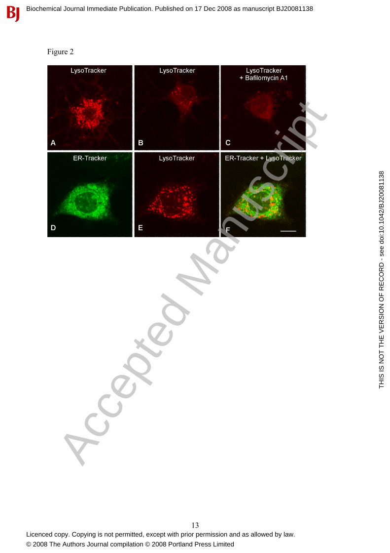
To determine the sub-cellular localization of acidic compartments in medulla neurons, we performed confocal microscopy of cells loaded with fluorescent weak base LysoTracker red. As shown in Figure 2, small discrete vesicles arranged in either a perinuclear fashion (Fig. 2A) or distributed more sparsely throughout the cytoplasm (Fig. 2B, E) were observed. Pretreatment with bafilomycin A1 (1 µM, 60 min) substantially reduced staining, as expected (Fig. 2C). In order to compare the distribution of acidic compartments with the endoplasmic reticulum, we imaged cells co-loaded with LysoTracker and ER-Tracker. As shown in Fig. 2D-F, the distribution of the two organelles was distinct with individual acidic vesicles surrounded by the dense endoplasmic reticulum.
The reduction of NAADP-mediated Ca2+ signals upon removal of extracellular Ca2+ (Fig. 1C) prompted us to probe for possible functional coupling of NAADP receptors to plasma membrane channels. We therefore measured electrical responses in nucleus ambiguus neurons in brainstem slices by the whole-cell patch-clamp technique. The mean resting membrane potential and input resistance of nucleus ambiguus neurons were -55.9 + 1.3 mV and 715.4 + 45.9 MΩ, respectively (n = 58). Nearly all neurons were silent; and injection of depolarizing currents (100 – 500 pA, 300 ms) elicited repetitive firings followed by a hyperpolarization (data not shown) as previously reported [45]. In the presence of tetrodotoxin (1 µM), NAADP-AM (500 nM) application depolarized 11 out of 19 neurons
Biochemical Journal Immediate Publication. Published on 17 Dec 2008 as manuscript BJ20081138
TH
IS IS
NO
T T
HE
VE
RS
ION
OF
RE
CO
RD
- s
ee d
oi:1
0.10
42/B
J200
8113
8
Acce
pted
Man
uscr
ipt
Licenced copy. Copying is not permitted, except with prior permission and as allowed by law.
© 2008 The Authors Journal compilation © 2008 Portland Press Limited

6
studied. The response of an exemplar neuron is shown in Fig. 3A, where NAADP-AM caused a depolarization of 10 mV. NAADP-mediated depolarization was reduced in amplitude in Ca2+-free saline (Fig. 3B) consistent with possible Ca2+ entry.
Because all of the above experiments were performed in the presence of intracellular EGTA, the effects of NAADP-AM on membrane potential are unlikely to be mediated by global changes in [Ca2+]i since these would be effectively buffered by the chelator [46]. We therefore considered the possibility that the effects of NAADP may be due to local [Ca2+]i changes. To test this, we performed patch-clamp experiments using an intracellular solution containing the fast calcium chelator, BAPTA in place of EGTA [46]. Under these conditions, cells failed to depolarize in response to NAADP-AM (Fig. 3C, F). These data suggest that the effects of NAADP on membrane potential are not direct but instead secondary to local changes in cytosolic Ca2+. To probe the role of acidic Ca2+ stores in this process, cells were pretreated with bafilomycin A1 (1 µM, 60 min) prior to NAADP-AM challenge. As shown in Fig. 3D, depolarization in response to NAADP-AM was abolished. The effects of NAADP-AM on membrane potential were also inhibited by pretreatment with ryanodine (1 µM, 10 min; Fig. 3E). Thus, the pharmacology of NAADP-mediated depolarization (summarized in Fig. 3F) and NAADP-mediated changes in global cytosolic Ca2+ (Fig. 1) are similar. Combined with the sensitivity of NAADP-mediated depolarization to slow and fast calcium chelators, we conclude that the effects of NAADP on membrane potential are due to coupled activation of subplasmalemmal NAADP-sensitive Ca2+ channels and ryanodine receptors. Thus, Ca2+ entry through this pathway might contribute to global cytosolic Ca2+ changes to NAADP, perhaps in conjunction with store-operated Ca2+ entry (Fig. 1). Although, our localization studies suggested that acidic compartments in these cells are predominantly cytoplasmic (Fig. 2) it is possible that a small subpopulation of peripheral acidic vesicles might not be resolvable using LysoTracker. Indeed, even in the large eggs of the sea urchin, acidic vesicles are relatively uniformly distributed, yet imaging of luminal pH clearly demonstrates heterogeneity in the spatial organization of NAADP-sensitive Ca2+ stores [47].
To further investigate the mechanism of NAADP-mediated depolarization, we examined current-voltage relationships. Subtraction of the two I-V curves obtained before and during perfusion with NAADP-AM reveals the presence of an inward current activated by NAADP (Fig. 4A-F). The mean reversal potential was ~ 0 mV (n = 6). As with depolarization, the NAADP-induced currents were markedly reduced in the presence of intracellular BAPTA in Ca2+-free saline (Fig. 4G) and following pretreatment with ryanodine (Fig. 4H). Since in our experimental conditions, the equilibrium potential for potassium ions is -98 mV and for sodium ions is +56 mV, the reversal potential of the NAADP-AM-induced current suggests an involvement of a non-selective cationic conductance. Indeed, the inward component of the NAADP-AM-induced current was abolished and the reversal potential shifted to more negative values (-40 mV) when extracellular Na+ was replaced with N-methyl-L-glucamine (NMDG) (Fig. 4I). Additionally, inward NAADP-mediated currents were readily detectable (but slightly reduced) when K+ in the intracellular solution was replaced with Cs+ to block voltage-gated K+ channels, and the slices pretreated with apamin and charybdotoxin to block Ca2+-activated K+ channels (Supplementary Fig. 2). Although depolarization was clearly reduced upon removing extracellular Ca2+ (Fig. 3B), we were unable to record an inward Ca2+ current, in the presence of NMDG (Fig. 4I). These data might suggest block of the channel by NMDG as reported for some non-selective cation channels [48] and/or the presence of an extracellular facilitatory Ca2+ binding site. Taken together, these data demonstrate functional coupling of NAADP-sensitive Ca2+ channels to Ca2+ activated non-selective cation channels.
NAADP-induced depolarization has previously been observed in starfish oocytes [49] however in those cells, the response was abolished upon removal of extracellular Ca2+ and
Biochemical Journal Immediate Publication. Published on 17 Dec 2008 as manuscript BJ20081138
TH
IS IS
NO
T T
HE
VE
RS
ION
OF
RE
CO
RD
- s
ee d
oi:1
0.10
42/B
J200
8113
8
Acce
pted
Man
uscr
ipt
Licenced copy. Copying is not permitted, except with prior permission and as allowed by law.
© 2008 The Authors Journal compilation © 2008 Portland Press Limited

7
the reversal potential of recorded currents was +50 mV suggesting activation of a Ca2+ current [50]. It is possible that the currents we observe are due to activation of TRPM2 channels which are Ca2+ permeable non-selective cation channels that show little permeability in the presence of NMDG and which are regulated by both cytosolic and extracellular Ca2+ [51] consistent with the properties we describe (Fig. 4). Indeed, TRPM2 channels (previously named TRPC7) are expressed in the medulla [52]. Although previous studies have proposed direct modulation of TRPM2 by NAADP [51], we note that whole-cell recordings were performed in intracellular solutions in which [Ca2+]i was unbuffered raising the possibility that the observed effects might be secondary store release as reported here. Intriguingly, such a mechanism offers an alternative explanation for the sensitivity of NAADP-mediated currents to a cyclic ADP-ribose antagonist [51] if mobilization of NAADP-sensitive stores is coupled to activation of ryanodine receptors.
In summary, we provide evidence for the presence of acidic NAADP-sensitive Ca2+ stores in medulla neurons. We show that activation of NAADP receptors in these cells using a novel cell permeable analogue is intimately associated with selective recruitment of an endoplasmic reticulum Ca2+ channel to mediate global cytosolic Ca2+ changes. Additionally, we provide new evidence for recruitment of non-selective cation channels on the cell surface by NAADP resulting in depolarization. Based on differential sensitivity of the latter to slow and fast Ca2+ chelators, we infer that this “chatter” is due to local sub membrane Ca2+ fluxes again involving coordinated mobilization of acidic and endoplasmic reticulum Ca2+ stores. Indeed, it is notable that such a cortical NAADP-sensitive store has been recently visualized in sea urchin eggs [47]. The dependence of the cortical flash in sea urchin eggs on both NAADP and L-type calcium channels is consisitent with NAADP acting indirectly possibly through prior mobilization of cortical calcium stores and subsequent depolarization. NAADP-mediated currents in starfish oocytes might also involve a cortical (endoplasmic reticulum) Ca2+ store based on their sensitivity to thapsigargin [50]. Thus, strategic placing of NAADP-sensitive Ca2+ stores may allow coordination of signaling events not only in the cell interior, but also at cell surface (Fig. 5). NAADP-mediated depolarization described here could provide a novel mechanism for excitation of cardiac vagal neurons possibly through NAADP synthesis via excitatory glutamatergic and/or cholinergic inputs [53].
ACKNOWLEDGEMENTS This study was supported by the NIH Grants NS18710 and HL51314 (to NJD) from the Department of Health and Human Services and by grants from the Biotechnology and Biological Sciences Research Council [grant number BB/D018110/1], Alzheimer’s Research Trust and Research into Ageing (to SP) and the Wellcome Trust and the Biotechnology and Biological Sciences Research Council [grant number BB/D012694/1] (to GCC). We thank Ch Li for useful discussions. REFERENCES 1. Lee, H. C. (2005) Nicotinic acid adenine dinucleotide phosphate (NAADP)-mediated
calcium signaling. J. Biol. Chem. 280, 33693-33696 2. Galione, A. (2006) NAADP, a new intracellular messenger that mobilizes Ca2+ from
acidic stores. Biochem. Soc. Trans. 34, 922-926 3. Clapper, D. L., Walseth, T. F., Dargie, P. J. and Lee, H. C. (1987) Pyridine nucleotide
metabolites stimulate calcium release from sea urchin egg microsomes desensitized to inositol trisphosphate. J. Biol. Chem. 262, 9561-9568
Biochemical Journal Immediate Publication. Published on 17 Dec 2008 as manuscript BJ20081138
TH
IS IS
NO
T T
HE
VE
RS
ION
OF
RE
CO
RD
- s
ee d
oi:1
0.10
42/B
J200
8113
8
Acce
pted
Man
uscr
ipt
Licenced copy. Copying is not permitted, except with prior permission and as allowed by law.
© 2008 The Authors Journal compilation © 2008 Portland Press Limited

8
4. Lee, H. C. and Aarhus, R. (1995) A derivative of NADP mobilizes calcium stores insensitive to inositol trisphosphate and cyclic ADP-ribose J. Biol. Chem. 270, 2152-2157
5. Berridge, G., Dickinson, G., Parrington. J., Galione. A. and Patel, S. (2002) Solubilization of receptors for the novel Ca2+-mobilizing messenger, nicotinic acid adenine dinucleotide phosphate. J. Biol. Chem. 277, 43717-43723
6. Galione, A. and Petersen, O. H. (2005) The NAADP receptor: new receptors or new regulation? Mol. Interv. 5, 73-79
7. Churchill, G. C., Okada, Y., Thomas, J. M., Genazzani, A. A., Patel, S. and Galione, A. (2002) NAADP mobilizes Ca2+ from reserve granules, lysosome-related organelles, in sea urchin eggs. Cell 111, 703-708
8. Christensen, K. A., Myers, J. T., Swanson, J. A. (2002) pH-dependent regulation of lysosomal calcium in macrophages. J. Cell Sci. 115, 599-607
9. Kinnear, N. P., Boittin, F. X., Thomas, J. M., Galione, A. and Evans, A. M. (2004) Lysosome-sarcoplasmic reticulum junctions. A trigger zone for calcium signaling by nicotinic acid adenine dinucleotide phosphate and endothelin-1. J. Biol. Chem. 279, 54319-54326
10. Yamasaki, M., Masgrau, R., Morgan, A. J., Churchill, G. C., Patel, S., Ashcroft, S. J. and Galione, A. (2004) Organelle selection determines agonist-specific Ca2+ signals in pancreatic acinar and beta cells. J. Biol. Chem. 279, 7234-7240
11. Brailoiu, E., Hoard, J. L., Filipeanu, C. M., Brailoiu, G. C., Dun, S. L., Patel, S. and Dun, N. J. (2005) Nicotinic acid adenine dinucleotide phosphate potentiates neurite outgrowth. J. Biol. Chem. 280, 5646-5650
12. Brailoiu, E., Churamani, D., Pandey, V., Brailoiu, G. C., Tuluc, F., Patel, S. and Dun, N. J. (2006) Messenger-specific role for nicotinic acid adenine dinucleotide phosphate in neuronal differentiation. J. Biol. Chem. 281, 15923-15928
13. Zhang, F., Zhang., G, Zhang, A. Y., Koeberl, M. J., Wallander, E. and Li, P. L. (2006) Production of NAADP and its role in Ca2+ mobilization associated with lysosomes in coronary arterial myocytes. Am. J. Physiol. Heart Circ. Physiol. 291, H274-H282
14. Kim, B. J., Park, K. H., Yim, C. Y., Takasawa, S., Okamoto, H., Im, M. J. and Kim, U. H. (2008) Generation of nicotinic acid adenine dinucleotide phosphate and cyclic ADP-ribose by glucagon-like peptide-1 evokes Ca2+ signal that is essential for insulin secretion in mouse pancreatic islets. Diabetes 57, 868-878
15. Gambara, G., Billington, R. A., Debidda, M., D'Alessio, A., Palombi, F., Ziparo, E., Genazzani, A. A. and Filippini, A. (2008) NAADP-induced Ca2+ signaling in response to endothelin is via the receptor subtype B and requires the integrity of lipid rafts/caveolae. J. Cell. Physiol. 216, 396-404
16. Cancela, J. M., Churchill, G. C. and Galione, A. (1999) Coordination of agonist-induced Ca2+-signalling patterns by NAADP in pancreatic acinar cells. Nature 398, 74-76
17. Boittin, F. X., Galione, A. and Evans, A. M. (2002) Nicotinic acid adenine dinucleotide phosphate mediates Ca2+ signals and contraction in arterial smooth muscle via a two-pool mechanism. Circ. Res. 91, 1168-1175
18. Churchill, G. C. and Galione, A. (2001) NAADP induces Ca2+ oscillations via a two-pool mechanism by priming IP3- and cADPR-sensitive Ca2+ stores. EMBO J. 20, 2666-2671
19. Santella, L., Kyozuka, K., Genazzani, A. A., De Riso, L. and Carafoli, E. (2000) Nicotinic acid adenine dinucleotide phosphate-induced Ca2+ release. Interactions among distinct Ca2+ mobilizing mechanisms in starfish oocytes. J. Biol. Chem. 275, 8301-8306
20. Macgregor, A., Yamasaki, M., Rakovic, S., Sanders, L., Parkesh, R., Churchill, G. C., Galione, A. and Terrar, D. A. (2007) NAADP controls cross-talk between distinct Ca2+ stores in the heart. J. Biol. Chem. 282, 5302-15311
Biochemical Journal Immediate Publication. Published on 17 Dec 2008 as manuscript BJ20081138
TH
IS IS
NO
T T
HE
VE
RS
ION
OF
RE
CO
RD
- s
ee d
oi:1
0.10
42/B
J200
8113
8
Acce
pted
Man
uscr
ipt
Licenced copy. Copying is not permitted, except with prior permission and as allowed by law.
© 2008 The Authors Journal compilation © 2008 Portland Press Limited

9
21. Patel, S., Churchill, G. C. and Galione, A. (2001) Coordination of Ca2+ signalling by NAADP. Trends Biochem. Sci. 26, 482-489
22. Bezin, S., Charpentier, G., Fossier, P. and Cancela J. M. (2006) The Ca2+-releasing messenger NAADP, a new player in the nervous system. J. Physiol. Paris 99, 111-118
23. Churchill, G. C., O'Neill, J. S., Masgrau, R., Patel, S., Thomas, J. M., Genazzani, A. A. and Galione, A. (2003) Sperm deliver a new second messenger: NAADP. Curr. Biol. 13, 125-128
24. Masgrau, R., Churchill, G. C., Morgan, A. J., Ashcroft, S. J. and Galione, A. (2003) NAADP: a new second messenger for glucose-induced Ca2+ responses in clonal pancreatic beta cells. Curr. Biol. 13, 247-251
25. Yamasaki, M., Thomas, J. M., Churchill, G. C., Garnham, C., Lewis, A. M., Cancela, J. M., Patel, S. and Galione, A. (2005) Role of NAADP and cADPR in the induction and maintenance of agonist-evoked Ca2+ spiking in mouse pancreatic acinar cells. Curr. Biol. 15, 874-878
26. Gasser, A., Bruhn, S., Guse, A. H. (2006) Second messenger function of nicotinic acid adenine dinucleotide phosphate revealed by an improved enzymatic cycling assay. J. Biol. Chem. 281, 16906-16913
27. Soares, S., Thompson, M., White, T., Isbell, A., Yamasaki, M., Prakash, Y., Lund, F. E., Galione, A. and Chini, E. N. (2007) NAADP as a second messenger: neither CD38 nor base-exchange reaction are necessary for in vivo generation of NAADP in myometrial cells. Am. J. Physiol. Cell. Physiol. 292, C227-C239
28. Lim, D., Kyozuka, K., Gragnaniello, G., Carafoli, E. and Santella, L. (2001) NAADP+ initiates the Ca2+ response during fertilization of starfish oocytes. FASEB J. 15, 2257-2267
29. Langhorst, M. F., Schwarzmann, N. and Guse, A. H. (2004) Ca2+ release via ryanodine receptors and Ca2+ entry: major mechanisms in NAADP-mediated Ca2+ signaling in T-lymphocytes. Cell Signal. 16, 1283-1289
30. Beck, A., Kolisek, M., Bagley, L. A., Fleig, A. and Penner, R. (2006) Nicotinic acid adenine dinucleotide phosphate and cyclic ADP-ribose regulate TRPM2 channels in T lymphocytes. FASEB J. 20, 962-964
31. Petersen, O. H. and Cancela, J. M. (1999) New Ca2+-releasing messengers: are they important in the nervous system? Trends Neurosci. 22, 488-495
32. Bak, J., White, P., Timár, G., Missiaen, L., Genazzani, A. A. and Galione, A. (1999) Nicotinic acid adenine dinucleotide phosphate triggers Ca2+ release from brain microsomes. Curr. Biol. 9, 751-754
33. Brailoiu, E., Miyamoto, M. D. and Dun, N. J. (2001) Nicotinic acid adenine dinucleotide phosphate enhances quantal neurosecretion at the frog neuromuscular junction: possible action on synaptic vesicles in the releasable pool. Mol. Pharmacol. 60, 718-724
34. Chameau, P., Van de Vrede, Y., Fossier, P. and Baux, G. (2001) Ryanodine-, IP3- and NAADP-dependent calcium stores control acetylcholine release. Pflugers Arch. 443, 289-296
35. Heidemann, A. C, Schipke, C. G. and Kettenmann, H. (2005) Extracellular application of nicotinic acid adenine dinucleotide phosphate induces Ca2+ signaling in astrocytes in situ. J. Biol. Chem. 280, 35630-35640
36. Patel, S., Churchill, G. C., Sharp, T. and Galione, A. (2000) Widespread distribution of binding sites for the novel Ca2+-mobilizing messenger, nicotinic acid adenine dinucleotide phosphate, in the brain. J. Biol. Chem. 275, 36495-36497
37. Mendelowitz, D. (1999) Advances in Parasympathetic Control of Heart Rate and Cardiac Function. News Physiol. Sci. 14, 155-161
Biochemical Journal Immediate Publication. Published on 17 Dec 2008 as manuscript BJ20081138
TH
IS IS
NO
T T
HE
VE
RS
ION
OF
RE
CO
RD
- s
ee d
oi:1
0.10
42/B
J200
8113
8
Acce
pted
Man
uscr
ipt
Licenced copy. Copying is not permitted, except with prior permission and as allowed by law.
© 2008 The Authors Journal compilation © 2008 Portland Press Limited

10
38. Parkesh, R., Lewis, A. M., Aley, P. K., Arredouani, A., Rossi, S., Tavares, R., Vasudevan, S. R., Rosen, D., Galione, A., Dowden, J. and Churchill, G. C. (2008) Cell-permeant NAADP: A novel chemical tool enabling the study of Ca2+ signalling in intact cells. Cell Calcium 43, 531-538
39. Brailoiu, G. C., Brailoiu, E., Chang, J. K. and Dun, N. J. (2008) Excitatory effects of HIV-1 Tat on cultured rat cerebral cortical neurons. Neuroscience 151, 701-710
40. Grynkiewicz, G., Poenie, M. and Tsien, R. Y. (1985) A new generation of Ca2+ indicators with greatly improved fluorescence properties. J. Biol. Chem. 260, 3440-3450
41. Schrlau, M. G., Brailoiu, E., Patel, S., Gogotsi, Y., Dun, N. J. and Bau, H. H. (2008) Carbon nanopipettes characterize calcium release pathways in breast cancer cells. Nanotechnology 19, 325102
42. Bouairi, E., Kamendi, H., Gorini, C. and Mendelowitz, D. (2006) Multiple Types of GABAA Receptors Mediate Inhibition in Brainstem Parasympathetic Cardiac Neurons in the Nucleus Ambiguus. J. Neurophysiol. 96, 3266-3272
43. Lloyd-Evans, E., Morgan, A. J., He, X., Smith, D. A., Elliot-Smith, E., Sillence, D. J., Churchill, G. C., Schuchman, E. H., Galione, A. and Platt, F. M. (2008) Niemann-Pick disease type C1 is a sphingosine storage disease that causes deregulation of lysosomal calcium. Nat. Med. 14, 1247-1255
44. Hohenegger, M., Suko, J., Gscheidlinger, R., Drobny, H. and Zidar, A. (2002) Nicotinic acid-adenine dinucleotide phosphate activates the skeletal muscle ryanodine receptor. Biochem. J. 367, 423-431
45. Mendelowitz, D. (1996) Firing properties of identified parasympathetic cardiac neurons in nucleus ambiguus. Am. J. Physiol. 271, H2609-H2614
46. Stern, M. D. (1992). Buffering of calcium in the vicinity of a channel pore. Cell Calcium 13, 183-192
47. Morgan, A. J. and Galione, A. (2007) Fertilization and nicotinic acid adenine dinucleotide phosphate induce pH changes in acidic Ca2+ stores in sea urchin eggs. J. Biol. Chem. 282, 37730-37737
48. Partridge, L. D. and Swandulla, D. (1988). Calcium-activated non-specific cation channels. Trends Neurosci. 11, 69-72
49. Moccia, F., Billington, R. A. and Santella, L. (2006) Pharmacological characterization of NAADP-induced Ca2+ signals in starfish oocytes. Biochem. Biophys. Res. Commun. 348, 329-336
50. Moccia, F., Lim, D., Nusco, G. A., Ercolano, E. and Santella, L. (2003) NAADP activates a Ca2+ current that is dependent on F-actin cytoskeleton. FASEB J. 17, 1907-1909
51. Starkus, J., Beck, A., Fleig, A. and Penner, R. (2007) Regulation of TRPM2 by extra- and intracellular calcium. J. Gen. Physiol. 130, 427-440
52. Nagamine, K., Kudoh, J., Minoshima, S., Kawasaki, K., Asakawa, S., Ito, F. and Shimizu, N. (1998) Molecular cloning of a novel putative Ca2+ channel protein (TRPC7) highly expressed in brain. Genomics. 54, 124-131
53. Wang, J., Irnaten, M., Neff, R. A., Venkatesan, P., Evans, C., Loewy, A. D., Mettenleiter, T. C. and Mendelowitz, D. (2001) Synaptic and neurotransmitter activation of cardiac vagal neurons in the nucleus ambiguus. Ann. N. Y. Acad. Sci. 940, 237-246
Biochemical Journal Immediate Publication. Published on 17 Dec 2008 as manuscript BJ20081138
TH
IS IS
NO
T T
HE
VE
RS
ION
OF
RE
CO
RD
- s
ee d
oi:1
0.10
42/B
J200
8113
8
Acce
pted
Man
uscr
ipt
Licenced copy. Copying is not permitted, except with prior permission and as allowed by law.
© 2008 The Authors Journal compilation © 2008 Portland Press Limited

11
FIGURE LEGENDS Figure 1 Effect of NAADP-AM on cytosolic Ca2+ concentration in cultured rat medullary neurons. Typical changes in [Ca2+]i of individual isolated medulla oblongata neurons stimulated with NAADP-AM either in the presence (A-B) or absence (C-F) of extracellular Ca2+. Cells were pretreated with bafilomycin A1 (1 µM, 60 min; D), ryanodine (1 µM, 10 min; E) or xestospongin C (10 µM, 15 min; F) as indicated. Figure 2. Localization of acidic compartments and endoplasmic reticulum in cultured rat medullary neurons. (A-B) Confocal images of LysoTracker Red fluorescence showing two different patterns of staining. Bafilomycin A1 (1 µM, 60 min) was added prior to addition of LysoTracker Red in C. (D-F) Co-localization of ER-tracker (D) and LysoTracker (E) fluorescence in the same cell. A merge of the two images is shown in F. Figure 3. Effect of NAADP-AM on membrane potential in nucleus ambiguus neurons. (A-E) Effect of NAADP-AM on resting membrane potential of nucleus ambiguus neurons from rat medullary slices. Experiments were performed either in the presence of extracellular Ca2+ (A, E) or Ca2+-free medium (B-D); tetrodotoxin (TTX, 1 µM) was included in the perfusion solution. Recordings in C were made using an intracellular solution which contained BAPTA in place of EGTA. Slices in D and E were pre-treated with bafilomycin A1 (BAF, 1 µM, 60 min) or ryanodine (1 µM, 10 min), respectively. Downward deflections are hyperpolarizing electrotonic potentials induced by constant current pulses (30 pA amplitude, 300 ms duration) used to monitor the membrane resistance. NAADP-AM was administered for the period indicated by the solid bar. F, Pooled data from the indicated number of neurons quantifying the extent of depolarization under the various conditions. * statistically significant difference as compared to the depolarizations in regular Krebs. ** statistically significant difference as compared to depolarizations in Ca2+-free Krebs solution (P<0.05). Figure 4. Effect of NAADP-AM on whole-cell currents recorded in nucleus ambiguus neurons. (A-B) Typical whole-cell currents before (-NAADP-AM) and during (+NAADP-AM) administration of NAADP-AM, in response to voltage command steps from a holding potential of -60mV to potentials varying from -120 mV to +60 mV (C). Steady-state I-V relationships before () and during () NAADP-AM superfusion are shown in panel D and the difference in currents in panel E. (F-I) Pooled difference currents (mean ± s.e.m from 4-6 neurons perfused with NAADP-AM in control Krebs solution (F), Ca2+-free Krebs solution and BAPTA-containing intracellular solution (G), control Krebs solution supplemented with ryanodine (1 µM) 10 min prior to recording (H) and modified Krebs solution in which NaCl was replaced with N-methyl-L-glucamine (NMDG) (I). Figure 5. Channel “chatter” in the medulla oblongata. Proposed model of channel cross-talk whereby NAADP receptors localized on acidic Ca2+ stores are functionally coupled to ryanodine receptors to mediate global Ca2+ signals in the cell interior and local Ca2+ signals in the plasma membrane to mediate depolarization.
Biochemical Journal Immediate Publication. Published on 17 Dec 2008 as manuscript BJ20081138
TH
IS IS
NO
T T
HE
VE
RS
ION
OF
RE
CO
RD
- s
ee d
oi:1
0.10
42/B
J200
8113
8
Acce
pted
Man
uscr
ipt
Licenced copy. Copying is not permitted, except with prior permission and as allowed by law.
© 2008 The Authors Journal compilation © 2008 Portland Press Limited

12
Figure 1
Biochemical Journal Immediate Publication. Published on 17 Dec 2008 as manuscript BJ20081138
TH
IS IS
NO
T T
HE
VE
RS
ION
OF
RE
CO
RD
- s
ee d
oi:1
0.10
42/B
J200
8113
8
Acce
pted
Man
uscr
ipt
Licenced copy. Copying is not permitted, except with prior permission and as allowed by law.
© 2008 The Authors Journal compilation © 2008 Portland Press Limited
13
Figure 2
Biochemical Journal Immediate Publication. Published on 17 Dec 2008 as manuscript BJ20081138
TH
IS IS
NO
T T
HE
VE
RS
ION
OF
RE
CO
RD
- s
ee d
oi:1
0.10
42/B
J200
8113
8
Acce
pted
Man
uscr
ipt
Licenced copy. Copying is not permitted, except with prior permission and as allowed by law.
© 2008 The Authors Journal compilation © 2008 Portland Press Limited

14
Figure 3
Biochemical Journal Immediate Publication. Published on 17 Dec 2008 as manuscript BJ20081138
TH
IS IS
NO
T T
HE
VE
RS
ION
OF
RE
CO
RD
- s
ee d
oi:1
0.10
42/B
J200
8113
8
Acce
pted
Man
uscr
ipt
Licenced copy. Copying is not permitted, except with prior permission and as allowed by law.
© 2008 The Authors Journal compilation © 2008 Portland Press Limited

15
Figure 4
Biochemical Journal Immediate Publication. Published on 17 Dec 2008 as manuscript BJ20081138
TH
IS IS
NO
T T
HE
VE
RS
ION
OF
RE
CO
RD
- s
ee d
oi:1
0.10
42/B
J200
8113
8
Acce
pted
Man
uscr
ipt
Licenced copy. Copying is not permitted, except with prior permission and as allowed by law.
© 2008 The Authors Journal compilation © 2008 Portland Press Limited

16
Figure 5
Biochemical Journal Immediate Publication. Published on 17 Dec 2008 as manuscript BJ20081138
TH
IS IS
NO
T T
HE
VE
RS
ION
OF
RE
CO
RD
- s
ee d
oi:1
0.10
42/B
J200
8113
8
Acce
pted
Man
uscr
ipt
Licenced copy. Copying is not permitted, except with prior permission and as allowed by law.
© 2008 The Authors Journal compilation © 2008 Portland Press Limited
Copyright © 2022 FDOKUMEN