NAADP Activates Two-Pore Channels on T Cell Cytolytic Granules to Stimulate Exocytosis and Killing
19
Current Biology 22, 1–7, December 18, 2012 ª2012 Elsevier Ltd All rights reserved http://dx.doi.org/10.1016/j.cub.2012.10.035 Report NAADP Activates Two-Pore Channels on T Cell Cytolytic Granules to Stimulate Exocytosis and Killing Lianne C. Davis, 1, * Anthony J. Morgan, 1 Ji-Li Chen, 2 Charlotte M. Snead, 1 Duncan Bloor-Young, 1 Eugene Shenderov, 2 Megan N. Stanton-Humphreys, 1,3 Stuart J. Conway, 3 Grant C. Churchill, 1 John Parrington, 1 Vincenzo Cerundolo, 2 and Antony Galione 1, * 1 Department of Pharmacology, University of Oxford, Mansfield Road, Oxford OX1 3QT, UK 2 MRC Human Immunology Unit, The Weatherall Institute of Molecular Medicine, University of Oxford, John Radcliffe Hospital, Headington, Oxford OX3 9DS, UK 3 Chemistry Research Laboratory, Department of Chemistry, University of Oxford, Mansfield Road, Oxford OX1 3TA, UK Summary A cytotoxic T lymphocyte (CTL) kills an infected or tumori- genic cell by Ca 2+ -dependent exocytosis of cytolytic gran- ules at the immunological synapse formed between the two cells. Although inositol 1,4,5-trisphosphate (IP 3 )- mediated Ca 2+ release from the endoplasmic reticulum activates the store-operated Ca 2+ -influx pathway that is necessary for exocytosis, it is not a sufficient stimulus [1–4]. Here we identify the Ca 2+ -mobilizing messenger nico- tinic acid adenine dinucleotide phosphate (NAADP) and its recently identified molecular target, two-pore channels (TPCs) [5–7], as being important for T cell receptor signaling in CTLs. We demonstrate that cytolytic granules are not only reservoirs of cytolytic proteins but are also the acidic Ca 2+ stores mobilized by NAADP via TPC channels on the gran- ules themselves, so that TPCs migrate to the immunological synapse upon CTL activation. Moreover, NAADP activates TPCs to drive exocytosis in a way that is not mimicked by global Ca 2+ signals induced by IP 3 or ionomycin, suggesting that critical, local Ca 2+ nanodomains around TPCs stimulate granule exocytosis. Hence, by virtue of the NAADP/TPC pathway, cytolytic granules generate Ca 2+ signals that lead to their own exocytosis and to cell killing. This study high- lights a selective role for NAADP in stimulating exocytosis crucial for immune cell function and may impact on stim- ulus-secretion coupling in wider cellular contexts. Results and Discussion The compartmentation of cell signaling pathways is important for maintaining the fidelity between extracellular stimuli and appropriate downstream responses. For Ca 2+ signaling, not all sources (or patterns) of intracellular Ca 2+ are equivalent, and Ca 2+ channels can differentially couple to cellular processes [1, 8]. In T cells, the exocytosis of cytolytic factors is clearly Ca 2+ dependent but, although Ca 2+ influx via STIM/Orai is a necessary pathway, it is not a sufficient stimulus for exocytosis because it requires the additional activation of protein kinases [2, 9]. We investigated whether other Ca 2+ channels couple more directly to exocytosis. All three major Ca 2+ -mobilizing second messengers—inositol 1,4,5-trisphos- phate (IP 3 ), cyclic ADP-ribose (cADPR), and nicotinic acid adenine dinucleotide phosphate (NAADP)—increase in concentration following T cell receptor activation [10, 11]. However, the molecular and organellar targets of NAADP are controversial in T cells [12], and the relationship between NAADP and exocytosis is unknown. To that end, we have investigated whether NAADP-dependent Ca 2+ signaling via a recently identified molecular target, the two-pore channel (TPC) [5–7, 13], is an overlooked molecular component of T cell cytolytic granule exocytosis. NAADP Mobilizes Acidic Ca 2+ Stores in Cytotoxic T Lymphocytes First, using a pharmacological approach, we characterized the intracellular Ca 2+ stores in a primary human cytotoxic T lymphocyte (CTL) clone and found pH-neutral as well as acidic Ca 2+ stores. Neutral stores were revealed by ionomycin, a Ca 2+ ionophore that can only act at neutral organelles [14], and cyclopiazonic acid (CPA), a sarcoendoplasmic reticulum Ca 2+ -ATPase inhibitor [15] (see Figures S1B–S1D available online). Similarly, agents that release Ca 2+ from acidic organ- elles also produced Ca 2+ responses, i.e., bafilomycin A1 (a V-type H + -ATPase inhibitor), nigericin and monensin (protono- phores), and glycyl-phenylalanine 2-naphthylamide (GPN, which osmotically lyses lysosomes [16]) (Figures S1E–S1I). The presence of acidic Ca 2+ stores is crucial because NAADP is predominantly (though not universally [12]) accepted to release Ca 2+ from acidic (endolysosomal) Ca 2+ stores [13, 17], thereby distinguishing it from IP 3 and cADPR that target the neutral endoplasmic reticulum (ER). To test whether NAADP could evoke Ca 2+ signals in CTLs, we indirectly introduced NAADP into the cytosol by bath appli- cation of a membrane-permeant ester precursor (NAADP/AM) that is converted to NAADP by intracellular esterases. NAADP/ AM elicited Ca 2+ oscillations, with the initial spike commonly comprising distinct first and second phases (Figures 1A and 1B); oscillations were blocked by the NAADP antagonist trans-Ned-19 (Figures 1A and 1B) [18]. The response to NAADP/AM was not an artifact of contaminating nonesterified (free) NAADP acting at plasma membrane receptors because equimolar extracellular NAADP was without effect (Figures 1A and 1B). Strikingly, NAADP/AM-induced Ca 2+ release dis- played a ‘‘bell-shaped’’ concentration-response curve (Fig- ures 1C and 1D), which is a hallmark of this second messenger [19]. Repeating experiments in Ca 2+ -free medium (to eliminate Ca 2+ influx), NAADP/AM still caused Ca 2+ oscillations, thus confirming intracellular stores as the primary Ca 2+ source (Figures S1J–S1N). That NAADP required acidic Ca 2+ stores was confirmed by inhibition of oscillations by bafilomycin A1, nigericin, monensin, or GPN (Figures 1E and 1F). Given that Ned-19 acts upon non-ER Ca 2+ channels [18], this provides compelling evidence that NAADP targets acidic organelles in primary CTLs, contrasting with results in a Jurkat cell line [20]. The long-standing ‘‘trigger hypothesis’’ or two-pool model [13, 21] describes NAADP as a provider of an initial ‘‘trigger’’ bolus of Ca 2+ that is subsequently amplified by Ca 2+ release *Correspondence: [email protected] (L.C.D.), antony.galione@ pharm.ox.ac.uk (A.G.) CURBIO 9936 Please cite this article in press as: Davis et al., NAADP Activates Two-Pore Channels on T Cell Cytolytic Granules to Stimulate Exocy- tosis and Killing, Current Biology (2012), http://dx.doi.org/10.1016/j.cub.2012.10.035
Transcript of NAADP Activates Two-Pore Channels on T Cell Cytolytic Granules to Stimulate Exocytosis and Killing
Current Biology 22, 1–7, December 18, 2012 ª2012 Elsevier Ltd All rights reserved http://dx.doi.org/10.1016/j.cub.2012.10.035
ReportNAADP Activates Two-Pore Channelson T Cell Cytolytic Granulesto Stimulate Exocytosis and Killing
Lianne C. Davis,1,* Anthony J. Morgan,1 Ji-Li Chen,2
Charlotte M. Snead,1 Duncan Bloor-Young,1
Eugene Shenderov,2 Megan N. Stanton-Humphreys,1,3
Stuart J. Conway,3 Grant C. Churchill,1 John Parrington,1
Vincenzo Cerundolo,2 and Antony Galione1,*1Department of Pharmacology, University of Oxford,Mansfield Road, Oxford OX1 3QT, UK2MRC Human Immunology Unit, The Weatherall Instituteof Molecular Medicine, University of Oxford, John RadcliffeHospital, Headington, Oxford OX3 9DS, UK3Chemistry Research Laboratory, Department of Chemistry,University of Oxford, Mansfield Road, Oxford OX1 3TA, UK
Summary
A cytotoxic T lymphocyte (CTL) kills an infected or tumori-genic cell by Ca2+-dependent exocytosis of cytolytic gran-ules at the immunological synapse formed between thetwo cells. Although inositol 1,4,5-trisphosphate (IP3)-mediated Ca2+ release from the endoplasmic reticulumactivates the store-operated Ca2+-influx pathway that isnecessary for exocytosis, it is not a sufficient stimulus[1–4]. Here we identify the Ca2+-mobilizing messenger nico-tinic acid adenine dinucleotide phosphate (NAADP) and itsrecently identified molecular target, two-pore channels(TPCs) [5–7], as being important for T cell receptor signalingin CTLs. We demonstrate that cytolytic granules are not onlyreservoirs of cytolytic proteins but are also the acidic Ca2+
stores mobilized by NAADP via TPC channels on the gran-ules themselves, so that TPCs migrate to the immunologicalsynapse upon CTL activation. Moreover, NAADP activatesTPCs to drive exocytosis in a way that is not mimicked byglobal Ca2+ signals induced by IP3 or ionomycin, suggestingthat critical, local Ca2+ nanodomains around TPCs stimulategranule exocytosis. Hence, by virtue of the NAADP/TPCpathway, cytolytic granules generate Ca2+ signals that leadto their own exocytosis and to cell killing. This study high-lights a selective role for NAADP in stimulating exocytosiscrucial for immune cell function and may impact on stim-ulus-secretion coupling in wider cellular contexts.
Results and Discussion
The compartmentation of cell signaling pathways is importantfor maintaining the fidelity between extracellular stimuli andappropriate downstream responses. For Ca2+ signaling, notall sources (or patterns) of intracellular Ca2+ are equivalent,and Ca2+ channels can differentially couple to cellularprocesses [1, 8]. In T cells, the exocytosis of cytolytic factorsis clearly Ca2+ dependent but, although Ca2+ influx viaSTIM/Orai is a necessary pathway, it is not a sufficient stimulusfor exocytosis because it requires the additional activation ofprotein kinases [2, 9]. We investigated whether other Ca2+
channels couple more directly to exocytosis. All three majorCa2+-mobilizing second messengers—inositol 1,4,5-trisphos-phate (IP3), cyclic ADP-ribose (cADPR), and nicotinic acidadenine dinucleotide phosphate (NAADP)—increase inconcentration following T cell receptor activation [10, 11].However, the molecular and organellar targets of NAADP arecontroversial in T cells [12], and the relationship betweenNAADP and exocytosis is unknown. To that end, we haveinvestigated whether NAADP-dependent Ca2+ signaling viaa recently identified molecular target, the two-pore channel(TPC) [5–7, 13], is an overlooked molecular component ofT cell cytolytic granule exocytosis.
NAADP Mobilizes Acidic Ca2+ Stores in Cytotoxic TLymphocytesFirst, using a pharmacological approach, we characterized theintracellular Ca2+ stores in a primary human cytotoxic Tlymphocyte (CTL) clone and found pH-neutral as well as acidicCa2+ stores. Neutral stores were revealed by ionomycin, a Ca2+
ionophore that can only act at neutral organelles [14], andcyclopiazonic acid (CPA), a sarcoendoplasmic reticulumCa2+-ATPase inhibitor [15] (see Figures S1B–S1D availableonline). Similarly, agents that release Ca2+ from acidic organ-elles also produced Ca2+ responses, i.e., bafilomycin A1 (aV-type H+-ATPase inhibitor), nigericin and monensin (protono-phores), and glycyl-phenylalanine 2-naphthylamide (GPN,which osmotically lyses lysosomes [16]) (Figures S1E–S1I).The presence of acidic Ca2+ stores is crucial because NAADPis predominantly (though not universally [12]) acceptedto release Ca2+ from acidic (endolysosomal) Ca2+ stores[13, 17], thereby distinguishing it from IP3 and cADPR thattarget the neutral endoplasmic reticulum (ER).To test whether NAADP could evoke Ca2+ signals in CTLs,
we indirectly introduced NAADP into the cytosol by bath appli-cation of a membrane-permeant ester precursor (NAADP/AM)that is converted to NAADP by intracellular esterases. NAADP/AM elicited Ca2+ oscillations, with the initial spike commonlycomprising distinct first and second phases (Figures 1A and1B); oscillations were blocked by the NAADP antagonisttrans-Ned-19 (Figures 1A and 1B) [18]. The response toNAADP/AM was not an artifact of contaminating nonesterified(free) NAADP acting at plasma membrane receptors becauseequimolar extracellular NAADP was without effect (Figures1A and 1B). Strikingly, NAADP/AM-induced Ca2+ release dis-played a ‘‘bell-shaped’’ concentration-response curve (Fig-ures 1C and 1D), which is a hallmark of this secondmessenger[19]. Repeating experiments in Ca2+-free medium (to eliminateCa2+ influx), NAADP/AM still caused Ca2+ oscillations, thusconfirming intracellular stores as the primary Ca2+ source(Figures S1J–S1N). That NAADP required acidic Ca2+ storeswas confirmed by inhibition of oscillations by bafilomycin A1,nigericin, monensin, or GPN (Figures 1E and 1F). Given thatNed-19 acts upon non-ER Ca2+ channels [18], this providescompelling evidence that NAADP targets acidic organelles inprimary CTLs, contrasting with results in a Jurkat cell line [20].The long-standing ‘‘trigger hypothesis’’ or two-pool model
[13, 21] describes NAADP as a provider of an initial ‘‘trigger’’bolus of Ca2+ that is subsequently amplified by Ca2+ release
*Correspondence: [email protected] (L.C.D.), [email protected] (A.G.)
CURBIO 9936
Please cite this article in press as: Davis et al., NAADP Activates Two-Pore Channels on T Cell Cytolytic Granules to Stimulate Exocy-tosis and Killing, Current Biology (2012), http://dx.doi.org/10.1016/j.cub.2012.10.035
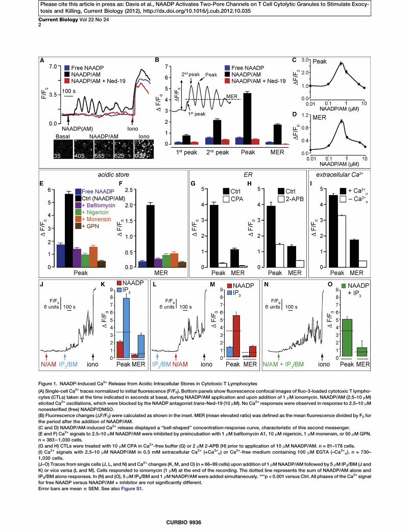
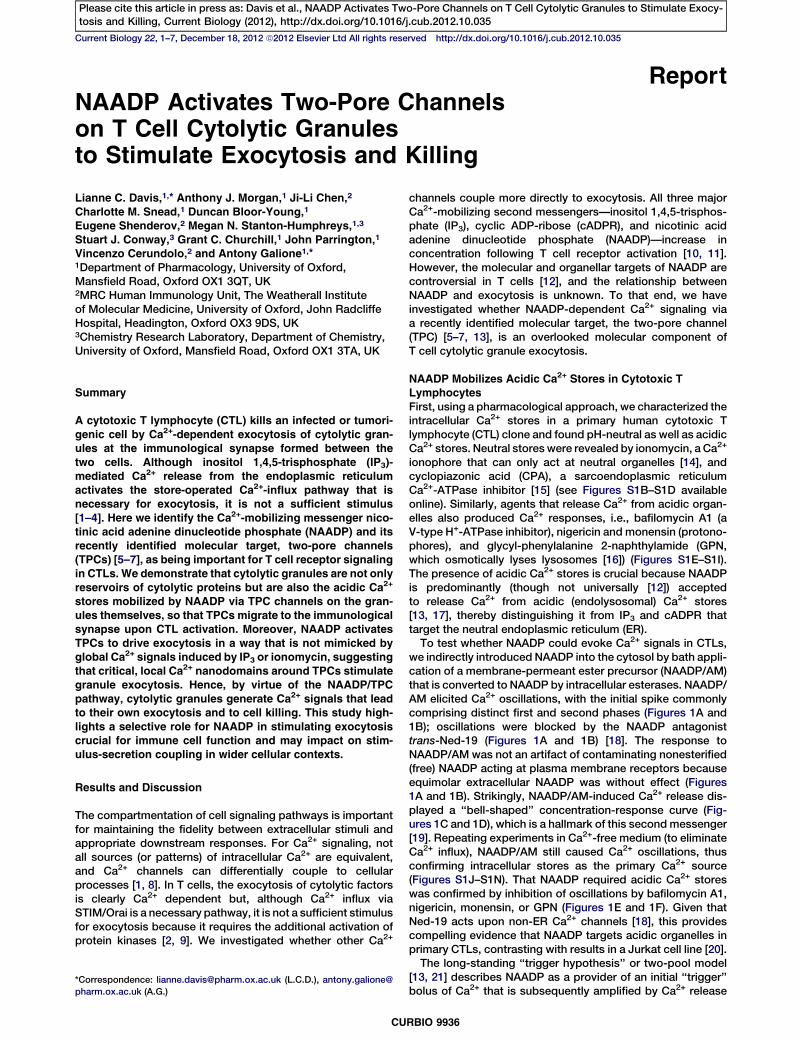
Figure 1. NAADP-Induced Ca2+ Release from Acidic Intracellular Stores in Cytotoxic T Lymphocytes
(A) Single-cell Ca2+ traces normalized to initial fluorescence (F/F0). Bottom panels show fluorescence confocal images of fluo-3-loaded cytotoxic T lympho-cytes (CTLs) taken at the time indicated in seconds at basal, during NAADP/AM application and upon addition of 1 mM ionomycin. NAADP/AM (2.5–10 mM)elicited Ca2+ oscillations, which were blocked by the NAADP antagonist trans-Ned-19 (10 mM). No Ca2+ responses were observed in response to 2.5–10 mMnonesterified (free) NAADP/DMSO.(B) Fluorescence changes (DF/F0) were calculated as shown in the inset. MER (mean elevated ratio) was defined as the mean fluorescence divided by F0 forthe period after the addition of NAADP/AM.(C and D) NAADP/AM-induced Ca2+ release displayed a ‘‘bell-shaped’’ concentration-response curve, characteristic of this second messenger.(E and F) Ca2+ signals to 2.5–10 mM NAADP/AM were inhibited by preincubation with 1 mM bafilomycin A1, 10 mM nigericin, 1 mMmonensin, or 50 mMGPN.n = 38321,030 cells.(G and H) CTLs were treated with 10 mM CPA in Ca2+-free buffer (G) or 2 mM 2-APB (H) prior to application of 10 mM NAADP/AM. n = 81–178 cells.(I) Ca2+ signals with 2.5–10 mM NAADP/AM in 0.5 mM extracellular Ca2+ (+Ca2+o) or Ca
2+-free medium containing 100 mM EGTA (–Ca2+o). n = 730–1,030 cells.(J–O) Traces from single cells (J, L, and N) and Ca2+ changes (K,M, andO) (n = 66–99 cells) upon addition of 1 mMNAADP/AM followed by 5 mM IP3/BM (J andK) or vice versa (L and M). Cells responded to ionomycin (1 mM) at the end of the recording. The dotted line represents the sum of NAADP/AM alone andIP3/BM alone responses. In (N) and (O), 5 mM IP3/BM and 1 mMNAADP/AMwere added simultaneously. ***p < 0.001 versus Ctrl. All phases of the Ca2+ signalfor free NAADP versus NAADP/AM + inhibitor are not significantly different.Error bars are mean 6 SEM. See also Figure S1.
Current Biology Vol 22 No 242
CURBIO 9936
Please cite this article in press as: Davis et al., NAADP Activates Two-Pore Channels on T Cell Cytolytic Granules to Stimulate Exocy-tosis and Killing, Current Biology (2012), http://dx.doi.org/10.1016/j.cub.2012.10.035

from the ER by virtue of the Ca2+ sensitivity of the IP3 receptor(IP3R) or ryanodine receptor (RyR), so-called Ca2+-inducedCa2+ release (CICR). We tested for the coinvolvement ofthe ER, first by depleting the ERwith the Ca2+-ATPase inhibitorCPA, which did indeed abrogate NAADP/AM responses(Figures 1G, S2C, and S1D). Also consistent with ER involve-ment is the fact that NAADP/AM stimulated Ca2+ influx(Figure 1I), a natural consequence of ER depletion and recruit-ment of the dominant store-regulated Ca2+ entry pathway inT cells.
Given that IP3R1–3 and RyR1 were all detected by PCR inthese primary T cells (Figures S3A and S3B), we tested whichER channel family was functionally important. We found noevidence of functional RyRs, either with or without NAADP/AM (Figures S1O–S1Q), conceivably due to a low RyR abun-dance [22]. In contrast, using a cell-permeant analog of themajor Ca2+-mobilizing second messenger IP3 (IP3/BM), IP3Rswere demonstrably active (Figures 1J–1O, 2G, 2H, S2A, andS2B), and complementary pieces of evidence suggested thatthey contribute to NAADP-evoked responses: first, IP3Rblockade with 2-APB (2-aminoxydiphenylborate) (Figures
Figure 2. NAADP Is Involved in PhysiologicalT Cell Receptor Activation
(A–C) Ca2+ signals in CTLs upon engagement withantigen-presenting cells (APCs) loaded with100 nM ESO 9C peptide. CTLs were incubatedwith 0.1% DMSO (Ctrl), 10 mM trans-Ned-19,1 mMbafilomycin A1, or 2 mMor 50 mM2-APBpriorto engagement and throughout the experiment.(A and B) Single-cell Ca2+ traces. Inset: confocalimages of CTL loadedwith fluo-3 and LysotrackerRed to label acidic organelles before (basal) andafter engagement with APCs.(C) Peak Ca2+ changes (DF/F0) and MER (meanvalue of fluorescence throughout the posten-gagement period) as normalized to F0. n = 130–239 cells.(D) Lysis of target cells pulsed with ESO 9Cpeptide by CTL (measured by release of 51Cr after2 hr) under conditions as in (A)–(C). n = 6.(E) Granzyme B secretion by CTLs 15 min afterantigen presentation, under conditions as in (A)–(C) and measured by ELISA. n = 6–15.(F–I) Ca2+ signal upon addition of NAADP/AM (F),IP3/BM (G and H), and ionomycin (I) to fluo-3-loaded CTL.(J and K) In the absence of antigen presentation,CTLs were stimulated with NAADP/AM in thepresence of 0.1% DMSO (Ctrl), 10 mM trans-Ned-19, or 1 mM bafilomycin A1 (J); CTLs weretreated with IP3/BM, 10 mM NAADP/AM, 10 mMnigericin, 1 mM ionomycin, 50 nM PMA, or acombination of 1 mM ionomycin and 50 nM PMA(K). After 30 min, supernatants were assayed forgranzyme B by ELISA (n = 3).***p < 0.001, **p < 0.01, *p < 0.05. Error bars aremean 6 SEM. See also Figure S2.
S2A and S2B) [23] profoundly reducedNAADP/AM-stimulated Ca2+ oscillations(Figure 1H). Conversely, costimulatingIP3Rs enhanced NAADP/AM-inducedCa2+ release (Figures 1J–1O), and ina synergistic (greater than additive)manner that depended upon the orderof addition, with the greatest effect
occurring when NAADP was added first. Together, thesedata are consistent with the trigger hypothesis wherebyNAADP provides the trigger Ca2+ from acidic stores that issubsequently amplified by IP3Rs on the ER [13, 19].
The T Cell Receptor Recruits the NAADP PathwayHaving defined NAADP signaling in CTLs, we determinedwhether it was involved during physiological T cell receptor(TCR) activation. CTLs were stimulated upon engagementwith peptide-loaded antigen-presenting cells (APCs), whichresulted in the expected two phases of Ca2+ increase: animmediate spike (Ca2+ release from intracellular stores),followed by a slower, sustained increase or Ca2+ oscillations(dependent on Ca2+ influx; Figures 2A and 2B [24]). AcidicCa2+ stores and NAADP did indeed contribute to TCR-inducedCa2+ signals as judged by the effect of inhibitors of the NAADPpathway: Ned-19 or bafilomycin A1 and NAADP self-desensiti-zation profoundly reduced the amplitudes of the Ca2+ peaks aswell as the mean fluorescence (an index of how sustained theresponse is; Figures 2A and 2C). In addition to NAADP, IP3Rsand Ca2+ influx were also recruited as expected (and required;
Cytolytic Granules as NAADP-Stimulated Ca2+ Stores3
CURBIO 9936
Please cite this article in press as: Davis et al., NAADP Activates Two-Pore Channels on T Cell Cytolytic Granules to Stimulate Exocy-tosis and Killing, Current Biology (2012), http://dx.doi.org/10.1016/j.cub.2012.10.035

Figure 2B): 2-APB attenuated Ca2+ oscillations at low (2 mM)and high (50 mM) concentrations, respectively (Figure 2C).Together, these results highlight a physiological role forNAADP-induced Ca2+ release that interleaves with Ca2+-induced Ca2+ release (IP3Rs) and Ca2+ influx.
NAADP Stimulates Cytolytic Granule ExocytosisWe next asked whether NAADP/acidic stores were importantfor exocytosis and target cell killing. CTL-dependent killingof target cells can be effected by two processes: (1) theCa2+-dependent vectorial exocytosis of cytolytic proteinsgranzyme B and perforin from CTL cytolytic granules (a typeof secretory lysosome [25]), and (2) the cell-surface expressionof the death receptor Fas ligand [26]. A link betweenCa2+ influxand cytolytic granule exocytosis is well established [3], buta role for acidic Ca2+ stores in driving secretion is lessunderstood. Therefore, we measured granzyme B secretionat early times (15–30 min) after APC engagement. Testing forNAADP and acidic Ca2+ stores, we found that Ned-19 orbafilomycin A1 substantially inhibited granzyme B exocytosis(Figure 2E) and this translated into an inhibition of cell killing(Figure 2D). Importantly, this was not merely a consequenceof a reduced global Ca2+ signal, because 2 mM and 50 mM2-APB also lowered the TCR Ca2+ signal to the same extent(Figure 2C) and yet secretion and cell killing were unaffected(Figures 2D and 2E). Bafilomycin A1 had a disproportionateeffect upon killing compared to exocytosis, which is probablydue to an additional effect upon the target cells themselves:bafilomycin A1 (and other V-ATPase inhibitors) have been
Figure 3. TPCs Are Necessary for TCR-TriggeredCa2+ Release via NAADP
(A–C) Ca2+ oscillations in Jurkat 1G4 cellsinduced by 2.5–10 mMNAADP/AM were inhibitedby 10 mM trans-Ned-19 or 1 mM bafilomycin A1.Single-cell Ca2+ traces (A) and summary ofmaximum peak Ca2+ changes and MER (B andC) are shown. n = 88–103 cells.(D–I) Jurkat 1G4 cells were nucleofected with 100pmol siRNA (nontargeting [NT], TPC1 and TPC2)or without siRNA (Mock) and incubated for 72 hr(D–G) or 48–72 hr (H and I). siRNA-treated cellswere stimulated with 10 mM NAADP/AM (D–F) orby contact with 10 mg/ml anti-human CD3-coatedcoverslips (G–I). Single-cell Ca2+ traces (D and G)and summary of peak Ca2+ changes (E–F and H–I)are shown for n = 213–383 cells from six indepen-dent siRNA experiments.Error bars are mean 6 SEM. See also Figure S3.
shown to profoundly block the per-forin-dependent cytotoxicity mediatedby CTLs [27]. In keeping with the Ca2+
data, 10 mM ryanodine did not affectTCR-stimulated granzyme B release(93% 6 8% of control, n = 8), excludinga role for RyRs in exocytosis. Insummary, these data implicate NAADPin TCR-stimulated exocytosis.
We directly tested whether NAADPcould stimulate exocytosis, even inthe absence of TCR activation. Remark-ably, application of NAADP/AM per sewas sufficient to evoke granzyme Bsecretion in a Ned-19- and bafilomycin
A1-sensitive manner (Figure 2J). Moreover, the hallmark bell-shaped NAADP/AM concentration-response curve previouslyobserved with Ca2+ signals (Figures 1C and 1D) was preservedwith granzyme B exocytosis (Figure 2J). Together, the dataindicate that NAADP can and does drive exocytosis.If NAADP simply acts by elevating global Ca2+, then any
experimental manipulation that mimics this should stimulateexocytosis. Therefore, we raised Ca2+ in other ways and as-sessed secretion. IP3/BM evoked robust Ca2+ signals inCTLs, and in a classic pattern dependent on the stimulus inten-sity: lower concentrations of IP3/BM stimulated oscillations,whereas higher concentrations gave a strong peak-and-plateau Ca2+ pattern (indicative of Ca2+ release and influxthrough Orai/STIM activation; Figures 2G and 2H). AlthoughIP3-induced Ca2+ signals mimic those seen following stimula-tion by physiological TCR engagement [24] or NAADP/AM(Figures 2A and 2F), granzyme B secretion was not observed(Figure 2K). Likewise, the Ca2+ ionophore ionomycin produceda strong Ca2+ signal (Figure 2I) but failed to evoke exocytosis(unless combined with phorbol esters to activate PKC/ERK[4, 26]; Figure 2K). Interestingly, releasing Ca2+ from the acidicCa2+ store using nigericin (Figure S1E) was not sufficientto evoke exocytosis, reinforcing the unique properties ofNAADP (Figure 2K). That is, in the absence of TCR activation,only NAADP-induced Ca2+ release coupled efficiently toexocytosis, thereby reinforcing the different roles of secondmessengers, i.e., NAADP and IP3 [13, 19].What underlies this differential coupling? A major difference
betweenNAADP and the other agents is that NAADP ultimately
Current Biology Vol 22 No 244
CURBIO 9936
Please cite this article in press as: Davis et al., NAADP Activates Two-Pore Channels on T Cell Cytolytic Granules to Stimulate Exocy-tosis and Killing, Current Biology (2012), http://dx.doi.org/10.1016/j.cub.2012.10.035

recruits three Ca2+ sources: acidic Ca2+ stores (Figures 1Eand 1F), the ER (Figures 1G and 1H), and, consequently, extra-cellular Ca2+ (Figure 1I). In contrast, IP3 and ionomycin onlyshare the latter two (neither mobilizes acidic Ca2+ stores[14]). This suggests that Ca2+ release from acidic storesby NAADP is a crucial permissive factor for exocytosis.Indeed, although Ca2+ influx via Orai/STIM is crucial forlymphocyte function, it cannot drive exocytosis without thepharmacological activation of protein kinases [2, 9]. Therefore,NAADP is the first Ca2+ pathway demonstrated to drive exocy-tosis without additional stimuli. Given that the global Ca2+
signal is a poor predictor of exocytosis, we hypothesize thatlocal Ca2+ nanodomains around the acidic Ca2+ stores arewhat distinguishes NAADP from the other stimuli. This under-scores the importance of where the Ca2+ is released, i.e., localversus global Ca2+ responses, and so we turned to identifyingthe molecular target of NAADP, and thereby its subcellularlocale.
NAADP-Dependent Ca2+ Signals during TCR ActivationRequire Two-Pore ChannelsRecently, the major ion channel target for NAADP has beenidentified as members of the TPC family that appropriately
Figure 4. TPCs Translocate to an ImmunologicalSynapse upon Stimulation with Anti-CD3
eYFP-tagged HsTPC1 and HsTPC2 (green) wereexpressed in Jurkat 1G4 cells and added to glasscoverslips coated with poly-L-lysine or 10 mg/mlanti-human CD3 antibody mounted in an imagingchamber, as depicted in the cartoon. Cells werelabeled with FM4-64 (red) to delineate the plasmamembrane edge of the cell.(A and B) Confocal images of cells attached topoly-L-lysine (Ctrl; left-hand side) or engagedwith anti-CD3 antibody (aCD3; right-hand side)showing z stacks through x and y axes (topimages) and 3D reconstruction of the z stack(bottom images). TPC localization was assessedthrough the z stack as a product its area and itsfluorescence, as depicted in (C)–(E). A slice depthof 0 indicates the bottom of the cell (i.e., thecontact zone between cell and the coverslip).(C and D) TPC1 (C) and TPC2 (D) localizationsignificantly alters from being present deeper inthe cell (slice depth 3–5 mm) toward aCD3, i.e.,the bottom of the cells (slice depth 0–2 mm)compared to control.(E) Comparison of TPC1 and TPC2 movement incells engagedwith anti-CD3. There was no signif-icant difference in the migration of the two iso-forms. TPC1 n = 32–43; TPC2 n = 52–69.Error bars are mean 6 SEM. See also Figure S4.
resideonacidiccompartmentsof theen-dolysosomal system in variousmamma-lian cell types [5–7]. To address theimportance of TPCs in TCR activation,we used Jurkat cells transduced withthe NY-ESO-1157–165-HLA A2-restrictedTCR 1G4 [25], hereafter referred to asJurkat 1G4 cells, because they areamenable to genetic manipulation andexpress the 1G4 TCR, which has thesame specificity as the TCR expressedby the 4D8 CD8+ T cell clone used
above. We established that Jurkat 1G4 cells mirrored the 4D8T cell clone in several key aspects: first, each cell typeexpresses both TPC1 and TPC2 isoforms as judged by RT-PCR (Figures S3C and S3F), and second, NAADP/AM elicitedCa2+ responses in Jurkat 1G4 cells that were sensitive toNed-19 and bafilomycin A1 (Figures 3A–3C), consistent withacidic Ca2+ store involvement [22, 28].The functional role of TPCs in NAADP-induced Ca2+ release
was tested by suppressing TPC1 and TPC2 expression withsmall interfering RNA (siRNA) knockdown (assessed by RT-qPCR and western blot; Figures S3G–S3I). Consequently,NAADP/AM-evoked Ca2+ release was reduced by TPCsiRNAs, with a more pronounced inhibition of Ca2+ signalsseen with TPC2 than with TPC1 siRNA (Figures 3D–3F andS3J). Statistical analysis suggested that only a subpopulationof cells were affected by siRNA (Figure S3J), presumably afunction of the transfection efficiency (although we could notempirically identify which cells were transfected). The Ca2+
signal was significantly reduced in amplitude (by 30%–54%)—a partial effect consistent with incomplete TPC knock-down (Figures S3G–S3I)—and underscored the importance ofTPCs for NAADP action. Because NAADP is important forTCR signaling (Figure 2), it follows that TPCs should likewise
Cytolytic Granules as NAADP-Stimulated Ca2+ Stores5
CURBIO 9936
Please cite this article in press as: Davis et al., NAADP Activates Two-Pore Channels on T Cell Cytolytic Granules to Stimulate Exocy-tosis and Killing, Current Biology (2012), http://dx.doi.org/10.1016/j.cub.2012.10.035
be required, and indeed, TCR-induced Ca2+ signals werereduced with either TPC1 or TPC2 siRNA (maximal inhibition44% for each isoform; Figures 3G–3I and S3K). Incidentally,RyRs were absent in Jurkat 1G4 cells (Figure S3E, as withother Jurkat T cell clones [22, 28]), confirming that RyRs arenot obligatory for NAADP action, though they have beenproposed as NAADP receptors by others [12, 13]. The dataaffirm both TPC isoforms as necessary components ofNAADP-dependent Ca2+ signals during TCR activation, andso CTLs join the emerging list of cells that utilize the NAADP/TPC couple [13, 16].
TPCs on Exocytotic Granules Translocate to theImmunological SynapseThus far, we had not determined precisely which acidic vesicleclass (or classes) the NAADP/TPC axis was associated with.To that end, we tagged TPC1 and TPC2 with fluorescentproteins and compared their distribution in Jurkat 1G4 cellswith other acidic vesicle markers. Both TPC1 (Figures S4A–S4C) and TPC2 (Figures S4D–S4F) colocalized with twomarkers of the cytolytic granules themselves (granzyme Band LAMP-1), although the TPC1 overlap was slightly lowerthan that for TPC2 (distribution differences between the twoisoforms are a recurring theme [13]). This overlap was notpeculiar to Jurkat 1G4 cells, because we observed a similarpattern in primary CTLs: using immunofluorescence, endoge-nous TPC2 extensively colocalized with granzyme B andLAMP-1 (Figures S4G–S4I). The specificity of TPC2 antibodylabeling was verified by preblocking with the immunogenicpeptide, which reduced labeling by 87.4% 6 0.4%. Unfortu-nately, we were unable to obtain specific staining with anti-TPC1 antibodies. NAADP therefore targets the exocytoticvesicles themselves.
That TPCs are found on cytolytic granules agrees with theNAADP acidic store pharmacology (Figures 1E and 1F)because cytolytic granules are the acidic, secretory lyso-somes of CTL and natural killer (NK) cells [25, 29]. Indeed,TPC2 was identified in the proteome of NK cell secretory lyso-some membranes [30], and such a localization provides theappropriate architecture for NAADP-evoked local Ca2+
domains that selectively couple to exocytosis. Thatsecretory vesicles act as Ca2+ stores has some precedents[13, 31, 32].
An important characteristic of cytolytic granules is that theyrapidly polarize along microtubules toward the immunologicalsynapse (IS) when the TCR is activated (Figures S4J and S4K)[25]. Because TPC1 and TPC2 are expressed on these gran-ules, they should also move to the IS. CTLs, including Jurkatclones [33], can form a pseudo-IS with anti-CD3-coated glasscoverslips, and so we examined the 3D distribution of fluores-cently tagged TPCs in adherent cells in the presence orabsence of antibody coating. Under control conditions withoutantibody, TPC puncta were distributed throughout the cell(Figures 4A–4D). However, when anti-CD3 was present at thecontact surface, a pronounced translocation of both TPC1and TPC2 from deeper regions of the cell to the contactsurface was observed (Figures 4A–4E). Figure 4E shows thatTPC1 and TPC2 translocate to the same extent, and their clus-tering may contribute to the high Ca2+ domains required forlytic granule exocytosis [3]. Moreover, immunolabeled TPC2in primary CTLs shows endogenous polarization to the ISwithin a primary CTL:target cell conjugate (Figures S4L andS4M). This is the first report of a directed movement and clus-tering of TPCs during a specific response.
ConclusionIn summary, TCR activation recruits NAADP to activate targetTPC channels resident on the exocytotic granules themselves,and TPCs consequently translocate toward the immunologicalsynapse. Therefore, these granules store and release the Ca2+
for their own exocytosis and deliver Ca2+ in an ‘‘autocrine’’fashion via TPCs, presumably acting in local perigranularCa2+ nanodomains [8]. In the absence of TCR activation, otherCa2+ signaling pathways, e.g., IP3, fail to mimic NAADP.Although the Orai1-STIM1 complex (CRAC channels) makesa substantial contribution to CTL Ca2+ signals [3], activationof Ca2+ influx per se is insufficient to drive exocytosis,requiring the additional presence of protein kinase activators[2, 9]; the NAADP/TPC pathway is therefore all the moreremarkable for being able to drive exocytosis. Clearly, CRACchannels are important for sustaining exocytosis [1, 3], butwe propose that local NAADP/acidic store/TPC signaling isan additional important component.
Supplemental Information
Supplemental Information includes four figures and Supplemental Experi-mental Procedures and can be found with this article online at http://dx.doi.org/10.1016/j.cub.2012.10.035.
Acknowledgments
We thank F.M. Platt and D.C. Anthony (University of Oxford) for use of theAmaxa Nucelofector II device and use of the Roche LightCycler 480 qPCRmachine, respectively. We are also grateful to N. Emptage and Z. Padamsey(University of Oxford) for advice on statistical analyses. We thank the Well-come Trust, the Medical Research Council, and Cancer Research UK (grantC399/A2291) for financial support.
Received: May 29, 2012Revised: September 7, 2012Accepted: October 19, 2012Published: November 21, 2012
References
1. Feske, S. (2007). Calcium signalling in lymphocyte activation anddisease. Nat. Rev. Immunol. 7, 690–702.
2. Grybko, M.J., Pores-Fernando, A.T., Wurth, G.A., and Zweifach, A.(2007). Protein kinase C activity is required for cytotoxic T cell lyticgranule exocytosis, but the theta isoform does not play a preferentialrole. J. Leukoc. Biol. 81, 509–519.
3. Pores-Fernando, A.T., and Zweifach, A. (2009). Calcium influx andsignaling in cytotoxic T-lymphocyte lytic granule exocytosis. Immunol.Rev. 231, 160–173.
4. Lancki, D.W., Weiss, A., and Fitch, F.W. (1987). Requirements fortriggering of lysis by cytolytic T lymphocyte clones. J. Immunol. 138,3646–3653.
5. Calcraft, P.J., Ruas, M., Pan, Z., Cheng, X., Arredouani, A., Hao, X.,Tang, J., Rietdorf, K., Teboul, L., Chuang, K.T., et al. (2009). NAADPmobilizes calcium from acidic organelles through two-pore channels.Nature 459, 596–600.
6. Brailoiu, E., Churamani, D., Cai, X., Schrlau, M.G., Brailoiu, G.C., Gao, X.,Hooper, R., Boulware, M.J., Dun, N.J., Marchant, J.S., and Patel, S.(2009). Essential requirement for two-pore channel 1 in NAADP-medi-ated calcium signaling. J. Cell Biol. 186, 201–209.
7. Zong, X., Schieder, M., Cuny, H., Fenske, S., Gruner, C., Rotzer, K.,Griesbeck, O., Harz, H., Biel, M., and Wahl-Schott, C. (2009). The two-pore channel TPCN2 mediates NAADP-dependent Ca2+-release fromlysosomal stores. Pflugers Arch. 458, 891–899.
8. Eggermann, E., Bucurenciu, I., Goswami, S.P., and Jonas, P. (2012).Nanodomain coupling between Ca2+ channels and sensors of exocy-tosis at fast mammalian synapses. Nat. Rev. Neurosci. 13, 7–21.
9. Ma, J.S., Haydar, T.F., and Radoja, S. (2008). Protein kinase C deltalocalizes to secretory lysosomes in CD8+ CTL and directly mediates
Current Biology Vol 22 No 246
CURBIO 9936
Please cite this article in press as: Davis et al., NAADP Activates Two-Pore Channels on T Cell Cytolytic Granules to Stimulate Exocy-tosis and Killing, Current Biology (2012), http://dx.doi.org/10.1016/j.cub.2012.10.035

TCR signals leading to granule exocytosis-mediated cytotoxicity.J. Immunol. 181, 4716–4722.
10. Gasser, A., Bruhn, S., and Guse, A.H. (2006). Second messengerfunction of nicotinic acid adenine dinucleotide phosphate revealed byan improved enzymatic cycling assay. J. Biol. Chem. 281, 16906–16913.
11. Guse, A.H., da Silva, C.P., Berg, I., Skapenko, A.L., Weber, K., Heyer, P.,Hohenegger, M., Ashamu, G.A., Schulze-Koops, H., Potter, B.V., andMayr, G.W. (1999). Regulation of calcium signalling in T lymphocytesby the second messenger cyclic ADP-ribose. Nature 398, 70–73.
12. Guse, A.H. (2009). Second messenger signaling: multiple receptors forNAADP. Curr. Biol. 19, R521–R523.
13. Morgan, A.J., Platt, F.M., Lloyd-Evans, E., and Galione, A. (2011).Molecular mechanisms of endolysosomal Ca2+ signalling in health anddisease. Biochem. J. 439, 349–374.
14. Fasolato, C., Zottini, M., Clementi, E., Zacchetti, D., Meldolesi, J., andPozzan, T. (1991). Intracellular Ca2+ pools in PC12 cells. Three intracel-lular pools are distinguished by their turnover and mechanisms of Ca2+
accumulation, storage, and release. J. Biol. Chem. 266, 20159–20167.
15. Goeger, D.E., Riley, R.T., Dorner, J.W., and Cole, R.J. (1988).Cyclopiazonic acid inhibition of the Ca2+-transport ATPase in rat skel-etal muscle sarcoplasmic reticulum vesicles. Biochem. Pharmacol. 37,978–981.
16. Galione, A., Morgan, A.J., Arredouani, A., Davis, L.C., Rietdorf, K., Ruas,M., and Parrington, J. (2010). NAADP as an intracellular messengerregulating lysosomal calcium-release channels. Biochem. Soc. Trans.38, 1424–1431.
17. Menteyne, A., Burdakov, A., Charpentier, G., Petersen, O.H., andCancela, J.M. (2006). Generation of specific Ca2+ signals from Ca2+
stores and endocytosis by differential coupling to messengers. Curr.Biol. 16, 1931–1937.
18. Naylor, E., Arredouani, A., Vasudevan, S.R., Lewis, A.M., Parkesh, R.,Mizote, A., Rosen, D., Thomas, J.M., Izumi, M., Ganesan, A., et al.(2009). Identification of a chemical probe for NAADP by virtualscreening. Nat. Chem. Biol. 5, 220–226.
19. Galione, A. (2011). NAADP receptors. Cold Spring Harb. Perspect. Biol.3, a004036.
20. Dammermann, W., and Guse, A.H. (2005). Functional ryanodinereceptor expression is required for NAADP-mediated local Ca2+
signaling in T-lymphocytes. J. Biol. Chem. 280, 21394–21399.
21. Cancela, J.M., Churchill, G.C., and Galione, A. (1999). Coordination ofagonist-induced Ca2+-signalling patterns by NAADP in pancreaticacinar cells. Nature 398, 74–76.
22. Bennett, D.L., Cheek, T.R., Berridge, M.J., De Smedt, H., Parys, J.B.,Missiaen, L., and Bootman, M.D. (1996). Expression and function of rya-nodine receptors in nonexcitable cells. J. Biol. Chem. 271, 6356–6362.
23. Bootman, M.D., Collins, T.J., Mackenzie, L., Roderick, H.L., Berridge,M.J., and Peppiatt, C.M. (2002). 2-aminoethoxydiphenyl borate(2-APB) is a reliable blocker of store-operated Ca2+ entry but an incon-sistent inhibitor of InsP3-induced Ca2+ release. FASEB J. 16, 1145–1150.
24. Chen, J.L., Morgan, A.J., Stewart-Jones, G., Shepherd, D., Bossi, G.,Wooldridge, L., Hutchinson, S.L., Sewell, A.K., Griffiths, G.M., van derMerwe, P.A., et al. (2010). Ca2+ release from the endoplasmic reticulumof NY-ESO-1-specific T cells is modulated by the affinity of TCR and bythe use of the CD8 coreceptor. J. Immunol. 184, 1829–1839.
25. Page, L.J., Darmon, A.J., Uellner, R., and Griffiths, G.M. (1998). L isfor lytic granules: lysosomes that kill. Biochim. Biophys. Acta 1401,146–156.
26. Esser, M.T., Krishnamurthy, B., and Braciale, V.L. (1996). Distinct T cellreceptor signaling requirements for perforin- or FasL-mediated cytotox-icity. J. Exp. Med. 183, 1697–1706.
27. Togashi, K., Kataoka, T., and Nagai, K. (1997). Characterization ofa series of vacuolar type H+-ATPase inhibitors on CTL-mediated cyto-toxicity. Immunol. Lett. 55, 139–144.
28. Hosoi, E., Nishizaki, C., Gallagher, K.L., Wyre, H.W., Matsuo, Y., and Sei,Y. (2001). Expression of the ryanodine receptor isoforms in immunecells. J. Immunol. 167, 4887–4894.
29. Burkhardt, J.K., Hester, S., Lapham, C.K., and Argon, Y. (1990). The lyticgranules of natural killer cells are dual-function organelles combiningsecretory and pre-lysosomal compartments. J. Cell Biol. 111, 2327–2340.
30. Casey, T.M., Meade, J.L., and Hewitt, E.W. (2007). Organelle proteo-mics: identification of the exocytic machinery associated with thenatural killer cell secretory lysosome. Mol. Cell. Proteomics 6, 767–780.
31. Petersen, O.H. (1996). Can Ca2+ be released from secretory granules orsynaptic vesicles? Trends Neurosci. 19, 411–413.
32. Mitchell, K.J., Lai, F.A., and Rutter, G.A. (2003). Ryanodine receptor typeI and nicotinic acid adenine dinucleotide phosphate receptors mediateCa2+ release from insulin-containing vesicles in living pancreatic beta-cells (MIN6). J. Biol. Chem. 278, 11057–11064.
33. Yi, J., Wu, X.S., Crites, T., and Hammer, J.A., 3rd. (2012). Actin retro-grade flow and actomyosin II arc contraction drive receptor clusterdynamics at the immunological synapse in Jurkat T cells. Mol. Biol.Cell 23, 834–852.
Cytolytic Granules as NAADP-Stimulated Ca2+ Stores7
CURBIO 9936
Please cite this article in press as: Davis et al., NAADP Activates Two-Pore Channels on T Cell Cytolytic Granules to Stimulate Exocy-tosis and Killing, Current Biology (2012), http://dx.doi.org/10.1016/j.cub.2012.10.035

Current Biology, Volume 22
Supplemental Information
NAADP Activates Two-Pore Channels
on T Cell Cytolytic Granules
to Stimulate Exocytosis and Killing L ianne C . Davis, Anthony J. Morgan, Ji-L i Chen, Charlotte M . Snead, Duncan Bloor-Young, Eugene Shenderov, Megan N . Stanton-Humphreys, Stuart J. Conway, G rant C . Churchill, John Par rington, V incenzo Cerundolo, and Antony Galione Supplemental Inventory
Supplemental Data
Supplemental Experimental Procedures
Supplemental References

Figure S1.

Figure S1, Related to Figure 1. Ca2+ Signals in CTL upon Stimulation with NAADP/AM

Figure S2, Related to Figure 2. 2-APB Blocks IP3Rs and CPA Empties ER Ca2+ Stores
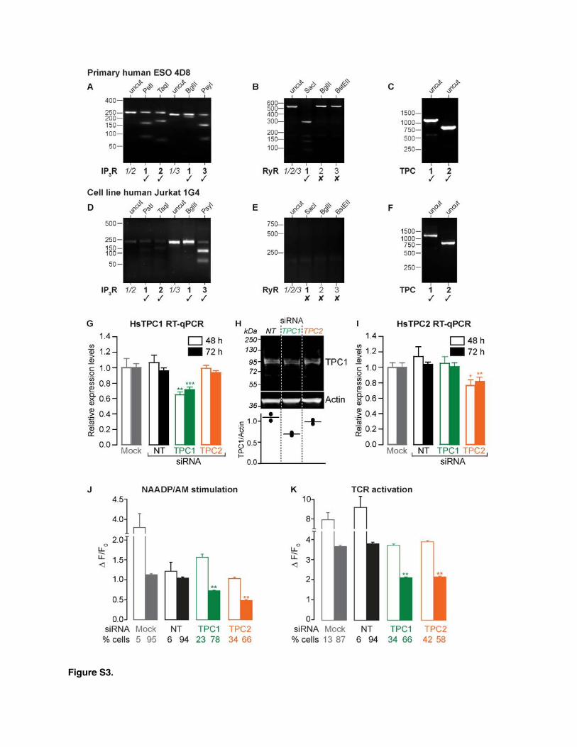
Figure S3.
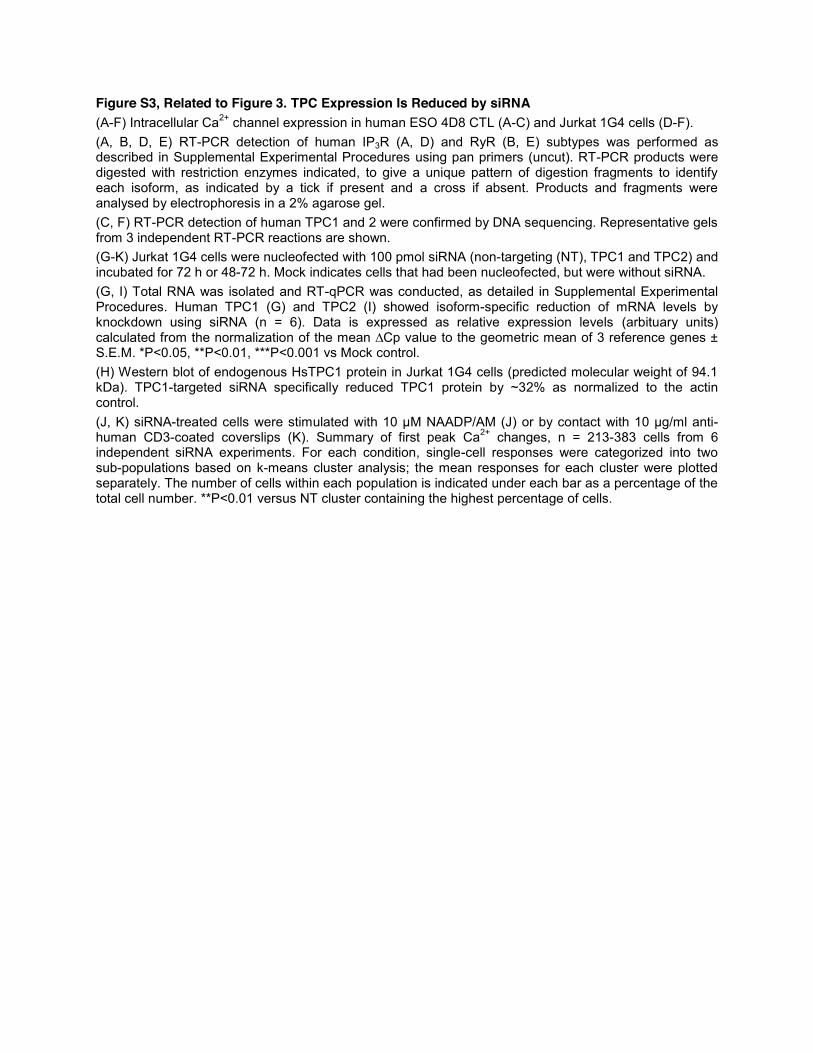
Figure S3, Related to Figure 3. TPC Expression Is Reduced by siRNA

Figure S4.

Figure S4, Related to Figure 4. TPCs Are Localized on Exocytotic (Cytolytic) Granules
Supplemental Experimental Procedures Cells and Reagents
Intracellular Ca2+ Measurements
Human Granzyme B ELISA
51Cr-Release Assay

Reverse Transcription and PCR Amplification (RT-PCR)
Restriction Enzyme Analysis of RT-PCR Products
Nucleofection of Jurkat 1G4 with Plasmid DNA and siRNA
RT-qPCR

Immunoblotting
Immunolocalization
TPC Translocation to the IS
Data Analysis

Supplemental References