The endo-lysosomal system as an NAADP-sensitive acidic Ca 2+ store: Role for the two-pore channels
22
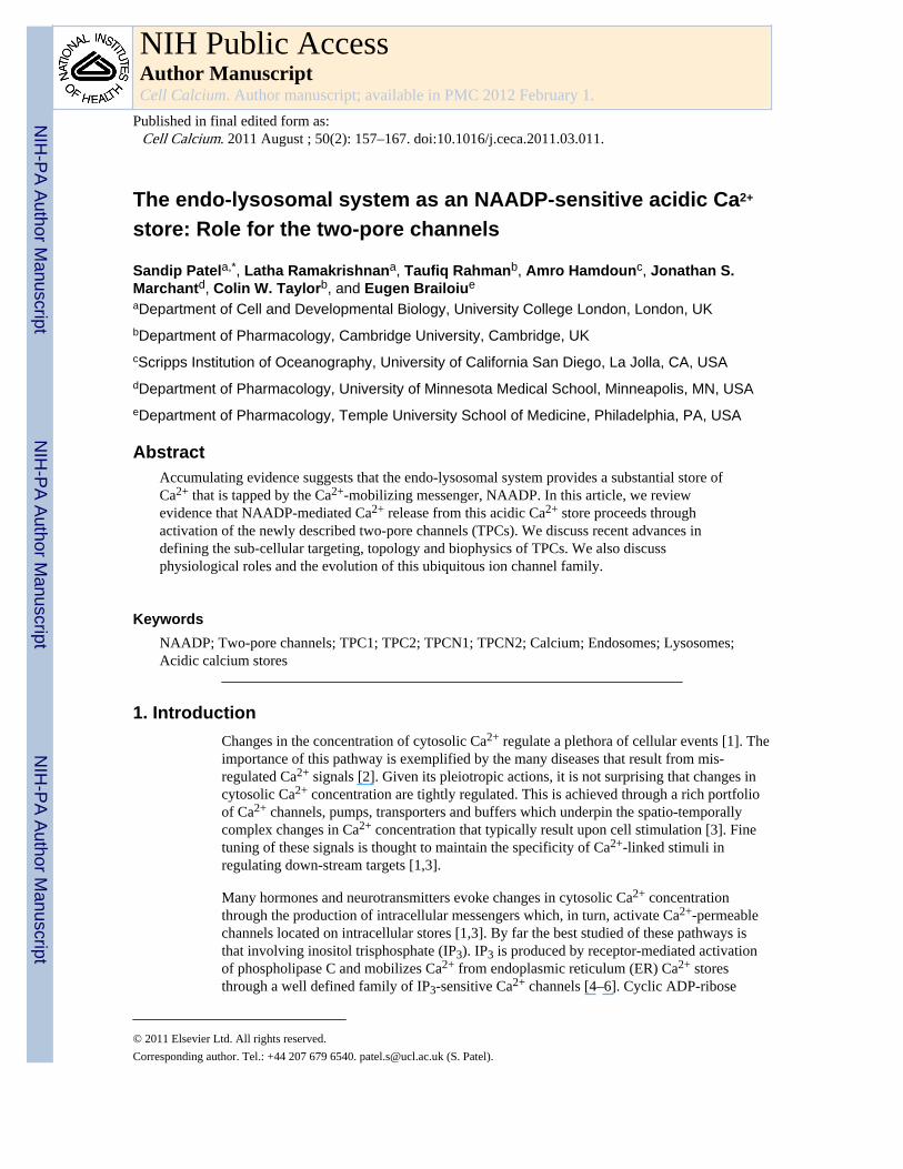
The endo-lysosomal system as an NAADP-sensitive acidic Ca 2+ store: Role for the two-pore channels Sandip Patel a,* , Latha Ramakrishnan a , Taufiq Rahman b , Amro Hamdoun c , Jonathan S. Marchant d , Colin W. Taylor b , and Eugen Brailoiu e a Department of Cell and Developmental Biology, University College London, London, UK b Department of Pharmacology, Cambridge University, Cambridge, UK c Scripps Institution of Oceanography, University of California San Diego, La Jolla, CA, USA d Department of Pharmacology, University of Minnesota Medical School, Minneapolis, MN, USA e Department of Pharmacology, Temple University School of Medicine, Philadelphia, PA, USA Abstract Accumulating evidence suggests that the endo-lysosomal system provides a substantial store of Ca 2+ that is tapped by the Ca 2+ -mobilizing messenger, NAADP. In this article, we review evidence that NAADP-mediated Ca 2+ release from this acidic Ca 2+ store proceeds through activation of the newly described two-pore channels (TPCs). We discuss recent advances in defining the sub-cellular targeting, topology and biophysics of TPCs. We also discuss physiological roles and the evolution of this ubiquitous ion channel family. Keywords NAADP; Two-pore channels; TPC1; TPC2; TPCN1; TPCN2; Calcium; Endosomes; Lysosomes; Acidic calcium stores 1. Introduction Changes in the concentration of cytosolic Ca 2+ regulate a plethora of cellular events [1]. The importance of this pathway is exemplified by the many diseases that result from mis- regulated Ca 2+ signals [2]. Given its pleiotropic actions, it is not surprising that changes in cytosolic Ca 2+ concentration are tightly regulated. This is achieved through a rich portfolio of Ca 2+ channels, pumps, transporters and buffers which underpin the spatio-temporally complex changes in Ca 2+ concentration that typically result upon cell stimulation [3]. Fine tuning of these signals is thought to maintain the specificity of Ca 2+ -linked stimuli in regulating down-stream targets [1,3]. Many hormones and neurotransmitters evoke changes in cytosolic Ca 2+ concentration through the production of intracellular messengers which, in turn, activate Ca 2+ -permeable channels located on intracellular stores [1,3]. By far the best studied of these pathways is that involving inositol trisphosphate (IP 3 ). IP 3 is produced by receptor-mediated activation of phospholipase C and mobilizes Ca 2+ from endoplasmic reticulum (ER) Ca 2+ stores through a well defined family of IP 3 -sensitive Ca 2+ channels [4–6]. Cyclic ADP-ribose © 2011 Elsevier Ltd. All rights reserved. Corresponding author. Tel.: +44 207 679 6540. [email protected] (S. Patel). NIH Public Access Author Manuscript Cell Calcium. Author manuscript; available in PMC 2012 February 1. Published in final edited form as: Cell Calcium. 2011 August ; 50(2): 157–167. doi:10.1016/j.ceca.2011.03.011. NIH-PA Author Manuscript NIH-PA Author Manuscript NIH-PA Author Manuscript
-
Upload
independent -
Category
Documents
-
view
1 -
download
0
Transcript of The endo-lysosomal system as an NAADP-sensitive acidic Ca 2+ store: Role for the two-pore channels
The endo-lysosomal system as an NAADP-sensitive acidic Ca2+
store: Role for the two-pore channels
Sandip Patela,*, Latha Ramakrishnana, Taufiq Rahmanb, Amro Hamdounc, Jonathan S.Marchantd, Colin W. Taylorb, and Eugen Brailoiue
aDepartment of Cell and Developmental Biology, University College London, London, UKbDepartment of Pharmacology, Cambridge University, Cambridge, UKcScripps Institution of Oceanography, University of California San Diego, La Jolla, CA, USAdDepartment of Pharmacology, University of Minnesota Medical School, Minneapolis, MN, USAeDepartment of Pharmacology, Temple University School of Medicine, Philadelphia, PA, USA
AbstractAccumulating evidence suggests that the endo-lysosomal system provides a substantial store ofCa2+ that is tapped by the Ca2+-mobilizing messenger, NAADP. In this article, we reviewevidence that NAADP-mediated Ca2+ release from this acidic Ca2+ store proceeds throughactivation of the newly described two-pore channels (TPCs). We discuss recent advances indefining the sub-cellular targeting, topology and biophysics of TPCs. We also discussphysiological roles and the evolution of this ubiquitous ion channel family.
KeywordsNAADP; Two-pore channels; TPC1; TPC2; TPCN1; TPCN2; Calcium; Endosomes; Lysosomes;Acidic calcium stores
1. IntroductionChanges in the concentration of cytosolic Ca2+ regulate a plethora of cellular events [1]. Theimportance of this pathway is exemplified by the many diseases that result from mis-regulated Ca2+ signals [2]. Given its pleiotropic actions, it is not surprising that changes incytosolic Ca2+ concentration are tightly regulated. This is achieved through a rich portfolioof Ca2+ channels, pumps, transporters and buffers which underpin the spatio-temporallycomplex changes in Ca2+ concentration that typically result upon cell stimulation [3]. Finetuning of these signals is thought to maintain the specificity of Ca2+-linked stimuli inregulating down-stream targets [1,3].
Many hormones and neurotransmitters evoke changes in cytosolic Ca2+ concentrationthrough the production of intracellular messengers which, in turn, activate Ca2+-permeablechannels located on intracellular stores [1,3]. By far the best studied of these pathways isthat involving inositol trisphosphate (IP3). IP3 is produced by receptor-mediated activationof phospholipase C and mobilizes Ca2+ from endoplasmic reticulum (ER) Ca2+ storesthrough a well defined family of IP3-sensitive Ca2+ channels [4–6]. Cyclic ADP-ribose
© 2011 Elsevier Ltd. All rights reserved.Corresponding author. Tel.: +44 207 679 6540. [email protected] (S. Patel).
NIH Public AccessAuthor ManuscriptCell Calcium. Author manuscript; available in PMC 2012 February 1.
Published in final edited form as:Cell Calcium. 2011 August ; 50(2): 157–167. doi:10.1016/j.ceca.2011.03.011.
NIH
-PA Author Manuscript
NIH
-PA Author Manuscript
NIH
-PA Author Manuscript
(cADPR) is another Ca2+-mobilizing messenger. It is produced by ADP-ribosyl cyclases andactivates ryanodine receptors [7,8]. IP3 and ryanodine receptors are structurally andfunctionally related and both reside on ER Ca2+ stores [4–6,8]. A key feature of thesechannels is their biphasic regulation by cytosolic Ca2+ whereby low concentrations stimulatechannel activity while higher concentrations inhibit [4–6,8]. This self regulatory mechanismis critical for the generation of spatiotemporally complex Ca2+ signals [1].
The most recently discovered Ca2+-mobilizing messenger is nicotinic acid adeninedinucleotide phosphate (NAADP) [9,10]. The Ca2+-mobilizing properties of NAADP werefirst recognized by Lee and colleagues using egg homogenates from sea urchin [11]. Muchless is known about this pathway compared to IP3 and cADPR pathways. NAADP isproduced upon cell stimulation with Ca2+-mobilizing agonists [12–14] although itsmechanism of synthesis is uncertain. Like cADPR, NAADP may be synthesized by ADP-ribosyl cyclases [15,16]. Perhaps the most surprising feature of NAADP is its ability totarget Ca2+-permeable channels on acidic Ca2+ stores that are clearly distinct from the ER[17]. Nevertheless, in intact cells, activation of these channels results in further release ofCa2+ from the ER [18]. NAADP is thus considered a “trigger” whereby it provides localrelease of Ca2+ that sensitizes neighbouring IP3 and ryanodine receptors to their respectivemessengers resulting in a larger Ca2+ release event (Fig. 1) [19].
The NAADP pathway is recruited in an agonist-selective manner notably by the sameagonists that were thought previously to couple exclusively to IP3 production [20]. Theseinclude endothelin-1 [13], cholecystokinin [14], and glutamate [21]. Such differentialrecruitment of intracellular Ca2+ release channels by different extracellular cues provides aplausible basis for generating heterogeneity in the Ca2+ signal. The resulting Ca2+ signalshave been implicated in a variety of cellular events including fertilisation [12,22], neuronalgrowth [23] and blood pressure control [24]. In this review, we discuss the evidence thatNAADP mediates Ca2+ release from the endo-lysosomal system through activation of anovel family of Ca2+-permeable channels—the two-pore channels (TPCs).
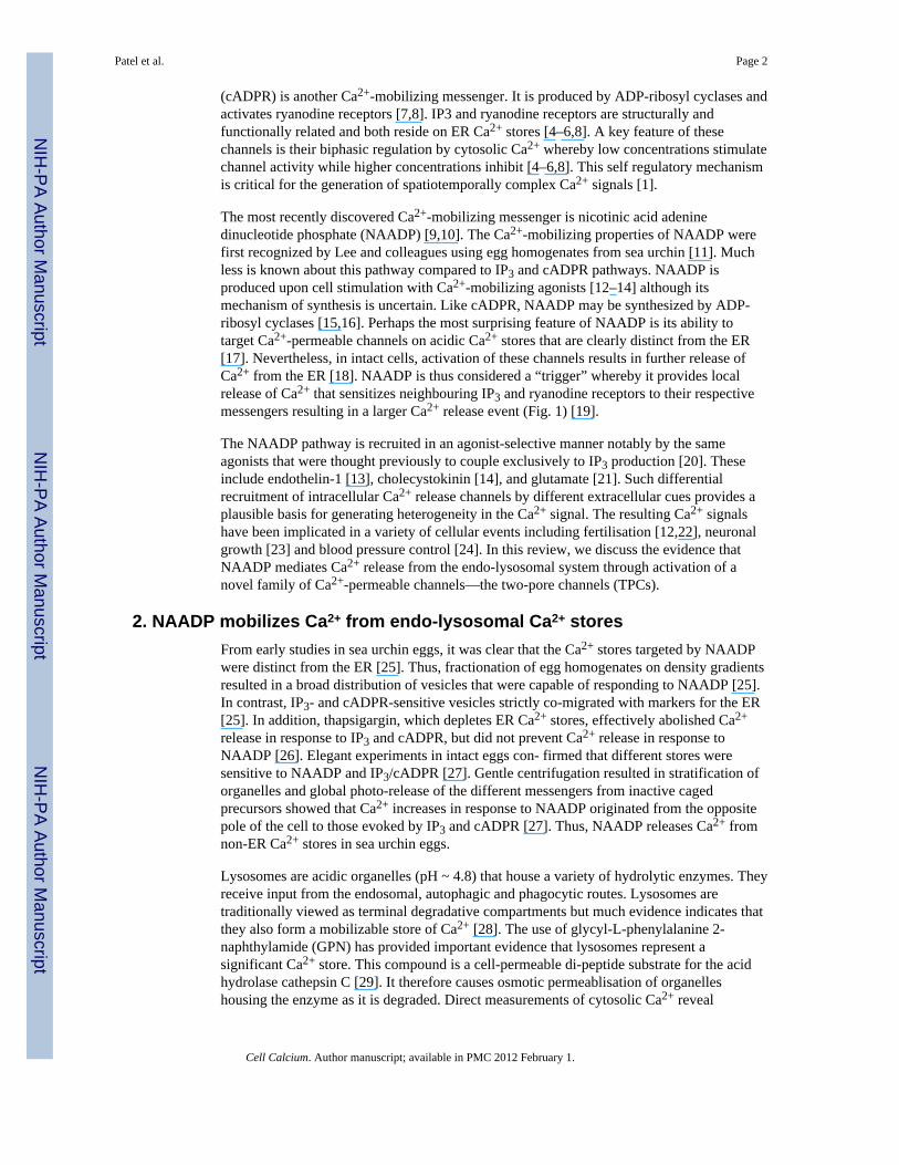
2. NAADP mobilizes Ca2+ from endo-lysosomal Ca2+ storesFrom early studies in sea urchin eggs, it was clear that the Ca2+ stores targeted by NAADPwere distinct from the ER [25]. Thus, fractionation of egg homogenates on density gradientsresulted in a broad distribution of vesicles that were capable of responding to NAADP [25].In contrast, IP3- and cADPR-sensitive vesicles strictly co-migrated with markers for the ER[25]. In addition, thapsigargin, which depletes ER Ca2+ stores, effectively abolished Ca2+
release in response to IP3 and cADPR, but did not prevent Ca2+ release in response toNAADP [26]. Elegant experiments in intact eggs con- firmed that different stores weresensitive to NAADP and IP3/cADPR [27]. Gentle centrifugation resulted in stratification oforganelles and global photo-release of the different messengers from inactive cagedprecursors showed that Ca2+ increases in response to NAADP originated from the oppositepole of the cell to those evoked by IP3 and cADPR [27]. Thus, NAADP releases Ca2+ fromnon-ER Ca2+ stores in sea urchin eggs.
Lysosomes are acidic organelles (pH ~ 4.8) that house a variety of hydrolytic enzymes. Theyreceive input from the endosomal, autophagic and phagocytic routes. Lysosomes aretraditionally viewed as terminal degradative compartments but much evidence indicates thatthey also form a mobilizable store of Ca2+ [28]. The use of glycyl-L-phenylalanine 2-naphthylamide (GPN) has provided important evidence that lysosomes represent asignificant Ca2+ store. This compound is a cell-permeable di-peptide substrate for the acidhydrolase cathepsin C [29]. It therefore causes osmotic permeablisation of organelleshousing the enzyme as it is degraded. Direct measurements of cytosolic Ca2+ reveal
Patel et al. Page 2
Cell Calcium. Author manuscript; available in PMC 2012 February 1.
NIH
-PA Author Manuscript
NIH
-PA Author Manuscript
NIH
-PA Author Manuscript
substantial Ca2+ signals in response to acute addition of GPN. This was first reported byHaller et al. in MDCK cells [30] and subsequently confirmed in a variety of other cellsincluding neurons and glia [21]. The harsh luminal environment of the lysosome with its lowpH and acid hydrolases, presents technical challenges for direct measurements of luminalCa2+. Nevertheless elegant work by Christensen et al. who used endocytosed dextran-basedCa2+ indicators and performed careful calibration of the indicators with respect to pH,determined a luminal Ca2+ concentration of ~500 μM [31]. This value is well within therange reported for the luminal Ca2+ concentration of the ER [32]. A more recent study usingfibroblasts obtained similar values for the lysosomal luminal Ca2+ concentration [33].Endosomes are also likely to contain significant concentrations of Ca2+ since they areformed during invagination of the plasma membrane and thus will incorporate extracelluarCa2+ which is present at ~1 mM. Direct measurements of endosomal Ca2+ are also limited.One study suggested that endosomal Ca2+ is rapidly lost as the endosomes acidify [34].However, another study in pancreatic acinar cells suggested that the Ca2+ content ofendosomal compartments is ~40 μM [35]. Thus, lysosomes and probably endosomes too aresignificant stores of Ca2+.
The first evidence that NAADP mobilizes Ca2+ from the endo-lysosomal system wasprovided by Churchill and colleagues [17]. Using sea urchin eggs, they demonstrated thatGPN-selectively blocked Ca2+ signals evoked by photorelease of NAADP, but not thoseevoked by IP3 or cADPR [17]. Subsequent studies showed that GPN also blocked NAADP-evoked Ca2+ signals in a variety of mammalian cells [20]. Additionally, Cancela andcolleagues have provided evidence that endosomes might express functional NAADP-sensitive Ca2+-permeable channels [36]. Bafilomycin A1 has also been widely demonstratedto block NAADP-evoked Ca2+ signals [17]. Bafilomycin A1 is an inhibitor of V-typeATPases that are responsible for the acidification of organelles [37]. Although themechanism of Ca2+ uptake into the endo-lysosomal system is obscure, it likely requires aproton gradient [31] because bafilomycin A1 causes loss of Ca2+. V-type ATPases areexpressed on a variety of organelles including lysosomes and endosomes. Bafilomycin A1does not therefore distinguish between them or indeed other acidic organelles such assecretory vesicles. Nevertheless, its inhibitory effect on NAADP-mediated Ca2+ signallingfurther supports the notion that NAADP mobilizes Ca2+ from acidic organelles.
3. NAADP activates two-pore channelsConsiderable progress in defining the NAADP-sensitive Ca2+ store was not, until recently,matched by progress in defining the molecular target of NAADP. In the last year or so,however, three independent groups have converged on the TPCs as likely NAADP targets.
TPCs were cloned in 2000 by Ishibashi et al. from rat [38] and by Furuichi et al. fromArabidopsis [39]. Both proteins display significant sequence similarity (albeit modest) tovoltage-gated Ca2+ and Na+ channels [38,39]. The localization of plant TPCs on the vacuole[40], an acidic Ca2+ store [28], prompted us and others to test the hypothesis that TPCs maybe the elusive target of NAADP in animal cells.
Our studies have focussed on sea urchin and human TPCs [41–43]. Three genes are presentin the sea urchin genome (SpTPC1–3), whereas only two (HsTPC1 and HsTPC2) arepresent in humans (see below). Overexpression of all isoforms in SKBR3 cells was found tomarkedly enhance NAADP-evoked Ca2+ signals consistent with a role as NAADP-sensitiveCa2+ channels [41–43]. In addition, TPC-evoked Ca2+ signals in response to NAADP wereabolished by pre-treating cells with bafilomycin A1 thereby suggesting that the signalsderived from acidic organelles [41–43]. Additionally, the Ca2+ signals evoked by NAADPwere partially sensitive to ryanodine, consistent with the amplification of the initial Ca2+
Patel et al. Page 3
Cell Calcium. Author manuscript; available in PMC 2012 February 1.
NIH
-PA Author Manuscript
NIH
-PA Author Manuscript
NIH
-PA Author Manuscript
signal by ryanodine receptors [41,43]. Notably the pharmacology of TPC-evoked Ca2+
signals, with respect to bafilomycin A1 and ryanodine, mirrored that of endogenousNAADP-evoked Ca2+ signals in these cells [44]. Accordingly, knockdown of endogenousTPC1 in SKBR3 cells using a siRNA-based approach substantially reduced Ca2+ signalsevoked by NAADP [41]. Thus, in this cell type, TPC1 appears to mediate the effects ofNAADP consistent with quantitative PCR analysis demonstrating levels of TPC1 transcriptsare higher than those of TPC2 [41]. These studies strongly implicated TPCs in NAADPaction [45].
Independent studies also support the notion that animal TPCs are NAADP-sensitive Ca2+
channels. Over-expression of sea urchin, human and mouse TPCs in HEK cells wasgenerally associated with enhanced NAADP-evoked Ca2+ signals [46–49]. Calcraft et al.who focussed on human TPC2, found that overexpression of this isoform also enhancedNAADP binding (~3-fold) consistent with TPCs as direct targets for NAADP [46]. Inaccord, immunoprecipitates of endogenous SpTPC1 and SpTPC2 from sea urchin eggsbound NAADP in an essentially irreversible manner in the presence, but not absence, of K+
[48]. The regulation of NAADP dissociation by K+ is a peculiar feature of endogenousNAADP receptors in this cell type [50]. The purity of the preparation, however, was notreported. Thus, a potential role for tightly associated accessory binding proteins cannot beexcluded. Importantly, Calcraft et al. also showed that NAADP-evoked Ca2+-dependent ioncurrents in pancreatic beta cells were lacking in TPC2 KO mice [46] suggesting that TPC2mediates NAADP-evoked Ca2+ release in this cell type.
In HEK cells expressing human TPC2, the NAADP responses were markedly biphasiccomprising an initial relatively small and slow release of Ca2+ followed by a larger moreabrupt Ca2+ signal [46]. Bafilomycin A1 abolished the Ca2+ signals, whereas thapsigarginblocked only the second phase [46]. TPC1-mediated Ca2+ signals appeared to support onlysmall localized changes [46]. The authors rationalized these findings in the context of thetrigger hypothesis, whereby the first and second phases represent release of Ca2+ from acidicorganelles and amplification by the ER, respectively [46]. This is an attractive proposal butthe TPC-mediated Ca2+ signals appear remarkably slow taking several minutes to peakcompared to endogenous NAADP-evoked Ca2+ signals, which peak in seconds [51]. Thekinetics also differed to those reported for human TPCs expressed in SKBR3 cells, whererapid and robust (global) responses were observed upon over-expression of either TPC1 orTPC2 [41–43]. This difference might reflect the different cell lines used for heterologousexpression. It is notable that SKBR3 cells express functional ryanodine receptors [44],whereas HEK cells do not, and that in several cell types NAADP preferentially recruitsryanodine receptors [13,24,52]. Consequently, the absence of functional ryanodine receptorsin HEK cells may have “loosened” coupling between activation of TPCs. Indeed, over-expression of mouse TPC2 in HEK cells appeared to be completely uncoupled from Ca2+
release from the ER (given its insensitivity to thapsigargin) and TPC1-evoked signals werenot resolvable [47]. However, a recent re-examination using the same HEK cells expressingHsTPC1 and HsTPC2 indicates that NAADP-evoked signals are rapid, robust and mono-phasic [49] and thus more comparable to those in SKBR3 cells [41–43] than initiallyreported [46]. A somewhat perplexing finding that remains to be explained is the apparentlack of functional activity of SpTPC3 expressed in HEK cells [48], which contrasts with ourfindings in SKBR3 cells [42]. Taken together, ample evidence from independentlaboratories implicate TPCs in NAADP action, although the nature of the NAADP-evokedCa2+ signals differ between studies possibly as a result of the different cell types used foranalysis.
Patel et al. Page 4
Cell Calcium. Author manuscript; available in PMC 2012 February 1.
NIH
-PA Author Manuscript
NIH
-PA Author Manuscript
NIH
-PA Author Manuscript
4. Sub-cellular targeting of TPCsBefitting their role as NAADP-sensitive Ca2+ channels responsible for Ca2+ release fromacidic Ca2+ stores, animal TPCs localize to the endo-lysosomal system. In our studies usingheterologously expressed TPCs, we found a punctate intracellular distribution for humanTPCs [41]. The acidic nature of the labelled vesicles was confirmed by the overlap indistribution of the TPCs with lysotracker red, a fluorescent weak base [45]. Co-expressionwith markers for endosomes (rhoB) and lysosomes (LAMP1) indicated that TPC1 isexpressed on both organelles [41]. In contrast, TPC2 appeared to be almost exclusivelylocalized on lysosomes [41,43]. Essentially similar results were obtained by Calcraft et al.[46]. Quantitative analysis of TPC1 distribution showed a similar overlap with markers forendosomes and lysosomes (see Supplementary Table 1 in [46]) whereas TPC2 overlappedpreferentially with the lysosomal marker. The lysosomal distribution of TPC2 wasconfirmed for the endogenous protein expressed in HEK cells using an anti-TPC2 antibody[46].
Many integral membrane proteins destined for the endo-lysosomal system are targeted bydileucine motifs [53]. Two classes of dileucine motifs have been described conforming toeither the DXXLL or D/E-XXX-LL consensus sequence [53]. We noted the presence ofconserved dileucine motifs in vertebrate TPCs [43]. One of these (corresponding to the lattermotif) was located in the N-terminus and was conserved in both TPC1 and TPC2 [43].Deletion of the N-terminus of human TPC2 or mutation of the leucine residues to alanineresulted in a predominant plasma membrane distribution [43]. These data suggest that thedileucine motif is normally responsible for targeting of TPC2 to lysosomes and thatinterfering with it results in default trafficking of TPC2 to the cell surface. Similarexperiments using truncated TPC1, however, failed to result in plasma membrane targeting[43]. Mutation of a second dileucine motif found in the C-terminus of TPC1 only was alsowithout effect as was combining both mutations [43]. These data suggest that TPC1 istargeted by very different means to TPC2, possibly through tyrosine-based motifs.
Although much experimental evidence is consistent with the trigger hypothesis forexplaining NAADP-mediated Ca2+ signals, direct measurements of the trigger in isolationare limited. Indeed, lack of putative trigger events in T-lymphocytes following ryanodinereceptor blockade has been interpreted as evidence of a direct effect of NAADP onryanodine receptors [54]. As noted above, even after overexpression of TPCs, a substantialcomponent of the resulting NAADP-evoked Ca2+ signal is sensitive to interfering with ERCa2+ stores [41,43,46–48] (but see [47]). Targeting of TPC2 to the plasma membrane bymanipulation of its di-leucine motif provided a unique opportunity to characterize thistrigger event in isolation. We reported that NAADP evoked Ca2+ signals in cells expressingplasma membrane-targeted TPC2 [43]. Importantly, in contrast to cells expressing full-length TPC2, the Ca2+ signals were largely insensitive to bafilomycin A1 and ryanodine, butthey were abolished by removal of extracellular Ca2+ [43]. These data provide evidence thatplasma membrane-targeted TPC2 mediates Ca2+ influx, and that the resulting Ca2+ signalsare not subject to appreciable amplification. Thus, by manipulating the subcellulardistribution of TPC2, we were able to uncouple trigger from amplification pathways [43].Such data argue against a direct effect of NAADP on ER Ca2+ channels. Intriguingly, theCa2+ signals in cells expressing plasma membrane targeted TPC2 were smaller and muchslower to peak than those expressing wild type TPC2. This is perhaps a consequence of thelack of amplification. Indeed, such modest sluggish signals resemble endogenous NAADP-evoked Ca2+ signals in sea urchin eggs treated with heparin and 8-amino cADPR to blockCa2+ release from the ER [55]. Alternatively, the differences might result from the differentlipid composition of the host membranes or the environments to which the luminal surfaceof TPC2 is exposed.
Patel et al. Page 5
Cell Calcium. Author manuscript; available in PMC 2012 February 1.
NIH
-PA Author Manuscript
NIH
-PA Author Manuscript
NIH
-PA Author Manuscript
Sea urchin TPCs also localize to acidic organelles when heterologously expressed inmammalian cells [42,48]. Interestingly, heterologous expression studies in oocytes from theclosely related starfish, indicate that all three isoforms localize to the cortex—a findingconfirmed for endogenous TPC3 in sea urchin eggs [48]. Such a distribution is consistentwith the initiation of endogenous NAADP-evoked Ca2+ signals in the cortex of both seaurchin eggs [12] and starfish oocytes [56]. However, these cortical Ca2+ signals are thoughtto arise from NAADP-evoked Ca2+ influx [12,57]. Moreover, the cortical distribution ofSpTPCs is difficult to reconcile with the localisation of endogenous NAADP-sensitive Ca2+
channels to reserve granules [17] which are distributed throughout the egg. As shown in Fig.2, SpTPCs over expressed in sea urchin embryos show a punctate perinuclear distributiontypical of the endo-lysosomal system. Clearly further work is required to determine thelocalisation of SpTPCs which may be dynamically regulated throughout development.Nevertheless, current evidence obtained using a variety of preparations place TPCs withinthe endo-lysosomal system—a location which seems necessary for subsequent recruitmentof ER Ca2+ channels upon NAADP stimulation.
5. Structure of TPCsBased on their sequence similarity to voltage-gated Ca2+/Na+ channels, TPCs are predictedto comprise two homologous domains each consisting of 6 trans-membrane regions with aputative pore-forming domain located between the 5th and 6th membrane-spanning regions[38,39]. They thus have a unique predicted structure corresponding to approximately half ofa voltage-sensitive Ca2+/Na+ channel (Fig. 3A). Consistent with this predicted topology,fluorophores placed at either terminus or after trans-membrane regions 4 and 6 of domain Iare all accessible to trypsin upon selective permeabilisation of the plasma membrane [58].Mutation of a highly conserved leucine residue in domain I ablates NAADP-mediated Ca2+
release by both HsTPC1 [41] and HsTPC2 [43]. These data are consistent with the positionof this residue within the first predicted pore. Indeed, the predicted luminal regionimmediately upstream of the pore has been confirmed for HsTPC2 based on accessibility toan anti-TPC2 antibody [58]. The corresponding region preceding pore 2 harbours a clusterof N-linked glycosylation sites. Mutation of these sites in HsTPC1 [58] and mouse TPC2[47] prevents N-glycosylation, thereby confirming their luminal location. Indeed, afluorophore placed at this position in TPC1 shows only limited accessibility to trypsin [58].Interestingly, several ion channels are regulated by N-glycosylation through sites close totheir pores [59–61]. TPCs also appear to be regulated in this fashion. Thus, mutation of theN-glycosylation sites in TPC1 preceding the second pore enhances NAADP-evoked Ca2+
release [58]. Glycosylation thus serves to inhibit TPC function. The functional architectureof TPCs is thus emerging and consistent with the structural assignment of TPCs to thefamily of voltage-gated ion channels.
6. Electrophysiological properties of TPCsThe electrophysiological characterization of TPCs is at present more advanced in higherplants than animals. Plant TPCs form the ubiquitous SV (slowly activating vacuolar)channels within the vacuolar membrane. SV channels are the most abundant ion channels inthe vacuolar membrane and so amenable to patch-clamp recording from isolated vacuoles[40,62,63]. They are almost entirely cation-selective with large single-channel conductances(γ K ~ 155pS) and they are Ca2+-permeable, but select weakly between cations (PCa/PK ~ 3)[62–65]. Plant TPC1 has sufficient sequence similarity with animal TPCs [39], particularlywithin the likely pore regions (Fig. 3B), to suggest that they may share some commonproperties. However there is presently no evidence to suggest that plant TPCs are gated byNAADP, although NAADP has been reported to release Ca2+ from the ER of plants [66].
Patel et al. Page 6
Cell Calcium. Author manuscript; available in PMC 2012 February 1.
NIH
-PA Author Manuscript
NIH
-PA Author Manuscript
NIH
-PA Author Manuscript
Whereas the large central vacuole of plants lends itself readily to patch-clamp recording, thesmall acidic organelles, within which animal TPC proteins are expressed, are smaller(diameter <0.5 μm) than the tip of a typical patch pipette (~1 μm) and so present moreformidable practical problems. Three different approaches, each with their strengths andweaknesses, have been applied to electrophysiological analysis of animal TPCs. We mutatedresidues within the N-terminus of human TPC2 that mediate its targeting to lysosomes, andthereby achieved functional expression of TPC2 within the plasma membrane. Here itbecomes accessible to conventional patch-clamp recording methods, with all theopportunities they provide for exquisitely high-resolution recording and rapid changes ofmedia [43]. This approach requires mutation of targeting residues with the attendantpossibility that the same residues might also influence gating; and re-targeting may separateTPC2 from regulatory proteins within the endo-lysosomal lumen, but the approach isotherwise minimally disruptive. The protein remains within a cellular membrane and itallows the behavior of TPC2 in exactly the same setting to be compared in intact cells (usingconventional Ca2+ indicators) and at the single-channel level.
Others have reconstituted partially purified human TPC2 into artificial lipid bilayers, fromwhich single-channel activity can be recorded [67]. This approach, at least when applied tocompletely purified protein, has the merit of allowing the behavior of a fully defined proteincomplex to be examined. The many disadvantages include the risk of damage duringpurification, the loss of essential accessory proteins, the need to reconstitute the protein intoan entirely artificial bilayer and the limited opportunity for rapid media changes that bilayersafford. A similar approach, but using a crude lysosomal fraction fused with a bilayer, wasused to examine the effects of NAADP on endogenous channels from liver [68]. The thirdapproach was to record murine TPC2 activity from artificially enlarged lysosomes [69].With this method, cells are first treated for several hours with vacuolin to cause lysosomefusion [70], the enlarged lysosomes are then isolated in media containing mM Ca2+ fromcell lysates, and fused with a planar patch-clamp chip to allow recording [69]. This method,which has so far been applied only to analysis of whole-lysosome currents, has the merit ofretaining TPC2 within a native lysosomal membrane (albeit modified by vacuolin), but itrequires rather harsh, and potentially damaging, isolation methods, luminal proteins arelikely to be lost when the patches are formed, and, as with bilayers, there is limitedopportunity to change media rapidly. Although it is still very early days for theelectrophysiological analysis of TPC2 channels from animals – the only reports werepublished in 2010 – it is worth comparing the results obtained with the different approaches.
Perhaps the most important conclusion from these studies is that mutations within either thefirst or second putative pore loop (the S5–6 loops of each domain) (Fig. 3) affect single-channel conductance [43] or ion selectivity [69], thereby establishing that TPC proteins areindeed pore-forming subunits of an NAADP-gated channel. The following features wereobserved in each of the three studies of TPC2 [43,67,69]. Channel activity was stimulated bysub-μM concentrations of NAADP and in two studies blocked by the antagonist, Ned-19[43,67]. In bilayers, activation by NAADP was inexplicably irreversible [67], whereas itrapidly reversed upon either removal of NAADP or addition of Ned-19 to the plasmamembrane patches [43]. The disparity may simply arise from inadequate washout ofNAADP from the bilayer chamber [67]. In all three studies, the currents had near linearcurrent–voltage (i–V) relationships, although we reported a slight inward rectification [43].This contrasts with the striking outwardly rectifying behavior of plant TPC1 [64]. Thedifference, by analogy to the structures of conventional voltage-gated ion channels, wherepositively charged residues (usually arginine) within the fourth transmembrane region (S4)comprise the voltage-sensor, may be due to the lesser numbers of such residues in the S4segments of animal TPC2 versus plant TPC proteins (Fig. 3B). The similar numbers ofpositively-charged residues in animal and plant TPC1, however, suggests that animal TPC1
Patel et al. Page 7
Cell Calcium. Author manuscript; available in PMC 2012 February 1.
NIH
-PA Author Manuscript
NIH
-PA Author Manuscript
NIH
-PA Author Manuscript
may be voltage-sensitive (Fig. 3B). The single-channel studies concur in suggesting thatTPC2 forms cation-selective channels with large monovalent cation conductances (γK ~300pS and γCs ~ 130pS), lesser Ca2+ conductances (γCa 15–40pS) and weak selectivitybetween cations (PCa/PK ~ 2.6) [43,67]. These ion-handing properties are broadly similar tothose of plant TPC1 [64], but they differ markedly from the third study of animal TPC2,which reported a permeability ratio (PCa/PK > 1000) comparable to that of the most Ca2+-selective plasma membrane Ca2+ channels [69]. It is noteworthy that here, ion selectivitywas measured at a luminal pH that mimics that of lysosomes (pH 4.6), perhaps suggestingthat luminal H+ influences ion selectivity, but that interpretation is difficult to reconcile withluminal pH having no obvious effect on the amplitude of the NAADP-evoked K+ currents inbilayer recordings [67]. In summary, and while we await further analyses, it seemsreasonable to suppose that TPC2 forms a large-conductance, non-selective, Ca2+-permeable,cation channel that is minimally regulated by membrane potential. The finer details of thesingle-channel behavior of animal TPCs are largely unexplored. There is evidence for sub-conductance states [67] and substantial open-channel noise is suggestive of either rapidflickering between conductances or block by permeating cations [43,67]. Openings thatoccur in bursts, effects of permeating cations on gating behavior, and the suggestion thatopenings may be coupled indicate that the gating mechanism for TPC channels is morecomplex than a simple switch between a single closed and open state. This complexity alsofinds a parallel in the gating behavior of plant TPC1 [63].
Finally, electrophysiological analyses provide access to the luminal surface of TPC2 andthereby allow effects of luminal regulators to be explored in ways that would be difficult inintact cells. As with plant TPC1 [64], both luminal Ca2+ and H+ may regulate TPC2 [67,69].Pitt et al. have suggested that high levels of luminal Ca2+ (100 μM to 1 mM) are required forNAADP to gate TPC2, while luminal H+ modestly reduced activity [67]. By contrast,channels in the planar patch-clamp were detected at acidic luminal pH, but not at pH 7.2[69]. This requirement for luminal H+ contrasts with the inhibitory effect of H+ on plantTPC1 [64], and nor can it be reconciled with the other studies in which NAADP stimulatedTPC2 activity at neutral luminal pH [43,67]. One possibility is that the very highconcentrations of luminal Ca2+ (60 mM) used in [69] may, by analogy with plant TPC1[64], have inhibited channel activity, and H+ might then partially alleviate that inhibition.Further work is clearly needed to resolve, ideally in a setting that retains luminal proteins,the possible roles of luminal Ca2+ and H+ in modulating the gating of TPC2 by NAADP.
7. Functional roles of TPCsNAADP has been shown to regulate a variety of cellular functions that include musclecontraction and differentiation. Here we discuss the role of TPCs in these establishedNAADP-dependent processes, and also their role in trafficking and pigmentation in whichNAADP signalling had not previously been implicated.
Much evidence indicates that NAADP regulates muscle contractility. In pulmonary arterysmooth muscle cells, NAADP generates local Ca2+ signals from bafilomycin A1-sensitiveCa2+ stores. These are subsequently amplified by ryanodine, but not IP3 receptors [13]. Theresulting global Ca2+ signals are sufficient to cause contraction [13]. Endothelin-1 elevatescellular levels of NAADP and its effect on cytosolic Ca2+ concentration was abolished bybafilomycin A1 [13]. A recent study has confirmed the role of TPCs in contraction ofsmooth muscle. Thus, in TPC2-deficient mice, NAADP-evoked contractions of detrusor andtaenia caecum muscle were absent [71]. Carbachol-evoked contractions in detrusor musclefrom wild-type mice were partially sensitive to NAADP-blockade. However, agonist-evokedcontractions were not reduced in muscle from TPC2 knock-out mice although thecontractions were insensitive to pharmacological blockade of NAADP [71]. These data
Patel et al. Page 8
Cell Calcium. Author manuscript; available in PMC 2012 February 1.
NIH
-PA Author Manuscript
NIH
-PA Author Manuscript
NIH
-PA Author Manuscript
suggest compensation may have occurred in the TPC2 knock-out mice to maintain agonist-evoked contractions. Notably, the contribution of ryanodine receptors to carbachol-evokedCa2+ signals was increased in the transgenic animals [71]. In this context, it is interesting tonote a recent comparison of Ca2+ signals in pancreatic acinar cells from wild type and CD38knock-out mice [16]. Previous studies had provided evidence that cholecystokinin is anNAADP-linked agonist based on the blockade of cholecystokinin-evoked Ca2+ signals byNAADP desensitization [18] and lysosomotropic agents [72], and the ability ofcholecystokinin to elevate NAADP levels [14]. In CD38-kockout mice, cholecystokinin-evoked NAADP elevations were abolished [16] suggesting that CD38 is responsible forNAADP production in these cells. However, the cholecystokinin-evoked Ca2+ signalsappeared not to be affected although as in the case of agonist-evoked contractions in TPC2knock-out mice [71], they became insensitive to depletion of acidic organelles [16]. Theseobservations once again suggest that compensation may have occurred upon depletion ofCD38 to maintain cholecystokinin-evoked Ca2+ signalling. In this case, this may be due toenhanced Ca2+ influx since removal of extracellular Ca2+ did reveal reducedcholecystokinin-evoked Ca2+ signals in the CD38-deficient mice [16]. Such compensationperhaps highlights the importance of the Ca2+-dependent function under NAADP control. Itmight also reflect coordinated regulation of the expression of the various Ca2+ channels andsynthetic enzymes in normal cells [73].
Differentiation is another process in which NAADP has been implicated. Using the PC12cell line, an extensively used model system to study neuronal differentiation, cellulardelivery of NAADP using NAADP-filled liposomes was sufficient to induce differentiation[74]. In parallel experiments, IP3 delivery was ineffective [74]. Direct measurements ofcytosolic Ca2+ concentration showed that NAADP-induced Ca2+ signals were reliant onboth acidic Ca2+ stores (based on their inhibition by bafilomycin A1 and GPN) and the ER(based on their partial inhibition by thapsigargin) [74]. Kinetic com-parison of the signals inresponse to NAADP and IP3 showed that the former were more sustained [74].Consequently, these unique Ca2+ signatures likely underlie the very different effect of themessengers on Ca2+-dependent output. Such data provide evidence to support the idea thatdifferential recruitment of Ca2+-mobilizing messengers may provide a mechanism for finetuning Ca2+ signals, which are in turn differentially decoded. A recent study using a cell-permeable analogue of NAADP, NAADP-AM [75], supports a role for NAADP inmediating differentiation [76]. Thus, as in PC12 cells, elevation of cellular NAADP levels inskeletal muscle cells promoted differentiation and siRNA-mediated inhibition of eitherTPC1 or TPC2 inhibited it [76]. This study provides further evidence for a functional rolefor TPCs in mediating NAADP action.
The endo-lysosomal system is a highly dynamic organelle network requiring fusion eventsbetween organelles [77]. A role for Ca2+ in these “constitutive” events has been appreciatedfor some time [78] and certainly its role in regulated secretion is established. Ca2+ is thoughtto be released from acidic organelles close to the fusion machinery thereby accounting forthe block of these events by the fast Ca2+ chelator, BAPTA, but not by the slower chelator,EGTA [79,80]. The identity of the target channel responsible for this local release of Ca2+
however has not been established but a clear candidate is TRP mucolipin-1 (TRPML1).TRPML1 derives its name from the finding that mutation of its gene results in the lysosomalstorage disorder, mucolipidosis IV (MLIV) [81]. That TRPML1 is likely a non-selectivecation channel (and thus permeable to Ca2+), and that MLIV is characterised by traffickingdefects [82] support the contention that Ca2+-dependent fusion events required for normalendo-lysosomal trafficking are mediated by TRPML1, possibly in response to PI(3,5)P2[83].
Patel et al. Page 9
Cell Calcium. Author manuscript; available in PMC 2012 February 1.
NIH
-PA Author Manuscript
NIH
-PA Author Manuscript
NIH
-PA Author Manuscript
A recent study also suggested a role for TPCs in trafficking within the endo-lysosomalsystem [48]. Cholera toxin B normally enters cells by endocytosis and is trafficked to theGolgi complex by retrograde transport. Over-expression of sea urchin TPC1 and TPC2 wassuggested to disrupt this pathway resulting in accumulation of the toxin in endosomes(although its exact localisation using markers was not reported) [48]. These data implicateTPCs in transport of cargo from endosomes to the trans-Golgi network, perhaps becauselocal Ca2+ signals are required for fusion of endosomes or endosome-derived vesicles withinthe trans-Golgi network. Disrupting trafficking from endosomes to the Golgi may affectrecycling of the mannose-6-phosphate receptor. This protein mediates delivery of lysosomalhydrolases from the Golgi to endosomes prior to their delivery to lysosomes. Consequentlydisruption of this pathway may alter lysosome biogenesis. Indeed, overexpression of TPCswas also associated with increased staining of cells with Lysotracker and an enlargement ofindividual lysosomes [48]. This phenotype is reminiscent of that we reported previously incells where lysosomal dysfunction was imposed by pharmacological inhibition of selectlysosomal hydrolases [84]. TPC dysfunction may thus precipitate lysosomal dysfunction.Conversely, lysosomal dysfunction may disrupt TPC function. For example in fibroblastsfrom patients suffering from the lysosomal storage disease, Nieman-Pick type C, lysosomalCa2+ concentration and associated global NAADP-evoked Ca2+ signals are reduced [33].Lysosomal dysfunction imposed by pharmacological means in neurons is also associatedwith deviant NAADP-evoked Ca2+ signals, although in this case they are enhanced [84].NAADP-evoked Ca2+ signals mediated by TPCs may therefore be intimately linked to bothlysosomal function and dysfunction.
Prior to identification of TPCs as NAADP targets, single nucleotide polymorphisms in thehuman TPC2 gene were associated with hair colour in northern Europeans [85]. Two non-synonymous variants were identified that associated with blond versus brown hair.Pigmentation is a complex process that involves the synthesis of melanins in lysosome-related organelles known as melanosomes [86]. Melansosomes are then transferred by an ill-defined mechanism from the melanocyte to neighbouring keratinocytes. The mechanismwhereby TPC2 and its variants regulate pigmentation is not known, but it is tempting tospeculate that TPC2 may be localized to the melanosome. Indeed, melanosomes may serveas Ca2+ stores [87] and there is evidence for the presence of functional NAADP-sensitiveCa2+ channels on other lysosome-related organelles [88]. Consequently, TPC2-mediatedCa2+ signals may regulate either the function of the melanosome or perhaps trafficking ofmelanosomes to keratinocytes. Alternatively, TPC2 may regulate melanogenesis via changesin melanosomal pH since melanin synthesis is regulated by pH [89] and mobilisation ofacidic Ca2+ stores by NAADP is associated with luminal alkanization [90]. Intriguingly,mutations/variants of several other proteins such as TRPML3, SLC24A5 and TRPM1 havealso been associated with pigmentation phenotypes [28,91]. Consequently, ion channelfluxes within the endo-lysosomal system may be a general mechanism involved inpigmentation [28,91].
8. Evolution of TPCsAs mentioned above, structurally speaking, the TPCs correspond to approximately one-halfof a voltage sensitive Ca2+/Na+ channel. Since the latter are thought to have evolved fromK+ channels [92], TPCs may represent an evolutionary intermediate between the two. Thusthe single repeat of the K+ channels may have been duplicated to generate TPCs, which maythen have undergone another round of duplication to generate 4-repeat Ca2+/Na+ channels(Fig. 4A).
A distinguishing feature between plant and animal TPCs is the presence in the linker regionconnecting the two domains in plants of two predicted EF hands [40], which in other
Patel et al. Page 10
Cell Calcium. Author manuscript; available in PMC 2012 February 1.
NIH
-PA Author Manuscript
NIH
-PA Author Manuscript
NIH
-PA Author Manuscript
proteins bind Ca2+. Indeed, it is established that the SV currents in plants which aremediated by TPCs are Ca2+-regulated [40]. The lack of EF hands in animal TPCs isconsistent with the insensitivity of NAADP-evoked Ca2+ release to cytosolic Ca2+ [93].Thus, whereas plant TPCs may amplify Ca2+ signals, animal TPCs likely trigger them (Fig.4A).
In plants, a single TPC has been described in Arabidopsis [39], rice [94] and wheat [95] andtwo closely related genes have been described in tobacco [96]. In animals, there is evidencefor both gene loss, and gene multiplication and divergence [41]. Most animal species withinthe protostome super phylum, for example, appear not to possess TPC genes [41,45,46].These include key model organisms such as worms and flies. In contrast, mostdeuterostomes possess three TPC genes [42]. These include sea urchins, which are basaldeuterostomes that have been extensively used for NAADP research [97]. The sequencesimilarity between TPC isoforms in animals is relatively low (30–40% for sea urchins)compared to other intracellular channels such as IP3 receptors indicating substantialdivergence.
The complete (three-member) ancestral family is retained in chordates such as frogs, fish,birds and most mammals, but strikingly, the TPC3 gene is absent in rodents and humans[42]. In cows, dogs and horses which possess the full TPC complement, the TPC3 gene islocated between NPHP1 and BUB1 [42]. Inspection of the corresponding human genomicsequences reveals TPC3-related sequences corresponding to the N-terminal third of TPC3[42]. However, there is no clear start site and there are multiple nucleotide insertions anddeletions that would result in the production of a truncated protein if transcribed andtranslated [42]. Partial TPC3-related sequences were identified in several other apesincluding our closest relative, the chimpanzee and also in the Rhesus monkey (an Old WorldMonkey) [42]. This analysis provides evidence that the TPC3 gene is undergoing loss in theprimate lineage.
Further inspection of more distantly-related primate genome sequences identified sequenceslikely corresponding to full-length TPC3 in a New World Monkey [98]. Near completesequences were also found in both tarsier and non-tarsier Prosimians [98]. Importantly, nodeleterious mutations were present. The TPC3 gene thus appears to be retained in theseorganisms (Fig. 4B) and suggests that degeneration of the gene in the primate lineage beganin the common ancestor of Cattarhines (apes and Old World monkeys) ~25 to 40 millionyears ago [98]. The TPC3 gene is therefore of major interest in an evolutionary contextbecause it is a rare example of an ion channel undergoing degeneration that began relativelyrecently. In summary, the TPC gene family has undergone remarkable diversi-ficationduring evolution.
9. OutlookThe molecular basis for Ca2+ release by NAADP has been subject to controversy [99,100],with competing claims for activation of Ca2+-permeable channels located on the ER, theplasma membrane, and as discussed in this review, acidic Ca2+ stores. Multiple lines ofevidence from independent groups are now converging on the TPCs as the target forNAADP on acidic stores of Ca2+. Indeed, the localisation of TPCs to the endo-lysosomalsystem, within which there is much protein trafficking, might well reconcile at least some ofthe apparent discrepant findings in the literature regarding the functional localisation ofNAADP targets (discussed in [45]). Clearly, over-expression of TPCs enhances cellularsensitivity to NAADP, but how NAADP binding is linked to activation (and inactivation) ofCa2+ release is not yet clear. The field is now ripe for structure–function analysis, and high-level expression and analysis of TPCs in a null background will certainly facilitate this. The
Patel et al. Page 11
Cell Calcium. Author manuscript; available in PMC 2012 February 1.
NIH
-PA Author Manuscript
NIH
-PA Author Manuscript
NIH
-PA Author Manuscript
basic topology of TPCs has now been defined, but not the quaternary structure. The basicbiophysical properties of TPCs are also emerging, but much more analysis is required todefine ion selectivity, gating mechanisms and regulation by luminal ions of the variousisoforms. Do TPCs interact with other proteins? Defining the TPC interactome will provideimportant information regarding the regulation of TPCs of which little is known at present.NAADP, via TPCs, recruits the activity of other Ca2+ channels, but until now this “chatter”[101] has been imaged using relatively low resolution microscopy methods. Can higherresolution methods be applied in cells expressing defined levels of TPCs to probe themicroarchitecture of the underlying elementary Ca2+ signals? Finally, armed with not onlymolecular tools to manipulate NAADP-mediated Ca2+ signals but also new chemical tools[102,103], we now have an ideal opportunity to probe the physiological role of TPCs. Mightfunctions already ascribed to the endo-lysosomal system uncover new roles for TPCs? AreTPCs mis-regulated in disease states in which the endo-lysosomal system is perturbed, andmight manipulating their levels/activity represent a novel therapeutic strategy? Answers tothese questions are likely to be forthcoming in the near future in what is now a rapidlyadvancing field.
AcknowledgmentsWe thank Dev Churamani, Robert Hooper, Chi Li and Desmond Tobin for useful discussion, and the Boguefoundation (University College London) for facilitating staff exchange. Work in SP’s laboratory is funded by theBBSRC. Work in CWT’s laboratory is funded by the Wellcome Trust. TR is a Drapers’ Research Fellow atPembroke College, Cambridge.
References1. Berridge MJ, Lipp P, Bootman MD. The versatility and universality of calcium signalling. Nat Rev
Mol Cell Biol. 2000; 1:11–21. [PubMed: 11413485]2. Rizzuto R, Pozzan T. When calcium goes wrong: genetic alterations of a ubiquitous signaling route.
Nat Genet. 2003; 34:135–141. [PubMed: 12776115]3. Clapham DE. Calcium signaling. Cell. 2007; 131:1047–1058. [PubMed: 18083096]4. Patel S, Joseph SK, Thomas AP. Molecular properties of inositol 1,4,5-trisphosphate receptors. Cell
Calcium. 1999; 25:247–264. [PubMed: 10378086]5. Taylor CW, Genazzani AA, Morris SA. Expression of inositol trisphosphate receptors. Cell
Calcium. 1999; 26:237–251. [PubMed: 10668562]6. Foskett JK, White C, Cheung KH, Mak DO. Inositol trisphosphate receptor Ca2+ release channels.
Physiol Rev. 2007; 87:593–658. [PubMed: 17429043]7. Lee HC. Mechanisms of calcium signaling by cyclic ADP-ribose and NAADP. Physiol Rev. 1997;
77:1133–1164. [PubMed: 9354813]8. Fill M, Copello JA. Ryanodine receptor calcium release channels. Physiol Rev. 2002; 82:893–922.
[PubMed: 12270947]9. Lee HC. Calcium signaling: NAADP ascends as a new messenger. Curr Biol. 2003; 13:R186–R188.
[PubMed: 12620209]10. Patel S. NAADP-induced Ca2+ release—a new signaling pathway. Biol Cell. 2004; 96:19–28.
[PubMed: 15093124]11. Lee HC, Aarhus R. A derivative of NADP mobilizes calcium stores insensitive to inositol
trisphosphate and cyclic ADP-ribose. J Biol Chem. 1995; 270:2152–2157. [PubMed: 7836444]12. Churchill GC, O‘Neil JS, Masgrau R, et al. Sperm deliver a new messenger: NAADP. Curr Biol.
2003; 13:125–128. [PubMed: 12546785]13. Kinnear NP, Boittin FX, Thomas JM, Galione A, Evans AM. Lysosome-sarcoplasmic reticulum
junctions: a trigger zone for calcium signalling by NAADP and endothelin-1. J Biol Chem. 2004;279:54319–54326. [PubMed: 15331591]
Patel et al. Page 12
Cell Calcium. Author manuscript; available in PMC 2012 February 1.
NIH
-PA Author Manuscript
NIH
-PA Author Manuscript
NIH
-PA Author Manuscript

14. Yamasaki M, Thomas JT, Churchill GC, et al. Role of NAADP and cADPR in the induction andmaintenance of agonist-evoked Ca2+ Spiking in mouse pancreatic acinar cells. Curr Biol. 2005;15:874–878. [PubMed: 15886108]
15. Kim SY, Cho BH, Kim UH. CD38-mediated Ca2+ signaling contributes to angiotensin II-inducedactivation of hepatic stellate cells: attenuation of hepatic fibrosis by CD38 ablation. J Biol Chem.2010; 285:576–582. [PubMed: 19910464]
16. Cosker F, Cheviron N, Yamasaki M, et al. The ecto-enzyme CD38 is a nicotinic acid adeninedinucleotide phosphate (NAADP) synthase that couples receptor activation to Ca2+ mobilizationfrom lysosomes in pancreatic acinar cells. J Biol Chem. 2010; 285:38251–38259. [PubMed:20870729]
17. Churchill GC, Okada Y, Thomas JM, Genazzani AA, Patel S, Galione A. NAADP mobilizes Ca2+
from reserve granules, lysosome-related organelles, in sea urchin eggs. Cell. 2002; 111:703–708.[PubMed: 12464181]
18. Cancela JM, Churchill GC, Galione A. Coordination of agonist-induced Ca2+-signalling patternsby NAADP in pancreatic acinar cells. Nature. 1999; 398:74–76. [PubMed: 10078532]
19. Guse AH, Lee HC. NAADP a universal Ca2+ trigger. Sci Signal. 2008; 1:re10. [PubMed:18984909]
20. Galione A, Morgan AJ, Arredouani A, et al. NAADP as an intracellular messenger regulatinglysosomal calcium-release channels. Biochem Soc Trans. 2010; 38:1424–1431. [PubMed:21118101]
21. Pandey V, Chuang CC, Lewis AM, et al. Recruitment of NAADP-sensitive acidic Ca2+ stores byglutamate. Biochem J. 2009; 422:503–512. [PubMed: 19548879]
22. Lim D, Kyozuka K, Gragnaniello G, Carafoli E, Santella L. NAADP+ initiates the Ca2+ responseduring fertilization of starfish oocytes. FASEB J. 2001; 15:2257–2267. [PubMed: 11641253]
23. Brailoiu E, Hoard JL, Filipeanu CM, et al. NAADP potentiates neurite outgrowth. J Biol Chem.2005; 280:5646–5650. [PubMed: 15528210]
24. Brailoiu GC, Gurzu B, Gao X, et al. Acidic NAADP-sensitive calcium stores in the endothelium:agonist-specific recruitment and role in regulating blood pressure. J Biol Chem. 2010; 285:37133–37137. [PubMed: 20876534]
25. Clapper DL, Walseth TF, Dargie PJ, Lee HC. Pyridine nucleotide metabolites stimulate calciumrelease from sea urchin egg microsomes desensitized to inositol trisphosphate. J Biol Chem. 1987;262:9561–9568. [PubMed: 3496336]
26. Genazzani AA, Galione A. Nicotinic acid-adenine dinucleotide phosphate mobilizes Ca2+ from athapsigargin-insensitive pool. Biochem J. 1996; 315:721–725. [PubMed: 8645149]
27. Lee HC, Aarhus R. Functional visualisation of the separate but interacting calcium stores sensitiveto NAADP and cyclic ADP-ribose. J Cell Sci. 2000; 113:4413–4420. [PubMed: 11082034]
28. Patel S, Docampo R. Acidic calcium stores open for business: expanding the potential forintracellular Ca2+ signaling. Trends Cell Biol. 2010; 20:277–286. [PubMed: 20303271]
29. Jadot M, Colmant C, Wattiaux-de CS, Wattiaux R. Intralysosomal hydrolysis of glycyl-L-phenylalanine 2-naphthylamide. Biochem J. 1984; 219:965–970. [PubMed: 6743255]
30. Haller T, Dietl P, Deetjen P, Volkl H. The lysosomal compartment as intracellular calcium store inMDCK cells: a possible involvement in InsP3-mediated Ca2+ release. Cell Calcium. 1996;19:157–165. [PubMed: 8689673]
31. Christensen KA, Myers JT, Swanson JA. pH-dependent regulation of lysosomal calcium inmacrophages. J Cell Sci. 2002; 115:599–607. [PubMed: 11861766]
32. Miyawaki A, llopis J, Heim R, McCaffery JM, Adams JA, Tsien RY. Fluorescent indicators forCa2+ based on green fluorescent proteins and calmodulin. Nature. 1997; 388:882–887. [PubMed:9278050]
33. Lloyd-Evans E, Morgan AJ, He X, et al. Niemann-Pick disease type C1 is a sphingosine storagedisease that causes deregulation of lysosomal calcium. Nat Med. 2008; 14:1247–1255. [PubMed:18953351]
34. Gerasimenko JV, Tepikin AV, Petersen OH, Gerasimenko OV. Calcium uptake via endocytosiswith rapid release from acidifying endosomes. Curr Biol. 1998; 8:1335–1338. [PubMed: 9843688]
Patel et al. Page 13
Cell Calcium. Author manuscript; available in PMC 2012 February 1.
NIH
-PA Author Manuscript
NIH
-PA Author Manuscript
NIH
-PA Author Manuscript

35. Sherwood MW, Prior IA, Voronina SG, et al. Activation of trypsinogen in large endocyticvacuoles of pancreatic acinar cells. Proc Natl Acad Sci USA. 2007; 104:5674–5679. [PubMed:17363470]
36. Menteyne A, Burdakov A, Charpentier G, Petersen OH, Cancela JM. Generation of specific Ca2+
signals from Ca2+ stores and endocytosis by differential coupling to messengers. Curr Biol. 2006;16:1931–1937. [PubMed: 17027490]
37. Huss M, Wieczorek H. Inhibitors of V-ATPases: old and new players. J Exp Biol. 2009; 212:341–346. [PubMed: 19151208]
38. Ishibashi K, Suzuki M, Imai M. Molecular cloning of a novel form (two-repeat) protein related tovoltage-gated sodium and calcium channels. Biochem Biophys Res Commun. 2000; 270:370–376.[PubMed: 10753632]
39. Furuichi T, Cunningham KW, Muto S. A putative two pore channel AtTPC1 mediates Ca2+ flux inArabidopsis leaf cells. Plant Cell Physiol. 2001; 42:900–905. [PubMed: 11577183]
40. Peiter E, Maathuis FJ, Mills LN, et al. The vacuolar Ca2+-activated channel TPC1 regulatesgermination and stomatal movement. Nature. 2005; 434:404–408. [PubMed: 15772667]
41. Brailoiu E, Churamani D, Cai X, et al. Essential requirement for two-pore channel 1 in NAADP-mediated calcium signaling. J Cell Biol. 2009; 186:201–209. [PubMed: 19620632]
42. Brailoiu E, Hooper R, Cai X, et al. An ancestral deuterostome family of two-pore channels mediatenicotinic acid adenine dinucleotide phosphate-dependent calcium release from acidic organelles. JBiol Chem. 2010; 285:2897–2901. [PubMed: 19940116]
43. Brailoiu E, Rahman T, Churamani D, et al. An NAADP-gated two-pore channel targeted to theplasma membrane uncouples triggering from amplifying Ca2+ signals. J Biol Chem. 2010;285:38511–38516. [PubMed: 20880839]
44. Schrlau MG, Brailoiu E, Patel S, Gogotsi Y, Dun NJ, Bau HM. Carbon nanopipettes characterizecalcium release pathways in breast cancer cells. Nanotechnology. 2008; 19:325102. [PubMed:21828806]
45. Patel S, Marchant JS, Brailoiu E. Two-pore channels: regulation by NAADP and customized rolesin triggering calcium signals. Cell Calcium. 2010; 47:480–490. [PubMed: 20621760]
46. Calcraft PJ, Ruas M, Pan Z, et al. NAADP mobilizes calcium from acidic organelles through two-pore channels. Nature. 2009; 459:596–600. [PubMed: 19387438]
47. Zong X, Schieder M, Cuny H, et al. The two-pore channel TPCN2 mediates NAADP-dependentCa2+-release from lysosomal stores. Pflugers Arch. 2009; 458:891–899. [PubMed: 19557428]
48. Ruas M, Rietdorf K, Arredouani A, et al. Purified TPC isoforms form NAADP receptors withdistinct roles for Ca2+ signaling and endo-lysosomal trafficking. Curr Biol. 2010; 20:703–709.[PubMed: 20346675]
49. Ogunbayo OA, Zhu Y, Rossi D, Sorrentino V, Ma J, Zhu MX, Evans AM. Cyclic adenosinediphosphate ribose activates ryanodine receptors, whereas NAADP activates two-pore domainchannels. J Biol Chem. 2011; 286:9136–9140. [PubMed: 21216967]
50. Dickinson GD, Patel S. Modulation of NAADP receptors by K+ ions: evidence for multipleNAADP receptor conformations. Biochem J. 2003; 375:805–812. [PubMed: 12914540]
51. Genazzani AA, Mezna M, Summerhill RJ, Galione A, Michelangeli F. Kinetic properties ofnicotinic acid adenine dinucleotide phosphate-induced Ca2+ release. J Biol Chem. 1997;272:7669–7675. [PubMed: 9065423]
52. Brailoiu GC, Brailoiu E, Parkesh R, et al. NAADP-mediated channel “chatter” in neurons of the ratmedulla oblongata. Biochem J. 2009; 419:91–97. [PubMed: 19090786]
53. Bonifacino JS, Traub LM. Signals for sorting of transmembrane proteins to endosomes andlysosomes. Annu Rev Biochem. 2003; 72:395–447. [PubMed: 12651740]
54. Dammermann W, Guse AH. Functional ryanodine receptor expression is required for NAADP-mediated local Ca2+ signaling in T-lymphocytes. J Biol Chem. 2005; 280:21394–21399.[PubMed: 15774471]
55. Churchill GC, Galione A. Spatial control of Ca2+ signalling by nicotinic acid adenine dinucleotidephosphate diffusion and gradients. J Biol Chem. 2000; 275:38687–38692. [PubMed: 11006280]
56. Santella L, Kyozuka K, Genazzani AA, De Riso L, Carafoli E. Nicotinic acid adenine dinucleotidephosphate-induced Ca2+ release. J Biol Chem. 2000; 275:8301–8306. [PubMed: 10722659]
Patel et al. Page 14
Cell Calcium. Author manuscript; available in PMC 2012 February 1.
NIH
-PA Author Manuscript
NIH
-PA Author Manuscript
NIH
-PA Author Manuscript
57. Moccia F, Lim D, Nusco GA, Ercolano E, Santella L. NAADP activates a Ca2+ current that isdependent on F-actin cytoskeleton. FASEB J. 2003; 13:1907–1909. [PubMed: 12923070]
58. Hooper R, Churamani D, Brailoiu E, Taylor CW, Patel S. Membrane topology of NAADP-sensitive two-pore channels and their regulation by N-linked glycosylation. J Biol Chem. 2011;286:9141–9149. [PubMed: 21173144]
59. Bennett ES. Isoform-specific effects of sialic acid on voltage-dependent Na+ channel gating:functional sialic acids are localized to the S5-S6 loop of domain I. J Physiol. 2002; 538:675–690.[PubMed: 11826157]
60. Noma K, Kimura K, Minatohara K, et al. Triple N-glycosylation in the long S5-P loop regulatesthe activation and trafficking of the Kv12.2 potassium channel. J Biol Chem. 2009; 284:33139–33150. [PubMed: 19808681]
61. Wirkner K, Hognestad H, Jahnel R, Hucho F, Illes P. Characterization of rat transient receptorpotential vanilloid 1 receptors lacking the N-glycosylation site N604. Neuroreport. 2005; 16:997–1001. [PubMed: 15931076]
62. Ward JM, Schroeder JI. Calcium-activated K+ channels and calcium-induced calcium release byslow vacuolar ion channels in guard cell vacuoles implicated in the control of stomatal closure.Plant Cell. 1994; 6:669–683. [PubMed: 12244253]
63. Scholz-Starke J, Naso A, Carpaneto A. A perspective on the slow vacuolar channel in vacuolesfrom higher plant cells. J Chem Inf Model. 2005; 45:1502–1506. [PubMed: 16309246]
64. Pottosin II, Schonknecht G. Vacuolar calcium channels. J Exp Bot. 2007; 58:1559–1569.[PubMed: 17355948]
65. Schulz-Lessdorf B, Hedrich R. Protons and calcium modulate SV-type channels in the vacuolar-lysosomal compartment—channel interaction with calmodulin inhibitors. Planta. 1995; 197:655–671.
66. Navazio L, Bewell MA, Siddiqua A, Dickinson GD, Galione A, Sanders D. Calcium release fromthe endoplasmic reticulum of higher plants elicited by the NADP metabolite nicotinic acid adeninedinucleotide phosphate. Proc Natl Acad Sci. 2000; 97:8693–8698. [PubMed: 10890899]
67. Pitt SJ, Funnell T, Sitsapesan M, et al. TPC2 is a novel NAADP-sensitive Ca2+-release channel,operating as a dual sensor of luminal pH and Ca2+ J Biol Chem. 2010; 285:24925–24932.[PubMed: 20547763]
68. Zhang F, Li PL. Reconstitution and characterization of a nicotinic acid adenine dinucleotidephosphate (NAADP)-sensitive Ca2+ release channel from liver lysosomes of rats. J Biol Chem.2007; 282:25259–25269. [PubMed: 17613490]
69. Schieder M, Rotzer K, Bruggemann A, Biel M, Wahl-Schott CA. Characterization of two-porechannel 2 (TPCN2)-mediated Ca2+ currents in isolated lysosomes. J Biol Chem. 2010;285:21219–21222. [PubMed: 20495006]
70. Dong XP, Cheng X, Mills E, et al. The type IV mucolipidosis-associated protein TRPML1 is anendo-lysosomal iron release channel. Nature. 2008; 455:992–996. [PubMed: 18794901]
71. Tugba Durlu-Kandilci N, Ruas M, Chuang KT, Brading A, Parrington J, Galione A. TPC2 proteinsmediate nicotinic acid adenine dinucleotide phosphate (NAADP)- and agonist-evoked contractionsof smooth muscle. J Biol Chem. 2010; 285:24925–24932. [PubMed: 20547763]
72. Yamasaki M, Masgrau R, Morgan AJ, et al. Organelle selection determines agonist-specific Ca2+
signals in pancreatic acinar and beta cells. J Biol Chem. 2004; 279:7234–7240. [PubMed:14660554]
73. Ritchie MF, Zhou Y, Soboloff J. Transcriptional mechanisms regulating Ca2+ homeostasis. CellCalcium. in press.
74. Brailoiu E, Churamani D, Pandey V, et al. Messenger-specific role for NAADP in neuronaldifferentiation. J Biol Chem. 2006; 281:15923–15928. [PubMed: 16595650]
75. Parkesh R, Lewis AM, Aley PK, et al. Cell-permeant NAADP: a novel chemical tool enabling thestudy of Ca2+ signalling in intact cells. Cell Calcium. 2007; 43:531–538. [PubMed: 17935780]
76. Aley PK, Mikolajczyk AM, Munz B, Churchill GC, Galione A, Berger F. Nicotinic acid adeninedinucleotide phosphate regulates skeletal muscle differentiation via action at two-pore channels.Proc Natl Acad Sci USA. 2010; 107:19927–19932. [PubMed: 21041635]
Patel et al. Page 15
Cell Calcium. Author manuscript; available in PMC 2012 February 1.
NIH
-PA Author Manuscript
NIH
-PA Author Manuscript
NIH
-PA Author Manuscript
77. Saftig P, Klumperman J. Lysosome biogenesis and lysosomal membrane proteins: traffickingmeets function. Nat Rev Mol Cell Biol. 2009; 10:623–635. [PubMed: 19672277]
78. Luzio JP, Bright NA, Pryor PR. The role of calcium and other ions in sorting and delivery in thelate endocytic pathway. Biochem Soc Trans. 2007; 35:1088–1091. [PubMed: 17956286]
79. Holroyd C, Kistner U, Annaert W, Jahn R. Fusion of endosomes involved in synaptic vesiclerecycling. Mol Biol Cell. 1999; 10:3035–3044. [PubMed: 10473644]
80. Pryor PR, Mullock BM, Bright NA, Gray SR, Luzio JP. The role of intraor-ganellar Ca2+ in lateendosome–lysosome heterotypic fusion and in the reformation of lysosomes from hybridorganelles. J Cell Biol. 2000; 149:1053–1062. [PubMed: 10831609]
81. Bargal R, Avidan N, Ben-Asher E, et al. Identification of the gene causing mucolipidosis type IV.Nat Genet. 2000; 26:118–123. [PubMed: 10973263]
82. Puertollano R, Kiselyov K. TRPMLs: in sickness and in health. Am J Physiol Renal Physiol. 2009;296:F1245–F1254. [PubMed: 19158345]
83. Dong XP, Shen D, Wang X, et al. PI(3,5)P2 controls membrane traffic by direct activation ofmucolipin Ca2+ release channels in the endolysosome. Nat Commun. 2010; 1:38. [PubMed:20802798]
84. Dickinson GD, Churchill GC, Brailoiu E, Patel S. Deviant NAADP-mediated Ca2+-signallingupon lysosome proliferation. J Biol Chem. 2010; 285:13321–13325. [PubMed: 20231291]
85. Sulem P, Gudbjartsson DF, Stacey SN, et al. Two newly identified genetic determinants ofpigmentation in Europeans. Nat Genet. 2008; 40:835–837. [PubMed: 18488028]
86. Tobin DJ. The cell biology of human hair follicle pigmentation. Pigment Cell Melanoma Res. 201087. Salceda R, Sanchez-Chavez G. Calcium uptake, release and ryanodine binding in melanosomes
from retinal pigment epithelium. Cell Calcium. 2000; 27:223–229. [PubMed: 10858668]88. Rosado JA. Acidic Ca2+ stores in platelets. Cell Calcium. 201089. Ito S, Wakamatsu K. Human hair melanins: what we have learned and have not learned from
mouse coat color pigmentation. Pigment Cell Melanoma Res. 201090. Morgan AJ, Galione A. NAADP induces pH changes in the lumen of acidic Ca2+ stores. Biochem
J. 2007; 402:301–310. [PubMed: 17117921]91. Patel S, Docampo R. In with the TRP channels: intracellular functions for TRPM1 and TRPM2.
Sci Signal. 2009; 2:e69.92. Strong M, Chandy KG, Gutman GA. Molecular evolution of voltage-sensitive ion channel genes:
on the origins of electrical excitability. Mol Biol Evol. 1993; 10:221–242. [PubMed: 7680747]93. Chini EN, Dousa TP. Nicotinate-adenine dinucleotide phosphate-induced Ca2+ release does not
behave as a Ca2+-induced Ca2+-release system. Biochem J. 1996; 316:709–711. [PubMed:8670142]
94. Kurusu T, Sakurai Y, Miyao A, Hirochika H, Kuchitsu K. Identification of a putative voltage-gatedCa2+-permeable channel (OsTPC1) involved in Ca2+ influx and regulation of growth anddevelopment in rice. Plant Cell Physiol. 2004; 45:693–702. [PubMed: 15215504]
95. Wang YJ, Yu JN, Chen T, et al. Functional analysis of a putative Ca2+ channel gene TaTPC1 fromwheat. J Exp Bot. 2005; 56:3051–3060. [PubMed: 16275671]
96. Kadota Y, Furuichi T, Ogasawara Y, et al. Identification of putative voltage-dependent Ca2+-permeable channels involved in cryptogein-induced Ca2+ transients and defense responses intobacco BY-2 cells. Biochem Biophys Res Commun. 2004; 317:823–830. [PubMed: 15081414]
97. Galione A, Patel S, Churchill GC. NAADP-induced calcium release in sea urchin eggs. Biol Cell.2000; 92:197–204. [PubMed: 11043408]
98. Cai X, Patel S. Degeneration of an intracellular ion channel in the primate lineage by relaxation ofselective constraints. Mol Biol Evol. 2010; 27:2352–2359. [PubMed: 20463046]
99. Galione A, Petersen OH. The NAADP receptor: new receptors or new regulation? Mol Interv.2005; 5:73–79. [PubMed: 15821155]
100. Guse AH. Second messenger signaling: multiple receptors for NAADP. Curr Biol. 2009;19:R521–R523. [PubMed: 19602416]
101. Patel S, Churchill GC, Galione A. Coordination of Ca2+ signalling by NAADP. Trends BiochemSci. 2001; 26:482–489. [PubMed: 11504624]
Patel et al. Page 16
Cell Calcium. Author manuscript; available in PMC 2012 February 1.
NIH
-PA Author Manuscript
NIH
-PA Author Manuscript
NIH
-PA Author Manuscript
102. Naylor E, Arredouani A, Vasudevan SR, et al. Identification of a chemical probe for NAADP byvirtual screening. Nat Chem Biol. 2009; 5:220–226. [PubMed: 19234453]
103. Dammermann W, Zhang B, Nebel M, et al. NAADP-mediated Ca2+ signaling via type 1ryanodine receptor in T cells revealed by a synthetic NAADP antagonist. Proc Natl Acad SciUSA. 2009; 106:10678–10683. [PubMed: 19541638]
Patel et al. Page 17
Cell Calcium. Author manuscript; available in PMC 2012 February 1.
NIH
-PA Author Manuscript
NIH
-PA Author Manuscript
NIH
-PA Author Manuscript
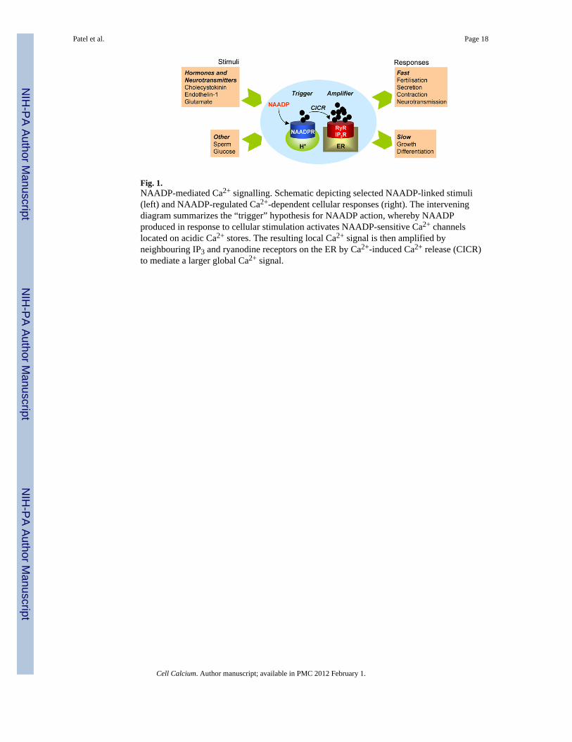
Fig. 1.NAADP-mediated Ca2+ signalling. Schematic depicting selected NAADP-linked stimuli(left) and NAADP-regulated Ca2+-dependent cellular responses (right). The interveningdiagram summarizes the “trigger” hypothesis for NAADP action, whereby NAADPproduced in response to cellular stimulation activates NAADP-sensitive Ca2+ channelslocated on acidic Ca2+ stores. The resulting local Ca2+ signal is then amplified byneighbouring IP3 and ryanodine receptors on the ER by Ca2+-induced Ca2+ release (CICR)to mediate a larger global Ca2+ signal.
Patel et al. Page 18
Cell Calcium. Author manuscript; available in PMC 2012 February 1.
NIH
-PA Author Manuscript
NIH
-PA Author Manuscript
NIH
-PA Author Manuscript
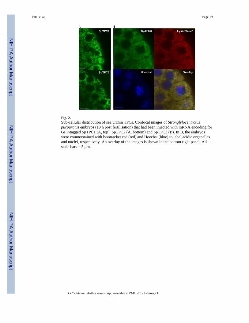
Fig. 2.Sub-cellular distribution of sea urchin TPCs. Confocal images of Stronglylocentrotuspurpuratus embryos (19 h post fertilisation) that had been injected with mRNA encoding forGFP-tagged SpTPC1 (A, top), SpTPC2 (A, bottom) and SpTPC3 (B). In B, the embryoswere counterstained with lysotracker red (red) and Hoechst (blue) to label acidic organellesand nuclei, respectively. An overlay of the images is shown in the bottom right panel. Allscale bars = 5 μm.
Patel et al. Page 19
Cell Calcium. Author manuscript; available in PMC 2012 February 1.
NIH
-PA Author Manuscript
NIH
-PA Author Manuscript
NIH
-PA Author Manuscript
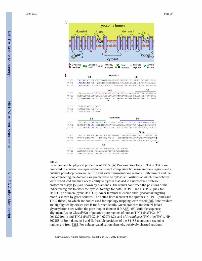
Fig. 3.Structural and biophysical properties of TPCs. (A) Proposed topology of TPCs. TPCs arepredicted to contain two repeated domains each comprising 6 trans-membrane regions and aputative pore-loop between the fifth and sixth transmembrane regions. Both termini and theloop connecting the domains are predicted to be cytosolic. Positions at which fluorophoreswere introduced and their accessibility to trypsin assessed in fluorescence proteaseprotection assays [58] are shown by diamonds. The results confirmed the positions of theindicated regions to either the cytosol (orange for both HsTPC1 and HsTPC2; pink forHsTPC1) or lumen (cyan; HsTPC1). An N-terminal dileucine endo-lysosomal targetingmotif is shown by green squares. The dotted lines represent the epitopes in TPC1 (pink) andTPC2 (black) to which antibodies used for topology mapping were raised [58]. Pore residuesare highlighted by circles (see B for further detail). Green branches indicate N-linkedglycosylation sites within the pore loop of domain II [47,58]. (B) Multiple sequencealignment (using ClustalW2) of putative pore regions of human TPC1 (HsTPC1, NP001137291.1) and TPC2 (HsTPC2, NP 620714.2), and of Arabidopsis TPC1 (AtTPC1, NP567258.1) from domains I and II. Possible positions of the S4–S6 membrane-spanningregions are from [38]. For voltage-gated cation channels, positively charged residues
Patel et al. Page 20
Cell Calcium. Author manuscript; available in PMC 2012 February 1.
NIH
-PA Author Manuscript
NIH
-PA Author Manuscript
NIH
-PA Author Manuscript

(usually arginine) aligned on one side of the S4 helix provide the voltage-sensor. Residuesthat might provide such an arrangement in TPC channels are highlighted in blue (arginine)and cyan (lysine) in each of the S4 regions. There are fewer such residues in HsTPC2 thanin HsTPC1 and AtTPC1. Mutation of a conserved Leu (L273 for HsTPC1 and L265 forHsTPC2, green circle) [41,43] or Asn (N257 for mouse TPC2, equivalent to N273 ofHsTPC2, yellow circle) [69] within the putative P-loop of domain I affect either Ca2+
release, conductance or ion selectivity, consistent with their proposed positions within thepore. A conserved acidic residue (D660 for HsTPC2 and E643 for mTPC2, blue circle) withinthe proposed P loop of domain II also appears to be critical for channel function [69],suggesting that the second putative P loop also contributes to formation of the pore.
Patel et al. Page 21
Cell Calcium. Author manuscript; available in PMC 2012 February 1.
NIH
-PA Author Manuscript
NIH
-PA Author Manuscript
NIH
-PA Author Manuscript
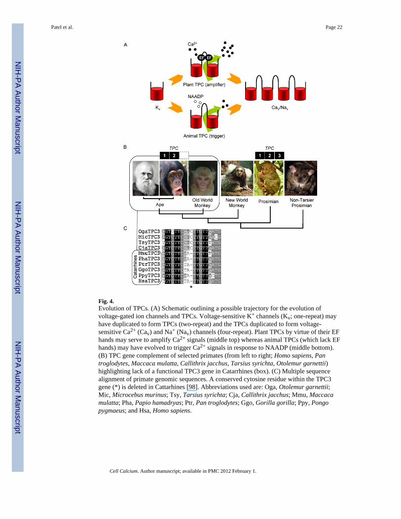
Fig. 4.Evolution of TPCs. (A) Schematic outlining a possible trajectory for the evolution ofvoltage-gated ion channels and TPCs. Voltage-sensitive K+ channels (Kv; one-repeat) mayhave duplicated to form TPCs (two-repeat) and the TPCs duplicated to form voltage-sensitive Ca2+ (Cav) and Na+ (Nav) channels (four-repeat). Plant TPCs by virtue of their EFhands may serve to amplify Ca2+ signals (middle top) whereas animal TPCs (which lack EFhands) may have evolved to trigger Ca2+ signals in response to NAADP (middle bottom).(B) TPC gene complement of selected primates (from left to right; Homo sapiens, Pantroglodytes, Maccaca mulatta, Callithrix jacchus, Tarsius syrichta, Otolemur garnettii)highlighting lack of a functional TPC3 gene in Catarrhines (box). (C) Multiple sequencealignment of primate genomic sequences. A conserved cytosine residue within the TPC3gene (*) is deleted in Cattarhines [98]. Abbreviations used are: Oga, Otolemur garnettii;Mic, Microcebus murinus; Tsy, Tarsius syrichta; Cja, Callithrix jacchus; Mmu, Maccacamulatta; Pha, Papio hamadryas; Ptr, Pan troglodytes; Ggo, Gorilla gorilla; Ppy, Pongopygmaeus; and Hsa, Homo sapiens.
Patel et al. Page 22
Cell Calcium. Author manuscript; available in PMC 2012 February 1.
NIH
-PA Author Manuscript
NIH
-PA Author Manuscript
NIH
-PA Author Manuscript