Protective Effects of Positive Lysosomal Modulation in Alzheimer's Disease Transgenic Mouse Models
16
Protective Effects of Positive Lysosomal Modulation in Alzheimer’s Disease Transgenic Mouse Models David Butler 1,2¤ , Jeannie Hwang 1,2,3 , Candice Estick 1,4 , Akiko Nishiyama 4 , Saranya Santhosh Kumar 4,5 , Clive Baveghems 2 , Hollie B. Young-Oxendine 3 , Meagan L. Wisniewski 3 , Ana Charalambides 2,3 , Ben A. Bahr 1,2,3,4,5 * 1 Neurosciences Program, University of Connecticut, Storrs, Connecticut, United States of America, 2 Department of Pharmaceutical Sciences, University of Connecticut, Storrs, Connecticut, United States of America, 3 William C. Friday Laboratory, Biotechnology Research and Training Center, University of North Carolina Pembroke, Pembroke, North Carolina, United States of America, 4 Department of Physiology and Neurobiology, University of Connecticut, Storrs, Connecticut, United States of America, 5 Department of Molecular and Cell Biology, University of Connecticut, Storrs, Connecticut, United States of America Abstract Alzheimer’s disease (AD) is an age-related neurodegenerative pathology in which defects in proteolytic clearance of amyloid b peptide (Ab) likely contribute to the progressive nature of the disorder. Lysosomal proteases of the cathepsin family exhibit up-regulation in response to accumulating proteins including Ab 1–42 . Here, the lysosomal modulator Z-Phe-Ala- diazomethylketone (PADK) was used to test whether proteolytic activity can be enhanced to reduce the accumulation events in AD mouse models expressing different levels of Ab pathology. Systemic PADK injections in APP SwInd and APPswe/ PS1DE9 mice caused 3- to 8-fold increases in cathepsin B protein levels and 3- to 10-fold increases in the enzyme’s activity in lysosomal fractions, while neprilysin and insulin-degrading enzyme remained unchanged. Biochemical analyses indicated the modulation predominantly targeted the active mature forms of cathepsin B and markedly changed Rab proteins but not LAMP1, suggesting the involvement of enhanced trafficking. The modulated lysosomal system led to reductions in both Ab immunostaining as well as Ab x-42 sandwich ELISA measures in APP SwInd mice of 10–11 months. More extensive Ab deposition in 20-22-month APPswe/PS1DE9 mice was also reduced by PADK. Selective ELISAs found that a corresponding production of the less pathogenic Ab 1–38 occurs as Ab 1–42 levels decrease in the mouse models, indicating that PADK treatment leads to Ab truncation. Associated with Ab clearance was the elimination of behavioral and synaptic protein deficits evident in the two transgenic models. These findings indicate that pharmacologically-controlled lysosomal modulation reduces Ab 1–42 accumulation, possibly through intracellular truncation that also influences extracellular deposition, and in turn offsets the defects in synaptic composition and cognitive functions. The selective modulation promotes clearance at different levels of Ab pathology and provides proof-of-principle for small molecule therapeutic development for AD and possibly other protein accumulation disorders. Citation: Butler D, Hwang J, Estick C, Nishiyama A, Kumar SS, et al. (2011) Protective Effects of Positive Lysosomal Modulation in Alzheimer’s Disease Transgenic Mouse Models. PLoS ONE 6(6): e20501. doi:10.1371/journal.pone.0020501 Editor: Tsuneya Ikezu, Boston University, United States of America Received December 14, 2010; Accepted May 3, 2011; Published June 10, 2011 Copyright: ß 2011 Butler et al. This is an open-access article distributed under the terms of the Creative Commons Attribution License, which permits unrestricted use, distribution, and reproduction in any medium, provided the original author and source are credited. Funding: This work was supported in part by National Institutes of Health grant R25 GM077634, the Oliver Smithies Grant from the North Carolina Biotechnology Center (Research Triangle Park, North Carolina), the Institute for the Study of Aging, and the University of Connecticut School of Pharmacy and Center for Students with Disabilities. The funding agencies had no role in study design, data collection and analysis, decision to publish, or preparation of the manuscript. Competing Interests: The authors have declared that no competing interests exist. * E-mail: [email protected] ¤ Current address: Center for Neurologic Disease, Harvard Institutes of Medicine, Boston, Massachusetts, United States of America Introduction Alzheimer’s disease (AD) is the most prevalent form of senile dementia, and is characterized by progressive compromise of synaptic integrity and cognitive functions. Ab accumulation in the brain is a hallmark of AD pathology, and Ab 1–42 species have been implicated in the disruption of synaptic function and neuronal loss [1]. In addition to extracellular deposition, accumulation of Ab also occurs intraneuron- ally [2–8], likely due to defective clearance and in many cases occurring prior to amyloid plaque formation. The clearance rates for Ab peptides were indeed found to be slower in AD patients as compared to the rates in cognitively normal individuals [9]. One conclusion is an imbalance in Ab production vs. clearance that implicates a plausible mechanism for the Ab dysregulation in the more common late-onset AD, and perhaps a contributing factor in familial AD. Intraneuronal Ab 1–42 is found in the brains of Alzheimer patients and individuals with mild cognitive impairment [2,10], thus there is growing evidence that such intracellular accumulation is an early indicator of neuronal compromise that correlates with cognitive decline. Synaptic dysfunction and deterioration are exhibited by Ab–containing neurons and the intraneuronal accumulation is associated with cognitive deficits in animal models [2,4,6–8,11]. It is widely held that reducing protein accumulation events is critical for slowing the progression of AD, especially those produced by the aggregation-prone Ab 1–42 peptide. Potential targets include Ab-degrading enzymes such as neprilysin, insulin- degrading enzyme, and endothelin-converting enzyme, and these proteases may in fact be responsible for Ab homeostasis in the brain [12–15]. The lysosomal hydrolase cathepsin B has also been found to cleave Ab 1–42 into less amyloidogenic species [16]. This is PLoS ONE | www.plosone.org 1 June 2011 | Volume 6 | Issue 6 | e20501
-
Upload
learn-uconn -
Category
Documents
-
view
0 -
download
0
Transcript of Protective Effects of Positive Lysosomal Modulation in Alzheimer's Disease Transgenic Mouse Models

Protective Effects of Positive Lysosomal Modulation inAlzheimer’s Disease Transgenic Mouse ModelsDavid Butler1,2¤, Jeannie Hwang1,2,3, Candice Estick1,4, Akiko Nishiyama4, Saranya Santhosh Kumar4,5,
Clive Baveghems2, Hollie B. Young-Oxendine3, Meagan L. Wisniewski3, Ana Charalambides2,3, Ben A.
Bahr1,2,3,4,5*
1 Neurosciences Program, University of Connecticut, Storrs, Connecticut, United States of America, 2 Department of Pharmaceutical Sciences, University of Connecticut,
Storrs, Connecticut, United States of America, 3 William C. Friday Laboratory, Biotechnology Research and Training Center, University of North Carolina Pembroke,
Pembroke, North Carolina, United States of America, 4 Department of Physiology and Neurobiology, University of Connecticut, Storrs, Connecticut, United States of
America, 5 Department of Molecular and Cell Biology, University of Connecticut, Storrs, Connecticut, United States of America
Abstract
Alzheimer’s disease (AD) is an age-related neurodegenerative pathology in which defects in proteolytic clearance of amyloidb peptide (Ab) likely contribute to the progressive nature of the disorder. Lysosomal proteases of the cathepsin familyexhibit up-regulation in response to accumulating proteins including Ab1–42. Here, the lysosomal modulator Z-Phe-Ala-diazomethylketone (PADK) was used to test whether proteolytic activity can be enhanced to reduce the accumulationevents in AD mouse models expressing different levels of Ab pathology. Systemic PADK injections in APPSwInd and APPswe/PS1DE9 mice caused 3- to 8-fold increases in cathepsin B protein levels and 3- to 10-fold increases in the enzyme’s activity inlysosomal fractions, while neprilysin and insulin-degrading enzyme remained unchanged. Biochemical analyses indicatedthe modulation predominantly targeted the active mature forms of cathepsin B and markedly changed Rab proteins but notLAMP1, suggesting the involvement of enhanced trafficking. The modulated lysosomal system led to reductions in both Abimmunostaining as well as Abx-42 sandwich ELISA measures in APPSwInd mice of 10–11 months. More extensive Abdeposition in 20-22-month APPswe/PS1DE9 mice was also reduced by PADK. Selective ELISAs found that a correspondingproduction of the less pathogenic Ab1–38 occurs as Ab1–42 levels decrease in the mouse models, indicating that PADKtreatment leads to Ab truncation. Associated with Ab clearance was the elimination of behavioral and synaptic proteindeficits evident in the two transgenic models. These findings indicate that pharmacologically-controlled lysosomalmodulation reduces Ab1–42 accumulation, possibly through intracellular truncation that also influences extracellulardeposition, and in turn offsets the defects in synaptic composition and cognitive functions. The selective modulationpromotes clearance at different levels of Ab pathology and provides proof-of-principle for small molecule therapeuticdevelopment for AD and possibly other protein accumulation disorders.
Citation: Butler D, Hwang J, Estick C, Nishiyama A, Kumar SS, et al. (2011) Protective Effects of Positive Lysosomal Modulation in Alzheimer’s Disease TransgenicMouse Models. PLoS ONE 6(6): e20501. doi:10.1371/journal.pone.0020501
Editor: Tsuneya Ikezu, Boston University, United States of America
Received December 14, 2010; Accepted May 3, 2011; Published June 10, 2011
Copyright: � 2011 Butler et al. This is an open-access article distributed under the terms of the Creative Commons Attribution License, which permitsunrestricted use, distribution, and reproduction in any medium, provided the original author and source are credited.
Funding: This work was supported in part by National Institutes of Health grant R25 GM077634, the Oliver Smithies Grant from the North Carolina BiotechnologyCenter (Research Triangle Park, North Carolina), the Institute for the Study of Aging, and the University of Connecticut School of Pharmacy and Center forStudents with Disabilities. The funding agencies had no role in study design, data collection and analysis, decision to publish, or preparation of the manuscript.
Competing Interests: The authors have declared that no competing interests exist.
* E-mail: [email protected]
¤ Current address: Center for Neurologic Disease, Harvard Institutes of Medicine, Boston, Massachusetts, United States of America
Introduction
Alzheimer’s disease (AD) is the most prevalent form of senile
dementia, and is characterized by progressive compromise of synaptic
integrity and cognitive functions. Ab accumulation in the brain is a
hallmark of AD pathology, and Ab1–42 species have been implicated in
the disruption of synaptic function and neuronal loss [1]. In addition to
extracellular deposition, accumulation of Ab also occurs intraneuron-
ally [2–8], likely due to defective clearance and in many cases occurring
prior to amyloid plaque formation. The clearance rates for Ab peptides
were indeed found to be slower in AD patients as compared to the rates
in cognitively normal individuals [9]. One conclusion is an imbalance
in Ab production vs. clearance that implicates a plausible mechanism
for the Ab dysregulation in the more common late-onset AD, and
perhaps a contributing factor in familial AD.
Intraneuronal Ab1–42 is found in the brains of Alzheimer
patients and individuals with mild cognitive impairment [2,10],
thus there is growing evidence that such intracellular accumulation
is an early indicator of neuronal compromise that correlates with
cognitive decline. Synaptic dysfunction and deterioration are
exhibited by Ab–containing neurons and the intraneuronal
accumulation is associated with cognitive deficits in animal models
[2,4,6–8,11]. It is widely held that reducing protein accumulation
events is critical for slowing the progression of AD, especially those
produced by the aggregation-prone Ab1–42 peptide. Potential
targets include Ab-degrading enzymes such as neprilysin, insulin-
degrading enzyme, and endothelin-converting enzyme, and these
proteases may in fact be responsible for Ab homeostasis in the
brain [12–15]. The lysosomal hydrolase cathepsin B has also been
found to cleave Ab1–42 into less amyloidogenic species [16]. This is
PLoS ONE | www.plosone.org 1 June 2011 | Volume 6 | Issue 6 | e20501
of particular interest since extracellular Ab1–42 can be taken up by
neurons in AD-vulnerable subfields and sequestered into lyso-
somes [11,17,18], thus lysosomal cathepsin activity may be
important for the clearance of the peptide.
The endosomal/lysosomal system plays an important role in
protein clearance, and its enhancement has been suggested as a
strategy to reduce aberrant protein accumulation in age-related
neurodegenerative disorders [19–24]. Interestingly, the endoso-
mal/lysosomal system exhibits evidence of regulatory events in
response to accumulating proteins found in AD [16,24–27] and
areas of the aged brain [20,28,29]. The cathepsin family of
lysosomal hydrolases appears to be particularly responsive to AD-
related proteins accumulating in neurons. Protein accumulation
stress, including that produced by Ab1–42, markedly up-regulates
the message, protein, and activity levels of the cysteine protease
cathepsin B (EC 3.4.22.1), the aspartyl protease cathepsin D (EC
3.4.23.5), and other isoforms [16,19,20,24]. These protease
responses may be reflective of early compensatory processes that
keep protein accumulation events partially in check and account
for the gradual nature of AD pathology. The response by
cathepsin B in APPSwInd mice failed to occur in older animals
[16], suggesting that reduced efficiency of this compensatory
pathway contributes to the age-related vulnerability of the brain.
To investigate the effects of lysosomal enhancement on protein
accumulation pathology, a positive modulator of the lysosomal
system was tested in transgenic mouse models of AD for its ability
to promote cathepsin activity and protein clearance. The
modulator selectively enhanced cathepsin B levels in the CNS,
resulting in reduced Ab1–42 levels and increased measures of a
truncated Ab1–38 peptide. Associated with the enhanced clearance
of intra- and extracellular Ab was the corresponding protection of
synaptic integrity and improved cognitive ability. These findings
provide further evidence that lysosomal enzymes can regulate the
level of Ab in the brain, and they indicate a minimally invasive
approach to enhance lysosomal degradation of Ab as a treatment
for AD.
Materials and Methods
Animals and Injection ScheduleAll transgenic mice and non-transgenic littermates were
obtained from Jackson Laboratories (Bar Harbor, ME) and
housed in vivarium facilities until the desired age. The APPswe/
PS1DE9 mice, strain B6C3-Tg(APPswe,PSEN1dE9)85Dbo/J
(APP-PS1; stock number 004462) were used at 20–22 months of
age. APPSwInd mice received from Jackson laboratories (stock
number 004661) expressed only 15% of the transgene copy
number normally expressed by the B6.Cg-Tg(PDGFB-APPS-
wInd)20 Lms/2J strain. The mice exhibited lower levels of Abdeposits compared to the original APPSwInd J20 line [30], and were
used at 10–11 months as a model of early Ab pathology. Genotype
was confirmed by PCR on tail DNAs. Non-transgenic Sprague-
Dawley rats (Charles River Laboratories, Wilmington, MA) were
used at 11–12 days postnatal to prepare hippocampal slice
cultures, following a routine protocol with Millicell-CM inserts
(Millipore, Bedford, MA) [19–21]. All studies were carried out in
strict accordance with the recommendations from the Guide for
the Care and Use of Laboratory Animals of the National Institutes
of Health. Animal use and analyses were conducted in accordance
with approved protocols from the Animal Care and Use
Committees of the University of Connecticut (Protocol A09-008)
and the University of North Carolina–Pembroke (Protocols 2009-
001 and 2010-003). Mice were handled daily for .1 week and
subsequently received daily i.p. injections of 18–20 mg/kg Z-Phe-
Ala-diazomethylketone (PADK), obtained from Bachem Amer-
icas, Inc. (N-1040; Torrance, CA). PADK solutions were initially
prepared at 24 mg/ml in dry DMSO, and slowly diluted with PBS
to 12 mg/ml. Control mice were injected with the corresponding
amount of vehicle (50% PBS and 50% DMSO). Consistent
lysosomal modulation results were obtained from at least three
PADK preparations represented by different lot numbers.
Behavioral ParadigmsMice were handled and familiarized with the T-maze and
suspended bar setup prior to the start of PADK injections. A day
before the end of the injection schedule, spontaneous alternation
behavior was assessed to measure episodic memory deficits in
APP-PS1 mice. Transgenic and control mice were placed at the
intersection of a T-maze, and entries across each arm’s threshold
were observed with a closed-circuit monitor for a 10-min period. A
minimum of 15 entries was used to determine percent alternations
when compared to total alternations possible. An alternation was a
succession of entries into 3 different arms of the maze. Mobility in
the T-maze or in a novel open field was assessed by grid crossings
in the first 3 min of exploration. APPSwInd mice and their
respective controls were assessed on the suspended rod test, in
which the mice were placed in the middle of a 1-cm diameter rod
suspended 40-cm over a padded service with platforms in sight
56 cm apart. Time of maintained uprightness was recorded on the
third trial as the mice attempted to reach a platform during a
period of 60 sec. They were also tested in an open field of
55 cm636 cm to determine exploratory distance during novel
exposure to the environment and during subsequent re-exposure
24 h later. Grid crossings were assessed for 5 min using a closed-
circuit camera.
Tissue PreparationImmediately following behavior testing, brain tissue was
removed and prepared for analyses. Some animals were
anesthetized and perfused with 4% paraformaldehyde prior to
dissecting brains for tissue sectioning and subsequent hematoxylin-
eosin staining or immunofluorescence protocols. For the remain-
ing mice, brains were removed and quickly dissected in ice-cold
buffer containing 0.32 M sucrose, 5 mM HEPES (pH 7.4), 1 mM
EDTA, 1 mM EGTA, and the protease inhibitors aprotinin,
leupeptin, bestatin, E-64, pepstatin A (each at 2 mg/ml), and 4-(2-
aminoethyl)benzenesulfonyl fluoride (0.3 mM) (unless otherwise
stated, reagents were from Sigma-Aldrich Co., St. Louis, MO).
Regions were separated from one hemi-forebrain and snap-frozen
in liquid nitrogen for later homogenization in lysis buffer
containing protease inhibitors. The other hemi-forebrain was
either 1) fixed in phosphate-buffered 4% paraformaldehyde for
immunocytochemistry, 2) mechanically homogenized in appro-
priate buffer conditions to collect the extracellular-enriched
fraction following centrifugation [31], or 3) homogenized in ice-
cold 0.3 M sucrose with 1 mM EDTA for the isolation of
lysosomes (see below).
Immunoblotting and ELISAEqual amounts of sample protein were separated on standard or
tris-tricine gradient gels, and transferred to nitrocellulose for
antibody staining. Antibodies utilized were developed against
cathepsin B (1:200; Millipore, Bedford, MA), cathepsin D (1:300;
Cortex Biochemicals, San Leandro, CA), neprilysin (1:300;
Millipore), insulin-degrading enzyme (1:200; Covance, Princeton,
NJ), a-secretase (1:500, against amino acids 732–748 of human
TNF-a converting enzyme; ProSci, Poway, CA), actin (1:1,000;
Sigma-Aldrich Co.), amino acids 1–16 of human Ab (6E10, 1:500;
Neuroprotective Lysosomal Modulation
PLoS ONE | www.plosone.org 2 June 2011 | Volume 6 | Issue 6 | e20501
Covance), human sAPPa (2B3, 1:100; IBL International, Ham-
burg, Germany), human sAPPb-sw (6A1, 1:200; IBL), Rab5a
(1:200; Santa Cruz Biotechnology, Santa Cruz, CA), Rab7 (1:100;
Santa Cruz Biotechnology), LAMP1 (1:200; GeneTex Inc., Irvine,
CA), synapsin II (1:200; Millipore), synaptophysin (Chemicon,
Temecula, CA), and the carboxy termini of AMPA receptor
subunit GluA1 [19]. Secondary antibodies were from Bio-Rad
(Richmond, CA) and immunoreactive bands were assessed for
integrated optical density with BIOQUANT software (Nashville,
TN). Equal aliquots of soluble homogenate fractions were also
assessed by ELISA protocols for the specific detection of Abx-42
and Abx-38 species using antibodies (12F4 and BA1–13, respec-
tively) and reagents from Covance. Chemiluminescence detection
was measured with a SpectraMax L Luminescence Reader
(Molecular Devices, Sunnyvale, CA) and converted to femtomoles
per milligram sample protein using standard curves generated with
pure Ab1-42 and Ab1-38 peptides (Bachem).
ImmunocytochemistryFixed tissue was cryoprotected and serial sectioned at 20-mm
thickness. Immunolabeling followed standard free-floating meth-
ods using anti-cathepsin B (Millipore), anti-NeuN (Invitrogen,
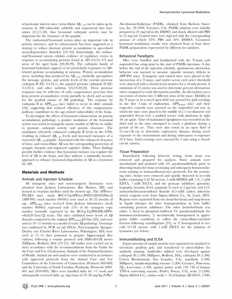
Figure 1. The lysosomal modulator PADK selectively enhances cathepsin B levels in APPSwInd mice. The 10–11-month transgenic mice(tg) were injected i.p. daily with either PADK (20 mg/kg; n = 13) or vehicle (veh; n = 10) for 9 days. Brain homogenates from the transgenic mice andfrom vehicle-treated wildtypes (wt; n = 13) were analyzed by immunoblot for the active form of cathepsin B (CB), neprilysin (nep), insulin-degradingenzyme (IDE), a-secretase (a-sec), and actin (A). Mean immunoreactivities6SEM were determined by image analysis and plotted. Hippocampalphotomicrographs from vehicle- (B) and PADK-treated mice (C) show cathepsin B immunostaining (green) in pyramidal neurons counterstained withanti-NeuN (red); view-field width is 75 mm. The CA1 zone was assessed for cathepsin B immunoreactivity (mean intensity6SEM) in the pyramidal layer(D) and for the number of cathepsin B-positive puncta per neuron (E). Tukey post hoc test: **P,0.001; unpaired t-test: ***P,0.0001.doi:10.1371/journal.pone.0020501.g001
Neuroprotective Lysosomal Modulation
PLoS ONE | www.plosone.org 3 June 2011 | Volume 6 | Issue 6 | e20501
Carlsbad, CA), 6E10 antibody (Covance), anti-LAMP1 (BD
Pharmingen, San Jose, CA), and monoclonal antibody that
selectively labels Ab1–42 (Covance). Immunofluorescence used
appropriate secondary antibodies from Invitrogen, and images
were captured with a Zeiss fluorescence microscope system. For
quantitative analysis of integrated fluorescence intensity, images
from the different treatment groups received the same gain,
exposure time, intensity threshold, and other measurement
parameters that were capsulated within each image file. Avidin-
biotin-peroxidase protocols used Vectastain kits (Vector Labora-
tories, Burlingame, CA) with 3,39-diaminobenzidine as the
chromogen, and images were acquired with a digital microscope
system (Olympus AX70). Densitometric quantification was
conducted with a BIOQUANT Image Analysis System. Treat-
ment groups were immunostained together and analyzed under
the same instrument settings. Equally spaced coronal sections
along the rostral-caudal axis of the hippocampus were used to
determine the average immunoreactivity intensity across four
different cortical view-fields for each mouse. To assess hippocam-
pal subfields, the CA1 field was investigated with view-fields of
CA1b and the initial area of CA1c. For CA3, view-fields were
selected from the zone immediately before the dentate gyrus
envelope. In the dentate gyrus, granule cells of the stratum
granulosum were counted in view-fields from the middle segment
of the inner blade. In addition to integrated fluorescence and
colormetric intensity measures, extracellular deposition was
assessed with an autothreshold function and the area of
immunostaining was expressed as percentage of the total view-
field area being evaluated.
b-Secretase Activity AssayCompounds were tested against the activity of b-secretase (b-site
APP cleaving enzyme-1; BACE1) using Sigma’s SensiZyme
BACE1 Activity Assay Kit. The kit uses a constructed procas-
pase-3 variant containing the modified cleavage sequence Gly-Ser-
Ser-Glu-Ile-Ser-Tyr-Glu-Val-Glu-Phe-Arg-Glu-Phe which is
cleaved by b-secretase after the Tyr-Glu residues [32]. Increasing
concentrations of potential inhibitors, including b-secretase
inhibitor IV (EMD Chemicals, Gibbstown, NJ), were incubated
with 10 ng/ml of C-terminal FLAG-tagged recombinant human
b-secretase from HEK 293 cells. Activity was expressed as
absorbance units produced by the caspase-3 colorimetric substrate
N-acetyl-Asp-Glu-Val-Asp-p-nitroanilide and measured with a
SpectraMax M3 Microplate Reader.
Lysosome Isolation and Cathepsin ActivityDissected hemi-brains were quickly separated into regions and
each immediately homogenized in ice-cold 0.3 M sucrose with
1 mM EDTA and centrifuged at 7506g for 10 min. The
collected supernatant was incubated with 2 mM CaCl2 at 37uCfor 5 min then layered over 24% Percoll in 0.32 M sucrose
containing 1 mM EDTA (pH 5.5). After centrifugation at
20,0006g for 18 min the gradients were fractionated, and those
determined to be negative (upper four fractions) or positive (lower
two) for cathepsin B by immunoblot were diluted with 5 volumes
of sucrose solution to separate organelles from the Percoll by
centrifuging at 40,0006g for 60 min. The pellets were resus-
pended and protein content determined with the Pierce BCA
assay (Thermo Scientific, Rockford, IL). The resulting fractions
were aliquoted, lysed in 18 mM citrate, and assessed for
cathepsin B proteolytic activity using the Z-Arg-Arg AMC
substrate, the fluorogenic Calbiochem assay kit (EMD Chemi-
cals), and the Molecular Devices SpectraMax M3 Microplate
Reader. Potential inhibitors were also tested in the cathepsin B
activity assay, using equal aliquots of Triton X-100 solubilized
brain homogenate pre-treated for 30 min with increasing
concentrations of the agents.
Statistical and Data AnalysesMeans of measures from image analysis of immunoblots and
tissue staining, ELISA tests, behavioral assessment, enzyme
activity, and other experiments were evaluated with unpaired t-
tests, unpaired Mann-Whitney U-tests, or across .2 treatment
groups with analyses of variance (ANOVA) followed by the
Tukey’s multiple comparison post hoc tests using Prism software
(GraphPad, San Diego, CA). Nonlinear regression was used to fit
enzyme activity inhibition data to one-site competitive binding
equations. Linear regression analyses tested for significant
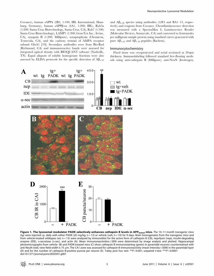
Table 1. PADK-mediated enhancement across brain regionsof transgenic mouse models.
Brain Region APPSwInd APP-PS1
neocortex 3.060.17 7.360.60
frontal cortex 3.260.33 6.861.3
hippocampus 4.360.38 8.460.85
mesencephalon 3.760.70 4.960.21
APPSwInd (10–11 months of age) and APPswe/PS1DE9 mice (APP-PS1; 20–22months) were injected i.p. daily with PADK (20 mg/kg; n = 11213) or vehicle(n = 10) for 9–11 days. Active cathepsin B in tissue homogenates was measuredby immunoblot, and the mean levels were compared to the respective meanimmunoreactivity in vehicle-injected transgenic samples to determine the foldincrease across brain regions (6SEM).doi:10.1371/journal.pone.0020501.t001
Table 2. PADK selectively enhances cathepsin B levels in two transgenic mouse models.
APPSwInd+veh APPSwInd+PADK APP2PS1+veh APP2PS1+PADK
CB 95.3614.2 404.6636.0*** 44.5615.2 376.0638.6***
nep 89.1619.2 95.3616.6 43.066.5 31.566.6
IDE 101.1611.0 76.466.5 96.168.3 89.566.0
a-sec 82.366.0 92.065.2 60.168.9 58.966.2
LAMP1 98.465.4 102.268.6 46.169.9 45.864.6
APPSwInd and APP-PS1 mice were injected i.p. daily with PADK (20 mg/kg; n = 11213) or vehicle (n = 10) for 9–11 days. Hippocampal homogenates were analyzed byimmunoblot and mean immunoreactivities are shown for active cathepsin B (CB), neprilysin (nep), insulin-degrading enzyme (IDE), a-secretase (a-sec), and LAMP1.***P,0.0001, unpaired t-test.doi:10.1371/journal.pone.0020501.t002
Neuroprotective Lysosomal Modulation
PLoS ONE | www.plosone.org 4 June 2011 | Volume 6 | Issue 6 | e20501
correlations between Ab species and cathepsin B enhancement or
between GluA1 immunoreactivity and the lysosomal enhance-
ment.
Results
To elicit lysosomal enhancement in mouse models of AD, we
administered Z-Phe-Ala-diazomethylketone (PADK), a weak
inhibitor of cathepsin B and L (cathepsin B IC50 = 9.462.4 mM)
as well as a lysosomal modulator previously shown to cause a
feedback response involving marked up-regulation of cathepsin
isoform expression in vitro [19–21]. The lysosomal modulator
(20 mg/kg) was injected i.p. daily for 9 days into 10–11-month
APPSwInd mice which express the human APP gene with the
Swedish (K670N/M671L) and Indiana (V717F) mutations [30],
resulting in a marked increase in the active isoform of cathepsin B
in the brain as compared to vehicle-injected transgenic mice
(Fig. 1A; ANOVA P,0.0001, post hoc test P,0.001; n = 13).
Cathepsin B immunoreactivity levels were enhanced .4 fold in
hippocampal samples, and 3-fold or greater increases were found
in samples from neocortex, frontal cortex, and mesencephalon
(Table 1). Measures of Ab-degrading proteases neprilysin and
insulin-degrading enzyme, as well as a-secretase which prevents
Ab production, were not altered (Fig. 1A and Table 2), thus the
PADK-mediated lysosomal modulation was produced in a
selective manner. Similar selectivity was also evident for the
PADK effect in 20–22-month APPswe/PS1DE9 mice (APP-PS1;
Table 2), which express a chimeric mouse/human APP and
human presenilin 1 directed to CNS neurons [33]. Significant
cathepsin B up-regulation was found in different brain regions of
the APP-PS1 mice, with hippocampus exhibiting the largest
increase of .8 fold (Table 1).
In immunocytochemistry images, intracellular cathepsin B was
revealed as punctate staining (green) characteristic of lysosomal
organelles in hippocampal CA1 pyramidal neurons (Fig. 1B), and
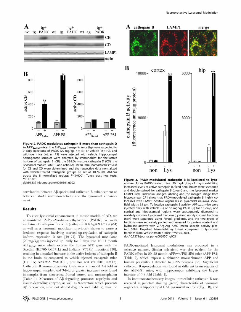
Figure 2. PADK modulates cathepsin B more than cathepsin Din APPSwInd mice. The APPSwInd transgenic mice (tg) were subjected to9 daily injections of PADK (20 mg/kg; n = 13) or vehicle (n = 10), andwildtype mice (wt; n = 13) were injected with vehicle. Hippocampalhomogenate samples were analyzed by immunoblot for the activeisoform of cathepsin B (CB), the 33-kDa mature cathepsin D (CD), thelysosomal marker LAMP1, and actin (A). Mean immunoreactivities6SEMfor CB and CD were determined and the respective data normalizedwith vehicle-treated transgenic groups (–) set at 100% (B). ANOVAacross the 8 normalized groups: P,0.0001; Tukey post hoc tests:**P,0.001.doi:10.1371/journal.pone.0020501.g002
Figure 3. PADK-modulated cathepsin B is localized to lyso-somes. From PADK-treated mice (20 mg/kg/day69 days) exhibitingincreased levels of active cathepsin B, fixed hemi-brains were sectionedand double-stained for cathepsin B (green) and the lysosomal markerLAMP1 (red). Individual antigen labeling and the merged image fromhippocampal CA1 show that PADK-modulated cathepsin B highly co-localizes with LAMP1-positive organelles in pyramidal neurons. View-field width: 35 mm. To localize cathepsin B activity, APPSwInd mice wereinjected daily with vehicle (–) or 18 mg/kg PADK (+) for 10 days, andcortical and hippocampal regions were subsequently dissected toisolate lysosomes. Lysosomal fractions (Lys) and non-lysosomal fractions(non) were separated using Percoll gradients, and the two types offractions were separately pooled and assessed for protein content andhydrolase activity with Z-Arg-Arg AMC (mean specific activity plot-ted6SEM). Unpaired Mann-Whitney U-test compared to lysosomalfractions from vehicle-treated mice: ***P,0.0001.doi:10.1371/journal.pone.0020501.g003
Neuroprotective Lysosomal Modulation
PLoS ONE | www.plosone.org 5 June 2011 | Volume 6 | Issue 6 | e20501
the intensity of such immunostaining was enhanced after PADK
treatment (Fig. 1C). Neurons were counterstained with anti-NeuN
(red), and PADK elicited no apparent change in neuronal density
or morphology. Quantitative analysis of the fluorescence intensity
across the stratum pyramidale confirmed an increase in cathepsin
B immunoreactivity (P,0.0001; Fig. 1D). On the other hand, the
number of cathepsin B-positive organelles per pyramidal neuron
(n = 62) was found to be unchanged in the view-fields (Fig. 1E),
and the lysosome-associated membrane glycoprotein LAMP1 was
also unaltered in blot samples from the different treatment groups
(Fig. 2A, Table 2). ANOVA assessment of two different cathepsins
(B and D) across all transgenic treatment groups found that only
cathepsin B increased with PADK treatment (Fig. 2). Thus, the 2-
to 3-fold increase in intracellular cathepsin B staining, in the
absence of a change in lysosome number, appears to represent the
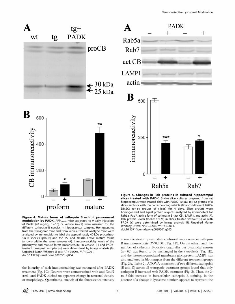
Figure 4. Mature forms of cathepsin B exhibit pronouncedmodulation by PADK. APPSwInd mice subjected to 9 daily injectionsof PADK (20 mg/kg; n = 10) or vehicle (n = 9) were assessed for thedifferent cathepsin B species in hippocampal samples. Homogenatesfrom the transgenic mice and from vehicle-treated wildtype mice wereanalyzed by immunoblot to label the approximately 40-kDa procathep-sin B species (proCB) and the 25- and 30-kDa active mature forms(arrows) within the same samples (A). Immunoreactivity levels of theproenzyme and mature forms (means6SEM) in vehicle- (–) and PADK-treated transgenic samples (+) were determined by image analysis (B).Unpaired Mann-Whitney U-test: *P = 0.0296, **P,0.001.doi:10.1371/journal.pone.0020501.g004
Figure 5. Changes in Rab proteins in cultured hippocampalslices treated with PADK. Stable slice cultures prepared from rathippocampus were treated daily with PADK (10 mM; n = 12 groups of 8slices each) or with the corresponding vehicle (final condition of 0.02%DMSO; n = 14 groups of slices) for 4 days. Slice groups werehomogenized and equal protein aliquots analyzed by immunoblot forRab5a, Rab7, active form of cathepsin B (act CB), LAMP1, and actin (A).Rab protein levels (means6SEM) in slices treated without (–) or withPADK (+) were determined by image analysis (B). Unpaired Mann-Whitney U-test: *P = 0.0208, ***P,0.0001.doi:10.1371/journal.pone.0020501.g005
Neuroprotective Lysosomal Modulation
PLoS ONE | www.plosone.org 6 June 2011 | Volume 6 | Issue 6 | e20501
primary PADK effect in neurons. Note that a one-way
nonparametric Kruskal-Wallis analysis followed by Dunn’s post
test of the 33-kDa cathepsin D form alone revealed significant
albeit small increases produced by PADK in the APPSwInd and
APP-PS1 samples (P,0.05).
To further test whether the intracellular modulation is actually
influencing lysosomal cathepsin B, localization of the PADK effect
was evaluated in brain tissue double-stained for cathepsin B and
LAMP1 (Fig. 3A). As evident in the merged immunofluorescence
image, the PADK-modulated cathepsin B co-localized with
LAMP1-positive organelles in CA1 pyramidal neurons. Together,
the findings indicate that the lysosomal modulator enhances
cathepsin B content in lysosomes. APPSwInd mice injected with
vehicle or 18 mg/kg PADK daily for 10 days were also assessed
for cathepsin B activity in isolated lysosomes using the Z-Arg-Arg
AMC probe (Fig. 3B). Rapidly dissected brain regions were
subjected to subcellular fractionation in Percoll gradients, and
equal protein aliquots from the lysosomal and non-lysosomal
fractions were evaluated for hydrolase activity that was blocked by
the cathepsin B inhibitor CA074me. Note that in the different
brain regions tested, only the lysosomal fractions exhibited PADK-
dependent increases in cathepsin B activity of 3-10 fold
(P,0.0001).
Hippocampal homogenates from the APPSwInd mice were
further analyzed in an attempt to understand how PADK
influences lysosomal levels of cathepsin B. Cathepsin B belongs
to the superfamily of papain-like cysteine proteases and is first
synthesized as a proenzyme. The approximately 40-kDa pro-
cathepsin B exhibited a marginally significant increase of 93627%
(mean6SEM; P,0.03) in the PADK-treated group as compared
to vehicle-injected transgenic mice samples (Fig. 4A and B). Within
the same immunoblot samples, the percent change in the 25–30-
kDa mature cathepsin B forms was found to be much larger than
the PADK effect on the proform of the enzyme (270631%,
P,0.001) (Fig. 4B). PADK’s range of effect in the two transgenic
models is 4- to 9-fold changes in the mature active isoforms in
hippocampus (Table 1), and even greater fold increases when
considering the increase in the 25-kDa species alone (see Fig. 4A).
These pronounce changes implicate trafficking as a component of
the PADK effect, since inactive procathepsin B is processed to the
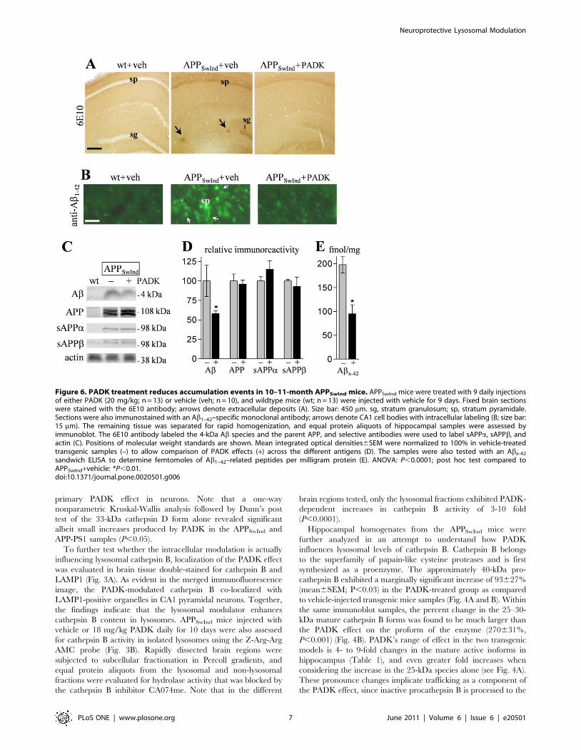
Figure 6. PADK treatment reduces accumulation events in 10–11-month APPSwInd mice. APPSwInd mice were treated with 9 daily injectionsof either PADK (20 mg/kg; n = 13) or vehicle (veh; n = 10), and wildtype mice (wt; n = 13) were injected with vehicle for 9 days. Fixed brain sectionswere stained with the 6E10 antibody; arrows denote extracellular deposits (A). Size bar: 450 mm. sg, stratum granulosum; sp, stratum pyramidale.Sections were also immunostained with an Ab1–42–specific monoclonal antibody; arrows denote CA1 cell bodies with intracellular labeling (B; size bar:15 mm). The remaining tissue was separated for rapid homogenization, and equal protein aliquots of hippocampal samples were assessed byimmunoblot. The 6E10 antibody labeled the 4-kDa Ab species and the parent APP, and selective antibodies were used to label sAPPa, sAPPb, andactin (C). Positions of molecular weight standards are shown. Mean integrated optical densities6SEM were normalized to 100% in vehicle-treatedtransgenic samples (–) to allow comparison of PADK effects (+) across the different antigens (D). The samples were also tested with an Abx-42
sandwich ELISA to determine femtomoles of Ab1–42–related peptides per milligram protein (E). ANOVA: P,0.0001; post hoc test compared toAPPSwInd+vehicle: *P,0.01.doi:10.1371/journal.pone.0020501.g006
Neuroprotective Lysosomal Modulation
PLoS ONE | www.plosone.org 7 June 2011 | Volume 6 | Issue 6 | e20501
active forms in late endosomes and lysosomes. To selectively
examine trafficking markers during defined lysosomal modulation,
hippocampal slice cultures were treated with PADK daily for 4
days, causing a .5-fold increase in active cathepsin B. In
conjunction with enhanced cathepsin B, the PADK-treated slices
exhibited a decrease in Rab5a (Fig. 5A), a marker of early
endosomes [34]. From the analysis of integrated optical densities,
control slices were found to contain more than 2-fold more Rab5a
than the PADK slices, whereas the lysosomal modulator increased
the level of Rab7 which controls the late step of endosomal
trafficking to lysosomes (Fig. 5B). In addition, as found in vivo,
LAMP1 was unchanged (control, 15667.2; PADK-treated slices,
14065.1; NS). Thus, positive modulation of trafficking likely plays
a major role in PADK’s ability to increase the content of active
cathepsin B in lysosomes and/or late endosomes.
We next tested whether PADK’s influence on the lysosomal
system is associated with enhanced Ab clearance. APPSwInd mice
of low transgene copy number were used at 10–11 months of age
in order to assess the PADK effects at an early stage of Abpathology. Tissue sections were stained with the 6E10 antibody,
which specifically recognizes amino acid residues 1–16 of human
Ab. In Figure 6A, intracellular staining is evident among neurons
of the stratum pyramidale and stratum granulosum in vehicle-
injected APPSwInd mice, while none was found in wildtype mice,
and the transgenics also exhibited sporadic extracellular deposits.
The daily PADK administration resulted in reduced deposits and
a marked decrease in the cellular labeling. Serial sections stained
with Ab1–42–specific monoclonal antibody confirmed that Ab1–42
is the primary species that accumulates intracellularly in the
APPSwInd brain (Fig. 6B), as previously reported for other AD
mouse models [35], and it is the same peptide selectively taken up
by neurons of the hippocampus [11]. The Ab1–42 staining intensity
in pyramidal neurons was diminished by the PADK treatment,
and similar degrees of reduced 6E10 labeling (263–73%; post hoc
tests P,0.001) were determined across different neuronal layers in
hippocampus and piriform cortex (Fig. 7).
Brain homogenate samples were also analyzed to determine
which APP fragments recognized by 6E10 were reduced by the
PADK treatment. The lysosomal modulator significantly reduced
a 6E10-labeled species of 4 kDa that coincided with the
electrophoretic migration of pure Ab1–42 (ANOVA P,0.0001,
post hoc test P,0.01), without affecting the parent hAPP protein
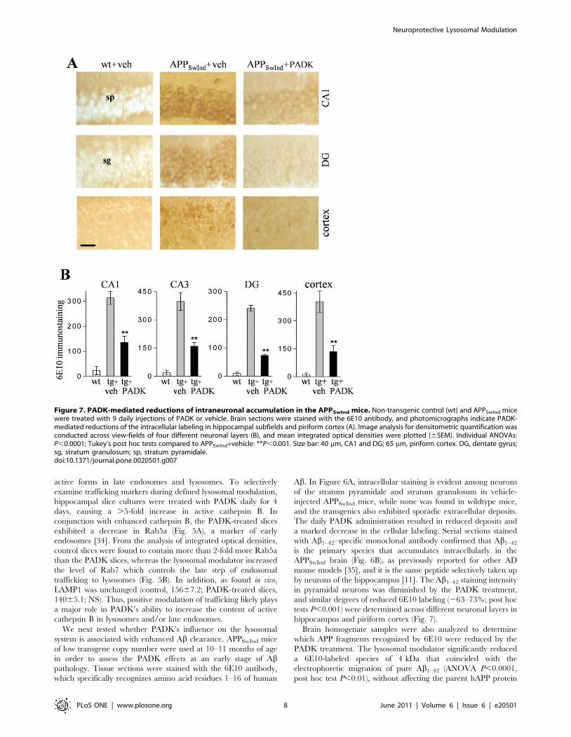
Figure 7. PADK-mediated reductions of intraneuronal accumulation in the APPSwInd mice. Non-transgenic control (wt) and APPSwInd micewere treated with 9 daily injections of PADK or vehicle. Brain sections were stained with the 6E10 antibody, and photomicrographs indicate PADK-mediated reductions of the intracellular labeling in hippocampal subfields and piriform cortex (A). Image analysis for densitometric quantification wasconducted across view-fields of four different neuronal layers (B), and mean integrated optical densities were plotted (6SEM). Individual ANOVAs:P,0.0001; Tukey’s post hoc tests compared to APPSwInd+vehicle: **P,0.001. Size bar: 40 mm, CA1 and DG; 65 mm, piriform cortex. DG, dentate gyrus;sg, stratum granulosum; sp, stratum pyramidale.doi:10.1371/journal.pone.0020501.g007
Neuroprotective Lysosomal Modulation
PLoS ONE | www.plosone.org 8 June 2011 | Volume 6 | Issue 6 | e20501
level expressed in the transgenic mouse brain (Fig. 6C and D).
Similar to the extent of reduction in the 4-kDa peptide, selective
measures using the Abx-42 sandwich ELISA were also significantly
decreased by PADK (Fig. 6E), and the decline within each
transgenic mouse correlated with the respective level of cathepsin
B enhancement in the brain (r = 20.76, P,0.05). Other
antibodies were used to selectively label sAPPa and sAPPb which
were unchanged after the PADK treatment period, indicating that
the APP cleavage activities of a- and b-secretase were not affected
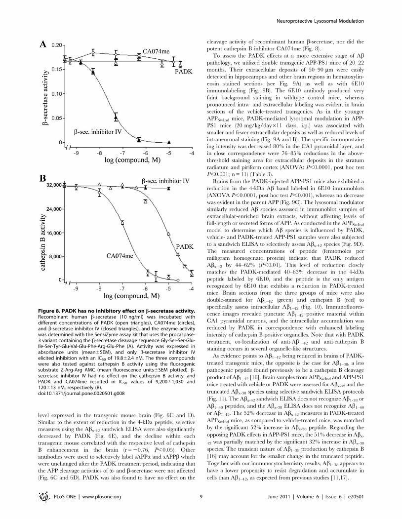
(Fig. 6C and 6D). PADK was also found to have no effect on the
cleavage activity of recombinant human b-secretase, nor did the
potent cathepsin B inhibitor CA074me (Fig. 8).
To assess the PADK effects at a more extensive stage of Abpathology, we utilized double transgenic APP-PS1 mice of 20–22
months. Their extracellular deposits of 50–90 mm were easily
detected in hippocampus and other brain regions in hematoxylin-
eosin stained sections (see Fig. 9A) as well as with 6E10
immunolabeling (Fig. 9B). The 6E10 antibody produced very
faint background staining in wildtype control mice, whereas
pronounced intra- and extracellular labeling was evident in brain
sections of the vehicle-treated transgenics. As in the younger
APPSwInd mice, PADK-mediated lysosomal modulation in APP-
PS1 mice (20 mg/kg/day611 days, i.p.) was associated with
smaller and fewer extracellular deposits as well as reduced levels of
intraneuronal staining (Fig. 9A and B). The specific immunostain-
ing intensity was decreased 80% in the CA1 pyramidal layer, and
in close correspondence were 76–85% reductions in the above-
threshold staining area for extracellular deposits in the stratum
radiatum and piriform cortex (ANOVA: P,0.0001, post hoc test
P,0.001; n = 11) (Table 3).
Brains from the PADK-injected APP-PS1 mice also exhibited a
reduction in the 4-kDa Ab band labeled in 6E10 immunoblots
(ANOVA P,0.0001, post hoc test P,0.001), whereas no decrease
was evident in the parent APP (Fig. 9C). The lysosomal modulator
similarly reduced Ab species assessed in immunoblot samples of
extracellular-enriched brain extracts, without affecting levels of
full-length or secreted forms of APP. As conducted in the APPSwInd
model to determine which Ab species is influenced by PADK,
vehicle- and PADK-treated APP-PS1 samples were also subjected
to a sandwich ELISA to selectively assess Abx-42 species (Fig. 9D).
The measured concentrations of peptide (femtomoles per
milligram homogenate protein) indicate that PADK reduced
Abx-42 by 44–62% (P,0.01). This level of reduction closely
matches the PADK-mediated 40–63% decrease in the 4-kDa
peptide labeled by 6E10, and the peptide is the only antigen
recognized by 6E10 that exhibits a reduction in PADK-treated
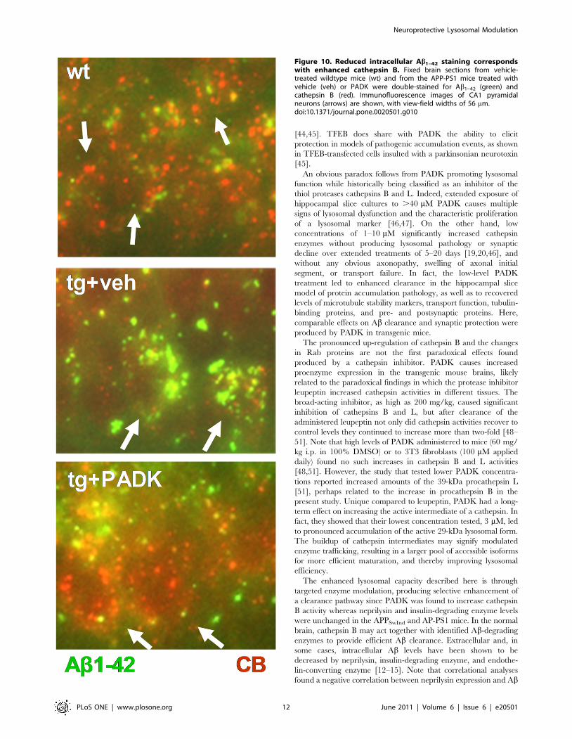
mice. Brain sections from the three groups of mice were also
double-stained for Ab1–42 (green) and cathepsin B (red) to
specifically assess intracellular Ab1–42 (Fig. 10). Immunofluores-
cence images revealed punctate Ab1–42–positive material within
CA1 pyramidal neurons, and the intracellular accumulation was
reduced by PADK in correspondence with enhanced labeling
intensity of cathepsin B-positive organelles. Note that with PADK
treatment, co-localization of anti-Ab1–42 and anti-cathepsin B
staining occurs in several organelle-like structures.
As evidence points to Ab1–42 being reduced in brains of PADK-
treated transgenic mice, the opposite is the case for Ab1–38, a less
pathogenic peptide found previously to be a cathepsin B cleavage
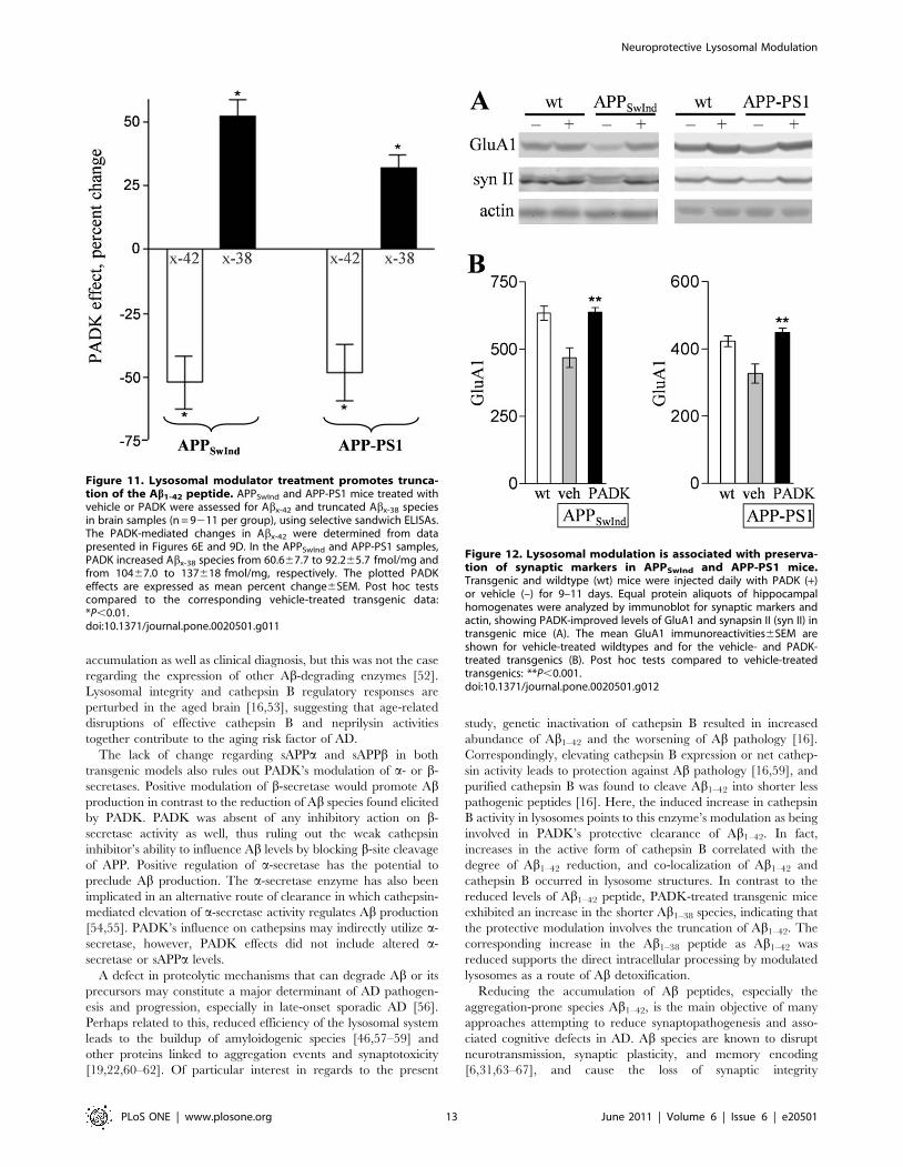
product of Ab1–42 [16]. Brain samples from APPSwInd and APP-PS1
mice treated with vehicle or PADK were assessed for Abx-42 and the
truncated Abx-38 species using selective sandwich ELISA protocols
(Fig. 11). The Abx-42 sandwich ELISA does not recognize Ab1-38 or
Ab1–40 peptides, and the Abx-38 ELISA does not recognize Ab1–40
or Ab1–42. The 52% decrease in Abx-42 measures in PADK-treated
APPSwInd mice, as compared to vehicle-treated mice, was matched
by the significant 52% increase in Abx-38 peptide. Regarding the
opposing PADK effects in APP-PS1 mice, the 51% decrease in Abx-
42 was partially matched by the significant 32% increase in Abx-38
species. The transient nature of Ab1–38 production by cathepsin B
[16] may account for the smaller change in the truncated peptide.
Together with our immunocytochemistry results, Ab1–38 appears to
have a lower propensity to resist degradation and accumulate in
cells than Ab1–42, as expected from previous studies [11,17].
Figure 8. PADK has no inhibitory effect on b-secretase activity.Recombinant human b-secretase (10 ng/ml) was incubated withdifferent concentrations of PADK (open triangles), CA074me (circles),and b-secretase inhibitor IV (closed triangles), and the enzyme activitywas determined with the SensiZyme assay kit that uses the procaspase-3 variant containing the b-secretase cleavage sequence Gly-Ser-Ser-Glu-Ile-Ser-Tyr-Glu-Val-Glu-Phe-Arg-Glu-Phe (A). Activity was expressed inabsorbance units (mean6SEM), and only b-secretase inhibitor IVelicited inhibition with an IC50 of 19.862.4 nM. The three compoundswere also tested against cathepsin B activity using the fluorogenicsubstrate Z-Arg-Arg AMC (mean fluorescence units6SEM plotted). b-secretase inhibitor IV had no effect on the cathepsin B activity, andPADK and CA074me resulted in IC50 values of 9,20061,030 and120613 nM, respectively (B).doi:10.1371/journal.pone.0020501.g008
Neuroprotective Lysosomal Modulation
PLoS ONE | www.plosone.org 9 June 2011 | Volume 6 | Issue 6 | e20501

The lysosomal modulator’s effects on Ab clearance were
associated with synaptic protection. In Figure 12A, hippocampal
samples from the two mouse models were analyzed by immunoblot
for the presynaptic protein synapsin II and the postsynaptic
glutamatergic marker GluA1. Similar to the extent of synaptic
decline seen in related transgenic mice [30,33], the synaptic proteins
exhibited deficits of 23–31% in APPSwInd and APP-PS1 mice as
compared to their respective age-matched wildtypes (P,0.01), while
actin levels remained unchanged. PADK treatment significantly
reduced the GluA1 deficit in the two mouse models, reaching levels
comparable to those found in non-transgenic control mice (Fig. 12B)
(ANOVA: P,0.001; n = 12220). Similar indications of synaptic
protection were found when assessing synapsin II and synaptophy-
sin. The integrity of hippocampal dendritic fields was also found
preserved in immunostained tissue sections, and the level of GluA1
immunoreactivity within each transgenic mouse correlated with the
respective extent of cathepsin B enhancement in the brain (r = 0.78,
P = 0.02).
Lastly, we assessed the mouse models to determine whether the
PADK-mediated clearance and synaptic protection translate to
improvements in behavioral tests. Although the APPSwInd mice
used in the present study express low levels of Ab deposits, their
Figure 9. PADK decreases intra- and extracellular 6E10 staining in APPswe/PS1DE9 (APP-PS1) mice of 20–22 months. The APP-PS1mice received 11 daily injections of PADK (i.p., 20 mg/kg; n = 11) or vehicle (veh; n = 10), and non-transgenic control mice (wt) received vehicleinjections. Fixed brain sections from the different groups were hematoxylin-eosin stained (A; arrows denote typical deposits) and 6E10immunolabeled (B), indicating that PADK treatment reduces intra- and extracellular deposition in hippocampus. Equal protein samples from vehicle-(–) and PADK-treated (+) APP-PS1 mouse brains were immunoblotted with 6E10 antibody to assess the 4-kDa Ab peptide and the parent hAPP, andwith selective antibodies to label sAPPa and sAPPb (C). Mean integrated optical densities6SEM for the different species were normalized to 100% asshown. The same brain samples were also tested by Abx-42 sandwich ELISA to determine femtomoles of peptide per milligram protein (D). ANOVA:P,0.0001; post hoc test compared to APP-PS1+vehicle: **P,0.001. Unpaired t-test: *P,0.01. Size bar: 400 mm, A; 50 mm, B. DG, dentate gyrus; sp,stratum pyramidale; sr, stratum radiatum.doi:10.1371/journal.pone.0020501.g009
Neuroprotective Lysosomal Modulation
PLoS ONE | www.plosone.org 10 June 2011 | Volume 6 | Issue 6 | e20501

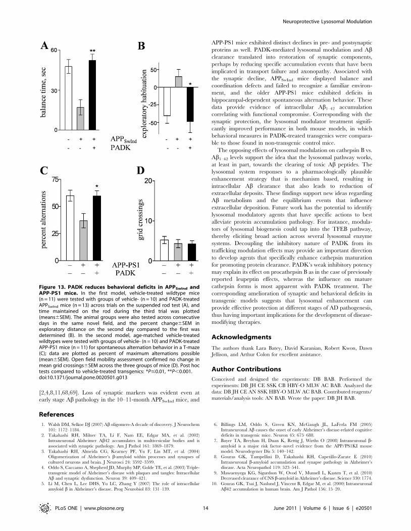
synaptic decline was associated with significant deficits on the
suspended rod test (Fig. 13A) as well as on the exploratory
habituation test (Fig. 13B), similar to the type of deficit reported
previously for APPSwInd mice of the same age range [36].
Compared to wildtypes, the vehicle-injected APPSwInd mice
exhibited a marked reduction in balance time while coordinating
to reach a safe platform, whereas the PADK-treated transgenics
performed at the same level as the control mice (post-hoc test
p,0.001) (Fig. 13A). A marked degree of disinhibition was also
displayed by the vehicle-treated transgenics on the second day of
open field exploration, but APPSwInd mice injected with PADK
were completely devoid of this behavioral defect (Fig. 13B). No
differences were found among the three groups of mice regarding
gross mobility during open field exploration, nor were any
differences evident during mobility testing on a slow rotating rod
(not shown). Also corresponding with synaptic marker decline, the
APP-PS1 mouse model of extensive Ab pathology expressed
cognitive deficiency in the hippocampal-dependent task of
spontaneous alternation behavior (Fig. 13C), similar to episodic
memory deficits reported previously [33,37]. The vehicle-treated
APP-PS1 mice exhibited reduced alternation behavior of alternate
arm entries in the T-maze as compared to wildtype mice
(P,0.0001). The 11-day PADK treatment resulted in significantly
improved alternations, reaching a performance level equal to that
of the age-matched wildtypes. Open field assessment confirmed no
change in exploratory mobility across the different groups of mice
(Fig. 13D). These data confirm in a second AD mouse model that
lysosomal modulation reduces behavioral deficits.
Discussion
Our study indicates that lysosomes are a site of modulation for
the targeted enhancement of cathepsin function to promote
clearance of the Ab peptide. The endosomal-lysosomal pathway
has been implicated as playing a role in the degradation of Ab, and
the impairment of this pathway likely contributes to the
accumulation of intraneuronal Ab and other proteins linked to
AD. Enhancement of the lysosomal system was achieved with the
modulatory agent PADK, resulting in the selective increase of
cathepsin B activity in lysosomes. The lysosomal modulation led to
reductions in both intracellular and extracellular Ab, to a degree
that ameliorates the phenotype of two AD mouse models with
distinctly different levels of Ab pathology. At the analogous stages
of the human disorder, reducing Ab accumulations is thought to
be essential for slowing AD progression.
When administered systemically, the PADK modulator in-
creased cathepsin B levels 4- to 8-fold across different brain regions
in APPSwInd and APP-PS1 mice. From the analysis of immuno-
fluorescence images, enhanced intracellular cathepsin B staining
was found to represent the primary PADK effect in neurons. As
cathepsin B increased in the transgenic mice, there was a
corresponding decrease in Ab1–42 measured by selective ELISA.
Brain immunocytochemistry with anti-Ab1–42 and 6E10 antibod-
ies suggests that lysosomal modulation leads to decreased levels of
intraneuronal deposition. Interestingly, reduction in extracellular
deposits occurred in correspondence with the reduced labeling of
intracellular Ab, providing evidence of an equilibrium relationship
between secreted Ab peptides and the extracellularly deposited
material. This fits well with Oddo et al. [35] which described the
accumulation of intracellular Ab as a source of extracellular
amyloid deposits. Note that PADK had no effect on hAPP
expression levels in the transgenic brains, whereas the 4-kDa Abspecies was markedly reduced. This 44–63% decrease in Ab likely
contributes to the reduced extracellular deposition as multiphoton
microscopy and microdialysis studies showed that reduction of Abby as little as 20–25% was capable of diminishing plaque deposits
[38]. The actions of PADK on cathepsin B and Ab indicate the
intraneuronal clearance process as an appropriate target for
offsetting protein accumulation pathology. Since extracellular
peptide taken up by vulnerable neurons is thought to contribute
significantly to the accumulation of intracellular Ab1–42
[11,17,18,39], the modulated lysosomal system may be exerting
its clearance effects directly on the internalized Ab1–42 peptide as
well as by increasing the turnover of Ab-containing APP
fragments. Lysosomal modulation in other cell types will be the
subject of follow-up studies since in microglia, internalized
oligomeric Ab has recently been shown to be trafficked to
lysosomes and degraded by cysteine proteases including cathepsin
B [40].
PADK-mediated lysosomal modulation appears to involve both
increased expression of cathepsin B and enhanced trafficking of
the enzyme. The modulation consisted of a modest increase in
procathepsin B as compared to larger 4- to 9-fold increases in the
mature active forms of the enzyme. Further evidence that PADK
has a positive effect on endo-lysosomal trafficking includes i)
cathepsin B hydrolytic activity was enhanced in isolated lysosomes
to a much greater degree than the enhancement of proenzyme in
brain samples, ii) active cathepsin B forms were enhanced in the
absence of any increase in the lysosomal marker LAMP1, iii) the
modulated cathepsin B co-localized with LAMP1 in neurons, and
iv) decreases in the Rab5a early endosome marker corresponded
with the enhanced levels of mature cathepsin B in hippocampal
tissue. Elevated expression of early endosome regulators, on the
other hand, occur in hippocampal neurons from individuals with
AD and mild cognitive impairment, suggesting that early
endosomal dysfunction contributes to AD progression [27,41].
The action of PADK may offset endocytic dysfunction and
promote protein clearance through enhanced trafficking of
cathepsin B and/or Ab-containing species from early endosomes
to late endosomes and lysosomes. Perhaps enhanced cathepsin B
maturation is facilitated by the small PADK-mediated increase in
mature cathepsin D, since previous studies found the latter to
activate procathepsin B into mature forms [42,43]. Regarding
PADK’s lack of influence on LAMP1, this would appear to rule
out any broad effect on lysosome production or the modulation of
TFEB, a transcription factor that regulates lysosomal biogenesis
Table 3. PADK decreases 6E10 immunostaining in APP-PS1mouse brain.
wt APP2PS1+veh APP2PS1+PADK
hippocampal sp(IOD)
129615.1 672658.9 241615.0**
hippocampal sr(area)
0.0760.02 2.9060.71 0.4960.08**
piriform cortex(area)
0.1360.03 3.8960.36 1.0460.21**
APP-PS1 mice were injected i.p. daily with PADK (20 mg/kg; n = 11) or vehicle(n = 10) for 11 days. Fixed tissue was sectioned and stained with the 6E10antibody. Image analysis for densitometric quantification of theimmunostaining (mean integrated optical density6SEM) was conducted acrossview-fields of the hippocampal CA1 stratum pyramidale (sp). Area of depositlabeling above background was also measured for view-fields of thehippocampal stratum radiatum (sr) and piriform cortex (mean percent of totalmeasured area6SEM). ANOVAs: P,0.0001; Tukey’s post hoc tests compared toAPP2PS1+vehicle.**P,0.001.doi:10.1371/journal.pone.0020501.t003
Neuroprotective Lysosomal Modulation
PLoS ONE | www.plosone.org 11 June 2011 | Volume 6 | Issue 6 | e20501
[44,45]. TFEB does share with PADK the ability to elicit
protection in models of pathogenic accumulation events, as shown
in TFEB-transfected cells insulted with a parkinsonian neurotoxin
[45].
An obvious paradox follows from PADK promoting lysosomal
function while historically being classified as an inhibitor of the
thiol proteases cathepsins B and L. Indeed, extended exposure of
hippocampal slice cultures to .40 mM PADK causes multiple
signs of lysosomal dysfunction and the characteristic proliferation
of a lysosomal marker [46,47]. On the other hand, low
concentrations of 1–10 mM significantly increased cathepsin
enzymes without producing lysosomal pathology or synaptic
decline over extended treatments of 5–20 days [19,20,46], and
without any obvious axonopathy, swelling of axonal initial
segment, or transport failure. In fact, the low-level PADK
treatment led to enhanced clearance in the hippocampal slice
model of protein accumulation pathology, as well as to recovered
levels of microtubule stability markers, transport function, tubulin-
binding proteins, and pre- and postsynaptic proteins. Here,
comparable effects on Ab clearance and synaptic protection were
produced by PADK in transgenic mice.
The pronounced up-regulation of cathepsin B and the changes
in Rab proteins are not the first paradoxical effects found
produced by a cathepsin inhibitor. PADK causes increased
proenzyme expression in the transgenic mouse brains, likely
related to the paradoxical findings in which the protease inhibitor
leupeptin increased cathepsin activities in different tissues. The
broad-acting inhibitor, as high as 200 mg/kg, caused significant
inhibition of cathepsins B and L, but after clearance of the
administered leupeptin not only did cathepsin activities recover to
control levels they continued to increase more than two-fold [48–
51]. Note that high levels of PADK administered to mice (60 mg/
kg i.p. in 100% DMSO) or to 3T3 fibroblasts (100 mM applied
daily) found no such increases in cathepsin B and L activities
[48,51]. However, the study that tested lower PADK concentra-
tions reported increased amounts of the 39-kDa procathepsin L
[51], perhaps related to the increase in procathepsin B in the
present study. Unique compared to leupeptin, PADK had a long-
term effect on increasing the active intermediate of a cathepsin. In
fact, they showed that their lowest concentration tested, 3 mM, led
to pronounced accumulation of the active 29-kDa lysosomal form.
The buildup of cathepsin intermediates may signify modulated
enzyme trafficking, resulting in a larger pool of accessible isoforms
for more efficient maturation, and thereby improving lysosomal
efficiency.
The enhanced lysosomal capacity described here is through
targeted enzyme modulation, producing selective enhancement of
a clearance pathway since PADK was found to increase cathepsin
B activity whereas neprilysin and insulin-degrading enzyme levels
were unchanged in the APPSwInd and AP-PS1 mice. In the normal
brain, cathepsin B may act together with identified Ab-degrading
enzymes to provide efficient Ab clearance. Extracellular and, in
some cases, intracellular Ab levels have been shown to be
decreased by neprilysin, insulin-degrading enzyme, and endothe-
lin-converting enzyme [12–15]. Note that correlational analyses
found a negative correlation between neprilysin expression and Ab
Figure 10. Reduced intracellular Ab1–42 staining correspondswith enhanced cathepsin B. Fixed brain sections from vehicle-treated wildtype mice (wt) and from the APP-PS1 mice treated withvehicle (veh) or PADK were double-stained for Ab1–42 (green) andcathepsin B (red). Immunofluorescence images of CA1 pyramidalneurons (arrows) are shown, with view-field widths of 56 mm.doi:10.1371/journal.pone.0020501.g010
Neuroprotective Lysosomal Modulation
PLoS ONE | www.plosone.org 12 June 2011 | Volume 6 | Issue 6 | e20501
accumulation as well as clinical diagnosis, but this was not the case
regarding the expression of other Ab-degrading enzymes [52].
Lysosomal integrity and cathepsin B regulatory responses are
perturbed in the aged brain [16,53], suggesting that age-related
disruptions of effective cathepsin B and neprilysin activities
together contribute to the aging risk factor of AD.
The lack of change regarding sAPPa and sAPPb in both
transgenic models also rules out PADK’s modulation of a- or b-
secretases. Positive modulation of b-secretase would promote Abproduction in contrast to the reduction of Ab species found elicited
by PADK. PADK was absent of any inhibitory action on b-
secretase activity as well, thus ruling out the weak cathepsin
inhibitor’s ability to influence Ab levels by blocking b-site cleavage
of APP. Positive regulation of a-secretase has the potential to
preclude Ab production. The a-secretase enzyme has also been
implicated in an alternative route of clearance in which cathepsin-
mediated elevation of a-secretase activity regulates Ab production
[54,55]. PADK’s influence on cathepsins may indirectly utilize a-
secretase, however, PADK effects did not include altered a-
secretase or sAPPa levels.
A defect in proteolytic mechanisms that can degrade Ab or its
precursors may constitute a major determinant of AD pathogen-
esis and progression, especially in late-onset sporadic AD [56].
Perhaps related to this, reduced efficiency of the lysosomal system
leads to the buildup of amyloidogenic species [46,57–59] and
other proteins linked to aggregation events and synaptotoxicity
[19,22,60–62]. Of particular interest in regards to the present
study, genetic inactivation of cathepsin B resulted in increased
abundance of Ab1–42 and the worsening of Ab pathology [16].
Correspondingly, elevating cathepsin B expression or net cathep-
sin activity leads to protection against Ab pathology [16,59], and
purified cathepsin B was found to cleave Ab1–42 into shorter less
pathogenic peptides [16]. Here, the induced increase in cathepsin
B activity in lysosomes points to this enzyme’s modulation as being
involved in PADK’s protective clearance of Ab1–42. In fact,
increases in the active form of cathepsin B correlated with the
degree of Ab1–42 reduction, and co-localization of Ab1–42 and
cathepsin B occurred in lysosome structures. In contrast to the
reduced levels of Ab1–42 peptide, PADK-treated transgenic mice
exhibited an increase in the shorter Ab1–38 species, indicating that
the protective modulation involves the truncation of Ab1–42. The
corresponding increase in the Ab1–38 peptide as Ab1–42 was
reduced supports the direct intracellular processing by modulated
lysosomes as a route of Ab detoxification.
Reducing the accumulation of Ab peptides, especially the
aggregation-prone species Ab1–42, is the main objective of many
approaches attempting to reduce synaptopathogenesis and asso-
ciated cognitive defects in AD. Ab species are known to disrupt
neurotransmission, synaptic plasticity, and memory encoding
[6,31,63–67], and cause the loss of synaptic integrity
Figure 11. Lysosomal modulator treatment promotes trunca-tion of the Ab1-42 peptide. APPSwInd and APP-PS1 mice treated withvehicle or PADK were assessed for Abx-42 and truncated Abx-38 speciesin brain samples (n = 9211 per group), using selective sandwich ELISAs.The PADK-mediated changes in Abx-42 were determined from datapresented in Figures 6E and 9D. In the APPSwInd and APP-PS1 samples,PADK increased Abx-38 species from 60.667.7 to 92.265.7 fmol/mg andfrom 10467.0 to 137618 fmol/mg, respectively. The plotted PADKeffects are expressed as mean percent change6SEM. Post hoc testscompared to the corresponding vehicle-treated transgenic data:*P,0.01.doi:10.1371/journal.pone.0020501.g011
Figure 12. Lysosomal modulation is associated with preserva-tion of synaptic markers in APPSwInd and APP-PS1 mice.Transgenic and wildtype (wt) mice were injected daily with PADK (+)or vehicle (–) for 9–11 days. Equal protein aliquots of hippocampalhomogenates were analyzed by immunoblot for synaptic markers andactin, showing PADK-improved levels of GluA1 and synapsin II (syn II) intransgenic mice (A). The mean GluA1 immunoreactivities6SEM areshown for vehicle-treated wildtypes and for the vehicle- and PADK-treated transgenics (B). Post hoc tests compared to vehicle-treatedtransgenics: **P,0.001.doi:10.1371/journal.pone.0020501.g012
Neuroprotective Lysosomal Modulation
PLoS ONE | www.plosone.org 13 June 2011 | Volume 6 | Issue 6 | e20501
[2,4,8,11,68,69]. Loss of synaptic markers was evident even at
early stage Ab pathology in the 10–11-month APPSwInd mice, and
APP-PS1 mice exhibited distinct declines in pre- and postsynaptic
proteins as well. PADK-mediated lysosomal modulation and Abclearance translated into restoration of synaptic components,
perhaps by reducing specific accumulation events that have been
implicated in transport failure and axonopathy. Associated with
the synaptic decline, APPSwInd mice displayed balance and
coordination defects and failed to recognize a familiar environ-
ment, and the older APP-PS1 mice exhibited deficits in
hippocampal-dependent spontaneous alternation behavior. These
data provide evidence of intracellular Ab1–42 accumulation
correlating with functional compromise. Corresponding with the
synaptic protection, the lysosomal modulator treatment signifi-
cantly improved performance in both mouse models, in which
behavioral measures in PADK-treated transgenics were compara-
ble to those found in non-transgenic control mice.
The opposing effects of lysosomal modulation on cathepsin B vs.
Ab1–42 levels support the idea that the lysosomal pathway works,
at least in part, towards the clearing of toxic Ab peptides. The
lysosomal system responses to a pharmacologically plausible
enhancement strategy that is mechanism based, resulting in
intracellular Ab clearance that also leads to reduction of
extracellular deposits. These findings support new ideas regarding
Ab metabolism and the equilibrium events that influence
extracellular deposition. Future work has the potential to identify
lysosomal modulatory agents that have specific actions to best
alleviate protein accumulation pathology. For instance, modula-
tors of lysosomal biogenesis could tap into the TFEB pathway,
thereby eliciting broad action across several lysosomal enzyme
systems. Decoupling the inhibitory nature of PADK from its
trafficking modulation effects may provide an important direction
to develop agents that specifically enhance cathepsin maturation
for promoting protein clearance. PADK’s weak inhibitory potency
may explain its effect on procathepsin B as in the case of previously
reported leupeptin effects, whereas the influence on mature
cathepsin forms is most apparent with PADK treatment. The
corresponding amelioration of synaptic and behavioral deficits in
transgenic models suggests that lysosomal enhancement can
provide effective protection at different stages of AD pathogenesis,
thus having important implications for the development of disease-
modifying therapies.
Acknowledgments
The authors thank Lara Batey, David Karanian, Robert Kwon, Dawn
Jellison, and Arthur Colon for excellent assistance.
Author Contributions
Conceived and designed the experiments: DB BAB. Performed the
experiments: DB JH CE SSK CB HBY-O MLW AC BAB. Analyzed the
data: DB JH CE AN SSK HBY-O MLW AC BAB. Contributed reagents/
materials/analysis tools: AN BAB. Wrote the paper: DB JH BAB.
References
1. Walsh DM, Selkoe DJ (2007) Ab oligomers-A decade of discovery. J Neurochem101: 1172–1184.
2. Takahashi RH, Milner TA, Li F, Nam EE, Edgar MA, et al. (2002)Intraneuronal Alzheimer Ab42 accumulates in multivesicular bodies and is
associated with synaptic pathology. Am J Pathol 161: 1869–1879.
3. Takahashi RH, Almeida CG, Kearney PF, Yu F, Lin MT, et al. (2004)Oligomerization of Alzheimer’s b-amyloid within processes and synapses of
cultured neurons and brain. J Neurosci 24: 3592–3599.
4. Oddo S, Caccamo A, Shepherd JD, Murphy MP, Golde TE, et al. (2003) Triple-
transgenic model of Alzheimer’s disease with plaques and tangles: IntracellularAb and synaptic dysfunction. Neuron 39: 409–421.
5. Li M, Chen L, Lee DHS, Yu LC, Zhang Y (2007) The role of intracellular
amyloid b in Alzheimer’s disease. Prog Neurobiol 83: 131–139.
6. Billings LM, Oddo S, Green KN, McGaugh JL, LaFerla FM (2005)Intraneuronal Ab causes the onset of early Alzheimer’s disease-related cognitive
deficits in transgenic mice. Neuron 45: 675–688.
7. Bayer TA, Breyhan H, Duan K, Rettig J, Wirths O (2008) Intraneuronal b-
amyloid is a major risk factor–novel evidence from the APP/PS1KI mouse
model. Neurodegener Dis 5: 140–142.
8. Gouras GK, Tampellini D, Takahashi RH, Capetillo-Zarate E (2010)
Intraneuronal b-amyloid accumulation and synapse pathology in Alzheimer’sdisease. Acta Neuropathol 119: 523–541.
9. Mawuenyega KG, Sigurdson W, Ovod V, Munsell L, Kasten T, et al. (2010)Decreased clearance of CNS b-amyloid in Alzheimer’s disease. Science 330: 1774.
10. Gouras GK, Tsai J, Naslund J, Vincent B, Edgar M, et al. (2000) Intraneuronal
Ab42 accumulation in human brain. Am J Pathol 156: 15–20.
Figure 13. PADK reduces behavioral deficits in APPSwInd andAPP-PS1 mice. In the first model, vehicle-treated wildtype mice(n = 11) were tested with groups of vehicle- (n = 10) and PADK-treatedAPPSwInd mice (n = 13) across trials on the suspended rod test (A), andtime maintained on the rod during the third trial was plotted(means6SEM). The animal groups were also tested across consecutivedays in the same novel field, and the percent change6SEM inexploratory distance on the second day compared to the first wasdetermined (B). In the second model, age-matched vehicle-treatedwildtypes were tested with groups of vehicle- (n = 10) and PADK-treatedAPP-PS1 mice (n = 11) for spontaneous alternation behavior in a T-maze(C); data are plotted as percent of maximum alternations possible(mean6SEM). Open field mobility assessment confirmed no change inmean grid crossings6SEM across the three groups of mice (D). Post hoctests compared to vehicle-treated transgenics: *P#0.01, **P,0.001.doi:10.1371/journal.pone.0020501.g013
Neuroprotective Lysosomal Modulation
PLoS ONE | www.plosone.org 14 June 2011 | Volume 6 | Issue 6 | e20501
11. Bahr BA, Hoffman KB, Yang AJ, Hess US, Glabe CG, et al. (1998) Amyloid bprotein is internalized selectively by hippocampal field CA1 and causes neurons
to accumulate amyloidogenic carboxyterminal fragments of the amyloid
precursor protein. J Comp Neurol 397: 139–147.
12. Hama E, Shirotani K, Iwata N, Saido TC (2004) Effects of neprilysin chimericproteins targeted to subcellular compartments on amyloid b peptide clearance in
primary neurons. J Biol Chem 279: 30259–30264.
13. Eckman EA, Eckman CB (2005) Ab-degrading enzymes: Modulators ofAlzheimer’s disease pathogenesis and targets for therapeutic intervention.
Biochem Soc Trans 33: 1101–1105.
14. Higuchi M, Iwata N, Saido TC (2005) Understanding molecular mechanisms ofproteolysis in Alzheimer’s disease: Progress toward therapeutic interventions.
Biochim Biophys Acta 1751: 60–67.
15. Bates KA, Verdile G, Li QX, Ames D, Hudson P, et al. (2009) Clearancemechanisms of Alzheimer’s amyloid-b peptide: Implications for therapeutic
design and diagnostic tests. Mol. Psychiatry 14: 469–486.
16. Mueller-Steiner S, Zhou Y, Roberson ED, Sun B, Chen J, et al. (2006)Antiamyloidogenic and neuroprotective functions of cathepsin B: Implications
for Alzheimer’s disease. Neuron 51: 703–714.
17. Burdick D, Kosmoski J, Knauer MF, Glabe CG (1997) Preferential adsorption,internalization and resistance to degradation of the major isoform of the
Alzheimer’s amyloid peptide, Ab1–42, in differentiated PC12 cells. Brain Res
746: 275–284.
18. Fuentealba RA, Liu Q, Zhang J, Kanekiyo T, Hu X, et al. (2010) Low-densitylipoprotein receptor-related protein 1 (LRP1) mediates neuronal Ab42 uptake
and lysosomal trafficking. PLoS ONE 5(7): e11884. doi:10.1371.
19. Bendiske J, Bahr BA (2003) Lysosomal activation is a compensatory responseagainst protein accumulation and synaptopathogenesis–An approach for slowing
Alzheimer’s disease? J Neuropathol Exp Neurol 62: 451–463.
20. Butler D, Brown QB, Chin DJ, Batey L, Karim S, et al. (2005) Cellularresponses to protein accumulation involve autophagy and lysosomal enzyme
activation. Rejuvenation Res 8: 227–237.
21. Ryzhikov S, Bahr BA (2008) Gephyrin alterations due to protein accumulationstress are reduced by the lysosomal modulator Z-Phe-Ala-diazomethylketone.
J Mol Neurosci 34: 131–139.
22. Lee HJ, Khoshaghideh F, Patel S, Lee SJ (2004) Clearance of a-synucleinoligomeric intermediates via the lysosomal degradation pathway. J Neurosci 24:
1888–1896.
23. Dehay B, Bove J, Rodrıguez-Muela N, Perier C, Recasens A, et al. (2010)Pathogenic lysosomal depletion in Parkinson’s disease. J Neurosci 30:
12535–12544.
24. Cataldo AM, Barnett JL, Berman SA, Li J, Quarless S, et al. (1995) Gene
expression and cellular content of cathepsin D in Alzheimer’s disease brain:Evidence for early up-regulation of the endosomal-lysosomal system. Neuron 14:
671–680.
25. Callahan LM, Vaules WA, Coleman PD (1999) Quantitative decrease insynaptophysin message expression and increase in cathepsin D message
expression in Alzheimer disease neurons containing neurofibrillary tangles.J Neuropathol Exp Neurol 58: 275–287.
26. Ginsberg SD, Hemby SE, Lee VM, Eberwine JH, Trojanowski JQ (2000)
Expression profile of transcripts in Alzheimer’s disease tangle-bearing CA1neurons. Ann Neurol 48: 77–87.
27. Nixon RA, Mathews PM, Cataldo AM (2001) The neuronal endosomal-
lysosomal system in Alzheimer’s disease. J Alzheimers Dis 3: 97–107.
28. Banay-Schwartz M, DeGuzman T, Kenessey A, Palkovits M, Lajtha A (1992)The distribution of cathepsin D activity in adult and aging human brain regions.
J Neurochem 58: 2207–2211.
29. Bi X, Yong AP, Zhou J, Gall CM, Lynch G (2000) Regionally selective changesin brain lysosomes occur in the transition from young adulthood to middle age in
rats. Neuroscience 97: 395–404.
30. Mucke L, Masliah E, Yu GQ, Mallory M, Rockenstein EM, et al. (2000) High-level neuronal expression of Ab 1-42 in wild-type human amyloid protein
precursor transgenic mice: Synaptotoxicity without plaque formation. J Neurosci
20: 4050–4058.
31. Lesne S, Koh MT, Kotilinek L, Kayed R, Glabe CG, et al. (2006) A specificamyloid-b protein assembly in the brain impairs memory. Nature 440: 352–357.
32. Verheijen JH, Huisman LG, van Lent N, Neumann U, Paganetti P, et al. (2006)
Detection of a soluble form of BACE-1 in human cerebrospinal fluid by asensitive activity assay. Clin Chem 52: 1168–1174.
33. Savonenko A, Xu GM, Melnikova T, Morton JL, Gonzales V, et al. (2005)
Episodic-like memory deficits in the APPswe/PS1dE9 mouse model ofAlzheimer’s disease: Relationships to b-amyloid deposition and neurotransmitter
abnormalities. Neurobiol Disease 18: 602–617.
34. Stenmark H (2009) Rab GTPases as coordinators of vesicle traffic. Nat Rev MolCell Biol 10: 513–525.
35. Oddo S, Caccamo A, Smith IF, Green KN, LaFerla FM (2006) A dynamic
relationship between intracellular and extracellular pools of Ab. Am J Pathol168: 184–194.
36. Esposito L, Raber J, Kekonius L, Yan F, Yu GQ, et al. (2006) Reduction in
mitochondrial superoxide dismutase modulates Alzheimer’s disease-like pathol-ogy and accelerates the onset of behavioral changes in human amyloid precursor
protein transgenic mice. J Neurosci 26: 5167–5179.
37. Scullion GA, Kendall DA, Marsden CA, Sunter D, Pardon MC (2011) Chronic
treatment with the a (2)-adrenoceptor antagonist fluparoxan prevents age-
related deficits in spatial working memory in APP6PS1 transgenic mice withoutaltering b-amyloid plaque load or astrocytosis. Neuropharmacology 60:
223–234.
38. Yan P, Bero AW, Cirrito JR, Xiao Q, Hu X, et al. (2009) Characterizing theappearance and growth of amyloid plaques in APP/PS1 mice. Neuroscience 29:
10706–10714.
39. Hu X, Crick SL, Bu G, Frieden C, Pappu RV, et al. (2009) Amyloid seedsformed by cellular uptake, concentration, and aggregation of the amyloid-bpeptide. Proc Natl Acad Sci USA 106: 20324–20329.
40. Yang CN, Shiao YJ, Shie FS, Guo BS, Chen PH, et al. (2011) Mechanismmediating oligomeric Ab clearance by naıve primary microglia. Neurobiol Dis
42: 221–230.
41. Ginsberg SD, Alldred MJ, Counts SE, Cataldo AM, Neve RL, et al. (2010)Microarray analysis of hippocampal CA1 neurons implicates early endosomal
dysfunction during Alzheimer’s disease progression. Biol Psychiatry 68:885–893.
42. Nishimura Y, Kawabata T, Kato K (1988) Identification of latent procathepsins
B and L in microsomal lumen: Characterization of enzymatic activation andproteolytic processing in vitro. Arch Biochern Biophys 261: 64–71.
43. van der Stappen JW, Williams AC, Maciewicz RA, Paraskeva C (1996)
Activation of cathepsin B, secreted by a colorectal cancer cell line requires lowpH and is mediated by cathepsin D. Int J Cancer 67: 547–554.
44. Sardiello M, Palmieri M, di Ronza A, Medina DL, Valenza M, et al. (2009) A
gene network regulating lysosomal biogenesis and function. Science 325:473–477.
45. Dehay B, Bove J, Rodrıguez-Muela N, Perier C, Recasens A, et al. (2010)
Pathogenic lysosomal depletion in Parkinson’s disease. J Neurosci 30:12535–12544.
46. Bahr BA, Abai B, Gall C, Vanderklish PW, Hoffman KB, et al. (1994) Induction
of b-amyloid-containing polypeptides in hippocampus: Evidence for aconcomitant loss of synaptic proteins and interactions with an excitotoxin.
Exp Neurol 129: 81–94.
47. Bahr BA (1995) Long-term hippocampal slices: A model system for investigating
synaptic mechanisms and pathologic processes. J Neuroscience Res 42: 294–305.
48. Sutherland JH, Greenbaum LM (1983) Paradoxical effect of leupeptin in vivo oncathepsin B activity. Biochem Biophys Res Commun 110: 332–338.
49. Salminen A (1984) Effects of the protease inhibitor leupeptin on proteolytic
activities and regeneration of mouse skeletal muscles after exercise injuries.Am J Pathol 117: 64–70.
50. Tanaka K, Ikegaki N, Ichihara A (1984) Purification and characterization of
hemoglobin-hydrolyzing acidic thiol protease induced by leupeptin in rat liver.J Biol Chem 259: 5937–5944.
51. Salminen A, Gottesman MM (1990) Inhibitor studies indicate that active
cathepsin L is probably essential to its own processing in cultured fibroblasts.Biochem J 272: 39–44.
52. Wang S, Wang R, Chen L, Bennett DA, Dickson DW, et al. (2010) Expression
and functional profiling of neprilysin, insulin-degrading enzyme, and endothelin-converting enzyme in prospectively studied elderly and Alzheimer’s brain.
J Neurochem 115: 47–57.
53. Butler D, Nixon RA, Bahr BA (2006) Potential compensatory responses throughautophagic/lysosomal pathways in neurodegenerative diseases. Autophagy 2:
234–237.
54. Hook G, Hook VY, Kindy M (2007) Cysteine protease inhibitors reduce brain
b-amyloid and b-secretase activity in vivo and are potential Alzheimer’s disease
therapeutics Biol Chem 388: 979–983.
55. Klein DM, Felsenstein KM, Brenneman DE (2009) Cathepsins B and L
differentially regulate amyloid precursor protein processing. J Pharmacol Exp
Ther 328: 813–821.
56. Selkoe DJ (2001) Clearing the brain’s amyloid cobwebs. Neuron 32: 177–180.
57. Hajimohammadreza I, Anderson VE, Cavanagh JB, Seville MP, Nolan CC, et al.
(1994) b-Amyloid precursor protein fragments and lysosomal dense bodies are foundin rat brain neurons after ventricular infusion of leupeptin. Brain Res 640: 25–32.
58. Mielke JG, Murphy MP, Maritz J, Bengualid KM, Ivy GO (1997) Chloroquine
administration in mice increases b-amyloid immunoreactivity and attenuateskainate-induced blood-brain barrier dysfunction. Neurosci Lett 227: 169–172.
59. Yang DS, Stavrides P, Mohan PS, Kaushik S, Kumar A, et al. (2011) Reversal of
autophagy dysfunction in the TgCRND8 mouse model of Alzheimer’s diseaseameliorates amyloid pathologies and memory deficits. Brain 134: 258–277.
60. Bendiske J, Caba E, Brown QB, Bahr BA (2002) Intracellular deposition,
microtubule destabilization, and transport failure: An ‘early’ pathogenic cascadeleading to synaptic decline. J Neuropathol Exp Neurol 61: 640–650.
61. Khurana V, Elson-Schwab I, Fulga TA, Sharp KA, Loewen CA, et al. (2010)
Lysosomal dysfunction promotes cleavage and neurotoxicity of tau in vivo. PLoSGenetics 6(7): e1001026.
62. Kanju PM, Parameshwaran K, Vaithianathan T, Sims CM, Huggins K, et al.(2007) Lysosomal dysfunction produces distinct alterations in synaptic a-amino-
3-hydroxy-5-methylisoxazolepropionic acid and N-methyl-D-aspartate receptor
currents in hippocampus. J Neuropathol Exp Neurol 66: 779–788.
63. Lambert MP, Barlow AK, Chromy BA, Edwards C, Freed R, et al. (1998)
Diffusible, nonfibrillar ligands derived from Ab1-42 are potent central nervous
system neurotoxins. Proc Natl Acad Sci USA 95: 6448–6453.
64. Walsh DM, Klyubin I, Fadeeva JV, Cullen WK, Anwyl R, et al. (2002) Naturally
secreted oligomers of amyloid b protein potently inhibit hippocampal long-term
potentiation in vivo. Nature 416: 535–539.
Neuroprotective Lysosomal Modulation
PLoS ONE | www.plosone.org 15 June 2011 | Volume 6 | Issue 6 | e20501

65. Cleary JP, Walsh DM, Hofmeister JJ, Shankar GM, Kuskowski MA, et al. (2005)
Natural oligomers of the amyloid-b protein specifically disrupt cognitivefunction. Nature Neurosci 8: 79–84.
66. Shankar GM, Li S, Mehta TH, Garcia-Munoz A, Shepardson NE, et al. (2008)
Amyloid-b protein dimers isolated directly from Alzheimer’s brains impairsynaptic plasticity and memory. Nature Med 14: 837– 842.
67. O’Nuallain B, Freir DB, Nicoll AJ, Risse E, Ferguson N, et al. (2010) Amyloid b-protein dimers rapidly form stable synaptotoxic protofibrils. J Neurosci 30:
14411–14419.
68. Lacor PN, Buniel MC, Furlow PW, Clemente AS, Velasco PT, et al. (2007) Aboligomer-induced aberrations in synapse composition, shape, and density
provide a molecular basis for loss of connectivity in Alzheimer’s disease.
J Neurosci 27: 796–807.
69. Shankar GM, Bloodgood BL, Townsend M, Walsh DM, Selkoe DJ, et al. (2007)
Natural oligomers of the Alzheimer amyloid-b protein induce reversible synapse
loss by modulating an NMDA-type glutamate receptor-dependent signaling
pathway. J Neurosci 27: 2866–2875.
Neuroprotective Lysosomal Modulation
PLoS ONE | www.plosone.org 16 June 2011 | Volume 6 | Issue 6 | e20501