Review Article New insights into autophagy in hepatocellular ...
Upload
independentCategory
view
1download
0
A block of autophagy in lysosomalstorage disorders
Carmine Settembre1,{, Alessandro Fraldi1,{, Luca Jahreiss2, Carmine Spampanato1,
Consuelo Venturi3,4, Diego Medina1, Raquel de Pablo1, Carlo Tacchetti3,4,
David C. Rubinsztein2,{ and Andrea Ballabio1,5,�
1Telethon Institute of Genetics and Medicine (TIGEM), Naples, Italy 2Department of Medical Genetics, Cambridge
Institute for Medical Research, Cambridge, UK 3Department of Experimental Medicine and 4MicroSCoBiO Research
Center and IFOM Center of Cell Oncology and Ultrastructure, University of Genoa, Genoa, Italy and 5Medical
Genetics, Department of Pediatrics, Federico II University, Naples, Italy
Received August 5, 2007; Revised and Accepted September 30, 2007
Most lysosomal storage disorders (LSDs) are caused by deficiencies of lysosomal hydrolases. While LSDswere among the first inherited diseases for which the underlying biochemical defects were identified, themechanisms from enzyme deficiency to cell death are poorly understood. Here we show that lysosomal sto-rage impairs autophagic delivery of bulk cytosolic contents to lysosomes. By studying the mouse models oftwo LSDs associated with severe neurodegeneration, multiple sulfatase deficiency (MSD) and mucopolysac-charidosis type IIIA (MPSIIIA), we observed an accumulation of autophagosomes resulting from defectiveautophagosome-lysosome fusion. An impairment of the autophagic pathway was demonstrated by the ineffi-cient degradation of exogenous aggregate-prone proteins (i.e. expanded huntingtin and mutatedalpha-synuclein) in cells from LSD mice. This impairment resulted in massive accumulation of polyubiquiti-nated proteins and of dysfunctional mitochondria which are the putative mediators of cell death. These dataidentify LSDs as ‘autophagy disorders’ and suggest the presence of common mechanisms in the patho-genesis of these and other neurodegenerative diseases.
INTRODUCTION
Deficiencies of specific lysosomal hydrolases in lysosomalstorage disorders (LSDs) cause accumulation of their unde-graded target substrates. However, it is not clear if these sub-strates themselves are the primary mediators of toxicity.Indeed, the biological pathways from lysosomal enzymedeficiency to cellular dysfunction are still largely unknown(1,2). Interestingly, despite the great structural diversity ofthe accumulating substrates in the different LSDs, these dis-orders share many phenotypic similarities, suggesting the pre-sence of common pathogenetic mechanisms. Many LSDs areassociated with progressive and severe neurodegenerationwhich represents the most difficult challenge for their
therapy (2). In previous studies, we detected severe neuro-degeneration in murine models of mucopolysaccharidosestype II and IIIA (MPSII and MPSIIIA, respectively) and ofmultiple sulfatase deficiency (MSD) (3–6).
The degradation of intracellular proteins is performed bytwo major mechanisms: the ubiquitin-proteasome system(UPS) and macroautophagy (hereafter referred to as auto-phagy). The latter is a lysosomal-dependent catabolic pathwaythrough which long-lived cytosolic proteins and organelles,such as mitochondria, are sequestered by double membrane ves-icles (autophagosomes) and ultimately degraded afterautophagosome-lysosome fusion (7). Many of the aggregate-prone proteins causing late-onset neurodegenerative conditions,such as Huntington’s and familial forms of Parkinson’s
†The authors wish it to be known that, in their opinion, the first two authors should be regarded as joint First Authors.‡Both D.C.R. and A.B. should be regarded as senior authors.
�To whom correspondence should be addressed at: Telethon Institute of Genetics and Medicine (TIGEM), Via P. Castellino 111, 80131 Napoli, Italy.Tel: þ39 0816132207; Fax: þ39 0815790919; Email: [email protected]
# The Author 2007. Published by Oxford University Press. All rights reserved.For Permissions, please email: [email protected]
Human Molecular Genetics, 2008, Vol. 17, No. 1 119–129doi:10.1093/hmg/ddm289Advance Access published on October 3, 2007
by guest on October 7, 2014
http://hmg.oxfordjournals.org/
Dow
nloaded from
diseases, are autophagy substrates (8). In addition, knock-outof autophagy genes causes abnormal protein accumulation inubiquitinated inclusions and neurodegeneration in mice (9,10).
We hypothesized that LSDs are associated with a lysosomaldysfunction that impairs the autophagic pathway ultimatelyleading to cell death. To test this hypothesis, we studied themouse models of two LSDs: MSD, which is caused by thedeficiency of the sulfatase modifying factor 1 (SUMF1) geneinvolved in the post-translational modification of sulfatases(6,11,12), and MPS-IIIA, caused by sulfamidase deficiency(13). Our results revealed a block of autophagic pathwayoccurs as a consequence of decreased ability of lysosomes tofuse with autophagosomes. This results in the cellular accumu-lation of toxic substrates which are the putative mediators ofcell death.
RESULTS
Increased autophagosome number in MSD
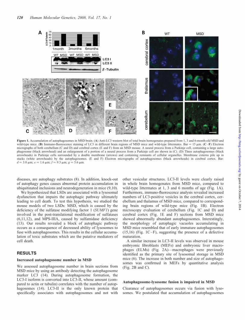
We assessed autophagosome number in brain sections fromMSD mice by using an antibody detecting the autophagosomemarker LC3 (14). During autophagosome formation, theLC3-I isoform is converted into LC3-II, whose amount (com-pared to actin or tubulin) correlates with the number of autop-hagosomes (14). LC3-II is the only known protein thatspecifically associates with autophagosomes and not with
other vesicular structures. LC3-II levels were clearly raisedin whole brain homogenates from MSD mice, compared towild-type littermates at 1, 3 and 6 months of age (Fig. 1A).Furthermore, immuno-fluorescence analysis revealed increasednumbers of LC3-positive vesicles in the cerebral cortex, cer-ebellum and thalamus of MSD mice, compared to correspond-ing brain regions of wild-type mice (Fig. 1B). Electronmicroscopy evaluation of cerebellum (Fig. 1C and D) andcerebral cortex (Fig. 1E and F) sections from MSD miceshowed abnormally abundant autophagosomes. Interestingly,the morphology of autophagic vacuoles accumulating inMSD mice resembled that of early immature autophagosomes(15,16) (Fig. 1C–F), suggesting the presence of a defectivematuration.
A similar increase in LC3-II levels was observed in mouseembryonic fibroblasts (MEFs) and embryonic liver macro-phages (ELMs) (Fig. 2A)—macrophages were previouslyidentified as the primary site of lysosomal storage in MSDmice (6). The increase in both number and size of autophago-somes was confirmed in MEFs by quantitative analysis(Fig. 2B and C).
Autophagosome-lysosome fusion is impaired in MSD
Clearance of autophagosomes occurs via fusion with lyso-somes. We postulated that accumulation of autophagosomes
Figure 1. Accumulation of autophagosomes in MSD brain. (A) Anti-LC3 western blot of total brain homogenates prepared from 1, 3 and 6-month-old MSD andwild-type mice. (B) Immuno-fluorescence staining of LC3 in different brain regions of MSD mice and wild-type littermates. Bar ¼ 15 mm. (C–F) Electronmicrographs of both cerebellum (C and D) and cerebral cortex (E and F) from an MSD mouse. A neural process from a Purkinje cell, containing a large auto-phagosome (black arrowhead) and an enlargement of a portion of a neural process from a Purkinje cell are shown in (C). (D) Three autophagosomes (blackarrowheads) in Purkinje cells surrounded by a double membrane (arrows) and containing remnants of cellular organelles. Membrane cisterns pile up instacks (white arrowheads) by the autophagosomes. (E and F) Electron micrographs of autophagosomes (black arrowheads) in cerebral cortex. Bar:d ¼ 3.0 mm; e ¼ 1.6 mm; f ¼ 9.3 mm; g ¼ 5.6 mm.
120 Human Molecular Genetics, 2008, Vol. 17, No. 1
by guest on October 7, 2014
http://hmg.oxfordjournals.org/
Dow
nloaded from

in MSD is due to defective clearance caused by impairedautophagosome-lysosome fusion. To test this hypothesis, weanalyzed the subcellular localization of the lysosomalmarker lgp120 (LAMP1) and the autophagosomal markerLC3 by confocal microscopy. These experiments demon-strated that the extent of lgp120/LC3 co-localization was sig-nificantly reduced (ranging from 40 to 50%) in MSDcompared to wild-type MEFs, thus indicating impairedautophagosome-lysosome fusion (Fig. 3A and B). This wasobserved both in normal medium (basal autophagy)(Fig. 3A) and in starved cells (induced autophagy) (Fig. 3B).
To characterize this impairment, we used drugs which eitherinduce or inhibit autophagy. Autophagy stimulation with rapa-mycin increased LC3-II levels in both MSD and wild-typeMEFs (Fig. 4). Moreover, LC3-II levels in MSD MEFs werefurther increased with bafilomycin A1, an inhibitorautophagosome-lysosome fusion (17), alone or in combination
with rapamycin, suggesting that the block of autophagy is notcomplete (Fig. 4).
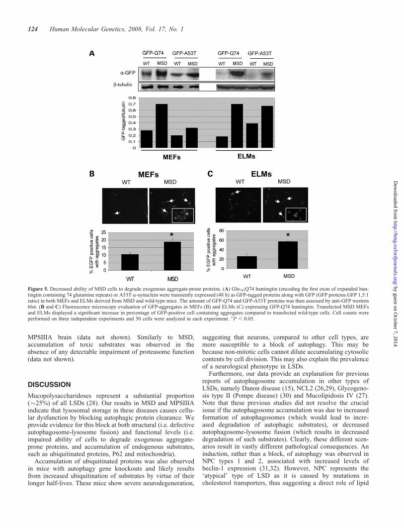
Decreased ability of MSD cells to degrade exogenousaggregate-prone proteins
Defective autophagosome-lysosome fusion may lead to animpairment of autophagy. We investigated the ability ofMSD cells to degrade aggregate-prone proteins which areautophagy substrates (18). These include the mutant huntingtinand A53T a-synuclein which are involved in Huntington andfamilial Parkinson diseases, respectively. Mutant huntingtinexon 1 constructs aggregate readily in tissue culture andform inclusions readily visible by light microscopy. The pro-portion of cells with such inclusion is linearly related to theexpression levels of the construct (19). The A53T a-synucleinconstruct does not form overt inclusions in the cell lines we
Figure 2. Accumulation of autophagosomes in MSD cultured cells. (A) Anti-LC3 western blot of protein extracts prepared from MEFs and ELMs derived fromMSD and wild-type mice in both normal and starved conditions. (B) MSD MEFs display several autophagosomes (arrowheads), compared to wild-type.Bar ¼ 9.3 mm. (C) Morphometric evaluation revealed that autophasomes were more numerous and larger in MSD MEFs compared to those observed in wild-type MEFs. �P , 0.05.
Human Molecular Genetics, 2008, Vol. 17, No. 1 121
by guest on October 7, 2014
http://hmg.oxfordjournals.org/
Dow
nloaded from

have studied (20). We expressed these mutant proteins in MSDcells to test the functionality of the autophagic pathway. TheGln74;Q74 huntingtin, which encodes the first exon of hunting-tin with 74 glutamine repeats, and the A53T a-synuclein werefused to green fluorescent protein (GFP) and transientlyexpressed in both MEFs and ELMs derived from MSDmice. Forty-eight hours after transfection, cells were collectedand GFP-fused proteins were detected by western blot.Figure 5A shows an increased accumulation of both types ofmutant proteins in MSD compared to wild-type cells. Inaddition, immuno-fluorescence analysis revealed that thenumber of GFP-Q74 aggregates was also significantly higherin MSD MEFs and ELMs compared to wild-type cells(Fig. 5B and C). Notably, when cells were analyzed atearlier time points, no significant differences in the accumu-lation of GFP-Q74 were observed between wild-type andMSD cells (Supplementary Material), this indicating thatonly at later time points accumulation of overexpressed pro-teins occurs. Moreover, GFP alone did not accumulate inMSD cells (Supplementary Material) demonstrating thatincreased levels of GFP-Q74 and GFP-A53T are due to
autophagic defective degradation and not to difference intransfection efficiency.
Taken together, these data indicate a dysfunction of autop-hagy with consequent decreased ability of MSD cells todegrade aggregate-prone proteins.
Polyubiquitinated proteins progressively accumulatein MSD neurons
Autophagy is responsible for constitutive protein turnover (8).This function appears to be particularly important in neuronalcells and is relevant to neurodegenerative diseases (21,22).Knockout of autophagy genes results in suppression ofautophagy and accumulation of inclusion bodies, whichcontain polyubiquitinated proteins, in neurons (9,10).We detected a massive and progressive accumulation ofubiquitin-positive inclusions in the cerebral cortex as wellas in other brain regions of MSD mice by both anti Ubimmuno-histochemical and immuno-fluorescence analyses(Fig. 6A and B and data not shown). Co-localization ofUbiquitin with NeuN neuronal marker indicates that
Figure 3. Defective autophagosome-lysosome fusion in MSD MEFs. (A and B) Co-localization of LAMP1 and LC3 in wild-type and MSD MEFs stained forLAMP1 (red) and LC3 (green). Confocal microscopy shows a reduction in the extent of co-localization of LAMP1 and LC3 proteins. The figure was selected toillustrate the basis for the assays we have quantified. The number of autophagosome-lysosome fusion events in MSD MEFs was quantified both in normal (A)and starved (B) serum conditions, as described in the Methods section.
122 Human Molecular Genetics, 2008, Vol. 17, No. 1
by guest on October 7, 2014
http://hmg.oxfordjournals.org/
Dow
nloaded from

ubiquitin inclusions are located in neurons (Fig. 6B). Pro-gressive accumulation of polyubiquitinated proteins wasalso detected by western blotting of brain homogenates(Fig. 6C). Importantly, analysis of chymotrypsin-like pro-teasome activity in MSD brain at several ages revealedthat proteasome function is not affected in MSD mice(data not shown), indicating that the accumulation of ubi-quitinated proteins is due to defective autophagy ratherthan to UPS impairment.
In addition, we found that P62/SQSTM1 significantlyaccumulates (Fig. 6D), and co-localizes with ubiquitin-positive inclusions (Fig. 6E) in brain from MSD mice. Thep62/SQSTM1 protein is known to be a common componentof ubiquitin-positive protein aggregates in neurodegenerativediseases (23), being involved in the targeting of polyubiquiti-nated proteins to the autophagosomes and selectively degradedvia the autophagic pathway (24).
Accumulation of dysfunctional mitochondria inMSD mice
Autophagy also plays a crucial role in the degradation and turn-over of cellular organelles like mitochondria. Indeed, it hasbeen suggested that autophagy selectively degrades dysfunc-tional mitochondria (25). Fragmented and dysfunctional mito-chondria have been reported to accumulate in patients withmucolipidosis types II, III and IV and in patients with neuronalceroid lipofuscinosis 2 (NCL2), suggesting that lysosomalstorage in these diseases impairs autophagy-mediated mito-
chondrial turnover (26,27). Electron microscopy analysisrevealed an increased number of mitochondria in MSD brainsections and MEFs (Fig. 7A and B). Consistently, an increaseof Cox4 (a mitochondrial marker) levels was detected bywestern blotting in MSD brain samples (Fig. 7C). To examinethe function of accumulating mitochondria, we measured themitochondrial membrane potential (DCm) in WT and MSDMEFs by using, a mitochondria-specific voltage dependentdye (DiOC6). As shown in Fig. 7D, MSD MEFs show a signifi-cant reduction in the DCm compared to wild-type cells in bothnormal and starved conditions, thus indicating that mitochon-dria accumulating in MSD are dysfunctional.
Impairment of autophagy in MPSIIIA
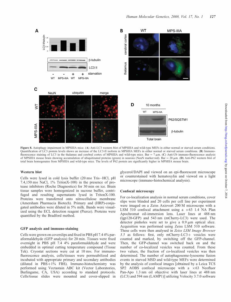
Overall, our data identify an impairment of autophagy inMSD, leading to the accumulation of polyubiquitinated pro-teins and of dysfunctional mitochondria. To investigatewhether this applies to other LSDs caused by defective hydro-lases, we analyzed the autophagic pathway in the murinemodel of MPSIIIA, which is also associated with severe neu-rodegeneration (4,5). The results obtained in MPSIIIA micewere similar to those of MSD mice. We detected increasedLC3II levels in MPSIIIA MEFs (Fig. 8A) as well as accumu-lation of autophagosomes in brain samples (Fig. 8B). Conse-quently, accumulation of ubiquitin-positive inclusions(Fig. 8C) and P62/SQSTM1 (Fig. 8D) were observed in thebrain of MPSIIIA mice. In addition, electron microscopyanalysis revealed an increased number of mitochondria in
Figure 4. Evaluation of LC3 levels in MSD and wild-type MEFs after induction and/or inhibition of autophagy. Both wild-type and MSD MEFs were treatedwith indicated drugs. Western-blot analysis (top) was performed using anti-LC3 antibodies. Quantitation (bottom) was performed by normalizing with actinlevels. The middle lane in the western blot is a lower exposure of the upper lane.
Human Molecular Genetics, 2008, Vol. 17, No. 1 123
by guest on October 7, 2014
http://hmg.oxfordjournals.org/
Dow
nloaded from
MPSIIIA brain (data not shown). Similarly to MSD,accumulation of toxic substrates was observed in theabsence of any detectable impairment of proteasome function(data not shown).
DISCUSSION
Mucopolysaccharidoses represent a substantial proportion(�25%) of all LSDs (28). Our results in MSD and MPSIIIAindicate that lysosomal storage in these diseases causes cellu-lar dysfunction by blocking autophagic protein clearance. Weprovide evidence for this block at both structural (i.e. defectiveautophagosome-lysosome fusion) and functional levels (i.e.impaired ability of cells to degrade exogenous aggregate-prone proteins, and accumulation of endogenous substrates,such as ubiquitinated proteins, P62 and mitochondria).
Accumulation of ubiquitinated proteins was also observedin mice with autophagy gene knockouts and likely resultsfrom increased ubiquitination of substrates by virtue of theirlonger half-lives. These mice show severe neurodegeneration,
suggesting that neurons, compared to other cell types, aremore susceptible to a block of autophagy. This may bebecause non-mitotic cells cannot dilute accumulating cytosoliccontents by cell division. This may also explain the prevalenceof a neurological phenotype in LSDs.
Furthermore, our data provide an explanation for previousreports of autophagosome accumulation in other types ofLSDs, namely Danon disease (15), NCL2 (26,29), Glycogeno-sis type II (Pompe disease) (30) and Mucolipidosis IV (27).Note that these previous studies did not resolve the crucialissue if the autophagosome accumulation was due to increasedformation of autophagosomes (which would lead to incre-ased degradation of autophagic substrates), or decreasedautophagosome-lysosome fusion (which results in decreaseddegradation of such substrates). Clearly, these different scen-arios result in vastly different pathological consequences. Aninduction, rather than a block, of autophagy was observed inNPC types 1 and 2, associated with increased levels ofbeclin-1 expression (31,32). However, NPC represents the‘atypical’ type of LSD as it is caused by mutations incholesterol transporters, thus suggesting a direct role of lipid
Figure 5. Decreased ability of MSD cells to degrade exogenous aggregate-prone proteins. (A) Gln74;Q74 huntingtin (encoding the first exon of expanded hun-tingtin containing 74 glutamine repeats) or A53T a-synuclein were transiently expressed (48 h) as GFP-tagged proteins along with GFP (GFP proteins:GFP 1,5:1ratio) in both MEFs and ELMs derived from MSD and wild-type mice. The amount of GFP-Q74 and GFP-A53T proteins was then assessed by anti-GFP westernblot. (B and C) Fluorescence microscopy evaluation of GFP-aggregates in MEFs (B) and ELMs (C) expressing GFP-Q74 huntingtin. Transfected MSD MEFsand ELMs displayed a significant increase in percentage of GFP-positive cell containing aggregates compared to transfected wild-type cells. Cell counts wereperformed on three independent experiments and 50 cells were analyzed in each experiment. �P , 0.05.
124 Human Molecular Genetics, 2008, Vol. 17, No. 1
by guest on October 7, 2014
http://hmg.oxfordjournals.org/
Dow
nloaded from

trafficking in the regulation of autophagy (32). Accordingly,we found no differences in beclin-1 expression betweentissues from MSD mice and wild-type littermates (data notshown).
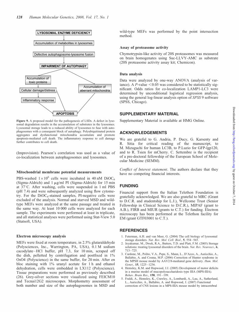
We propose a model which identifies a block of autophagyas a crucial component in the pathogenesis of LSDs (Fig. 9).According to this model, lysosomal accumulation of unde-graded substrates results in defective fusion between auto-phagosomes and lysosomes and causes a block of theautophagic pathway. As a consequence of this block, toxicproteins and dysfuctional mitochondria accumulate, ultimatelyleading to apoptosis, either directly or through the induction ofchronic inflammation and cytokine release (6). Indeed, cellswith impaired autophagy have an increased susceptibility tomitochondria-mediated apoptosis (33,34). It is interesting tonote that bafilomycin A1, a proton-pump inhibitor whichattenuates lysosomal acidification, results in similar blocksin autophagosome-lysosome fusion as LSDs, suggesting thatthere may be ways that defective lysosomal function feedback to inhibit autophagosome-lysosome fusion.
Importantly, we found that the ubiquitin-proteasome degra-dation is not impaired in our LSD mouse models. Proteasomedysfunction may lead to the accumulation of ubiquitinatedinclusions (35) and has been associated to neurodegenerativediseases (36–38). The finding that UPS is functional in ourLSD models allows us to conclude that the block of autophagypathway is the only mechanism accounting for the accu-mulation of ubiquitinated proteins which are the putativemediators of cell death in LSDs. Interestingly, a recent workfrom Pandey et al. (39) showed that autophagy and protea-
some are compensatory interacting systems, and pointedto the role of autophagy in rescuing protein degradationdeficiency due to the proteasome impairment. This findingraises the possibility to exploit new therapeutic approachesfor LSDs based on pharmacological induction of proteasomefunction in order to compensate for autophagy deficiency.
Our model defines LSDs as ‘autophagy disorders’, resem-bling more common neurodegenerative diseases such asAlzheimer (AD), Parkinson (PD) and Huntington (HD) dis-eases. While there are major differences in the initial stepsinvolved in all these diseases (i.e. impaired degradation ofpolyubiquitinated proteins in LSDs versus expression ofaggregate-prone proteins in AD, PD and HD), our datasuggest that they may share common mechanisms suggestingthe possibility of overlapping therapeutic strategies.
METHODS
Generation of MEFs and ELMs
MEFs were isolated by trypsinization of littermate embryosisolated at E14 and grown in DMEM supplemented with20% FBS and penicillin/streptomycin. Fetal liver cells wereisolated from E14.5 embryos by mechanical homogenizationand filtering through a 40 mm cell-strained. The cells wereresuspended in DMEM plus 10% fetal bovine serum andallowed to attach to plastic. Adherent macrophages (obtainedby washing wells in DMEM to remove non-adherent cells)were cultured in macrophage medium (PAA) and repurifiedby immune-separation using CD11b-coating magnetic beads
Figure 6. Accumulation of ubiquitin-positive inclusions and of P62 in MSD brain. (A) Immuno-histochemical staining of ubiquitinated proteins in the cerebralcortex of a 1-month-old wild-type mouse and of 1 and 3 month-old MSD mice. Bar ¼ 30 mm. (B) Anti-Ub (green) and anti-NeuN (red) immuno-fluorecence ofcerebral cortex sections derived from MSD and wild-type mice showing the presence of ubiquitin-positive inclusions in MSD neurons. Bar ¼ 20 mm.(C) Anti-Ub western blot from total brain homogenates derived from 1 and 3 month-old MSD mice and wild-type littermates. (D) Western blot analysis oftotal brain homogenates showing progressive accumulation of P62 in MSD. (E) Immuno-fluorescence with anti-P62 (red) and anti-Ub (green) showed significantco-localization of P62 with polyubiquitinated proteins in the brain of MSD mice. Bar ¼ 12 mm.
Human Molecular Genetics, 2008, Vol. 17, No. 1 125
by guest on October 7, 2014
http://hmg.oxfordjournals.org/
Dow
nloaded from

(MACs technology, Miltenyi Biotec). Macrophages werecharacterized by the expression of MOMA-2, and F4/80 anti-gens by immuno-fluorescence.
Transfections and drug treatments
Sub-confluent cells (MEFs or ELMs) were transfected usinglipofectamineTM 2000 (Invitrogen) according manufacturer’sprotocols. For co-localization experiments in normal serum con-ditions, sub-confluent MEFs were co-transfected with 0.5 mglgp120-GFP and 1 mg mCherry-LC3mCherry-LC3 and culturedin full medium for 24 h. For drug treatments, cells were treatedfor 14 h with 0.2 mg/ml rapamycin (Sigma), 200 nM bafilomycinA1 (Upstate).
Cloning of mCherry-hLC3B construct
Human LC3B was subcloned from pGEX-6P-1 into pcDNA3(Invitrogen) using BamHI and EcoRI (both NEB). mCherrypRSET-B was amplified by PCR with the following primers:5’-TA CCG AGC TCG GTA CCC GCC ACC AT-3’ and
3’-G CTG TAC AAG CAA GGA TCC TGC-5’. The resultingfragments were purified, digested with KpnI and EcoRI (bothNEB) and sub-cloned in frame into the 5’ end of hLC3BpcDNA3.
Antibodies
Primary antibodies were: rabbit polyclonal anti-LC3 (NovusBiological), rat monoclonal anti-mouse LAMP1 (Develop-mental Studies Hybridoma Bank, Iowa), rabbit polyclonalanti-ubiquitin (DakoCytomation), mouse monoclonal anti-NeuN (Chemicon), mouse monoclonal P62/SQSTM1 (BD),rabbit polyclonal anti-tubulin (Cell signaling), rabbit polyclo-nal anti-actin (Sigma) and mouse monoclonal anti-COX4(Clontech). Secondary antibodies were: goat anti-rabbit oranti-rat conjugated to Alexa Fluor 488 or 594 (MolecularProbes, Eugene, OR, USA). HRP-conjugated anti-mouse oranti-rabbit IgG (Amersham); biotinylated donkey anti-rabbit(Jackson ImmunoReasearch).
Figure 7. Accumulation of dysfunctional mitochondria in MEFs and brain of MSD mice. (A and B) Electron microscopy analysis of the brain cortex neuronsfrom MSD mice and wild-type littermates (bar: wild-type ¼ 2.1 mm; MSD ¼ 1.8 mm). MSD neurons contain a significantly higher number of mitochondria (m)compared to wild type neurons as also evident from quantitative analysis (B) (�P , 0.05). (C) Western blot analysis using antibodies recognizing Cox4, a mito-chondrial marker, shows increased levels of Cox4 in MSD compared to wild-type MEFs. (D) Wild-type and MSD MEFs were grown in either normal serum orstarved conditions (4 h). Cells were then stained with 40 nM DiOC6 and 1 mg/ml propidium iodine. DYm was measured by flow cytometry. Propidium iodine wasused as counterstain. All experiments were performed in triplicate and analyzed using Stat-View software and ANOVA test. Results were considered significantif P , 0.05.
126 Human Molecular Genetics, 2008, Vol. 17, No. 1
by guest on October 7, 2014
http://hmg.oxfordjournals.org/
Dow
nloaded from
Western blot
Cells were lysed in cold lysis buffer (20 mM Tris–HCl, pH7.4,150 mM NaCl, 1% TritonX-100) in the presence of pro-tease inhibitors (Roche Diagnostics) for 30 min on ice. Braintissue samples were homogenized in sucrose buffer, centri-fuged and resulting supernatants lysed in TritonX-100.Proteins were transferred onto nitrocellulose membrane(Amersham Pharmacia Biotech). Primary and (HRP)-conju-gated antibodies were diluted in 5% milk. Bands were visual-ized using the ECL detection reagent (Pierce). Proteins werequantified by the Bradford method.
GFP analysis and immuno-staining
Cells were grown on coverslips and fixed in PBS pH 7.4 4% par-aformaldehyde (GFP analysis) for 20 min. Tissues were fixedovernight in PBS pH 7.4 4% paraformaldehyde and wereembedded in optimal cutting temperature compound (TissueTek). Cryostat sections were cut at 10 mM. For immuno-fluorescence analysis, cells/tissues were permeabilized andincubated with appropriate primary and secondary antibodies(diluted in PBSþ1% FBS). Immuno-histochemistry wasperformed using Vectastain ABC kit (Vector Laboratories,Burlingame, CA, USA) according to standard protocols.Cells/tissue slides were mounted and cover-slipped in
glycerol/DAPI and viewed on an epi-fluorescent microscopeor counterstained with hematoxylin and viewed on a lightmicroscope (immuno-histochemical analysis).
Confocal microscopy
For co-localization analysis in normal serum conditions, coverslips were blinded and 20 cells per cell line per experimentwere imaged on a Zeiss Axiovert 200 M microscope with aLSM 510 confocal attachment using a �63 1.4 NA PlanApochromat oil-immersion lens. Laser lines at 488 nm(lgp120-GFP) and 543 nm (mCherry-LC3) were used. Thedetector pinholes were set to give a 0.9 mm optical slice.Acquisition was performed using Zeiss LSM 510 software.These cells were then analysed in Zeiss LSM Image Browser3.5 as follows: first, only mCherry-LC3þ vesicles werecounted and marked, by switching off the GFP-channel.Then, the GFP-channel was switched back on and thenumber of co-localized vesicles was counted. From thesetwo values, the fraction of co-localized vesicles was thendetermined. The number of autophagosome-lysosome fusionevents in starved MSD and wild-type MEFs were determinedby the analysis of confocal images [obtained by a Leica TCSSP2 AOBS confocal microscope with a �63 NeofluorPan-Apo 1.3 nm oil objective with laser lines at 488 nm(LC3) and 594 nm (LAMP1)] utilizing Volocity 3.7.0 software
Figure 8. Autophagy impairment in MPSIIIA mice. (A) Anti-LC3 western blot of MPSIIIA and wild-type MEFs in either normal or starved serum conditions.Quantification of LC3 protein levels shows an increase of the LC3-II isoform in MPSIIIA MEFs in either normal or starved serum conditions. (B) Immuno-fluorescence staining of LC3 in the thalamus and cerebral cortex of MPSIIIA and wild-type mice. Bar ¼ 7 mm. (C) Anti-Ub immuno-fluorescence analysisof MPSIIIA mouse brain showing accumulation of ubiquitinated proteins (green) in neurons (NeuN marker:red). Bar ¼ 20 mm. (D) Anti-P62 western blot oftotal brain homogenates from MPSIIIA and wild-type mice. The levels of P62 protein are significantly higher in MPSIIIA mouse brain.
Human Molecular Genetics, 2008, Vol. 17, No. 1 127
by guest on October 7, 2014
http://hmg.oxfordjournals.org/
Dow
nloaded from
(Improvision). Pearson’s correlation was used as a value ofco-localization between autophagosomes and lysosomes.
Mitochondrial membrane potential measurements
PBS-washed 1�106 cells were incubated in 40 nM DiOC6
(Sigma-Aldrich) and 1 mg/ml PI (Sigma-Aldrich) for 15 minat 378C. After washing, cells were suspended in 1 ml PBS(pH 7.4) and were subsequently analyzed using flow cytome-try. For the DiOC6-stained samples, PI-negative cells wereexcluded of the analysis. Normal and starved MSD and wild-type MEFs were analyzed at the same passage and treated inthe same way. At least 10 000 cells were analyzed for eachsample. The experiments were performed at least in triplicate,and all statistical analyses were performed using Stat-View 5.0(Statsoft, USA).
Electron microscopy analysis
MEFs were fixed at room temperature, in 2.5% glutaraldehyde(Polysciences, Inc., Warrington, PA, USA), 0.1 M sodiumcacodylate–HCl buffer, pH 7.3, for 10 min, scraped offthe dish, pelletted by centrifugation and postfixed in 1%OsO4 (Polyscience) in the same buffer, for 20 min. After enbloc staining with 1% uranyl acetate for 1 h and ethanoldehydration, cells were embedded in LX112 (Polyscience).Tissue preparations were performed as previously described(26). Grey-silver sections were visualized using FEICM10and Tecnai12G2 microscopes. Morphometry assessment ofboth number and size of the autophagosomes in MSD and
wild-type MEFs was performed by the point intersectionmethod.
Assay of proteasome activity
Chymotrypsin-like activity of 20S proteasomes was measuredon brain homogenates using Suc-LLVY-AMC as substrate(20S proteasome activity assay kit; Chemicon).
Data analysis
Data were analyzed by one-way ANOVA (analysis of var-iance). A P-value ,0.05 was considered to be statistically sig-nificant. Odds ratios for co-localization LAMP1-LC3 weredetermined by unconditional logistical regression analysis,using the general log-linear analysis option of SPSS 9 software(SPSS, Chicago).
SUPPLEMENTARY MATERIAL
Supplementary Material is available at HMG Online.
ACKNOWLEDGEMENTS
We are grateful to G. Andria, P. Ducy, G. Karsenty andR. Sitia for critical reading of the manuscript, toM. Mizuguchi for human LC3B, to P.Luzio for GFP-lgp120,and to R. Tsien for mCherry. C. Settembre is the recipientof a pre-doctoral fellowship of the European School of Mole-cular Medicine (SEMM).
Conflict of Interest statement. The authors declare that theyhave no competing financial interests.
FUNDING
Financial support from the Italian Telethon Foundation isgratefully acknowledged. We are also grateful to MRC (Grantto D.C.R. and studentship for L.J.), Wellcome Trust (SeniorFellowship in Clinical Science to D.C.R.), MIPAF (grant toA.B.), FIRB and MIUR (grants to C.T.) for funding. Electronmicroscopy has been performed at the Telethon facility forEM (grant GTF03001 to C.T.).
REFERENCES
1. Futerman, A.H. and van Meer, G. (2004) The cell biology of lysosomalstorage disorders. Nat. Rev. Mol. Cell. Biol., 5, 554–565.
2. Jeyakumar, M., Dwek, R.A., Butters, T.D. and Platt, F.M. (2005) Storagesolutions: treating lysosomal disorders of the brain. Nat. Rev. Neurosci., 6,713–725.
3. Cardone, M., Polito, V.A., Pepe, S., Mann, L., D’Azzo, A., Auricchio, A.,Ballabio, A. and Cosma, M.P. (2006) Correction of Hunter syndrome inthe MPSII mouse model by AAV2/8-mediated gene delivery. Hum. Mol.Genet., 15, 1225–1236.
4. Hemsley, K.M. and Hopwood, J.J. (2005) Development of motor deficitsin a murine model of mucopolysaccharidosis type IIIA (MPS-IIIA).Behav. Brain Res., 158, 191–199.
5. Fraldi, A., Hemsley, K., Crawley, A., Lombardi, A., Lau, A., Sutherland,L., Auricchio, A., Ballabio, A. and Hopwood, J. (2007) Functionalcorrection of CNS lesions in a MPS-IIIA mouse model by intracerebral
Figure 9. A proposed model for the pathogenesis of LSDs. A defect in lyso-somal degradation results in the accumulation of substrates in the lysosomes.Lysosomal storage leads to a reduced ability of lysosomes to fuse with auto-phagosomes with a consequent block of autophagy. Polyubiquitinated proteinaggregates and dysfunctional mitochondria accumulate and promoteapoptosis-mediated cell death. The inflammatory response to cell damagefurther contributes to cell death.
128 Human Molecular Genetics, 2008, Vol. 17, No. 1
by guest on October 7, 2014
http://hmg.oxfordjournals.org/
Dow
nloaded from
AAV-mediated delivery of sulfamidase and SUMF1 genes. Hum. Mol.
Genet., 2007; doi: 10.1093/hmg/ddm223.6. Settembre, C., Annunziata, I., Spampanato, C., Zarcone, D., Cobellis, G.,
Nusco, E., Zito, E., Tacchetti, C., Cosma, M.P. and Ballabio, A. (2007)Systemic inflammation and neurodegeneration in a mouse model ofmultiple sulfatase deficiency. Proc. Natl Acad. Sci. USA, 104, 4506–4511.
7. Mizushima, N., Ohsumi, Y. and Yoshimori, T. (2002) Autophagosomeformation in mammalian cells. Cell. Struct. Funct., 27, 421–429.
8. Rubinsztein, D.C. (2006) The roles of intracellular protein-degradationpathways in neurodegeneration. Nature, 443, 780–786.
9. Komatsu, M., Waguri, S., Chiba, T., Murata, S., Iwata, J., Tanida, I.,Ueno, T., Koike, M., Uchiyama, Y., Kominami, E. et al. (2006) Loss ofautophagy in the central nervous system causes neurodegeneration inmice. Nature, 441, 880–884.
10. Hara, T., Nakamura, K., Matsui, M., Yamamoto, A., Nakahara, Y.,Suzuki-Migishima, R., Yokoyama, M., Mishima, K., Saito, I., Okano, H.et al. (2006) Suppression of basal autophagy in neural cells causesneurodegenerative disease in mice. Nature, 441, 885–889.
11. Cosma, M.P., Pepe, S., Annunziata, I., Newbold, R.F., Grompe, M.,Parenti, G. and Ballabio, A. (2003) The multiple sulfatase deficiency geneencodes an essential and limiting factor for the activity of sulfatases. Cell,113, 445–456.
12. Dierks, T., Schmidt, B., Borissenko, L.V., Peng, J., Preusser, A.,Mariappan, M. and von Figura, K. (2003) Multiple sulfatase deficiency iscaused by mutations in the gene encoding the humanC(alpha)-formylglycine generating enzyme. Cell, 113, 435–444.
13. Bhattacharyya, R., Gliddon, B., Beccari, T., Hopwood, J.J. and Stanley, P.(2001) A novel missense mutation in lysosomal sulfamidase is the basis ofMPS III A in a spontaneous mouse mutant. Glycobiology, 11, 99–103.
14. Kabeya, Y., Mizushima, N., Ueno, T., Yamamoto, A., Kirisako, T., Noda, T.,Kominami, E., Ohsumi, Y. and Yoshimori, T. (2000) LC3, a mammalianhomologue of yeast Apg8p, is localized in autophagosome membranes afterprocessing. EMBO J., 19, 5720–5728.
15. Tanaka, Y., Guhde, G., Suter, A., Eskelinen, E.L., Hartmann, D.,Lullmann-Rauch, R., Janssen, P.M., Blanz, J., von Figura, K. and Saftig,P. (2000) Accumulation of autophagic vacuoles and cardiomyopathy inLAMP-2-deficient mice. Nature, 406, 902–906.
16. Boland, B. and Nixon, R.A. (2006) Neuronal macroautophagy: fromdevelopment to degeneration. Mol. Aspects Med., 27, 503–519.
17. Yamamoto, A., Tagawa, Y., Yoshimori, T., Moriyama, Y., Masaki, R. andTashiro, Y. (1998) Bafilomycin A1 prevents maturation of autophagicvacuoles by inhibiting fusion between autophagosomes and lysosomes inrat hepatoma cell line, H-4-II-E cells. Cell. Struct. Funct., 23, 33–42.
18. Ravikumar, B., Duden, R. and Rubinsztein, D.C. (2002) Aggregate-proneproteins with polyglutamine and polyalanine expansions are degraded byautophagy. Hum. Mol. Genet., 11, 1107–1117.
19. Narain, Y., Wyttenbach, A., Rankin, J., Furlong, R.A. and Rubinsztein,D.C. (1999) A molecular investigation of true dominance in Huntington’sdisease. J. Med. Genet., 36, 739–746.
20. Webb, J.L., Ravikumar, B., Atkins, J., Skepper, J.N. and Rubinsztein,D.C. (2003) Alpha-synuclein is degraded by both autophagy and theproteasome. J. Biol. Chem., 278, 25009–25013.
21. Nixon, R.A. (2006) Autophagy in neurodegenerative disease: friend, foeor turncoat? Trends Neurosci., 29, 528–535.
22. Komatsu, M., Ueno, T., Waguri, S., Uchiyama, Y., Kominami, E. andTanaka, K. (2007) Constitutive autophagy: vital role in clearance ofunfavorable proteins in neurons. Cell Death Differ., 14, 887–894.
23. Zatloukal, K., Stumptner, C., Fuchsbichler, A., Heid, H., Schnoelzer, M.,Kenner, L., Kleinert, R., Prinz, M., Aguzzi, A. and Denk, H. (2002) p62 Is
a common component of cytoplasmic inclusions in protein aggregationdiseases. Am. J. Pathol., 160, 255–263.
24. Bjorkoy, G., Lamark, T., Brech, A., Outzen, H., Perander, M., Overvatn,A., Stenmark, H. and Johansen, T. (2005) p62/SQSTM1 forms proteinaggregates degraded by autophagy and has a protective effect onhuntingtin-induced cell death. J. Cell Biol., 171, 603–614.
25. Kim, I., Rodriguez-Enriquez, S. and Lemasters, J.J. (2007) Selectivedegradation of mitochondria by mitophagy. Arch. Biochem. Biophys., 462,245–253.
26. Koike, M., Shibata, M., Waguri, S., Yoshimura, K., Tanida, I., Kominami,E., Gotow, T., Peters, C., von Figura, K., Mizushima, N. et al. (2005)Participation of autophagy in storage of lysosomes in neurons from mousemodels of neuronal ceroid-lipofuscinoses (Batten disease). Am. J. Pathol.,167, 1713–1728.
27. Jennings, J.J., Jr, Zhu, J.H., Rbaibi, Y., Luo, X., Chu, C.T. and Kiselyov,K. (2006) Mitochondrial aberrations in mucolipidosis Type IV. J. Biol.Chem., 281, 39041–39050.
28. Neufeld, E.F. and Muenzer, J. (2001) The mucopolysaccharidoses. TheMetabolic and Molecular Basis of Inherited Disease. Mc Graw-Hill,New York, pp. 3421–3452.
29. Cao, Y., Espinola, J.A., Fossale, E., Massey, A.C., Cuervo, A.M.,MacDonald, M.E. and Cotman, S.L. (2006) Autophagy is disrupted in aknock-in mouse model of juvenile neuronal ceroid lipofuscinosis. J. Biol.Chem., 281, 20483–20493.
30. Fukuda, T., Ewan, L., Bauer, M., Mattaliano, R.J., Zaal, K., Ralston, E.,Plotz, P.H. and Raben, N. (2006) Dysfunction of endocytic andautophagic pathways in a lysosomal storage disease. Ann. Neurol., 59,700–708.
31. Ko, D.C., Milenkovic, L., Beier, S.M., Manuel, H., Buchanan, J. andScott, M.P. (2005) Cell-autonomous death of cerebellar purkinje neuronswith autophagy in Niemann-Pick type C disease. PLoS Genet., 1, 81–95.
32. Pacheco, C.D., Kunkle, R. and Lieberman, A.P. (2007) Autophagy inNiemann-Pick C disease is dependent upon Beclin-1 and responsive tolipid trafficking defects. Hum. Mol. Genet, 16, 1495–1503.
33. Boya, P., Gonzalez-Polo, R.A., Casares, N., Perfettini, J.L., Dessen, P.,Larochette, N., Metivier, D., Meley, D., Souquere, S., Yoshimori, T. et al.(2005) Inhibition of macroautophagy triggers apoptosis. Mol. Cell. Biol.,25, 1025–1040.
34. Ravikumar, B., Berger, Z., Vacher, C., O’Kane, C.J. and Rubinsztein,D.C. (2006) Rapamycin pre-treatment protects against apoptosis. Hum.Mol. Genet., 15, 1209–1216.
35. Rideout, H.J., Larsen, K.E., Sulzer, D. and Stefanis, L. (2001)Proteasomal inhibition leads to formation of ubiquitin/alpha-synuclein-immunoreactive inclusions in PC12 cells. J. Neurochem.,78, 899–908.
36. Shimura, H., Hattori, N., Kubo, S., Mizuno, Y., Asakawa, S., Minoshima,S., Shimizu, N., Iwai, K., Chiba, T., Tanaka, K. et al. (2000) FamilialParkinson disease gene product, parkin, is a ubiquitin-protein ligase. Nat.Genet., 25, 302–305.
37. Bence, N.F., Sampat, R.M. and Kopito, R.R. (2001) Impairment of theubiquitin-proteasome system by protein aggregation. Science, 292, 1552–1555.
38. Diaz-Hernandez, M., Hernandez, F., Martin-Aparicio, E., Gomez-Ramos,P., Moran, M.A., Castano, J.G., Ferrer, I., Avila, J. and Lucas, J.J. (2003)Neuronal induction of the immunoproteasome in Huntington’s disease.J. Neurosci., 23, 11653–11661.
39. Pandey, U.B., Nie, Z., Batlevi, Y., McCray, B.A., Ritson, G.P., Nedelsky,N.B., Schwartz, S.L., DiProspero, N.A., Knight, M.A., Schuldiner, O.et al. (2007) HDAC6 rescues neurodegeneration and provides an essentiallink between autophagy and the UPS. Nature, 447, 859–863.
Human Molecular Genetics, 2008, Vol. 17, No. 1 129
by guest on October 7, 2014
http://hmg.oxfordjournals.org/
Dow
nloaded from
Copyright © 2022 FDOKUMEN