University of Groningen Lysosome Biogenesis and Autophagy ...
548
University of Groningen Lysosome Biogenesis and Autophagy Reggiori, Fulvio; Klumperman, J Published in: LYSOSOMES DOI: 10.1002/9781118978320.ch2 IMPORTANT NOTE: You are advised to consult the publisher's version (publisher's PDF) if you wish to cite from it. Please check the document version below. Document Version Publisher's PDF, also known as Version of record Publication date: 2016 Link to publication in University of Groningen/UMCG research database Citation for published version (APA): Reggiori, F., & Klumperman, J. (2016). Lysosome Biogenesis and Autophagy. In F. R. Maxfield, J. M. Willard, & S. Lu (Eds.), LYSOSOMES: Biology, Diseases, and Therapeutics (pp. 7-31). Elsevier. https://doi.org/10.1002/9781118978320.ch2 Copyright Other than for strictly personal use, it is not permitted to download or to forward/distribute the text or part of it without the consent of the author(s) and/or copyright holder(s), unless the work is under an open content license (like Creative Commons). The publication may also be distributed here under the terms of Article 25fa of the Dutch Copyright Act, indicated by the “Taverne” license. More information can be found on the University of Groningen website: https://www.rug.nl/library/open-access/self-archiving-pure/taverne- amendment. Take-down policy If you believe that this document breaches copyright please contact us providing details, and we will remove access to the work immediately and investigate your claim. Downloaded from the University of Groningen/UMCG research database (Pure): http://www.rug.nl/research/portal. For technical reasons the number of authors shown on this cover page is limited to 10 maximum. Download date: 30-03-2022
-
Upload
khangminh22 -
Category
Documents
-
view
0 -
download
0
Transcript of University of Groningen Lysosome Biogenesis and Autophagy ...
University of Groningen
Lysosome Biogenesis and AutophagyReggiori, Fulvio; Klumperman, J
Published in:LYSOSOMES
DOI:10.1002/9781118978320.ch2
IMPORTANT NOTE: You are advised to consult the publisher's version (publisher's PDF) if you wish to cite fromit. Please check the document version below.
Document VersionPublisher's PDF, also known as Version of record
Publication date:2016
Link to publication in University of Groningen/UMCG research database
Citation for published version (APA):Reggiori, F., & Klumperman, J. (2016). Lysosome Biogenesis and Autophagy. In F. R. Maxfield, J. M.Willard, & S. Lu (Eds.), LYSOSOMES: Biology, Diseases, and Therapeutics (pp. 7-31). Elsevier.https://doi.org/10.1002/9781118978320.ch2
CopyrightOther than for strictly personal use, it is not permitted to download or to forward/distribute the text or part of it without the consent of theauthor(s) and/or copyright holder(s), unless the work is under an open content license (like Creative Commons).
The publication may also be distributed here under the terms of Article 25fa of the Dutch Copyright Act, indicated by the “Taverne” license.More information can be found on the University of Groningen website: https://www.rug.nl/library/open-access/self-archiving-pure/taverne-amendment.
Take-down policyIf you believe that this document breaches copyright please contact us providing details, and we will remove access to the work immediatelyand investigate your claim.
Downloaded from the University of Groningen/UMCG research database (Pure): http://www.rug.nl/research/portal. For technical reasons thenumber of authors shown on this cover page is limited to 10 maximum.
Download date: 30-03-2022

�
� �
�
LYSOSOMES
Biology, Diseases, and Therapeutics
Edited by
FREDERICK R. MAXFIELDJAMES M. WILLARDSHUYAN LU

�
� �
�
Copyright © 2016 by John Wiley & Sons, Inc. All rights reserved
Published by John Wiley & Sons, Inc., Hoboken, New JerseyPublished simultaneously in Canada
No part of this publication may be reproduced, stored in a retrieval system, or transmitted in any form orby any means, electronic, mechanical, photocopying, recording, scanning, or otherwise, except aspermitted under Section 107 or 108 of the 1976 United States Copyright Act, without either the priorwritten permission of the Publisher, or authorization through payment of the appropriate per-copy fee tothe Copyright Clearance Center, Inc., 222 Rosewood Drive, Danvers, MA 01923, (978) 750-8400, fax(978) 750-4470, or on the web at www.copyright.com. Requests to the Publisher for permission shouldbe addressed to the Permissions Department, John Wiley & Sons, Inc., 111 River Street, Hoboken, NJ07030, (201) 748-6011, fax (201) 748-6008, or online at http://www.wiley.com/go/permission.
Limit of Liability/Disclaimer of Warranty: While the publisher and author have used their best efforts inpreparing this book, they make no representations or warranties with respect to the accuracy orcompleteness of the contents of this book and specifically disclaim any implied warranties ofmerchantability or fitness for a particular purpose. No warranty may be created or extended by salesrepresentatives or written sales materials. The advice and strategies contained herein may not be suitablefor your situation. You should consult with a professional where appropriate. Neither the publisher norauthor shall be liable for any loss of profit or any other commercial damages, including but not limited tospecial, incidental, consequential, or other damages.
For general information on our other products and services or for technical support, please contact ourCustomer Care Department within the United States at (800) 762-2974, outside the United States at(317) 572-3993 or fax (317) 572-4002.
Wiley also publishes its books in a variety of electronic formats. Some content that appears in print maynot be available in electronic formats. For more information about Wiley products, visit our web site atwww.wiley.com.
Library of Congress Cataloging-in-Publication Data:
Names: Maxfield, Frederick R., editor. | Willard, James M., 1955- , editor. |Lu, Shuyan, 1971- , editor.
Title: Lysosomes : biology, diseases, and therapeutics / edited by FrederickR. Maxfield, James M. Willard, Shuyan Lu.
Other titles: Lysosomes (Maxfield)Description: Hoboken, New Jersey : John Wiley & Sons, Inc., [2016] | Includes
bibliographical references and index.Identifiers: LCCN 2016006834 (print) | LCCN 2016008790 (ebook) | ISBN
9781118645154 (cloth) | ISBN 9781118978306 (pdf) | ISBN 9781118978313(epub)
Subjects: | MESH: LysosomesClassification: LCC QH603.L9 (print) | LCC QH603.L9 (ebook) | NLM QU 350 |
DDC 571.6/55–dc23LC record available at https://lccn.loc.gov/2016006834
Printed in the United States of America10 9 8 7 6 5 4 3 2 1

�
� �
�
CONTENTS
PREFACE xiiiLIST OF CONTRIBUTORS xvii
1 Lysosomes: An Introduction 1Frederick R. Maxfield
1.1 Historical Background, 2References, 4
2 Lysosome Biogenesis and Autophagy 7Fulvio Reggiori and Judith Klumperman
2.1 Introduction, 72.2 Pathways to the Lysosomes, 10
2.2.1 Biosynthetic Transport Routes to the Lysosome, 102.2.2 Endocytic Pathways to the Lysosome, 102.2.3 Autophagy Pathways to the Lysosome, 122.2.4 The ATG Proteins: The Key Regulators of Autophagy, 14
2.3 Fusion and Fission between the Endolysosomal and AutophagyPathways, 162.3.1 Recycling Endosomes and Autophagosome Biogenesis, 162.3.2 Autophagosome Fusion with Late Endosomes and
Lysosomes, 172.3.3 Autophagic Lysosomal Reformation, 18
iii

�
� �
�
iv CONTENTS
2.4 Diseases, 192.4.1 Lysosome-Related Disorders (LSDs), 192.4.2 Lysosomes in Neurodegeneration and Its Links
to Autophagy, 202.4.3 Autophagy-Related Diseases, 20
2.5 Concluding Remarks, 22Acknowledgments, 23References, 23
3 Multivesicular Bodies: Roles in Intracellular and IntercellularSignaling 33Emily R. Eden, Thomas Burgoyne, and Clare E. Futter
3.1 Introduction, 333.2 Downregulation of Signaling by Sorting onto ILVs, 353.3 Upregulation of Signaling by Sorting onto ILVs, 383.4 Intercellular Signaling Dependent on Sorting onto ILVs, 393.5 Conclusion, 44
References, 45
4 Lysosomes and Mitophagy 51Dominik Haddad and Patrik Verstreken
4.1 Summary, 514.2 Mitochondrial Significance, 514.3 History of Mitophagy, 524.4 Mechanisms of Mitophagy, 53
4.4.1 Mitophagy in Yeast, 544.4.2 Mitophagy in Mammals, 55
4.5 Conclusion, 57Acknowledgments, 57References, 58
5 Lysosome Exocytosis and Membrane Repair 63Rajesh K. Singh and Abigail S. Haka
5.1 Introduction, 635.2 Functions of Lysosome Exocytosis, 63
5.2.1 Specialized Lysosome-Related Organelles, 645.2.2 Lysosome Exocytosis for Membrane Repair, 655.2.3 Lysosome Exocytosis as a Source of Membrane, 665.2.4 Lysosome Exocytosis for Extracellular Degradation, 665.2.5 Lysosome Exocytosis and Delivery of Proteins to the Cell
Surface, 68

�
� �
�
CONTENTS v
5.3 Mechanisms of Lysosome Exocytosis, 685.3.1 Maturation of Lysosomes and Lysosome-Related
Organelles, 695.3.2 Transport of Lysosomes to the Plasma Membrane, 705.3.3 Tethering of Lysosomes to the Plasma Membrane, 725.3.4 Lysosome Fusion with the Plasma Membrane, 755.3.5 Calcium-Dependent Exocytosis, 76
5.4 Conclusion, 76Acknowledgments, 77References, 77
6 Role of Lysosomes in Lipid Metabolism 87Frederick R. Maxfield
6.1 Introduction, 876.2 Endocytic Uptake of Lipoproteins, 886.3 Lipid Metabolism in Late Endosomes and Lysosomes, 916.4 Autophagy and Lysosomal Lipid Turnover, 946.5 Lysosomal Lipid Hydrolysis and Metabolic Regulation, 956.6 Summary, 96
References, 96
7 TFEB, Master Regulator of Cellular Clearance 101Graciana Diez-Roux and Andrea Ballabio
7.1 Lysosome, 1017.2 The Transcriptional Regulation of Lysosomal Function, 1027.3 TFEB Subcellular Regulation is Regulated by Its Phosphorylation, 1047.4 A Lysosome-to-Nucleus Signaling Mechanism, 1057.5 TFEB and Cellular Clearance in Human Disease, 106
7.5.1 Lysosomal Storage Disorders, 1077.5.2 Neurodegenerative Disorders, 1097.5.3 Metabolic Syndrome, 1107.5.4 Cancer, Inborn Errors of Metabolism, Immunity,
and Longevity, 110References, 111
8 Lysosomal Membrane Permeabilization in Cell Death 115Urška Repnik and Boris Turk
8.1 Introduction, 1158.2 Cell Death Modalities, 1168.3 Lysosomal Membrane Permeabilization (LMP) and Cell Death, 117
8.3.1 Mechanisms of LMP, 118

�
� �
�
vi CONTENTS
8.3.2 Upstream of LMP: Direct Insult Versus MolecularSignaling, 121
8.3.3 Signaling Downstream of LMP, 1248.4 Conclusion, 127
Acknowledgments, 127References, 128
9 The Lysosome in Aging-Related Neurodegenerative Diseases 137Ralph A. Nixon
9.1 Introduction, 1379.2 Lysosome Function in Aging Organisms, 1399.3 Lysosomes and Diseases of Late Age Onset, 142
9.3.1 Cardiovascular Disease, 1429.4 Lysosomes in Aging-Related Neurodegenerative Diseases, 144
9.4.1 Alzheimer’s Disease (AD), 1459.4.2 Parkinson’s Disease and Related Disorders, 1509.4.3 Diffuse Lewy Body Disease (DLB), 1559.4.4 Frontotemporal Lobar Degeneration (FTLD), 155
9.5 Conclusion, 158Acknowledgments, 158References, 159
10 Lysosome and Cancer 181Marja Jäättelä and Tuula Kallunki
10.1 Introduction, 18110.2 Lysosomal Function and Its Importance for Cancer Development
and Progression, 18110.3 Cancer-Induced Changes in Lysosomal Function, 182
10.3.1 Increased Activity of Lysosomal Enzymes, 18210.3.2 Altered Lysosome Membrane Permeability, 18410.3.3 Increased Lysosome Size, 18410.3.4 Altered Lysosome Trafficking – Increased Lysosomal
Exocytosis, 18510.4 Cancer-Induced Changes in Lysosome Composition, 185
10.4.1 Changes in Lysosomal Hydrolases, 18510.4.2 Changes in the Lysosomal Membrane Proteins, 192
10.5 Molecular Changes Involving Lysosomal Integrity, 19310.5.1 Cancer-Associated Changes in Lysosomal Sphingolipid
Metabolism, 19310.5.2 Targeting Lysosomal Membrane Integrity, 195
10.6 Conclusion, 196References, 197

�
� �
�
CONTENTS vii
11 The Genetics of Sphingolipid Hydrolases and Sphingolipid StorageDiseases 209Edward H. Schuchman and Calogera M. Simonaro
11.1 Introduction and Overview, 20911.2 Acid Ceramidase Deficiency: Farber Disease, 21011.3 Acid Sphingomyelinase Deficiency: Types A and B Niemann–Pick
Disease, 21311.4 Beta-Glucocerebrosidase Deficiency: Gaucher Disease, 21511.5 Galactocerebrosidase Deficiency: Krabbe Disease/Globoid Cell
Leukodystrophy, 21811.6 Arylsulfatase a Deficiency: Metachromatic Leukodystrophy, 21911.7 Alpha-Galactosidase a Deficiency: Fabry Disease, 22111.8 Beta-Galactosidase Deficiency: GM1 Gangliosidosis, 22411.9 Hexosaminidase A and B Deficiency: GM2 Gangliosidoses, 22611.10 Sphingolipid Activator Proteins, 229
References, 231
12 Lysosome-Related Organelles: Modifications of the LysosomeParadigm 239Adriana R. Mantegazza and Michael S. Marks
12.1 Differences Between LROs and Secretory Granules, 24012.2 Physiological Functions of LROs, 24012.3 LRO Biogenesis, 244
12.3.1 Chediak–Higashi Syndrome and Gray PlateletSyndrome, 244
12.3.2 Hermansky–Pudlak Syndrome, 24612.3.3 Melanosome Biogenesis, 24712.3.4 HPS and Melanosome Maturation, 24812.3.5 HPS and the Biogenesis of Other LROs, 25012.3.6 HPS and Neurosecretory Granule Biogenesis, 25012.3.7 Weibel–Palade Body Biogenesis, 251
12.4 LRO Motility, Docking, and Secretion, 25212.5 LROs and Immunity to Pathogens, 253
12.5.1 Cytolytic Granules, 25312.5.2 Familial Hemophagocytic Lymphohistiocytosis and Cytolytic
Granule Secretion, 25412.5.3 Azurophilic Granules, 25512.5.4 NADPH Oxidase-Containing LROs, 25512.5.5 IRF7-Signaling LROs and Type I Interferon Induction, 25612.5.6 MIICs: LROs or Conventional Late
Endosome/Lysosomes?, 25612.5.7 Phagosomes and Autophagosomes as New Candidate
LROs, 258

�
� �
�
viii CONTENTS
12.6 Perspectives, 260Acknowledgments, 260References, 260
13 Autophagy Inhibition as a Strategy for Cancer Therapy 279Xiaohong Ma, Shengfu Piao, Quentin Mcafee, and Ravi K. Amaravadi
13.1 Stages and Steps of Autophagy, 28213.2 Induction of Autophagy, 28313.3 Studies in Mouse Models Unravel the Dual Roles of Autophagy
in Tumor Biology, 28513.4 Clinical Studies on Autophagy’s Dual Role in Tumorigenesis, 28613.5 Mouse Models Provide the Rationale for Autophagy Modulation in the
Context of Cancer Therapy, 28813.6 Multiple Druggable Targets in the Autophagy Pathway, 29113.7 Overview of Preclinical Autophagy Inhibitors and Evidence Supporting
Combination with Existing and New Anticancer Agents, 29213.8 Proximal Autophagy Inhibitors, 29313.9 Quinolines: From Antimalarials to Prototypical Distal Autophagy
Inhibitors, 29313.10 Summary for the Clinical Trials for CQ/HCQ, 29513.11 Developing More Potent Anticancer Autophagy Inhibitors, 29813.12 Summary, Conclusion, and Future Directions, 30013.13 In Summary, 302
References, 302
14 Autophagy Enhancers, are we there Yet? 315Shuyan Lu and Ralph A. Nixon
14.1 Introduction, 31514.2 Autophagy Impairment and Diseases, 31614.3 Autophagy Enhancer Screening, 317
14.3.1 Methods for Monitoring Autophagy, 31714.3.2 Autophagy Enhancers Identified from Early Literature, 32614.3.3 mTOR Inhibitors, 331
14.4 Other Agents that Boost Autophagy and Lysosomal Functions, 33514.4.1 HDAC Inhibition, 33614.4.2 pH Restoration, 33714.4.3 TRP Activator, 33714.4.4 TFEB Overexpression/Activation, 33814.4.5 Lysosomal Efficiency, 33814.4.6 MicroRNA, 339
14.5 Concluding Remarks, 340References, 341

�
� �
�
CONTENTS ix
15 Pharmacological Chaperones as Potential Therapeutics for LysosomalStorage Disorders: Preclinical Research to Clinical Studies 357Robert E. Boyd, Elfrida R. Benjamin, Su Xu, Richie Khanna, and Kenneth J.Valenzano
15.1 Introduction, 35715.2 Fabry Disease, 35915.3 Gaucher Disease, 36315.4 GM2 Gangliosidoses (Tay–Sachs/Sandhoff Diseases), 36715.5 Pompe Disease, 36815.6 PC-ERT Combination Therapy, 370
References, 372
16 Endosomal Escape Pathways for Delivery of Biologics 383Philip L. Leopold
16.1 Introduction, 38316.2 Endosome Characteristics, 38416.3 Delivery of Nature’s Biologics: Lessons on Endosomal Escape
from Pathogens, 38916.3.1 Viruses, 39016.3.2 Bacteria, Protozoa, and Fungi, 39216.3.3 Toxins, 394
16.4 Endosomal Escape Using Engineered Systems, 39516.4.1 Peptides and Polymers, 39616.4.2 Lipids, 39816.4.3 Other Chemical and Physical Strategies, 399
16.5 Conclusion, 399References, 400
17 Lysosomes and Antibody–Drug Conjugates 409Michelle Mack, Jennifer Kahler, Boris Shor, Michael Ritchie,Maureen Dougher, Matthew Sung, and Puja Sapra
17.1 Introduction, 40917.2 Receptor Internalization, 41017.3 Antibody–Drug Conjugates, 41317.4 Mechanisms of Resistance to ADCs, 41617.5 Summary, 417
References, 417
18 The Mechanisms and Therapeutic Consequences of Amine-ContainingDrug Sequestration in Lysosomes 423Nadia Hamid and Jeffrey P. Krise
18.1 Introduction, 423

�
� �
�
x CONTENTS
18.2 Lysosomal Trapping Overview, 42418.3 Techniques to Assess Lysosomal Trapping, 42718.4 Influence of Lysosomotropism on Drug Activity, 42918.5 Influence of Lysosomal Trapping on Pharmacokinetics, 43518.6 Pharmacokinetic Drug–Drug Interactions Involving Lysosomes, 438
References, 440
19 Lysosome Dysfunction: an Emerging Mechanism of Xenobiotic-InducedToxicity 445Shuyan Lu, Bart Jessen, Yvonne Will, and Greg Stevens
19.1 Introduction, 44519.2 Compounds that Impact Lysosomal Function, 446
19.2.1 Lysosomotropic Compounds, 44619.2.2 Nonlysosomotropic Compounds, 451
19.3 Cellular Consequences, 45219.3.1 Effect of Drugs on pH and Lysosomal Volume, 45219.3.2 Effects on Lysosomal Enzymes, 45319.3.3 Lysosomal Substrate Accumulation, 45419.3.4 Lysosomal Membrane Permeabilization (LMP) and Cell
Death, 45419.3.5 Membrane Trafficking Changes, 45519.3.6 Other Cellular Impacts, 458
19.4 Impaired Lysosomal Function as a Mechanism for Organ Toxicity, 46119.4.1 Liver Toxicity, 46219.4.2 Kidney Toxicity, 46419.4.3 Retinal, 46619.4.4 Peripheral Neuropathy, 46619.4.5 Muscle Toxicity, 46719.4.6 Tumorigenesis, 46819.4.7 General Considerations for Organ Toxicity, 469
19.5 Concluding Remarks, 471References, 472
20 Lysosomes and Phospholipidosis in Drug Developmentand Regulation 487James M. Willard and Albert De Felice
20.1 Introduction, 48720.2 FDA Involvement, 48820.3 Autophagy and DIPL, 48920.4 Early Experience with Lethal DIPL, 48920.5 Clinical and Nonclinical Expressions of DIPL, 490
20.5.1 Clinical, 49020.5.2 Nonclinical, 491

�
� �
�
CONTENTS xi
20.6 Physical Chemistry, 49120.7 Quantitative Structure–Activity Relationship (QSAR), 49220.8 Toxicogenomics, 49320.9 Fluorescence, Dye, and Immunohistochemical Methods
for Screening, 49420.10 FDA Database and QSAR Modeling, 49420.11 Linking Phospholipidosis and Overt Toxicity, 49420.12 Phospholipidosis and QT Interval Prolongation, 49620.13 DIPL Mechanisms, 50020.14 Treatment, 50120.15 Discussion, 50120.16 Future Directions and Recommendations, 505
References, 506
INDEX 513

�
� �
�
PREFACE
There has been a resurgence in interest in lysosomes based on exciting new discov-eries over the past decade. Lysosomal function was observed microscopically in thelate 19th century, and lysosomes were purified in the 1950s by the group of Chris-tian De Duve [1]. During the same period, accumulation of undigested material incells was observed in pathological examination of tissues from patients with a varietyof diseases [2–4]. With the biochemical and morphological characterization of lyso-somes, the linkage of the accumulated material with these organelles led to significantinsights into the functional importance of lysosomes.
In the second half of the 20th century, there were groundbreaking studies of thebiology and biochemistry of lysosomes [5–9]. These studies were linked closely withrapid developments in understanding fundamental cellular biological processes suchas secretion and endocytosis. As a result, an increasingly detailed picture emerged ofthe biogenesis of lysosomes and their functional role in digesting internalized cargo[10,11]. As understanding of lysosomal function increased, mechanism-based strate-gies for treating lysosomal diseases emerged. These included substrate reductiontherapies (e.g., for Gaucher disease) [12,13] and enzyme replacement therapies [14].
While there continued to be advances in basic cell biology and biochemistry,as well as in new therapeutic modalities, many investigators had a sense that theexciting era of discovery in lysosome biology was ending in the early 2000s. As anexample, the Gordon Conference on “Lysosomes,” which for many years was oneof the premier meetings on membrane traffic, changed its name to “Lysosomes andEndocytosis” in 2004.
Several related areas of investigation have blossomed over the past decade, andthese have brought lysosomes back into the forefront of basic cell biology and
xiii

�
� �
�
xiv PREFACE
biochemistry. One of these areas is autophagy. This process for lysosomal digestionof cytoplasmic organelles had been known for decades, but there were few handleson how to study it. With genetic studies leading to identification of key molecularcomponents in the formation of autophagosomes and their subsequent fusion withlysosomes, it became possible to analyze this process in detail. As a result, autophagyis now recognized as playing a key role in processes including maintenance oforganelle integrity, catabolism of lipid droplets, and responses to stress [15,16]. Addi-tionally, autophagy is essential for the survival and proliferation of some cancer cells,making it a novel target for development of therapies [17,18]. Furthermore, geneticand molecular biological data accentuate the broad importance of the lysosomein aging and age-related diseases, including cardiovascular and neurodegenerativediseases, which make improving lysosome function an attractive target.
One of the most exciting recent developments has been the recognition thatlysosomes are key regulators of signaling processes that regulate metabolism. Theelucidation of the mTOR signaling pathways has shown that hydrolytic activity inlysosomes is used by the cell to sense nutrient status [19]. Among other activities,mTOR regulates autophagy to enhance the availability of new molecular buildingblocks when lysosomal production of catabolites is reduced. In another relatedarea, it was recognized a few years ago that there is a coordinated transcriptionalregulation of the genes involved in lysosome biogenesis [20,21].
Along with these basic science developments, there have been important advancesin the understanding of lysosomal storage disorders and in new methods for treatment.In some cases, this is beginning to turn these devastating diseases into conditions thatcan be managed. At the same time, there is increasing recognition that drugs usedfor various purposes can interact with lysosomal processes. A dramatic example ofthis is the discovery of mTOR as a mechanistic target for the immunosuppressive drugrapamycin [22]. Many pharmacological drugs in widespread use can affect lysosomalfunction [23–26], and it is important to understand the impact of these effects.
With all of these interrelated advances in understanding of lysosome biology, itseemed worthwhile to assemble an updated and integrated book on lysosomes. Thereare several notable earlier books on lysosomes, and a few of them will be cited herewith apologies to the authors whose contributions may have been overlooked. EricHoltzman [27] wrote a classic monograph that is still worth reading for its historicalbackground and insights into the role of lysosomes in biology. This was followed afew years later by a book by Brian Storrie and Robert Murphy [28]. A book by PaulSaftig [29] focused on the basic biology and function of lysosomes. There have beenseveral excellent books on lysosomal storage disorders, including one by Fran Plattand Steven Walkley [30]. More recently, there was a book emphasizing methods forthe study of lysosomes [31].
The current book is intended for a broad audience of researchers interested inmultiple facets of lysosome biology. Chapters 1–7 and 12 cover fundamental roles oflysosomes in physiological processes; Chapters 8–11 discusses involvement of lyso-somes in various pathological conditions; Chapters 13–20 focus on the contribution oflysosomes in various aspects of drug development, including the lysosomal pathway

�
� �
�
REFERENCES xv
as a target for drug discovery, toxicity, and special pharmacokinetics attributed tolysosomal accumulation and sequestration
We thank all contributors who provided their chapters despite other pressingresponsibilities. We also thank our editors for their diligent effort and David B. Iaeafor the cover illustration.
We hope that the broad scope, which includes both basic science and clinical appli-cations, can promote a productive interchange among scientists working across thespectrum of lysosomal studies and nurture drug development efforts targeting lyso-some pathways. Ultimately, discovery of new drugs that could improve lysosomalfunction will benefit multiple therapeutics areas.
REFERENCES
[1] Novikoff AB, Beaufay H, De Duve C. Electron microscopy of lysosome rich fractionsfrom rat liver. J Biophys Biochem Cytol 1956;2:179–184.
[2] Hers HG. alpha-Glucosidase deficiency in generalized glycogen storage disease(Pompe’s disease). Biochem J 1963;86:11–16.
[3] Baudhuin P, Hers HG, Loeb H. An electron microscopic and biochemical study of typeII glycogenosis. Lab Invest 1964;13:1139–1152.
[4] Kjellman B, Gamstorp I, Brun A, Ockerman PA, Palmgren B. Mannosidosis: a clinicaland histopathologic study. J Pediatr 1969;75:366–373.
[5] Novikoff AB, Novikoff PM. Cytochemical contributions to differentiating GERL fromthe Golgi apparatus. Histochem J 1977;9:525–551.
[6] Novikoff AB. The endoplasmic reticulum: a cytochemist’s view (a review). Proc NatlAcad Sci U S A 1976;73:2781–2787.
[7] Griffiths G, Hoflack B, Simons K, Mellman I, Kornfeld S. The mannose 6-phosphatereceptor and the biogenesis of lysosomes. Cell 1988;52:329–341.
[8] Hasilik A, Neufeld EF. Biosynthesis of lysosomal enzymes in fibroblasts. Synthesis asprecursors of higher molecular weight. J Biol Chem 1980;255:4937–4945.
[9] Kaplan A, Fischer D, Achord D, Sly W. Phosphohexosyl recognition is a general char-acteristic of pinocytosis of lysosomal glycosidases by human fibroblasts. J Clin Invest1977;60:1088–1093.
[10] Kornfeld S, Mellman I. The biogenesis of lysosomes. Annu Rev Cell Biol 1989;5:483–525.
[11] Mukherjee S, Ghosh RN, Maxfield FR. Endocytosis. Physiol Rev 1997;77:759–803.[12] Zimran A, Elstein D. Gaucher disease and the clinical experience with substrate reduction
therapy. Philos Trans R Soc Lond B Biol Sci 2003;358:961–966.[13] Elstein D, Hollak C, Aerts JM, van Weely S, Maas M, et al. Sustained therapeutic effects
of oral miglustat (Zavesca, N-butyldeoxynojirimycin, OGT 918) in type I Gaucher dis-ease. J Inherit Metab Dis 2004;27:757–766.
[14] Desnick RJ, Schuchman EH. Enzyme replacement and enhancement therapies: lessonsfrom lysosomal disorders. Nat Rev Genet 2002;3:954–966.
[15] Kaur J, Debnath J. Autophagy at the crossroads of catabolism and anabolism. Nat RevMol Cell Biol 2015;16:461–472.
[16] Mizushima N, Levine B, Cuervo AM, Klionsky DJ. Autophagy fights disease throughcellular self-digestion. Nature 2008;451:1069–1075.
�
� �
�
xvi PREFACE
[17] Kenific CM, Debnath J. Cellular and metabolic functions for autophagy in cancer cells.Trends Cell Biol 2015;25:37–45.
[18] Amaravadi RK, Lippincott-Schwartz J, Yin XM, Weiss WA, Takebe N, et al. Princi-ples and current strategies for targeting autophagy for cancer treatment. Clin Cancer Res2011;17:654–666.
[19] Sancak Y, Bar-Peled L, Zoncu R, Markhard AL, Nada S, et al. Ragulator-Rag complextargets mTORC1 to the lysosomal surface and is necessary for its activation by aminoacids. Cell 2010;141:290–303.
[20] Palmieri M, Impey S, Kang H, di Ronza A, Pelz C, et al. Characterization of the CLEARnetwork reveals an integrated control of cellular clearance pathways. Hum Mol Genet2011;20:3852–3866.
[21] Sardiello M, Palmieri M, di Ronza A, Medina DL, Valenza M, et al. A gene networkregulating lysosomal biogenesis and function. Science 2009;325:473–477.
[22] Abraham RT. Regulation of the mTOR signaling pathway: from laboratory bench to bed-side and back again. F1000 Biol Rep 2009;1:8.
[23] Lu S, Jessen B, Strock C, Will Y. The contribution of physicochemical properties tomultiple in vitro cytotoxicity endpoints. Toxicol In Vitro 2012;26:613–620.
[24] Ashoor R, Yafawi R, Jessen B, Lu S. The contribution of lysosomotropism to autophagyperturbation. PLoS One 2013;8:e82481.
[25] Khoh-Reiter S, Sokolowski SA, Jessen B, Evans M, Dalvie D, et al. Contribution ofmembrane trafficking perturbation to retinal toxicity. Toxicol Sci 2015;145:383–395.
[26] Slavov SH, Wilkes JG, Buzatu DA, Kruhlak NL, Willard JM, et al. Computational iden-tification of a phospholipidosis toxicophore using (13)C and (15)N NMR-distance basedfingerprints. Bioorg Med Chem 2014;22:6706–6714.
[27] Holtzman E. Lysosomes (Cellular Organelles). Springer; 1989.
[28] Storrie B, Murphy RF. Endosomes and Lysosomes: A Dynamic Relationship, Volume 1(Advances in Cellular and Molecular Biology of Membranes and Organelles). ElsevierScience; 1993.
[29] Saftig P. Lysosomes. Springer; 2005.
[30] Platt FM, Walkley SU. Lysosomal Disorders of the Brain: Recent Advances in Molecularand Cellular Pathogenesis and Treatment. Oxford University Press; 2004.
[31] Platt FM, Platt N. Lysosomes and lysosomal Diseases. Academic Press; 2015.

�
� �
�
LIST OF CONTRIBUTORS
Ravi K. Amaravadi, Department of Medicine, University of Pennsylvania,Philadelphia, PA, USA
Andrea Ballabio, Telethon Institute of Genetics and Medicine (TIGEM), Naples,Italy; Medical Genetics, Department of Translational Medicine, Federico II Uni-versity, Naples, Italy; Department of Molecular and Human Genetics, Baylor Col-lege of Medicine, Houston, TX, USA; Jan and Dan Duncan Neurological ResearchInstitute, Texas Children Hospital, Houston, TX, USA
Elfrida R. Benjamin, Amicus Therapeutics, Cranbury, NJ, USA
Robert E. Boyd, Amicus Therapeutics, Cranbury, NJ, USA
Thomas Burgoyne, Department of Cell Biology, UCL Institute of Ophthalmology,University College London, London, UK
Graciana Diez-Roux, Telethon Institute of Genetics and Medicine (TIGEM),Naples, Italy
Maureen Dougher, Oncology Research Unit, World Wide Research and Develop-ment, Pfizer Inc., Pearl River, NY, USA
Emily R. Eden, Department of Cell Biology, UCL Institute of Ophthalmology, Uni-versity College London, London, UK
Albert De Felice, Division of Cardiovascular and Renal Products, Office of DrugEvaluation I, Center for Drug Evaluation and Research, United States Food andDrug Administration, Silver Spring, MD, USA
xvii

�
� �
�
xviii LIST OF CONTRIBUTORS
Clare E. Futter, Department of Cell Biology, UCL Institute of Ophthalmology, Uni-versity College London, London, UK
Dominik Haddad, VIB Center for the Biology of Disease, KU Leuven, Leuven,Belgium; Laboratory of Neuronal Communication, Leuven Institute for Neurode-generative Disease (LIND), Center for Human Genetics, KU Leuven, Leuven,Belgium; Gladstone Institutes of Neurological Disease, University of California,San Francisco, CA, USA
Abigail S. Haka, Department of Biochemistry, Weill Cornell Medical College, NewYork, NY, USA
Nadia Hamid, Department of Pharmaceutical Chemistry, The University of Kansas,Lawrence, KS, USA
Marja Jäättelä, Cell Death and Metabolism, Danish Cancer Society Research Cen-ter, Danish Cancer Society, Copenhagen, Denmark
Bart Jessen, Drug Safety Research and Development, Pfizer Inc., San Diego,CA, USA
Jennifer Kahler, Oncology Research Unit, World Wide Research and Development,Pfizer Inc., Pearl River, NY, USA
Tuula Kallunki, Cell Death and Metabolism, Danish Cancer Society Research Cen-ter, Danish Cancer Society, Copenhagen, Denmark
Richie Khanna, Amicus Therapeutics, Cranbury, NJ, USA
Judith Klumperman, Department of Cell Biology, University Medical CenterUtrecht, Utrecht, The Netherlands
Jeffrey P. Krise, Department of Pharmaceutical Chemistry, The University ofKansas, Lawrence, KS, USA
Philip L. Leopold, Department of Chemistry, Chemical Biology, and BiomedicalEngineering, Stevens Institute of Technology, Hoboken, NJ, USA
Shuyan Lu, Drug Safety Research and Development, Pfizer Inc., San Diego,CA, USA
Xiaohong Ma, Department of Medicine, University of Pennsylvania, Philadelphia,PA, USA
Michelle Mack, Oncology Research Unit, World Wide Research and Development,Pfizer Inc., Pearl River, NY, USA
Adriana R. Mantegazza, Department of Pathology and Laboratory Medicine, Chil-dren’s Hospital of Philadelphia Research Institute, Philadelphia, PA, USA; Depart-ments of Pathology and Laboratory Medicine and Physiology, Perelman Schoolof Medicine; University of Pennsylvania, Philadelphia, PA, USA
�
� �
�
LIST OF CONTRIBUTORS xix
Michael S. Marks, Department of Pathology and Laboratory Medicine, Children’sHospital of Philadelphia Research Institute, Philadelphia, PA, USA; Departmentsof Pathology and Laboratory Medicine and Physiology, Perelman School ofMedicine, University of Pennsylvania, Philadelphia, PA, USA
Frederick R. Maxfield, Department of Biochemistry, Weill Cornell Medical College,New York, NY, USA
Quentin Mcafee, Department of Medicine, University of Pennsylvania, Philadelphia,PA, USA
Ralph A. Nixon, Center for Dementia Research, Nathan S. Kline Institute, Orange-burg, NY, USA; Department of Psychiatry, New York University Langone MedicalCenter, New York, NY, USA; Department of Cell Biology, New York UniversityLangone Medical Center, New York, NY, USA
Shengfu Piao, Department of Medicine, University of Pennsylvania, Philadelphia,PA, USA
Fulvio Reggiori, Department of Cell Biology, University Medical Center Utrecht,Utrecht, The Netherlands; Department of Cell Biology, The University MedicalCenter Groningen, Groningen, The Netherlands
Urška Repnik, Department of Biochemistry and Molecular and Structural Biology,Jožef Stefan Institute, Ljubljana, Slovenia; Department of Biosciences, Universityof Oslo, Oslo, Norway
Michael Ritchie, Oncology Research Unit, World Wide Research and Development,Pfizer Inc., Pearl River, NY, USA
Puja Sapra, Oncology Research Unit, World Wide Research and Development,Pfizer Inc., Pearl River, NY, USA
Edward H. Schuchman, Department of Genetics and Genomic Sciences, IcahnSchool of Medicine at Mount Sinai, New York, NY, USA
Boris Shor, Oncology Research Unit, World Wide Research and Development, PfizerInc., Pearl River, NY, USA
Calogera M. Simonaro, Department of Genetics and Genomic Sciences, IcahnSchool of Medicine at Mount Sinai, New York, NY, USA
Rajesh K. Singh, Department of Biochemistry, Weill Cornell Medical College, NewYork, NY, USA
Greg Stevens, Drug Safety Research and Development, Pfizer Inc., San Diego,CA, USA
Matthew Sung, Oncology Research Unit, World Wide Research and Development,Pfizer Inc., Pearl River, NY, USA

�
� �
�
xx LIST OF CONTRIBUTORS
Boris Turk, Department of Biochemistry and Molecular and Structural Biology,Jožef Stefan Institute, Ljubljana, Slovenia; Center of Excellence CIPKEBIP,Ljubljana, Slovenia; Faculty of Chemistry and Chemical Technology, Universityof Ljubljana, Ljubljana, Slovenia
Kenneth J. Valenzano, Amicus Therapeutics, Cranbury, NJ, USA
Patrik Verstreken, VIB Center for the Biology of Disease, KU Leuven, Leuven,Belgium; Laboratory of Neuronal Communication, Leuven Institute for Neurode-generative Disease (LIND), Center for Human Genetics, KU Leuven, Leuven,Belgium; Gladstone Institutes of Neurological Disease, University of California,San Francisco, CA, USA
Yvonne Will, Drug Safety Research and Development, Pfizer Inc., Groton, CT, USA
James M. Willard, Division of Cardiovascular and Renal Products, Office of DrugEvaluation I, Center for Drug Evaluation and Research, United States Food andDrug Administration, Silver Spring, MD, USA
Su Xu, Amicus Therapeutics, Cranbury, NJ, USA
�
� �
�
1LYSOSOMES: AN INTRODUCTION
Frederick R. MaxfieldDepartment of Biochemistry, Weill Cornell Medical College, New York, NY, USA
Late endosomes and lysosomes (LE/Ly) are the main digestive organelles ofeukaryotic cells. They contain a variety of enzymes and accessory proteins [1] thatare capable of the hydrolysis of many biological molecules. The LE/Ly are main-tained at an acidic pH, and most lysosomal enzymes are acid hydrolases with acidicpH optima. Substrates are delivered to LE/Ly either by endocytic processes (e.g.,receptor-mediated endocytosis, pinocytosis, and phagocytosis) or by autophagicprocesses in which autophagosomes containing cellular content fuse with the LE/Ly.
The history of lysosomal studies has been discussed in several publications [2–5].The study of lysosomal digestion began in the late 19th century with microscopicobservations of cells ingesting material. In the same period, physicians began to seeabnormal storage of material in pathology specimens from patients with unusual ill-nesses. In the 1950s and 1960s, lysosomes were purified by differential centrifugationtechniques, and the linkage was established between these organelles and the micro-scopic observations of storage material in what then became known as lysosomalstorage diseases. These discoveries were followed by contributions from many labo-ratories that have led to a fairly detailed understanding of the biogenesis, composition,and function of lysosomal organelles.
There have been several recent advances in our understanding of lysosomal func-tion. In particular, we now have a much better understanding of the mechanismsunderlying the regulation of lysosome formation (see Chapters 2 and 7). In addition,
Lysosomes: Biology, Diseases, and Therapeutics, First Edition.Edited by Frederick R. Maxfield, James M. Willard and Shuyan Lu.© 2016 John Wiley & Sons, Inc. Published 2016 by John Wiley & Sons, Inc.
1

�
� �
�
2 LYSOSOMES: AN INTRODUCTION
while autophagy has been known for many years, there have been dramatic increasesin our understanding of its molecular mechanisms in the past decade (see Chapter 2).
A brief note about nomenclature may be useful. As with other organelles, theacidic digestive organelles are heterogeneous and undergo rapid changes due to vari-ous membrane trafficking processes. Several years ago, a definition was proposed [6]in which the organelles that were actively receiving new lysosomal enzymes fromthe trans-Golgi network would be called late endosomes, while more mature acidicdigestive organelles would retain the name lysosomes. This distinction remains use-ful in describing two broad groups of acidic digestive organelles, but it does lead tosome semantic difficulties. For example, in many cells, digestion of endocytosed pro-teins by lysosomal hydrolases occurs mainly in the late endosomes. Thus, “lysosomaldigestion” is nearly complete before the endocytosed material can enter “lysosomes.”In addition, even within the same cell there can be significant differences in the prop-erties (size, morphology, enzyme content, substrate content, etc.) in organelles thatare broadly grouped as late endosomes or lysosomes. These difficulties arise becauseof the intrinsically dynamic nature of these organelles, which are rapidly exchangingmembrane and lumenal content with each other and with other organelles. Unfortu-nately, a completely accurate and descriptive terminology may not be possible.
1.1 HISTORICAL BACKGROUND
The study of lysosomes began with the microscopic observations summarized byMetchnikoff [7] in the late 19th century. He and his contemporaries reported theirdirect microscopic analysis on the uptake of foreign particles into cells, and the subse-quent digestion of these particles. In some cases, Metchnikoff observed that ingestedpieces of litmus would change from blue to red, implying an acid environment sur-rounding the ingested particles. Metchnikoff emphasized that aspects of phagocyticuptake were evolutionarily conserved from single cell organisms through leukocytesobtained from mammals.
One of the ongoing themes in studies of lysosomes is that there is a dynamicinterplay between the study of diseases and the contributions of basic science. Inparallel with the developments in the cell biology of digestion of internalized mate-rial, there were descriptions beginning in the 19th century of various storage diseasesin which cellular accumulations of material were observed upon pathological exami-nation of tissues from patients with a variety of diseases [3]. These included Gaucher,Tay–Sachs [8], and Niemann–Pick diseases among others. Many of these storage dis-orders were recognized to have a hereditary component, and several of them were alsorecognized to lead to accumulation of certain molecular species, especially lipids.
The modern era of lysosome study began with the purification of lysosomesby Christian de Duve and his colleagues in the 1950s, and a review of thesediscoveries presents many of the important details [5]. De Duve’s laboratory soughtto understand the localization of glucose-6-phosphatase in liver fractions as part ofstudy of the mechanisms of insulin action. Among the phosphatase activities thatthey studied was acid phosphatase, and they noticed that its activity was latent and

�
� �
�
HISTORICAL BACKGROUND 3
could only be observed when membranes were disrupted. At first they consideredthat this might suggest a mitochondrial localization, but improved centrifugationtechniques led to separation of the acid phosphatase fraction from the mitochondria.In 1955, they published a paper describing isolation of particles that contained fiveenzyme activities with acidic pH optima [9]. Alex Novikoff had also been workingon the distribution of various enzymes in cells. After visiting de Duve’s laboratory,Novikoff carried out the first electron microscopic studies of the newly isolatedorganelles. To do this, he would carry fresh samples from the de Duve laboratoryin Belgium to the new electron microscope available in the laboratory of AlbertClaude in Paris [10]. He observed that the fractions were enriched in dense bodiesabout 370 nm in length, and he also noted that they resembled in many ways similarstructures observed in hepatocytes [11]. Working independently, Straus [12] isolated“droplets” from rat kidneys and showed that these were enriched in acid phosphataseand other enzymes [13]. Straus also showed that similar, but larger, droplets wereobserved after intraperitoneal injection of egg white and reported that the injectedprotein could be found in these droplets. This linked the degradative organelles withthe uptake of extracellular material.
After the discovery of lysosomes by de Duve and his coworkers, the idea thatlysosomal defects might be the underlying cause of the storage diseases began to beconsidered. The clear demonstration of association of these storage disorders withlysosomes was first made by Hers [14,15] when the defect in a glycogen storagedisease was shown to be a deficiency in the lysosomal enzyme acid maltase. Thiswas followed by the association of several of the storage disorders, now classified aslysosomal storage disorders, with a deficiency in specific enzymes.
Work from Neufeld and others [16] showed that enzymes secreted by one cellcould be added to the culture medium of a cell lacking a particular enzyme andcorrect the storage defect. It was found, however, that enzymes secreted from I-celldisease fibroblasts could not correct the enzyme deficiencies in other cells. This andother observations led to the hypothesis that there must be a tag of some type onlysosomal enzymes and there must be receptors on the surface of cells that couldselectively endocytose the tagged enzymes [4]. Shortly thereafter, it was foundthat the uptake of β-glucuronidase could be inhibited by mannose-6-phosphateand that the uptake of the enzyme could also be blocked by pretreatment with aphosphatase [17]. Mannose-6-phosphate glycoconjugates were then identified onmany lysosomal enzymes.
The cation-independent mannose-6-phosphate receptor was first isolated frombovine liver in 1981 [18], and the sequence of the human receptor was determinedin 1988 [19]. It was found to be identical to an independently identified receptor forinsulin-like growth factor II (IGF-II) [20]. A second mannose-6-phosphate receptor(the cation-dependent mannose-6-phosphate receptor) was identified in 1985 [21].
In 1978, Ohkuma and Poole [22], who were colleagues of de Duve, used the uptakeof fluorescein–dextran into lysosomes to measure the pH of these organelles accu-rately based on the pH dependence of fluorescein fluorescence. They also showedthat lysosomal acidification required ATP and that weak bases could increase the pHof these organelles. Most of the lysosomal hydrolases have acidic pH optima, and it

�
� �
�
4 LYSOSOMES: AN INTRODUCTION
seems likely that this pH dependence is protective for events that lead to disruptionof lysosomes since the enzymes will have greatly reduced activity at cytoplasmic orextracellular pH.
From the 1980s to the present, there has been an explosive growth in the numberof studies of various aspects of lysosome biology, and many aspects of lysosomalbiogenesis and function are becoming well understood. It is particularly gratifyingthat understanding of the basic biochemistry and cell biology is leading to therapiesfor several of the lysosomal storage disorders. In addition, understanding of endocytictargeting to digestive organelles is leading to development of very selectively targeteddelivery of therapeutic agents. Many of these studies are summarized in the chaptersof this book.
REFERENCES
[1] Lubke T, Lobel P, Sleat DE. Proteomics of the lysosome. Biochim Biophys Acta 2009;1793:625–635.
[2] Holtzman E. Lysosomes. New York: Plenum; 1989. 439 p.
[3] Platt FM, Walkley SU. Lysosomal Disorders of Brain: Recent Advances in Molecularand Cellular Pathogenesis and Treatment. Oxford; New York: Oxford University Press;2004. xxvii, 447 p.p.
[4] Neufeld EF, Lim TW, Shapiro LJ. Inherited disorders of lysosomal metabolism. AnnuRev Biochem 1975;44:357–376.
[5] Bainton DF. The discovery of lysosomes. J Cell Biol 1981;91:66s–76s.
[6] Kornfeld S, Mellman I. The biogenesis of lysosomes. Annu Rev Cell Biol 1989;5:483–525.
[7] Metchnikoff E. Lectures on the Comparative Pathology of Inflammation. London: Paul,Kegan, Trench & Trabner; 1893.
[8] Sachs B, Strauss I. The cell changes in amaurotic family idiocy. J Exp Med 1910;12:685–695.
[9] De Duve C, Pressman BC, Gianetto R, Wattiaux R, Appelmans F. Tissue fractionationstudies. 6. Intracellular distribution patterns of enzymes in rat-liver tissue. Biochem J1955;60:604–617.
[10] Essner ES. A tribute to Alex B. Novikoff. J Histochem Cytochem 1987;35:937–938.
[11] Novikoff AB, Beaufay H, De Duve C. Electron microscopy of lysosome-rich fractionsfrom rat liver. J Biophys Biochem Cytol 1956;2:179–184.
[12] Straus W. Isolation and biochemical properties of droplets from the cells of rat kidney. JBiol Chem 1954;207:745–755.
[13] Straus W. Concentration of acid phosphatase, ribonuclease, desoxyribonuclease,beta-glucuronidase, and cathepsin in droplets isolated from the kidney cells of normalrats. J Biophys Biochem Cytol 1956;2:513–521.
[14] Hers HG. alpha-Glucosidase deficiency in generalized glycogen storage disease(Pompe’s disease). Biochem J 1963;86:11–16.
[15] Lejeune N, Thines-Sempoux D, Hers HG. Tissue fractionation studies. 16. Intracellulardistribution and properties of alpha-glucosidases in rat liver. Biochem J 1963;86:16–21.

�
� �
�
REFERENCES 5
[16] Fratantoni JC, Hall CW, Neufeld EF. Hurler and Hunter syndromes: mutual correctionof the defect in cultured fibroblasts. Science 1968;162:570–572.
[17] Kaplan A, Achord DT, Sly WS. Phosphohexosyl components of a lysosomal enzyme arerecognized by pinocytosis receptors on human fibroblasts. Proc Natl Acad Sci U S A1977;74:2026–2030.
[18] Sahagian GG, Distler J, Jourdian GW. Characterization of a membrane-associatedreceptor from bovine liver that binds phosphomannosyl residues of bovine testicularbeta-galactosidase. Proc Natl Acad Sci U S A 1981;78:4289–4293.
[19] Oshima A, Nolan CM, Kyle JW, Grubb JH, Sly WS. The human cation-independent man-nose 6-phosphate receptor. Cloning and sequence of the full-length cDNA and expressionof functional receptor in COS cells. J Biol Chem 1988;263:2553–2562.
[20] Morgan DO, Edman JC, Standring DN, Fried VA, Smith MC, et al. Insulin-like growthfactor II receptor as a multifunctional binding protein. Nat Geosci 1987;329:301–307.
[21] Hoflack B, Kornfeld S. Purification and characterization of a cation-dependent mannose6-phosphate receptor from murine P388D1 macrophages and bovine liver. J Biol Chem1985;260:12008–12014.
[22] Ohkuma S, Poole B. Fluorescence probe measurement of the intralysosomal pH in liv-ing cells and the perturbation of pH by various agents. Proc Natl Acad Sci U S A1978;75:3327–3331.

�
� �
�
2LYSOSOME BIOGENESIS ANDAUTOPHAGY
Fulvio Reggiori1,2
Department of Cell Biology, University Medical Center Utrecht,Utrecht, The Netherlands ;Department of Cell Biology, The University Medical Center Groningen, Groningen, TheNetherlands
Judith Klumperman1
Department of Cell Biology, The University Medical Center Groningen, Groningen, TheNetherlands
2.1 INTRODUCTION
Lysosomes degrade biological components acquired by endocytosis, the majorcellular pathway for internalization of extracellular material, and macroautophagy,the main system in the cell for turnover of organelles and large cytoplasmic proteinaggregates. Endocytosis starts with the formation of an endocytic vesicle that budsfrom the plasma membrane and fuses with an early endosome (EE). EEs subsequentlymature into late endosomes (LEs), which eventually fuse with lysosomes (Figure 2.1).Proteins not destined for degradation return to the plasma membrane directly fromEEs or via specialized recycling endosomes (REs). Macroautophagy begins with thesequestration of parts of the cytoplasm by a cistern known as the phagophore or iso-lation membranes, which expands into a double membrane vesicle called autophago-some. The autophagosome fuses with LEs and/or lysosomes to form an amphisomeor an autolysosome, respectively (Figure 2.1). Autolysosomes are generally largerand more irregularly shaped than lysosomes, with a highly variable content in whichsometimes remnants of cytoplasmic components, for example ribosomes or mito-chondria, are seen (Figure 2.2; [1]). While the onset of autophagy is independent of
Lysosomes: Biology, Diseases, and Therapeutics, First Edition.Edited by Frederick R. Maxfield, James M. Willard and Shuyan Lu.© 2016 John Wiley & Sons, Inc. Published 2016 by John Wiley & Sons, Inc.
7

�
� �
�
8 LYSOSOME BIOGENESIS AND AUTOPHAGY
Early
endosomeRecycling
endosome
PAS
PhagophoreAutophagosome
Amphisome
AutolysosomeProtolysosome
Endolysosomal pathways
Autophagy
Merging between endolysosomal pathways and autophagy
Microautophagy
Lysosome
ALR
CMA
Late
endosome/
MVB
ILV
ILV formation
Fast/direct
recycling
Slow/indirect
recycling
Figure 2.1 Schematic drawing depicting the endocytic and autophagy pathways to the lyso-somes. ALR, autolysosome reformation; CMA, chaperone-mediated autophagy; ILV, intralu-minal vesicle; MVB, multivesicular body; PAS, phagophore assembly site. (See color platesection for the color representation of this figure.)

�
� �
�
INTRODUCTION 9
EE EE
RE
ER
LE LE
M
Ly
Ly
(a) (b)
(c) (d)
Figure 2.2 Gallery of electron micrographs providing characteristic examples of endolyso-somal compartments. From (a) to (d) a sequence of early-to-late endolysosomal intermediatesis shown. Arrows in (a) and (b) point to the bilayered flat clathrin coat harboring proteinmachinery involved in intraluminal vesicle (ILV) formation. Early endosomes (EE) and lateendosomes (LE) contain an increasing number of ILVs. Lysosomes (LY) show typical mem-brane lamella. Pictures were taken from distinct cell models: (a) embryonic zebrafish, (b)human prostate cancer (PC3) cell, (c) activated mouse B cell, (d) HeLa cell. ER, endoplasmicreticulum; M, mitochondrion; RE, recycling endosome. Bar = 200 nm. (a–c). Courtesy of Annde Mazière, Department of Cell Biology, University Medical Center, Utrecht, The Netherlands.
endocytosis, the formation of autophagosomes requires membranes derived from REsand completion of the autophagy process, which is characterized by the degradationand recycling to the cytoplasm of the encapsulated material, requires lysosomes.This intimate connection is the reason behind the recognition that lysosome-relateddisorders (LSDs) often affect both endocytosis and macroautophagy and abnormal-ities in lysosomal functioning become more prominent under starvation conditions.Over the past decades, important progress has been made to reveal the molecules and

�
� �
�
10 LYSOSOME BIOGENESIS AND AUTOPHAGY
membrane compartments that underlie endocytosis and autophagy. In this chapter, wegive an overview of these two major degradative intracellular pathways and highlightthe emerging cross talks between them, in healthy and diseased conditions.
2.2 PATHWAYS TO THE LYSOSOMES
2.2.1 Biosynthetic Transport Routes to the Lysosome
Lysosomes contain more than 50 different hydrolytic enzymes involved in substratedegradation and more than 150 lysosomal membrane proteins (LMPs) involved inlysosome stability and regulation of the interior lysosomal milieu [2,3]. After synthe-sis in the endoplasmic reticulum (ER), lysosomal proteins traverse the Golgi complexto enter the trans-Golgi network (TGN), where the majority of the enzymes are directlysorted to the endolysosomal system. Most of the soluble lysosomal enzymes are mod-ified in the Golgi complex with mannose-6-phosphate (M6P) residues, which in theTGN bind to a M6P receptor (MPR), that is, the 300 kDa cation-independent M6Preceptor (CI-MPR) or the 46 kDa cation-dependent M6P receptor (CD-MPR) [4].MPRs with or without their cargo [5] enter clathrin-coated vesicles that travel directlyfrom the TGN to the EEs. This sorting step requires the heterotetrameric adaptor pro-tein complex (AP)-1 and the Golgi-localized, γ-ear-containing, Arf-binding (GGA)protein family, which recognize specific motifs in the cytosolic tail of the MPRs andcatalyze the recruitment of clathrin [6–8]. These clathrin-coated vesicles can also beused for the sorting of LMPs from TGN to EEs [9,10].
In addition, a clathrin-independent transport route has recently been discoveredthat delivers LMPs, but not MPRs, directly from the TGN to LE [11,12]. VPS41, orig-inally identified as part of the yeast HOPS tethering complex, and the SNARE (solu-ble N-ethylmaleimide-sensitive factor attachment protein receptors) protein VAMP7are required for fusion of these TGN-derived carriers with LEs, but the signals thatsort LMPs into this pathway are still unresolved. Newly synthesized lysosomal hydro-lases and LMPs that escape sorting in the TGN enter a default pathway to the plasmamembrane, where they can subsequently be targeted to lysosomes via endocytosis.Low levels of the MPRs at the plasma membrane thereby mediate the endocytosis ofthese secreted hydrolases [13].
2.2.2 Endocytic Pathways to the Lysosome
There are several distinct subtypes of endocytic vesicles that bud from the plasmamembrane and mediate entry into the cell. These differ by cargo and machineryproteins and include dynamin- and/or clathrin-dependent and independent pathways[14]. The commonly accepted model is that endocytic vesicles subsequentlyfuse with each other or directly with preexisting EEs, a network of dynamicallyinteracting compartments at the cell’s periphery [15]. RAB GTPases are smallmonomeric G proteins that can switch between an active (GTP-bound) and aninactive (GDP-bound) state, which provide not only the means to spatially andtemporally control intracellular traffic and signaling but also organelle identity.

�
� �
�
PATHWAYS TO THE LYSOSOMES 11
EE-localized RAB5 is considered the master regulator of the endocytic pathway,which exerts its action by attracting a variety of effectors proteins [16]. While mostEEs are positive for Rab5 and its effector EEA1, subpopulations of EEs with aspecific function exist, for example, EEs that lack EEA1 but contain RAB5 andAPPL1 or APPL2, two RAB5 effectors that act as intermediates in signaling betweenthe plasma membrane and the nucleus [17].
EEs have a mild pH of 5.9–6.8 and a complex structure consisting of a vacuo-lar body with an approximate diameter of 100–500 nm, from which tubular mem-brane domains emerge (Figure 2.2a and b). These tubules generate membrane car-riers that fuse with either the plasma membrane (the so-called fast or direct recy-cling pathway), the REs (the so-called slow or indirect recycling pathway), the TGN[18,19], or the LEs [20,21]. Internalized cargo proteins destined for degradation inlysosomes, such as the epidermal growth factor receptor (EGFR) and growth hor-mone receptor (GHR), are retained in the EE vacuole by their incorporation intointraluminal vesicles (ILVs) that form through the budding of the endosomal limitingmembrane (Figure 2.1). ILV formation starts in the EEs and continue in LEs. The pro-cess of cargo selection and ILV formation generally involves the ubiquitin-dependentESCRT (endosomal sorting complexes required for transport) complexes, and alsoother mechanisms have been described (for recent review, see Ref. [22]). Hrs andSTAM, two components of the ESCRT complex are localized to a characteristic bilay-ered, flat clathrin coat that forms patches on the EE vacuole [23]. Cargoes destinedfor lysosomal degradation, such as EGFR and GHR, are concentrated in this coatvia interaction between ubiquitin residues appended to them and ubiquitin-bindingdomains in Hrs and STAM, whereas recycling proteins are not [24] implicating a rolefor clathrin in retention and concentration of proteins prior to their sorting into ILVs.Of note, the process of ILV formation is different from the process of microautophagyoccurring at the lysosome.
EEs mature into LEs through a mechanism that involves multiple rounds ofmembrane fusion and fission during which the protein and lipid composition of theEEs change and they acquire more ILVs. This results in globular-shaped LEs withan approximate diameter of 250–1000 nm and a pH of ∼5–6. They also containnumerous ILVs and for this reason they are often also termed multivesicular bodies(MVBs) (Figure 2.2c). During the EE to LE maturation, Rab5 is replaced by Rab7and the phosphoinositides phosphatidylinositol-3-phosphate (PI3P) is convertedinto phosphatidylinositol-3,5-biphosphate [25,26]. The RAB5/RAB7 switch alsorequires a cytosolic complex formed by SAND1/Mon1 and Ccz1, which depends onPI3P concentrations [27] and acts as the activating guanosine exchange factor (GEF)for RAB7 [28,29]. For a detailed list of the changes accompanying the EE to LEswitch, see the reviews [2,30].
LEs can fuse with other LEs (homotypic fusion) or with lysosomes (heterotypicfusion). Lysosomes are vacuoles with an approximate diameter of 200–1000 nm anda pH of 4.5–5 (Figure 2.2d). The shape and content of lysosomes are more heteroge-neous (Figure 2.2d) than that of LEs and depend on the type and amount of cargo,degree of degradation and also on the cell type. Degradation of cargo starts in LEsbut is optimal in lysosomes. Glycoproteins, oligosaccharides, and also lipid bilayers

�
� �
�
12 LYSOSOME BIOGENESIS AND AUTOPHAGY
and DNA/RNA are degraded in lysosomes into basic metabolites, including aminoacids and sugars, which are then released into the cytosol through lysosomal mem-brane transporters. Here they can be used for either de novo synthesis of cellularconstituents or as a source of energy [31].
REs are generated as tubules with a 60–100 nm diameter that can extend over adistance of several micrometers, a pH of ∼6.4, which form from the nonclathrin-coated areas of the main EE vacuole (Figure 2.2a). In some cells, REs associate intoan endosomal recycling compartment that is typically located near the microtubule-organizing center in the perinuclear area (reviewed by Ref. [22]). REs are generallydefined by the presence of RAB4 [32,33], RAB11 [34–36], the RAB11-FIP family ofRAB11-interacting proteins, and the SNAREs cellubrevin/VAMP3 and syntaxin 13.Members of the large protein family of sorting nexins (SNXs) have been attributedto distinct recycling pathways emerging from REs [37]. SNXs can detect and/orinduce membrane curvature and, thus, mediate the formation of tubules. REs consistof (a network of) branched tubules with multiple (clathrin-coated) buds [38], whichform exits from which cargo proteins can travel to distinct cellular destinations [19].While most pathways emerging from REs divert from the degradative track, RE canalso be an intermediate station en route to the lysosomes. The adaptor protein AP-3is associated with an RE exit that transports LMPs from REs to LEs/lysosomes[20,21]. Hence, LMPs that reach REs can via this AP3-mediated exit be transportedback toward the main degradative pathway to the lysosomes.
2.2.3 Autophagy Pathways to the Lysosome
Lysosomes are not only reached by endocytosis but also by autophagic processes.Three autophagic transport pathways deliver cytoplasmic components for turnoverin the lysosome: chaperone-mediated autophagy (CMA), microautophagy andmacroautophagy (Figure 2.1). CMA is devoted to the transport of specific cytoplas-mic proteins, while microautophagy and macroautophagy also deliver other cellularconstituents, including large protein complexes, aggregates, and organelles.
Proteins destined for degradation by CMA possess a KFERQ motif or pseudo-KFERQ sequences, which are generated through regulated posttranslational modi-fications such as phosphorylation and acetylation [39]. Recognition of these motifsby the cytosolic chaperone HSC70 leads to substrate recruitment onto the surface oflysosomes. There, the targeted substrates bind to monomeric LAMP-2A, triggeringthe assembly of this LMP into a high-molecular-weight complex, which mediatestranslocation of the associated protein into the lysosomal lumen, an event that alsorequires the intralysosomal form of HSC70 [39]. CMA is thus directly dependent onthe function of LMPs, providing an example for the intricate relationship between theendocytic and the autophagy pathways at the molecular level.
Microautophagy is generally described as a process that involves the selectionof a specific cargo at the lysosomal limiting membrane, followed by an inwardinvagination and a pinching-off event, which lead to the sequestration of the cargointo the lysosome [40]. The molecular bases of microautophagy, however, stillremain mysterious.

�
� �
�
PATHWAYS TO THE LYSOSOMES 13
Macroautophagy, hereafter referred to as autophagy, is the most widely studiedform of autophagy and characterized by the sequestration of cytoplasmic cargoesby autophagosomes (Figure 2.1). This type of autophagy is used for the degrada-tion of a wide range of substrates, which differ in origin and size. Upon autophagyinduction, the conserved factors involved in this pathway, the autophagy-related (Atg)proteins, assemble at a specialized site that has been named the phagophore assem-bly site or the preautophagosomal structure (PAS). The precise ultrastructure of thePAS is not known, but it is considered the organizational center of the autophagymachinery. At the PAS, the ATG proteins together with specific SNAREs, putativelymediate the biogenesis of the phagophore (Figure 2.3) by orchestrated fusion ofGolgi-, RE-, and plasma membrane-derived membranes [42–44]. The ATG proteinsare also involved in the subsequent elongation of the phagophore into an autophago-some, a process that requires acquisition of extra membranes. The frequent proximityand contact points between the growing phagophores and the ER have led to thehypothesis that lipids necessary for membrane elongation can directly transfer fromthe ER to nascent autophagosomes [45–47]. By contrast, the involvement of ERexit sites (ERES) in autophagy has underlined the possibility that vesicular trafficcould also play a critical role [48,49]. Growing phagophores are also often detectedadjacent to the ER–mitochondria contact sites [50,51], explaining how mitochondriacould also be involved in supplying membranes, possibly by direct lipid transfer [52].Complete autophagosomes fuse with LEs or lysosomes to enable cargo degradation(Figure 2.3). Like for the endocytic cargo, the resulting metabolites are transportedinto the cytoplasm by lysosomal membrane transporters. Moreover, this flux of aminoacids provides a feedback mechanism to stop autophagy and restore lysosome forma-tion through the reactivation of the mTORC1 complex.
Autophagosome
ERAutolysosome
Phagophore
Figure 2.3 Electron micrograph showing the three main intermediates of macroautophagy:phagophore, autophagosome and autolysosome. Picture taken from Akt knockdown humanprostate cell (PC3) [41]. ER, endoplasmic reticulum. Bar= 200 nm. Courtesy of Ann de Maz-ière, Department of Cell Biology, University Medical Center, Utrecht, The Netherlands.

�
� �
�
14 LYSOSOME BIOGENESIS AND AUTOPHAGY
2.2.4 The ATG Proteins: The Key Regulators of Autophagy
The central actors of the autophagy process are the ATG genes. Based on theirorganization in complexes and interactions, the ATG genes have been dividedinto five functional clusters that compose the core autophagy machinery [53,54](Figure 2.4). Autophagosome biogenesis is initiated by the ULK/ATG1 complex,composed of the ULK1 or ULK2 kinase, ATG13, FIP200, and ATG101. Activationof the ULK complex and subsequent translocation from the cytosol to the PAS,possibly from REs, triggers the assembly of the rest of the autophagy machinery.Several signaling cascades regulating autophagy, including the one centered aroundmTOR, modulate the kinase activity of the ULK complex [55]. In particular,phosphorylation of the subunits of the ULK/ATG1 complex leads to recruitment ofthe autophagy-specific class III phosphatidylinositol 3-kinase (PtdIns3K) complexto the PAS [56,57]. This complex is formed by VPS34, p150, BECLIN1/ATG6, andATG14L, and interacts with various factors such as AMBRA1 and VMP1, which
ULK complex
ATG12 ubiquitin-like
system
ATG8 ubiquitin-like
system
Ptdlns3K complex
Autophagy activation/phagophore biogenesis
Phagophore elongation
Auto
phagy p
rogre
ssio
n
ULK1, ULK2
ATG13
FIB200/ATG17
ATG101
VPS34 ATG9A
VPS15
ATG12 LC3/ATG8
ATG7 ATG4
ATG5 ATG3
ATG16L1
ATG10 ATG7
BECLIN1/ATG6
ATG2A, ATG2B
WIPIs
ATG14
VMP1
DFCP1
ATG9 cycling system
Figure 2.4 Schematic overview of the distinct Atg complexes required for autophagy. TheULK and PtsIns3K complex and the ATG9 cycling system are key in the organization of thePAS and biogenesis of the phagophore. The two ubiquitin-like systems appear to be mostlyinvolved into the elongation of the phagophore into an autophagosome. WIPIs is a proteinfamily with four members, that is, WIPI1-4. LC3/ATG* is composed of six proteins: LC3A–Cand GABARAPL1–3. There are also four isoforms of ATG4, that is, ATG4A–D.

�
� �
�
PATHWAYS TO THE LYSOSOMES 15
also regulate PtdIns3K complex localization and activity [53,54]. The generation ofphosphatiylinositol-3-phosphate (PtdIns3P) at the PAS also triggers the recruitmentof WIPI (WD-repeat protein interacting with phosphoinositides) proteins andDFCP1, two other proteins that mediate the formation of the phagophore andassociate to membrane through binding PtdIns3P [53,54]. In this context, it isinteresting to note that RAB5 can bind VPS34 and Beclin1 subunits of the PtdIns3Kcomplex and that this interaction is relevant to the regulation of autophagy [58].
Another protein essential for the initial steps of autophagosome formation is thetransmembrane protein ATG9, which together with ATG2 and members of the WIPIprotein family, at least WIPI4, forms a second functional cluster [53,54]. ATG9 local-izes to different compartments of the endolysosomal system, that is, REs, TGN, LE,and plasma membrane, and dynamically associates with the PAS [44,59,60]. Whilethe precise function of ATG9 is still unknown, it has been proposed that it is requiredto provide some of the initial membranes that organize the PAS [61,62]. Togetherwith the ULK and PtdIns3K complexes, ATG9 is therefore considered a key factorin generating the phagophore.
The subsequent elongation and closure of the phagophore cistern is driven bytwo ubiquitin-like conjugation systems: ATG7/ATG10/ATG5/ATG12 and ATG7/ATG3/ATG8. In the first, the E1-like ATG7 and the E2-like ATG10 enzymes cova-lently link the ubiquitin-like protein ATG12 to ATG5. The ATG12–ATG5 conjugatesubsequently associates to ATG16L1 [53,54]. The ATG12–ATG5/ATG16L1 complexthen recruits the second ubiquitin-like conjugation system to the PAS [53,54] by con-jugating phosphatidylethanolamine (PE) to members of the LC3 (or ATG8) proteinfamily (LC3A, LC3B, LC3C, GABARAP-L1, GABARAP-L2, and GABARAP-L3)[63]. All the members of this protein family are posttranslationally processed at theirC-terminus by ATG4 to generate the nonlipidated LC3-I form. Upon autophagyinduction, LC3-I is conjugated to PE on both the inner and outer membranes of thegrowing phagophore through the action of ATG7, the E2-like enzyme ATG3 and theATG12–ATG5/ATG16L1 complex. The lipidated form of LC3, known as LC3-II,is a multitask factor. Together with the ATG5–ATG12/ATG16L1 complex, LC3-IIis thought to form a protein coat that drives the formation of autophagosomes,possibly by deforming membranes [64]. Moreover, LC3-II appears to have fusogenicproperties [65,66] that could be essential for sealing the growing phagophore and/orthe subsequent fusion of autophagosomes with lysosomes [67,68]. Finally, LC3-II isinvolved in microtubule-dependent transport of autophagosomes toward lysosomesby interacting with microtubules and motor proteins [69].
Under starvation conditions, the autophagy pathway is a nonselective process forbulk degradation of cytoplasmic components. However, there are numerous situa-tions in which cells can use autophagy to specifically eliminate unwanted structures,including damaged or superfluous organelles, and invading microorganisms [70]. Theselective types of autophagy rely on autophagy receptors, which mediate the specificsequestration of cargo into autophagosomes. Autophagy receptors simultaneouslybind to both the cargo and the pool of LC3-II present at the internal surface of thegrowing autophagosome [70,71]. The interaction between LC3 and the autophagyreceptors is in most cases mediated through a specific amino acid sequence in the
�
� �
�
16 LYSOSOME BIOGENESIS AND AUTOPHAGY
autophagy receptors: the LC3-interacting region (LIR) or Atg8-interacting motif(AIM) [70,71]. The binding of autophagy receptors to the cargo, in contrast, involveseither the recognition of a specific determinant (e.g., in case of yeast Atg32 andmammalian NIX with mitochondria) or the interaction between an ubiquitin-bindingdomain present within the autophagy receptors (e.g., p62/SQSTM1 and NBR1) withubiquitin moieties conjugated to the cargo.
2.3 FUSION AND FISSION BETWEEN THE ENDOLYSOSOMALAND AUTOPHAGY PATHWAYS
Cross talk between the endocytic and autophagic pathways occurs at many levels:transcriptional regulation, protein sharing, and compartmental connections. A centerstage in the regulation of these interactions is taken by the transcription factor EB(TFEB) and the mTORC1 complex. For recent reviews hereon, we refer to Ref. [72]and Chapter 7 of this book. Here we focus on the fusion and fission events betweencompartments of the endolysosomal system and autophagic membranes, respectively.
2.3.1 Recycling Endosomes and Autophagosome Biogenesis
An interaction between endocytic vesicles, ATG proteins, and REs occur at an earlystage of autophagosome biogenesis. In particular, plasma membrane–associatedATG9 can enter clathrin-coated vesicles to travel to EEA1-positive EEs [44]. Bycontrast, cytoplasmic ATG16L associates with a distinct pool of clathrin-coatedendocytic vesicles, which fuse between themselves and with Rab11-positive REsthus bypassing EEA1-positive EEs [43,73], which very likely are also positive forULK1 [74]. The membrane deforming PX-BAR protein SNX18 facilitates deliveryof ATG16L1 to the REs [75]. ATG9-positive recycling tubules emerging fromEEA1-positive EEs can subsequently fuse with ATG16L1/RAB11 positive REs,which bring together these two proteins, a step that appears to be critical to initiatephagophore formation. The SNARE critical for the fusion of ATG9 and ATG16L1positive membranes is VAMP3, which is present on REs. Knockdown of VAMP3results in the accumulation of ATG9 in EEs and a decrease in autophagosomeformation [44]. Several studies have shown that vesicles derived from REs arerecruited to the site where autophagosomes are formed [44,74], a process thatis increased upon autophagy induction [74,76]. Overexpression of the putativeRabGAP TBC1D14, which associates to ULK1, impairs autophagosome formationwhile Rab11 depletion blocks autophagy [74]. The Rab substrate for TBC1D14 isstill unknown. It binds activated RAB11 but is not a GAP for RAB11. Altogether,these data show that membrane traffic from EEs to REs is involved in autophagosomeformation and that REs can probably serve as a membrane source and key regulatorfor the early stages of autophagosome biogenesis.

�
� �
�
FUSION AND FISSION: ENDOLYSOSOMAL AND AUTOPHAGY PATHWAYS 17
2.3.2 Autophagosome Fusion with Late Endosomes and Lysosomes
When complete, autophagosomes first fuse with LE to form amphisomes and thenwith lysosomes, thus generating autolysosomes [1]. In addition, lysosomes can fusedirectly with an autophagosome to also form autolysosomes. The relevance of amphi-somes is underlined by the fact that disruption of LE homeostasis, for example, bydeleting ESCRT complex subunits, leads to the accumulation of autophagosomes andimpairs the progression of autophagy [77]. Efficient fusion between lysosomes andautophagosomes requires the coordinated transport of these two organelles to theperinuclear area [78,79]. Starvation causes an increase in the intracellular pH, whichinduces lysosome relocalization to the perinuclear area [79,80]. Under the same con-ditions, mTORC1 inhibition triggers the formation of autophagosomes, which aretransported to the same region of the cell by an interaction with microtubules [69].
The molecular mechanism regulating fusion of autophagosomes with lysosomeshas not yet been fully established, but some key players are identified. Importantly,all the factors that have been involved so far in the RAB7-dependent fusion of LEswith lysosomes have also been shown to be essential for autolysosome formation.In both yeast and mammalian cells, impairment or deletion of Rab7 results in theaccumulation of autophagosomes, indicating that this Rab GTPase is required forautophagosome–LE/lysosome fusion [81–83]. The hexameric homotypic fusion andprotein sorting (HOPS) complex is a downstream effector of Rab7 involved in mem-brane tethering [84–86]. Recent studies in Drosophila and mammalian cells showedthat loss of each of the six HOPS subunits results in the accumulation of autophago-somes and a block in the degradation of the autophagic cargo [84,86]. Moreover, theGEF of RAB7, the SAND1/Mon1 and Ccz1 complex, is also essential for fusion ofautophagosomes with the vacuole at least in yeast [87].
More than 30 SNAREs safeguard fusion specificity and drive the fusion process inmammalian cells. Several SNAREs located in the endolysosomal system have beenshown to be involved in the fusion of yeast autophagosomes with vacuoles, but untillately no SNAREs were found on autophagosomes. It was shown in both Drosophilaand mammalian cells that upon starvation the SNARE SYNTAXIN17 redistributesin part from an ER/mitochondria onto autophagosomes [88,89]. The recruitment ofSYNTAXIN17 to autophagosomes is reduced by mutations in the two glycine-richtransmembrane motifs that appear to regulate the solubility of this protein [89], butit is still unclear how the translocation of syntaxin 17 is spatially and temporarilyregulated. On the autophagosome, SYNTAXIN17 binds and recruits the HOPS com-plex and associates with the lysosomal SNAREs SNAP-29 and VAMP8 to carry outits fusogenic function [86,88,89]. Depletion of SYNTAXIN17 results in the accumu-lation of autophagosomes, underscoring its importance in autolysosome biogenesis[86,88]. Interestingly, SYNTAXIN17 was originally localized to the smooth ER andimplicated in the dynamics of this compartment in steroidogenic cells [90]. At leastin partial agreement with this original observation, a subpopulation of SYNTAXIN17also binds ATG14L and recruits it to ER–mitochondria contact sites, where it has been

�
� �
�
18 LYSOSOME BIOGENESIS AND AUTOPHAGY
proposed to be important for autophagosome formation [51]. Whether SYNTAXIN17is also present in isolation membranes remains a subject of debate [51,91].
Besides these classical fusion machinery proteins, more unexpected proteins arealso important for the fusion between autophagosome and lysosome. For example,in patients suffering from Danon disease, the LAMP-2 deficiency causes an accumu-lation of autophagosomes leading to cardiomyopathy and myopathy [92]. Similarly,pancreatitis due to the depletion of LAMP-2 from a combination of alcohol exposureand endotoxemia correlates with the accumulation of autophagosomes and a rela-tive paucity of autolysosomes [93]. How LAMP-2 participates in the fusion betweenautophagosomes and lysosomes is unclear.
2.3.3 Autophagic Lysosomal Reformation
Cells under sustained starvation lose their lysosomes (an event accompanied by theformation of autolysosomes) within 4 h but show a restored lysosome populationafter 8 h [94]. An important source for these de novo formed lysosomes is aprocess called autophagic lysosomal reformation (ALR) ([94]; Figure 2.1). Theonset of ALR is accompanied by the generation of long tubules that extend in amicrotubule-dependent manner from autolysosomes. Interestingly, these tubulesare positive for LAMP-1 but are nonacidic and lack lysosomal enzymes, such ascathepsin D, which remains segregated in the vacuolar body of autolysosomes.The LAMP-positive tubules detach from the autolysosome through vesiculationand form globular compartments that have been named protolysosomes. It hasbeen hypothesized that these catabolically inactive organelles are involved in thereformation of lysosomes.
The ALR process is regulated by mTOR. After prolonged starvation, the trans-port of metabolites from autolysosomes to the cytoplasm results in the restorationof mTOR activity, which not only attenuates autophagy but also induces the gen-eration of the LAMP-1-positive tubules from autolysosomes [94]. For example, inDrosophila, the sugar transporter activity of Spinster is required to restore nutri-ent levels and reactivation of mTOR [95]. Interestingly, also fibroblasts from LSDpatients (e.g., Pompe and Niemann–Pick diseases) show impaired mTOR reactiva-tion and no ALR [94]. Thus, mTOR via ALR links the nutrient status of a cell tothe induction or cessation of autophagy, and in addition controls the number of lyso-somes. This feedback mechanism is therefore important to prevent autophagic celldeath by prolonged autophagy [96] and seems to play a relevant role in the patho-physiology of LSDs.
The generation of reformation tubules from autolysosomes requires dissoci-ation of RAB7 and the concomitant recruitment of clathrin [94,97]. This latterevent is triggered by conversion of phosphatidylinositol-4-phosphate (PI4P) intophosphatidylinositol-4,5-biphosphate (PI4,5P2), which recruits the clathrin adaptorAP2 that in turn mediates the association of clathrin. The GTPase DYNAMIN2 isalso recruited to the reformation tubules and its depletion prevents the fission ofprotolysosomes from the reformation tubules [98]. Intriguingly, PtdIns(4,5)P2, AP2,DYNAMIN2 as well as clathrin are well known for their role in clathrin-mediated

�
� �
�
DISEASES 19
endocytosis of transmembrane proteins from the plasma membrane. In normalgrowth conditions, these molecules are localized at the plasma membrane and not onlysosomes. It is rather unexpected, but highly interesting, that the same machineryappears to regulate different clathrin-mediated sorting events depending on the nutri-tional status of the cell. In contrast to clathrin-mediated endocytosis, the recruitmentof AP2 to autolysosomes does not seem to require transmembrane proteins, such asLAMP-1, which contains an AP2 binding motif, but just PI(4,5)P2 [97]. This raisesthe question how sorting of lysosomal enzymes and LMPs is achieved during ALR.One clue is provided by the study of Sridhar et al. [99]. The synthesis of PI4P isunder control of four different phosphatidylinositol 4-kinases (PI4Ks) in mammaliancells. A particular pool of one of the PI4K isoforms, PI4KIIIβ, associates with thelysosomal membrane. In PI4KIIIβ knockdown cells, the absence of PI4KIIIβ andPI4P leads to the constitutive formation of tubules from the surface of lysosomes.Notably, these tubules also contain cathepsin D and LC3 in addition to LAMP-1 andLAMP-2, and recruitment of clathrin and AP2 to lysosomes is markedly increased.These data show that in control cells PI4KIIIβ prevents tubule formation fromlysosomes, but during ALR is required for retention of lysosomal enzymes in theautolysosomal lumen. How PI4P and PI4,5P2 prevent lysosomal enzymes to enterthe lysosomal reformation tubules is a question that still remains unanswered.
Over time, protolysosomes formed during ALR acquire a novel set of lysosomalenzymes and become functionally active. The molecular pathways underlying therefueling of protolysosomes are still unknown. For example, it remains to be estab-lished whether the MPRs are required. Alternative transport routes might involveyet undiscovered pathways of lysosomal enzyme transport and/or fusion betweenprotolysosomes and LE or lysosomes. The process of protolysosome biogenesisis seemingly reminiscent to the formation of a temporary hybrid organelle uponfusion between LEs and lysosomes, from which small, dense lysosomes can reform,possibly to avoid an excessive expansion of the lysosome volume. In this case,however, the reformed lysosomes contain lysosomal enzymes and are functionallyactive [100,101].
2.4 DISEASES
2.4.1 Lysosome-Related Disorders (LSDs)
To date, almost 60 different inherited LSDs are known. LSDs are caused by mutationsin genes encoding for proteins that directly affect lysosomal functioning, includ-ing lysosomal hydrolases and LMPs. Specific examples of LSDs are discussed inChapter 11. Although most LSDs have a defect in the (optimal) functioning of justone specific protein, accumulation of nondegraded material in the lysosome inte-rior severely disturbs numerous functions of this organelle, including completion ofthe final stages of autophagy (i.e., autophagosome fusion and autophagosomal cargodegradation). Accordingly, a growing number of studies are highlighting that numer-ous LSDs display an impairment in the autophagic flux [102]. Thus, lysosomal dys-functioning and defects in autophagy are intrinsically linked and a common feature of

�
� �
�
20 LYSOSOME BIOGENESIS AND AUTOPHAGY
LSDs. Strikingly, the pathology of most LSDs also involves neurodegeneration [103],probably because the impairment in autophagy causes an accumulation of cytoplas-mic aggregates [104].
2.4.2 Lysosomes in Neurodegeneration and Its Links to Autophagy
The pathophysiological connection between the endolysosomal system andautophagy is not limited to LSDs. This is particularly relevant in neurodegenerativediseases such as Alzheimer’s, Parkinson’s, and Huntington’s diseases, amyotrophiclateral sclerosis (ALS), and ataxias, which are caused or accompanied by an aggre-gation of aberrant proteins (detailed discussion in Chapter 9). This is also underlinedby the observation that upregulation of autophagy can decrease the effects of toxicaggregates [105].
Recently, it was proposed that the genes such as SNCA, LRRK2, and GBAassociated with the Lewy bodies typical of Parkinson’s disease (PD) all affectlysosomal functioning [106]. In addition, forms of PD caused by mutations in VPS35or ATP31A2, a gene involved in recycling from endosomes and in acidification ofthe lysosome, respectively, may very likely also result from the disruption of theautophagy–endolysosomal system [106]. The direct intracerebral delivery of agentsthat enhance the activity of Aβ degrading enzymes is considered one of the mostpromising approaches for future AD treatment [107].
In Alzheimer’s disease (AD), impairment of lysosomal proteolysis is among theearliest pathogenic events [108] and progression of the disease is accompanied bya continuum of pathological changes that ultimately lead to the massive accumula-tion of LEs, lysosomes, autolysosomes, and autophagosomes [109–112] (reviewedin Ref. [113]). Mutations in PS1, the major causative gene for early-onset familialAD, directly impair lysosome proteolysis [112,114]. An extra copy of APP, anothergene leading to familial AD, triggers an abnormal activation of RAB5 [113]. Finally,high-dietary LDL cholesterol and overexpression of the APOE E4 allele of APOE,a mediator of neuronal cholesterol transport and major risk factor for late onset ofAD, also result in aberrant RAB5 activation [115]. The picture that emerges is thatmutations causing an early onset of AD impair lysosomal proteolysis and functions,which in turn result in the accumulation of autophagosomes and/or autolysosomeswith undigested material, which contribute to the pathogenesis worsening.
2.4.3 Autophagy-Related Diseases
Because of the central role of autophagy in cell and organismal physiology, hereditarygenetic disorders caused by a mutation of an ATG gene are extremely rare. Not sur-prisingly, the diseases reported so far caused by a mutation in one of the ATG genesare either de novo mutations or generating a hypomorph allele.
2.4.3.1 Crohn’s Disease Crohn’s disease (CD) is a chronic form of inflammatorybowel disease that can affect any part of the gastrointestinal tract. It is a complexillness where the genome, microbiome, and environment determine the onset and

�
� �
�
DISEASES 21
development of the disease [116]. More than 90 distinct genomic susceptibilityloci have been identified to be associated with an increased risk of CD. Thoseinclude three genes that are linked to autophagy: NOD2, ATG16L1, and IRGM. Thecoding polymorphism in ATG16L1 (i.e., T300A) appears to lead to a decrease inselective types of autophagy resulting in altered cytokine signaling and reducedantibacterial defense [117]. The NOD2 protein is an intracellular sensor for bacteriaand positively regulates autophagy in response to bacterial infections [118,119].Cells carrying loss-of-function variants of NOD2 that have been associated with anincreased susceptibility for CD display defects in antibacterial autophagy [118,119].Similarly, the human immunity-related GTPase IRGM is required for IFN-γ-inducedautophagy that controls infections [120]. The polymorphism in IRGM alters abinding site for miR-196 and causes deregulation of IRGM-dependent removal ofintracellular pathogens [121]. Recently, additional genes linked to autophagy suchas ULK1, MTMR3, and LKKR2 have been associated to CD [122–124].
2.4.3.2 Vici Syndrome The Vici syndrome is a rare, hereditary multisystemdisorder characterized by callosal agenesis, cataracts, cardiomyopathy, combinedimmunodeficiency, and hypopigmentation. A major causative gene for this syn-drome is EPG5 [125], the human homolog of Caenorhabditis elegans epg-5, whichencodes a protein with a key role in autophagy in multicellular organisms [126].EPG5 is a 2579-amino acid protein with no obvious known domains, which isinvolved in the late steps of autophagy because knockdown in mammalian cellsleads to an accumulation of nondegradative autolysosomes, whereas overexpressionaccelerates autophagic catabolism [126]. Similarly, skeletal muscle tissues andfibroblasts from Vici syndrome patients display an accumulation of autophagosomescaused by an impairment in either the fusion of these carriers with lysosomesor in cargo breakdown [125]. The histopathological features are also consistentwith an autophagy defect, as autophagic vacuoles containing abnormal material,such as aberrant mitochondria, are prominent [125]. This symptomology is inpart recapitulated in the knockout mice [127]. Interestingly, EPG5 is probably notexclusively involved in autophagy but rather in multiple pathways terminating at thelysosome because phagolysosome formation is also defective in the absence of epg-5in C. elegans [128] and degradation of endocytosed surface receptors is impaired inEPG5 knockdown cells [127]. The mechanism of function and interacting partnersof EPG5 still need to be revealed.
2.4.3.3 Neurodegeneration with Brain Iron Accumulation (NBIA) NBIA dis-eases comprise a set of single-gene disorders that manifest a range of neurologicalphenotypes, with a common feature of high iron levels in basal ganglia. Recently,it has been revealed that de novo mutations in WDR45/WIPI4 are associated withNBIA [129–132]. Wdr45 is one of the four human homologs of yeast Atg18, andstudies in C. elegans, Drosophila melanogaster, and human cell lines have shown thatthis protein is essential for autophagy and interacts with Atg2’s counterparts in theseorganisms [133–136]. Indeed, cells from NBIA patients carrying a WDR45 muta-tion display a defect in autophagy [129]. However, it remains unclear whether this
�
� �
�
22 LYSOSOME BIOGENESIS AND AUTOPHAGY
defect actually causes NBIA disorders since other genes mutated in NBIA (PANK2,C19orf12, and PLA2G6) are not involved in autophagy and Atg18 has additionalcellular functions in yeast [137–139].
2.4.3.4 Hereditary Spastic Paraparesis (HSP) Hereditary spastic paraparesis(HSP) is a group of clinically and genetically heterogeneous neurodegenerativedisorders that is characterized by progressive spasticity and hyperreflexia of thelower limbs due to the axonal degeneration of the corticospinal tracts. There arehereditary forms of HSP caused by mutations in genes that are linked to autophagy.The first is a point mutation in TECPR2, which leads to a truncated version ofthe protein that is rapidly degraded by the proteasome [140]. While the precisefunction of TECPR2 is unknown, this protein interacts with all the members of theLC3 protein family, and when knocked down, autophagy is impaired [134,140].However in patient skin fibroblasts the autophagy defect is not very prominent,possibly because TECPR2 is principally expressed in human brains, the tissueaffected most by the pathology [140]. Another protein found mutated in HSPpatients is spastizin (or FYVE-CENT), a PtdIns3P-binding protein that in additionto a cytosolic distribution also localizes to EEs, ER, microtubules, transport vesicles,and the midbodies formed during cell division [141–143]. Spastizin binds to thePtsIns3K complex that contains UVRAG and RUBICON, but not ATG14L, via aninteraction with BECLIN1 [142,144]. This PtsIns3K complex has been implicatedin the maturation of autophagosomes through its involvement in LE biogenesis[145]. In neuronal and nonneuronal cells carrying the HSP spastizin mutant formsor lacking this protein, autophagosome maturation is compromised even if theUVRAG- and Rubicon-containing PtsIns3K complex is still assembled [144]. Theaccumulation of immature autophagosomes is also observed in fibroblasts frompatient.
2.4.3.5 Cancer Autophagy can function in both tumor suppression and tumor pro-gression [146]. Monoallelic deletion of BECLIN1 frequently occurs in human breast,ovarian, and prostate tumors [147,148]. In support to this, clinical studies have asso-ciated poor prognosis and aggressive tumor phenotypes with aberrant expression ofBeclin1 in tumor tissue [149,150]. The sequence and the expression of other coreATG proteins such as ULK1/Atg1, ATG2B, ATG5, GABARAP/Atg8 ATG12, andATG16L1, and autophagy-associated proteins such as UVRAG and BIF-1 have alsobeen found to be altered in several cancers [151]. For a deeper discussion of this topic,see Chapter 13.
2.5 CONCLUDING REMARKS
It is becoming increasingly clear that the endocytic and autophagy pathways areintrinsically interconnected at many organizational and regulatory stages, both inhealth and disease. In principle, any defects in the endocytic pathway could alsoaffect autophagy progression [77,152]. The next obvious challenge is to address how

�
� �
�
REFERENCES 23
lysosomal functions could regulate autophagy and vice versa how autophagy couldmodulate lysosomal functioning [153]. For example, a block of the endolysosomalmaturation may trigger autophagy to correct cellular defects [154]. Solving thistype of questions will yield important insights for the development of therapeuticapproaches to treat autophagy–lysosome-related diseases.
ACKNOWLEDGMENTS
We thank our many colleagues who have contributed to the work described hereinand apologize to those whose work we failed to cite. We want to particularly thankour colleagues Suzanne van Dijk and Viola Oorschot for the preparation of elec-tron microscopy samples, Ann de Mazière for providing Figures 2.2 and 2.3, andRene Scriwanek for the figure preparation. J.K. is supported by NWO (VICI grant918.56.611). F.R. is supported by ALW Open Program (821.02.017 and 822.02.014),DFG-NWO cooperation (DN82-303), and ZonMW VICI (016.130.606) grants.
REFERENCES
[1] Eskelinen EL, Kovacs AL. Double membranes vs. lipid bilayers, and their significancefor correct identification of macroautophagic structures. Autophagy 2011;7:931–932.
[2] Saftig P, Klumperman J. Lysosome biogenesis and lysosomal membrane proteins: traf-ficking meets function. Nat Rev Mol Cell Biol 2009;10:623–635.
[3] Schwake M, Schroder B, Saftig P. Lysosomal membrane proteins and their central rolein physiology. Traffic 2013;14:739–748.
[4] Ghosh P, Dahms NM, Kornfeld S. Mannose 6-phosphate receptors: new twists in thetale. Nat Rev Mol Cell Biol 2003;4:202–212.
[5] van Meel E, Klumperman J. TGN exit of the cation-independent mannose 6-phosphatereceptor does not require acid hydrolase binding. Cell Logist 2014;4:e954441.
[6] Ghosh P, Kornfeld S. The GGA proteins: key players in protein sorting at the trans-Golginetwork. Eur J Cell Biol 2004;83:257–262.
[7] Ghosh P, Kornfeld S. The cytoplasmic tail of the cation-independent mannose6-phosphate receptor contains four binding sites for AP-1. Arch Biochem Biophys2004;426:225–230.
[8] Bonifacino JS, Traub LM. Signals for sorting of transmembrane proteins to endosomesand lysosomes. Annu Rev Biochem 2003;72:395–447.
[9] Honing S, Griffith J, Geuze HJ, Hunziker W. The tyrosine-based lysosomal targetingsignal in LAMP-1 mediates sorting into Golgi-derived clathrin-coated vesicles. EMBOJ 1996;15:5230–5239.
[10] Hunziker W, Geuze HJ. Intracellular trafficking of lysosomal membrane proteins.Bioessays 1996;18:379–389.
[11] Pols MS, van Meel E, Oorschot V, ten Brink C, Fukuda M, et al. hVps41 and VAMP7function in direct TGN to late endosome transport of lysosomal membrane proteins. NatCommun 2013;4:1361.
�
� �
�
24 LYSOSOME BIOGENESIS AND AUTOPHAGY
[12] Swetha MG, Sriram V, Krishnan KS, Oorschot VM, ten Brink C, et al. Lysoso-mal membrane protein composition, acidic pH and sterol content are regulated via alight-dependent pathway in metazoan cells. Traffic 2011;12:1037–1055.
[13] Braulke T, Mach L, Hoflack B, Glossl J. Biosynthesis and endocytosis of lysoso-mal enzymes in human colon carcinoma SW 1116 cells: impaired internalization ofplasma membrane-associated cation-independent mannose 6-phosphate receptor. ArchBiochem Biophys 1992;298:176–181.
[14] Kumari S, Mg S, Mayor S. Endocytosis unplugged: multiple ways to enter the cell. CellRes 2010;20:256–275.
[15] Klumperman J, Raposo G. The complex ultrastructure of the endolysosomal system.Cold Spring Harb Perspect Biol 2014;6:a016857.
[16] Zeigerer A, Gilleron J, Bogorad RL, Marsico G, Nonaka H, et al. Rab5 is necessary forthe biogenesis of the endolysosomal system in vivo. Nature 2012;485:465–470.
[17] Miaczynska M, Christoforidis S, Giner A, Shevchenko A, Uttenweiler-Joseph S, et al.APPL proteins link Rab5 to nuclear signal transduction via an endosomal compartment.Cell 2004;116:445–456.
[18] Johannes L, Wunder C. Retrograde transport: two (or more) roads diverged in an endo-somal tree? Traffic 2011;12:956–962.
[19] Bonifacino JS, Rojas R. Retrograde transport from endosomes to the trans-Golgi net-work. Nat Rev Mol Cell Biol 2006;7:568–579.
[20] Peden AA, Oorschot V, Hesser BA, Austin CD, Scheller RH, et al. Localization of theAP-3 adaptor complex defines a novel endosomal exit site for lysosomal membraneproteins. J Cell Biol 2004;164:1065–1076.
[21] Theos AC, Tenza D, Martina JA, Hurbain I, Peden AA, et al. Functions of adaptor protein(AP)-3 and AP-1 in tyrosinase sorting from endosomes to melanosomes. Mol Biol Cell2005;16:5356–5372.
[22] Klumperman J, Raposo G. The complex ultrastructure of the endolysosomal system.In: Schmid SL, Sorkin A, Zerial M, editors. Endocytosis. New York: CSH LaboratoryPress; 2014. p 113–134.
[23] Sachse M, Ramm G, Strous G, Klumperman J. Endosomes: multipurpose designs forintegrating housekeeping and specialized tasks. Histochem Cell Biol 2002;117:91–104.
[24] Sachse M, Urbe S, Oorschot V, Strous GJ, Klumperman J. Bilayered clathrin coats onendosomal vacuoles are involved in protein sorting toward lysosomes. Mol Biol Cell2002;13:1313–1328.
[25] Rink J, Ghigo E, Kalaidzidis Y, Zerial M. Rab conversion as a mechanism of progressionfrom early to late endosomes. Cell 2005;122:735–749.
[26] Gary JD, Wurmser AE, Bonangelino CJ, Weisman LS, Emr SD. Fab1p is essential forPtdIns(3)P 5-kinase activity and the maintenance of vacuolar size and membrane home-ostasis. J Cell Biol 1998;143:65–79.
[27] Nordmann M, Cabrera M, Perz A, Brocker C, Ostrowicz C, et al. The Mon1–Ccz1complex is the GEF of the late endosomal Rab7 homolog Ypt7. Curr Biol 2010;20:1654–1659.
[28] Poteryaev D, Datta S, Ackema K, Zerial M, Spang A. Identification of the switch inearly-to-late endosome transition. Cell 2010;141:497–508.
[29] Kinchen JM, Ravichandran KS. Identification of two evolutionarily conserved genesregulating processing of engulfed apoptotic cells. Nature 2010;464:778–782.
[30] Huotari J, Helenius A. Endosome maturation. EMBO J 2011;30:3481–3500.

�
� �
�
REFERENCES 25
[31] Kolter T, Sandhoff K. Lysosomal degradation of membrane lipids. FEBS Lett 2010;584:1700–1712.
[32] van der Sluijs P, Hull M, Webster P, Male P, Goud B, et al. The small GTP-bindingprotein Rab4 controls an early sorting event on the endocytic pathway. Cell 1992;70:729–740.
[33] Sheff DR, Daro EA, Hull M, Mellman I. The receptor recycling pathway contains twodistinct populations of early endosomes with different sorting functions. J Cell Biol1999;145:123–139.
[34] Ullrich O, Reinsch S, Urbe S, Zerial M, Parton RG. Rab11 regulates recycling throughthe pericentriolar recycling endosome. J Cell Biol 1996;135:913–924.
[35] Ren M, Xu G, Zeng J, De Lemos-Chiarandini C, Adesnik M, et al. Hydrolysis of GTP onRab11 is required for the direct delivery of transferrin from the pericentriolar recyclingcompartment to the cell surface but not from sorting endosomes. Proc Natl Acad Sci US A 1998;95:6187–6192.
[36] Wilcke M, Johannes L, Galli T, Mayau V, Goud B, et al. Rab11 regulates the compart-mentalization of early endosomes required for efficient transport from early endosomesto the trans-Golgi network. J Cell Biol 2000;151:1207–1220.
[37] van Weering JR, Cullen PJ. Membrane-associated cargo recycling by tubule-basedendosomal sorting. Semin Cell Dev Biol 2014;31:40–47.
[38] Stoorvogel W, Oorschot V, Geuze HJ. A novel class of clathrin-coated vesicles buddingfrom endosomes. J Cell Biol 1996;132:21–33.
[39] Kaushik S, Cuervo AM. Chaperone-mediated autophagy: a unique way to enter the lyso-some world. Trends Cell Biol 2012;22:407–417.
[40] Li WW, Li J, Bao JK. Microautophagy: lesser-known self-eating. Cell Mol Life Sci2012;69:1125–1136.
[41] Degtyarev M, De Maziere A, Orr C, Lin J, Lee BB, et al. Akt inhibition promotesautophagy and sensitizes PTEN-null tumors to lysosomotropic agents. J Cell Biol2008;183:101–116.
[42] Nair U, Jotwani A, Geng J, Gammoh N, Richerson D, et al. SNARE proteins are requiredfor macroautophagy. Cell 2011;146:290–302.
[43] Moreau K, Ravikumar B, Renna M, Puri C, Rubinsztein DC. Autophagosome precursormaturation requires homotypic fusion. Cell 2011;146:303–317.
[44] Puri C, Renna M, Bento CF, Moreau K, Rubinsztein DC. Diverse autophagosome mem-brane sources coalesce in recycling endosomes. Cell 2013;154:1285–1299.
[45] Hayashi-Nishino M, Fujita N, Noda T, Yamaguchi A, Yoshimori T, et al. A subdomainof the endoplasmic reticulum forms a cradle for autophagosome formation. Nat CellBiol 2009;11:1433–1437.
[46] Yla-Anttila P, Vihinen H, Jokitalo E, Eskelinen EL. 3D tomography reveals connectionsbetween the phagophore and endoplasmic reticulum. Autophagy 2009;5:1180–1185.
[47] Karanasios E, Stapleton E, Manifava M, Kaizuka T, Mizushima N, et al. Dynamic asso-ciation of the ULK1 complex with omegasomes during autophagy induction. J Cell Sci2013;126:5224–5238.
[48] Graef M, Friedman JR, Graham C, Babu M, Nunnari J. ER exit sites are physi-cal and functional core autophagosome biogenesis components. Mol Biol Cell 2013;24:2918–2931.
[49] Ge L, Melville D, Zhang M, Schekman R. The ER–Golgi intermediate compartment isa key membrane source for the LC3 lipidation step of autophagosome biogenesis. Elife2013;2:e00947.

�
� �
�
26 LYSOSOME BIOGENESIS AND AUTOPHAGY
[50] Bockler S, Westermann B. Mitochondrial ER contacts are crucial for mitophagy in yeast.Dev Cell 2014;28:450–458.
[51] Hamasaki M, Furuta N, Matsuda A, Nezu A, Yamamoto A, et al. Autophagosomes format ER–mitochondria contact sites. Nature 2013;495:389–393.
[52] Hailey DW, Rambold AS, Satpute-Krishnan P, Mitra K, Sougrat R, et al. Mito-chondria supply membranes for autophagosome biogenesis during starvation. Cell2010;141:656–667.
[53] Lamb CA, Yoshimori T, Tooze SA. The autophagosome: origins unknown, biogenesiscomplex. Nat Rev Mol Cell Biol 2013;14:759–774.
[54] Mizushima N, Yoshimori T, Ohsumi Y. The role of Atg proteins in autophagosomeformation. Annu Rev Cell Dev Biol 2011;27:107–132.
[55] Yang Z, Klionsky DJ. Mammalian autophagy: core molecular machinery and signalingregulation. Curr Opin Cell Biol 2010;22:124–131.
[56] Jao CC, Ragusa MJ, Stanley RE, Hurley JH. A HORMA domain in Atg13 mediates PI3-kinase recruitment in autophagy. Proc Natl Acad Sci U S A 2013;110:5486–5491.
[57] Russell RC, Tian Y, Yuan H, Park HW, Chang YY, et al. ULK1 induces autophagyby phosphorylating Beclin-1 and activating VPS34 lipid kinase. Nat Cell Biol2013;15:741–750.
[58] Ao X, Zou L, Wu Y. Regulation of autophagy by the Rab GTPase network. Cell DeathDiffer 2014;21:348–358.
[59] Young ARJ, Chan EYW, Hu XW, Köchl R, Crawshaw SG, et al. Starvation andULK1-dependent cycling of mammalian Atg9 between the TGN and endosomes. J CellSci 2006;119:3888–3900.
[60] Orsi A, Razi M, Dooley HC, Robinson D, Weston AE, et al. Dynamic and transientinteractions of Atg9 with autophagosomes, but not membrane integration, are requiredfor autophagy. Mol Biol Cell 2012;23:1860–1873.
[61] Mari M, Griffith J, Rieter E, Krishnappa L, Klionsky DJ, et al. An Atg9-containingcompartment that functions in the early steps of autophagosome biogenesis. J Cell Biol2010;190:1005–1022.
[62] Yamamoto H, Kakuta S, Watanabe TM, Kitamura A, Sekito T, et al. Atg9 vesicles arean important membrane source during early steps of autophagosome formation. J CellBiol 2012;198:219–233.
[63] Shpilka T, Mizushima N, Elazar Z. Ubiquitin-like proteins and autophagy at a glance.J Cell Sci 2012;125:2343–2348.
[64] Kaufmann A, Beier V, Franquelim HG, Wollert T. Molecular mechanism of autophagicmembrane-scaffold assembly and disassembly. Cell 2014;156:469–481.
[65] Weidberg H, Shpilka T, Shvets E, Abada A, Shimron F, et al. LC3 and GATE-16 Ntermini mediate membrane fusion processes required for autophagosome biogenesis.Dev Cell 2011;20:444–454.
[66] Nakatogawa H, Ichimura Y, Ohsumi Y. Atg8, a ubiquitin-like protein required forautophagosome formation, mediates membrane tethering and hemifusion. Cell 2007;130:165–178.
[67] Weidberg H, Shvets E, Shpilka T, Shimron F, Shinder V, et al. LC3 and GATE-16/GABARAP subfamilies are both essential yet act differently in autophagosome biogen-esis. EMBO J 2010;29:1792–1802.
�
� �
�
REFERENCES 27
[68] Manil-Segalen M, Lefebvre C, Jenzer C, Trichet M, Boulogne C, et al. The C. elegansLC3 acts downstream of GABARAP to degrade autophagosomes by interacting withthe HOPS subunit VPS39. Dev Cell 2014;28:43–55.
[69] Monastyrska I, Rieter E, Klionsky DJ, Reggiori F. Multiple roles of the cytoskeleton inautophagy. Biol Rev Camb Philos Soc 2009;84:431–448.
[70] Reggiori F, Komatsu M, Finley K, Simonsen A. Autophagy: more than a nonselectivepathway. Int J Cell Biol 2012;2012:219625.
[71] Kraft C, Peter M, Hofmann K. Selective autophagy: ubiquitin-mediated recognition andbeyond. Nat Cell Biol 2010;12:836–841.
[72] Settembre C, Fraldi A, Medina DL, Ballabio A. Signals from the lysosome: a con-trol centre for cellular clearance and energy metabolism. Nat Rev Mol Cell Biol2013;14:283–296.
[73] Ravikumar B, Moreau K, Jahreiss L, Puri C, Rubinsztein DC. Plasma mem-brane contributes to the formation of pre-autophagosomal structures. Nat Cell Biol2010;12:747–757.
[74] Longatti A, Lamb CA, Razi M, Yoshimura S, Barr FA, et al. TBC1D14 regulatesautophagosome formation via Rab11- and ULK1-positive recycling endosomes. J CellBiol 2012;197:659–675.
[75] Knaevelsrud H, Soreng K, Raiborg C, Haberg K, Rasmuson F, et al. Membrane remod-eling by the PX-BAR protein SNX18 promotes autophagosome formation. J Cell Biol2013;202:331–349.
[76] Szatmari Z, Kis V, Lippai M, Hegedus K, Farago T, et al. Rab11 facilitates cross-talkbetween autophagy and endosomal pathway through regulation of Hook localization.Mol Biol Cell 2014;25:522–531.
[77] Filimonenko M, Stuffers S, Raiborg C, Yamamoto A, Malerod L, et al. Functional mul-tivesicular bodies are required for autophagic clearance of protein aggregates associatedwith neurodegenerative disease. J Cell Biol 2007;179:485–500.
[78] Pous C, Codogno P. Lysosome positioning coordinates mTORC1 activity andautophagy. Nat Cell Biol 2011;13:342–344.
[79] Korolchuk VI, Saiki S, Lichtenberg M, Siddiqi FH, Roberts EA, et al. Lysosomal posi-tioning coordinates cellular nutrient responses. Nat Cell Biol 2011;13:453–460.
[80] Heuser J. Changes in lysosome shape and distribution correlated with changes in cyto-plasmic pH. J Cell Biol 1989;108:855–864.
[81] Kirisako T, Baba M, Ishihara N, Miyazawa K, Ohsumi M, et al. Formation process ofautophagosome is traced with Apg8/Aut7p in yeast. J Cell Biol 1999;147:435–446.
[82] Jager S, Bucci C, Tanida I, Ueno T, Kominami E, et al. Role for Rab7 in maturation oflate autophagic vacuoles. J Cell Sci 2004;117:4837–4848.
[83] Gutierrez MG, Munafo DB, Beron W, Colombo MI. Rab7 is required for the nor-mal progression of the autophagic pathway in mammalian cells. J Cell Sci 2004;117:2687–2697.
[84] Takats S, Pircs K, Nagy P, Varga A, Karpati M, et al. Interaction of the HOPS com-plex with Syntaxin 17 mediates autophagosome clearance in Drosophila. Mol Biol Cell2014;25:1338–1354.
[85] Rieder SE, Emr SD. A novel RING finger protein complex essential for a late step inprotein transport to the yeast vacuole. Mol Biol Cell 1997;8:2307–2327.
�
� �
�
28 LYSOSOME BIOGENESIS AND AUTOPHAGY
[86] Jiang P, Nishimura T, Sakamaki Y, Itakura E, Hatta T, et al. The HOPS complex medi-ates autophagosome–lysosome fusion through interaction with syntaxin 17. Mol BiolCell 2014;25:1327–1337.
[87] Wang C-W, Stromhaug PE, Shima J, Klionsky DJ. The Ccz1–Mon1 protein com-plex is required for the late step of multiple vacuole delivery pathways. J Biol Chem2002;277:47917–47927.
[88] Takats S, Nagy P, Varga A, Pircs K, Karpati M, et al. Autophagosomal syntaxin17-dependent lysosomal degradation maintains neuronal function in Drosophila. J CellBiol 2013;201:531–539.
[89] Itakura E, Kishi-Itakura C, Mizushima N. The hairpin-type tail-anchored SNAREsyntaxin 17 targets to autophagosomes for fusion with endosomes/lysosomes. Cell2012;151:1256–1269.
[90] Steegmaier M, Oorschot V, Klumperman J, Scheller RH. Syntaxin 17 is abundant insteroidogenic cells and implicated in smooth endoplasmic reticulum membrane dynam-ics. Mol Biol Cell 2000;11:2719–2731.
[91] Hegedus K, Takats S, Kovacs AL, Juhasz G. Evolutionarily conserved role and phys-iological relevance of a STX17/Syx17 (syntaxin 17)-containing SNARE complex inautophagosome fusion with endosomes and lysosomes. Autophagy 2013;9:1642–1646.
[92] Saftig P, Beertsen W, Eskelinen EL. LAMP-2: a control step for phagosome andautophagosome maturation. Autophagy 2008;4:510–512.
[93] Fortunato F, Burgers H, Bergmann F, Rieger P, Buchler MW, et al. Impaired autolyso-some formation correlates with Lamp-2 depletion: role of apoptosis, autophagy, andnecrosis in pancreatitis. Gastroenterology 2009;137:350–360, e351–e355.
[94] Yu L, McPhee CK, Zheng L, Mardones GA, Rong Y, et al. Termination of autophagyand reformation of lysosomes regulated by mTOR. Nature 2010;465:942–946.
[95] Rong Y, McPhee CK, Deng S, Huang L, Chen L, et al. Spinster is required forautophagic lysosome reformation and mTOR reactivation following starvation. ProcNatl Acad Sci U S A 2011;108:7826–7831.
[96] Yu L, Strandberg L, Lenardo MJ. The selectivity of autophagy and its role in cell deathand survival. Autophagy 2008;4:567–573.
[97] Rong Y, Liu M, Ma L, Du W, Zhang H, et al. Clathrin and phosphatidylinositol-4,5-bisphosphate regulate autophagic lysosome reformation. Nat Cell Biol 2012;14:924–934.
[98] Schulze RJ, Weller SG, Schroeder B, Krueger EW, Chi S, et al. Lipid droplet breakdownrequires dynamin 2 for vesiculation of autolysosomal tubules in hepatocytes. J Cell Biol2013;203:315–326.
[99] Sridhar S, Patel B, Aphkhazava D, Macian F, Santambrogio L, et al. The lipid kinasePI4KIIIbeta preserves lysosomal identity. EMBO J 2013;32:324–339.
[100] Mullock BM, Bright NA, Fearon CW, Gray SR, Luzio JP. Fusion of lysosomes with lateendosomes produces a hybrid organelle of intermediate density and is NSF dependent.J Cell Biol 1998;140:591–601.
[101] Bright NA, Reaves BJ, Mullock BM, Luzio JP. Dense core lysosomes can fuse withlate endosomes and are re-formed from the resultant hybrid organelles. J Cell Sci1997;110:2027–2040.
[102] Lieberman AP, Puertollano R, Raben N, Slaugenhaupt S, Walkley SU, et al. Autophagyin lysosomal storage disorders. Autophagy 2012;8:719–730.

�
� �
�
REFERENCES 29
[103] Pastores GM, Maegawa GH. Clinical neurogenetics: neuropathic lysosomal storage dis-orders. Neurol Clin 2013;31:1051–1071.
[104] Komatsu M, Kominami E, Tanaka K. Autophagy and neurodegeneration. Autophagy2006;2:315–317.
[105] Tan CC, Yu JT, Tan MS, Jiang T, Zhu XC, et al. Autophagy in aging and neu-rodegenerative diseases: implications for pathogenesis and therapy. Neurobiol Aging2014;35:941–957.
[106] Manzoni C, Lewis PA. Dysfunction of the autophagy/lysosomal degradation pathwayis a shared feature of the genetic synucleinopathies. FASEB J 2013;27:3424–3429.
[107] Miners JS, Barua N, Kehoe PG, Gill S, Love S. Abeta-degrading enzymes: potential fortreatment of Alzheimer disease. J Neuropathol Exp Neurol 2011;70:944–959.
[108] Torres M, Jimenez S, Sanchez-Varo R, Navarro V, Trujillo-Estrada L, et al. Defectivelysosomal proteolysis and axonal transport are early pathogenic events that worsen withage leading to increased APP metabolism and synaptic Abeta in transgenic APP/PS1hippocampus. Mol Neurodegener 2012;7:59.
[109] Nixon RA. Endosome function and dysfunction in Alzheimer’s disease and other neu-rodegenerative diseases. Neurobiol Aging 2005;26:373–382.
[110] Nixon RA, Wegiel J, Kumar A, Yu WH, Peterhoff C, et al. Extensive involvement ofautophagy in Alzheimer disease: an immuno-electron microscopy study. J NeuropatholExp Neurol 2005;64:113–122.
[111] Nixon RA, Yang DS. Autophagy failure in Alzheimer’s disease – locating the primarydefect. Neurobiol Dis 2011;43:38–45.
[112] Lee JH, Yu WH, Kumar A, Lee S, Mohan PS, et al. Lysosomal proteolysis andautophagy require presenilin 1 and are disrupted by Alzheimer-related PS1 mutations.Cell 2010;141:1146–1158.
[113] Ihara Y, Morishima-Kawashima M, Nixon R. The ubiquitin-proteasome system and theautophagic-lysosomal system in Alzheimer disease. Cold Spring Harb Perspect Med2012;2:a006361.
[114] Avrahami L, Eldar-Finkelman H. GSK-3 and lysosomes meet in Alzheimer’s disease.Commun Integr Biol 2013;6:e25179.
[115] Ji ZS, Mullendorff K, Cheng IH, Miranda RD, Huang Y, et al. Reactivity of apolipopro-tein E4 and amyloid beta peptide: lysosomal stability and neurodegeneration. J BiolChem 2006;281:2683–2692.
[116] Marcuzzi A, Bianco AM, Girardelli M, Tommasini A, Martelossi S, et al. Geneticand functional profiling of Crohn’s disease: autophagy mechanism and susceptibilityto infectious diseases. Biomed Res Int 2013;2013:297501.
[117] Lassen KG, Kuballa P, Conway KL, Patel KK, Becker CE, et al. Atg16L1 T300A vari-ant decreases selective autophagy resulting in altered cytokine signaling and decreasedantibacterial defense. Proc Natl Acad Sci U S A 2014;111:7741–7746.
[118] Cooney R, Baker J, Brain O, Danis B, Pichulik T, et al. NOD2 stimulation inducesautophagy in dendritic cells influencing bacterial handling and antigen presentation.Nat Med 2010;16:90–97.
[119] Travassos LH, Carneiro LA, Ramjeet M, Hussey S, Kim YG, et al. Nod1 and Nod2 directautophagy by recruiting ATG16L1 to the plasma membrane at the site of bacterial entry.Nat Immunol 2010;11:55–62.

�
� �
�
30 LYSOSOME BIOGENESIS AND AUTOPHAGY
[120] Singh SB, Davis AS, Taylor GA, Deretic V. Human IRGM induces autophagy to elim-inate intracellular mycobacteria. Science 2006;313:1438–1441.
[121] Brest P, Lapaquette P, Souidi M, Lebrigand K, Cesaro A, et al. A synonymous variantin IRGM alters a binding site for miR-196 and causes deregulation of IRGM-dependentxenophagy in Crohn’s disease. Nat Genet 2011;43:242–245.
[122] Barrett JC, Hansoul S, Nicolae DL, Cho JH, Duerr RH, et al. Genome-wide associ-ation defines more than 30 distinct susceptibility loci for Crohn’s disease. Nat Genet2008;40:955–962.
[123] Henderson P, van Limbergen JE, Wilson DC, Satsangi J, Russell RK. Genetics ofchildhood-onset inflammatory bowel disease. Inflamm Bowel Dis 2011;17:346–361.
[124] Henckaerts L, Cleynen I, Brinar M, John JM, Van Steen K, et al. Genetic varia-tion in the autophagy gene ULK1 and risk of Crohn’s disease. Inflamm Bowel Dis2011;17:1392–1397.
[125] Cullup T, Kho AL, Dionisi-Vici C, Brandmeier B, Smith F, et al. Recessive mutationsin EPG5 cause Vici syndrome, a multisystem disorder with defective autophagy. NatGenet 2013;45:83–87.
[126] Tian Y, Li Z, Hu W, Ren H, Tian E, et al. C. elegans screen identifies autophagy genesspecific to multicellular organisms. Cell 2010;141:1042–1055.
[127] Zhao H, Zhao YG, Wang X, Xu L, Miao L, et al. Mice deficient in Epg5 exhibit selectiveneuronal vulnerability to degeneration. J Cell Biol 2013;200:731–741.
[128] Li W, Zou W, Yang Y, Chai Y, Chen B, et al. Autophagy genes function sequen-tially to promote apoptotic cell corpse degradation in the engulfing cell. J Cell Biol2012;197:27–35.
[129] Saitsu H, Nishimura T, Muramatsu K, Kodera H, Kumada S, et al. De novo mutationsin the autophagy gene WDR45 cause static encephalopathy of childhood with neurode-generation in adulthood. Nat Genet 2013;45:445–449, 449e441.
[130] Haack TB, Hogarth P, Kruer MC, Gregory A, Wieland T, et al. Exome sequencingreveals de novo WDR45 mutations causing a phenotypically distinct, X-linked dominantform of NBIA. Am J Hum Genet 2012;91:1144–1149.
[131] Hayflick SJ, Kruer MC, Gregory A, Haack TB, Kurian MA, et al. β-Propeller protein-associated neurodegeneration: a new X-linked dominant disorder with brain iron accu-mulation. Brain 2013;136:1708–1717.
[132] Verhoeven WM, Egger JI, Koolen DA, Yntema H, Olgiati S, et al. Beta-propellerprotein-associated neurodegeneration (BPAN), a rare form of NBIA: novel mutationsand neuropsychiatric phenotype in three adult patients. Parkinsonism Relat Disord2013;20:332–336.
[133] Nagy P, Hegedus K, Pircs K, Varga A, Juhasz G. Different effects of Atg2 and Atg18mutations on Atg8a and Atg9 trafficking during starvation in Drosophila. FEBS Lett2014;588:408–413.
[134] Behrends C, Sowa ME, Gygi SP, Harper JW. Network organization of the humanautophagy system. Nature 2010;466:68–76.
[135] Velikkakath AK, Nishimura T, Oita E, Ishihara N, Mizushima N. Mammalian Atg2proteins are essential for autophagosome formation and important for regulation of sizeand distribution of lipid droplets. Mol Biol Cell 2012;23:896–909.
[136] Lu Q, Yang P, Huang X, Hu W, Guo B, et al. The WD40 repeat PtdIns(3)P-bindingprotein EPG-6 regulates progression of omegasomes to autophagosomes. Dev Cell2011;21:343–357.

�
� �
�
REFERENCES 31
[137] Michell RH, Dove SK. A protein complex that regulates PtdIns(3,5)P2 levels. EMBO J2009;28:86–87.
[138] Krick R, Henke S, Tolstrup J, Thumm M. Dissecting the localization and function ofAtg18, Atg21 and Ygr223c. Autophagy 2008;4:896–910.
[139] Efe JA, Botelho RJ, Emr SD. Atg18 regulates organelle morphology and Fab1kinase activity independent of its membrane recruitment by phosphatidylinositol3,5-bisphosphate. Mol Biol Cell 2007;18:4232–4244.
[140] Oz-Levi D, Ben-Zeev B, Ruzzo EK, Hitomi Y, Gelman A, et al. Mutation in TECPR2reveals a role for autophagy in hereditary spastic paraparesis. Am J Hum Genet2012;91:1065–1072.
[141] Murmu RP, Martin E, Rastetter A, Esteves T, Muriel MP, et al. Cellular distribution andsubcellular localization of spatacsin and spastizin, two proteins involved in hereditaryspastic paraplegia. Mol Cell Neurosci 2011;47:191–202.
[142] Sagona AP, Nezis IP, Pedersen NM, Liestol K, Poulton J, et al. PtdIns(3)P controlscytokinesis through KIF13A-mediated recruitment of FYVE-CENT to the midbody.Nat Cell Biol 2010;12:362–371.
[143] Hanein S, Martin E, Boukhris A, Byrne P, Goizet C, et al. Identification of the SPG15gene, encoding spastizin, as a frequent cause of complicated autosomal-recessive spasticparaplegia, including Kjellin syndrome. Am J Hum Genet 2008;82:992–1002.
[144] Vantaggiato C, Crimella C, Airoldi G, Polishchuk R, Bonato S, et al. Defectiveautophagy in spastizin mutated patients with hereditary spastic paraparesis type 15.Brain 2013;136:3119–3139.
[145] Matsunaga K, Saitoh T, Tabata K, Omori H, Satoh T, et al. Two Beclin 1-binding pro-teins, Atg14L and Rubicon, reciprocally regulate autophagy at different stages. Nat CellBiol 2009;11:385–396.
[146] Choi AM, Ryter SW, Levine B. Autophagy in human health and disease. N Engl J Med2013;368:651–662.
[147] Liang XH, Jackson S, Seaman M, Brown K, Kempkes B, et al. Induction of autophagyand inhibition of tumorigenesis by beclin 1. Nature 1999;402:672–676.
[148] Aita VM, Liang XH, Murty VV, Pincus DL, Yu W, et al. Cloning and genomic organi-zation of beclin 1, a candidate tumor suppressor gene on chromosome 17q21. Genomics1999;59:59–65.
[149] Koukourakis MI, Giatromanolaki A, Sivridis E, Pitiakoudis M, Gatter KC, et al. Beclin1 over- and underexpression in colorectal cancer: distinct patterns relate to prognosisand tumour hypoxia. Br J Cancer 2010;103:1209–1214.
[150] Giatromanolaki A, Koukourakis MI, Koutsopoulos A, Chloropoulou P, Liberis V, et al.High Beclin 1 expression defines a poor prognosis in endometrial adenocarcinomas.Gynecol Oncol 2011;123:147–151.
[151] Lebovitz CB, Bortnik SB, Gorski SM. Here, there be dragons: charting autophagy-related alterations in human tumors. Clin Cancer Res 2012;18:1214–1226.
[152] Platt FM, Boland B, van der Spoel AC. The cell biology of disease: lysosomal storagedisorders: the cellular impact of lysosomal dysfunction. J Cell Biol 2012;199:723–734.
[153] Shen HM, Mizushima N. At the end of the autophagic road: an emerging understandingof lysosomal functions in autophagy. Trends Biochem Sci 2014;39:61–71.
[154] Michelet X, Legouis R. Autophagy in endosomal mutants: desperately seeking to sur-vive. Worm 2012;1:216–220.

�
� �
�
3MULTIVESICULAR BODIES: ROLESIN INTRACELLULAR ANDINTERCELLULAR SIGNALING
Emily R. Eden, Thomas Burgoyne, and Clare E. FutterDepartment of Cell Biology, UCL Institute of Ophthalmology, University College London,London, UK
3.1 INTRODUCTION
Multivesicular endosomes/bodies (MVBs) are endosomes that contain smallintraluminal vesicles (ILVs) within their lumen. They were first identified in studiesfollowing the trafficking of internalized epidermal growth factor receptor (EGFR),where EGF-stimulated EGFR was found to accumulate on the ILVs of MVBs (seeFigure 3.1) before delivery to the lysosome for degradation [1,2]. Proteins, such astransferrin receptor, destined for recycling to the plasma membrane, remain on thelimiting membrane of the MVB before return to the cell surface [3]. MVBs wereshown to undergo a gradual maturation process involving accumulation of ILVs,sorting of lysosomally directed proteins onto the ILVs and the gradual removal ofrecycling proteins via tubular extensions [4–6]. When all the recycling proteinshave been removed, the MVB fuses directly with the lysosome and the contentsare degraded [4,7]. At the time of these studies, it was already clear that MVBswould have a role in downregulating signaling through delivering activated receptortyrosine kinases (RTKs) for degradation. The demonstration that RTKs remainactive after endocytosis [8–10], suggesting the possibility of a more acute role forsorting onto ILVs of MVBs in regulating RTK signaling because sequestration on
Lysosomes: Biology, Diseases, and Therapeutics, First Edition.Edited by Frederick R. Maxfield, James M. Willard and Shuyan Lu.© 2016 John Wiley & Sons, Inc. Published 2016 by John Wiley & Sons, Inc.
33

�
� �
�
34 MULTIVESICULAR BODIES
MVB
Lysosome
Figure 3.1 Electron micrograph of an MVB. Hela cells were stimulated with EGF inthe presence of anti-EGFR 10 nm gold conjugate and prepared for transmission electronmicroscopy. The image shows an MVB with EGFR (gold particles) localized on the ILVs. TheMVB-containing discrete ILVs are readily distinguished from the electron-dense lysosome thatcontains characteristic multilamellar membranous whorls. Scale bar = 200 nm.
ILVs removes the catalytic domain of the receptor from the cytosol and thereforelimits its ability to interact with downstream signaling molecules.
During the last two decades, two major sets of discoveries have transformed theinterest and activity in the field of MVB biology. The first is the identification ofmolecular mechanisms regulating ILV cargo sorting and ILV formation and the sec-ond is the discovery that fusion with the lysosome is not the only possible fate of anMVB. Identification of molecular components of the MVB sorting machinery was animportant prerequisite for establishing the role of that sorting in regulating signaling.However, the machinery was initially elusive, partly because ILV formation withinMVBs involves budding away from the cytosol and so has the reverse topologyand is differently regulated compared with other better characterized buddingevents in the cell that are mediated by coat proteins such as clathrin. The bestestablished machinery regulating both cargo sorting onto ILVs and ILV formationis the endosomal sorting complex required for transport (ESCRT) machinery, butthere are multiple populations of MVBs and ILVs within them [11–13] and ESCRT-independent mechanisms of ILV formation also exist. Studies analyzing the effectsof interfering with components of the ILV sorting machinery on signaling haverevealed that sorting onto ILVs can have both negative and positive effects onsignaling. As expected, sorting onto ILVs generally has negative effects on RTKsignaling, presumably because the kinase domain and signaling proteins associatedwith it are sequestered from the cytoplasm. Less expected is the demonstrationthat inhibitors of signaling pathways can also be sequestered onto ILVs of MVBs,thereby enhancing certain signaling pathways.
�
� �
�
DOWNREGULATION OF SIGNALING BY SORTING ONTO ILVs 35
A second major discovery is that fusion with the lysosome is not the only poten-tial fate of MVBs. In specialized cell types, they are on the biosynthetic pathwayof lysosome-related organelles, such as melanosomes, and in many cell types theycan also fuse with the cell surface and release the ILVs, now termed exosomes, intothe extracellular space. The role of exosomes in mediating intercellular communica-tion between cells of the immune system has been well established but more recentlyattention has focused on the role of exosomes in mediating intercellular signaling.Release of the contents of MVBs into the extracellular space opens up the possibilityof sorting onto ILVs providing a means of signaling to neighboring or even distantcells. The relationship between ILVs destined for the lysosome and those destinedfor release from the cell and how they are segregated is not clear.
Many studies have analyzed the effects of inhibiting endocytosis on signaltransduction and revealed that, although many signaling pathways can be activatedfrom the plasma membrane, some require endocytosis for maximal activation. In thisreview, we do not examine the role of endocytosis per se but, rather, focus on the roleof MVBs. We review the ways in which sorting onto ILVs of MVBs can downregulateintracellular signaling, taking the canonical RTK, EGF receptor, as an example. Wethen examine the evidence for a role of sequestration of inhibitory factors on ILVsin the upregulation of signaling, focusing on the role of sequestration of glycogensynthase kinase (GSK3) in upregulating Wnt signaling. Finally, we review howsorting onto ILVs and the subsequent release of those ILVs as exosomes can promoteintercellular signaling, taking long-range signaling of Notch ligands as an example.
3.2 DOWNREGULATION OF SIGNALING BY SORTING ONTO ILVs
Ligand-stimulated EGFR tyrosine kinase signaling can promote cell survival, pro-liferation, differentiation, or motility, depending upon the cellular context. Overex-pressed EGFR or EGFR carrying activating mutations are a feature of many humancancers and so the EGFR is a target for cancer therapeutics. Ligand binding to theEGFR kinase promotes receptor dimerization, activation of the RTK activity andtrans-autophosphorylation of the receptor, and generates sites for the recruitment ofphosphotyrosine-binding domain (PTB)-containing components of signaling path-ways. Ligand-stimulated EGFRs are endocytosed via a number of different mecha-nisms and a proportion of endocytosed EGFR is delivered to the lysosome via sortingonto the ILVs of MVBs.
EGF receptors are sorted onto ILVs via the ESCRT machinery, which hasrecently been extensively reviewed elsewhere [14–16]. It consists of a series ofprotein complexes, ESCRT0-III, and accessory proteins, components of which bindubiquitinated cargo and sort them into domains on the perimeter membrane of theMVB. ESCRTs also promote membrane invagination and ultimately the buddingof the ILV, which is accompanied by vacuolar protein sorting-associated protein 4(Vps4)-mediated release of ESCRT components from the MVB-limiting membrane.Depletion of various ESCRT components has been shown to result in sustained

�
� �
�
36 MULTIVESICULAR BODIES
RTK signaling in cultured cells [17–19]. Furthermore, enhanced EGFR signalingand MAP kinase signaling has been reported in ESCRT-mutant cells in Drosophilatissues [20,21]. One complication of studies interfering with the ESCRT machineryis that ESCRT-mutant cells can activate a powerful apoptosis program [22,23].However, when proapoptotic signaling is prevented, a major overproliferation isobserved in ESCRT-mutant cells [24], supporting the notion that sorting onto theILVs downregulates signaling.
As suggested earlier, a complication in the interpretation of this type of study is thatESCRT components have multiple functions in addition to sorting ubiquitinated cargoonto ILVs. These include the promotion of recycling, which has been demonstratedfor certain G protein-coupled receptors [25], abscission during cytokinesis [26,27]and they also have a role in handling of microRNAs [24,28]. An alternative approachthat allows the role of the ESCRT machinery in regulating sorting of EGFR ontoILVs to be distinguished from other ESCRT roles is to analyze ligand-stimulatedsignaling by an EGFR that is deficient in ubiquitination and so cannot engage theESCRT machinery. This receptor is efficiently endocytosed [29], but rather than beingsorted onto ILVs the receptor is recycled and signaling via the ERK MAP kinasepathway is prolonged [30].
Does sorting onto ILVs play a major/direct role in regulating signaling from theEGFR by removal of the catalytic domain of the EGFR from the cytosol or is the mainrole of sequestration on ILVs in signal modulation the prevention of recycling? Thedemonstration that some EGFR signaling pathways, such as the ERK MAP kinasepathway, are unaffected by inhibition of endocytosis [31] suggests that for these path-ways inhibition of EGFR return to the plasma membrane may be the main role of theESCRT machinery in their regulation. For other pathways, such as the B/AKT path-way, where endocytosis is required for their sustained activation [31], implying thatthey signal from the perimeter membrane of endosomes, sequestration on ILVs maydirectly regulate their potency.
Endocytosis has been shown to regulate the ability of EGFR to interact with tyro-sine phosphatases that downregulate EGFR signaling by removing sites for recruit-ment of PTB domain-containing proteins [32]. EGFR on the perimeter membrane ofMVBs comes into contact with a protein tyrosine phosphatase, PTP1B, on the cyto-plasmic face of the ER via membrane contacts that form between the MVB perimetermembrane and the ER [33]. This interaction must occur before sorting of EGFR ontoILVs and so it is possible that the EGFR is already dephosphorylated before ILVtargeting. PTP1B both dephosphorylates the EGFR and promotes its sorting ontoILVs, possibly through dephosphorylation of components of the ESCRT machinery[33,34]. Thus, a combination of dephosphorylation, followed by rapid sequestrationonto ILVs to prevent rephosphorylation, could serve to dampen signaling at the levelof the MVB (illustrated in Figure 3.2).
It is important to note that signaling proteins downstream of the EGFR kinasemay be sequestered on ILVs along with the EGFR. Dephosphorylation of the EGFRbefore sorting onto ILVs may trigger dissociation of PTB domain-containing com-ponents of signaling pathways that associate with autophosphorylated EGFR. Alter-natively sequestration of components of signaling pathways along with the EGFR

�
� �
�
DOWNREGULATION OF SIGNALING BY SORTING ONTO ILVs 37
Lysosomes
“Late”
MVBs
“Early”
MVBs
Clathrin
EGF
P
P P P
Hrs
PTP1B
ESCRT
ESCRT
Ub
Ub
P
P
Hrs
EGFR
Ub
(a) (b)
Figure 3.2 Downregulation of EGFR signaling by sorting onto ILVs. (a) EGFRs at the cellsurface dimerize on ligand binding and are internalized into early endosomes where sortingof ligand–receptor complexes destined for lysosomal degradation onto ILVs begins (“early”MVBs). EGFR-containing ILVs accumulate in the maturing MVB and when all recyclingproteins have been removed the “late” MVB fuses with the lysosome and the contents aredegraded. (b) Signaling from the EGFR tyrosine kinase on the limiting membrane of the MVBis dampened by a combination of dephosphorylation and sequestration away from the cytosolon ILVs. Ubiquitination-dependent interaction with Hrs, and subsequent recruitment of theESCRT machinery, concentrates EGFR on the MVB-limiting membrane where both the EGFRand ESCRTs can potentially interact with ER-localized PTP1B via direct membrane contactsbetween the ER and MVBs. ESCRTs then promote sequestration of EGFR on ILVs of MVBS,which ultimately fuse with the lysosome, ensuring signal termination. (See color plate sectionfor the color representation of this figure.)
could contribute to downregulation of signaling. In addition, signals propagated fromthe EGFR may continue after the EGFR itself has been degraded or sequestered onILVs. An ERK MAP kinase-containing signaling complex forms on the perimetermembrane of late endosomes [35], but the importance of ILV sorting in regulation ofsignaling from this complex is not clear.
Finally, the importance of sequestration of EGFR and downstream signalingproteins away from the cytosol in directly regulating signaling begs the questionof whether or not this is an irreversible process. Back fusion of ILVs with theperimeter membrane has been demonstrated for ILVs bearing Semliki Forestvirus [36] and anthrax toxin [37] in a mechanism involving Alix and the lipid,lysobisphosphatidic acid (LBPA) [38]. However, ligand-stimulated EGFR are

�
� �
�
38 MULTIVESICULAR BODIES
carried in a separate population of MVBs to those that label for LBPA [11] andwhether EGFR-containing ILVs can back fuse remains to be established.
3.3 UPREGULATION OF SIGNALING BY SORTING ONTO ILVs
In contrast to EGFR signaling, where many (though not all) signaling pathwaysdownstream of the receptor are activated from the plasma membrane, endocytosisis required for Wnt signaling. Wnt signaling is ultimately mediated through thestabilization of β-catenin [39]. In the absence of Wnt signaling, β-catenin levels arekept low through GSK3-mediated phosphorylation, which targets β-catenin for ubiq-uitination and degradation by the proteasome. Wnt signaling inhibits GSK3 activity,allowing newly synthesized β-catenin to escape phosphorylation and ubiquitination.Accumulated β-catenin can then enter the nucleus and coactivate transcription ofWnt-responsive genes. The means whereby Wnt signaling inhibits GSK activity arecontroversial but one proposed mechanism involves the sequestration of GSK3 onthe ILVs of MVBs, rendering it inaccessible to newly translated β-catenin [40]. Thus,sequestration of GSK3 on the ILVs results in β-catenin evading GSK3-mediatedphosphorylation and resulting ubiquitination and degradation and instead beingtransported to the nucleus where it activates transcription of target genes.
When Wnt ligands bind to their Frizzled (Fz) receptors and low-densitylipoprotein-related protein (LRP) coreceptors on the plasma membrane, disheveled(Dvl) proteins are recruited to the complex. This results in the recruitment ofAxin, which, in unstimulated cells, is in an inhibitory complex with adenomatouspolyposis coli (APC), casein kinase 1 (CK1) and GSK3 that targets β-catenin fordegradation by facilitating the GSK3-mediated phosphorylation of β-catenin andresulting recruitment of the E3 ubiquitin ligase. Dvl polymers activate the phos-phorylation of LRP5/6 by CK1, which creates docking sites for Axin, enhancingits recruitment, together with other components of the signalosome. A complexseries of homo- and hetero-oligomerization events and phosphorylations by thetwo kinases, CK1 and GSK3, lead to the formation of a large protein complex, theWnt signalosome. One proposed mechanism of GSK3 inhibition within this proteincomplex is the generation of a pseudosubstrate for GSK3 in the phosphorylatedcytoplasmic domain of LRP5/6 that prevents GSK3 phosphorylating other proteins[41,42]. However, several groups have shown that the Wnt signaling complex isendocytosed. The mechanism of internalization is unclear since dependence onclathrin [43] and caveolin [44] have both been reported. A recent study indicated thatinternalization of the Wnt signaling complex was dependent upon dissociation of ap120-catenin/cadherin complex [45]. In all these studies, interfering with endocytosisof the Wnt signalosome, albeit in different ways, inhibited Wnt signaling.
Focusing on the role of MVBs in Wnt signaling, the finding that a proportionof GSK3 and other components of the Wnt signaling complex becomes proteaseresistant in digitonin-permeabilized but not Triton-permeabilized cells suggestedthat GSK3 became sequestered from the cytosol and immunoEM confirmed thatthe endocytosed protein became sequestered on the ILVs of MVBs [40]. This

�
� �
�
INTERCELLULAR SIGNALING DEPENDENT ON SORTING ONTO ILVs 39
sequestration was inhibited by depletion of the ESCRT0 component, Hrs, orexpression of dominant negative Vps4, implicating the ESCRT machinery in thesorting of GSK3 onto ILVs [40]. Importantly, Hrs depletion/Vps4 mutant expressioninhibited Wnt-stimulated β-catenin stabilization, suggesting that one mechanism ofGSK3 inhibition is sequestration of GSK3 away from cytosolic substrates, allowingnewly synthesized β-catenin to escape phosphorylation, accumulate in the cytosoland subsequently be transported to the nucleus (illustrated in Figure 3.3). Supportfor the idea of sequestration of GSK3 in MVBs as a mechanism of Wnt signalingcame from the recent demonstration that dissociation of the p120-catenin/cadherincomplex was necessary for sequestration of GSK3 within MVBs and Wnt-stimulatedβ-catenin stabilization [45].
The above scheme (Figure 3.3) suggests that sequestration of GSK3 onto ILVsmust be very efficient and if it is sequestered onto the same ILVs as activated EGFR,the normal destination of those MVBs would be fusion with the lysosome and degra-dation. However, total cellular levels of GSK3 did not change on Wnt signaling [40],suggesting either that it is only a small proportion of the GSK3 that is active and there-fore sequestered onto ILVs and subsequently degraded or that the GSK3-containingILVs can back fuse with the limiting membrane and thus escape lysosomal degra-dation. Alternatively, GSK3 could be sequestered in a population of MVBs distinctfrom those that are delivered to the lysosome and degraded.
Many questions remain about the molecular regulation of the sorting of the Wntsignaling complex on to the ILVs of MVBs. GSK3 is sorted onto ILVs in a complexwith LRPs, Fx, Dvl, Axin, and CK1 in a manner that appears dependent on compo-nents of the ESCRT machinery. However, it is not known whether any components ofthe Wnt complex become ubiquitinated, providing a potential means of engaging theESCRT machinery. Furthermore, the relationship between the MVBs that sequesterthe Wnt signaling complex and those that sequester-activated RTKs such as EGFR isnot clear.
Finally, the demonstration that an inhibitory protein, such as GSK, can besequestered on ILVs to activate signaling raises questions about the interpretationof studies where the functions of ESCRT machinery in signal regulation have beeninvestigated. Depletion of components of the ESCRT machinery may have effectson the availability of a range of signaling regulators in addition to the one that is thetarget of the study.
3.4 INTERCELLULAR SIGNALING DEPENDENT ON SORTINGONTO ILVs
The first demonstration that release of ILVs into the extracellular space as exosomeswas not simply a means of getting rid of unwanted material from the cell came fromstudies showing that exosomes from B lymphocytes could present antigen [46].Since then the role of exosomes in the immune system has been extensively studiedand more recently a plethora of roles have been attributed to exosomes, including theintercellular traffic of cell fate determining signaling molecules. These molecules are

�
� �
�
40 MULTIVESICULAR BODIES
WNT
WNT Fz
Fz
Fz
FzFzFz
Fz
Fz
WN
T
WNT
WN
T
WN
TW
NT
WNT
LR
P5/6 L
RP
5/6
LRP5/6
LRP5/
6
LRP5/6
LRP5/6LRP5/6
DVL
AXIN
CK1α GSK3
GSK3
GSK3
GSK3
GSK3
GSK3
GSK3
β-catenin
β-catenin
β-catenin
β-ca
teni
n
β-ca
teni
n
β-ca
teni
n
β-catenin
β-ca
tenin
β-ca
teni
nCK1γ
CK1α
CK1α
CK1α
CK1α
CK1α
CK1α
P
P
P
HRS
Proteasome
Nucleus
Transcriptional activation Transcriptional activation
HRS
MVB
EE
MVB
Ub
P
P
P
P
P PP
P
AXIN
APC
AXIN
AXIN
AXIN
AXIN
AXIN
DVL
DVL
DVL
DVL
DVL
DVL
Figure 3.3 Upregulation of Wnt signaling by sorting onto ILVs. In resting cells, cytosolicβ-catenin levels are kept low through the activity of an inhibitory complex that contains GSK3,which phosphorylates β-catenin, promoting its ubiquitination and proteosomal degradation.When Wnt ligands bind Frizzled receptors (Fz) and LRP coreceptors at the cell surface, amultiprotein complex is recruited that includes disheveled (Dvl), the scaffold protein, Axin,the kinases, CK1γ, and β-catenin ESCRT-dependent sequestration of the Wnt signaling com-plex onto the ILVs of MVBs prevents GSK3-mediated phosphorylation of newly synthesizedβ-catenin, allowing cytosolic β-catenin levels to rise, leading to its transport to the nucleus toactivate transcription of Wnt target genes. The lack of sequestration of the Wnt signaling com-plex onto ILVs in the absence of Hrs renders GSK3 able to phosphorylate newly synthesizedβ-catenin, leading to ubiquitination and degradation, thus preventing β-catenin-mediated tran-scriptional activation in the nucleus. (See color plate section for the color representation ofthis figure.)

�
� �
�
INTERCELLULAR SIGNALING DEPENDENT ON SORTING ONTO ILVs 41
transported in the extracellular environment, forming spatial and temporal gradients,orchestrating morphogenesis during development and adult tissue homeostasis.The main families of such molecules, Hedgehog, Wnts, Notch ligands, TGF-β,EGF and FGF have all been found associated with exosomes [47]. Here we focuson the membrane-associated Notch ligands, delta/serrate/Lag2 (DSL), which untilrecently were thought to activate the transmembrane protein, Notch, solely via directcell:cell contact between the signal-sending (DSL-producing) cell and the signal-receiving (Notch-expressing) cell. Notch activation triggers S2 proteolysis of theextracellular domain of Notch and then the S3 γ-secretase cleavage of the Notch intra-cellular domain, which can then shuttle to the nucleus, bind transcriptional cofactors,and activate the transcription of target genes. The trafficking of Notch ligands, Notchregulators, and Notch itself to promote and inhibit Notch signaling via the canonicalpathway and the ligand-independent Deltex-dependent pathway is complex. This isparticularly so during asymmetric cell division, when regulators of Notch signalingare partitioned unequally between daughter cells (reviewed by Furthauer andGonzalez-Gaitan [48]). In this chapter, we focus on the canonical pathway and onthe potential importance of Notch signaling via exosomes and therefore the sortingof ligand and receptor within MVBs in both the signal-sending cell and the signal-receiving cell.
A number of studies have demonstrated that endocytosis of the DSL familyof Notch ligands by the signal-sending cell is necessary to activate Notch in thesignal-receiving cell [49–52]. One proposal is that the DSL ligand is activated inendosomes and so must be endocytosed and recycled to plasma membrane lipidmicrodomains to be able to stimulate Notch [53]. Another proposal is that Delta andNotch on the signal-sending and signal-receiving cells, respectively, interact andendocytosis of Delta by the signal-sending cell “pulls” on Notch, altering its physicalconformation, promoting S2 cleavage of the extracellular domain, which may beinternalized with Delta in the signal-sending cell [54,55]. This creates a substrate forthe subsequent S3 cleavage and release of the Notch intracellular domain for nucleartranslocation and transcriptional activation in the signal-receiving cell. The findingof the Notch ligand, Delta-like Notch ligand DLL-4, associated with exosomes fromtumor cells and endothelial cells suggests another possibility [56,57]. Endocytosis ofDelta could be required for sorting onto ILVs before subsequent release as exosomes.One pathway of DSL ligand endocytosis requires ubiquitination and interaction withEpsin [58,59]. As described earlier, ubiquitination could allow engagement of theESCRT machinery and sorting onto ILVs of MVBs. The formation of ILVs thatare subsequently released as exosomes has been proposed to be regulated by anESCRT-independent mechanism involving sphingomyelinase-mediated formationof the cone-shaped lipid ceramide [60], which can promote membrane curvature.However, ESCRT components are present on exosomes [61] and recently depletionof ESCRT components was shown to impact on exosome formation [62–64].ESCRTs have frequently been found to have a negative impact on Notch signaling,but this is largely due to effects on ligand-independent Notch trafficking [65–68]where delivery of Notch to the limiting membrane of late endosomes is requiredfor extracellular domain S2 cleavage prior to γ-secretase-mediated S3 cleavage.

�
� �
�
42 MULTIVESICULAR BODIES
In this case, cleavage and/or release of the Notch intracellular domain is inhibitedby targeting onto the ILVs of MVBs. Whether the ESCRT machinery has a role intargeting of DSL ligands to ILVs for release as exosomes is not clear.
Once targeted to ILVs the MVB must move to and fuse with the plasma mem-brane of the signal-sending cell before release of exosomes. A number of membersof the Rab family of low-molecular-weight GTPases have been implicated in this pro-cess, including Rabs 11 [69,70], 35 [71], and Rab27a and b [72]. Like most secretoryevents, fusion of MVBs with the plasma membrane is also regulated by calcium [69]and SNAREs (soluble NSF attachment protein receptors) [70] and a role for pH andthe vacuolar ATPase [73] has also recently been reported. There appears to be somespecificity in the fusion machinery, probably depending on the cell type and cargoand the specific machinery required for release of DSL-containing exosomes has notbeen reported.
What happens when exosomes reach the signal-receiving cell? Delta-like 4 (Dll4)-containing exosomes can elicit both stimulation and inhibition of Notch signaling inendothelial cells, promoting endothelial sprout formation and inhibiting angiogene-sis, respectively [56,74]. The trafficking events that underlie this signaling upon thearrival of signal-bearing exosomes at the receiving cell are not clear. Both Dll4 andNotch are internalized in the signal-receiving cell in a manner that depends on Notchexpression by the receiving cell [56]. Could this occur following fusion of exosomesat the cell surface? Microvesicles bearing the oncogenic EGFR mutant EGFRVlllhave been shown to transfer oncogenic mutant receptor and signaling capacity tononexpressing cells, suggesting fusion of exosomes at the plasma membrane [75].Whether these microvesicles are bona fide exosomes is not clear. Exosomal fusionwith the plasma membrane [76,77], macropinocytosis [78], endocytosis [79], andphagocytosis [80] have all been reported to occur upon arrival of exosomes at thereceiving cell. Exosome fate may depend on cargo and cell type, and whether thefunction is exosome clearance, such as occurs upon macropinocytic uptake of exo-somes by microglia followed by exosome degradation [78], or whether the func-tion is antigen presentation, transfer of microRNAs, or modulation of signaling inthe receiving cell. Fusion of exosomes at the signal-receiving cell surface woulddeliver Dll4 to the same membrane as Notch, allowing interaction in the “cis” con-formation. Cis interaction between Notch and Notch ligands in the same cell couldinhibit Notch signaling through competing for trans-ligand–Notch interactions orthrough promoting Notch endocytosis. Dll4-containing exosomes caused a markeddepletion of Notch from the receiving cell surface and ultimately Notch degrada-tion [56]. Conceivably, the selective sorting of either Notch or Notch ligand onto theILV would allow Notch–Delta interaction in the transconformation (see Figure 3.4).If conditions within the MVB were favorable for S2 cleavage, retention of Notchon the limiting membrane could potentially allow S3 cleavage of the intracellulardomain of Notch, release into the cytosol and nuclear transport. If Notch were sortedonto the ILV, then S3 cleavage would release the intracellular domain into the ILVand back fusion would be required for release into the cytosol. If exosomes do notfuse at the cell surface and the entire exosome is endocytosed, then, as shown inFigure 3.4, there could be the intriguing situation of a readymade MVB formed

�
� �
�
INTERCELLULAR SIGNALING DEPENDENT ON SORTING ONTO ILVs 43
Signal /
sending
cell
Signal /
receiving
cellNucleus
Delta
Notch
NICD
(i) (ii)(iii)
ILV
Nucleus
Signal /
receiving
cell
Figure 3.4 Intercellular Notch signaling mediated by ligand-bearing exosomes. Exosomescontaining the Notch ligand, Delta, are released from the signal-sending cell and bind to Notchon the cell surface of the signal-receiving cell. Both Notch and Delta are endocytosed by thesignal-receiving cell, but it is unclear how. In the left-hand panel, Delta-bearing exosomes fuseat the cell surface and then Delta and Notch on the same membrane are endocytosed. (i) Deltais selectively sorted onto the ILVs of MVBs where it can bind Notch on the MVB-limitingmembrane. Conditions in the MVB may be favorable for S2 and then S3 cleavage releasingthe Notch intracellular domain that can then traffic to the nucleus. (ii) Notch is selectively tar-geted to the ILV where it can bind Delta on the limiting membrane of the MVB. In this case,if S2 and S3 cleavage occurs, the intracellular domain of Notch would be sequestered withinthe ILV and would require back fusion of the ILV with the limiting membrane for release intothe cytosol and traffic to the nucleus. (iii) Both Notch and Delta are sorted onto ILVs and thisserves to target both proteins to the lysosome for degradation. In the right-hand panel, theexosomes are endocytosed forming an MVB where the limiting membrane is derived fromthe signal-receiving cell and the ILVs are derived from the signal-sending cell. In this case,Delta–Notch interactions could be continued after endocytosis. In all cases, the ultimate desti-nation of the MVB in the signal-receiving cell that contains Delta and Notch, with or withoutthe Notch intracellular domain, is likely to be the lysosome. (See color plate section for thecolor representation of this figure.)

�
� �
�
44 MULTIVESICULAR BODIES
immediately after internalization with no requirement for MVB sorting machinery.The signal-receiving cell would contain MVBs with the limiting membrane from thesignal-receiving cell but the ILVs within the lumen of the MVB would have comefrom a different (the signal-sending) cell. DII4 on the ILVs could continue to inter-act with Notch on the perimeter membrane of the MVB in the “trans” conformationleading either to Notch cleavage and release of the intracellular domain or deliveryto the lysosome for degradation.
Clearly, much remains to be resolved, both with respect to the molecular mecha-nisms regulating sorting of cell fate determining molecules onto ILVs that are subse-quently released as exosomes and the fate of those exosomes at the receiving cell.
3.5 CONCLUSION
ESCRT-dependent sorting onto the ILVS of MVBs plays a major role in downreg-ulating signaling from RTKs by promoting delivery of the RTK to the lysosome fordegradation. Sorting onto ILVs also sequesters the RTK and possibly downstream sig-naling molecules away from the cytoplasm and so may also have a more direct role insignal downregulation. ESCRT-depending sorting onto the ILVs of MVBs can alsoplay a role in upregulating signaling by sequestering inhibitory molecules, such asGSK3, from their cytoplasmic substrates, such as β-catenin. Sorting of cell fate deter-mining signaling molecules, such as Notch ligands, onto ILVs and their subsequentrelease as exosomes may allow short- or long-range signaling to other cells.
There is a major gap in our understanding of the relationship between the MVBsand the ILVs within them that regulate signaling in different ways. Do they all usethe same mechanism of cargo sorting onto ILVs and ILV formation? Sorting ofRTKs onto ILVs via ubiquitin-dependent engagement of the ESCRT machinery hasbeen well established. The sorting of the Wnt signalosome appears to depend on theESCRT machinery but the means whereby this machinery is engaged is not clear.Both ESCRT-dependent and -independent mechanisms have been proposed forsorting onto ILVs that are subsequently released as exosomes. The mechanisms thatsort cell fate determining molecules into ILVs are not clear. Furthermore, severalstudies have indicated that different mechanisms of ILV formation can occur on thesame membrane. If this is the case, then how are ILVs with different fates segregated?Another major question is how reversible is sorting of signaling cargo onto ILVs?This is very difficult to address experimentally but is of particular importance incases where inhibitory molecules are sequestered from the cytoplasmic targets onILVs. A final major question relating to ILV fate is what happens to exosomesbearing cell fate determining molecules on arrival at the signal-receiving cell?
The requirement for high-resolution techniques to visualize ILVs, the multipleroles of the ESCRT and other MVB sorting machineries, and cross talk betweensignaling pathways are among the challenges faced by those analyzing the roleof ILV formation in regulating signaling. However, recent discoveries point to awide-ranging role of these little vesicles in regulating cell fate and behavior.

�
� �
�
REFERENCES 45
REFERENCES
[1] Haigler HT, McKanna JA, Cohen S. Direct visualization of the binding and internaliza-tion of a ferritin conjugate of epidermal growth factor in human carcinoma cells A-431.J Cell Biol 1979;81:382–395.
[2] Miller K, Beardmore J, Kanety H, Schlessinger J, Hopkins CR. Localization of the epi-dermal growth factor (EGF) receptor within the endosome of EGF-stimulated epidermoidcarcinoma (A431) cells. J Cell Biol 1986;102:500–509.
[3] Hopkins CR. Intracellular routing of transferrin and transferrin receptors in epidermoidcarcinoma A431 cells. Cell 1983;35:321–330.
[4] Futter CE, Pearse A, Hewlett LJ, Hopkins CR. Multivesicular endosomes containinginternalized EGF-EGF receptor complexes mature and then fuse directly with lysosomes.J Cell Biol 1996;132:1011–1023.
[5] Dunn KW, Maxfield FR. Delivery of ligands from sorting endosomes to late endosomesoccurs by maturation of sorting endosomes. J Cell Biol 1992;117:301–310.
[6] van Deurs B, Holm PK, Kayser L, Sandvig K, Hansen SH. Multivesicular bodies inHEp-2 cells are maturing endosomes. Eur J Cell Biol 1993;61:208–224.
[7] van Deurs B, Holm PK, Kayser L, Sandvig K. Delivery to lysosomes in the humancarcinoma cell line HEp-2 involves an actin filament-facilitated fusion between matureendosomes and preexisting lysosomes. Eur J Cell Biol 1995;66:309–323.
[8] Futter CE, Felder S, Schlessinger J, Ullrich A, Hopkins CR. Annexin I is phosphorylatedin the multivesicular body during the processing of the epidermal growth factor receptor.J Cell Biol 1993;120:77–83.
[9] Wada I, Lai WH, Posner BI, Bergeron JJ. Association of the tyrosine phosphorylatedepidermal growth factor receptor with a 55-kD tyrosine phosphorylated protein at thecell surface and in endosomes. J Cell Biol 1992;116:321–330.
[10] Nesterov A, Reshetnikova G, Vinogradova N, Nikolsky N. Functional state of the epi-dermal growth factor-receptor complexes during their internalization in A-431 cells. MolCell Biol 1990;10:5011–5014.
[11] White IJ, Bailey LM, Razi Aghakhani M, Moss SE, Futter CE. EGF stimulates annexin1-dependent inward vesiculation in a multivesicular endosome subpopulation. Embo J2006;25:1–12.
[12] Edgar JR, Eden ER, Futter CE. Hrs- and CD63-dependent competing mechanisms makedifferent sized endosomal intraluminal vesicles. Traffic 2014;15:197–211.
[13] Gillooly DJ, Morrow IC, Lindsay M, Gould R, Bryant NJ, et al. Localization of phos-phatidylinositol 3-phosphate in yeast and mammalian cells. Embo J 2000;19:4577–4588.
[14] Schuh AL, Audhya A. The ESCRT machinery: from the plasma membrane to endosomesand back again. Crit Rev Biochem Mol Biol 2014;49:242–261.
[15] Henne WM, Stenmark H, Emr SD. Molecular mechanisms of the membrane sculptingESCRT pathway. Cold Spring Harb Perspect Biol 2013;5:a016766.
[16] Hanson PI, Cashikar A. Multivesicular body morphogenesis. Annu Rev Cell Dev Biol2012;28:337–362.
[17] Babst M, Odorizzi G, Estepa EJ, Emr SD. Mammalian tumor susceptibility gene 101(TSG101) and the yeast homologue, Vps23p, both function in late endosomal trafficking.Traffic 2000;1:248–258.

�
� �
�
46 MULTIVESICULAR BODIES
[18] Malerod L, Stuffers S, Brech A, Stenmark H. Vps22/EAP30 in ESCRT-II mediates endo-somal sorting of growth factor and chemokine receptors destined for lysosomal degrada-tion. Traffic 2007;8:1617–1629.
[19] Bache KG, Stuffers S, Malerod L, Slagsvold T, Raiborg C, et al. The ESCRT-III sub-unit hVps24 is required for degradation but not silencing of the epidermal growth factorreceptor. Mol Biol Cell 2006;17:2513–2523.
[20] Lloyd TE, Atkinson R, Wu MN, Zhou Y, Pennetta G, et al. Hrs regulates endo-some membrane invagination and tyrosine kinase receptor signaling in Drosophila. Cell2002;108:261–269.
[21] Vaccari T, Rusten TE, Menut L, Nezis IP, Brech A, et al. Comparative analysis ofESCRT-I, ESCRT-II and ESCRT-III function in Drosophila by efficient isolation ofESCRT mutants. J Cell Sci 2009;122:2413–2423.
[22] Krempler A, Henry MD, Triplett AA, Wagner KU. Targeted deletion of the Tsg101gene results in cell cycle arrest at G1/S and p53-independent cell death. J Biol Chem2002;277:43216–43223.
[23] Komada M, Soriano P. Hrs, a FYVE finger protein localized to early endosomes, isimplicated in vesicular traffic and required for ventral folding morphogenesis. Genes Dev1999;13:1475–1485.
[24] Alwan HA, van Leeuwen JE. UBPY-mediated epidermal growth factor receptor (EGFR)de-ubiquitination promotes EGFR degradation. J Biol Chem 2007;282:1658–1669.
[25] Hanyaloglu AC, McCullagh E, von Zastrow M. Essential role of Hrs in a recycling mech-anism mediating functional resensitization of cell signaling. Embo J 2005;24:2265–2283.
[26] Carlton JG, Martin-Serrano J. Parallels between cytokinesis and retroviral budding: a rolefor the ESCRT machinery. Science 2007;316:1908–1912.
[27] Mueller M, Adell MA, Teis D. Membrane abscission: first glimpse at dynamic ESCRTs.Curr Biol 2012;22:R603–605.
[28] Lee YS, Pressman S, Andress AP, Kim K, White JL, et al. Silencing by small RNAs islinked to endosomal trafficking. Nat Cell Biol 2009;11:1150–1156.
[29] Huang F, Goh LK, Sorkin A. EGF receptor ubiquitination is not necessary for its inter-nalization. Proc Natl Acad Sci U S A 2007;104:16904–16909.
[30] Eden ER, Huang F, Sorkin A, Futter CE. The role of EGF receptor ubiquitination inregulating its intracellular traffic. Traffic 2012;13:329–337.
[31] Goh LK, Huang F, Kim W, Gygi S, Sorkin A. Multiple mechanisms collectively regu-late clathrin-mediated endocytosis of the epidermal growth factor receptor. J Cell Biol2010;189:871–883.
[32] Haj FG, Verveer PJ, Squire A, Neel BG, Bastiaens PI. Imaging sites of receptordephosphorylation by PTP1B on the surface of the endoplasmic reticulum. Science2002;295:1708–1711.
[33] Eden ER, White IJ, Tsapara A, Futter CE. Membrane contacts between endosomes andER provide sites for PTP1B-EGF receptor interaction. Nat Cell Biol 2010;12:267–272.
[34] Stuible M, Abella JV, Feldhammer M, Nossov M, Sangwan V, et al. PTP1B tar-gets the endosomal sorting machinery: dephosphorylation of regulatory sites on theendosomal sorting complex required for transport component STAM2. J Biol Chem2010;285:23899–23907.
[35] Teis D, Wunderlich W, Huber LA. Localization of the MP1-MAPK scaffold com-plex to endosomes is mediated by p14 and required for signal transduction. Dev Cell2002;3:803–814.

�
� �
�
REFERENCES 47
[36] Le Blanc I, Luyet PP, Pons V, Ferguson C, Emans N, et al. Endosome-to-cytosol transportof viral nucleocapsids. Nat Cell Biol 2005;7:653–664.
[37] Abrami L, Lindsay M, Parton RG, Leppla SH, van der Goot FG. Membrane insertionof anthrax protective antigen and cytoplasmic delivery of lethal factor occur at differentstages of the endocytic pathway. J Cell Biol 2004;166:645–651.
[38] Bissig C, Lenoir M, Velluz MC, Kufareva I, Abagyan R, et al. Viral infection controlledby a calcium-dependent lipid-binding module in ALIX. Dev Cell 2013;25:364–373.
[39] Logan CY, Nusse R. The Wnt signaling pathway in development and disease. Annu RevCell Dev Biol 2004;20:781–810.
[40] Taelman VF, Dobrowolski R, Plouhinec JL, Fuentealba LC, Vorwald PP, et al. Wnt sig-naling requires sequestration of glycogen synthase kinase 3 inside multivesicular endo-somes. Cell 2010;143:1136–1148.
[41] Cselenyi CS, Jernigan KK, Tahinci E, Thorne CA, Lee LA, et al. LRP6 transduces acanonical Wnt signal independently of Axin degradation by inhibiting GSK3’s phospho-rylation of beta-catenin. Proc Natl Acad Sci U S A 2008;105:8032–8037.
[42] Mi K, Dolan PJ, Johnson GV. The low density lipoprotein receptor-related protein 6interacts with glycogen synthase kinase 3 and attenuates activity. J Biol Chem 2006;281:4787–4794.
[43] Blitzer JT, Nusse R. A critical role for endocytosis in Wnt signaling. BMC Cell Biol2006;7:28.
[44] Yamamoto H, Komekado H, Kikuchi A. Caveolin is necessary for Wnt-3a-dependentinternalization of LRP6 and accumulation of beta-catenin. Dev Cell 2006;11:213–223.
[45] Vinyoles M, Del Valle-Perez B, Curto J, Vinas-Castells R, Alba-Castellon L, et al. Multi-vesicular GSK3 sequestration upon Wnt signaling is controlled by p120-catenin/cadherininteraction with LRP5/6. Mol Cell 2014;53:444–457.
[46] Raposo G, Nijman HW, Stoorvogel W, Liejendekker R, Harding CV, et al. B lympho-cytes secrete antigen-presenting vesicles. J Exp Med 1996;183:1161–1172.
[47] Wendler F, Bota-Rabassedas N, Franch-Marro X. Cancer becomes wasteful: emergingroles of exosomes(dagger) in cell-fate determination. J Extracell Vesicles 2013;2:22390.
[48] Furthauer M, Gonzalez-Gaitan M. Endocytic regulation of notch signalling during devel-opment. Traffic 2009;10:792–802.
[49] Le Borgne R, Bardin A, Schweisguth F. The roles of receptor and ligand endocytosis inregulating Notch signaling. Development 2005;132:1751–1762.
[50] Le Borgne R, Schweisguth F. Unequal segregation of Neuralized biases Notch activationduring asymmetric cell division. Dev Cell 2003;5:139–148.
[51] Le Borgne R, Schweisguth F. Notch signaling: endocytosis makes delta signal better.Curr Biol 2003;13:R273–275.
[52] Seugnet L, Simpson P, Haenlin M. Requirement for dynamin during Notch signaling inDrosophila neurogenesis. Dev Biol 1997;192:585–598.
[53] Heuss SF, Ndiaye-Lobry D, Six EM, Israel A, Logeat F. The intracellular region of Notchligands Dll1 and Dll3 regulates their trafficking and signaling activity. Proc Natl AcadSci U S A 2008;105:11212–11217.
[54] Chitnis A. Why is delta endocytosis required for effective activation of notch? Dev Dyn2006;235:886–894.
[55] Le Borgne R. Regulation of Notch signalling by endocytosis and endosomal sorting. CurrOpin Cell Biol 2006;18:213–222.

�
� �
�
48 MULTIVESICULAR BODIES
[56] Sheldon H, Heikamp E, Turley H, Dragovic R, Thomas P, et al. New mechanism forNotch signaling to endothelium at a distance by Delta-like 4 incorporation into exosomes.Blood 2010;116:2385–2394.
[57] Ristorcelli E, Beraud E, Mathieu S, Lombardo D, Verine A. Essential role of Notch signal-ing in apoptosis of human pancreatic tumoral cells mediated by exosomal nanoparticles.Int J Cancer 2009;125:1016–1026.
[58] Wang W, Struhl G. Drosophila Epsin mediates a select endocytic pathway that DSL lig-ands must enter to activate Notch. Development 2004;131:5367–5380.
[59] Wang W, Struhl G. Distinct roles for Mind bomb, Neuralized and Epsin in mediatingDSL endocytosis and signaling in Drosophila. Development 2005;132:2883–2894.
[60] Trajkovic K, Hsu C, Chiantia S, Rajendran L, Wenzel D, et al. Ceramide triggers buddingof exosome vesicles into multivesicular endosomes. Science 2008;319:1244–1247.
[61] Buschow SI, Liefhebber JM, Wubbolts R, Stoorvogel W. Exosomes contain ubiquitinatedproteins. Blood Cells Mol Dis 2005;35:398–403.
[62] Colombo M, Moita C, van Niel G, Kowal J, Vigneron J, et al. Analysis of ESCRT func-tions in exosome biogenesis, composition and secretion highlights the heterogeneity ofextracellular vesicles. J Cell Sci 2013;126:5553–5565.
[63] Tamai K, Shiina M, Tanaka N, Nakano T, Yamamoto A, et al. Regulation of hepatitis Cvirus secretion by the Hrs-dependent exosomal pathway. Virology 2012;422:377–385.
[64] Tamai K, Tanaka N, Nakano T, Kakazu E, Kondo Y, et al. Exosome secretion of den-dritic cells is regulated by Hrs, an ESCRT-0 protein. Biochem Biophys Res Commun2010;399:384–390.
[65] Hori K, Sen A, Kirchhausen T, Artavanis-Tsakonas S. Synergy between theESCRT-III complex and Deltex defines a ligand-independent Notch signal. J Cell Biol2011;195:1005–1015.
[66] Yamada K, Fuwa TJ, Ayukawa T, Tanaka T, Nakamura A, et al. Roles of Drosophila del-tex in Notch receptor endocytic trafficking and activation. Genes Cells 2011;16:261–272.
[67] Wilkin M, Tongngok P, Gensch N, Clemence S, Motoki M, et al. Drosophila HOPSand AP-3 complex genes are required for a Deltex-regulated activation of notch in theendosomal trafficking pathway. Dev Cell 2008;15:762–772.
[68] Hori K, Fostier M, Ito M, Fuwa TJ, Go MJ, et al. Drosophila deltex mediates suppressorof Hairless-independent and late-endosomal activation of Notch signaling. Development2004;131:5527–5537.
[69] Savina A, Fader CM, Damiani MT, Colombo MI. Rab11 promotes docking and fusionof multivesicular bodies in a calcium-dependent manner. Traffic 2005;6:131–143.
[70] Koles K, Nunnari J, Korkut C, Barria R, Brewer C, et al. Mechanism of evenness inter-rupted (Evi)-exosome release at synaptic boutons. J Biol Chem 2012;287:16820–16834.
[71] Hsu C, Morohashi Y, Yoshimura S, Manrique-Hoyos N, Jung S, et al. Regulation ofexosome secretion by Rab35 and its GTPase-activating proteins TBC1D10A-C. J CellBiol 2010;189:223–232.
[72] Ostrowski M, Carmo NB, Krumeich S, Fanget I, Raposo G, et al. Rab27a and Rab27bcontrol different steps of the exosome secretion pathway. Nat Cell Biol 2010;12:19–30;sup pp. 11–13.
[73] Liegeois S, Benedetto A, Garnier JM, Schwab Y, Labouesse M. The V0-ATPase mediatesapical secretion of exosomes containing Hedgehog-related proteins in Caenorhabditiselegans. J Cell Biol 2006;173:949–961.

�
� �
�
REFERENCES 49
[74] Sharghi-Namini S, Tan E, Ong LL, Ge R, Asada HH. Dll4-containing exosomes inducecapillary sprout retraction in a 3D microenvironment. Sci Rep 2014;4:4031.
[75] Al-Nedawi K, Meehan B, Micallef J, Lhotak V, May L, et al. Intercellular transfer of theoncogenic receptor EGFRvIII by microvesicles derived from tumour cells. Nat Cell Biol2008;10:619–624.
[76] Parolini I, Federici C, Raggi C, Lugini L, Palleschi S, et al. Microenvironmental pH is akey factor for exosome traffic in tumor cells. J Biol Chem 2009;284:34211–34222.
[77] Montecalvo A, Larregina AT, Shufesky WJ, Stolz DB, Sullivan ML, et al. Mechanismof transfer of functional microRNAs between mouse dendritic cells via exosomes. Blood2012;119:756–766.
[78] Fitzner D, Schnaars M, van Rossum D, Krishnamoorthy G, Dibaj P, et al. Selective trans-fer of exosomes from oligodendrocytes to microglia by macropinocytosis. J Cell Sci2011;124:447–458.
[79] Morelli AE, Larregina AT, Shufesky WJ, Sullivan ML, Stolz DB, et al. Endocy-tosis, intracellular sorting, and processing of exosomes by dendritic cells. Blood2004;104:3257–3266.
[80] Feng D, Zhao WL, Ye YY, Bai XC, Liu RQ, et al. Cellular internalization of exosomesoccurs through phagocytosis. Traffic 2010;11:675–687.

�
� �
�
4LYSOSOMES AND MITOPHAGY
Dominik HaddadVIB Center for the Biology of Disease, KU Leuven, Leuven, Belgium; Laboratory ofNeuronal Communication, Leuven Institute for Neurodegenerative Disease (LIND), Centerfor Human Genetics, KU Leuven, Leuven, Belgium; Gladstone Institutes of NeurologicalDisease, University of California, San Francisco, CA, USA
Patrik VerstrekenVIB Center for the Biology of Disease, KU Leuven, Leuven, Belgium; Laboratory ofNeuronal Communication, Leuven Institute for Neurodegenerative Disease (LIND), Centerfor Human Genetics, KU Leuven, Leuven, Belgium
4.1 SUMMARY
Mitochondrial autophagy or “self-eating” is a catabolic process of mitochondrialdegradation by lysosomes. In contrast to nonselective autophagy (discussed inChapter 2), mitophagy is the selective elimination of dysfunctional or unwantedmitochondria during periods of stress or during regulated development [1,2]. Giventhe fundamental role of mitochondria in energy production, signaling, and cellularsurvival, mitophagy emerged as a central mechanism in regulating mitochondrialhomeostasis and quality control in health and disease [3,4].
4.2 MITOCHONDRIAL SIGNIFICANCE
Since the initial discovery of mitochondria in the 19th century, accumulating evidencecontinues to reshape our views of mitochondrial functions and their vital role in man-aging various cellular processes such as metabolic homeostasis, energy production,stress response, and cell death [5].
Lysosomes: Biology, Diseases, and Therapeutics, First Edition.Edited by Frederick R. Maxfield, James M. Willard and Shuyan Lu.© 2016 John Wiley & Sons, Inc. Published 2016 by John Wiley & Sons, Inc.
51

�
� �
�
52 LYSOSOMES AND MITOPHAGY
Mitochondria are relatively small in size with a diameter of about 0.5–1 μm and alength that can be up to 5 μm [6], but depending on the cell type and cellular state thisnumber varies significantly. Mitochondrial shape may vary from spheres to filamen-tous structures, but they are always composed of two enclosed membranes [6,7]. Thetwo membranes (outer and inner) have different biochemical and physical character-istics that define their functions, with the outer membrane forming a partial selectivebarrier toward the cytoplasm while the inner membrane (where the major enzymaticmachinery resides) forms long invaginations called “cristae” enclosing the matrix [7].Each mitochondrion is formed by approximately 1500 distinct proteins [8]. Theseproteins are mainly encoded by nuclear DNA, and only 13 proteins are encoded bymitochondrial DNA [9]. Hence, specific mitochondrial functions are largely definedby the cell type these organelles reside in.
Mitochondria have numerous basic functions and they play a central role in energymetabolism through adenosine-5′-triphosphate (ATP) synthesis [10]. Mitochondriahave also additional functions that are fundamental for cellular homeostasis such ascalcium storage, release, and buffering [11]. Calcium has an intrinsic role in mod-ulating mitochondrial ATP production. In addition, calcium plays an important rolein controlling the function of many proteins and enzymes and therefore has a criticalimpact on numerous biological pathways such as neurotransmitter release, autophagy,and apoptosis [11,12]. In addition to their ability to buffer calcium, mitochondriaplay a central role in reactive oxygen species (ROS) homeostasis [13]. ROS (suchas hydrogen peroxide, superoxide anion, and hydroxyl radical) are an essential com-ponent of different signaling pathways but can also damage the cellular machinery.Mitochondria are the main source of intracellular ROS [14].
Overall, mitochondrial dysfunction may affect several aspects of cellular life rang-ing from functional failures to apoptosis and cell death. Keeping mitochondria at theiroptimal functionality and integrity is critical for cellular survival [15,16]. Mitochon-drial defects may arise from various origins such as mutated proteins or damagedmitochondrial components due to oxidative stress, all of which may affect mitochon-drial structure and/or function. Consequently, if mitochondria become dysfunctional,multiple restoration mechanisms can be triggered, including mitophagy and lysoso-mal degradation. Here, defective mitochondria or, by virtue of mitochondrial fission,parts of mitochondria are eliminated and the mitochondrial pool remains healthy,protecting against several disorders that are associated with abnormal mitochondria[15,17] (more discussion in Chapter 9).
4.3 HISTORY OF MITOPHAGY
In 1955, Christian De Duve discovered different cytoplasmic granules containing acidhydrolases using a new scheme of cellular fractionation, where he referred to thesegranules as “lysosomes” to implicate their enrichment in hydrolytic enzymes [18].Shortly thereafter, the first lysosomal engulfment of mitochondria was observed byClark in 1957 using electron microscopy, where he described “large cytoplasmicbodies” that include “altered mitochondria” surrounded by dense membranes [19].

�
� �
�
MECHANISMS OF MITOPHAGY 53
Similarly, Ashford and Porter in 1962 noticed upon glucagon treatment ofhepatocytes promoting autophagy, a significant increase in the number of lysosomesthat contain mitochondria “in various stages of breakdown or hydrolysis” [20]. How-ever, the term “autophagy” of mitochondria was only introduced and described in1963 [21], where mitochondria are first sequestered by membrane structures (prelyso-some), then acid hydrolases are added to form an active “lysosome” and mitochondriaare degraded to form a “postlysosomal body” [22]. Later, in 2005, Lemasters [23] pro-posed the term “mitophagy” based on similarities to the peroxisomal autophagy (pex-ophagy), suggesting the selective removal of cytoplasmic organelles by autophagy.
One example of selective autophagy is “pexophagy,” where yeast grown onmethanol as a sole source of carbon and energy has a significant increase in peroxiso-mal mass required for specialized metabolic pathways. Once methanol-rich media iswithdrawn, selective autophagy of excess peroxisomes takes place while the turnoverrate of other organelles remains unaffected [24]. This indicates that autophagy ofcytoplasmic organelles is a selective process that vigorously adapts to metabolicneeds. Similarly, oxidative stress may damage mitochondrial membranes andcause nonspecific pore opening, the mitochondrial permeability transition (MPT),uncoupling of oxidative phosphorylation and consequently bioenergetic failure [25].When severe, MPT activates caspases and apoptosis [26], whereas under mild stress,MPT pore formation induces autophagy, which selectively eliminates damagedmitochondria [27]. Furthermore, starvation induces autophagy in yeast and cytoplas-mic organelles degradation. However, mutation in the yeast mitochondrial outer-membrane protein Uth1p, which is the first gene identified to control autophagy,significantly inhibits mitochondrial degradation while elimination of other cytosoliccomponents occurs normally [28]. Hence, mitophagy is a selective degradationprocess of damaged organelles that promotes the maintenance of a healthy pool ofmitochondria and sustains the bioenergetic capacity of the cell.
4.4 MECHANISMS OF MITOPHAGY
Once mitochondria are targeted for elimination, autophagy is induced and degrada-tion of dysfunctional or unwanted mitochondria is initiated. However, it was proposedthat an early response mechanism exists, where an initial quality control pathwayprecedes the clearance of damaged mitochondria by autophagy. Indeed, mild stressinduces the biogenesis of mitochondrial vesicles enriched for oxidized proteins andtargeted for degradation by the lysosome [29]. This first line of defense operates at anearly stage before the recruitment of the full blown autophagic machinery. This mech-anism would be part of the quality control system that specifically removes damagedmitochondrial components via vesicular carriers and lysosomal degradation withoutthe need to eliminate the entire organelle at once. Nonetheless, if this initial qualitycontrol intervention becomes overwhelmed and mitochondria are irreversibly dam-aged, autophagy is triggered and targeted mitochondria are eliminated [30].
Interestingly, an important observation that precedes mitophagy is mitochondrialfragmentation [31], a process that may in part also overlap with the production of
�
� �
�
54 LYSOSOMES AND MITOPHAGY
mitochondrial vesicles destined for degradation. In order to sustain cellular needs andfunction, mitochondria are highly dynamic organelles undergoing constant fissionand fusion events. This dynamic process helps mitochondria mix their content,preserve their integrity, and keep their physiological homeostasis balanced [32,33].While mitochondrial length can reach up to 5 μm, autophagosomes do not exceed1 μm causing sterical problems for their capacity to engulf mitochondria duringautophagy [34]. Furthermore, when mitochondria undergo fission events, it gives riseto mitochondria with a different membrane potential spectrum, where the daughterorganelle with lower membrane potential are unlikely to fuse again with “healthymitochondria” and are then degraded by autophagy [35]. However, mitochondrialfission per se is not enough to induce mitophagy, suggesting the involvement ofother signals that trigger mitophagy in parallel or upstream of fission [34].
Once initiated, autophagy can be divided into two major steps: induction of thegeneral autophagic machinery (described in Chapter 2) and priming of targeted mito-chondria for autophagic recognition and degradation (discussed below). Mitophagybegins with engulfment of the targeted organelle by a double-membrane structure(autophagosome), these membranes may originate from different sources such asthe plasma membrane [36], Golgi [37], endoplasmic reticulum (ER) [38], and evenmitochondria themselves [39]. Next, autophagosomes fuse with lysosomes for degra-dation and formation of the “autolysosome.” More than 30 autophagy-related genes(Atg) have been discovered in yeast, and most of these are well conserved acrossspecies. Nonetheless, the exact mechanism of autophagy (as well as mitophagy) orhow it is regulated at specific cellular compartments (e.g., in neurons at the synapse)is not yet fully understood and remains to be elucidated.
4.4.1 Mitophagy in Yeast
Mitophagy exists in mammalian cells [19], but the molecular mechanisms and geneticpathways were initially identified in yeast. The first genetic factor discovered to beinvolved in mitophagy was Uth1p, a member of the SUN family. This protein ismostly localized to the outer mitochondrial membrane (OMM) and is required fordegradation of dysfunctional mitochondria during starvation [28]. Another compo-nent required for stationary-phase mitophagy is Aup1p, a member of the proteinphosphatase family localized to the intermembrane space [40].
Further screening of mitophagy modulators revealed dozens of new genes. Oneimportant outcome was the identification of the 60-kDa OMM protein Atg32 thatharbors its carboxy terminal in the intermembrane space while the amino terminalfaces toward the cytosol [41,42]. Atg32 is required for selective autophagy duringhigh respiration growth, but not during starvation, and is highly expressed underoxidizing conditions, reflecting its importance for mitochondrial quality control.Interestingly, under mitophagy-inducing conditions, Atg32 can interact with Atg11,known to recruit organelles into autophagosomes via Atg8 (also referred to as LC3),or directly with Atg8 through its WXXL-like Atg8-binding motif, thereby recruitingmitochondria to autophagosomes and initiating selective mitochondrial degrada-tion [41,42] (Figure 4.1). Numerous other genes have been linked to mitophagy but

�
� �
�
MECHANISMS OF MITOPHAGY 55
Atg32
Atg11
p62
Ubiquitin
NIX
Binp3
FUNC1NIX
Parkin
PINK1
HDAC6Lysosomaldegradation
Fertilized egg
Red blood celldevelopment
Damagedmitochondria
Hypoxia
Mammalian
Yeast
Maternal
Paternal
Atg8
LC3/Atg8
Figure 4.1 Summary of mitophagy in yeast and mammalian cells. Mitochondria requirespecific targeting signals to recruit the autophagic machinery and mediate autophagosome for-mation and lysosomal degradation. (See color plate section for the color representation of thisfigure.)
with different levels of necessity, which might indicate the existence of multiplemitophagy pathways for different purposes that remain to be uncovered [43].
4.4.2 Mitophagy in Mammals
During early stages of embryogenesis, mitophagy is needed in fertilized oocytes toeliminate paternal mitochondria [44,45]. Upon oocyte fertilization, autophagy is acti-vated and autophagosomes are formed, causing the selective degradation of paternalmitochondria at lysosomes (Figure 4.1). When autophagy is blocked, paternal mito-chondria are fragmented and fusion incompetent inhibiting their capacity to fuse withmaternal mitochondria [45]. Mitochondrial DNA is mostly maternally inherited [46]and the evolutionary benefit and molecular mechanism of this selective autophagy ofpaternal mitochondria remains elusive.
Another type of selective mitochondrial elimination is mitophagy during red bloodcell maturation [47] (Figure 4.1). As mature red blood cells (erythrocytes) lack mito-chondria, autophagy efficiently eliminates those organelles throughout their matu-ration, and it is proposed that this elimination is necessary for the red blood cellfunction in oxygen transport. During the red blood cell terminal differentiation phase,
�
� �
�
56 LYSOSOMES AND MITOPHAGY
expression of the OMM NIP3-like protein X (NIX) is significantly increased [48] andNIX knockout mice suffer from anemia and maintain residual mitochondria in theirerythrocytes, reducing their survival [49]. It is proposed that NIX plays an essentialrole in red blood cell mitophagy by integrating mitochondria into autophagosomesand subsequently lysosomal degradation of unwanted mitochondria [50]. This pro-cess is mediated by NIX binding to the microtubule-associated protein light chain 3(LC3; the Atg8 yeast homologue) via its WXXL-like motif facing the cytosol as NIXlacking WXXL-like motif does not rescue mitophagy in NIX-deficient cells [51,52].Although the mechanism of mitophagy induction in red blood cell maturation is notyet discovered, other players that affect this process are identified such as ULK1and ATG7, key components of the autophagic cascade required for mitochondrialdegradation [53,54].
Mitophagy is also induced under hypoxic conditions to eliminate mitochondriathat might cause oxidative stress by increasing ROS levels and altering oxygen home-ostasis [55] (Figure 4.1). NIX and its homologue Bnip3 (both share a BH3 domain)localize to the OMM and their expression is highly increased under hypoxic con-ditions by the hypoxia-inducible factor-1 (HIF-1). High Bnip3 levels affect the Bcl2and Beclin1 interaction and have been linked to Atg5-dependent autophagy via nucle-ation of preautophagosomal membranes mediated by Beclin1 [55]. In addition to NIXand Bnip3, FUNDC1 is another OMM protein that directly interacts with LC3 andfacilitates mitochondrial engulfment by autophagosomes and lysosomal degradationunder hypoxic conditions [56].
NIX is required for mitophagy in red blood cells during their differentiation;however, depolarization of mitochondria with uncouplers such as carbonilcyanidep-trifluoromethoxyphenylhydrazone (FCCP) rescues mitophagy in NIX-deficientcells [49]. Therefore, dissipating mitochondrial membrane potential triggers alterna-tive mitophagy pathways that are NIX-independent. Indeed, dissipating the electro-chemical gradient across the inner mitochondrial membrane using acute treatment ofthe mitochondrial uncoupler carbonyl cyanide m-chlorophenyl hydrazone (CCCP)leads to mitochondrial depolarization and autophagy initiation [57] (Figure 4.1).Under steady conditions, PINK1 a serine/threonine kinase that maintains electrontransport chain function in mitochondria [58–61] is constitutively cleaved by themitochondrial-processing protease (MPP) followed by the presenilin-associatedrhomboid-like protease (PARL). But upon dissipation of the mitochondrial membranepotential by CCCP, PINK1 processing is inhibited causing its accumulation on theOMM [62–65]. PINK1 accumulation triggers the relocalization of Parkin, a cytosolicE3 ubiquitin ligase, to mitochondria [57,63]. Accumulation of PINK1 alone is suffi-cient to relocalize Parkin, as PINK1 targeted to other cytoplasmic components suchas peroxisomes or lysosomes also recruits Parkin to the respective organelles [66].Once autophagy is initiated, Parkin interacts with autophagy factors, for example,Ambra1, known to activate class III PI3K required for phagophore formation [67].
To selectively degrade mitochondria, these organelles are labeled with ubiquitin.Indeed, several mitochondrial targets have been identified to be ubiquitinated in aParkin (E3 ubiquitin ligase)-dependent manner [68–71]. This process is mediated

�
� �
�
ACKNOWLEDGMENTS 57
by the E2 ubiquitin conjugating enzyme Ube2a. The absence of E2 activity blocksCCCP-induced mitophagy because ubiquitin is not transferred to Parkin and to Parkinsubstrates, and Parkin cannot relocalize to defective mitochondria anymore [72]. Pro-teomic analyses identified OMM proteins such as Miro, Mitofusin, hFis1, and Tom70[60,71,73,74] to be modified with lysine 48-linked polyubiquitin chains, suggesting apossible involvement of proteasome-dependent degradation pathways. Alternatively,other mitochondrial targets such as the OMM protein VDAC are modified with lysine63-linked polyubiquitin chains and this is necessary for mitophagy initiation. There-fore, both types of ubiquitin linkage (via K48 and K63) are mediated by Parkin andrequired for mitophagy [43,70–72,75].
Following mitochondrial ubiquitination, adaptor proteins such as ubiquitin-bindingadaptor protein 62 (p62/SQSTRM1) or histone deacetylase 6 (HDAC6) are requiredfor the autophagic machinery recruitment to the targeted organelles. p62 linksmitochondria to the autophagic machinery by binding both ubiquitin and LC3, whichinitiates the formation of autophagosomes and subsequently fusion with lysosomesfor degradation [68,70].
4.5 CONCLUSION
Although mitophagy is a defined type of selective autophagy and expected to be con-served among eukaryotes, homologues of several key components involved in yeastmitophagy have not yet been characterized in mammalian systems. One potentialexplanation for such distinction is the purpose of mitophagy in each system. In yeast,for instance, mitophagy is involved in cell adaptation to diverse nutritional conditions,while in mammalian cells their role could vary during specific conditions such asdevelopmental processes (e.g., maturation of red blood cells), starvation, or elimina-tion of damaged mitochondria. Indeed, while dissipation of mitochondrial membranepotential seems a critical trigger for mitophagy in many mammalian cells, it hasno effect on yeast cells. This distinction suggests that mitophagy senses mitochon-drial depolarization for its initiation in mammalian cells versus growth conditions inyeast. Furthermore, mitophagy activates distinct molecular pathways depending oncell types in response to variable conditions affecting cell homeostasis. Accumula-tion of damaged mitochondria and defective lysosomal degradation has been linkedto numerous human disorders such as neurodegeneration and cancer (discussed inChapters 9 and 10). While some aspects of mitophagy are revealed, future discover-ies will shed light on understanding the fine complexity and necessity of this systemin cellular life and disease etiology.
ACKNOWLEDGMENTS
Support to the Verstreken lab is provided by an ERC starting grant (260678), FWOgrants, an IUAP by BELSPO, a GOA from the Research Fund KU Leuven, the Her-cules Foundation, and VIB.

�
� �
�
58 LYSOSOMES AND MITOPHAGY
REFERENCES
[1] Kroemer G, Marino G, Levine B. Autophagy and the integrated stress response. Mol Cell2010;40:280–293.
[2] Mizushima N, Levine B. Autophagy in mammalian development and differentiation. NatCell Biol 2010;12:823–830.
[3] Gottlieb RA, Carreira RS. Autophagy in health and disease. 5. Mitophagy as a way oflife. Am J Physiol Cell Physiol 2010;299:C203–C210.
[4] Ashrafi G, Schwarz TL. The pathways of mitophagy for quality control and clearance ofmitochondria. Cell Death Differ 2013;20:31–42.
[5] Galluzzi L, Kepp O, Trojel-Hansen C, Kroemer G. Mitochondrial control of cellular life,stress, and death. Circ Res 2012;111:1198–1207.
[6] Cereghetti GM, Scorrano L. The many shapes of mitochondrial death. Oncogene 2006;25:4717–4724.
[7] Alberts B, Johnson A, Lewis J, Raff M, Roberts K, Walter P. Molecular Biology of theCell. New York: Garland Science; 2002.
[8] Lopez MF, Kristal BS, Chernokalskaya E, Lazarev A, Shestopalov AI, Bogdanova A,Robinson M. High-throughput profiling of the mitochondrial proteome using affinityfractionation and automation. Electrophoresis 2000;21:3427–3440.
[9] DiMauro S, Schon EA. Mitochondrial respiratory-chain diseases. N Engl J Med 2003;348:2656–2668.
[10] Reichert AS, Neupert W. Mitochondriomics or what makes us breathe. Trends Genet2004;20:555–562.
[11] Rizzuto R, Pozzan T. Microdomains of intracellular Ca2+: molecular determinants andfunctional consequences. Physiol Rev 2006;86:369–408.
[12] Campanella M, Pinton P, Rizzuto R. Mitochondrial Ca2+ homeostasis in health and dis-ease. Biol Res 2004;37:653–660.
[13] Bae YS, Oh H, Rhee SG, Yoo YD. Regulation of reactive oxygen species generation incell signaling. Mol Cells 2011;32:491–509.
[14] Jezek P, Hlavata L. Mitochondria in homeostasis of reactive oxygen species in cell, tis-sues, and organism. Int J Biochem Cell Biol 2005;37:2478–2503.
[15] Baker MJ, Tatsuta T, Langer T. Quality control of mitochondrial proteostasis. ColdSpring Harb Perspect Biol 2011;3:pii: a007559.
[16] Lin MT, Beal MF. Mitochondrial dysfunction and oxidative stress in neurodegenerativediseases. Nature 2006;443:787–795.
[17] Novak I. Mitophagy: a complex mechanism of mitochondrial removal. Antioxid RedoxSignal 2012;17:794–802.
[18] De Duve C, Pressman BC, Gianetto R, Wattiaux R, Appelmans F. Tissue fractionationstudies. 6. Intracellular distribution patterns of enzymes in rat-liver tissue. Biochem J1955;60:604–617.
[19] Clark SL Jr. Cellular differentiation in the kidneys of newborn mice studies with theelectron microscope. J Biophys Biochem Cytol 1957;3:349–362.
[20] Ashford TP, Porter KR. Cytoplasmic components in hepatic cell lysosomes. J Cell Biol1962;12:198–202.

�
� �
�
REFERENCES 59
[21] De Duve C. Lysosomes. London: J. & A. Churchill; 1963.
[22] De Duve C, Wattiaux R. Functions of lysosomes. Annu Rev Physiol 1966;28:435–492.
[23] Lemasters JJ. Selective mitochondrial autophagy, or mitophagy, as a targeted defenseagainst oxidative stress, mitochondrial dysfunction, and aging. Rejuvenation Res 2005;8:3–5.
[24] Kiel JA, Komduur JA, van der Klei IJ, Veenhuis M. Macropexophagy in Hansenula poly-morpha: facts and views. FEBS Lett 2003;549:1–6.
[25] Kowaltowski AJ, Castilho RF, Vercesi AE. Mitochondrial permeability transition andoxidative stress. FEBS Lett 2001;495:12–15.
[26] Kim JS, He L, Lemasters JJ. Mitochondrial permeability transition: a common pathwayto necrosis and apoptosis. Biochem Biophys Res Commun 2003;304:463–470.
[27] Elmore SP, Qian T, Grissom SF, Lemasters JJ. The mitochondrial permeability transitioninitiates autophagy in rat hepatocytes. FASEB J 2001;15:2286–2287.
[28] Kissova I, Deffieu M, Manon S, Camougrand N. Uth1p is involved in the autophagicdegradation of mitochondria. J Biol Chem 2004;279:39068–39074.
[29] Soubannier V, McLelland GL, Zunino R, Braschi E, Rippstein P, Fon EA, McBride HM.A vesicular transport pathway shuttles cargo from mitochondria to lysosomes. Curr Biol2012;22:135–141.
[30] McLelland GL, Soubannier V, Chen CX, McBride HM, Fon EA. Parkin and PINK1 func-tion in a vesicular trafficking pathway regulating mitochondrial quality control. EMBO J2014;33:282–295.
[31] Gomes LC, Scorrano L. High levels of Fis1, a pro-fission mitochondrial protein, triggerautophagy. Biochim Biophys Acta 2008;1777:860–866.
[32] Chan DC. Mitochondria: dynamic organelles in disease, aging, and development. Cell2006;125:1241–1252.
[33] Westermann B. Merging mitochondria matters: cellular role and molecular machinery ofmitochondrial fusion. EMBO Rep 2002;3:527–531.
[34] Gomes LC, Scorrano L. Mitochondrial morphology in mitophagy and macroautophagy.Biochim Biophys Acta 2013;1833:205–212.
[35] Twig G, Elorza A, Molina AJ, Mohamed H, Wikstrom JD, Walzer G, Stiles L, HaighSE, Katz S, Las G, Alroy J, Wu M, Py BF, Yuan J, Deeney JT, Corkey BE, ShirihaiOS. Fission and selective fusion govern mitochondrial segregation and elimination byautophagy. EMBO J 2008;27:433–446.
[36] Ravikumar B, Moreau K, Jahreiss L, Puri C, Rubinsztein DC. Plasma membranecontributes to the formation of pre-autophagosomal structures. Nat Cell Biol 2010;12:747–757.
[37] Kim S, Naylor SA, DiAntonio A. Drosophila Golgi membrane protein Ema pro-motes autophagosomal growth and function. Proc Natl Acad Sci U S A 2012;109:E1072–E1081.
[38] Hayashi-Nishino M, Fujita N, Noda T, Yamaguchi A, Yoshimori T, Yamamoto A. Asubdomain of the endoplasmic reticulum forms a cradle for autophagosome formation.Nat Cell Biol 2009;11:1433–1437.
[39] Hailey DW, Rambold AS, Satpute-Krishnan P, Mitra K, Sougrat R, Kim PK, Lippincott-Schwartz J. Mitochondria supply membranes for autophagosome biogenesis during star-vation. Cell 2010;141:656–667.

�
� �
�
60 LYSOSOMES AND MITOPHAGY
[40] Tal R, Winter G, Ecker N, Klionsky DJ, Abeliovich H. Aup1p, a yeast mitochondrialprotein phosphatase homolog, is required for efficient stationary phase mitophagy andcell survival. J Biol Chem 2007;282:5617–5624.
[41] Okamoto K, Kondo-Okamoto N, Ohsumi Y. Mitochondria-anchored receptor Atg32mediates degradation of mitochondria via selective autophagy. Dev Cell 2009;17:87–97.
[42] Kanki T, Wang K, Klionsky DJ. A genomic screen for yeast mutants defective inmitophagy. Autophagy 2010;6:278–280.
[43] Youle RJ, Narendra DP. Mechanisms of mitophagy. Nat Rev Mol Cell Biol 2011;12:9–14.
[44] Al RS, Louvet-Vallee S, Djeddi A, Sachse M, Culetto E, Hajjar C, Boyd L, Legouis R,Galy V. Postfertilization autophagy of sperm organelles prevents paternal mitochondrialDNA transmission. Science 2011;334:1144–1147.
[45] Sato M, Sato K. Degradation of paternal mitochondria by fertilization-triggeredautophagy in C. elegans embryos. Science 2011;334:1141–1144.
[46] Ankel-Simons F, Cummins JM. Misconceptions about mitochondria and mammalianfertilization: implications for theories on human evolution. Proc Natl Acad Sci U S A1996;93:13859–13863.
[47] Schweers RL, Zhang J, Randall MS, Loyd MR, Li W, Dorsey FC, Kundu M,Opferman JT, Cleveland JL, Miller JL, Ney PA. NIX is required for programmedmitochondrial clearance during reticulocyte maturation. Proc Natl Acad Sci U S A2007;104:19500–19505.
[48] Aerbajinai W, Giattina M, Lee YT, Raffeld M, Miller JL. The proapoptotic fac-tor Nix is coexpressed with Bcl-xL during terminal erythroid differentiation. Blood2003;102:712–717.
[49] Sandoval H, Thiagarajan P, Dasgupta SK, Schumacher A, Prchal JT, Chen M,Wang J. Essential role for Nix in autophagic maturation of erythroid cells. Nature2008;454:232–235.
[50] Novak I, Dikic I. Autophagy receptors in developmental clearance of mitochondria.Autophagy 2011;7:301–303.
[51] Novak I, Kirkin V, McEwan DG, Zhang J, Wild P, Rozenknop A, Rogov V, Lohr F,Popovic D, Occhipinti A, Reichert AS, Terzic J, Dotsch V, Ney PA, Dikic I. Nix is aselective autophagy receptor for mitochondrial clearance. EMBO Rep 2010;11:45–51.
[52] Schwarten M, Mohrluder J, Ma P, Stoldt M, Thielmann Y, Stangler T, Hersch N, Hoff-mann B, Merkel R, Willbold D. Nix directly binds to GABARAP: a possible crosstalkbetween apoptosis and autophagy. Autophagy 2009;5:690–698.
[53] Kundu M, Lindsten T, Yang CY, Wu J, Zhao F, Zhang J, Selak MA, Ney PA, ThompsonCB. Ulk1 plays a critical role in the autophagic clearance of mitochondria and ribosomesduring reticulocyte maturation. Blood 2008;112:1493–1502.
[54] Zhang J, Randall MS, Loyd MR, Dorsey FC, Kundu M, Cleveland JL, Ney PA. Mito-chondrial clearance is regulated by Atg7-dependent and -independent mechanisms duringreticulocyte maturation. Blood 2009;114:157–164.
[55] Zhang H, Bosch-Marce M, Shimoda LA, Tan YS, Baek JH, Wesley JB, GonzalezFJ, Semenza GL. Mitochondrial autophagy is an HIF-1-dependent adaptive metabolicresponse to hypoxia. J Biol Chem 2008;283:10892–10903.
[56] Liu L, Feng D, Chen G, Chen M, Zheng Q, Song P, Ma Q, Zhu C, Wang R, Qi W,Huang L, Xue P, Li B, Wang X, Jin H, Wang J, Yang F, Liu P, Zhu Y, Sui S, Chen Q.
�
� �
�
REFERENCES 61
Mitochondrial outer-membrane protein FUNDC1 mediates hypoxia-induced mitophagyin mammalian cells. Nat Cell Biol 2012;14:177–185.
[57] Narendra D, Tanaka A, Suen DF, Youle RJ. Parkin is recruited selectively to impairedmitochondria and promotes their autophagy. J Cell Biol 2008;183:795–803.
[58] Morais VA, Verstreken P, Roethig A, Smet J, Snellinx A, Vanbrabant M, Haddad D,Frezza C, Mandemakers W, Vogt-Weisenhorn D, Van Coster R, Wurst W, Scorrano L,De Strooper B. Parkinson’s disease mutations in PINK1 result in decreased Complex Iactivity and deficient synaptic function. EMBO Mol Med 2009;1:99–111.
[59] Gautier CA, Kitada T, Shen J. Loss of PINK1 causes mitochondrial functional defectsand increased sensitivity to oxidative stress. Proc Natl Acad Sci U S A 2008;105:11364–11369.
[60] Morais VA, Haddad D, Craessaerts K, De Bock PJ, Swerts J, Vilain S, Aerts L, Over-bergh L, Grunewald A, Seibler P, Klein C, Gevaert K, Verstreken P, De Strooper B.PINK1 loss-of-function mutations affect mitochondrial complex I activity via NdufA10ubiquinone uncoupling. Science 2014;344:203–207.
[61] Vos M, Esposito G, Edirisinghe JN, Vilain S, Haddad DM, Slabbaert JR, Van Meensel S,Schaap O, De Strooper B, Meganathan R, Morais VA, Verstreken P. Vitamin K2 is a mito-chondrial electron carrier that rescues pink1 deficiency. Science 2012;336:1306–1310.
[62] Meissner C, Lorenz H, Weihofen A, Selkoe DJ, Lemberg MK. The mitochondrialintramembrane protease PARL cleaves human Pink1 to regulate Pink1 trafficking. J Neu-rochem 2011;117:856–867.
[63] Narendra DP, Jin SM, Tanaka A, Suen DF, Gautier CA, Shen J, Cookson MR, Youle RJ.PINK1 is selectively stabilized on impaired mitochondria to activate Parkin. PLoS Biol2010;8:e1000298.
[64] Deas E, Plun-Favreau H, Gandhi S, Desmond H, Kjaer S, Loh SH, Renton AE, HarveyRJ, Whitworth AJ, Martins LM, Abramov AY, Wood NW. PINK1 cleavage at positionA103 by the mitochondrial protease PARL. Hum Mol Genet 2011;20:867–879.
[65] Greene AW, Grenier K, Aguileta MA, Muise S, Farazifard R, Haque ME, McBrideHM, Park DS, Fon EA. Mitochondrial processing peptidase regulates PINK1 processing,import and Parkin recruitment. EMBO Rep 2012;13:378–385.
[66] Lazarou M, Jin SM, Kane LA, Youle RJ. Role of PINK1 binding to the TOM complex andalternate intracellular membranes in recruitment and activation of the E3 ligase Parkin.Dev Cell 2012;22:320–333.
[67] Van Humbeeck C, Cornelissen T, Hofkens H, Mandemakers W, Gevaert K, De StrooperB, Vandenberghe W. Parkin interacts with Ambra1 to induce mitophagy. J Neurosci2011;31:10249–10261.
[68] Lee JY, Nagano Y, Taylor JP, Lim KL, Yao TP. Disease-causing mutations in Parkinimpair mitochondrial ubiquitination, aggregation, and HDAC6-dependent mitophagy. JCell Biol 2010;189:671–679.
[69] Matsuda N, Sato S, Shiba K, Okatsu K, Saisho K, Gautier CA, Sou YS, Saiki S, KawajiriS, Sato F, Kimura M, Komatsu M, Hattori N, Tanaka K. PINK1 stabilized by mitochon-drial depolarization recruits Parkin to damaged mitochondria and activates latent Parkinfor mitophagy. J Cell Biol 2010;189:211–221.
[70] Geisler S, Holmstrom KM, Skujat D, Fiesel FC, Rothfuss OC, Kahle PJ, Springer W.PINK1/Parkin-mediated mitophagy is dependent on VDAC1 and p62/SQSTM1. Nat CellBiol 2010;12:119–131.

�
� �
�
62 LYSOSOMES AND MITOPHAGY
[71] Chan NC, Salazar AM, Pham AH, Sweredoski MJ, Kolawa NJ, Graham RL, Hess S,Chan DC. Broad activation of the ubiquitin-proteasome system by Parkin is critical formitophagy. Hum Mol Genet 2011;20:1726–1737.
[72] Haddad DM, Vilain S, Vos M, Esposito G, Matta S, Kalscheuer VM, Craessaerts K,Leyssen M, Nascimento RM, Vianna-Morgante AM, De Strooper B, Van Esch H, MoraisVA, Verstreken P. Mutations in the intellectual disability gene Ube2a cause neuronal dys-function and impair Parkin-dependent mitophagy. Mol Cell 2013;50:831–843.
[73] Liu S, Sawada T, Lee S, Yu W, Silverio G, Alapatt P, Millan I, Shen A, Saxton W,Kanao T, Takahashi R, Hattori N, Imai Y, Lu B. Parkinson’s disease-associated kinasePINK1 regulates Miro protein level and axonal transport of mitochondria. PLoS Genet2012;8:e1002537.
[74] Ziviani E, Tao RN, Whitworth AJ. Drosophila Parkin requires PINK1 for mitochon-drial translocation and ubiquitinates mitofusin. Proc Natl Acad Sci U S A 2010;107:5018–5023.
[75] Lim KL, Dawson VL, Dawson TM. Parkin-mediated lysine 63-linked polyubiquitination:a link to protein inclusions formation in Parkinson’s and other conformational diseases?Neurobiol Aging 2006;27:524–529.

�
� �
�
5LYSOSOME EXOCYTOSISAND MEMBRANE REPAIR
Rajesh K. Singh and Abigail S. HakaDepartment of Biochemistry, Weill Cornell Medical College, New York, NY, USA
5.1 INTRODUCTION
It has long been recognized that lysosomes serve as the degradative machinery withinthe cell where larger molecules, such as proteins and polysaccharides, are catabolizedinto their basic components. In addition to their role in intracellular degradation,lysosomes can be trafficked to the cell periphery where they release their lumenalcontents into the extracellular space by fusing with the plasma membrane. Lysosomeexocytosis occurs in a large variety of cell types and is used to regulate a number ofbiologically important processes. While these processes are diverse, several commonthemes have emerged regarding the functions of lysosome exocytosis. Thus, the roleof lysosomes in cellular biology has been expanded, and, in addition to the conven-tional view that lysosomes are terminal compartments of the endocytic pathway, it isnow accepted that lysosomes behave as regulated secretory vesicles.
5.2 FUNCTIONS OF LYSOSOME EXOCYTOSIS
Lysosomes are acidic organelles that participate not only in intracellular degrada-tion but also in a wide variety of other cellular functions. Ca2+-triggered fusion oflysosomes and other intracellular vesicles with the plasma membrane is a highly con-served phenomenon. Once considered a pathway exclusive to specialized secretory
Lysosomes: Biology, Diseases, and Therapeutics, First Edition.Edited by Frederick R. Maxfield, James M. Willard and Shuyan Lu.© 2016 John Wiley & Sons, Inc. Published 2016 by John Wiley & Sons, Inc.
63

�
� �
�
64 LYSOSOME EXOCYTOSIS AND MEMBRANE REPAIR
cells, we now know that Ca2+-regulated exocytosis is a ubiquitous cellular process[1]. Investigations of regulated exocytosis in nonspecialized cells, such as fibroblastsand epithelial cells have identified conventional lysosomes as the major intracellularcompartment capable of reacting to Ca2+ by fusing with the plasma membrane [2].The capacity to sense Ca2+ rises and respond by lysosome exocytosis is a processpresent in a large number of cell types and serves a variety of effector functions [3].
5.2.1 Specialized Lysosome-Related Organelles
Lysosome-related organelle is a term used to describe the modified lysosomalvesicles found in specialized secretory cells [4–6]. The usage and definition ofthe term “lysosome-related organelle” can vary greatly. Some prefer to call mostsecretory organelles related to lysosomes “secretory lysosomes,” including lyticgranules of CD8-positive cytotoxic T lymphocytes (CTLs), melanosomes, plateletdense granules, azurophil granules of neutrophils, and the ruffled border secretoryvesicles of osteoclasts [1,7,8]. Others use the term “lysosome-related organelle”to encompass these secretory organelles [9,10]. In this chapter, we refer to suchorganelles as “lysosome-related organelles.” A key distinction between conventionallysosomes and lysosome-related organelles originated from the mistaken beliefthat conventional lysosomes were unable to undergo exocytic events. However,it is now understood that conventional lysosomes can also undergo secretion in awide variety of cell types. Similar to conventional lysosomes, lysosome-relatedorganelles have an acidic lumen and contain high concentrations of lysosomal acidhydrolases. However, unlike conventional lysosomes, which are multivesicular [4,6],lysosome-related organelles have a heterogeneous morphology and contain a densecore that holds specialized mediators specifically destined for secretion. Examples ofsuch contents include melanin in melanocytes, proinflammatory mediators serotoninand histamine in mast cells, and perforin and granzyme in CTLs [11–13]. Further,lysosome-related organelles contain unique surface proteins that are specificallyrequired for secretion and are not used during conventional lysosome exocytosis.For instance, the exocytic regulator Rab27a and its effector proteins are used by themajority of secretory cells but are not present on conventional lysosomes [6,14]. Exo-cytosis of lysosome-related organelles has been studied extensively and may provideinsights into the molecular machinery regulating conventional lysosome exocytosis.
Exocytosis of lysosome-related organelles results in the release of their contentsinto the extracellular space, where they perform a number of effector functions. Oneof the best studied examples is the Ca2+-dependent degranulation and target cellkilling by CTLs [15]. CTLs function to kill tumor cells or virally infected cells bymaking an intimate contact with the target cell called the immunological synapse [16].CTLs secrete contents contained in lysosome-related organelles, such as perforin andgranzyme A, into the immunological synapse where these proteins elicit cytotoxiceffects on the infected cell [17]. Actin depletion at the immunological synapse initi-ates events leading to lysosome-related organelle exocytosis, and integrins are usedto seal the contact area thereby containing the secreted lysosomal contents [16,18].

�
� �
�
FUNCTIONS OF LYSOSOME EXOCYTOSIS 65
Lysosome-related organelles can be exocytosed by a large variety of cell types inaddition to CTLs and perform a number of other biologically important functions.Exocytosis and/or shedding of melanosome-rich packages containing melanin fromthe tips of melanocyte dendrites is required for their delivery to the extracellularspace prior to their uptake by keratinocytes, where they protect nuclear DNA fromlight-induced damage in the skin [19,20]. Blood clotting relies on lysosome-relatedorganelle exocytosis of platelet dense granules [21]. Mast cells exocytose histamineand other proinflammatory molecules from specialized secretory granules thatare responsible for allergic and antiparasitic responses [22]. The exocytosis oflysosome-related organelles regulates many diverse processes and is therefore ofconsiderable physiological importance in the context of disease and homeostasis.
5.2.2 Lysosome Exocytosis for Membrane Repair
The discovery that exocytosis of conventional lysosomes occurred in nonspecializedcells, such as fibroblasts and epithelial cells, arose during the study of cellular woundrepair. Bacterial toxins, such as Streptolysin-O, create pores in the plasma membraneof cells, which must be sealed to avoid cellular necrosis or apoptosis. Ca2+ influxthrough plasma membrane wounds triggers a rapid repair response that is essential forcell survival. Calcium ionophores can induce the secretion of predocked conventionallysosomes in many different cell types [3] and pore-forming toxins in the presence ofextracellular calcium cause lysosome exocytosis in a variety of cells [2]. Interferingwith the exocytosis of lysosomes inhibits the ability of cells to reseal plasma mem-brane wounds, establishing these organelles as important mediators of plasma mem-brane repair [23]. Originally, it was thought that lysosomal exocytosis functioned topromote resealing of the wound solely by providing a source of new membrane. In thisscenario, resealing is mediated by the addition of intracellular lysosomal membraneto the cell surface through exocytosis [24,25]. The additional membrane providedby Ca2+-dependent exocytosis also results in a decrease in plasma membrane ten-sion that is required for bilayer resealing [26–28]. However, it is now appreciatedthat the delivery of lysosomal contents also plays a role in wound healing. In partic-ular, acid sphingomyelinase, a lysosomal hydrolase, promotes membrane repair byconverting membrane sphingomyelin into ceramide. The generation of ceramide pro-motes endocytosis and clearance of membrane lesions [29]. In human patients andmurine models, mutations in the genes required for lysosome exocytosis-mediatedplasma membrane repair cause muscular degenerative diseases, thus highlighting theimportance of understanding cellular wound healing [30–32].
Plasma membrane repair mechanisms are also important in infection as they can behijacked to evade host defenses. After attachment of the parasite Trypanosoma cruzito the cell, lysosomes surrounding the area fuse with the nascent parasitic vacuole andprovide a source of membrane that promotes parasite internalization [33,34]. Inhibi-tion of lysosome exocytosis or acid sphingomyelinase blocks the entry of T. cruzi intocells [33,34]. Mycobacterium tuberculosis infection of cells also induces lesions inthe plasma membrane. Macrophages use lysosome exocytosis in an attempt to repair

�
� �
�
66 LYSOSOME EXOCYTOSIS AND MEMBRANE REPAIR
these lesions but M. tuberculosis can interfere with this repair mechanism to promotemacrophage necrosis and evade host defenses [35].
5.2.3 Lysosome Exocytosis as a Source of Membrane
In addition to providing a source of new membrane during cellular wound heal-ing, lysosome exocytosis to the nascent phagosome provides membrane to facilitatephagocytosis. This is of particular importance to promote efficient phagocytosis oflarge particles, allowing the phagosome to surround the particle and close [36,37].The nervous system is also thought to rely on lysosome exocytosis to provide a sourceof membrane for neuron dendrite growth. Neurons are specialized cells that requireextension and branching of long processes, called dendrites, in order to make con-nections with other neurons. Recent studies have suggested that lysosome exocytosisoccurs at the tips of newly forming dendrites, which may provide a source of mem-brane to promote the extension of these protrusions [38].
5.2.4 Lysosome Exocytosis for Extracellular Degradation
Perhaps not surprisingly, akin to their function within the cell, lysosomes can aiddegradation of moieties outside of the cell. This occurs in cases where the target to bedegraded cannot be internalized by standard endocytic or phagocytic mechanisms.The best-studied example transpires during bone remodeling. Osteoclasts aremultinucleated cells of hematopoietic origin that degrade the bone matrix. Theysecrete lysosomal contents onto areas of bone that are destined for remodeling andresorption [39]. Osteoclasts confine the area of degradation by forming an intimatecontact with the region of bone to be remodeled. This contact is facilitated by afilamentous actin (F-actin) ring and adhesion molecules, called integrins, that forma sealing zone [40]. Lysosomal contents are secreted into the contact zone and areconfined to this area. Lysosome exocytosis also delivers vacuolar ATPases to thesealing zone [41]. Vacuolar ATPases pump protons from the cell into the contact area,which causes acidification and greater activity of lysosomal enzymes that functionmore efficiently at low pH. Interestingly, the osteoclast forms elaborate membraneprotrusions in the contact area called a ruffled border. As in wound repair, lysosomeexocytosis is thought to provide the extra source of membrane required to form theruffled border. The ruffled border can be extensive and provides a large surface areafor the release of protons and hydrolases and the uptake of catabolized material [42].During this process, the osteoclast is polarized, and lysosome exocytosis is thoughtto be a driving force to generate and maintain cell polarity [43].
Lysosome exocytosis-mediated bone remodeling plays an important role in skele-tal pathologies. Recently, it was found that mice with impaired osteoclast lysosomeexocytosis have lower bone mineral density, known as osteopenia, due to defectiveremodeling and bone formation [44]. The impaired exocytosis prevents osteoclastsecretion of lysosomal enzymes required to degrade and remodel bone (in particu-lar cathepsin K). Interestingly, osteoclasts have both lysosome-related organelles andconventional lysosomes. The lysosome-related organelles contain cathepsin K, which

�
� �
�
FUNCTIONS OF LYSOSOME EXOCYTOSIS 67
degrades type I collagen, the major component of bone matrix and is the main lyso-somal hydrolase used to degrade bone [45]. Osteoclasts also have a second distinctpopulation of lysosomes, which lack cathepsin K but contain cathepsin D. Upon bind-ing of bone matrix proteins and osteoclast integrins, lysosome-related organelles fusewith the plasma membrane and initiate bone resorption [46]. However, cathepsin D,which is not thought to play a role in bone remodeling [47], is not released duringthe stimulatory event, suggesting that these lysosomes may serve a more conven-tional role in degradation within the cell [46]. Future studies are needed to examinelysosome heterogeneity in other types of secretory cells.
An additional example of lysosome exocytosis functioning to degrade extracellu-lar species occurs during macrophage catabolism of aggregated lipoproteins. Duringatherosclerosis, low-density lipoproteins (LDLs) become aggregated and retained inthe arterial wall [48,49]. Macrophages engage the LDL aggregates forming a stablecontact zone into which lysosomal contents are secreted [50] (Figure 5.1). Again,lysosomal enzymes are confined to the area of degradation by local actin polymer-ization that restricts passage of molecules from the extracellular compartment to theoutside environment [51]. As in osteoclasts, vacuolar ATPases pump protons intothe extracellular compartment, and an acidic pH allows activity of lysosomal acidhydrolases delivered by exocytosis. Akin to the ruffled border in osteoclasts, exten-sive membrane ruffling is observed in macrophage contact areas with aggregated LDL
10nm
Figure 5.1 Lysosome exocytosis is required for extracellular degradation. Electron micro-graph of a macrophage digesting aggregated LDL, labeled with colloidal gold, sequestered indeep membrane invaginations (arrow). During atherogenesis, macrophages make an extracel-lular degradative compartment to catabolize aggregated LDL. Formation of the compartmentis likely aided by extensive plasma membrane ruffling at contact sites (asterisks). Lysosomalenzymes are delivered to the contact area through exocytosis and the compartment is acidifiedby plasma membrane vacuolar ATPase, allowing activity of lysosomal enzymes. This processpromotes extracellular degradation of aggregated LDL prior to uptake of partially digestedmaterial and ensuing foam cell formation. Portions of aggregated LDL that have not beensequestered by the macrophage can also be seen (arrowheads). Image courtesy of I. Groshevaand F.R. Maxfield.

�
� �
�
68 LYSOSOME EXOCYTOSIS AND MEMBRANE REPAIR
(Figure 5.1). The membrane required to form this ruffled border may be provided bylysosome exocytosis. The interaction results in the hydrolysis of LDL cholesterylesters and transfer of free cholesterol to the macrophage with subsequent foam cellformation, a key event in atherogenesis.
5.2.5 Lysosome Exocytosis and Delivery of Proteins to the Cell Surface
In the same way that lysosome exocytosis delivers vacuolar ATPases to the ruffledborder in osteoclasts and macrophages, in dendritic cells, fusion of an intracel-lular compartment with the plasma membrane delivers major histocompatibilitycomplex class II (MHCII) to their surface [52]. These MHC class II compartments(MIICs) are thought to be either a lysosome-related organelle or a conventional lateendosome/lysosome (discussed in depth in Chapter 12). Dendritic cells sample thelocal environment by endocytosis of antigens, in an effort to identify pathogens andinitiate an immune response. MHCII, which is present in dendritic cell MIICs, isimportant in antigen presentation. Endocytosed antigens are delivered to lysosomesbut are only partially degraded [53] because dendritic cell lysosomes are less acidicthan lysosomes in most cells, thus dampening the activity of many lysosomalhydrolases [54]. Partially degraded peptides are then loaded onto MHCII, and thiscomplex is presented on the cell surface. Delivery of the MHCII–antigen complexoccurs by fusion of tubular MIICs with the plasma membrane during the process ofdendritic cell maturation [52]. Cell surface presentation is important for recognitionby other leukocytes, such as T lymphocytes, which then become activated andinitiate an immune response [55].
5.3 MECHANISMS OF LYSOSOME EXOCYTOSIS
Despite its importance in a wide range of processes, current understanding of themolecular machinery regulating conventional lysosome exocytosis is limited. How-ever, recent studies have begun to unravel the mechanisms governing exocytosis ofspecialized lysosome-related organelles, and many of the key proteins involved arenow known. Consideration of proteins involved in lysosome-related organelle exo-cytosis may provide important insights into the regulatory machinery likely to beinvolved in more general lysosome exocytosis.
There are two main types of exocytosis that occur within the cell. The first isregulated exocytosis. In this process, cells exhibit polarized exocytosis secondary toan extracellular stimulatory signal. The regulated exocytosis of lysosomes involvesintegration of major cytoarchitectural changes with precisely timed and often targetedvesicular transport to the extracellular space. The second type of exocytosis is consti-tutive exocytosis. Molecules such as growth factors and cytokines that are requiredin the extracellular space for tissue homeostasis can be secreted constitutively. Suchmolecules are synthesized and trafficked through the trans-Golgi network and pack-aged into vesicles distinct from lysosomes and lysosome-related organelles, whichthen fuse with the plasma membrane in a constant manner [56]. In unstimulated

�
� �
�
MECHANISMS OF LYSOSOME EXOCYTOSIS 69
fibroblasts, epithelial cells, and macrophages, constitutive lysosome exocytosisoccurs at a low frequency, though the rate is slightly higher in macrophages, forreasons that are not known [2]. This is because in general, lysosome exocytosis isa regulated process that requires a specific stimulus to induce exocytosis [4,57].Very little is known regarding the purpose of constitutive exocytosis of lysosomes.Therefore, we limit our discussion to stimulus-induced exocytosis of lysosomes.
In resting cells, levels of intracellular calcium are strictly maintained. However,upon stimulation, cell surface receptor-mediated signaling leads to an increase inintracellular calcium. This stimulates regulated exocytosis of lysosomes (Figure 5.2).While lysosome exocytosis is predominately regulated by calcium, other factors canalso stimulate exocytosis, such as the depletion of membrane cholesterol [58].
5.3.1 Maturation of Lysosomes and Lysosome-Related Organelles
While the steps regulating lysosome maturation have been the subject of muchstudy [6,59], the requirement for maturation of lysosomes prior to exocytosis is
Lateendosome
Lysosomal hydrolases
Lysosome
Depolymerization
CorticalF-actin
Accessoryproteins
t-SNARE
Ca2+
v-SNAREM
icrotu
bule
s Exocytosis
Plasma membrane
Figure 5.2 Schematic of mechanisms involved in lysosome exocytosis. Lysosomes acquirelysosomal hydrolases from late endosomes. Signaling through ligation of cell surface receptorsinduces microtubule-dependent movement of lysosomes toward the plasma membrane. Localdepolymerization of cortical actin can promote docking of lysosomes at the plasma membranein some systems. While in other cell types, tethering of lysosomes to cortical actin appearsto be necessary for exocytosis to occur. Engagement of vesicular membrane-associated andtarget membrane-associated SNAREs occurs with the help of accessory proteins, such as cal-cium sensing synaptotagmins. The binding of SNAREs makes lysosome fusion with the plasmamembrane more energetically favorable and influx of calcium stimulates exocytosis to occur.This releases lysosomal hydrolases and other secretory products into the extracellular envi-ronment where they mediate various effector functions. (See color plate section for the colorrepresentation of this figure.)

�
� �
�
70 LYSOSOME EXOCYTOSIS AND MEMBRANE REPAIR
not well understood. By contrast, the maturation of lysosome-related organellesprior to fusion with the plasma membrane has been studied in detail (discussed indepth in Chapter 12). A critical maturation step is the fusion of exocytic vesicleswith immature lysosome-related organelles [60]. Exocytic vesicles are formed fromthe fusion of late endosomes and recycling endosomes [61] and deliver proteinsnecessary for fusion with the plasma membrane [60]. In mast cells and melanosomes,lysosome-related organelles maturation can be visualized by the progressive buildupof secretory cargo in these organelles [11,62,63].
5.3.2 Transport of Lysosomes to the Plasma Membrane
An early step in the process of lysosome exocytosis is their transport to the plasmamembrane. Rab GTPases are a family of evolutionary conserved small GTPases thatare important regulators of intracellular trafficking [56,64]. Members of the RabGTPase family have been shown to control regulated exocytosis of lysosome-relatedorganelles that are secreted in response to external stimuli. Rab GTPases aretypically present on the lysosome-related organelle, and upon stimulation of the cell,they become activated and bind to a discrete set of effector proteins. Rab effectorproteins are involved in both the transport of lysosomes to the plasma membrane viamicrotubules and actin as well as their docking at the membrane prior to exocytosis.
5.3.2.1 Microtubules in Lysosome Transport Lysosomes and secretory vesiclesin many cell types are transported within the cell through interaction with the micro-tubule network [33,65,66]. The spatial targeting of lysosomes is regulated throughassociation with the microtubule motor proteins kinesin and dynein. Kinesins anddyneins can bind to microtubules by their heavy chains and to lysosomes by their lightchains [67]. The heavy chains are responsible for the motor activity and move pro-cessively along microtubules in a manner that requires ATP hydrolysis [67]. Micro-tubules emerge from a structure called the microtubule-organizing center (MTOC),which is the major site of microtubule nucleation within the cell [68]. Kinesin heavychains move along microtubules toward the plus end (the end distal to the MTOC),while dynein heavy chains move toward the minus end (the end proximal to theMTOC), transporting lysosomes with them. Lysosome dispersion within the cell andanterograde/retrograde transport of lysosomes toward the cell periphery/interior istherefore dependent on kinesin and dynein.
Several studies have attempted to characterize the interaction of kinesin/dyneinlight chains with lysosomes. Kinesin light chain interaction with lysosomes is thoughtto occur due to binding to the lysosomal proteins SifA, kinesin-interacting proteinand the small GTPase Arl8 in its GTP-bound active form [69]. Dynein light chainscan interact with Rab7-interacting lysosomal protein (RILP), which binds the smallGTPase Rab7 in its active GTP-bound form in fibroblast-like Rat2 cells [70]. Inthis way, active Rab7 and RILP regulate lysosome minus end transport by recruit-ing dynein motors to lysosomes [71]. Dynein is known to interact with the accessoryprotein dynactin, which is required for minus end transport. Overexpression of thedynamitin (p50) subunit of dynactin disrupts dynein/dynactin complex formation and

�
� �
�
MECHANISMS OF LYSOSOME EXOCYTOSIS 71
redistributes lysosomes to the cell periphery in HeLa cells [72]. Therefore, it seemsthat under resting conditions in the cell, this “tug of war” between kinesin and dyneinregulates lysosome distribution within the cell.
In CTLs, the MTOC polarizes toward the cell to be killed, so minus end transportallows lysosome-related organelles to be trafficked toward the target cell [73]. Thisfunction requires Rab7 and the effector RILP, which interact with dynein and couplelysosome-related organelles to minus end transport [74]. The adaptor protein AP-3has also been implicated in promoting minus end microtubule-dependent transport oflysosome-related organelles in CTLs [75], and consistent with this, depletion of AP-3in HEK293T cells causes lysosomes to accumulate at the cell periphery [76]. How-ever, AP-3 does not localize to lysosome-related organelles; therefore, it is likely thatAP-3 does not interact directly with the dynein/dynactin complex but rather regulatesthe sorting of cargoes important for minus end transport [75,76].
Exogenous factors can also influence lysosome distribution and transport. Choles-terol is known to regulate lysosome distribution, and its depletion redistributes lyso-somes to the cell periphery [58]. However, the role of microtubules in this processhas not been evaluated. Cholesterol regulation of organelle distribution was recentlysuggested to be mediated by cholesterol binding to the Rab7 effector Orp1-like, inlate endosomes [77], and this may also be true for lysosomes. In live cell experiments,macrophage lysosomes make tubular structures that extend radially and are dependenton microtubules [78,79]. Formation of tubular lysosomes can be stimulated by exoge-nous factors, such as lipopolysaccharide and phorbol-12-myristate 13-acetate, andtheir formation is dependent on dynein, kinesin, Rab7, RILP, FYCO1, Arl8, and SKIP[80,81]. Tubular lysosomes were also found to be motile more often than punctuatelysosomes [80], suggesting a link between microtubule-dependent motility of lyso-somes and their morphology. Interestingly, in some cell types, microtubules appearto be less important in lysosome secretion [82]. This perhaps suggests that in certaincircumstances, lysosomes in close proximity to the plasma membrane do not requiremicrotubule transport and can be selectively exocytosed.
Rab7 is a protein known to be associated with the lysosome [71]. Rab7 func-tion has been characterized in the endocytic pathway, where it promotes lateendosome/lysosome fusion. However, it also induces microtubule-based motility oflysosomes within the cell. It is presumably through this aspect of Rab7 function that itplays a role in lysosome-related organelles exocytosis in CTLs [74]. This is supportedby the fact that in CTLs, as discussed above, Rab7 interacts with the effector RILP thatrecruits the microtubule motor protein dynein [74]. Rab7 also likely plays a role inlysosome-related organelle exocytosis from osteoclasts [43,83]. Further, Rab7-GTPis known to interact with Rac1, so it has been suggested that this interaction maypromote switching of microtubule to actin-based transport of lysosomes [84].
5.3.2.2 Cortical Actin in Lysosome Exocytosis The role of cortical actin in exo-cytosis has been debated in recent years. Some studies suggest that F-actin is requiredto tether lysosome-related organelles close to the plasma membrane prior to exocy-tosis while others find that F-actin creates a barrier that requires remodeling priorto exocytosis. In melanocytes, cortical actin seems to play an important role in the

�
� �
�
72 LYSOSOME EXOCYTOSIS AND MEMBRANE REPAIR
peripheral distribution of melanosomes prior to exocytosis. Microtubule-based trans-port of melanosomes is followed by their peripheral tethering to cortical actin dueto the actions of the Rab27a/Melanophilin/Myosin Va complex [85,86]. This is arequirement for pigment transfer as deficiency of any protein in this complex leadsto pigment dilution in the skin of mice and humans [87–92]. Similarly, melanosomesecretion from retinal pigment epithelium, the color producing cell in the eye, requiresa Rab27a/MyRIP/Myosin VIIa complex to perform tethering to cortical actin prior toexocytosis [93]. Melanocytes are not the only cell types to use this tethering function.In neural PC12 cells, Rab27a and MyRIP link secretory granules to F-actin and thisinteraction has been proposed to control the motion of granules to exocytic sites [94].
While some lysosome-related organelles show a requirement for peripheral teth-ering to cortical actin prior to exocytosis, there is also evidence that cortical actinacts as a barrier to lysosome secretion and must be remodeled to allow exocytosisto occur (Figure 5.2). Actin depolymerization prior to stimulation with a calciumionophore increased apical lysosome exocytosis from Madin–Darby canine kidneyepithelial cells [82]. Depolymerization of F-actin also increased lysosome exocytosisfrom NRK cells in response to calcium, while F-actin stabilization using phalloidinreduced lysosome exocytosis in this system [95]. Further, entry of T. cruzi into cells,which is dependent on lysosome exocytosis, is enhanced under conditions where actinis depolymerized [33]. F-actin depolymerization prior to stimulation also increasesgranule exocytosis from mast cells [66,96,97]. These studies support a barrier func-tion for F-actin in exocytosis, in which actin polymerization slows the transport oflysosome-related organelles [98,99]. Recently, the secretory machinery itself wasproposed to remodel F-actin in the local environment of secretory granules, therebypromoting exocytosis. The Rab27 effector synaptotagmin-like protein 1 (Slp1) wasfound to recruit RhoA-GAP GMIP and inhibit RhoA signaling on secretory gran-ules. In neutrophils, this promotes remodeling of F-actin around secretory granulesallowing them to traverse cortical actin prior to exocytosis in neutrophils [99]. Furtherstudies are needed to determine the mechanisms by which the secretory machineryin other cell types is able to overcome this F-actin barrier prior to exocytosis as wellas if and when actin remodeling is necessary.
5.3.3 Tethering of Lysosomes to the Plasma Membrane
Currently, the Rab GTPases regulating lysosome exocytosis remain largelyuncharacterized, though their role in exocytosis of lysosome-related organelleshas been intensively studied (Table 5.1). Several “secretory” Rab GTPases havebeen identified, including Rab27 and Rab3, which are related through aminoacid sequence similarity. Many studies have focused on the role of Rab27 andRab3 as they are expressed in a variety of different cell types. Rab27a regulatesexocytosis of lysosome-related organelles from CD8 positive CTLs [12]. WhileRab27a-deficient ashen CTLs polarize correctly to the site of contact with the targetcell, lysosome-related organelles do not dock with the plasma membrane, suggestinga role for Rab27a in this process. CD4-positive T helper lymphocytes typicallyuse mechanisms distinct from CTLs to kill target cells, but there have been reports

�
� �
�
MECHANISMS OF LYSOSOME EXOCYTOSIS 73
TABLE 5.1 Summary of Secretion Machinery Used by Different Cell Types forExocytosis of Various Secretory Organelles
Cell Type Organelle Proteins RegulatingSecretion
References
Fibroblast Lysosome Synaptotagmin VIISNAP-23Syntaxin4VAMP-7
[100][101][101][101]
Osteoclast Lysosome-relatedorganelle
Synaptotagmin VIIRab7
[39][37]
CD4+ T cells Secretory granule Rab27a [102]CD8+ T cells (CTLs) Lysosome-related
organelleRab27aMunc13-4Slp1Slp2Slp3Kinesin-1Syntaxin 11VAMP-8Vti1bVAMP-2AP-3Rab7RILP
[102,103][104][105][105,106][107][107][108,109][110,111][111][112][71][70][70]
NK cells Lysosome-relatedorganelle
Rab27aMunc13-4Syntaxin 11VAMP-4VAMP-7
[57,94,102][57,113][108,109][114][114]
Mast cell Secretory granule Rab27a/bMunc13-4Doc2αRab3dSynaptotagmin IIVAMP-8VAMP-7SNAP-23Syntaxin-4
[93,115][116–118][118][119][120][121–123][121][121][121]
Neutrophil Azurophilic granule Rab27aRab27bMunc13-4Slp1Syntaxin 11
[124][125][126][126][108]
Neutrophil Specific granuleTertiary granule
Rab27a [127]
(continued)
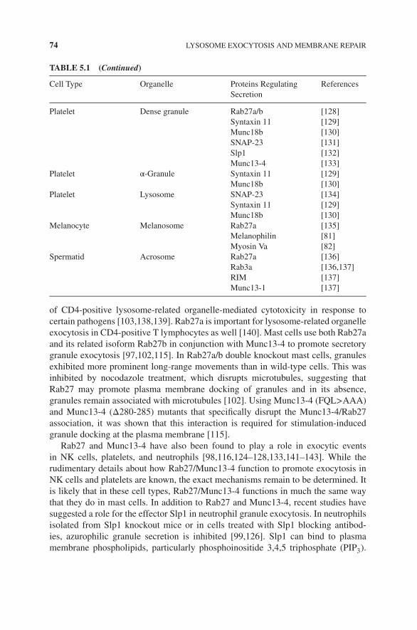
�
� �
�
74 LYSOSOME EXOCYTOSIS AND MEMBRANE REPAIR
TABLE 5.1 (Continued)
Cell Type Organelle Proteins RegulatingSecretion
References
Platelet Dense granule Rab27a/bSyntaxin 11Munc18bSNAP-23Slp1Munc13-4
[128][129][130][131][132][133]
Platelet α-Granule Syntaxin 11Munc18b
[129][130]
Platelet Lysosome SNAP-23Syntaxin 11Munc18b
[134][129][130]
Melanocyte Melanosome Rab27aMelanophilinMyosin Va
[135][81][82]
Spermatid Acrosome Rab27aRab3aRIMMunc13-1
[136][136,137][137][137]
of CD4-positive lysosome-related organelle-mediated cytotoxicity in response tocertain pathogens [103,138,139]. Rab27a is important for lysosome-related organelleexocytosis in CD4-positive T lymphocytes as well [140]. Mast cells use both Rab27aand its related isoform Rab27b in conjunction with Munc13-4 to promote secretorygranule exocytosis [97,102,115]. In Rab27a/b double knockout mast cells, granulesexhibited more prominent long-range movements than in wild-type cells. This wasinhibited by nocodazole treatment, which disrupts microtubules, suggesting thatRab27 may promote plasma membrane docking of granules and in its absence,granules remain associated with microtubules [102]. Using Munc13-4 (FQL>AAA)and Munc13-4 (Δ280-285) mutants that specifically disrupt the Munc13-4/Rab27association, it was shown that this interaction is required for stimulation-inducedgranule docking at the plasma membrane [115].
Rab27 and Munc13-4 have also been found to play a role in exocytic eventsin NK cells, platelets, and neutrophils [98,116,124–128,133,141–143]. While therudimentary details about how Rab27/Munc13-4 function to promote exocytosis inNK cells and platelets are known, the exact mechanisms remain to be determined. Itis likely that in these cell types, Rab27/Munc13-4 functions in much the same waythat they do in mast cells. In addition to Rab27 and Munc13-4, recent studies havesuggested a role for the effector Slp1 in neutrophil granule exocytosis. In neutrophilsisolated from Slp1 knockout mice or in cells treated with Slp1 blocking antibod-ies, azurophilic granule secretion is inhibited [99,126]. Slp1 can bind to plasmamembrane phospholipids, particularly phosphoinositide 3,4,5 triphosphate (PIP3).

�
� �
�
MECHANISMS OF LYSOSOME EXOCYTOSIS 75
This may suggest that Slp1 promotes exocytosis by recruiting Rab27-positivegranules to membrane microdomains rich in PIP3 and allowing them to dock thereprior to calcium-dependent exocytosis. Consistent with this, HL-60 neutrophil-likecells that were downregulated for Slp1 displayed increased LAMP-3-positive granulevelocity and displacement, suggestive of greater microtubule-based transport anddecreased granule docking at the plasma membrane [126].
Rab3, which exists in four isoforms (Rab3a, 3b, 3c, and 3d), is related to Rab27in terms of structure [64]. Rab3a has been implicated in the regulation of exocyto-sis from sperm cells called spermatids [136]. Spermatids contain one large acidicdense-core granule that is related to a lysosome, called an acrosome. Upon contactwith an oocyte during the process of fertilization, the acrosome is exocytosed [144].This releases digestive enzymes onto the oocyte and helps the spermatid to breakthrough the hard shell of the oocyte [145]. This process is similar to that describedpreviously for osteoclast bone resorption and macrophage degradation of aggregatedLDL. Acrosome exocytosis is regulated by both Rab27 and Rab3. A sequential modelof action has been proposed in which Rab27 recruits a guanine nucleotide exchangefactor for Rab3 that allows Rab3a to become activated [119]. Rab3d is thought toregulate exocytosis from other granulocyte cell types, such as eosinophils [146] andmast cells [101,110,134,147], though much less is known about their regulation.
5.3.4 Lysosome Fusion with the Plasma Membrane
Recent work has demonstrated a key role for Soluble NSF Attachment REceptor(SNARE) proteins in membrane fusion events within the cell. SNARE proteins areexpressed on each of the membranes destined to fuse. They each contain at least oneα-helical domain that can bind to specific α-helical domains of other SNARE proteins.These helices form a specific and stable four-helix bundle and induce energeticallyfavorable conditions to allow membrane fusion events to take place (Figure 5.2).The vesicular SNARE (v-SNARE) VAMP-7 and the target SNAREs (t-SNAREs)syntaxin 4 and SNAP-23 are involved in lysosome exocytosis in response to wound-ing in NRK cells [112]. In platelets, SNAP-23, syntaxin 2, and syntaxin 4 werefound to be involved in exocytosis of conventional lysosomes [114]. For the exocyto-sis of lysosome-related organelles, additional SNAREs have been implicated. Theseinclude VAMP-2 and VAMP-8 for lysosome-related organelles in CTLs [121,122],VAMP-4 and VAMP-7 in NK cells [129], VAMP-7 and VAMP-8 in mast cell granuleexocytosis [108,109], and Syntaxin 11 in platelets, neutrophils, NK cells, and CTLs[148–150] (Table 5.1). Recent work has indicated that lytic granules in NK cells donot fuse completely with the plasma membrane [100]. This type of exocytosis is oftenreferred to as “kiss-and-run” and is also known to occur in the context of adrenalchromaffin cell granule release [151,152]. Similarly, in NK cells, “kiss-and-run” exo-cytosis may promote reclamation of lytic granule membrane and proteins; however,the factors influencing incomplete fusion of lysosome-related organelles with theplasma membrane are not well understood.

�
� �
�
76 LYSOSOME EXOCYTOSIS AND MEMBRANE REPAIR
5.3.5 Calcium-Dependent Exocytosis
Lysosome exocytosis in many cell types has been shown to be calcium dependent.The protein Synaptotagmin VII (Syt VII) is thought to function as a calciumsensor for the exocytosis of lysosomes. Syt VII is important for calcium-dependentexocytosis of lysosomes in fibroblasts [104] and mediates cell invasion by T. cruzithat is dependent on calcium-regulated exocytosis of lysosomes [105]. It is alsorequired for plasma membrane repair in response to scratch-induced wounding ofNRK cells [23] and to repair M. tuberculosis-induced plasma membrane lesions [35].In addition, Syt VII regulates calcium-dependent lysosome exocytosis at the rearof the cell. This can facilitate cell detachment and efficient migration of T cellsand neutrophils [106], lysosome exocytosis in osteoclasts [44], and to nascentphagosomes in macrophages [37]. Lysosomes are the major vesicular compartmentto exocytose in response to calcium in astrocytes [107], which is important forATP release [111], and Syt VII is required for lysosome exocytosis as a source ofmembrane to promote neurite outgrowth [38]. The relationship between Syt VIIand SNARE proteins in lysosome exocytosis has not been fully characterized, butone study found that calcium influx into wounded NRK cells stimulates formationof a complex consisting of SNAP-23/Syntaxin 4/VAMP7, which interacts withSyt VII [112]. This would allow Syt VII to confer calcium sensitivity to theSNARE fusion machinery (Figure 5.2). Another regulator of lysosome exocytosisis transcription factor EB, which can promote the fusion of lysosomes with theplasma membrane. The overexpression of transcription factor EB raises intracellularCa2+ levels through the activation of the lysosomal Ca2+ channel MCOLN1 [113].Interestingly, a recent study found a pool of lysosomes that can be exocytosed inresponse to cholesterol depletion in the plasma membrane independently of SytVII and that this exocytosis occurred more efficiently in the absence of Ca2+ [58].This suggests the existence of a distinct pool of lysosomes that may utilize differentsecretory proteins to undergo exocytosis in a calcium-independent manner.
5.4 CONCLUSION
Lysosome exocytosis plays an important role in diverse biological processes such asplasma membrane repair following mechanical or bacterial cellular damage, immuneresponses, parasite entry into cells, bone remodeling, phagocytosis of large parti-cles, and extracellular degradation of disease-associated substrates. While significantprogress has been made in understanding how lysosome exocytosis is regulated invarious physiological systems, there is much yet to be learned. Parallels can be drawnbetween the exocytic regulation of conventional lysosomes and lysosome-relatedorganelles. This is likely to serve as a template to guide future studies attempting tounderstand lysosome exocytosis in different cell types and systems. While our currentunderstanding of the secretory machinery regulating lysosome exocytosis is limited,it is clear that elucidation of such mechanisms will have direct significance for humanhealth. Discoveries may offer new strategies and drug targets for the prevention andtreatment of a wide array of human diseases.
�
� �
�
REFERENCES 77
ACKNOWLEDGMENTS
We would like to thank Frederick Maxfield for helpful discussions and feedback thatcontributed to the chapter. We would also like to thank Leona Cohen-Gould at theEM and Histology Core at Weill Cornell Medical College for her generous help andsupport in generating the original image in Figure 5.1. We apologize to those whosework we omitted.
REFERENCES
[1] Idone V, Tam C, Andrews NW. Two-way traffic on the road to plasma membrane repair.Trends Cell Biol 2008;18:552–559.
[2] Rodriguez A, Webster P, Ortego J, Andrews NW. Lysosomes behave as Ca2+-regulatedexocytic vesicles in fibroblasts and epithelial cells. J Cell Biol 1997;137:93–104.
[3] Jaiswal JK, Andrews NW, Simon SM. Membrane proximal lysosomes are the majorvesicles responsible for calcium-dependent exocytosis in nonsecretory cells. J Cell Biol2002;159:625–635.
[4] Blott EJ, Griffiths GM. Secretory lysosomes. Nat Rev Mol Cell Biol 2002;3:122–131.
[5] Griffiths GM. Secretory lysosomes – a special mechanism of regulated secretion inhaemopoietic cells. Trends Cell Biol 1996;6:329–332.
[6] Luzio JP, Hackmann Y, Dieckmann NM, Griffiths GM. The biogenesis of lysosomesand lysosome-related organelles. Cold Spring Harb Perspect Biol 2014;6:a016840.
[7] Page LJ, Darmon AJ, Uellner R, Griffiths GM. L is for lytic granules: lysosomes thatkill. Biochim Biophys Acta 1998;1401:146–156.
[8] Griffiths G. What’s special about secretory lysosomes? Semin Cell Dev Biol2002;13:279–284.
[9] Marks MS, Heijnen HF, Raposo G. Lysosome-related organelles: unusual compart-ments become mainstream. Curr Opin Cell Biol 2013;25:495–505.
[10] Harris DA, Kim K, Nakahara K, Vasquez-Doorman C, Carthew RW. Cargo sortingto lysosome-related organelles regulates siRNA-mediated gene silencing. J Cell Biol2011;194:77–87.
[11] Raposo G, Tenza D, Mecheri S, Peronet R, Bonnerot C, et al. Accumulation of majorhistocompatibility complex class II molecules in mast cell secretory granules and theirrelease upon degranulation. Mol Biol Cell 1997;8:2631–2645.
[12] Stinchcombe JC, Barral DC, Mules EH, Booth S, Hume AN, et al. Rab27a is requiredfor regulated secretion in cytotoxic T lymphocytes. J Cell Biol 2001;152:825–834.
[13] Wasmeier C, Romao M, Plowright L, Bennett DC, Raposo G, et al. Rab38 and Rab32control post-Golgi trafficking of melanogenic enzymes. J Cell Biol 2006;175:271–281.
[14] Fukuda M. Rab27 effectors, pleiotropic regulators in secretory pathways. Traffic2013;14:949–963.
[15] Barry M, Bleackley RC. Cytotoxic T lymphocytes: all roads lead to death. Nat RevImmunol 2002;2:401–409.
[16] Grakoui A, Bromley SK, Sumen C, Davis MM, Shaw AS, et al. The immunologicalsynapse: a molecular machine controlling T cell activation. Science 1999;285:221–227.
�
� �
�
78 LYSOSOME EXOCYTOSIS AND MEMBRANE REPAIR
[17] Trapani JA, Smyth MJ. Functional significance of the perforin/granzyme cell death path-way. Nat Rev Immunol 2002;2:735–747.
[18] Ritter AT, Asano Y, Stinchcombe JC, Dieckmann NMG, Chen B, et al. Actin depletioninitiates events leading to granule secretion at the immunological synapse. Immunity2015;42:864–876.
[19] Tarafder AK, Bolasco G, Correia MS, Pereira FJ, Iannone L, et al. Rab11b mediatesmelanin transfer between donor melanocytes and acceptor keratinocytes via coupledexo/endocytosis. J Invest Dermatol 2014;134:1056–1066.
[20] Wu XS, Masedunskas A, Weigert R, Copeland NG, Jenkins NA, et al. Melanoregulinregulates a shedding mechanism that drives melanosome transfer from melanocytes tokeratinocytes. Proc Natl Acad Sci U S A 2012;109:E2101–E2109.
[21] Fitch-Tewfik JL, Flaumenhaft R. Platelet granule exocytosis: a comparison with chro-maffin cells. Front Endocrinol (Lausanne) 2013;4:77.
[22] Abraham SN, St John AL. Mast cell-orchestrated immunity to pathogens. Nat RevImmunol 2010;10:440–452.
[23] Reddy A, Caler E, Andrews NW. Plasma membrane repair is mediated by Ca2+-regulated exocytosis of lysosomes. Molecular Biology of the Cell 2001;12:266a.
[24] McNeil PL. Repairing a torn cell surface: make way, lysosomes to the rescue. J Cell Sci2002;115:873–879.
[25] McNeil PL, Vogel SS, Miyake K, Terasaki M. Patching plasma membrane disruptionswith cytoplasmic membrane. J Cell Sci 2000;113:1891–1902.
[26] Dai J, Sheetz MP. Regulation of endocytosis, exocytosis, and shape by membrane ten-sion. Cold Spring Harbor Symp Quant Biol 1995;60:567–571.
[27] Togo T, Alderton JM, Bi GQ, Steinhardt RA. The mechanism of facilitated cell mem-brane resealing. J Cell Sci 1999;112:719–731.
[28] Togo T, Krasieva TB, Steinhardt RA. A decrease in membrane tension precedes suc-cessful cell-membrane repair. Mol Biol Cell 2000;11:4339–4346.
[29] Tam C, Idone V, Devlin C, Fernandes MC, Flannery A, et al. Exocytosis of acid sph-ingomyelinase by wounded cells promotes endocytosis and plasma membrane repair.J Cell Biol 2010;189:1027–1038.
[30] Chakrabarti S, Kobayashi KS, Flavell RA, Marks CB, Miyake K, et al. Impaired mem-brane resealing and autoimmune myositis in synaptotagmin VII-deficient mice. J CellBiol 2003;162:543–549.
[31] Bansal D, Miyake K, Vogel SS, Groh S, Chen CC, et al. Defective membrane repair indysferlin-deficient muscular dystrophy. Nature 2003;423:168–172.
[32] Liu J, Aoki M, Illa I, Wu C, Fardeau M, et al. Dysferlin, a novel skeletal musclegene, is mutated in Miyoshi myopathy and limb girdle muscular dystrophy. Nat Genet1998;20:31–36.
[33] Tardieux I, Webster P, Ravesloot J, Boron W, Lunn JA, et al. Lysosome recruitmentand fusion are early events required for trypanosome invasion of mammalian cells. Cell1992;71:1117–1130.
[34] Rodriguez A, Samoff E, Rioult MG, Chung A, Andrews NW. Host cell invasion bytrypanosomes requires lysosomes and microtubule/kinesin-mediated transport. J CellBiol 1996;134:349–362.

�
� �
�
REFERENCES 79
[35] Divangahi M, Chen M, Gan H, Desjardins D, Hickman TT, et al. Mycobacteriumtuberculosis evades macrophage defenses by inhibiting plasma membrane repair. NatImmunol 2009;10:899–906.
[36] Braun V, Fraisier V, Raposo G, Hurbain I, Sibarita JB, et al. TI-VAMP/VAMP7 isrequired for optimal phagocytosis of opsonised particles in macrophages. EMBO J2004;23:4166–4176.
[37] Czibener C, Sherer NM, Becker SM, Pypaert M, Hui E, et al. Ca2+ and synaptotagminVII-dependent delivery of lysosomal membrane to nascent phagosomes. J Cell Biol2006;174:997–1007.
[38] Arantes RM, Andrews NW. A role for synaptotagmin VII-regulated exocytosis oflysosomes in neurite outgrowth from primary sympathetic neurons. J Neurosci 2006;26:4630–4637.
[39] Lacombe J, Karsenty G, Ferron M. Regulation of lysosome biogenesis and functions inosteoclasts. Cell Cycle 2013;12:2744–2752.
[40] Matsumoto T, Nagase Y, Hirose J, Tokuyama N, Yasui T, et al. Regulation of boneresorption and sealing zone formation in osteoclasts occurs through protein kinaseB-mediated microtubule stabilization. J Bone Miner Res 2013;28:1191–1202.
[41] Yang DQ, Feng S, Chen W, Zhao H, Paulson C, et al. V-ATPase subunit ATP6AP1(Ac45) regulates osteoclast differentiation, extracellular acidification, lysosomal traf-ficking, and protease exocytosis in osteoclast-mediated bone resorption. J Bone MinerRes 2012;27:1695–1707.
[42] Stenbeck G. Formation and function of the ruffled border in osteoclasts. Semin Cell DevBiol 2002;13:285–292.
[43] Zhao H, Laitala-Leinonen T, Parikka V, Vaananen HK. Downregulation of smallGTPase Rab7 impairs osteoclast polarization and bone resorption. J Biol Chem 2001;276:39295–39302.
[44] Zhao H, Ito Y, Chappel J, Andrews NW, Teitelbaum SL, et al. Synaptotagmin VII reg-ulates bone remodeling by modulating osteoclast and osteoblast secretion. Dev Cell2008;14:914–925.
[45] Saftig P, Hunziker E, Wehmeyer O, Jones S, Boyde A, et al. Impaired osteoclastic boneresorption leads to osteopetrosis in cathepsin-K-deficient mice. Proc Natl Acad Sci U SA 1998;95:13453–13458.
[46] van Meel E, Boonen M, Zhao H, Oorschot V, Ross FP, et al. Disruption of the Man-6-Ptargeting pathway in mice impairs osteoclast secretory lysosome biogenesis. Traffic2011;12:912–924.
[47] Zhao H. Membrane trafficking in osteoblasts and osteoclasts: new avenues for under-standing and treating skeletal diseases. Traffic 2012;13:1307–1314.
[48] Smith EB, Massie IB, Alexander KM. The release of an immobilized lipoproteinfraction from atherosclerotic lesions by incubation with plasmin. Atherosclerosis1976;25:71–84.
[49] Tabas I, Williams KJ, Boren J. Subendothelial lipoprotein retention as the initiat-ing process in atherosclerosis: update and therapeutic implications. Circulation 2007;116:1832–1844.
[50] Haka AS, Grosheva I, Chiang E, Buxbaum AR, Baird BA, et al. Macrophages create anacidic extracellular hydrolytic compartment to digest aggregated lipoproteins. Mol BiolCell 2009;20:4932–4940.

�
� �
�
80 LYSOSOME EXOCYTOSIS AND MEMBRANE REPAIR
[51] Grosheva I, Haka AS, Qin C, Pierini LM, Maxfield FR. Aggregated LDL in contact withmacrophages induces local increases in free cholesterol levels that regulate local actinpolymerization. Arterioscler Thromb Vasc Biol 2009;29:1615–1621.
[52] Chow A, Toomre D, Garrett W, Mellman I. Dendritic cell maturation triggers ret-rograde MHC class II transport from lysosomes to the plasma membrane. Nature2002;418:988–994.
[53] Savina A, Jancic C, Hugues S, Guermonprez P, Vargas P, et al. NOX2 controls phagoso-mal pH to regulate antigen processing during crosspresentation by dendritic cells. Cell2006;126:205–218.
[54] Jancic C, Savina A, Wasmeier C, Tolmachova T, El-Benna J, et al. Rab27a regulatesphagosomal pH and NADPH oxidase recruitment to dendritic cell phagosomes. NatCell Biol 2007;9:367–378.
[55] Bousso P. T-cell activation by dendritic cells in the lymph node: lessons from themovies. Nat Rev Immunol 2008;8:675–684.
[56] Stenmark H. Rab GTPases as coordinators of vesicle traffic. Nat Rev Mol Cell Biol2009;10:513–525.
[57] Carta S, Lavieri R, Rubartelli A. Different members of the IL-1 family come out indifferent ways: DAMPs vs. cytokines? Front Immunol 2013;4:123.
[58] Hissa B, Duarte JG, Kelles LF, Santos FP, del Puerto HL, et al. Membrane cholesterolregulates lysosome-plasma membrane fusion events and modulates Trypanosoma cruziinvasion of host cells. PLoS Neglected Trop Dis 2012;6:e1583.
[59] Luzio JP, Pryor PR, Bright NA. Lysosomes: fusion and function. Nat Rev Mol Cell Biol2007;8:622–632.
[60] Menager MM, Menasche G, Romao M, Knapnougel P, Ho CH, et al. Secretory cytotoxicgranule maturation and exocytosis require the effector protein hMunc13-4. Nat Immunol2007;8:257–267.
[61] van der Sluijs P, Zibouche M, van Kerkhof P. Late steps in secretory lysosome exocy-tosis in cytotoxic lymphocytes. Front Immunol 2013;4:359.
[62] Wasmeier C, Hume AN, Bolasco G, Seabra MC. Melanosomes at a glance. J Cell Sci2008;121:3995–3999.
[63] Wernersson S, Pejler G. Mast cell secretory granules: armed for battle. Nat Rev Immunol2014;14:478–494.
[64] Pereira-Leal JB, Seabra MC. Evolution of the Rab family of small GTP-binding pro-teins. J Mol Biol 2001;313:889–901.
[65] Carta S, Tassi S, Semino C, Fossati G, Mascagni P, et al. Histone deacetylase inhibitorsprevent exocytosis of interleukin-1beta-containing secretory lysosomes: role of micro-tubules. Blood 2006;108:1618–1626.
[66] Martin-Verdeaux S, Pombo I, Iannascoli B, Roa M, Varin-Blank N, et al. Evidenceof a role for Munc18-2 and microtubules in mast cell granule exocytosis. J Cell Sci2003;116:325–334.
[67] Schliwa M, Woehlke G. Molecular motors. Nature 2003;422:759–765.
[68] Tucker JB. Spatial organization of microtubule-organizing centers and microtubules.J Cell Biol 1984;99:55s–62s.
[69] Rosa-Ferreira C, Munro S. Arl8 and SKIP act together to link lysosomes to kinesin-1.Dev Cell 2011;21:1171–1178.
�
� �
�
REFERENCES 81
[70] Tan SC, Scherer J, Vallee RB. Recruitment of dynein to late endosomes and lysosomesthrough light intermediate chains. Mol Biol Cell 2011;22:467–477.
[71] Jordens I, Fernandez-Borja M, Marsman M, Dusseljee S, Janssen L, et al. The Rab7effector protein RILP controls lysosomal transport by inducing the recruitment ofdynein–dynactin motors. Curr Biol 2001;11:1680–1685.
[72] Burkhardt JK, Echeverri CJ, Nilsson T, Vallee RB. Overexpression of the dynamitin(p50) subunit of the dynactin complex disrupts dynein-dependent maintenance of mem-brane organelle distribution. J Cell Biol 1997;139:469–484.
[73] Stinchcombe JC, Majorovits E, Bossi G, Fuller S, Griffiths GM. Centrosome polar-ization delivers secretory granules to the immunological synapse. Nature 2006;443:462–465.
[74] Daniele T, Hackmann Y, Ritter AT, Wenham M, Booth S, et al. A role for Rab7 in themovement of secretory granules in cytotoxic T lymphocytes. Traffic 2011;12:902–911.
[75] Clark RH, Stinchcombe JC, Day A, Blott E, Booth S, et al. Adaptor protein 3-dependentmicrotubule-mediated movement of lytic granules to the immunological synapse. NatImmunol 2003;4:1111–1120.
[76] Ivan V, Martinez-Sanchez E, Sima LE, Oorschot V, Klumperman J, et al. AP-3 andRabip4′ coordinately regulate spatial distribution of lysosomes. PLoS ONE 2012;7:e48142.
[77] van der Kant R, Fish A, Janssen L, Janssen H, Krom S, et al. Late endosomal trans-port and tethering are coupled processes controlled by RILP and the cholesterol sensorORP1L. J Cell Sci 2013;126:3462–3474.
[78] Swanson JA, Locke A, Ansel P, Hollenbeck PJ. Radial movement of lysosomes alongmicrotubules in permeabilized macrophages. J Cell Sci 1992;103(Pt 1):201–209.
[79] Swanson J, Bushnell A, Silverstein SC. Tubular lysosome morphology and distributionwithin macrophages depend on the integrity of cytoplasmic microtubules. Proc NatlAcad Sci U S A 1987;84:1921–1925.
[80] Mrakovic A, Kay JG, Furuya W, Brumell JH, Botelho RJ. Rab7 and Arl8 GTPases arenecessary for lysosome tubulation in macrophages. Traffic 2012;13:1667–1679.
[81] Hollenbeck PJ, Swanson JA. Radial extension of macrophage tubular lysosomes sup-ported by kinesin. Nature 1990;346:864–866.
[82] Xu J, Toops KA, Diaz F, Carvajal-Gonzalez JM, Gravotta D, et al. Mechanism of polar-ized lysosome exocytosis in epithelial cells. J Cell Sci 2012;125:5937–5943.
[83] DeSelm CJ, Miller BC, Zou W, Beatty WL, van Meel E, et al. Autophagy pro-teins regulate the secretory component of osteoclastic bone resorption. Dev Cell 2011;21:966–974.
[84] Sun Y, Buki KG, Ettala O, Vaaraniemi JP, Vaananen HK. Possible role of directRac1-Rab7 interaction in ruffled border formation of osteoclasts. J Biol Chem 2005;280:32356–32361.
[85] Hume AN, Collinson LM, Hopkins CR, Strom M, Barral DC, et al. The leaden geneproduct is required with Rab27a to recruit myosin Va to melanosomes in melanocytes.Traffic 2002;3:193–202.
[86] Wu XS, Rao K, Zhang H, Wang F, Sellers JR, et al. Identification of an organelle receptorfor myosin-Va. Nat Cell Biol 2002;4:271–278.
[87] Matesic LE, Yip R, Reuss AE, Swing DA, O’Sullivan TN, et al. Mutations in Mlph,encoding a member of the Rab effector family, cause the melanosome transport defectsobserved in leaden mice. Proc Natl Acad Sci U S A 2001;98:10238–10243.
�
� �
�
82 LYSOSOME EXOCYTOSIS AND MEMBRANE REPAIR
[88] Wilson SM, Yip R, Swing DA, O’Sullivan TN, Zhang Y, et al. A mutation in Rab27acauses the vesicle transport defects observed in ashen mice. Proc Natl Acad Sci U S A2000;97:7933–7938.
[89] Pastural E, Barrat FJ, Dufourcq-Lagelouse R, Certain S, Sanal O, et al. Griscelli diseasemaps to chromosome 15q21 and is associated with mutations in the myosin-Va gene.Nat Genet 1997;16:289–292.
[90] Menasche G, Pastural E, Feldmann J, Certain S, Ersoy F, et al. Mutations in RAB27Acause Griscelli syndrome associated with haemophagocytic syndrome. Nat Genet2000;25:173–176.
[91] Mercer JA, Seperack PK, Strobel MC, Copeland NG, Jenkins NA. Novel myosin heavychain encoded by murine dilute coat colour locus. Nature 1991;349:709–713.
[92] Menasche G, Ho CH, Sanal O, Feldmann J, Tezcan I, et al. Griscelli syndrome restrictedto hypopigmentation results from a melanophilin defect (GS3) or a MYO5A F-exondeletion (GS1). J Clin Invest 2003;112:450–456.
[93] Lopes VS, Ramalho JS, Owen DM, Karl MO, Strauss O, et al. The ternary Rab27a-Myrip-Myosin VIIa complex regulates melanosome motility in the retinal pigmentepithelium. Traffic 2007;8:486–499.
[94] Desnos C, Schonn JS, Huet S, Tran VS, El-Amraoui A, et al. Rab27A and its effectorMyRIP link secretory granules to F-actin and control their motion towards release sites.J Cell Biol 2003;163:559–570.
[95] Rodriguez A, Martinez I, Chung A, Berlot CH, Andrews NW. cAMP regulatesCa2+-dependent exocytosis of lysosomes and lysosome-mediated cell invasion by try-panosomes. J Biol Chem 1999;274:16754–16759.
[96] Foger N, Jenckel A, Orinska Z, Lee KH, Chan AC, et al. Differential regulation of mastcell degranulation versus cytokine secretion by the actin regulatory proteins Coronin1aand Coronin1b. J Exp Med 2011;208:1777–1787.
[97] Singh RK, Mizuno K, Wasmeier C, Wavre-Shapton ST, Recchi C, et al. Distinct andopposing roles for Rab27a/Mlph/MyoVa and Rab27b/Munc13-4 in mast cell secretion.FEBS J 2013;280:892–903.
[98] Liu D, Meckel T, Long EO. Distinct role of rab27a in granule movement at the plasmamembrane and in the cytosol of NK cells. PLoS ONE 2010;5:e12870.
[99] Johnson JL, Monfregola J, Napolitano G, Kiosses WB, Catz SD. Vesicular traf-ficking through cortical actin during exocytosis is regulated by the Rab27a effectorJFC1/Slp1 and the RhoA-GTPase-activating protein Gem-interacting protein. Mol BiolCell 2012;23:1902–1916.
[100] Liu D, Martina JA, Wu XS, Hammer JA 3rd, Long EO. Two modes of lytic gran-ule fusion during degranulation by natural killer cells. Immunol Cell Biol 2011;89:728–738.
[101] Roa M, Paumet F, Le Mao J, David B, Blank U. Involvement of the ras-like GTPaserab3d in RBL-2H3 mast cell exocytosis following stimulation via high affinity IgEreceptors (Fc epsilonRI). J Immunol 1997;159:2815–2823.
[102] Mizuno K, Tolmachova T, Ushakov DS, Romao M, Abrink M, et al. Rab27b regulatesmast cell granule dynamics and secretion. Traffic 2007;8:883–892.
[103] Yasukawa M, Ohminami H, Yakushijin Y, Arai J, Hasegawa A, et al. Fas-independentcytotoxicity mediated by human CD4+ CTL directed against herpes simplexvirus-infected cells. J Immunol 1999;162:6100–6106.

�
� �
�
REFERENCES 83
[104] Martinez I, Chakrabarti S, Hellevik T, Morehead J, Fowler K, et al. SynaptotagminVII regulates Ca(2+)-dependent exocytosis of lysosomes in fibroblasts. J Cell Biol2000;148:1141–1149.
[105] Caler EV, Chakrabarti S, Fowler KT, Rao S, Andrews NW. The exocytosis-regulatoryprotein synaptotagmin VII mediates cell invasion by Trypanosoma cruzi. J Exp Med2001;193:1097–1104.
[106] Colvin RA, Means TK, Diefenbach TJ, Moita LF, Friday RP, et al. Synaptotagmin-mediated vesicle fusion regulates cell migration. Nat Immunol 2010;11:495–502.
[107] Li D, Ropert N, Koulakoff A, Giaume C, Oheim M. Lysosomes are the major vesicularcompartment undergoing Ca2+-regulated exocytosis from cortical astrocytes. J Neu-rosci 2008;28:7648–7658.
[108] Sander LE, Frank SP, Bolat S, Blank U, Galli T, et al. Vesicle associated mem-brane protein (VAMP)-7 and VAMP-8, but not VAMP-2 or VAMP-3, are required foractivation-induced degranulation of mature human mast cells. Eur J Immunol 2008;38:855–863.
[109] Tiwari N, Wang CC, Brochetta C, Ke G, Vita F, et al. VAMP-8 segregates mast cell-preformed mediator exocytosis from cytokine trafficking pathways. Blood2008;111:3665–3674.
[110] Pombo I, Martin-Verdeaux S, Iannascoli B, Le Mao J, Deriano L, et al. IgE recep-tor type I-dependent regulation of a Rab3D-associated kinase: a possible link inthe calcium-dependent assembly of SNARE complexes. J Biol Chem 2001;276:42893–42900.
[111] Zhang Z, Chen G, Zhou W, Song A, Xu T, et al. Regulated ATP release from astrocytesthrough lysosome exocytosis. Nat Cell Biol 2007;9:945–953.
[112] Rao SK, Huynh C, Proux-Gillardeaux V, Galli T, Andrews NW. Identification ofSNAREs involved in synaptotagmin VII-regulated lysosomal exocytosis. J Biol Chem2004;279:20471–20479.
[113] Medina DL, Fraldi A, Bouche V, Annunziata F, Mansueto G, et al. Transcriptional acti-vation of lysosomal exocytosis promotes cellular clearance. Dev Cell 2011;21:421–430.
[114] Chen D, Lemons PP, Schraw T, Whiteheart SW. Molecular mechanisms of plateletexocytosis: role of SNAP-23 and syntaxin 2 and 4 in lysosome release. Blood2000;96:1782–1788.
[115] Elstak ED, Neeft M, Nehme NT, Voortman J, Cheung M, et al. The munc13-4-rab27complex is specifically required for tethering secretory lysosomes at the plasma mem-brane. Blood 2011;118:1570–1578.
[116] Wood SM, Meeths M, Chiang SC, Bechensteen AG, Boelens JJ, et al. Dif-ferent NK cell-activating receptors preferentially recruit Rab27a or Munc13-4 toperforin-containing granules for cytotoxicity. Blood 2009;114:4117–4127.
[117] Feldmann J, Callebaut I, Raposo G, Certain S, Bacq D, et al. Munc13-4 is essential forcytolytic granules fusion and is mutated in a form of familial hemophagocytic lympho-histiocytosis (FHL3). Cell 2003;115:461–473.
[118] Holt O, Kanno E, Bossi G, Booth S, Daniele T, et al. Slp1 and Slp2-a localize to theplasma membrane of CTL and contribute to secretion from the immunological synapse.Traffic 2008;9:446–457.
[119] Bustos MA, Lucchesi O, Ruete MC, Mayorga LS, Tomes CN. Rab27 and Rab3 sequen-tially regulate human sperm dense-core granule exocytosis. Proc Natl Acad Sci U S A2012;109:E2057–E2066.

�
� �
�
84 LYSOSOME EXOCYTOSIS AND MEMBRANE REPAIR
[120] Menasche G, Menager MM, Lefebvre JM, Deutsch E, Athman R, et al. A newly iden-tified isoform of Slp2a associates with Rab27a in cytotoxic T cells and participates tocytotoxic granule secretion. Blood 2008;112:5052–5062.
[121] Loo LS, Hwang LA, Ong YM, Tay HS, Wang CC, et al. A role for endobrevin/VAMP8in CTL lytic granule exocytosis. Eur J Immunol 2009;39:3520–3528.
[122] Matti U, Pattu V, Halimani M, Schirra C, Krause E, et al. Synaptobrevin2 is thev-SNARE required for cytotoxic T-lymphocyte lytic granule fusion. Nat Commun2013;4:1439.
[123] Kurowska M, Goudin N, Nehme NT, Court M, Garin J, et al. Terminal transport of lyticgranules to the immune synapse is mediated by the kinesin-1/Slp3/Rab27a complex.Blood 2012;119:3879–3889.
[124] Munafo DB, Johnson JL, Ellis BA, Rutschmann S, Beutler B, et al. Rab27a is a keycomponent of the secretory machinery of azurophilic granules in granulocytes. BiochemJ 2007;402:229–239.
[125] Johnson JL, Brzezinska AA, Tolmachova T, Munafo DB, Ellis BA, et al. Rab27a andRab27b regulate neutrophil azurophilic granule exocytosis and NADPH oxidase activityby independent mechanisms. Traffic 2010;11:533–547.
[126] Brzezinska AA, Johnson JL, Munafo DB, Crozat K, Beutler B, et al. The Rab27aeffectors JFC1/Slp1 and Munc13-4 regulate exocytosis of neutrophil granules. Traffic2008;9:2151–2164.
[127] Herrero-Turrion MJ, Calafat J, Janssen H, Fukuda M, Mollinedo F. Rab27a regu-lates exocytosis of tertiary and specific granules in human neutrophils. J Immunol2008;181:3793–3803.
[128] Tolmachova T, Abrink M, Futter CE, Authi KS, Seabra MC. Rab27b regulates num-ber and secretion of platelet dense granules. Proceed Natl Acad Sci U S A 2007;104:5872–5877.
[129] Krzewski K, Gil-Krzewska A, Watts J, Stern JN, Strominger JL. VAMP4- andVAMP7-expressing vesicles are both required for cytotoxic granule exocytosis in NKcells. Eur J Immunol 2011;41:3323–3329.
[130] Dressel R, Elsner L, Novota P, Kanwar N, Fischer von Mollard G. The exocytosis of lyticgranules is impaired in Vti1b- or Vamp8-deficient CTL leading to a reduced cytotoxicactivity following antigen-specific activation. J Immunol 2010;185:1005–1014.
[131] Marcenaro S, Gallo F, Martini S, Santoro A, Griffiths GM, et al. Analysis of naturalkiller-cell function in familial hemophagocytic lymphohistiocytosis (FHL): defectiveCD107a surface expression heralds Munc13-4 defect and discriminates between geneticsubtypes of the disease. Blood 2006;108:2316–2323.
[132] Neeft M, Wieffer M, de Jong AS, Negroiu G, Metz CH, et al. Munc13-4 is an effectorof rab27a and controls secretion of lysosomes in hematopoietic cells. Mol Biol Cell2005;16:731–741.
[133] Shirakawa R, Higashi T, Tabuchi A, Yoshioka A, Nishioka H, et al. Munc13-4 is aGTP-Rab27-binding protein regulating dense core granule secretion in platelets. J BiolChem 2004;279:10730–10737.
[134] Carroll K, Ray K, Helm B, Carey E. Differential expression of Rab3 isoforms in high-and low-secreting mast cell lines. Eur J Cell Biol 2001;80:295–302.
[135] Higashio H, Nishimura N, Ishizaki H, Miyoshi J, Orita S, et al. Doc2 alpha andMunc13-4 regulate Ca(2+)-dependent secretory lysosome exocytosis in mast cells. JImmunol 2008;180:4774–4784.

�
� �
�
REFERENCES 85
[136] Michaut M, Tomes CN, De Blas G, Yunes R, Mayorga LS. Calcium-triggered acrosomalexocytosis in human spermatozoa requires the coordinated activation of Rab3A andN-ethylmaleimide-sensitive factor. Proc Natl Acad Sci U S A 2000;97:9996–10001.
[137] Melicoff E, Sansores-Garcia L, Gomez A, Moreira DC, Datta P, et al. Synaptotagmin-2controls regulated exocytosis but not other secretory responses of mast cells. J BiolChem 2009;284:19445–19451.
[138] Appay V, Zaunders JJ, Papagno L, Sutton J, Jaramillo A, et al. Characterization ofCD4(+) CTLs ex vivo. J Immunol 2002;168:5954–5958.
[139] Ochoa MT, Stenger S, Sieling PA, Thoma-Uszynski S, Sabet S, et al. T-cell release ofgranulysin contributes to host defense in leprosy. Nat Med 2001;7:174–179.
[140] Haddad EK, Wu X, Hammer JA 3rd, Henkart PA. Defective granule exocytosis inRab27a-deficient lymphocytes from Ashen mice. J Cell Biol 2001;152:835–842.
[141] Singh RK, Liao W, Tracey-White D, Recchi C, Tolmachova T, et al. Rab27a-mediatedprotease release regulates neutrophil recruitment by allowing uropod detachment. J CellSci 2012;125:1652–1656.
[142] Pivot-Pajot C, Varoqueaux F, de Saint BG, Bourgoin SG. Munc13-4 regulates granulesecretion in human neutrophils. J Immunol 2008;180:6786–6797.
[143] Johnson JL, Hong H, Monfregola J, Kiosses WB, Catz SD. Munc13-4 restricts motil-ity of Rab27a-expressing vesicles to facilitate lipopolysaccharide-induced priming ofexocytosis in neutrophils. J Biol Chem 2011;286:5647–5656.
[144] Breitbart H, Rubinstein S, Lax Y. Regulatory mechanisms in acrosomal exocytosis. RevReprod 1997;2:165–174.
[145] Mayorga LS, Tomes CN, Belmonte SA. Acrosomal exocytosis, a special type of regu-lated secretion. IUBMB Life 2007;59:286–292.
[146] Nishio H, Suda T, Sawada K, Miyamoto T, Koike T, et al. Molecular cloning of cDNAencoding human Rab3D whose expression is upregulated with myeloid differentiation.Biochim Biophys Acta 1999;1444:283–290.
[147] Tuvim MJ, Adachi R, Chocano JF, Moore RH, Lampert RM, et al. Rab3D, a smallGTPase, is localized on mast cell secretory granules and translocates to the plasmamembrane upon exocytosis. Am J Respir Cell Mol Biol 1999;20:79–89.
[148] Ye S, Karim ZA, Al Hawas R, Pessin JE, Filipovich AH, et al. Syntaxin-11, but notsyntaxin-2 or syntaxin-4, is required for platelet secretion. Blood 2012;120:2484–2492.
[149] D’Orlando O, Zhao F, Kasper B, Orinska Z, Muller J, et al. Syntaxin 11 is requiredfor NK and CD8+ T-cell cytotoxicity and neutrophil degranulation. Eur J Immunol2013;43:194–208.
[150] Bryceson YT, Rudd E, Zheng C, Edner J, Ma D, et al. Defective cytotoxic lymphocytedegranulation in syntaxin-11 deficient familial hemophagocytic lymphohistiocytosis 4(FHL4) patients. Blood 2007;110:1906–1915.
[151] Henkel AW, Kang G, Kornhuber J. A common molecular machinery for exocytosis andthe ‘kiss-and-run’ mechanism in chromaffin cells is controlled by phosphorylation. JCell Sci 2001;114:4613–4620.
[152] Wang N, Lee AK, Yan L, Simpson MR, Tse A, et al. Granule matrix property and rapid“kiss-and-run” exocytosis contribute to the different kinetics of catecholamine releasefrom carotid glomus and adrenal chromaffin cells at matched quantal size. Can J PhysiolPharmacol 2012;90:791–801.

�
� �
�
6ROLE OF LYSOSOMES IN LIPIDMETABOLISM
Frederick R. MaxfieldDepartment of Biochemistry, Weill Cornell Medical College, New York, NY, USA
6.1 INTRODUCTION
Many of the acid hydrolases in late endosomes and lysosomes (LE/Ly) participatein the breakdown of lipids. Lipids are delivered to lysosomes by endocytic uptakeof lipoproteins, as components of the lipid bilayer involved in vesicle transport, byautophagy, and by cytoplasmic lipid transport proteins. Lipids can leave lysosomes byvesicle transport, by export to cytoplasmic lipid transport proteins, or by lysosomesecretion. For several reasons, specialized mechanisms are required for hydrolysisof lipids in LE/Ly. First, the membrane integrity of the LE/Ly must be maintainedeven as some of the lipid components that were once part of its limiting bilayer arehydrolyzed, so the outer limiting membrane must be protected from hydrolysis evenas extensive lipid hydrolysis is taking place. Second, many of the lipid componentsare highly insoluble in water, so mechanisms must be employed to allow enzymaticaccess to cleavage sites. In some cases (e.g., cholesterol), special mechanisms arerequired to solubilize the hydrophobic products of hydrolysis. The mechanisms usedby LE/Ly to hydrolyze lipids and some of the disease states that occur when thesemechanisms fail are discussed in this chapter. The role of lysosomes in respondingto nutrient status is also discussed.
Lysosomes: Biology, Diseases, and Therapeutics, First Edition.Edited by Frederick R. Maxfield, James M. Willard and Shuyan Lu.© 2016 John Wiley & Sons, Inc. Published 2016 by John Wiley & Sons, Inc.
87

�
� �
�
88 ROLE OF LYSOSOMES IN LIPID METABOLISM
6.2 ENDOCYTIC UPTAKE OF LIPOPROTEINS
Probably the best characterized example of lysosomal digestion of lipids is the hydrol-ysis of cholesteryl esters that are present in the core of low-density lipoprotein (LDL)particles taken into the cell by receptor-mediated endocytosis [1–3] (Figure 6.1). LDLreceptors are expressed on the surface of most nucleated mammalian cells, and theybind LDL particles, which are derived from larger lipoproteins, very low-densitylipoproteins (VLDLs), that are secreted from the liver. The VLDL is acted on by
Lipid droplet
Phagophore
Autophagosome
FA/cholesterol
Clathrin
LDLLDL receptor
Sorting endosome
pH ~ 6.2
ERC
pH 6.5
LAL
AutolysosomeLysosome
pH 4.5–5.0
Late endosome
pH 5.5
LE/Ly
Figure 6.1 Schematic diagram of lipoprotein and lipid droplet degradation by lysosomes.Low-density lipoproteins (LDLs) bind to receptors on the surface of cells and are rapidly inter-nalized via clathrin-coated pits. The contents of the clathrin-coated pits are delivered to sortingendosomes, which are transient organelles with an internal pH of about 6.2. The low pH causesLDL to dissociate from its receptor, and the empty receptors are rapidly exported from thesorting endosome in narrow diameter tubules. The receptors return to the cell surface eitherdirectly or after passing through an intermediate organelle, the endocytic recycling compart-ment (ERC). The sorting endosomes mature into late endosomes, which are somewhat moreacidic and have lysosomal enzymes that have been delivered from the trans-Golgi network.Membrane invaginations at the surface of the late endosomes create internal vesicles that areenriched in BMP. Lysosomal enzymes, which are activated by the low pH, and these digest theinternalized LDL. Membrane lipids in the bilayer are mainly digested on the internal vesicles,which protects the limiting membrane against leakage. Lipid droplets can be surrounded byphagophores to create an autophagosome, which fuses with late endosomes or lysosomes tocreate a digestive autolysosome. Figure created by David B. Iaea. (See color plate section forthe color representation of this figure.)

�
� �
�
ENDOCYTIC UPTAKE OF LIPOPROTEINS 89
a nonlysosomal enzyme, lipoprotein lipase, which is held on the surface of certainendothelial cells by a GPI-anchored receptor, GPIHBP1 [4–6]. The lipase hydrolyzesthe triacylglycerides in the core of VLDL, leaving smaller cholesteryl ester-rich LDLparticles that circulate throughout the body but not the central nervous system. LDLreceptors, which bind the circulating LDL, are internalized via clathrin-coated pitsand delivered to early sorting endosomes, which maintain an internal pH of about 6.2[7,8]. This pH is sufficiently acidic to cause dissociation of LDL from its receptor, andthe LDL receptors then are free to recycle while the LDL remains in the early sort-ing endosome as it matures into a late endosome (LE) [7,9]. Unless retained in thesorting endosomes by a signal, most membrane components are returned to the cellsurface. This is accomplished by an iterative process in which newly formed vesiclesthat are pinched off from the surface continue to fuse with a sorting endosome forseveral minutes [7,10]. During this time, narrow diameter tubules, which have a highsurface-to-volume ratio, repeatedly pinch off from the sorting endosome and deliverthe membrane components (and a small amount of the volume) back to the surfaceeither directly or through an intermediate compartment called the endocytic recyclingcompartment (ERC) [10–12].
In the LE, the LDL begins to encounter lysosomal enzymes that have been deliv-ered from the trans-Golgi network [13]. The lysosomal proteases digest the proteincomponents, and a single enzyme, lysosomal acid lipase, digests the cholesterylesters and triacylglycerides in the core of lipoproteins [14]. This releases cholesteroland a fatty acid from the cholesteryl esters and glycerol and fatty acids from thetriacylglycerides. (Deficiencies in lysosomal acid lipase lead to Wollman disease orcholesteryl ester storage disease [14].) Cholesterol is highly insoluble in water, soit needs to be bound to transport proteins when it is not in a bilayer. In the LE/Ly, asoluble protein called NPC2 serves this role [15]. It now appears that NPC2 deliversthe cholesterol to an LE/Ly membrane protein, NPC1, which helps to shuttle thecholesterol to the limiting membrane of the LE/Ly [16–18]. The itinerary of choles-terol after insertion into the limiting membrane is uncertain, but it may be transportedby one or more cytoplasmic sterol-binding proteins and delivered to other organelles[19,20]. Homozygous defects in either NPC1 or NPC2 cause Niemann–Pick Cdisease, which is characterized by abnormal accumulation of cholesterol and otherlipids in LE/Ly. β-Cyclodextrins can chelate cholesterol and facilitate its exchangeamong membranes. When delivered into the LE/Ly, β-cyclodextrins can bypassthe requirement for both NPC1 and NPC2, suggesting that delivery of sterol to thelimiting membrane of the LE/Ly is the essential role of these proteins [20]. Clinicaltrials for treatment of NPC patients with β-cyclodextrins have been initiated [21].
Some LE are also described as multivesicular bodies (MVBs) because their lumenis filled with small vesicles and inward budding tubules. The internal vesicles areformed by the invagination of the limiting membrane into the lumen of the LE underthe control of a set of proteins called the endosomal sorting complexes requiredfor transport (ESCRT) complex [22]. These inward invaginations are enriched inproteins that had been mono-ubiquitinated, and their inclusion in these structureshelps prevent their recycling. The internal vesicles are highly enriched in an unusuallipid, bis(monoacylglycero)phosphate (BMP) (also called lysobisphosphatidic acid in

�
� �
�
90 ROLE OF LYSOSOMES IN LIPID METABOLISM
some older publications), a negatively charged glycerophospholipid with an unusualsn-1;sn-1′ structural configuration, that is present at very low levels elsewhere inthe cell [23,24]. Under some circumstances, these internal vesicles containing BMPcould hold some of the cholesterol that is released by the action of lysosomal acidlipase, but typically these vesicles become enriched in ceramide following hydrol-ysis of sphingomyelin (SM) by acid sphingomyelinase. The ceramide can displacecholesterol from lipid bilayers [25], so normally these internal membranes would beexpected to have low cholesterol content.
In peripheral cells (i.e., outside the central nervous system), the cholesterol derivedfrom LDL uptake provides some of the cholesterol used by cells for incorporationinto lipid bilayers. Excess cholesterol is esterified by acyl-coA-cholesterol acyltrans-ferase (ACAT), an ER-associated enzyme, and the cholesteryl esters are stored incytoplasmic lipid droplets [26]. Some cholesterol is effluxed from the plasma mem-brane, mainly by delivery to high-density lipoproteins (HDLs) [27]. Small amountsof cholesterol are converted into oxysterols, which play regulatory roles in cells bybinding to oxysterol-binding proteins and to some nuclear receptors [28,29].
Most cells can also synthesize cholesterol, so the total cholesterol in a cell is a bal-ance between uptake and synthesis versus export and conversion to oxysterols (or bilesalts in the liver). The major regulator of total cell cholesterol is the SREBP2 tran-scriptional regulator [30]. SREBP2 is a membrane protein with two transmembranedomains. In conditions of high cholesterol, SREBP2 is retained in the ER by bindingto SCAP and Insig [31]. When cholesterol levels are reduced, SCAP and SREBP2 arereleased from Insig, and they are transported to the Golgi apparatus via COP-II trans-port vesicles. In the Golgi, SREBP2 is cleaved sequentially by two endoproteases,releasing a cytoplasmic fragment that is translocated into the nucleus, where it acti-vates transcription of several genes involved in the synthesis of cholesterol. SREBP2also activates the transcription of the LDL receptor, so it can simultaneously increasecholesterol uptake and synthesis by cells.
Two important cell types for lipid metabolism are hepatocytes and macrophages.Hepatocytes are the key cells in mammals for regulation of lipoproteins and choles-terol. Dietary cholesterol is packaged in intestinal cells into large lipoproteins calledchylomicrons that contain ApoB48 and other apolipoproteins, including ApoE. Theseparticles are enriched in triacylglycerides and to a lesser extent cholesteryl esters.After being acted on by lipoprotein lipase, the chylomicron remnants are internalizedby hepatocytes by binding to a variety of receptors [32,33], and they are hydrolyzed inLE/Ly as described earlier. Some of the fatty acids and cholesterol are reesterified andpackaged into VLDL particles, which are large lipoproteins containing ApoB100and other apolipoproteins including ApoE. The core of the VLDL is also mainlytriacylglycerides with some cholesteryl esters. Similar to chylomicrons, the VLDLis acted on by lipoprotein lipase, hydrolyzing the triacylglycerides and leaving acholesteryl ester-enriched LDL particle, which can be taken up by many cell types asdescribed earlier. When overall cholesterol levels are in balance, LDL receptors onthe hepatocyte play a major role in clearing LDL from the circulation by endocytosisinto hepatocytes. Since cholesterol is not catabolized, the main pathway for clear-ance from the body is conversion to bile salts in hepatocytes and excretion [34]. In

�
� �
�
LIPID METABOLISM IN LATE ENDOSOMES AND LYSOSOMES 91
conditions of excess LDL cholesterol, the hepatic LDL receptors are downregulated,and LDL levels in the circulation rise due to reduced hepatic uptake.
Recently, it has been found that a circulating protein, proprotein convertase sub-tilisin/kexin type 9 (PCSK9), promotes downregulation of LDL receptors in certaintissues, including liver [35]. PCSK9 binds to the LDL receptor and redirects it fromits normal recycling itinerary toward lysosomal degradation by a mechanism that isnot well understood at present. Antibody therapies to inactivate PCSK9 have recentlybeen approved for treatment of elevated LDL in some individuals [36].
Some LDL percolates through the endothelium, especially in areas of high shearstress, and this LDL can accumulate in the wall of blood vessels. Macrophages nor-mally clear this LDL and digest it by lysosomal hydrolases. Excess cholesterol inthe macrophages is effluxed to HDL and can return to the liver. Under conditions ofelevated LDL cholesterol, the cellular cholesterol load in the macrophages is beyondthe capacity of the efflux system, and the excess cholesterol is esterified by ACATand stored as lipid droplets. Under conditions of extreme cholesterol loading, themacrophages become filled with lipid droplets that are enriched in cholesteryl esters,and these macrophage foam cells can eventually die as a consequence of the lipidloading [37–39].
6.3 LIPID METABOLISM IN LATE ENDOSOMES AND LYSOSOMES
Lipids are brought into LE/Ly by vesicle transport, and LE/Ly are a significant sitefor the catabolism of these lipids. It should be noted, however, that breakdown andremodeling of lipids occur in many membranes of the cell, and this contributes to thevariation in lipid composition among membrane organelles [40]. Much of our knowl-edge about the various lipid degrading enzymes in LE/Ly was initiated by studies ofpatients with lysosomal storage diseases, as discussed later in this section. Most lipiddegradation products that are formed in LE/Ly are subsequently used by the cell tocreate new lipids. For example, in human fibroblasts, it was found that 90% of thesphingoid base incorporated into glucosylceramide was recycled after degradation ofsphingolipids, and only 10% was newly synthesized [41]. Fatty acids released fromLE/Ly following lipid hydrolysis are also used for energy production.
Hydrolysis of membrane lipids in lysosomes presents a special problem because itis essential to maintain the integrity of the limiting membrane while allowing efficientdegradation of lipids delivered to the LE/Ly. Protection of the limiting membrane isachieved by having a dense enzyme-resistant glycocalyx composed of the glycocon-jugates of lysosomal membrane proteins – the lysosomal integral membrane proteins(LIMPs) and lysosome-associated membrane proteins (LAMPs) [42,43]. The invagi-nated membranes and internal vesicles formed by the ESCRT complexes lack thisglycocalyx, and they are sites of lipid hydrolysis as well as the degradation of inte-gral membrane proteins. The special properties of these internal vesicles have beendescribed in detail in several reviews [43–45].
Glycerolipids can be cleaved by several lysosomal phospholipases, includingisoenzymes of acid phospholipase A2 (group XV PLA2), which have broad speci-ficity and some preference for phosphatidylcholine and phosphatidylethanolamine

�
� �
�
92 ROLE OF LYSOSOMES IN LIPID METABOLISM
[43,46]. Sphingomyelin (SM) and glycosphingolipids (GSLs), which have phos-phorylcholine or glycoconjugates attached to the hydroxyl group of ceramide,have more complex degradation mechanisms [44,45]. The degradation of the GSLsbegins with stepwise removal of monosaccharides from the terminal ends of theglycoconjugates. These ends of the glycoconjugates protrude into the aqueous phaseand are easily reached by the acid hydrolases in the LE/Ly. However, the last fewsugars (i.e., those closest to the membrane) can only be removed with the assistanceof sphingolipid activator proteins (saposins or SAPs) that solubilize the glycolipidsand present them to acid hydrolases. Four saposins, SAP A–D, are derived by pro-teolytic hydrolysis of a precursor protein, prosaposin, which is delivered to LE/Lyby the mannose-6-phoshate receptor system. Another soluble activator protein, theGM2-activator protein (GM2-AP), is essential for the degradation of gangliosideGM2 by β-hexosaminidase A [47]. GM2-AP selectively extracts gangliosides such asGM1 and GM2 from membranes and presents them in the aqueous phase to the activesites of acid hydrolases. The GM2-AP is an essential component of the enzymaticcomplex. Lack of GM2-AP leads to a lysosomal storage disorder with accumulationof the GM2 ganglioside. GM2-AP and other activator proteins are sometimes called“liftases” because they have no enzymatic activity of their own, but they pull the lipidsubstrates out of the bilayer and present them to the hydrolases that cleave them [48].
SAPs A–D are small, highly homologous, protease-resistant proteins that are sol-uble in the lumen of LE/Ly. They are homologous to other lipid-binding proteins,including some of the pore-forming lipid-binding proteins released by bacteria. Thesaposins have positively charged surfaces that can interact with negatively chargedmembranes. One of the key features of the internal vesicles in LE is that they arehighly enriched in BMP, which retains a negative charge even in the acid environ-ment inside LE/Ly [49,50]. It has been estimated that more than half of the lipidsin some of the internal vesicles are BMP [51]. This negatively charged lipid pro-vides a favorable docking platform for the saposins [49]. Although all four of thesaposins are able to extract lipids from membrane bilayers, they differ in their speci-ficity for presenting sphingolipids to acid hydrolases as seen by the differences inthe lipids that accumulate when different saposins are defective in human lysosomalstorage diseases [45]. Degradation of sphingomyelin by acid sphingomyelinase doesnot require an activator protein because acid sphingomyelinase has a SAP homologydomain, which carries out the same function [45].
The important role of the anionic lipid BMP has implications for understandingsome drug-induced effects on sphingolipid breakdown (see Chapter 20). It is knownthat many amphiphilic weak bases can cause accumulation of phospholipids, sph-ingolipids, and cholesterol in LE/Ly [52]. Because the uncharged species of a weakbase can cross a membrane by passive diffusion much more rapidly than the positivelycharged form, weak bases will accumulate in acidic organelles such as LE/Ly sinceit is the uncharged species that equilibrates across the membrane (see Chapter 18). Inacidic organelles, the fraction of molecules in the protonated, positively charged formis increased. An amphiphilic weak base (i.e., one with a hydrophobic portion in addi-tion to the titratable weak base – usually an amine) will also partition into membrane

�
� �
�
LIPID METABOLISM IN LATE ENDOSOMES AND LYSOSOMES 93
bilayers. When these positively charged weak bases associate with the internal vesi-cles in LE/Ly, they neutralize the negative charge imparted by BMP. This causes thesaposins and acid sphingomyelinase to dissociate from the internal vesicles in theLE/Ly. When released from the internal vesicles, these proteins are more susceptibleto proteolytic degradation, further reducing their activity [43].
An interesting aspect of this is the pH dependence of enzymes involved in sph-ingolipid hydrolysis. The pH of endolysosomal organelles drops from early sortingendosomes (pH≈ 6.2) to LEs (pH≈ 5.5) to lysosomes (pH 5.0 or below) [7,53].These pH changes alter the activity of enzymes involved in sphingolipid catabolism.The enzymes acid sphingomyelinase and glucosylceramide-β-glucosidase, whichproduce ceramide from sphingomyelin and glucosylceramide, respectively, have pHoptima near pH 5.5 [45]. This favors ceramide production at the pH found in LEs.In contrast, acid ceramidase, which converts ceramide into sphingosine, has a pHoptimum near 4.0, and at pH 5.5 it actually favors the reverse reaction convertingsphingosine to ceramide. These reactions all keep ceramide relatively high in theLE/MVBs. The ceramide competes with cholesterol for sites in the lipid bilayerbecause both of these lipids have small headgroups that need to be shielded fromthe aqueous phase [25]. High ceramide on internal membranes in MVBs may helpto deliver cholesterol to NPC2 by reducing the stability of the cholesterol in thebilayer (i.e., increasing its chemical activity coefficient) [49,54]. Sphingosine has apositive charge and would neutralize the negative charges from BMP as discussedabove. This would promote dissociation of saposins and acid sphingomyelinasefrom internal membranes and slow down sphingolipid catabolism.
As described in Chapter 11, many lysosomal storage diseases are associated withabnormal lipid accumulation. It has been noted that several of these disorders withdifferent primary deficiencies lead to a similar spectrum of lipid accumulation [55].Thus, several disorders lead to accumulation of sphingolipids and cholesterol in thestorage organelles. This similar lipid profile arising from different primary causes canbe understood in terms of the general properties of lipid biophysics and the biochem-istry of lipid degradation. For example, Niemann–Pick disease type C arises fromrecessive mutations in genes encoding NPC1 and NPC2 [56]. As expected, choles-terol levels are increased in the LE/Ly of cells with homozygous defects in either theNPC1 or NPC2 genes. However, these cells also accumulate sphingomyelin, othersphingolipids, and BMP. In neuronal cells, the accumulation of gangliosides is partic-ularly evident [57]. Studies of the biophysics of membranes have demonstrated thatsphingomyelin and cholesterol have an energetically favorable association in lipidbilayers [58], so the increased cholesterol may stabilize sphingomyelin and slow itsdegradation by acid sphingomyelinase. Ceramide, the product of sphingomyelinase,would displace cholesterol from membranes, so the cholesterol:sphingomyelin inter-actions would be mutually reinforcing [25]. In fact, when acid sphingomyelinaselevels are increased in the LE/Ly of Niemann–Pick C mutant cells, the cholesterolstorage in these organelles is greatly reduced [59]. The same cholesterol:sphingolipidinteractions would lead to increased cholesterol content in LE/Ly of cells with defectsin acid sphingomyelinase or in GSL hydrolysis.

�
� �
�
94 ROLE OF LYSOSOMES IN LIPID METABOLISM
6.4 AUTOPHAGY AND LYSOSOMAL LIPID TURNOVER
Autophagy, which is discussed in Chapter 2, plays important roles in lipid degrada-tion in lysosomes. In this process, portions of the cytoplasm, including organelles,are engulfed by membranes to form sealed autophagosomes. Lysosomes fuse withthe autophagosomes to form autophagolysosomes, which digest the contents of theautophagosomes. As with other catabolic processes in lysosomes, the breakdownproducts are then released back into the cell and can be reused.
Autophagy may play an important role in the synthesis of BMP. The biosyn-thetic route to production of BMP is unknown, but potential precursors includephosphatidylglycerol (PG) and cardiolipin [49]. Studies with mutant cells lackingthe ability to synthesize PG in the ER strongly implicate this as the main precursorfor BMP. Cardiolipin is found almost exclusively in mitochondria. There is not amajor lipid vesicle transport pathway directly from the ER directly to the LE/Ly,and there is no vesicle transport from mitochondria to LE/Ly. Thus, it is not clearhow either PG or cardiolipin would be delivered to LE/Ly in sufficient abundanceto serve as an adequate precursor pool for production of BMP. One possibility isthat these lipids are delivered to lysosomes by autophagy of ER segments or ofmitochondria (mitophagy) [49]. Another possibility is that PG is transported fromthe ER to LE/Ly at membrane contact sites, which have been observed betweenthese organelles and are recognized to play important roles in many aspects of lipidtransport between organelles.
In the past few years, it has been shown that autophagy of lipid droplets and lyso-somal breakdown of the cholesteryl ester and triacylglycerides in their cores is animportant component of the catabolism of lipid droplets. It had long been thought thatbreakdown of the neutral lipids in the core of lipid droplets was carried out exclusivelyby cytoplasmic enzymes with pH optima near 7 [60]. Recent studies have shown thata significant amount of the catabolism of lipid droplets in hepatocytes [60] and inmacrophages [61,62] follows sequestration of the lipid droplets in autophagosomesand subsequent fusion with lysosomes. The cholesteryl esters and triacylglyceridesare then hydrolyzed by lysosomal acid lipase, the enzyme that is also responsible forbreakdown of these lipids when delivered to LE/Ly in the core of lipoproteins.
The balance between formation and breakdown of lipid droplets in hepatocytes isimportant for the overall metabolic regulation of the organism. In the presence of highglucose after a meal, fatty acids are synthesized and stored as TGs in lipid droplets.During periods of fasting, these lipid droplets are utilized to provide fatty acids forgluconeogenesis to produce glucose, which is the main energy source for the brain.Thus, the formation and breakdown of these lipid droplets in hepatocytes must beunder tight regulatory control, which is only partially understood at present. Inhi-bition of autophagy in hepatocytes by 3-methyladenine led to a significant increasein stored TGs in the cells [60]. There was a further increase upon inhibition of alllipolysis, indicating that autophagy was only one component of TG turnover. Theassociation of lipid droplets with autophagic membranes could be observed by bothoptical and electron microscopies [60]. Treatment with rapamycin, which releasesthe inhibition of autophagy by mTOR, leads to a decrease in lipid droplets and in the

�
� �
�
LYSOSOMAL LIPID HYDROLYSIS AND METABOLIC REGULATION 95
amount of TG in the cells. Starvation increased the association of lipid droplets withautophagosomes, indicating that the rates of autophagic flux of lipid droplets can bemetabolically regulated.
Macrophages are another cell type in which the formation and breakdown oflipid droplets play an important role in normal physiology and in disease [39].Macrophages ingest cholesterol deposited in lipoproteins in the wall of bloodvessels. Some of this cholesterol is exported to HDLs and returned to the liver,where it can be converted into bile salts or repackaged into lipoproteins. Excesscholesterol in the macrophages is esterified by ACAT and stored in lipid droplets.As in the liver, the prevailing view had long been that the cholesteryl ester in lipiddroplets was hydrolyzed by neutral hydrolases in the cytoplasm, although the relativeimportance of specific neutral cholesteryl ester hydrolases has remained uncertain.Recently, it was shown that autophagy of lipid droplets and lysosomal hydrolysis oftheir cholesteryl esters by lysosomal acid lipase plays a significant role in releasingcholesterol contained in lipid droplets [61,62]. The cholesterol can then be shuttledout of the lysosomes by NPC2 and NPC1 as described earlier and delivered tovarious cellular compartments including the plasma membrane, where it can beexported from the cell onto HDL carriers.
6.5 LYSOSOMAL LIPID HYDROLYSIS AND METABOLICREGULATION
Recent studies have started to elucidate mechanistic links between nutrient sensingand the regulation of lysosomal lipid catabolism (Chapter 7). Transcriptional factorssuch as transcription factor EB (TFEB; see Chapter 7) regulate lysosome biogen-esis and autophagy, including the autophagy of lipid droplets. Under conditions ofnutrient abundance, the mechanistic target of rapamycin complex 1 (mTORC1) isactivated at the lysosomal surface, and this stimulates lipogenesis through the SREBPtranscription factors and adipogenesis through the PPARγ transcription factors. Theactivation of mTORC1 also inhibits autophagy. Conversely, low levels of amino acidsin the lysosomes cause inactivation of mTORC1, with decreases of lipid synthe-sis and increased autophagy. The stimulated autophagy includes lipophagy, leadingto hydrolysis of triacylglycerides by lysosomal acid lipase in autophagolysosomes.This provides a source of fatty acids for both energy production and synthesis ofphospholipids.
TFEB is a member of the microphthalmia-associated transcription factor (MITF)family. Under the condition of nutrient abundance, it is mainly held in the cytoplasmdue to phosphorylation of key serine residues by active mTORC1. When nutrientlevels are reduced, mTORC1 is inactivated, and the lack of phosphorylation allowsTFEB to enter the nucleus. TFEB stimulates the synthesis of proteins involved inlysosome biogenesis and autophagy. The main regulators of mTORC1 activation areamino acids in the lumen of the organelles. It is unclear whether lipid catabolismin lysosomes can also influence mTORC1 signaling directly. In addition, it remainsunclear how lipophagy is regulated beyond the transcriptional regulation discussed

�
� �
�
96 ROLE OF LYSOSOMES IN LIPID METABOLISM
here. It seems likely that molecules recruited to the surface of lipid droplets can regu-late lipophagy, but such regulatory mechanisms are not well characterized at present.
6.6 SUMMARY
There has been enormous progress in elucidating mechanisms of lipid catabolism inLE/Ly. Many of the important pathways of lysosomal lipid metabolism have beenelucidated by analysis of the metabolic defects in inherited lysosomal storage disor-ders. Basic biochemical and genetic studies of cells from these patients have revealedsurprisingly complex and interrelated mechanisms for lipid breakdown. One uniquefeature of lipid breakdown in LE/Ly is the important role for internal vesicle mem-branes that are enriched in BMP, a lipid that is found almost exclusively in theseorganelles. Restricting lipid hydrolysis mainly to these internal membranes helps topreserve the integrity of the limiting membrane. Often, the accumulation of one lipid,due to a defective hydrolytic enzyme, leads to accumulation of several other lipidsand to expansion of the internal membranes. These accumulations can be understoodin terms of the biophysical association of certain lipids such as cholesterol and sphin-golipids, which will mutually stabilize each other in the bilayer in a way that partiallyprotects them from extraction. In recent years, it has become apparent that autophagyplays an important role in the turnover of lipids in lipid droplets. This means thatlysosomal turnover is important in the digestion of cholesteryl esters and triacylglyc-erides in the core of the droplets. It seems likely that there will be further discoverieslinking the biology of the LE/Ly to regulatory mechanisms governing lipid synthesisand degradation.
REFERENCES
[1] Brown MS, Goldstein JL. A receptor-mediated pathway for cholesterol homeostasis. Sci-ence 1986;232(4746):34–47. PubMed PMID: 3513311.
[2] Goldstein JL, Brown MS. The LDL receptor. Arterioscler Thromb Vasc Biol 2009;29(4):431–438. DOI: 10.1161/ATVBAHA.108.179564. PubMed PMID: 19299327; PubMedCentral PMCID: PMC2740366.
[3] Goldstein JL, Brown MS. A century of cholesterol and coronaries: from plaques to genesto statins. Cell 2015;161(1):161–172. DOI: 10.1016/j.cell.2015.01.036. PubMed PMID:25815993.
[4] Beigneux AP, Weinstein MM, Davies BS, Gin P, Bensadoun A, Fong LG, et al. GPI-HBP1 and lipolysis: an update. Curr Opin Lipidol 2009;20(3):211–216. DOI: 10.1097/MOL.0b013e32832ac026. PubMed PMID: 19369870; PubMed Central PMCID:PMC2810420.
[5] Beigneux AP, Davies BS, Bensadoun A, Fong LG, Young SG. GPIHBP1, a GPI-anchored protein required for the lipolytic processing of triglyceride-rich lipoproteins. JLipid Res 2009;50(Suppl):S57–S62. DOI: 10.1194/jlr.R800030-JLR200PubMed PMID:18854402; PubMed Central PMCID: PMC2674691.
�
� �
�
REFERENCES 97
[6] Goulbourne CN, Gin P, Tatar A, Nobumori C, Hoenger A, Jiang H, et al. The GPIHBP1-LPL complex is responsible for the margination of triglyceride-rich lipoproteins in cap-illaries. Cell Metab 2014;19(5):849–860. DOI: 10.1016/j.cmet.2014.01.017. PubMedPMID: 24726386; PubMed Central PMCID: PMC4143151.
[7] Mukherjee S, Ghosh RN, Maxfield FR. Endocytosis. Physiol Rev 1997;77(3):759–803.PubMed PMID: 9234965.
[8] Presley JF, Mayor S, McGraw TE, Dunn KW, Maxfield FR. Bafilomycin A1 treatmentretards transferrin receptor recycling more than bulk membrane recycling. J Biol Chem1997;272(21):13929–13936. PubMed PMID: 9153255.
[9] Dunn KW, Maxfield FR. Delivery of ligands from sorting endosomes to late endosomesoccurs by maturation of sorting endosomes. J Cell Biol 1992;117(2):301–310PubMedPMID: 1560027; PubMed Central PMCID: PMC2289412.
[10] Dunn KW, McGraw TE, Maxfield FR. Iterative fractionation of recycling receptorsfrom lysosomally destined ligands in an early sorting endosome. J Cell Biol 1989;109(6 Pt 2):3303–3314PubMed PMID: 2600137; PubMed Central PMCID: PMC2115921.
[11] Mayor S, Presley JF, Maxfield FR. Sorting of membrane components from endo-somes and subsequent recycling to the cell surface occurs by a bulk flow process.J Cell Biol 1993;121(6):1257–1269PubMed PMID: 8509447; PubMed Central PMCID:PMC2119709.
[12] Hao M, Maxfield FR. Characterization of rapid membrane internalization and recycling.J Biol Chem 2000;275(20):15279–15286. PubMed PMID: 10809763.
[13] Braulke T, Bonifacino JS. Sorting of lysosomal proteins. Biochim Biophys Acta 2009;1793(4):605–614. DOI: 10.1016/j.bbamcr.2008.10.016. PubMed PMID: 19046998.
[14] Du H, Sheriff S, Bezerra J, Leonova T, Grabowski GA. Molecular and enzymatic anal-yses of lysosomal acid lipase in cholesteryl ester storage disease. Mol Genet Metab1998;64(2):126–134. DOI: 10.1006/mgme.1998.2707. PubMed PMID: 9705237.
[15] Friedland N, Liou HL, Lobel P, Stock AM. Structure of a cholesterol-binding proteindeficient in Niemann–Pick type C2 disease. Proc Natl Acad Sci U S A 2003;100(5):2512–2517. DOI: 10.1073/pnas.0437840100. PubMed PMID: 12591954; PubMed Cen-tral PMCID: PMC151372.
[16] Wang ML, Motamed M, Infante RE, Abi-Mosleh L, Kwon HJ, Brown MS, et al. Iden-tification of surface residues on Niemann–Pick C2 essential for hydrophobic handoffof cholesterol to NPC1 in lysosomes. Cell Metab 2010;12(2):166–173. DOI: 10.1016/j.cmet.2010.05.016PubMed PMID: 20674861; PubMed Central PMCID: PMC3034247.
[17] Infante RE, Wang ML, Radhakrishnan A, Kwon HJ, Brown MS, Goldstein JL.NPC2 facilitates bidirectional transfer of cholesterol between NPC1 and lipid bilay-ers, a step in cholesterol egress from lysosomes. Proc Natl Acad Sci U S A 2008;105(40):15287–15292. DOI: 10.1073/pnas.0807328105. PubMed PMID: 18772377;PubMed Central PMCID: PMC2563079.
[18] Deffieu MS, Pfeffer SR. Niemann–Pick type C 1 function requires lumenal domainresidues that mediate cholesterol-dependent NPC2 binding. Proc Natl Acad Sci U S A2011;108(47):18932–18936. DOI: 10.1073/pnas.1110439108PubMed PMID: 22065-762; PubMed Central PMCID: PMC3223457.
[19] Du X, Kumar J, Ferguson C, Schulz TA, Ong YS, Hong W, et al. A role foroxysterol-binding protein-related protein 5 in endosomal cholesterol trafficking. J CellBiol 2011;192(1):121–135doi: 10.1083/jcb.201004142. PubMed PMID: 21220512;PubMed Central PMCID: PMCPMC3019559.

�
� �
�
98 ROLE OF LYSOSOMES IN LIPID METABOLISM
[20] Rosenbaum AI, Maxfield FR. Niemann–Pick type C disease: molecular mechanisms andpotential therapeutic approaches. J Neurochem 2011;116(5):789–795. DOI: 10.1111/j.1471-4159.2010.06976.x. PubMed PMID: 20807315; PubMed Central PMCID:PMCPMC3008286.
[21] Ottinger EA, Kao ML, Carrillo-Carrasco N, Yanjanin N, Shankar RK, Janssen M,et al. Collaborative development of 2-hydroxypropyl-beta-cyclodextrin for the treatmentof Niemann–Pick type C1 disease. Curr Top Med Chem 2014;14(3):330–339PubMedPMID: 24283970; PubMed Central PMCID: PMCPMC4048128.
[22] Henne WM, Buchkovich NJ, Emr SD. The ESCRT pathway. Dev Cell 2011;21(1):77–91.DOI: 10.1016/j.devcel.2011.05.015. PubMed PMID: 21763610.
[23] Kobayashi T, Beuchat MH, Lindsay M, Frias S, Palmiter RD, Sakuraba H, et al. Lateendosomal membranes rich in lysobisphosphatidic acid regulate cholesterol transport.Nat Cell Biol 1999;1(2):113–118. DOI: 10.1038/10084. PubMed PMID: 10559883.
[24] Kobayashi T, Stang E, Fang KS, de Moerloose P, Parton RG, Gruenberg J. A lipid asso-ciated with the antiphospholipid syndrome regulates endosome structure and function.Nature 1998;392(6672):193–197. DOI: 10.1038/32440. PubMed PMID: 9515966.
[25] Megha LE. Ceramide selectively displaces cholesterol from ordered lipid domains (rafts):implications for lipid raft structure and function. J Biol Chem 2004;279(11):9997–10004.DOI: 10.1074/jbc.M309992200. PubMed PMID: 14699154.
[26] Chang TY, Chang CC, Ohgami N, Yamauchi Y. Cholesterol sensing, trafficking,and esterification. Annu Rev Cell Dev Biol 2006;22:129–157. DOI: 10.1146/annurev.cellbio.22.010305.104656. PubMed PMID: 16753029.
[27] Rosenson RS, Brewer HB Jr, Davidson WS, Fayad ZA, Fuster V, Goldstein J, et al.Cholesterol efflux and atheroprotection: advancing the concept of reverse choles-terol transport. Circulation 2012;125(15):1905–1919. DOI: 10.1161/CIRCULATION-AHA.111.066589. PubMed PMID: 22508840; PubMed Central PMCID: PMC4159082.
[28] Olkkonen VM. Macrophage oxysterols and their binding proteins: roles in atheroscle-rosis. Curr Opin Lipidol 2012;23(5):462–470. DOI: 10.1097/MOL.0b013e328356dba0.PubMed PMID: 22814702.
[29] Spann NJ, Glass CK. Sterols and oxysterols in immune cell function. Nat Immunol2013;14(9):893–900. DOI: 10.1038/ni.2681. PubMed PMID: 23959186.
[30] Brown MS, Goldstein JL. A proteolytic pathway that controls the cholesterol contentof membranes, cells, and blood. Proc Natl Acad Sci U S A 1999;96(20):11041–11048.PubMed PMID: 10500120; PubMed Central PMCID: PMC34238.
[31] Radhakrishnan A, Goldstein JL, McDonald JG, Brown MS. Switch-like control ofSREBP-2 transport triggered by small changes in ER cholesterol: a delicate bal-ance. Cell Metab 2008;8(6):512–521. DOI: 10.1016/j.cmet.2008.10.008. PubMed PMID:19041766; PubMed Central PMCID: PMC2652870.
[32] Havel RJ. Receptor and non-receptor mediated uptake of chylomicron remnants by theliver. Atherosclerosis 1998;141(Suppl 1):S1–S7. PubMed PMID: 9888635.
[33] Mahley RW, Ji ZS. Remnant lipoprotein metabolism: key pathways involving cell-surfaceheparan sulfate proteoglycans and apolipoprotein E. J Lipid Res 1999;40(1):1–16.PubMed PMID: 9869645.
[34] Dawson PA. Chapter 12 - Bile acid metabolism. In: McLeod NDRS, editor. Biochemistryof Lipids, Lipoproteins and Membranes. Sixth ed. Boston: Elsevier; 2016. p 359–389.

�
� �
�
REFERENCES 99
[35] Wang Y, Huang Y, Hobbs HH, Cohen JC. Molecular characterization of proproteinconvertase subtilisin/kexin type 9-mediated degradation of the LDLR. J Lipid Res2012;53(9):1932–1943. DOI: 10.1194/jlr.M028563. PubMed PMID: 22764087; PubMedCentral PMCID: PMCPMC3413232.
[36] Giunzioni I, Tavori H. New developments in atherosclerosis: clinical potential of PCSK9inhibition. Vasc Health Risk Manag 2015;11:493–501. DOI: 10.2147/VHRM.S74692.PubMed PMID: 26345307; PubMed Central PMCID: PMCPMC4554462.
[37] Moore KJ, Tabas I. Macrophages in the pathogenesis of atherosclerosis. Cell2011;145(3):341–355. DOI: 10.1016/j.cell.2011.04.005. PubMed PMID: 21529710;PubMed Central PMCID: PMC3111065.
[38] Tabas I, Garcia-Cardena G, Owens GK. Recent insights into the cellular biology ofatherosclerosis. J Cell Biol 2015;209(1):13–22. DOI: 10.1083/jcb.201412052. PubMedPMID: 25869663; PubMed Central PMCID: PMC4395483.
[39] Maxfield FR, Tabas I. Role of cholesterol and lipid organization in disease. Nature2005;438(7068):612–621. DOI: 10.1038/nature04399. PubMed PMID: 16319881.
[40] Maxfield FR, Menon AK. Chapter 14 - Intramembrane and intermembrane lipid trans-port. In: McLeod NDRS, editor. Biochemistry of Lipids, Lipoproteins and Membranes.6th ed. Boston: Elsevier; 2016. p 415–436.
[41] Gillard BK, Clement RG, Marcus DM. Variations among cell lines in the synthesisof sphingolipids in de novo and recycling pathways. Glycobiology 1998;8(9):885–890.PubMed PMID: 9675221.
[42] Saftig P, Klumperman J. Lysosome biogenesis and lysosomal membrane proteins: traf-ficking meets function. Nat Rev Mol Cell Biol 2009;10(9):623–635. http://www.nature.com/nrm/journal/v10/n9/suppinfo/nrm2745_S1.html.
[43] Schulze H, Kolter T, Sandhoff K. Principles of lysosomal membrane degradation:cellular topology and biochemistry of lysosomal lipid degradation. Biochim Bio-phys Acta 2009;1793(4):674–683. DOI: 10.1016/j.bbamcr.2008.09.020. PubMed PMID:19014978.
[44] Schulze H, Sandhoff K. Sphingolipids and lysosomal pathologies. Biochim Biophys Acta2014;1841(5):799–810. DOI: 10.1016/j.bbalip.2013.10.015. PubMed PMID: 24184515.
[45] Kolter T, Sandhoff K. Lysosomal degradation of membrane lipids. FEBS Lett 2010;584(9):1700–1712. DOI: 10.1016/j.febslet.2009.10.021. PubMed PMID: 19836391.
[46] Stahelin RV. Chapter 8 - Phospholipid catabolism. In: McLeod NDRS, editor. Biochem-istry of Lipids, Lipoproteins and Membranes. 6th ed. Boston: Elsevier; 2016. p 237–257.
[47] Conzelmann E, Sandhoff K. Purification and characterization of an activator protein forthe degradation of glycolipids GM2 and GA2 by hexosaminidase A. Hoppe Seylers ZPhysiol Chem 1979;360(12):1837–1849. PubMed PMID: 527942.
[48] Wendeler M, Werth N, Maier T, Schwarzmann G, Kolter T, Schoeniger M, et al. Theenzyme-binding region of human GM2-activator protein. FEBS J 2006;273(5):982–991.DOI: 10.1111/j.1742-4658.2006.05126.x.
[49] Gallala HD, Sandhoff K. Biological function of the cellular lipid BMP-BMP as akey activator for cholesterol sorting and membrane digestion. Neurochem Res 2011;36(9):1594–1600. DOI: 10.1007/s11064-010-0337-6. PubMed PMID: 21136156.
[50] Wilkening G, Linke T, Uhlhorn-Dierks G, Sandhoff K. Degradation of membrane-boundganglioside GM1. Stimulation by bis(monoacylglycero)phosphate and the activator pro-teins SAP-B and GM2-AP. J Biol Chem 2000;275(46):35814–35819. DOI: 10.1074/jbc.M006568200. PubMed PMID: 10942779.

�
� �
�
100 ROLE OF LYSOSOMES IN LIPID METABOLISM
[51] Kobayashi T, Beuchat MH, Chevallier J, Makino A, Mayran N, Escola JM,et al. Separation and characterization of late endosomal membrane domains. J BiolChem 2002;277(35):32157–32164. DOI: 10.1074/jbc.M202838200. PubMed PMID:12065580.
[52] Shayman JA, Abe A. Drug induced phospholipidosis: an acquired lysosomal stor-age disorder. Biochim Biophys Acta 2013;1831(3):602–611. DOI: 10.1016/j.bbalip.2012.08.013. PubMed PMID: 22960355; PubMed Central PMCID: PMCPMC3528828.
[53] Maxfield FR, McGraw TE. Endocytic recycling. Nat Rev Mol Cell Biol 2004;5(2):121–132. DOI: 10.1038/nrm1315. PubMed PMID: 15040445.
[54] Lange Y, Ye J, Steck TL. Activation mobilizes the cholesterol in the late endosomes-lysosomes of Niemann–Pick type C cells. PLoS One 2012;7(1):e30051doi: 10.1371/journal.pone.0030051. PubMed PMID: 22276143; PubMed Central PMCID: PMCPMC3262792.
[55] Pagano RE, Puri V, Dominguez M, Marks DL. Membrane traffic in sphingolipid storagediseases. Traffic 2000;1(11):807–815. PubMed PMID: 11208071.
[56] Patterson MC, Hendriksz CJ, Walterfang M, Sedel F, Vanier MT, Wijburg F, et al. Rec-ommendations for the diagnosis and management of Niemann–Pick disease type C: anupdate. Mol Genet Metab 2012;106(3):330–344. DOI: 10.1016/j.ymgme.2012.03.012.PubMed PMID: 22572546.
[57] Walkley SU, Vanier MT. Secondary lipid accumulation in lysosomal disease. BiochimBiophys Acta 2009;1793(4):726–736. DOI: 10.1016/j.bbamcr.2008.11.014. PubMedPMID: 19111580; PubMed Central PMCID: PMCPMC4382014.
[58] Radhakrishnan A, McConnell H. Condensed complexes in vesicles containing choles-terol and phospholipids. Proc Natl Acad Sci U S A 2005;102(36):12662–12666.DOI: 10.1073/pnas.0506043102. PubMed PMID: 16120676; PubMed Central PMCID:PMCPMC1200296.
[59] Devlin C, Pipalia NH, Liao X, Schuchman EH, Maxfield FR, Tabas I. Improvementin lipid and protein trafficking in Niemann–Pick C1 cells by correction of a secondaryenzyme defect. Traffic 2010;11(5):601–615. DOI: 10.1111/j.1600-0854.2010.01046.x.PubMed PMID: 20412078; PubMed Central PMCID: PMCPMC3000737.
[60] Singh R, Kaushik S, Wang Y, Xiang Y, Novak I, Komatsu M, et al. Autophagy regu-lates lipid metabolism. Nature 2009;458(7242):1131–1135. DOI: 10.1038/nature07976.PubMed PMID: 19339967; PubMed Central PMCID: PMCPMC2676208.
[61] Ouimet M, Franklin V, Mak E, Liao X, Tabas I, Marcel YL. Autophagy regulates choles-terol efflux from macrophage foam cells via lysosomal acid lipase. Cell Metab 2011;13(6):655–667. DOI: 10.1016/j.cmet.2011.03.023. PubMed PMID: 21641547; PubMedCentral PMCID: PMCPMC3257518.
[62] Ouimet M, Marcel YL. Regulation of lipid droplet cholesterol efflux from macrophagefoam cells. Arterioscler Thromb Vasc Biol 2012;32(3):575–581. DOI: 10.1161/ATVBAHA.111.240705. PubMed PMID: 22207731.

�
� �
�
7TFEB, MASTER REGULATOROF CELLULAR CLEARANCE
Graciana Diez-RouxTelethon Institute of Genetics and Medicine (TIGEM), Naples, Italy
Andrea BallabioTelethon Institute of Genetics and Medicine (TIGEM), Naples, Italy; Medical Genetics,Department of Translational Medicine, Federico II University, Naples, Italy; Department ofMolecular and Human Genetics, Baylor College of Medicine, Houston, TX, USA; Jan andDan Duncan Neurological Research Institute, Texas Children Hospital, Houston, TX, USA
7.1 LYSOSOME
Lysosome is the main digestive compartment of the cell. This organelle degrades awide variety of structurally diverse substances, such as proteins, glycosaminoglycans(GAGs), nucleic acids, oligosaccharides, and complex lipids into their buildingblocks, which are either recycled in biosynthetic pathways or further degraded togenerate energy (Figure 7.1). Numerous functions, including the turnover of cellularproteins, autophagy, cholesterol homeostasis, downregulation of surface receptors,release of endocytosed nutrients, inactivation of pathogenic organisms, repair of theplasma membrane, and bone remodeling, have been shown to depend on normallysosomal function. The lysosome is delimited by a single-layer lipid membrane. Anacidic internal pH is maintained by an ATP-dependent proton pump. Lysosomal pro-teins include acidic hydrolases, soluble accessory proteins, and lysosomal membraneproteins, for most of which the precise function is unknown. Lysosomal biogenesisinvolves maturation of the early endosome to form a multivesicular body (i.e., late
Lysosomes: Biology, Diseases, and Therapeutics, First Edition.Edited by Frederick R. Maxfield, James M. Willard and Shuyan Lu.© 2016 John Wiley & Sons, Inc. Published 2016 by John Wiley & Sons, Inc.
101

�
� �
�
102 TFEB, MASTER REGULATOR OF CELLULAR CLEARANCE
Endoplasmic reticulum
Nucleus
Autophagosome
Autophagy
Phagosome
Lysosome
Late endosome
Phagocytosis
Early endosome
Clathrin bud
Golgi
Clathrin bud
Endocytosis
Figure 7.1 Central role of lysosomes in key cellular processes. (See color plate section forthe color representation of this figure.)
endosome) followed by fusion to lysosomes and by lysosome reformation [1].Lysosomal hydrolases are imported into lysosomes by a mannose-6-phosphate(M6P) trafficking pathway that involves binding to the M6P receptors. Otherproteins are transported to lysosomes by alternative receptors such as the lysosomalintegral membrane protein LIMP-2 and sortilin. Cellular and foreign material to bedegraded can reach the lysosome via endocytosis, phagocytosis, autophagy, or directtransport. Autophagy is a lysosomal degradation pathway with a crucial role in healthand disease. A growing number of intracellular substrates such as polyubiquitinatedproteins, protein aggregates, and dysfunctional organelles are degraded, and theircomponents recycled, by autophagy. During autophagy, a double-membrane vesicle(autophagosome AV) originates in the proximity of the ER and targets cytosoliccargoes to lysosomes. A crucial step of the autophagic pathway is fusion of theautophagosome with the lysosome. A schematic view of lysosome biogenesis and ofthe autophagic pathway is depicted in Figure 7.1.
7.2 THE TRANSCRIPTIONAL REGULATION OF LYSOSOMALFUNCTION
The cell has developed strategies to facilitate the coordination of complex functions,such as cellular clearance, by concentrating these processes in specialized organelles.As the degradative requirements of the cell vary depending upon tissue type, age, andenvironmental conditions, it was reasonable to expect the existence of systems thatallow regulation of lysosomal function. This hypothesis led to the discovery of a gene

�
� �
�
THE TRANSCRIPTIONAL REGULATION OF LYSOSOMAL FUNCTION 103
Co-expression analysis of lysosomal genes using publicly
available microarray data
In vivo Perturbations
(genetic, envir., drugs)
Measure response
(microarrays)Leam Computer model
Learning
algorithm
Promoter analysis of lysosomal genes
Lysosomal gene are
highly co-expressed
Lysosomal gene promoters
contain E-boxes
The clear
gene network
Knockout
Drug
RNA
Overexpress
Figure 7.2 Systems biology approach used to discover the CLEAR network. The clear net-work is inferred by integrating the coexpression analysis of microarray data and promoteranalysis. Pattern discovery analysis of the promoter regions of the known lysosomal genesresulted in the identification of a palindromic 10-base preferentially located at 200 bp fromthe transcription start site. The CLEAR consensus sequence overlaps that of the E-box (CAN-NTG), a known target site for basic helix–loop–helix transcription factors. (See color platesection for the color representation of this figure.)
regulatory network (CLEAR: Coordinated Lysosomal Enhancement And Regulation)that controls lysosomal biogenesis and function [2]. The coordinated regulation oflysosomal genes was discovered using both coexpression and promoter analyses. Thelatter resulted in the identification of a palindromic sequence of 10 bases, preferen-tially located at 200 bp from the transcription start site. This sequence, named theCLEAR consensus sequence, overlaps with that of the E-box, a known target forbasis helix–loop–helix transcription factors (Figure 7.2).
Members of the microphthalmia–transcription factor E (MiT/TFE) subfamily ofbHLH factors were found to bind sequences identical to the CLEAR consensus. TheMiT/TFE subfamily is composed of four members in humans: MITF, TFE3, TFEB,and TFEC [3]. All members of the family share a similar structure and are capableof homo- or heterodimerizing with each other in order to bind to DNA and activatetranscription. MiTF and TFEC have a restricted pattern of expression in specific celltypes (MiTF: melanocytes, osteoclasts, mast cells, macrophages, NK cells, B cellsand heart; TFEC, cells of myeloid origin), while TFEB and TFE3 have a ubiquitouspattern of expression [3–7].
TFEB binds to CLEAR sites and positively regulates the expression of lysoso-mal genes. TFEB overexpression in cells increases the number of lysosomes andenhances the degradative capabilities of the cells, as a result of an induction of lyso-somal gene expression [2]. A detailed analysis on TFEB target genes in HeLa cells,

�
� �
�
104 TFEB, MASTER REGULATOR OF CELLULAR CLEARANCE
using promoter, transcriptome, and ChIP-seq analyses, identified 471 TFEB directtargets representing the essential components of the CLEAR network in this cell type,which include genes regulating the full spectrum of lysosomal catabolism, includ-ing degradation of proteins, glycosaminoglycans, sphingolipids, and glycogen [8].This analysis also revealed that the CLEAR network is involved in the regulation oflysosome-associated processes, including autophagy, exo- and endocytosis, phago-cytosis, and immune response. These data suggested that the function of the CLEARnetwork, and TFEB, may extend beyond the regulation of lysosomal function andimpact other important cellular processes related to cellular catabolism [8]. Recentstudies suggest that the CLEAR network may be remarkably different in differenttissues and cell types. For instance, in liver, TFEB regulates genes involved in lipidcatabolism [9].
TFEB overexpression was also shown to induce autophagy by increasing thenumber of autophagosomes and enhancing lysosome-to-autophagosome fusion anddegradation of long-lived proteins, which are known autophagy substrates [10].TFEB also has a direct impact on lysosomal exocytosis, a process by whichlysosomes secrete their content into the extracellular matrix by fusing with theplasma membrane through a Ca2+-regulated mechanism [11,12]. TFEB regulatesdifferent aspects of this process including docking of lysosomes to the plasmamembrane and fusion of lysosomes to the plasma membrane via the calcium channelmucolipin-1 [13].
Other members of the MiT/TFE family, such as TFE3, have been shown to have afunction similar to that of TFEB (and similar regulation as described in the followingsection). In fact, both TFE3 and MITF have been shown to increase the expres-sion of lysosomal and autophagic genes and to stimulate lysosomal biogenesis andautophagy [14,15]. More studies are necessary to determine the functional redun-dancy of this family of transcription factors and the physiological relevance of each.
7.3 TFEB SUBCELLULAR REGULATION IS REGULATEDBY ITS PHOSPHORYLATION
In most cell types, TFEB is located in the cytoplasm under normal nutrient con-ditions. However, in response to certain stimuli, such as starvation, TFEB translo-cates from the cytoplasm to the nucleus [2,16]. The translocation of TFEB to thenucleus is mediated by the phosphorylation status of two particular serines, Ser142and Ser211. When these two serines are phosphorylated, TFEB is located predomi-nantly in the cytoplasm, while when they are unphosphorylated it is mainly nuclear.Mutations of either of these serines into alanines, preventing their phosphorylation,result in a nuclear, and constitutively active, TFEB [16–19]. ERK2 kinase was foundto phosphorylate TFEB S142 and consequently its inhibition leads to TFEB nucleartranslocation [16]. The mTOR complex 1 (mTORC1) kinase complex is also able tophosphorylate TFEB and plays a critical role in determining TFEB subcellular local-ization and activity [17–20]. mTORC1 phosphorylates both TFEB S142 and S211[16,19]. When S211 is phosphorylated, TFEB binds to several isoforms of the 14-3-3

�
� �
�
A LYSOSOME-TO-NUCLEUS SIGNALING MECHANISM 105
protein family and this binding retains TFEB in the cytoplasm [17,18]. This mech-anism is also shared by other members of the bHLH family of transcription factors,such as MITF and TFE3, which are closely related to TFEB [7,18].
Interestingly, the transcriptional response of TFEB to nutrients is mediated byan autoregulatory feedback loop in which TFEB regulates its own expression in astarvation-dependent manner [9]. Thus, the regulation of TFEB activity by nutrientsinvolves a rapid, phosphorylation-dependent, post-transcriptional switch, which isresponsible for the nuclear translocation of TFEB and a transcriptional autoregulatorycomponent for a slower, more sustained, response.
While the role of the kinases that phosphorylate TFEB has been studied by sev-eral groups [17–19,21], the phosphatase(s) involved in its dephosphorylation haveremained elusive for some time. Presumably, kinases and phosphatases would havean opposite effect of TFEB subcellular localization, the former promoting a cyto-plasmic, while the latter a nuclear localization. A high content (HC) screening ofa phosphatase siRNA library using a cellular assay based on cytoplasm-to-nucleusshuttling of TFEB during starvation was used to identify phosphatases that dephos-phorylate TFEB, thus promoting its nuclear translocation. This approach led to theidentification of calcineurin, a calcium-dependent serine–threonine phosphatase [22],which binds and dephosphorylates TFEB [23]. Calcineurin inhibition, either by phar-macologic or genetic approaches, suppresses TFEB activity even during starvation.Conversely, calcineurin overexpression and constitutive activation have the oppositeeffect. Interestingly, the effect of calcineurin on TFEB subcellular localization over-rides that of mTORC1, as the inhibition of both calcineurin and mTOR activities leadsto a cytoplasmic localization of TFEB [23].
7.4 A LYSOSOME-TO-NUCLEUS SIGNALING MECHANISM
The discovery that the mTORC1 kinase complex, a master growth regulator thatis stimulated by nutrients and growth factors, exerts its activity on the lysosomalsurface has shed new light on the role of the lysosome in signaling processes[24,25]. The lysosomal localization of mTORC1 suggests that the lysosome acts asa link between the regulation of anabolic and catabolic processes. Growth factors,hormones, amino acids, glucose, stress, and oxygen are the major activators ofmTORC1, which in turn positively regulates protein, mRNA and lipid biosynthesis,and ATP production [24,26,27]. When nutrients are present, mTORC1 is activated,promotes anabolic process and inhibits autophagy through the direct phosphorylationand inhibition of the kinase complex ULK1–Atg13–FIP200 (unc-51-like kinase1/mammalian autophagy-related gene 13/focal adhesion kinase family-interactingprotein of 200 kDa) [28–30], which is required to induce autophagosome biogenesis[31,32]. When mTORC1 is inhibited, by either starvation or drugs, autophagy isstimulated. mTORC1 is part of a complex signaling machinery that sits on thelysosomal surface. This machinery includes the Rag GTPases proteins, whichregulate mTORC1 subcellular localization [26] and Rheb GTPases, responsible formTORC1 activation [26]. Rag GTPases are activated by nutrients, in particular,

�
� �
�
106 TFEB, MASTER REGULATOR OF CELLULAR CLEARANCE
amino acids, and are responsible for the translocation of mTORC1 to the lysosomalsurface. Upon starvation, the tuberous sclerosis complex (TSC1/2) maintains Rheb inits GDP-bound status, thereby blocking the activation of mTORC1 [33]. mTORC1 isfurther regulated by additional lysosome-associated proteins that include Ragulator,which is responsible for both the subcellular localization of the Rags and regulationof their nucleotide-binding state [25]. The sensing of amino acids by the lysosomalproton pump v-ATPase is necessary to activate the Ragulator complex that exerts aGEF activity toward the Rag proteins, thus promoting the translocation of mTORC1to the lysosomal surface [25]. Additional GTPase-activating protein (GAP) com-plexes, such as GATOR1 and folliculin-FNIP1/2, promote the GTPase activity of Ragproteins [34,35]. More recently, an additional component of the lysosomal aminoacid machinery that controls mTORC1 has been described, SLC38A9, a member ofthe solute carrier (SLC) family. SLC38A9 is a transmembrane protein that associateswith the lysosomal Rag-Ragulator complex. SLC38A9 overexpression rendersmTORC1 insensitive to amino acid depletion, thus active during starvation [36,37].
Interestingly, the cytoplasmic form of TFEB is found both free in the cytosol andanchored to the lysosomal surface, where it interacts with mTORC1 [19]. Cellularconditions that alter lysosomal function such as stress and lysosomal inhibition leadto mTORC1 inactivation and induce TFEB nuclear translocation [17–19]. Thus, thelysosome controls its own biogenesis by sending a signal to the nucleus via TFEB.
Lysosomal Ca2+ release plays a crucial role in the activation of calcineurin, thephosphatase that dephosphorylates TFEB [23]. This process is mediated by the cal-cium channel mucolipin-1 (MCOLN1), also known as transient receptor potential cal-cium channel MucoLipin subfamily member 1 (TRPML1), which is localized on thelysosomal surface. Inhibition or downregulation of MCOLN1 significantly reducedcytoplasm-to-nucleus shuttling of TFEB after starvation [23]. Interestingly, the induc-tion of autophagy and lysosomal biogenesis via TFEB requires MCOLN1-mediatedcalcineurin activation, thus linking lysosomal calcium signaling to autophagy reg-ulation [23]. Together these findings identify the lysosome as a central hub for thesignaling pathways that regulate cellular homeostasis (Figure 7.3).
7.5 TFEB AND CELLULAR CLEARANCE IN HUMAN DISEASE
Lysosomal dysfunction is a major contributor to the pathogenesis of a significantnumber of human diseases, including neurodegenerative disorders, disorders oflipid and glucose metabolism, infectious diseases, bone diseases, and disordersof the immune system. Even aging has been associated to decline in lysosomalfunction [38]. Therefore, boosting lysosomal activity and pathways related tocellular clearance could have a significant impact on a number of human conditions.Therapeutic strategies aimed at rescuing and/or enhancing lysosomal and autophagicfunction may impact lysosomal storage disorders (LSDs) as well as commonneurodegenerative diseases.
The function of TFEB, and the associated CLEAR network, has a direct impact oneach step associated with cellular clearance: regulation of cargo delivery of substrates

�
� �
�
TFEB AND CELLULAR CLEARANCE IN HUMAN DISEASE 107
Nucleus
Cytoplasm
STOPPositive
feedback
loopP
P
TFEB
TFEB
mTORC1MCOLN1
mTORC1
TFEB
TFEB
Lysosomal–autophagic
gene expression
TFEBP
Cn Cn
[Ca2+]c
LysosomeLysosome
Anabolism Catabolism
Starvation,
exercise, etc.
MC
OLN
1
14-3-3
TFEB
Figure 7.3 Model depicting Ca2+-mediated regulation of TFEB. This figure illustrates howtranscription factor TFEB is induced by starvation and mediates the starvation response. TFEB,in adequate nutrition condition, is phosphorylated by mTORC1 on the lysosomal surface. Thiskeeps TFEB inactive by cytosolic sequestration. During starvation, mTORC1 is released fromthe lysosomal surface and becomes inactive while a calcium-dependent serine–threonine phos-phatase, calcineurin, dephosphorylates TFEB. Thus, TFEB can no longer be phosphorylatedby mTORC1 and its dephosphorylation promotes its nuclear translocation, where it inducesits own transcription. Therefore, starvation regulates TFEB activity through a dual mechanismthat involves a posttranslational modification (phosphorylation) and a transcriptional autoreg-ulatory loop. Once in the nucleus, TFEB regulates the expression of genes involved in thelysosomal–autophagy pathway. (See color plate section for the color representation of thisfigure.)
by inducing autophagosome biogenesis and autophagosome–lysosome fusion, pro-motion of substrate degradation by increasing the levels of lysosomal enzymes, induc-tion of lysosomal exocytosis with the consequent release of storage material outsidethe cells (Figure 7.4). The major impact that TFEB has on cellular clearance identifiesthis pathway as a possible therapeutic target for a variety of diseases (Figure 7.5).
7.5.1 Lysosomal Storage Disorders
LSDs are characterized by progressive accumulation of undigested macromoleculeswithin the cell due to lysosomal dysfunction. LSDs include nearly 50 differentinherited disorders, each sharing a genetic defect that renders the lysosomal system

�
� �
�
108 TFEB, MASTER REGULATOR OF CELLULAR CLEARANCE
Lysosomal
exocytosisLysosomal
biogenesis
TFEB
Autophagy
Cellular clearanceCellular clearance
Figure 7.4 TFEB activation on cellular mechanisms that lead to cellular clearance. TFEBcontrols lysosomal biogenesis, autophagosome biogenesis, autophagosome–lysosome fusion,and lysosomal exocytosis. The concerted action of these processes leads to cellular clearance.(See color plate section for the color representation of this figure.)
CTRL HDAd-TFEB
MSD
PDGlycogen Glycogen
HTT
PD+Ad-TFEB
HDAd-AFP HDAd-TFEB
Copper sensor 3 Copper sensor 3
Bacteria
Anti
fact
HLH-30
?
?
Cytoprotection
HLH-30
HLH-30
Epithelial cell
MSD + AAV-TFEB
Multiple sulfatase deficiency
Pompe disease
Diet-induced obesity
TFEB
Huntingron’s disease
GFP
α-sys+GFP α-sys+TFEB
TFEB
Alzheimer’s disease
Parkinson’s disease
Spinal-bulbar muscular atrophyBacterial infections
(in C. Elegans)
α-1-antitrypsin
deficiency
Wilson disease
Control HDAd-TFEB
exFLAG-TFEB
Figure 7.5 Diseases that respond to TFEB-mediated clearance. The major impact that TFEBhas on cellular clearance identifies this pathway as a possible therapeutic target for a varietyof diseases. (See color plate section for the color representation of this figure.)

�
� �
�
TFEB AND CELLULAR CLEARANCE IN HUMAN DISEASE 109
dysfunctional and unable to degrade specific materials normally processed withinthe cell. As a consequence, many tissues and organ systems are affected, includingbrain, viscera, bone, and cartilage, with early-onset central nervous system (CNS)dysfunction predominating. The clinical features of these disorders vary widely.Most are fatal within the first two decades of life following many years of worseningdisease. The progressive nature of phenotype development is one of the hallmarksof LSDs.
Viral-mediated gene transfer of TFEB in mouse models of LSDs, including mul-tiple sulfatase deficiency (MSD) and Pompe disease, resulted in clearance lysosomalstorage, in spite of the differences in the nature of accumulating substrates (e.g.,glycogen in Pompe disease and GAG accumulation in MSD and MPSIIIA) [13,39].Other secondary effects of storage, such as inflammation and apoptosis, were alsoreduced. The proposed mechanism of cellular clearance is through the induction oflysosomal exocytosis, as a way to empty the cells from the unwanted storage of undi-gested substrates. Interestingly, the clearance effect mediated by TFEB required afunctional autophagic pathway [39].
7.5.2 Neurodegenerative Disorders
A series of experimental evidences indicate that lysosomal and autophagy dysfunc-tion are associated to the disease pathogenesis of common neurodegenerative disor-ders such as Parkinson’s disease (PD), Alzheimer’s disease (AD), and Huntington’sdisease (HD) [40,41]. Therefore, several studies addressed whether boosting lyso-somal and autophagic function through TFEB overexpression could arrest or delaydisease progression in several models of common neurodegenerative disorders.
TFEB overexpression resulted in beneficial effects in two mouse models ofPD, one pharmacologically induced [42] and the other obtained by α-synucleinoverexpression. In both models, AAV-mediated TFEB overexpression had beneficialeffects, significantly reducing death of dopaminergic neurons [42,43]. Clearanceof α-synuclein oligomers and significant neuroprotection was also observed [43].Interestingly, inhibition of TFEB expression, by the overexpression of a microRNAknown to target TFEB, exacerbated the toxicity of α-synuclein [43].
TFEB was also shown to effectively reduce neurofibrillary tangle pathology andimprove behavioral and synaptic defects as well as neurodegeneration in a mousemodel of AD, induced by the overexpression of tau. TFEB was shown to specificallytarget hyperphosphorylated and misfolded tau species [44], and not influence the lev-els of wild-type soluble tau. Similar results were also obtained in a mouse model ofHD, in which TFEB was shown to be the downstream effector of the ability of PGC1αto promote cellular clearance and decrease neurotoxicity in this disease model [45].
Finally, TFEB function was found to be inhibited in a mouse model of X-linkedspinal and bulbar muscular atrophy (SBMA), due to polyglutamine-expanded andro-gen receptor (polyQ-AR). PolyQ-AR was shown to interfere with TFEB function,while normal AR induces TFEB activation. TFEB overexpression rescued metabolicand autophagic flux defects in SBMA cell lines [46].

�
� �
�
110 TFEB, MASTER REGULATOR OF CELLULAR CLEARANCE
The fact that TFEB overexpression has such a strong impact on the phenotypeof a wide variety of neurodegenerative diseases further strengthens the link betweenneurodegenerative disorders and lysosomal-autophagic dysfunction.
7.5.3 Metabolic Syndrome
There is accumulating evidence that the lysosomal-autophagic pathway directlyinfluences lipid metabolism. In fact, in a process known as macrolipophagy [47,48],lipid droplets are transported to lysosomes via autophagic vesicles, where they arehydrolyzed into free fatty acids (FFAs) and glycerol. The link is further strengthenedby evidence indicating that autophagy levels in the liver directly influence the pheno-type of genetically induced obese mice and that anomalies in lysosomal lipases, suchas seen in Wolman disease, can lead to severe intracellular fat accumulation. Uponstarvation, the organism induces a starvation response that includes the degradationof lipids to produce energy. One of the immediate responders to starvation isTFEB, which translocates to the nucleus upon nutrient deprivation and activates theexpression of target genes.
In the liver, TFEB exerts an integrated control on cellular lipid metabolism by acti-vating genes involved in several steps of lipid breakdown, such as lipophagy, fatty acidoxidation, and ketogenesis [9]. This effect is mediated by a TFEB-dependent activa-tion of PGC1α and PPARα, key mediators of the lipid catabolism during the starvationresponse [9,49,50]. Interestingly, when mice overexpress TFEB, they are leaner andshow increased levels of lipid catabolism as well as increased energy expenditure.Most importantly, when TFEB was overexpressed in mouse models of obesity, bothgenetically and diet induced, it rescued the effects of metabolic syndrome [9].
7.5.4 Cancer, Inborn Errors of Metabolism, Immunity, and Longevity
The activation of the autophagic/lysosomal pathway has also been shown to havebeneficial effects in other disorders and disease conditions. For instance, the overex-pression of TFEB in the liver of a mouse model of α1-antitrypsin (ATZ) deficiencyresulted in the clearance of mutated protein aggregates and rescue of liver fibrosisphenotype. Once again in this case, the increase in ATZ degradation was accompa-nied by an increase in the autophagic flux, decreased liver fibrosis, and decreasedapoptosis, which are the key features of the disease [51].
Using alternative animal models, such as Caenorhabditis elegans, TFEB wasshown to have a direct effect on the transcription of cytoprotective, antimicrobial,and autophagy genes, suggesting that TFEB has an important role in host responseto infection and innate immunity [52].
Gene fusions caused by chromosomal translocations that involve either TFE3 orTFEB are associated with a specific type of renal cell carcinoma (RCC); however,the molecular mechanisms underlying the renal-specific tumorigenesis of thesegenes remain largely unclear. Determining which pathways are most importantin TFEB/TFE3-dependent oncogenesis will be critical in discovering the mostpromising therapeutic targets.
�
� �
�
REFERENCES 111
Finally, of particular interest is the recent finding that TFEB promotes longevity inC. elegans [53], suggesting that modulation of lysosomal function may even preventaging and prolong lifespan [52].
To date, therapeutic strategies for disorders due to intracellular accumulationof storage material have been tailored to the specific gene or disease, significantlyincreasing the costs of preclinical investigation. The global enhancement of cellularclearance as the basis for a new therapeutic approach may overcome this majorlimitation and set the basis to develop therapies for a large number of disorders.More need to be understood on this mechanism to be able to fine-tune its activity, tounderstand potential unwanted effects, and maximize its benefits.
REFERENCES
[1] Luzio JP, Pryor PR, Bright NA. Lysosomes: fusion and function. Nat Rev Mol Cell Biol2007;8:622–632.
[2] Sardiello M, Palmieri M, di Ronza A, Medina DL, Valenza M, et al. A gene networkregulating lysosomal biogenesis and function. Science 2009;325:473–477.
[3] Steingrimsson E, Copeland NG, Jenkins NA. Melanocytes and the microphthalmia tran-scription factor network. Ann Rev Genet 2004;38:365–411.
[4] Massari ME, Murre C. Helix-loop-helix proteins: regulators of transcription in eukaryoticorganisms. Mol Cell Biol 2000;20:429–440.
[5] Meadows NA, Sharma SM, Faulkner GJ, Ostrowski MC, Hume DA, et al. The expressionof Clcn7 and Ostm1 in osteoclasts is coregulated by microphthalmia transcription factor.J Biol Chem 2007;282:1891–1904.
[6] Rehli M, Lichanska A, Cassady AI, Ostrowski MC, Hume DA. TFEC is a macrophage-restricted member of the microphthalmia-TFE subfamily of basic helix-loop-helixleucine zipper transcription factors. J Immunol 1999;162:1559–1565.
[7] Martina JA, Diab HI, Li H, Puertollano R. Novel roles for the MiTF/TFE family of tran-scription factors in organelle biogenesis, nutrient sensing, and energy homeostasis. CellMol Life Sci 2014;71:2483–2497.
[8] Palmieri M, Impey S, Kang H, di Ronza A, Pelz C, et al. Characterization of the CLEARnetwork reveals an integrated control of cellular clearance pathways. Hum Mol Genet2011;20:3852–3866.
[9] Settembre C, De Cegli R, Mansueto G, Saha PK, Vetrini F, et al. TFEB controls cel-lular lipid metabolism through a starvation-induced autoregulatory loop. Nat Cell Biol2013;15:647–658.
[10] Cuervo AM. Cell biology. Autophagy’s top chef. Science 2011;332:1392–1393.
[11] Chavez RA, Miller SG, Moore HP. A biosynthetic regulated secretory pathway in con-stitutive secretory cells. J Cell Biol 1996;133:1177–1191.
[12] Coorssen JR, Schmitt H, Almers W. Ca2+ triggers massive exocytosis in Chinese hamsterovary cells. EMBO J 1996;15:3787–3791.
[13] Medina DL, Fraldi A, Bouche V, Annunziata F, Mansueto G, et al. Transcriptionalactivation of lysosomal exocytosis promotes cellular clearance. Dev Cell 2011;21:421–430.

�
� �
�
112 TFEB, MASTER REGULATOR OF CELLULAR CLEARANCE
[14] Martina JA, Diab HI, Lishu L, Jeong AL, Patange S, et al. The nutrient-responsivetranscription factor TFE3 promotes autophagy, lysosomal biogenesis, and clearance ofcellular debris. Sci Signal 2014;7:ra9.
[15] Ploper D, Taelman VF, Robert L, Perez BS, Titz B, et al. MITF drives endolysosomalbiogenesis and potentiates Wnt signaling in melanoma cells. Proc Natl Acad Sci U S A2015;112:E420–E429.
[16] Settembre C, Di Malta C, Polito VA, Garcia Arencibia M, Vetrini F, et al. TFEB linksautophagy to lysosomal biogenesis. Science 2011;332:1429–1433.
[17] Martina JA, Chen Y, Gucek M, Puertollano R. MTORC1 functions as a transcrip-tional regulator of autophagy by preventing nuclear transport of TFEB. Autophagy2012;8:903–914.
[18] Roczniak-Ferguson A, Petit CS, Froehlich F, Qian S, Ky J, et al. The transcription factorTFEB links mTORC1 signaling to transcriptional control of lysosome homeostasis. SciSignaling 2012;5:ra42.
[19] Settembre C, Zoncu R, Medina DL, Vetrini F, Erdin S, et al. A lysosome-to-nucleussignalling mechanism senses and regulates the lysosome via mTOR and TFEB. EMBOJ 2012;31:1095–1108.
[20] Pena-Llopis S, Vega-Rubin-de-Celis S, Schwartz JC, Wolff NC, Tran TA, et al. Regula-tion of TFEB and V-ATPases by mTORC1. EMBO J 2011;30:3242–3258.
[21] Ferron M, Settembre C, Shimazu J, Lacombe J, Kato S, et al. A RANKL-PKCbeta-TFEBsignaling cascade is necessary for lysosomal biogenesis in osteoclasts. Genes Dev2013;27:955–969.
[22] Stewart AA, Ingebritsen TS, Manalan A, Klee CB, Cohen P. Discovery of a Ca2+-and calmodulin-dependent protein phosphatase: probable identity with calcineurin(CaM-BP80). FEBS Lett 1982;137:80–84.
[23] Medina DL, Di Paola S, Peluso I, Armani A, De Stefani D, et al. Lysosomal cal-cium signalling regulates autophagy through calcineurin and TFEB. Nat Cell Biol2015;17:288–299.
[24] Sancak Y, Bar-Peled L, Zoncu R, Markhard AL, Nada S, et al. Ragulator-Rag complextargets mTORC1 to the lysosomal surface and is necessary for its activation by aminoacids. Cell 2010;141:290–303.
[25] Zoncu R, Bar-Peled L, Efeyan A, Wang S, Sancak Y, et al. mTORC1 senses lysosomalamino acids through an inside-out mechanism that requires the vacuolar H(+)-ATPase.Science 2011;334:678–683.
[26] Laplante M, Sabatini DM. mTOR signaling in growth control and disease. Cell2012;149:274–293.
[27] Efeyan A, Zoncu R, Chang S, Gumper I, Snitkin H, et al. Regulation of mTORC1by the Rag GTPases is necessary for neonatal autophagy and survival. Nature2012;493:679–683.
[28] Ganley IG, Lam du H, Wang J, Ding X, Chen S, et al. ULK1.ATG13.FIP200 com-plex mediates mTOR signaling and is essential for autophagy. J Biol Chem 2009;284:12297–12305.
[29] Hosokawa N, Hara T, Kaizuka T, Kishi C, Takamura A, et al. Nutrient-dependentmTORC1 association with the ULK1-Atg13-FIP200 complex required for autophagy.Mol Biol Cell 2009;20:1981–1991.

�
� �
�
REFERENCES 113
[30] Jung CH, Jun CB, Ro SH, Kim YM, Otto NM, et al. ULK-Atg13-FIP200 com-plexes mediate mTOR signaling to the autophagy machinery. Mol Biol Cell 2009;20:1992–2003.
[31] Chan EY, Kir S, Tooze SA. siRNA screening of the kinome identifies ULK1 as a mul-tidomain modulator of autophagy. J Biol Chem 2007;282:25464–25474.
[32] Hara T, Takamura A, Kishi C, Iemura S, Natsume T, et al. FIP200, a ULK-interactingprotein, is required for autophagosome formation in mammalian cells. J Cell Biol2008;181:497–510.
[33] Inoki K, Li Y, Xu T, Guan KL. Rheb GTPase is a direct target of TSC2 GAP activity andregulates mTOR signaling. Genes Dev 2003;17:1829–1834.
[34] Tsun ZY, Bar-Peled L, Chantranupong L, Zoncu R, Wang T, et al. The folliculin tumorsuppressor is a GAP for the RagC/D GTPases that signal amino acid levels to mTORC1.Mol Cell 2013;52:495–505.
[35] Bar-Peled L, Chantranupong L, Cherniack AD, Chen WW, Ottina KA, et al. A tumorsuppressor complex with GAP activity for the Rag GTPases that signal amino acid suffi-ciency to mTORC1. Science 2013;340:1100–1106.
[36] Rebsamen M, Pochini L, Stasyk T, de Araujo ME, Galluccio M, et al. SLC38A9 is a com-ponent of the lysosomal amino acid sensing machinery that controls mTORC1. Nature2015;519:477–481.
[37] Wang S, Tsun ZY, Wolfson RL, Shen K, Wyant GA, et al. Metabolism. Lysosomalamino acid transporter SLC38A9 signals arginine sufficiency to mTORC1. Science2015;347:188–194.
[38] Rubinsztein DC, Marino G, Kroemer G. Autophagy and aging. Cell 2011;146:682–695.
[39] Spampanato C, Feeney E, Li L, Cardone M, Lim JA, et al. Transcription factor EB (TFEB)is a new therapeutic target for Pompe disease. EMBO Mol Med 2013;5:691–706.
[40] Wong E, Cuervo AM. Autophagy gone awry in neurodegenerative diseases. Nat Neurosci2010;13:805–811.
[41] Harris H, Rubinsztein DC. Control of autophagy as a therapy for neurodegenerative dis-ease. Nat Rev Neurol 2012;8:108–117.
[42] Dehay B, Bove J, Rodriguez-Muela N, Perier C, Recasens A, et al. Pathogenic lysosomaldepletion in Parkinson’s disease. J Neurosci 2010;30:12535–12544.
[43] Decressac M, Mattsson B, Weikop P, Lundblad M, Jakobsson J, et al. TFEB-mediatedautophagy rescues midbrain dopamine neurons from alpha-synuclein toxicity. Proc NatAcad Sci U S A 2013;110:E1817–E1826.
[44] Polito VA, Li H, Martini-Stoica H, Wang B, Yang L, et al. Selective clearance of aberranttau proteins and rescue of neurotoxicity by transcription factor EB. EMBO Mol Med2014;6:1142–1160.
[45] Tsunemi T, Ashe TD, Morrison BE, Soriano KR, Au J, et al. PGC-1alpha rescues Hunt-ington’s disease proteotoxicity by preventing oxidative stress and promoting TFEB func-tion. Science Translational Medicine 2012;4:142ra197.
[46] Cortes CJ, Miranda HC, Frankowski H, Batlevi Y, Young JE, et al. Polyglutamine-expanded androgen receptor interferes with TFEB to elicit autophagy defects in SBMA.Nat Neurosci 2014;17:1180–1189.
[47] Singh R, Cuervo AM. Autophagy in the cellular energetic balance. Cell Metab 2011;13:495–504.

�
� �
�
114 TFEB, MASTER REGULATOR OF CELLULAR CLEARANCE
[48] Singh R, Kaushik S, Wang Y, Xiang Y, Novak I, et al. Autophagy regulates lipidmetabolism. Nature 2009;458:1131–1135.
[49] Finck BN, Kelly DP. PGC-1 coactivators: inducible regulators of energy metabolism inhealth and disease. J Clin Invest 2006;116:615–622.
[50] Kersten S, Desvergne B, Wahli W. Roles of PPARs in health and disease. Nature2000;405:421–424.
[51] Pastore N, Blomenkamp K, Annunziata F, Piccolo P, Mithbaokar P, et al. Gene transferof master autophagy regulator TFEB results in clearance of toxic protein and correctionof hepatic disease in alpha-1-anti-trypsin deficiency. EMBO Mol Med 2013;5:397–412.
[52] Visvikis O, Ihuegbu N, Labed SA, Luhachack LG, Alves AM, et al. Innate host defenserequires TFEB-mediated transcription of cytoprotective and antimicrobial genes. Immu-nity 2014;40:896–909.
[53] Lapierre LR, De Magalhaes Filho CD, McQuary PR, Chu CC, Visvikis O, et al. TheTFEB orthologue HLH-30 regulates autophagy and modulates longevity in Caenorhab-ditis elegans. Nat Commun 2013;4:2267.

�
� �
�
8LYSOSOMAL MEMBRANEPERMEABILIZATION IN CELL DEATH
Urška RepnikDepartment of Biochemistry and Molecular and Structural Biology, Jožef Stefan Institute,Ljubljana, Slovenia; Department of Biosciences, University of Oslo, Oslo, Norway
Boris TurkDepartment of Biochemistry and Molecular and Structural Biology, Jožef Stefan Institute,Ljubljana, Slovenia; Center of Excellence CIPKEBIP, Ljubljana, Slovenia; Faculty ofChemistry and Chemical Technology, University of Ljubljana, Ljubljana, Slovenia
8.1 INTRODUCTION
The organelles of the endocytic pathway comprise early endosomes, late endosomes,lysosomes, and hybrid organelles between the last two. As explained in the earlierchapters, pH decreases and degradative potential increases along the endocytic path-way. This supports the function of the late endocytic compartments (hereafter referredto as lysosomes), which is degradation of cargo, delivered either by endocytosis,phagocytosis, or autophagy [1]. However, more than 60 different hydrolases packedin vesicles lined by a single phospholipid bilayer pose a threat if released into thecytosol as realized already by de Duve [2–4] when characterizing these organelles.Nevertheless, the lysosomal membrane seems sturdy and lysosomal membrane per-meabilization (LMP) takes place either as a result of a direct insult to lysosomes or inresponse to stress signaling [4]. In either case, LMP interferes with the degradativefunction of the endocytic pathway. Moreover, in the event of considerable perme-abilization, the luminal hydrolases are released into the cytosol. On the one hand,
Lysosomes: Biology, Diseases, and Therapeutics, First Edition.Edited by Frederick R. Maxfield, James M. Willard and Shuyan Lu.© 2016 John Wiley & Sons, Inc. Published 2016 by John Wiley & Sons, Inc.
115

�
� �
�
116 LYSOSOMAL MEMBRANE PERMEABILIZATION IN CELL DEATH
these hydrolases can generate mediators that signal programmed cell death; on theother hand, the extensive LMP can lead to widespread hydrolysis in the cytoplasm,which causes nonprogrammed cell necrosis. Herein, LMP is discussed in the con-text of cell death together with the agents that cause it. In addition to exogenouscompounds that directly destabilize lysosomal membrane, the endogenous signalingevents that lead to LMP are described. Finally, cell death signaling downstream ofLMP is summarized.
8.2 CELL DEATH MODALITIES
In multicellular organisms, cell death process has evolved into an array of regulatedsignaling pathways that can respond to external and internal signals and limit theirpotentially harmful consequences for the sake of preserving the homeostasis of theorganism. Based on the ultrastructure, cell death is apoptotic or nonapoptotic. Apop-totic cells show chromatin and cytoplasm condensation, membrane blebbing, andapoptotic body formation. Nonapoptotic cell death is characterized by oncosis (dilata-tion of the ER and the nuclear envelope, swelling of the cytosol), which leads to theloss of plasma membrane integrity. Based on biochemical criteria, additional typesof cell death were identified. At the highest level of classification, cell death can beprogrammed and nonprogrammed, also termed accidental. While nonprogrammedcell death is a consequence of a catastrophic event and can only be prevented withthe removal of the stressor, programmed cell death is “characterized” with a spe-cific biochemical pathway [3,4]. Apoptosis and programmed necrosis represent twomorphologically distinct types of programmed cell death.
Biochemically, apoptosis is characterized by activation of the executionercaspases-3 and -7. Depending on the upstream signaling, apoptosis can be extrinsicor intrinsic, but the principles of the activation of the pathway-specific “initiatorcaspases” are similar. Extrinsic apoptosis is initiated upon binding of cognate ligands(TNF-α, TRAIL, FasL, or CD95L) to death receptors on the plasma membrane.This leads to receptor conformational changes and the assembly of the multiproteinplatform termed death-inducing signaling complex (DISC), which mediates theinitiator caspase-8 activation. Activated caspase-8 can proteolytically activateexecutioner caspases or recruits the intrinsic apoptotic pathway via the cleavageof the BCL-2 protein family member BID. In the intrinsic pathway, the criticalevent is mitochondrial outer membrane permeabilization (MOMP). The integrityof the mitochondrial outer membrane (MOM) is regulated by BCL-2 proteins[5,6]. MOMP enables translocation of proapoptotic factors into the cytosol. Amongthem, cytochrome c and cytosol-resident apoptotic peptidase activating factor(APAF1) form the apoptosome, which serves as a platform for the activation of theinitiator capsase-9. Activated caspase-9 then proteolytically activates the executionercaspases-3 and -7, which degrade several cellular components thereby causing celldemise [7,8].
Programmed necrosis includes necroptosis and pyroptosis. Similarly to theextrinsic apoptosis, necroptosis is initiated by ligation of the same death receptors;

�
� �
�
LYSOSOMAL MEMBRANE PERMEABILIZATION AND CELL DEATH 117
however, the multiprotein complex that is assembled is different and thereby itmediates a different biological response. In addition to death receptors, necroptosiscan also be initiated by pattern-recognition receptors, which include toll-like recep-tors (TLRs) either on the plasma membrane or in the endosomal limiting membrane.The executioner mechanisms of necroptosis are complex and include generation ofROS and activation of hydrolytic enzymes, which collectively affect mitochondrial,lysosomal, and plasma membrane integrity [9]. Pyroptosis is a proinflammatory celldeath pathway and characteristic for cells that can express caspase-1, most notablymacrophages. Pyroptosis stimuli, which include viral DNA and RNA, intracellularbacteria, and crystalline material accumulated in endosomes, activate NOD-likereceptors (NLRs). This leads to the assembly of the inflammasome, which serves asa platform for caspase-1 activation. Caspase-1 activation depends on potassium orchloride ion efflux and calcium mobilization; however, they alone are not sufficientfor caspase-1 activation. Cell demise in pyroptosis seems to be largely mediated bythe proteolytic activity of caspase-1, which either directly degrades structural andfunctional elements or proteolytically activates other hydrolases [10].
8.3 LYSOSOMAL MEMBRANE PERMEABILIZATION (LMP)AND CELL DEATH
In the context of cell death, LMP commonly refers to the permeabilization of thelimiting membrane of lysosomes, late endosomes, and hybrid organelles betweenthese two, as they all harbor hydrolases, which can promote cell death [11]. LMPcan contribute to different cell death pathways. Actually, the same exogenous com-pound can initiate different cell death modalities depending on the intensity of thestimulus and cell death signaling machinery of the affected cell. In general, it waspostulated that limited LMP initiates signaling that leads to or amplifies apoptosis,whereas extensive LMP results in general proteolysis/hydrolysis and causes nonpro-grammed (accidental) necrosis [12–16]. The intensity of the stimulus determines theextent of the LMP and its reversibility. Minor destabilization would enable the lossof ion gradients, including the proton gradient, which would cause the loss of acidicpH in the lumen of the endosomes and lysosomes [17]. This would in turn affectthe activity of luminal hydrolases whose stability and/or activity depends on acidicenvironment. If minor destabilization is transient, then the degradative potential ofendosomes could be restored and the cell saved from more severe consequences,which can take place if luminal hydrolases are released into the cytosol. However,this requires larger membrane lesions to allow the translocation of lysosomal hydro-lases (25–80 kDa) into the cytosol. It was recently shown that upon such considerabledestabilization of the lysosomal membrane cellular and tissue homeostasis dependson the sequestration of damaged lysosomes by autophagy [18].
Signaling events both upstream and downstream of LMP are poorly understood.As described below proteolytic activation of proapoptotic messengers and general-ized proteolysis seem the most important mechanism through which LMP promotescell death. An indirect effect of LMP and consequent loss of lysosomal degradative

�
� �
�
118 LYSOSOMAL MEMBRANE PERMEABILIZATION IN CELL DEATH
capacity is also the interruption of the autophagic flux. By sequestering bulk cyto-plasm or specifically damaged organelles, autophagy removes damaged organellesand provides metabolites that can fuel glycolysis. This aspect of autophagy may influ-ence the ability of cells to overcome the stress and avoid cell death [15,19].
8.3.1 Mechanisms of LMP
8.3.1.1 Characteristics of the Lysosomal Membranes The inner leaflet ofthe lysosomal membrane is lined by a thick glycocalyx, which is composed ofthe carbohydrate parts of lysosomal integral membrane proteins (LIMPs) andlysosome-associated membrane proteins (LAMPs) and protects the limiting mem-brane from the degradative enzymes [20]. Compared to the plasma membrane, thelysosomal limiting membrane contains less cholesterol and sphingolipids, but thecontent of cholesterol is still higher than that in the intracellular membranes of otherorganelles [21]. However, the lipid composition of the limiting membrane differsconsiderably from the intralysosomal membrane of intraluminal vesicles, which startforming by ESCRT-dependent sorting already early in the endocytic pathway [22]and represent the platform for membrane degradation [23]. The intralysosomalmembranes are characterized by the increased content of negatively charged lipidbis(monoacylglycero)phosphate (BMP) and to a lesser extent phosphatidylinositoland dolichol phosphate [23]. These negative charges facilitate the adhesion of thesoluble positively charged hydrolases and activator proteins [24]. Cholesterol isalmost absent from intralysosomal membranes and sphingomyelin is progressivelydegraded to ceramide by the acid sphingomyelinase (SMase) [23]. Changes inits lipid composition can sensitize the lysosomal membrane to LMP [25]. Sphin-gomyelin and cholesterol form liquid ordered phase, which represents the so-calledmembrane rafts and give the membrane stability [21]. SMase, which degradessphingosine to ceramide, sensitizes lysosomes to damage, including oxidativeinjury, by reducing sphingomyelin content [26]. Accumulation of ceramide perturbsthe mechanical integrity of membranes, which can lead to increased membranepermeability or even membrane rupture [27]. On the other hand, ceramidase candegrade ceramide to sphingosine, which can also affect membrane permeability byrigidifying lipid acyl chains [28] and was shown to be associated with increased riskfor LMP [29]. In addition, sphingosine would act as a cationic amphiphile and inducelipidosis by neutralizing the negative charge of BMP and displacing lysosomalproteins from membranes [23]. In contrast to pH 4.5, at pH 5.5, ceramide catalyzesthe reverse reaction, which maintains low level of sphingosine in endosomes [30].Another factor suggested to participate in sphingomyelin metabolism is Hsp70,which was found to exhibit a protective effect on the lysosomal membrane thatwas associated with its role as an essential cofactor for lysosomal sphingomyelinmetabolism [31,32]. Depletion of cholesterol by adding methyl cyclodextrin to cellculture sensitized lysosomes to osmotic stress or membranolytic action exertedby Gly-Phe-β-naphthylamide [33]. This is possibly due to decreased mechanical

�
� �
�
LYSOSOMAL MEMBRANE PERMEABILIZATION AND CELL DEATH 119
rigidity of the membrane as well as increased permeability to water, potassium ions,and protons [21,34,35]. Moreover, increased content of cholesterol was identified asa protective factor against LMP [36].
8.3.1.2 Selective Targeting of the Lysosomal Membrane Selective permeabiliza-tion of the lysosomal membrane entails lysosomal localization of the causative agentsor the susceptible factor. The former applies mostly to compounds applied externally(exogenous factors) and the latter to internal signaling. Selective accumulation ofexogenous compounds can be achieved by several routes. Membrane-nonpermeablecompounds can be taken up by endocytosis and transferred along the endocytic path-way to accumulate in its late compartments. Membrane-permeable compounds candiffuse throughout the cell, but in order to be retained and to accumulate inside theendocytic organelles, they need to be modified in the lumen in such a way to becomeimpermeable. Lysosomotropic amines are thus made impermeable by protonation inthe acidic lysosomal lumen [37], whereas esterified compounds are retained in thelysosomes by de-esterification carried out by luminal hydrolases [38,39]. However,additional enzymatic modifications could be involved in other types of compounds.
Mediators that can transmit cellular stress signals into LMP are mostly localized inthe limiting membrane and their modification destabilizes membrane in such a waythat the membrane permeability increases. Lysosomal membranes are particularlyprone to ROS-mediated damage, because of the lysosomal iron content, which canproduce intralysosomal ROS in the Fenton reaction [40]. The initial burst of ROS maybe generated inside lysosomes [41] or outside, for example, by destabilized mitochon-dria [42]. However, hardly any of these accumulation mechanisms is entirely specificfor lysosomes, and, therefore, one has to be aware that LMP might not be the onlyconsequence of the treatment with a particular LMP-inducing compound [43].
The two main mechanisms of LMP include direct membranolytic activity andosmotic lysis (Figure 8.1).
Mechanisms of LMP
Direct membranolytic activity
Membrane pore/forming proteins
– BAX, BAK
Lysosomotropic detergents
– MSDH
Membranolytic peptides
– (LeuLeu)nOMe– (Gly-Phe)n-β-naphthylamide
Crystalline material
– alum– silica– asbestos– uric acid
Osmotic lysis
Directly by the
accumulated compound
Indirectly via changes in membrane
permeability for small solutes
Enzymatic modification
of membrane lipids
Phospolipase A2Phospolipase CPhospolipase DSphingomyelinaseCeramidase
ROS
Oxidation of
membrane lipids
Dipeptide methyl esters
– LeuLeuOMe
Lysosomotropic amines– Chloroquine
Dipetide β-naphthylamide
– Gly-Phe β-naphthylamide
Figure 8.1 Mechanisms of LMP.

�
� �
�
120 LYSOSOMAL MEMBRANE PERMEABILIZATION IN CELL DEATH
8.3.1.3 Direct Membrane Lysis Direct membranolytic activity is usually asso-ciated with detergents. These amphiphilic molecules partition between the waterphase and the phospholipid bilayer. With their increasing concentration, a pointis reached at which the membranes are solubilized and mixed micelles consistingof membrane lipids and detergent molecules are formed [44]. Lysosomotropicamines with linear-chain hydrocarbon “tails” induce LMP due to detergent activity.They include O-methyl-serine dodecylamide hydrochloride (MSDH) [13] andN-dodecylimidazole [45–47]. Lipofuscin, which is generated in lysosomes by oxida-tive polymerization of protein and lipid residues, may be an endogenous compoundwith membranolytic activity on lysosomal membranes [48]. In particular, long-livedpostmitotic cells are prone to accumulation of this nondegradable compound, as theycannot dilute it by cell division [49].
Apart from lipid detergents, also peptides can exert membranolytic activity, asdemonstrated by antimicrobial peptides, in particular, amphipathic α-helical peptides.Initially, these accumulate at the bilayer surface like a carpet. In the next step, pep-tide accumulation leads to thinning of the bilayer, which in turn leads to conditionsallowing a localized collapse of the lipid bilayer by the formation of discrete pores orsimply a detergent-like disintegration of the bilayer structure [50]. Upon intralysoso-mal oligomerization also certain dipeptide methyl esters and β-naphthylamides exertmembranolytic properties [51].
8.3.1.4 Osmotic Lysis If lysosomal membrane permeability to otherwise nonper-meable solutes increases, the net influx of solutes will increase the osmolarity in thelumen and cause an influx of water, which may lead to osmotic lysis. Such increasedpermeability could be mediated by increased transient water defects of the membrane.These act as mobile free volumes that can carry small molecules and ions acrossthe membrane. Their frequency can be increased by the modification of membranelipids [52] or the incorporation of external perturbants [53,54]. Another example ismembrane lipids that can be modified by ROS-mediated lipid peroxidation or enzy-matically [52]. Lipid peroxidation oxidizes functional groups in lipid tails, whichcauses their conformational change so that the oxidized tails bend toward the waterphase and the oxygen atoms form hydrogen bonds with water and the polar lipidheadgroup. Membrane lipids are also modified by lipases, including cytosolic phos-pholipase 2 (cPLA2), phospholipase C (PLC), SMase, and ceramidase [28,55,56].Arachidonic acid generated by cPLA2 and phosphatide acid generated mainly byphospholipase D both increase lysosomal permeability to potassium ions and pro-tons [56,57]. In addition, detergents at sublytic concentrations can act as externalperturbants that affect membrane permeability [58].
Increased permeability for small solutes and even larger molecules can also bemediated by pore-forming proteins. These proteins are long enough to transversethe length of the hydrophobic core of the membrane. Notable examples includecomplement membrane attack complex [59], perforin [60], and BAX and BAKmolecules [61].
Rather than increasing osmotic pressure across the lysosomal membrane bychanging permeability to small solutes, compounds themselves can directly build

�
� �
�
LYSOSOMAL MEMBRANE PERMEABILIZATION AND CELL DEATH 121
up osmotic pressure. This is true for compounds that accumulate inside lysosomesafter being modified to become impermeable. Lysosomotropic amines, such aschloroquine or ammonium chloride, mediate the so-called “proton sponge effect,”which is underlined with the rise in pH and the increase in the lysosomal volume.However, the osmotic lysis is not commonly observed [47,62]. This is in contrastto dipeptide methyl esters, such as LeuLeuOMe, or dipeptide-β-naphthylamides,such as Gly-Phe-β-naphthylamide, which were among the first compounds that wereshown to destabilize lysosomal membranes. However, they are also oligomerizedby lysosomal enzymes, in particular, cathepsin C, and comparison with lysoso-motropic amines indicates that efficient permeabilization of lysosomal membranesby dipeptide methyl esters or β-naphthylamides is mediated largely by detergent-likeproperties of oligopeptides [38,39,51].
8.3.2 Upstream of LMP: Direct Insult Versus Molecular Signaling
Historically, the role of LMP in cell death signaling has been demonstrated withexogenous compounds, which directly target the lysosomal membrane or osmoticstability (Figure 8.1). Among best-studied compounds that induce apoptosis viaLMP are dipeptide methyl esters [63–65]. LeuLeuOMe has even been tested inclinical trials for ex vivo purging of perforin-positive T cells in order to preventa graft-versus-host disease after allogeneic bone marrow transplantation [66,67].Another group of agents that can induce LMP-triggered apoptosis are lysoso-motropic detergents [13,46,47]. However, when added to cells, these compoundscan destabilize the plasma membrane and impair its barrier function causingnecrotic cell death even below the critical micelle concentration [13,45,68,69].Natural compounds include quinolone antibiotics ciprofloxacin and norfloxacin.Norfloxacin triggers ROS-mediated LMP after UV light activation [70]. Similarly,aminoglycoside antibiotic gentamicin [41] and covalent conjugates between thephotosensitizer chlorin(e6) and hyperbranched poly(ether-ester) (HPEE) [71] alsotrigger LMP by local generation of ROS. These compounds present candidates fortherapeutic agents that would induce cell death specifically in targeted cells, forexample, cancer cells [72], as also discussed in Chapter 10.
Crystalline materials, such as aluminum hydroxide (alum), silicon dioxide(silica), uric acid, and asbestos, permeabilize phagolysosomal membranes andinduce pyroptosis. However, the uptake of these small crystals is limited to cells thatcan phagocytose these particulate compounds, most notably macrophages [73,74].The exact mechanism of phagosomal membrane destabilization is not known, butit was shown that ROS are generated by a NADPH oxidase upon phagocytosis ofasbestos or silica particles [74]. The current understanding of the immunogeniceffect of alum adjuvants is that it mediates cell death, which transiently induces therelease of endogenous danger signals and proinflammatory cytokines. These in turnactivate antigen-presenting cells and thereby increase the immune response to theantigen [75]. To date, there is no report that LMP could initiate necroptosis.
As mentioned, LMP can occur also as a consequence of molecular signaling thatwas initiated outside the endocytic pathway (Figure 8.2). The paramount question

�
� �
�
122 LYSOSOMAL MEMBRANE PERMEABILIZATION IN CELL DEATH
Direct insult
Death receptor activation
Upstream
Downstream
Membranolytic
compounds
Osmotically
active
compounds
Cathepsins
tBID Degradation
of antiapoptotic
BCL-2 proteins
Degradation
of XIAP
General
proteolysis/hydrolysis
Inflammasome
activation MOMP
Caspase
activation
Nucleases?
DNA damage
p53 translocation
to the nucleus
NOXA upregulation
MOMP
?
Cell death signaling
MOMP/reperfusion
Fenton
reactionROS
Membrane lipid
modification
Lipid
peroxidation
Degradation
of Hsp70
Ca2+ mobilization Caspase-8
Acid lipase
activationCytosolic lipase
activation
Calpain
activationBIM and BAXtBID
Membrane lipid
modification
LMP
Figure 8.2 Molecular events upstream and downstream of LMP. Most exogenous com-pounds cause LMP as a consequence of direct targeting of the lysosomal membrane or osmoticstability. In contrast, LMP can also occur in consequence of cell death signaling. The mostlikely factors that mediate LMP downstream of MOMP activation are ROS and increased intra-cellular calcium concentration linked to activation of several lipases, which collectively affectthe osmotic stability of lysosomes. Downstream of LMP, the best-studied effector mechanismis the proteolytic activity of lysosomal cathepsins that translocate to the cytosol. In the case ofmoderate LMP, cell response is dominated by the proteolytic activation of BID and degrada-tion of antiapoptotic BCL-2 proteins and caspase inhibitors. Extensive LMP can cause generalproteolysis and thereby necrosis.
is whether LMP occurs early or late in the signaling cascade, so to say upstream ordownstream of the point that irreversibly commits the cell to death. Most data indicatethat LMP, unless a consequence of a direct insult on lysosomal membranes, amplifiescell death signaling beyond the point of no return represented by mitochondria desta-bilization, in apoptosis, pyroptosis, or necroptosis, and contributes to the executionphase of cell death [9,76–80].
The critical regulators of mitochondrial integrity are the BCL-2 proteins and it isthe interactions between its antiapoptotic and proapoptotic members that regulate theintegrity of outer mitochondrial membrane in response to several stress modalities.The effector proteins BAX and BAK can homooligomerize in the outer mitochondrialmembrane and thereby form pores through which proapoptotic agents, includingcytochrome c, are released into the cytosol. Stress affects the transcriptional andposttranscriptional modulation of BH3-only proapoptotic proteins, which cansequester and neutralize the antiapoptotic proteins. There are two models to explain

�
� �
�
LYSOSOMAL MEMBRANE PERMEABILIZATION AND CELL DEATH 123
the activation of the effector molecules BAX and BAK. According to the “directmodel,” BAX and BAK activation depends on the interaction with the so-calledBH3-only activators BID and BIM [5]. In contrast, “indirect model” suggests thatthe activity of BAX and BAK is independent of the association with BH3-onlyactivators and instead the BH3-only proteins sequester antiapopotic BCL-2 proteins,which inhibit BAX and BAK, which in turn releases BAX and BAK activity [81].
Some studies suggest that BCL-2 proteins may also regulate the integrity of thelysosomal membrane. It was reported that BAX can permeabilize the lysosomalmembrane after treatment with STS [82] and TRAIL. The latter was shown toinduce a recruitment of multifunctional sorting protein phosphofurin acidic clustersorting protein-2 (PACS-2) on death receptor-5 (DR5)-positive endosomes, whichthen in turn recruits BIM and BAX [83]. In addition, caspase-8 generated tBID wasreported to be able to permeabilize lysosomal membranes on its own. Phosphatidicacid facilitates the insertion of tBID deeply into lipid bilayers, where it undergoeshomooligomerization and triggers the formation of highly curved nonbilayer lipidphases [84].
LMP upstream of MOMP was observed also early after TNF treatment. It wassuggested to be mediated by sphingosine–cathepsin B interaction [85], caspase-8-mediated cleavage of caspase-9 [86], or increased sphingosine generation [29].Other studies suggested that also for TNF-α LMP occurs downstream of MOMPand depends on mitochondrial ROS generation [78,79]. Similarly, late LMP thatoccurs after caspase activation was shown to take place in extrinsic apoptoticpathways initiated by ligation of TRAIL or Fas receptors [77,87,88]. In fibroblastsand monocytes exposed to etoposide, ultraviolet light, FasL, or interleukin-3(IL-3) deprivation, which represent stimuli that initiate intrinsic apoptotic pathway,lysosomes were not directly perforated by Bax/Bak but by effectors downstream ofthe apoptosome. Neither Bax nor Bak was localized to the lysosomes [80]. Probablythe most common mediator of LMP is ROS that have been generated in consequenceto mitochondrial destabilization and caspase activation. Critical for the generation ofmitochondrial ROS is caspase-3-mediated cleavage of the p75 subunit of complex Iof the electron transport chain [89].
In the execution phase of cell death, including apoptosis, necroptosis, and pyropto-sis, cytosolic Ca2+ concentration increases [9,90,91]. This activates cytosolic lipases,which can modify lipids in lysosomal membranes and thereby change their perme-ability for ions and small metabolites, leading to osmotic lysis of lysosomes [92].Moreover, increased ceramide generation may lead to aggregation and assembly ofNADPH subunits, whereby ROS are generated [93]. Increased calcium concentra-tion was shown to generate H2O2 and thereby oxidative stress [94]. Ca2+-mediatedactivation of cPLA2 has also been involved in lysosomal membrane destabilizationinduced by heavy metals [95] or 17-β-estradiol [96].
In neurons exposed to ischemia, LMP is postulated to occur with combinedaction of calpains and ROS. Immediately after ischemia, cytosolic Ca2+ concen-tration increases and activates μ-calpains. Upon reperfusion, hydroxyl radicalsgenerated within lysosomes through the Fenton reaction generate an oxidatedlipid 4-hydroxy-2-nonenal (HNE). This can in turn carbonylate proteins, including

�
� �
�
124 LYSOSOMAL MEMBRANE PERMEABILIZATION IN CELL DEATH
Hsp70.1. Carbonylated Hsp70.1 can be cleaved by μ-calpains. From the pointof Hsp70.1 inactivation, several mechanisms were proposed to contribute to celldeath [97]. Hypothetically, lysosomal membrane is destabilized by the increase insphingomyelin, whose degradation depends on Hsp70.1 enhanced activity of acidSMase [98].
In addition, proteasome inhibitors [99] and microtubule stabilizing agents [100]have been shown to mediate cell death via LMP, although detailed molecular signal-ing has not been established.
For many compounds, the evidence that they can induce LMP is often not com-pelling, even more challenging is the evaluation of the critical role of the putativeLMP for cell death. As mentioned earlier, targeting of LMP-inducing compoundsis not entirely specific for the endocytic pathway and potential effects on other cellorganelles should be investigated. When LMP is the result of complex molecular sig-naling, the cause and the consequence relationship is considerably more complex andtherefore extremely challenging to decipher [101].
8.3.3 Signaling Downstream of LMP
In general, limited damage to lysosomes triggers apoptosis and an extensive oneresults in necrosis [12–16]. In both cases, the signaling seems to be mediated bylysosomal hydrolases and/or ROS. However, extensive LMP, which may occur afteran intense direct insult of lysosomes or in the execution phase of different cell deathmodalities, leads to general hydrolysis and contributes to cell demise [13,102,103](Figure 8.2).
8.3.3.1 Lysosomal Cathepsins Lysosomal cathepsins, among them cysteinecathepsins and aspartic cathepsin D, are the most common effector molecules associ-ated with cell death signaling downstream of LMP [16]. These enzymes are the majorlysosomal proteases and among the smallest lysosomal hydrolases (25–30 kDa) thatoften translocate to the cytosol during LMP. Cysteine cathepsins are members ofpapain-like cysteine proteases. In humans, 11 cysteine cathepsins have been found,including cathepsins B, C (dipeptidyl peptidase I, DPPI), F, H, K, L, O, S, V, W, andX. They all share the same core structure and are all monomers of 30 kDa with theexception of cathepsin C, which is a homotetramer. Cysteine cathepsins are synthe-sized as inactive precursors and require acidic pH for proteolytic processing to activeforms, which usually takes place in late endosomes and lysosomes. The enzymes aredelivered to these organelles by the mannose 6-phosphate receptor-mediated proteinsorting. Cathepsins have a broad specificity for substrates, consistent with theirrole of recycling enzymes, and exhibit considerable redundancy [104]. In contrastto the aspartic cathepsin D, the catalytic mechanism of cysteine cathepsins is notdependent on acidic pH. However, they are structurally unstable at neutral pH, whichmakes their activity short lived. There are though considerable differences betweenindividual members in their half-life at neutral pH, ranging from a few minutes in thecase of CatL to a few hours for CatS [105–108]. Their activity can be prolonged bybinding to various ligands, including substrates, glycosaminoglycans (GAGs), and
�
� �
�
LYSOSOMAL MEMBRANE PERMEABILIZATION AND CELL DEATH 125
even inhibitors that may under special circumstances serve as cathepsin “reservoirs”[106,107,109,110]. The endogenous inhibitors include cystatin C in the extracellularspace and stefins and serpins in the cytosol [16,111]. Interestingly, transcriptionfactor Stat3 upregulates cathepsins B and L and downregulates their cytosolicinhibitor serpin SpiA2, which collectively promotes LMP-mediated apoptosis inmammary epithelium [112]. The translocation of cysteine cathepsins from lysosomesto the cytosol has been demonstrated by enzymatic activity or immunodetectionin the cytosolic fractions of cell extracts, or by immunolocalization in fixed cells[113]. Their involvement in cell death signaling has also been demonstrated by usingspecific synthetic inhibitors, gene knockdown or knockout systems [64,99,114].
8.3.3.2 Lysosomal Apoptotic Pathway Collectively, the proteolytic cell death sig-naling by cathepsins is referred to as the lysosomal apoptotic pathway (Figure 8.3).Initially in a cell-free system, it was established that cysteine cathepsins trigger apop-tosis by a cleavage of the BID molecule rather than by a direct cleavage of executionercaspases-3 and -7 [63,115]. In cell experiments, it was shown that LMP triggered byhydroxychloroquine (HCQ) or LeuLeuOMe does not suffice to trigger caspase activa-tion and that Bax/Bak-dependent MOMP is a critical step of LMP-induced cell death[64,116]. The BID protein belongs to proapoptotic BH3-only BCL-2 proteins. It mayact as a direct activator of pore-forming effector molecules BAX and BAK or/andindirectly promote their activity by sequestering antiapoptotic BCL-2 proteins [6]. Itcontains an unstructured, the so-called bait loop, which is cleaved by several proteasesassociated with cell death, including initiator caspase-8 [117], granzyme B [118],and calpain [119]. Cysteine cathepsins can cleave mouse BID at several sites, mostnotably at Arg65 [63]. These exact cleavage sites are not entirely preserved in thehuman BID; however, similar cleavage fragments are generated [64]. Cathepsin Dalso cleaves mouse BID in the bait loop, however at Trp48 [120]. A truncated formtBID then translocates to the mitochondrial membrane via specific targeting medi-ated by cardiolipin [121]. In the next step, tBID recruits soluble BAX and assistshomooligomerization of BAX or the membrane-resident BAK into pores spanningthe MOM. Upstream of MOMP, cathepsins not only proteolytically activate tBID butalso proteolytically inactivate or degrade antiapoptotic BCL-2 homologues BCL-2,BCL-xL, and MIC-1, and thereby promote the activity of BAX and BAK proteins,which target the integrity of mitochondria [64]. In this way, LMP is transmitted toMOMP, which is critical for signaling in the intrinsic apoptotic pathway (Figure 8.3).In addition to cytochrome c, other proapoptotic factors are translocated through thepores in the MOM, among them SMAC/DIABLO that antagonizes and the serine pro-tease HtrA2/Omi that degrades the inhibitors of apoptosis proteins (IAPs) [7], therebypromoting caspase activity. Similarly, cysteine cathepsins can degrade XIAP [64],thereby simultaneously promoting apoptosis upstream and downstream of MOMP.Interestingly, tBID pathway is independent of p53 and so cancer cells with mutantp53 remain sensitive for the lysosomal apoptotic pathway [122], which offers anopportunity for therapeutic approaches [72].

�
� �
�
126 LYSOSOMAL MEMBRANE PERMEABILIZATION IN CELL DEATH
Late endosomes
lysosomes
endolysosomes
Lysosomal
cathepsins
Antiapoptotic
BCL-2
BAX
BID
XIAP
Apoptosis
LMP
APAF1
HtrA2 / Omi
SMAC / DIABLO
cyt c
Pore in MOM
Pro-caspase-9
Pro-caspase-3,7
Caspase-9
Caspases-3,7
ApoptosometBID
BAXor
BAK
Figure 8.3 Lysosomal apoptotic pathway. Upon LMP, lysosomal cysteine cathepsins andaspartic cathepsin D can be translocated to the cytosol, where they promote apoptotic sig-naling upstream and downstream of MOMP by proteolytic activation of BID and degrada-tion of antiapoptotic BCL-2 proteins and caspase inhibitor X-linked inhibitor of apoptosis(XIAP) (bold arrows). Truncated BID (tBID) transmits LMP to MOMP via recruitment ofBAX and homooligomerization of BAX and BAK in the outer mitochondrial membrane (regu-lar arrows) and/or sequestration of antiapoptotic BCL-2 proteins. Downstream of MOMP (lightarrows), cytochrome c, which is translocated from the intermembrane space of mitochondriainto the cytosol, mediates the assembly of the apoptosome, which serves as a platform forthe activation of the initiator caspase-9. Caspase-9 then activates executioner caspases-3 and-7. Additional proapoptotic factors are translocated through the pores in the MOM, includ-ing SMAC/DIABLO that antagonizes and the serine protease HtrA2/Omi that degrades theinhibitors of apoptotic proteins (IAPs), thereby promoting caspase activity.
8.3.3.3 Alternative Signaling For many compounds that induce cell deathvia LMP, the signaling downstream of LMP is not understood. In some cases,it was demonstrated that cell death is largely independent of lysosomal cathep-sins [123,124]. In others, lysosomal cathepsins even seem to have a protectiverole [125], which may be linked to the protective role of autophagy. Moreover,even when lysosomal cathepsins seem to be involved, there may be considerabledifferences in the execution phase in particular with regard to caspase activation.Caspases may be activated, yet cell death cannot be prevented with caspase inhibitors[124,126], which suggests that the cell has experienced severe caspase-independent

�
� �
�
LYSOSOMAL MEMBRANE PERMEABILIZATION AND CELL DEATH 127
injuries that cause metabolic imbalance [3]. Alternatively, caspases may not get acti-vated at all, which suggests that cell death in such cases is nonapoptotic [127–129].
There are few mechanistic reports on BID-independent apoptotic signalingpathways downstream of LMP. Oxidative stress induced by H2O2 or exogenousiron loading induced LMP that leads to an increase in oxidative DNA damageand p53-dependent Noxa expression, which in turn induces MOMP and eventualactivation of apoptosis signaling [130]. The involvement of p53 nuclear translocationhas also been shown upon oxysterol (oxidized derivatives of cholesterol)-mediatedLMP and cysteine cathepsin translocation to the cytosol [131].
Compared to apoptosis, pyroptosis has been established rather recently and thusmuch less studied. In pyroptosis that is seemingly initiated by LMP, the mechanismof NLRP inflammasome activation is not understood and the role of cysteine cathep-sins seems controversial [101]. LMP and translocation of cysteine cathepsins intothe cytosol either appears essential [73,132,133], or LMP was observed to occurdownstream of inflammasome activation [103]. So far, ROS generated by phagosomalNADPH have been identified as the only direct link between LMP and inflammasomeactivation [74].
8.4 CONCLUSION
The map of cell death signaling has gained complexity with regard to the diversityof distinct pathways and the cross talk between them. Lysosomes harbor numeroushydrolases, which can promote cell death if released into the cytosol, similarly toapoptotic factors contained in the intermembrane space of mitochondria. The poten-tial of lysosomes to generate ROS seems to be another important factor involved inboth LMP itself and its downstream cell death signaling. LMP, which is a conse-quence of a direct insult on lysosomes, can initiate lysosomal pathway of apoptosis,necrosis, or pyroptosis. In addition, LMP that occurs downstream of MOMP amplifiescell death signaling likely in any cell death modality, including extrinsic and intrinsicapoptosis, pyroptosis, and necroptosis. In the context of the role of LMP in cell death,our understanding in particular of the effectors that signal cell death downstream ofLMP is modest. At present, proteolytic regulation of the BCL-2 proteins by cysteinecathepsins and aspartic cathepsin D seems the predominant mechanism [134]; how-ever, experimental data suggest that it is not the only one. Understanding the roleof LMP in cell death and the factors regulating it holds promise for an alternativeapproach for drug delivery, therapeutic induction of cell death in cancer cells, or itsprevention in ischemic tissue.
ACKNOWLEDGMENTS
The work was supported by grants P1–0140, J1–4121, and J1–3602 from the SloveneResearch Agency to B.T.

�
� �
�
128 LYSOSOMAL MEMBRANE PERMEABILIZATION IN CELL DEATH
REFERENCES
[1] Huotari J, Helenius A. Endosome maturation. EMBO J 2011;30:3481–3500.
[2] de Duve C. Cellular pathology. J Sci Med Lille 1959;77:437–443.
[3] Kroemer G, Galluzzi L, Vandenabeele P, Abrams J, Alnemri ES, et al. Classification ofcell death: recommendations of the Nomenclature Committee on Cell Death 2009. CellDeath Differ 2009;16:3–11.
[4] Leist M, Jaattela M. Four deaths and a funeral: from caspases to alternative mechanisms.Nat Rev Mol Cell Biol 2001;2:589–598.
[5] Chipuk JE, Moldoveanu T, Llambi F, Parsons MJ, Green DR. The BCL-2 familyreunion. Mol Cell 2010;37:299–310.
[6] Czabotar PE, Lessene G, Strasser A, Adams JM. Control of apoptosis by the BCL-2protein family: implications for physiology and therapy. Nat Rev Mol Cell Biol2014;15:49–63.
[7] Riedl SJ, Salvesen GS. The apoptosome: signalling platform of cell death. Nat Rev MolCell Biol 2007;8:405–413.
[8] Taylor RC, Cullen SP, Martin SJ. Apoptosis: controlled demolition at the cellular level.Nat Rev Mol Cell Biol 2008;9:231–241.
[9] Vandenabeele P, Galluzzi L, Vanden Berghe T, Kroemer G. Molecular mechanisms ofnecroptosis: an ordered cellular explosion. Nat Rev Mol Cell Biol 2010;11:700–714.
[10] Bergsbaken T, Fink SL, Cookson BT. Pyroptosis: host cell death and inflammation. NatRev Microbiol 2009;7:99–109.
[11] Repnik U, Hafner Cesen M, Turk B. The endolysosomal system in cell death and sur-vival. Cold Spring Harb Perspect Biol 2013;5:a008755.
[12] Boya P, Kroemer G. Lysosomal membrane permeabilization in cell death. Oncogene2008;27:6434–6451.
[13] Li W, Yuan X, Nordgren G, Dalen H, Dubowchik GM, et al. Induction of cell death bythe lysosomotropic detergent MSDH. FEBS Lett 2000;470:35–39.
[14] Kagedal K, Zhao M, Svensson I, Brunk UT. Sphingosine-induced apoptosis is depen-dent on lysosomal proteases. Biochem J 2001;359:335–343.
[15] Repnik U, Stoka V, Turk V, Turk B. Lysosomes and lysosomal cathepsins in cell death.Biochim Biophys Acta 2012;1824:22–33.
[16] Turk B, Turk V. Lysosomes as "suicide bags" in cell death: myth or reality? J Biol Chem2009;284:21783–21787.
[17] Steinberg BE, Huynh KK, Brodovitch A, Jabs S, Stauber T, et al. A cation counterfluxsupports lysosomal acidification. J Cell Biol 2010;189:1171–1186.
[18] Maejima I, Takahashi A, Omori H, Kimura T, Takabatake Y, et al. Autophagy sequestersdamaged lysosomes to control lysosomal biogenesis and kidney injury. EMBO J2013;32:2336–2347.
[19] Wang Y, Singh R, Massey AC, Kane SS, Kaushik S, et al. Loss of macroautophagypromotes or prevents fibroblast apoptosis depending on the death stimulus. J Biol Chem2008;283:4766–4777.
[20] Carlsson SR, Roth J, Piller F, Fukuda M. Isolation and characterization of human lyso-somal membrane glycoproteins, h-lamp-1 and h-lamp-2. Major sialoglycoproteins car-rying polylactosaminoglycan. J Biol Chem 1988;263:18911–18919.

�
� �
�
REFERENCES 129
[21] van Meer G, Voelker DR, Feigenson GW. Membrane lipids: where they are and howthey behave. Nat Rev Mol Cell Biol 2008;9:112–124.
[22] Raiborg C, Stenmark H. The ESCRT machinery in endosomal sorting of ubiquitylatedmembrane proteins. Nature 2009;458:445–452.
[23] Schulze H, Kolter T, Sandhoff K. Principles of lysosomal membrane degradation: cel-lular topology and biochemistry of lysosomal lipid degradation. Biochim Biophys Acta2009;1793:674–683.
[24] Gallala HD, Sandhoff K. Biological function of the cellular lipid BMP-BMP as akey activator for cholesterol sorting and membrane digestion. Neurochem Res 2011;36:1594–1600.
[25] Johansson AC, Appelqvist H, Nilsson C, Kagedal K, Roberg K, et al. Regulationof apoptosis-associated lysosomal membrane permeabilization. Apoptosis 2010;15:527–540.
[26] Zager RA, Burkhart KM, Johnson A. Sphingomyelinase and membrane sphingomyelincontent: determinants of Proximal tubule cell susceptibility to injury. J Am Soc Nephrol2000;11:894–902.
[27] Lopez-Montero I, Monroy F, Velez M, Devaux PF. Ceramide: from lateral segregationto mechanical stress. Biochim Biophys Acta 2010;1798:1348–1356.
[28] Goni FM, Alonso A. Biophysics of sphingolipids I. Membrane properties of sphin-gosine, ceramides and other simple sphingolipids. Biochim Biophys Acta 2006;1758:1902–1921.
[29] Ullio C, Casas J, Brunk UT, Sala G, Fabrias G, et al. Sphingosine mediates TNF-alpha-induced lysosomal membrane permeabilization and ensuing programmed cell death inhepatoma cells. J Lipid Res 2012;53:1134–1143.
[30] Okino N, He X, Gatt S, Sandhoff K, Ito M, et al. The reverse activity of human acidceramidase. J Biol Chem 2003;278:29948–29953.
[31] Kirkegaard T, Roth AG, Petersen NH, Mahalka AK, Olsen OD, et al. Hsp70 stabilizeslysosomes and reverts Niemann–Pick disease-associated lysosomal pathology. Nature2010;463:549–553.
[32] Gyrd-Hansen M, Nylandsted J, Jaattela M. Heat shock protein 70 promotes cancer cellviability by safeguarding lysosomal integrity. Cell Cycle 2004;3:1484–1485.
[33] Jadot M, Andrianaivo F, Dubois F, Wattiaux R. Effects of methylcyclodextrin on lyso-somes. Eur J Biochem 2001;268:1392–1399.
[34] Deng D, Jiang N, Hao SJ, Sun H, Zhang GJ. Loss of membrane cholesterol influ-ences lysosomal permeability to potassium ions and protons. Biochim Biophys Acta2009;1788:470–476.
[35] Hao SJ, Hou JF, Jiang N, Zhang GJ. Loss of membrane cholesterol affects lysosomalosmotic stability. Gen Physiol Biophys 2008;27:278–283.
[36] Appelqvist H, Nilsson C, Garner B, Brown AJ, Kagedal K, et al. Attenuation ofthe lysosomal death pathway by lysosomal cholesterol accumulation. Am J Pathol2011;178:629–639.
[37] de Duve C, de Barsy T, Poole B, Trouet A, Tulkens P, et al. Commentary. Lysoso-motropic agents. Biochem Pharmacol 1974;23:2495–2531.
[38] Goldman R, Kaplan A. Rupture of rat liver lysosomes mediated by L-amino acid esters.Biochim Biophys Acta 1973;318:205–216.

�
� �
�
130 LYSOSOMAL MEMBRANE PERMEABILIZATION IN CELL DEATH
[39] Reeves JP. Accumulation of amino acids by lysosomes incubated with amino acidmethyl esters. J Biol Chem 1979;254:8914–8921.
[40] Kurz T, Terman A, Gustafsson B, Brunk UT. Lysosomes in iron metabolism, ageingand apoptosis. Histochem Cell Biol 2008;129:389–406.
[41] Denamur S, Tyteca D, Marchand-Brynaert J, Van Bambeke F, Tulkens PM, et al. Roleof oxidative stress in lysosomal membrane permeabilization and apoptosis induced bygentamicin, an aminoglycoside antibiotic. Free Radic Biol Med 2011;51:1656–1665.
[42] Turrens JF. Mitochondrial formation of reactive oxygen species. J Physiol2003;552:335–344.
[43] Hafner Cesen M, Repnik U, Turk V, Turk B. Siramesine triggers cell death throughdestabilisation of mitochondria, but not lysosomes. Cell Death Dis 2013;4:e818.
[44] Helenius A, Simons K. Solubilization of membranes by detergents. Biochim BiophysActa 1975;415:29–79.
[45] Boyer MJ, Horn I, Firestone RA, Steele-Norwood D, Tannock IF. pH dependent cyto-toxicity of N-dodecylimidazole: a compound that acquires detergent properties underacidic conditions. Br J Cancer 1993;67:81–87.
[46] Miller DK, Griffiths E, Lenard J, Firestone RA. Cell killing by lysosomotropic deter-gents. J Cell Biol 1983;97:1841–1851.
[47] Wilson PD, Firestone RA, Lenard J. The role of lysosomal enzymes in killing ofmammalian cells by the lysosomotropic detergent N-dodecylimidazole. J Cell Biol1987;104:1223–1229.
[48] Schutt F, Bergmann M, Kopitz J, Holz FG. Detergent-like effects of the lipofuscinretinoid component A2-E in retinal pigment epithelial cells. Ophthalmologe 2002;99:861–865.
[49] Terman A, Brunk UT. Lipofuscin. Int J Biochem Cell Biol 2004;36:1400–1404.
[50] Sato H, Feix JB. Peptide-membrane interactions and mechanisms of membrane destruc-tion by amphipathic alpha-helical antimicrobial peptides. Biochim Biophys Acta2006;1758:1245–1256.
[51] Thiele DL, Lipsky PE. Mechanism of L-leucyl-L-leucine methyl ester-mediated killingof cytotoxic lymphocytes: dependence on a lysosomal thiol protease, dipeptidyl pepti-dase I, that is enriched in these cells. Proc Natl Acad Sci U S A 1990;87:83–87.
[52] Wong-Ekkabut J, Xu Z, Triampo W, Tang IM, Tieleman DP, et al. Effect of lipid per-oxidation on the properties of lipid bilayers: a molecular dynamics study. Biophys J2007;93:4225–4236.
[53] Deamer DW, Bramhall J. Permeability of lipid bilayers to water and ionic solutes. ChemPhys Lipids 1986;40:167–188.
[54] Hu JS, Li YB, Wang JW, Sun L, Zhang GJ. Mechanism of lysophosphatidylcholine-induced lysosome destabilization. J Membr Biol 2007;215:27–35.
[55] Wang X, Zhao HF, Zhang GJ. Mechanism of cytosol phospholipase C andsphingomyelinase-induced lysosome destabilization. Biochimie 2006;88:913–922.
[56] Zhang G, Yi YP, Zhang GJ. Effects of arachidonic acid on the lysosomal ion perme-ability and osmotic stability. J Bioenerg Biomembr 2006;38:75–82.
[57] Yi YP, Wang X, Zhang G, Fu TS, Zhang GJ. Phosphatidic acid osmotically destabi-lizes lysosomes through increased permeability to K+ and H+. Gen Physiol Biophys2006;25:149–160.

�
� �
�
REFERENCES 131
[58] Ahyayauch H, Bennouna M, Alonso A, Goni FM. Detergent effects on mem-branes at subsolubilizing concentrations: transmembrane lipid motion, bilayer per-meabilization, and vesicle lysis/reassembly are independent phenomena. Langmuir2010;26:7307–7313.
[59] Wang Y, Bjes ES, Esser AF. Molecular aspects of complement-mediated bacterialkilling. Periplasmic conversion of C9 from a protoxin to a toxin. J Biol Chem 2000;275:4687–4692.
[60] Lopez JA, Susanto O, Jenkins MR, Lukoyanova N, Sutton VR, et al. Perforin forms tran-sient pores on the target cell plasma membrane to facilitate rapid access of granzymesduring killer cell attack. Blood 2013;121:2659–2668.
[61] Westphal D, Kluck RM, Dewson G. Building blocks of the apoptotic pore: howBax and Bak are activated and oligomerize during apoptosis. Cell Death Differ2014;21:196–205.
[62] Maclean KH, Dorsey FC, Cleveland JL, Kastan MB. Targeting lysosomal degradationinduces p53-dependent cell death and prevents cancer in mouse models of lymphoma-genesis. J Clin Invest 2008;118:79–88.
[63] Cirman T, Oresic K, Mazovec GD, Turk V, Reed JC, et al. Selective disruption oflysosomes in HeLa cells triggers apoptosis mediated by cleavage of Bid by multiplepapain-like lysosomal cathepsins. J Biol Chem 2004;279:3578–3587.
[64] Droga-Mazovec G, Bojic L, Petelin A, Ivanova S, Romih R, et al. Cysteine cathepsinstrigger caspase-dependent cell death through cleavage of bid and antiapoptotic Bcl-2homologues. J Biol Chem 2008;283:19140–19150.
[65] Uchimoto T, Nohara H, Kamehara R, Iwamura M, Watanabe N, et al. Mechanism ofapoptosis induced by a lysosomotropic agent, L-Leucyl-L-Leucine methyl ester. Apop-tosis 1999;4:357–362.
[66] Filicko-O’Hara J, Grosso D, Flomenberg PR, Friedman TM, Brunner J, et al. Antivi-ral responses following L-leucyl-L-leucine methyl ester (LLME)-treated lymphocyteinfusions: graft-versus-infection without graft-versus-host disease. Biol Blood MarrowTransplant 2009;15:1609–1619.
[67] Friedman TM, Filicko-O’Hara J, Mookerjee B, Wagner JL, Grosso DA, et al. T cellrepertoire complexity is conserved after LLME treatment of donor lymphocyte infu-sions. Biol Blood Marrow Transplant 2007;13:1439–1447.
[68] Aranzazu Partearroyo M, Ostolaza H, Goni FM, Barbera-Guillem E. Surfactant-inducedcell toxicity and cell lysis. A study using B16 melanoma cells. Biochem Pharmacol1990;40:1323–1328.
[69] Forster S, Scarlett L, Lloyd JB. The effect of lysosomotropic detergents on thepermeability properties of the lysosome membrane. Biochim Biophys Acta 1987;924:452–457.
[70] Boya P, Andreau K, Poncet D, Zamzami N, Perfettini JL, et al. Lysosomal membranepermeabilization induces cell death in a mitochondrion-dependent fashion. J Exp Med2003;197:1323–1334.
[71] Li P, Zhou G, Zhu X, Li G, Yan P, et al. Photodynamic therapy with hyperbranchedpoly(ether-ester) chlorin(e6) nanoparticles on human tongue carcinoma CAL-27 cells.Photodiagnosis Photodyn Ther 2012;9:76–82.
[72] Fehrenbacher N, Jaattela M. Lysosomes as targets for cancer therapy. Cancer Res2005;65:2993–2995.
�
� �
�
132 LYSOSOMAL MEMBRANE PERMEABILIZATION IN CELL DEATH
[73] Hornung V, Bauernfeind F, Halle A, Samstad EO, Kono H, et al. Silica crystals andaluminum salts activate the NALP3 inflammasome through phagosomal destabilization.Nat Immunol 2008;9:847–856.
[74] Dostert C, Petrilli V, Van Bruggen R, Steele C, Mossman BT, et al. Innate immuneactivation through Nalp3 inflammasome sensing of asbestos and silica. Science2008;320:674–677.
[75] Kool M, Soullie T, van Nimwegen M, Willart MA, Muskens F, et al. Alum adjuvantboosts adaptive immunity by inducing uric acid and activating inflammatory dendriticcells. J Exp Med 2008;205:869–882.
[76] Vanden Berghe T, Vanlangenakker N, Parthoens E, Deckers W, Devos M, et al. Necrop-tosis, necrosis and secondary necrosis converge on similar cellular disintegration fea-tures. Cell Death Differ 2010;17:922–930.
[77] Spes A, Sobotic B, Turk V, Turk B. Cysteine cathepsins are not critical forTRAIL- and CD95-induced apoptosis in several human cancer cell lines. Biol Chem2012;393:1417–1431.
[78] Klaric M, Tao S, Stoka V, Turk B, Turk V. Cysteine cathepsins are not criticalfor TNF-alpha-induced cell death in T98G and U937 cells. Biochim Biophys Acta2009;1794:1372–1377.
[79] Huai J, Vogtle FN, Jockel L, Li Y, Kiefer T, et al. TNF-alpha-induced lysosomalmembrane permeability is downstream of MOMP and triggered by caspase-mediatedNDUFS1 cleavage and ROS formation. J Cell Sci 2013;126:4015–4025.
[80] Oberle C, Huai J, Reinheckel T, Tacke M, Rassner M, et al. Lysosomal membranepermeabilization and cathepsin release is a Bax/Bak-dependent, amplifying event ofapoptosis in fibroblasts and monocytes. Cell Death Differ 2010;17:1167–1178.
[81] Willis SN, Fletcher JI, Kaufmann T, van Delft MF, Chen L, et al. Apoptosis initi-ated when BH3 ligands engage multiple Bcl-2 homologs, not Bax or Bak. Science2007;315:856–859.
[82] Kagedal K, Johansson AC, Johansson U, Heimlich G, Roberg K, et al. Lysosomalmembrane permeabilization during apoptosis--involvement of Bax? Int J Exp Pathol2005;86:309–321.
[83] Werneburg NW, Bronk SF, Guicciardi ME, Thomas L, Dikeakos JD, et al. Tumornecrosis factor-related apoptosis-inducing ligand (TRAIL) protein-induced lysosomaltranslocation of proapoptotic effectors is mediated by phosphofurin acidic cluster sort-ing protein-2 (PACS-2). J Biol Chem 2012;287:24427–24437.
[84] Zhao K, Zhou H, Zhao X, Wolff DW, Tu Y, et al. Phosphatidic acid mediates the tar-geting of tBid to induce lysosomal membrane permeabilization and apoptosis. J LipidRes 2012;53:2102–2114.
[85] Werneburg NW, Guicciardi ME, Bronk SF, Gores GJ. Tumor necrosis factor-alpha-associated lysosomal permeabilization is cathepsin B dependent. Am J PhysiolGastrointest Liver Physiol 2002;283:G947–G956.
[86] Gyrd-Hansen M, Farkas T, Fehrenbacher N, Bastholm L, Hoyer-Hansen M, et al.Apoptosome-independent activation of the lysosomal cell death pathway by caspase-9.Mol Cell Biol 2006;26:7880–7891.
[87] Bojic L, Petelin A, Stoka V, Reinheckel T, Peters C, et al. Cysteine cathepsins arenot involved in Fas/CD95 signalling in primary skin fibroblasts. FEBS Lett 2007;581:5185–5190.

�
� �
�
REFERENCES 133
[88] Wattiaux R, Wattiaux-de Coninck S, Thirion J, Gasingirwa MC, Jadot M. Lysosomesand Fas-mediated liver cell death. Biochem J 2007;403:89–95.
[89] Ricci JE, Munoz-Pinedo C, Fitzgerald P, Bailly-Maitre B, Perkins GA, et al. Disruptionof mitochondrial function during apoptosis is mediated by caspase cleavage of the p75subunit of complex I of the electron transport chain. Cell 2004;117:773–786.
[90] Boehning D, Patterson RL, Sedaghat L, Glebova NO, Kurosaki T, et al. Cytochrome cbinds to inositol (1,4,5) trisphosphate receptors, amplifying calcium-dependent apopto-sis. Nat Cell Biol 2003;5:1051–1061.
[91] Mattson MP, Chan SL. Calcium orchestrates apoptosis. Nat Cell Biol 2003;5:1041–1043.
[92] Orrenius S, Zhivotovsky B, Nicotera P. Regulation of cell death: the calcium-apoptosislink. Nat Rev Mol Cell Biol 2003;4:552–565.
[93] Li PL, Zhang Y. Cross talk between ceramide and redox signaling: implications forendothelial dysfunction and renal disease. Handb Exp Pharmacol 2013;216:171–197.
[94] Tripathi A, Chaube SK. High cytosolic free calcium level signals apoptosis throughmitochondria-caspase mediated pathway in rat eggs cultured in vitro. Apoptosis2012;17:439–448.
[95] Marchi B, Burlando B, Moore MN, Viarengo A. Mercury- and copper-induced lysoso-mal membrane destabilisation depends on [Ca2+]i dependent phospholipase A2 activa-tion. Aquat Toxicol 2004;66:197–204.
[96] Burlando B, Marchi B, Panfoli I, Viarengo A. Essential role of Ca2+ -dependent phos-pholipase A2 in estradiol-induced lysosome activation. Am J Physiol Cell Physiol2002;283:C1461–C1468.
[97] Yamashima T. Hsp70.1 and related lysosomal factors for necrotic neuronal death. J Neu-rochem 2012;120:477–494.
[98] Petersen NH, Kirkegaard T, Olsen OD, Jaattela M. Connecting Hsp70, sphingolipidmetabolism and lysosomal stability. Cell Cycle 2010;9:2305–2309.
[99] Berndtsson M, Beaujouin M, Rickardson L, Havelka AM, Larsson R, et al. Inductionof the lysosomal apoptosis pathway by inhibitors of the ubiquitin-proteasome system.Int J Cancer 2009;124:1463–1469.
[100] Broker LE, Huisman C, Span SW, Rodriguez JA, Kruyt FA, et al. Cathepsin B mediatescaspase-independent cell death induced by microtubule stabilizing agents in non-smallcell lung cancer cells. Cancer Res 2004;64:27–30.
[101] Repnik U, Hafner Cesen M, Turk B. Lysosomal membrane permeabilization in celldeath: concepts and challenges. Mitochondrion 2014;19(Pt A):49–57.
[102] Messner B, Ploner C, Laufer G, Bernhard D. Cadmium activates a programmed,lysosomal membrane permeabilization-dependent necrosis pathway. Toxicol Lett2012;212:268–275.
[103] Lima H Jr, Jacobson LS, Goldberg MF, Chandran K, Diaz-Griffero F, et al. Role oflysosome rupture in controlling Nlrp3 signaling and necrotic cell death. Cell Cycle2013;12:1868–1878.
[104] Turk V, Stoka V, Vasiljeva O, Renko M, Sun T, et al. Cysteine cathepsins: from struc-ture, function and regulation to new frontiers. Biochim Biophys Acta 2012;1824:68–88.
[105] Turk B, Dolenc I, Lenarcic B, Krizaj I, Turk V, et al. Acidic pH as a physiologicalregulator of human cathepsin L activity. Eur J Biochem 1999;259:926–932.

�
� �
�
134 LYSOSOMAL MEMBRANE PERMEABILIZATION IN CELL DEATH
[106] Turk B, Dolenc I, Turk V, Bieth JG. Kinetics of the pH-induced inactivation of humancathepsin L. Biochemistry 1993;32:375–380.
[107] Turk B, Bieth JG, Bjork I, Dolenc I, Turk D, et al. Regulation of the activity of lysosomalcysteine proteinases by pH-induced inactivation and/or endogenous protein inhibitors,cystatins. Biol Chem Hoppe Seyler 1995;376:225–230.
[108] Kirschke H, Wiederanders B, Brömme D, Rinne A. Cathepsin S from bovine spleen.Purification, distribution, intracellular localization and action on proteins. Biochem J1989;264:467–473.
[109] Almeida PC, Nantes IL, Chagas JR, Rizzi CC, Faljoni-Alario A, et al. Cathepsin Bactivity regulation. Heparin-like glycosaminogylcans protect human cathepsin B fromalkaline pH-induced inactivation. J Biol Chem 2001;276:944–951.
[110] Almeida PC, Nantes IL, Rizzi CC, Judice WA, Chagas JR, et al. Cysteine proteinaseactivity regulation. A possible role of heparin and heparin-like glycosaminoglycans. JBiol Chem 1999;274:30433–30438.
[111] Turk B, Turk D, Salvesen GS. Regulating cysteine protease activity: essential role ofprotease inhibitors as guardians and regulators. Curr Pharm Des 2002;8:1623–1637.
[112] Kreuzaler PA, Staniszewska AD, Li W, Omidvar N, Kedjouar B, et al. Stat3 controlslysosomal-mediated cell death in vivo. Nat Cell Biol 2011;13:303–309.
[113] Ivanova S, Repnik U, Bojic L, Petelin A, Turk V, et al. Lysosomes in apoptosis. MethodsEnzymol 2008;442:183–199.
[114] Vasiljeva O, Korovin M, Gajda M, Brodoefel H, Bojic L, et al. Reduced tumour cellproliferation and delayed development of high-grade mammary carcinomas in cathepsinB-deficient mice. Oncogene 2008;27:4191–4199.
[115] Stoka V, Turk B, Schendel SL, Kim TH, Cirman T, et al. Lysosomal protease pathwaysto apoptosis. Cleavage of bid, not pro-caspases, is the most likely route. J Biol Chem2001;276:3149–3157.
[116] Boya P, Gonzalez-Polo RA, Poncet D, Andreau K, Vieira HL, et al. Mitochondrialmembrane permeabilization is a critical step of lysosome-initiated apoptosis inducedby hydroxychloroquine. Oncogene 2003;22:3927–3936.
[117] Li H, Zhu H, Xu CJ, Yuan J. Cleavage of BID by caspase 8 mediates the mitochondrialdamage in the Fas pathway of apoptosis. Cell 1998;94:491–501.
[118] Pinkoski MJ, Waterhouse NJ, Heibein JA, Wolf BB, Kuwana T, et al. GranzymeB-mediated apoptosis proceeds predominantly through a Bcl-2-inhibitable mitochon-drial pathway. J Biol Chem 2001;276:12060–12067.
[119] Gil-Parrado S, Fernandez-Montalvan A, Assfalg-Machleidt I, Popp O, Bestvater F, et al.Ionomycin-activated calpain triggers apoptosis. A probable role for Bcl-2 family mem-bers. J Biol Chem 2002;277:27217–27226.
[120] Appelqvist H, Johansson AC, Linderoth E, Johansson U, Antonsson B, et al.Lysosome-mediated apoptosis is associated with cathepsin D-specific processing of bidat Phe24, Trp48, and Phe183. Ann Clin Lab Sci 2012;42:231–242.
[121] Lutter M, Fang M, Luo X, Nishijima M, Xie X, et al. Cardiolipin provides specificityfor targeting of tBid to mitochondria. Nat Cell Biol 2000;2:754–761.
[122] Erdal H, Berndtsson M, Castro J, Brunk U, Shoshan MC, et al. Induction of lysosomalmembrane permeabilization by compounds that activate p53-independent apoptosis.Proc Natl Acad Sci U S A 2005;102:192–197.

�
� �
�
REFERENCES 135
[123] Melo FR, Lundequist A, Calounova G, Wernersson S, Pejler G. Lysosomal mem-brane permeabilization induces cell death in human mast cells. Scand J Immunol2011;74:354–362.
[124] Rammer P, Groth-Pedersen L, Kirkegaard T, Daugaard M, Rytter A, et al. BAMLETactivates a lysosomal cell death program in cancer cells. Mol Cancer Ther 2010;9:24–32.
[125] Marques C, Oliveira CS, Alves S, Chaves SR, Coutinho OP, et al. Acetate-induced apop-tosis in colorectal carcinoma cells involves lysosomal membrane permeabilization andcathepsin D release. Cell Death Dis 2013;4:e507.
[126] Mediavilla-Varela M, Pacheco FJ, Almaguel F, Perez J, Sahakian E, et al. Docetaxel-induced prostate cancer cell death involves concomitant activation of caspase and lyso-somal pathways and is attenuated by LEDGF/p75. Mol Cancer 2009;8:68.
[127] Hornick JR, Vangveravong S, Spitzer D, Abate C, Berardi F, et al. Lysosomal membranepermeabilization is an early event in Sigma-2 receptor ligand mediated cell death inpancreatic cancer. J Exp Clin Cancer Res 2012;31:41.
[128] Mena S, Rodriguez ML, Ponsoda X, Estrela JM, Jaattela M, et al. Pterostilbene-inducedtumor cytotoxicity: a lysosomal membrane permeabilization-dependent mechanism.PLoS One 2012;7:e44524.
[129] Villalpando Rodriguez GE, Torriglia A. Calpain 1 induce lysosomal permeabiliza-tion by cleavage of lysosomal associated membrane protein 2. Biochim Biophys Acta2013;1833:2244–2253.
[130] Eno CO, Zhao G, Venkatanarayan A, Wang B, Flores ER, et al. Noxa couples lysosomalmembrane permeabilization and apoptosis during oxidative stress. Free Radic Biol Med2013;65:26–37.
[131] Li W, Laskar A, Sultana N, Osman E, Ghosh M, et al. Cell death induced by 7-oxysterolsvia lysosomal and mitochondrial pathways is p53-dependent. Free Radic Biol Med2012;53:2054–2061.
[132] Duewell P, Kono H, Rayner KJ, Sirois CM, Vladimer G, et al. NLRP3 inflamma-somes are required for atherogenesis and activated by cholesterol crystals. Nature 2010;464:1357–1361.
[133] Tseng WA, Thein T, Kinnunen K, Lashkari K, Gregory MS, et al. NLRP3 inflammasomeactivation in retinal pigment epithelial cells by lysosomal destabilization: implicationsfor age-related macular degeneration. Invest Ophthalmol Vis Sci 2013;54:110–120.
[134] Turk B, Turk D, Turk V. Protease signalling: the cutting edge. EMBO J 2012;31:1630–1643.

�
� �
�
9THE LYSOSOME IN AGING-RELATEDNEURODEGENERATIVE DISEASES
Ralph A. NixonDepartments of Psychiatry and Cell Biology, New York University Langone Medical Center,New York, NY, USA; Center for Dementia Research, Nathan S. Kline Institute, Orangeburg,NY, USA
9.1 INTRODUCTION
The efficiency with which lysosomes receive, hydrolyze, and recycle substrates is avital aspect of cell homeostasis that becomes increasingly important during aging.Substrate delivery to lysosomes occurs through several routes, including endocyto-sis, macroautophagy, chaperone-mediated autophagy (CMA), and macroautophagy,as summarized in Figure 9.1 and extensively reviewed elsewhere. As the only stepcommon to these pathways, lysosomes are vital determinants of the cell’s success inmaintaining quality control over its components and may be the compartment mostvulnerable to failure in neurodegenerative disease states [1]. Because postmitotic cellssuch as neurons and cardiac myocytes have fewer options than dividing cells forremoving unwanted materials, they are particularly reliant on the lysosomal system.Neurons are further challenged by their extreme geometric asymmetry that placesmany compartments that sequester substrates at long distances from lysosomes, thedegradative compartments that are located mainly in cell bodies. Beyond these intrin-sic limitations, cellular aging superimposes additional challenges to lysosome effi-ciency. Indeed, declining efficiency of lysosomal-related degradation is believed tobe a cardinal mechanism underlying aging at the cell and organismal levels and one
Lysosomes: Biology, Diseases, and Therapeutics, First Edition.Edited by Frederick R. Maxfield, James M. Willard and Shuyan Lu.© 2016 John Wiley & Sons, Inc. Published 2016 by John Wiley & Sons, Inc.
137

�
� �
�
138 THE LYSOSOME IN AGING-RELATED NEURODEGENERATIVE DISEASES
Macroautophagy
Endocytosis
Chaperone-mediated autophagy
Microautophagy
Expansion
Isolation membrane phagophore AutophagosomeMetabolite
effluxLysosome
Late endosome/MVB
Early endosome
Plasma membrane
Misfolded protein
KFERQ motif
Binding
Recognition
Receptor-mediated
Fluid phase
LAMP2a
Hsc70
Hsc70
LysoHsc70
hsp9
0
hsp90
hsp90
Completion Fusion
Unfolding Translocation
Figure 9.1 Major routes of substrate delivery to lysosomes. (a) Macroautophagy is character-ized by the sequestration of structures targeted for degradation into double-membrane vesiclescalled autophagosomes. Fully formed autophagosomes may first fuse with late endosomes toform an amphisome before fusion with hydrolase-filled lysosomes, which causes degradationof the inner limiting membrane releasing the hydrolases into the lumen of the created autolyso-some. Introduction of a fully activated proton pump (V-ATPase) induces full acidification ofthe autolysosomal lumen necessary to activate acid hydrolases for optimal digestion of sub-strates. The resulting metabolites are transported into the cytoplasm and used for synthesisof new macromolecules or as a source of energy. (b) During chaperone-mediated autophagy,proteins carrying the pentapeptide KFERQ-like sequence are recognized by the Hsc70 chaper-one, which then associates with the integral lysosome membrane protein LAMP-2A, triggeringits oligomerization. This event leads to the translocation of the bound protein into the lyso-some interior through a process that requires Hsc70. (c) Microautophagy involves “bulk” orchaperone-mediated internalization and degradation of cytoplasmic substrates into late endo-some/MVB or lysosomal compartments by a process of membrane invagination followed bymembrane scission to release the cargo into the lysosomal lumen for degradation. (d) Het-erophagy involves the lysosomal degradation of plasma membrane components and exogenoussubstrates after they are internalized by bulk or receptor-mediated endocytosis. After selectedproteins are sorted to different cellular destinations or recycled to the plasma membrane,proteins targeted for degradation are trafficked to late endosomes/MVB, which fuse with alysosome or with autophagosomes to effect degradation. (See color plate section for the colorrepresentation of this figure.)
that promotes the emergence of late-onset neurodegenerative diseases characterizedby proteinopathy.
In this chapter, lysosomal system aging is discussed as a key factor in determiningthe onset and progression of neurodegenerative diseases in late life. After briefly con-sidering the dysfunction of lysosomes in nonneural tissues in the context of aging, wereview information on four late age-onset neurodegenerative diseases: Alzheimer’s

�
� �
�
LYSOSOME FUNCTION IN AGING ORGANISMS 139
disease (AD), Parkinson’s disease (PD) and related disorders, Lewy body disease,and frontotemporal dementia (FD). In each of these disorders, lysosomal dysfunctioncontributes critically to the functional decline and loss of neurons and to developmentof the signature proteinopathies characterized in each of these disorders.
9.2 LYSOSOME FUNCTION IN AGING ORGANISMS
In familial forms of many common late age-onset degenerative diseases, pathogenicmisfolded proteins are produced continuously from an early age and often manydecades before the protein accumulates abnormally in the neuron or inducesevident cellular damage. This observation suggests that additional essentialdisease-promoting factors emerge during the aging process. A likely vital contri-bution of cellular aging to degenerative disease in late adulthood is diminishedproteostasis [2] and particularly a decline in the efficiency of the lysosomal system,which is a well-established pathological hallmark of cellular aging [3–5]. At theorganismal and cellular levels, aging also involves a progressive loss of physiologicalintegrity through multiple pathways [4], including shortening of telomeres [6], accu-mulation of extrachromosomal DNA [7] and oxygen-free radicals [8], and an alteredbalance of cell cycle regulating factors [9] as well as alterations of insulin/mTORsignaling, protein secretion, and epigenetic gene regulation [10]. In many of theseaspects of cell aging, progressive lysosomal degradative deficits can be implicatedas either a primary driving force or as a direct consequence of another aging-relatedimpairment. The close relationship between lysosomes and cellular aging is under-scored by evidence that the dozen or so experimental manipulations known to extendlifespan in a wide range of organisms share the property of increasing the activityor efficiency of autophagy and lysosomal function [2,11]. Conversely, autophagyimpairment can induce a range of senescence-related changes in certain cells [12–14].
The activity of mTOR, a molecular rheostat that balances autophagy and proteinsynthetic activities, is particularly critical in mediating lifespan extension and isinfluenced by a broad array of signaling pathways, environmental and disease factors[15,16]. Although mTOR and autophagy induction, and not lysosomes per se, areusually the focus in longevity studies, mTOR activity and lysosome biogenesis arecross-regulated by transcription factors, such as TFEB and TFE3, which modulatethe expression of genes encoding most lysosomal components and additionalcomponents of the autophagosome machinery [2,17]. The upregulation of autophagyflux via mTOR inhibition in lifespan extension models, therefore, includes majorchanges in lysosomal function. Indeed, the lysosome and specifically the activityof the lysosomal proton pump, V-ATPase, has recently been singled out in yeast asa critical determinant of longevity [18–20] related to its control of lysosomal pHand thus the hydrolytic capacity of lysosomes for clearing damaged mitochondriaand modified substrates generated during cellular aging. The mechanism involvesthe amino acid sensing function of the lysosomal vacuole, which, in turn, regulatesmTOR activity [20]. During Ras-induced senescence, lysosomes spatially linkmTOR and autophagy in a compartment known as the TOR-autophagy spatialcoupling compartment [21].

�
� �
�
140 THE LYSOSOME IN AGING-RELATED NEURODEGENERATIVE DISEASES
Although a need for greater lysosome (and autophagy) capacity is expected incells as they age, evidence suggests that lysosome system efficiency and capacityfor clearing compromised organelles decline with age, which is considered funda-mental to cellular aging. Mitochondrial turnover is entirely dependent on autophagy(mitophagy) and adequate lysosomal hydrolytic capacity. Mitochondria in aging cellsbecome enlarged and less efficiently sequestered by mitophagy [22]. These enlargedmitochondria have reduced inner membrane potential and produce ATP less effi-ciently, making them more vulnerable to acute damage, which can then trigger mito-chondrial membrane permeabilization and apoptosis or necrosis. In younger cells, anefficient autophagic turnover of dysfunctional or damaged mitochondria counteractsthese structural changes. Conversely, in certain disease states, such as PD discussedlater, the expression of specific genes that regulate mitophagy is decreased and com-pounds aging-related inefficiencies. Extensive nuclear changes that accompany cellsenescence, the state of irreversible cell cycle arrest, include the remodeling of chro-matin and progressive loss of total histones, which are, in part, lysosome-dependentprocesses. The autophagic-lysosomal degradation of chromatin released from nucleiis believed to drive senescence by making cell cycle reentry less likely [10]. It couldbe speculated that impaired lysosomal function, as seen in AD for example, promotesthe abortive attempts of neurons to reenter the cell cycle, which is associated withneurodegeneration in this disease [23].
Beyond effects on specific organelles, cellular aging involves an increasing andcumulative oxidative damage to cytosolic and membrane-associated proteins and arise in translational errors leading to larger synthesis of defective proteins [24–27].Many of the covalent modifications of proteins common in aging such as oxidation,glycation, phosphorylation, deamidation, carbonyl modification, and misfolding [28]reduce the proteolytic susceptibility of the proteins to proteolysis [29]. Free oxygenradicals, which accumulate with age, cross-link proteins through isopeptide bondsand confer additional resistance to proteolysis. Moreover, disease-related gene muta-tions promote misfolding and aggregation of specific pathogenic proteins or theirmetabolites and thereby alter proteolytic susceptibility [30]. The cell’s primary lineof defense against these mounting aging- and disease-related substrate modificationsare mainly two proteolytic clearance systems, the ubiquitin proteasome system (UPS)and the autophagic pathway [26,31–33]. These two systems act somewhat interde-pendently as suggested by the shared use of overlapping families of adaptor proteinssuch as ubiquitin and p62, which earmark substrates for elimination [34] and the ten-dency of lysosomal degradation to become upregulated when proteasome activity isinhibited [35,36].
The increased need for lysosomal degradation to clear the growing burden ofdamaged constituents in aging cells is, unfortunately, not matched by an increaseddegradative capacity because overall proteolysis actually declines with age in a widerange of organisms and tissues. This decline is greatest for long-lived proteins, whichare most often lysosomal substrates, while declines in activity of the ubiquitin protea-some pathway may be less marked [37,38], except in certain pathological states [39].A third proteolytic capability of cells, the calpain–calpastatin system [40,41], mayselectively initiate turnover of certain proteins, but its capacity for only carrying out

�
� �
�
LYSOSOME FUNCTION IN AGING ORGANISMS 141
limited proteolysis results in the generation of protein fragments that must also becleared by lysosomes and the UPS. Notably, the activities of calpains increase inmost aging tissues [41,42], which may further contribute to an overburdening of thetwo main clearance systems.
Besides effects on the proteolytic substrates, cell aging may also adversely impactlysosomes directly. One manifestation of this impact, seen especially in postmitoticcells, is an accumulation of the age pigment [43] lipofuscin within lysosomalcompartments, which is a hallmark of cell aging [44]. Lipofuscin content of cellsis linearly correlated with age in various organisms [45], although environmentalfactors can independently affect net lipofuscin accumulation in a given organ-ism [46]. Lipofuscin is an autofluorescent polymeric pigment composed of aldehydecross-linked protein fragments, oxidized lipids, carbohydrates, and trace amount ofmetals, especially iron [47,48]. It is formed by the iron-catalyzed peroxidation ofdecomposed lipids principally from vesicular organelles such as autophagocytosedmitochondria and its presence reflects the impaired intralysosomal degradation ofthese autophagic substrates under conditions of growing oxidative stress withinaging cells. Loss of lysosomal enzyme activity, as seen experimentally usinginhibitors of protease or acidification, increases the opportunity for lipid peroxida-tion, and accelerates lipofuscin generation as well as other cardinal manifestationsof brain aging [5,49–51]. The influence of lipopigment accumulation on lysosomalfunction was initially thought to be negligible, but further study has documentedlipofuscin-dependent decreases in activities of lysosomal cysteine proteases [48,52]and, interestingly, also the proteasome [36].
Although the accumulation of lipofuscin in aging cells suggests dysfunctionalhydrolysis within lysosomes, reliable information on the hydrolytic efficacy oflysosomes in living cells is still fragmentary and a better understanding is hamperedby the limitations of available methods to assess lysosomal activity in vivo. Hydro-lase activities measured in vitro are a poor reflection of the total activities withinlysosomes in living cells. It is, therefore, difficult to interpret observations that thein vitro activities of cathepsin D and certain lysosomal cysteine proteases may behigher in aging rat liver [53] or in rat brain [54,55] and that in vitro β-galactosidaseactivity may be elevated in aging human fibroblasts. Compensatory increases incathepsin protein expression in compromised lysosomes may yield higher activityin vitro even though the in situ activity is lowered because of defective lysosomalacidification [56] or other luminal lysosomal changes. Age-related changes in theintralysosomal pH, levels of endogenous lysosomal cysteine protease inhibitors(e.g., cystatins B and C) [57], membrane stability [58], or in myriad other possibleconditions within the intraluminal environment of the lysosome, may well explainthe overall decreased lysosomal activity in aging cells, although these possibilitiesare yet to be fully investigated.
Another clear indication of the diminished function of aging lysosomes is thestriking decline in the activity of CMA seen in many cell types and tissues [59,60].Underlying this decline are age-related changes in lysosomal membrane lipid com-position that alter the dynamics and stability of LAMP-2A in lysosomes [60–62]without changing transcription, synthesis, and lysosomal targeting of the LAMP-2A
�
� �
�
142 THE LYSOSOME IN AGING-RELATED NEURODEGENERATIVE DISEASES
protein during lysosome biogenesis. The loss of LAMP-2A by degradation in thelysosomal lumen reduces the ability of the lysosomes to bind and translocate sub-strate proteins into the lumen [62]. Age-dependent declines in CMA may be promotedby increased oxidative stress and attempts to eliminate oxidatively damaged pro-teins [63]. That these CMA deficits accelerate other aspects of the aging process isshown by the improvement in the health span of aged mice [64] when an exogenouscopy of LAMP-2A is expressed in the liver.
9.3 LYSOSOMES AND DISEASES OF LATE AGE ONSET
In light of the adverse impact of aging on lysosome function, it is not surprising thatdiseases that manifest at an old age would have a lysosomal component. Less wellappreciated is that in some of these disorders, the disease-related mechanism itselfhelps to cripple the lysosomal system and also contributes significantly to primarypathogenesis. This is particularly an emerging theme in recent conceptualizations ofneurodegenerative disease mechanisms. We first briefly consider a few examples oflysosome-aging interactions in nonneural disorders and later discuss major neurode-generative disorders that are most prevalent at advanced age in the following sections.
9.3.1 Cardiovascular Disease
Lipofuscin accumulates robustly within aging cardiac myocytes and other long-livedpostmitotic cells, like neurons, because it is neither degraded nor exocytosed effi-ciently [5]. Along with lipofuscin accumulation, reflecting incomplete degradationof autophagocytosed material, myocardial aging is characterized by the abnormalaccretion of lysosomal “waste” material, especially defective enlarged mitochondria.Arising by oxidative damage and/or altered fission, these enlarged mitochondriaare believed to be autophagocytosed less efficiently than smaller ones, leading tothe progressive accumulation characteristic “giant” mitochondria in aged cardiacmyocytes [22].
Normal arteries express very low levels of cathepsins, but these proteases becomeabundantly expressed and secreted in atherosclerotic vessels of elderly individuals[65–67] stimulated in part by increased release of inflammatory cytokines [65,66,68].Cysteine cathepsins K, L, and S contribute especially to the formation and progres-sion of the atherosclerotic plaque and may influence its stability and thus thrombo-genic potential [69]. The effect of cathepsins in atherosclerotic lesions is balancedby the expression of endogenous cathepsin inhibitors, the cystatins, which normallybuffer protease activity, but the levels of cystatin C are reduced in atheroscleroticlesions [70]. This situation likely contributes to increased degradation of the extra-cellular matrix of the vessel, degradation of the elastic lamina, and formation oflarger plaques [71]. Lysosomal membrane permeabilization (LMP)-dependent apop-tosis can be initiated in macrophages by lipids, including the oxidized lipoproteinparticles and oxysterols accumulating in lysosomes of the affected vessels [72] [73].

�
� �
�
LYSOSOMES AND DISEASES OF LATE AGE ONSET 143
Age-related macular degeneration (AMD), the leading cause of central visionloss in the elderly in developed countries, is a progressive degenerative diseaseinvolving multiple genetic and environmental factors impinging mainly on theretinal pigment epithelium (RPE). AMD is a disease of aging, rising in incidenceexponentially 8- to 10-fold between ages 50 and 90 (https://nei.nih.gov/eyedata/amd). Interestingly, the variant B mutation in cystatin C, a widely expressed lysoso-mal protease inhibitor is associated with both AMD and Alzheimer’s disease [74]and is one of several parallels that have been noted in these two disorders [75,76].Links were recently suggested between mutations in the Npc1 and Npc2 genethat cause Niemann–Pick type C (NPC), a lysosomal storage disease, and retinaldegeneration associated with marked AMD-like lipofuscin accumulation withinthe RPE [77]. Age, genetics, diet, smoking, and many cardiovascular factors causeaging-related functional changes in the RPE, choroid, and neural retina, and thereis increasing evidence that long-term oxidative stress, impaired autophagy clear-ance, and inflammasome-mediated inflammation in the RPE are early pathogenicevents. Each RPE cell phagocytoses and digests the material produced by 30–50overlying photoreceptor cells, which shed 10% of their mass daily. The intense andcontinual phagocytic activity of RPE cells results in the progressive accumulationof lipofuscin and other indigestible products in their lysosomal compartments [78].Unlike lipofuscins found in other tissues, which are composed mainly of protein,RPE lipofuscin consists predominantly of lipid-bisretinoids (LBs) and only 2%protein [79]. Once N-retinylidene-N-ethanolamine (A2E) and other less abundantbisretinoids are formed, they are refractory to all known lysosomal hydrolases andprogressively accumulate in the lysosomes of RPE cells [80,81]. The concept thatLB accumulation causes retinal degeneration is supported by evidence showing thatexcessive LBs are toxic for cultured RPE cells [82,83], photoreceptors overlyingA2E-laden RPE become more susceptible to degeneration [84] and in Stargardt’sdisease, excessive accumulation of LBs precedes macular neurodegeneration [85].Mice carrying null mutations in Abca4 and Rdh8, two genes linked to familialforms of AMD, develop blindness, basal laminar deposits, and focal accumulationsof extracellular debris between the RPE and the Bruch membrane (drusen) [86].Notably, beta cyclodextrins (β-CDs), which bind LBs and protect them againstoxidation, markedly reduce the LB content of RPE in the Abca4-Rdh8 doubleknockout (DKO) mice model of AMD [87].
Lysosomes may be a convergence point of multiple factors contributing to AMDpathogenesis. Iron-rich lysosomes are sensitive to oxidative stress, and diffusionof peroxides into this compartment causes lipofuscin to form in normal agingpostmitotic cells. Iron contributes to the pathogenesis of AMD as a source of freeradicals that damage the tissue [88,89]. Iron levels increase normally with age inthe neural retina [90] [91,92]. When the greater abundance of iron observed invulnerable of AMD-affected eyes [93,94] is mimicked in an AMD mouse model,the mice exhibit age-dependent retinal iron accumulation and retinal degenerationwith AMD features [95,96]. When the ARPE-19 cell model of AMD is exposedto increased iron to mimic the iron accumulation with age, these cells display

�
� �
�
144 THE LYSOSOME IN AGING-RELATED NEURODEGENERATIVE DISEASES
markedly decreased phagocytosis activity, interrupted cathepsin D processing, andreduced cathepsin D activity [97]. As previously discussed, increased iron-catalyzedfree radical generation within lysosomes generates lipid peroxidation products,such as 4-hydroxynonenal (HNE) and malondialdehyde (MDA), known to increaselipofuscinogenesis and reduce autophagy activity in RPE cells [98] by directlyinhibiting autophagic sequestration of cytoplasmic material and by inactivatinglysosomal cysteine proteases via covalent binding to the active site, thereby creatingproteolysis-resistant substrates that become competitive protease inhibitors [99]. Inaddition, A2-E is a potent inhibitor of the ATPase proton pump [100] and its accu-mulation increases lysosomal pH and this inhibits lysosomal hydrolases. In supportof the importance of this effect, several approaches used to reacidify lysosomes,including stimulating beta-adrenergic, A2A adenosine, and D5 dopamine receptors,and targeting acidic nanoparticles to RPE lysosomes, have been shown to lowerlysosomal pH and improve degradation of outer segments within compromised RPEcells from aged ABCA4(−/−) mice and other AMD models [101].
Finally, a number of AMD-associated insults may disrupt the integrity of RPElysosomes. For example, the lipofuscin component A2E has been shown to perme-abilize RPE lysosomes via its detergent properties [102]. Destabilization of RPElysosomes induces NLRP3 inflammasome activation, which may contribute to AMDpathology through the release of the proinflammatory cytokine IL-1β and throughcaspase-1-mediated cell death, known as “pyroptosis” [103].
9.4 LYSOSOMES IN AGING-RELATED NEURODEGENERATIVEDISEASES
Varying degrees of lysosomal-related pathology have been reported in mostmajor adult-onset neurodegenerative diseases, including AD [104,105], PD [106],amyotrophic lateral sclerosis (ALS) [107,108], Huntington’s disease (HD) [109],and several others [109]. Historically, the possible pathogenic significance ofthis lysosomal pathology has been largely dismissed as generalized secondary orend-stage responses of the lysosomal system to neuronal compromise. More recently,however, with the emerging appreciation of lysosome and autophagy involvement inneurodegenerative disease mechanisms, the concern is now increasing that modestAV/lysosome changes in a disease tend to be overinterpreted as pathogenicallymeaningful without performing adequate quantitative analytical assessments. Whereextensive quantitative analyses of this pathology have been carried out, as in AD,for example, the findings strongly point to a disease process significantly involvinglysosomes, which has revealed important clues to the underlying pathobiology.
Compelling genetic evidence now links mutation-driven lysosome dysfunction tothe development of familial forms of multiple late-onset neurodegenerative diseases[1,110,111] and to the mechanism of neuronal cell death itself [112,113]. In a growingarray of aging-related neurodegeneration diseases, the suspected pathogenic proteinis often an integral or associated regulatory component of the lysosome or the largerlysosomal network (e.g., autophagy or endosomal lysosomal pathway). In many of

�
� �
�
LYSOSOMES IN AGING-RELATED NEURODEGENERATIVE DISEASES 145
these cases, the disease mutation has also been shown to disrupt a critical functionof the lysosomal system. Equally compelling supporting evidence is the growingnumber of instances in which the same lysosomal system gene can cause either aneurodegenerative disease of late adult onset or a congenital lysosomal storage disor-der (LSD), depending on the mutation or mutant gene dosage. In neurodegenerativediseases characterized by signature patterns of proteinopathy, the pathogenic proteinis very frequently a known substrate of lysosomal mediated pathways (e.g., macroau-tophagy or CMA) and its appearance as inclusions or aggregates is related in part toa lysosomal effect induced by the protein. Finally, the striking therapeutic effectsachieved by modulating autophagy or lysosome efficacy in mouse models of majorlate age-onset neurodegenerative diseases underscore the significance of lysosomalsystem dysfunction as a pathogenic mechanism and as a target for future therapies.We consider four major neurodegenerative diseases that develop with increased inci-dence in old age (AD, PD, diffuse Lewy body diseases, and FD) and their earlieronset familial variants as follows.
9.4.1 Alzheimer’s Disease (AD)
AD, the most common dementia and a disease involving widespread neuronal celldeath, increases exponentially in incidence after the age of 65, underscoring theclose relationship of pathogenesis to brain aging. Autosomal-dominant mutationsin one of three genes presenilin 1 (PS1), presenilin 2 (PS2), and amyloid precursorprotein (APP) (APP gene duplication in the case of Down syndrome), account forless than 5% of AD cases. Risk in the remaining 95% of AD cases is influencedsubstantially by the epsilon 4 allele of APOE and more modestly by a growingnumber of other genes. AD is defined by the coexistence of two neuropathologicalfeatures: intraneuronal aggregates of the microtubule-associated protein tau (neu-rofibrillary tangles) and extracellular lesions known as “senile or neuritic plaques,”which are foci of degenerating or dystrophic axons and dendrites associated withdeposits of beta-amyloid peptide (Aβ), a cleavage product of APP able to formamyloid fibrils and toxic Aβ oligomers. Equally characteristic of AD pathology,however, are changes in the endosomal lysosomal system, beginning with very earlyappearing disease-specific enlargement of early endosomes and increased lysosomebiogenesis and subsequently autophagy failure evidenced by profuse accumulationof autophagic vacuoles (AVs), especially autolysosomes, in affected neurons.Lysosomal dysfunction and enormously swollen dystrophic neurites containingmainly AVs are widespread in affected brain regions and are highly character-istic of neuropathology in AD compared to that in the several other adult-onsetdegenerative diseases sufficiently studied. Such accumulations of autophagic andendocytic “waste” in autolysosomes are, however, reminiscent of those seen insome primary LSDs [114]. Notably, LSDs (e.g., NPC and mucopolysaccharidosistype IIB [115,116]) are among the few disorders in which neurofibrillary tangles,a hallmark lesion of AD, develop in some cases, along with other features of ADpathology [117–120]. In NPC, for example, highly disease-selective abnormalitiesof endosomes, elevated levels of the beta-site APP-cleaving enzyme (BACE) and
�
� �
�
146 THE LYSOSOME IN AGING-RELATED NEURODEGENERATIVE DISEASES
BACE-cleaved carboxyl-terminal fragments of APP (βCTFs), and modest extracel-lular Aβ deposition arise in the context of endosomal trafficking of cholesterol andaccumulation of unesterified cholesterol in late endosomes/lysosomes. Interestingly,allele-selective influences of apolipoprotein E (APOE) genotype on pathology arealso seen in both NPC and AD [121].
Converging lines of evidence point to defective autolysosomal proteolysis is beingprincipally responsible for disruption of autophagy in AD. Enlarged substrate-ladenautolysosomes are the principal organelles in giant neuritic swellings [104], andsimilar neuritic dystrophy and selective accumulation of AVs can be reproducedby blocking lysosomal proteolysis pharmacologically or genetically in vitro or invivo (reviewed in Reference [122]). The morphological and functional deficits inAD models that overexpress mutant APP develop lysosomal deficits, not unlikethose in the well-studied presenilin-1 loss-of-function models although differentmechanisms are likely involved (see below). The endosomal–lysosomal dysfunctionin these mouse models includes impaired axonal transport of selected cargoes,increased amyloidogenesis, reduced amyloid clearance, and lowered survival ofneurons [122,123]. The pathologic significance of autophagy failure in AD is furthersupported by evidence that selectively enhancing lysosomal proteolysis substantiallyameliorates AD-related pathologies (e.g., Aβ deposition, tau pathology), synapticdysfunction, and cognitive deficits in AD models [124–126]. This range of thera-peutic effects has been seen in vivo in mouse AD models in which the endogenouslysosomal cysteine protease inhibitors, cystatin C [124] or cystatin B, were deleted toincrease cysteine protease activation or cathepsin B was overexpressing or pharma-cologically modulated to enhance lysosomal proteolysis [124,126–130]. Rapamycinadministration prior to development of AD pathology and presumably before lysoso-mal failure delays the onset and diminishes the severity of the AD phenotype whileadministration after the appearance of pathology has little beneficial effect high-lighting the importance of lysosomal failure [127]. Although the mTOR inhibitionactions of rapamycin to induce autophagy may be a major factor in the preventativeeffects, the additional effects of activating the transcriptional factors that regulatelysosomal biogenesis, for example, TFEB, TFE3, are also likely to be important.
Although the known functions of the genes causing early-onset AD argue stronglyfor a pathogenic role of APP and its metabolites in AD, the three autosomal-dominantcausative AD genes and many of the other genes that confer increased AD risk alsohave direct or close functional relationships to the endosomal–lysosomal system,which is notably also the organellar system most active in generating and clearingAPP metabolites [123]. Emerging evidence indicates that, independently of toxiceffects mediated by Aβ, the alterations of the key genes and many of the newlyidentified genetic risk factors adversely alter lysosomal network function. Given thatendosomal–lysosomal and autophagic pathways are strongly implicated in both thegeneration and clearance of Aβ and thus, the negative effects of AD genes on APPprocessing and on lysosomal system function are mechanistically intertwined [123].Key genetic risk factors and proteins linked to AD pathogenesis impair lysosomefunction either directly (e.g., presenilins) or by altering processes upstream inlysosomal pathways (e.g., APP), which requires aged and failing lysosomes to deal

�
� �
�
LYSOSOMES IN AGING-RELATED NEURODEGENERATIVE DISEASES 147
with a substantially greater burden of less digestible substrates, including Aβ, whichcan further corrupt lysosome function.
The most direct genetic link between lysosomal dysfunction and AD pathogene-sis are mutations of PS1, the most common cause of early-onset familial AD [131].PS1, a ubiquitous protein with 9 transmembrane domains, exists in the endoplasmicreticulum (ER) as a 65 kDa holoprotein and elsewhere as a cleaved two-chain cat-alytic subunit of the gamma (γ)-secretase enzyme complex that mediates intramem-branous cleavage of well over 25 different substrates [132,133]. Notable among itsactions is generation of Aβ from a carboxyl-terminal fragment of APP generatedby BACE-1 (ß-APP cleaving enzyme) [134]. PS1 mutations in AD confer loss offunction to γ-secretase [135] resulting in reduced levels of Aβ peptides [136,137]and often, though not invariably, a modest increase in the proportion of a toxic 42amino acid peptide (Aß42) prone to greater aggregation relative to a less toxic 40amino acid form (Aß40) [133,135,138]. Although PS1 pathogenicity in AD is oftenascribed to this minor shift in Aβ42 and 40 and the promotion of Aβ oligomer-ization, the evidence for this mechanism is correlational rather than direct. Addi-tional or alternative explanations for pathogenicity of PS1 mutations have also beensought [139] involving alternative actions of PS1 as a component of γ-secretase [140]or via its secretase-independent roles as a holoprotein, which include disruption ofits roles in lysosomal acidification [141], Wnt signaling [142], and cellular calciumregulation [143–145].
Mutations of PS1, which accelerate AD onset, considerably exacerbate lysosomalsystem pathology in early-onset AD or mouse models of AD [146] and similar lyso-somal pathology can be induced by deleting PS1 [141,147,148]. Exploring the basisfor these effects, Lee et al. [141] demonstrated that PS1 holoprotein acts as a chap-erone in the ER for the V-ATPase V0a1 subunit, a six-pass transmembrane protein(TMEM) core subunit of the proton pump principally responsible for full acidifica-tion of lysosomes. While glycosylation at one candidate site on the V0a1 subunitdid not influence V-ATPase stability [149], recent studies (Lee, unpublished) haveshown that glycosylation failure at a second V0a1 site, an event normally facilitatedby PS1 holoprotein, leads to V0a1 protein instability and increased V-ATPase degra-dation by ERAD before it can be delivered to lysosomes as earlier proposed [141].In diverse cell types lacking PS1 and in fibroblasts from patients with AD-causingPS1 mutations, the V0a1 subunit of the V-ATPase is poorly glycosylated [150] and isassociated with reduced assembly and function of the V-ATPase leading to a failure tofully acidify lysosomes, which is a requirement for lysosomal protease activation andautophagy [141,151] (Lee et al., 2014, submitted). The role of PS1 in lysosome acid-ification has been confirmed in various systems [151–155], and recent evidence hasshown that normalizing lysosomal acidification fully reverses defective autophagyand lysosome function in PS1-deficient cells [149].
The failure of lysosomal acidification under conditions of PS1 loss of functionhas additional untoward effects on the signaling roles of lysosomes and lysosometrafficking. Loss of PS1 holoprotein function has previously been implicated in medi-ating γ-secretase-independent effects of AD-causing PS1 mutations (or PS1 deletion)that dysregulate calcium homeostasis at the ER via multiple mechanisms [156–158].

�
� �
�
148 THE LYSOSOME IN AGING-RELATED NEURODEGENERATIVE DISEASES
Lysosome mechanisms, however, have recently emerged as an additional importantfactor driving calcium dysregulation. Although ER contains the largest stores of cel-lular calcium [159], lysosomes are the second most abundant calcium store [160].Abnormal efflux of calcium from lysosomes [149,161] via the TRPML1 calciumchannel is a major contribution to cytosolic calcium elevations in PS1-deficient cells(Lee et al., submitted) and arises as a direct consequence of defective lysosomalacidification. Notably, reversing calcium efflux abnormalities alone does not rescueeither acidification or autophagy deficits, whereas correcting lysosomal pH rescuescalcium and autophagy deficits indicating that lysosomal calcium dysregulation isa secondary consequence of elevated pH. Although the lysosomal calcium effluxabnormality in PS1-deficient cells does not drive autophagy/lysosomal abnormali-ties, it does cause activation of calpains [162] and calcium effectors, such as pro-tein kinases (e.g., cdk5, ERK 1/2), which potentially mediate tau hyperphosphory-lation [162]. In addition, signaling that regulates endolysosomal compartments isspecifically impeded when lysosomal proteolysis is inhibited by blocking acidifica-tion or directly inhibiting cathepsins, promoting the selective accumulation of theseorganelles within axon swellings [163]. Local cytoskeletal protein hyperphosphory-lation within dystrophic neurites also seen under these conditions [163] may involvecalcium release from accumulating deacidified autolysosomes.
Mutations of APP and APP gene duplication in the context of Down syndromecause AD in which severe autophagy neuropathology develops with a late age onset,as seen in all forms of AD and in mouse models. In TgCRND8 mice overexpressinghuman APP with two FAD mutations [126,164], florid autophagic-lysosomal pathol-ogy includes strikingly enlarged autolysosomes containing incompletely digestedautophagic materials, including APP metabolites, membranous structures, and alipopigment component. Stimulating lysosomal proteolytic efficiency in TgCRND8mice by deleting an endogenous inhibitor of lysosomal cysteine proteases (cystatin B)prevents development of these pathological features, decreases extracellular amyloiddeposition, and ameliorates learning and memory deficits [126], supporting thepathogenic significance of autophagic-lysosomal dysfunction, and specificallydeficient lysosomal proteolysis in AD. Similar therapeutic effects have also beenreported in another AD model of β-amyloidosis after overexpressing cathepsin B ordeleting cystatin C [124,130].
One likely contributor to the pathogenic actions of APP in all forms of AD, besidesAβ, is the effect of the β-cleaved carboxyl-terminal fragment (βCTF) of APP onthe endosomal–lysosomal system. The earliest disease-specific pathologic change insporadic AD appearing before amyloid is deposited in the neocortex is the enlarge-ment of Rab5- and Rab7-positive endosomes [165,166], and upregulated transcrip-tion of genes related to endocytosis, such as Rab5, Rab7, and Rab4 [167]. Recruit-ment to endosomes of the proteins encoded by these genes promotes fusion andabnormal enlargement of early and late endosomes [105,165], pathological accel-eration of endocytosis, and diverse additional deficits demonstrated in mouse mod-els [165,167,168].
The same pathological endosomal response mediated by βCTF [168] is seenin beginning decades before classical AD neuropathology in Down syndrome
�
� �
�
LYSOSOMES IN AGING-RELATED NEURODEGENERATIVE DISEASES 149
(trisomy 21), a cause of early-onset AD, attributed to an extra copy of App on thetrisomic region of chromosome 21 [169]. The downstream consequences of thisabnormal endocytic response include disrupted neurotrophin signaling, cholinergicneuronal degenerative changes [170], and increased apoptotic signaling [171]. Theacceleration of endocytosis in cells of individuals with Down syndrome causesincreased protein and lipid accumulation in endosomes and slowed lysosomal degra-dation of endocytic cargoes [165] as well as additional impairments of proteolysiswithin lysosomes (Jiang et al., submitted). Rab5 activation and the endosomalphenotype are exacerbated by both FAD mutations of APP [171] [172] and byinheritance of the ε4 allele of APOE, an endocytic transporter of cholesterol and amajor genetic risk factor for late-onset AD [166] and by dietary cholesterol itself,another suspected AD risk factor. By upregulating endocytosis, elevated dietarycholesterol alone or overexpression of its receptor APOE (particularly APOE4)elevate βCTF levels and also lead to increased delivery of Aβ1-42 to lysosomes[173,174]. Expression in a hAPP mouse model of AD of an APOE ε4 allele, but notthe APOE ε3 allele, increases levels of intracellular Aβ in lysosomes, altering theirfunction and causing neurodegeneration of hippocampal CA1, entorhinal, and septalneurons [175]. As in several AD mouse models, various lipid storage materials (e.g.,cholesterol, sphingolipids) accumulate in endolysosomal compartments within cellsfrom individuals with Down syndrome. In several settings, these lipid accumulationshave been shown to promote lysosome deacidification [176–178] and to alterthe processing and clearance of APP and its metabolites [179]. Notably, chronicadministration of cyclodextrin, an agent that promotes clearance of cholesterolfrom lysosomes, reduces amyloidogenesis and improves cognitive function in aPS/APP mouse model [180]. Interestingly, among a very few additional pathologicalconditions in which AD-like endosomal pathology develop is NPC, a disorder ofcholesterol homeostasis. Although less well characterized, a growing number ofAD risk genes for late-onset AD (e.g., BINI, CLU, PICALM, ABCA7, MS4A4A)identified through genome-wide screens and association studies have direct linksto endocytic regulation in part because of the actions on these processes, APPprocessing [181,182]. Thus, in sporadic AD and under conditions of increasedgenetic and environmental AD risk, accelerated endocytosis mediated by Rab5and βCTF seems to be a common pathway in AD leading to increased delivery ofsubstrates into degradative compartments.
Aβ peptide can be sequestered or generated from APP during autophagy [183]although it is normally then degraded in lysosomes [184–186]. Although lesswell studied as “Aβ degrading proteases” than the zinc metallopeptidase fam-ily [187,188], cathepsins are now considered an important route for Aβ/amyloidclearance [123,125,130] and human neurons may be particularly dependent onthis mechanism [189]. Among its various processing routes, tau also undergoeslysosomal degradation by CMA upon hsc70 recognition of one of the two targetingmotifs in its C-terminus [190]. Mutant tau variants, however, abnormally bind toLAMP-2A and are only partially internalized. The portion of the protein that gainsentry into the lysosomal lumen is trimmed, resulting in the formation of smalleramyloidogenic tau fragments at the lysosomal membrane that oligomerize directly at

�
� �
�
150 THE LYSOSOME IN AGING-RELATED NEURODEGENERATIVE DISEASES
the surface of lysosomes and may disrupt lysosomal membrane integrity and blockCMA of other substrates [190].
Slowed degradation of Aβ and other substrates within lysosomes encourages fur-ther oxidation of substrates [191] and additional lysosomal dysfunction. In thoseinheriting the ApoE4 risk allele, a unique proteolytic product of ApoE4, and notApoE3 or ApoE2, may be generated in lysosomes, which yields a “molten glob-ule” structure that induces reactive intermediates and destabilizes lysosomal mem-branes leading to lysosomal leakage and apoptosis [174]. Also, the expression ofApoE4, but not the ApoE3 allele, increases levels of intracellular A-beta peptide (Aβ),enlarges lysosomes and alters their morphology in a mouse AD model and causesneurodegeneration of neurons typically vulnerable in AD [175]. Similarly, overex-pression of human Aβ 42, but not Aβ 40, in Drosophila neurons induces age-relatedautophagic/lysosomal dysfunction and neurotoxicity [192] believed to arise fromlysosomal membrane destabilization mediated directly by Aβ [193,194] or by incom-pletely degraded oxidized autophagic substrates [191]. Neurons cannot dilute by celldivision any toxic protein buildup and are, therefore, particularly vulnerable to poten-tially toxic, mutant, oxidized, and aggregated proteins and peptide fragments. Accu-mulation of these substrates in lysosomes, promoted by increasing hydrolytic dys-function, are conditions that may increase membrane permeability and release ofhydrolases into the cytoplasm, even from otherwise intact lysosomes [195]. A closeconnection between lysosomal network dysfunction and mechanisms of neurodegen-eration is well documented [114].
9.4.2 Parkinson’s Disease and Related Disorders
In PD, the death of neurons involves mainly the dopaminergic population in thesubstantia nigra. These neurons develop signature Lewy body inclusions containingfibrillar aggregates of α-synuclein (SNCA), a cytosolic protein that dynamicallyinteracts with membranes [196] and influences vesicle behavior, neurotransmission,and synaptic plasticity [197]. Point mutations of the α-synuclein gene (PARK1locus) are causative for an autosomal-dominant form of PD [198–203], and singlenucleotide polymorphisms (SNPs) in the SNCA gene increase PD susceptibil-ity [204]. Additional families with multiplication (duplication or triplication) ofits allele (PARK4 locus) [205,206] suggest that even increased levels of normalα-synuclein may be causal for PD or other synucleinopathies. Besides SNCA,causative genes for PD include autosomal-dominant mutations in leucine-rich repeatkinase (LRRK2) or glucocerebrosidase (GBA) or autosomal recessive mutationsof ATP13a2, VPS35, UCH-L1 (ubiquitin carboxy-terminal hydrolase-L1 Parkin),PINK1, and DJ-1. For these last three recessive genetic disorders, the relationshipto the synucleinopathies is unclear since neuropathological reports are limited forPINK1- and PARK7-related PD cases [207] and LBs are infrequent in the fewPARK2 cases studied. Nevertheless, PARK2 and PINK1 are known to regulate theautophagy of mitochondria (mitophagy) [208].
Although they have relevant pathogenic effects on the lysosomal system andpotentially on other processes, most of the genes mutated in PD have an impact on

�
� �
�
LYSOSOMES IN AGING-RELATED NEURODEGENERATIVE DISEASES 151
lysosomal α-synuclein clearance leading to its accumulation. α-Synuclein undergoesvarious types of posttranslational modification [209–213], affecting its turnover andoligomerization into putatively neurotoxic oligomers [196,210,214–216] that may bereleased to propagate the pathological process in a prion-like manner from one neuronto neighboring neurons [200,217] possibly involving the endosomal–lysosomal path-way [218]. α-Synuclein may be released by calcium-dependent exocytosis [219,220],a phenomenon exacerbated by inhibition of lysosome function [221].
Most α-synuclein, especially overexpressed and misfolded aggregate-proneforms, is degraded by macroautophagy, which is upregulated when CMA, the othermain degenerative pathway, becomes compromised in disease states [222,223].Abnormal α-synuclein accumulation implicates lysosomal dysfunction as a primaryor secondary consequence of α-synuclein mishandling, which is reflected by anaccumulation of autolysosomes when either mutant or wild-type α-synucleinis overexpressed [223,224]. Moreover, in rat PC12 cells, expression of mutantα-synuclein can decrease lysosomal acidification [225] and slow lysosomal proteinturnover [226]. Interestingly, the characteristic α-synuclein aggregation of PD isalso a feature of Sanfilippo syndrome, a congenital LSD caused by α-N-acetyl-glucosaminidase (NAGLU) gene mutations. An SNP in NAGLU was also associatedwith PD risk [227]. The pathogenic importance of the lysosomal dysfunction inducedby α-synuclein overexpression can also be appreciated from the neuroprotectionprovided by viral-mediated overexpression of transcription factor EB (TFEB),LAMP2a, or Beclin-1 in rodent models of PD [223,228,229], which upregulatedifferent aspects of autophagy, including the lysosomal clearance steps.
Several other PD-related genetic factors have also been shown to disrupt lyso-somal function and the general clearance of autophagic substrates. Loss-of-functionmutations of the lysosomal acid β-glucosidase gene (GBA) in the homozygous statecause Gaucher disease, a congenital LSD leading to lysosomal accumulation of glu-cocerebrosidase and reduced lysosomal content of ceramide. In heterozygous carrierswho are not affected developmentally by Gaucher disease, the same mutations greatlyincrease the risk of developing PD [230,231]. A number of recent studies have high-lighted a reciprocal relationship between α-synuclein and GCase. Synuclein levelsmarkedly increase in the presence of disease-associated mutations [232] due to pref-erential resistance to lysosomal proteolysis [233] that may involve direct interactionbetween α-synuclein and GCase [234]. The compromised lysosomal degradation andneurodegeneration seen after deleting the gene encoding GCase in mice can be largelyprevented by overexpressing wild-type but not mutant forms of GCase [235].
A common property linking pathogenic actions of some mutant proteins in PD,as well as several other neurodegenerative diseases, is their ability to disrupt CMA,which is dysfunctional in both familial [226,236–238] and sporadic [236,239] PD.CMA-targeting motifs are present in the majority of PD-related proteins, and thetwo most commonly mutated proteins in patients with familial PD, α-synuclein andLRRK2, are known to be degraded in lysosomes via CMA [226,236–239] as is athird PD-related gene product, UCH-L1 [240]. Despite their normal chaperoneddelivery to the lysosomal membrane via cytosolic hsc70, pathogenic mutant variantsof α-synuclein, LRRK2, and UCH-L1 fail to reach the lysosomal lumen to be

�
� �
�
152 THE LYSOSOME IN AGING-RELATED NEURODEGENERATIVE DISEASES
AD – App mutation/triplication,EE
LE
CMA
Microtubules
LysoA
L
Lyso
LysoHsc70
PD
LA
MP
2
KF
ER
O
AP
β-CTF
ALS – Alsin, C9orf72,
ALS – CHMP2B
Frontotemporal dementia – CHMP2B, p97/VCP
ALS, FTD – p97/VCP, p62, OPTN
Parkinson’ s Disease –
Pink, Parkin
PAS
PD, FTD
tau, α-synuclein,
UCH-L1, LRRK2
Hereditary spastic paraplegia – Spg4, Spartin
Charcot–Marie tooth type 2B – rab 7,
FIG4, and SH3TC2
ALS, MND, CMT – dynein, dynactin complex
Frontotemporal dementia –TMEM106B, GRN
Hereditary spastic paraplegia – Spg15, Spg11
LSDs (juvenile/adult forms): e.g.,
• Niemann–Pick Type C
• Mucopolysaccharidosis IIIB, IIB
• Gaucher’ s (GBA) Parkinson’ s
• Neuronal ceroid lipofuscinosis) PD (ATPase13a2), ?FTP (GRN)
• Chediak–Higashi (CHS/LYST) PD/dementia
AD – Presenilins 1, 2
Parkinson’ s Disease – ATPase 13A2, ATP6AP2
LRRK2, α-synuclein, GBA, VPS35
ApoE ε4 (high risk)
Figure 9.2 Genetic evidence strongly implicates the lysosomal network in the pathogenesisof neurodegenerative disease. The diagram identifies selected neurological disorders in whichthe pathogenic gene encodes a protein that plays a vital role in lysosomal network function-ing. Mutations of the indicated genes are causative for familial forms of each disease and havealso been shown to disrupt lysosomal function directly or through malfunctions of anothercompartment in the lysosomal network. Because of an extensive cross-talk among vesicularcompartments comprising the lysosomal network, a defect in any component of the variouspathways to lysosomes can potentially impede the efficiency of lysosomal digestion and sig-naling. (See color plate section for the color representation of this figure.)
degraded by CMA [226,236] due to their aberrant association with LAMP-2A[226,236,240]. LRRK2 mutant proteins show enhanced lysosomal binding in thepresence of other CMA substrates, which then interferes with the proper organizationof the active CMA translocon (Figure 9.2). The toxic interactions of α-synucleinand LRRK2 mutants with the CMA transporter preclude not only their own degra-dation but also inhibit the degradation of other CMA substrates and α-synuclein inparticular [226,236,240]. Wild-type α-synuclein modified by dopamine seems tocause similar CMA dysfunction suggesting that this form of α-synuclein toxicityapplies to sporadic and familial forms of PD [241] and may partly explain the roleof environmental or cellular stressors (e.g., pesticides, oxidative stress) in develop-mental PD [242]. Even in the absence of noticeable posttranslational modifications,an increase in the cellular levels of either α-synuclein [226] or LRRK2 [236] beyonda tolerable threshold, has very similar inhibitory effects on CMA activity. In fact,these two proteins seem to potentiate each other’s toxic effect on CMA [236].
The most common autosomal-dominant form of PD and a familial variant thatclosely resembles sporadic PD clinically [243] involves mutations of LRRK2. Similarto SNCA, increased activity of this kinase associated with aggregate formation

�
� �
�
LYSOSOMES IN AGING-RELATED NEURODEGENERATIVE DISEASES 153
has been a prevailing hypothesis for mutant LRRK2-mediated toxicity [244–246],although only a few LRRK2 mutations have been shown to enhance LRRK2 kinaseactivity [244,247]. The range of pathogenic mutations identified along the moleculehave suggested other pathogenic effects of misfolded LRRK2, including disruptionof proteostasis [248]. Neurons ectopically expressing mutant LRRK2 develop aPD-like cellular phenotype and degenerate at a rate predicted from the accumulatedlevel of diffuse mutant LRRK2 [249]. Additional findings suggest that LRRK2levels are more important than kinase activity per se in predicting toxicity [249].Overexpressing the C-terminus of Hsp70 interacting protein or downregulatingHsp90, both of which interact with LRRK2, reduces mutant LRRK2-inducedtoxicity in a kinase-independent manner. Interaction with either of these proteins isassociated with lower steady-state levels of mutant LRRK2 [250,251].
α-Synuclein may play a role in LRRK2-mediated toxicity by modulating LRRK2levels. α-Synuclein accumulates in cells with elevated LRRK2 levels independentlyof its kinase activity [252,253], and, in PD patients that have synuclein pathol-ogy due to LRRK2 mutations [254], symptoms occur earlier if certain geneticvariants in the synuclein gene [255] are present along with the LRRK2 mutation.In neurons ectopically expressing mutant LRRK, these levels are significantlylower in the absence of α-synuclein. Because α-synuclein buildup disrupts proteinhomeostasis pathways [226,256,257] and lowering α-synuclein levels enhancesthem [257], synuclein accumulation may impede mutant LRRK2 clearance andpromote its toxicity. However, the increase in α-synuclein by LRRK2 is probablyinsufficient to fully explain the range of mutant LRRK2 toxic effects, whichinclude dysfunction in vesicular trafficking, neurotransmitter release, cytoskeletaldynamics, protein degradation [258], and mitochondrial dynamics [259,260]. Therange of possible LRRK2 functions is expanding [261], and roles in autophagyand endosomal trafficking (via GTPase regulation) have been proposed as par-ticularly relevant to pathogenesis [262,263]. Recently, three LRRK2 interactorswere identified that are risk factors for PD, including the small GTPase Rab7 (acausative gene in Charcot–Marie–Tooth neuropathy), cyclin G-associated kinase,and BCL2-associated athanogene (BAG5) [264]. Altered LRRK2 activity due toPD-related mutations has been linked to defects in endosomal–lysosomal trafficking[263] [265], lysosomal pH and calcium regulation [266] and CMA [236]. LRRK2deletion in mice or siRNA knockdown in human cells prominently alters markers ofautophagy and the lysosomal pathway [267,268], suggesting a possible physiologicalrole in regulating autophagy [266,269], although the mechanisms are unclear. Inter-estingly, LRRK2 inhibition has been shown to stimulate macroautophagy, suggestingthat two putative pathogenic LRRK2-related mechanisms may be interlinked [268].
Kufor-Rakeb syndrome, a neurodegenerative dementia, associated with early-onset parkinsonism [270] is caused by mutations in the P5-type ATPase 13a2 gene(ATP13a2) within the PARK9 PD susceptibility locus. ATP13a2 is a lysosomalprotein involved in heavy metal transport and possibly local lipid dynamics duringvesicle formation and membrane fusion [271] and in vitro studies show that mutantproteins do not traffic correctly to the lysosome [272,273]. Loss of ATP13a2 incellular and animal models results in the accumulation of autophagosomes and

�
� �
�
154 THE LYSOSOME IN AGING-RELATED NEURODEGENERATIVE DISEASES
lysosomes with reduced proteolytic activity possibly due to defective lysosomalacidification [272,274,275]. Although the presence of α-synuclein aggregates or LBhas not yet been established in human cases [271], either knockout and knockdownof ATP13a2 causes an accumulation of insoluble α-synuclein [276] [275,277], alongwith NCL-like neuropathology [278], and induces neurotoxicity. Interestingly, in thiscontext, loss of ATP13A2 function is also associated with the LSD, neuronal ceroidlipofuscinosis (NCL) in a strain of dogs and in at least one family [277,279,280].Overexpression of ATP13A2 suppressed the toxicity of overexpressed α-synucleinin animal and neuronal PD models [281].
Mutations in vacuolar protein sorting35 (VPS35) cause a parkinsonian syndromeclinically similar to sporadic PD [282,283]. VPS35 is part of the retromer com-plex mediating transport of endosomes back to the trans-Golgi and in the sorting ofreceptors for hydrolases, thus affecting vacuole/lysosomal function and biogenesis.Loss of function in VPS35 induces vacuolar/lysosomal abnormalities [284] includ-ing accelerating MPR turnover, impeding CatD maturation, and inducing α-synucleinaccumulation [285]. Manipulating LRRK2 and a candidate gene for the PARK16locus, RAB7L1, alters endosomal trafficking, linking to the previously reported func-tion of VPS35 [265].
Hereditary spastic paraplegias (HSPs) are clinically and genetically heteroge-neous neurological disorders characterized by length-dependent axonopathy ofcorticospinal motor neurons, resulting in progressive spasticity and weakness of thelegs. Two nearly identical HSP gene variants, SPG15 and SPG11, exhibit distinctivefeatures of early-onset parkinsonism, cognitive impairment, white matter changes,retinal abnormalities, and lens opacities. Although several different pathogenicmechanisms have been suggested for these two autosomal recessive disorders[286–288], pathogenic alterations in lysosomes or autophagy are favored in recentstudies [287,288]. Recent studies [289] show that the SPG15 protein spastizin andSPG11 protein spatacsin play pivotal roles in autophagic lysosome reformation(ALR), a pathway generating new lysosomes. Loss of spastizin or spatacsin results indepletion of free lysosomes and accumulation of autolysosomes. Depletion withoutaffecting fusion of autophagosomes and lysosomes, as previously suggested [287], oraltering the acidic environment of autolysosome. However, ALR via lysosome tubu-lation is significantly impaired, causing accumulation of autolysosomes [287] [288]
A mouse model of PD resulting from the exposure of the neurotoxin 1-methyl-4-phenyl-1,2,3,6-tetrahydropyridine (MPTP) shows accumulation of autophagosomesand an early decrease in lysosome number in dopaminergic neurons as a result oflysosomal membrane destabilization and cytosolic release of cathepsins [290]. Mito-chondrial dysfunction and oxidative stress induced by the neurotoxin MPTP is char-acteristic of this well-characterized neurotoxin model of PD [291,292]. Lysosomenumber drops in dopaminergic neurons very early after MPTP exposure, related inpart to LMP and in part due to Bax internalization within lysosomes [293]. Effects ofcytosolic release of cathepsins are partially prevented by cathepsin inhibitors. Theseevents and the secondary accumulation of autophagosomes and neurodegenerationare attenuated by genetic or pharmacological activation of TFEB or rapamycin, bothof which increase lysosomal biogenesis [290] in addition to inducing autophagy.

�
� �
�
LYSOSOMES IN AGING-RELATED NEURODEGENERATIVE DISEASES 155
9.4.3 Diffuse Lewy Body Disease (DLB)
Another relatively common synucleinopathy is characterized by adult-onset demen-tia, parkinsonism, and the presence of Lewy bodies (LBs) throughout the CNS(central nervous system) and PNS (peripheral nervous system) and not infrequentlyvarying degrees of amyloid pathology [294]. Except for a few cases of familial aggre-gation, DLB is generally considered a sporadic disorder. While the genetics of ADand PD are generally not overlapping, a small number of reports suggest an involve-ment of APOE (an AD risk factor) and GBA (a PD causative factor) as risk allelesfor the development of DLB [295–297]. In addition, two additional PD-related geneswere recently identified as DLB risk factors: SNCA involving a distinct risk haplotypefrom PD and SCARB2, or lysosomal integral membrane protein 2 (LIMP2), whichis required for endolysosomal biogenesis and maintenance [298] and acts as a lyso-somal receptor for GBA targeting [299]. While only a weak risk factor for PD [283],LIMP2 seems to be a relatively strong risk factor for LBD. Localization of LRRK2to enlarged endolysosomal compartment seen in PD is also seen in LBD [300].
9.4.4 Frontotemporal Lobar Degeneration (FTLD)
FTLD is the second most common dementia in people younger than 65 years and25% of FTLD cases appear after this age [301]. A family history is positive in about30–50% of FTLD cases [302,303]. In addition to cognitive impairment, motor dys-function is often detected and may meet criteria for ALS in some of these cases [304],reflecting mounting evidence that ALS and FTLD are at opposite ends of a singledisease continuum. The pathological hallmarks of the major subtype, FTLD-TDP,are intracellular ubiquitin and TAR DNA-binding protein (TDP)-43 positive inclu-sions [305,306], which distinguish this common variant from other FTLD types, suchas FTLD-tau, FTLD-FUS (fused in sarcoma) [307] or FTLD-DPR [308]. TDP-43,a DNA- and RNA-binding protein involved in transcription and splicing, is hyper-phosphorylated, proteolytically processed, and frequently mislocalized to the cyto-plasm in FTLD-TDP [305,306,309]. Autosomal recessive loss-of-function mutationsin the progranulin (GRN) gene [310,311], which severely reduce tissue GRN levels[312,313], account for up to 20% of familial FTLD-TDP cases [314,315] and are onlya rare cause of ALS [316]. Additional missense mutations reduce functional GRNby impairing secretion or inducing premature degradation of the misfolded protein[317–319]. Mutations of C9orf72, which induce DNA-repeat expansion, account foranother 20% of familial FTD TDP and 5–7% of the apparently sporadic FTLD cases.Mutations of C9orf72, unlike GRN, are also a common cause of fALS.
Endolysosomal mechanisms are increasingly suspected in FTLD-TDP pathogene-sis. As with synucleinopathies, the same gene (GRN) has been shown to cause eitherFTLD or LSD. Although rare so far, homozygous mutations of GRN were identi-fied in two siblings with dementia associated with an adult-onset form of NCL, afamily of childhood neurodegenerative diseases characterized by massive storageof lipofuscin [320] and caused by recessive mutations in genes mainly involved inlysosomal processing (PPT1, TPP1, CLN3, CLN5, CLN6, MFSD8, CLN8, CTSD,

�
� �
�
156 THE LYSOSOME IN AGING-RELATED NEURODEGENERATIVE DISEASES
or DNAJC5) [305,320,321]. The clinical picture in these siblings, which includedretinopathy, seizures, and ataxia, contrasts with the FTLD-TDP phenotype due to het-erozygous GRN mutations, where behavioral and cognitive impairments are rarelyaccompanied by these additional features. Interestingly, progranulin-deficient miceexhibit [313,322] pathobiochemical features of both GRN-associated FTLD-TDPand NCL. Supporting the potential pathological overlap of FTLD and NCL are fur-ther observations that mice lacking cathepsin D, Ctsd (−/−), a model for NCL, showelevations of pathologically phosphorylated TDP-43 similar to those in mice lackingGrn. Accompanying the neuronal storage of abnormal autofluorescent lipopigmentand characteristic ultrastructural features of NCL in GRN-deleted mice are elevatedlevels of the NCL storage components saposin D and subunit c of mitochondrial ATPsynthase (SCMAS).
GRN-deleted mice have increased levels of TMEM 106B, a lysosomal proteinknown as a risk factor for FTLD-TDP pathology [323–325] and a genetic factor thatconfers risk for cognitive impairment in ALS controls (Ryan Vass). TMEM106Bvariants that promote FTLD-TDP increase levels of TMEM106B mRNA andprotein [326], which can be demonstrated in GRN mutation carriers [324,327].Moreover, microRNA-132, which represses TMEM106B expression [328], is halvedin FTLD-TDP, thus promoting pathologically high levels of TMEM106B. Evidencesuggests that TMEM106B and progranulin mechanisms are interrelated. Endoge-nous neuronal TMEM106B colocalizes with progranulin in LAMP-1-positive lateendolysosomes [323–325] and TMEM106B overexpression increases intracellularlevels of progranulin. TMEM106B risk genotype correlates with age at onsetin GRN(_) FTLD-TDP [329], and both protein and mRNA for TMEM106Bare increased in expression and abnormally localized in neuronal processes inGRN(_)FTLD-TDP cases.
The TMEM106B interactors, VPS11 and VPS13D, are known to participate inendocytic vesicle maturation and in delivery to lysosomes [330–334] and molecularinteractions of TMEM106B with these and other endocytosis-related proteins maycontribute to the delayed degradation of endocytic cargoes when TMEM106B isoverexpressed [323]. Overexpression of TMEM106B in neurons inhibits lysosometransport [323,324] and leads to accumulation of enlarged lysosomes in the cell soma.Reduced TMEM106B not only increases the percentage of actively transported lyso-somes but also impairs acidification of endolysosomes and mannose-6-phosphate-receptor trafficking [335].
Further suggesting the involvement of lysosome in FTLD [336,337] are mutationsin the gene encoding valosin-containing protein (VCP, also known as p97) [338,339],a conserved and highly abundant multifunctional protein and member of the classII AAA ATPAse family (ATPases associated with diverse cellular activities) [340].VCP is responsible for IBMPFD, a disorder characterized by disabling muscleweakness, IBM-inclusion body myopathy, osteolytic bone lesions (Paget’s disease),and FD [339]. VCP is considered an FTLD-related gene, due to its characteristiclanguage and/or behavioral dysfunction and FTLD-TDP D type pathology, whichfeatures prominent ubiquitinated inclusions in multiple tissues. VCP mutationsare also responsible for autosomal-dominant fALS in an Italian family in which

�
� �
�
LYSOSOMES IN AGING-RELATED NEURODEGENERATIVE DISEASES 157
other genetic causes of ALS were excluded in small percentages of other fALScases [341]. VCP regulates endolysosomal sorting of endocytosed ubiquitinatedcargoes [342] and VCP mutation is known to disrupt selective autophagy. VCPdepletion or the expression of IBMPFD-linked mutants of VCP in cell modelscauses immature autophagosomes containing ubiquitinated substrates to accumulate[336,343]. Mutations of CHMP2B (loss-of-function charged multivesicular bodyprotein – 2B), causing frontotemporal dementia linked to chromosome 3 (FTD3),disrupt ESCRT machinery, impair amphisome formation, and lead to accumulationof ubiquitin-positive aggregates in neurons.
C9orf72 expansion can cause FTLD-TDP, classical ALS, or combination of bothsymptom complexes [344]. TDP-43 accumulates in neuronal and oligodendroglialinclusions within various brain regions. Also observed specifically in C9orf72expansion cases are ubiquitin/p62-positive, TDP-43 negative neuronal inclusions,which contain dipeptide-repeat (DPR) proteins generated by unconventionalrepeat-associated translation of C9orf72 transcripts [344,345]. C9orf72 interactswith proteostasis molecules actin and ubiquilin-2, the latter involved in binding andtransporting ubiquitinated cargo to the proteasome and autophagosome [346,347].Notably, mutations of UBQLN2 are present in rare cases of ALS with FTLD [347],and SQSTM1 (p62) mutations have now been found in both ALS and FTLD[348–350]. Similarly, rare ALS-associated mutations of another selective autophagyreceptor, OPTN, cause this protein to accumulate in TDP-43-positive neuronalinclusions [351]. Collectively, these observations imply that a cluster of risk genesfor FTLD (and ALS), including C9orf72, may engage trafficking of cargoes fordelivery to the autophagosome and/or proteasome. In C9orf72 expansion disease,nuclear RNA foci also form throughout the brain [344,345]. Wild-type C9orf72interacts with ALS-related heterogeneous nuclear ribonucleoproteins, hnRNPA2/B1and hnRNPA1, which shuttle continuously between the nucleus and cytoplasm andaccumulate in cytoplasmic SGs during cellular stress [352], as, for example, underconditions of proteasome inhibition, raising the possibility that C9orf72 interferencewith cargo delivery could trigger this response.
C9orf72 is believed to regulate aspects of endosomal and autophagic vacuole traf-ficking [345]. Consistent with its predicted activity as a Rab guanine exchange factoractivity capable of activating Rab5, C9orf72 in neurons colocalizes with Rab pro-teins (Rab1, Rab5, Rab7, and Rab11) and with LC3-positive vesicles implicated inendocytic transport and autophagy and this colocalization increases in ALS patientmotor neurons. Depletion of C9orf72 inhibits internalization of TrkB receptor andShiga toxin transport from the plasma membrane to Golgi apparatus. Ectopic over-expression of C9orf72 activates autophagy and its silencing attenuates autophagyinduction suggesting a role in regulating autophagy activation in response to ROS,a well-established stimulus for autophagy induction and also for C9orf72 expression.
Genetic linkage in FTLD families with autosomal-dominant disinhibition,dementia, parkinsonism, and amyotrophy was first demonstrated on chromosome17q21 (FTDP-17) [353] and traced to missense or splice site mutations of the tau(MAPT) gene, which disrupt the normal equimol proportions of tau isoforms witheither three or four repeat microtubule binding domains and drive aggregation into

�
� �
�
158 THE LYSOSOME IN AGING-RELATED NEURODEGENERATIVE DISEASES
ubiquitinated tau inclusions within neurons and, in some cases, glia [354,355]. Tauhas a CMA-targeting motif and, as in other neurodegenerative diseases involvingmisfolded and aggregated pathogenic proteins, mutant tau corrupts both its ownlysosomal luminal delivery but that of other substrates [190,226,236]. Lysosomedysfunction, however, is unlikely to account for the entire phenotype in this disease,given the number of other toxic gain of function effects of mutant tau on calciumhomeostasis and calpain activation, seen in this FTLD variant [162].
9.5 CONCLUSION
The past 5 years has witnessed a remarkable convergence of genetic and molecular-biological data underscoring the broad importance of the lysosome and its interact-ing compartments in the pathogenesis of late age-onset neurodegenerative diseases.In view of the unique properties of neurons among other cell types, the heavy relianceon efficient lysosomal clearance mechanisms is not surprising. Equally unsurprisingis the vulnerability to the lysosomal system impairments in postmitotic cells such asneurons expected to survive over the lifetime of the organism. Lysosomal vulnera-bility is magnified by aging, which adds further progressive impediments to efficientsubstrate delivery and clearance at a period in life when increasing damage to proteinsand membranes may require an even greater capacity for quality control and clear-ance. Understanding the critical role evidently played by the lysosomal system in cel-lular aging and organismal lifespan is an important frontier of biology holding manyclues to neurodegenerative disease development and therapeutic strategies. Beyondinfluences of aging, increasing numbers of genes responsible for forms of AD, PD,and possibly other proteopathies have been shown to have specific and often directroles in regulating lysosomal function and causative gene mutations in these disordersare known to corrupt these functions. It may now be reasonable to view the realm ofdegenerative lysosomal disorders as a continuum across the lifespan [114] with con-genital disorders involving severe lysosomal dysfunction at one end and at the otherend, adult and late age-onset disorders involving milder, though still disease-selectivelysosomal dysfunction, which emerge as cellular aging adds a final set of insults tothe lysosome. Challenges lie ahead in identifying how to target the lysosomal net-work effectively as a disease therapy; nevertheless, recent advances in this field are aclear positive sign that the next decade will bring exciting progress toward this goal.
ACKNOWLEDGMENTS
We are grateful to Nicole Gogel for assistance with manuscript preparation and toCorrinne Peterhoff for assistance with figure preparation and artwork. Studies fromour laboratories have been supported by the National Institute on Aging and theAlzheimer’s Association.

�
� �
�
REFERENCES 159
REFERENCES
[1] Nixon RA. The role of autophagy in neurodegenerative disease. Nat Med 2013;19:983–997.
[2] Vilchez D, Simic MS, Dillin A. Proteostasis and aging of stem cells. Trends Cell Biol2014;24:161–170.
[3] Lynch G, Bi X. Lysosomes and brain aging in mammals. Neurochem Res2003;28:1725–1734.
[4] Cuervo AM, Dice JF. When lysosomes get old. Exp Gerontol 2000;35:119–131.
[5] Terman A, Kurz T, Gustafsson B, Brunk UT. The involvement of lysosomes in myocar-dial aging and disease. Curr Cardiol Rev 2008;4:107–115.
[6] Wright WE, Brasiskyte D, Piatyszek MA, Shay JW. Experimental elongation oftelomeres extends the lifespan of immortal x normal cell hybrids. EMBO J 1996;15:1734–1741.
[7] Sinclair DA, Guarente L. Extrachromosomal rDNA circles – a cause of aging in yeast.Cell 1997;91:1033–1042.
[8] Harman D. The biologic clock: the mitochondria? J Am Geriatr Soc 1972;20:145–147.
[9] Afshari C, Barret J. Molecular genetics of in vitro cellular senescence. In: Holbrrok N,Martin G, Lockshin R, editors. Cellular Aging and Cell Death. New York: Wiley-Liss,Inc.; 1996. p 109–121.
[10] Ivanov A, Pawlikowski J, Manoharan I, van Tuyn J, Nelson DM, et al. Lysosome-mediated processing of chromatin in senescence. J Cell Biol 2013;202:129–143.
[11] Chin RM, Fu X, Pai MY, Vergnes L, Hwang H, et al. The metabolite alpha-ketoglutarateextends lifespan by inhibiting ATP synthase and TOR. Nature 2014;510:397–401.
[12] Rubinsztein DC, Marino G, Kroemer G. Autophagy and aging. Cell 2011;146:682–695.
[13] Markaki M, Tavernarakis N. Metabolic control by target of rapamycin and autophagyduring ageing – a mini-review. Gerontology 2013;59:340–348.
[14] Kang HT, Lee KB, Kim SY, Choi HR, Park SC. Autophagy impairment induces pre-mature senescence in primary human fibroblasts. PLoS One 2011;6:e23367.
[15] Kenyon CJ. The genetics of ageing. Nature 2010;464:504–512.
[16] Zoncu R, Efeyan A, Sabatini DM. mTOR: from growth signal integration to cancer,diabetes and ageing. Nat Rev Mol Cell Biol 2011;12:21–35.
[17] Lapierre LR, De Magalhaes Filho CD, McQuary PR, Chu CC, Visvikis O, et al. TheTFEB orthologue HLH-30 regulates autophagy and modulates longevity in Caenorhab-ditis elegans. Nat Commun 2013;4:2267.
[18] Ruckenstuhl C, Netzberger C, Entfellner I, Carmona-Gutierrez D, Kickenweiz T, et al.Lifespan extension by methionine restriction requires autophagy-dependent vacuolaracidification. PLoS Genet 2014;10:e1004347.
[19] Stephan J, Franke J, Ehrenhofer-Murray AE. Chemical genetic screen in fission yeastreveals roles for vacuolar acidification, mitochondrial fission, and cellular GMP levelsin lifespan extension. Aging Cell 2013;12:574–583.
[20] Hughes AL, Gottschling DE. An early age increase in vacuolar pH limits mitochondrialfunction and lifespan in yeast. Nature 2012;492:261–265.

�
� �
�
160 THE LYSOSOME IN AGING-RELATED NEURODEGENERATIVE DISEASES
[21] Narita M, Young AR, Arakawa S, Samarajiwa SA, Nakashima T, et al. Spatial couplingof mTOR and autophagy augments secretory phenotypes. Science 2011;332:966–970.
[22] Terman A, Dalen H, Eaton JW, Neuzil J, Brunk UT. Mitochondrial recycling and agingof cardiac myocytes: the role of autophagocytosis. Exp Gerontol 2003;38:863–876.
[23] Herrup K. The contributions of unscheduled neuronal cell cycle events to the death ofneurons in Alzheimer’s disease. Front Biosci (Elite Ed) 2012;4:2101–2109.
[24] Terman A. Garbage catastrophe theory of aging: imperfect removal of oxidative dam-age? Redox Rep 2001;6:15–26.
[25] Sohal RS, Mockett RJ, Orr WC. Mechanisms of aging: an appraisal of the oxidativestress hypothesis. Free Radic Biol Med 2002;33:575–586.
[26] Levine B, Klionsky DJ. Development by self-digestion; molecular mechanisms and bio-logical functions of autophagy. Dev Cell 2004;6:463–477.
[27] Roy AK, Oh T, Rivera O, Mubiru J, Song CS, et al. Impacts of transcriptional regulationon aging and senescence. Ageing Res Rev 2002;1:367–380.
[28] Gafni A. Structural modifications of proteins during aging. J Am Geriatr Soc 1997;45:871–880.
[29] Sukharev SA, Pleshakova OV, Moshnikova AB, Sadovnikov VB, Gaziev AI. Age- andradiation-dependent changes in carbonyl content, susceptibility to proteolysis, and anti-genicity of soluble rat liver proteins. Comp Biochem Physiol B Biochem Mol Biol1997;116:333–338.
[30] Soto C. Unfolding the role of protein misfolding in neurodegenerative diseases. Nat RevNeurosci 2003;4:49–60.
[31] Grune T, Shringarpure R, Sitte N, Davies K. Age-related changes in protein oxidationand proteolysis in mammalian cells. J Gerontol A Biol Sci Med Sci 2001;56:B459–467.
[32] Goldberg AL. Protein degradation and protection against misfolded or damaged pro-teins. Nature 2003;426:895–899.
[33] Berke SJ, Paulson HL. Protein aggregation and the ubiquitin proteasome pathway: gain-ing the UPPer hand on neurodegeneration. Curr Opin Genet Dev 2003;13:253–261.
[34] Dikic I, Johansen T, Kirkin V. Selective autophagy in cancer development and therapy.Cancer Res 2010;70:3431–3434.
[35] Fortun J, Dunn WA Jr, Joy S, Li J, Notterpek L. Emerging role for autophagy in theremoval of aggresomes in Schwann cells. J Neurosci 2003;23:10672–10680.
[36] Sitte N, Huber M, Grune T, Ladhoff A, Doecke WD, et al. Proteasome inhibition bylipofuscin/ceroid during postmitotic aging of fibroblasts. FASEB J 2000;14:1490–1498.
[37] DeMartino GN, Slaughter CA. The proteasome, a novel protease regulated by multiplemechanisms. J Biol Chem 1999;274:22123–22126.
[38] Shibatani T, Ward WF. Effect of age and food restriction on alkaline protease activityin rat liver. J Gerontol A Biol Sci Med Sci 1996;51:B175–178.
[39] Shang F, Gong X, Palmer HJ, Nowell TR Jr, Taylor A. Age-related decline in ubiquitinconjugation in response to oxidative stress in the lens. Exp Eye Res 1997;64:21–30.
[40] Carafoli E, Molinari M. Calpain: a protease in search of a function? Biochem BiophysRes Commun 1998;247:193–203.
[41] Glaser T, Schwarz-Benmeir N, Barnoy S, Barak S, Eshhar Z, et al. Calpain (Ca(2+)-dependent thiol protease) in erythrocytes of young and old individuals. Proc Natl AcadSci U S A 1994;91:7879–7883.

�
� �
�
REFERENCES 161
[42] Saito K, Elce JS, Hamos JE, Nixon RA. Widespread activation of calcium-activatedneutral proteinase (calpain) in the brain in Alzheimer disease: a potential molecularbasis for neuronal degeneration. Proc Natl Acad Sci U S A 1993;90:2628–2632.
[43] Jolly RD, Dalefield RR, Palmer DN. Ceroid, lipofuscin and the ceroid-lipofuscinoses(Batten disease). J Inherit Metab Dis 1993;16:280–283.
[44] Terman A, Brunk U. Lipofuscin. Int J Biochem Cell Biol 2004;36:1400–1404.
[45] Strehler BL, Mark DD, Mildvan AS. GEE MV: rate and magnitude of age pigmentaccumulation in the human myocardium. J Gerontol 1959;14:430–439.
[46] Sheehy MR. Quantitative comparison of in situ lipofuscin concentration with solubleautofluorescence intensity in the crustacean brain. Exp Gerontol 1996;31:421–432.
[47] Brunk UT, Terman A. Lipofuscin: mechanisms of age-related accumulation and influ-ence on cell function. Free Radic Biol Med 2002;33:611–619.
[48] Seehafer SS, Pearce DA. You say lipofuscin, we say ceroid: defining autofluorescentstorage material. Neurobiol Aging 2006;27:576–588.
[49] Bednarski E, Ribak CE, Lynch G. Suppression of cathepsins B and L causes a prolif-eration of lysosomes and the formation of meganeurites in hippocampus. J Neurosci1997;17:4006–4021.
[50] Terman A, Brunk UT. Ceroid/lipofuscin formation in cultured human fibroblasts:the role of oxidative stress and lysosomal proteolysis. Mech Ageing Dev 1998;104:277–291.
[51] Ivy GO, Schottler F, Wenzel J, Baudry M, Lynch G. Inhibitors of lysosomal enzymes:accumulation of lipofuscin-like dense bodies in the brain. Science 1984;226:985–987.
[52] Amano T, Nakanishi H, Kondo T, Tanaka T, Oka M, et al. Age-related changes in cel-lular localization and enzymatic activities of cathepsins B, L and D in the rat trigeminalganglion neuron. Mech Ageing Dev 1995;83:133–141.
[53] Keppler D, Walter R, Perez C, Sierra F. Increased expression of mature cathepsin B inaging rat liver. Cell Tissue Res 2000;302:181–188.
[54] Nakanishi H, Tominaga K, Amano T, Hirotsu I, Inoue T, et al. Age-related changes inactivities and localizations of cathepsins D, E, B, and L in the rat brain tissues. Experi-mental Neurology 1994;126:119–128.
[55] Porta E, Llesuy S, Monserrat AJ, Benavides S, Travacio M. Changes in cathepsin Band lipofuscin during development and aging in rat brain and heart. Gerontology1995;41(Suppl 2):81–93.
[56] Xue X, Wang LR, Sato Y, Jiang Y, Berg M, et al. Single-walled carbon nanotubes alle-viate autophagic/lysosomal defects in primary glia from a mouse model of Alzheimer’sdisease. Nano Lett 2014;14:5110–5117.
[57] Kay P, Yang YC, Hiscott P, Gray D, Maminishkis A, et al. Age-related changes ofcystatin C expression and polarized secretion by retinal pigment epithelium: potentialage-related macular degeneration links. Invest Ophthalmol Vis Sci 2014;55:926–934.
[58] Nakamura Y, Takeda M, Suzuki H, Morita H, Tada K, et al. Lysosome instability inaged rat brain. Neurosci Lett 1989;97:215–220.
[59] Cuervo AM, Dice JF. Age-related decline in chaperone-mediated autophagy. J BiolChem 2000;275:31505–31513.
[60] Kiffin R, Kaushik S, Zeng M, Bandyopadhyay U, Zhang C, et al. Altered dynam-ics of the lysosomal receptor for chaperone-mediated autophagy with age. J Cell Sci2007;120:782–791.

�
� �
�
162 THE LYSOSOME IN AGING-RELATED NEURODEGENERATIVE DISEASES
[61] Kaushik S, Massey A, Cuervo AM. Lysosome membrane lipid microdomains: novelregulators of chaperone-mediated autophagy. EMBO J 2006;25:3921–3933.
[62] Rodriguez-Navarro JA, Kaushik S, Koga H, Dall’Armi C, Shui G, et al. Inhibitoryeffect of dietary lipids on chaperone-mediated autophagy. Proc Natl Acad Sci U S A2012;109:E705–714.
[63] Massey AC, Kaushik S, Sovak G, Kiffin R, Cuervo AM. Consequences of the selectiveblockage of chaperone-mediated autophagy. Proc Nat Acad Sci 2006;103:5805–5810.
[64] Zhang C, Cuervo AM. Restoration of chaperone-mediated autophagy in aging liverimproves cellular maintenance and hepatic function. Nat Med 2008;14:959–965.
[65] Sukhova GK, Shi GP, Simon DI, Chapman HA, Libby P. Expression of the elastolyticcathepsins S and K in human atheroma and regulation of their production in smoothmuscle cells. J Clin Invest 1998;102:576–583.
[66] Liu J, Sukhova GK, Yang JT, Sun J, Ma L, et al. Cathepsin L expression and regulation inhuman abdominal aortic aneurysm, atherosclerosis, and vascular cells. Atherosclerosis2006;184:302–311.
[67] Li W, Yuan XM. Increased expression and translocation of lysosomal cathepsinscontribute to macrophage apoptosis in atherogenesis. Ann N Y Acad Sci 2004;1030:427–433.
[68] Cheng XW, Kuzuya M, Nakamura K, Di Q, Liu Z, et al. Localization of cysteine pro-tease, cathepsin S, to the surface of vascular smooth muscle cells by association withintegrin alphanubeta3. Am J Pathol 2006;168:685–694.
[69] Rodgers KJ, Watkins DJ, Miller AL, Chan PY, Karanam S, et al. Destabilizing roleof cathepsin S in murine atherosclerotic plaques. Arterioscler Thromb Vasc Biol2006;26:851–856.
[70] Shi GP, Sukhova GK, Grubb A, Ducharme A, Rhode LH, et al. Cystatin C deficiencyin human atherosclerosis and aortic aneurysms. J Clin Invest 1999;104:1191–1197.
[71] Bengtsson E, To F, Grubb A, Hakansson K, Wittgren L, et al. Absence of the proteaseinhibitor cystatin C in inflammatory cells results in larger plaque area in plaque regres-sion of apoE-deficient mice. Atherosclerosis 2005;180:45–53.
[72] Yuan XM, Li W, Brunk UT, Dalen H, Chang YH, et al. Lysosomal destabilization duringmacrophage damage induced by cholesterol oxidation products. Free Radic Biol Med2000;28:208–218.
[73] Appelqvist H, Waster P, Kagedal K, Ollinger K. The lysosome: from waste bag to poten-tial therapeutic target. J Mol Cell Biol 2013;5:214–226.
[74] Paraoan L, Hiscott P, Gosden C, Grierson I. Cystatin C in macular and neuronal degener-ations: implications for mechanism(s) of age-related macular degeneration. Vision Res2010;50:737–742.
[75] Tatton W, Chen D, Chalmers-Redman R, Wheeler L, Nixon R, et al. Hypothesis for acommon basis for neuroprotection in glaucoma and Alzheimer’s disease: anti-apoptosisby alpha-2-adrenergic receptor activation. Surv Ophthalmol 2003;48(Suppl 1):25–37.
[76] Ohno-Matsui K. Parallel findings in age-related macular degeneration and Alzheimer’sdisease. Prog Retin Eye Res 2011;30:217–238.
[77] Claudepierre T, Paques M, Simonutti M, Buard I, Sahel J, et al. Lack of Niemann–Picktype C1 induces age-related degeneration in the mouse retina. Mol Cell Neurosci 2010;43:164–176.

�
� �
�
REFERENCES 163
[78] Sparrow JR, Boulton M. RPE lipofuscin and its role in retinal pathobiology. Exp EyeRes 2005;80:595–606.
[79] Ng KP, Gugiu B, Renganathan K, Davies MW, Gu X, et al. Retinal pigment epitheliumlipofuscin proteomics. Mol Cell Proteomics 2008;7:1397–1405.
[80] Wu Y, Zhou J, Fishkin N, Rittmann BE, Sparrow JR. Enzymatic degradation ofA2E, a retinal pigment epithelial lipofuscin bisretinoid. J Am Chem Soc 2011;133:849–857.
[81] Sparrow JR, Kim SR, Cuervo AM, Bandhyopadhyayand U. A2E, a pigment of RPElipofuscin, is generated from the precursor, A2PE by a lysosomal enzyme activity. AdvExp Med Biol 2008;613:393–398.
[82] Schutt F, Davies S, Kopitz J, Holz FG, Boulton ME. Photodamage to human RPEcells by A2-E, a retinoid component of lipofuscin. Invest Ophthalmol Vis Sci 2000;41:2303–2308.
[83] Sparrow JR, Cai B. Blue light-induced apoptosis of A2E-containing RPE: involvementof caspase-3 and protection by Bcl-2. Invest Ophthalmol Vis Sci 2001;42:1356–1362.
[84] Holz FG, Bellman C, Staudt S, Schutt F, Volcker HE. Fundus autofluorescence anddevelopment of geographic atrophy in age-related macular degeneration. Invest Oph-thalmol Vis Sci 2001;42:1051–1056.
[85] Allikmets R, Singh N, Sun H, Shroyer NF, Hutchinson A, et al. A photoreceptorcell-specific ATP-binding transporter gene (ABCR) is mutated in recessive Stargardtmacular dystrophy. Nat Genet 1997;15:236–246.
[86] Maeda A, Maeda T, Golczak M, Palczewski K. Retinopathy in mice induced by dis-rupted all-trans-retinal clearance. J Biol Chem 2008;283:26684–26693.
[87] Nociari MM, Lehmann GL, Perez Bay AE, Radu RA, Jiang Z, et al. Beta cyclodextrinsbind, stabilize, and remove lipofuscin bisretinoids from retinal pigment epithelium. ProcNatl Acad Sci U S A 2014;111:E1402–1408.
[88] He X, Hahn P, Iacovelli J, Wong R, King C, et al. Iron homeostasis and toxicity in retinaldegeneration. Prog Retin Eye Res 2007;26:649–673.
[89] Wong RW, Richa DC, Hahn P, Green WR, Dunaief JL. Iron toxicity as a potential factorin AMD. Retina 2007;27:997–1003.
[90] Chen H, Liu B, Lukas TJ, Suyeoka G, Wu G, et al. Changes in iron-regulatory proteinsin the aged rodent neural retina. Neurobiol Aging 2009;30:1865–1876.
[91] Hahn P, Ying GS, Beard J, Dunaief JL. Iron levels in human retina: sex difference andincrease with age. Neuroreport 2006;17:1803–1806.
[92] Sergeant C, Vesvres MH, Pravikoff MS, Deves G, Yefimova M, et al. Trace metals inhuman retina during aging period. Metal Ion Biol Med 2004;8:491–495.
[93] Hahn P, Milam AH, Dunaief JL. Maculas affected by age-related macular degenera-tion contain increased chelatable iron in the retinal pigment epithelium and Bruch’smembrane. Arch Ophthalmol 2003;121:1099–1105.
[94] Dentchev T, Hahn P, Dunaief JL. Strong labeling for iron and the iron-handling proteinsferritin and ferroportin in the photoreceptor layer in age-related macular degeneration.Arch Ophthalmol 2005;123:1745–1746.
[95] Hadziahmetovic M, Dentchev T, Song Y, Haddad N, He X, et al. Ceruloplas-min/hephaestin knockout mice model morphologic and molecular features of AMD.Invest Ophthalmol Vis Sci 2008;49:2728–2736.

�
� �
�
164 THE LYSOSOME IN AGING-RELATED NEURODEGENERATIVE DISEASES
[96] Hahn P, Qian Y, Dentchev T, Chen L, Beard J, et al. Disruption of ceruloplasmin andhephaestin in mice causes retinal iron overload and retinal degeneration with features ofage-related macular degeneration. Proc Natl Acad Sci U S A 2004;101:13850–13855.
[97] Chen H, Lukas TJ, Du N, Suyeoka G, Neufeld AH. Dysfunction of the retinal pig-ment epithelium with age: increased iron decreases phagocytosis and lysosomal activity.Invest Ophthalmol Vis Sci 2009;50:1895–1902.
[98] Kurz T, Eaton JW, Brunk UT. Redox activity within the lysosomal compartment: impli-cations for aging and apoptosis. Antioxid Redox Signal 2010;13:511–523.
[99] Krohne TU, Kaemmerer E, Holz FG, Kopitz J. Lipid peroxidation products reduce lyso-somal protease activities in human retinal pigment epithelial cells via two differentmechanisms of action. Exp Eye Res 2010;90:261–266.
[100] Bergmann M, Schutt F, Holz FG, Kopitz J. Inhibition of the ATP-driven proton pump inRPE lysosomes by the major lipofuscin fluorophore A2-E may contribute to the patho-genesis of age-related macular degeneration. FASEB J 2004;18:562–564.
[101] Guha S, Coffey EE, Lu W, Lim JC, Beckel JM, et al. Approaches for detecting lysosomalalkalinization and impaired degradation in fresh and cultured RPE cells: evidence for arole in retinal degenerations. Exp Eye Res 2014;126:68–76.
[102] Schutt F, Bergmann M, Holz FG, Kopitz J. Isolation of intact lysosomes from humanRPE cells and effects of A2-E on the integrity of the lysosomal and other cellular mem-branes. Graefes Arch Clin Exp Ophthalmol 2002;240:983–988.
[103] Tseng WA, Thein T, Kinnunen K, Lashkari K, Gregory MS, et al. NLRP3 inflammasomeactivation in retinal pigment epithelial cells by lysosomal destabilization: implicationsfor age-related macular degeneration. Invest Ophthalmol Vis Sci 2013;54:110–120.
[104] Nixon RA, Wegiel J, Kumar A, Yu WH, Peterhoff C, et al. Extensive involvement ofautophagy in Alzheimer disease: an immuno-electron microscopy study. J NeuropatholExp Neurol 2005;64:113–122.
[105] Cataldo AM, Barnett JL, Pieroni C, Nixon RA. Increased neuronal endocytosis andprotease delivery to early endosomes in sporadic Alzheimer’s disease: neuropatho-logic evidence for a mechanism of increased beta-amyloidogenesis. J Neurosci 1997;17:6142–6151.
[106] Anglade P, Vyas S, Javoy-Agid F, Herrero MT, Michel PP, et al. Apoptosis andautophagy in nigral neurons of patients with Parkinson’s disease. Histol Histopathol1997;12:25–31.
[107] Nakano I, Shibata T, Uesaka Y. On the possibility of autolysosomal processing ofskein-like inclusions. Electron microscopic observation in a case of amyotrophic lateralsclerosis. J Neurol Sci 1993;120:54–59.
[108] Sasaki S. Autophagy in spinal cord motor neurons in sporadic amyotrophic lateral scle-rosis. J Neuropathol Exp Neurol 2011;70:349–359.
[109] Rudnicki DD, Pletnikova O, Vonsattel JP, Ross CA, Margolis RL. A comparison ofhuntington disease and huntington disease-like 2 neuropathology. J Neuropathol ExpNeurol 2008;67:366–374.
[110] Harris H, Rubinsztein DC. Control of autophagy as a therapy for neurodegenerativedisease. Nat Rev Neurol 2011;8:108–117.
[111] Cuervo AM, Wong E. Chaperone-mediated autophagy: roles in disease and aging. CellRes 2014;24:92–104.

�
� �
�
REFERENCES 165
[112] Nixon RA, Yang D. Autophagy and neuronal cell death in neurological disorders. ColdSpring Harb Perspect Biol 2012;4:a008839.
[113] Boya P. Lysosomal function and dysfunction: mechanism and disease. Antioxid RedoxSignal 2012;17:766–774.
[114] Nixon RA, Yang DS, Lee JH. Neurodegenerative lysosomal disorders: a continuum fromdevelopment to late age. Autophagy 2008;4:590–599.
[115] Ohmi K, Kudo LC, Ryazantsev S, Zhao HZ, Karsten SL, et al. Sanfilippo syndrome typeB, a lysosomal storage disease, is also a tauopathy. Proc Natl Acad Sci U S A 2009;106:8332–8337.
[116] Ryazantsev S, Yu WH, Zhao HZ, Neufeld EF, Ohmi K. Lysosomal accumulation ofSCMAS (subunit c of mitochondrial ATP synthase) in neurons of the mouse model ofmucopolysaccharidosis III B. Mol Genet Metab 2007;90:393–401.
[117] Chevrier M, Brakch N, Lesueur C, Genty D, Ramdani Y, et al. Autophagosome matu-ration is impaired in Fabry disease. Autophagy 2010;6:589–599.
[118] Fukuda T, Ewan L, Bauer M, Mattaliano RJ, Zaal K, et al. Dysfunction of endocytic andautophagic pathways in a lysosomal storage disease. Ann Neurol 2006;59:700–708.
[119] Koike M, Shibata M, Waguri S, Yoshimura K, Tanida I, et al. Participation ofautophagy in storage of lysosomes in neurons from mouse models of neuronalceroid-lipofuscinoses (Batten disease). Am J Pathol 2005;167:1713–1728.
[120] Settembre C, Fraldi A, Rubinsztein DC, Ballabio A. Lysosomal storage diseases as dis-orders of autophagy. Autophagy 2008;4:113–114.
[121] Nixon RA. Niemann–Pick Type C disease and Alzheimer’s disease: the APP-endosomeconnection fattens up. Am J Pathol 2004;164:757–761.
[122] Nixon RA, Yang DS. Autophagy failure in Alzheimer’s disease-locating the primarydefect. Neurobiol Dis 2011;43:38–45.
[123] Nixon RA. Autophagy, amyloidogenesis and Alzheimer disease. J Cell Sci 2007;120:4081–4091.
[124] Sun B, Zhou Y, Halabisky B, Lo I, Cho SH, et al. Cystatin C-cathepsin B axis regulatesamyloid beta levels and associated neuronal deficits in an animal model of Alzheimer’sdisease. Neuron 2008;60:247–257.
[125] Butler D, Hwang J, Estick C, Nishiyama A, Kumar SS, et al. Protective effects of posi-tive lysosomal modulation in Alzheimer’s disease transgenic mouse models. PLoS One2011;6:e20501.
[126] Yang DS, Stavrides P, Mohan PS, Kaushik S, Kumar A, et al. Reversal of autophagydysfunction in the TgCRND8 mouse model of Alzheimer’s disease ameliorates amyloidpathologies and memory deficits. Brain 2011;134:258–277.
[127] Majumder S, Richardson A, Strong R, Oddo S. Inducing autophagy by rapamycinbefore, but not after, the formation of plaques and tangles ameliorates cognitive deficits.PLoS One 2011;6:e25416.
[128] Spilman P, Podlutskaya N, Hart MJ, Debnath J, Gorostiza O, et al. Inhibition of mTORby rapamycin abolishes cognitive deficits and reduces amyloid-beta levels in a mousemodel of Alzheimer’s disease. PLoS One 2010;5:e9979.
[129] Greco SJ, Bryan KJ, Sarkar S, Zhu X, Smith MA, et al. Leptin reduces pathology andimproves memory in a transgenic mouse model of Alzheimer’s disease. J AlzheimersDis 2010;19:1155–1167.

�
� �
�
166 THE LYSOSOME IN AGING-RELATED NEURODEGENERATIVE DISEASES
[130] Mueller-Steiner S, Zhou Y, Arai H, Roberson ED, Sun B, et al. Antiamyloidogenic andneuroprotective functions of cathepsin B: implications for Alzheimer’s disease. Neuron2006;51:703–714.
[131] Sherrington R, Rogaev EI, Liang Y, Rogaeva EA, Levesque G, et al. Cloning of agene bearing missense mutations in early-onset familial Alzheimer’s disease. Nature1995;375:754–760.
[132] Citron M, Westaway D, Xia W, Carlson G, Diehl T, et al. Mutant presenilins ofAlzheimer’s disease increase production of 42- residue amyloid beta-protein in bothtransfected cells and transgenic mice. Nat Med 1997;3:67–72.
[133] De Strooper B, Saftig P, Craessaerts K, Vanderstichele H, Guhde G, et al. Defi-ciency of presenilin-1 inhibits the normal cleavage of amyloid precursor protein. Nature1998;391:387–390.
[134] Cai H, Wang Y, McCarthy D, Wen H, Borchelt DR, et al. BACE1 is the major beta-secretase for generation of Abeta peptides by neurons. Nat Neurosci 2001;4:233–234.
[135] Shioi J, Georgakopoulos A, Mehta P, Kouchi Z, Litterst CM, et al. FAD mutants unableto increase neurotoxic Abeta 42 suggest that mutation effects on neurodegeneration maybe independent of effects on Abeta. J Neurochem 2007;101:674–681.
[136] Bentahir M, Nyabi O, Verhamme J, Tolia A, Horre K, et al. Presenilin clinical muta-tions can affect γ-secretase activity by different mechanisms. Journal of Neurochemistry2006;96:732–742.
[137] Kumar-Singh S, Theuns J, Van Broeck B, Pirici D, Vennekens K, et al. Mean age-of-onset of familial Alzheimer disease caused by presenilin mutations correlates with bothincreased Abeta42 and decreased Abeta40. Hum Mutat 2006;27:686–695.
[138] Steiner H, Duff K, Capell A, Romig H, Grim MG, et al. A Loss of function mutationof presenilin-2 interferes with amyloid beta-peptide production and notch signaling. JBiol Chem 1999;274:28669–28673.
[139] Pimplikar SW, Nixon RA, Robakis NK, Shen J, Tsai LH. Amyloid-independent mech-anisms in Alzheimer’s disease pathogenesis. J Neurosci 2010;30:14946–14954.
[140] Shen J, Kelleher RJ 3rd. The presenilin hypothesis of Alzheimer’s disease: evidence fora loss-of-function pathogenic mechanism. Proc Natl Acad Sci U S A 2007;104:403–409.
[141] Lee JH, Yu WH, Kumar A, Lee S, Mohan PS, et al. Lysosomal proteolysis andautophagy require presenilin 1 and are disrupted by Alzheimer-related PS1 mutations.Cell 2010;141:1146–1158.
[142] Kang DE, Soriano S, Frosch MP, Collins T, Naruse S, et al. Presenilin 1 facilitates theconstitutive turnover of beta-catenin: differential activity of Alzheimer’s disease-linkedPS1 mutants in the beta-catenin-signaling pathway. J Neurosci 1999;19:4229–4237.
[143] Tu H, Nelson O, Bezprozvanny A, Wang Z, Lee SF, et al. Presenilins form ER Ca2+leak channels, a function disrupted by familial Alzheimer’s disease-linked mutations.Cell 2006;126:981–993.
[144] Zampese E, Fasolato C, Kipanyula MJ, Bortolozzi M, Pozzan T, et al. Presenilin 2 mod-ulates endoplasmic reticulum (ER)-mitochondria interactions and Ca2+ cross-talk. ProcNatl Acad Sci U S A 2011;108:2777–2782.
[145] Shilling D, Mak DO, Kang DE, Foskett JK. Lack of evidence for presenilins as endo-plasmic reticulum Ca2+ leak channels. J Biol Chem 2012;287:10933–10944.
[146] Cataldo AM, Peterhoff CM, Schmidt SD, Terio NB, Duff K, et al. Presenilin mutations infamilial Alzheimer disease and transgenic mouse models accelerate neuronal lysosomalpathology. J Neuropathol Exp Neurol 2004;63:821–830.

�
� �
�
REFERENCES 167
[147] Esselens C, Oorschot V, Baert V, Raemaekers T, Spittaels K, et al. Presenilin 1 mediatesthe turnover of telencephalin in hippocampal neurons via an autophagic degradativepathway. J Cell Biol 2004;166:1041–1054.
[148] Wilson CA, Murphy DD, Giasson BI, Zhang B, Trojanowski JQ, et al. Degradativeorganelles containing mislocalized α- and β-synuclein proliferate in presenilin-1 nullneurons. J Cell Biol 2004;165:335–346.
[149] Coen K, Flannagan R, Baron S, Carraro-Lacroix L, Wang D, et al. Lysosomal calciumhomeostasis defects, not proton pump defects, cause endo-lysosomal dysfunction inPSEN-deficient cells. J Cell Biol 2012;198:23–35.
[150] Lee J-H, Wolfe D, McBrayer M, Haslett L, Kumar A, et al. Presenilin Maintains Lysoso-mal Calcium Homeostasis by Regulating v-ATPase-Mediated Lysosomal Acidification.San Diego, CA, USA: Society for Neuroscience; 2013.
[151] Wolfe DM, Lee JH, Kumar A, Lee S, Orenstein SJ, et al. Autophagy failure inAlzheimer’s disease and the role of defective lysosomal acidification. Eur J Neurosci2013;37:1949–1961.
[152] Dobrowolski R, Vick P, Ploper D, Gumper I, Snitkin H, et al. Presenilin deficiencyor lysosomal inhibition enhances Wnt signaling through relocalization of GSK3 to thelate-endosomal compartment. Cell Rep 2012;2:1316–1328.
[153] Torres M, Jimenez S, Sanchez-Varo R, Navarro V, Trujillo-Estrada L, et al. Defectivelysosomal proteolysis and axonal transport are early pathogenic events that worsen withage leading to increased APP metabolism and synaptic Abeta in transgenic APP/PS1hippocampus. Mol Neurodegener 2012;7:59.
[154] Avrahami L, Farfara D, Shaham-Kol M, Vassar R, Frenkel D, et al. Inhibition of GSK-3ameliorates beta-amyloid(A-beta) pathology and restores lysosomal acidification andmTOR activity in the Alzheimer’s disease mouse model: in vivo and in vitro studies. JBiol Chem 2013;288:1295–1306.
[155] Sawa A. Neuronal cell death in Down’s syndrome. J Neural Transm Suppl 1999;57:87–97.
[156] LaFerla FM. Calcium dyshomeostasis and intracellular signalling in Alzheimer’s dis-ease. Nat Rev Neurosci 2002;3:862–872.
[157] Mattson MP. ER calcium and Alzheimer’s disease: in a state of flux. Sci Signal2010;3:pe10.
[158] Stutzmann GE, Caccamo A, LaFerla FM, Parker I. Dysregulated IP3 signaling in cor-tical neurons of knock-in mice expressing an Alzheimer’s-linked mutation in prese-nilin1 results in exaggerated Ca2+ signals and altered membrane excitability. J Neurosci2004;24:508–513.
[159] Alvarez J, Montero M, Garcia-Sancho J. Subcellular Ca(2+) dynamics. News PhysiolSci 1999;14:161–168.
[160] Lloyd-Evans E, Platt FM. Lysosomal Ca(2+) homeostasis: role in pathogenesis of lyso-somal storage diseases. Cell Calcium 2011;50:200–205.
[161] Neely Kayala KM, Dickinson GD, Minassian A, Walls KC, Green KN, et al. Presenilin-null cells have altered two-pore calcium channel expression and lysosomal calcium:implications for lysosomal function. Brain Res 2012;1489:8–16.
[162] Rao MV, McBrayer MK, Campbell J, Kumar A, Hashim A, et al. Specific calpain inhi-bition by calpastatin prevents tauopathy and neurodegeneration and restores normallifespan in tau P301L mice. J Neurosci 2014;34:9222–9234.

�
� �
�
168 THE LYSOSOME IN AGING-RELATED NEURODEGENERATIVE DISEASES
[163] Lee S, Sato Y, Nixon RA. Lysosomal proteolysis inhibition selectively disrupts axonaltransport of degradative organelles and causes an Alzheimer’s-like axonal dystrophy. JNeurosci 2011;31:7817–7830.
[164] Chishti MA, Yang DS, Janus C, Phinney AL, Horne P, et al. Early-onset amyloid depo-sition and cognitive deficits in transgenic mice expressing a double mutant form ofamyloid precursor protein 695. J Biol Chem 2001;276:21562–21570.
[165] Cataldo AM, Mathews PM, Boiteau AB, Hassinger LC, Peterhoff CM, et al. Down syn-drome fibroblast model of Alzheimer-related endosome pathology. Accelerated endo-cytosis promotes late endocytic defects. Am J Pathol 2008;173:370–384.
[166] Cataldo AM, Peterhoff CM, Troncoso JC, Gomez-Isla T, Hyman BT, et al. Endocyticpathway abnormalities precede amyloid beta deposition in sporadic Alzheimer’s diseaseand Down syndrome: differential effects of APOE genotype and presenilin mutations.Am J Pathol 2000;157:277–286.
[167] Ginsberg SD, Alldred MJ, Counts SE, Cataldo AM, Neve RL, et al. Microarray anal-ysis of hippocampal CA1 neurons implicates early endosomal dysfunction duringAlzheimer’s disease progression. Biol Psychiatry 2010;68:885–893.
[168] Jiang Y, Mullaney KA, Peterhoff CM, Che S, Schmidt SD, et al. Alzheimer’s-relatedendosome dysfunction in Down syndrome is Abeta-independent but requires APP andis reversed by BACE-1 inhibition. Proc Natl Acad Sci U S A 2010;107:1630–1635.
[169] Cataldo AM, Petanceska S, Peterhoff CM, Terio NB, Epstein CJ, et al. App gene dosagemodulates endosomal abnormalities of Alzheimer’s disease in a segmental trisomy 16mouse model of down syndrome. J Neurosci 2003;23:6788–6792.
[170] Salehi A, Delcroix JD, Belichenko PV, Zhan K, Wu C, et al. Increased App expressionin a mouse model of Down’s syndrome disrupts NGF transport and causes cholinergicneuron degeneration. Neuron 2006;51:29–42.
[171] Laifenfeld D, Patzek LJ, McPhie DL, Chen Y, Levites Y, et al. Rab5 mediatesan amyloid precursor protein signaling pathway that leads to apoptosis. J Neurosci2007;27:7141–7153.
[172] Cataldo A, Rebeck GW, Ghetri B, Hulette C, Lippa C, et al. Endocytic disturbancesdistinguish among subtypes of Alzheimer’s disease and related disorders. Ann Neurol2001;50:661–665.
[173] Cossec JC, Simon A, Marquer C, Moldrich RX, Leterrier C, et al. Clathrin-dependentAPP endocytosis and Abeta secretion are highly sensitive to the level of plasma mem-brane cholesterol. Biochim Biophys Acta 2010;1801:846–852.
[174] Ji ZS, Mullendorff K, Cheng IH, Miranda RD, Huang Y, et al. Reactivity of apolipopro-tein E4 and amyloid beta peptide: lysosomal stability and neurodegeneration. J BiolChem 2006;281:2683–2692.
[175] Belinson H, Lev D, Masliah E, Michaelson DM. Activation of the amyloid cascade inapolipoprotein E4 transgenic mice induces lysosomal activation and neurodegenerationresulting in marked cognitive deficits. J Neurosci 2008;28:4690–4701.
[176] Karaca I, Tamboli IY, Glebov K, Richter J, Fell LH, et al. Deficiency of sphingosine-1-phosphate lyase impairs lysosomal metabolism of the amyloid precursor protein. J BiolChem 2014;289:16761–16772.
[177] Kimura N, Yanagisawa K. Endosomal accumulation of GM1 ganglioside-bound amy-loid beta-protein in neurons of aged monkey brains. Neuroreport 2007;18:1669–1673.

�
� �
�
REFERENCES 169
[178] Grimm MO, Rothhaar TL, Hartmann T. The role of APP proteolytic processing in lipidmetabolism. Exp Brain Res 2012;217:365–375.
[179] Tamboli IY, Hampel H, Tien NT, Tolksdorf K, Breiden B, et al. Sphingolipid storageaffects autophagic metabolism of the amyloid precursor protein and promotes Abetageneration. J Neurosci 2011;31:1837–1849.
[180] Yao J, Ho D, Calingasan NY, Pipalia NH, Lin MT, et al. Neuroprotection by cyclodex-trin in cell and mouse models of Alzheimer disease. J Exp Med 2012;209:2501–2513.
[181] Tosto G, Reitz C. Genome-wide association studies in Alzheimer’s disease: a review.Curr Neurol Neurosci Rep 2013;13:381.
[182] Chouraki V, Seshadri S. Genetics of Alzheimer’s disease. Adv Genet 2014;87:245–294.
[183] Yu WH, Cuervo AM, Kumar A, Peterhoff CM, Schmidt SD, et al. Macroautophagy – anovel β-amyloid peptide-generating pathway activated in Alzheimer’s disease. J CellBiol 2005;171:87–98.
[184] Bahr BA, Bendiske J. The neuropathogenic contributions of lysosomal dysfunction. JNeurochem 2002;83:481–489.
[185] Florez-McClure ML, Hohsfield LA, Fonte G, Bealor MT, Link CD. Decreased insulin-receptor signaling promotes the autophagic degradation of beta-amyloid peptide in C.elegans. Autophagy 2007;3:569–580.
[186] Heinrich M, Wickel M, Schneider-Brachert W, Sandberg C. Gahr J, et al. Cathepsin Dtargeted by acid sphingomyelinase-derived ceramide EMBO J 1999;18:5252–5263.
[187] Eckman EA, Eckman CB. Abeta-degrading enzymes: modulators of Alzheimer’sdisease pathogenesis and targets for therapeutic intervention. Biochem Soc Trans2005;33:1101–1105.
[188] Guenette SY. Mechanisms of Abeta clearance and catabolism. Neuromolecular Med2003;4:147–160.
[189] LeBlanc AC, Goodyer CG. Role of endoplasmic reticulum, endosomal–lysosomal com-partments, and microtubules in amyloid precursor protein metabolism of human neu-rons. J Neurochem 1999;72:1832–1842.
[190] Wang Y, Martinez-Vicente M, Kruger U, Kaushik S, Wong E, et al. Tau fragmenta-tion, aggregation and clearance: the dual role of lysosomal processing. Hum Mol Genet2009;18:4153–4170.
[191] Kurz T, Terman A, Gustafsson B, Brunk UT. Lysosomes and oxidative stress in agingand apoptosis. Biochim Biophys Acta 2008;1780:1291–1303.
[192] Ling D, Song HJ, Garza D, Neufeld TP, Salvaterra PM. Abeta42-induced neurodegen-eration via an age-dependent autophagic-lysosomal injury in Drosophila. PLoS One2009;4:e4201.
[193] Glabe C. Intracellular mechanisms of amyloid accumulation and pathogenesis inAlzheimer’s disease. J Mol Neurosci 2001;17:137–145.
[194] Yang AJ, Chandswangbhuvana D, Margol L, Glabe CG. Loss of endosomal/lysosomalmembrane impermeability is an early event in amyloid Abeta1-42 pathogenesis. J Neu-rosci Res 1998;52:691–698.
[195] Kroemer G, Martin SJ. Caspase-independent cell death. Nat Med 2005;11:725–730.
[196] Auluck PK, Caraveo G, Lindquist S. alpha-Synuclein: membrane interactions and tox-icity in Parkinson’s disease. Annu Rev Cell Dev Biol 2010;26:211–233.
�
� �
�
170 THE LYSOSOME IN AGING-RELATED NEURODEGENERATIVE DISEASES
[197] Bellani S, Sousa VL, Ronzitti G, Valtorta F, Meldolesi J, et al. The regulation of synapticfunction by alpha-synuclein. Commun Integr Biol 2010;3:106–109.
[198] Polymeropoulos MH, Lavedan C, Leroy E, Ide SE, Dehejia A, et al. Mutation in thealpha-synuclein gene identified in families with Parkinson’s disease. Science 1997;276:2045–2047.
[199] Athanassiadou A, Voutsinas G, Psiouri L, Leroy E, Polymeropoulos MH, et al. Geneticanalysis of families with Parkinson disease that carry the Ala53Thr mutation in the geneencoding alpha-synuclein. Am J Hum Genet 1999;65:555–558.
[200] Puschmann A, Wszolek ZK, Farrer M, Gustafson L, Widner H, et al. Alpha-synucleinmultiplications with parkinsonism, dementia or progressive myoclonus? ParkinsonismRelat Disord 2009;15:390–392.
[201] Spira PJ, Sharpe DM, Halliday G, Cavanagh J, Nicholson GA. Clinical and pathologi-cal features of a Parkinsonian syndrome in a family with an Ala53Thr alpha-synucleinmutation. Ann Neurol 2001;49:313–319.
[202] Ki CS, Stavrou EF, Davanos N, Lee WY, Chung EJ, et al. The Ala53Thr mutation inthe alpha-synuclein gene in a Korean family with Parkinson disease. Clin Genet 2007;71:471–473.
[203] Choi JM, Woo MS, Ma HI, Kang SY, Sung YH, et al. Analysis of PARK genes in aKorean cohort of early-onset Parkinson disease. Neurogenetics 2008;9:263–269.
[204] Simon-Sanchez J, Schulte C, Bras JM, Sharma M, Gibbs JR, et al. Genome-wideassociation study reveals genetic risk underlying Parkinson’s disease. Nat Genet 2009;41:1308–1312.
[205] Singleton AB, Farrer M, Johnson J, Singleton A, Hague S, et al. alpha-Synuclein locustriplication causes Parkinson’s disease. Science 2003;302:841.
[206] Chartier-Harlin MC, Kachergus J, Roumier C, Mouroux V, Douay X, et al. Alpha-synuclein locus duplication as a cause of familial Parkinson’s disease. Lancet 2004;364:1167–1169.
[207] Houlden H, Singleton AB. The genetics and neuropathology of Parkinson’s disease.Acta Neuropathol 2012;124:325–338.
[208] Deas E, Wood NW, Plun-Favreau H. Mitophagy and Parkinson’s disease: the PINK1-parkin link. Biochim Biophys Acta 2011;1813:623–633.
[209] Giasson BI, Duda JE, Murray IV, Chen Q, Souza JM, et al. Oxidative damage linkedto neurodegeneration by selective alpha-synuclein nitration in synucleinopathy lesions723. Science 2000;290:985–989.
[210] Fujiwara H, Hasegawa M, Dohmae N, Kawashima A, Masliah E, et al. alpha-Synucleinis phosphorylated in synucleinopathy lesions. Nat Cell Biol 2002;4:160–164.
[211] Tofaris GK, Razzaq A, Ghetti B, Lilley KS, Spillantini MG. Ubiquitination of alpha-synuclein in Lewy bodies is a pathological event not associated with impairment ofproteasome function. J Biol Chem 2003;278:44405–44411.
[212] Anderson JP, Walker DE, Goldstein JM, de Laat R, Banducci K, et al. Phosphorylationof Ser-129 is the dominant pathological modification of alpha-synuclein in familial andsporadic Lewy body disease. J Biol Chem 2006;281:29739–29752.
[213] Dorval V, Fraser PE. Small ubiquitin-like modifier (SUMO) modification of nativelyunfolded proteins tau and alpha-synuclein. J Biol Chem 2006;281:9919–9924.
[214] Norris EH, Giasson BI, Ischiropoulos H, Lee VM. Effects of oxidative and nitrativechallenges on alpha-synuclein fibrillogenesis involve distinct mechanisms of proteinmodifications. J Biol Chem 2003;278:27230–27240.
�
� �
�
REFERENCES 171
[215] Winner B, Jappelli R, Maji SK, Desplats PA, Boyer L, et al. In vivo demonstration thatalpha-synuclein oligomers are toxic. Proc Natl Acad Sci U S A 2011;108:4194–4199.
[216] Cremades N, Cohen SI, Deas E, Abramov AY, Chen AY, et al. Direct observa-tion of the interconversion of normal and toxic forms of alpha-synuclein. Cell 2012;149:1048–1059.
[217] Hansen C, Angot E, Bergstrom AL, Steiner JA, Pieri L, et al. alpha-Synuclein propagatesfrom mouse brain to grafted dopaminergic neurons and seeds aggregation in culturedhuman cells. J Clin Invest 2011;121:715–725.
[218] Sigurdsson EM. Tau immunotherapy and imaging. Neurodegener Dis 2014;12:103–106.
[219] Lee HJ, Patel S, Lee SJ. Intravesicular localization and exocytosis of alpha-synucleinand its aggregates. J Neurosci 2005;25:6016–6024.
[220] Emmanouilidou E, Melachroinou K, Roumeliotis T, Garbis SD, Ntzouni M, et al.Cell-produced alpha-synuclein is secreted in a calcium-dependent manner by exosomesand impacts neuronal survival. J Neurosci 2010;30:6838–6851.
[221] Alvarez-Erviti L, Seow Y, Schapira AH, Gardiner C, Sargent IL, et al. Lysosomal dys-function increases exosome-mediated alpha-synuclein release and transmission. Neuro-biol Dis 2011;42:360–367.
[222] Ebrahimi-Fakhari D, Cantuti-Castelvetri I, Fan Z, Rockenstein E, Masliah E, et al. Dis-tinct roles in vivo for the ubiquitin-proteasome system and the autophagy-lysosomalpathway in the degradation of alpha-synuclein. J Neurosci 2011;31:14508–14520.
[223] Spencer B, Potkar R, Trejo M, Rockenstein E, Patrick C, et al. Beclin 1 gene transferactivates autophagy and ameliorates the neurodegenerative pathology in alpha-synucleinmodels of Parkinson’s and Lewy body diseases. J Neurosci 2009;29:13578–13588.
[224] Yu WH, Dorado B, Figueroa HY, Wang L, Planel E, et al. Metabolic activity determinesefficacy of macroautophagic clearance of pathological oligomeric alpha-synuclein. AmJ Pathol 2009;175:736–747.
[225] Stefanis L, Larsen KE, Rideout HJ, Sulzer D, Greene LA. Expression of A53Tmutant but not wild-type alpha-synuclein in PC12 cells induces alterations of theubiquitin-dependent degradation system, loss of dopamine release, and autophagic celldeath. J Neurosci 2001;21:9549–9560.
[226] Cuervo AM, Stefanis L, Fredenburg R, Lansbury PT, Sulzer D. Impaired degradation ofmutant α-synuclein by chaperone-mediated autophagy. Science 2004;305:1292–1295.
[227] Winder-Rhodes SE, Garcia-Reitbock P, Ban M, Evans JR, Jacques TS, et al. Genetic andpathological links between Parkinson’s disease and the lysosomal disorder Sanfilipposyndrome. Mov Disord 2012;27:312–315.
[228] Decressac M, Mattsson B, Weikop P, Lundblad M, Jakobsson J, et al. TFEB-mediatedautophagy rescues midbrain dopamine neurons from alpha-synuclein toxicity. Proc NatlAcad Sci U S A 2013;110:E1817–1826.
[229] Xilouri M, Brekk OR, Landeck N, Pitychoutis PM, Papasilekas T, et al. Boostingchaperone-mediated autophagy in vivo mitigates alpha-synuclein-induced neurodegen-eration. Brain 2013;136:2130–2146.
[230] Westbroek W, Gustafson AM, Sidransky E. Exploring the link between glucocerebrosi-dase mutations and parkinsonism. Trends Mol Med 2011;17:485–493.
[231] Sidransky E, Lopez G. The link between the GBA gene and parkinsonism. Lancet Neu-rol 2012;11:986–998.

�
� �
�
172 THE LYSOSOME IN AGING-RELATED NEURODEGENERATIVE DISEASES
[232] Cullen V, Sardi SP, Ng J, Xu YH, Sun Y, et al. Acid beta-glucosidase mutants linkedto Gaucher disease, Parkinson disease, and Lewy body dementia alter alpha-synucleinprocessing. Ann Neurol 2011;69:940–953.
[233] Mazzulli JR, Xu YH, Sun Y, Knight AL, McLean PJ, et al. Gaucher disease gluco-cerebrosidase and alpha-synuclein form a bidirectional pathogenic loop in synucle-inopathies. Cell 2011;146:37–52.
[234] Yap TL, Gruschus JM, Velayati A, Westbroek W, Goldin E, et al. Alpha-synucleininteracts with glucocerebrosidase providing a molecular link between Parkinson andGaucher diseases. J Biol Chem 2011;286:28080–28088.
[235] Sardi SP, Clarke J, Kinnecom C, Tamsett TJ, Li L, et al. CNS expression of gluco-cerebrosidase corrects alpha-synuclein pathology and memory in a mouse model ofGaucher-related synucleinopathy. Proc Natl Acad Sci U S A 2011;108:12101–12106.
[236] Orenstein SJ, Kuo SH, Tasset I, Arias E, Koga H, et al. Interplay of LRRK2 withchaperone-mediated autophagy. Nat Neurosci 2013;16:394–406.
[237] Mak SK, McCormack AL, Manning-Bog AB, Cuervo AM, Di Monte DA. Lysosomaldegradation of alpha-synuclein in vivo. J Biol Chem 2010;285:13621–13629.
[238] Vogiatzi T, Xilouri M, Vekrellis K, Stefanis L. Wild type alpha-synuclein is degradedby chaperone-mediated autophagy and macroautophagy in neuronal cells. J Biol Chem2008;283:23542–23556.
[239] Martinez-Vicente M, Talloczy Z, Kaushik S, Massey AC, Mazzulli J, et al. Dopamine-modified alpha-synuclein blocks chaperone-mediated autophagy. J Clin Invest 2008;118:777–788.
[240] Kabuta T, Furuta A, Aoki S, Furuta K, Wada K. Aberrant interaction between Parkinsondisease-associated mutant UCH-L1 and the lysosomal receptor for chaperone-mediatedautophagy. J Biol Chem 2008;283:23731–23738.
[241] Xilouri M, Vogiatzi T, Vekrellis K, Park D, Stefanis L. Aberrant alpha-synuclein conferstoxicity to neurons in part through inhibition of chaperone-mediated autophagy. PLoSOne 2009;4:e5515.
[242] Oueslati A, Fournier M, Lashuel HA. Role of post-translational modifications in mod-ulating the structure, function and toxicity of alpha-synuclein: implications for Parkin-son’s disease pathogenesis and therapies. Prog Brain Res 2010;183:115–145.
[243] Rudenko IN, Chia R, Cookson MR. Is inhibition of kinase activity the only therapeuticstrategy for LRRK2-associated Parkinson’s disease? BMC Med 2012;10:20.
[244] Greggio E, Jain S, Kingsbury A, Bandopadhyay R, Lewis P, et al. Kinase activityis required for the toxic effects of mutant LRRK2/dardarin. Neurobiol Dis 2006;23:329–341.
[245] MacLeod D, Dowman J, Hammond R, Leete T, Inoue K, et al. The familial Parkinson-ism gene LRRK2 regulates neurite process morphology. Neuron 2006;52:587–593.
[246] Lee BH, Lee MJ, Park S, Oh DC, Elsasser S, et al. Enhancement of proteasome activityby a small-molecule inhibitor of USP14. Nature 2010;467:179–184.
[247] West AB, Moore DJ, Choi C, Andrabi SA, Li X, et al. Parkinson’s disease-associatedmutations in LRRK2 link enhanced GTP-binding and kinase activities to neuronal tox-icity. Hum Mol Genet 2007;16:223–232.
[248] Gidalevitz T, Ben-Zvi A, Ho KH, Brignull HR, Morimoto RI. Progressive disrup-tion of cellular protein folding in models of polyglutamine diseases. Science 2006;311:1471–1474.

�
� �
�
REFERENCES 173
[249] Skibinski G, Nakamura K, Cookson MR, Finkbeiner S. Mutant LRRK2 toxicity in neu-rons depends on LRRK2 levels and synuclein but not kinase activity or inclusion bodies.J Neurosci 2014;34:418–433.
[250] Wang L, Xie C, Greggio E, Parisiadou L, Shim H, et al. The chaperone activity of heatshock protein 90 is critical for maintaining the stability of leucine-rich repeat kinase 2.J Neurosci 2008;28:3384–3391.
[251] Ko HS, Bailey R, Smith WW, Liu Z, Shin JH, et al. CHIP regulates leucine-richrepeat kinase-2 ubiquitination, degradation, and toxicity. Proc Natl Acad Sci U S A2009;106:2897–2902.
[252] Lin X, Parisiadou L, Gu XL, Wang L, Shim H, et al. Leucine-rich repeat kinase 2 regu-lates the progression of neuropathology induced by Parkinson’s-disease-related mutantalpha-synuclein. Neuron 2009;64:807–827.
[253] Sanchez-Danes A, Richaud-Patin Y, Carballo-Carbajal I, Jimenez-Delgado S, Caig C,et al. Disease-specific phenotypes in dopamine neurons from human iPS-based modelsof genetic and sporadic Parkinson’s disease. EMBO Mol Med 2012;4:380–395.
[254] Biskup S, West AB. Zeroing in on LRRK2-linked pathogenic mechanisms in Parkin-son’s disease. Biochim Biophys Acta 2009;1792:625–633.
[255] Botta-Orfila T, Ezquerra M, Pastor P, Fernandez-Santiago R, Pont-Sunyer C, et al.Age at onset in LRRK2-associated PD is modified by SNCA variants. J Mol Neurosci2012;48:245–247.
[256] Snyder H, Mensah K, Theisler C, Lee J, Matouschek A, et al. Aggregated andmonomeric alpha-synuclein bind to the S6’ proteasomal protein and inhibit proteasomalfunction. J Biol Chem 2003;278:11753–11759.
[257] Fornai F, Schluter OM, Lenzi P, Gesi M, Ruffoli R, et al. Parkinson-like syndromeinduced by continuous MPTP infusion: convergent roles of the ubiquitin-proteasomesystem and alpha-synuclein. Proc Natl Acad Sci U S A 2005;102:3413–3418.
[258] Greggio E, Bisaglia M, Civiero L, Bubacco L. Leucine-rich repeat kinase 2 andalpha-synuclein: intersecting pathways in the pathogenesis of Parkinson’s disease? MolNeurodegener 2011;6:6.
[259] Nakamura K, Nemani VM, Azarbal F, Skibinski G, Levy JM, et al. Direct membraneassociation drives mitochondrial fission by the Parkinson disease-associated proteinalpha-synuclein. J Biol Chem 2011;286:20710–20726.
[260] Wang X, Yan MH, Fujioka H, Liu J, Wilson-Delfosse A, et al. LRRK2 regulates mito-chondrial dynamics and function through direct interaction with DLP1. Hum Mol Genet2012;21:1931–1944.
[261] Manzoni C. LRRK2 and autophagy: a common pathway for disease. Biochem Soc Trans2012;40:1147–1151.
[262] Xiong Y, Coombes CE, Kilaru A, Li X, Gitler AD, et al. GTPase activity plays a keyrole in the pathobiology of LRRK2. PLoS Genet 2010;6:e1000902.
[263] Dodson MW, Zhang T, Jiang C, Chen S, Guo M. Roles of the DrosophilaLRRK2 homolog in Rab7-dependent lysosomal positioning. Hum Mol Genet 2012;21:1350–1363.
[264] Beilina A, Rudenko IN, Kaganovich A, Civiero L, Chau H, et al. Unbiased screen forinteractors of leucine-rich repeat kinase 2 supports a common pathway for sporadic andfamilial Parkinson disease. Proc Natl Acad Sci U S A 2014;111:2626–2631.

�
� �
�
174 THE LYSOSOME IN AGING-RELATED NEURODEGENERATIVE DISEASES
[265] MacLeod DA, Rhinn H, Kuwahara T, Zolin A, Di Paolo G, et al. RAB7L1 interacts withLRRK2 to modify intraneuronal protein sorting and Parkinson’s disease risk. Neuron2013;77:425–439.
[266] Gomez-Suaga P, Luzon-Toro B, Churamani D, Zhang L, Bloor-Young D, et al. Leucine-rich repeat kinase 2 regulates autophagy through a calcium-dependent pathway involv-ing NAADP. Hum Mol Genet 2012;21:511–525.
[267] Tong Y, Giaime E, Yamaguchi H, Ichimura T, Liu Y, et al. Loss of leucine-rich repeatkinase 2 causes age-dependent bi-phasic alterations of the autophagy pathway. Mol Neu-rodegener 2012;7:2.
[268] Manzoni C, Mamais A, Dihanich S, McGoldrick P, Devine MJ, et al. Pathogenic Parkin-son’s disease mutations across the functional domains of LRRK2 alter the autophagic/lysosomal response to starvation. Biochem Biophys Res Commun 2013;441:862–866.
[269] Alegre-Abarrategui J, Christian H, Lufino MM, Mutihac R, Venda LL, et al. LRRK2 reg-ulates autophagic activity and localizes to specific membrane microdomains in a novelhuman genomic reporter cellular model. Hum Mol Genet 2009;18:4022–4034.
[270] Ramirez A, Heimbach A, Grundemann J, Stiller B, Hampshire D, et al. Hereditaryparkinsonism with dementia is caused by mutations in ATP13A2, encoding a lysosomaltype 5 P-type ATPase. Nat Genet 2006;38:1184–1191.
[271] van Veen S, Sorensen DM, Holemans T, Holen HW, Palmgren MG, et al. Cellularfunction and pathological role of ATP13A2 and related P-type transport ATPases inParkinson’s disease and other neurological disorders. Front Mol Neurosci 2014;7:48.
[272] Park JS, Mehta P, Cooper AA, Veivers D, Heimbach A, et al. Pathogenic effects of novelmutations in the P-type ATPase ATP13A2 (PARK9) causing Kufor-Rakeb syndrome, aform of early-onset parkinsonism. Hum Mutat 2011;32:956–964.
[273] Podhajska A, Musso A, Trancikova A, Stafa K, Moser R, et al. Common pathogeniceffects of missense mutations in the P-type ATPase ATP13A2 (PARK9) associated withearly-onset parkinsonism. PLoS One 2012;7:e39942.
[274] Dehay B, Ramirez A, Martinez-Vicente M, Perier C, Canron MH, et al. Loss of P-typeATPase ATP13A2/PARK9 function induces general lysosomal deficiency and leads toParkinson disease neurodegeneration. Proc Nat Acad Sci 2012;109:9611–9616.
[275] Usenovic M, Tresse E, Mazzulli JR, Taylor JP, Krainc D. Deficiency of ATP13A2 leadsto lysosomal dysfunction, alpha-synuclein accumulation, and neurotoxicity. J Neurosci2012;32:4240–4246.
[276] Yeger-Lotem E, Riva L, Su LJ, Gitler AD, Cashikar AG, et al. Bridging high-throughputgenetic and transcriptional data reveals cellular responses to alpha-synuclein toxicity.Nat Genet 2009;41:316–323.
[277] Schultheis PJ, Fleming SM, Clippinger AK, Lewis J, Tsunemi T, et al. Atp13a2-deficient mice exhibit neuronal ceroid lipofuscinosis, limited alpha-synuclein accumu-lation and age-dependent sensorimotor deficits. Hum Mol Genet 2013;22:2067–2082.
[278] Matsui H, Uemura N, Yamakado H, Takeda S, Takahashi R. Exploring the patho-genetic mechanisms underlying Parkinson’s disease in medaka fish. J Parkinsons Dis2014;4:301–310.
[279] Farias FH, Zeng R, Johnson GS, Wininger FA, Taylor JF, et al. A truncating mutationin ATP13A2 is responsible for adult-onset neuronal ceroid lipofuscinosis in Tibetanterriers. Neurobiol Dis 2011;42:468–474.

�
� �
�
REFERENCES 175
[280] Bras J, Verloes A, Schneider SA, Mole SE, Guerreiro RJ. Mutation of the parkin-sonism gene ATP13A2 causes neuronal ceroid-lipofuscinosis. Hum Mol Genet 2012;21:2646–2650.
[281] Gitler AD, Chesi A, Geddie ML, Strathearn KE, Hamamichi S, et al. Alpha-synucleinis part of a diverse and highly conserved interaction network that includes PARK9 andmanganese toxicity. Nat Genet 2009;41:308–315.
[282] Vilarino-Guell C, Wider C, Ross OA, Dachsel JC, Kachergus JM, et al. VPS35 muta-tions in Parkinson disease. Am J Hum Genet 2011;89:162–167.
[283] Zimprich A, Benet-Pages A, Struhal W, Graf E, Eck SH, et al. A mutation in VPS35,encoding a subunit of the retromer complex, causes late-onset Parkinson disease. Am JHum Genet 2011;89:168–175.
[284] Arighi CN, Hartnell LM, Aguilar RC, Haft CR, Bonifacino JS. Role of the mammalianretromer in sorting of the cation-independent mannose 6-phosphate receptor. J Cell Biol2004;165:123–133.
[285] Miura E, Hasegawa T, Konno M, Suzuki M, Sugeno N, et al. VPS35 dysfunction impairslysosomal degradation of alpha-synuclein and exacerbates neurotoxicity in a Drosophilamodel of Parkinson’s disease. Neurobiol Dis 2014;71:1–13.
[286] Voeltz GK, Prinz WA, Shibata Y, Rist JM, Rapoport TA. A class of membrane proteinsshaping the tubular endoplasmic reticulum. Cell 2006;124:573–586.
[287] Hu J, Shibata Y, Voss C, Shemesh T, Li Z, et al. Membrane proteins of the endoplasmicreticulum induce high-curvature tubules. Science 2008;319:1247–1250.
[288] Park SH, Zhu PP, Parker RL, Blackstone C. Hereditary spastic paraplegia proteinsREEP1, spastin, and atlastin-1 coordinate microtubule interactions with the tubular ERnetwork. J Clin Invest 2010;120:1097–1110.
[289] Chang J, Lee S, Blackstone C. Spastic paraplegia proteins spastizin and spatacsin medi-ate autophagic lysosome reformation. J Clin Invest 2014;124:5249–5262.
[290] Dehay B, Bove J, Rodriguez-Muela N, Perier C, Recasens A, et al. Pathogenic lysosomaldepletion in Parkinson’s disease. J Neurosci 2010;30:12535–12544.
[291] Yokoyama H, Kuroiwa H, Yano R, Araki T. Targeting reactive oxygen species, reac-tive nitrogen species and inflammation in MPTP neurotoxicity and Parkinson’s disease.Neurol Sci 2008;29:293–301.
[292] Bezard E, Przedborski S. A tale on animal models of Parkinson’s disease. Mov Disord2011;26:993–1002.
[293] Bove J, Martinez-Vicente M, Dehay B, Perier C, Recasens A, et al. BAX chan-nel activity mediates lysosomal disruption linked to Parkinson disease. Autophagy2014;10:889–900.
[294] Marui W, Iseki E, Kato M, Akatsu H, Kosaka K. Pathological entity of dementiawith Lewy bodies and its differentiation from Alzheimer’s disease. Acta Neuropathol2004;108:121–128.
[295] Clark LN, Kartsaklis LA, Wolf Gilbert R, Dorado B, Ross BM, et al. Associa-tion of glucocerebrosidase mutations with dementia with Lewy bodies. Arch Neurol2009;66:578–583.
[296] Kobayashi S, Tateno M, Park TW, Utsumi K, Sohma H, et al. Apolipoprotein E4 fre-quencies in a Japanese population with Alzheimer’s disease and dementia with Lewybodies. PLoS One 2011;6:e18569.

�
� �
�
176 THE LYSOSOME IN AGING-RELATED NEURODEGENERATIVE DISEASES
[297] Hardy J, Crook R, Prihar G, Roberts G, Raghavan R, et al. Senile dementia of the Lewybody type has an apolipoprotein E epsilon 4 allele frequency intermediate between con-trols and Alzheimer’s disease. Neurosci Lett 1994;182:1–2.
[298] Coutinho MF, Prata MJ, Alves S. A shortcut to the lysosome: the mannose-6-phosphate-independent pathway. Mol Genet Metab 2012;107:257–266.
[299] Gaspar P, Kallemeijn WW, Strijland A, Scheij S, Van Eijk M, et al. Action myoclonus-renal failure syndrome: diagnostic applications of activity-based probes and lipid anal-ysis. J Lipid Res 2014;55:138–145.
[300] Higashi S, Moore DJ, Yamamoto R, Minegishi M, Sato K, et al. Abnormal localizationof leucine-rich repeat kinase 2 to the endosomal–lysosomal compartment in Lewy bodydisease. J Neuropathol Exp Neurol 2009;68:994–1005.
[301] Onyike CU, Diehl-Schmid J. The epidemiology of frontotemporal dementia. Int RevPsychiatry 2013;25:130–137.
[302] Seelaar H, Rohrer JD, Pijnenburg YA, Fox NC, van Swieten JC. Clinical, genetic andpathological heterogeneity of frontotemporal dementia: a review. J Neurol NeurosurgPsychiatry 2011;82:476–486.
[303] Bennion Callister J, Pickering-Brown SM. Pathogenesis/genetics of frontotemporaldementia and how it relates to ALS. Exp Neurol 2014;262:84–90.
[304] Burrell JR, Kiernan MC, Vucic S, Hodges JR. Motor neuron dysfunction in frontotem-poral dementia. Brain 2011;134:2582–2594.
[305] Arai T, Hasegawa M, Akiyama H, Ikeda K, Nonaka T, et al. TDP-43 is a componentof ubiquitin-positive tau-negative inclusions in frontotemporal lobar degeneration andamyotrophic lateral sclerosis. Biochem Biophys Res Commun 2006;351:602–611.
[306] Neumann M, Sampathu DM, Kwong LK, Truax AC, Micsenyi MC, et al. UbiquitinatedTDP-43 in frontotemporal lobar degeneration and amyotrophic lateral sclerosis. Science2006;314:130–133.
[307] Mackenzie IR, Neumann M, Bigio EH, Cairns NJ, Alafuzoff I, et al. Nomenclature andnosology for neuropathologic subtypes of frontotemporal lobar degeneration: an update.Acta Neuropathol 2010;119:1–4.
[308] Mori K, Weng SM, Arzberger T, May S, Rentzsch K, et al. The C9orf72 GGGGCCrepeat is translated into aggregating dipeptide-repeat proteins in FTLD/ALS. Science2013;339:1335–1338.
[309] Janssens J, Van Broeckhoven C. Pathological mechanisms underlying TDP-43driven neurodegeneration in FTLD-ALS spectrum disorders. Hum Mol Genet 2013;22:R77–87.
[310] Baker M, Mackenzie IR, Pickering-Brown SM, Gass J, Rademakers R, et al. Mutationsin progranulin cause tau-negative frontotemporal dementia linked to chromosome 17.Nature 2006;442:916–919.
[311] Cruts M, Gijselinck I, van der Zee J, Engelborghs S, Wils H, et al. Null mutations inprogranulin cause ubiquitin-positive frontotemporal dementia linked to chromosome17q21. Nature 2006;442:920–924.
[312] Finch N, Baker M, Crook R, Swanson K, Kuntz K, et al. Plasma progranulin levels pre-dict progranulin mutation status in frontotemporal dementia patients and asymptomaticfamily members. Brain 2009;132:583–591.
[313] Ghidoni R, Benussi L, Glionna M, Franzoni M, Binetti G. Low plasma progranulinlevels predict progranulin mutations in frontotemporal lobar degeneration. Neurology2008;71:1235–1239.

�
� �
�
REFERENCES 177
[314] Gass J, Cannon A, Mackenzie IR, Boeve B, Baker M, et al. Mutations in progranulin area major cause of ubiquitin-positive frontotemporal lobar degeneration. Hum Mol Genet2006;15:2988–3001.
[315] Gijselinck I, Van Broeckhoven C, Cruts M. Granulin mutations associated with fron-totemporal lobar degeneration and related disorders: an update. Hum Mutat 2008;29:1373–1386.
[316] Schymick JC, Talbot K, Traynor BJ. Genetics of sporadic amyotrophic lateral sclerosis.Hum Mol Genet 2007;16 Spec No. 2:R233–R242.
[317] Mukherjee O, Wang J, Gitcho M, Chakraverty S, Taylor-Reinwald L, et al. Molecu-lar characterization of novel progranulin (GRN) mutations in frontotemporal dementia.Hum Mutat 2008;29:512–521.
[318] van der Zee J, Le Ber I, Maurer-Stroh S, Engelborghs S, Gijselinck I, et al. Mutationsother than null mutations producing a pathogenic loss of progranulin in frontotemporaldementia. Hum Mutat 2007;28:416.
[319] Wang J, Van Damme P, Cruchaga C, Gitcho MA, Vidal JM, et al. Pathogenic cysteinemutations affect progranulin function and production of mature granulins. J Neurochem2010;112:1305–1315.
[320] Smith KR, Damiano J, Franceschetti S, Carpenter S, Canafoglia L, et al. Strikinglydifferent clinicopathological phenotypes determined by progranulin-mutation dosage.Am J Hum Genet 2012;90:1102–1107.
[321] Amritraj A, Wang Y, Revett TJ, Vergote D, Westaway D, et al. Role of cathepsin D inU18666A-induced neuronal cell death: potential implication in Niemann–Pick type Cdisease pathogenesis. J Biol Chem 2013;288:3136–3152.
[322] Ghoshal N, Dearborn JT, Wozniak DF, Cairns NJ. Core features of frontotemporaldementia recapitulated in progranulin knockout mice. Neurobiol Dis 2012;45:395–408.
[323] Brady OA, Zheng Y, Murphy K, Huang M, Hu F. The frontotemporal lobar degenera-tion risk factor, TMEM106B, regulates lysosomal morphology and function. Hum MolGenet 2013;22:685–695.
[324] Chen-Plotkin AS, Unger TL, Gallagher MD, Bill E, Kwong LK, et al. TMEM106B, therisk gene for frontotemporal dementia, is regulated by the microRNA-132/212 clusterand affects progranulin pathways. J Neurosci 2012;32:11213–11227.
[325] Lang CM, Fellerer K, Schwenk BM, Kuhn PH, Kremmer E, et al. Membrane orientationand subcellular localization of transmembrane protein 106B (TMEM106B), a major riskfactor for frontotemporal lobar degeneration. J Biol Chem 2012;287:19355–19365.
[326] Van Deerlin VM, Sleiman PM, Martinez-Lage M, Chen-Plotkin A, Wang LS, et al.Common variants at 7p21 are associated with frontotemporal lobar degeneration withTDP-43 inclusions. Nat Genet 2010;42:234–239.
[327] Finch N, Carrasquillo MM, Baker M, Rutherford NJ, Coppola G, et al. TMEM106Bregulates progranulin levels and the penetrance of FTLD in GRN mutation carriers.Neurology 2011;76:467–474.
[328] Gotzl JK, Mori K, Damme M, Fellerer K, Tahirovic S, et al. Common pathobiochemi-cal hallmarks of progranulin-associated frontotemporal lobar degeneration and neuronalceroid lipofuscinosis. Acta Neuropathol 2014;127:845–860.
[329] Cruchaga C, Graff C, Chiang HH, Wang J, Hinrichs AL, et al. Association ofTMEM106B gene polymorphism with age at onset in granulin mutation carriers andplasma granulin protein levels. Arch Neurol 2011;68:581–586.

�
� �
�
178 THE LYSOSOME IN AGING-RELATED NEURODEGENERATIVE DISEASES
[330] Bankaitis VA, Johnson LM, Emr SD. Isolation of yeast mutants defective in proteintargeting to the vacuole. Proc Natl Acad Sci U S A 1986;83:9075–9079.
[331] Banta LM, Robinson JS, Klionsky DJ, Emr SD. Organelle assembly in yeast: charac-terization of yeast mutants defective in vacuolar biogenesis and protein sorting. J CellBiol 1988;107:1369–1383.
[332] Rieder SE, Emr SD. A novel RING finger protein complex essential for a late step inprotein transport to the yeast vacuole. Mol Biol Cell 1997;8:2307–2327.
[333] Rampoldi L, Dobson-Stone C, Rubio JP, Danek A, Chalmers RM, et al. A con-served sorting-associated protein is mutant in chorea-acanthocytosis. Nat Genet 2001;28:119–120.
[334] Park JS, Neiman AM. VPS13 regulates membrane morphogenesis during sporulationin Saccharomyces cerevisiae. J Cell Sci 2012;125:3004–3011.
[335] Stagi M, Klein ZA, Gould TJ, Bewersdorf J, Strittmatter SM. Lysosome size, motilityand stress response regulated by fronto-temporal dementia modifier TMEM106B. MolCell Neurosci 2014;61:226–240.
[336] Tresse E, Salomons FA, Vesa J, Bott LC, Kimonis V, et al. VCP/p97 is essential formaturation of ubiquitin-containing autophagosomes and this function is impaired bymutations that cause IBMPFD. Autophagy 2010;6:217–227.
[337] Urwin H, Authier A, Nielsen JE, Metcalf D, Powell C, et al. Disruption of endo-cytic trafficking in frontotemporal dementia with CHMP2B mutations. Hum Mol Genet2010;19:2228–2238.
[338] Watts GD, Thomasova D, Ramdeen SK, Fulchiero EC, Mehta SG, et al. Novel VCPmutations in inclusion body myopathy associated with Paget disease of bone and fron-totemporal dementia. Clin Genet 2007;72:420–426.
[339] Watts GD, Wymer J, Kovach MJ, Mehta SG, Mumm S, et al. Inclusion body myopathyassociated with Paget disease of bone and frontotemporal dementia is caused by mutantvalosin-containing protein. Nat Genet 2004;36:377–381.
[340] Muller JM, Deinhardt K, Rosewell I, Warren G, Shima DT. Targeted deletion of p97(VCP/CDC48) in mouse results in early embryonic lethality. Biochem Biophys ResCommun 2007;354:459–465.
[341] Johnson JO, Mandrioli J, Benatar M, Abramzon Y, Van Deerlin VM, et al. Exomesequencing reveals VCP mutations as a cause of familial ALS. Neuron 2010;68:857–864.
[342] Ritz D, Vuk M, Kirchner P, Bug M, Schutz S, et al. Endolysosomal sorting of ubiq-uitylated caveolin-1 is regulated by VCP and UBXD1 and impaired by VCP diseasemutations. Nat Cell Biol 2011;13:1116–1123.
[343] Ju JS, Fuentealba RA, Miller SE, Jackson E, Piwnica-Worms D, et al. Valosin-containing protein (VCP) is required for autophagy and is disrupted in VCP disease.J Cell Biol 2009;187:875–888.
[344] Mackenzie IR, Frick P, Neumann M. The neuropathology associated with repeat expan-sions in the C9ORF72 gene. Acta Neuropathol 2014;127:347–357.
[345] Farg MA, Sundaramoorthy V, Sultana JM, Yang S, Atkinson RA, et al. C9ORF72,implicated in amytrophic lateral sclerosis and frontotemporal dementia, regulates endo-somal trafficking. Hum Mol Genet 2014;23:3579–3595.
[346] Brettschneider J, Van Deerlin VM, Robinson JL, Kwong L, Lee EB, et al. Pattern ofubiquilin pathology in ALS and FTLD indicates presence of C9ORF72 hexanucleotideexpansion. Acta Neuropathol 2012;123:825–839.

�
� �
�
REFERENCES 179
[347] Deng HX, Chen W, Hong ST, Boycott KM, Gorrie GH, et al. Mutations in UBQLN2cause dominant X-linked juvenile and adult-onset ALS and ALS/dementia. Nature2011;477:211–215.
[348] Hirano M, Nakamura Y, Saigoh K, Sakamoto H, Ueno S, et al. Mutations in thegene encoding p62 in Japanese patients with amyotrophic lateral sclerosis. Neurology2013;80:458–463.
[349] Le Ber I, Camuzat A, Guerreiro R, Bouya-Ahmed K, Bras J, et al. SQSTM1 muta-tions in French patients with frontotemporal dementia or frontotemporal dementia withamyotrophic lateral sclerosis. JAMA Neurol 2013;70:1403–1410.
[350] Rubino E, Rainero I, Chio A, Rogaeva E, Galimberti D, et al. SQSTM1 mutationsin frontotemporal lobar degeneration and amyotrophic lateral sclerosis. Neurology2012;79:1556–1562.
[351] Ito H, Nakamura M, Komure O, Ayaki T, Wate R, et al. Clinicopathologic study onan ALS family with a heterozygous E478G optineurin mutation. Acta Neuropathol2011;122:223–229.
[352] Guil S, Long JC, Caceres JF. hnRNP A1 relocalization to the stress granules reflects arole in the stress response. Mol Cell Biol 2006;26:5744–5758.
[353] Hutton M, Lendon CL, Rizzu P, Baker M, Froelich S, et al. Association of mis-sense and 5’-splice-site mutations in tau with the inherited dementia FTDP-17. Nature1998;393:702–705.
[354] Grover A, Houlden H, Baker M, Adamson J, Lewis J, et al. 5’ splice site mutations intau associated with the inherited dementia FTDP-17 affect a stem-loop structure thatregulates alternative splicing of exon 10. J Biol Chem 1999;274:15134–15143.
[355] Hutton M. Missense and splice site mutations in tau associated with FTDP-17: multiplepathogenic mechanisms 1023. Neurology 2001;56:S21–S25.

�
� �
�
10LYSOSOME AND CANCER
Marja Jäättelä and Tuula KallunkiCell Death and Metabolism, Danish Cancer Society Research Center, Danish CancerSociety, Copenhagen, Denmark
10.1 INTRODUCTION
Cancer development and progression induces fundamental changes in lysosomes,which in turn imposes profound effects in tumor invasion and metastasis and in sen-sitivity to anticancer treatments. Enhanced lysosomal hydrolase activity, lysosometrafficking, and extracellular secretion of lysosomal hydrolases (e.g., cathepsins andheparanase) are important hallmarks of invasive cancer and are associated with itsmetastatic capacity. On the other hand, lysosomal membrane permeabilization (LMP)that often leads to lysosomal cell death (LCD) may be useful as an anticancer treat-ment, provided that it is specially targeted toward cancer cells. In this chapter, wepresent the most central cancer-associated alterations in the function and composi-tion of lysosomes and discuss their importance for tumorigenesis and the possibilitiesthey may provide for the development of anticancer treatments.
10.2 LYSOSOMAL FUNCTION AND ITS IMPORTANCE FOR CANCERDEVELOPMENT AND PROGRESSION
Lysosomes are membrane-bounded acidic organelles that are filled with powerfulhydrolytic enzymes. From their functional point of view, lysosomes can be describedas cellular recycling centers [1–3]. They receive material via endocytosis, autophagy,
Lysosomes: Biology, Diseases, and Therapeutics, First Edition.Edited by Frederick R. Maxfield, James M. Willard and Shuyan Lu.© 2016 John Wiley & Sons, Inc. Published 2016 by John Wiley & Sons, Inc.
181

�
� �
�
182 LYSOSOME AND CANCER
and phagocytosis, and their most basal function in cancer, as well as in healthy organ-isms, is to degrade and destruct larger, unnecessary, worn out, and sometimes evenharmful cellular and extracellular components for the production of energy and build-ing material. Accordingly, lysosomes compose an important source of cellular nutri-ents and building blocks for rapidly dividing cancer cells.
Lysosomes are also contributing to cellular fate by functioning as specific end-points of well-controlled degradation programs. For example, endocytic downregu-lation of the epidermal growth factor receptor (EGFR/ErbB1) tyrosine kinase is animportant and tightly regulated process that is executed in lysosomes [4,5]. Its impair-ment is associated to cancer, since the defective downregulation of EGFR can lead toincreased, uncontrolled signaling. In many cancers, EGFR signaling is facilitated andone of the mechanisms contributing to this is its cancer-associated increased recyclingand escape from lysosomal degradation [5]. Another example of lysosomes contribut-ing to cell fate is the lysosome-mediated processing of chromatin that can contributeto cellular senescence, proliferation arrest, and tumor suppression [6]. Thus, pro-cesses that affect lysosomal function may have profound and multiple effects on thecell fate.
In addition to downregulating important cellular processes and supporting cellsurvival and growth by providing nutrients and building blocks, lysosomes and lyso-somal hydrolases are also involved in tissue remodeling as for mammary gland invo-lution [7,8], a process that can contribute to cancer progression. Moreover, invadingcancer cells can harness the cellular clearance mechanism called lysosomal exocy-tosis to empty their digestive contents to the extracellular space [9–11]. Upon secre-tion to extracellular space, lysosomal hydrolases can induce and facilitate invasion.Numerous studies have shown that specific inhibition of lysosomal hydrolase activitycan significantly delay invasion and metastasis in various in vivo model systems asreviewed earlier by us [11].
Lysosomes are central contributors of cancer development and progression. Theactive involvement of lysosomes in these processes makes them attractive anticancertherapy targets. Thus, understanding the lysosomal function and its alteration duringcancer development and progression can assist in the development of novel lysosomebased anticancer therapies.
10.3 CANCER-INDUCED CHANGES IN LYSOSOMAL FUNCTION
Cancer-induced changes in lysosomal function are summarized in Figure 10.1.
10.3.1 Increased Activity of Lysosomal Enzymes
Lysosomes contain over 50 hydrolases including proteinases, glycosidases, phos-phatases, sulfatases, nucleases, and lipases. Since early 1950s, several studies havereported increased activities of various lysosomal enzymes in solid tumors in com-parison to their tissue of origin [12]. Especially, increased biogenesis and activity

�
� �
�
CANCER-INDUCED CHANGES IN LYSOSOMAL FUNCTION 183
Cell growth
H+
H+
H+
H+
LMP
LCD
Lysosomal
leakage
Transformed cancer cell
Normal epithelial cell
ECM degradation
Acidification
Angiogenesis
Invasion
Figure 10.1 Cancer development induces changes in the lysosomal function. Normal,healthy cells undergo dramatic changes that affect their lysosomal function upon transfor-mation to cancer cells. Cancer development and progression induces lysosomal biogenesisincreasing the expression of various lysosomal hydrolases (gray). It alters the lysosomal mem-brane integrity, sensitizing them to LMP and lysosomal leakage that can lead to LCD. Itadditionally increases the size of lysosomes and alters their distribution in a manner where nor-mally mostly perinuclear lysosomes adapt pericellular locations close to the invasive cellularprotrusions at the cell membrane, which allows them to secrete or exocytose their hydrolyticcontents by a process called “lysosomal exocytosis.” Upon reaching the extracellular space,lysosomal hydrolases can induce cell growth, extracellular matrix degradation, invasion, angio-genesis, and extracellular acidification. (See color plate section for the color representation ofthis figure.)
of cathepsins, heparanase, acid ceramidase, and vacuolar-type ATPase have beenimplicated in the progression of different types of cancers and linked to metastaticdisease and poor prognosis [11]. The increased activity of lysosomal hydrolases, suchas cathepsins and heparanase, is mostly resulting from their cancer-induced increasedgene expression and their pharmacological or genetic inhibition can hinder invasionand metastasis in in vivo model systems [11]. Of these enzymes, cysteine cathepsinB is the most studied and thus more is known about its regulation in cancer in com-parison to the others. Oncogenic Ras, Src, and ErbB2 can increase cysteine cathepsinmRNA and protein expression leading to increased hydrolase activity and increasedinvasion [13–16]. Moreover, increased cathepsin B activity can sensitize cancer cellsto LMP and cathepsin B is also one of the most central mediators of LCD [17].

�
� �
�
184 LYSOSOME AND CANCER
10.3.2 Altered Lysosome Membrane Permeability
Oncogene-driven transformation alters lysosomal membranes and sensitizes them toLMP. The sensitization is associated with increased cysteine cathepsin, especiallycathepsin B, activity [11,18]. This sensitization of lysosomal membranes by cathepsinB is most likely mediated by its intralysosomal digestion of the highly glycosylated,lysosome-associated membrane proteins, which form a proteolyse protective glyco-calyx shield of the inner lysosomal membrane [14,19]. Sensitized lysosomal mem-branes have increased susceptibility for leakage, which can lead to LCD [18,20,21].Accordingly, drugs that can specifically induce LMP in cancer cells are currentlyconsidered as promising anticancer remedies.
Regulation of lysosomal membrane integrity is an important biological process,which is not fully understood. In addition to cathepsin B, lysosomal membraneproteins (LAMPs) and other glycosylated membrane proteins, lysosomal membraneintegrity is also regulated by lysosomal sphingomyelin catabolism, where increasein the level of sphingomyelin or sphingosine has a destabilizing effect on lysosomalmembranes and increase in the activity of acid sphingomyelinase (ASM) has a stabi-lizing effect [21]. Thus, a promising cancer therapeutic remedy could, for example,involve ASM inhibition that would lead to the induction of LMP and LCD in cancercells [22]. LMP can also be induced by the inhibition of sphingosine kinase 1, anenzyme that converts sphingosine into sphingosine-1-phosphate [23]. Interestingly,cathepsin B can cleave sphingosine kinase 1 leading to its degradation [24], which islikely to contribute to the oncogene-induced destabilization of lysosomal membranes.
10.3.3 Increased Lysosome Size
Lysosome size varies between 0.1 and 1.2 μm. Tumor cells have larger lysosomesthan noncancerous cells [9,14]. Lysosome size is regulated by a balance betweenvesicle fusion and fission and can be reversibly altered by, for example, acidifyingthe extracellular environment [9]. Acidification of the environment results in largerlysosomal vesicles. This occurs more frequently in highly metastatic human breastcancer cells than in less metastatic breast cancer cells or mammary epithelial cells [9].Increase in the intracellular Ca2+ can trigger homotypic lysosomal fusion that canlead to increase in the lysosome size. Similarly, deficient lysosome fission can giverise to larger lysosomes. The absence of the Chediak–Higashi gene CHS1/Lyst givesrise to enlarged lysosomes in a disease called Chediak–Higashi syndrome most likelydue to decreased rate of lysosome fission [25]. It is not fully known what causes theincreased lysosome sizes in malignant cancer cells beyond increased extracellularacidification. Larger lysosomes have shown to be more sensitive for breakage [26].Some LMP-inducing anticancer drugs increase lysosomal volume prior to inducingcell death. For example, treatment of HeLa human cervical cancer cells with vin-cristine, a widely used microtubule-destabilizing anticancer drug, induces increasedlysosome volume and LCD [27]. Similar observations have been reported already40 years ago from studies where mouse mammary carcinoma cells were treated withradiotherapy or chemotherapy (cyclophosphamide) resulting in increased lysosomesize and contents followed by degradative changes in the tumor cells [28,29].

�
� �
�
CANCER-INDUCED CHANGES IN LYSOSOME COMPOSITION 185
10.3.4 Altered Lysosome Trafficking – Increased Lysosomal Exocytosis
Cancer cells have altered lysosomal trafficking [9,14,30]. Oncogenic activation ofteninduces pericellular distribution of lysosomes leading to their accumulation to theclose vicinity of cell membrane at the invasive cellular protrusions, for example,invadosomes. This is likely to be promoted by extracellular acidosis, which is typ-ical for malignant cancer cells [31]. In noncancerous cells, lysosomal distributionalters according to the nutritional status of the cells [32]. Normally, lysosomes clusteraround the nucleus in nutrient starvation conditions, and they move along micro-tubules to a more peripheral location in nutrient-rich conditions [32].
In cancer cells, pericellular distribution of lysosomes enables them to exocytosetheir highly hydrolytic contents into the extracellular space allowing the release oflysosomal proteases and further acidification of the extracellular milieu. In turn, theacidified extracellular milieu provides optimal conditions for lysosomal hydrolases.There is a vast amount of evidence indicating that the lysosomal hydrolases (e.g.,cathepsins and heparanase) have an important extracellular role in cancer progressionand are contributing to cancer invasion and metastasis [11,33,34].
10.4 CANCER-INDUCED CHANGES IN LYSOSOME COMPOSITION
Cancer-induced changes in lysosomal function are closely linked to changes in itscomposition, as summarized in Figure 10.2. We describe in detail the most importantmolecular changes affecting lysosomal components in connection to cancer in thefollowing sections.
10.4.1 Changes in Lysosomal Hydrolases
10.4.1.1 Cathepsins Cathepsins comprise three subgroups: aspartate cathepsins(cathepsins D and E), cysteine cathepsins (cathepsins B, C, F, H, K, L, O, S, V/U/L2,W, X/Z/Y), and serine cathepsins (cathepsins A and G). Cathepsins, like other lyso-somal hydrolases, have optimal pH at 4.5 or lower. However, cathepsins can alsofunction in higher pH, which in some cases can change their functional parame-ters such as substrate specificity. For example, cathepsin B, which is primarily anexopeptidase in acidic lysosomal environment, will gain endopeptidase activity inneutral pH [35,36]. Cysteine cathepsins are overexpressed in variety of tumor cellsand tumor associated cells such as fibroblasts, macrophages, osteoclasts, neutrophils,mast cells, myoepithelial cells, T lymphocytes and endothelial cells, however thediverse expression of cathepsins, tumor type and stage dependently, suggests forvariable roles for cathepsins during progression of different types of cancers [30].Increased expression, activity, and secretion of cathepsins can enhance tumor growth,angiogenesis, invasion, and metastasis. Cathepsins, especially cathepsins B, L, and D,are often overexpressed in cancer. In addition to cancer cells, cathepsin overexpres-sion can also be detected in tumor-associated macrophages, leucocytes, fibroblasts,osteoclasts, and in myoepitehlial and endothelial cells [11,30]. All cathepsin-targetedcancer therapeutics aim at inhibiting its extracellular activity.
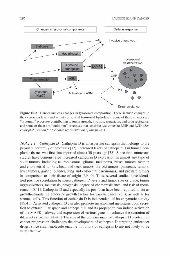
�
� �
�
186 LYSOSOME AND CANCER
Changes in lysosomal components Cellular response
Invasive phenotype
Sialidase
Heparinase
Cysteine
cathepsins
Lysosomal
destabilization
H+
H+
H+
Transformation
Lysosomal
Hsp70Activation of ASM
LAMPS
ASMLMP
Drug resistance
Figure 10.2 Cancer induces changes in lysosomal composition. These include changes inthe expression levels and activity of several lysosomal hydrolases. Some of these changes are“protumor” processes contributing to tumor growth, invasion, metastasis, and drug resistance,and some of them are “antitumor” processes that sensitize lysosomes to LMP and LCD. (Seecolor plate section for the color representation of this figure.)
10.4.1.1.1 Cathepsin D Cathepsin D is an aspartate cathepsin that belongs to thepepsin superfamily of proteases [37]. Increased levels of cathepsin D in human neo-plastic tissues was first time reported almost 30 years ago [38]. Since then, numerousstudies have demonstrated increased cathepsin D expression in almost any type ofsolid tumors, including neuroblastoma, glioma, melanoma, breast tumors, ovarianand endometrial tumors, head and neck tumors, thyroid tumors, pancreatic tumors,liver tumors, gastric, bladder, lung and colorectal carcinomas, and prostate tumorsin comparison to their tissue of origin [39,40]. Thus, several studies have identi-fied positive correlation between cathepsin D levels and tumor size or grade, tumoraggressiveness, metastasis, prognosis, degree of chemoresistance, and risk of recur-rence [40,41]. Cathepsin D and especially its pro-form have been reported to act asgrowth-stimulating autocrine growth factors for various cancer cells, as well as forstromal cells. This function of cathepsin D is independent of its enzymatic activity[39,41]. Activated cathepsin D can also promote invasion and metastasis upon secre-tion to extracellular space and cathepsin D and its propeptide can induce activationof the MAPK pathway and expression of various genes to enhance the secretion ofdifferent cytokines [41–43]. The role of the protease inactive cathepsin D pro-form incancer progression challenges the development of cathepsin D targeting anticancerdrugs, since small-molecule enzyme inhibitors of cathepsin D are not likely to bevery effective.

�
� �
�
CANCER-INDUCED CHANGES IN LYSOSOME COMPOSITION 187
10.4.1.1.2 Cathepsin B Cathepsin B is a cysteine cathepsin and belongs to thepapain superfamily of proteases. In human colorectal and breast cancers, the expres-sion of cathepsin B is increased in cancer cells as well as in the tumor-associatedcells such as macrophages and stromal fibroblasts [11,44,45]. Increased expressionand secretion of cathepsin B have also been reported in brain, lung, and prostatecancers. In general, increased cathepsin B protein levels detected in immunohisto-chemical staining correlate well with higher mRNA levels in cancer cells and tis-sues, indicating a transcriptional upregulation of cathepsin B gene (CTSB) in variouscancers [46]. In tumors, cathepsin B expression is predominantly increased in thetumor cells at the invasive front of the tumors [47,48]. Overexpression of oncogenicRas [14–16] or ErbB2 [13,14] can induce cathepsin B expression and activity. TheErbB2-induced expression of CTSB is transcriptionally regulated, however indepen-dently of the lysosomal biogenesis regulator transcription factor EB (TFEB) [13,49].Instead, ErbB2 induces the myeloid zinc finger transcription factor 1 (MZF1), whichbinds to an ErbB2-inducible enhancer element in the first intron of CTSB gene invivo. The ErbB2-mediated activity of MZF1 is regulated via an ErbB2-activated sig-naling network consisting of PAK4, 5 and 6 cdc42bpβ, PKCα, and MAPK-ERK2serine–threonine kinases as well as TGFβRI and 2 [13]. Interestingly, MAPK-ERK2activity, which is essential for the ErbB2-induced activation of MZF1 and expres-sion of CTSB, can inhibit the nuclear import of TFEB [50], further supporting theobservations that ErbB2 induces CTSB expression independently of TFEB [13]. Inmalignant, highly invasive tumors overexpressed cathepsin B is often secreted intothe extracellular space where it associates with the tumor cell surface [16,51,52].
The active role of cathepsin B in cancer is strongly supported by murine models.In the RIP1-Tag2/RT2 (RT2) pancreatic islet cell carcinogenesis model mice developmultiple tumors in their pancreatic islets due to overexpression of oncogenic SV40T-antigen in insulin producing ß-cells. These mice express high levels of cathepsin B[33,53] and its genetic inactivation results in impaired tumor formation, angiogene-sis, and invasiveness [53]. The suggested mechanism of how cathepsin B contributesto increased tumor formation involves secretion of cathepsin B from tumor cellsand tumor-associated macrophages and cathepsin B-mediated direct cleavage of theepithelial transmembrane protein E-cadherin as well as activation of the urokinaseplasminogen activator by cleavage of its propeptide [53,54]. In addition to activatingplasmin and various matrix metalloproteases, cathepsin B can also directly degradeextracellular matrix by cleaving laminin, type VI collagen, and tenascin-C [55–57].Similarly, Ctsb deficiency significantly delays the onset and growth of primary mam-mary tumors as well as formation of lung metastases in murine mammary tumor viruspolyoma middle-T antigen (PyMT) transgenic mice and cathepsin B overexpressionin these mice promotes mammary tumor growth and metastasis [58,59]. In the PyMTmodel, the expression of cathepsin B in the actual cancer cells rather than in thestromal cells promotes tumor progression [60].
Since cathepsin B is overexpressed in various cancers and several studies showbeneficial effects of its inhibition by RNA interference or pharmacological com-pounds in vitro and its genetic or pharmacological inhibition in vivo in mouse studies,it is believed that targeting cathepsin B could have significant therapeutic effect [61].

�
� �
�
188 LYSOSOME AND CANCER
However, even though many compounds that can inhibit cathepsin B have been iso-lated and developed, none of them have shown to be potential enough for clinics [61].
10.4.1.1.3 Cathepsin L Cathepsin L is ubiquitously expressed cysteine cathepsinthat is often overexpressed in human tumors [45]. It is capable of cleaving variousextracellular matrix proteins such as laminin, fibronectin, and collagens as well asE-cadherin and proheparanase [53,62–65]. Several studies conclude that cathepsinL plays an important role in cancer cell invasion and migration by decreasingcell–cell adhesion and increasing extracellular matrix degradation [30,66]. Studieselucidating the role of cathepsin L in angiogenesis have contradictory results,suggesting that it is either angiogenesis promoting or not involved at all [65]. Forexample, in glioma patients, cathepsin L expression is not associated significantlywith new vasculature of astrocytic tumors [67] but a cathepsin L-specific inhibitorNapSul-Ile-Trp-CHO (NSITC) displays potent antiangiogenic activity in vitro inchick chorioallantoic membrane and mouse Matrigel models of angiogenesis [68].Crossing of an RT2 transgenic mice modeling pancreatic carcinoma with cathepsinL knockout mice showed decreased tumor growth and invasiveness when comparedto the corresponding cathepsin B knockout mice. However, these mice showed nosigns of reduction in the angiogenic switch on the contrary to the correspondingcathepsin B knockout mice [53]. It is to be noted that these pancreatic tumorsexhibited increased expression of several cathepsins and thus it is possible that someother cathepsins were able to compensate the loss of cathepsin L [53].
In colorectal cancer, nuclear staining of cathepsin L increases and cytoplasmicstaining decreases with advancing tumor stage and is associated with poor progno-sis [69]. Indeed, a short form of cathepsin L that lacks the signal peptide needed forlysosomal targeting has been found in nucleus. There it cleaves histone H3 and spe-cific transcription factors, such as the CCAAT-displacement protein/cut homeobox(CDP/Cux), altering gene expression patterns and leading into delay in cell cycleprogression and contributing to oncogenic transformation [70–72]. Interestingly, Rastransformation can increase both the production of short nuclear cathepsin L isoformsand the cleavage of CDP/Cux [72].
Inhibition of cathepsin L can reverse cancer cell’s resistance to various chemother-apeutic drugs. This is suggested to occur due to cathepsin L inhibition-mediatedstabilization and increased availability of cytoplasmic and nuclear targets includ-ing estrogen receptor-α, Bcr-Abl, topoisomerase-II α, histone deacetylase 1, and theandrogen receptor [68,73].
However, contradicting to the beneficial role of cathepsin L inhibition in manyother types of cancers, inactivation of cathepsin L, for example, in mouse intestinalepithelia promotes tumor progression [74]. Similarly, in mouse skin cancer models,genetic inactivation of cathepsin L results in increased tumorigenesis [75,76]. Indeed,cathepsin L has been found to be essential for the negative regulation of the growthfactor and growth factor receptor recycling in keratinocytes. Thus, its inhibitionwill result in increased receptor levels at the plasma membrane as well as increasedlevels of growth factors [77]. Moreover, transgenic overexpression of serine proteaseinhibitor hurpin, also known as serpin, a specific inhibitor of cathepsin L, mainly

�
� �
�
CANCER-INDUCED CHANGES IN LYSOSOME COMPOSITION 189
expressed in skin epidermis, leads to increased skin cancer progression. Hurpinoverexpressing mouse skin undergoes less apoptosis than corresponding wild-typeskin upon exposure to UVB radiation [78], suggesting for a possible involvement ofcathepsin L in apoptosis regulation. Due to recent reports indicating that cathepsinL deficiency can promote tumor progression, its usefulness as therapeutic target hasbecome questionable.
10.4.1.1.4 Cathepsin K Unlike cysteine cathepsins B and L that are expressedubiquitously, expression of cysteine cathepsin K is mainly restricted to skeleton andparticularly in osteoclasts, where it is the predominant cysteine cathepsin [79]. Ithas a central role in osteoclastic resorption and inactivating mutants of the humancathepsin K gene (CTSK) result in pycnodysostosis characterized by short stature,skull deformities, and skeletal abnormalities [80]. Cathepsin K is expressed inbreast and prostate carcinomas and its expression is enhanced in bone metastasesof these cancers when compared to corresponding primary tumors [81–83]. Severalcathepsin K inhibitors (e.g., CKI, AFG-495, balicatib, and odanacatib) are devel-oped to treat osteoporosis-associated bone loss [79,84,85]. Of these, for example,CKI can reduce breast cancer–induced osteolysis and skeletal tumor burden in amouse experimental skeletal metastasis model [82]. However, most successful ofcathepsin K inhibitors, thus far, is odanacatib that is currently on phase III trialas a treatment to reduce the risk of breast cancer–induced bone metastasis (http://www.cancer.gov/drugdictionary). Interestingly, cathepsin K is also expressed inmelanoma where its expression correlates with advanced metastatic disease. Indeed,pharmacological inhibition of cathepsin K with a rather unspecific cathepsin Kinhibitor Boc-I decreases melanoma cell invasion through the Matrigel basementmembrane matrix [86].
10.4.1.1.5 Additional Cysteine Cathepsins The expression of cysteine cathepsinsF, H, S, V (also known as L2 or U), and Z (also known as Y or X) is also increased incancer. In addition to cathepsins B and L, increase in the expression of cathepsins Kand V has been observed in breast carcinomas [83,87]. Furthermore, cathepsin V isoverexpressed in renal, ovarian, colorectal, and squamous cell carcinomas, cathepsinS and Z expression is increased in prostate carcinoma, and expression of cathepsin Zis increased in gastric cancers [88–90].
In a mouse RT2 model of pancreatic carcinoma, pancreatic tumors exhibitincreased expression of cathepsins C, S, H, and Z in addition to cathepsins B andL and treatment of mice with a broad-spectrum pharmacological cell-permeablecathepsin inhibitor led to delayed proliferation, angiogenic switch, tumor vascu-larity, and invasion [91]. Genetic inactivation of cathepsin S in this model systemresulted in impaired tumor formation and angiogenesis, similar to the inactivationof cathepsin B [53]. In the RT2 model, the absence of cathepsin B, L, or S resultedin delayed tumor invasion [53]. E-cadherin, which is a suppressor of invasion, wasidentified as a target for cathepsins B, L, and S but not C in these tumors, andE-cadherin levels were well maintained in knockout RT2 pancreatic carcinomascompared to the corresponding RT2 wild-type tumors [53].
�
� �
�
190 LYSOSOME AND CANCER
Increased expression of cathepsins B and Z is also detectable on the surface of thecancer cells in the polyoma middle-T oncogene (MMTV-PyMT) induced mammarybreast carcinomas [58]. Genetic inactivation of cathepsin B or Z slightly delays theappearance of tumors as well as lung metastases [92,93], but their combined inacti-vation results in significant delay in tumor burden and reduction in the number andsize of metastases [92]. Cathepsin S, which is mainly expressed by endothelial cells,can function in angiogenesis by degrading antiangiogenic, type IV collagen-derivedpeptides and generating proangiogenic peptides by cleavage of laminin-5 [94].
10.4.1.2 Deregulation of Cystatins: Endogenous Cathepsin Inhibitors Theincreased activity of cysteine cathepsins is often connected to the cancer-associatedderegulation of cystatins, which are endogenous cysteine cathepsin inhibitors [95].There are four main cystatins (A, B, C, and M). They are epigenetically silenced inbreast, brain, lung, and pancreatic cancers, linking them to the increased activity ofcysteine cathepsins in these tissues [96]. The functional importance of cystatins incancer is demonstrated, for example, by an experimental human fibrosarcoma lungmetastasis model in which overexpression of cystatin C reduces metastasis [97] andby in vitro invasion assays where overexpression of cystatin M or S can suppressbreast cancer and melanoma cell invasion [95,96].
10.4.1.3 Pharmacological Targeting of Cysteine Cathepsins Of all the cysteinecathepsins, only inhibitors of cathepsin K, the osteoclast-specific cysteine cathepsin,have reached the clinics. In most cases, often more than one cysteine cathepsin isupregulated upon oncogene-driven cell transformation in vitro, in preclinical murinecancer models as well as in human cancer. Their highly overlapping substrate speci-ficities and prooncogenic functions in tumorigenesis suggest that their combinedinhibition might be required for efficient anticancer therapy. In line with this, the com-bined inactivation of Ctsb and Ctsz in PyMT mice [92,93], treatment of mice with acell-permeable, broad-spectrum cysteine cathepsin inhibitor JPM-OEt was more effi-cient than single depletions of Ctsb, Ctsl, or Ctss in reducing angiogenic switching inthe murine RT2 pancreatic carcinoma model [33,53]. On the contrary, JPM-OEt is notefficient in the PyMT murine mammary cancer model and fails to reduce metastasisin a breast cancer bone metastasis model in which the non-cell-permeable cathepsin Binhibitor, CA-074, is effective [98,99]. This may be due to differences in the bioavail-ability of these inhibitors in different tissues [99]. Another possible explanation is thatdifferent cysteine cathepsins may still have unexplored opposing roles in some can-cers. However, the currently available preclinical data is encouraging and supportsthe continuation of the development of broad-spectrum cysteine cathepsin inhibitorsas cancer therapeutics [100]. However, more basic research on the function and local-ization of individual cathepsins is needed to identify the optimal treatment modalities.
10.4.1.4 Heparanase Heparanase is an endo-β-d-glucuronidase and the mostimportant mammalian enzyme degrading heparan sulfate. Heparanase is processedin lysosomes and late endosomes and in addition to these it is also active in extra-cellular matrix. Cleavage of heparan sulfate side chains from the heparan sulfate

�
� �
�
CANCER-INDUCED CHANGES IN LYSOSOME COMPOSITION 191
proteoglycans by heparanase leads to the disassembly of the extracellular matrix andis associated with tissue remodeling, cell movement, cancer progression, metastasis,and angiogenesis [34]. Heparan sulfate binds and sequesters several growth factors,cytokines, and chemokines into the extracellular matrix and cell surface and itscleavage will therefore also increase their release and tissue availability [101,102].Heparanase expression is induced in nearly all human cancers [103,104] andincreased heparanase activity correlates with the metastatic potential of tumorcells, contributes to remodeling of the extracellular matrix and is associated withneovascularization [105,106]. Cancer cells can secrete heparanase in response tosuitable stimulus such as ATP, ADP, and adenosine nucleotides with similar kineticsthan cathepsin D [107]. Heparanase is also found in the body fluids of patients withmetastatic cancer, indicating its secretion in vivo [108,109].
Heparan and its derivatives, sulfated oligoshaccarides suramin and suramin ana-logues, have potent anti-metastatic activity in mouse models of metastatic cancers[110–112]. Heparin itself is a close mimic of heparan sulfate and a natural heparanaseinhibitor. However, it is not clinically usable due to its potent anticoagulant activity.Thus, heparin modifications have been largely used for the development of efficientheparanase inhibitors [113]. These include SST0001, which is currently in Phase I/IIclinical trial for myeloma treatment [113]. Another heparin derivative is M402 thatis also currently in Phase I/II clinical trial in combination with gemcitabine againstmetastatic pancreatic cancer [113]. The PI-88, a heparanase inhibitor with potentantiangiogenic activity [114,115], is currently on Phase III clinical trial as “the adju-vant treatment of subjects with hepatitis virus-related hepatocellular carcinoma aftersurgical resection” (http://www.cancer.gov/drugdictionary). Moreover, an additionalheparan sulfate mimetic PG545 that is a potent heparanase and angiogenesis inhibitorhas shown to be promising when tested in preclinical murine cancer models [116] andis now on Phase I clinical trial (http://www.cancer.gov/drugdictionary). All of theseheparanase inhibitors aim at inhibiting the extracellular heparanase activity.
10.4.1.5 Sialidase 1/Neuraminidase 1 Aberrant glycosylation is one of the typicalfeatures of cancer cells. Changes in sialylation have been connected with the cancercell invasiveness and metastatic potential [117]. There are four types of mammaliansialidases (Neu1, Neu2, Neu3, and Neu4). Of these, Neu1 is the predominant siali-dase found in lysosomes [118]. When total sialidase activity is generally increasedin cancer, Neu1 activity is decreased and, for example, in rat 3Y1 fibroblasts trans-formed with src or v-fos and mouse adenocarcinoma colon 26 cells, Neu1 expressioncorrelates negatively with the metastatic potential [117]. Ectopic expression ofNeu1 in human colon cancer HT-29 cells significantly decreases and its depletionincreases their potential to metastasize into liver in a mouse xenograft model. Themetastasis-suppressing function of Neu1 is mediated via sialylation of β4 integrin,resulting in its decreased phosphorylation and signaling [119]. Moreover, Neu1 isa negative regulator of lysosomal exocytosis, thus decreased Neu1 levels may con-tribute to increased lysosomal exocytosis supporting its antimetastatic activity [120].Excessive lysosomal exocytosis in Neu1-deficient cells can be inhibited by silencingof lysosome-associated membrane protein 1 (LAMP1), suggesting that oversialylated

�
� �
�
192 LYSOSOME AND CANCER
LAMP1 is a key mediator of excessive exocytosis of lysosomal proteases in thesecells [120]. Increased sialylation of LAMP1 and lysosome-associated membraneprotein 2 (LAMP2) can be observed in some human cancers, and, for example, incolorectal cancer it strongly correlates with the metastatic potential [121]. Becausesialylated LAMPs are essential for the lysosomal membrane stability [122], theexcessive sialylation of LAMPs may protect lysosomal membranes from destabi-lization and thus enhance cancer cell survival. The fact that its inactivation promotescancer progression makes sialidase a challenging target for anticancer treatment.
10.4.2 Changes in the Lysosomal Membrane Proteins
10.4.2.1 Lysosomal Membrane Proteins LAMP1 and LAMP2 The most abun-dant and the most studied lysosomal membrane proteins are LAMP1 and LAMP2.Several less studied lysosomal membrane proteins exist whose role or changes incancer are not established and thus they are not discussed here [3,19,123]. LAMP1and 2 are type-1 transmembrane proteins containing a large and heavily glycosylatedluminal domain and a short C-terminal cytosolic domain. Together they compriseover 50% of the lysosomal membrane proteins and are essential for the maintenanceof the structural integrity of the lysosomal membranes [14,19].
Oncogenic transformation of fibroblasts decreases the levels of LAMPs on theirlysosomal membranes thus increasing their sensitivity to LMP and LCD [14]. Theglycosylated tails of LAMPs form a sugar coat called glycocalyx to the inner surfaceof the limiting membrane protecting the lysosomal membrane from degradation bylysosomal hydrolases [19]. LAMPs have shown to exhibit overlapping and distinctfunctions in lysosomal trafficking and exocytosis, chaperone-mediated autophagy,autophagosome–lysosome fusion, and cholesterol transport [19,120,124], all ofwhich may be affected when LAMP levels are altered.
10.4.2.2 V-ATPase Vacuolar-type H+ ATPase (V-ATPase) is a proton pump that isassociated with the membranes of endosomes, lysosomes, and secretory vesicles, andit is also found at the plasma membranes of many cell types. V-ATPase is importantfor the acidification of lysosomes and endosomes is also involved in the regulationof the pH of the cytosol and possibly also of the extracellular space [125]. Its properfunction is necessary for the maintenance of the highly acidic pH of the lysosomallumen and for the maintenance of the calcium homeostasis in lysosomes [126,127].
In spite of the cancer-associated increase in acid production, the entire V-ATPasecomplex does not seem to be upregulated in cancer cells. Instead, the increasedexpression of specific subunits has been observed and is suggested to contribute toaltered pump activity. Thus, overexpression of the gene encoding for the V1 subunitC (ATP6V1C1) in oral squamous carcinoma cells can enhance the pump activityby promoting the assembly of V0 and V1 subcomplexes [128]. On the other hand,the V0 subunit C (ATP6V0C) is upregulated in cisplatin-resistant cells [129]. Theexpression pattern of the subunit isoforms can affect the subcellular localizationof the V-ATPase. The appearance of the V0 a3 isoform favors plasma membranelocalization of this type of V-ATPase [130], whereas the depletion of a3 isoform in
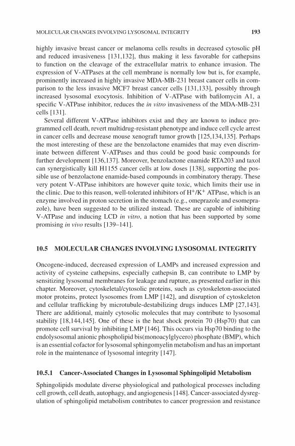
�
� �
�
MOLECULAR CHANGES INVOLVING LYSOSOMAL INTEGRITY 193
highly invasive breast cancer or melanoma cells results in decreased cytosolic pHand reduced invasiveness [131,132], thus making it less favorable for cathepsinsto function on the cleavage of the extracellular matrix to enhance invasion. Theexpression of V-ATPases at the cell membrane is normally low but is, for example,prominently increased in highly invasive MDA-MB-231 breast cancer cells in com-parison to the less invasive MCF7 breast cancer cells [131,133], possibly throughincreased lysosomal exocytosis. Inhibition of V-ATPase with bafilomycin A1, aspecific V-ATPase inhibitor, reduces the in vitro invasiveness of the MDA-MB-231cells [131].
Several different V-ATPase inhibitors exist and they are known to induce pro-grammed cell death, revert multidrug-resistant phenotype and induce cell cycle arrestin cancer cells and decrease mouse xenograft tumor growth [125,134,135]. Perhapsthe most interesting of these are the benzolactone enamides that may even discrim-inate between different V-ATPases and thus could be good basic compounds forfurther development [136,137]. Moreover, benzolactone enamide RTA203 and taxolcan synergistically kill H1155 cancer cells at low doses [138], supporting the pos-sible use of benzolactone enamide-based compounds in combinatory therapy. Thesevery potent V-ATPase inhibitors are however quite toxic, which limits their use inthe clinic. Due to this reason, well-tolerated inhibitors of H+/K+ ATPase, which is anenzyme involved in proton secretion in the stomach (e.g., omeprazole and esomepra-zole), have been suggested to be utilized instead. These are capable of inhibitingV-ATPase and inducing LCD in vitro, a notion that has been supported by somepromising in vivo results [139–141].
10.5 MOLECULAR CHANGES INVOLVING LYSOSOMAL INTEGRITY
Oncogene-induced, decreased expression of LAMPs and increased expression andactivity of cysteine cathepsins, especially cathepsin B, can contribute to LMP bysensitizing lysosomal membranes for leakage and rupture, as presented earlier in thischapter. Moreover, cytoskeletal/cytosolic proteins, such as cytoskeleton-associatedmotor proteins, protect lysosomes from LMP [142], and disruption of cytoskeletonand cellular trafficking by microtubule-destabilizing drugs induces LMP [27,143].There are additional, mainly cytosolic molecules that may contribute to lysosomalstability [18,144,145]. One of these is the heat shock protein 70 (Hsp70) that canpromote cell survival by inhibiting LMP [146]. This occurs via Hsp70 binding to theendolysosomal anionic phospholipid bis(monoacylglycero) phosphate (BMP), whichis an essential cofactor for lysosomal sphingomyelin metabolism and has an importantrole in the maintenance of lysosomal integrity [147].
10.5.1 Cancer-Associated Changes in Lysosomal Sphingolipid Metabolism
Sphingolipids modulate diverse physiological and pathological processes includingcell growth, cell death, autophagy, and angiogenesis [148]. Cancer-associated dysreg-ulation of sphingolipid metabolism contributes to cancer progression and resistance

�
� �
�
194 LYSOSOME AND CANCER
Sphingomyelin
Ceramide
Sphingosine
Sphingosine-1-P
Acid sphingomyelinase (SMPD1)
Acid ceramidase (ASAH1)
sphingosine kinase
Figure 10.3 Lysosomal sphingolipid catabolism. Sphingomyelin is an abundant structurallipid of biological membranes. Acid sphingomyelinase catalyzes sphingomyelin into ceramide,which is a membrane lipid that can be associated with lipid rafts and function as a signalingmolecule regulating cell death and autophagy. Acid ceramidase converts ceramide into sph-ingosine, which is a soluble detergent that can also function as a signaling molecule. Finally,sphingosine kinase phosphorylates sphingosine turning it into sphingosine-1-phosphate, a sol-uble signaling molecule that can bind to cell surface receptor promoting cell survival andautophagy.
to chemotherapeutics [149]. Sphingomyelinases and ceramidases are key enzymesof sphingolipid catabolism (Figure 10.3). Human cells express several forms of theseenzymes with varying optimal pH. Here, we present only their lysosomal forms thatcan function in low pH, that is, ASM and acid ceramidase (AC). They regulate the for-mation and degradation of ceramide, one of the most studied sphingolipids. The vastinterest to ceramide in cancer owes to its capacity to induce programmed cell deathand inhibit cell proliferation [150]. Genetic inactivation of acid sphingomyelinase,which is the major sphingomyelinase in lysosomes, protects mice against variouscellular stresses including ionizing radiation and treatment with cisplatin, assum-ably due to deficient ceramide production, supporting the apoptosis-promoting roleof ceramide [151].
Sphingolipids and sphingolipid metabolites such as ceramide regulate cell sur-vival with diverse mechanisms that are not completely understood. The proapoptoticactivity of ASM is mostly attributed to its localization to the plasma membrane

�
� �
�
MOLECULAR CHANGES INVOLVING LYSOSOMAL INTEGRITY 195
in response to various stresses [152,153]. This induces generation of ceramide atthe plasma membrane contributing to the formation of ordered membrane domainsthat can enhance death receptor function and lead to the induction of apoptosis[151–153]. ASM activity is generally considered to negatively regulate cancerprogression, mainly due to the proapoptotic properties of ceramide [154,155].Contrasting this, lysosomal ASM has an opposite effect on cell survival, sinceits activity is necessary for the maintenance of the lysosomal membrane integrity[22,144,147]. Another mechanism how ASM can positively contribute to cancerprogression is by ceramide-mediated activation of pre-pro cathepsin D resultingin increased activity of cathepsin D [156]. Interestingly, a database search revealsa highly significant cancer-associated reduction in the mRNA level of SMPD1encoding for ASM in multiple studies of gastrointestinal, hepatocellular, salivarygland, renal, and head and neck carcinomas. A modest but significant increase inSMPD1 expression was found in single studies of hairy cell leukemia and smolderingmyeloma (www.oncomine.org). The decreased SMPD1 expression in several cancersmay contribute to the increased sensitivity of transformed cells to LCD pathways.On the other hand, the SMPD1 mRNA levels do not necessarily reflect the levels andactivity of ASM protein, which are further contributed by its stability, binding withBMP and association with Hsp70 [146,147,157].
Lysosomal ceramide is degraded by AC to produce sphingosine, which diffusesrapidly from lysosomes to cytosol and is converted to sphingosine-1-phosphate [158,159]. Aberrant expression of AC has been attributed to several human cancers [160].Inhibition of AC activity is expected to lead to increased ceramide levels, which canstimulate programmed cell death. This has been verified with different cancer andprimary cell lines using various AC inhibitors such as N-oleoylethanolamine, B13,and De-MAPP [160,161]. Although this data suggests that AC inhibition could bea good cancer therapy strategy, it is to be noticed that the specificity of many ofthese inhibitors toward AC in respect to, for example, other ceramidases remainsto be verified. In addition to survival, sphingolipid metabolism is also involved inother cellular processes that are important in cancer including proliferation, cell cycleprogression, and inflammation, which have increased its interest as a potential cancertherapy target [159].
10.5.2 Targeting Lysosomal Membrane Integrity
Cancer cells frequently escape from spontaneous and therapy-induced apoptosisdue to mutational defects in the caspase-dependent apoptosis pathways [162].Thus, identification and utilization of caspase-independent cellular death programsto selectively kill cancer cells have become increasingly important [17,163,164].Cancer development and progression involves dramatic changes in the compositionand integrity of lysosomal membranes, which provides intriguing possibilitiesfor drug-induced LMP and LCD [14,17,22,165]. Several established anticanceragents induce LCD [17]. These include DNA-damaging drugs camptothecin,cisplatin, and etoposide [14,166,167]; microtubule targeting drugs discodermolide,docetaxel, epothilone B, paclitaxel, and vincristine [27,143,168]; V-ATPase

�
� �
�
196 LYSOSOME AND CANCER
inhibitors omeprazole and bafilomycin A1 [139,169,170] among many others [17].Interestingly, in all of these cases, LCD can be mediated by single cathepsins B, D,or L, or their different combinations [17].
Recently, lysosomotropic detergents that specifically destabilize lysosomal mem-branes in cancer cells have evolved as highly promising, potential anticancer remedies[17,21]. These include siramesine [22,171,172], BAMLET [173], hydroxychloro-quine [174], LeuLeuOMe [175], sphingosine [176], mefloquine [177], and SU11652and sunitinib [178]. Recent work with siramesine has led to the identification ofa group of compounds, a subset of cationic amphiphilic drugs, which specificallysensitize lysosomal membranes of cancer cells by a mechanism that involves directinhibition of ASM [22]. Interestingly, these compounds involve some feasible antide-pressants and antiallergy remedies [22,179]. In addition to ASM inhibition, theirdetergent-like activity and ability to induce the production of reactive oxygen speciesis likely to further contribute to LMP and LCD [22]. Very exciting and promisingproperties associated with these LCD-inducing agents are that they can also effi-ciently kill multidrug-resistant cancer cells [17,22].
10.6 CONCLUSION
Lysosomes are packed with powerful hydrolases that upon release from the lysoso-mal compartment can degrade the environment that they encounter. This imposesopposing outcomes to cancer cells depending on where the lysosomes “leak,” thuschallenging the design of the lysosome targeting therapies. In principle, lysosomalcontent that encounters the cytosol will most likely induce cell death and destruc-tion of the cancer cell. On the contrary, leakage of lysosomal hydrolases into theextracellular space can facilitate cancer cell invasion by degrading the extracellularmatrix. Thus, these two options provide two different possibilities to intervene. Con-sequently, two types of promising anticancer therapy strategies involving lysosomesare currently being explored: inhibition of extracellular lysosomal hydrolase activitiesand induction of lysosomal permeabilization and leakage within cancer cells.
Long-term research on cysteine cathepsins and heparanase has led to the devel-opment of several highly promising anticancer agents with a strategy to inhibitinvasion and metastasis by inhibiting the activity of these proteases [11,113,180].Many of these are already in clinical trials. Moreover, recent identification of severalcommonly used anticancer remedies as compounds that can induce cancer-specificLMP [17] together with the identification of a widely used subset of cationicamphiphilic drugs including tricyclic antidepressants, antihistamines, and calciumchannel blockers as direct ASM inhibitors capable of destabilizing lysosomalmembranes in cancer cells [22] has rapidly provided many novel and feasiblepossibilities to intervene cancer progression by targeting lysosomes in cancer cells.Especially, important and promising are the observations indicating that they cantarget apoptosis-deficient cancer cells as well as those that have become multidrugresistant [17,22,178]. Since cathepsins are important mediators of both invasion andLCD, more research is needed to dissect those cancers that would mostly benefit

�
� �
�
REFERENCES 197
from cathepsin inhibition from those that would need full cathepsin activity for theexecution of cell death. One possible option could be to combine both processes:the induction of cell death and the inhibition of invasion. This could, for example,be achieved by combining LCD-inducing treatments with treatments that inhibitlysosomal exocytosis. This would require more research on lysosomal exocytosisand its regulation in cancer.
REFERENCES
[1] Kolter T, Sandhoff K. Lysosomal degradation of membrane lipids. FEBS Lett 2010;584:1700–1712.
[2] Pryor PR, Luzio JP. Delivery of endocytosed membrane proteins to the lysosome.Biochim Biophys Acta 2009;1793:615–624.
[3] Saftig P, Klumperman J. Lysosome biogenesis and lysosomal membrane proteins: traf-ficking meets function. Nat Rev Mol Cell Biol 2009;10:623–635.
[4] Grandal MV, Madshus IH. Epidermal growth factor receptor and cancer: control ofoncogenic signalling by endocytosis. J Cell Mol Med 2008;12:1527–1534.
[5] Mellman I, Yarden Y. Endocytosis and cancer. Cold Spring Harb Perspect Biol 2013;5:a016949.
[6] Ivanov A, Pawlikowski J, Manoharan I, van Tuyn J, Nelson DM, et al. Lysosome-mediated processing of chromatin in senescence. J Cell Biol 2013;202:129–143.
[7] Kreuzaler PA, Staniszewska AD, Li W, Omidvar N, Kedjouar B, et al. Stat3 controlslysosomal-mediated cell death in vivo. Nat Cell Biol 2011;13:303–309.
[8] Watson CJ, Kreuzaler PA. The role of cathepsins in involution and breast cancer. J Mam-mary Gland Biol Neoplasia 2009;14:171–179.
[9] Glunde K, Guggino SE, Solaiyappan M, Pathak AP, Ichikawa Y, et al. Extracellu-lar acidification alters lysosomal trafficking in human breast cancer cells. Neoplasia2003;5:533–545.
[10] Liu Y, Zhou Y, Zhu K. Inhibition of glioma cell lysosome exocytosis inhibits gliomainvasion. PLoS One 2012;7:e45910.
[11] Kallunki T, Olsen OD, Jaattela M. Cancer-associated lysosomal changes: friends orfoes? Oncogene 2013;32:1995–2004.
[12] Allison AC. Lysosomes in cancer cells. J Clin Pathol Suppl (R Coll Pathol) 1974;7:43–50.
[13] Rafn B, Nielsen CF, Andersen SH, Szyniarowski P, Corcelle-Termeau E, et al. ErbB2-driven breast cancer cell invasion depends on a complex signaling network acti-vating myeloid zinc finger-1-dependent cathepsin B expression. Mol Cell 2012;45:764–776.
[14] Fehrenbacher N, Bastholm L, Kirkegaard-Sorensen T, Rafn B, Bottzauw T, et al. Sen-sitization to the lysosomal cell death pathway by oncogene-induced down-regulation oflysosome-associated membrane proteins 1 and 2. Cancer Res 2008;68:6623–6633.
[15] Kim K, Cai J, Shuja S, Kuo T, Murnane MJ. Presence of activated ras correlates withincreased cysteine proteinase activities in human colorectal carcinomas. Int J Cancer1998;79:324–333.
�
� �
�
198 LYSOSOME AND CANCER
[16] Sloane BF, Moin K, Sameni M, Tait LR, Rozhin J, et al. Membrane association ofcathepsin B can be induced by transfection of human breast epithelial cells with c-Ha-rasoncogene. J Cell Sci 1994;107(Pt 2):373–384.
[17] Groth-Pedersen L, Jaattela M. Combating apoptosis and multidrug resistant cancers bytargeting lysosomes. Cancer Lett 2013;332:265–274.
[18] Boya P, Kroemer G. Lysosomal membrane permeabilization in cell death. Oncogene2008;27:6434–6451.
[19] Eskelinen EL. Roles of LAMP-1 and LAMP-2 in lysosome biogenesis and autophagy.Mol Aspects Med 2006;27:495–502.
[20] Fehrenbacher N, Jaattela M. Lysosomes as targets for cancer therapy. Cancer Res2005;65:2993–2995.
[21] Aits S, Jaattela M. Lysosomal cell death at a glance. J Cell Sci 2013;126:1905–1912.
[22] Petersen NH, Olsen OD, Groth-Pedersen L, Ellegaard AM, Bilgin M, et al.Transformation-associated changes in sphingolipid metabolism sensitize cells to lyso-somal cell death induced by inhibitors of acid sphingomyelinase. Cancer Cell 2013;24:379–393.
[23] Mora R, Dokic I, Kees T, Huber CM, Keitel D, et al. Sphingolipid rheostat alterationsrelated to transformation can be exploited for specific induction of lysosomal cell deathin murine and human glioma. Glia 2010;58:1364–1383.
[24] Taha TA, Kitatani K, Bielawski J, Cho W, Hannun YA, et al. Tumor necrosis factorinduces the loss of sphingosine kinase-1 by a cathepsin B-dependent mechanism. J BiolChem 2005;280:17196–17202.
[25] Durchfort N, Verhoef S, Vaughn MB, Shrestha R, Adam D, et al. The enlarged lyso-somes in beige j cells result from decreased lysosome fission and not increased lysosomefusion. Traffic 2012;13:108–119.
[26] Ono K, Kim SO, Han J. Susceptibility of lysosomes to rupture is a determinant forplasma membrane disruption in tumor necrosis factor alpha-induced cell death. MolCell Biol 2003;23:665–676.
[27] Groth-Pedersen L, Ostenfeld MS, Hoyer-Hansen M, Nylandsted J, Jaattela M. Vin-cristine induces dramatic lysosomal changes and sensitizes cancer cells to lysosome-destabilizing siramesine. Cancer Res 2007;67:2217–2225.
[28] Brandes D, Anton E. The role of lysosomes in cellular lytic processes. 3. Electron his-tochemical changes in mammary tumors after treatment with cytoxan and vitamin A.Lab Invest 1966;15:987–1006.
[29] Brandes D, Sloan KW, Anton E, Bloedorn F. The effect of x-irradiation on the lyso-somes of mouse mammary gland carcinomas. Cancer Res 1967;27:731–746.
[30] Mohamed MM, Sloane BF. Cysteine cathepsins: multifunctional enzymes in cancer.Nat Rev Cancer 2006;6:764–775.
[31] Chiche J, Brahimi-Horn MC, Pouyssegur J. Tumour hypoxia induces a metabolic shiftcausing acidosis: a common feature in cancer. J Cell Mol Med 2010;14:771–794.
[32] Korolchuk VI, Saiki S, Lichtenberg M, Siddiqi FH, Roberts EA, et al. Lysosomal posi-tioning coordinates cellular nutrient responses. Nat Cell Biol 2011;13:453–460.
[33] Joyce JA, Hanahan D. Multiple roles for cysteine cathepsins in cancer. Cell Cycle2004;3:1516–1619.

�
� �
�
REFERENCES 199
[34] Li JP. Heparin, heparan sulfate and heparanase in cancer: remedy for metastasis? Anti-cancer Agents Med Chem 2008;8:64–76.
[35] Nagler DK, Storer AC, Portaro FC, Carmona E, Juliano L, et al. Major increase inendopeptidase activity of human cathepsin B upon removal of occluding loop contacts.Biochemistry 1997;36:12608–12615.
[36] Almeida PC, Nantes IL, Chagas JR, Rizzi CC, Faljoni-Alario A, et al. Cathepsin Bactivity regulation. Heparin-like glycosaminogylcans protect human cathepsin B fromalkaline pH-induced inactivation. J Biol Chem 2001;276:944–951.
[37] Zaidi N, Maurer A, Nieke S, Kalbacher H. Cathepsin D: a cellular roadmap. BiochemBiophys Res Commun 2008;376:5–9.
[38] Reid WA, Valler MJ, Kay J. Immunolocalization of cathepsin D in normal and neoplastichuman tissues. J Clin Pathol 1986;39:1323–1330.
[39] Liaudet-Coopman E, Beaujouin M, Derocq D, Garcia M, Glondu-Lassis M, et al.Cathepsin D: newly discovered functions of a long-standing aspartic protease in cancerand apoptosis. Cancer Lett 2006;237:167–179.
[40] Leto G, Tumminello FM, Crescimanno M, Flandina C, Gebbia N. Cathepsin D expres-sion levels in nongynecological solid tumors: clinical and therapeutic implications. ClinExp Metastasis 2004;21:91–106.
[41] Benes P, Vetvicka V, Fusek M. Cathepsin D – many functions of one aspartic protease.Crit Rev Oncol Hematol 2008;68:12–28.
[42] Benes P, Vashishta A, Saraswat-Ohri S, Fusek M, Pospisilova S, et al. Effect of pro-cathepsin D activation peptide on gene expression of breast cancer cells. Cancer Lett2006;239:46–54.
[43] Leto G, Sepporta MV, Crescimanno M, Flandina C, Tumminello FM. Cathepsin L inmetastatic bone disease: therapeutic implications. Biol Chem 2010;391:655–664.
[44] Mason SD, Joyce JA. Proteolytic networks in cancer. Trends Cell Biol 2011;21:228–237.
[45] Jedeszko C, Sloane BF. Cysteine cathepsins in human cancer. Biol Chem 2004;385:1017–1027.
[46] Yan S, Sloane BF. Molecular regulation of human cathepsin B: implication in patholo-gies. Biol Chem 2003;384:845–854.
[47] Sinha AA, Gleason DF, Deleon OF, Wilson MJ, Sloane BF. Localization of a biotiny-lated cathepsin B oligonucleotide probe in human prostate including invasive cells andinvasive edges by in situ hybridization. Anat Rec 1993;235:233–240.
[48] Demchik LL, Sameni M, Nelson K, Mikkelsen T, Sloane BF. Cathepsin B and gliomainvasion. Int J Dev Neurosci 1999;17:483–494.
[49] Rafn B, Kallunki T. A way to invade: a story of ErbB2 and lysosomes. Cell Cycle2012;11:2415–2416.
[50] Settembre C, Ballabio A. TFEB regulates autophagy: an integrated coordination of cel-lular degradation and recycling processes. Autophagy 2011;7:1379–1381.
[51] Frosch BA, Berquin I, Emmert-Buck MR, Moin K, Sloane BF. Molecular regulation,membrane association and secretion of tumor cathepsin B. APMIS 1999;107:28–37.
[52] Sameni M, Elliott E, Ziegler G, Fortgens PH, Dennison C, et al. Cathepsin B and D arelocalized at the surface of human breast cancer cells. Pathol Oncol Res 1995;1:43–53.

�
� �
�
200 LYSOSOME AND CANCER
[53] Gocheva V, Zeng W, Ke D, Klimstra D, Reinheckel T, et al. Distinct roles for cysteinecathepsin genes in multistage tumorigenesis. Genes Dev 2006;20:543–556.
[54] Gocheva V, Chen X, Peters C, Reinheckel T, Joyce JA. Deletion of cathepsin H per-turbs angiogenic switching, vascularization and growth of tumors in a mouse model ofpancreatic islet cell cancer. Biol Chem 2010;391:937–945.
[55] Mai J, Sameni M, Mikkelsen T, Sloane BF. Degradation of extracellular matrix proteintenascin-C by cathepsin B: an interaction involved in the progression of gliomas. BiolChem 2002;383:1407–1413.
[56] Buck MR, Karustis DG, Day NA, Honn KV, Sloane BF. Degradation of extracellular-matrix proteins by human cathepsin B from normal and tumour tissues. Biochem J1992;282(Pt 1):273–278.
[57] Kobayashi H, Schmitt M, Goretzki L, Chucholowski N, Calvete J, et al. Cathepsin Befficiently activates the soluble and the tumor cell receptor-bound form of the proenzymeurokinase-type plasminogen activator (Pro-uPA). J Biol Chem 1991;266:5147–5152.
[58] Vasiljeva O, Papazoglou A, Kruger A, Brodoefel H, Korovin M, et al. Tumor cell-derived and macrophage-derived cathepsin B promotes progression and lung metastasisof mammary cancer. Cancer Res 2006;66:5242–5250.
[59] Sevenich L, Werner F, Gajda M, Schurigt U, Sieber C, et al. Transgenic expression ofhuman cathepsin B promotes progression and metastasis of polyoma-middle-T-inducedbreast cancer in mice. Oncogene 2011;30:54–64.
[60] Bengsch F, Buck A, Gunther SC, Seiz JR, Tacke M, et al. Cell type-dependentpathogenic functions of overexpressed human cathepsin B in murine breast cancer pro-gression. Oncogene 2013;33:4474–4484.
[61] Gondi CS, Rao JS. Cathepsin B as a cancer target. Expert Opin Ther Targets 2013;17:281–291.
[62] Abboud-Jarrous G, Atzmon R, Peretz T, Palermo C, Gadea BB, et al. Cathepsin L isresponsible for processing and activation of proheparanase through multiple cleavagesof a linker segment. J Biol Chem 2008;283:18167–18176.
[63] Corvo I, Cancela M, Cappetta M, Pi-Denis N, Tort JF, et al. The major cathepsin Lsecreted by the invasive juvenile Fasciola hepatica prefers proline in the S2 subsite andcan cleave collagen. Mol Biochem Parasitol 2009;167:41–47.
[64] Ishidoh K, Kominami E. Procathepsin L degrades extracellular matrix proteins inthe presence of glycosaminoglycans in vitro. Biochem Biophys Res Commun 1995;217:624–631.
[65] Lankelma JM, Voorend DM, Barwari T, Koetsveld J, Van der Spek AH, et al. CathepsinL, target in cancer treatment? Life Sci 2010;86:225–233.
[66] Gocheva V, Joyce JA. Cysteine cathepsins and the cutting edge of cancer invasion. CellCycle 2007;6:60–64.
[67] Strojnik T, Kavalar R, Trinkaus M, Lah TT. Cathepsin L in glioma progression: com-parison with cathepsin B. Cancer Detect Prev 2005;29:448–455.
[68] Rebbaa A, Chu F, Sudha T, Gallati C, Dier U, et al. The anti-angiogenic activity ofNSITC, a specific cathepsin L inhibitor. Anticancer Res 2009;29:4473–4481.
[69] Sullivan S, Tosetto M, Kevans D, Coss A, Wang L, et al. Localization of nuclear cathep-sin L and its association with disease progression and poor outcome in colorectal cancer.Int J Cancer 2009;125:54–61.

�
� �
�
REFERENCES 201
[70] Ceru S, Konjar S, Maher K, Repnik U, Krizaj I, et al. Stefin B interacts with histonesand cathepsin L in the nucleus. J Biol Chem 2010;285:10078–10086.
[71] Duncan EM, Muratore-Schroeder TL, Cook RG, Garcia BA, Shabanowitz J, et al.Cathepsin L proteolytically processes histone H3 during mouse embryonic stem celldifferentiation. Cell 2008;135:284–294.
[72] Goulet B, Baruch A, Moon NS, Poirier M, Sansregret LL, et al. A cathepsin L isoformthat is devoid of a signal peptide localizes to the nucleus in S phase and processes theCDP/Cux transcription factor. Mol Cell 2004;14:207–219.
[73] Zheng X, Chu F, Chou PM, Gallati C, Dier U, et al. Cathepsin L inhibition suppressesdrug resistance in vitro and in vivo: a putative mechanism. Am J Physiol Cell Physiol2009;296:C65–74.
[74] Boudreau F, Lussier CR, Mongrain S, Darsigny M, Drouin JL, et al. Loss of cathep-sin L activity promotes claudin-1 overexpression and intestinal neoplasia. FASEB J2007;21:3853–3865.
[75] Dennemarker J, Lohmuller T, Mayerle J, Tacke M, Lerch MM, et al. Deficiency for thecysteine protease cathepsin L promotes tumor progression in mouse epidermis. Onco-gene 2010;29:1611–1621.
[76] Benavides F, Perez C, Blando J, Contreras O, Shen J, et al. Protective role of cathepsinL in mouse skin carcinogenesis. Mol Carcinog 2011;51:352–361.
[77] Reinheckel T, Hagemann S, Dollwet-Mack S, Martinez E, Lohmuller T, et al. The lyso-somal cysteine protease cathepsin L regulates keratinocyte proliferation by control ofgrowth factor recycling. J Cell Sci 2005;118:3387–3395.
[78] Walz M, Kellermann S, Bylaite M, Andree B, Ruther U, et al. Expression of the humanCathepsin L inhibitor hurpin in mice: skin alterations and increased carcinogenesis. ExpDermatol 2007;16:715–723.
[79] Grabowskal U, Chambers TJ, Shiroo M. Recent developments in cathepsin K inhibitordesign. Curr Opin Drug Discov Devel 2005;8:619–630.
[80] Gelb BD, Shi GP, Chapman HA, Desnick RJ. Pycnodysostosis, a lysosomal diseasecaused by cathepsin K deficiency. Science 1996;273:1236–1238.
[81] Brubaker KD, Vessella RL, True LD, Thomas R, Corey E. Cathepsin K mRNAand protein expression in prostate cancer progression. J Bone Miner Res 2003;18:222–230.
[82] Le Gall C, Bellahcene A, Bonnelye E, Gasser JA, Castronovo V, et al. A cathepsin Kinhibitor reduces breast cancer induced osteolysis and skeletal tumor burden. CancerRes 2007;67:9894–9902.
[83] Littlewood-Evans AJ, Bilbe G, Bowler WB, Farley D, Wlodarski B, et al. The osteoclast-associated protease cathepsin K is expressed in human breast carcinoma. Cancer Res1997;57:5386–5390.
[84] Lewiecki EM. Odanacatib, a cathepsin K inhibitor for the treatment of osteoporo-sis and other skeletal disorders associated with excessive bone remodeling. IDrugs2009;12:799–809.
[85] Podgorski I. Future of anticathepsin K drugs: dual therapy for skeletal disease andatherosclerosis? Future Med Chem 2009;1:21–34.
[86] Quintanilla-Dieck MJ, Codriansky K, Keady M, Bhawan J, Runger TM. Cathepsin Kin melanoma invasion. J Invest Dermatol 2008;128:2281–2288.

�
� �
�
202 LYSOSOME AND CANCER
[87] Santamaria I, Velasco G, Cazorla M, Fueyo A, Campo E, et al. Cathepsin L2, a novelhuman cysteine proteinase produced by breast and colorectal carcinomas. Cancer Res1998;58:1624–1630.
[88] Krueger S, Kalinski T, Hundertmark T, Wex T, Kuster D, et al. Up-regulation of cathep-sin X in Helicobacter pylori gastritis and gastric cancer. J Pathol 2005;207:32–42.
[89] Fernandez PL, Farre X, Nadal A, Fernandez E, Peiro N, et al. Expression of cathepsinsB and S in the progression of prostate carcinoma. Int J Cancer 2001;95:51–55.
[90] Nagler DK, Kruger S, Kellner A, Ziomek E, Menard R, et al. Up-regulation of cathepsinX in prostate cancer and prostatic intraepithelial neoplasia. Prostate 2004;60:109–119.
[91] Joyce JA, Baruch A, Chehade K, Meyer-Morse N, Giraudo E, et al. Cathepsin cysteineproteases are effectors of invasive growth and angiogenesis during multistage tumori-genesis. Cancer Cell 2004;5:443–453.
[92] Sevenich L, Schurigt U, Sachse K, Gajda M, Werner F, et al. Synergistic antitumoreffects of combined cathepsin B and cathepsin Z deficiencies on breast cancer progres-sion and metastasis in mice. Proc Natl Acad Sci U S A 2010;107:2497–2502.
[93] Vasiljeva O, Korovin M, Gajda M, Brodoefel H, Bojic L, et al. Reduced tumour cellproliferation and delayed development of high-grade mammary carcinomas in cathepsinB-deficient mice. Oncogene 2008;27:4191–4199.
[94] Wang B, Sun J, Kitamoto S, Yang M, Grubb A, et al. Cathepsin S controls angiogen-esis and tumor growth via matrix-derived angiogenic factors. J Biol Chem 2006;281:6020–6029.
[95] Cox JL. Cystatins and cancer. Front Biosci (Landmark Ed) 2009;14:463–474.
[96] Rivenbark AG, Coleman WB. Epigenetic regulation of cystatins in cancer. Front Biosci(Landmark Ed) 2009;14:453–462.
[97] Kopitz C, Anton M, Gansbacher B, Kruger A. Reduction of experimental humanfibrosarcoma lung metastasis in mice by adenovirus-mediated cystatin C overexpressionin the host. Cancer Res 2005;65:8608–8612.
[98] Withana NP, Blum G, Sameni M, Slaney C, Anbalagan A, et al. Cathepsin B inhibitionlimits bone metastasis in breast cancer. Cancer Res 2012;72:1199–1209.
[99] Schurigt U, Sevenich L, Vannier C, Gajda M, Schwinde A, et al. Trial of the cysteinecathepsin inhibitor JPM-OEt on early and advanced mammary cancer stages in theMMTV-PyMT-transgenic mouse model. Biol Chem 2008;389:1067–1074.
[100] Palermo C, Joyce JA. Cysteine cathepsin proteases as pharmacological targets in cancer.Trends Pharmacol Sci 2008;29:22–28.
[101] Bernfield M, Gotte M, Park PW, Reizes O, Fitzgerald ML, et al. Functions of cell surfaceheparan sulfate proteoglycans. Annu Rev Biochem 1999;68:729–777.
[102] Folkman J, Klagsbrun M, Sasse J, Wadzinski M, Ingber D, et al. A heparin-bindingangiogenic protein – basic fibroblast growth factor – is stored within basement mem-brane. Am J Pathol 1988;130:393–400.
[103] Friedmann Y, Vlodavsky I, Aingorn H, Aviv A, Peretz T, et al. Expression of heparanasein normal, dysplastic, and neoplastic human colonic mucosa and stroma. Evidence forits role in colonic tumorigenesis. Am J Pathol 2000;157:1167–1175.
[104] Ilan N, Elkin M, Vlodavsky I. Regulation, function and clinical significance of hep-aranase in cancer metastasis and angiogenesis. Int J Biochem Cell Biol 2006;38:2018–2039.

�
� �
�
REFERENCES 203
[105] Parish CR, Freeman C, Hulett MD. Heparanase: a key enzyme involved in cell invasion.Biochim Biophys Acta 2001;1471:M99–108.
[106] Vlodavsky I, Friedmann Y. Molecular properties and involvement of heparanase in can-cer metastasis and angiogenesis. J Clin Invest 2001;108:341–347.
[107] Shafat I, Vlodavsky I, Ilan N. Characterization of mechanisms involved in secretion ofactive heparanase. J Biol Chem 2006;281:23804–23811.
[108] Davidson B, Shafat I, Risberg B, Ilan N, Trope CG, et al. Heparanase expressioncorrelates with poor survival in metastatic ovarian carcinoma. Gynecol Oncol 2007;104:311–319.
[109] Quiros RM, Rao G, Plate J, Harris JE, Brunn GJ, et al. Elevated serum heparanase-1levels in patients with pancreatic carcinoma are associated with poor survival. Cancer2006;106:532–540.
[110] Marchetti D, Reiland J, Erwin B, Roy M. Inhibition of heparanase activity andheparanase-induced angiogenesis by suramin analogues. Int J Cancer 2003;104:167–174.
[111] Vlodavsky I, Mohsen M, Lider O, Svahn CM, Ekre HP, et al. Inhibition of tumor metas-tasis by heparanase inhibiting species of heparin. Invasion Metastasis 1994;14:290–302.
[112] Norrby K. Low-molecular-weight heparins and angiogenesis. APMIS 2006;114:79–102.
[113] Pisano C, Vlodavsky I, Ilan N, Zunino F. The potential of heparanase as a therapeutictarget in cancer. Biochem Pharmacol 2014;89:12–19.
[114] Basche M, Gustafson DL, Holden SN, O’Bryant CL, Gore L, et al. A phase I biologicaland pharmacologic study of the heparanase inhibitor PI-88 in patients with advancedsolid tumors. Clin Cancer Res 2006;12:5471–5480.
[115] Liu CJ, Lee PH, Lin DY, Wu CC, Jeng LB, et al. Heparanase inhibitor PI-88 as adjuvanttherapy for hepatocellular carcinoma after curative resection: a randomized phase II trialfor safety and optimal dosage. J Hepatol 2009;50:958–968.
[116] Dredge K, Hammond E, Handley P, Gonda TJ, Smith MT, et al. PG545, a dual hep-aranase and angiogenesis inhibitor, induces potent anti-tumour and anti-metastatic effi-cacy in preclinical models. Br J Cancer 2011;104:635–642.
[117] Miyagi T. Aberrant expression of sialidase and cancer progression. Proc Jpn Acad SerB Phys Biol Sci 2008;84:407–418.
[118] Bonten E, van der Spoel A, Fornerod M, Grosveld G, d’Azzo A. Characterization ofhuman lysosomal neuraminidase defines the molecular basis of the metabolic storagedisorder sialidosis. Genes Dev 1996;10:3156–3169.
[119] Uemura T, Shiozaki K, Yamaguchi K, Miyazaki S, Satomi S, et al. Contribution ofsialidase NEU1 to suppression of metastasis of human colon cancer cells through desia-lylation of integrin beta4. Oncogene 2009;28:1218–1229.
[120] Yogalingam G, Bonten EJ, van de Vlekkert D, Hu H, Moshiach S, et al. Neuraminidase1 is a negative regulator of lysosomal exocytosis. Dev Cell 2008;15:74–86.
[121] Saitoh O, Wang WC, Lotan R, Fukuda M. Differential glycosylation and cell surfaceexpression of lysosomal membrane glycoproteins in sublines of a human colon cancerexhibiting distinct metastatic potentials. J Biol Chem 1992;267:5700–5711.
[122] Eskelinen EL, Tanaka Y, Saftig P. At the acidic edge: emerging functions for lysosomalmembrane proteins. Trends Cell Biol 2003;13:137–145.

�
� �
�
204 LYSOSOME AND CANCER
[123] Honing S, Griffith J, Geuze HJ, Hunziker W. The tyrosine-based lysosomal targetingsignal in lamp-1 mediates sorting into Golgi-derived clathrin-coated vesicles. EMBO J1996;15:5230–5239.
[124] Schneede A, Schmidt CK, Holtta-Vuori M, Heeren J, Willenborg M, et al. Role forLAMP-2 in endosomal cholesterol transport. J Cell Mol Med 2011;15:280–295.
[125] Hernandez A, Serrano G, Herrera-Palau R, Perez-Castineira JR, Serrano A. Intraor-ganellar acidification by V-ATPases: a target in cell proliferation and cancer therapy.Recent Pat Anticancer Drug Discov 2010;5:88–98.
[126] Mindell JA. Lysosomal acidification mechanisms. Annu Rev Physiol 2012;74:69–86.
[127] Morgan AJ, Platt FM, Lloyd-Evans E, Galione A. Molecular mechanisms of endolyso-somal Ca2+ signalling in health and disease. Biochem J 2011;439:349–374.
[128] Otero-Rey EM, Somoza-Martin M, Barros-Angueira F, Garcia-Garcia A. Intracellu-lar pH regulation in oral squamous cell carcinoma is mediated by increased V-ATPaseactivity via over-expression of the ATP6V1C1 gene. Oral Oncol 2008;44:193–199.
[129] Torigoe T, Izumi H, Ishiguchi H, Uramoto H, Murakami T, et al. Enhanced expressionof the human vacuolar H+-ATPase c subunit gene (ATP6L) in response to anticanceragents. J Biol Chem 2002;277:36534–36543.
[130] Toei M, Saum R, Forgac M. Regulation and isoform function of the V-ATPases. Bio-chemistry 2010;49:4715–4723.
[131] Hinton A, Sennoune SR, Bond S, Fang M, Reuveni M, et al. Function of a subunitisoforms of the V-ATPase in pH homeostasis and in vitro invasion of MDA-MB231human breast cancer cells. J Biol Chem 2009;284:16400–16408.
[132] Nishisho T, Hata K, Nakanishi M, Morita Y, Sun-Wada GH, et al. The a3 isoform vac-uolar type H(+)-ATPase promotes distant metastasis in the mouse B16 melanoma cells.Mol Cancer Res 2011;9:845–855.
[133] Sennoune SR, Bakunts K, Martinez GM, Chua-Tuan JL, Kebir Y, et al. VacuolarH+-ATPase in human breast cancer cells with distinct metastatic potential: distributionand functional activity. Am J Physiol Cell Physiol 2004;286:C1443–1452.
[134] Perez-Sayans M, Somoza-Martin JM, Barros-Angueira F, Rey JM, Garcia-Garcia A.V-ATPase inhibitors and implication in cancer treatment. Cancer Treat Rev 2009;35:707–713.
[135] Spugnini EP, Citro G, Fais S. Proton pump inhibitors as anti vacuolar-ATPases drugs:a novel anticancer strategy. J Exp Clin Cancer Res 2010;29:44.
[136] Sugimoto Y, Konoki K, Murata M, Matsushita M, Kanazawa H, et al. Design, synthesis,and biological evaluation of fluorinated analogues of salicylihalamide. J Med Chem2009;52:798–806.
[137] Huss M, Wieczorek H. Inhibitors of V-ATPases: old and new players. J Exp Biol 2009;212:341–346.
[138] Whitehurst AW, Bodemann BO, Cardenas J, Ferguson D, Girard L, et al. Synthetic lethalscreen identification of chemosensitizer loci in cancer cells. Nature 2007;446:815–819.
[139] Scaringi L, Cornacchione P, Ayroldi E, Corazzi L, Capodicasa E, et al. Omeprazoleinduces apoptosis in jurkat cells. Int J Immunopathol Pharmacol 2004;17:331–342.
[140] Luciani F, Spada M, De Milito A, Molinari A, Rivoltini L, et al. Effect of proton pumpinhibitor pretreatment on resistance of solid tumors to cytotoxic drugs. J Natl CancerInst 2004;96:1702–1713.

�
� �
�
REFERENCES 205
[141] De Milito A, Canese R, Marino ML, Borghi M, Iero M, et al. pH-dependent antitumoractivity of proton pump inhibitors against human melanoma is mediated by inhibitionof tumor acidity. Int J Cancer 2010;127:207–219.
[142] Groth-Pedersen L, Aits S, Corcelle-Termeau E, Petersen NH, Nylandsted J, et al. Identi-fication of cytoskeleton-associated proteins essential for lysosomal stability and survivalof human cancer cells. PLoS One 2012;7:e45381.
[143] Broker LE, Huisman C, Span SW, Rodriguez JA, Kruyt FA, et al. Cathepsin B mediatescaspase-independent cell death induced by microtubule stabilizing agents in non-smallcell lung cancer cells. Cancer Res 2004;64:27–30.
[144] Kirkegaard T, Jaattela M. Lysosomal involvement in cell death and cancer. BiochimBiophys Acta 2009;1793:746–754.
[145] Kroemer G, Jaattela M. Lysosomes and autophagy in cell death control. Nat Rev Cancer2005;5:886–897.
[146] Nylandsted J, Gyrd-Hansen M, Danielewicz A, Fehrenbacher N, Lademann U, et al.Heat shock protein 70 promotes cell survival by inhibiting lysosomal membrane per-meabilization. J Exp Med 2004;200:425–435.
[147] Kirkegaard T, Roth AG, Petersen NH, Mahalka AK, Olsen OD, et al. Hsp70 stabilizeslysosomes and reverts Niemann–Pick disease-associated lysosomal pathology. Nature2010;463:549–553.
[148] Kolter T. A view on sphingolipids and disease. Chem Phys Lipids 2011;164:590–606.
[149] Ryland LK, Fox TE, Liu X, Loughran TP, Kester M. Dysregulation of sphingolipidmetabolism in cancer. Cancer Biol Ther 2011;11:138–149.
[150] Saddoughi SA, Ogretmen B. Diverse functions of ceramide in cancer cell death andproliferation. Adv Cancer Res 2013;117:37–58.
[151] Jenkins RW, Canals D, Hannun YA. Roles and regulation of secretory and lysosomalacid sphingomyelinase. Cell Signal 2009;21:836–846.
[152] Carpinteiro A, Dumitru C, Schenck M, Gulbins E. Ceramide-induced cell death inmalignant cells. Cancer Lett 2008;264:1–10.
[153] Dumitru CA, Gulbins E. TRAIL activates acid sphingomyelinase via a redox mecha-nism and releases ceramide to trigger apoptosis. Oncogene 2006;25:5612–5625.
[154] Ogretmen B, Hannun YA. Biologically active sphingolipids in cancer pathogenesis andtreatment. Nat Rev Cancer 2004;4:604–616.
[155] Canals D, Perry DM, Jenkins RW, Hannun YA. Drug targeting of sphingolipidmetabolism: sphingomyelinases and ceramidases. Br J Pharmacol 2011;163:694–712.
[156] Heinrich M, Wickel M, Schneider-Brachert W, Sandberg C, Gahr J, et al. Cathepsin Dtargeted by acid sphingomyelinase-derived ceramide. EMBO J 1999;18:5252–5263.
[157] Hantschel M, Pfister K, Jordan A, Scholz R, Andreesen R, et al. Hsp70 plasma mem-brane expression on primary tumor biopsy material and bone marrow of leukemicpatients. Cell Stress Chaperones 2000;5:438–442.
[158] Shida D, Takabe K, Kapitonov D, Milstien S, Spiegel S. Targeting SphK1 as a newstrategy against cancer. Curr Drug Targets 2008;9:662–673.
[159] Ozbayraktar FB, Ulgen KO. Molecular facets of sphingolipids: mediators of diseases.Biotechnol J 2009;4:1028–1041.
[160] Park JH, Schuchman EH. Acid ceramidase and human disease. Biochim Biophys Acta2006;1758:2133–2138.

�
� �
�
206 LYSOSOME AND CANCER
[161] Liu X, Zeidan YH, Elojeimy S, Holman DH, El-Zawahry AM, et al. Involvement ofsphingolipids in apoptin-induced cell killing. Mol Ther 2006;14:627–636.
[162] Hanahan D, Weinberg RA. Hallmarks of cancer: the next generation. Cell 2011;144:646–674.
[163] Leist M, Jaattela M. Four deaths and a funeral: from caspases to alternative mechanisms.Nat Rev Mol Cell Biol 2001;2:589–598.
[164] Cesen MH, Pegan K, Spes A, Turk B. Lysosomal pathways to cell death and their ther-apeutic applications. Exp Cell Res 2012;318:1245–1251.
[165] Fehrenbacher N, Gyrd-Hansen M, Poulsen B, Felbor U, Kallunki T, et al. Sensitizationto the lysosomal cell death pathway upon immortalization and transformation. CancerRes 2004;64:5301–5310.
[166] Paquet C, Sane AT, Beauchemin M, Bertrand R. Caspase- and mitochondrialdysfunction-dependent mechanisms of lysosomal leakage and cathepsin B activationin DNA damage-induced apoptosis. Leukemia 2005;19:784–791.
[167] Emert-Sedlak L, Shangary S, Rabinovitz A, Miranda MB, Delach SM, et al. Involve-ment of cathepsin D in chemotherapy-induced cytochrome c release, caspase activation,and cell death. Mol Cancer Ther 2005;4:733–742.
[168] Mediavilla-Varela M, Pacheco FJ, Almaguel F, Perez J, Sahakian E, et al. Docetaxel-induced prostate cancer cell death involves concomitant activation of caspase and lyso-somal pathways and is attenuated by LEDGF/p75. Mol Cancer 2009;8:68.
[169] De Milito A, Iessi E, Logozzi M, Lozupone F, Spada M, et al. Proton pump inhibitorsinduce apoptosis of human B-cell tumors through a caspase-independent mechanisminvolving reactive oxygen species. Cancer Res 2007;67:5408–5417.
[170] Nakashima S, Hiraku Y, Tada-Oikawa S, Hishita T, Gabazza EC, et al. VacuolarH+-ATPase inhibitor induces apoptosis via lysosomal dysfunction in the human gastriccancer cell line MKN-1. J Biochem 2003;134:359–364.
[171] Ostenfeld MS, Fehrenbacher N, Hoyer-Hansen M, Thomsen C, Farkas T, et al. Effectivetumor cell death by sigma-2 receptor ligand siramesine involves lysosomal leakage andoxidative stress. Cancer Res 2005;65:8975–8983.
[172] Ostenfeld MS, Hoyer-Hansen M, Bastholm L, Fehrenbacher N, Olsen OD, et al. Anti-cancer agent siramesine is a lysosomotropic detergent that induces cytoprotectiveautophagosome accumulation. Autophagy 2008;4:487–499.
[173] Rammer P, Groth-Pedersen L, Kirkegaard T, Daugaard M, Rytter A, et al. BAMLETactivates a lysosomal cell death program in cancer cells. Mol Cancer Ther 2010;9:24–32.
[174] Boya P, Gonzalez-Polo RA, Poncet D, Andreau K, Vieira HL, et al. Mitochondrialmembrane permeabilization is a critical step of lysosome-initiated apoptosis inducedby hydroxychloroquine. Oncogene 2003;22:3927–3936.
[175] Cirman T, Oresic K, Mazovec GD, Turk V, Reed JC, et al. Selective disruption oflysosomes in HeLa cells triggers apoptosis mediated by cleavage of Bid by multiplepapain-like lysosomal cathepsins. J Biol Chem 2004;279:3578–3587.
[176] Kagedal K, Zhao M, Svensson I, Brunk UT. Sphingosine-induced apoptosis is depen-dent on lysosomal proteases. Biochem J 2001;359:335–343.
[177] Sukhai MA, Prabha S, Hurren R, Rutledge AC, Lee AY, et al. Lysosomal disrup-tion preferentially targets acute myeloid leukemia cells and progenitors. J Clin Invest2013;123:315–328.

�
� �
�
REFERENCES 207
[178] Ellegaard AM, Groth-Pedersen L, Oorschot V, Klumperman J, Kirkegaard T, et al. Suni-tinib and SU11652 inhibit acid sphingomyelinase, destabilize lysosomes, and inhibitmultidrug resistance. Mol Cancer Ther 2013;12:2018–2030.
[179] Jahchan NS, Dudley JT, Mazur PK, Flores N, Yang D, et al. A drug repositioningapproach identifies tricyclic antidepressants as inhibitors of small cell lung cancer andother neuroendocrine tumors. Cancer Discov 2013;3:1364–1377.
[180] Appelqvist H, Waster P, Kagedal K, Ollinger K. The lysosome: from waste bag to poten-tial therapeutic target. J Mol Cell Biol 2013;5:214–226.

�
� �
�
11THE GENETICS OF SPHINGOLIPIDHYDROLASES AND SPHINGOLIPIDSTORAGE DISEASES
Edward H. Schuchman and Calogera M. SimonaroDepartment of Genetics and Genomic Sciences, Icahn School of Medicine at Mount Sinai,New York, NY, USA
11.1 INTRODUCTION AND OVERVIEW
Sphingolipids are a diverse group of over 100 bioactive lipids involved in manyaspects of cellular function (see Ref. [1–4] for recent reviews). They share a com-mon sphingosine (2-amino-4-octadecene-1,3-diol) backbone, which may be linkedvia an amide bond to a fatty acid (forming ceramide) or phosphorylated to formsphingosine-1-phosphate. Ceramide itself also may be modified to form glycosph-ingolipids, sphingomyelin, or ceramide-1-phosphate.
The metabolism of sphingolipids is complex and carefully regulated [5–7]. Abnor-mal metabolism can have profound effects on cellular function, leading to enhancedcell death, proliferation, and/or abnormal cell differentiation. In general, the synthe-sis and breakdown of sphingolipids occurs in distinct cellular compartments. Forexample, numerous enzymes contribute to the de novo synthesis of sphingolipids,mostly found in the endoplasmic reticulum and Golgi apparatus. In contrast, thebreakdown of sphingolipids occurs primarily within endosomes and lysosomes by aseries of hydrolytic enzymes that function optimally at acidic pH. Hydrolysis of sph-ingolipids also may occur in nonendosomal/lysosomal compartments, for example,by enzymes located at the plasma membrane or within mitochondria. Overall, over100 enzymes participate in the synthesis or breakdown of sphingolipids.
Lysosomes: Biology, Diseases, and Therapeutics, First Edition.Edited by Frederick R. Maxfield, James M. Willard and Shuyan Lu.© 2016 John Wiley & Sons, Inc. Published 2016 by John Wiley & Sons, Inc.
209

�
� �
�
210 THE GENETICS OF SPHINGOLIPID HYDROLASES
Historically, most information concerning the importance of sphingolipids inhuman disease stems from studies of the sphingolipid storage diseases [8]. Thesedisorders are each due to deficient function of one or more of the hydrolytic enzymesfound within the endosomal/lysosomal system, resulting in sphingolipid accumu-lation [9]. Each of these diseases is inherited as a Mendelian trait (recessive orX-linked), the cDNAs/genes encoding the respective enzymes have been isolated, andmany mutations causing the human diseases have been identified. In addition, geneticabnormalities in several of these enzymes/genes have now been linked to more com-mon diseases (e.g., Parkinson’s disease and beta-glucocerebrosidase) [10]. Table 11.1lists the sphingolipid storage diseases and the respective enzymes involved.
This review focuses on the genetics of sphingolipid storage diseases and relatedhydrolytic enzymes. Each of the diseases/enzymes will be briefly reviewed, with anemphasis on the importance of genetic mutations in disease.
11.2 ACID CERAMIDASE DEFICIENCY: FARBER DISEASE
As with other lysosomal storage disorders, patients with Farber disease (also knownas Farber Lipogranulomatosis) may exhibit a wide variation of phenotypes rangingfrom early-onset neurological forms to attenuated forms that lack neurologicalinvolvement. A “triad” of clinical manifestations is typical of Farber disease, includ-ing painful and progressively deformed joints, subcutaneous nodules, particularlynear joints, and progressive hoarseness due to laryngeal involvement [11]. Abouthalf of the reported patients have neurological deficits [12], but this is likely to beoverrepresented since these patients are on the severe end of the spectrum and themost likely to be diagnosed. Moreover, moderate and attenuated cases primarilypresent as idiopathic juvenile arthritis and are seen by pediatric rheumatologists whooften have little or no knowledge of the disease.
Acid ceramidase (EC 3.5.1.23) was first identified and purified in the 1960s.It was recognized as the enzyme deficient in Farber disease in the 1970s [13].The full-length cDNAs and genes encoding AC have been isolated from humans,mice, and other species. The human cDNA encodes a 395-amino-acid precursorpolypeptide, and the gene is located on chromosomal region 8p22. Of note, thenewly synthesized AC precursor is inactive and only assumes an active form afterN-glycosylation and the internal cleavage of a peptide bond, resulting in alpha andbeta subunits. Interestingly, this cleavage event is carried out by AC itself, and itis therefore considered a “self-regulating” enzyme. In addition, AC associates withother lipid hydrolases within lysosomes and other cell compartments, including acidsphingomyelinase (ASM) (see Section 11.3). The importance of this multienzymecomplex on AC activity, as well as on sphingolipid metabolism in general, is notclearly understood. Finally, full AC activity, at least in lysosomes, depends on theexpression of a sphingolipid activator protein (SAP-D), which is expressed by adistinct gene (see Section 11.9).
Acid ceramidase is an “amidase” that hydrolyzes the amide bond between sphin-gosine and the fatty acid in ceramide. Ceramides are a heterogenous group of lipids

�
� �
�
TA
BL
E11
.1G
enet
ics
ofH
uman
Sphi
ngol
ipid
Hyd
rola
ses
and
Dis
ease
s
Sphi
ngol
ipid
Hyd
rola
se/
Act
ivat
orD
isea
sePr
imar
yA
ccum
ulat
ing
Lip
idG
ene
Des
igna
tion
Gen
eL
ocat
ion
Aci
dce
ram
idas
eFa
rber
dise
ase
Cer
amid
eA
SAH
18p
22A
cid
sphi
ngom
yelin
ase
Nie
man
n–Pi
ck(t
ypes
Aan
dB
)Sp
hing
omye
linSM
PD1
11p1
5.4-
p15.
1B
eta-
gluc
ocer
ebro
sida
seG
auch
erdi
seas
eG
luco
sylc
eram
ide
GB
A1
1q21
Bet
a-ga
lact
ocer
ebro
sida
seK
rabb
edi
seas
eG
alac
tosy
lcer
amid
eG
AL
C14
q31
Ary
lsul
fata
seA
Met
achr
omat
icle
ukod
ystr
ophy
Sulf
atid
eA
RSA
22q1
3.31
-qte
rA
lpha
-gal
acto
sida
seA
Fabr
ydi
seas
eG
lobo
trio
sylc
eram
ide
GL
AX
q22.
1B
eta-
gala
ctos
idas
eG
M1
gang
liosi
dosi
sG
M1
gang
liosi
deG
LB
13p
22.3
1H
exos
amin
idas
eG
M2
gang
liosi
dosi
sH
exos
amin
idas
eA
Tay–
Sach
sdi
seas
eG
M2
gang
liosi
deH
EX
A15
q24
Hex
osam
inid
ase
BSa
ndho
ffdi
seas
eG
M2
gang
liosi
deH
EX
B5q
13G
M2
activ
ator
Tay–
Sach
sA
/Bva
rian
tG
M2
gang
liosi
deG
MA
25q
31.1
Pros
apos
inV
aria
ble
Var
iabl
ePS
AP
10q2
1-q2
2
211

�
� �
�
212 THE GENETICS OF SPHINGOLIPID HYDROLASES
defined by the length of their fatty acid chains. While the substrate specificity of ACis not entirely clear, ceramides with fatty acid chains shorter than 12 carbons can-not be efficiently hydrolyzed by the enzyme in vitro. In vivo, it is likely that AC canrecognize and cleave ceramides of varying chain lengths. The ceramides that accu-mulate in Farber disease patients are primarily found within lysosomes, leading to theoriginal designation of AC as a lysosomal enzyme. AC also has maximal activity atlysosomal (acidic) pH, although it has been localized to sites other than the lysosomesas well. It is therefore not entirely clear to what extent the biological activity of ACdepends on the acidic environment, and in fact a “reverse” ceramidase reaction hasbeen attributed to the enzyme at more neutral pH [14]. Numerous in vitro assays havebeen developed for AC, and some can be used to diagnose Farber disease in culturedcells from suspected patients, or prenatally using amniocytes or cultured chorionicvilli. However, due to the very hydrophobic nature of ceramides, it remains one ofthe more difficult diagnostic enzyme assays to carry out.
To date, only about 100 Farber disease patients have been reported in the liter-ature [15]. Several cases of hydrops fetalis due to acid ceramidase deficiency alsohave been reported. Complete inactivation of the AC gene in mice leads to very earlyembryonic lethality (four-cell stage), suggesting that severe mutations in the humanASAH1 gene will lead to embryonic lethality as well [16]. Of note, mouse embryoslacking AC can be rescued by the addition of recombinant AC (obtained from overex-pressing Chinese hamster ovary [CHO] cells) to the culture media. Moreover, addi-tion of recombinant AC to the culture media during in vitro fertilization (mouse,bovine) enhances embryo production and quality, resulting in more live births [17].
Thus, AC is an essential enzyme required for very early mammalian develop-ment. One explanation for the lethal phenotype in mice is that in the absence of thisenzymatic activity, early-stage embryos (four cells in mice) undergo apoptosis due toceramide accumulation. It should be recognized, however, that an additional conse-quence of such AC “loss-of-function” during embryogenesis is reduced production ofsphingosine, which in turn is converted to S1P, an important mitogenic/proliferativelipid. Thus, AC mutations also may reduce S1P production, contributing to the lethal-ity as well. Indeed, embryos lacking AC can be partially rescued by S1P.
As noted earlier, the first relationship between AC and human disease arose fromstudies on Farber disease. To date, over 25 mutations have been described in theASAH1 gene leading to Farber disease (e.g., see Ref. [15] for review and Ref. [18]).Most are point mutations, although several splice variants leading to small deletionshave been described as well. To date, no patients have been found homozygous fora complete loss-of-function mutation, likely because such individuals do not surviveembryonic development. Farber disease is inherited as an autosomal recessive disor-der, and to develop the disorder mutations must be inherited on both ASAH1 alleles.No reports of clinical findings in the parents of affected patients (obligate carriers)have been published; however, such individuals are not usually followed by cliniciansand follow-up analysis is lacking.
Recently, a “knock-in” mouse model of Farber disease has been reported where aspecific human ASAH1 mutation (P361R) was expressed [19]. These animals exhib-ited massive ceramide storage in tissues, a shortened lifespan, and an early-onset

�
� �
�
ACID SPHINGOMYELINASE DEFICIENCY: TYPES A AND B NIEMANN–PICK DISEASE 213
inflammatory profile typical of Farber disease patients. In addition, a conditional ACknockout mouse has been developed, where AC can be inactivated at various stagesof development or in specific tissues [20].
In addition to Farber disease, a second rare genetic disorder – spinal muscularatrophy with myoclonic epilepsy (SMA-PME) – was found to be due to ACdeficiency and mutations in the ASAH1 gene [21,22]. These patients exhibit massiveceramide storage in motor neurons of the spinal cord and markedly reduced ACactivity in cultured cells, but have an otherwise distinct phenotype from Farberdisease patients. The molecular basis of this phenotypic heterogeneity is notunderstood, but SMA-PME clearly represents a new sphingolipid storage diseasethat might benefit from AC-mediate therapies.
Other than Farber disease and SMA-PME, which are due to the deficiency of AC,constitutive overexpression of the AC gene occurs in many types of cancer [23,24].This may promote tumorigenesis and/or metastasis due to overproduction of S1Por may lead to chemoresistance due to an enhanced ability to hydrolyze proapop-totic ceramide [2,25]. The molecular basis of AC overexpression in cancer has notbeen studied extensively, but at least in some cases it appears to be due to elevatedtranscription. Gain-of-function mutations also could explain this finding. A recentreport also showed that the ASAH1 gene was downregulated in a Chinese populationof schizophrenic patients and was associated with two single nucleotide polymor-phisms in this gene [26]. Due to the central role that ceramide plays in cell signaling,AC-related diseases may be more common than currently known since the enzyme isessential for maintaining the proper balance between this lipid, sphingosine, and S1P.
Finally, it must be noted that several other ceramidase activities have beendescribed in humans, and at least four other genes have been cloned [27]. Theseceramidases are defined by their unique pH, subcellular location, and other biologicalproperties. These enzymes also play important roles in sphingolipid metabolismand/or signaling and are likely to contribute to human disease pathogenesis. Thebiology of the other ceramidases has been reviewed extensively elsewhere [27].
11.3 ACID SPHINGOMYELINASE DEFICIENCY: TYPES A AND BNIEMANN–PICK DISEASE
Types A and B Niemann–Pick disease (NPD) result from mutations in the gene(SMPD1) encoding ASM (EC 3.1.4.12) [28,29]. Patients with Type A NPD presentin early infancy with hepatosplenomegaly and failure to thrive and develop a rapidlydegenerative neurological phenotype that leads to death by 3 years of age. In contrast,patients with Type B NPD have a later-onset disease and usually lack neurologicalfindings. Such individuals also frequently present with hepatosplenomegaly andmay have pulmonary involvement as well. Dyslipidemia (high LDL-C, low HDL-C,and high triglycerides) is also common. Patients with Type B NPD may surviveinto adulthood, although their lifespan is generally shortened by the disease. Thecause of death is unknown but may occur from complications related to liverdisease, trauma-induced bleeding episodes due to thrombocytopenia, early-onsetcardiovascular disease, or pulmonary disease.
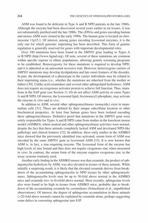
�
� �
�
214 THE GENETICS OF SPHINGOLIPID HYDROLASES
ASM was found to be deficient in Type A and B NPD patients in the late 1960s.Although the enzyme had been discovered several years earlier in rat tissues, it wasnot substantially purified until the late 1980s. The cDNAs and genes encoding humanand mouse ASM were cloned in the early 1990s. The human gene is located on chro-mosome 11p15.1. Of interest, among genes encoding lysosomal enzymes, it is theonly one for which genomic imprinting has been described. This form of geneticregulation is generally reserved for genes with important developmental roles.
Over 100 mutations have been found in the SMPD1 gene leading to Types Aor B NPD (http://www.hgmd.org). Of note, several of these mutations are commonwithin specific regions or ethnic populations, allowing genetic screening programsto be established. Homozygosity for these mutations is required to develop NPD,and it is inherited as an autosomal recessive trait. However, heterozygous carriers ofSMPD1 mutations may develop dyslipidemia and late-onset features of the disorder.In part, the development of a phenotype in the carrier individuals may be related totheir imprinting status (i.e., whether the mutations are inherited from the mother orfather) [30]. Unlike acid ceramidase and several other sphingolipid hydrolases, ASMdoes not require an exogenous activator protein to achieve full function. Thus, muta-tions in the SAP gene (see Section 11.10) do not affect ASM activity or cause TypesA and B NPD. Of interest, the lysosomal lipid, bis(monacyl) phosphate, may activatethe enzyme in vitro and in vivo.
In addition to ASM, several other sphingomyelinases (nonacidic) exist in mam-malian cells [31]. These are defined by their unique subcellular location or otherbiochemical properties. At least four human genes have been isolated encodingthese sphingomyelinases. Definitive proof that mutations in the SMPD1 gene weresolely responsible for Types A and B NPD came from studies in the knockout mousemodel (ASMKO), where neutral and other sphingomyelinase activities were normal,despite the fact that these animals completely lacked ASM and developed NPD-likepathology and clinical features [32]. In addition, these early studies in the ASMKOmice showed that the previously identified zinc-activated, serum form of ASM wasencoded by the same SMPD1 gene as lysosomal ASM [33]. It is now known thatASM is, in fact, a zinc-requiring enzyme. The lysosomal form of the enzyme hashigh levels of zinc bound and thus does not require exogenous zinc when measuredin vitro. In contrast, the serum form of the enzyme requires exogenous zinc in theassay systems routinely used.
Another early finding in the ASMKO mouse was that ceramide, the product of sph-ingomyelin hydrolysis by ASM, was also elevated in tissues of these animals. Whileinitially a surprising result, it is likely that the elevated ceramide derives from break-down of the accumulating sphingomyelin in NPD tissues by other sphingomyeli-nases. Sphingomyelin levels may be up to 30-fold above normal in the ASMKOmice and ceramide two- to fivefold above normal. Most recently, sphingosine levelsalso were found to be high in tissues from ASMKO mice, probably due to break-down of the accumulating ceramide by ceramidases (Schuchman et al., unpublishedobservations). Of interest, the degree of sphingosine accumulation in these animals(>20-fold above normal) cannot be explained by ceramide alone, perhaps suggestingsome defect in converting sphingosine into S1P.
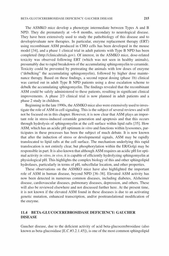
�
� �
�
BETA-GLUCOCEREBROSIDASE DEFICIENCY: GAUCHER DISEASE 215
The ASMKO mice develop a phenotype intermediate between Types A and BNPD. They die prematurely at ∼6–8 months, secondary to neurological disease.They have been extensively used to study the pathobiology of this disease and todevelop/evaluate new therapies. In particular, enzyme replacement therapy (ERT)using recombinant ASM produced in CHO cells has been developed in the mousemodel [34], and a phase 1 clinical trial in adult patients with Type B NPD has beencompleted (http://clinicaltrials.gov). Of interest, in the ASMKO mice, dose-relatedtoxicity was observed following ERT (which was not seen in healthy animals),presumably due to rapid breakdown of the accumulating sphingomyelin to ceramide.Toxicity could be prevented by pretreating the animals with low doses of enzyme(“debulking” the accumulating sphingomyelin), followed by higher dose mainte-nance therapy. Based on these findings, a second repeat dosing (phase 1b) clinicalwas carried out in adult Type B NPD patients using a dose escalation scheme todebulk the accumulating sphingomyelin. The findings revealed that the recombinantASM could be safely administered to these patients, resulting in significant clinicalimprovements. A phase 2/3 clinical trial is now planned in adult patients and aphase 2 study in children.
Beginning in the late 1990s, the ASMKO mice also were extensively used to inves-tigate the role of ASM in cell signaling. This is the subject of several reviews and willnot be focused on in this chapter. However, it is now clear that ASM plays an impor-tant role in stress-induced ceramide generation and apoptosis and that this occursthrough hydrolysis of sphingomyelin at the cell surface within lipid rafts [35]. HowASM, which has an acidic pH optimum in vitro and functions within lysosomes, par-ticipates in these processes has been the subject of much debate. It is now knownthat after the induction of stress or developmental signals, ASM may be rapidlytranslocated to lipid rafts at the cell surface. The mechanism underlying this rapidtranslocation is not entirely clear, but phosphorylation within the ER/Golgi may beresponsible in part. It is also known that although ASM requires an acidic pH for opti-mal activity in vitro, in vivo, it is capable of efficiently hydrolyzing sphingomyelin atphysiological pH. This highlights the complex biology of this and other sphingolipidhydrolases, particularly in terms of pH, subcellular location, and other properties.
These observations on the ASMKO mice have also highlighted the importantrole of ASM in human disease, beyond NPD [36–38]. Elevated ASM activity hasnow been detected in numerous common diseases, including diabetes, Alzheimerdisease, cardiovascular diseases, pulmonary diseases, depression, and others. Thesewill also be reviewed elsewhere and not discussed further here. At the present time,it is not known if the elevated ASM found in these diseases is due to an activatinggenetic mutation, enhanced transcription, and/or posttranslational modification ofthe enzyme.
11.4 BETA-GLUCOCEREBROSIDASE DEFICIENCY: GAUCHERDISEASE
Gaucher disease, due to the deficient activity of acid beta-glucocerebrosidase (alsoknown as beta-glucosidase [E.C.#3.2.1.45]), is one of the most common sphingolipid

�
� �
�
216 THE GENETICS OF SPHINGOLIPID HYDROLASES
storage disorders [128,129]. There are three clinical subtypes of Gaucher disease,including an early-onset, neurological form (Type 2), an intermediate form (Type 3),and a late-onset, nonneurological form (Type 1) [39,40]. All Gaucher patientspresent with hepatosplenomegaly, but the progression of the disease and survivalare markedly different. For example, Type 3 children may only survive until 2–3years of age, while Type 1 patients may survive into adulthood. If left untreated,the hematological and bone lesions in Type 1 patients progress with age, seriouslylimiting quality of life. A massively enlarged spleen and frequent bone fracturesmay become common, requiring splenectomy, joint replacement, or other surgicalinterventions [41,42]. Bleeding is also a common presenting feature of Gaucherdisease due to thrombocytopenia. Pulmonary involvement may similarly occur.Of note, neoplastic disorders, particularly of the blood forming organs, are morecommon in Gaucher disease patients than in the general population. These includechronic lymphocytic leukemia, multiple myeloma, and Hodgkin and non-Hodgkinlymphoma. The molecular basis of this increased cancer risk remains unknown [43].
All Gaucher patients are characterized by the presence of “Gaucher” cells in vari-ous organs, lipid-filled cells derived from the monocyte/macrophage system. Gauchercells have a distinctive appearance by light microscopy and can be readily distin-guished from cells of other sphingolipid storage disorders. Gaucher cells are dis-tributed throughout the body wherever macrophages reside. The largest numbers arefound in the spleen, sinusoids of the liver, bone marrow, and lymph nodes.
The major accumulating lipid in Gaucher disease is N-acyl-sphingosyl-1-0-β-d-glucoside (glucosylceramide, also known as glucosylcerebroside; GC). GC is widelydistributed in mammalian tissues in small quantities as a metabolic intermediate inthe synthesis and degradation of complex glycosphingolipids, such as gangliosidesor globoside. The deacylated form of GC, glucosylsphingosine, is not only normallyfound in tissues but also accumulates in Gaucher disease cells. GC levels in plasmamay be 2- to 20-fold above normal.
The major site of GC turnover in cells is lysosome. Beta-glucocerebrosidase isnormally found in this organelle and functions optimally at an acidic pH. As withthe other lipid hydrolases, in vitro detection of this enzymatic activity can be diffi-cult, and there have been various factors (lipids, protein, and sugars) shown to influ-ence the assay systems. In vivo, sphingolipid activator-derived peptides, particularlysaposin (SAP) C, appear to be required for full enzymatic activity (see Section 11.10).Nonacidic glucocerebrosidases also have been described and are encoded by distinctgenes. Whether these activities can be modifying factors for the Gaucher phenotypehas been the subject of much discussion but remains unclear.
The gene encoding beta-glucocerebrosidase (GBA1) has been mapped to chro-mosome 1q21. It is ∼7 kb and contains 11 exons. Of note, a 5 kb “pseudogene”is located immediately downstream from the GBA1 gene. This pseudogene hasmaintained a high degree of homology with the functional gene and is transcribed.However, the nucleotide changes within the pseudogene sequence prevent theoccurrence of a long open reading frame, precluding the possibility of expressingan active beta-glucocerebrosidase [44]. Full-length cDNAs from many species alsohave been obtained that can be used to express the active enzyme.

�
� �
�
BETA-GLUCOCEREBROSIDASE DEFICIENCY: GAUCHER DISEASE 217
Numerous mutations have been found in the GBA1 gene causing Gaucherdisease [45]. As described for NPD above, several common mutations are foundin specific populations, and DNA-based screening is now routinely carried out inthe Ashkenazi Jewish population [46]. Estimates of the Gaucher disease carrierfrequency in this population range from ∼1:10 to 1:25. Some of the mutationsalso can be used to predict the occurrence of severe or mild Gaucher phenotypes,assisting in family planning and genetic counseling.
Recently, mutations in the GBA1 gene also have been associated with the occur-rence of Parkinson’s disease, although the precise relationship between the GBA1gene, glucocerebrosidase activity, and Parkinson’s disease remains uncertain [47].One of the first clues suggesting this linkage came from the small number of Gaucherpatients who exhibited Parkinson-like features, while stronger associations later camefrom Gaucher relatives who were carriers for GBA1 mutations. Currently, heterozy-gous GBA1 mutations are considered to be one of the most common genetic riskfactor for Parkinson’s disease [48].
A complete knockout mouse model for Gaucher was constructed and had an earlyneonatal lethal phenotype, dying within 34 days after birth. These animals had mas-sive GC storage, and while the cause of death was not entirely clear, skin abnormal-ities were prevalent. Several knock-in models also have been constructed, and someexhibited longer survival and later-onset GC storage in macrophages, more closelyresembling Gaucher disease patients [49,50].
Gaucher disease was the first sphingolipid storage disease for which ERT wasdeveloped in the early 1990s, initially using beta-glucocerebrosidase purified fromplacentae (Ceredase) and subsequently using recombinant enzyme produced in CHOcells (Cerezyme) [51,52]. For nonneurological patients, ERT was life-changing,resulting in reduction in spleen size and correction of the hematological abnormali-ties. Bone disease has been much less responsive to ERT and remains a challenge inthe treatment of Gaucher disease, as does treatment of the brain disease [51]. Since theintroduction of Cerezyme, several other recombinant forms of GC also have becomeavailable, and in general similar clinical responses have been observed. Anothertreatment strategy currently available for some Gaucher patients uses small-moleculeinhibitors of GC synthesis (Miglustat) to slow the accumulation of this lipid [53,54].The target of this inhibitor is the enzyme glucosylceramide synthase.
Of note, treatment of Gaucher patients by either ERT or with Miglustat shouldelevate ceramide and could potentially result in ceramide-mediated toxicity. To date,however, such toxicity has not been observed in animal models or patients. There areseveral factors that might explain this observation. First, compared to sphingomyelinin NPD, GC is generally found in small quantities in cells and is not a major struc-tural component of cell membranes. Thus, accumulation of GC in Gaucher cells ismore exclusively found in the lysosomes than NPD and does not disrupt plasmamembrane structure and signaling to the same extent as sphingomyelin. In addition,ceramide produced by ASM (compared to GC) is likely to remain within the cellmembrane. Finally, the ERT dose typically used to treat Gaucher patients (1.6 mg/kg,every 2 weeks) may be below the threshold needed to induce ceramide-mediatedtoxicity.

�
� �
�
218 THE GENETICS OF SPHINGOLIPID HYDROLASES
11.5 GALACTOCEREBROSIDASE DEFICIENCY: KRABBEDISEASE/GLOBOID CELL LEUKODYSTROPHY
Infantile globoid cell leukodystrophy (GLD), also known as Krabbe disease, is arapidly progressive, fatal neurological disease of infants [55]. The disease usuallybegins between 3 and 6 months of life with symptoms that may include irritabilityor hypersensitivity to stimuli. Patients rapidly exhibit severe mental and motor dete-rioration and rarely survive beyond the second year [56]. Clinical manifestations inclassical GLD patients are limited to the nervous system. Blindness and deafnessare common, and peripheral neuropathy is almost always detectable. Patients withlater-onset forms of the disease, including adult-onset forms, may present with blind-ness, spastic paraparesis, and dementia [57,58].
The presence of numerous multinucleated globoid cells, almost total loss of myelinand oligodendrocytes, and astrocytic gliosis in the white matter are the morphologicbasis of the disease. “Globoid cells” in Krabbe disease are enlarged macrophagesthat contain undigested galactosylceramide and other lipids. Demyelination, axonaldegeneration, fibrosis, and histiocytic infiltration are also common in the peripheralnervous system. Consistent with the myelin loss, the white matter is severelydepleted of all lipids, particularly glycolipids. Under normal circumstances, galacto-sylceramide is present almost exclusively in the myelin sheath. The ratio of galacto-sylceramide to sulfatide (3-0 sulfogalactosylceramide) is abnormally high in GLD.
The genetic cause of Krabbe disease is mutations in the gene encoding galac-tocerebrosidase beta-galactosidase (GALC; E.C. #3.2.1.46). This lysosomal enzymenormally degrades galactosylceramide to ceramide and galactose. It is postulated thataccumulation of a toxic metabolite, psychosine (galactosylsphingosine), which is alsoa substrate for the missing enzyme, leads to early destruction of the oligodendrocytes.The total brain content of galactosylceramide is not increased in patients with Krabbedisease, supporting this hypothesis. It should be noted that mammalian tissues containtwo genetically distinct lysosomal beta-galactosidases, GALC and GM1-gangliosidebeta-galactosidase (see Section 11.8). These two enzymes have overlapping substratespecificities in vitro, which can present complications when attempting to make theenzymatic diagnosis of Krabbe disease [59].
The human GALC gene has been mapped to chromosome 14q24.3-32.1, andthe genomic and/or full-length cDNA sequences have been cloned from multiplespecies. The human cDNA encodes a 669 amino acid polypeptide with six potentialN-glycosylation sites. The fully glycosylated precursor polypeptide is ∼85 kDa andis processed into ∼50 and 30 kDa subunits. To date, over 70 mutations in the GALCgene have been identified in infantile and late-onset GALC-deficient patients (http://www.hgmd.org). Most of these mutations are “private,” occurring in individualfamilies, but some may be commonly found in specific regions or ethnicities. Amongthese, the most common mutation occurs in patients of European ancestry and deletesa large segment of the GALC gene. Several polymorphisms also have been found inthe GALC gene, and some of these may alter amino acids and thus potentially affectthe function of the GALC polypeptide, although not enough to cause GLD.

�
� �
�
ARYLSULFATASE A DEFICIENCY: METACHROMATIC LEUKODYSTROPHY 219
In general, carriers of GALC mutations are considered clinically normal, althoughone interesting report did show an association between the risk for primary open-angle glaucoma and a heterozygous deletion of the GALC gene [60]. Anotherinteresting recent paper also showed that GALC contributed to the maintenance ofa functional hematopoietic stem cell niche, suggesting that genetic alterations inthe GALC gene may be associated with hematological conditions as well. Indeed,hematopoietic stem cells retrieved from Twitcher mice (GALC deficient) had defectsin their frequency, proliferation, clonogenic potential, and engraftment.
As noted above, in 1972, Miyatake and Suzuki formulated a hypothesis to explainthe unusually rapid and complete destruction of oligodendrocytes in GLD. It wasknown at that time that psychosine (galactosyl sphingosine), due to its free aminogroup, was highly cytotoxic and that this lipid served as a substrate for GALC but notGM1 beta-galactosidase. In the central nervous system (CNS), psychosine is primar-ily formed within oligodendrocytes, the site of myelin synthesis, and this may explainthe selective destruction of these cells in GLD brains. Indeed, in the white matter ofbrains from Krabbe patients, psychosine levels may be elevated up to 100-fold, whilegalacotosylceramide levels are either normal or only elevated slightly.
GALC deficiency occurs in a number of naturally occurring animal models,including mice, sheep, several breeds of dogs, and the rhesus monkey. The mousemodel, in particular (also known as the “Twitcher” mouse), has been exploited fornumerous therapeutic studies [61]. Affected mice develop clinical signs at ∼20 days,with stunted growth, twitching, and hind leg weakness. By 40 days, they reach anear-terminal stage. Twitcher mice have been treated by ERT, gene therapies, BMT(bone marrow transplantation), and other modalities [62–64]. BMT, in particular,may have positive therapeutic effects; however, all of the treated mice eventuallystill died of characteristic pathological findings. Thus, this and the other availabletherapies may slow the disease progress to some degree but not prevent it.
Since 2006, New York State in the United States has screened all newborns for theoccurrence of Krabbe disease. Pilot screening programs are now underway in otherstates and parts of the world. This screening is accomplished by GALC activity assayson dried blood spots, followed by DNA analyses. The rationale for this screeningprogram is that the early identification of infants with Krabbe disease will permitearly BMT and other therapeutic interventions, improving the quality of life [56].However, since BMT has not been curative in the animal models, screening for severeneurological disorders such as this remains controversial.
11.6 ARYLSULFATASE A DEFICIENCY: METACHROMATICLEUKODYSTROPHY
Metachromatic leukodystrophy (MLD) is an autosomal recessively inherited disor-der in which desulfation of 3-0-sulfogalactosylsphingosine or 3-0-sulfogalactosylglycolipids is deficient. Among the accumulating glycolipids in MLD are sulfatide(galactosylceramide-3-sulfate), lysosulfatide (i.e., similar to sulfatide but lacking

�
� �
�
220 THE GENETICS OF SPHINGOLIPID HYDROLASES
the fatty acid in the ceramide moiety), lactosylceramide-3-sulfate, and others.These sulfated glycolipids normally occur in the myelin sheaths in the central andperipheral nervous systems, and, to a lesser extent, in visceral organs.
The clinical onset and severity of MLD is highly variable [65]. The late infantileform is usually recognized in the second year of life and fatal in early childhood. Thejuvenile form presents between age four and puberty; the adult form may occur atany age after puberty. In both of the later-onset variants, gait disturbance and mentalregression are the earliest signs. In the childhood variants, other common signs areblindness, loss of speech, quadriparesis, peripheral neuropathy, and seizures [66].
In 1963, Austin et al. reported the deficiency of arylsulfatase A (ASA, E.C. 3.1.6.8)activity in MLD, also known as sulfatide sulfatase, and mutations in this gene (ARSA)account for the vast majority of MLD patients. There are also two other minor, geneticcauses of MLD. One is due to mutations in the gene (PSAP, see Section 11.9) encod-ing the SAP, saposin B. Mutations in the saposin B encoding region of the PSAP genelead to deficient ASA activity, and a clinical picture indistinguishable from MLD.In fact, these observations in MLD patients provided the first in vivo proof that suchSAPs were required for in vivo activity of this and other sphingolipid hydrolases. Inaddition to ASA and SAP B mutations, patients with multiple sulfatase deficiency,due to mutations in the gene encoding sulfatase-modifying factor 1 (SUMF-1), alsohave a reduction in ASA and other sulfatase activities and develop features of MLD.
As with the other sphingolipid hydrolases, full-length genomic and cDNA se-quences encoding ASA have been isolated from several species. The human ARSAgene has been mapped to chromosomal region 22q13. It is a remarkably simplegene and encompasses only 3.2 kb divided into eight exons. As noted above, almostall cases of MLD are due to mutations in this gene, except for the rare cases ofPSAP or SUMF-1 mutations. The human ARSA gene encodes a 507 amino acidpolypeptide precursor that undergoes posttranslational modifications much likeother lysosomal enzymes (e.g., N-glycosylation). Also, as with all known eukaryoticsulfatases, a formylglycine residue is substituted for a cysteine in human ASA. Theformylglycine residue is generated by oxidation of the thiol group to an aldehydeby the enzyme SUMF-1. Mutations in the gene encoding this cysteine-modifyingenzyme lead to the synthesis of inactive sulfatases and are the cause for multiplesulfatase deficiency [67].
Over 100 MLD-causing mutations have been described in the ARSA gene. Themost common mutation in patients with late-infantile MLD is a G to A transitionthat eliminates the donor splice site at the start of intron 2 [68]. This causes a lossof all ASA enzymatic activity, producing a severe early-onset phenotype. In contrast,in adults with MLD the most frequent mutation is a C to T transition that results insubstitution of a leucine for proline in amino acid residue 426. Patients with this muta-tion have a small amount of residual enzyme activity and therefore exhibit late-onsetMLD. Common mutations in specific populations have also been described.
Notably, there also are two mutations that cause “pseudodeficiency” of ASAactivity. The term “pseudodeficiency” reflects the fact that these mutations causesevere reduction of ASA activity in vitro but do not result in MLD [69]. One of these

�
� �
�
ALPHA-GALACTOSIDASE A DEFICIENCY: FABRY DISEASE 221
mutations alters an N-glycosylation site, but this has no effect on enzyme function.The other affects a polyadenylation site in the mRNA. The pseudodeficiency allelesmay be found in ∼3–8% of the normal population and, therefore, pose a diagnosticchallenge since the artificial in vitro assay systems may reveal very low ASA activity,even though the individual is highly unlikely to develop MLD.
Alterations in the ARSA gene also have been linked with the occurrence of severalcommon diseases. For example, as early as 1991, Kappler et al. found that amonga small set of patients with multiple sclerosis (MS) there was a higher incidence ofthe ASA pseudodeficiency alleles than in the general population [70]. In addition,variants of the ASA protein and/or gene have been associated with depression andalcoholism. MLD itself also has been associated with several psychiatric disorders,and there is at least one report where polymorphisms in the genes encoding the sero-tonin and dopamine pathways affected the MLD phenotype [71].
Other than humans, naturally occurring mutations causing MLD do not occur inother species. ASA knockout mice have been constructed that exhibit a complete lossof ASA activity and sulfatide storage in multiple organs [67]. While these animalsexhibit some neurological deficits, they do not develop the predicted severe pheno-type of MLD and live a near-normal lifespan. They also do not develop demyelinatingdisease. Thus, these observations in the mouse suggest the existence of compensatingpathways in this species that do not exist in humans, perhaps providing a link to noveltherapies. Several therapies have been evaluated in the MLD knockout mouse model,including BMT, ERT, and gene therapies [72,73]. BMT also has been extensivelyused in MLD patients. While it is not effective in infantile cases, BMT is often recom-mended for the later-onset cases [74,75]. However, the data supporting an improvedclinical outcome in the late-onset MLD cases are fragmented and incomplete, and itis unclear whether the benefit outweighs the potential risks.
11.7 ALPHA-GALACTOSIDASE A DEFICIENCY: FABRY DISEASE
Fabry disease is inherited as an X-linked recessive trait, the only such X-linkeddisease among the sphingolipid storage disorders [76,77]. Therefore, all males(“hemizygotes”) carrying mutations in the Fabry gene (GLA) develop symptoms ofthe disease although the severity may vary depending on the nature of the individualmutation. Female heterozygotes who inherit one copy of the mutant gene on oneof their X chromosomes may also exhibit features of the disease depending on thepattern of X-inactivation (“Lyonization”) that occurs in their individual tissues.
The gene responsible for Fabry disease encodes an enzyme, alpha-galactosidase A(E.C.#3.2.1.22), that is required to hydrolyze glycosphingolipids with terminalalpha-galactosyl moieties, predominately globotriosylceramide, and to a lesserextent galabiosylceramide and blood group B substances. Affected males haveextensive deposition of these lipids in body fluids and in the lysosomes of endothe-lial, perithelial, and smooth muscle cells of blood vessels. Deposition also occursin ganglion cells, and in many cell types in the heart, kidney, eyes, and most othertissues [78].

�
� �
�
222 THE GENETICS OF SPHINGOLIPID HYDROLASES
Clinical manifestations in classically affected hemizygotes with no detectablealpha-galactosidase A activity include the onset during childhood or adolescenceof pain and paresthesias in the extremities, angiokeratomas in the skin and mucousmembranes, and hypohidrosis. Corneal and lenticular opacities also are earlyfindings. With increasing age, proteinuria, hyposthenuria, and lymphedema appear.Severe renal impairment leads to hypertension and uremia. Death usually occurs inadulthood from renal failure or from cardiac or cerebrovascular disease. Atypicalvariant males with residual alpha-galactosidase A activity may be asymptomatic orhave late-onset disease. Heterozygous females may have an attenuated form of thedisease depending on the pattern of X-inactivation. They usually are asymptomaticalthough can present with clinical disease as severe as hemizygous males.
As noted above, in Fabry disease glycosphingolipids with terminal alpha-d-galactosyl residues are not properly broken down due to the alpha-galactosidaseA enzyme defect, and these lipids therefore accumulate in cells. The predominantaccumulating lipid is globotriaosylceramide (Gal(a1-4)Gal(a1-4)Glc(b1-1′)Cer).Since alpha-galactosidase A is mostly found in lysosomes, the major site of lipidaccumulation is this organelle. Elevated globotriaosylceramide is found in mosttissues but predominantly in kidney.
Other minor accumulating lipids include galabiosylceramide (Gala1-4)(Gal(b1-1′)Cer) and the blood group B and P glycosphingolipids [79]. In human erythrocytes,there are two neutral glycosphingolipids with terminal alpha galactosyl residues thatinhibit blood group B-specific hemagglutination. The structure of these blood groupB glycosphingolipids has been determined, and they are found at high levels in Fabrypatients. This raises an interesting and unique point about Fabry disease. Fabryhemizygotes and heterozygotes who have blood groups B and AB accumulate fourglycosphingolipids, while those who are blood group A or O only accumulate two(globotriaosylceramide and galabiosylceramide). A fifth neutral glycosphingolipidthat can accumulate in Fabry is the P1 blood group antigen, which also has terminalalpha-galactosyl residues. The role of these individual blood types on the Fabryphenotype is an interesting area of ongoing research.
In humans, there are two alpha-galactosidases (alpha-galactosidases A and B).Fabry disease is caused by mutations in the gene (GLA) encoding the “A” type [80].However, when measuring alpha-galactosidase activity in blood from Fabry patientsin vitro, classic hemizygotes may exhibit up to 25% of normal activity due to thepresence of the “B” form. Alpha-galactosidase B is encoded by a distinct gene(NAGA) and is now known to be an alpha-N-acetylgalactosaminidase that recognizesthese moieties in glycoproteins, complex carbohydrates, and other molecules.Because the artificial substrates routinely used to measure alpha-galactosidaseactivity do not readily distinguish these enzymes, substantial misdiagnosis can occurby simple enzyme testing.
The human gene encoding alpha-galactosidase A resides on chromosomal regionXq22. It encompasses ∼12 kb and contains seven exons. The full-length humancDNA contains a 1290 bp open reading frame that encodes an unglycosylated
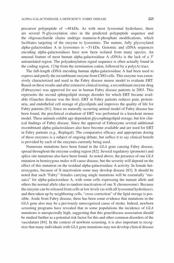
�
� �
�
ALPHA-GALACTOSIDASE A DEFICIENCY: FABRY DISEASE 223
precursor polypeptide of ∼48 kDa. As with most lysosomal hydrolases, thereare several N-glycosylation sites in the predicted polypeptide sequence andthe oligosaccharide chains undergo mannose-6-phosphate modifications, whichfacilitates targeting of the enzyme to lysosomes. The mature, fully glycosylatedalpha-galactosidase A in lysosomes is ∼51 kDa. Genomic and cDNA sequencesencoding alpha-galactosidases have now been isolated from many species. Anunusual feature of most human alpha-galactosidase A cDNAs is the lack of a 3′
untranslated region. The polyadenylation signal sequence is often actually found inthe coding region, 12 bp from the termination codon, followed by a poly(A) tract.
The full-length cDNA encoding human alpha-galactosidase A had been used toexpress and purify the recombinant enzyme from CHO cells. This enzyme was exten-sively characterized and used in the Fabry disease mouse model to evaluate ERT.Based on these results and after extensive clinical testing, a recombinant enzyme drug(Fabrazyme) was approved for use in human Fabry disease patients in 2003. Thisrepresents the second sphingolipid storage disorder for which ERT became avail-able (Gaucher disease was the first). ERT in Fabry patients reduces pain, protein-uria, and endothelial cell storage of glycolipids and improves the quality of life forFabry patients [81]. Since no naturally occurring animal model of Fabry disease hasbeen found, the preclinical evaluation of ERT was performed in a knockout mousemodel. These animals exhibit age-dependent glycosphingolipid storage, but few clin-ical findings of Fabry disease. Since the approval of Fabrazyme several additionalrecombinant alpha-galactosidases also have become available and are used for ERTin Fabry patients (e.g., Replagal). The comparative efficacy and appropriate dosingof these enzymes is a subject of ongoing debate, but suffice it to say clinical benefitis provided by each of the enzymes currently being used.
Numerous mutations have been found in the GLA gene causing Fabry disease,spread throughout the enzyme-coding region [82]. Several regulatory (promoter) andsplice site mutations also have been found. As noted above, the presence of one GLAmutation in hemizygous males will cause disease, but the severity will depend on theeffect of this mutation on the residual alpha-galactosidase A activity. In female het-erozygotes, because of X-inactivation some may develop disease [83]. It should benoted that such “Fabry” females carrying single mutations will be essentially “mo-saics” for alpha-galactosidase A, with some cells expressing the mutant allele andothers the normal allele (due to random inactivation of one X chromosome). Becausethe enzyme can be released from cells at low levels (as with all lysosomal hydrolases),and then taken up by neighboring cells, “cross-correction” of the lipid storage is pos-sible. Aside from Fabry disease, there has been some evidence that mutations in theGLA gene also may be a previously unrecognized cause of stroke. Indeed, newbornscreening programs have revealed that in some populations the incidence of GLAmutations is unexpectedly high, suggesting that this gene/disease association shouldbe studied further as a potential risk factor for this and other common disorders of thevasculature [84]. In the context of newborn screening, it is also important to recog-nize that many individuals with GLA gene mutations may not develop clinical disease
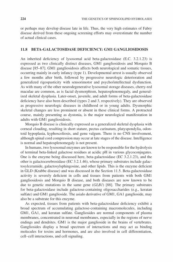
�
� �
�
224 THE GENETICS OF SPHINGOLIPID HYDROLASES
or perhaps may develop disease late in life. Thus, the very high estimates of Fabrydisease derived from these ongoing screening efforts may overestimate the numberof actual clinical cases.
11.8 BETA-GALACTOSIDASE DEFICIENCY: GM1 GANGLIOSIDOSIS
An inherited deficiency of lysosomal acid beta-galactosidase (E.C. 3.2.1.23) isexpressed as two clinically distinct diseases, GM1 gangliosidosis and Morquio Bdisease [85–87]. GM1 gangliosidosis affects both neurological and somatic tissues,occurring mainly in early infancy (type 1). Developmental arrest is usually observeda few months after birth, followed by progressive neurologic deterioration andgeneralized rigospasticity with sensorimotor and psycho/intellectual dysfunction.As with many of the other neurodegenerative lysosomal storage diseases, cherry-redmaculae are common, as is facial dysmorphism, hepatosplenomegaly, and general-ized skeletal dysplasia. Later-onset, juvenile, and adult forms of beta-galactosidasedeficiency have also been described (types 2 and 3, respectively). They are observedas progressive neurologic diseases in childhood or in young adults. Dysmorphicskeletal changes are less prominent or absent in these clinical forms. A protractedcourse, mainly presenting as dystonia, is the major neurological manifestation inadults with GM1 gangliosidosis.
Morquio B disease is clinically expressed as a generalized skeletal dysplasia withcorneal clouding, resulting in short stature, pectus carinatum, platyspondylia, odon-toid hypoplasia, kyphoscoliosis, and genu valgum. There is no CNS involvement,although spinal cord compression may occur at late stages of the disease. Intelligenceis normal and hepatosplenomegaly is not present.
In humans, two lysosomal enzymes are known to be responsible for the hydrolysisof terminal beta-linked galactose residues at acidic pH in various glycoconjugates.One is the enzyme being discussed here, beta-galactosidase (EC 3.2.1.23), and theother is galactocerebrosidase (EC 3.2.1.46), whose primary substrates include galac-tosylceramide, galactosylsphingosine, and other lipids. This is the enzyme deficientin GLD (Krabbe disease) and was discussed in the Section 11.5. Beta-galactosidaseactivity is severely deficient in cells and tissues from patients with both GM1gangliosidosis and Morquio B disease, and both diseases are now known to bedue to genetic mutations in the same gene (GLB1) [88]. The primary substratesfor beta-galactosidase include galactose-containing oligosaccharides (e.g., keratansulfate) and GM1 ganglioside. The asialo derivative of GM1, GA1 ganglioside, mayalso be a substrate for this enzyme.
As expected, tissues from patients with beta-galactosidase deficiency exhibit abroad spectrum of accumulating galactose-containing macromolecules, includingGM1, GA1, and keratan sulfate. Gangliosides are normal components of plasmamembranes, concentrated in neuronal membranes, especially in the regions of nerveendings and dendrites. GM1 is the major ganglioside in the brains of vertebrates.Gangliosides display a broad spectrum of interactions and may act as bindingmolecules for toxins and hormones, and are also involved in cell differentiation,cell–cell interactions, and cell signaling.

�
� �
�
BETA-GALACTOSIDASE DEFICIENCY: GM1 GANGLIOSIDOSIS 225
In the brains of patients with GM1 gangliosidosis, storage of GM1 is the mostprominent observation [89]. The accumulating GM1 has the same fatty acid com-position, sugar composition and sequence, and glycosidic linkages as normal GM1.Visceral organs also show storage of GM1 ganglioside but to a lesser degree. Also toa lesser degree, GA1 ganglioside may accumulate in the brain of GM1 gangliosido-sis patients, as do glycosylceramide, lactosylceramide, and GM2 ganglioside. Whitematter shows chemical manifestations of myelin breakdown, include low proteolipidprotein, low total lipid, and the presence of esterified cholesterol [90].
Other than gangliosides, the other major storage molecule in beta-galactosidase-deficient patients is keratan sulfate [91]. This glycosaminoglycan normally exists ina proteoglycan linked with chondroitin sulfate. It is found primarily within connec-tive tissues, particularly cartilage, explaining the skeletal dysplasia typical of manybeta-galactosidase-deficient patients. After proteolysis and release from the proteo-glycan, free keratan sulfate chains are hydrolyzed by a series of exoenzymes, includ-ing beta-galactosidase.
It should be noted that another protein, referred to as protective protein/cathepsinA (PPCA), has been associated with both beta-galactosidase and another lyso-somal hydrolase, neuraminidase, and stabilizes and in some cases activates theenzymes [92]. A genetic deficiency of this protective protein results in combineddeficiency of beta-galactosidase and neuraminidase (galactosialidosis). The geneencoding PPCA is distinct from both of these enzymes and will not be discussedfurther here.
The gene encoding beta-galactosidase (GLB1) is located in chromosomal region3p21.33. Genes encoding this enzyme also have been isolated from mice and severalother species. The full-length human GLB1 cDNA encodes a 677 amino acid polypep-tide with beta-galactosidase activity. Mutations in the GLB1 gene are responsible forboth GM1 gangliosidosis and Morquio B disease. As with other lysosomal diseases,most are point mutations altering single amino acids, although small deletions, splicesite mutations, and other alterations also have been identified. To date, no correlationhas been made between the location or type of GLB1 mutations and the occurrenceof GM1 gangliosidosis or Morquio B disease.
The pathogenesis of beta-galactosidase deficiency is complex. GM1 gangliosidestimulates neurite outgrowth, affects neuronal differentiation, and enhances the actionof nerve growth factor [93]. Golgi and electron microscopic studies of cortical neu-rons from this and several other neurological lysosomal diseases exhibit large neuralprocesses (meganeurites), which may be explained by GM1 accumulation. The extentof meganeurite development is related to the onset, severity, and clinical course ofthe disease. In other studies, significant changes in neuronal connectivity were foundin the cerebral cortex of animals with beta-galactosidase deficiency, and cholinergicfunction was altered as well. Phosphoinositol-specific phospholipase C and adenylcyclase activities were altered in the membranes of cerebral cortex from GM1 gan-gliosidosis cats. In addition, the beta subunit of cholera toxin, which binds specificallyto GM1 in the outer leaflet of the cell membrane, was found to induce an increase ofcalcium and manganese influx in N18 cells, and this has been associated with acti-vation of an L-type voltage-dependent calcium channel. This channel has important

�
� �
�
226 THE GENETICS OF SPHINGOLIPID HYDROLASES
implications for neural development, and its reliance on GM1 ganglioside is alsolikely related to the pathogenesis of GM1 storage. These and other data suggest vari-ous morphologic and metabolic aberrations occur in the brains of GM1 gangliosidosispatients and animals, and can be linked to the biology of GM1.
Naturally occurring animal models of GM1 gangliosidosis have been reported incats, dogs, sheep, and calves. The cat model in particular has been studied in somedetail, and is a neurologic disorder with clinical, morphological, biochemical, andgenetic similarities to human GM1 gangliosidosis [94]. Affected kittens appear nor-mal at birth, but tremors of the head and hind limbs are first noted at 2–3 monthsof age, followed by generalized dysmetria and spastic quadriplegia. GM1 ganglio-side and other galactose-containing oligosaccharides accumulate in the tissues ofaffected animals. A genetic knockout of beta-galactosidase activity also has beengenerated in mice by gene targeting [95]. Progressive and severe neurodegenerationoccurs in these animals, and GM1 and GA1 gangliosides accumulate in the brain.Unlike patients, GA1 ganglioside may accumulate in beta-galactosidase deficiencymice to a greater extent than GM1, which may be explained by the fact that there is aunique neuraminidase in mice that converts GM1 to GA1. Skeletal dysplasia, whichis characteristic of patients with beta-galactosidase deficiency, does not occur in themice. This also is due to the unique metabolism of keratan sulfate in mice comparedto humans.
11.9 HEXOSAMINIDASE A AND B DEFICIENCY: GM2GANGLIOSIDOSES
The GM2 gangliosidoses are a group of genetic disorders caused by excessive accu-mulation of GM2 ganglioside and related glycolipids in lysosomes, mainly of neu-ronal cells [96]. The general cause of GM2 gangliosidosis is deficient activity ofthe enzyme beta-hexosaminidase (E.C. 3.2.1.30). The enzymatic hydrolysis of GM2ganglioside by this enzyme requires that it be complexed with a substrate-specificcofactor, the GM2 activator. There are two isozymes of beta-hexosaminidase, Hex A,which is composed of alpha and beta subunits, and Hex B, which is composed of twobeta subunits. Hex A and B can only act on the GM2/GM2 activator complex.
Defects in any of three genes can lead to GM2 gangliosidosis, HEXA, whichencodes the alpha subunit of Hex A, HEXB, which encodes the beta subunit of HexA and Hex B, or GM2A, which encodes the GM2 activator protein [97]. Three clin-ical forms of GM2 gangliosidosis occur from mutations in these genes: Tay–Sachsdisease and variants, resulting from mutations of the HEXA gene, and resulting indeficient activity of Hex A; Sandhoff disease and variants, resulting from mutationsof the HEXB gene, and resulting in deficient activity of both Hex A and Hex B, andGM2 activator deficiency, caused by mutations in the GM2A gene and character-ized by normal Hex A and Hex B, but the inability to form a functional gangliosideGM2/GM2 activator complex.
GM2 gangliosidosis can present with a wide spectrum of severity, rangingfrom infantile-onset, rapidly progressive neurodegenerative disease culminating
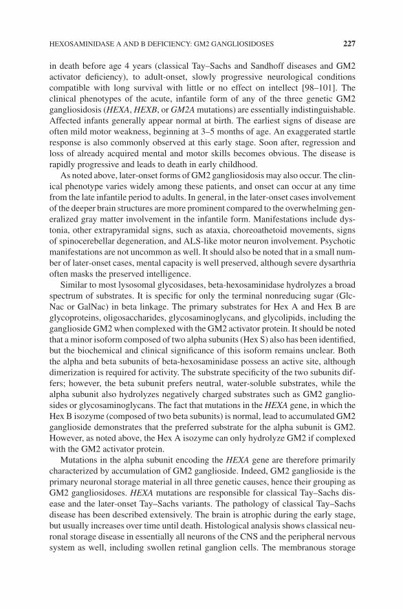
�
� �
�
HEXOSAMINIDASE A AND B DEFICIENCY: GM2 GANGLIOSIDOSES 227
in death before age 4 years (classical Tay–Sachs and Sandhoff diseases and GM2activator deficiency), to adult-onset, slowly progressive neurological conditionscompatible with long survival with little or no effect on intellect [98–101]. Theclinical phenotypes of the acute, infantile form of any of the three genetic GM2gangliosidosis (HEXA, HEXB, or GM2A mutations) are essentially indistinguishable.Affected infants generally appear normal at birth. The earliest signs of disease areoften mild motor weakness, beginning at 3–5 months of age. An exaggerated startleresponse is also commonly observed at this early stage. Soon after, regression andloss of already acquired mental and motor skills becomes obvious. The disease israpidly progressive and leads to death in early childhood.
As noted above, later-onset forms of GM2 gangliosidosis may also occur. The clin-ical phenotype varies widely among these patients, and onset can occur at any timefrom the late infantile period to adults. In general, in the later-onset cases involvementof the deeper brain structures are more prominent compared to the overwhelming gen-eralized gray matter involvement in the infantile form. Manifestations include dys-tonia, other extrapyramidal signs, such as ataxia, choreoathetoid movements, signsof spinocerebellar degeneration, and ALS-like motor neuron involvement. Psychoticmanifestations are not uncommon as well. It should also be noted that in a small num-ber of later-onset cases, mental capacity is well preserved, although severe dysarthriaoften masks the preserved intelligence.
Similar to most lysosomal glycosidases, beta-hexosaminidase hydrolyzes a broadspectrum of substrates. It is specific for only the terminal nonreducing sugar (Glc-Nac or GalNac) in beta linkage. The primary substrates for Hex A and Hex B areglycoproteins, oligosaccharides, glycosaminoglycans, and glycolipids, including theganglioside GM2 when complexed with the GM2 activator protein. It should be notedthat a minor isoform composed of two alpha subunits (Hex S) also has been identified,but the biochemical and clinical significance of this isoform remains unclear. Boththe alpha and beta subunits of beta-hexosaminidase possess an active site, althoughdimerization is required for activity. The substrate specificity of the two subunits dif-fers; however, the beta subunit prefers neutral, water-soluble substrates, while thealpha subunit also hydrolyzes negatively charged substrates such as GM2 ganglio-sides or glycosaminoglycans. The fact that mutations in the HEXA gene, in which theHex B isozyme (composed of two beta subunits) is normal, lead to accumulated GM2ganglioside demonstrates that the preferred substrate for the alpha subunit is GM2.However, as noted above, the Hex A isozyme can only hydrolyze GM2 if complexedwith the GM2 activator protein.
Mutations in the alpha subunit encoding the HEXA gene are therefore primarilycharacterized by accumulation of GM2 ganglioside. Indeed, GM2 ganglioside is theprimary neuronal storage material in all three genetic causes, hence their grouping asGM2 gangliosidoses. HEXA mutations are responsible for classical Tay–Sachs dis-ease and the later-onset Tay–Sachs variants. The pathology of classical Tay–Sachsdisease has been described extensively. The brain is atrophic during the early stage,but usually increases over time until death. Histological analysis shows classical neu-ronal storage disease in essentially all neurons of the CNS and the peripheral nervoussystem as well, including swollen retinal ganglion cells. The membranous storage

�
� �
�
228 THE GENETICS OF SPHINGOLIPID HYDROLASES
bodies characteristic of Tay–Sachs disease upon electron microscopic examinationare composed of cholesterol, phospholipid, and GM2 ganglioside. Increased apop-totic neurons have also been observed. The neuronal pathology in the acute or sub-acute forms of Tay–Sachs disease is generally less severe than that in the infantileform and tends to be more prominent in the hypothalamus, cerebellum, brain stem,and spinal cord.
In the Sandhoff disease variants, due to mutations in the HEXB gene, the patholog-ical and clinical findings are very similar to those of Tay–Sachs. However, yellowingof the cerebral cortex and deeper structures has been documented in Sandhoff disease,possibly due to accumulated asialoganglioside. Also, additional accumulation of sph-ingoglycolipids with a terminal hexosamine residue and fragments of undigestedglycoprotein in systemic organs has been found. Patients with GM2A mutations areindistinguishable from those with HEXA or HEXB mutations.
The HEXA gene, encoding the alpha subunit, is located within the chromosomalregion 15q23-q24. It is ∼35 kb in length and contains 14 exons. The HEXB gene,encoding the beta subunit, is located at chromosomal region 5q13 and is ∼45 kb inlength that is divided into 14 exons. Analysis of these genes strongly suggests thatthey arose from a common ancestor. They share a very common exon structure, and inboth genes the promoter activity resides within ∼150 bp of the initiating ATG codon.Overall, the alpha and beta subunits share∼57% amino acid identity. The GM2A genemaps to chromosomal region 5q31.1-31.3. In addition, an evolutionarily related pseu-dogene, GM2AP, maps to chromosome 3. The GM2A gene is ∼16 kb and containsfour exons.
Numerous mutations have been described in the HEXA gene responsible forTay–Sachs disease and its variants. Infantile Tay–Sachs disease occurs most com-monly among individuals of Ashkenazi Jewish ancestry (carrier frequency ∼1:25),and there are two mutations that are the most frequent; one is a 4 bp insertion inexon 11, and the second is a donor splice site mutation in intron 11 [102]. Bothof these mutations result in severely deficient or absent HEXA mRNA expression,leading to absent Hex A and Hex S activities. Another common HEXA mutationcausing infantile Tay–Sachs disease occurs in French Canadian patients and is dueto a deletion that extends from ∼2 kb upstream of the 5′ end of the gene into intron 1[103,104]. This also results in the absence of Hex A mRNA and protein. Manyother mutations in the HEXA gene have been described causing classical Tay–Sachsdisease, effecting protein processing, catalytic activity, and/or mRNA processing. Inaddition, mutations causing the later-onset forms are known, and generally resultin expression of HEXA mRNA and residual enzyme activity. An important HEXAmutation is also known as the B1 variant, in which there is normal activity towardthe artificial substrates generally used to measure beta-hexosaminidase activity, butno activity of the mutant enzyme toward GM2 ganglioside. Individuals with thismutation pose a unique diagnostic challenge since they will appear enzymaticallynormal but develop severe disease. Many mutations in the HEXB gene causingSandhoff disease and its variants also have been described. A much small number ofmutations in the GM2A gene are known. As noted above, the fact that mutations in

�
� �
�
SPHINGOLIPID ACTIVATOR PROTEINS 229
this gene caused a severe clinical disease provided the first proof of the physiologicalsignificance of the GM2/GM2 activator complex [105,106].
Naturally occurring mutations causing GM2 gangliosidosis have been describedin dogs, cats, and pigs [107–109]. In all species, abundant neuronal storage is themajor finding. Visceral storage is only found in the feline model, however. Mega-neurite formation, which has been observed in human GM2 gangliosidosis patients,occurs in dog and cat models. A number of groups also have independently generatedmurine models of Tay–Sachs, Sandhoff, and GM2 activator deficiency in mice usinggene targeting strategies. In general, knockout of the HEXA gene in mice (Tay–Sachsdisease) results in a much milder neurological disease than predicted from the humancases [110]. Mutations in the HEXB gene in mice results in a more severe phenotype,with onset of clinical disease at ∼3 months associated with excessive neuronal GM2storage [130]. Mutations in the GM2A gene of mice also resulted in a much milderdisease than predicted [111].
In part, the distinction between the mouse models and the human disorder are dueto the distinct degradation of GM2 ganglioside by these species [112]. In humans,GM2 is degraded nearly exclusively by Hex A in collaboration with the activatorprotein to yield the ganglioside GM3. In contrast, in mice GM2 can be degraded bytwo different pathways. One is identical to the human pathway, and the second isessentially unique to the mouse and is initiated by sialidase acting on GM2 to yieldGA2 ganglioside. The GA2 is then degraded by either Hex A with activator protein,or to a lesser extent by Hex B. This explains the mild phenotype due to alpha subunitmutations (HEXA), since the Hex B can act on the GA2 produced by sialidase. Incontrast, HEXB mutations result in deficiency of both enzymes and a complete blockof degradation.
11.10 SPHINGOLIPID ACTIVATOR PROTEINS
As discussed in the earlier sections, the lysosomal degradation of sphingolipidsrequires small nonenzymatic glycoproteins, referred to as “SAPs” [113,114]. Theseinclude the saposin proteins (SAPs A–D, and GM2 activator protein). There are twogenes responsible for these proteins. One is GMA2, which encodes the GM2 activatorprotein and is located on chromosome 5 [115], and the other is PSAP, located onchromosome 10 and encoding the saposin precursor protein and the individual SAPsA–D, which are derived from proteolytic cleavage of the precursor [116]. The GM2activator protein and resulting mutations (responsible for the “A/B” Tay–Sachsvariant) were discussed in the previous section and will not be discussed further here.
The PSAP gene encodes the SAP precursor protein (prosaposin), with a totalof 524 amino acids and 5 N-glycosylation sites [116]. There are four homologousdomains located within the precursor protein, each of ∼80 amino acids. A majorportion of the newly synthesized precursor is exported to the cell surface and thenimported into the lysosomal compartment, where it is processed to the mature SAPA–D proteins. Notably, unlike most lysosomal proteins the prosaposin protein is

�
� �
�
230 THE GENETICS OF SPHINGOLIPID HYDROLASES
transported to the lysosome via its interaction with the alternative receptor, sortilin[117,118]. The occurrence of the nonprocessed SAP precursor in body fluids andits neurotrophic properties indicates that its function may not be restricted to beingthe precursor of the individual SAPs. For example, prosaposin has been found inmilk, serum, and seminal fluid, and treatment of cell lines with PSAP increased cellsurvival and was antiapoptotic. Indeed, serum prosaposin levels are increased inpatients with advanced prostate cancer. Of interest is whether these effects of PSAPon cell survival are a direct effect of the protein itself, or rather the effect on theindividual processed SAPs on the activation of the sphingolipid hydrolases.
In vitro, SAP-A stimulates beta-glucocerebrosidase (the enzyme deficient inGaucher disease) and beta-galactocerebrosidase (the enzyme deficient in Krabbedisease) activities in the presence of detergents. SAP-B is a nonspecific glycolipidbinding protein that stimulates the hydrolysis of ∼20 or more glycolipids by differentenzymes in vitro, including the hydrolysis of sulfatide by ASA (the enzyme deficientin MLD). SAP-C stimulates the in vitro activities of glucosyl- and galactosylcere-brosidases, as well as sphingomyelin by ASM (the enzyme deficient in Types Aand B NPD) [119]. SAP-D is required for the degradation of ceramide by AC (theenzyme deficient in Farber disease [120].
Analysis of mutations in the genes encoding the PSAP protein have providedimportant insights into the role of the SAP proteins for the in vivo hydrolysis of sphin-golipids and their relationship to human disease. Of note, a SAP precursor deficiencywas found in a child who died at 16 weeks and who was homozygous for a mutation inthe initiation codon of the PSAP gene. The complete absence of the precursor proteinand four resultant SAPs led to a generalized accumulation of ceramide, glucosylce-ramide, galactosylceramide, sulfatide, lactosylceramide, digalactosylceramide, andother sphingolipids. The symptoms of the disease resembled that of type 2 Gaucherdisease. Other point mutations within the region of the PSAP gene encoding SAPB caused accumulation of sulfatide and some other sphingolipids, and a clinicalcourse resembling that of juvenile MLD. Mutations in the SAP-C region only ledto accumulation of glucosylceramide, similar to juvenile Gaucher disease [121]. Nosphingomyelin storage was observed in individuals with SAP-C mutations, and thishas been explained by the fact that there is a SAP-like domain within ASM that com-pensates for the loss of SAP-C in these patients. Patients with mutations in the SAP-Aregion of the PSAP gene resemble those with Krabbe disease (GLD), and accu-mulate galactosylcerebroside within the CNS and peripheral nervous system [122].There are no human patients with SAP-D mutations, but the requirement by AC forSAP-D has been confirmed in vivo in mutant mice, who accumulate ceramides withalpha-hydroxylated fatty acids mainly in the brain and kidney, and develop Purkinjecell loss and defects in the urinary system [123,124].
A mouse model of SAP precursor deficiency has also been constructed by genetargeting, and the clinical and biochemical features resembled that of the human dis-ease [125]. Mice developed massive sphingolipid storage and neurological diseaseby ∼20 days, and died by ∼35–38 days. Several knock-in models expressing specifichuman mutations have also been constructed, confirming the in vivo importance ofthese proteins [126,127].

�
� �
�
REFERENCES 231
REFERENCES
[1] Chen H, Tran J-TA, Brush RS, Saadi A, Rahman AK, et al. Ceramide signaling in retinaldegeneration. Adv Exp Med Biol 2012;723:553–558.
[2] Furuya H, Shimizu Y, Kawamori T. Sphingolipids in cancer. Cancer Metastasis Rev2011;30:567–576.
[3] Petrache I, Petrusca DN, Bowler RP, Kamocki K. Involvement of ceramide in cell deathresponses in the pulmonary circulation. Proc Am Thorac Soc 2011;8:492–496.
[4] Podbielska M, Krotkiewski H, Hogan EL. Signaling and regulatory functions ofbioactive sphingolipids as therapeutic targets in multiple sclerosis. Neurochem Res2012;37:1154–1169.
[5] Mullen TD, Hannun YA, Obeid LM. Ceramide synthases at the centre of sphingolipidmetabolism and biology. Biochem J 2012;441:789–802.
[6] Wennekes T, van den Berg RJBHN, Boot RG, van der Marel GA, Overkleeft HS, et al.Glycosphingolipids – nature, function, and pharmacological modulation. Angew ChemInt Ed Engl 2009;48:8848–8869.
[7] Worgall TS. Sphingolipid synthetic pathways are major regulators of lipid homeostasis.Adv Exp Med Biol 2011;721:139–148.
[8] Schulze H, Sandhoff K. Lysosomal lipid storage diseases. Cold Spring Harb PerspectBiol 2011;3:a004804.
[9] Cox TM, Cachon-Gonzalez MB. The cellular pathology of lysosomal diseases. J Pathol2012;226:241–254.
[10] Young SA, Mina JG, Denny PW, Smith TK. Sphingolipid and ceramide homeostasis:potential therapeutic targets. Biochem Res Int 2012;2012:248135–248135.
[11] Farber S, Cohen J, Uzman LL. Lipogranulomatosis; a new lipo-glycoprotein storagedisease. J Mt Sinai Hosp N Y 1957;24:816–837.
[12] Ahmad A, Mazhar AU, Anwar M. Farber disease: a rare neurodegenerative disorder. JColl Physicians Surg Pak 2009;19:67–68.
[13] Sugita M, Dulaney JT, Moser HW. Ceramidase deficiency in Farber’s disease (lipogran-ulomatosis). Science 1972;178:1100–1102.
[14] Okino N, He X, Gatt S, Sandhoff K, Ito M, et al. The reverse activity of human acidceramidase. J Biol Chem 2003;278:29948–29953.
[15] Park J-H, Schuchman EH. Acid ceramidase and human disease. Biochim Biophys Acta2006;1758:2133–2138.
[16] Li C-M, Park J-H, Simonaro CM, He X, Gordon RE, et al. Insertional mutagenesis ofthe mouse acid ceramidase gene leads to early embryonic lethality in homozygotes andprogressive lipid storage disease in heterozygotes. Genomics 2002;79:218–224.
[17] Eliyahu E, Shtraizent N, Martinuzzi K, Barritt J, He X, et al. Acid ceramidase improvesthe quality of oocytes and embryos and the outcome of in vitro fertilization. FASEB J2010;24:1229–1238.
[18] Bashyam MD, Chaudhary AK, Kiran M, Reddy V, Nagarajaram HA, et al. Molecularanalyses of novel ASAH1 mutations causing Farber lipogranulomatosis: analyses ofexonic splicing enhancer inactivating mutation. Clin Genet 2014;86:530–538.
[19] Alayoubi AM, Wang JC, Au BC, Carpentier S, Garcia V, et al. Systemic ceramideaccumulation leads to severe and varied pathological consequences. EMBO Mol Med2013;5:827–842.

�
� �
�
232 THE GENETICS OF SPHINGOLIPID HYDROLASES
[20] Eliyahu E, Shtraizent N, Shalgi R, Schuchman EH. Construction of conditional acidceramidase knockout mice and in vivo effects on oocyte development and fertility. CellPhysiol Biochem 2012;30:735–748.
[21] Zhou J, Tawk M, Tiziano FD, Veillet J, Bayes M, et al. Spinal muscular atrophy associ-ated with progressive myoclonic epilepsy is caused by mutations in ASAH1. Am J HumGenet 2012;91:5–14.
[22] Dyment DA, Sell E, Vanstone MR, Smith AC, Garandeau D, et al. Evidence for clin-ical, genetic and biochemical variability in spinal muscular atrophy with progressivemyoclonic epilepsy. Clin Genet 2014;86:558–563.
[23] Park J-H, Eliyahu E, Narla G, DiFeo A, Martignetti JA, et al. KLF6 is one transcriptionfactor involved in regulating acid ceramidase gene expression. Biochim Biophys Acta2005;1732:82–87.
[24] Beckham TH, Lu P, Cheng JC, Zhao D, Turner LS, et al. Acid ceramidase-mediated pro-duction of sphingosine 1-phosphate promotes prostate cancer invasion through upregu-lation of cathepsin B. Int J Cancer 2012;131:2034–2043.
[25] Flowers M, Fabrias G, Delgado A, Casas J, Abad JL, et al. C6-ceramide and targetedinhibition of acid ceramidase induce synergistic decreases in breast cancer cell growth.Breast Cancer Res Treat 2012;133:447–458.
[26] Zhang H, Li D, Su Y, Jiang S, Xu Y, et al. Identification of the N-acylsphingosineamidohydrolase 1 gene (ASAH1) for susceptibility to schizophrenia in a Han Chinesepopulation. World J Biol Psychiatry 2012;13:106–113.
[27] Mao C, Obeid LM. Ceramidases: regulators of cellular responses mediated byceramide, sphingosine, and sphingosine-1-phosphate. Biochim Biophys Acta 2008;1781:424–434.
[28] McGovern MMSE. In: Pagon RABT, Dolan CR, Stephens K, Adam MP, editors. AcidSphingomyelinase Deficiency. Seattle (WA): University of Washington; 2009.
[29] Schuchman EH. The pathogenesis and treatment of acid sphingomyelinase-deficientNiemann–Pick disease. Int J Clin Pharmacol Ther 2009;47(Suppl 1):48–57.
[30] Simonaro CM, Park JH, Eliyahu E, Shtraizent N, McGovern MM, et al. Imprintingat the SMPD1 locus: implications for acid sphingomyelinase-deficient Niemann–Pickdisease. Am J Hum Genet 2006;78:865–870.
[31] Clarke CJ, Wu BX, Hannun YA. The neutral sphingomyelinase family: identifying bio-chemical connections. Adv Enzyme Regul 2011;51:51–58.
[32] Horinouchi K, Erlich S, Perl DP, Ferlinz K, Bisgaier CL, et al. Acid sphingomyeli-nase deficient mice: a model of types A and B Niemann–Pick disease. Nat Genet 1995;10:288–293.
[33] Schissel SL, Keesler GA, Schuchman EH, Williams KJ, Tabas I. The cellular traffickingand zinc dependence of secretory and lysosomal sphingomyelinase, two products of theacid sphingomyelinase gene. J Biol Chem 1998;273:18250–18259.
[34] Miranda SR, He X, Simonaro CM, Gatt S, Dagan A, et al. Infusion of recombinanthuman acid sphingomyelinase into Niemann–Pick disease mice leads to visceral, butnot neurological, correction of the pathophysiology. FASEB J 2000;14:1988–1995.
[35] Zeidan YH, Hannun YA. The acid sphingomyelinase/ceramide pathway: biomedicalsignificance and mechanisms of regulation. Curr Mol Med 2010;10:454–466.
[36] Becker KA, Grassme H, Zhang Y, Gulbins E. Ceramide in Pseudomonas aeruginosainfections and cystic fibrosis. Cell Physiol Biochem 2010;26:57–66.

�
� �
�
REFERENCES 233
[37] Bikman BT, Summers SA. Sphingolipids and hepatic steatosis. Adv Exp Med Biol2011;721:87–97.
[38] Truman J-P, Al Gadban MM, Smith KJ, Hammad SM. Acid sphingomyelinase inmacrophage biology. Cell Mol Life Sci 2011;68:3293–3305.
[39] Lo SM, Choi M, Liu J, Jain D, Boot RG, et al. Phenotype diversity in type 1 Gaucherdisease: discovering the genetic basis of Gaucher disease/hematologic malignancy phe-notype by individual genome analysis. Blood 2012;119:4731–4740.
[40] Pastores GM. Neuropathic Gaucher disease. Wien Med Wochenschr 2010;160:605–608.
[41] Mikosch P, Hughes D. An overview on bone manifestations in Gaucher disease. WienMed Wochenschr 2010;160:609–624.
[42] Goker-Alpan O. Therapeutic approaches to bone pathology in Gaucher disease: past,present and future. Mol Genet Metab 2011;104:438–447.
[43] Choy FYM, Campbell TN. Gaucher disease and cancer: concept and controversy. Int JCell Biol 2011;2011:150450–150450.
[44] Brown JT, Lahey C, Laosinchai-Wolf W, Hadd AG. Polymorphisms in the glucocere-brosidase gene and pseudogene urge caution in clinical analysis of Gaucher diseaseallele c.1448T>C (L444P). BMC Med Genet 2006;7:69–69.
[45] Hruska KS, LaMarca ME, Scott CR, Sidransky E. Gaucher disease: mutation andpolymorphism spectrum in the glucocerebrosidase gene (GBA). Hum Mutat 2008;29:567–583.
[46] Scott SA, Edelmann L, Liu L, Luo M, Desnick RJ, et al. Experience with carrierscreening and prenatal diagnosis for 16 Ashkenazi Jewish genetic diseases. Hum Mutat2010;31:1240–1250.
[47] Kinghorn KJ. Pathological looping in the synucleinopathies: investigating the linkbetween Parkinson’s disease and Gaucher disease. Dis Model Mech 2011;4:713–715.
[48] Anheim M, Elbaz A, Lesage S, Durr A, Condroyer C, et al. Penetrance of Parkinsondisease in glucocerebrosidase gene mutation carriers. Neurology 2012;78:417–420.
[49] Farfel-Becker T, Vitner EB, Futerman AH. Animal models for Gaucher diseaseresearch. Dis Model Mech 2011;4:746–752.
[50] Sardi SP, Singh P, Cheng SH, Shihabuddin LS, Schlossmacher MG. Mutant GBA1expression and synucleinopathy risk: first insights from cellular and mouse models.Neurodegener Dis 2012;10:195–202.
[51] Hughes DA, Pastores GM. The pathophysiology of GD – current understanding andrationale for existing and emerging therapeutic approaches. Wien Med Wochenschr2010;160:594–599.
[52] Lachmann RH. Enzyme replacement therapy for lysosomal storage diseases. Curr OpinPediatr 2011;23:588–593.
[53] Abian O, Alfonso P, Velazquez-Campoy A, Giraldo P, Pocovi M, et al. Therapeu-tic strategies for Gaucher disease: miglustat (NB-DNJ) as a pharmacological chaper-one for glucocerebrosidase and the different thermostability of velaglucerase alfa andimiglucerase. Mol Pharm 2011;8:2390–2397.
[54] Benito JM, Garcia Fernandez JM, Ortiz Mellet C. Pharmacological chaperone therapyfor Gaucher disease: a patent review. Expert Opin Ther Pat 2011;21:885–903.
[55] Kohlschutter A, Eichler F. Childhood leukodystrophies: a clinical perspective. ExpertRev Neurother 2011;11:1485–1496.

�
� �
�
234 THE GENETICS OF SPHINGOLIPID HYDROLASES
[56] Duffner PK, Barczykowski A, Jalal K, Yan L, Kay DM, et al. Early infantile Krabbedisease: results of the World-Wide Krabbe Registry. Pediatr Neurol 2011;45:141–148.
[57] Malandrini A, D’Eramo C, Palmeri S, Gaudiano C, Gambelli S, et al. Peripheral neu-ropathy in late-onset Krabbe disease: report of three cases. Neurol Sci 2013;34:79–83.
[58] Perlman SJ, Mar S. Leukodystrophies. Adv Exp Med Biol 2012;724:154–171.
[59] Sakai N. Pathogenesis of leukodystrophy for Krabbe disease: molecular mechanism andclinical treatment. Brain Dev 2009;31:485–487.
[60] Liu Y, Gibson J, Wheeler J, Kwee LC, Santiago-Turla CM, et al. GALC deletionsincrease the risk of primary open-angle glaucoma: the role of Mendelian variants incomplex disease. PLoS One 2011;6:e27134.
[61] Luzi P, Rafi MA, Zaka M, Curtis M, Vanier MT, et al. Generation of a mouse with lowgalactocerebrosidase activity by gene targeting: a new model of globoid cell leukodys-trophy (Krabbe disease). Mol Genet Metab 2001;73:211–223.
[62] Neri M, Ricca A, di Girolamo I, Alcala’-Franco B, Cavazzin C, et al. Neural stemcell gene therapy ameliorates pathology and function in a mouse model of globoid cellleukodystrophy. Stem Cells 2011;29:1559–1571.
[63] Reddy AS, Kim JH, Hawkins-Salsbury JA, Macauley SL, Tracy ET, et al. Bone marrowtransplantation augments the effect of brain- and spinal cord-directed adeno-associatedvirus 2/5 gene therapy by altering inflammation in the murine model of globoid-cellleukodystrophy. J Neurosci 2011;31:9945–9957.
[64] Lin D-S, Hsiao C-D, Liau I, Lin S-P, Chiang M-F, et al. CNS-targeted AAV5 gene trans-fer results in global dispersal of vector and prevention of morphological and functiondeterioration in CNS of globoid cell leukodystrophy mouse model. Mol Genet Metab2011;103:367–377.
[65] Renaud DL. Lysosomal disorders associated with leukoencephalopathy. Seminars inneurology. 2012;32(1):51–4.
[66] Kehrer C, Blumenstock G, Gieselmann V, Krageloh-Mann I. The natural course ofgross motor deterioration in metachromatic leukodystrophy. Dev Med Child Neurol2011;53:850–855.
[67] Gieselmann V, Zlotogora J, Harris A, Wenger DA, Morris CP. Molecular genetics ofmetachromatic leukodystrophy. Hum Mutat 1994;4:233–242.
[68] Lugowska A, Poninska J, Krajewski P, Broda G, Ploski R. Population carrier rates ofpathogenic ARSA gene mutations: is metachromatic leukodystrophy underdiagnosed?PLoS One 2011;6:e20218.
[69] Tinsa F, Caillaud C, Vanier MT, Bousnina D, Boussetta K, et al. An unusual homozy-gous arylsulfatase: a pseudodeficiency in a metachromatic leukodystrophy Tunisianpatient. J Child Neurol 2010;25:82–86.
[70] Baronica KB, Mlinac K, Ozretic D, Vladic A, Bognar SK. Arylsulfatase a gene poly-morphisms in relapsing remitting multiple sclerosis: genotype-phenotype correlationand estimation of disease progression. Coll Antropol 2011;35(Suppl 1):11–16.
[71] Kumperscak HG, Dolzan V, Videtic A, Plesnicar BK. Polymorphisms in genes encodingthe serotonin and dopamine pathways in two sisters with metachromatic leukodystrophy.J Int Med Res 2008;36:1123–1128.
[72] Matthes F, Stroobants S, Gerlach D, Wohlenberg C, Wessig C, et al. Efficacy of enzymereplacement therapy in an aggravated mouse model of metachromatic leukodystrophydeclines with age. Hum Mol Genet 2012;21:2599–2609.

�
� �
�
REFERENCES 235
[73] Stroobants S, Gerlach D, Matthes F, Hartmann D, Fogh J, et al. Intracerebroventricularenzyme infusion corrects central nervous system pathology and dysfunction in a mousemodel of metachromatic leukodystrophy. Hum Mol Genet 2011;20:2760–2769.
[74] Solders G, Celsing G, Hagenfeldt L, Ljungman P, Isberg B, et al. Improved periph-eral nerve conduction, EEG and verbal IQ after bone marrow transplantation for adultmetachromatic leukodystrophy. Bone Marrow Transplant 1998;22:1119–1122.
[75] Gassas A, Raiman J, White L, Schechter T, Clarke J, et al. Long-term adaptive func-tioning outcomes of children with inherited metabolic and genetic diseases treated withhematopoietic stem cell transplantation in a single large pediatric center: parents’ per-spective. J Pediatr Hematol Oncol 2011;33:216–220.
[76] Tarabuso AL. Fabry disease. Skinmed 2011;9:173–177.
[77] Toyooka K. Fabry disease. Curr Opin Neurol 2011;24:463–468.
[78] Aerts JM, Boot RG, van Eijk M, Groener J, Bijl N, et al. Glycosphingolipids and insulinresistance. Adv Exp Med Biol 2011;721:99–9119.
[79] Linthorst GE, Folman CC, Aerts JM, Hollak CE. Blood group does not correlate withdisease severity in patients with Fabry disease (alpha-galactosidase A deficiency). BloodCells Mol Dis 2003;31:324–326.
[80] Tomasic IB, Metcalf MC, Guce AI, Clark NE, Garman SC. Interconversion of the speci-ficities of human lysosomal enzymes associated with Fabry and Schindler diseases. JBiol Chem 2010;285:21560–21566.
[81] Rozenfeld P, Neumann PM. Treatment of Fabry disease: current and emerging strate-gies. Curr Pharm Biotechnol 2011;12:916–922.
[82] Schaefer E, Mehta A, Gal A. Genotype and phenotype in Fabry disease: analysis of theFabry Outcome Survey. Acta Paediatr Suppl 2005;94:87–92; discussion 79.
[83] Bouwman MG, Rombach SM, Schenk E, Sweeb A, Wijburg FA, et al. Prevalence ofsymptoms in female Fabry disease patients: a case-control survey. J Inherit Metab Dis2012;35:891–898.
[84] Spacil Z, Elliott S, Reeber SL, Gelb MH, Scott CR, et al. Comparative triplex tandemmass spectrometry assays of lysosomal enzyme activities in dried blood spots using fastliquid chromatography: application to newborn screening of Pompe, Fabry, and Hurlerdiseases. Anal Chem 2011;83:4822–4828.
[85] Brunetti-Pierri N, Scaglia F. GM1 gangliosidosis: review of clinical, molecular, andtherapeutic aspects. Mol Genet Metab 2008;94:391–396.
[86] Morita M, Saito S, Ikeda K, Ohno K, Sugawara K, et al. Structural bases of GM1 gan-gliosidosis and Morquio B disease. J Hum Genet 2009;54:510–515.
[87] Caciotti A, Garman SC, Rivera-Colon Y, Procopio E, Catarzi S, et al. GM1 gangliosi-dosis and Morquio B disease: an update on genetic alterations and clinical findings.Biochim Biophys Acta 2011;1812:782–790.
[88] Hofer D, Paul K, Fantur K, Beck M, Burger F, et al. GM1 gangliosidosis and MorquioB disease: expression analysis of missense mutations affecting the catalytic site of acidbeta-galactosidase. Hum Mutat 2009;30:1214–1221.
[89] Nada R, Gupta K, Lal SB, Vasishta RK. An autopsy case of infantile GM1 gangliosidosiswith adrenal calcification. Metab Brain Dis 2011;26:307–310.
[90] Steenweg ME, Vanderver A, Blaser S, Bizzi A, de Koning TJ, et al. Magnetic resonanceimaging pattern recognition in hypomyelinating disorders. Brain 2010;133:2971–2982.
�
� �
�
236 THE GENETICS OF SPHINGOLIPID HYDROLASES
[91] Ohto U, Usui K, Ochi T, Yuki K, Satow Y, et al. Crystal structure of human beta-galactosidase: structural basis of Gm1 gangliosidosis and morquio B diseases. J BiolChem 2012;287:1801–1812.
[92] Tatano Y, Takeuchi N, Kuwahara J, Sakuraba H, Takahashi T, et al. Elastogenesis incultured dermal fibroblasts from patients with lysosomal beta-galactosidase, protectiveprotein/cathepsin A and neuraminidase-1 deficiencies. J Med Invest 2006;53:103–112.
[93] van der Voorn JP, Kamphorst W, van der Knaap MS, Powers JM. The leukoen-cephalopathy of infantile GM1 gangliosidosis: oligodendrocytic loss and axonal dys-function. Acta Neuropathol 2004;107:539–545.
[94] Claro E, Wallace MA, Fain JN, Nair BG, Patel TB, et al. Altered phosphoinositide-specific phospholipase C and adenylyl cyclase in brain cortical membranes of cats withGM1 and GM2 gangliosidosis. Brain Res Mol Brain Res 1991;11:265–271.
[95] Itoh M, Matsuda J, Suzuki O, Ogura A, Oshima A, et al. Development of lysosomal stor-age in mice with targeted disruption of the beta-galactosidase gene: a model of humanG(M1)-gangliosidosis. Brain Dev 2001;23:379–384.
[96] Maegawa G. GM2 gangliosidosis: the prototype of lysosomal storage disorders. DevMed Child Neurol 2012;54:104–105.
[97] Cordeiro P, Hechtman P, Kaplan F. The GM2 gangliosidoses databases: allelic variationat the HEXA, HEXB, and GM2A gene loci. Genet Med 2000;2:319–327.
[98] Bley AE, Giannikopoulos OA, Hayden D, Kubilus K, Tifft CJ, et al. Natural history ofinfantile G(M2) gangliosidosis. Pediatrics 2011;128:e1233–1241.
[99] Smith NJ, Winstone AM, Stellitano L, Cox TM, Verity CM. GM2 gangliosidosis in aUK study of children with progressive neurodegeneration: 73 cases reviewed. Dev MedChild Neurol 2012;54:176–182.
[100] Jain A, Kohli A, Sachan D. Infantile Sandhoff’s disease with peripheral neuropathy.Pediatr Neurol 2010;42:459–461.
[101] Osher E, Fattal-Valevski A, Sagie L, Urshanski N, Amir-Levi Y, et al. Pyrimethamineincreases beta-hexosaminidase A activity in patients with Late Onset Tay Sachs. MolGenet Metab 2011;102:356–363.
[102] Frisch A, Colombo R, Michaelovsky E, Karpati M, Goldman B, et al. Origin and spreadof the 1278insTATC mutation causing Tay–Sachs disease in Ashkenazi Jews: geneticdrift as a robust and parsimonious hypothesis. Hum Genet 2004;114:366–376.
[103] De Braekeleer M, Hechtman P, Andermann E, Kaplan F. The French Canadian Tay–Sachs disease deletion mutation: identification of probable founders. Hum Genet 1992;89:83–87.
[104] Martin DC, Mark BL, Triggs-Raine BL, Natowicz MR. Evaluation of the risk forTay–Sachs disease in individuals of French Canadian ancestry living in New England.Clin Chem 2007;53:392–398.
[105] Peleg L, Meltzer F, Karpati M, Goldman B. GM2 gangliosidosis B1 variant: biochem-ical and molecular characterization of hexosaminidase A. Biochem Mol Med 1995;54:126–132.
[106] Tanaka A, Hoang LTN, Nishi Y, Maniwa S, Oka M, et al. Different attenuated pheno-types of GM2 gangliosidosis variant B in Japanese patients with HEXA mutations atcodon 499, and five novel mutations responsible for infantile acute form. J Hum Genet2003;48:571–574.

�
� �
�
REFERENCES 237
[107] Yamato O, Matsuki N, Satoh H, Inaba M, Ono K, et al. Sandhoff disease in a goldenretriever dog. J Inherit Metab Dis 2002;25:319–320.
[108] Kosanke SD, Pierce KR, Read WK. Morphogenesis of light and electron microscopiclesions in porcine GM2-gangliosidosis. Vet Pathol 1979;16:6–17.
[109] Bradbury AM, Morrison NE, Hwang M, Cox NR, Baker HJ, et al. Neurodegenerativelysosomal storage disease in European Burmese cats with hexosaminidase beta-subunitdeficiency. Mol Genet Metab 2009;97:53–59.
[110] Yamanaka S, Johnson MD, Grinberg A, Westphal H, Crawley JN, et al. Targeted dis-ruption of the Hexa gene results in mice with biochemical and pathologic features ofTay–Sachs disease. Proc Natl Acad Sci U S A 1994;91:9975–9979.
[111] Phaneuf D, Wakamatsu N, Huang JQ, Borowski A, Peterson AC, et al. Dramaticallydifferent phenotypes in mouse models of human Tay–Sachs and Sandhoff diseases. HumMol Genet 1996;5:1–14.
[112] Baek RC, Martin DR, Cox NR, Seyfried TN. Comparative analysis of brain lipids inmice, cats, and humans with Sandhoff disease. Lipids 2009;44:197–205.
[113] Sandhoff K, Kolter T. Processing of sphingolipid activator proteins and the topology oflysosomal digestion. Acta Biochim Pol 1998;45:373–384.
[114] Kolter T, Sandhoff K. Lysosomal degradation of membrane lipids. FEBS Lett 2010;584:1700–1712.
[115] Wendeler M, Lemm T, Weisgerber J, Hoernschemeyer J, Bartelsen O, et al. Expressionof recombinant human GM2-activator protein in insect cells: purification and character-ization by mass spectrometry. Protein Expr Purif 2003;27:259–266.
[116] Misasi R, Hozumi I, Inuzuka T, Capozzi A, Mattei V, et al. Biochemistry and neurobi-ology of prosaposin: a potential therapeutic neuro-effector. Cent Nerv Syst Agents MedChem 2009;9:119–131.
[117] Wahe A, Kasmapour B, Schmaderer C, Liebl D, Sandhoff K, et al. Golgi-to-phagosometransport of acid sphingomyelinase and prosaposin is mediated by sortilin. J Cell Sci2010;123:2502–2511.
[118] Yuan L, Morales CR. Prosaposin sorting is mediated by oligomerization. Exp Cell Res2011;317:2456–2467.
[119] Kolzer M, Ferlinz K, Bartelsen O, Hoops SL, Lang F, et al. Functional characterizationof the postulated intramolecular sphingolipid activator protein domain of human acidsphingomyelinase. Biol Chem 2004;385:1193–1195.
[120] Linke T, Wilkening G, Sadeghlar F, Mozcall H, Bernardo K, et al. Interfacial regulationof acid ceramidase activity. Stimulation of ceramide degradation by lysosomal lipidsand sphingolipid activator proteins. J Biol Chem 2001;276:5760–5768.
[121] Vaccaro AM, Motta M, Tatti M, Scarpa S, Masuelli L, et al. Saposin C mutationsin Gaucher disease patients resulting in lysosomal lipid accumulation, saposin Cdeficiency, but normal prosaposin processing and sorting. Hum Mol Genet 2010;19:2987–2997.
[122] Grossi S, Regis S, Rosano C, Corsolini F, Uziel G, et al. Molecular analysis of ARSAand PSAP genes in twenty-one Italian patients with metachromatic leukodystrophy:identification and functional characterization of 11 novel ARSA alleles. Hum Mutat2008;29:220–230.
[123] Oya Y, Nakayasu H, Fujita N, Suzuki K, Suzuki K. Pathological study of mice with totaldeficiency of sphingolipid activator proteins (SAP knockout mice). Acta Neuropathol1998;96:29–40.

�
� �
�
238 THE GENETICS OF SPHINGOLIPID HYDROLASES
[124] Tohyama J, Oya Y, Ezoe T, Vanier MT, Nakayasu H, et al. Ceramide accumulationis associated with increased apoptotic cell death in cultured fibroblasts of sphingolipidactivator protein-deficient mouse but not in fibroblasts of patients with Farber disease.J Inherit Metab Dis 1999;22:649–662.
[125] Matsuda J, Yoneshige A, Suzuki K. The function of sphingolipids in the nervous system:lessons learnt from mouse models of specific sphingolipid activator protein deficiencies.J Neurochem 2007;103(Suppl 1):32–38.
[126] Sun Y, Witte DP, Ran H, Zamzow M, Barnes S, et al. Neurological deficits and gly-cosphingolipid accumulation in saposin B deficient mice. Hum Mol Genet 2008;17:2345–2356.
[127] Sun Y, Ran H, Zamzow M, Kitatani K, Skelton MR, et al. Specific saposin C deficiency:CNS impairment and acid beta-glucosidase effects in the mouse. Hum Mol Genet 2010;19:634–647.
[128] Campbell TN, Choy FY. Gaucher disease and the synucleinopathies: refining the rela-tionship. Orphanet J Rare Dis 2012;7:12.
[129] Lee JY, Lee BH, Kim GH, Jung CW, Lee J, et al. Clinical and genetic characteristics ofGaucher disease according to phenotypic subgroups. Korean J Pediatr 2012;55:48–53.
[130] Gulinello M, Chen F, Dobrenis K. Early deficits in motor coordination and cognitivedysfunction in a mouse model of the neurodegenerative lysosomal storage disorder,Sandhoff disease. Behav Brain Res 2008;193:315–319.

�
� �
�
12LYSOSOME-RELATED ORGANELLES:MODIFICATIONS OF THE LYSOSOMEPARADIGM
Adriana R. Mantegazza and Michael S. MarksDepartment of Pathology and Laboratory Medicine, Children’s Hospital of PhiladelphiaResearch Institute, Philadelphia, PA, USA; Departments of Pathology and LaboratoryMedicine and Physiology, Perelman School of Medicine, University of Pennsylvania,Philadelphia, PA, USA
Lysosomes are essential organelles for the physiological maintenance and viabilityof all metazoan cells, with conserved functions in nutrient and ion homeostasis, cellsignaling, macromolecule turnover, and plasma membrane repair among others. Thecomplex physiology of multicellular organisms requires certain cell types to fulfillneeds that exploit some features of lysosomes but that are specialized for specificfunctions – such as cell-mediated cytolysis by cytotoxic T lymphocytes (CTLs) orregulated secretion of clotting factors, surfactants, or other specialized cargoes. Manysuch cells have evolved to generate either modified forms of lysosomes or completelydistinct membranous structures to fulfill these functions. Such organelles are calledlysosome-related organelles (LROs), and are broadly defined as cell type-specificcompartments that share features of lysosomal or endosomal organelles but are func-tionally and morphologically distinct. Since LROs are required for the physiology ofthe organism but not for the viability of the cell that generates them, heritable defectsin the formation, composition, motility, or secretion of LROs often result in disease.In this chapter, we briefly describe the variety of LROs that have been defined todate in mammals and model organisms, relate recent insights into the biogenesis and
Lysosomes: Biology, Diseases, and Therapeutics, First Edition.Edited by Frederick R. Maxfield, James M. Willard and Shuyan Lu.© 2016 John Wiley & Sons, Inc. Published 2016 by John Wiley & Sons, Inc.
239

�
� �
�
240 LYSOSOME-RELATED ORGANELLES
secretion of a few model LROs and how these insights relate to disease, and end witha focus on LROs of the immune system and perspectives for future investigation.
12.1 DIFFERENCES BETWEEN LROs AND SECRETORY GRANULES
LROs have been rather difficult to precisely define, even by experts in the field.It is generally agreed that all LROs contain essential functional elements ofendolysosomal compartments, and the late endosomal/lysosomal tetraspanin CD63has been raised as a potential common component of most, if not all, LROs (e.g., seeRefs [1–3]). This distinguishes LROs from classical regulated secretory granules,such as the dense core granules of chromaffin cells or pancreatic β cells, in whichthe functional components are derived exclusively from the secretory pathway.However, the degree to which critical LRO components derive from the secretorypathway varies. At one end of the spectrum, all protein components of melanosomesthat have been studied to date pass through endosomal compartments prior to arrivalat their destination. At the other end, the long von Willebrand factor (vWF) polymersof Weibel–Palade bodies (WPBs) assemble and bud from the Golgi and are joined byendosome-derived factors such as the membrane proteins CD63 and P-selectin [4].Most LROs are secretory organelles that release their contents upon cellular stimu-lation; indeed, LROs have also been referred to as “secretory lysosomes” [5] basedlargely on the behavior of LROs that double as the host cell’s lysosome, such as lyticgranules of CTLs and natural killer (NK) cells. However, this term belies the factthat many LROs coexist with bona fide lysosomes in their host cells and that bonafide lysosomes can also be secreted under certain conditions [6,7]. Moreover, not allLROs are secreted. For example, notochord vacuoles function to provide structuralrigidity to the cells of the inner notochord layer [8], NOX2-containing LROsfuse with nascent phagosomes [9,10], melanosomes in ocular pigment cells areretained intracellularly, and it is not yet clear whether epidermal melanocytes secretemelanin or whether their melanosome-enriched tips are engulfed by neighboring ker-atinocytes [11,12]. Here we consider LROs as tissue-specific membranous structuresin which the origins of a substantial fraction of their components can be traced to theendolysosomal system. Table 12.1 provides a list of compartments that conform tothis definition and that have been referred to as LROs or secretory lysosomes in theliterature. Electron micrographs of examples of these LROs are shown in Figure 12.1.
12.2 PHYSIOLOGICAL FUNCTIONS OF LROs
LROs fulfill a variety of functions within metazoans. Many LROs are found inhematopoietic cells (Table 12.1 and Figure 12.1), and contribute to important func-tions in innate or adaptive immunity. For example, the lytic granules of CTLs or NKcells store pore-forming components such as perforin and cytolytic agents such asgranzymes; signal-dependent release of these components into the synapse betweena CTL or NK cell and a target cell lead to target cell permeabilization and death [14].

�
� �
�
TA
BL
E12
.1L
RO
san
dT
heir
Hos
tC
ells
,Fun
ctio
ns,a
ndC
argo
es
LR
OC
ellT
ype
Phys
iolo
gic
Func
tion
Sam
ple
Car
goes
Vert
ebra
tes
Mel
anos
ome
Mel
anoc
ytes
,eye
pigm
entc
ells
(ret
inal
pigm
ente
pith
elia
,ot
hers
)
Mel
anin
prod
uctio
nan
dst
orag
efo
rpi
gmen
tatio
nTy
rosi
nase
,tyr
osin
ase-
rela
ted
prot
eins
,OC
A2,
PME
L
Wei
bel–
Pala
debo
dyE
ndot
helia
lcel
lsSt
orag
ean
dre
leas
eof
hem
osta
tican
dpr
oinfl
amm
ator
yfa
ctor
svo
nW
illeb
rand
fact
or,P
-sel
ectin
Cyt
olyt
icgr
anul
eC
ytot
oxic
Tce
lls,N
Kce
llsTa
rget
edde
stru
ctio
nof
vira
llyin
fect
edor
canc
erou
sce
llsPe
rfor
in,g
ranz
ymes
,lys
osom
alen
zym
esD
ense
gran
ule
Plat
elet
sSt
orag
ean
dre
leas
eof
smal
lmol
ecul
esfo
rpl
atel
etad
hesi
onSe
roto
nin,
calc
ium
,AD
P,A
TP,
poly
phos
phat
esα-
Gra
nule
Plat
elet
sSt
orag
ean
dre
leas
eof
prot
eins
for
plat
elet
hem
osta
sis,
infla
mm
atio
n,an
giog
enes
is,o
ther
s
von
Will
ebra
ndfa
ctor
,pla
tele
tfa
ctor
-4,fi
brin
ogen
Lam
ella
rbo
dyA
lveo
lar
type
IIep
ithel
ialc
ells
Synt
hesi
s,st
orag
e,an
dse
cret
ion
ofsu
rfac
tant
Surf
acta
ntpr
otei
nsA
-D,
pero
xire
doxi
n6,
AB
CA
3B
asop
hilic
gran
ule
Mas
tcel
ls,b
asop
hils
Stor
age
and
secr
etio
nof
hist
amin
esH
ista
min
eA
zuro
phili
c(p
rim
ary)
gran
ules
Neu
trop
hils
,eos
inop
hils
Stor
age
and
rele
ase
into
phag
osom
esof
mic
robi
cida
lcom
poun
dsM
yelo
pero
xida
se,e
last
ase,
cath
epsi
nG
,lys
ozym
e,de
fens
ins
Spec
ific
(sec
onda
ry)
gran
ules
Neu
trop
hils
Stor
age
and
rele
ase
ofm
icro
bici
dal
com
poun
dsan
dan
tioxi
dant
activ
ityN
AD
PHox
idas
e,ge
latin
ase,
lact
ofer
rin,
lyso
zym
eG
elat
inas
e(t
ertia
ry)
gran
ules
Neu
trop
hils
Stor
age
and
rele
ase
ofm
icro
bici
dal
com
poun
dsG
elat
inas
e
Ost
eocl
asts
ecre
tory
lyso
som
eO
steo
clas
tsB
one
dige
stio
nC
athe
psin
K,v
AT
Pase
,tar
trat
eac
id-r
esis
tant
phos
phat
ase
(con
tinu
ed)
241

�
� �
�
TA
BL
E12
.1(C
ontin
ued)
LR
OC
ellT
ype
Phys
iolo
gic
Func
tion
Sam
ple
Car
goes
Acr
osom
eSp
erm
cells
Egg
pene
trat
ion
duri
ngfe
rtili
zatio
nA
cros
in,m
atri
xm
etal
lopr
otei
nase
sN
otoc
hord
vacu
ole
Not
ocho
rdin
ner
cells
Stru
ctur
alri
gidi
tyof
the
noto
chor
dL
AM
P1,v
AT
Pase
,?M
HC
clas
sII
com
part
men
tA
ctiv
ated
dend
ritic
cells
,Bly
mph
ocyt
esA
ntig
enpr
oces
sing
for
pres
enta
tion
toT
lym
phoc
ytes
MH
Ccl
ass
II,H
LA
-DM
,ca
thep
sin
SN
OX
2+in
hibi
tory
lyso
som
eC
onve
ntio
nald
endr
itic
cells
Neu
tral
izat
ion
ofph
agos
omes
for
cros
s-pr
esen
tatio
nN
AD
PHox
idas
eco
mpl
ex
IRF7
sign
alin
gly
soso
me
Plas
mac
ytoi
dde
ndri
ticce
llsTo
ll-lik
ere
cept
orsi
gnal
ing
for
type
Iin
terf
eron
prod
uctio
nT
RA
F3,I
RF7
,TL
R7,
TL
R9
Phag
osom
eM
acro
phag
es,n
eutr
ophi
ls,
dend
ritic
cells
,oth
ers
Path
ogen
dest
ruct
ion,
antig
enpr
esen
tatio
nV
aria
ble
Inve
rteb
rate
sG
utgr
anul
esC
.ele
gans
Inte
stin
alce
lls(c
oelo
moc
ytes
)St
orag
eco
mpa
rtm
ent?
Zin
c,M
RP4
,oth
erA
BC
tran
spor
ters
,UM
Psy
ntha
se-1
Eye
pigm
entg
ranu
les
(D.m
elan
ogas
ter)
Ocu
lar
pigm
entc
ells
Lig
htab
sorp
tion
duri
ngvi
sion
AB
Ctr
ansp
orte
rs,p
igm
ents
242

�
� �
�
PHYSIOLOGICAL FUNCTIONS OF LROs 243
(a) (b)
(c) (d)
(e)
Figure 12.1 Examples of LRO ultrastructure. Shown are transmission electron micrographs(courtesy of G. Raposo, Insitut Curie and CNRS, Paris, France) of a few LROs described inthis chapter. (a) Melanosomes in a human melanoma cell line; (b) azurophilic granules from aneosinophil; (c) cytolytic granules in a cytotoxic T cell; (d) MIIC from dendritic cells (immuno-gold labeled for MHC-II); (e) a Weibel–Palade body in a human umbilical vein endothelialcell. Raposo et al. [13]. Copyright 2007, Reproduced with permission of Elsevier.
Similarly, the azurophilic granules of eosinophils and the basophilic granules ofbasophils and mast cells store effectors that are released upon cell stimulation todamage extracellular microbes and activate proinflammatory pathways. Componentsdelivered to maturing phagosomes upon fusion with inhibitory NOX2 compartmentsin dendritic cells (DCs) or with a number of morphologically distinct granules

�
� �
�
244 LYSOSOME-RELATED ORGANELLES
in neutrophils destroy internalized microbes and protect phagocytosed antigensfrom degradation [15,16]. Stimulus-dependent secretion of a variety of LROs fromplatelets (α granules and dense granules) and endothelial cells (WPBs) functionin hemostasis by providing factors that stimulate platelet adherence and thrombusformation at sites of blood vessel damage [17,18]. Additional LROs in hematopoieticcells serve nonsecretory roles, including the antigen processing compartments ofprofessional antigen presenting cells [19] and interferon regulatory factor 7 (IRF7)signaling compartments in plasmacytoid DCs [20].
LROs are also found in nonhematopoietic cell types and fulfill unusual roles inphysiology. Melanosomes in pigment cells of the eye and skin function in the synthe-sis and storage of melanin pigments to support photoprotection and visual develop-ment [21]. Lamellar bodies within alveolar type 2 epithelial cells (not to be confusedwith the lamellar bodies of the skin, which are distinct structures) function in the syn-thesis, storage, and secretion of surfactant to facilitate lung plasticity [22]. Recently,vacuoles within vertebrate notochords that provide turgidity for structural supporthave been shown to have features of LROs [8], as do acrosomes in sperm – whichfunction in the penetration of the egg during fertilization [23]. LROs have also beendescribed in invertebrates, including the pigment granules within pigment cells ofthe compound eyes of insects such as Drosophila melanogaster and the gut granuleswithin intestinal cells of Caenorhabditis elegans. The latter serve as interesting modelsystems for the biogenesis and maintenance of LROs.
12.3 LRO BIOGENESIS
LROs fall into two distinct subsets: those that correspond to traditional lysosomesthat have been functionally “accessorized” by the expression of tissue-specific con-stituents and those that coexist with traditional lysosomes within their cell types [24].Some LROs, such as cytolytic granules in CTLs and NK cells, are emerging as ahybrid of these two subsets, in which traditional lysosomes that harbor lytic granulecomponents are further matured upon cellular stimulation by fusion with other endo-cytic organelles [25]. The formation of nascent LROs in cells that generate them hasbeen a topic of interest for some time, in large part due to heritable diseases in whichthe biogenesis of LRO subsets is impaired such as Chediak–Higashi syndrome (CHS),Gray Platelet syndrome (GPS), and Hermansky–Pudlak syndrome (HPS). Here, weillustrate several examples in which our understanding of LRO biogenesis is sub-stantially mature and in which the products of the genes that are defective in thesediseases participate (Figure 12.2).
12.3.1 Chediak–Higashi Syndrome and Gray Platelet Syndrome
CHS is characterized by oculocutaneous albinism due to melanosome malformation,bleeding diathesis due to platelet storage pool deficiency, severe immunodeficiencydue at least in part to defective cytolytic granules and neutropenia, and frequent neu-rological defects [26–28]. Numerous cell types from CHS patients harbor enlarged
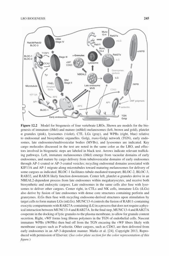
�
� �
�
LRO BIOGENESIS 245
mMel
Stage IV
Stage III
vWF
CD63Lysosome
NBEAL2
Munc
13-4
Munc
13-4
Rab27a
Rab27a
Rab11
α-Granules
iMel
Stage II
Tyr
PMEL
Amyloid
fibrils
TYRP1
OCA2
BLOC-1
BLOC-2
Microtubule
KIF13A
AP1 AP1
CD63
Granzymes
perforin
mLG
iLG
iLG
P-selectin
vWF polymers
iWPB
mWPB
AP3
AP3
BLOC-1
Early endosomesTGN
Golgi
Late endosome
MVB
Rab38/Rab32
BLOC-3
Figure 12.2 Model for biogenesis of four vertebrate LROs. Shown are models for the bio-genesis of immature (iMel) and mature (mMel) melanosomes (left, brown and gold), plateletα granules (pink), lysosomes (violet), CTL LGs (gray), and WPBs (right, blue) relativeto endosomal and biosynthetic organelles. Golgi, trans-Golgi network (TGN), early endo-somes, late endosomes/multivesicular bodies (MVBs), and lysosomes are indicated. Keycargo molecules discussed in the text are noted in the same color as the LRO, and effec-tors involved in biogenetic steps are labeled in black text. Arrows indicate relevant traffick-ing pathways. Left, immature melanosomes (iMel) emerge from vacuolar domains of earlyendosomes, and mature by cargo delivery from tubulovesicular domains of early endosomesthrough AP-1-coated or AP-3-coated vesicles; recycling endosomal domains associated withKIF13A and AP-1 migrate along microtubules toward maturing melanosomes for delivery ofsome cargoes as indicated. BLOC-1 facilitates tubule-mediated transport; BLOC-2, BLOC-3,RAB32, and RAB38 likely function downstream. Center left, platelet α granules derive in anNBEAL2-dependent process from late endosomes within megakaryocytes, and receive bothbiosynthetic and endocytic cargoes. Late endosomes in the same cells also fuse with lyso-somes to deliver other cargoes. Center right, in CTLs and NK cells, immature LGs (iLGs)also derive by fusion of late endosomes with dense core structures containing perforin andgranzymes. iLGs then fuse with recycling endosome-derived structures upon stimulation bytarget cells to form mature LGs (mLGs). MUNC13-4 controls the fusion of RAB11-containingexocytic compartments with RAB27A-containing iLG in a process that does not require a phys-ical interaction between MUNC13-4 and RAB27A. In the final step, MUNC13-4 and RAB27Acooperate in the docking of lytic granules to the plasma membrane, to allow for granule contentsecretion. Right, vWF forms long fibrous polymers in the TGN of endothelial cells. Nascentimmature WPBs (iWPBs) then bud off from the TGN encasing the vWF fibers likely withmembrane cargoes such as P-selectin. Other cargoes, such as CD63, are then delivered fromearly endosomes in an AP-3-dependent manner. Marks et al. [24]. Copyright 2013, Repro-duced with permission of Elsevier. (See color plate section for the color representation of thisfigure.)

�
� �
�
246 LYSOSOME-RELATED ORGANELLES
lysosomes and/or LROs, whereas organelles of earlier endocytic compartments arecomparatively normal [29,30]. The defective gene in CHS and its beige mouse model,CHS1, encodes a 3801-residue cytoplasmic protein called LYST [31,32]. How LYSTfunctions is still unknown. Although some data suggest that LYST positively regu-lates fission from lysosomal fusion intermediates [33], the majority of data support theview that mammalian LYST and its homologue in Dictyostelium discoideum, LvsB,function as negative regulators of lysosome or LRO fusion [29,30,34], such that theenlarged organelles in patient cells result from uncontrolled fusion.
LYST is a member of a small family of proteins with a conserved BEACH domain,a pleckstrin homology domain that might engage phosphatidylinositides [35], andWD domain repeats that are thought to form an interface for protein:protein interac-tions (reviewed in Ref. [36]). Intriguingly, mutations in several other family membersalso result in disease. In particular, mutations in the family member neurobeachin-like2 (NBEAL2) result in GPS, a syndrome characterized by the absence of α granulesin platelets [37–39]. Analyses of a mouse model of GPS suggest that NBEAL2 func-tions during α granule formation within megakaryocytes (Figure 12.2, center panel)and that loss of NBEAL2 correlates with uncontrolled release of α granule contentsand concomitant defects in megakaryocyte development [40].
12.3.2 Hermansky–Pudlak Syndrome
HPS is a genetically heterogeneous group of disorders characterized in all casesby oculocutaneous albinism due to melanosome malformation in pigment cells andby bleeding diathesis primarily due to an absence of dense granules in platelets.Some HPS isoforms are also associated with a lethal lung fibrosis and/or immun-odeficiency [41–43]. The ten genes that have been identified to date as defectivein HPS patients encode subunits of four multisubunit protein complexes – adaptorprotein (AP)-3 and Biogenesis of lysosome-related organelles complex (BLOC) -1,-2 and -3 – that are thought to regulate membrane trafficking steps required for thematuration of LROs, particularly, melanosomes, platelet dense granules, and alveo-lar lamellar bodies (reviewed in Refs [24,43–46]). Mutations in orthologous genesand several additional subunits of the same complexes result in a similar disorder inmice, and orthologous complexes regulate the biogenesis of gut granules in C. ele-gans [47–49] and ocular pigment granules in D. melanogaster [50–58]. In addition,mutations in vacuolar protein sorting 33A (Vps33a) – encoding a SNARE-interactingSec1/Mun18 (SM) family member and a subunit of the homotypic fusion and proteinsorting (HOPS) tethering complex – and in Rab38 – encoding a tissue-restricted RABGTPase – underlie a similar disorder in rodents [59–63]. Mutations in genes encod-ing additional HOPS subunits and orthologues of RAB38 underlie LRO defects ininvertebrates [47,49,64–67].
The products of all of these genes appear to regulate the delivery of cargoesrequired for the maturation of LROs such as melanosomes. AP-3 is a member ofthe heterotetrameric adaptor protein complexes that facilitate protein sorting intotransport carriers for post-Golgi trafficking in eukaryotes, and AP-3 specificallyfacilitates protein sorting to lysosomes in cells that lack LROs [68,69]. The yeast
�
� �
�
LRO BIOGENESIS 247
HOPS complex facilitates homotypic fusion of the lysosome-like vacuole and fusionof the vacuole with transport vesicles, and mammalian HOPS is thought to functionat multiple endocytic fusion events [70]. BLOC-3 has recently been shown tofunction as a guanine nucleotide exchange factor (GEF) for RAB38 and the highlyrelated RAB32 GTPase [71]. The molecular functions of BLOC-1 and BLOC-2 areless well understood, and how any of these proteins function during LRO biogenesisis just beginning to unfold. Their function during melanosome biogenesis is bestunderstood (Figure 12.2, left panel) and is described in the following sections.
12.3.3 Melanosome Biogenesis
The pigment-storing organelles within melanocytes and developing retinal pigmentepithelia (RPE) have emerged as excellent models for LRO biogenesis due to an easilyidentifiable morphology, a wealth of genetic variants, excellent cultured cell models,and well-characterized cargoes [46,72]. Melanosomes in mammalian cells that makepredominantly black and brown eumelanins mature through four distinct morphologi-cal stages. The first two stages lack pigment but are characterized by development of afibrillar matrix upon which melanins deposit in later stages [73]. Stage I melanosomesare accessible to the endocytic pathway and correspond to vacuolar domains of earlyendosomes with flat clathrin lattices on their cytoplasmic face and few intraluminalvesicles [74] but differ from early endosomes in other cell types by their hyperacid-ification and content of nascent amyloid fibrils that emerge from the intraluminalvesicles [73–76]. These fibrils, composed of the pigment cell-specific premelanosome(PMEL) protein (reviewed in Ref. [77]), elongate and assemble into sheets duringmaturation of ellipsoidal stage II melanosomes [76]. As stage I melanosomes matureinto stage II, they become inaccessible to endocytic material [74]; how this occursconcomitant with maturation of early endosomes to endocytic-accessible late endo-somes is still not understood, but likely reflects fission of a common precursor. This issuggested by the finding that distinct classes of intraluminal vesicles [78] are associ-ated with distinct proteolytic fragments of PMEL destined either for lysosome-boundlate endosomes or stage II melanosomes [2].
Intriguingly, PMEL is synthesized as a transmembrane glycoprotein that becomesproteolytically processed en route to and/or within stage I melanosomes [75,79–82]to a lumenal fragment that has biophysical characteristics of amyloid [79,83]. PMELwas the first described mammalian member of a family of “functional amyloid” pro-teins that exploit the amyloid fold – commonly associated with neurodegenerativediseases such as Alzheimer, Parkinson, and Creutzfeldt–Jakob diseases – for physio-logical advantage [84]. A number of distinct domains within PMEL have amyloido-genic capacity in vitro [85–89], but N-terminal domains appear to be most critical invivo [85,90]. How PMEL adopts an amyloid fold within melanosome precursors andaverts pathology is a subject of intensive investigation. Indeed, certain PMEL genemutations in animal hypopigmentation models are associated with amyloid forms thatare more detrimental than inactivating mutations for pigment accumulation and per-haps for cell viability [91–95], suggesting that the process and/or timing of amyloidformation is exquisitely controlled.

�
� �
�
248 LYSOSOME-RELATED ORGANELLES
12.3.4 HPS and Melanosome Maturation
Maturation of nonpigmented stage II melanosomes to pigmented stage III and IVmelanosomes requires the delivery of melanogenic enzymes (such as Tyrosinase[TYR] and Tyrosinase-related protein-1 [TYRP1]), and membrane transporters (suchas OCA2 and ATP7A) that modify the lumenal environment to favor melanogenesis(Figure 12.2, left panel). The delivery of these cargoes occurs by vesicular transportfrom distinct domains of early endosomes and is regulated in part by BLOC-1,BLOC-2, AP-3, and the RAB32 and RAB38 GTPases that are defective in HPSmodels (reviewed in Ref. [46]). Except for the major cohort of TYR, most cargoesanalyzed to date absolutely require BLOC-1 for their export from vacuolar domainsof early endosomes toward melanosomes [96–99]. BLOC-1-dependent transportoccurs through tubular elements of the recycling endosome pathway [100] that are“pulled” along microtubules by the kinesin-3 motor, KIF13A [101]. Thus, depletionof KIF13A results in hypopigmentation and entrapment of BLOC-1-dependentcargoes in enlarged early endosomes [100]. Recruitment of KIF13A and sorting ofmany of these cargoes toward melanosomes requires AP-1, another member of theclathrin-associated tetrameric adaptor family, which appears to play a dual role ofsorting adaptor for many melanosomal cargoes and recruiter of KIF13A [100].
How BLOC-1 functions in this pathway is not clear. BLOC-1 consists of eightsubunits, of which three and five, respectively, are encoded by genes that aredefective in HPS and its mouse models. A cohort of BLOC-1 associates withAP-3 [102], and in neurons, BLOC-1 cooperates with AP-3 in the sorting of anumber of transmembrane cargoes into synaptic vesicles [103–105], but BLOC-1functions to sort both AP-3-dependent and -independent cargoes in melanocytes[97–99,106]. BLOC-1 interacts physically with a number of additional effectors thatmight facilitate sorting, formation, or stabilization of transport carriers, or eventualfusion of transport carriers with melanosomes. These include the SNARE proteinssyntaxin-13 and SNAP-25 [107–110], actin regulatory proteins such as Arp2/3and WASH [111], phosphatidylinositol 4-kinase IIα [112], and a number of othercandidates identified by quantitative mass spectrometry analyses [113]. Moreover,a BLOC-1-like complex in the yeast Saccharomyces cerevisiae is responsiblefor recruiting the GTPase-activating protein for the master early endosomal RabGTPase, RAB5 [114], suggesting that mammalian BLOC-1 might participate inRAB exchange to promote the formation of transport carriers. A low-resolutionrotary shadowed structure of recombinant BLOC-1 suggests that the complexadopts a curvilinear shape [115], as expected for a protein that induces or stabilizesmembrane curvature on tubular transport intermediates [116].
BLOC-2 functions downstream of BLOC-1 and regulates the melanosomal deliv-ery of a similar set of cargoes [97,102,117–119]. BLOC-2 binds a cohort of BLOC-1in cells and, similar to BLOC-1, decorates tubular elements of the early endosomalsystem [102]. Moreover, BLOC-2 appears to be an effector of RAB32 and RAB38,with which it partially colocalizes on melanosomes or structures closely apposed tomelanosomes [120]. How BLOC-2 functions is not yet known. BLOC-2 consists ofthree large subunits, all of which are products of genes that are defective in HPS

�
� �
�
LRO BIOGENESIS 249
variants and mouse models. The only clear structural domains in any of them are aWD40 repeat in the HPS5 subunit [121] and a clathrin binding box that is capableof engaging clathrin in vitro [122], but the functional relevance of these features isnot clear. When overexpressed in HeLa cells, the HPS6 subunit interacts with a com-ponent of the dynein/dynactin complex [123], but whether this reflects a function ofintact BLOC-2 is not yet clear.
The genes that are disrupted in HPS are all ubiquitously expressed (albeit per-haps at different levels in different cell types). How are then tissue-specific effectsobserved? The answer likely lies at least in part in the collaboration of BLOC-1, -2,-3 and AP-3 with tissue-restricted Rab GTPases. In particular, RAB32 and RAB38play important roles in the biogenesis of melanosomes and other LROs. These twoRab proteins localize in part to melanosomes and in part to presumed transport inter-mediates in melanocytes [63,71,120], as well as to subsets of lamellar bodies inalveolar type II cells [124] and of dense granule-like compartments in a megakary-ocytoid cell line [125]. TYR and TYRP1 fail to be properly delivered to maturingmelanosomes upon simultaneous depletion of these two highly homologous RABproteins in immortalized mouse melanocytes and a human melanoma cell line [63,71]or in RAB38-depleted RPE [61]. BLOC-3 – composed of two subunits, HPS1 andHPS4, that are targeted by mutation in human and mouse HPS models – was recentlyshown to function as a GEF i.e., activator) for these two RAB proteins [71]. Accord-ingly, similar phenotypes were observed in a BLOC-3-depleted human melanoma cellline [71] and in RPE and interfollicular skin melanocytes in mouse HPS1 and HPS4models [126]. However, the lack of a similar phenotype in choroidal melanocytes orhair follicle melanocytes from these same models [127] suggests that either an addi-tional GEF must exist for RAB32 and RAB38 in these cells or the requirements forRAB32/38 activation in these cells are more limited.
What do RAB32 and RAB38 do? In epidermal melanocytes, these two proteinsplay partially redundant roles in cargo transport, although unique effects on somecargoes have been observed specifically upon RAB32 knockdown [120]. Recent evi-dence supports the view that both RAB GTPases, in their GTP-bound form, phys-ically engage AP-1, AP-3, and BLOC-2, suggesting an important role throughoutcargo transfer to melanocytes [120], and to the myosin motor, Myosin Vc [128].The latter effector apparently plays contrasting roles in melanosome biogenesis, asknockdown of Myosin Vc with inhibitory RNA depletes cargoes such as TYRP1from melanosomes but paradoxically enhances pigmentation. Part of the latter effectseems to reflect a reduction in release of melanin that occurs constitutively in themelanoma cell line used in this study [128], perhaps reflecting melanin exocytosis. Anadditional effector of RAB32 and RAB38 is VPS9-ankyrin-repeat protein (VARP),a large scaffolding protein that also functions as a GEF for RAB21 and engages theretromer complex and the closed conformation of the vSNARE, VAMP7/TI-VAMP,through distinct binding sites [129–134]. Extinguished expression of VARP, VAMP7,or retromer impairs melanosome protein transport [133,135,136]. The RAB32/38binding site is dispensable for VARP recruitment to endosomes and for endosomalrecycling in nonmelanocytic cells [129] but is necessary for melanosomal proteintransport [136], suggesting that VARP plays a specialized role in melanocytes.

�
� �
�
250 LYSOSOME-RELATED ORGANELLES
12.3.5 HPS and the Biogenesis of Other LROs
As predicted from the symptoms of HPS, BLOC-1, -2, and -3, AP-3 and RAB32/RAB38 have important functions in the generation of other LROs besidesmelanosomes. In patients and mouse models of all forms of HPS, platelets lackdetectable dense granules by whole mount electron microscopy and have reducedstorage pools of serotonin and adenine nucleotides [137–142]. In addition, plateletsfrom mouse HPS models and two HPS case studies show impaired secretion of αgranules and lysosomes, as well as of protein disulfide isomerase from the so-called“T granules,” to low doses of agonists [143–145]; the α granule and T granulesecretion defects are secondary to impaired signaling due to loss of secreted densegranule contents, but BLOC-1 and AP-3 likely regulate cargo delivery to lysosomessince lysosome secretion from platelets lacking these complexes is intrinsicallyimpaired [143]. The result of all of these defects in HPS is impaired plateletaggregation at sites of vascular damage [143,145,146], correlating with excessivebleeding in patients (reviewed in Ref. [147]). RAB38-deficient rats also present withbleeding diathesis due to absent dense granules [62,148], and depletion of RAB38or RAB32 from a megakaryocytoid cell line resulted in reduced size and mobility ofdense granule-like compartments [125]. The cargoes that localize to dense granulesand that might be regulated by these components are not yet known but likely includetransmembrane transporters that pump calcium, adenine nucleotides, serotonin, andpolyphosphates into the dense granule lumen. Among the potential cargoes are thevesicular serotonin transporter VMAT2 [125], the ABC transporter MRP4 [149], anda member of the sugar–nucleotide transporter family, SLC35D3, which is depletedin HPS model platelets [150] and which is the target of mutation in a mouse densegranule storage deficiency [151].
HPS patients that lack BLOC-3 or AP-3 also present with a lethal lung fibro-sis [147], and mouse models lacking many of the HPS complexes individually orin combination have a distinct lung disorder [152,153] and heightened susceptibil-ity to lung irritants [154,155]. This appears to result from defects in the formationof lamellar bodies, the LROs of alveolar type II cells in which surfactant is synthe-sized and stored, and correlates with lung disorders in HPS model mice [156–159].In addition, RAB38-deficient rats present with progressive and severe lung fibro-sis like BLOC-3-deficient HPS1 and HPS4 patients [62]. Consistently, RAB38 isexpressed in alveolar type II epithelial cells (AT2) [160] and RAB38-deficient cellsharbor abnormal surfactant levels and abnormally enlarged lamellar bodies [124,161].The cargoes that are transported to lamellar bodies in an HPS-dependent manner arenot yet identified.
12.3.6 HPS and Neurosecretory Granule Biogenesis
Although not considered traditional LROs, certain synaptic vesicles in neuronal sub-populations are also affected by HPS mutations. AP-3 regulates the formation ofsynaptic vesicles from early endosomes in PC12 cells [162]. Neurons express uniqueAP-3 β and μ chains, and only the AP-3B form that harbors these subunits func-tions in synaptic vesicle recycling [163]; hence mocha mice that lack the single AP-3
�
� �
�
LRO BIOGENESIS 251
δ chain or the neuron-specific μ3B chain have neurological phenotypes, but HPStype 2 (HPS2) patients and pearl mice with mutations in nonneuronal AP3B1 do not[164,165]. Neuronal AP-3 deficiency leads to an imbalance in the composition ofsynaptic vesicles in neurons, characterized by a depletion of cargoes such as the zinctransporter ZnT3, the glutamate transporters Vglut1 and VGAT, the chloride chan-nel ClC3, SNAREs such as VAMP2, and enzymes such as phosphatidylinositol (4)kinase IIα [103,166–168]. Some of the same cargoes are also depleted in neuronsfrom BLOC-1-deficient mice [103,105,116], particularly in certain neuronal subpop-ulations [104]. In particular, BLOC-1 mutations interfere with sorting to neurites andnerve terminals [169]. These mutations are also associated with altered neurotrans-mitter release and behavioral abnormalities in mice [170–172], and polymorphisms inBLOC-1 subunit genes are associated with increased risk for schizophrenia in humans(reviewed in Ref. [173]).
12.3.7 Weibel–Palade Body Biogenesis
HPS, CHS, and GPS clearly impact the formation of LROs for which cargoes arederived exclusively from the endocytic pathway. However, not all LROs appear tobe formed via these pathways. A particularly well-characterized example is the for-mation of WPBs in endothelial cells [174] (Figure 12.2, right panel). The primaryfunction of these elongated “cigar”-shaped organelles is to assemble vWF into largepolymers or fibers for agonist-induced secretion; the fibers then serve as a mech-anism to recruit platelets and clotting factors to sites of endothelial cell damage[175–177]. vWF polymers assemble in the Golgi complex following proteolytic mat-uration of pro-vWF to the mature form [178,179]. Unlike PMEL in melanocytes,vWF does not adopt an amyloid fold, but rather assembles into regularly spacedhelices that can extend up to 3–5 μm in length and that coalesce into paracrystallinestructures [180–182]. An exciting recent study showed that the length of the helicesis determined by the degree to which Golgi stacks assemble into the Golgi ribbonin endothelial cells; “quanta” of vWF polymers within individual Golgi stacks areassembled into longer polymers in the trans-Golgi network (TGN) within a Golgiribbon structure but remain as smaller quanta if ribbon formation is blocked [183].The increased length of vWF polymers is required for optimum capture of plateletsat sites of vascular damage [183,184].
WPB precursors bud from the TGN in a process that requires clathrin and its adap-tor AP-1 in an unconventional manner [185]. Rather than regulating cargo buddinginto clathrin-coated vesicles, AP-1 and clathrin nearly completely coat the nascentWPBs [185] (Figure 12.2, right panel). This likely provides a structural support forthe organelles during their formation or maturation but is not required to maintain theshape of fully formed WPBs [185]. Once segregated from the Golgi, WPBs matureby recruitment of additional membrane cargoes, such as P-selectin/CD62p and thetetraspanin CD63, from the endosomal system [186]. Interestingly, recruitment ofCD63 requires AP-3, whereas the recruitment of P-selectin does not and relies ontargeting determinants in several topological domains [186]. P-selectin is thought tobe retained within WPBs through a direct interaction of the lumenal domain with

�
� �
�
252 LYSOSOME-RELATED ORGANELLES
vWF [187]. The RAB GTPase, RAB27A, is recruited to mature WPBs in a man-ner that is dependent on the vWF cargo [188], where it facilitates vWF secretion inresponse to secretagogues and regulates the degree of vWF polymerization releasedinto the extracellular space [189–191].
Smaller vWF polymers, P-selectin and the RAB27A-related GTPase, RAB27B,are also major components of α granules in platelets, which have a distinct morphol-ogy [192]. Whether this morphology reflects differing levels of vWF biosynthesis,fractured Golgi elements in the megakaryocyte precursors, or another regulatory stepis not yet understood.
12.4 LRO MOTILITY, DOCKING, AND SECRETION
A number of diseases result from defects in LRO motility or secretion. Griscellisyndrome types 1, 2, and 3 (GS1, GS2, and GS3) reflect defects in the intracellu-lar transport of selected LROs due to mutations in the genes encoding Myosin VA,RAB27A, and MLPH (Melanophilin), respectively [193]. A feature of each of thesediseases is cutaneous albinism due to defective positioning of mature melanosomeswithin melanocytes and consequent failure to transfer melanosomes to keratinocytes[194–201]. In melanocytes, RAB27A forms a tripartite complex with the linker pro-tein, MLPH, and the processive actin motor, Myosin VA, that links melanosomes tothe peripheral actin cytoskeleton. This linkage captures melanosomes from bidirec-tional movement along microtubules, in which minus-end-directed motions predomi-nate; thus, loss of any of these components results in accumulation of melanosomes inthe pericentriolar region [202,203]. Myosin VA in this complex functions either as atether for melanosomes on peripheral actin [202,203] or to mediate long-range move-ments toward the cell periphery [204]. The same RAB27A/MLPH/Myosin VA com-plex impedes spontaneous secretion of immature WPBs from endothelial cells [190].In RPE, RAB27A and MLPH associate with a distinct motor, Myosin VIIA, to facili-tate motility of melanosomes into the apical processes that interdigitate with the outersegments of photoreceptors [205,206].
GS3 (MLPH deficiency) is associated primarily with hypopigmentation, butpatients with GS1 and GS2 suffer from a number of additional symptoms. DefectiveMyosin VA in GS1 is most often associated with neurological impairment due toimportant roles of this myosin motor in neurons [207], particularly in the devel-opment of dendritic spines [208]. The neuronal function of Myosin VA does notrequire association with MLPH; consequently, some GS1 patients and dilute mousemodels with particular Myosin VA mutations present either with pigmentation orneurologic defects, but not both [209,210].
GS2 (RAB27A deficiency) is not associated with neurological defects but ratherwith immune defects and often with a hyperinflammatory syndrome referred toas hemophagocytic lymphohistiocytosis (HLH) [199]. The latter is primarily dueto defects in cytolytic granule mobilization within CTLs [211,212]. When boundto GTP, RAB27A engages numerous effectors that play roles in the maturation ofcytolytic granules and their positioning toward the immunological synapse (IS)

�
� �
�
LROs AND IMMUNITY TO PATHOGENS 253
in CTLs and NK cells [213,214]. These are further discussed in the followingtext in the context of cytolytic granule maturation and secretion. RAB27A and itseffectors are also required for proper positioning and exocytosis of numerous otherLROs and non-LRO compartments, including mast cell granules [215], azurophilicgranules [216,217], tertiary and specific granules of neutrophils [218,219], and eveninsulin-containing dense core secretory granules [220]. RAB27A and its homologue,RAB27B, also regulate distinct steps in the release of exosomes [221], and RAB27Bcontrols dense granule release in platelets [222].
Other aspects in the regulation of LRO secretion have been discovered in the con-text of hyperproliferative disorders of the immune system like that observed in GS2and CHS patients. These are discussed as follows in the broader context of LROs inadaptive and innate immunity.
12.5 LROs AND IMMUNITY TO PATHOGENS
LROs are particularly enriched within cells of the adaptive and innate immune sys-tems, and diseases associated with their formation, positioning, or secretion lead toimmunodeficiency and often to hyperproliferative immune disorders. Investigationsinto the etiology of these disorders has extended our understanding of general princi-ples of LRO biogenesis and secretion, and raise interesting questions regarding howLROs might be defined.
12.5.1 Cytolytic Granules
Directed secretion of the contents of CTL and NK cell cytolytic granules into thesynapse with target cells is necessary for antiviral immunity, cancer immunosurveil-lance, and downregulation of immune responses. Hence, defects in cytolytic granulebiogenesis or secretion lead to heightened susceptibility to viral infection and cancer,and often to the typically fatal hyperinflammatory disorder HLH. HLH is charac-terized by an exacerbated yet ineffective immune response [223,224] in which aug-mented cytokine secretion by CTLs and NK cells recruits and activates phagocytes;these cells then produce additional cytokines, leading to a “cytokine storm” that sus-tains inflammation and harms infiltrated tissues [225]. HLH is often observed in GS2and CHS patients [199,226], but surprisingly only rarely in HPS2 patients, despiteimpaired antiviral immunity [227].
Impaired cytolytic activity by CTLs in CHS, HPS2, and GS2 does not seem tobe due to abnormal composition of these granules. Perforin and granzyme are cor-rectly localized to the lytic granules in AP-3-deficient CTLs [228] (but lysosomalpools of perforin are reduced in NK cells, suggesting possible defects in NK celldifferentiation or in selective trafficking to granules [229]). The defect within CTLsin CHS, HPS2, and GS2 resides primarily in the secretion of the granule contents.In HPS2, granules are unable to efficiently engage and move along microtubules forpositioning at the IS and for subsequent polarized content secretion [228]. Move-ment of the granules along microtubules is driven by the minus-end-directed motor,

�
� �
�
254 LYSOSOME-RELATED ORGANELLES
dynein [230]. Once the granules are polarized toward the microtubule organizing cen-ter (MTOC), the MTOC moves toward the IS at the plasma membrane, ferrying theassociated cytolytic granules with it [230,231]. This step requires RAB27A in com-plex with its effector, Slp3, and the kinesin-1 microtubule motor [232]. During thisprocess, the perforin-containing lytic granules fuse with polarized exocytic vesiclesthat carry the recycling endosome-associated RAB11A [233] (Figure 12.2, centerpanel). Fusion requires the RAB27A effector MUNC13-4 [233], which is discussedlater, and enables functional maturation of the perforin-containing granules for exo-cytosis. Thus, the final transport step and docking at the IS are blocked in GS2 CTLs[199,233]. Motility of effector-associated granules is also blocked in CHS [29]. Thepolarization of the MTOC to the IS is tightly controlled by the strength of T-cell sig-naling, at least in part via activation of the LCK tyrosine kinase [234], and requiresthe dynein motor [230,235].
12.5.2 Familial Hemophagocytic Lymphohistiocytosis and Cytolytic GranuleSecretion
While HLH is observed together with pigmentation defects in CHS and GS2, HLHcan also manifest as a primary immunodeficiency disorder with mild or absent symp-toms of oculocutaneous albinism. In this case, HLH is due to mutations in othergenes involved in the exocytosis of lytic granules and comprises a group of diseasescalled familial HLH (FHL) [236]. The affected gene in FHL type 2 (FHL2) encodesperforin, a cytolytic granule cargo that is the main effector of cytolytic activity intarget cells. By contrast, the affected genes in FHL types 3, 4, and 5 encode compo-nents of the cytolytic granule secretory machinery: MUNC13-4 (defective in FHL3),Syntaxin-11 (STX11; defective in FHL4) and syntaxin-binding protein-2 (STXBP2,also referred to as MUNC18-2 in humans or Munc18-b in mice; defective in FHL5).FHL types 3–5 are also associated with bleeding diathesis, which is caused by impair-ment in agonist-stimulated release of α granules, dense granules, and lysosomes fromplatelets [237–240]. This indicates that cytolytic granules and platelet LROs and lyso-somes share mechanisms of exocytosis.
The RAB27A effector MUNC13-4 plays at least two distinct roles in cytolyticgranule release. First, as described above, MUNC13-4 controls the fusion ofRAB11-containing exocytic compartments with RAB27A-containing “immature”lytic granules in a process that does not require a physical interaction betweenMUNC13-4 and RAB27A [233]. Second, MUNC13-4 and RAB27A cooperatein the docking of lytic granules to the plasma membrane, a necessary step insecretion [241,242]. MUNC13-4 also binds directly to the SNARE complex con-taining STX11 [237], and thus likely facilitates SNARE complex formation duringexocytosis.
STX11 is a member of the syntaxin family of SNARE proteins. SNAREs onopposing membranes form a stable four-helix bundle that brings the membranesinto close apposition and thus drives their fusion [243]. STX11 is an unusual targetSNARE (tSNARE) protein in that it lacks a transmembrane helix, and instead istethered to the membrane by a lipid anchor [244,245]. In FHL4 patients (with

�
� �
�
LROs AND IMMUNITY TO PATHOGENS 255
mutations in STX11), lytic granule secretion in CTLs and NK cells is blocked,indicating that an STX11-containing tSNARE complex mediates a fusion steprequired for LRO exocytosis [246]. Recent findings suggest that STX11 functionsduring the final exocytic step [247], but the localization of STX11 to intracellularstructures that lack RAB27A in CTLs and NK cells [246,248], and the finding thatCTL-dependent killing under certain circumstances in FHL4 patients is intact [248]raises the possibility that STX11 might additionally – or instead – function in thefusion of RAB11- and RAB27A-containing compartments to form mature granules.
The CTL deficiencies observed in FHL5 patients with mutations in STXBP2 aresimilar to those of FHL4. Indeed, the MUNC18-2 product of the STXBP2 gene is amember of the Sec1-Munc18 family of syntaxin “chaperones” that stabilizes STX11and facilitates its SNARE complex formation [249]. In platelets, Munc18b facilitatesthe formation of an STX11 complex with the Qbc SNARE SNAP-23 on the plasmamembrane and the R-SNARE VAMP8 on granule membranes to mediate fusion andgranule content release [237,239]. MUNC18-2 in CTLs and NK cells likely facili-tates the formation of a similar complex to facilitate either maturation or exocytosisof lytic granules. Intriguingly, a recent report identified a dominant negative mutantof STXBP2 in an FHL5 patient that bound tightly to STX11 itself but impaired for-mation of a four-helix bundle SNARE complex, suggesting that SNARE complexassembly is indeed a key function for MUNC18-2 [250].
12.5.3 Azurophilic Granules
HPS2 patients suffer from recurrent bacterial infections, in part due to chronicneutropenia [251]. The basis for neutropenia is not completely understood, but ithas been associated with impaired trafficking of elastase to azurophilic granules inthe absence of AP-3 [229,252,253]. Elastase is required for normal differentiationof myeloid progenitors to mature neutrophils [254]. Consequently, HPS2 patientsshow arrested maturation of neutrophil precursors at the promyelocyte stage [253].However, neutrophil counts increase during infections, and recurrent infectionspersist even upon treatment of patients with granulocyte-colony stimulating factor toincrease neutrophil numbers [255–257]. This suggests the presence of other immunedysfunctions in these patients. Azurophilic granules are also required for neutrophilbactericidal activity, and defective fusion of these granules with phagosomes hasbeen observed in CHS [258].
12.5.4 NADPH Oxidase-Containing LROs
Chronic granulomatous disease (CGD) is an immunodeficiency disorder in whichpigmentation is normal (and hence there is no defect in melanosome formation) butin which a number of immune system LROs fail to function properly. Immunod-eficiency is evidenced by recurrent respiratory, intestinal, and cutaneous bacterialinfections and delayed clearance of viruses [251]. These immune defects areattributed to impaired activity of the “phagocyte-specific” NADPH oxidase (NOX2)in phagocytes, including conventional DCs. The gp91phox subunit of NOX2 is

�
� �
�
256 LYSOSOME-RELATED ORGANELLES
stored in “specific granules” in neutrophils or in less well-defined compartmentsin macrophages that fuse with nascent phagosomes following phagocytosis oflarge particles such as bacteria; there it generates reactive oxygen species thatare thought to damage the phagocytosed cargoes [259]. In DCs, gp91phox is alsostored within LROs that are mobilized to fuse with nascent phagosomes [10,260](Figure 12.3, left). These LROs have been called “inhibitory LROs” due to theirability to limit acidification when recruited to phagosomes in phagocytes. Thereactive oxygen species generated by NOX2 capture protons in phagosomes andtherefore counteract the vacuolar ATPase activity in DCs, preventing acidification.This activity limits the degradation of bacterial antigens within phagosomes topreserve antigenic peptides [10]. DCs from patients and mouse models of CGDshow increased phagosomal acidification and impaired antigen cross-presentationdue to the defective recruitment of NOX2 to phagosomes, perhaps contributing toimpaired immune responses to infections [10,260]. Recruitment of inhibitory LROsto phagosomes is mediated by RAB27A [261]; delayed recruitment of NOX2 tonascent phagosomes in DCs in the RAB27A-deficient ashen mouse model of GS2leads to increased acidification and consequently increased protein degradation andimpaired antigen cross-presentation to naïve CD8+ T cells [261].
12.5.5 IRF7-Signaling LROs and Type I Interferon Induction
Plasmacytoid DCs (pDCs) are a specialized DC subset that functions largely inthe production of type-I interferon (IFN) and plays an important role during viralinfections. The pDCs recognize viral DNA at least in part through endosomaltoll-like receptors (TLRs) including TLR7 and TLR9 [262]. TLR9 is activatedby proteolytic processing within endosomes and then signals through the adaptorprotein MyD88 via two bifurcating pathways. One pathway leads to NF-κBactivation and the production of proinflammatory cytokines. The other pathwayrequires activation of the adaptor protein TNF receptor-associated factor 3 (TRAF3),and leads to phosphorylation and activation of the transcription factor IRF7 tostimulate production of type I IFN [263]. Interestingly, these two signaling cascadesare initiated in different compartments [20] (Figure 12.3, right). NF-κB activationis initiated from a VAMP3-containing early endosomal compartment that lacksthe lysosomal-associated membrane protein 2 (LAMP2). By contrast, TRAF3 isactivated on a distinct LRO-like compartment that contains LAMP2 and to whichIRF7 is recruited [20]. Trafficking of TLR9 from endosomes to this IRF7 signalingLRO requires AP-3 [20] and also likely BLOC-1 and BLOC-2 [264]. A potentialcargo of this pathway is SLC15A4 [264], a transporter recently shown to facilitatecargo escape from phagosomes in conventional DCs [265]. The defective productionof type-I IFN by pDCs in the absence of AP-3 might contribute to the recurrent viralinfections observed in HPS2 patients.
12.5.6 MIICs: LROs or Conventional Late Endosome/Lysosomes?
The T-cell receptor on CD4+ lymphocytes recognizes antigenic peptides bound tomajor histocompatibility complex class II (MHC-II) molecules on the surface of

�
� �
�
LROs AND IMMUNITY TO PATHOGENS 257
Antigen
Endocytosis
1
2
Early
endosome
Phagocytosis
Early phagosome
Inhibitory LRO
Early
endosome
NF-κB
endosome IRF7 LRO
Cleaved TLR9Cleaved TLR9
Viral
DNA
Viral
DNA
Signaling
Pro-inflammatory
cytokines
GolgiLate phagosome
Tubular MIIC
Type-1 IFN
Nucleus
IRF7NF-κB
Autophagosome
MH
C-I
I pre
senta
tion
Antigen
degradation
Bacteria or
other particles
MHC-II:peptide
AP-3
MAMP/DAMP
PRR
ATG
NOX2
RAB27A
VAMP3
LAMP2
Antigen
loading
Ag
degradation/
loading
Ag
degradation/
loading
Late
endosome/ MIIC
Golgi
Lysosome
DC pDC
Figure 12.3 Model for LRO function in conventional DCs and pDCs. Left, LROs and phago-some maturation in DCs. 1. Antigens captured by endocytosis or macropinocytosis enterthe endolysosomal system and are degraded to smaller peptides. Peptides encounter MHC-IImolecules as endosomes mature to late endosomes/MIICs and lysosomes. Antigen loadingmostly occurs within late endosomes/MIICs and lysosomes that are enriched in the regula-tory component HLA-DM. From these compartments, peptide-loaded MHC-II molecules aredelivered to the cell surface (black straight arrow) for presentation to T cells. 2. Particulateantigens captured by phagocytosis are degraded in phagosomes as the phagosomes mature.Maturation is achieved by the acquisition of content (including MHC-II) from early and lateendosomes/MIICs and lysosomes by both direct fusion (open arrows) and vesicular transport(turquoise arrows; dashed arrows indicate possible pathways for MHC-II transport that are notyet confirmed). Antigen is loaded onto MHC-II predominantly in late phagosomes. Phagosomematuration is supported by autophagy (ATG; light violet) proteins, which might derive fromautophagosomes themselves or independently from the cytosol, and by signaling from patternrecognition receptors (PRRs, black arrow) such as TLRs. PRRs such as TLR4 are deliveredto phagosomes from early endosomes in an AP-3-dependent manner (mauve dashed arrow).From late phagosomes, peptide-loaded MHC-II is delivered to the cell surface either directly(not shown) or via an intermediate tubular MHC-II storage compartment for presentation toT cells (black straight arrow). Delivery to or from this compartment appears to be regulatedby PRR signaling from the phagosome (dashed black arrows). Right, IRF7 LROs and type IIFN signaling in pDCs. TLR9 is trafficked from the biosynthetic pathway to VAMP-3-positiveearly endosomes. Here TLR9 is cleaved and becomes competent for signaling to activate proin-flammatory cytokine expression through NF-κB (NF-κB endosome). Cleaved TLR9 is thentargeted in an AP-3-dependent manner to a LAMP2-positive LRO (IRF7 LRO) harboring theadaptor TRAF3. Here TLR9 signals through IRF7 to induce the transcription of type-1 inter-feron genes. Adapted from Traffic 14:135–152, Mantegazza AR, Magalhaes JG, Amigorena S,Marks MS, Presentation of phagocytosed antigen by MHC class I and II, 2013 and from Sci-ence 329:1530–1534, Sasai M, Linehan MM, Iwasaki A, Bifurcation of toll-like receptor 9signaling by adaptor protein 3, 2010. (See color plate section for the color representation ofthis figure.)

�
� �
�
258 LYSOSOME-RELATED ORGANELLES
antigen presenting cells, which include DCs, B lymphocytes, activated macrophages,and a few other cell types [266]. In Epstein–Barr virus transformed B lymphocytesand conventional DCs, MHC-II accumulates in multivesicular and multilaminarcompartments called MIICs that harbor lysosomal proteins such as CD63, LAMP1,and cathepsins [267–269] as well as the antigen processing chaperones HLA-DMand HLA-DO [270,271] (Figure 12.3, left). MIICs were historically described asLROs due to the unusually uniform multilamellar electron dense morphology ofthe “later” MIIC compartments [267] (Figure 12.1), which could be induced byexpression of MHC-II alone [272]. However, it is no longer clear whether MIICsare true LROs or merely conventional late endosomes and lysosomes enriched in aspecial cargo. Indeed, antigen processing and assembly with MHC-II has been shownto occur in different MHC-II-containing compartments in APCs, including early andlate endosomes, lysosomes, and phagosomes [273]. Therefore, MIICs might not beas clearly differentiated from conventional endolysosomes as once thought. On theother hand, antigen processing compartments in DCs are particularly sensitive tofunctional and morphological changes in response to signals from receptors such asTLRs. For example, TLR stimulation of DCs leads to a reorganization of the MIICsto a tubular morphology, favoring the delivery of peptide:MHC-II complexes to theplasma membrane at the IS with T cells [274–277]. Tubular MIICs may coexist withbona fide unstimulated lysosomes in DCs [276,277], and thus might more strictlyadhere to the LRO definition.
While MHC-II-dependent antigen presentation is impaired in CHS [278], it isnot generally disrupted in HPS [279,280] (although see the following section for aunique exception in HPS2). However, AP-3 deficiency in HPS2 or its mouse modelscauses impaired presentation of microbial lipid antigens on human CD1b moleculesor the mouse orthologue, CD1d [281–283]. CD1b binding to AP-3 is required for itslocalization to MIICs in B cells and for appropriate sorting to the plasma membrane[281–283]. This defect in antigen presentation, together with the defects described asfollows in phagosomal TLR activation and antigen presentation, may in part accountfor the increased susceptibility to bacterial infections in HPS2.
12.5.7 Phagosomes and Autophagosomes as New Candidate LROs
Phagosomes are intracellular structures that surround large internalized particlesfollowing their uptake through the specialized mechanism of phagocytosis. Basedon the definition of LROs as specialized cell-type specific organelles derivedfrom the endosomal system, one could argue that maturing phagosomes can beconsidered to be LROs (Figure 12.3, left). We base this argument on the followingcriteria: (i) Phagosomes are only generated in specific phagocytic cell types suchas macrophages, neutrophils, and DCs. (ii) Although they form by the uniqueprocess of phagocytosis and thus do not strictly derive from endosomes, theymature by continuously receiving input from the endosomal system [284,285].(iii) Although they eventually fuse with lysosomes, they harbor unique cargoessuch as NOX2 and perform unique functions within the cell that differentiate
�
� �
�
LROs AND IMMUNITY TO PATHOGENS 259
them from conventional lysosomes. (iv) Maturing phagosomes fuse with otherphagocyte-specific LROs such as NOX2 positive LROs and MIICs, and coexist withconventional lysosomes. (v) They harbor MHC molecules and provide peptides forantigen presentation and thus behave functionally like autonomous MIICs [286].(vi) At least in conventional DCs, TLR stimulation induces the extension of tubulesfrom maturing phagosomes that facilitate MHC-II presentation much like theextension of tubules from endolysosomes. However, these tubules appear to functionin a different way from endolysosomal tubules and do not seem to drive surfaceexpression of MHC-II [287]. (vii) Finally, phagosome maturation is influenced byAP-3 and is altered in models of HPS2. In conventional DCs, AP-3 facilitates therecruitment of TLR4 and possibly other TLRs to maturing phagosomes, favoringthe MHC-II-dependent presentation of phagosomal antigens and TLR-dependentproinflammatory cytokine stimulation [288]. Hence, in the mouse pearl model ofHPS2, the impaired capacity for MHC-II presentation and reduced proinflammatorycytokine production from phagocytosed bacteria in DCs leads to reduced T-cellactivation and IFN-γ secretion [288]. These defects in DC performance likelycontribute to the recurrent bacterial infections observed in HPS2 patients even afterrestoration of normal neutrophil counts [41].
Another degradative process that also converges with lysosomes and LROs ismacroautophagy. This process, generally referred to simply as autophagy, involvesthe enclosure of intracellular components – including organelles – into a double-membrane structure called the autophagosome. This structure ultimately fuseswith lysosomes for degradation of its contents [289,290]. Autophagosomes and themachinery involved in their formation intersect LROs in a number of ways. Likephagosomes, autophagosomes represent a source of peptides for MHC-II presentation[291,292], and thus can feed into the MIIC pathway (Figure 12.3, left). As autophago-somes mature, they also receive input from the endosomal system; indeed, compo-nents involved in endosome maturation, such as the ESCRTs, are key components inautophagosome maturation and fusion with lysosomes [293–295]. More recently, theHOPS complex – which is required for the biogenesis of LROs such as melanosomes,platelet dense granules and α granules, eye pigment granules in D. melanogaster andgut granules in C. elegans, is required for autophagosome fusion with lysosomesthrough an interaction with the autophagosomal SNARE syntaxin 17 [296,297].
Components of the autophagy pathway have also been implicated in the bio-genesis of LROs. Depletion of a number of autophagy components in a humanmelanoma cell line impairs pigmentation, and the lipidated form of LC3 mightassociate with melanosomes of distinct stages [298,299]. Similarly, TLR-dependentphagosome maturation in DCs involves recruitment of LC3 in a process requiringthe key regulators of autophagosome formation, ATG5 and ATG7 [300,301]. ATG5and ATG7 have also recently been implicated in agonist-stimulated vWF releasefrom WPBs in epithelial cells, reflecting either storage of a cohort of vWF inautophagosomes or contribution of these components in WPB biogenesis [302].Thus, the formation of autophagosomal intermediates might play a more generalrole in LRO biogenesis.

�
� �
�
260 LYSOSOME-RELATED ORGANELLES
12.6 PERSPECTIVES
Over the last two to three decades, LROs have emerged as key effectors of metazoanphysiology and important models for subcellular compartmentation and function.Analyses of LROs have provided insights into molecular mechanisms controllingorganelle formation, nonredundant protein sorting pathways, novel modes of secre-tion, new physiological mechanisms, and the pathogenesis of a host of inheriteddiseases. In addition, the unusual structural components of some LROs – such as thefibrillar structures underlying melanosomes and WPBs – have given us insights intounexpected areas such as the role of oligomer size in cell physiology and the controlof amyloid formation. Future studies will likely focus on integrating the “parts list”that human diseases have provided for us to understand how different components ofbiogenetic and secretory machinery function together to effect processes such as theformation of transport intermediates or the exocytosis and expulsion of large cargoes.Analyses of some of these processes within living organisms are likely to provide newinsights into considerations that are not typically addressed under culture conditions,such as physical requirements of secretion of large cargoes against hemostatic pres-sure (e.g., see Ref. [303]). At the same time, the exponential rise in the ability to definegenetic mutations in patients will provide a rich source of new information concerningcomponents required for LRO biogenesis, secretion and function and on how thesecomponents might interact with each other. This information will in turn provide newavenues for inexpensive diagnostic tools and therapies for LRO-related diseases.
ACKNOWLEDGMENTS
The authors thank current and former members of the Marks laboratory, and particu-larly thank Graça Raposo and the members of her laboratory for many years of collab-orative research, helpful discussions, and the original images shown in Figures 12.1and 12.2. We also thank our many colleagues, particularly Dan Cutler, Miguel Seabra,Gillian Griffiths, Juan Bonifacino, Esteban Dell’Angelica, Scottie Robinson, JohnHammer, Dick Swank, Wei Li, Greg Hermann, Sebastian Amigorena, Akiko Iwasaki,and Sergio Grinstein, for thoughtful discussions that contributed to this chapter. Weapologize to those whose work we omitted.
REFERENCES
[1] Doyle EL, Ridger V, Ferraro F, Turmaine M, Saftig P, et al. CD63 is an essential cofactorto leukocyte recruitment by endothelial P-selectin. Blood 2011;118:4265–4273.
[2] van Niel G, Charrin S, Simoes S, Romao M, Rochin L, et al. The Tetraspanin CD63integrates ESCRT-independent and dependent sorting at a common endosome. Dev Cell2011;21:708–721.
[3] Kraft S, Jouvin MH, Kulkarni N, Kissing S, Morgan ES, et al. The tetraspanin CD63 isrequired for efficient IgE-mediated mast cell degranulation and anaphylaxis. J Immunol2013;191:2871–2878.

�
� �
�
REFERENCES 261
[4] Hannah MJ, Williams R, Kaur J, Hewlett LJ, Cutler DF. Biogenesis of Weibel–Paladebodies. Semin Cell Dev Biol 2002;13:313–324.
[5] Stinchcombe J, Bossi G, Griffiths GM. Linking albinism and immunity: the secrets ofsecretory lysosomes. Science 2004;305:55–59.
[6] Andrews NW. Regulated secretion of conventional lysosomes. Trends Cell Biol 2000;10:316–321.
[7] McNeil PL, Steinhardt RA. Plasma membrane disruption: repair, prevention, adaptation.Annu Rev Cell Dev Biol 2003;19:697–731.
[8] Ellis K, Bagwell J, Bagnat M. Notochord vacuoles are lysosome-related organelles thatfunction in axis and spine morphogenesis. J Cell Biol 2013;200:667–679.
[9] Nordenfelt P, Tapper H. Phagosome dynamics during phagocytosis by neutrophils. JLeukocyte Biol 2011;90:271–284.
[10] Savina A, Jancic C, Hugues S, Guermonprez P, Vargas P, et al. NOX2 controls phagoso-mal pH to regulate antigen processing during crosspresentation by dendritic cells. Cell2006;126:205–218.
[11] Wu XS, Masedunskas A, Weigert R, Copeland NG, Jenkins NA, et al. Melanoregulinregulates a shedding mechanism that drives melanosome transfer from melanocytes tokeratinocytes. Proc Natl Acad Sci U S A 2012;109:E2101–E2109.
[12] Tarafder AK, Bolasco G, Correia MS, Pereira FJ, Iannone L, et al. Rab11b mediatesmelanin transfer between donor melanocytes and acceptor keratinocytes via coupledexo/endocytosis. J Invest Dermatol 2014;134:1056–1066.
[13] Raposo G, Marks MS, Cutler DF. Lysosome-related organelles: driving post-Golgi com-partments into specialisation. Curr Opin Cell Biol 2007;19(4):394–401.
[14] Stinchcombe JC, Griffiths GM. Secretory mechanisms in cell-mediated cytotoxicity.Annu Rev Cell Dev Biol 2007;23:495–517.
[15] Segal AW. How neutrophils kill microbes. Annu Rev Immunol 2005;23:197–223.
[16] Mantegazza AR, Magalhaes JG, Amigorena S, Marks MS. Presentation of phagocytosedantigens by MHC class I and II. Traffic 2013;14:135–152.
[17] Machlus KR, Italiano JE Jr. The incredible journey: from megakaryocyte developmentto platelet formation. J Cell Biol 2013;201:785–796.
[18] Ren Q, Ye S, Whiteheart SW. The platelet release reaction: just when you thoughtplatelet secretion was simple. Curr Opin Hematol 2008;15:537–541.
[19] Peters PJ, Raposo G, Neefjes JJ, Oorschot V, Leijendekker RL, et al. Major histocom-patibility complex class II compartments in human B lymphoblastoid cells are distinctfrom early endosomes. J Exp Med 1995;182:325–334.
[20] Sasai M, Linehan MM, Iwasaki A. Bifurcation of Toll-like receptor 9 signaling by adap-tor protein 3. Science 2010;329:1530–1534.
[21] Marks MS, Seabra MC. The melanosome: membrane dynamics in black and white.Nature Rev Mol Cell Biol 2001;2:738–748.
[22] Schmitz G, Muller G. Structure and function of lamellar bodies, lipid-protein complexesinvolved in storage and secretion of cellular lipids. J Lipid Res 1991;32:1539–1570.
[23] Wang H, Wan H, Li X, Liu W, Chen Q, et al. Atg7 is required for acrosome biogenesisduring spermatogenesis in mice. Cell Res 2014;24:852–869.
[24] Marks MS, Heijnen HFG, Raposo G. Lysosome-related organelles: unusual compart-ments become mainstream. Curr Opin Cell Biol 2013;25:495–505.

�
� �
�
262 LYSOSOME-RELATED ORGANELLES
[25] de Saint BG, Ménasché G, Fischer A. Molecular mechanisms of biogenesis and exocy-tosis of cytotoxic granules. Nat Rev Immunol 2010;10:568–579.
[26] Shiflett SL, Kaplan J, Ward DM. Chediak–Higashi syndrome: a rare disorder of lyso-somes and lysosome related organelles. Pigment Cell Res 2002;15:251–257.
[27] Introne W, Boissy RE, Gahl WA. Clinical, molecular, and cell biological aspects ofChediak–Higashi Syndrome. Mol Genet Metabol 1999;68:283–303.
[28] Kaplan J, De Domenico I, Ward DM. Chediak–Higashi syndrome. Curr Opin Hematol2008;15:22–29.
[29] Sepulveda FE, Burgess A, Heiligenstein X, Goudin N, Ménager MM, et al. LYST con-trols the biogenesis of the endosomal compartment required for secretory lysosomefunction. Traffic 2015. DOI: 10.1111/tra.12244.
[30] Holland P, Torgersen ML, Sandvig K, Simonsen A. LYST affects lysosome size andquantity, but not trafficking or degradation through autophagy or endocytosis. Traffic2014;15:1390–1405.
[31] Barbosa MD, Nguyen QA, Tchernev VT, Ashley JA, Detter JC, et al. Identificationof the homologous beige and Chediak–Higashi syndrome genes. Nature 1996;382:262–265.
[32] Perou CM, Moore KJ, Nagle DL, Misumi DJ, Woolf EA, et al. Identification ofthe murine beige gene by YAC complementation and positional cloning. Nat Genet1996;13:303–308.
[33] Durchfort N, Verhoef S, Vaughn MB, Shrestha R, Adam D, et al. The enlarged lyso-somes in beige j cells result from decreased lysosome fission and not increased lysosomefusion. Traffic 2012;13:108–119.
[34] Falkenstein K, De Lozanne A. Dictyostelium LvsB has a regulatory role in endosomalvesicle fusion. J Cell Sci 2014;127:4356–4367.
[35] Ward DM, Shiflett SL, Huynh D, Vaughn MB, Prestwich G, et al. Use of expressionconstructs to dissect the functional domains of the CHS/beige protein: identification ofmultiple phenotypes. Traffic 2003;4:403–415.
[36] Cullinane AR, Schäffer AA, Huizing M. The BEACH is hot: a LYST of emerging rolesfor BEACH-domain containing proteins in human disease. Traffic 2013;14:749–766.
[37] Albers CA, Cvejic A, Favier R, Bouwmans EE, Alessi MC, et al. Exome sequenc-ing identifies NBEAL2 as the causative gene for gray platelet syndrome. Nat Genet2011;43:735–737.
[38] Gunay-Aygun M, Falik-Zaccai TC, Vilboux T, Zivony-Elboum Y, Gumruk F, et al.NBEAL2 is mutated in gray platelet syndrome and is required for biogenesis of plateletα-granules. Nat Genet 2011;43:732–734.
[39] Kahr WH, Hinckley J, Li L, Schwertz H, Christensen H, et al. Mutations in NBEAL2,encoding a BEACH protein, cause gray platelet syndrome. Nat Genet 2011;43:738–740.
[40] Kahr WH, Lo RW, Li L, Pluthero FG, Christensen H, et al. Abnormal megakaryocytedevelopment and platelet function in Nbeal2(-/-) mice. Blood 2013;122:3349–3358.
[41] Huizing M, Helip-Wooley A, Westbroek W, Gunay-Aygun M, Gahl WA. Disordersof lysosome-related organelle biogenesis: clinical and molecular genetics. Annu RevGenomics Hum Genet 2008;9:359–386.
[42] Wei ML. Hermansky–Pudlak syndrome: a disease of protein trafficking and organellefunction. Pigment Cell Res 2006;19:19–42.

�
� �
�
REFERENCES 263
[43] Wei A-H, Li W. Hermansky–Pudlak syndrome: pigmentary and non-pigmentary defectsand their pathogenesis. Pigment Cell Melanoma Res 2013;26:176–192.
[44] Dell’Angelica EC. The building BLOC(k)s of lysosomes and related organelles. CurrOpin Cell Biol 2004;16:458–464.
[45] Di Pietro SM, Dell’Angelica EC. The cell biology of Hermansky–Pudlak syndrome:recent advances. Traffic 2005;6:525–533.
[46] Sitaram A, Marks MS. Mechanisms of protein delivery to melanosomes in pigmentcells. Physiology 2012;27:85–99.
[47] Hermann GJ, Schroeder LK, Hieb CA, Kershner AM, Rabbitts BM, et al. Geneticanalysis of lysosomal trafficking in Caenorhabditis elegans. Mol Biol Cell 2005;16:3273–3288.
[48] Hermann GJ, Scavarda E, Weis AM, Saxton DS, Thomas LL, et al. C. elegans BLOC-1functions in trafficking to lysosome-related gut granules. PLoS One 2012;7:e43043.
[49] Delahaye JL, Foster OK, Vine A, Saxton DS, Curtin TP, et al. Caenorhabditis elegansHOPS and CCZ-1 mediate trafficking to lysosome-related organelles independently ofRAB-7 and SAND-1. Mol Biol Cell 2014;25:1073–1096.
[50] Cheli VT, Daniels RW, Godoy R, Hoyle DJ, Kandachar V, et al. Genetic modifiers ofabnormal organelle biogenesis in a Drosophila model of BLOC-1 deficiency. Hum MolGenet 2010;19:861–878.
[51] Ooi CE, Moreira JE, Dell’Angelica EC, Poy G, Wassarman DA, et al. Altered expressionof a novel adaptin leads to defective pigment granule biogenesis in the Drosophila eyecolor mutant garnet. EMBO J 1997;16:4508–4518.
[52] Simpson F, Peden AA, Christopoulou L, Robinson MS. Characterization of theadaptor-related protein complex, AP-3. J Cell Biol 1997;137:835–845.
[53] Mullins C, Hartnell LM, Wassarman DA, Bonifacino JS. Defective expression of themu3 subunit of the AP-3 adaptor complex in the Drosophila pigmentation mutantcarmine. Mol Gen Genet 1999;262:401–412.
[54] Mullins C, Hartnell LM, Bonifacino JS. Distinct requirements for the AP-3 adaptor com-plex in pigment granule and synaptic vesicle biogenesis in Drosophila melanogaster.Mol Gen Genet 2000;263:1003–1014.
[55] Kretzschmar D, Poeck B, Roth H, Ernst R, Keller A, et al. Defective pigment gran-ule biogenesis and aberrant behavior caused by mutations in the Drosophila AP-3betaadaptin gene ruby. Genetics 2000;155:213–223.
[56] Falcón-Pérez JM, Romero-Calderón R, Brooks ES, Krantz DE, Dell’Angelica EC. TheDrosophila pigmentation gene pink (p) encodes a homologue of human Hermansky–Pudlak Syndrome 5 (HPS5). Traffic 2007;8:154–168.
[57] Syrzycka M, McEachern LA, Kinneard J, Prabhu K, Fitzpatrick K, et al. The pinkgene encodes the Drosophila orthologue of the human Hermansky–Pudlak syndrome5 (HPS5) gene. Genome 2007;50:548–556.
[58] Cheli VT, Dell’angelica EC. Early origin of genes encoding subunits of biogenesis oflysosome-related organelles complex-1, -2 and -3. Traffic 2010;11:579–586.
[59] Suzuki T, Oiso N, Gautam R, Novak EK, Panthier JJ, et al. The mouse organellar bio-genesis mutant buff results from a mutation in Vps33a, a homologue of yeast vps33 andDrosophila carnation. Proc Natl Acad Sci U S A 2003;100:1146–1150.
[60] Brooks BP, Larson DM, Chan CC, Kjellstrom S, Smith RS, et al. Analysis of ocu-lar hypopigmentation in Rab38cht/cht mice. Invest Ophthalmol Vis Sci 2007;48:3905–3913.

�
� �
�
264 LYSOSOME-RELATED ORGANELLES
[61] Lopes VS, Wasmeier C, Seabra MC, Futter CE. Melanosome maturation defectin Rab38-deficient retinal pigment epithelium results in instability of immaturemelanosomes during transient melanogenesis. Mol Biol Cell 2007;18:3914–3927.
[62] Oiso N, Riddle SR, Serikawa T, Kuramoto T, Spritz RA. The rat Ruby (R) locus isRab38: identical mutations in Fawn-hooded and Tester-Moriyama rats derived from anancestral Long Evans rat sub-strain. Mamm Genome 2004;15:307–314.
[63] Wasmeier C, Romao M, Plowright L, Bennett DC, Raposo G, et al. Rab38 an Rab32control early post-Golgi trafficking of melanogenic enzymes. J Cell Biol 2006;175:271–281.
[64] Warner TS, Sinclair DA, Fitzpatrick KA, Singh M, Devlin RH, et al. The light geneof Drosophila melanogaster encodes a homologue of VPS41, a yeast gene involved incellular-protein trafficking. Genome 1998;41:236–243.
[65] Ma J, Plesken H, Treisman JE, Edelman-Novemsky I, Ren M. Lightoid and Claret: a rabGTPase and its putative guanine nucleotide exchange factor in biogenesis of Drosophilaeye pigment granules. Proc Natl Acad Sci U S A 2004;101:11652–11657.
[66] Pulipparacharuvil S, Akbar MA, Ray S, Sevrioukov EA, Haberman AS, et al.Drosophila Vps16A is required for trafficking to lysosomes and biogenesis of pigmentgranules. J Cell Sci 2005;118:3663–3673.
[67] Sevrioukov EA, He JP, Moghrabi N, Sunio A, Kramer H. A role for the deep orangeand carnation eye color genes in lysosomal delivery in Drosophila. Mol Cell 1999;4:479–486.
[68] Hirst J, Irving C, Borner GH. Adaptor protein complexes AP-4 and AP-5: newplayers in endosomal trafficking and progressive spastic paraplegia. Traffic 2013;14:153–164.
[69] Dell’Angelica EC. AP-3-dependent trafficking and disease: the first decade. Curr OpinCell Biol 2009;21:552–559.
[70] Balderhaar HJ, Ungermann C. CORVET and HOPS tethering complexes – coordinatorsof endosome and lysosome fusion. J Cell Sci 2013;126:1307–1316.
[71] Gerondopoulos A, Langemeyer L, Liang J-R, Linford A, Barr FA. BLOC-3 mutated inHermansky–Pudlak syndrome is a Rab32/38 guanine nucleotide exchange factor. CurrBiol 2012;22:2135–2139.
[72] Raposo G, Marks MS. Melanosomes – dark organelles enlighten endosomal membranetransport. Nat Rev Mol Cell Biol 2007;8:786–798.
[73] Seiji M, Fitzpatrick TM, Simpson RT, Birbeck MSC. Chemical composition and termi-nology of specialized organelles (melanosomes and melanin granules) in mammalianmelanocytes. Nature 1963;197:1082–1084.
[74] Raposo G, Tenza D, Murphy DM, Berson JF, Marks MS. Distinct protein sort-ing and localization to premelanosomes, melanosomes, and lysosomes in pigmentedmelanocytic cells. J Cell Biol 2001;152:809–823.
[75] Berson JF, Harper D, Tenza D, Raposo G, Marks MS. Pmel17 initiates premelanosomemorphogenesis within multivesicular bodies. Mol Biol Cell 2001;12:3451–3464.
[76] Hurbain I, Geerts WJC, Boudier T, Marco S, Verkleij A, et al. Electron tomographyof early melanosomes: implications for melanogenesis and the generation of fibrillaramyloid sheets. Proc Natl Acad Sci U S A 2008;105:19726–19731.
[77] Watt B, van Niel G, Raposo G, Marks MS. PMEL: a pigment cell-specific model forfunctional amyloid formation. Pigment Cell Melanoma Res 2013;26:300–315.

�
� �
�
REFERENCES 265
[78] Edgar JR, Eden ER, Futter CE. Hrs- and CD63-dependent competing mechanisms makedifferent sized endosomal intraluminal vesicles. Traffic 2014;15:197–211.
[79] Berson JF, Theos AC, Harper DC, Tenza D, Raposo G, et al. Proprotein conver-tase cleavage liberates a fibrillogenic fragment of a resident glycoprotein to initiatemelanosome biogenesis. J Cell Biol 2003;161:521–533.
[80] Kummer MP, Maruyama H, Huelsmann C, Baches S, Weggen S, et al. Formationof Pmel17 amyloid is regulated by juxtamembrane metalloproteinase cleavage, andthe resulting C-terminal fragment is a substrate for gamma-secretase. J Biol Chem2009;284:2296–2306.
[81] Leonhardt RM, Vigneron N, Rahner C, Cresswell P. Proprotein convertases processPmel17 during secretion. J Biol Chem 2011;286:9321–9337.
[82] Rochin L, Hurbain I, Serneels L, Fort C, Watt B, et al. BACE2 processes PMEL toform the melanosome amyloid matrix in pigment cells. Proc Natl Acad Sci U S A2013;110:10658–10663.
[83] Fowler DM, Koulov AV, Alory-Jost C, Marks MS, Balch WE, et al. Functional amyloidformation within mammalian tissue. PLoS Biol 2006;4:e6.
[84] Fowler DM, Koulov AV, Balch WE, Kelly JW. Functional amyloid – from bacteria tohumans. Trends Biochem Sci 2007;32:217–224.
[85] Watt B, van Niel G, Fowler DM, Hurbain I, Luk KC, et al. N-terminal domains elicitformation of functional Pmel17 amyloid fibrils. J Biol Chem 2009;284:35543–35555.
[86] Hu KN, McGlinchey RP, Wickner RB, Tycko R. Segmental polymorphism in a func-tional amyloid. Biophys J 2011;101:2242–2450.
[87] McGlinchey RP, Shewmaker F, McPhie P, Monterroso B, Thurber K, et al. The repeatdomain of the melanosome fibril protein Pmel17 forms the amyloid core promotingmelanin synthesis. Proc Natl Acad Sci U S A 2009;106:13731–13736.
[88] McGlinchey RP, Shewmaker F, Hu KN, McPhie P, Tycko R, et al. The repeat domainsof melanosome matrix protein Pmel17 orthologs form amyloid fibrils at the acidicmelanosomal pH. J Biol Chem 2011;286:8385–8393.
[89] Pfefferkorn CM, McGlinchey RP, Lee JC. Effects of pH on aggregation kineticsof the repeat domain of a functional amyloid, Pmel17. Proc Natl Acad Sci U S A2010;107:21447–21452.
[90] Leonhardt RM, Vigneron N, Hee JS, Graham M, Cresswell P. Critical residues in thePMEL/Pmel17 N-terminus direct the hierarchical assembly of melanosomal fibrils. MolBiol Cell 2013;24:964–981.
[91] Watt B, Tenza D, Lemmon MA, Kerje S, Raposo G, et al. Mutations in or near thetransmembrane domain alter PMEL amyloid formation from functional to pathogenic.PLoS Genet 2011;7:e1002286.
[92] Brunberg E, Andersson L, Cothran G, Sandberg K, Mikko S, et al. A missense mutationin PMEL17 is associated with the Silver coat color in the horse. BMC Genet 2006;7:46.
[93] Kerje S, Sharma P, Gunnarsson U, Kim H, Bagchi S, et al. The Dominant white, Dun andSmoky color variants in chicken are associated with insertion deletion polymorphismsin the PMEL17 gene. Genetics 2004;168:1507–1518.
[94] Hamilton H. A study of the physiological properties of melanophores with special ref-erence to their role in feather coloration. Anat Rec 1940;78:525–548.
[95] Hellström AR, Watt B, Fard SS, Tenza D, Mannström P, et al. Inactivation of the Pmelgene alters melanosome shape but has only a subtle effect on visible pigmentation. PLoSGenet 2011;7:e1002285.

�
� �
�
266 LYSOSOME-RELATED ORGANELLES
[96] Cullinane AR, Curry JA, Carmona-Rivera C, Summers CG, Ciccone C, et al. A BLOC-1mutations screen reveals that PLDN is mutated in Hermansky–Pudlak syndrome type 9.Am J Human Genet 2011;88:778–787.
[97] Setty SRG, Tenza D, Truschel ST, Chou E, Sviderskaya EV, et al. BLOC-1 is requiredfor cargo-specific sorting from vacuolar early endosomes toward lysosome-relatedorganelles. Mol Biol Cell 2007;18:768–780.
[98] Setty SRG, Tenza D, Sviderskaya EV, Bennett DC, Raposo G, et al. Cell-specificATP7A transport sustains copper-dependent tyrosinase activity in melanosomes. Nature2008;454:1142–1146.
[99] Sitaram A, Dennis MK, Chaudhuri R, De Jesus-Rojas W, Tenza D, et al. Differentialrecognition of a dileucine-based sorting signal by AP-1 and AP-3 reveals a require-ment for both BLOC-1 and AP-3 in delivery of OCA2 to melanosomes. Mol Biol Cell2012;23:3178–3192.
[100] Delevoye C, Hurbain I, Tenza D, Sibarita J-B, Uzan-Gafsou S, et al. AP-1 and KIF13Acoordinate endosomal sorting and positioning during melanosome biogenesis. J CellBiol 2009;187:247–264.
[101] Delevoye C, Miserey-Lenkei S, Montagnac G, Gilles-Marsens F, Paul-Gilloteaux P,et al. Recycling endosome tubule morphogenesis from sorting endosomes requires thekinesin motor KIF13A. Cell Rep 2014;6:445–454.
[102] Di Pietro SM, Falcon-Perez JM, Tenza D, Setty SRG, Marks MS, et al. BLOC-1 interactswith BLOC-2 and the AP-3 complex to facilitate protein trafficking on endosomes. MolBiol Cell 2006;17:4027–4038.
[103] Newell-Litwa K, Salazar G, Smith Y, Faundez V. Roles of BLOC-1 and adaptorprotein-3 complexes in cargo sorting to synaptic vesicles. Mol Biol Cell 2009;20:1441–1453.
[104] Newell-Litwa K, Chintala S, Jenkins S, Pare J, McGaha L, et al. Hermansky–Pudlakprotein complexes, AP-3 and BLOC-1, differentially regulate presynaptic compositionin the striatum and hippocampus. J Neurosci 2010;30:820–831.
[105] Salazar G, Craige B, Styers ML, Newell-Litwa KA, Doucette MM, et al. BLOC-1 com-plex deficiency alters the targeting of Adaptor Protein complex-3 cargoes. Mol Biol Cell2006;17:4014–4026.
[106] Theos AC, Tenza D, Martina JA, Hurbain I, Peden AA, et al. Functions of AP-3 andAP-1 in tyrosinase sorting from endosomes to melanosomes. Mol Biol Cell 2005;16:5356–5372.
[107] Ghiani CA, Starcevic M, Rodriguez-Fernandez IA, Nazarian R, Cheli VT, et al.The dysbindin-containing complex (BLOC-1) in brain: developmental regulation,interaction with SNARE proteins and role in neurite outgrowth. Mol Psychiatry2010;15:204–215.
[108] Huang L, Kuo YM, Gitschier J. The pallid gene encodes a novel, syntaxin 13-interactingprotein involved in platelet storage pool deficiency. Nature Genet 1999;23:329–332.
[109] Moriyama K, Bonifacino JS. Pallidin is a component of a multi-protein complexinvolved in the biogenesis of lysosome-related organelles. Traffic 2002;3:666–677.
[110] Ilardi JM, Mochida S, Sheng ZH. Snapin: a SNARE-associated protein implicated insynaptic transmission. Nature Neurosci 1999;2:119–124.
[111] Ryder PV, Vistein R, Gokhale A, Seaman MN, Puthenveedu M, et al. The WASH com-plex, and endosomal Arp2/3 activator, interacts with the Hermansky–Pudlak syndrome
�
� �
�
REFERENCES 267
complex BLOC-1 and its cargo phosphatidylinositol-4 kinase type II alpha. Mol BiolCell 2013;24:2269–2284.
[112] Salazar G, Zlatic S, Craige B, Peden AA, Pohl J, et al. Hermansky–Pudlak syndromeprotein complexes associate with phosphatidylinositol-4-kinase type II alpha in neu-ronal and non-neuronal cells. J Biol Chem 2009;284:1790–1802.
[113] Gokhale A, Larimore J, Werner E, So L, Moreno-De-Luca A, et al. Quantitative pro-teomic and genetic analyses of the schizophrenia susceptibility factor dysbindin identifynovel roles of the biogenesis of lysosome-related organelles complex 1. J Neurosci2012;32:3697–3711.
[114] John Peter AT, Lachmann J, Rana M, Bunge M, Cabrera M, et al. The BLOC-1 com-plex promotes endosomal maturation by recruiting the Rab5 GTPase-activating proteinMsb3. J Cell Biol 2013;201:97–111.
[115] Lee HH, Nemecek D, Schindler C, Smith WJ, Ghirlando R, et al. Assembly and architec-ture of Biogenesis of Lysosome-related Organelles Complex-1 (BLOC-1). J Biol Chem2012;287:5882–5890.
[116] Suetsugu S, Kurisu S, Takenawa T. Dynamic shaping of cellular membranes by phos-pholipids and membrane-deforming proteins. Physiol Rev 2014;94:1219–1248.
[117] Helip-Wooley A, Westbroek W, Dorward HM, Koshoffer A, Huizing M, et al. Impropertrafficking of melanocyte-specific proteins in Hermansky–Pudlak syndrome type-5. JInvest Dermatol 2007;127:1471–1478.
[118] Richmond B, Huizing M, Knapp J, Koshoffer A, Zhao Y, et al. Melanocytes derivedfrom patients with Hermansky–Pudlak Syndrome types 1, 2, and 3 have distinct defectsin cargo trafficking. J Invest Dermatol 2005;124:420–427.
[119] Boissy RE, Richmond B, Huizing M, Helip-Wooley A, Zhao Y, et al. Melanocyte-specific proteins are aberrantly trafficked in melanocytes of Hermansky–Pudlaksyndrome-type 3. Am J Pathol 2005;166:231–240.
[120] Bultema JJ, Ambrosio AL, Burek CL, Di Pietro SM. BLOC-2, AP-3, and AP-1 functionin concert with Rab38 and Rab32 to mediate protein trafficking to lysosome-relatedorganelles. J Biol Chem 2012;287:19550–19563.
[121] Zhang Q, Zhao B, Li W, Oiso N, Novak EK, et al. Ru2 and Ru encode mouse orthologsof the genes mutated in human Hermansky–Pudlak syndrome types 5 and 6. NatureGenet 2003;33:145–153.
[122] Helip-Wooley A, Westbroek W, Dorward H, Mommaas M, Boissy RE, et al. Associa-tion of the Hermansky–Pudlak syndrome type-3 protein with clathrin. BMC Cell Biol2005;6:33.
[123] Li K, Yang L, Zhang C, Niu Y, Li W, et al. HPS6 interacts with dynactin p150Glued tomediate retrograde trafficking and maturation of lysosomes. J Cell Sci 2014;127(Pt 21):4574–4588.
[124] Zhang L, Yu K, Robert KW, DeBolt KM, Hong N, et al. Rab38 targets to lamellar bodiesand normalizes their sizes in lung alveolar type II epithelial cells. Am J Physiol LungCell Mol Physiol 2011;301:461–477.
[125] Ambrosio AL, Boyle JA, Di Pietro SM. Mechanism of platelet dense granule biogenesis:study of cargo transport and function of Rab32 and Rab38 in a model system. Blood2012;120:4072–4081.
[126] Nguyen T, Wei ML. Hermansky–Pudlak HPS1/pale ear gene regulates epidermal anddermal melanocyte development. J Invest Dermatol 2007;127:421–428.

�
� �
�
268 LYSOSOME-RELATED ORGANELLES
[127] Nguyen T, Novak EK, Kermani M, Fluhr J, Peters LL, et al. Melanosome morphologiesin murine models of Hermansky–Pudlak syndrome reflect blocks in organelle develop-ment. J Invest Dermatol 2002;119:1156–1164.
[128] Bultema JJ, Boyle JA, Malenke PB, Martin FE, Dell’Angelica EC, et al. Myosin vcinteracts with rab32 and rab38 proteins and works in the biogenesis and secretion ofmelanosomes. J Biol Chem 2014;289:33513–33528.
[129] Hesketh GG, Pérez-Dorado I, Jackson LP, Wartosch L, Schäfer IB, et al. VARP isrecruited on to endosomes by direct interaction with retromer, where together they func-tion in export to the cell surface. Dev Cell 2014;29:591–606.
[130] Zhang X, He X, Fu XY, Chang Z. Varp is a Rab21 guanine nucleotide exchange factorand regulates endosome dynamics. J Cell Sci 2006;119:1053–1062.
[131] Wang F, Zhang H, Zhang X, Wang Y, Ren F, et al. Varp interacts with Rab38 andfunctions as its potential effector. Biochem Biophys Res Commun 2008;372:162–167.
[132] Burgo A, Sotirakis E, Simmler MC, Verraes A, Chamot C, et al. Role of Varp, aRab21 exchange factor and TI-VAMP/VAMP7 partner, in neurite growth. EMBO Rep2009;10:1117–1124.
[133] Tamura K, Ohbayashi N, Maruta Y, Kanno E, Itoh T, et al. Varp is a novelRab32/38-binding protein that regulates Tyrp1 trafficking in melanocytes. Mol Biol Cell2009;20:2900–2908.
[134] Schäfer IB, Hesketh GG, Bright NA, Gray SR, Pryor PR, et al. The binding of Varp toVAMP7 traps VAMP7 in a closed, fusogenically inactive conformation. Nat Struct MolBiol 2012;19:1300–1309.
[135] McGough IJ, Steinberg F, Gallon M, Yatsu A, Ohbayashi N, et al. Identification ofmolecular heterogeneity in SNX27-retromer-mediated endosome-to-plasma-membranerecycling. J Cell Sci 2014;127:4940–4953.
[136] Tamura K, Ohbayashi N, Ishibashi K, Fukuda M. Structure-function analysis ofVPS9-ankyrin-repeat protein (Varp) in the trafficking of tyrosinase-related protein 1 inmelanocytes. J Biol Chem 2011;286:7507–7521.
[137] Gerritsen SM, Akkerman JW, Nijmeijer B, Sixma JJ, Witkop CJ, et al. The Hermansky–Pudlak syndrome. Evidence for a lowered 5-hydroxytryptamine content in platelets ofheterozygotes. Scand J Haematol 1977;18:249–256.
[138] Novak EK, Hui SW, Swank RT. Platelet storage pool deficiency in mouse pigment muta-tions associated with seven distinct genetic loci. Blood 1984;63:536–544.
[139] Novak EK, Sweet HO, Prochazka M, Parentis M, Soble R, et al. Cocoa: a new mousemodel for platelet storage pool deficiency. Br J Haematol 1988;69:371–378.
[140] Rao GH, White JG. An improved method for measuring endogenous serotonin inplatelets of patients with Hermansky–Pudlak syndrome. Thromb Res 1988;51:225–227.
[141] White JG. Serotonin storage organelles in human megakaryocytes. Am J Pathol 1971;63:403–410.
[142] Witkop CJ, Krumwiede M, Sedano H, White JG. Reliability of absent platelet densebodies as a diagnostic criterion for Hermansky–Pudlak syndrome. Am J Hematol1987;26:305–311.
[143] Meng R, Wu J, Harper DC, Wang Y, Kowalska MA, et al. Defective release of a granuleand lysosome contents from platelets in mouse Hermansky–Pudlak syndrome models.Blood 2015;125:1623–1632.

�
� �
�
REFERENCES 269
[144] Rendu F, Maclouf J, Launay JM, Boinot C, Levy-Toledano S, et al. Hermansky–Pudlakplatelets: further studies on release reaction and protein phosphorylations. Am J Hema-tol 1987;25:165–174.
[145] Sharda A, Kim SH, Jasuja R, Gopal S, Flaumenhaft R, et al. Defective PDI releasefrom platelets and endothelial cells impairs thrombus formation in Hermansky–Pudlaksyndrome. Blood 2015;125:1633–1642.
[146] Graham GJ, Ren Q, Dilks JR, Blair P, Whiteheart SW, et al. Endobrevin/VAMP-8-dependent dense granule release mediates thrombus formation in vivo. Blood 2009;114:1083–1090.
[147] Seward SL Jr, Gahl WA. Hermansky–Pudlak syndrome: health care throughout life.Pediatrics 2013;132:153–160.
[148] Ninkovic I, White JG, Rangel-Filho A, Datta YH. The role of Rab38 in platelet densegranule defects. J Thromb Haemost 2008;6:2143–2151.
[149] Jedlitschky G, Tirschmann K, Lubenow LE, Nieuwenhuis HK, Akkerman JW, et al.The nucleotide transporter MRP4 (ABCC4) is highly expressed in human plateletsand present in dense granules, indicating a role in mediator storage. Blood 2004;104:3603–3610.
[150] Meng R, Wang Y, Yao Y, Zhang Z, Harper DC, et al. SLC35D3 delivery from megakary-ocyte early endosomes is required for platelet dense granule biogenesis and differen-tially defective in Hermansky–Pudlak syndrome models. Blood 2012;120:404–414.
[151] Chintala S, Tan J, Gautam R, Rusiniak ME, Guo X, et al. The Slc35d3 gene, encod-ing an orphan nucleotide sugar transporter, regulates platelet dense granules. Blood2007;109:1533–1540.
[152] McGarry MP, Borchers M, Novak EK, Lee NA, Ohtake PJ, et al. Pulmonary pathologiesin pallid mice result from nonhematopoietic defects. Exp Mol Pathol 2002;72:213–220.
[153] McGarry MP, Reddington M, Novak EK, Swank RT. Survival and lung pathology ofmouse models of Hermansky–Pudlak syndrome and Chediak–Higashi syndrome. ProcSoc Exp Biol Med 1999;220:162–168.
[154] Young LR, Borchers MT, Allen HL, Gibbons RS, McCormack FX. Lung-restrictedmacrophage activation in the pearl mouse model of Hermansky–Pudlak syndrome. JImmunol 2006;176:4361–4368.
[155] Young LR, Pasula R, Gulleman PM, Deutsch GH, McCormack FX. Susceptibility ofHermansky–Pudlak mice to bleomycin-induced type II cell apoptosis and fibrosis. AmJ Respir Cell Mol Biol 2007;37:67–74.
[156] Guttentag SH, Akhtar A, Tao JQ, Atochina E, Rusiniak ME, et al. Defective surfactantsecretion in a mouse model of Hermansky–Pudlak syndrome. Am J Respir Cell MolBiol 2005;33:14–21.
[157] Gautam R, Novak EK, Tan J, Wakamatsu K, Ito S, et al. Interaction of Hermansky–Pudlak syndrome genes in the regulation of lysosome-related organelles. Traffic 2006;7:779–792.
[158] Lyerla TA, Rusiniak ME, Borchers M, Jahreis G, Tan J, et al. Aberrant lung structure,composition, and function in a murine model of Hermansky–Pudlak syndrome. Am JPhysiol Lung Cell Mol Physiol 2003;285:L643–653.
[159] Young LR, Gulleman PM, Bridges JP, Weaver TE, Deutsch GH, et al. The alveolarepithelium determines susceptibility to lung fibrosis in Hermansky–Pudlak syndrome.Am J Respir Crit Care Med 2012;186:1014–1024.
�
� �
�
270 LYSOSOME-RELATED ORGANELLES
[160] Osanai K, Iguchi M, Takahashi K, Nambu Y, Sakuma T, et al. Expression and local-ization of a novel Rab small G protein (Rab38) in the rat lung. Am J Pathol 2001;158:1665–1675.
[161] Osanai K, Higuchi J, Oikawa R, Kobayashi M, Tsuchihara K, et al. Altered lung sur-factant system in a Rab38-deficient rat model of Hermansky–Pudlak syndrome. Am JLung Cell Mol Physiol 2010;298:L243–L251.
[162] Faúndez V, Horng J-T, Kelly RB. A function for the AP3 coat complex in synapticvesicle formation from endosomes. Cell 1998;93:423–432.
[163] Blumstein J, Faundez V, Nakatsu F, Saito T, Ohno H, et al. The neuronal form of adap-tor protein-3 is required for synaptic vesicle formation from endosomes. J Neurosci2001;21:8034–8042.
[164] Nakatsu F, Okada M, Mori F, Kumazawa N, Iwasa H, et al. Defective function ofGABA-containing synaptic vesicles in mice lacking the AP-3B clathrin adaptor. J CellBiol 2004;167:293–302.
[165] Kantheti P, Qiao X, Diaz ME, Peden AA, Meyer GE, et al. Mutation in AP-3d in themocha mouse links endosomal transport to storage deficiency in platelets, melanosomes,and synaptic vesicles. Neuron 1998;21:111–122.
[166] Salazar G, Love R, Werner E, Doucette MM, Cheng S, et al. The zinc transporter ZnT3interacts with AP-3 and it is preferentially targeted to a distinct synaptic vesicle subpop-ulation. Mol Biol Cell 2004;15:575–587.
[167] Salazar G, Craige B, Wainer BH, Guo J, De Camilli P, et al. Phosphatidylinositol-4-kinase type II a is a component of AP-3-derived vesicles. Mol Biol Cell 2005;16:3692–3704.
[168] Salazar G, Love R, Styers ML, Werner E, Peden A, et al. AP-3-dependent mechanismscontrol the targeting of a chloride channel (ClC-3) in neuronal and non-neuronal cells.J Biol Chem 2004;279:25430–25439.
[169] Larimore J, Tornieri K, Ryder PV, Gokhale A, Zlatic SA, et al. The schizophrenia sus-ceptibility factor dysbindin and its associated complex sort cargoes from cell bodies tothe synapse. Mol Biol Cell 2011;22:4854–4867.
[170] Chen XW, Feng YQ, Hao CJ, Guo XL, He X, et al. DTNBP1, a schizophrenia suscep-tibility gene, affects kinetics of transmitter release. J Cell Biol 2008;181:791–801.
[171] Erway LC, Fraser AS, Hurley LS. Prevention of congenital otolith defect in pallidmutant mice by manganese supplementation. Genetics 1971;67:97–108.
[172] Trune DR, Lim DJ. The behavior and vestibular nuclear morphology of otoconia-deficient pallid mutant mice. J Neurogenet 1983;1:53–69.
[173] Ryder PV, Faundez V. Schizophrenia: the “BLOC” may be in the endosomes. Sci Signal2009;2:pe66.
[174] Weibel ER, Palade GE. New cytoplasmic components in arterial endothelia. J Cell Biol1964;23:101–112.
[175] Michaux G, Cutler DF. How to roll an endothelial cigar: the biogenesis ofWeibel–Palade bodies. Traffic 2004;5:69–78.
[176] Nightingale T, Cutler D. The secretion of von Willebrand factor from endothelial cells;an increasingly complicated story. J Thromb Haemost 2013;11:192–201.
[177] Wagner DD. The Weibel–Palade body: the storage granule for von Willebrand factorand P-selectin. Thromb Haemost 1993;70:105–110.

�
� �
�
REFERENCES 271
[178] Journet AM, Saffaripour S, Cramer EM, Tenza D, Wagner DD. von Willebrand factorstorage requires intact prosequence cleavage site. Eur J Cell Biol 1993;60:31–41.
[179] Wagner DD, Marder VJ. Biosynthesis of von Willebrand protein by human endothe-lial cells: processing steps and their intracellular localization. J Cell Biol 1984;99:2123–2130.
[180] Berriman JA, Li S, Hewlett LJ, Wasilewski S, Kiskin FN, et al. Structural organizationof Weibel–Palade bodies revealed by cryo-EM of vitrified endothelial cells. Proc NatlAcad Sci U S A 2009;106:17407–17412.
[181] Valentijn KM, Valentijn JA, Jansen KA, Koster AJ. A new look at Weibel–Paladebody structure in endothelial cells using electron tomography. J Struct Biol 2008;161:447–458.
[182] Zenner HL, Collinson LM, Michaux G, Cutler DF. High-pressure freezing providesinsights into Weibel–Palade body biogenesis. J Cell Sci 2007;120:2117–2125.
[183] Ferraro F, Kriston-Vizi J, Metcalf DJ, Martin-Martin B, Freeman J, et al. A two-tierGolgi-based control of organelle size underpins the functional plasticity of endothelialcells. Dev Cell 2014;29:292–304.
[184] Michaux G, Abbitt KB, Collinson LM, Haberichter SL, Norman KE, et al. The physio-logical function of von Willebrand’s factor depends on its tubular storage in endothelialWeibel–Palade bodies. Dev Cell 2006;10:223–232.
[185] Lui-Roberts WWY, Collinson LM, Hewlett LJ, Michaux G, Cutler DF. AnAP-1/clathrin coat plays a novel and essential role in forming the Weibel–Palade bodiesof endothelial cells. J Cell Biol 2005;170:627–636.
[186] Harrison-Lavoie KJ, Michaux G, Hewlett L, Kaur J, Hannah MJ, et al. P-selectin andCD63 use different mechanisms for delivery to Weibel–Palade bodies. Traffic 2006;7:647–662.
[187] Michaux G, Pullen TJ, Haberichter SL, Cutler DF. P-selectin binds to the D′-D3domains of von Willebrand factor in Weibel–Palade bodies. Blood 2006;107:3922–3924.
[188] Hannah MJ, Hume AN, Arribas M, Williams R, Hewlett LJ, et al. Weibel–Palade bodiesrecruit Rab27 by a content-driven, maturation-dependent mechanism that is independentof cell type. J Cell Sci 2003;116:3939–3948.
[189] Nightingale TD, Pattni K, Hume AN, Seabra MC, Cutler DF. Rab27a and MyRIP reg-ulate the amount and multimeric state of VWF released from endothelial cells. Blood2009;113:5010–5018.
[190] Rojo Pulido I, Nightingale TD, Darchen F, Seabra MC, Cutler DF, et al. Myosin Vaacts in concert with Rab27a and MyRIP to regulate acute von-Willebrand factor releasefrom endothelial cells. Traffic 2011;12:1371–1382.
[191] Bierings R, Hellen N, Kiskin N, Knipe L, Fonseca AV, et al. The interplay between theRab27A effectors Slp4-a and MyRIP controls hormone-evoked Weibel–Palade bodyexocytosis. Blood 2012;120:2757–2767.
[192] van Nispen tot Pannerden H, de Haas F, Geerts W, Posthuma G, van Dijk S, et al. Theplatelet interior revisited: electron tomography reveals tubular alpha-granule subtypes.Blood 2010;116:1147–1156.
[193] Van Gele M, Dynoodt P, Lambert J. Griscelli syndrome: a model system to study vesic-ular trafficking. Pigment Cell Melanoma Res 2009;22:268–282.

�
� �
�
272 LYSOSOME-RELATED ORGANELLES
[194] Hume AN, Collinson LM, Rapak A, Gomes AQ, Hopkins CR, et al. Rab27a regulates theperipheral distribution of melanosomes in melanocytes. J Cell Biol 2001;152:795–808.
[195] Hume AN, Collinson LM, Hopkins CR, Strom M, Barral DC, et al. The leaden geneproduct is required with rab27a to recruit myosin Va to melanosomes in melanocytes.Traffic 2002;3:193–202.
[196] Wu X, Rao K, Bowers MB, Copeland NG, Jenkins NA, et al. Rab27a enables myosinVa-dependent melanosome capture by recruiting the myosin to the organelle. J Cell Sci2001;114:1091–1100.
[197] Wu XS, Rao K, Zhang H, Wang F, Sellers JR, et al. Identification of an organelle receptorfor myosin-Va. Nature Cell Biol 2002;4:271–278.
[198] Wu X, Wang F, Rao K, Sellers JR, Hammer JA III. Rab27a is an essential componentof melanosome receptor for myosin Va. Mol Biol Cell 2002;13:1735–1749.
[199] Ménasché G, Pastural E, Feldmann J, Certain S, Ersoy F, et al. Mutations in RAB27Acause Griscelli syndrome associated with haemophagocytic syndrome. Nature Genet2000;25:173–176.
[200] Matesic LE, Yip R, Reuss AE, Swing DA, O’Sullivan TN, et al. Mutations in Mlph,encoding a member of the Rab effector family, cause the melanosome transport defectsobserved in leaden mice. Proc Natl Acad Sci U S A 2001;98:10238–10243.
[201] Bahadoran P, Aberdam E, Mantoux F, Busca R, Bille K, et al. Rab27a: a key tomelanosome transport in human melanocytes. J Cell Biol 2001;152:843–850.
[202] Wu X, Bowers B, Wei Q, Kocher B, Hammer JA 3rd.. Myosin V associates withmelanosomes in mouse melanocytes: evidence that myosin V is an organelle motor.J Cell Sci 1997;110:847–859.
[203] Wu X, Bowers B, Rao K, Wei Q, Hammer JA III. Visualization of melanosome dynamicswithin wild-type and dilute melanocytes suggests a paradigm for myosin V function invivo. J Cell Biol 1998;143:1899–1918.
[204] Evans RD, Robinson C, Briggs DA, Tooth DJ, Ramalho JS, et al. Myosin-Va anddynamic actin oppose microtubules to drive long-range organelle transport. Curr Biol2014;24:1743–1750.
[205] Futter CE, Ramalho JS, Jaissle GB, Seeliger MW, Seabra MC. The role of Rab27a inthe regulation of melanosome distribution within retinal pigment epithelial cells. MolBiol Cell 2004;15:2264–2275.
[206] Lopes VS, Ramalho JS, Owen DM, Karl MO, Strauss O, et al. The ternary Rab27a-Myrip-myosin VIIa complex regulates melanosome motility in the retinal pigmentepithelium. Traffic 2007;8:486–499.
[207] Hammer JA 3rd., Wagner W. Functions of class V myosins in neurons. J Biol Chem2013;288:28428–28434.
[208] Wagner W, Brenowitz SD, Hammer JA 3rd.. Myosin-Va transports the endoplasmicreticulum into the dendritic spines of Purkinje neurons. Nat Cell Biol 2011;13:40–48.
[209] Ménasché G, Ho CH, Sanal O, Feldmann J, Tezcan I, et al. Griscelli syndrome restrictedto hypopigmentation results from a melanophilin defect (GS3) or a MYO5A F-exondeletion (GS1). J Clin Invest 2003;112:450–456.
[210] Huang JD, Mermall V, Strobel MC, Russell LB, Mooseker MS, et al. Moleculargenetic dissection of mouse unconventional myosin-VA: tail region mutations. Genetics1998;148:1963–1972.

�
� �
�
REFERENCES 273
[211] Haddad EK, Wu X, Hammer JA, Henkart PA. Defective granule exocytosis inRab27a-deficient lymphocytes from Ashen mice. J Cell Biol 2001;152:835–842.
[212] Stinchcombe JC, Barral DC, Mules EH, Booth S, Hume AN, et al. Rab27a is requiredfor regulated secretion in cytotoxic T lymphocytes. J Cell Biol 2001;152:825–834.
[213] Fukuda M. Rab27 effectors, pleiotropic regulators in secretory pathways. Traffic2013;14:949–963.
[214] Krzewski K, Cullinane AR. Evidence for defective Rab GTPase-dependent cargo trafficin immune disorders. Exp Cell Res 2013;319:2360–2367.
[215] Singh RK, Mizuno K, Wasmeier C, Wavre-Shapton ST, Recchi C, et al. Distinct andopposing roles for Rab27a/Mlph/MyoVa and Rab27b/Munc13-4 in mast cell secretion.FEBS J 2013;280:892–903.
[216] Kim JD, Willetts L, Ochkur S, Srivastava N, Hamburg R, et al. An essential role forRab27a GTPase in eosinophil exocytosis. J Leukocyte Biol 2013;94:1265–1274.
[217] Munafó DB, Johnson JL, Ellis BA, Rutschmann S, Beutler B, et al. Rab27a is a keycomponent of the secretory machinery of azurophilic granules in granulocytes. BiochemJ 2007;402:229–239.
[218] Herrero-Turrión MJ, Calafat J, Janssen H, Fukuda M, Mollinedo F. Rab27a regu-lates exocytosis of tertiary and specific granules in human neutrophils. J Immunol2008;181:3793–3803.
[219] Brzezinska AA, Johnson JL, Munafo DB, Crozat K, Beutler B, et al. The Rab27aeffectors JFC1/Slp1 and Munc13-4 regulate exocytosis of neutrophil granules. Traffic2008;9:2151–2164.
[220] Yi Z, Yokota H, Torii S, Aoki T, Hosaka M, et al. The Rab27a/granuphilin com-plex regulates the exocytosis of insulin-containing dense-core granules. Mol Cell Biol2002;22:1858–1867.
[221] Ostrowski M, Carmo NB, Krumeich S, Fanget I, Raposo G, et al. Rab27a and Rab27bcontrol different steps of the exosome secretion pathway. Nat Cell Biol 2010;12:19–30.
[222] Tolmachova T, Abrink M, Futter CE, Authi KS, Seabra MC. Rab27b regulates numberand secretion of platelet dense granules. Proc Natl Acad Sci U S A 2007;104:5872–5877.
[223] Aricò M, Janka G, Fischer A, Henter JI, Blanche S, et al. Hemophagocytic lymphohis-tiocytosis. Report of 122 children from the International Registry. FHL Study Group ofthe Histiocyte Society. Leukemia 1996;10:197–203.
[224] Janka GE. Familial and acquired hemophagocytic lymphohistiocytosis. Annu Rev Med2012;63:233–246.
[225] Ménasché G, Feldmann J, Fischer A, de Saint BG. Primary hemophagocytic syndromespoint to a direct link between lymphocyte cytotoxicity and homeostasis. Immunol Rev2005;203:165–179.
[226] Jessen B, Maul-Pavicic A, Ufheil H, Vraetz T, Enders A, et al. Subtle differences inCTL cytotoxicity determine susceptibility to hemophagocytic lymphohistiocytosis inmice and humans with Chediak–Higashi syndrome. Blood 2011;118:4620–4629.
[227] Jessen B, Bode SF, Ammann S, Chakravorty S, Davies G, et al. The risk of hemophago-cytic lymphohistiocytosis in Hermansky–Pudlak syndrome type 2. Blood 2013. DOI:10.1182/blood-2012-1110-463166.
[228] Clark RH, Stinchcombe JC, Day A, Blott E, Booth S, et al. Adaptor protein 3-dependentmicrotubule-mediated movement of lytic granules to the immunological synapse.Nature Immunol 2003;4:1111–1120.

�
� �
�
274 LYSOSOME-RELATED ORGANELLES
[229] Fontana S, Parolini S, Vermi W, Booth S, Gallo F, et al. Innate immunity defects inHermansky–Pudlak type 2 syndrome. Blood 2006;107:4857–4864.
[230] Stinchcombe JC, Majorovits E, Bossi G, Fuller S, Griffiths GM. Centrosome polar-ization delivers secretory granules to the immunological synapse. Nature 2006;443:462–465.
[231] Stinchcombe JC, Salio M, Cerundolo V, Pende D, Arico M, et al. Centriole polarisationto the immunological synapse directs secretion from cytolytic cells of both the innateand adaptive immune systems. BMC Biol 2011;9:45.
[232] Kurowska M, Goudin N, Nehme NT, Court M, Garin J, et al. Terminal transport of lyticgranules to the immune synapse is mediated by the kinesin-1/Slp3/Rab27a complex.Blood 2012;119:3879–3889.
[233] Ménager MM, Ménasché G, Romao M, Knapnougel P, Ho CH, et al. Secretory cytotoxicgranule maturation and exocytosis require the effector protein hMunc13-4. Nat Immunol2007;8:257–267.
[234] Tsun A, Qureshi I, Stinchcombe JC, Jenkins MR, de la Roche M, et al. Centrosomedocking at the immunological synapse is controlled by Lck signaling. J Cell Biol2011;192:663–674.
[235] Mentlik AN, Sanborn KB, Holzbaur EL, Orange JS. Rapid lytic granule convergence tothe MTOC in natural killer cells is dependent on dynein but not cytolytic commitment.Mol Biol Cell 2010;21:2241–2256.
[236] Sieni E, Cetica V, Mastrodicasa E, Pende D, Moretta L, et al. Familial hemophago-cytic lymphohistiocytosis: a model for understanding the human machinery of cellularcytotoxicity. Cell Mol Life Sci 2012;69:29–40.
[237] Al Hawas R, Ren Q, Ye S, Karim ZA, Filipovich AH, et al. Munc18b/STXBP2 isrequired for platelet secretion. Blood 2012;120:2493–2500.
[238] Ren Q, Wimmer C, Chicka MC, Ye S, Ren Y, et al. Munc13-4 is a limiting factor in thepathway required for platelet granule release and hemostasis. Blood 2010;116:869–877.
[239] Ye S, Karim ZA, Al Hawas R, Pessin JE, Filipovich AH, et al. Syntaxin-11, but notsyntaxin-2 or syntaxin-4, is required for platelet secretion. Blood 2012;120:2484–2492.
[240] Nakamura L, Bertling A, Brodde MF, Zur SU, Schulz AS, et al. First characterization ofplatelet secretion defect in patients with familial hemophagocytic lymphohistiocytosistype 3 (FHL-3). Blood 2015;125:412–414.
[241] Neeft M, Wieffer M, de Jong AS, Negroiu G, Metz CHG, et al. Munc13-4 Is an effectorof Rab27a and controls secretion of lysosomes in haematopoietic cells. Mol Biol Cell2005;16:731–741.
[242] Elstak ED, Neeft M, Nehme NT, Voortman J, Cheung M, et al. The munc13-4-rab27complex is specifically required for tethering secretory lysosomes at the plasma mem-brane. Blood 2011;118:1570–1578.
[243] Rizo J, Südhof TC. The membrane fusion enigma: SNAREs, Sec1/Munc18 proteins,and their accomplices – guilty as charged? Annu Rev Cell Dev Biol 2012;28:279–308.
[244] Tang BL, Low DY, Hong W. Syntaxin 11: a member of the syntaxin family with-out a carboxyl terminal transmembrane domain. Biochem Biophys Res Commun1998;245:627–632.
[245] Valdez AC, Cabaniols JP, Brown MJ, Roche PA. Syntaxin 11 is associated withSNAP-23 on late endosomes and the trans-Golgi network. J Cell Sci 1999;112:845–854.

�
� �
�
REFERENCES 275
[246] Arneson LN, Brickshawana A, Segovis CM, Schoon RA, Dick CJ, et al. Cuttingedge: syntaxin 11 regulates lymphocyte-mediated secretion and cytotoxicity. J Immunol2007;179:3397–3401.
[247] Halimani M, Pattu V, Marshall MR, Chang HF, Matti U, et al. Syntaxin11 serves asa t-SNARE for the fusion of lytic granules in human cytotoxic T lymphocytes. Eur JImmunol 2014;44:573–584.
[248] Dabrazhynetskaya A, Ma J, Guerreiro-Cacais AO, Arany Z, Rudd E, et al. Syntaxin 11marks a distinct intracellular compartment recruited to the immunological synapse ofNK cells to colocalize with cytotoxic granules. J Cell Mol Med 2012;16:129–141.
[249] Côte M, Ménager MM, Burgess A, Mahlaoui N, Picard C, et al. Munc18-2 deficiencycauses familial hemophagocytic lymphohistiocytosis type 5 and impairs cytotoxic gran-ule exocytosis in patient NK cells. J Clin Invest 2009;119:3765–3773.
[250] Spessott WA, Sanmillan ML, McCormick ME, Patel N, Villanueva J, et al. Hemophago-cytic lymphohistiocytosis caused by dominant negative mutations in STXBP2 thatinhibit SNARE-mediated membrane fusion. Blood 2015;125:1566–1577.
[251] Dotta L, Parolini S, Prandini A, Tabellini G, Antolini M, et al. Clinical, laboratory andmolecular signs of immunodeficiency in patients with partial oculo-cutaneous albinism.Orphanet J Rare Dis 2013;8:168.
[252] Meng R, Bridgman R, Toivio-Kinnucan M, Niemeyer GP, Vernau W, et al. Neutrophilelastase processing defect in cyclic hematopoietic dogs. Exp Hematol 2010;38:104–115.
[253] Horwitz MS, Duan Z, Korkmaz B, Lee HH, Mealiffe ME, et al. Neutrophil elastase incyclic and severe congenital neutropenia. Blood 2007;109:1817–1824.
[254] Aprikyan AA, Dale DC. Mutations in the neutrophil elastase gene in cyclic and congen-ital neutropenia. Curr Opin Immunol 2001;13:535–538.
[255] Chiang P-W, Spector E, Thomas M, Frei-Jones M. Novel mutation causing Hermansky–Pudlak Syndrome Type 2. Pediatr Blood Cancer 2010;55:1438.
[256] Shotelersuk V, Dell’Angelica EC, Hartnell L, Bonifacino JS, Gahl WA. A new vari-ant of Hermansky–Pudlak syndrome due to mutations in a gene responsible for vesicleformation. Am J Med 2000;108:423–427.
[257] Wenham M, Grieve S, Cummins M, Jones ML, Booth S, et al. Two patients withHermansky–Pudlak syndrome type 2 and novel mutations in AP3B1. Haematologica2010;95:333–337.
[258] White JG, Clawson CC. The Chédiak-Higashi syndrome; the nature of the giant neu-trophil granules and their interactions with cytoplasm and foreign particulates. I. Pro-gressive enlargement of the massive inclusions in mature neutrophils. II. Manifestationsof cytoplasmic injury and sequestration. III. Interactions between giant organelles andforeign particulates. Am J Pathol 1980;98:151–196.
[259] Nunes P, Demaurex N, Dinauer MC. Regulation of the NADPH oxidase and associatedion fluxes during phagocytosis. Traffic 2013;14:1118–1131.
[260] Mantegazza AR, Savina A, Vermeulen M, Perez L, Geffner J, et al. NADPH oxidasecontrols phagosomal pH and antigen cross-presentation in human dendritic cells. Blood2008;112:4712–4722.
[261] Jancic C, Savina A, Wasmeier C, Tolmachova T, El-Benna J, et al. Rab27a regulatesphagosomal pH and NADPH oxidase recruitment to dendritic cell phagosomes. NatureCell Biol 2007;9:367–378.

�
� �
�
276 LYSOSOME-RELATED ORGANELLES
[262] Iwasaki A. A virological view of innate immune recognition. Annu Rev Microbiol2012;66:177–196.
[263] Kawai T, Akira S. The role of pattern-recognition receptors in innate immunity: updateon Toll-like receptors. Nat Immunol 2010;11:373–384.
[264] Blasius AL, Arnold CN, Georgel P, Rutschmann S, Xia Y, et al. Slc15a4, AP-3, andHermansky–Pudlak syndrome proteins are required for Toll-like receptor signaling inplasmacytoid dendritic cells. Proc Natl Acad Sci U S A 2010;107:19973–19978.
[265] Nakamura N, Lill JR, Phung Q, Jiang Z, Bakalarski C, et al. Endosomes are specializedplatforms for bacterial sensing and NOD2 signalling. Nature 2014;509:240–244.
[266] Cresswell P. Assembly, transport, and function of MHC class II molecules. Annu RevImmunol 1994;12:259–293.
[267] Peters PJ, Neefjes JJ, Oorschot V, Ploegh HL, Geuze HJ. Segregation of MHC class IImolecules from MHC class I molecules in the Golgi complex for transport to lysosomalcompartments. Nature 1991;349:669–676.
[268] Kleijmeer MJ, Ossevoort MA, van Veen CJ, van Hellemond JJ, Neefjes JJ, et al. MHCclass II compartments and the kinetics of antigen presentation in activated mouse spleendendritic cells. J Immunol 1995;154:5715–5724.
[269] Nijman HW, Kleijmeer MJ, Ossevoort MA, Oorschot VM, Vierboom MP, et al. Anti-gen capture and major histocompatibility class II compartments of freshly isolated andcultured human blood dendritic cells. J Exp Med 1995;182:163–174.
[270] Sanderson F, Kleijmeer MJ, Kelly A, Verwoerd D, Tulp A, et al. Accumulation ofHLA-DM, a regulator of antigen presentation, in MHC class-II compartments. Science1994;266:1566–1569.
[271] Liljedahl M, Kuwana T, Fung-Leung W-P, Jackson MR, Peterson PA, et al. HLA-DO isa lysosomal resident which requires association with HLA-DM for efficient intracellulartransport. EMBO J 1996;15:4817–4824.
[272] Calafat J, Nijenhuis M, Janssen H, Tulp A, Dusseljee S, et al. Major histocompatibilitycomplex class II molecules induce the formation of endocytic MIIC-like structures. JCell Biol 1994;126:967–977.
[273] Vyas JM, Van der Veen AG, Ploegh HL. The known unknowns of antigen processingand presentation. Nat Rev Immunol 2008;8:607–618.
[274] Chow A, Toomre D, Garrett W, Mellman I. Dendritic cell maturation triggers ret-rograde MHC class II transport from lysosomes to the plasma membrane. Nature2002;418:988–994.
[275] Boes M, Cerny J, Massol R, Op den Brouw M, Kirchhausen T, et al. T-cell engagementof dendritic cells rapidly rearranges MHC class II transport. Nature 2002;418:983–988.
[276] Kleijmeer M, Ramm G, Schuurhuis D, Griffith J, Rescigno M, et al. Reorganization ofmultivesicular bodies regulates MHC class II antigen presentation by dendritic cells. JCell Biol 2001;155:53–64.
[277] Turley SJ, Inaba K, Garrett WS, Ebersold M, Unternaehrer J, et al. Transport ofpeptide-MHC class II complexes in developing dendritic cells. Science 2000;288:522–527.
[278] Faigle W, Raposo G, Tenza D, Pinet V, Vogt AB, et al. Deficient peptide loading andMHC class II endosomal sorting in a human genetic immunodeficiency disease: theChediak–Higashi syndrome. J Cell Biol 1998;141:1121–1134.
�
� �
�
REFERENCES 277
[279] Caplan S, Dell’Angelica EC, Gahl WA, Bonifacino JS. Trafficking of major histocom-patibility complex class II molecules in human B-lymphoblasts deficient in the AP-3adaptor complex. Immunol Lett 2000;72:113–117.
[280] Sevilla LM, Richter SS, Miller J. Intracellular transport of MHC class II and associ-ated invariant chain in antigen presenting cells from AP-3-deficient mocha mice. CellImmunol 2001;210:143–153.
[281] Sugita M, Cao X, Watts GFM, Rogers RA, Bonifacino JS, et al. Failure of traffickingand antigen presentation by CD1 in AP-3-deficient cells. Immunity 2002;16:697–706.
[282] Briken V, Jackman RM, Dasgupta S, Höning S, Porcelli SA. Intracellular traffickingpathway of newly synthesized CD1b molecules. EMBO J 2002;21:825–834.
[283] Elewaut D, Lawton AP, Nagarajan NA, Maverakis E, Khurana A, et al. The adaptorprotein AP-3 is required for CD1d-mediated antigen presentation of glycosphingolipidsand development of Vα14i NKT cells. J Exp Med 2003;198:1133–1146.
[284] Fairn GD, Grinstein S. How nascent phagosomes mature to become phagolysosomes.Trends Cell Biol 2012;33:397–405.
[285] Kinchen JM, Ravichandran KS. Phagosome maturation: going through the acid test. NatRev Mol Cell Biol 2008;9:781–795.
[286] Blander JM, Medzhitov R. On regulation of phagosome maturation and antigen presen-tation. Nat Immunol 2007;7:1029–1035.
[287] Mantegazza AR, Zajac AL, Twelvetrees A, Holzbaur ELF, Amigorena S, et al. TLR-dependent phagosome tubulation in dendritic cells promotes phagosome cross-talk to optimize MHC-II antigen presentation. Proc Natl Acad Sci U S A 2014;111:15508–15513.
[288] Mantegazza AR, Guttentag SH, El-Benna J, Sasai M, Iwasaki A, et al. Adaptor Protein-3in dendritic cells facilitates phagosomal Toll-like receptor signaling and antigen presen-tation to CD4+ T cells. Immunity 2012;36:782–794.
[289] Klionsky DJ, Codogno P. The mechanism and physiological function of macroau-tophagy. J Innate Immun 2013;5:427–433.
[290] Yang Z, Klionsky DJ. Eaten alive: a history of macroautophagy. Nat Cell Biol 2010;12:814–822.
[291] Nimmerjahn F, Milosevic S, Behrends U, Jaffee EM, Pardoll DM, et al. Major histocom-patibility complex class II-restricted presentation of a cytosolic antigen by autophagy.Eur J Immunol 2003;33:1250–1259.
[292] Paludan C, Schmid D, Landthaler M, Vockerodt M, Kube D, et al. Endogenous MHCclass II processing of a viral nuclear antigen after autophagy. Science 2005;307:593–596.
[293] Lee J-A, Beigneux A, Ahmad ST, Young SG, Gao F-B. ESCRT-III dysfunction causesautophagosome accumulation and neurodegeneration. Curr Biol 2007;17:1561–1567.
[294] Rusten TE, Vaccari T, Lindmo K, Rodahl LMW, Nezis IP, et al. ESCRTs and Fab1regulate distinct steps of autophagy. Curr Biol 2007;17:1817–1825.
[295] Rusten TE, Simonsen A. ESCRT functions in autophagy and associated disease. CellCycle 2008;7:1166–1172.
[296] Jiang P, Nishimura T, Sakamaki Y, Itakura E, Hatta T, et al. The HOPS complex medi-ates autophagosome-lysosome fusion through interaction with syntaxin 17. Mol BiolCell 2014;25:1327–1337.

�
� �
�
278 LYSOSOME-RELATED ORGANELLES
[297] Liang C, Lee JS, Inn KS, Gack MU, Li Q, et al. Beclin1-binding UVRAG targets theclass C Vps complex to coordinate autophagosome maturation and endocytic trafficking.Nat Cell Biol 2008;10:776–787.
[298] Ganesan AK, Ho H, Bodemann B, Petersen S, Aruri J, et al. Genome-wide siRNA-basedfunctional genomics of pigmentation identifies novel genes and pathways that impactmelanogenesis in human cells. PLoS Genet 2008;4:e1000298.
[299] Ho H, Kapadia R, Al-Tahan S, Ahmad S, Ganesan AK. WIPI1 coordinates melanogenicgene transcription and melanosome formation via TORC1 inhibition. J Biol Chem2011;286:12509–12523.
[300] Sanjuan MA, Dillon CP, Tait SW, Moshiach S, Dorsey F, et al. Toll-like receptor sig-nalling in macrophages links the autophagy pathway to phagocytosis. Nature 2007;450:1253–1257.
[301] Lee HK, Mattei LM, Steinberg BE, Alberts P, Lee YH, et al. In vivo requirement forAtg5 in antigen presentation by dendritic cells. Immunity 2010;32:227–239.
[302] Torisu T, Torisu K, Lee IH, Liu J, Malide D, et al. Autophagy regulates endothe-lial cell processing, maturation and secretion of von Willebrand factor. Nat Med2013;19:1281–1287.
[303] Masedunskas A, Sramkova M, Parente L, Sales KU, Amornphimoltham P, et al. Rolefor the actomyosin complex in regulated exocytosis revealed by intravital microscopy.Proc Natl Acad Sci U S A 2011;108:13552–13557.

�
� �
�
13AUTOPHAGY INHIBITION AS ASTRATEGY FOR CANCER THERAPY
Xiaohong Ma, Shengfu Piao, Quentin Mcafee, and Ravi K.AmaravadiDepartment of Medicine, University of Pennsylvania, Philadelphia, PA, USA
Within the cell, protein degradation is carried out by at least two mechanisms, theproteasome and lysosome. For the degradation of larger molecules and organelles,lysosome-dependent autophagy is the only mechanism available. Our understandingof the role autophagy plays in disease pathology is reaching the point where therapeu-tic modulation of autophagy in a number of diseases may be possible. This chapterfocuses on targeting lysosomal function to block autophagy in hopes of augmentingthe effectiveness of cancer therapy.
Autophagy can be subdivided into at least three forms: macroautophagy, microau-tophagy, and chaperone-mediated autophagy. Each defined on the basis by the wayof the degradative material delivered to the lysosome [1–4]. Microautophagy is anonselective process that involves direct engulfment of cytoplasmic cargo by thelysosomal membrane [5,6], while chaperone-mediated autophagy is selective for spe-cific KFERQ-containing proteins through their interaction with heat shock cognate70 (HSC70) and lysosome-associated membrane glycoprotein 2A (LAMP2A) [7,8].Both microautophagy and chaperone-mediated autophagy directly deliver cytoplas-mic components to the lysosomal compartment. In contrast, macroautophagy is themost multifunctional and well-studied form of autophagy and consists of the gener-ation of double-membraned autophagic vesicles known as autophagosomes, which
Lysosomes: Biology, Diseases, and Therapeutics, First Edition.Edited by Frederick R. Maxfield, James M. Willard and Shuyan Lu.© 2016 John Wiley & Sons, Inc. Published 2016 by John Wiley & Sons, Inc.
279

�
� �
�
280 AUTOPHAGY INHIBITION AS A STRATEGY FOR CANCER THERAPY
sequester cytoplasmic material before delivering it to the lysosome for degradation.For this reason, we refer to macroautophagy hereafter as autophagy.
Autophagy can also be described as canonical autophagy and noncanonicalautophagy. The molecular machinery of canonical autophagy is directed by anumber of autophagy-related (ATG) genes (see Table 13.1) as well as non-ATGproteins such as phosphatidylinositol 3-kinase (PtdIns3KC3), p150 and activatingmolecule in Beclin 1-regulated autophagy (AMBRA1). Although far less wellunderstood, noncanonical autophagy can occur in an ATG7- or ATG5-deficientbackground [32] through the unconventional biogenesis of autophagosomes [33].The observation of noncanonical autophagy complicates that view of autophagyas a druggable target for therapy in human disease, since specific modulation of acanonical autophagy protein may be compensated for by an increase in the activityof noncanonical autophagy. One common denominator for all of the autophagyprograms is that ultimately they rely on lysosomal degradation.
Autophagy plays a critical role in a number of disease states, so it might makesense to either induce or inhibit autophagy to treat disease depending on the cellularcontext. While there is some evidence that autophagic cell death plays a role in spe-cific human disease contexts, more studies have provided strong evidence indicatingautophagy can increase the survival of both normal and malignant cells under stressfulconditions both in vitro and in vivo [34–36]. The critical role of autophagy in the sur-vival of cancer cells is further underscored by its cell survival role in other biologicalcontexts. For instance, during organismal development, autophagy is needed for dif-ferentiation, development, and survival [37]. The autophagy gene BECN1 (Beclin 1)on chromosome 17q21, a human homologue of ATG6/VPS30 in yeast, is essential forearly embryonic development. Homozygous deletion of Becn1 in mice results in earlyembryonic lethality, whereas monoallelic deletion (Becn1 +/−) reproduces a com-monly observed genotype in human cancers and results in the development of sponta-neous malignancy in mice [38]. ATG5 and ATG7 are also essential autophagy genes;complete knockout of either Atg5 or Atg7 in mice have indicated that these genesare important for mammalian embryogenesis and survival during the early neonatalstarvation period [39–41] (see Chapter 5). Defective autophagy has been implicatedin many pathological conditions including neurodegeneration, cancer, heart disease,and infectious disease [42–45].
Based on this potential role of autophagy as a survival pathway in many cellcontexts including advanced cancer cells, our group and others have focused oninhibiting autophagy in the context of cancer therapy. This chapter first provides adetailed description of the steps and stages of autophagy and the cellular regulatorsof autophagy. Next, the evidence that autophagy is a tumor suppressor and tumorpromoting pathway in cancer cells depending on the cellular context is reviewed.Finally, chemical and pharmacological inhibitors of autophagy, and their use in lab-oratory experiments and early-phase clinical trials in cancer patients are reviewed.We focus especially on lysosomal autophagy inhibitors including antimalarialaminoquinolines.

�
� �
�
AUTOPHAGY INHIBITION AS A STRATEGY FOR CANCER THERAPY 281
TABLE 13.1 Yeast Autophagy Genes and Mammalian Homologs
Yeast Mammal Major Function References
Atg1 Ulk1 and Ulk 2 Serine/threonine protein kinase [9,10]Atg13 Atg13 ULK substrate [9,10]
Atg101 Atg13-binding protein [11]Atg17 FIP200 (RB1CC1) Modulates response of autophagy [9]Atg24 (Snx4) PtdIns(3)P-binding protein [12]
Atg6 (Vps30) Beclin 1 Component of PI3K complex [13]Atg14 Atg14L Component of PI3K complex [14,15]Vps34 PIK3C3 Kinase [13]Vps15 PIK3R4 Component of PI3K complex [13]Vps38 UVRAG Involved in activation and
maturation[15]
Atg18 WIPI proteins Binds PtdIns(3)P and PtdIns(3,5)P2 [16]Atg21 WIPI proteins Binds PtdIns(3)P and PtdIns(3,5)P2 [17]
Atg8 MAPLC3, GABARAP,GATE-16, mATG8L
Ubiquitin-like conjugation system [18,19]
ATG3 Ubiquitin-conjugating enzyme (E2)analogue
[20]
ATG4AATG4B
Cysteine protease that processesAtg8/LC3
[21,22]
ATG5 Component of theAtg12–Atg5–Atg16 complex
[23]
ATG7 Ubiquitin-activating (E1) enzyme [24]ATG10 Similar to the E2
ubiquitin-conjugating enzyme[25]
ATG12 Ubiquitin-like protein [23]Atg16 ATG16L1
ATG16L2Component of the
Atg12–Atg5–Atg16 complex[26]
Atg9 mAtg9A and ATG9B Transmembrane protein, serves as alipid carrier
[27,28]
Atg12 Ubiquitin-like protein [23]Ypt1 Rab1 Required for localization of Atg8 to
the PAS[29]
Ypt7 Rab7 Small GTP-binding protein [30]Sec18 NSF ATPase responsible for SNARE
disassembly[31]
Abbreviations: Atg, autophagy related; ULK, Unc51-like kinase; FIP200, focal adhesion kinase (FAK)family interacting protein of 200 kDa; RB1CC1, retinoblastoma 1-inducible coiled coil-1; Beclin 1. Bcl-2interacting myosin/moesin-like coiled-coil protein 1; Vps, vacuolar protein sorting; UVRAG, UV irradi-ation resistance-associated gene; WIPI, WD repeat protein interacting with phosphoinositides; GATE-16,Golgi-associated ATPase enhancer of 16 kDa/GABARAPL2; GABARAP, gamma-aminobutyric acidreceptor-associated protein; LC3, microtubule-associated protein 1 light chain 3; TSC1/2, tuberous scle-rosis complex 1/2; Ypt, yeast protein; PE, phosphatidylethanolamine.

�
� �
�
282 AUTOPHAGY INHIBITION AS A STRATEGY FOR CANCER THERAPY
13.1 STAGES AND STEPS OF AUTOPHAGY
Autophagy consists of two stages. The proximal stage is characterized by the for-mation of autophagosome (Steps 1–3 below), and the distal stage is defined by theformation of autolysosome and degradation of vesicle contents (Steps 4–5 below).At present, the proximal stage of autophagy is relatively well studied, whereas themolecular mechanisms controlling the distal stage of autophagy remain poorly under-stood. The whole process of autophagy proceeds through the following sequentialsteps (Figure 13.1):
Step 1, Initiation: The initiation of autophagy begins by activation of the Atg1complex, also known as the ULK complex, which is composed of themammalian Atg1 homologue Unc-51-like kinases 1 or 2 (mammalian Atg1homologue Unc-51-like kinases 1 or 2, ULK1 and ULK2), the mammalianautophagy-related 13 homologue (ATG13). 5′ adenosine monophosphate-activated protein kinase (AMPK), and mammalian target of rapamycin(mTOR) regulate autophagy through direct phosphorylation of ULK1 [9,10].The tight regulation of ULK activity by intracellular energy and nutrient levelsis critical for autophagy initiation, and an activated ULK complex inducesautophagy by phosphorylating Beclin 1 and activating Vps34, a class IIIPtdIns3KC3 [9–11,46].
Step 2, Nucleation: Nucleation of membrane that eventually becomes theautophagic vesicle depends on the Beclin 1–Vps34–X complex, where X canbe ATG14L, Rubicon, Ambra, or others depending on the cellular and sub-cellular context. Stimulation of this complex generates phosphatidylinositol-3-phosphate (PI3P), which promotes autophagosomal membrane nucleationarising from various sources in the cell, including plasma membrane [47],mitochondria [48], endoplasmic reticulum (ER) [49], or the Golgi apparatus[32]. ATG14L can directly interact with Beclin 1 and enhance VPS34 activityto induce autophagosome membrane nucleation [50].
Step 3, Elongation and maturation: As the autophagic vesicle forms, elongationand maturation require two ubiquitin-like conjugation systems. ATG12 is con-jugated to ATG5 by ATG7 (an E1-like ligase) and ATG10 (an E2-like ligase)enzymes. ATG5-ATG12 forms a complex with ATG16L, which participatesin maturation of the autophagosome. The second ubiquitylation-like reactioninvolves the conjugation of the ubiquitin-like protein microtubule-associatedprotein1 light chain 3 (LC3-I) to the lipid phosphatidylethanolamine (PE)to form membrane-associated LC3-II through ATG7 and ATG3 (an E2-likeligase). LC3-1 is generated from the cleavage of the precursor forms of LC3by the cysteine protease ATG4. ATG5-ATG12 conjugation depends on Vps34function, and is positively regulated by the small GTPase Rab5 [51]. ATG4has a critical function by both processing LC3 precursors and deconjugatingLC3-PE. Once LC3 is integrated into the bilayer, it recruits cargo adaptor
�
� �
�
INDUCTION OF AUTOPHAGY 283
proteins (also known as autophagy receptors), such as p62, Nbr1, or NIX.These proteins, in turn, recruit cargo from the cytoplasm (i.e., ubiquitinatedprotein aggregates in the case of p62 and damaged organelles in the case ofNIX) to promote AV closure and maturation.
Step 4, Fusion and degradation: Once autophagic vesicles are formed and cargo isengulfed the next step is fusion of the autophagosome with the lysosome. Thisfusion event involves the trafficking of autophagosomes along microtubulesand fusion with the lysosome, forming the autolysosome. This process is notwell understood. What is known is that it is regulated by lysosomal membraneproteins such as LAMP2, small GTPases such as Rab7, adaptor proteins such asendosomal sorting complex required for transport (ESCRT) [11,52] and othervesicular structures such as multivesicular bodies (MVBs) [52,53]. Within theacidic environment of the autophagolysosome, the cytosolic cargo is degradedvia pH-dependent hydrolases.
Step 5, Recycling and autophagic lysosome reformation: The breakdown prod-ucts of degradation within the autolysosome that include sugars, amino acids,and nucleic acids are likely released back into the cytosol through permeasessuch as Spinster [54]. AV components not exposed to lysosomal hydrolases arerecycled via a system involving multiple components of the outer membranesuch as ATG9, ATG2, ATG18, and ATG21. Alternatively, autophagosomes mayalso fuse with the plasma membrane and release their contents [55]. The fusionof lysosomes and autophagosomes rapidly leads to depletion of free lysosomesand thus a cellular mechanism is required for maintaining lysosome homeosta-sis. Recent evidence indicates the presence of a lysosomal recycling mechanismafter cargo degradation within the autolysosome [56].
13.2 INDUCTION OF AUTOPHAGY
Autophagy can be induced by many upstream signaling cascades. For instance,as mentioned, mTORC1 [57] is a major controller of autophagy, and the intimateconnection between the lysosome and mTORC1 is being more appreciated. ThemTOR protein resides in mTORC1, which contains the regulatory-associated proteinof mTOR (Raptor), G protein beta subunit-like protein (GβL), and proline-richAKT/PKB substrate 40 kDa (PRAS40), which can regulate the mTOR substrate,the ULK1/2 complex. In response to abundant nutrient supply and/or growth factoractivation, mTORC1 inhibits autophagy through inhibiting ULK1. Class I PI3K acti-vates mTOR, and therefore inhibits autophagy [58]. Rapamycin inhibits mTORC1and is therefore a powerful activator of autophagy. The ULK1 kinase complexreceives signals from the mTORC1 and controls the initiation of autophagy. OncemTORC1 kinase activity is inhibited, the ULK1 complex is activated. MTORC1 isbound to lysosomal membranes through the Ragulator complex [59]. MTORC1 hasassociated proteins that act as sensors of amino acid concentrations in the cytoplasm

�
� �
�
284 AUTOPHAGY INHIBITION AS A STRATEGY FOR CANCER THERAPY
Starvation
rapamycin
ULK1
complex mTORC1
AMPK
• Plasma membrane
• Mitochondria
• ER
• Golgi
Atg7
Atg12
Atg16
Atg5–
Atg12
conjugation
system
LC3–PE
conjugation
system
Lysosome
HCQ, CQ, Baf A1, Lys05
Hydrolases inhibitorAAFFA
LAMP2
LC3 II–PE
Atg3
3-MA, spautin-1,
wortmannin, LY294002
Atg7 Atg4B
Pro-LC3
PE
Atg5
Activated
ULK1/2
complex
Ambra1
Beclin 1
VPS34
complex
1. Initiation
2. Nucleation
3a. Elongation
3b. Maturation
4. Fusion and degradation
5. Recycling and
autophagic lysosome
reformation
mTORC1
P
Pl3
P
Atg10
LC3 I
Figure 13.1 Molecular signaling of macroautophagy.
as well as within the lysosomal lumen. This sensing mechanism is dependent on thelysosomal H+-vacuolar ATPase [60]. Finally, mTOR signaling and MAPK signalingcoordinately regulate lysosomal biogenesis through control of key transcriptionfactors, such as transcription factor EB (TFEB) [61]. AMPK is another key regulatorof autophagy, and can do so by either downregulating mTORC1 signaling or throughdirect phosphorylation and disinhibition of the ULK1 complex (see Section 13.6).Other pathways that regulate autophagy include the unfolded protein response(UPR)/endoplasmic reticulum stress (ER stress) response [62,63] and the regulation

�
� �
�
STUDIES IN MOUSE MODELS 285
of autophagy through DNA damage and p53 at the transcriptional level. For instance,the ER stress response gene activating transcription factor 4 (ATF4) induces LC3gene expression [64], and p53 induces transcription of multiple ATG genes [65].Throughout the process of autophagy, the lysosomal positioning tightly regulatesmTORC1 signaling and in turn regulates other signaling [66,67], which profoundlyinfluences autophagosome formation and autophagosome–lysosome fusion, aswell as autophagic flux by acting both at the proximal stage and the distal stagesof autophagy [68]. The translational significance of these molecular connectionsis that therapeutic modulation in cancer with almost any drug will affect one ormore of these pathways upstream of autophagy. Therefore, the majority of cancertherapeutics tend to activate autophagy [69–73].
13.3 STUDIES IN MOUSE MODELS UNRAVEL THE DUAL ROLES OFAUTOPHAGY IN TUMOR BIOLOGY
Autophagy plays a complex and controversial role in oncogenesis and tumor progres-sion. The dual function of autophagy in cancer, as both a tumor suppressor mechanismand mechanism of cell survival, has been widely recognized. Autophagy levels withintumor cells and within cells within the tumor microenvironment may have a differentinfluence on early tumorigenesis, progression of established cancer, and the impactof therapeutic interventions.
On the one hand, autophagy is a degradative process that was originally desig-nated as type II programmed cell death [74]. Autophagy, if elevated or persistentcan lead to autophagic, apoptotic, or necrotic cell death due to depletion of cellularcomponents [75]. Autophagy can limit tumorigenesis through the elimination ofdamaged proteins and dysfunctional organelles thus mitigating oxidative stress andgenomic instability that is characteristic of cancer cells [76]. These are, in general,arguments that support the notion that autophagy should be promoted and notinhibited in cancer cells.
In contrast, it is becoming clear that in later stages of tumor progression,autophagy’s role as a garbage disposal and recycling plant enhance the survival ofcancer cells and engender therapeutic resistance to cancer therapies. In establishedtumors, genetic suppression of autophagy significantly diminishes the ability ofcancer cells to withstand starvation and increases cell death in response to metabolicstress [35].
In mice, homozygous deletion of Beclin 1 results in embryonic lethality, whereasmonoallelic loss of Becn1 (Becn1 +/−) leads to spontaneous tumorigenesis, iden-tifying Beclin 1 as a haploinsufficient tumor suppressor protein [38,77]. BECN1 ismonoallelically deleted in 40–75% of sporadic human breast cancers and ovariancancers [78,79]. To date, Becn1 heterozygote mice are the only “autophagy-deficient”mice that develop spontaneous malignant tumors. It is important to understand thatBecn1 heterozygote mice also have concurrently low levels of the tumor suppres-sor p53, due to their common regulation by specific deubiquitinases [80]. It is alsoimportant to note that the retained allele of Becn1 in Becn1 +/− cells is always wild

�
� �
�
286 AUTOPHAGY INHIBITION AS A STRATEGY FOR CANCER THERAPY
type, and the protein expression of Beclin in Becn1 +/− cells can be equal to Becn1+/+ cells if cells are stressed. Therefore, the relative “autophagy deficiency of theBecn1 +/− model needs to be evaluated further. It is likely for this reason and othersthat many studies have instead used genetic inhibition of Atg7 or Atg5 to render cells“autophagy deficient.” Given the possibility of a noncanonical autophagy programthat is independent of Beclin, ATG5 or ATG7, there is concern that no model systeminvolving proliferating cells can be truly and completely autophagy deficient.
Despite its role in preventing early tumor development, once tumors are estab-lished, tumor cell autophagy-related survival function can lead to tumor dormancy,progression, and therapeutic resistance [81]. Cancer cells have high metabolicdemands due to increased cellular proliferation and a challenging microenvironment.One of the first demonstrations of autophagy’s potential role as a cell survival mech-anism in cancer cells came from studying the effects of growth factor withdrawalin growth factor–dependent cancer cells defective in the apoptotic proteins Baxand Bak. Growth factor withdrawal in Bax/Bak-deficient cells resulted in growtharrest and cell death, but even after many weeks in culture, a subpopulation of cellssurvived that were highly autophagic. If the growth factor was added back to themedium, these cells would resume exponential growth. If the lysosomal inhibitorchloroquine was added to cells undergoing autophagy, the cells would die providingclear evidence that autophagy was promoting survival in these deprived cells [34].
These findings have been explored further in animal models. It is becomingclear that cancers driven by mutations in the MAPK pathway are often “ad-dicted” to autophagy. Cancer cells need high levels of autophagy to maintainmitochondrial function for tumorigenesis and tumor growth in Kras-driven [82]and BrafV600E-induced [83,84] non-small-cell lung cancer mouse models. Lackof Atg5 or Atg7 in Kras-driven pancreatic cancer cells causes accumulation ofdysfunctional mitochondria upon nutrient starvation, impairing tumor growth.Autophagy deficiency also impairs the tumorigenicity of KRAS-transformed cellsand human cancer cell lines with KRAS activation [82,85,86]. Similarly, autophagycan oppose p53-mediated tumor suppression function and promote breast cancerinduced by partner and localizer of Brca2 (Palb2) loss [87]. Increased basal levels ofautophagy were detected in human pancreatic cancer cell lines and tumor specimens,and autophagy can promote tumor cell growth by maintaining cellular energyproduction. Inhibition of autophagy resulted in tumor regression and extended thelife span of nude mice harboring p53 mutant pancreatic xenografts [88]. In most ofthese mouse model studies across tumor types, lack of Atg7 or Atg5 accelerated earlygrowth of premalignant cells but prevented or delayed the growth of aggressive andmetastatic late stage tumors (Table 13.2). The recurring theme in these studies is thatautophagy can be exploited by cancer cells to generate nutrients and energy duringvarious stresses, including the stress of maintaining a rapidly proliferative cancer.
13.4 CLINICAL STUDIES ON AUTOPHAGY’S DUAL ROLE INTUMORIGENESIS
Measurements of the expression of autophagy genes in cancer tissues and correlationto clinical outcomes have started to yield some important but conflicting results

�
� �
�
TA
BL
E13
.2M
ouse
Mod
elof
Can
cers
wit
hG
enet
ical
lyM
anip
ulat
edA
utop
hagy
Can
cer
Type
Gen
etic
Bac
kgro
und
Aut
opha
gyG
ene
Man
ipul
ated
Impa
cton
Ear
lyT
umor
igen
esis
Impa
cton
Est
ablis
hed
Tum
orG
row
thR
efer
ence
s
Lun
gK
ras-
mut
ant,
p53+
/+A
tg5
dele
tion
Acc
eler
ated
Del
ayed
[89]
Lun
gK
ras-
mut
ant,
p53−
/−A
tg5
dele
tion
Not
exam
ined
Acc
eler
ated
[89]
Lun
gK
ras-
mut
ant,
p53+
/+A
tg7
dele
tion
Del
ayed
[90]
Lun
gB
RA
FV
600E
Atg
7de
letio
nA
ccel
erat
edD
elay
ed[8
3]Pa
ncre
asK
ras-
mut
ant,
p53+
/+A
tg7
dele
tion
Atg
5de
letio
nA
ccel
erat
edD
elay
ed[9
1]Pa
ncre
asK
ras-
mut
ant,
p53−
/−A
tg7
dele
tion
Atg
5de
letio
nA
ccel
erat
edA
ccel
erat
ed[3
1]B
reas
tPa
lb2−
/−,P
53+
/+B
ecn1
mon
oalle
liclo
ssD
elay
edD
elay
ed[8
7]B
reas
tPa
lb2−
/−,P
53−
/−B
ecn1
mon
oalle
liclo
ssN
oef
fect
Acc
eler
ated
[87]
287

�
� �
�
288 AUTOPHAGY INHIBITION AS A STRATEGY FOR CANCER THERAPY
on the role of autophagy in cancer. Numerous studies have looked at Beclin 1expression and have come to conflicting views on the prognostic significance ofBeclin 1 expression in cancer tissues [92–96]. One study identified elevated levelsof LC3 puncta in the vast majority of advanced solid tumors [97]. High levelsof autophagy in melanoma tumors predicted poor response to chemotherapy andshortened survival in melanoma [98]. On the other hand, one study has found thatpromoter methylation and silencing of ATG5 was associated with more aggressivefeatures of primary melanoma [99], supporting the role of autophagy as a tumorsuppressor mechanism in early stages of malignancy. Additional work understandingthe significance of autophagy levels in normal, premalignant, and tumor tissue aspotential prognostic and predictive markers are sorely needed, but few methods thatcan be used to measure autophagy are available for clinical specimens [100].
13.5 MOUSE MODELS PROVIDE THE RATIONALE FOR AUTOPHAGYMODULATION IN THE CONTEXT OF CANCER THERAPY
The impact of autophagy inhibition, through RNAi targeting of essential autophagygenes, or pharmacological inhibitors, on cancer therapy has been evaluated indifferent tumor models, resulting in contrasting outcomes: prosurvival or prodeath,depending on the specific context. Broadly speaking, silencing of some essentialautophagy genes, such as ATG3, ATG4B, ATG5, Atg6/BECN1, ATG7, ATG10, andATG12 has been found to increase cell death to a wide spectrum of therapeuticstresses (detailed in Table 13.3). These data indicate that autophagy is generallya survival mechanism for cancer cells especially for advanced tumor cells inresponse to inherent stress or treatment-associated genotoxic and metabolic stress[137,138], and considered a mechanism for acquired resistance of tumor cells tochemotherapeutic agents. These studies have largely been conducted in vitro andimmunodeficient mouse models (xenograft tumors in nude mice). While most ofthese studies have found that genetic or pharmacological autophagy inhibitionaugments anticancer properties of a wide range of anticancer agents, more workis needed in immunocompetent mouse models. At least one study has found thatgenetic autophagy inhibition with Atg7 knockdown blunted antitumor immunityelicited by chemotherapy in a syngeneic mouse flank tumor model [139]. In thispaper, the investigators found a role for autophagy in releasing ATP from damagedcells, which serves as a key chemoattractant for cytotoxic T cells to enter thetumor microenvironment. If this is true, then the therapeutic strategy that shouldbe pursued to augment existing cancer therapies is concurrent autophagy inductionrather than autophagy inhibition. Other groups have found the opposite role forautophagy in tumor immunity. One group found that hypoxia-induced autophagycan increase the granzyme B degradation thus alleviating natural killer cell-mediatedcell killing in breast cancer cell both in vitro and in vivo, while autophagy inhibitionby suppressing Beclin 1 can restore granzyme B level in vitro and induce tumorregression by facilitating NK-mediated tumor cell killing [140].

�
� �
�
TA
BL
E13
.3P
recl
inic
alSt
udie
sSu
ppor
ting
Aut
opha
gyIn
hibi
tion
inC
ance
rT
reat
men
t
Tre
atm
ent
Mec
hani
smC
ance
rTy
peA
gent
sU
sed
toB
lock
Aut
opha
gyR
efer
ence
s
Irra
diat
ion
Gen
otox
icst
ress
Bre
asta
ndco
lore
ctal
RN
Ai(
Bec
lin1,
Atg
3,A
tg4B
,A
tg5)
[101
]
Irra
diat
ion
Glio
ma
Bafi
lom
ycin
A1(
Baf
A),
RN
Ai(
Bec
lin1,
Atg
5)[1
02]
Cis
plat
inG
enot
oxic
stre
ssSk
in3-
MA
,RN
Ai(
Atg
5)[1
03]
Phot
odyn
amic
ther
apy
(pho
tose
nsiti
zer:
hype
rici
n)E
R-d
irec
ted
oxid
ativ
est
ress
Fibr
osar
com
a3-
MA
,RN
Ai(
Atg
5)[1
04]
Cer
vica
l3-
MA
,RN
Ai(
Atg
5)[1
05]
Bla
dder
carc
inom
aR
NA
i(A
tg5)
[106
]M
elan
oma
RN
Ai(
Atg
5)[1
06]
Tam
oxif
enH
orm
one
ther
apy
Bre
ast
3-M
A,R
NA
i(B
eclin
1)[1
07]
And
roge
nde
priv
atio
nH
orm
one
ther
apy
Pros
tate
3-M
A,R
NA
i(B
eclin
1)[1
08]
Imat
inib
Tyro
sine
kina
sein
hibi
tor
(TK
I)C
hron
icm
yelo
idle
ukem
ia(C
ML
)C
hlor
oqui
ne(C
Q),
Baf
A,R
NA
i(A
tg5,
Atg
7)[1
09]
CM
L3-
MA
,RN
Ai(
Bec
lin1,
Atg
7)[1
10]
Gas
troi
ntes
tinal
stro
mal
tum
ors
(GIS
Ts)
CQ
,Qui
nacr
ine,
RN
Ai(
Atg
7,A
tg12
,Lam
p-2)
[111
]
PI-1
03Ty
rosi
neki
nase
inhi
bito
rG
liom
a3-
MA
,Baf
A,m
onen
sin,
RN
Ai
(Lam
p-2)
[112
]
Sara
catin
ibTy
rosi
neki
nase
inhi
bito
rPr
osta
te3-
MA
,CQ
,RN
Ai(
Atg
7)[1
13]
Que
rcet
inTy
rosi
neki
nase
inhi
bito
rG
astr
icC
Q,R
NA
i(B
eclin
1,A
tg5)
[114
]B
orte
zom
ibPr
otea
som
ein
hibi
tor
Bre
ast
RN
Ai[
LC
3B,A
TF4
,his
tone
deac
etyl
ases
(HD
AC
)][1
15]
Tra
stuz
umab
Ant
i-H
ER
2m
onoc
lona
lan
tibod
yB
reas
t3-
MA
,Baf
A,R
NA
i(L
C3)
[116
]
Sube
royl
anili
dehy
drox
amic
acid
(SA
HA
)H
DA
Cin
hibi
tor
CM
L3-
MA
,CQ
[117
]
5-Fl
uoro
urac
il(5
-FU
)T
hym
idyl
ate
synt
hase
inhi
bito
rC
olor
ecta
l3-
MA
,RN
Ai(
Atg
7)[1
18]
289

�
� �
�
TA
BL
E13
.3(C
ontin
ued)
Tre
atm
ent
Mec
hani
smC
ance
rTy
peA
gent
sU
sed
toB
lock
Aut
opha
gyR
efer
ence
s
N-(
4-hy
drox
yphe
nyl)
retin
amid
e(4
-HPR
)A
ngio
gene
sis
inhi
bito
rG
liom
a3-
MA
,Baf
A[1
19]
Sulf
orap
hane
NF-κB
inhi
bito
rB
reas
tB
afA
[120
]C
olor
ecta
l3-
MA
[121
]Pe
gyla
ted
argi
nine
deim
inas
e(A
DI-
PEG
20)
Arg
inin
ede
priv
atio
nPr
osta
teC
Q,R
NA
i(B
eclin
1)[1
22]
Tem
ozol
omid
eG
enot
oxic
stre
ssM
elan
oma
HC
Q,R
NA
i(A
tg5)
[98]
PLX
4720
BR
AF
inhi
bito
rM
elan
oma
Lys0
5[1
23]
Cam
ptot
heci
nD
NA
topo
isom
eras
eII
inhi
bito
rB
reas
t3-
MA
,Baf
A,
RN
Ai(
Bec
lin1,
Atg
7)[1
24]
Vor
inos
tat
HD
AC
inhi
bito
rC
olor
ecta
lC
Q,R
NA
i(A
tg7)
[125
]5-
FUT
hym
idyl
ate
synt
hase
inhi
bito
rC
olor
ecta
l3-
MA
[126
]
Cis
plat
inG
enot
oxic
stre
ssE
soph
agea
l3-
MA
[127
]Te
moz
olom
ide
Gen
otox
icst
ress
Mal
igna
ntgl
iom
a3-
MA
,Baf
A[1
28]
Tam
oxif
enH
orm
one
ther
apy
Bre
ast
3-M
A,R
NA
i(B
eclin
1,A
tg5,
Atg
7)[1
29]
Fasl
odex
Hor
mon
eth
erap
yB
reas
tR
NA
i(B
eclin
1)[1
30]
INN
O-4
06Ty
rosi
neki
nase
inhi
bito
rC
ML
CQ
[131
]Im
atin
ibTy
rosi
neki
nase
inhi
bito
rC
ML
CQ
[132
]T
PAM
egak
aryo
cytic
diff
eren
tiatio
nC
ML
CQ
[132
]
TR
AIL
TN
Fre
cept
orC
olor
ecta
l3-
MA
,RN
Ai(
Bec
lin1,
Atg
5,A
tg7,
UV
RA
G)
[133
]
TFT
,5-F
UT
hym
idyl
ate
synt
hase
inhi
bito
rC
olor
ecta
l3-
MA
[134
]
Imat
inib
Tyro
sine
kina
sein
hibi
tor
Mal
igna
ntgl
iom
aB
afilo
myc
inA
[135
]FK
228
HD
AC
inhi
bito
rM
alig
nant
rhab
doid
tum
orC
Q[1
36]
Abb
revi
atio
ns:3
MA
,3-m
ethy
lade
nine
;CQ
,chl
oroq
uine
;HC
Q,h
ydro
xych
loro
quin
e.
290

�
� �
�
MULTIPLE DRUGGABLE TARGETS IN THE AUTOPHAGY PATHWAY 291
13.6 MULTIPLE DRUGGABLE TARGETS IN THE AUTOPHAGYPATHWAY
There are multiple druggable targets in the autophagy pathways [141]. Autophagyinitiation is controlled by ULK1 complex, consisting of ULK1 or ULK2, FIP200,ATG101, and ATG13. Activated mTORC1 inhibits autophagy by causing hyperphos-phorylation of ATG13, reducing its interaction with ATG1/ULK1, and by controllingphosphorylation of autophagy effectors such as the Beclin 1–Vps34 complex. Thekinase domain of ULK1 and ULK2 could be attractive targets for future drug devel-opment. Proteomic studies investigating how inhibition of the mTORC1 pathwayaffects the global features of autophagy control showed no large-scale changes in coreconjugation, lipid kinase, and recycling complexes. This finding implies that post-translational modifications may be involved in AV accumulation when the autophagypathway is activated and may be a potential means to control autophagy [142].
AV nucleation represents a second major autophagy control point, involvingVps34 and interacting partners Beclin 1 and p150. Beclin has become a candidatefor drug development for both autophagy inhibitors and autophagy promoters. Asdescribed below, the specific and potent autophagy inhibitor (Spautin, describedbelow) was found to impact Beclin levels [80]. A novel fusion peptide calledtat-Beclin 1 was found to specifically activate Beclin [143,144] and could eventuallyserve as a new class of specific autophagy inducers. Vps34 is a class III lipidkinase, and existing chemical platforms have already identified numerous candidateinhibitors of Vps34. For example, drugs that interfere with recruitment of Vps34to membranes, including wortmannin and 3-methyladenine, are powerful proximalinhibitors of autophagy.
ATG8 family proteins are central coordinators of proper AV membrane develop-ment [145] and maturation and represent a third autophagy control point. LC3, themost widely studied ATG8 family member, is cleaved by ATG4 and conjugated to PEby an ATG7- and ATG3-dependent activation and transfer cascade. ATG4 is a cys-teine protease, and efforts are underway to develop peptidomimetic inhibitors of itsprotease activity [146,147]. ATG7 is an E2-like ligase, and at least one pharmaceu-tical company is developing an ATG7 inhibitor [147]. LC3-PE conjugation is veryimportant for AV elongation and maturation including the cargo recruitment by thecargo adaptor proteins, such as p62, Nbr1, and Nix, and the transfer of AV cargoto lysosomes.
The main concern with inhibiting the targets stated earlier (ULK1, ATG7, andATG4) is that components of autophagy exhibit a high degree of redundancy. Thereare five ULK homologues and four ATG4 homologues [148]. While ATG7 has nogenetic redundancy, the description of a noncanonical autophagy program [32] inAtg7 −/− and Atg5 −/− cells suggests that autophagy is such a core function withinthe cell, that in the absence of canonical autophagy signaling, the cell may recruitother components of the endovesicular system to serve as the cargo collectors anddelivery system needed to maintain a clean intracellular environment.
Delivery and degradation of AV contents represents a fourth autophagy controlpoint. Because AVs and lysosomes move along microtubules, drugs that disrupt
�
� �
�
292 AUTOPHAGY INHIBITION AS A STRATEGY FOR CANCER THERAPY
microtubules, such as nocodazole, colchicines, taxanes, and vinca alkaloids, inhibitAV fusion with lysosomes, resulting in AV accumulation. Rab GTPases likelyplay a role in vesicle maturation and fusion with lysosomes, but currently thereare no known chemical approaches that have provided clinical leads to targetingRab GTPases effectively. Lysosomes are acidic organelles, with their digestinghydrolases dependent on low pH. Consequently, agents such as bafilomycin A1or chloroquine derivatives, which disrupt the vacuolar H-ATPase responsible foracidifying lysosomes, block autophagy in its final step, resulting in the accumulationof AVs. In the following section, the available tool compounds to inhibit autophagyin the laboratory are reviewed.
13.7 OVERVIEW OF PRECLINICAL AUTOPHAGY INHIBITORS ANDEVIDENCE SUPPORTING COMBINATION WITH EXISTING AND NEWANTICANCER AGENTS
In laboratory studies, the inhibition of autophagy can be accomplished with anumber of tool compounds including 3-methyladenine (3-MA), selective and potentautophagy inhibitor 1 (Spautin-1), bafilomycin A (Baf A) and CQ/HCQ. Table 13.3provides a partial list of preclinical studies that demonstrate that chemical autophagyinhibition enhances the efficacy of anticancer therapy. The literature has explodedwith these reports, so it is not possible to review every study in detail. What isremarkable is that the synergy between autophagy inhibitors and anticancer agentsspans a wide variety of histotypes and holds true with a wide variety of anticanceragents including alkylating agents, tyrosine kinase inhibitors, protease inhibitors,HDAC inhibitors, angiogenesis inhibitors, proteasome inhibitor, and hormonaltherapy. One area of active research is to understand the mechanistic underpinningsof how some of the synergistic combinations manifest. For instance, melanomasthat contains a point mutation in the BRAF proto-oncogene that encodes for BRAFV600E is well known for resistance to chemotherapies and immunomodulatingtherapies. PLX4032 is a selective BRAF inhibitor and, in clinical trials, has consis-tently induced tumor responses in the majority of patients with BRAF V600E mutantmelanoma. However, duration of response has been limited due to the developmentof acquired resistance. From our recent study, we found that after PLX4032 treatmentin melanoma cell lines, mutant BRAF bound the ER stress gatekeeper GRP78,and the disassociation of GRP78 from the PKR-like ER-kinase (PERK) promoteda PERK-dependent ER stress response. The ER stress subsequently activatedautophagy, which is responsible for survival and drug resistance. Combined BRAFand autophagy inhibition promoted tumor regression in BRAFi-resistant xenografts.Our data provide a rationale for combinational approaches targeting the resistancepathway [123].
Understanding the mechanistic underpinnings of how cancer therapies initi-ate autophagy provides a roadmap to multiple possible cancer drug-autophagyinhibitor combinations. Much like our study in BRAF mutant melanoma, most ofthe autophagy inhibition studies carried out in Table 13.3 use a lysosomotropicchloroquine derivative to block autophagy distally as proof of principle, mainly

�
� �
�
QUINOLINES 293
because this approach is the most immediately translatable. One key point that needsfurther research is needed to determine if proximal autophagy inhibition would bemore or less effective than distal autophagy inhibition. Toward the end, we nextreview the characteristics and proximal autophagy inhibitors briefly and provide amore extensive historical and mechanistic perspective on quinolines.
13.8 PROXIMAL AUTOPHAGY INHIBITORS
3-MA is a proximal autophagy inhibitor, which can prevent autophagic vesicle fromforming by inhibiting multiple forms of PI3 kinases includingVps34, a key enzymeof the Beclin 1–Vps34 complex involved in the initiation of autophagosome for-mation [149]. Preclinical studies have shown that 3-MA when given at millimolarconcentrations can enhance killing of cancer cells when combined with a variety oftherapeutic stresses, including alkylating agents, tyrosine kinase inhibitors, hormonetherapy, TNF-related apoptosis-inducing ligand (TRAIL), and photodynamic therapy(Table 13.3). Given the high concentrations needed for 3-MA to block autophagy andthe promiscuity of the compound at these concentrations it is unlikely that a 3-MAderivative will ever be suitable for in vivo or clinical testing.
Spautin-1 is a newly developed small-molecule inhibitor of autophagy [80],which can promote the degradation of Beclin 1–Vps34 complexes through inhibitingthe ubiquitin-specific peptidases (USP)10 and USP13 that are responsible forpreventing Beclin 1 from undergoing proteasomal degradation. By inhibiting theseUSPs, Spautin, therefore, induces degradation of beclin and thus inhibits autophagy.Spautin-1 did not affect cell growth and survival of human breast cancer Bcap-37cells under normal culture conditions but enhanced the cell death upon starvation.Similarly, spautin-1 could induce the death of the ovarian cancer ES-2 and OVCAR-3cells in glucose-free condition [80]. Another study shows that in confluent conditions,in subclones of colon cancer cell line HCT16 engineered to harbor all 17 differentcancer-associated mutant p53, spautin-1 treatment increased the degradation of themutant p53 protein through increased chaperon-mediated autophagy (CMA) activitythat was produced by effective inhibition of macroautophagy. In addition, depletionof mutant p53 expression due to macroautophagy inhibition sensitizes the death ofdormant cancer cells under nonproliferating conditions [150]. Spautin-1 selectivelyinduced cell death of mutant p53-expressing cancer cell lines under confluency butnot in dispersed growth conditions. In complete medium with unstressed cells, no orlittle effect of spautin-1 on cell survival was detected in wild-type p53 or p53-nullcancer cells. Recently, one study has shown that Spautin can enhance tyrosine kinaseinhibitor imatinib-induced cell apoptosis in chronic myeloid leukemia [151].
13.9 QUINOLINES: FROM ANTIMALARIALS TO PROTOTYPICALDISTAL AUTOPHAGY INHIBITORS
Chloroquine and related quinolines, the prototypical lysosomotropic autophagyinhibitors, were developed primarily as antimalarial drugs. Significant development

�
� �
�
294 AUTOPHAGY INHIBITION AS A STRATEGY FOR CANCER THERAPY
of quinolines has taken place and continues to take place in the context of treatingmalaria. Observations that many quinoline antimalarials inhibit autophagy inmammalian cells have prompted development of the use of quinolines for treatmentof cancer and investigation of the role of autophagy in other processes.
Development of quinoline antimalarials began in earnest with the synthesis ofchloroquine in 1934. After the First World War, chloroquine, which was inexpensiveand highly effective, became the dominant monotherapy used against malaria [152].However, resistance to chloroquine was first reported in 1961 (reviewed inRef. [153]) and quinoline resistance in general has become pervasive in Plasmodiumfalciparum [154,155].
The most cited mechanism of action of quinolines in regard to their antimalarialproperties is that quinolines prevent the plasmodium species from successfullysequestering hemin, a toxic byproduct of the organism’s digestion of hemoglobin. Inthe erythrocytic stage of its life cycle, plasmodium species phagocytize hemoglobin,which is delivered intracellularly to its acidic lysosome-like digestive vacuole,where hemoglobin undergoes proteolytical degradation liberating amino acids.Amino acids produced from the breakdown of hemoglobin are used as food, whilethe iron-containing free hemin (protoporphyrin IX) resulting from this breakdownposes a threat to the organism. Free hemin is prone to catalyzing Fenton reactionsthat generate reactive oxygen species, which in turn causes lipid peroxidationand bursting of the food vacuole [156]. Damage to the organism is prevented bythe biomineralization of the abundant hemin molecules into a harmless insolublecrystal, hemozoin. Chloroquine and other quinolines are thought to interfere withthis biomineralization process by interacting directly with free hemin, preventingsequestration of hemin in this crystal, leaving toxic quantities of hemin in solu-tion [157]. While interference with hemin sequestration is considered to be thecanonical mechanism of action of aminoquinolines, the precise detailed mechanismsby which quinolines inhibit hemin polymerization and promote the formation ofreactive oxygen species remains unsettled [158–160].
For decades, quinolines have been clinically useful for the treatment of malaria,but since the 1960s [154], single agent quinolines for the treatment of malaria hasfallen out of favor due to emergence of plasmodium strains that have mutations in theABC-like drug efflux pump pfCRT (P. falciparum chloroquine resistance transporter)and pfMDR (P. falciparum multidrug resistance transporter) [155,161]. Both of thesetransporters remove quinolines from the organism’s digestive vacuole, the main site ofquinoline drug action. No other mutations in the parasite genome have been identifieddefinitely to explain the resistance to quinolines.
One byproduct of the extensive investigation of the use of quinolines in malariahas been the investigation of the action of chloroquine on the vacuolar systemin mammalian cells as it was beginning to be described in the 1960s through the1970s [162,163]. Chloroquine has been used in mammalian cell culture experimentsto improve DNA transfection efficiency [164], enhance antigen presentation forimmunology research [165], and increase the expression of specific proteins thatmay be degraded through autophagy. For each of these experimental purposes,chloroquine’s effects on autophagy and its intersection with each of the intended

�
� �
�
SUMMARY FOR THE CLINICAL TRIALS FOR CQ/HCQ 295
consequences was likely only partially appreciated, though the connection betweenchloroquine and mammalian autophagy was made by Fedorko all the way back in1967 [166]. While an autophagy-like pathway with multiple conserved autophagyproteins (ATGs and others) exists in malaria [159], the common mechanism betweenchloroquine’s activities in malaria and chloroquine’s ability to inhibit autophagy inmammalian cells has not been established.
A defining mechanism of aminoquinolines is their ability to be concentrated withinthe lysosome by a process known as lysosomotropism or proton trapping [163,167].This process is general, and any weak base that is hydrophobic at cytosolic pH andcapable of diffusing into the lysosome will become concentrated there according toits permeability, pKa, and the pHs of the two compartments. Chloroquine can beconcentrated several hundred- to several thousand-fold in the lysosome or digestivevacuole [168–170] and the lysosomal compartment can represent the bulk of the cell’sdrug burden [171]. This partitioning can sequester compounds away from their siteof action, as with daunorubicin [172], and more generally the effect is exploited bymultidrug-resistant cancer cells to sequester toxic chemotherapeutics [173]. Alterna-tively, if a compound not normally sequestered in the lysosome is appended with basicamine groups, the compound will then be trapped within the lysosome [167,174].Together, these effects confer the ability to target a compound to or away from thelysosome, or render a compound unable to reach its intended target. Because of thepivotal role of autophagy in multiple broadly diverse disorders and processes, espe-cially cancer, and the measured successes of current upstream autophagy inhibitors,the development of inhibitors that specifically target the final and indispensable stageof autophagy – the lysosome – development of more effective lysosomal autophagyinhibitors is critical.
Though quinolines have been thoroughly investigated in malaria, less has beendone in the context of understanding their mechanism of action as an autophagyinhibitor in cancer cells. Due to their availability and favorable pharmacology, numer-ous preclinical trials have established chloroquine and hydroxychloroquine as thefirst potential autophagy inhibitors that should be tested in clinical trials. Hydroxy-chloroquine, because of its safety and history of use in humans for treating maladiesranging from malaria to rheumatoid arthritis, has been the focus of many ongoingoncology clinical trials (reviewed in Section 13.10). However, despite the fact thatmicromolar concentrations of chloroquine are the most reproducible means of mod-ulating autophagy in vitro, concerns have been raised that clinically achieving micro-molar concentrations with currently available quinolines may prove to be difficult[175,176]. For this reason, more potent and specific inhibitors of autophagy that targetthe lysosome are sorely needed.
13.10 SUMMARY FOR THE CLINICAL TRIALS FOR CQ/HCQ
Currently there are six trials involving HCQ combinations, five in human cancerpatients and one on dog cancer patients, that have completed enrollment and havereported outcomes (Table 13.4). In all mature clinical trials except one there have

�
� �
�
TA
BL
E13
.4C
ompl
eted
Clin
ical
Tri
als
for
HC
Q
Tri
alT
hera
peut
icA
gent
sPa
thw
ayD
isea
seSi
tes/
Fund
ing
DLT
/MT
DN
Uni
que
Feat
ures
1Ph
ase
I/II
tem
ozol
omid
e/ra
diat
ion+
HC
QD
NA
dam
age
Glio
ma
AB
TC
(15
cent
ers)
,NC
IM
yelo
-sup
pres
sion
at80
0m
g60
0m
gM
TD
92Su
rviv
alou
tcom
e.N
atio
nalt
rial
2Ph
ase
Ibo
rtez
omib+
HC
QPr
otea
som
eM
yelo
ma
Penn
,M
illen
ium
Non
eM
AD
1200
mg
30C
ompa
riso
nof
puri
fied
tum
orce
llsw
ithPB
MC
3Ph
ase
Ite
moz
olom
ide+
HC
QD
NA
dam
age
Solid
tum
ors,
mel
anom
aPe
nn,M
erck
Non
eM
AD
1200
mg
40H
igh
dose
Che
mo
and
HC
Qw
ellt
oler
ated
Clin
ical
activ
ity4
Phas
eI
tem
siro
limus
+H
CQ
mT
OR
Solid
tum
ors,
mel
anom
aPe
nn,
Pfize
rN
one
MA
D12
00m
g35
Seri
alPE
Tsc
ans,
tum
orbi
opsi
es;
clin
ical
activ
ity5
Phas
eI
vori
nost
at+
HC
QH
DA
CSo
lidtu
mor
s,re
nal,
colo
nSa
nA
nton
io,
Mer
ck,N
CI
Gra
de3
fatig
ueat
800
mg
600
mg
MT
D
24T
umor
biop
sies
;cl
inic
alac
tivity
6Ph
ase
Ido
xoru
bici
n+
HC
QD
NA
dam
age
Dog
lym
phom
aC
olor
ado
Stat
eSe
psis
,dea
th,
12.5
mg/
kgM
TD
2710
0%cl
inic
albe
nefit
Abb
revi
atio
ns:H
CQ
,hyd
roxy
chlo
roqu
ine;
AB
TC
,Adu
ltB
rain
Tum
orC
onso
rtiu
m;N
CI,
Nat
iona
lCan
cer
Inst
itute
;DLT
,dos
e-lim
iting
toxi
city
;MT
D,m
axim
umto
ler-
ated
dose
;MA
D,m
axim
umad
min
iste
red
dose
.
296

�
� �
�
SUMMARY FOR THE CLINICAL TRIALS FOR CQ/HCQ 297
been no recurrent dose-limiting toxicities identified and the maximal administereddose of 1200 mg HCQ per day in combination with different anticancer agents waseasily achieved. In most trials, grade 2 anorexia, nausea, fatigue, and diarrhea werethe most common side effects attributable to high-dose HCQ. Some of these clinicaltrials have produced encouraging signs of activity [177–180]. The most encouragingHCQ trial as far as antitumor activity is a phase I trial of temsirolimus and HCQ inadvanced melanoma patients. We have observed a 74% stable disease rate in contrastto a 0% stable disease rate observed with temsirolimus alone in previous phase IItrial [180]. Some of the conclusions that can be drawn from this series of papers areoutlined as follows.
Autophagy modulation: These six clinical trials are the first deliberate attemptto modulate autophagy therapeutically on cancer or any disease. Each trialincluded a pharmacodynamic assay to determine if autophagy was indeedmodulated by an HCQ-containing therapeutic regimen. The conclusion wasthat HCQ exposure in patients dictated whether or not autophagy inhibitioncould be detected in patient tissues using mainly an electron microscopy-basedassay. Only patients with high HCQ exposure had detectable accumulation ofautophagic vesicles. Even in these patients, the degree of therapy-associatedautophagic vesicle accumulation was one- to twofold higher than the baselinesample. These studies indicate that HCQ can modulate autophagy in patients,and it is likely that more potent inhibitors are needed to block autophagy moreprofoundly.
Dosing and concentration achieved: The highest concentration of HCQ achievedin the blood was roughly 5000 ng/mL, which translates into 11.5 μM HCQ.Concentrations in tumor may be 10- to 100-fold much higher. In all clinicaltrials, HCQ concentration range is from 200 to 1200 mg per day. In the doglymphoma trial [181], concentrations of HCQ were100-fold higher in the tumorthan in the plasma. This sort of tissue concentration may be critical if HCQ isto have any future role in anticancer therapy.
Safety: The safety of adding HCQ to cancer regimens was established in mul-tiple combinations and multiple malignancies. Further development of HCQcombinations is now possible, and novel autophagy inhibitors can enter clini-cal trials without an a priori assumption that blocking autophagy is too toxic.Some HCQ combinations produced dose-limiting toxicities while others didnot. For instance, low-dose temozolomide without interruption [182] producedmore toxicity than high-dose temozolomide with scheduled drug holidays [180]when combined with HCQ.
Efficacy: While clinical responses were not a primary endpoint in some the stud-ies, there were a number of remarkable clinical responses and prolonged stabledisease in patients with refractory cancers including myeloma, melanoma, renalcell carcinoma, and colon cancer. These results suggest that autophagy inhibi-tion could be an active therapeutic approach in a number of cancers. Futurepreclinical studies should focus on identifying biomarkers that can predict sen-sitivity to autophagy inhibitors.

�
� �
�
298 AUTOPHAGY INHIBITION AS A STRATEGY FOR CANCER THERAPY
In a phase III randomized study of carmustine, radiation and low-dose chloro-quine, (NCT00224978), CQ at 150 mg once daily combined with chemoradiationimproved the median survival after surgery from 11 months (SOC alone) to 24months [183]. These results were not statistically significant but were neverthelessprovocative. However, in the adult brain tumor consortium phase I/II trial of temo-zolomide, radiation, and hydroxychloroquine, no significant improvement in survivalwas observed compared to historical controls. The lack of a survival benefit in our trialmay be related to lack of effective and consistent autophagy inhibition in this popu-lation, as the dose of HCQ was lower, and the pharmacokinetic variability was high.
Biomarkers of autophagy modulation: Markers of autophagy inhibition and vesicleaccumulation were consistently more pronounced and observed in tumor tissue thanin PBMC. Therefore, EM-based measurement of autophagy in patient samples is fea-sible in a clinical research setting. However, better markers of tumor cell autophagythat can be clinically translated are sorely needed because following autophagy in eas-ily accessible surrogate tissue does not fully reflect tumor autophagy. A significantpharmacokinetic–pharmacodynamic interaction was discovered in multiple trials thatcorrelated HCQ exposure with autophagic vesicle accumulation in peripheral bloodmononuclear cells. The degree of autophagy inhibition (AV accumulation by EM)achieved in most patients was usually one- to twofold over baseline. Therefore, we areintrigued to speculate that more potent autophagy inhibitors may produce more sub-stantial clinical benefit. The pharmacokinetics of HCQ in cancer patients taking othercancer drugs demonstrated a higher degree of variability across studies than in pre-vious studies conducted for single agent hydroxychloroquine in rheumatoid arthritispatients. The pharmacology of HCQ could hamper its utility as an effective anticanceragent and improved pharmacology could be a focus for future autophagy inhibitors.
Selection of patients: As is typical for a first-in-class inhibitor, the patients that par-ticipated in these first autophagy modulation studies were not preselected based ona biomarker of sensitivity. If an enriched patient population within a specific malig-nancy or based on a specific biomarker could be identified, and a significant incremen-tal benefit of adding HCQ to existing cancer therapies is found in this subpopulation,incorporation of this cheap well-studied drug into the cancer pharmacopeia couldhave a remarkable impact on the burden of cancer care. This is especially true inthe developing world, where cancer rates are exploding, and the expense of cancertherapy is poised to bankrupt economies.
13.11 DEVELOPING MORE POTENT ANTICANCER AUTOPHAGYINHIBITORS
While other groups are developing potent and specific inhibitors of upstream compo-nents of autophagy (as described earlier), further efforts to develop clinically usefuldrugs that more effectively impair lysosomal function and therefore autophagy is afocus of intense research. An underexploited avenue of elaboration on the quinolinestructure is the use of polyvalent quinolines that include two or more quinoline ringswith varying linkage, orientation, and substituents. The use of doubling or tripling

�
� �
�
DEVELOPING MORE POTENT ANTICANCER AUTOPHAGY INHIBITORS 299
structural motifs offers multiple advantages [184]. In the case of a target with multi-ple binding sites, multimerism may allow multiple sites to be bound with a single drugmolecule, promoting a cooperative like binding action, but within the same molecule,which by virtue of multimerism is already present in close proximity. In the case of atarget that is undefined, the use of a flexible linker can allow the two active moietiesto conform the target without specific knowledge of how they should be positioned.In the malarial literature, examples of bisquinolines already exist and have showedsome improvement over the activities of chloroquine in malaria [185–188]; however,no dimeric chloroquines have been developed for clinical use.
Recently, our group synthesized a series of dimeric chloroquine analogues to eval-uate the basic requirements for inhibiting autophagy in mammalian cancer cells with adimeric chloroquine molecule (Figure 13.2)[189]. Compounds synthesized were cho-sen to evaluate the activity of dimeric versus monomeric chloroquine compounds, theimportance of the 4-chloro substituent, and the effect of the linker joining the quino-line rings, with HCQ serving as a reference compound to be improved upon. Lys01was chosen as the first prototype to be investigated. Lys02 was designed to evaluatethe linker present in Lys02 but not be dimeric. Lys03 was chosen to confirm the neces-sity of the 4-chloro group to the bisquinoline scaffold. Lys04 was chosen to evaluatethe aminoalkyl linkage compared to a polyether linkage.
HN
HNN
NH2 HN
N
NO ON
NH
HNN
NCl
NCl
N
NHHN
NCl
OO
Cl
Chloroquine Lys01
Lys02 Lys03
Lys04
N
NNH
N ClCl
Figure 13.2 Chloroquine and Lys-series autophagy inhibitors.
�
� �
�
300 AUTOPHAGY INHIBITION AS A STRATEGY FOR CANCER THERAPY
When initially evaluated for ability to inhibit autophagy vial LC3-I/II immunoblot-ting and proliferation via MTT, all of the compounds (including hydroxychloroquine)were active at some concentration. However, Lys01, which combined dimeric4-chloroquinoline groups linked by an aminoalkyl group, was significantly moreactive than all of the other compounds at inhibiting autophagy and proliferation incancer cells. Lys01 showed an approximately 10-fold improvement in the ability toinduce GFP-LC3 puncta formation than hydroxychloroquine. That the compoundwas a distal autophagy inhibitor through the lysosome was confirmed using electronmicroscopy and the bafilomycin clamp assay, and its ability to deacidify the lysosomeand cause lysosomal membrane permeabilization was confirmed using LysoTrackerred and acridine orange. Treatment of mouse xenografts with the soluble Lys01Tris-HCl salt, Lys05 produced significantly greater antitumor activity compared tohydroxychloroquine and significant reduction in tumor growth at doses as low as10 mg/kg over 14 days. In both treated tumor and cell fractions, Lys05 showed asignificantly greater ability to concentrate in the lysosome. Higher doses of 80 mg/kginduced toxicity in the form of apparent colonic pseudo-obstruction and Paneth cellgranule dysmorphia, a symptom set characteristic of Crohn’s disease patients withinactivating polymorphisms in the essential upstream autophagy gene ATG16L1,as well as mice hypomorphic for Atg16L1mice [190,191]. That the phenotype ofLys05 treatment resembles the phenotype of a deficiency of an essential autophagyprotein [192] shows that Lyso5 has a profound effect on the autophagic system atand beyond the lysosome.
Lys05 has shown that dimeric chloroquine compounds can have significantlyimproved autophagy inhibiting activity in cancer than the clinically availableautophagy inhibitors and that there is significant room for the development of thesecompounds. Mechanistic understanding of quinoline action across kingdoms willcontinue facilitate understanding of the biology quinolines and their interaction withthe endovesicular system. Further exploitation of the structural diversity of dimericquinolines and allow for rationally designed and specifically targeted anticancerautophagy inhibitors.
13.12 SUMMARY, CONCLUSION, AND FUTURE DIRECTIONS
Drug resistance has been the major barrier for the treatment of metastatic cancers,and autophagy has emerged as a drug-resistance mechanism in clinic cancer therapies[123,141]. Autophagy inhibition through genetic modification or pharmacologicalinhibitors has been shown to sensitize cancer cells to different treatment modalitiesin both preclinical animal models and clinic trials. However, the role autophagyplays in cancer is complex and context-specific, our understanding of autophagyin tumorigenesis and in cancer therapy is still incomplete and some key questionsremained unsolved.
Most cancer therapeutics induce autophagy, but the consequence of autophagyinduction is not very clear, and the sensitivity of tumor cells in response to autophagyinhibition may depend on the genetic background and epigenetic modification.

�
� �
�
SUMMARY, CONCLUSION, AND FUTURE DIRECTIONS 301
A better understanding of the mechanisms of autophagy in tumorigenesis and howautophagy is regulated in tumor cells and inducted in cancer therapy can help identifywhich tumor types are more “addicted” to autophagy, and more accurate and infor-mative biomarkers are wanted to properly monitor the tumor autophagy status andidentify patients who can benefit from the autophagy inhibition treatment, and deter-mine which drugs to combine with autophagy inhibition. More efforts are needed tounderstand the following aspects to better guide the autophagy inhibition in clinicalcancer treatment:
1. The complex cross talk between autophagy and apoptosis, and other signalsor pathways. The interplay between apoptosis and autophagy is quite compli-cated [193] and has not been well elucidated. Many signaling pathways inducedby cell-intrinsic stress regulate both autophagy and apoptosis. For example,p53 can regulate both apoptosis and autophagy, and autophagy inhibition canhave both proapoptotic and antiapoptotic effect, and Bcl-2 family membersare dual regulators of apoptosis and autophagy, Bcl-2, Bcl-XL, Bcl-w, andMcl-1 can not only inhibit apoptosis but also inhibit autophagy, while proapop-totic BH3-only proteins such as BNIP3, Bad, Noxa, and Puma can induceautophagy [194]. Autophagy primarily acts as a protective mechanism thatmay prevent cell death. There are complicated interaction between regulatoryelements of autophagy and apoptosis, for example, the inhibitory interactionbetween BCL-2 and Beclin 1 and the interaction between LC3B and Fas. Sim-ilarly, better understanding of the cross talk between autophagy and ER stress,and the autophagy and DNA repair is also very helpful to target autophagyinhibition in cancer treatment.
2. The interaction of autophagy with immune response to cancer. A large amountof evidence has shown autophagy can profoundly influence the immune sys-tems [195]. It is clear that autophagy plays a pivotal role in both the innateimmune responses and adaptive immune responses such as antigen presentationand lymphocyte development [43,196–199]. Currently, most experiments havegenerally been performed in xenograft models, thereby eliminating the involve-ment of the immune system, while immunosurveillance plays very critical rolein cancer prevention and therapy, and it may also play a critical role in determin-ing effectiveness of autophagy inhibition in chemosensitization or radiosensi-tization. Autophagy can either increase anticancer immune responses inducedby chemotherapeutic agents in mice [139] or decrease tumor cell susceptibilityto NK cell-mediated lysis under hypoxia [140]. Moreover, chloroquine treat-ment improves CD8 immunity during vaccination [165]. Many unansweredquestions remain to be tackled in the field of autophagy and immunity.
3. To develop specific and potent autophagy inhibitors. Currently, there are noclinically available specific autophagy inhibitors. All preclinical reports andclinical trials investigating pharmacological inhibition of autophagy haveused chloroquine or hydroxychloroquine, which interfere with lysosomalfunction and block autophagy at a later stage. The capacity for sensitizationby chloroquine appears to be quite wide-ranging, with dramatic effects for
�
� �
�
302 AUTOPHAGY INHIBITION AS A STRATEGY FOR CANCER THERAPY
some drugs/tumor models and modest or minimal effects in others. Thepharmacology of HCQ could hamper its utility as an effective anticancer agentand improved pharmacology could be a focus for future autophagy inhibitors.More specific and potent autophagy inhibitors are critical for the clinicalapplication of autophagy inhibition. However, autophagy inhibitors may betoxic to normal tissue because of the autophagy’s cytoprotective role. There-fore, the side-effect profile of these compounds needs to be closely monitored.Besides inhibiting autophagy, CQ and HCQ may affect the tumor/cancertherapy through other mechanisms. It will be important to establish whetherany anticancer activity of HCQ is due to autophagy impairment, as it may actby other mechanisms [200].
4. Identify better markers of tumor autophagy and biomarkers for patients’ selec-tion. Accurate and informative cancer biomarkers hold significant promisefor improvements in the selection of the most effective therapeutic strategies.Better markers of tumor cell autophagy that can be clinically translated aresorely needed, because currently although Beclin 1 by immunohistochemistryas a measure of autophagy competence, and the measurement of AV numberdirectly by electron microscopy, LC3, and p62 levels as markers of autophagymodulation, no autophagy marker fully reflects tumor autophagy. We needbetter ways to assess and monitor autophagy in human patient samplesto determine which tumors are addicted to autophagy, and which tumorswill respond favorably to autophagy inhibitors. Future preclinical studiesshould focus on identifying markers that can predict sensitivity to autophagyinhibitors and best drugs to combine with autophagy inhibition. Properbiomarkers are needed to identify the best subsets of patients can most benefitfrom a specific treatment.
13.13 IN SUMMARY
Autophagy can both suppress tumorigenesis and promote the tumor cell growth inestablished cancers. More studies are needed to better understand how autophagy isregulated in tumor cells, the cross talk between autophagy and apoptosis, autophagyand ER stress, autophagy and immune systems, and the specific mechanism by whichautophagy confers treatment resistance. An increased understanding of autophagy incancer is important for identifying better autophagy biomarker to detect which typesof tumor to be treated combined with autophagy intervention, which group of patientscan be most benefited from autophagy inhibition and developing most specific andpotent autophagy inhibitors, and eventually benefit the patients with the treatment.
REFERENCES
[1] Yang Z, Klionsky DJ. Eaten alive: a history of macroautophagy. Nat Cell Biol 2010;12:814–822.
�
� �
�
REFERENCES 303
[2] Mizushima N, Yoshimori T, Ohsumi Y. The role of Atg proteins in autophagosomeformation. Annu Rev Cell Dev Biol 2011;27:107–132.
[3] Bandyopadhyay U, Kaushik S, Varticovski L, Cuervo AM. The chaperone-mediatedautophagy receptor organizes in dynamic protein complexes at the lysosomal mem-brane. Mol Cell Biol 2008;28:5747–5763.
[4] Cuervo AM. Autophagy: many paths to the same end. Mol Cell Biochem 2004;263:55–72.
[5] Ahlberg J, Glaumann H. Uptake – microautophagy – and degradation of exogenousproteins by isolated rat liver lysosomes. Effects of pH, ATP, and inhibitors of proteolysis.Exp Mol Pathol 1985;42:78–88.
[6] Ahlberg J, Marzella L, Glaumann H. Uptake and degradation of proteins by isolated ratliver lysosomes. Suggestion of a microautophagic pathway of proteolysis. Lab Invest1982;47:523–532.
[7] Orenstein SJ, Cuervo AM. Chaperone-mediated autophagy: molecular mechanisms andphysiological relevance. Semin Cell Dev Biol 2010;21:719–726.
[8] Kaushik S, Bandyopadhyay U, Sridhar S, Kiffin R, Martinez-Vicente M, et al.Chaperone-mediated autophagy at a glance. J Cell Sci 2011;124:495–499.
[9] Jung CH, Jun CB, Ro SH, Kim YM, Otto NM, et al. ULK-Atg13-FIP200 com-plexes mediate mTOR signaling to the autophagy machinery. Mol Biol Cell 2009;20:1992–2003.
[10] Mizushima N. The role of the Atg1/ULK1 complex in autophagy regulation. Curr OpinCell Biol 2010;22:132–139.
[11] Kim J, Kundu M, Viollet B, Guan KL. AMPK and mTOR regulate autophagy throughdirect phosphorylation of Ulk1. Nat Cell Biol 2011;13:132–141.
[12] Nice DC, Sato TK, Stromhaug PE, Emr SD, Klionsky DJ. Cooperative binding of thecytoplasm to vacuole targeting pathway proteins, Cvt13 and Cvt20, to phosphatidylinos-itol 3-phosphate at the pre-autophagosomal structure is required for selective autophagy.J Biol Chem 2002;277:30198–30207.
[13] Funderburk SF, Wang QJ, Yue Z. The Beclin 1-VPS34 complex – at the crossroads ofautophagy and beyond. Trends Cell Biol 2010;20:355–362.
[14] Matsunaga K, Saitoh T, Tabata K, Omori H, Satoh T, et al. Two Beclin 1-binding pro-teins, Atg14L and Rubicon, reciprocally regulate autophagy at different stages. Nat CellBiol 2009;11:385–396.
[15] Itakura E, Kishi C, Inoue K, Mizushima N. Beclin 1 forms two distinct phosphatidyli-nositol 3-kinase complexes with mammalian Atg14 and UVRAG. Mol Biol Cell 2008;19:5360–5372.
[16] Guan J, Stromhaug PE, George MD, Habibzadegah-Tari P, Bevan A, et al. Cvt18/Gsa12is required for cytoplasm-to-vacuole transport, pexophagy, and autophagy in Saccha-romyces cerevisiae and Pichia pastoris. Mol Biol Cell 2001;12:3821–3838.
[17] Stromhaug PE, Reggiori F, Guan J, Wang CW, Klionsky DJ. Atg21 is a phosphoinosi-tide binding protein required for efficient lipidation and localization of Atg8 duringuptake of aminopeptidase I by selective autophagy. Mol Biol Cell 2004;15:3553–3566.
[18] Kabeya Y, Mizushima N, Ueno T, Yamamoto A, Kirisako T, et al. LC3, a mammalianhomologue of yeast Apg8p, is localized in autophagosome membranes after processing.EMBO J 2000;19:5720–5728.

�
� �
�
304 AUTOPHAGY INHIBITION AS A STRATEGY FOR CANCER THERAPY
[19] Kabeya Y, Mizushima N, Yamamoto A, Oshitani-Okamoto S, Ohsumi Y, et al. LC3,GABARAP and GATE16 localize to autophagosomal membrane depending on form-IIformation. J Cell Sci 2004;117:2805–2812.
[20] Tanida I, Tanida-Miyake E, Komatsu M, Ueno T, Kominami E. Human Apg3p/Aut1phomologue is an authentic E2 enzyme for multiple substrates, GATE-16, GABARAP,and MAP-LC3, and facilitates the conjugation of hApg12p to hApg5p. J Biol Chem2002;277:13739–13744.
[21] Tanida I, Sou YS, Minematsu-Ikeguchi N, Ueno T, Kominami E. Atg8L/Apg8L isthe fourth mammalian modifier of mammalian Atg8 conjugation mediated by humanAtg4B, Atg7 and Atg3. FEBS J 2006;273:2553–2562.
[22] Li M, Hou Y, Wang J, Chen X, Shao ZM, et al. Kinetics comparisons of mammalianAtg4 homologues indicate selective preferences toward diverse Atg8 substrates. J BiolChem 2011;286:7327–7338.
[23] Mizushima N, Noda T, Yoshimori T, Tanaka Y, Ishii T, et al. A protein conjugationsystem essential for autophagy. Nature 1998;395:395–398.
[24] Tanida I, Mizushima N, Kiyooka M, Ohsumi M, Ueno T, et al. Apg7p/Cvt2p: a novelprotein-activating enzyme essential for autophagy. Mol Biol Cell 1999;10:1367–1379.
[25] Mizushima N, Sugita H, Yoshimori T, Ohsumi Y. A new protein conjugation sys-tem in human. The counterpart of the yeast Apg12p conjugation system essential forautophagy. J Biol Chem 1998;273:33889–33892.
[26] Mizushima N, Noda T, Ohsumi Y. Apg16p is required for the function of the Apg12p-Apg5p conjugate in the yeast autophagy pathway. EMBO J 1999;18:3888–3896.
[27] Webber JL, Tooze SA. New insights into the function of Atg9. FEBS Lett 2010;584:1319–1326.
[28] Young AR, Chan EY, Hu XW, Kochl R, Crawshaw SG, et al. Starvation and ULK1-dependent cycling of mammalian Atg9 between the TGN and endosomes. J Cell Sci2006;119:3888–3900.
[29] Lynch-Day MA, Bhandari D, Menon S, Huang J, Cai H, et al. Trs85 directs a Ypt1GEF, TRAPPIII, to the phagophore to promote autophagy. Proc Natl Acad Sci U S A2010;107:7811–7816.
[30] Balderhaar HJ, Arlt H, Ostrowicz C, Brocker C, Sundermann F, et al. The Rab GTPaseYpt7 is linked to retromer-mediated receptor recycling and fusion at the yeast late endo-some. J Cell Sci 2010;123:4085–4094.
[31] Nair U, Jotwani A, Geng J, Gammoh N, Richerson D, et al. SNARE proteins are requiredfor macroautophagy. Cell 2011;146:290–302.
[32] Nishida Y, Arakawa S, Fujitani K, Yamaguchi H, Mizuta T, et al. Discovery ofAtg5/Atg7-independent alternative macroautophagy. Nature 2009;461:654–658.
[33] Codogno P, Mehrpour M, Proikas-Cezanne T. Canonical and non-canonical autophagy:variations on a common theme of self-eating? Nat Rev Mol Cell Biol 2012;13:7–12.
[34] Lum JJ, Bauer DE, Kong M, Harris MH, Li C, et al. Growth factor regulation ofautophagy and cell survival in the absence of apoptosis. Cell 2005;120:237–248.
[35] Mathew R, Karantza-Wadsworth V, White E. Role of autophagy in cancer. Nat RevCancer 2007;7:961–967.
[36] Degenhardt K, Mathew R, Beaudoin B, Bray K, Anderson D, et al. Autophagy promotestumor cell survival and restricts necrosis, inflammation, and tumorigenesis. Cancer Cell2006;10:51–64.

�
� �
�
REFERENCES 305
[37] Mizushima N, Levine B. Autophagy in mammalian development and differentiation.Nat Cell Biol 2010;12:823–830.
[38] Yue Z, Jin S, Yang C, Levine AJ, Heintz N. Beclin 1, an autophagy gene essential forearly embryonic development, is a haploinsufficient tumor suppressor. Proc Natl AcadSci U S A 2003;100:15077–15082.
[39] Kuma A, Hatano M, Matsui M, Yamamoto A, Nakaya H, et al. The role of autophagyduring the early neonatal starvation period. Nature 2004;432:1032–1036.
[40] Komatsu M, Waguri S, Ueno T, Iwata J, Murata S, et al. Impairment of starvation-induced and constitutive autophagy in Atg7-deficient mice. J Cell Biol 2005;169:425–434.
[41] Tsukamoto S, Kuma A, Murakami M, Kishi C, Yamamoto A, et al. Autophagy is essen-tial for preimplantation development of mouse embryos. Science 2008;321:117–120.
[42] Komatsu M, Waguri S, Chiba T, Murata S, Iwata J, et al. Loss of autophagy in the centralnervous system causes neurodegeneration in mice. Nature 2006;441:880–884.
[43] Choi AM, Ryter SW, Levine B. Autophagy in human health and disease. N Engl J Med2013;368:1845–1846.
[44] Levine B, Kroemer G. Autophagy in the pathogenesis of disease. Cell 2008;132:27–42.
[45] Fujita N, Yoshimori T. Ubiquitination-mediated autophagy against invading bacteria.Curr Opin Cell Biol 2011;23:492–497.
[46] Russell RC, Tian Y, Yuan H, Park HW, Chang YY, et al. ULK1 induces autophagyby phosphorylating Beclin-1 and activating VPS34 lipid kinase. Nat Cell Biol 2013;15:741–750.
[47] Ravikumar B, Moreau K, Jahreiss L, Puri C, Rubinsztein DC. Plasma membranecontributes to the formation of pre-autophagosomal structures. Nat Cell Biol 2010;12:747–757.
[48] Hailey DW, Rambold AS, Satpute-Krishnan P, Mitra K, Sougrat R, et al. Mito-chondria supply membranes for autophagosome biogenesis during starvation. Cell2010;141:656–667.
[49] Hayashi-Nishino M, Fujita N, Noda T, Yamaguchi A, Yoshimori T, et al. A subdomainof the endoplasmic reticulum forms a cradle for autophagosome formation. Nat CellBiol 2009;11:1433–1437.
[50] Kim HJ, Zhong Q, Sheng ZH, Yoshimori T, Liang C, et al. Beclin-1-interactingautophagy protein Atg14L targets the SNARE-associated protein Snapin to coordinateendocytic trafficking. J Cell Sci 2012;125:4740–4750.
[51] Ravikumar B, Imarisio S, Sarkar S, O’Kane CJ, Rubinsztein DC. Rab5 modulates aggre-gation and toxicity of mutant huntingtin through macroautophagy in cell and fly modelsof Huntington disease. J Cell Sci 2008;121:1649–1660.
[52] Djeddi A, Michelet X, Culetto E, Alberti A, Barois N, et al. Induction of autophagyin ESCRT mutants is an adaptive response for cell survival in C. elegans. J Cell Sci2012;125:685–694.
[53] Fader CM, Colombo MI. Autophagy and multivesicular bodies: two closely related part-ners. Cell Death Differ 2009;16:70–78.
[54] Rong Y, McPhee CK, Deng S, Huang L, Chen L, et al. Spinster is required forautophagic lysosome reformation and mTOR reactivation following starvation. ProcNatl Acad Sci U S A 2011;108:7826–7831.

�
� �
�
306 AUTOPHAGY INHIBITION AS A STRATEGY FOR CANCER THERAPY
[55] Manjithaya R, Anjard C, Loomis WF, Subramani S. Unconventional secretion of Pichiapastoris Acb1 is dependent on GRASP protein, peroxisomal functions, and autophago-some formation. J Cell Biol 2010;188:537–546.
[56] Chen Y, Yu L. Autophagic lysosome reformation. Exp Cell Res 2013;319:142–146.
[57] Kim DH, Sarbassov DD, Ali SM, King JE, Latek RR, et al. mTOR interacts with raptorto form a nutrient-sensitive complex that signals to the cell growth machinery. Cell2002;110:163–175.
[58] Kroemer G, Marino G, Levine B. Autophagy and the integrated stress response. MolCell 2010;40:280–293.
[59] Sancak Y, Bar-Peled L, Zoncu R, Markhard AL, Nada S, et al. Ragulator-Rag complextargets mTORC1 to the lysosomal surface and is necessary for its activation by aminoacids. Cell 2010;141:290–303.
[60] Zoncu R, Bar-Peled L, Efeyan A, Wang S, Sancak Y, et al. mTORC1 senses lysosomalamino acids through an inside-out mechanism that requires the vacuolar H(+)-ATPase.Science 2011;334:678–683.
[61] David R. Autophagy: TFEB perfects multitasking. Nat Rev Mol Cell Biol 2011;12:404.
[62] Shen J, Chen X, Hendershot L, Prywes R. ER stress regulation of ATF6 localization bydissociation of BiP/GRP78 binding and unmasking of Golgi localization signals. DevCell 2002;3:99–111.
[63] Rouschop KM, van den Beucken T, Dubois L, Niessen H, Bussink J, et al. The unfoldedprotein response protects human tumor cells during hypoxia through regulation of theautophagy genes MAP1LC3B and ATG5. J Clin Invest 2010;120:127–141.
[64] Rzymski T, Milani M, Pike L, Buffa F, Mellor HR, et al. Regulation of autophagy byATF4 in response to severe hypoxia. Oncogene 2010;29:4424–4435.
[65] Kenzelmann Broz D, Spano Mello S, Bieging KT, Jiang D, Dusek RL, et al. Globalgenomic profiling reveals an extensive p53-regulated autophagy program contributingto key p53 responses. Genes Dev 2013;27:1016–1031.
[66] Korolchuk VI, Rubinsztein DC. Regulation of autophagy by lysosomal positioning.Autophagy 2011;7:927–928.
[67] Pous C, Codogno P. Lysosome positioning coordinates mTORC1 activity andautophagy. Nat Cell Biol 2011;13:342–344.
[68] Laplante M, Sabatini DM. mTOR signaling in growth control and disease. Cell 2012;149:274–293.
[69] Degtyarev M, De Maziere A, Orr C, Lin J, Lee BB, et al. Akt inhibition promotesautophagy and sensitizes PTEN-null tumors to lysosomotropic agents. J Cell Biol2008;183:101–116.
[70] Carew JS, Kelly KR, Nawrocki ST. Autophagy as a target for cancer therapy: new devel-opments. Cancer Manag Res 2012;4:357–365.
[71] Chen N, Karantza V. Autophagy as a therapeutic target in cancer. Cancer Biol Ther2011;11:157–168.
[72] Maes H, Rubio N, Garg AD, Agostinis P. Autophagy: shaping the tumor microenviron-ment and therapeutic response. Trends Mol Med 2013;19:428–446.
[73] Li X, Xu HL, Liu YX, An N, Zhao S, et al. Autophagy modulation as a target for anti-cancer drug discovery. Acta Pharmacol Sin 2013;34:612–624.

�
� �
�
REFERENCES 307
[74] Kroemer G, Galluzzi L, Vandenabeele P, Abrams J, Alnemri ES, et al. Classification ofcell death: recommendations of the Nomenclature Committee on Cell Death 2009. CellDeath Differ 2009;16:3–11.
[75] Amaravadi RK, Thompson CB. The roles of therapy-induced autophagy and necrosisin cancer treatment. Clin Cancer Res 2007;13:7271–7279.
[76] Mathew R, Kongara S, Beaudoin B, Karp CM, Bray K, et al. Autophagy suppressestumor progression by limiting chromosomal instability. Genes Dev 2007;21:1367–1381.
[77] Qu X, Yu J, Bhagat G, Furuya N, Hibshoosh H, et al. Promotion of tumorigenesis by het-erozygous disruption of the beclin 1 autophagy gene. J Clin Invest 2003;112:1809–1820.
[78] Liang XH, Jackson S, Seaman M, Brown K, Kempkes B, et al. Induction of autophagyand inhibition of tumorigenesis by beclin 1. Nature 1999;402:672–676.
[79] Saito H, Inazawa J, Saito S, Kasumi F, Koi S, et al. Detailed deletion mapping of chro-mosome 17q in ovarian and breast cancers: 2-cM region on 17q21.3 often and commonlydeleted in tumors. Cancer Res 1993;53:3382–3385.
[80] Liu J, Xia H, Kim M, Xu L, Li Y, et al. Beclin1 controls the levels of p53 by regulatingthe deubiquitination activity of USP10 and USP13. Cell 2011;147:223–234.
[81] Yang ZJ, Chee CE, Huang S, Sinicrope F. Autophagy modulation for cancer therapy.Cancer Biol Ther 2011;11:169–176.
[82] Guo JY, Chen HY, Mathew R, Fan J, Strohecker AM, et al. Activated Ras requiresautophagy to maintain oxidative metabolism and tumorigenesis. Genes Dev 2011;25:460–470.
[83] Strohecker AM, Guo JY, Karsli-Uzunbas G, Price SM, Chen GJ, et al. Autophagysustains mitochondrial glutamine metabolism and growth of BrafV600E-driven lungtumors. Cancer Discov 2013;3:1272–1285.
[84] Strohecker AM, White E. Autophagy promotes driven lung tumorigenesis by preservingmitochondrial metabolism. Autophagy 2014;10:384–385.
[85] Guo JY, Karsli-Uzunbas G, Mathew R, Aisner SC, Kamphorst JJ, et al. Autophagysuppresses progression of K-ras-induced lung tumors to oncocytomas and maintainslipid homeostasis. Genes Dev 2013;27:1447–1461.
[86] Guo JY, Xia B, White E. Autophagy-mediated tumor promotion. Cell 2013;155:1216–1219.
[87] Huo Y, Cai H, Teplova I, Bowman-Colin C, Chen G, et al. Autophagy opposesp53-mediated tumor barrier to facilitate tumorigenesis in a model of PALB2-associatedhereditary breast cancer. Cancer Discov 2013;3:894–907.
[88] Yang S, Wang X, Contino G, Liesa M, Sahin E, et al. Pancreatic cancers requireautophagy for tumor growth. Genes Dev 2011;25:717–729.
[89] Rao S, Tortola L, Perlot T, Wirnsberger G, Novatchkova M, et al. A dual role forautophagy in a murine model of lung cancer. Nat Commun 2014;5:3056.
[90] Guo JY, White E. Autophagy is required for mitochondrial function, lipid metabolism,growth and fate of KRAS (G12D)-driven lung tumors. Autophagy 2013;9:1636–1638.
[91] Rosenfeldt MT, O’Prey J, Morton JP, Nixon C, MacKay G, et al. p53 status determinesthe role of autophagy in pancreatic tumour development. Nature 2013;504:296–300.
[92] Wang J, Pan XL, Ding LJ, Liu DY, Da-Peng L, et al. Aberrant expression of Beclin-1and LC3 correlates with poor prognosis of human hypopharyngeal squamous cell car-cinoma. PLoS One 2013;8:e69038.
�
� �
�
308 AUTOPHAGY INHIBITION AS A STRATEGY FOR CANCER THERAPY
[93] Lin HX, Qiu HJ, Zeng F, Rao HL, Yang GF, et al. Decreased expression of Beclin1 correlates closely with Bcl-xL expression and poor prognosis of ovarian carcinoma.PLoS One 2013;8:e60516.
[94] Geng QR, Xu DZ, He LJ, Lu JB, Zhou ZW, et al. Beclin-1 expression is a significantpredictor of survival in patients with lymph node-positive gastric cancer. PLoS One2012;7:e45968.
[95] Spowart JE, Townsend KN, Huwait H, Eshragh S, West NR, et al. The autophagy proteinLC3A correlates with hypoxia and is a prognostic marker of patient survival in clear cellovarian cancer. J Pathol 2012;228:437–447.
[96] Morani F, Titone R, Pagano L, Galetto A, Alabiso O, et al. Autophagy and thyroidcarcinogenesis: genetic and epigenetic links. Endocr Relat Cancer 2014;21:R13–R29.
[97] Lazova R, Camp RL, Klump V, Siddiqui SF, Amaravadi RK, et al. Punctate LC3Bexpression is a common feature of solid tumors and associated with proliferation, metas-tasis, and poor outcome. Clin Cancer Res 2012;18:370–379.
[98] Ma XH, Piao S, Wang D, McAfee QW, Nathanson KL, et al. Measurements of tumor cellautophagy predict invasiveness, resistance to chemotherapy, and survival in melanoma.Clin Cancer Res 2011;17:3478–3489.
[99] Liu H, He Z, von Rutte T, Yousefi S, Hunger RE, et al. Down-regulation of autophagy-related protein 5 (ATG5) contributes to the pathogenesis of early-stage cutaneousmelanoma. Sci Transl Med 2013;5:202ra123.
[100] Galluzzi L, Aaronson SA, Abrams J, Alnemri ES, Andrews DW, et al. Guidelines forthe use and interpretation of assays for monitoring cell death in higher eukaryotes. CellDeath Differ 2009;16:1093–1107.
[101] Apel A, Herr I, Schwarz H, Rodemann HP, Mayer A. Blocked autophagy sensitizesresistant carcinoma cells to radiation therapy. Cancer Res 2008;68:1485–1494.
[102] Lomonaco SL, Finniss S, Xiang C, Decarvalho A, Umansky F, et al. The induction ofautophagy by gamma-radiation contributes to the radioresistance of glioma stem cells.Int J Cancer 2009;125:717–722.
[103] Claerhout S, Verschooten L, Van Kelst S, De Vos R, Proby C, et al. Concomitant inhi-bition of AKT and autophagy is required for efficient cisplatin-induced apoptosis ofmetastatic skin carcinoma. Int J Cancer 2010;127:2790–2803.
[104] Rubio N, Coupienne I, Di Valentin E, Heirman I, Grooten J, et al. Spatiotemporalautophagic degradation of oxidatively damaged organelles after photodynamic stressis amplified by mitochondrial reactive oxygen species. Autophagy 2012;8:1312–1324.
[105] Dewaele M, Martinet W, Rubio N, Verfaillie T, de Witte PA, et al. Autophagy pathwaysactivated in response to PDT contribute to cell resistance against ROS damage. J CellMol Med 2011;15:1402–1414.
[106] Garg AD, Dudek AM, Ferreira GB, Verfaillie T, Vandenabeele P, et al. ROS-inducedautophagy in cancer cells assists in evasion from determinants of immunogenic celldeath. Autophagy 2013;9:1292–1307.
[107] Samaddar JS, Gaddy VT, Duplantier J, Thandavan SP, Shah M, et al. A role formacroautophagy in protection against 4-hydroxytamoxifen-induced cell death and thedevelopment of antiestrogen resistance. Mol Cancer Ther 2008;7:2977–2987.
[108] Li M, Jiang X, Liu D, Na Y, Gao GF, et al. Autophagy protects LNCaP cells underandrogen deprivation conditions. Autophagy 2008;4:54–60.

�
� �
�
REFERENCES 309
[109] Bellodi C, Lidonnici MR, Hamilton A, Helgason GV, Soliera AR, et al. Target-ing autophagy potentiates tyrosine kinase inhibitor-induced cell death in Philadelphiachromosome-positive cells, including primary CML stem cells. J Clin Invest 2009;119:1109–1123.
[110] Crowley LC, Elzinga BM, O’Sullivan GC, McKenna SL. Autophagy induction byBcr-Abl-expressing cells facilitates their recovery from a targeted or nontargeted treat-ment. Am J Hematol 2011;86:38–47.
[111] Gupta A, Roy S, Lazar AJ, Wang WL, McAuliffe JC, et al. Autophagy inhibition andantimalarials promote cell death in gastrointestinal stromal tumor (GIST). Proc NatlAcad Sci U S A 2010;107:14333–14338.
[112] Li X, Fan Z. The epidermal growth factor receptor antibody cetuximab inducesautophagy in cancer cells by downregulating HIF-1alpha and Bcl-2 and activating thebeclin 1/hVps34 complex. Cancer Res 2010;70:5942–5952.
[113] Wu Z, Chang PC, Yang JC, Chu CY, Wang LY, et al. Autophagy blockade sensitizesprostate cancer cells towards Src family kinase inhibitors. Genes Cancer 2010;1:40–49.
[114] Wang K, Liu R, Li J, Mao J, Lei Y, et al. Quercetin induces protective autophagyin gastric cancer cells: involvement of Akt-mTOR- and hypoxia-induced factor1alpha-mediated signaling. Autophagy 2011;7:966–978.
[115] Milani M, Rzymski T, Mellor HR, Pike L, Bottini A, et al. The role of ATF4 stabilizationand autophagy in resistance of breast cancer cells treated with Bortezomib. Cancer Res2009;69:4415–4423.
[116] Vazquez-Martin A, Oliveras-Ferraros C, Menendez JA. Autophagy facilitates the devel-opment of breast cancer resistance to the anti-HER2 monoclonal antibody trastuzumab.PLoS One 2009;4:e6251.
[117] Carew JS, Nawrocki ST, Kahue CN, Zhang H, Yang C, et al. Targeting autophagy aug-ments the anticancer activity of the histone deacetylase inhibitor SAHA to overcomeBcr-Abl-mediated drug resistance. Blood 2007;110:313–322.
[118] Li J, Hou N, Faried A, Tsutsumi S, Kuwano H. Inhibition of autophagy augments5-fluorouracil chemotherapy in human colon cancer in vitro and in vivo model. Eur JCancer 2010;46:1900–1909.
[119] Tiwari M, Bajpai VK, Sahasrabuddhe AA, Kumar A, Sinha RA, et al. Inhibition ofN-(4-hydroxyphenyl)retinamide-induced autophagy at a lower dose enhances cell deathin malignant glioma cells. Carcinogenesis 2008;29:600–609.
[120] Kanematsu S, Uehara N, Miki H, Yoshizawa K, Kawanaka A, et al. Autophagy inhibi-tion enhances sulforaphane-induced apoptosis in human breast cancer cells. AnticancerRes 2010;30:3381–3390.
[121] Nishikawa T, Tsuno NH, Okaji Y, Shuno Y, Sasaki K, et al. Inhibition of autophagypotentiates sulforaphane-induced apoptosis in human colon cancer cells. Ann SurgOncol 2010;17:592–602.
[122] Kim RH, Coates JM, Bowles TL, McNerney GP, Sutcliffe J, et al. Arginine deimi-nase as a novel therapy for prostate cancer induces autophagy and caspase-independentapoptosis. Cancer Res 2009;69:700–708.
[123] Ma XH, Piao SF, Dey S, McAfee Q, Karakousis G, et al. Targeting ER stress-inducedautophagy overcomes BRAF inhibitor resistance in melanoma. J Clin Invest 2014;124:1406–1417.

�
� �
�
310 AUTOPHAGY INHIBITION AS A STRATEGY FOR CANCER THERAPY
[124] Abedin MJ, Wang D, McDonnell MA, Lehmann U, Kelekar A. Autophagy delaysapoptotic death in breast cancer cells following DNA damage. Cell Death Differ2007;14:500–510.
[125] Carew JS, Medina EC, Esquivel JA 2nd, Mahalingam D, Swords R, et al. Autophagyinhibition enhances vorinostat-induced apoptosis via ubiquitinated protein accumula-tion. J Cell Mol Med 2010;14:2448–2459.
[126] Li J, Hou N, Faried A, Tsutsumi S, Takeuchi T, et al. Inhibition of autophagy by 3-MAenhances the effect of 5-FU-induced apoptosis in colon cancer cells. Ann Surg Oncol2009;16:761–771.
[127] Liu D, Yang Y, Liu Q, Wang J. Inhibition of autophagy by 3-MA potentiates cisplatin-induced apoptosis in esophageal squamous cell carcinoma cells. Med Oncol2011;28:105–111.
[128] Kanzawa T, Germano IM, Komata T, Ito H, Kondo Y, et al. Role of autophagyin temozolomide-induced cytotoxicity for malignant glioma cells. Cell Death Differ2004;11:448–457.
[129] Schoenlein PV, Periyasamy-Thandavan S, Samaddar JS, Jackson WH, Barrett JT.Autophagy facilitates the progression of ERalpha-positive breast cancer cells to antie-strogen resistance. Autophagy 2009;5:400–403.
[130] Clarke R, Shajahan AN, Riggins RB, Cho Y, Crawford A, et al. Gene network signalingin hormone responsiveness modifies apoptosis and autophagy in breast cancer cells. JSteroid Biochem Mol Biol 2009;114:8–20.
[131] Kamitsuji Y, Kuroda J, Kimura S, Toyokuni S, Watanabe K, et al. The Bcr-Abl kinaseinhibitor INNO-406 induces autophagy and different modes of cell death execution inBcr-Abl-positive leukemias. Cell Death Differ 2008;15:1712–1722.
[132] Mishima Y, Terui Y, Mishima Y, Taniyama A, Kuniyoshi R, et al. Autophagy andautophagic cell death are next targets for elimination of the resistance to tyrosine kinaseinhibitors. Cancer Sci 2008;99:2200–2208.
[133] Han J, Hou W, Goldstein LA, Lu C, Stolz DB, et al. Involvement of protectiveautophagy in TRAIL resistance of apoptosis-defective tumor cells. J Biol Chem 2008;283:19665–19677.
[134] Bijnsdorp IV, Peters GJ, Temmink OH, Fukushima M, Kruyt FA. Differential activationof cell death and autophagy results in an increased cytotoxic potential for trifluorothymi-dine compared to 5-fluorouracil in colon cancer cells. Int J Cancer 2010;126:2457–2468.
[135] Shingu T, Fujiwara K, Bogler O, Akiyama Y, Moritake K, et al. Inhibition of autophagyat a late stage enhances imatinib-induced cytotoxicity in human malignant glioma cells.Int J Cancer 2009;124:1060–1071.
[136] Watanabe M, Adachi S, Matsubara H, Imai T, Yui Y, et al. Induction of autophagy inmalignant rhabdoid tumor cells by the histone deacetylase inhibitor FK228 through AIFtranslocation. Int J Cancer 2009;124:55–67.
[137] Kondo Y, Kanzawa T, Sawaya R, Kondo S. The role of autophagy in cancer developmentand response to therapy. Nat Rev Cancer 2005;5:726–734.
[138] Gewirtz DA. Autophagy, senescence and tumor dormancy in cancer therapy. Autophagy2009;5:1232–1234.
[139] Michaud M, Martins I, Sukkurwala AQ, Adjemian S, Ma Y, et al. Autophagy-dependentanticancer immune responses induced by chemotherapeutic agents in mice. Science2011;334:1573–1577.

�
� �
�
REFERENCES 311
[140] Baginska J, Viry E, Berchem G, Poli A, Noman MZ, et al. Granzyme B degradationby autophagy decreases tumor cell susceptibility to natural killer-mediated lysis underhypoxia. Proc Natl Acad Sci U S A 2013;110:17450–17455.
[141] Amaravadi RK, Lippincott-Schwartz J, Yin XM, Weiss WA, Takebe N, et al. Principlesand current strategies for targeting autophagy for cancer treatment. Clin Cancer Res2011;17:654–666.
[142] Behrends C, Sowa ME, Gygi SP, Harper JW. Network organization of the humanautophagy system. Nature 2010;466:68–76.
[143] Shoji-Kawata S, Sumpter R, Leveno M, Campbell GR, Zou Z, et al. Identification of acandidate therapeutic autophagy-inducing peptide. Nature 2013;494:201–206.
[144] Liu Y, Shoji-Kawata S, Sumpter RM Jr, Wei Y, Ginet V, et al. Autosis is a Na+,K+-ATPase-regulated form of cell death triggered by autophagy-inducing peptides, starva-tion, and hypoxia-ischemia. Proc Natl Acad Sci U S A 2013;110:20364–20371.
[145] Sou YS, Waguri S, Iwata J, Ueno T, Fujimura T, et al. The Atg8 conjugation system isindispensable for proper development of autophagic isolation membranes in mice. MolBiol Cell 2008;19:4762–4775.
[146] Shu CW, Madiraju C, Zhai D, Welsh K, Diaz P, et al. High-throughput fluores-cence assay for small-molecule inhibitors of autophagins/Atg4. J Biomol Screen 2011;16:174–182.
[147] Gorski SM, Ries J, Lum JJ. Targeting autophagy: the Achilles’ heel of cancer.Autophagy 2012;8:1279–1280.
[148] Marino G, Uria JA, Puente XS, Quesada V, Bordallo J, et al. Human autophagins, afamily of cysteine proteinases potentially implicated in cell degradation by autophagy.J Biol Chem 2003;278:3671–3678.
[149] Seglen PO, Gordon PB. 3-Methyladenine: specific inhibitor of autophagic/lysosomalprotein degradation in isolated rat hepatocytes. Proc Natl Acad Sci U S A 1982;79:1889–1892.
[150] Vakifahmetoglu-Norberg H, Kim M, Xia HG, Iwanicki MP, Ofengeim D, et al.Chaperone-mediated autophagy degrades mutant p53. Genes Dev 2013;27:1718–1730.
[151] Shao S, Li S, Qin Y, Wang X, Yang Y, et al. Spautin-1, a novel autophagy inhibitor,enhances imatinib-induced apoptosis in chronic myeloid leukemia. Int J Oncol 2014;44:1661–1668.
[152] Krafts K, Hempelmann E, Skorska-Stania A. From methylene blue to chloroquine: abrief review of the development of an antimalarial therapy. Parasitol Res 2012;111:1–6.
[153] Payne D. Spread of chloroquine resistance in Plasmodium falciparum. Parasitol Today1987;3:241–246.
[154] Le Bras J, Durand R. The mechanisms of resistance to antimalarial drugs in Plasmodiumfalciparum. Fundam Clin Pharmacol 2003;17:147–153.
[155] Ecker A, Lehane AM, Clain J, Fidock DA. PfCRT and its role in antimalarial drugresistance. Trends Parasitol 2012;28:504–514.
[156] Sugioka Y, Suzuki M, Sugioka K, Nakano M. A ferriprotoporphyrin IX-chloroquinecomplex promotes membrane phospholipid peroxidation. A possible mechanism forantimalarial action. FEBS Lett 1987;223:251–254.
[157] Yayon A, Cabantchik ZI, Ginsburg H. Identification of the acidic compartment of Plas-modium falciparum-infected human erythrocytes as the target of the antimalarial drugchloroquine. EMBO J 1984;3:2695–2700.

�
� �
�
312 AUTOPHAGY INHIBITION AS A STRATEGY FOR CANCER THERAPY
[158] Roepe PD. Molecular and physiologic basis of quinoline drug resistance in Plasmodiumfalciparum malaria. Future Microbiol 2009;4:441–455.
[159] Gaviria D, Paguio MF, Turnbull LB, Tan A, Siriwardana A, et al. A process similarto autophagy is associated with cytocidal chloroquine resistance in Plasmodium falci-parum. PLoS One 2013;8:e79059.
[160] Haynes RK, Cheu KW, Chan HW, Wong HN, Li KY, et al. Interactions betweenartemisinins and other antimalarial drugs in relation to the cofactor model – a unifyingproposal for drug action. ChemMedChem 2012;7:2204–2226.
[161] Pleeter P, Lekostaj JK, Roepe PD. Purified Plasmodium falciparum multi-drug resis-tance protein (PfMDR 1) binds a high affinity chloroquine analogue. Mol BiochemParasitol 2010;173:158–161.
[162] Weissmann G. Labilization and stabilization of lysosomes. Fed Proc 1964;23:1038–1044.
[163] de Duve C, de Barsy T, Poole B, Trouet A, Tulkens P, et al. Commentary. Lysoso-motropic agents. Biochem Pharmacol 1974;23:2495–2531.
[164] Luthman H, Magnusson G. High efficiency polyoma DNA transfection of chloroquinetreated cells. Nucleic Acids Res 1983;11:1295–1308.
[165] Accapezzato D, Visco V, Francavilla V, Molette C, Donato T, et al. Chloroquineenhances human CD8+ T cell responses against soluble antigens in vivo. J Exp Med2005;202:817–828.
[166] Fedorko M. Effect of chloroquine on morphology of cytoplasmic granules in maturinghuman leukocytes – an ultrastructural study. J Clin Invest 1967;46:1932–1942.
[167] Ndolo RA, Luan Y, Duan S, Forrest ML, Krise JP. Lysosomotropic properties ofweakly basic anticancer agents promote cancer cell selectivity in vitro. PLoS One2012;7:e49366.
[168] Trapp S, Rosania GR, Horobin RW, Kornhuber J. Quantitative modeling of selectivelysosomal targeting for drug design. Eur Biophys J 2008;37:1317–1328.
[169] Ohkuma S, Poole B. Cytoplasmic vacuolation of mouse peritoneal macrophages and theuptake into lysosomes of weakly basic substances. J Cell Biol 1981;90:656–664.
[170] Wibo M, Poole B. Protein degradation in cultured cells. II. The uptake of chloroquineby rat fibroblasts and the inhibition of cellular protein degradation and cathepsin B1. JCell Biol 1974;63:430–440.
[171] Bulychev A, Trouet A, Tulkens P. Uptake and intracellular distribution of neutral red incultured fibroblasts. Exp Cell Res 1978;115:343–355.
[172] Binaschi M, Bigioni M, Cipollone A, Rossi C, Goso C, et al. Anthracyclines: selectednew developments. Curr Med Chem Anticancer Agents 2001;1:113–130.
[173] Chen VY, Rosania GR. The great multidrug-resistance paradox. ACS Chem Biol2006;1:271–273.
[174] Duvvuri M, Konkar S, Funk RS, Krise JM, Krise JP. A chemical strategy to manip-ulate the intracellular localization of drugs in resistant cancer cells. Biochemistry2005;44:15743–15749.
[175] Fan QW, Cheng C, Hackett C, Feldman M, Houseman BT, et al. Akt and autophagycooperate to promote survival of drug-resistant glioma. Sci Signal 2010;3:ra81.
[176] Saleem A, Dvorzhinski D, Santanam U, Mathew R, Bray K, et al. Effect of dual inhibi-tion of apoptosis and autophagy in prostate cancer. Prostate 2012;72:1374–1381.

�
� �
�
REFERENCES 313
[177] Algazy KM SL, Demichele A, Vaughn DJ, Torigian DA, Chang CY, Redlinger M,Davis LE, O’Dwyer PJ, Amaravadi RK (2011) Combined mTOR inhibition andautophagy inhibition: phase I trial of temsirolimus and hydroxchloroquine in patientswith advanced solid tumors. AACR annual meeting Abstract 4500.
[178] Fecher LASL, Evans T, Chang CY, Gallagher M, Kramer A, O’Dwyer PJ, Amar-avadi RK (2011) Combining chemotherapy with autophagy inhibition: phase I trialof dose-intense temozolomide and hydroxchloroquine in patients with advanced solidtumors. AACR annual meeting 2011 abstract 2521.
[179] Vogl DTSE, Bradner JE, Davis L, Carroll M, Nichols CW, Shank D, Carberry M, SwiderCR, Mangan P, Shelley B, Thompson CB, Amaravadi RK. Combined autophagy andproteasome inhibition for multiple myeloma: preliminary results of a phase 1/2 trial ofhydroxychloroquine and standard dose bortezomib for relapsed or refractory myeloma.Blood 2008;112:3684(ASH abstract).
[180] Rangwala R, Chang YC, Hu J, Algazy KM, Evans TL, et al. Combined MTOR andautophagy inhibition: phase I trial of hydroxychloroquine and temsirolimus in patientswith advanced solid tumors and melanoma. Autophagy 2014;10:1391–1402.
[181] Barnard RA, Wittenburg LA, Amaravadi RK, Gustafson DL, Thorburn A, et al. PhaseI clinical trial and pharmacodynamic evaluation of combination hydroxychloroquineand doxorubicin treatment in pet dogs treated for spontaneously occurring lymphoma.Autophagy 2014;10:1415–1425.
[182] Rosenfeld MR, Ye X, Supko JG, Desideri S, Grossman SA, et al. A phase I/II trial ofhydroxychloroquine in conjunction with radiation therapy and concurrent and adjuvanttemozolomide in patients with newly diagnosed glioblastoma multiforme. Autophagy2014;10:1359–1368.
[183] Sotelo J, Briceno E, Lopez-Gonzalez MA. Adding chloroquine to conventional treat-ment for glioblastoma multiforme: a randomized, double-blind, placebo-controlled trial.Ann Intern Med 2006;144:337–343.
[184] Vance D, Shah M, Joshi A, Kane RS. Polyvalency: a promising strategy for drug design.Biotechnol Bioeng 2008;101:429–434.
[185] Vennerstrom JL, Ellis WY, Ager AL Jr, Andersen SL, Gerena L, et al. Bisquinolines. 1.N,N-bis(7-chloroquinolin-4-yl)alkanediamines with potential against chloroquine-resistant malaria. J Med Chem 1992;35:2129–2134.
[186] Raynes K. Bisquinoline antimalarials: their role in malaria chemotherapy. Int J Parasitol1999;29:367–379.
[187] Ridley RG, Matile H, Jaquet C, Dorn A, Hofheinz W, et al. Antimalarial activityof the bisquinoline trans-N1,N2-bis (7-chloroquinolin-4-yl)cyclohexane-1,2-diamine:comparison of two stereoisomers and detailed evaluation of the S,S enantiomer, Ro47-7737. Antimicrob Agents Chemother 1997;41:677–686.
[188] Girault S, Grellier P, Berecibar A, Maes L, Lemiere P, et al. Antiplasmodial activity andcytotoxicity of bis-, tris-, and tetraquinolines with linear or cyclic amino linkers. J MedChem 2001;44:1658–1665.
[189] McAfee Q, Zhang Z, Samanta A, Levi SM, Ma XH, et al. Autophagy inhibitor Lys05has single-agent antitumor activity and reproduces the phenotype of a genetic autophagydeficiency. Proc Natl Acad Sci U S A 2012;109:8253–8258.
[190] Cadwell K, Liu JY, Brown SL, Miyoshi H, Loh J, et al. A key role for autophagyand the autophagy gene Atg16l1 in mouse and human intestinal Paneth cells. Nature2008;456:259–263.

�
� �
�
314 AUTOPHAGY INHIBITION AS A STRATEGY FOR CANCER THERAPY
[191] Adolph TE, Tomczak MF, Niederreiter L, Ko HJ, Bock J, et al. Paneth cells as a site oforigin for intestinal inflammation. Nature 2013;503:272–276.
[192] Fujita N, Itoh T, Omori H, Fukuda M, Noda T, et al. The Atg16L complex speci-fies the site of LC3 lipidation for membrane biogenesis in autophagy. Mol Biol Cell2008;19:2092–2100.
[193] Marino G, Niso-Santano M, Baehrecke EH, Kroemer G. Self-consumption: the interplayof autophagy and apoptosis. Nat Rev Mol Cell Biol 2014;15:81–94.
[194] Levine B, Sinha S, Kroemer G. Bcl-2 family members: dual regulators of apoptosis andautophagy. Autophagy 2008;4:600–606.
[195] Amaravadi RK. Cancer. Autophagy in tumor immunity. Science 2011;334:1501–1502.
[196] Deretic V, Levine B. Autophagy, immunity, and microbial adaptations. Cell HostMicrobe 2009;5:527–549.
[197] Levine B, Mizushima N, Virgin HW. Autophagy in immunity and inflammation. Nature2011;469:323–335.
[198] Crotzer VL, Blum JS. Autophagy and adaptive immunity. Immunology 2010;131:9–17.
[199] Crotzer VL, Blum JS. Autophagy and its role in MHC-mediated antigen presentation. JImmunol 2009;182:3335–3341.
[200] Maycotte P, Aryal S, Cummings CT, Thorburn J, Morgan MJ, et al. Chloroquinesensitizes breast cancer cells to chemotherapy independent of autophagy. Autophagy2012;8:200–212.

�
� �
�
14AUTOPHAGY ENHANCERS, ARE WETHERE YET?
Shuyan LuDrug Safety Research and Development, Pfizer Inc., San Diego, CA, USA
Ralph A. NixonDepartment of Cell Biology, New York University Langone Medical Center, New York, NY,USA
14.1 INTRODUCTION
Autophagy is a self-eating process by which long-lived proteins and organelles withincells are delivered to lysosomes, ultimately degraded and recycled. This evolution-arily conserved process plays a critical role in preserving cellular homeostasis andmaintaining a balance between synthesis and degradation/recycling under conditionsof changing nutrition or stress. At least three forms of autophagy have been identified,including macroautophagy, microautophagy, and chaperone-mediated autophagy(CMA). Among these subtypes, macroautophagy is the best characterized formand the process involves autophagosomal sequestration of targets, which are thendelivered to lysosomes for degradation. Discovery of core autophagy-related genes(Atgs) initiated a molecular-genetic era for autophagy research and a rapid expansionof knowledge about autophagy. Many Atgs are involved in the multiple steps ofautophagosome formation, which include the initiation of a phagophore, elongation,and closure of this structure, followed by the final maturation step, fusion of theautophagosome with lysosomes. Current data suggest that membranes from multipleorganelle sources potentially contribute to autophagosome biogenesis, including
Lysosomes: Biology, Diseases, and Therapeutics, First Edition.Edited by Frederick R. Maxfield, James M. Willard and Shuyan Lu.© 2016 John Wiley & Sons, Inc. Published 2016 by John Wiley & Sons, Inc.
315

�
� �
�
316 AUTOPHAGY ENHANCERS, ARE WE THERE YET?
mitochondria, endoplasmic reticulum, Golgi, plasma membrane, and nucleus. Amodest basal level of autophagy occurs under normal physiological conditions, butits rate can be modulated by various stimuli. The best characterized mechanism ofautophagy regulation is through the canonical mTOR signaling pathways. WhenmTOR1 activity is downregulated under conditions of nutrient depletion, autophagyis induced by phosphorylation and activation of the ULK complex that initiatesphagophore formation. Additional pathways that act independently of mTORmay also play a critical role in regulating autophagy. For instance, intracytosoliccalcium, cAMP, and 1,4,5-inositol trisphosphate (IP3) have been shown to modulateautophagy [1,2]. Mounting evidence has revealed that autophagy is involved invarious physiological processes, including regulating nutrient supply essential forsurvival and maintaining quality control of intracellular proteins and organelles. Micethat lack autophagic activity due to a knockout of various Atgs die either in uteroor within 24 h after birth [3–5]. The vital role of constitutive autophagy maintainingcellular/tissue homeostasis is supported by various conditional knockout studies. Forexample, mice with a liver-specific knockout of Atg7 exhibit various liver lesions,including hepatomegaly and hepatocyte hypertrophy [4], and mice lacking Atg5 incardiac muscle display cardiac hypertrophy and left ventricular dilation [6].
14.2 AUTOPHAGY IMPAIRMENT AND DISEASES
The link between autophagy and human disease was first demonstrated in studiesshowing that decreased expression of beclin 1 (mammalian homolog of year Atg6)contributes to the development or progression of human malignancies [7]. Recentefforts have revealed the role of autophagy dysfunction in additional pathologicalconditions such as aging and other aging-related diseases including neurodegenera-tive disorders, metabolic diseases, cardiomyopathy, and age-related macular degen-eration. Reduced degradation of proteins and organelles may contribute to accumu-lation of damaged proteins and organelles and to the aging process. Both macroau-tophagy and CMA become markedly less efficient during the aging process, whichcontributes to malfunction of many biological processes [8] and may accelerate cellaging itself [9]. Decreased hepatic autophagy has been observed in both genetic anddietary mouse models of obesity and insulin resistance [10]. Surprisingly, restor-ing autophagy by increasing Atg7 expression alone can counteract insulin resistanceand improve hepatic fat metabolism [10]. Impaired mitophagy (a specialized formof macroautophagy for eliminating dysfunctional mitochondria) is believed to con-tribute to cardiac aging [11]. Recent data also suggested that autophagy deficiencyis associated with age-related macular degeneration [12]. AMPK activator AICARcan accelerate removal of protein aggregates and improve survival of retinal pig-ment epithelial cells in this disorder. Because of their large expanses of dendriticand axonal cytoplasm, neurons rely heavily on autophagy as evidenced by obser-vations that the central nervous system (CNS) is usually the most severely affectedorgan in various lysosomal storage disorders (LSDs) [13]. Indeed, mounting dataindicate a key role of impaired autophagy in a range of neurodegenerative conditions,

�
� �
�
AUTOPHAGY ENHANCER SCREENING 317
such as AD (Alzheimer’s disease), PD (Parkinson’s disease), HD (Huntington’s dis-ease), and ALS (amyotrophic lateral sclerosis). Behavioral defects, neuronal loss,and abundant accumulation of polyubiquitinylated proteins were observed in miceafter conditional knockout of Atg5 or Atg7 [14,15] underscoring the role of basalautophagy in the CNS and supporting the notion that compromised autophagy canlead to neurodegeneration. A mechanism underlying disease pathogenesis of manyneurodegenerative diseases is the adoption of an abnormal conformation by certainproteins, for example, Aβ (amyloid-β) and tau in AD, α-syn (α-synuclein) in PD,and Htt (huntingtin) in HD [16], all of which are potential autophagy substrates.Notably, neurons, cardiomyocytes, and retinal pigment epithelial cells are all termi-nally differentiated postmitotic cells and cell division is not an available mechanismto dilute accumulated waste. Efficient removal of damaged proteins or organelles byautophagy is, therefore, particularly vital in these cells to preserve homeostasis dur-ing aging and in disease. Not surprisingly, the brain, heart, and eyes are particularlyvulnerable to damage related to autophagy impairment.
14.3 AUTOPHAGY ENHANCER SCREENING
Given its pivotal role in multiple diseases, and especially neurodegeneration,autophagy deficiency is an attractive therapeutic target. Effort has focused particu-larly on identifying small molecules that can enhance autophagy, lower the burdenof toxic protein aggregates, and ultimately provide therapeutic benefit. Indeed,autophagy upregulation is associated with beneficial effects on diverse disease-associated phenotypes in cell, fly, worm, and mouse models [17]. For instance,autophagy enhancement using mTOR inhibitors (e.g., rapamycin and analogs)improves deficits in numerous neurodegeneration models, including AD, PD, HD,and ALS [18]. Several researchers have conducted extensive compound screeningsto identify positive autophagy regulators, which has resulted in a range of interestingchemical leads [19–21].
14.3.1 Methods for Monitoring Autophagy
As shown in Table 14.1, most investigators have used multiple methods to demon-strate the steady-state accumulation of autophagosomes. Transmission electronmicroscopy (TEM) was the first technology used to detect autophagosomes, themorphological hallmark of autophagy. Despite limitations of this technology, TEMis extremely informative and remains one of the most used methodologies tomonitor autophagy. Atg8/LC3 is currently the most widely used protein markersof autophagosomes. LC3 is initially synthesized in an unprocessed form, which isconverted into an LC3-I form by removal of amino acids from the C terminus. Boundprincipally to microtubules, LC3-I is modified to LC3-II upon PE-conjugation, whichbecomes associated with the phagophore membrane. Changes in autophagosomeformation are commonly assessed by measuring the abundance of LC3-II on Westernblots or detecting the presence of LC3-II on vesicular organelles with LC3-specific

�
� �
�
TA
BL
E14
.1L
ist
ofA
utop
hagy
Enh
ance
rsR
epor
ted
inth
eL
iter
atur
e
Com
poun
dB
asic
pKaa
clog
Pa
Mod
eof
Act
ion/
Phar
mac
olog
ical
Cla
ss
Prim
ary
Seco
ndar
yIn
Vivo
Oth
erFi
ndin
gsb
Ref
eren
ces
2′,5
′ -D
ideo
xyad
enos
ine
Ade
nyly
lcyc
lase
inhi
bito
ra
g[1
]2-
Deo
xygl
ucos
e0
−1.
46In
hibi
tsth
epr
oduc
tion
ofgl
ucos
e-6-
pa,
bE
Rst
ress
[22]
5-Fl
uoro
urac
il−
8−
0.66
Pyri
mid
ine
anal
oga,
bi
[23]
A23
187
8.27
6.01
Cal
cium
iono
phor
ea,
b,c
ER
stre
ss[2
4]A
icar
3.71
−4.
12In
hibi
tsA
MPK
a,c
[25]
Am
ioda
rone
8.47
7.64
Cla
ssiii
antia
rrhy
thm
ic2
a,b,
c,e
f,g
RO
S[1
,19,
21,
26,2
7]A
pici
din
03.
31H
isto
nede
acet
ylas
e(h
dac)
inhi
bito
ra,
d,e
ER
stre
ssan
dR
OS
[28,
29]
Ars
enic
trio
xide
0−
0.13
a,b,
c,d
ER
stre
ss[3
0,31
]A
zd80
556.
484.
79A
TP-
com
petit
ive
inhi
bito
rsof
both
mT
OR
C1
and
mT
OR
C2
a,d,
e[3
2]
Bet
a-el
emen
e0
5.77
Tra
ditio
nalC
hine
sem
edic
ine
a,b,
e[3
3]
Bor
tezo
mib
−0.
71.
53Pr
otea
som
ein
hibi
tor
a,b,
c[3
4]B
refe
ldin
A0
2.7
Prot
ein
tran
spor
tinh
ibito
ra,
b,c
ER
stre
ss[2
4]B
ufal
in0
2.78
Phyt
oche
mic
als
inhe
rbal
med
icin
ea,
b,c
fR
OS
[35]
Cal
past
atin
Cal
pain
inhi
bito
rsa,
bf,
g[1
]C
alpe
ptin
Cal
pain
inhi
bito
rsa,
bf,
g[1
]
318

�
� �
�
Cam
ptot
heci
n3.
071.
22C
ytot
oxic
quin
olin
eal
kalo
idb,
c,d
[36]
Car
bam
azep
ine
−3.
82.
77A
ntie
pile
ptic
s2(M
IPsy
ntha
sein
hibi
tor)
af,
gX
[37]
Car
filzo
mib
4.96
4.2
Prot
easo
me
inhi
bito
ra,
bf
[38]
Cel
ecox
ib0
4.01
Cox
-2se
lect
ive
nons
tero
idal
anti-
infla
mm
ator
y
a,b,
df
ER
stre
ss[3
9]
Cep
hara
nthi
ne7.
616.
29N
atur
alal
kalo
idsm
all
mol
ecul
esa,
bf
[40]
Cet
uxim
abE
pide
rmal
grow
thfa
ctor
rece
ptor
inhi
bito
ra,
b,c,
d[4
1]
Chl
orpr
omaz
ine
9.2
4.54
Ant
ipsy
chot
ica
[42]
Cis
plat
in5.
060.
04Pl
atin
um-c
onta
inin
gan
tican
cer
drug
sa,
b,c
LM
P[4
3,44
]
Clo
nidi
ne8.
162.
49Im
idaz
olin
e-1
rece
ptor
agon
ists
a,b
g[1
]
Coi
bam
ide
An-
Met
hyl-
stab
ilize
dde
psip
eptid
ea,
b,c,
df
[45]
Com
poun
dC
8.53
2.03
Inhi
bits
AM
PKac
tivity
a,c,
d,e
f,g
[46,
47]
Cur
cum
in0
2.64
a,b
iL
MP
[47]
Dau
rici
ne7.
884.
34N
atur
alal
kalo
idsm
all
mol
ecul
esa,
bf
[40]
Def
orol
imus
03.
91In
hibi
tsm
TO
RC
1a,
bX
[48–
50]
Dex
amet
haso
ne−
3.3
1.68
Glu
coco
rtic
oid2
b,c
f[5
1]E
b10
890
6.5
Vita
min
Dre
cept
orag
onis
ta,
c[5
2]
(con
tinu
ed)
319
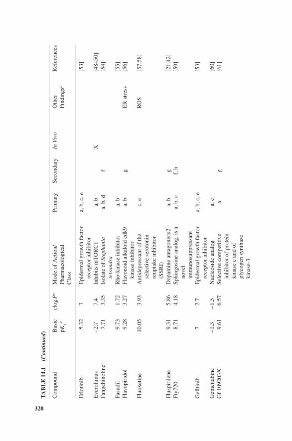
�
� �
�
TA
BL
E14
.1(C
ontin
ued)
Com
poun
dB
asic
pKaa
clog
Pa
Mod
eof
Act
ion/
Phar
mac
olog
ical
Cla
ss
Prim
ary
Seco
ndar
yIn
Vivo
Oth
erFi
ndin
gsb
Ref
eren
ces
Erl
otin
ib5.
323
Epi
derm
algr
owth
fact
orre
cept
orin
hibi
tor
a,b,
c,e
[53]
Eve
rolim
us−
2.7
7.4
Inhi
bits
mT
OR
C1
a,b
X[4
8–50
]Fa
ngch
inol
ine
7.71
3.35
Isol
ate
ofSt
epha
nia
tetr
andr
aa,
b,d
f[5
4]
Fasu
dil
9.73
1.72
Rho
-kin
ase
inhi
bito
ra,
b[5
5]Fl
avop
irid
ol9.
283.
27Fl
avon
oid
alka
loid
cdk9
kina
sein
hibi
tor
a,b
gE
Rst
ress
[56]
Fluo
xetin
e10
.05
3.93
Ant
idep
ress
anto
fth
ese
lect
ive
sero
toni
nre
upta
kein
hibi
tor
(SSR
I)
c,e
RO
S[5
7,58
]
Flus
piri
lene
9.31
5.86
Dop
amin
ean
tago
nist
s2a,
bg
[21,
42]
Fty7
208.
714.
18Sp
hing
osin
ean
alog
,is
ano
vel
imm
unos
uppr
essa
nt
a,b,
cf,
h[5
9]
Gefi
tinib
72.
7E
pide
rmal
grow
thfa
ctor
rece
ptor
inhi
bito
ra,
b,c,
e[5
3]
Gem
cita
bine
−1.
3−
1.5
Nuc
leos
ide
anal
oga,
c[6
0]G
f10
9203
X9.
616.
57Se
lect
ive
com
petit
ive
inhi
bito
rof
prot
ein
kina
sec
and
ofgl
ycog
ensy
ntha
seki
nase
-3
ag
[61]
320

�
� �
�
Gin
seno
side
F20
3.24
Ster
oid
glyc
osid
esfr
omgi
nsen
ga,
b,d,
e[6
2]
Imat
inib
7.55
2.89
Tyro
sine
kina
sein
hibi
tor
b,c,
ef
LM
P[6
3,64
]Im
ipra
min
e9.
494.
36T
ricy
clic
antid
epre
ssan
ta,
b,c,
d[6
5]Is
olie
nsin
ine
7.94
4.06
Nat
ural
alka
loid
smal
lm
olec
ules
a,b
f[4
0]
L-6
90,3
300
−0.
06IM
Pase
inhi
bito
ra,
bg
[66]
Lat
repi
rdin
e9.
053.
49A
ntih
ista
min
edr
uga,
bg,
i[6
7]L
iens
inin
e7.
944.
06N
atur
alal
kalo
idsm
all
mol
ecul
esa,
bf
[40]
Lith
ium
00
IMPa
sein
hibi
tor
a,b
gX
[66,
68]
Lop
eram
ide
9.41
4.77
Ca2+
chan
nelb
lock
ers
a,b
g[1
,21,
42]
Map
rotil
ine
10.62
4.36
Tetr
acyc
lican
tidep
ress
ant
c,e
[57]
Mes
orid
azin
e8.
193.
57A
ntip
sych
otic
a[4
2]M
etfo
rmin
12.33
−1.
25A
ntid
iabe
ticdr
uga,
c[2
5]M
inox
idil
4.34
1.3
K+
AT
Pch
anne
lope
ner2
ag
[1]
N(1
0)-s
ubst
itute
dph
enox
azin
e9.
24.
52a,
b,c
f,g
[42]
Nel
finav
ir4.
728.
18Pr
otea
sein
hibi
tors
a,b
[69]
Ner
vegr
owth
fact
orN
AN
Aa,
c,d
[70]
Nf4
49Gαs
inhi
bito
ra
g[1
]N
icar
dipi
ne8.
183.
82C
a2+ch
anne
lblo
cker
sa,
bg
[1,2
1]N
iclo
sam
ide
6.89
3.91
Ant
ipar
asiti
c2a,
bh
[19]
Nig
uldi
pine
8.45
6.04
Ca2+
chan
nelb
lock
ers
a,b
g[1
,21,
42]
Nilo
tinib
5.64
4.42
Tyro
sine
kina
sein
hibi
tor
a,d
f[7
1]N
imod
ipin
e5.
413.
05C
a2+ch
anne
lblo
cker
sa,
bg
[1,2
1]N
itren
dipi
ne5.
432.
88C
a2+ch
anne
lblo
cker
sa,
bg
[1,2
1]N
ortr
ipty
line
10.47
4.43
Tri
cycl
ican
tidep
ress
ant
ag
[42]
Npi
-005
20
2.28
Prot
easo
me
inhi
bito
ra,
b,c
[34]
(con
tinu
ed)
321

�
� �
�
TA
BL
E14
.1(C
ontin
ued)
Com
poun
dB
asic
pKaa
clog
Pa
Mod
eof
Act
ion/
Phar
mac
olog
ical
Cla
ss
Prim
ary
Seco
ndar
yIn
Vivo
Oth
erFi
ndin
gsb
Ref
eren
ces
Nvp
-Bez
235
6.41
4.73
Dua
lpi3
k-m
TO
Rin
hibi
tor
a,d
[72]
Oba
tocl
ax7.
916.
31In
hibi
tor
ofth
ebc
l-2
fam
ilyof
prot
eins
a,b
[73]
Opr
ozom
ib1.
621.
5Pr
otea
som
ein
hibi
tor
a,b
f[3
8]O
su-H
dac4
2H
isto
nede
acet
ylas
esin
hibi
tor
a,b,
cE
Rst
ress
[74]
Peni
trem
A0
6.36
Inhi
bits
high
cond
ucta
nce
Ca2+
-act
ivat
edK
+
chan
nel2
a,b
g[2
1]
Perh
exili
ne10
.58
5.53
Ca2+
chan
nelb
lock
era,
bh
[19]
Phen
ethy
lis
othi
ocya
nate
(Pei
tc)
03.
47U
nkno
wn
a,b,
c,d
[75]
Pi10
32.
973.
04D
ualp
i3k
and
mT
OR
inhi
bito
ra,
bf,
i[7
6]
Pim
ozid
e8.
385.
83A
ntip
sych
otic
a,b
g[1
,21,
42]
Prom
azin
e9.
23.
93A
ntip
sych
otic
a[4
2]Pr
omet
hazi
ne9.
054.
29A
ntih
ista
min
ea
[42]
Pter
ostil
bene
04.
06a,
c[7
7]Q
uerc
etin
−4
2.16
a,d,
eh
X[7
8]R
apam
ycin
−3
7.45
Inhi
bits
mT
OR
C1
a,b
g,i
X[4
8–50
]
322

�
� �
�
Res
vera
trol
−5.
73.
4A
ctiv
ates
sirt
uin-
1(h
isto
nede
acet
ylas
es),
inhi
bits
p70
s6ki
nase
a,b,
c,d
LM
P[7
9–81
]
Rilm
enid
ine
7.88
1.48
Imid
azol
ine-
1re
cept
orag
onis
tsa,
bg
X[1
,82]
Ros
iglit
azon
e6.
53.
02A
ntid
iabe
ticdr
ugin
the
thia
zolid
ined
ione
clas
sa,
b,c
[83]
Rot
tleri
n0
4.8
Inhi
bito
rof
PKC
delta
and
calm
odul
inki
nase
iii2
a,b,
cf,
h[8
4,85
]
Safin
gol
8.57
5.33
Lyso
-sph
ingo
lipid
prot
ein
kina
sec
inhi
bito
ra,
c,d,
eR
OS
[86]
Sert
ralin
e9.
475.
08A
ntid
epre
ssan
tof
the
SSR
Ic
[87]
Sira
mes
ine
9.45
4.36
a,b,
cL
MP
and
RO
S[8
8–90
]
Smer
100
2.71
Unk
now
na,
bg
X[9
1]Sm
er18
03.
68U
nkno
wn
a,b
gX
[91]
Smer
286.
122.
1U
nkno
wn
a,b
gX
[91]
Sper
mid
ine
10.9
−1.
1In
hibi
tshi
ston
eac
etyl
tran
sfer
ases
a,b
[79]
Tam
oxif
en8.
766.
35E
stro
gen
rece
ptor
anta
goni
stc
LM
Pan
dR
OS
[92–
94]
Tem
ozol
omid
e−
3.6
−0.
28C
hem
othe
rapy
drug
/alk
ylat
ing
agen
ta,
b,c,
df
[95]
Tem
siro
limus
−3.
97.
13In
hibi
tsm
TO
R1
a,b
X[4
8–50
]Te
tran
drin
e7.
73.
23C
alci
umch
anne
lblo
cker
a,b,
d,e
RO
S[9
6]T
haps
igar
gin
04.
44Sa
rco/
endo
plas
mic
retic
ulum
Ca2+
AT
Pase
inhi
bito
r
a,b,
cE
Rst
ress
[24]
(con
tinu
ed)
323

�
� �
�
TA
BL
E14
.1(C
ontin
ued)
Com
poun
dB
asic
pKaa
clog
Pa
Mod
eof
Act
ion/
Phar
mac
olog
ical
Cla
ss
Prim
ary
Seco
ndar
yIn
Vivo
Oth
erFi
ndin
gsb
Ref
eren
ces
Thi
orid
azin
e8.
935.
47A
ntip
sych
otic
a,d
[42]
Tori
n14.
194.
04A
TP-
com
petit
ive
mT
OR
inhi
bito
ra,
b[9
7]
TR
AIL
a,b,
cf
LM
P[9
8,99
]T
reha
lose
−3.
6−
4.8
Che
mic
alch
aper
one
a,b
f,g
X[1
00,1
01]
Tri
fluop
eraz
ine
8.39
5.03
Ant
ipsy
chot
ica,
bg
LM
Pan
dR
OS
[21,
42,
102]
Tri
flupr
omaz
ine
9.2
4.81
Ant
ipsy
chot
ica
[42]
Tun
icam
ycin
8.41
−1.
22M
ixtu
reof
hom
olog
ous
antib
iotic
sa,
b,c
LM
Pan
dE
Rst
ress
[24,
103]
Val
proi
cac
id0
2.58
Ant
iepi
lept
ics
(MIP
synt
hase
inhi
bito
r)a,
b,c,
d,e
LM
Pan
dR
OS
[104
,105
]
Ver
apam
il9.
685.
04C
a2+ch
anne
lblo
cker
sa,
bg
[1,2
1]V
orin
osta
t2
−3.
5H
isto
nede
acet
ylas
esin
hibi
tor
a,b,
cE
Rst
ress
[74]
Y-2
7632
10.74
1.04
Rho
-ass
ocia
ted
prot
ein
kina
sein
hibi
tor
ag
[106
]
Lis
tof
invi
tro
assa
ysth
atar
eus
edto
dem
onst
rate
auto
phag
yin
duct
ion
from
liter
atur
ein
clud
esth
efo
llow
ing:
a,in
crea
sein
LC
3II
prot
ein
leve
l(w
este
rnbl
ot);
b,L
C3
punc
ta;c
,aut
opha
gic
vacu
oles
byel
ectr
onm
icro
scop
y;d,
incr
ease
dve
sicu
lar
orga
nelle
sus
ing
acri
dine
oran
geor
lyso
trac
ker;
e,up
regu
latio
nof
auto
phag
yor
lyso
som
alge
nes;
f,in
hibi
tor
bloc
king
assa
yus
ing
bafil
omyc
inA
orch
loro
quin
e;g,
clea
ranc
eof
auto
phag
icsu
bstr
ate;
h,fr
eeG
FPas
sess
men
t;i,
p62
abun
danc
eas
sess
men
t.aC
ompo
und’
sph
ysic
oche
mic
alpr
oper
ties
basi
cpK
aan
dcl
ogP
are
obta
ined
from
ChE
MB
L[1
07].
bE
Rst
ress
,end
opla
smic
retic
ulum
stre
ss;R
OS,
reac
tive
oxyg
ensp
ecie
s;L
MP,
lyso
som
alm
embr
ane
perm
eabi
lizat
ion.
324

�
� �
�
AUTOPHAGY ENHANCER SCREENING 325
antibodies using immunofluorescence (IF) labeling. An additional advantage ofan immunohistochemical approach is the capability to colocalize LC3 with otherorganelle markers. LC3 tagged with a fluorescent protein such as GFP may beintroduced into cells to detect autophagosomes and has recently been used to assessautophagy changes in animals in vivo [108]. Both endpoints (LC3-GFP puncta andLC3 IF puncta) can be quantified with Image J or the high content imaging platformfor automatic image capturing and quantification. So far there is no specific dye tostain autophagosomes although acridine orange is frequently used in the literature todemonstrate development of acidic vesicular organelles (AVOs) as a surrogate markerfor autophagosome [78]. However, staining from acridine orange mainly reflects thechange of lysosome since this is a dye that accumulates in acidic lysosomes [109].
One challenge in screening for autophagy modulators is to discern enhancers frominhibitors, both of which may increase the number of autophagosomes. All previouslymentioned methods reflect autophagosomes only in their static state rather revealinginformation about the dynamic balance between autophagosome formation and clear-ance (“autophagy flux”). Because increased autophagosome number can indicateautophagy induction or failed autophagosome clearance (e.g., blocked fusion withlysosomes or degradation by lysosomes), secondary assays are typically required tofurther understand autophagy flux. Direct evidence of autophagy flux activity can beobtained by measuring the degradation of long-lived proteins; however, this methodinvolves somewhat labor-intensive radiolabeling of the cells and subsequent separa-tion of acid-soluble from acid-insoluble radioactivity. Its wide application is furtherlimited by the need to use inhibitors to establish specificity of proteolytic systemsinvolved, potential for released amino acids to be metabolized and low autophagicactivity of cells under certain conditions. The most common assay used in the liter-ature is the determination of autophagosome formation rates by measuring levels ofLC3-II in the presence and absence of an inhibitor (e.g., chloroquine, bafilomycinA, or proteases inhibitors) that blocks LC3 degradation. A rise in LC3-II levels inthe presence of inhibitors can therefore reflect autophagosome formation. While rel-atively easy to apply once proper conditions of inhibitor concentration and incubationtime are established, this assay provides no information about the lysosomal steps ofautophagy (fusion and degradation). Therefore, this endpoint should not be consid-ered as a reliable endpoint to evaluate autophagic flux. The substrate targeting protein,p62 protein, also called sequestosome 1 (SQSTM1), binds to ubiquitinated proteinsfacilitating their sequestration and ultimate delivery to lysosomes for degradation,during which process p62 is also degraded. Since p62 accumulates when autophagyis inhibited and its levels decrease when autophagy is induced, p62 turnover hasbeen used as a marker of autophagic flux. Special consideration needs to be takenwhen using p62 data for autophagy enhancers since reduction of p62 synthesis bypoor cell health could confound the interpretation. A dose-dependent response cancertainly increase the confidence of the data interpretation using this assay. In addi-tion, free GFP abundance relative to complete fusion of GFP-LC3 has also been usedas a marker to reflect autophagic flux [110]. GFP-LC3-containing autophagosomeswill be degraded after fusion with lysosomes. However, the free GFP moiety gen-erated from the GFP-LC3 degradation is relatively resistant to proteolysis; hence,

�
� �
�
326 AUTOPHAGY ENHANCERS, ARE WE THERE YET?
the appearance of free GFP to some extent reflects cargo delivery (GFP-LC3) fromthe autophagosome to the lysosome and subsequent degradation. Caution must beused, however, when conducting the GFP-LC3 cleavage assay as a determinant ofautophagic flux because GFP fragments can be further degraded and thus do notaccumulate when lysosomal acidity is high, for example, during starvation [111].Recently, tandem mRFP/mCherry-GFP fluorescence microscopy has been particu-larly designed to monitor flux. The principle of the assay is based on the differentialpH dependence of green and red fluorescent protein tags: GFP signal is typicallyquenched in a strongly acidic environment while mRFP and mCherry remain fluores-cent. This assay can be used in the format of single time point data, where an increaseof total red+green+ and red+green− vesicular compartments indicates autophago-some formation (i.e., induction of autophagy) and an increase of red+green− com-partments alone reflects successful autophagosome–lysosome fusion and lysosomeacidification associated with lysosomal clearance of autophagy substrate. Since theautophagosome is a transient organelle existing for less than 10 min before fusingwith the lysosome [112], evaluating the dynamic behavior of autophagosomes pro-vides more critical information on autophagic flux than single time point data. Tan-dem technology can be used to capture this dynamic behavior in real time and revealthe rate of formation and clearance of autophagosomes over time. This techniquerequires a high-resolution microscope (e.g., confocal) with temperature and CO2 con-trol to facilitate the kinetic monitoring. In addition, upregulation of Atgs (e.g., Atg5and Atg7) has also been used as supporting evidence for autophagy activation [26,53].However, most of the Atgs do not always show significant changes in mRNA andprotein levels when autophagy is induced and the extent of increase varies amongdifferent cell and tissue types. More importantly, only induction of autophagy can beconcluded from the Atgs data and this assay still does not provide information aboutthe lysosomal degradation step.
14.3.2 Autophagy Enhancers Identified from Early Literature
Because of the potential of therapeutic benefits of autophagy enhancement,large-scale screening for autophagy modulators has been carried out in various labs[19–21,42]. There are many additional reports of fortuitously identified autophagyenhancers/activators in the literature (Table 14.1). Compounds exhibiting autophagymodulation activity are structurally and therapeutically diverse. Categorization ofthese agents is mostly based on mechanism of action and inducers can be broadlygrouped as mTOR-dependent and mTOR-independent mechanisms. Compoundswork through the mTOR-dependent pathway include not only classic mTORinhibitors, such as rapamycin and torin 1, but also compounds from differentpharmacological classes, for example, calcium channel blockers (perhexiline andverapamil), antiarrhythmics (amiodarone), and antiparasitics (niclosamide) [19].Nevertheless, for a large number of compounds, the mechanism(s) by which theymodulate autophagy have yet to be identified.
Interestingly, many compounds (e.g., tamoxifen, amiodarone, and vera-pamil) that are believed to be autophagy enhancers are basic lipophilic, which

�
� �
�
AUTOPHAGY ENHANCER SCREENING 327
allows the compound to accumulate in acidic organelles (i.e., lysosome) by pHpartitioning [113]. Lysosomotropic compounds were recently shown to inhibitautophagy by perturbing lysosomal function [114]. Historically, vinblastine, amicrotubule disruptor, was once believed to be an autophagy “enhancer” but lateridentified to actually inhibit autophagosome–lysosome fusion. It may thereforebe prudent to reevaluate the classification of these basic lipophilic compounds asautophagy enhancers.
Lysosomes are the final destination of autophagosomal substrates for degrada-tion. Lysosomes are membrane-enclosed compartments filled with acid hydrolyticenzymes (e.g., cathepsins) used to digest macromolecules and are found in the cytosolof nearly all mammalian cells. For optimal activity of acid hydrolases, lysosomesmust maintain a low internal pH of about 4–5. The acidic pH in the lumen is achievedby the vacuolar H+ ATPase, which uses the energy of ATP hydrolysis to pump H+into the lysosome. The pH gradient between the lysosomal lumen and the cytosol(pH≈ 7.2) can drive overaccumulation of basic lipophilic compounds via pH par-titioning. Generally, lipophilic free bases can pass through the plasma membraneand lysosome membrane; however, the majority will lose permeability and becometrapped inside lysosomes due to their protonation by the acidic lysosomal pH. Forinstance, chloroquine, a well-known lysosomotropic compound, can easily reach con-centrations in excess of 20 mM inside lysosomes equal to a several hundred-foldhigh level relative to that outside of the cells [113]. Two physicochemical proper-ties, basic pKa (acid dissociation constant for the conjugate acid for the weak base)and clog P (partition coefficient between octanol and water, representing membranepermeability) both affect the accumulation by influencing the extent of lysosomaltrapping and regulating the kinetics of passive permeation. The compounds that accu-mulate in lysosomes are classified as lysosomotropic agents. It was demonstratedpreviously that compounds with certain physicochemical properties (basic pKa > 6.5and clog P> 2) tend to be lysosomotropic [115]. To understand if physicochemicalproperties could contribute to autophagy modulation by the compounds identified sofar, we evaluated relationships between physicochemical properties (i.e., basic pKaand clog P) and autophagy modulation. Surprisingly, half of autophagy “enhancers”fit into the profile of being basic with pKa > 6, in which majority of them are lipophilicwith clog P> 2 (Table 14.1 and Figure 14.1). It is noteworthy that commonly known“autophagy enhancers” amiodarone and tamoxifen are located in the basic lipophilicregion (Figure 14.1)
It has been well established that lysosomotropic compounds can impair lysosomefunction potentially through multiple mechanisms. Lysosomotropic compounds,chloroquine and methylamine, increase lysosomal pH drastically (0.5–2.0 pH units)after accumulation [116]. In addition, significant pH elevation by chloroquine isobserved in vivo [117]. In a previous study [115], lysosomotropic compoundswere shown to decrease staining of cells by Lysotracker, which requires low pHto fluoresce. An increase in pH is expected to suppress lysosomal degradationsince most lysosome enzyme activities have acidic pH optima. An increase of pHmay also decrease the fusion capability of lysosomes in some cells as seen forchloroquine [118] although pH dependence of lysosome fusion is not universally

�
� �
�
328 AUTOPHAGY ENHANCERS, ARE WE THERE YET?
8
7
6
5
4
clog P
3
2
1
0
–1
–2
–3
–4
–8 –6 –4 –2 0 2 4
Basic pKa
Amniodarone
Tamoxifen
Perhexiline
Nortriptyline
Fluoxetine
Compound C
6 8 10 12
Imipramine
Figure 14.1 Correlation of physicochemical properties with autophagy screening: A scatterplot showing the distribution of autophagy enhancers within the clog P-basic pKa physico-chemical property space. A group of compounds clustered within the area (blue dotted square)where the clog P was >2 and basic pKa was greater than 6. (See color plate section for thecolor representation of this figure.)
observed [119]. Lysosomotropic compounds have also been shown to decreaselysosomal enzyme activity. For instance, chlorpromazine and chloroquine inhibit thelysosomal phospholipase A1 in vitro [120,121] and desipramine, chlorpromazine,and chloroquine downregulate acid ceramidase [122]. Multiple lysosomotropiccompounds can redistribute the mannose 6-phosphate receptor from the trans-Golginetwork to endosomes and concomitantly increase the secretion of lysosomalenzymes causing intracellular lysosomal enzyme levels to decline [123] furtherexacerbating lysosomal dysfunction.
Given that lysosomal substrate degradation is an essential step of autophagy,impairment of lysosomal function, by definition, impairs autophagy activity. Notsurprisingly, various lysosomotropic compounds have recently been shown to beautophagy inhibitors [114]. Interestingly in the same study, transcription of plentifullong-lived proteins, including cytoskeletal and extracellular matrix proteins, wassuppressed further suggesting that autophagic turnover of these proteins is hindered.Notably, lysosomotropic compounds also impact other trafficking process (i.e.,endocytosis and phagocytosis process) that delivers materials to lysosomes. Thelysosomotropic compounds chloroquine, tamoxifen, and verapamil decrease phago-cytosis activity [124,125] and chloroquine is recognized as a clathrin-dependent

�
� �
�
AUTOPHAGY ENHANCER SCREENING 329
endocytosis inhibitor [126]. Amiodarone impairs trafficking through late endosomesand induces a Niemann–Pick C-like phenotype [127]. In addition, multiple cationicamphiphilic compounds (e.g., amiodarone, fluoxetine, nortriptyline, and imipramine)identified as “autophagy enhancers” are reported to induce phospholipidosis (PLD).Although the toxicological impact of PLD is still debatable, it is believed to bea drug-induced LSD characterized by the excess accumulation of phospholipids.Multiple mechanisms are proposed to contribute to PLD including lysosomaldysfunction via inhibition of lysosomal phospholipase enzyme activity or transport.Basic pKa and clog P are important descriptors for in silico prediction of phospho-lipidosis [128]. Taken together, the evidence supports the conclusion that compoundswith basic lipophilic properties have the propensity to impair lysosomal functions,consequently hampering autophagy activity rather than boosting it.
The foregoing evidence on current autophagy enhancer screening assays raisesthe critical question of whether they are sufficient to distinguish enhancers frominhibitors. The most commonly used autophagy flux assay is LC3 assessment withand without lysosome inhibitors. Compounds that increase LC3 abundance togetherwith lysosomal proteolysis inhibitor treatment, as compared to single agent effecton LC3 in the absence of inhibitor, have often been interpreted as showing that thecompound increased “autophagic flux.” Autophagic flux in this context is a measureof autophagosome formation and provides no information about the lysosomalsteps of autophagy. More appropriately, autophagic flux should reflect the activityof the entire autophagic process from substrate sequestration through completesubstrate degradation. TFEB (transcription factor EB) stimulates the expressionof genes for lysosome biogenesis as well as many involved in autophagosomeformation and fusion of autophagosomes with lysosomes [129,130]. Recently,TFE3, another family member of the microphthalmia-associated transcription factor(MiTF) and TFE (miTF/TFE) families, was also shown to increase the expressionof the proteins involved in autophagy and lysosomal biogenesis [131]. Interestingly,both transcriptional factors respond to lysosomal dysfunction, as shown by nucleartranslocation of both TFEB and TFE3 after chloroquine treatment [130,131]. Possi-bly these transcriptional factors are activated as a compensatory feedback responseto overcome the lysosomal stress triggered by this lysosomal inhibitor, whichlikely results in reduced signaling to the lysosomal amino acid sensing complexthat regulates TFEB phosphorylation and translocation. Further experiments arerequired to confirm whether or not other basic lipophilic compounds trigger a similarnuclear translocation of TFEB and TFE3 to increase the gene expression of theprotein involved in autophagosome formation and lysosomal biogenesis. In thisregard, LAMP1 expression is increased by the basic lipophilic compound imatinib[63] and various autophagy genes (e.g., beclin 1, Atg5, or Atg7) are upregulatedby amiodarone, gefitinib, and compound C [26,46,53]. In addition, as observedfor chloroquine, multiple basic lipophilic compounds, including perhexiline, com-pound C, siramesine, and imipramine, decrease mTOR signaling, suggesting thatnuclear translocation of TFEB and TFE3 may be occurring since mTOR activationcontributes to the cytosolic localization of these transcription factors [130,131].It is therefore plausible that basic lipophilic compounds generally act similarly to

�
� �
�
330 AUTOPHAGY ENHANCERS, ARE WE THERE YET?
the mechanism proposed for chloroquine, namely by decreasing mTOR activity,triggering nuclear translocation of TFEB and TFE3, and ultimately inducing thegene expression of lysosomal and autophagy genes as a feedback response tothe accumulation of substrates in autolysosomes, often referred to as “autophagicstress.” Accordingly, activation of autophagy demonstrated by lysosomal inhibitorassay might not be sufficient to identify effective autophagy enhancers. Similarprecautions need to be taken when interpreting the upregulation of autophagyor lysosome genes as evidence of autophagy enhancement since basic lipophilicautophagy inhibitors can activate those genes following lysosomal stress. Increasedexpression of autophagy or lysosome genes may indicate initiation of autophago-some or lysosome biogenesis but does not necessarily reflect increased autophagyactivity, which should be a measure of both autophagosome formation and clearance.
An additional assay previously used to support autophagy enhancement is mea-surements of numbers of AVOs labeled by acridine orange. The development ofAVOs, a characteristic of autophagy [41,78], can be monitored by lysosomotropicacridine orange, which selectively accumulates in acidic organelles, enabling quan-titation of both number and size of lysosomes, although the lysosome expansion canimply higher or lower lysosomal activity. Multiple lysosomotropic compounds, forexample, chloroquine that could perturb lysosomal functions have been shown to trig-ger marked expansion of lysosomal volume [132,133]. In addition, lysosomal volumeexpands in multiple lysosomal storage diseases, which are also associated with lyso-somal dysfunction [134,135]. Reduction of lysosomal volume in these disease stateshas been used as a phenotypic screening to identify the compounds that could improvelysosomal function. [135,136]. Therefore, increase of AVO might not be the appropri-ate marker for identifying true autophagy enhancers due to the potential associationwith lysosomal dysfunction.
Assays based on p62 turnover and the tandem-tagged GFP-mCherry-LC3 probecan provide suitable appraisals of autophagy flux. Induction of autophagy by arsenictrioxide has been demonstrated by TEM, IF staining with EGFP-LC3, and increasedLC3 in the presence of the lysosomal proton pump (vacuolar ATPase) inhibitorbafilomycin A [30,137,138]. However, arsenic trioxide inhibited autophagy flux asevidenced by the tandem-tagged GFP-mCherry-LC3 probe assay [139]. In this assay,starved cells exhibit both yellow and red puncta but cells treated with arsenic trioxideexhibited persistent yellow/orange puncta, similar to the effects of bafilomycin A,indicating a block of autophagy flux. Arsenic also induced accumulation of p62puncta, further indicating inhibition of autophagy flux. Although arsenic is notbasic lipophilic, lysosomal uptake of heavy metals is well documented [140,141].Indeed, arsenic caused rapid destabilization of lysosomes [142], which mightunderlie the negative effect of arsenic on autophagy. Thapsigargin, an inhibitor ofthe ER SERCA calcium pump, has been frequently used as an autophagy inducer[24,143], as reflected by increased LC3 in the presence of bafilomycin A [143];however, in the tandem GFP-mCherry-LC3 assay, loss of GFP signal is delayed afterthapsigargin indicating impaired fusion of autophagosomes with lysosomes [144].Fluoxetine, nortriptyline, and imipramine have all been classified as “autophagyenhancers” based on earlier studies, but they were recently shown to increase p62
�
� �
�
AUTOPHAGY ENHANCER SCREENING 331
protein abundance in a dose-dependent manner [114], supporting an inhibition ofautophagic flux.
Collectively, these findings suggest that many autophagy enhancers identified sofar might not represent true enhancers of autophagy flux. In particular, compoundswith basic and lipophilic moieties need to be evaluated very carefully due to theirhigh potential to disturb lysosomal function. Even compounds lacking a basiclipophilic moiety may also perturb lysosome function as illustrated by arsenictrioxide, which destabilized lysosomal membranes [142]. TRAIL triggers lysosomalmembrane permeabilization (LMP) via death receptor-5 [98]. LMP and relocationof cathepsins contribute to the cell death by curcumin [47]. Pharmacologicalinducers of ER stress, such as tunicamycin, have also been reported to associate withLMP [103]. Notably, LMP has recently been shown to stimulate protein aggregateformation in neurons [145]. In addition, oxidative stress or ER stress was associatedwith multiple putative autophagy enhancers including amiodarone, apicidin, andfluoxetine (Table 14.1). Compounds with liability of LMP, oxidative stress, or ERstress are more likely to weaken their therapeutic benefit for conditions requiringautophagy enhancement.
Since all membrane trafficking including autophagy, phagocytosis, and endocy-tosis converge on lysosomes compounds that promote autophagy flux should notcompromise lysosomal function. For instance, chloroquine, a lysosomotropic com-pounds that inhibit lysosomal function, is a well-known autophagy inhibitor and hasalso been shown as an inhibitor for endocytosis and phagocytosis [124,126]. As wediscussed previously, many reported “autophagy enhancers” target lysosomes eitherdue to their lysosomotropic nature (e.g., amiodarone and latrepirdine) or by compro-mising lysosomal membrane integrity (e.g., TRAIL). Analysis of other traffickingprocesses, such as endocytosis and phagocytosis, could provide further certainty onthe lysosomal functions. In other words, if compounds shown to augment autophagyperturb endocytic, phagocytic, or lysosomal functions, extra consideration shouldbe taken when classifying these compounds as “autophagy enhancers.” Especiallyif “increased” autophagy is surmised on the basis of only elevated LC3 expressionwith proteolysis inhibitors, increased expression of Atgs, or increased AVOs, this“increase” may merely reflect a compensatory response to lysosomal dysfunction.Therefore, evaluation of the general function of lysosomes could provide additionalsupporting evidence for autophagy modulation.
14.3.3 mTOR Inhibitors
Mammalian target of rapamycin (mTOR) is a conserved serine/threonine proteinkinase that plays a significant role in cell growth, proliferation, and protein synthesisby regulating the activities of factors such as 4E-BP1, S6K, and Akt. mTOR1 is alsoa master negative regulator of autophagy by blocking the ULK1-ATG13-FIP200complex. Various mTOR inhibitors were developed to enhance autophagosomebiogenesis and thereby promote clearance of aggregate-prone proteins. Notably,genetic or pharmacological inhibition of TOR activity multiple model systemsis associated with life span extension [146–149], possibly through inhibitory
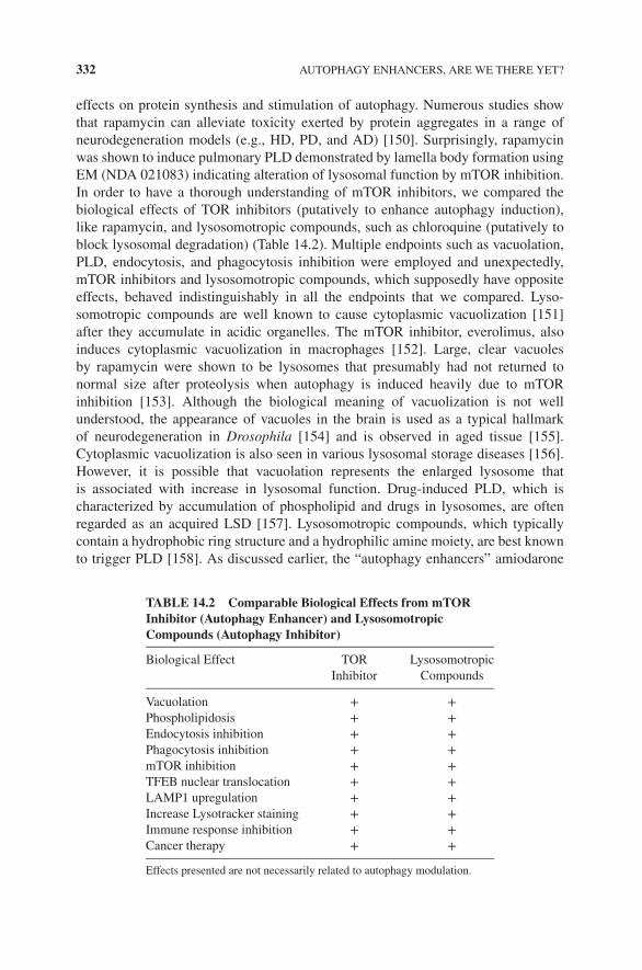
�
� �
�
332 AUTOPHAGY ENHANCERS, ARE WE THERE YET?
effects on protein synthesis and stimulation of autophagy. Numerous studies showthat rapamycin can alleviate toxicity exerted by protein aggregates in a range ofneurodegeneration models (e.g., HD, PD, and AD) [150]. Surprisingly, rapamycinwas shown to induce pulmonary PLD demonstrated by lamella body formation usingEM (NDA 021083) indicating alteration of lysosomal function by mTOR inhibition.In order to have a thorough understanding of mTOR inhibitors, we compared thebiological effects of TOR inhibitors (putatively to enhance autophagy induction),like rapamycin, and lysosomotropic compounds, such as chloroquine (putatively toblock lysosomal degradation) (Table 14.2). Multiple endpoints such as vacuolation,PLD, endocytosis, and phagocytosis inhibition were employed and unexpectedly,mTOR inhibitors and lysosomotropic compounds, which supposedly have oppositeeffects, behaved indistinguishably in all the endpoints that we compared. Lyso-somotropic compounds are well known to cause cytoplasmic vacuolization [151]after they accumulate in acidic organelles. The mTOR inhibitor, everolimus, alsoinduces cytoplasmic vacuolization in macrophages [152]. Large, clear vacuolesby rapamycin were shown to be lysosomes that presumably had not returned tonormal size after proteolysis when autophagy is induced heavily due to mTORinhibition [153]. Although the biological meaning of vacuolization is not wellunderstood, the appearance of vacuoles in the brain is used as a typical hallmarkof neurodegeneration in Drosophila [154] and is observed in aged tissue [155].Cytoplasmic vacuolization is also seen in various lysosomal storage diseases [156].However, it is possible that vacuolation represents the enlarged lysosome thatis associated with increase in lysosomal function. Drug-induced PLD, which ischaracterized by accumulation of phospholipid and drugs in lysosomes, are oftenregarded as an acquired LSD [157]. Lysosomotropic compounds, which typicallycontain a hydrophobic ring structure and a hydrophilic amine moiety, are best knownto trigger PLD [158]. As discussed earlier, the “autophagy enhancers” amiodarone
TABLE 14.2 Comparable Biological Effects from mTORInhibitor (Autophagy Enhancer) and LysosomotropicCompounds (Autophagy Inhibitor)
Biological Effect TORInhibitor
LysosomotropicCompounds
Vacuolation + +Phospholipidosis + +Endocytosis inhibition + +Phagocytosis inhibition + +mTOR inhibition + +TFEB nuclear translocation + +LAMP1 upregulation + +Increase Lysotracker staining + +Immune response inhibition + +Cancer therapy + +
Effects presented are not necessarily related to autophagy modulation.
�
� �
�
AUTOPHAGY ENHANCER SCREENING 333
and tamoxifen are classic inducers of PLD [159] and mTOR inhibitors, includingeverolimus, sirolimus, and temsirolimus [160], are all reported to induce PLD, sug-gesting that they perturb lysosomal functions. Besides PLD induction, both mTORinhibitors and lysosomotropic compounds may also impair other aspects of mem-brane trafficking, such as endocytosis and phagocytosis. Chloroquine is recognizedas a clathrin-dependent endocytosis inhibitor [126] and the phagocytosis of rod outersegments is inhibited by chloroquine and tamoxifen in retinal pigment epithelial cellcultures [124]. Azithromycin, a lysosomotropic antibiotic, delays sequestration ofreceptor-bound transferrin and peroxidase–antiperoxidase immune complexes intocell-surface endocytic pits and vesicles [161]. Although the mechanism is not clear,mTOR may play a critical role in clathrin-mediated endocytosis [162], as suggestedby the ability of rapamycin to reduce albumin uptake [163]. Rapamycin also impairsmacropinocytosis and mannose receptor-mediated endocytosis of antigens by bonemarrow-derived DCs [164] and decreases phagocytosis of bacteria and apoptoticcells [165].
Many lysosomotropic compounds, for example, niclosamide and imipraminehave been shown to lower mTOR1 activity [65,166], possibly attributable to lysoso-mal dysfunction that reduces the generation of amino acids critical for the mTOR.TFEB is a master regulator of lysosomal biogenesis and autophagy. Interestingly,both mTOR inhibitor and lysosomotropic compounds can trigger TFEB nucleartranslocation [130,167], leading to the upregulation of a broad range of lysosomalgenes and increased lysosomal biogenesis [168,169]. Lysosomotropic compoundscan robustly increase lysotracker staining [132,170]. Similarly, both allostericmTOR inhibitor (rapamycin) [146] and catalytic inhibitors (PP242 and Torin1) [169]also markedly increase lysotracker fluorescence intensity. Increased lysotrackerstaining in these situations indicates increased numbers or size of lysosomal-relatedcompartments, which can potentially reflect both an attempted adaptive responseand a pathological phenomenon (lysosomal degradative impairment) as seen inlysosomal storage diseases and aging.
Autophagy and autophagy proteins have multifaceted roles in the innate and adap-tive immune responses, for example, antigen presentation and lymphocyte develop-ment [171]. Surprisingly, with supposedly opposite actions on autophagy, both typesof agents (mTOR inhibitors and chloroquine) are shown to inhibit immune response.Tor kinase has multiple effects on innate and adaptive immune responses. The mTORinhibitors sirolimus and everolimus block the response of T- and B-cell activation bycytokines, which prevents cell-cycle progression and proliferation [172]. Rapamycinhas also been shown to suppress the mature dendritic cell function and decreasethe production of cytokine IFNγ produced by dendritic cells [173]. As noted ear-lier, rapamycin has an inhibitory effect on dendritic cells endocytosis [164], whichcould further decrease the immune response. The mTOR inhibitor (e.g., rapamycin)has been used as an immunosuppressant to prevent transplant rejection. The efficacyof the lysosomotropic compound hydroxychloroquine in the treatment of systemiclupus erythematosus and rheumatoid arthritis has been well demonstrated [174,175],and it has become a standard component of therapy for patients with these diseases.Anti-inflammatory response by hydroxychloroquine could be ascribed to lysosomal
�
� �
�
334 AUTOPHAGY ENHANCERS, ARE WE THERE YET?
dysfunction, which diminishes the formation of peptide–MHC protein complexesrequired to stimulate CD4+ T cells and results in downregulation of the immuneresponse against autoantigenic peptides. Additional lysosomotropic compounds, forexample, fluoxetine and citalopram have also shown anti-inflammatory effects inarthritis animal models [176].
In addition, both mTOR inhibitors and lysosomotropic compounds, such aschloroquine, have been used in cancer therapy. Currently, rapamycin analog,temsirolimus, has been used for the treatment of renal cell carcinoma and everolimusfor various cancer types [177]. Owing to their nature of autophagy inhibition,lysosomotropic compounds, for example, chloroquine or, more commonly, hydrox-ychloroquine in combination with other antitumor agents have been explored innumerous oncology clinical trials [178]. Interestingly, the combination of hydroxy-chloroquine with the mTOR inhibitor demonstrated an encouraging clinical outcomein a phase I trial with advanced melanoma patients [179].
The unanticipated similarity between mTOR inhibitors and lysosomotropic com-pounds further accentuate the challenge of autophagy enhancer screening, grant-ing some of the effects could be attributed to pharmacological actions unrelated toautophagy. Data interpretation of endpoints for autophagy screening needs to be con-sidered carefully. In addition, the comparable biological effects raised a critical ques-tion about the role of mTOR on lysosomal function. The ability of mTOR inhibitorsand lysosomotropic compounds to induce PLD and inhibit endocytosis and phago-cytosis suggests that both classes of compounds may have a negative impact onlysosomal functions. An increase of lysotracker staining from both types of agentsfurther supports this notion since a similar phenotype is often observed when lysoso-mal functions is perturbed, for example, cells from LSD patients.
As discussed earlier, it is understandable as to why lysosomotropic compoundshave global negative impact on lysosomes. A negative impact of mTOR inhibitors isseemingly more puzzling, given their ability to stimulate TFEB-mediated lysosomalbiogenesis. Recent findings, however, reveal positive roles of mTOR activity in traf-ficking processes that lead to lysosomes and lysosomal function. Although the mech-anisms have not been fully elucidated, mTOR activity may influence microtubule andactin dynamics through the actions of ROCKs, which play a critical role in regulat-ing actin polymerization and associated cytoskeletal rearrangements. mTOR inhibi-tion by rapamycin abolishes ROCK-1 synthesis in macrophages thereby inhibitingchemotaxis and phagocytosis [180]. Rapamycin slows microtubule polymerizationand lowers binding of Bik1P to microtubules [181]. mTOR hyperactivation seemsto disturb microtubule organization [182], suggesting that a proper mTOR activitybalance is important in this process. Disruption of the cytoskeleton obviously hin-ders various trafficking processes, which prevents cargo delivery to lysosomes andsubstrate degradation. Beyond these effects, mTOR activity is required to gener-ate protolysosomal tubules and facilitate their maturation into functional lysosomesunder conditions of nutrition deprivation [183]. This process, described as autophagiclysosome reformation (ALR), is observed after prolonged starvation and blockedby rapamycin treatment. Similar phenomena such as ALR were also observed in

�
� �
�
OTHER AGENTS THAT BOOST AUTOPHAGY AND LYSOSOMAL FUNCTIONS 335
phagosome maturation process where phagosomes and entotic vacuoles undergo afission process, which redistributes vacuolar contents into lysosomal networks [184].mTOR inhibition slows down the shrinkage of phagosomes indicating that membranefission is regulated by mTOR. Recently, mTOR was also shown to positively reg-ulate lysosomal ATP-sensitive two-pore Na+ channels, which modulate membranepotential and may influence pH stability of lysosomes under certain cellular condi-tions [185]. This finding underscores the reciprocal relationship between mTOR andlysosomes in which amino acids generated during lysosomal proteolysis modulatemTOR activity and, in turn, potentially regulate additional endosomal and lysosomalfunctions via lysosomal ATP-sensitive two-pore Na+ channels and additional changesin intralumenal pH and ion fluxes.
Although it has been demonstrated that phosphorylation of TFEB by mTORC1contributes to the cytoplasmic location of TFEB [131,167,186], contradictory resultshave also been reported, in which TFEB nuclear translocation was promoted bymTORC1 activity [163]. The latter study employed an mTORC1 overactivationmodel by knocking out tuberous sclerosis complex 2(TSC2), which negativelyregulates mTORC1. Statistically significant overrepresentation of lysosomal genesand especially genes encoding vacuolar H+-ATPase (V-ATPase) was identified withhigher expression when mTOR was activated in Tsc2−/− cells, but low expressionin TSC2−/− with rapamycin treatment indicating V-ATPase genes were tightlyregulated by mTORC1. Interestingly, TFEB depletion downregulates V-ATPaseexpression suggesting that this mTORC1-dependent V-ATPase upregulation requiresTFEB. TSC inactivation indeed promoted nuclear translocation of TFEB and thiswas reverted by rapamycin treatment suggesting that TFEB nuclear localization ismTORC1 dependent. The multiple roles of mTORC1 on various biological processes(e.g., protein synthesis and cell growth) may well complicate the interpretation ofthe net outcomes of mTORC1 modulation and its ultimate impact on autophagy.It is plausible that mTORC1 might play varying roles in lysosomal functionunder different physiological/pathological conditions. It is worth noting that thesubstantial benefits of rapamycin in mouse models of different neurodegenerativediseases may also derive from autophagy-independent therapeutic effects such asrapamycin’s antiaging properties, reduced protein translation [18], or change inimmune responses. Further characterization of the role of mTORC1 especiallyon late stages of autophagy, phagocytosis, and lysosomal function under diverseconditions should provide a stronger mechanistic rationale for mTORC1 modulationas a therapeutic approach to neurodegeneration.
14.4 OTHER AGENTS THAT BOOST AUTOPHAGY AND LYSOSOMALFUNCTIONS
As a promising therapeutic approach, autophagy enhancement has attracted exten-sive research interest. Given the foregoing discussion about the negative impact onlysosomal function by some of the current “autophagy enhancers” and the complex

�
� �
�
336 AUTOPHAGY ENHANCERS, ARE WE THERE YET?
effects of mTOR inhibition on autophagy, further efforts are warranted for identifyingpositive autophagy regulators especially the ones that increase autophagic substrateclearance. Emerging evidence suggests lysosomal clearance defects as a mechanismfor various adult-onset neurodegenerative diseases, supported by findings on neu-ronal cell death in LSDs [17]. When the lysosomal clearance process is impaired,autophagy induction might exacerbate the pathology. As a result, the success of anyautophagy enhancer in a given condition may depend on relieving blockages in lyso-somal clearance. Lysosomal function or general membrane trafficking as therapeutictargets in neurodegeneration is relatively unexplored. Nonetheless, numerous thera-peutic targets have been suggested to have positive effect, including cathepsin activityenhancement, pH restoration, and increasing lysosomal stability [17].
14.4.1 HDAC Inhibition
Histone deacetylase inhibitors (HDAC inhibitors, HDACi) are a class of compoundsthat interfere with the function of histone deacetylase, an enzyme with diverse bio-logical effects owing to broad actions on gene transcription. HDACi clear the excessaccumulation of cholesterol and correct cholesterol storage defects in human NPC1mutant cells [187]. HDACi have also been shown to correct the cellular phenotypeof cystic fibrosis [188]. How HDACi restore the phenotype is not thoroughly under-stood and multiple mechanisms have been suggested in the literature. Proteostasis ofvarious misfolded proteins could potentially contribute to the efficacy observed sinceHDACi increase expression and restore activity of NPC1, glucocerebrosidase, andα1-antitrypsin [187,189], thereby promoting lysosomal function. Protein acetylationmay regulate autophagy and HDACi promote autophagy via acetylation of ULK1and Atgs [190–192]. Moreover, increased tubulin acetylation by HDACi and subse-quent recruitment of kinesin and dynein may also increase flux of vesicles duringautophagy [193]. Interestingly, HDAC6 inhibition rescues both anterograde and ret-rograde transport [193,194]. Many studies have indeed demonstrated that HDACi canameliorate deficits in mouse models of a wide range of neurologic disorders includingHD, PD, AD, and ALS [195–198]. Many questions about HDAC inhibition, how-ever, remain unanswered. For example, effects of HDACi on autophagy may be celltype-specific since HDACi suppress autophagy in cardiomyocytes [199] but induceautophagy in other cell types (e.g., skeletal muscle cell) [200]. Also, restoration ofprotein function by HDACi may be selective: increased expression of NPC1 pro-tein with dramatic correction of NPC phenotype is observed with HDACi treatmentusing human fibroblast carrying NPC1 mutation; however, HDACi has no effect onHDAC in an NPC2 mutant human fibroblast line [187]. Moreover, acetylation mayregulate autophagy in opposite directions: KAT2B regulates autophagy negatively,whereas KAT5 upregulates autophagy [201]. Underlying mechanisms that contributeto these different effects require additional studies. Functional substrate identificationcould improve the druggability of HDACi and more selective HDACi could poten-tially achieve greater efficacy with less toxicity.

�
� �
�
OTHER AGENTS THAT BOOST AUTOPHAGY AND LYSOSOMAL FUNCTIONS 337
14.4.2 pH Restoration
Lumen acidity of lysosomes is critical not only for optimal lysosomal enzyme activ-ity during autophagy but also for efficient membrane trafficking. Abnormal lyso-somal pH could be a key aspect in diseases of accumulation. For example, prese-nilin 1 mutations, which are the most common genetic form of early-onset familialAD, elevate pH and reduce activation of cathepsins [202,203]. Even a few tenthsof a unit increase may be sufficient to reduce the activity of certain lysosomal pro-teases, such as cathepsin D [204]. Restoring lysosomal pH could have broad ther-apeutic benefits by enhancing degradation and vesicular trafficking [202]. Indeed,acidic nanoparticles that were targeted to lysosomes of retinal pigment epitheliumcells in a model of macular degeneration produced a sustained lowering of lysosomalpH and improved degradative activity [205]. Multiple signaling molecules, whichcould potentially be therapeutic targets, play roles in regulating lysosomal pH. Forexample, cAMP elevation can lower lysosome pH and restore cathepsin D activityin fibroblasts from patients with familial AD due to a presenilin 1 mutation [203].Phosphodiesterase PDE10A inhibition has also been shown to ameliorate striataland cortical pathology in an HD animal model, further supporting cAMP regula-tion as a treatment approach for lysosomal accumulation diseases. Glycogen syn-thase kinase-3 (GSK 3) has also attracted considerable interest in neurodegenerationresearch. Among its multiple effects, inhibition of GSK3 restores lysosomal acidifica-tion by rescuing impaired glycosylation and lysosomal delivery of the v-ATPase sub-unit V0a1 in a presenilin 1/APP model of AD and consequently reducing β-amyloidpathology [206].
14.4.3 TRP Activator
Mucolipin transient receptor potential channel 1 (TRPML1) is believed to be a Ca2+
and Fe2+ dually permeable cation channel [207], which predominantly resides in lateendosomes and lysosomes [208]. The critical role of TRPML1 in lysosomal functionis supported by the lysosomal storage disease mucolipidosis type IV, which is causedby mutations in the gene MCOLN1 (codes for TRPML1). Increasing TRPML1 activ-ity by its agonist is adequate to restore lysosomal degradative functions and pre-vent abnormal lipid accumulation, possibly by controlling Ca2+-dependent lysosomaltrafficking [209]. In addition, TRPML1 might also facilitate H+ import and lyso-somal pH acidification [210]. Both particle ingestion and lysosomal exocytosis areinhibited by synthetic TRPML1 blockers and are defective in macrophages isolatedfrom TRPML1 knockout mice further supporting the positive role of TRPML1 inmembrane trafficking [211]. Although these findings support the therapeutic bene-fit for LSDs, similar approaches might also be beneficial for other neurodegener-ative diseases such as AD and PD, which involve defects in lysosomal hydrolyticactivity [17,212].

�
� �
�
338 AUTOPHAGY ENHANCERS, ARE WE THERE YET?
14.4.4 TFEB Overexpression/Activation
Since the discovery of TFEB, overexpression or activation of TFEB has becomean appealing therapeutic approach, especially targeting LSDs. TFEB overexpres-sion has been shown to reduce glycosaminoglycans in mouse models of MSD andMPSIIA [213] and glycogen load and lysosomal size in a Pompe disease model [214].In the hepatic disease associated with alpha-1-anti-trypsin (AAT) deficiency, genetransfer of TFEB increases clearance of the mutant AAT protein and corrects thepathological phenotype in liver [215]. Remarkably, in an MPTP cellular model of PD,TFEB overexpression reversed MPP+-induced lysosomal depletion and attenuatedMPP+-induced cell death [216]. More recently, TFEB has been shown to partiallyblock proteotoxicity by PGC1α in a mouse model of HD [217]. The therapeutic ben-efits of TFEB may be attributed to its multiple biological functions as a multitaskingtranscription factor. Increased lysosomal biogenesis, autophagy, and lysosomal exo-cytosis by TFEB can collectively contribute to amelioration of toxicity exerted byvarious pathogenic proteins. In addition, TFEB activation has been shown to enhancefolding, trafficking, and lysosomal activity of a destabilized glucocerebrosidase andrescue the activity of a beta-hexosaminidase mutant [218], which could further con-tribute to the efficacy of TFEB in LSD and neurodegenerative disorders. Obviously,a small molecule approach to promote nuclear translocation could be a very powerfulapproach to upregulate lysosomal function; however, additional caution needs to betaken to discern the true TFEB enhancing agent from the molecules that induce TFEBnuclear translocation after lysosomal stress (e.g., CQ) [167]. One potential caveatfor this approach is that pathogenic proteins released into the extracellular space byTFEB upregulation might not be cleared adequately [219]. Reuptake of pathogenicproteins such as α-synuclein and amyloid-β could seed the misfolding of their normalconformers and promote prion-like spreading [16].
14.4.5 Lysosomal Efficiency
The function of lysosomes is critically dependent on soluble lysosomal hydrolases aswell as on lysosomal membrane integrity. Numerous promising therapeutic directionshave been evaluated to improve lysosomal efficiency. Enzyme replacement thera-pies, which replace an enzyme in patients in whom that particular enzyme is defi-cient, are quite successful for multiple LSDs, including Gaucher disease, Fabry dis-ease, and Pompe’s disease. Genetic deletion of cystatin B, an endogenous lysosomalcysteine protease inhibitor, enhanced multiple cathepsin activities, reduced abnor-mal accumulations of amyloid-ß peptide, rescued autophagic-lysosomal pathology,and restored normal cognition in a mouse model of AD [220]. Recently, the sameapproach also promoted clearance of accumulated lipids in this disease model [221]Similarly, genetic ablation of cystatin C in a similar mouse AD model significantlydecreased plaque load and restored normal synaptic plasticity [222]. Pharmacologi-cal chaperone therapy has also proven effective in several models of LSDs [223]. Inprinciple, these reversible small molecule inhibitors bind to a target protein in the ER,

�
� �
�
OTHER AGENTS THAT BOOST AUTOPHAGY AND LYSOSOMAL FUNCTIONS 339
stabilize its correct conformation, and thus enable a functional enzyme to traffic tolysosomes. This principle can potentially apply to neurodegenerative diseases, suchas AD, to promote the folding and proper function of vATPase [202] or boost enzymeactivity to clear lipid storage that can impair lysosomal function [224].
Promoting lysosomal membrane stability can also have therapeutic benefit. LMPimpairs lysosomal function, accelerates protein aggregate formation, and contributesto neuronal cell death [88,145,225]. Heat shock protein 70 (Hsp70) acts as a molecu-lar chaperone to stabilize lysosomal membranes [136] and in recombinant form, wasable to enhance the enzymatic activity of endogenous mutated acid sphingomyeli-nase and reverse lysosomal pathology in fibroblasts derived from Niemann–Pick dis-ease. Calpains are abnormally activated in AD [226] and may act as a mediator ofLMP [227]. Calpain inhibitors have striking therapeutic effects in mouse modelsof AD and several other neurological disorders [228–231]. The effects of calpaininhibitors toward inducing autophagy and protecting against LMP, in addition toreducing cytotoxicity of calpain overactivation, have created considerable excitementaround calpain inhibitor development.
14.4.6 MicroRNA
MicroRNAs (miRNAs), small noncoding RNAs ∼22 nucleotides in length have beenshown to regulate gene expression at the posttranscriptional and translational lev-els. miRNAs guide the binding of the RNA-induced silencing complex to the targetmRNA via base-pair interactions with mRNA transcripts, resulting in target mRNAdegradation and/or translational inhibition. In general, each individual miRNA hasmultiple target proteins. miRNAs play an important role in various biological pro-cess, such as proliferation, differentiation, and apoptosis [232]. Recently, miRNAshave been found to modulate mammalian autophagy. Proteins involved in multi-ple steps of autophagy, such as induction, nucleation, and elongation, have beenshown to be targeted by various miRNAs [233,234]. For instance, miR181a atten-uates autophagy through Atg5 [235]. Interestingly, an emerging role of miRNAsin neurodegeneration has also been revealed. An exclusive group of miRNAs, suchas let-7 miRNAs, are expressed in brain [236]. Critical neuronal functions such asplasticity and memory may be regulated by miRNAs [237] and global disruption ofmiRNAs triggers neurodegeneration [238]. In Drosophila, loss of miR-34 triggers alate-onset brain degenerative state, whereas miR-34 upregulation extends life spanand alleviates neurodegeneration by polyglutamine disease protein [239], providingthe evidence for involvement of specific miRNAs in neuronal functions. Notably, ofthe multiple miRNAs that become dysregulated in neurodegenerative diseases [240],many are involved in autophagy regulation [235]. Some miRNAs, such as miR-7,have been shown to induce autophagy [241] and downregulate α-synuclein proteinexpression through the 3-untranslated region (UTR) of α-synuclein mRNA [242],both of which could contribute to the neuron protection. By contrast, miR-128 aggra-vates α-synuclein toxicity by downregulating TFEB [243]. While miRNA researchis still in its infancy, the multitude of ways that miRNAs regulate autophagy and

�
� �
�
340 AUTOPHAGY ENHANCERS, ARE WE THERE YET?
influence neurodegeneration suggest that miRNA modulation could be a viable targetin the future treatment of neurodegeneration. A phase I clinical trial of miR-34 foroncology has recently started and could provide a proof of principle for using miR-NAs in the clinic. A major challenge for the clinical use of miRNAs in neurodegener-ative disorders is the efficiency of delivery into the brain. Possibly intranasal deliverycould provide a noninvasive method of bypassing the blood–brain barrier to rapidlydeliver therapeutic agents [244]. Although multiple target modulation by miRNAsprovides therapeutic benefits, pathways, or targets, off-target adverse effects representanother potential limitation. Ideally, miRNAs that are only associated with disease arethe optimal targets. Further understanding the role of miRNAs under pathologicalconditions could provide the rationale for miRNA target selection.
14.5 CONCLUDING REMARKS
Mounting evidence supports the pathologic role of autophagy impairment and lysoso-mal dysfunction in the development of neurodegeneration, which provides a strongrationale for developing therapeutics to modulate autophagy by targeting steps inautophagy substrate recognition and sequestration, and increasing the efficiency ofautophagy substrate clearance by modulating lysosome trafficking and hydrolyticfunction. As promising as these therapeutic directions are, there is still a criticalneed to understand the molecule mechanism of pathogenesis of neurodegenerationand further decipher the complex regulation of membrane trafficking. For instance,the existence of an autophagy protein interactome including over 400 interacting pro-teins not only underscores the intricacy of autophagy regulation [245] but also revealsan encouraging breath of druggable candidate targets for autophagy modulation. Abetter understanding of this network could shed light on strategic interventions withmore specific therapeutic outcomes and fewer off-target effects. As we discussed ear-lier, compounds of interest are not yet fully characterized, especially their dual effecton both early and late stages of membrane trafficking. Positive effects on upstreamsteps in autophagy (autophagy substrate recognition/sequestration) could be maskedby blockage at downstream steps of autophagy (e.g., lysosomal hydrolysis of sub-strate) with an overall negative effect on autophagic substrate turnover as seen withniclosamide [166]. Current screening paradigms can be further enhanced to betterunderstand the multiple biological effects of a compound. In particular, comprehen-sive efforts are required to distinguish autophagy enhancers from inhibitors.
Another major barrier for compound characterization is the scarcity of tools tomeasure their activity in vivo and establish drug efficacy. Most assay methods involvestatic endpoints, which do not provide much information on the kinetics of autophagicsubstrate turnover. Novel techniques are especially needed to ideally evaluate thevarious stages of autophagosome clearance and substrate degradation. These moredynamic assessments of autophagy turnover will better establish target engagementby a test compound and the development of surrogate biomarkers will enable newapproaches for noninvasive monitoring. Although genetically engineered animals arevery popular efficacy models for neurodegeneration study, the translation between
�
� �
�
REFERENCES 341
efficacy of a therapy in preclinical models and success in clinical trials is still verypoor [246]. Improvements in disease modeling, study design, and standardizationacross multiple models will be needed to enhance predictive power.
Additional important consideration for the application of autophagy augmenta-tion strategies in a neurodegenerative disease setting is the stage of the disease. At anearly stage prior to a general failure of lysosomal function, enhancement of autophagycould provide therapeutic benefit by reinstating normal autophagic rate to eliminatethe formation of aggregates. Conversely, in the late stage of the disease where lyso-somal function has become markedly impaired, simple autophagy stimulation mightbe not only ineffective but also detrimental. A better understanding of the molecu-lar mechanisms of disease could obviously strengthen the foundation for therapeuticstrategies. Autophagy enhancement can act as a double-edged sword in neurodegen-eration diseases. On the one side are the cytoprotective roles of increased autophagy;however, on the other side, unchecked autophagy is a distinct mechanism for non-apoptotic cell death in some neuropathological states, such as ischemia and exposureto certain toxins [247].Similarly, LMP induces necrosis of neurons [227,248] andupregulation of lysosomal activity conceivably might facilitate cell death throughcathepsin release. When boosting lysosomal activity, increasing lysosomal membranestability might be necessary to minimize the potential for cellular damage. Balancingbeneficial and potential detrimental roles of autophagy regulation will be an importantconsideration in developing effective autophagy modulators.
Beyond the requirements for optimizing activity and selectivity of candidate ther-apeutics, an additional challenge with drug development for neurodegeneration isachieving sufficient bioavailability to the CNS from systemic administration due tothe blood–brain barrier. Interestingly, both basicity and lipophilicity can promote pen-etration through the blood–brain barrier [249] and the CNS drugs have an averageclog P of 2.8 and an average basic pKa of 8.4 [250]. Because of their lysosomotropicproperties, potential lysosomal dysfunction by this type of autophagy modulator maylessen the efficacy, as discussed before. The use of liposome or polymeric nanoparti-cles could potentially facilitate drug entry in the brain [249]. Intranasal administrationis also an emerging area of investigation due to the rapid uptake of the drug, ease ofself-administration, and potential for frequent, chronic dosing [251].
REFERENCES
[1] Williams A, Sarkar S, Cuddon P, Ttofi EK, Saiki S, et al. Novel targets for Huntington’sdisease in an mTOR-independent autophagy pathway. Nat Chem Biol 2008;4:295–305.
[2] Criollo A, Maiuri MC, Tasdemir E, Vitale I, Fiebig AA, et al. Regulation of autophagyby the inositol trisphosphate receptor. Cell Death Differ 2007;14:1029–1039.
[3] Kuma A, Hatano M, Matsui M, Yamamoto A, Nakaya H, et al. The role of autophagyduring the early neonatal starvation period. Nature 2004;432:1032–1036.
[4] Komatsu M, Waguri S, Ueno T, Iwata J, Murata S, et al. Impairment of starvation-induced and constitutive autophagy in Atg7-deficient mice. J Cell Biol 2005;169:425–434.

�
� �
�
342 AUTOPHAGY ENHANCERS, ARE WE THERE YET?
[5] Sou YS, Waguri S, Iwata J, Ueno T, Fujimura T, et al. The Atg8 conjugation system isindispensable for proper development of autophagic isolation membranes in mice. MolBiol Cell 2008;19:4762–4775.
[6] Nakai A, Yamaguchi O, Takeda T, Higuchi Y, Hikoso S, et al. The role of autophagyin cardiomyocytes in the basal state and in response to hemodynamic stress. Nat Med2007;13:619–624.
[7] Liang XH, Jackson S, Seaman M, Brown K, Kempkes B, et al. Induction of autophagyand inhibition of tumorigenesis by beclin 1. Nature 1999;402:672–676.
[8] Martinez-Vicente M, Sovak G, Cuervo AM. Protein degradation and aging. Exp Geron-tol 2005;40:622–633.
[9] Ma Q, Qiang J, Gu P, Wang Y, Geng Y, et al. Age-related autophagy alterations in thebrain of senescence accelerated mouse prone 8 (SAMP8) mice. Exp Gerontol 2011;46:533–541.
[10] Yang L, Li P, Fu S, Calay ES, Hotamisligil GS. Defective hepatic autophagy in obesitypromotes ER stress and causes insulin resistance. Cell Metab 2010;11:467–478.
[11] Dutta D, Calvani R, Bernabei R, Leeuwenburgh C, Marzetti E. Contribution of impairedmitochondrial autophagy to cardiac aging: mechanisms and therapeutic opportunities.Circ Res 2012;110:1125–1138.
[12] Viiri J, Amadio M, Marchesi N, Hyttinen JM, Kivinen N, et al. Autophagy activationclears ELAVL1/HuR-mediated accumulation of SQSTM1/p62 during proteasomal inhi-bition in human retinal pigment epithelial cells. PLoS One 2013;8:e69563.
[13] Desnick RJ, Schuchman EH. Enzyme replacement and enhancement therapies: lessonsfrom lysosomal disorders. Nat Rev Genet 2002;3:954–966.
[14] Komatsu M, Waguri S, Chiba T, Murata S, Iwata J, et al. Loss of autophagy in the centralnervous system causes neurodegeneration in mice. Nature 2006;441:880–884.
[15] Hara T, Nakamura K, Matsui M, Yamamoto A, Nakahara Y, et al. Suppression ofbasal autophagy in neural cells causes neurodegenerative disease in mice. Nature 2006;441:885–889.
[16] Costanzo M, Zurzolo C. The cell biology of prion-like spread of protein aggregates:mechanisms and implication in neurodegeneration. Biochem J 2013;452:1–17.
[17] Nixon RA. The role of autophagy in neurodegenerative disease. Nat Med 2013;19:983–997.
[18] Bove J, Martinez-Vicente M, Vila M. Fighting neurodegeneration with rapamycin:mechanistic insights. Nat Rev Neurosci 2011;12:437–452.
[19] Balgi AD, Fonseca BD, Donohue E, Tsang TC, Lajoie P, et al. Screen for chemicalmodulators of autophagy reveals novel therapeutic inhibitors of mTORC1 signaling.PLoS One 2009;4:e7124.
[20] Sarkar S, Perlstein EO, Imarisio S, Pineau S, Cordenier A, et al. Small moleculesenhance autophagy and reduce toxicity in Huntington’s disease models. Nat Chem Biol2007;3:331–338.
[21] Zhang L, Yu J, Pan H, Hu P, Hao Y, et al. Small molecule regulators of autophagyidentified by an image-based high-throughput screen. Proc Natl Acad Sci U S A 2007;104:19023–19028.
[22] Xi H, Kurtoglu M, Liu H, Wangpaichitr M, You M, et al. 2-deoxy-d-glucose acti-vates autophagy via endoplasmic reticulum stress rather than ATP depletion. CancerChemother Pharmacol 2011;67:899–910.

�
� �
�
REFERENCES 343
[23] Li J, Hou N, Faried A, Tsutsumi S, Kuwano H. Inhibition of autophagy augments5-fluorouracil chemotherapy in human colon cancer in vitro and in vivo model. Eur JCancer 2010;46:1900–1909.
[24] Ding WX, Ni HM, Gao W, Hou YF, Melan MA, et al. Differential effects of endoplasmicreticulum stress-induced autophagy on cell survival. J Biol Chem 2007;282:4702–4710.
[25] Buzzai M, Jones RG, Amaravadi RK, Lum JJ, DeBerardinis RJ, et al. Systemic treat-ment with the antidiabetic drug metformin selectively impairs p53-deficient tumor cellgrowth. Cancer Res 2007;67:6745–6752.
[26] Lee KY, Oh S, Choi YJ, Oh SH, Yang YS, et al. Activation of autophagy rescuesamiodarone-induced apoptosis of lung epithelial cells and pulmonary toxicity in rats.Toxicol Sci 2013;136:193–204.
[27] Nicolescu AC, Ji Y, Comeau JL, Hill BC, Takahashi T, et al. Direct mitochon-drial dysfunction precedes reactive oxygen species production in amiodarone-inducedtoxicity in human peripheral lung epithelial HPL1A cells. Toxicol Appl Pharmacol2008;227:370–379.
[28] Ahn MY, Ahn SG, Yoon JH. Apicidin, a histone deaceylase inhibitor, induces bothapoptosis and autophagy in human oral squamous carcinoma cells. Oral Oncol 2011;47:1032–1038.
[29] Choi JH, Lee JY, Choi AY, Hwang KY, Choe W, et al. Apicidin induces endoplasmicreticulum stress- and mitochondrial dysfunction-associated apoptosis via phospholipaseCgamma1- and Ca(2+)-dependent pathway in mouse Neuro-2a neuroblastoma cells.Apoptosis 2012;17:1340–1358.
[30] Kanzawa T, Zhang L, Xiao L, Germano IM, Kondo Y, et al. Arsenic trioxide inducesautophagic cell death in malignant glioma cells by upregulation of mitochondrial celldeath protein BNIP3. Oncogene 2005;24:980–991.
[31] Yang X, An L, Li X. Arsenic trioxide induced endoplasmic reticulum stress in laryngealsquamous cell line Hep-2 cells. Auris Nasus Larynx 2014;41:81–83.
[32] Hu M, Huang H, Zhao R, Li P, Li M, et al. AZD8055 induces cell death associ-ated with autophagy and activation of AMPK in hepatocellular carcinoma. Oncol Rep2014;31:649–656.
[33] Liu J, Hu XJ, Jin B, Qu XJ, Hou KZ, et al. Beta-elemene induces apoptosis as well as pro-tective autophagy in human non-small-cell lung cancer A549 cells. J Pharm Pharmacol2012;64:146–153.
[34] Zhu K, Dunner K Jr, McConkey DJ. Proteasome inhibitors activate autophagy as a cyto-protective response in human prostate cancer cells. Oncogene 2010;29:451–462.
[35] Xie CM, Chan WY, Yu S, Zhao J, Cheng CH. Bufalin induces autophagy-mediated celldeath in human colon cancer cells through reactive oxygen species generation and JNKactivation. Free Radic Biol Med 2011;51:1365–1375.
[36] Abedin MJ, Wang D, McDonnell MA, Lehmann U, Kelekar A. Autophagy delaysapoptotic death in breast cancer cells following DNA damage. Cell Death Differ2007;14:500–510.
[37] Hidvegi T, Ewing M, Hale P, Dippold C, Beckett C, et al. An autophagy-enhancingdrug promotes degradation of mutant alpha1-antitrypsin Z and reduces hepatic fibrosis.Science 2010;329:229–232.
[38] Zang Y, Thomas SM, Chan ET, Kirk CJ, Freilino ML, et al. The next generation protea-some inhibitors carfilzomib and oprozomib activate prosurvival autophagy via inductionof the unfolded protein response and ATF4. Autophagy 2012;8:1873–1874.

�
� �
�
344 AUTOPHAGY ENHANCERS, ARE WE THERE YET?
[39] Huang S, Sinicrope FA. Celecoxib-induced apoptosis is enhanced by ABT-737 and byinhibition of autophagy in human colorectal cancer cells. Autophagy 2010;6:256–269.
[40] Law BY, Chan WK, Xu SW, Wang JR, Bai LP, et al. Natural small-molecule enhancersof autophagy induce autophagic cell death in apoptosis-defective cells. Sci Rep 2014;4:5510.
[41] Li X, Fan Z. The epidermal growth factor receptor antibody cetuximab inducesautophagy in cancer cells by downregulating HIF-1alpha and Bcl-2 and activating thebeclin 1/hVps34 complex. Cancer Res 2010;70:5942–5952.
[42] Tsvetkov AS, Miller J, Arrasate M, Wong JS, Pleiss MA, et al. A small-molecule scaf-fold induces autophagy in primary neurons and protects against toxicity in a Huntingtondisease model. Proc Natl Acad Sci U S A 2010;107:16982–16987.
[43] Guo XL, Li D, Hu F, Song JR, Zhang SS, et al. Targeting autophagy potentiateschemotherapy-induced apoptosis and proliferation inhibition in hepatocarcinoma cells.Cancer Lett 2012;320:171–179.
[44] Nilsson C, Roberg K, Grafstrom RC, Ollinger K. Intrinsic differences in cisplatin sen-sitivity of head and neck cancer cell lines: correlation to lysosomal pH. Head Neck2010;32:1185–1194.
[45] Hau AM, Greenwood JA, Lohr CV, Serrill JD, Proteau PJ, et al. Coibamide A inducesmTOR-independent autophagy and cell death in human glioblastoma cells. PLoS One2013;8:e65250.
[46] Vucicevic L, Misirkic M, Janjetovic K, Vilimanovich U, Sudar E, et al. CompoundC induces protective autophagy in cancer cells through AMPK inhibition-independentblockade of Akt/mTOR pathway. Autophagy 2011;7:40–50.
[47] Chen QY, Shi JG, Yao QH, Jiao DM, Wang YY, et al. Lysosomal membrane permeabi-lization is involved in curcumin-induced apoptosis of A549 lung carcinoma cells. MolCell Biochem 2012;359:389–398.
[48] Ravikumar B, Duden R, Rubinsztein DC. Aggregate-prone proteins with polyglu-tamine and polyalanine expansions are degraded by autophagy. Hum Mol Genet2002;11:1107–1117.
[49] Ravikumar B, Vacher C, Berger Z, Davies JE, Luo S, et al. Inhibition of mTOR inducesautophagy and reduces toxicity of polyglutamine expansions in fly and mouse modelsof Huntington disease. Nat Genet 2004;36:585–595.
[50] Bjorkoy G, Lamark T, Brech A, Outzen H, Perander M, et al. p62/SQSTM1 forms pro-tein aggregates degraded by autophagy and has a protective effect on huntingtin-inducedcell death. J Cell Biol 2005;171:603–614.
[51] Laane E, Tamm KP, Buentke E, Ito K, Kharaziha P, et al. Cell death induced by dexam-ethasone in lymphoid leukemia is mediated through initiation of autophagy. Cell DeathDiffer 2009;16:1018–1029.
[52] Hoyer-Hansen M, Bastholm L, Mathiasen IS, Elling F, Jaattela M. Vitamin D analogEB1089 triggers dramatic lysosomal changes and Beclin 1-mediated autophagic celldeath. Cell Death Differ 2005;12:1297–1309.
[53] Han W, Pan H, Chen Y, Sun J, Wang Y, et al. EGFR tyrosine kinase inhibitors acti-vate autophagy as a cytoprotective response in human lung cancer cells. PLoS One2011;6:e18691.
[54] Wang N, Pan W, Zhu M, Zhang M, Hao X, et al. Fangchinoline induces autophagic celldeath via p53/sestrin2/AMPK signalling in human hepatocellular carcinoma cells. Br JPharmacol 2011;164:731–742.

�
� �
�
REFERENCES 345
[55] Iorio F, Bosotti R, Scacheri E, Belcastro V, Mithbaokar P, et al. Discovery of drug modeof action and drug repositioning from transcriptional responses. Proc Natl Acad SciU S A 2010;107:14621–14626.
[56] Mahoney E, Lucas DM, Gupta SV, Wagner AJ, Herman SE, et al. ER stress andautophagy: new discoveries in the mechanism of action and drug resistance of thecyclin-dependent kinase inhibitor flavopiridol. Blood 2012;120:1262–1273.
[57] Cloonan SM, Williams DC. The antidepressants maprotiline and fluoxetine induceType II autophagic cell death in drug-resistant Burkitt’s lymphoma. Int J Cancer 2011;128:1712–1723.
[58] Lee CS, Kim YJ, Jang ER, Kim W, Myung SC. Fluoxetine induces apoptosis in ovariancarcinoma cell line OVCAR-3 through reactive oxygen species-dependent activation ofnuclear factor-kappaB. Basic Clin Pharmacol Toxicol 2010;106:446–453.
[59] Zhang N, Qi Y, Wadham C, Wang L, Warren A, et al. FTY720 induces necrotic celldeath and autophagy in ovarian cancer cells: a protective role of autophagy. Autophagy2010;6:1157–1167.
[60] Mukubou H, Tsujimura T, Sasaki R, Ku Y. The role of autophagy in the treat-ment of pancreatic cancer with gemcitabine and ionizing radiation. Int J Oncol 2010;37:821–828.
[61] Jiang Q, Wang Y, Li T, Shi K, Li Z, et al. Heat shock protein 90-mediated inactivationof nuclear factor-kappaB switches autophagy to apoptosis through becn1 transcriptionalinhibition in selenite-induced NB4 cells. Mol Biol Cell 2011;22:1167–1180.
[62] Mai TT, Moon J, Song Y, Viet PQ, Phuc PV, et al. Ginsenoside F2 induces apop-tosis accompanied by protective autophagy in breast cancer stem cells. Cancer Lett2012;21:144–153.
[63] Ertmer A, Huber V, Gilch S, Yoshimori T, Erfle V, et al. The anticancer drug imatinibinduces cellular autophagy. Leukemia 2007;21:936–942.
[64] Puissant A, Colosetti P, Robert G, Cassuto JP, Raynaud S, et al. Cathepsin B release afterimatinib-mediated lysosomal membrane permeabilization triggers BCR-ABL cleavageand elimination of chronic myelogenous leukemia cells. Leukemia 2010;24:115–124.
[65] Jeon SH, Kim SH, Kim Y, Kim YS, Lim Y, et al. The tricyclic antidepressantimipramine induces autophagic cell death in U-87MG glioma cells. Biochem BiophysRes Commun 2011;413:311–317.
[66] Sarkar S, Floto RA, Berger Z, Imarisio S, Cordenier A, et al. Lithium induces autophagyby inhibiting inositol monophosphatase. J Cell Biol 2005;170:1101–1111.
[67] Steele JW, Ju S, Lachenmayer ML, Liken J, Stock A, et al. Latrepirdine stimulatesautophagy and reduces accumulation of alpha-synuclein in cells and in mouse brain.Mol Psychiatry 2013;18:882–888.
[68] Jia DD, Zhang L, Chen Z, Wang CR, Huang FZ, et al. Lithium chloride alleviates neu-rodegeneration partly by inhibiting activity of GSK3beta in a SCA3 Drosophila model.Cerebellum 2013;12:892–901.
[69] Gills JJ, Lopiccolo J, Tsurutani J, Shoemaker RH, Best CJ, et al. Nelfinavir, A leadHIV protease inhibitor, is a broad-spectrum, anticancer agent that induces endoplas-mic reticulum stress, autophagy, and apoptosis in vitro and in vivo. Clin Cancer Res2007;13:5183–5194.
[70] Hansen K, Wagner B, Hamel W, Schweizer M, Haag F, et al. Autophagic celldeath induced by TrkA receptor activation in human glioblastoma cells. J Neurochem2007;103:259–275.
�
� �
�
346 AUTOPHAGY ENHANCERS, ARE WE THERE YET?
[71] Yu HC, Lin CS, Tai WT, Liu CY, Shiau CW, et al. Nilotinib induces autophagy in hep-atocellular carcinoma through AMPK activation. J Biol Chem 2013;288:18249–18259.
[72] Chresta CM, Davies BR, Hickson I, Harding T, Cosulich S, et al. AZD8055 isa potent, selective, and orally bioavailable ATP-competitive mammalian target ofrapamycin kinase inhibitor with in vitro and in vivo antitumor activity. Cancer Res2010;70:288–298.
[73] Heidari N, Hicks MA, Harada H. GX15-070 (obatoclax) overcomes glucocorti-coid resistance in acute lymphoblastic leukemia through induction of apoptosis andautophagy. Cell Death Dis 2010;1:e76.
[74] Liu YL, Yang PM, Shun CT, Wu MS, Weng JR, et al. Autophagy potentiates theanti-cancer effects of the histone deacetylase inhibitors in hepatocellular carcinoma.Autophagy 2010;6:1057–1065.
[75] Bommareddy A, Hahm ER, Xiao D, Powolny AA, Fisher AL, et al. Atg5 regulatesphenethyl isothiocyanate-induced autophagic and apoptotic cell death in human prostatecancer cells. Cancer Res 2009;69:3704–3712.
[76] Fan QW, Cheng C, Hackett C, Feldman M, Houseman BT, et al. Akt and autophagycooperate to promote survival of drug-resistant glioma. Sci Signal 2010;3:ra81.
[77] Wang Y, Ding L, Wang X, Zhang J, Han W, et al. Pterostilbene simultaneously inducesapoptosis, cell cycle arrest and cyto-protective autophagy in breast cancer cells. Am JTransl Res 2012;4:44–51.
[78] Wang K, Liu R, Li J, Mao J, Lei Y, et al. Quercetin induces protective autophagyin gastric cancer cells: involvement of Akt-mTOR- and hypoxia-induced factor1alpha-mediated signaling. Autophagy 2011;7:966–978.
[79] Morselli E, Marino G, Bennetzen MV, Eisenberg T, Megalou E, et al. Spermidine andresveratrol induce autophagy by distinct pathways converging on the acetylproteome. JCell Biol 2011;192:615–629.
[80] Opipari AW Jr, Tan L, Boitano AE, Sorenson DR, Aurora A, et al. Resveratrol-inducedautophagocytosis in ovarian cancer cells. Cancer Res 2004;64:696–703.
[81] Mena S, Rodriguez ML, Ponsoda X, Estrela JM, Jaattela M, et al. Pterostilbene-inducedtumor cytotoxicity: a lysosomal membrane permeabilization-dependent mechanism.PLoS One 2012;7:e44524.
[82] Rose C, Menzies FM, Renna M, Acevedo-Arozena A, Corrochano S, et al. Rilmeni-dine attenuates toxicity of polyglutamine expansions in a mouse model of Huntington’sdisease. Hum Mol Genet 2010;19:2144–2153.
[83] Wu J, Wu JJ, Yang LJ, Wei LX, Zou DJ. Rosiglitazone protects against palmitate-induced pancreatic beta-cell death by activation of autophagy via 5’-AMP-activatedprotein kinase modulation. Endocrine 2013;44:87–98.
[84] Kumar D, Shankar S, Srivastava RK. Rottlerin induces autophagy and apoptosis inprostate cancer stem cells via PI3K/Akt/mTOR signaling pathway. Cancer Lett 2013;343:179–189.
[85] Singh BN, Kumar D, Shankar S, Srivastava RK. Rottlerin induces autophagy whichleads to apoptotic cell death through inhibition of PI3K/Akt/mTOR pathway in humanpancreatic cancer stem cells. Biochem Pharmacol 2012;84:1154–1163.
[86] Ling LU, Tan KB, Lin H, Chiu GN. The role of reactive oxygen species and autophagyin safingol-induced cell death. Cell Death Dis 2011;2:e129.

�
� �
�
REFERENCES 347
[87] Chen J, Korostyshevsky D, Lee S, Perlstein EO. Accumulation of an antidepressant invesiculogenic membranes of yeast cells triggers autophagy. PLoS One 2012;7:e34024.
[88] Boya P, Kroemer G. Lysosomal membrane permeabilization in cell death. Oncogene2008;27:6434–6451.
[89] Ostenfeld MS, Hoyer-Hansen M, Bastholm L, Fehrenbacher N, Olsen OD, et al.Anti-cancer agent siramesine is a lysosomotropic detergent that induces cytoprotectiveautophagosome accumulation. Autophagy 2008;4:487–499.
[90] Cesen MH, Repnik U, Turk V, Turk B. Siramesine triggers cell death through destabil-isation of mitochondria, but not lysosomes. Cell Death Dis 2013;4:e818.
[91] Sarkar S, Rubinsztein DC. Small molecule enhancers of autophagy for neurodegenera-tive diseases. Mol Biosyst 2008;4:895–901.
[92] Bursch W, Ellinger A, Kienzl H, Torok L, Pandey S, et al. Active cell death inducedby the anti-estrogens tamoxifen and ICI 164 384 in human mammary carcinoma cells(MCF-7) in culture: the role of autophagy. Carcinogenesis 1996;17:1595–1607.
[93] Cho KS, Yoon YH, Choi JA, Lee SJ, Koh JY. Induction of autophagy and cell deathby tamoxifen in cultured retinal pigment epithelial and photoreceptor cells. Invest Oph-thalmol Vis Sci 2012;53:5344–5353.
[94] Kallio A, Zheng A, Dahllund J, Heiskanen KM, Harkonen P. Role of mitochon-dria in tamoxifen-induced rapid death of MCF-7 breast cancer cells. Apoptosis 2005;10:1395–1410.
[95] Kanzawa T, Germano IM, Komata T, Ito H, Kondo Y, et al. Role of autophagy intemozolomide-induced cytotoxicity for malignant glioma cells. Cell Death Differ 2004;11:448–457.
[96] Gong K, Chen C, Zhan Y, Chen Y, Huang Z, et al. Autophagy-related gene 7 (ATG7)and reactive oxygen species/extracellular signal-regulated kinase regulate tetrandrine-induced autophagy in human hepatocellular carcinoma. J Biol Chem 2012;287:35576–35588.
[97] Thoreen CC, Kang SA, Chang JW, Liu Q, Zhang J, et al. An ATP-competitive mam-malian target of rapamycin inhibitor reveals rapamycin-resistant functions of mTORC1.J Biol Chem 2009;284:8023–8032.
[98] Werneburg NW, Bronk SF, Guicciardi ME, Thomas L, Dikeakos JD, et al. Tumornecrosis factor-related apoptosis-inducing ligand (TRAIL) protein-induced lysosomaltranslocation of proapoptotic effectors is mediated by phosphofurin acidic cluster sort-ing protein-2 (PACS-2). J Biol Chem 2012;287:24427–24437.
[99] Han J, Hou W, Goldstein LA, Lu C, Stolz DB, et al. Involvement of protectiveautophagy in TRAIL resistance of apoptosis-defective tumor cells. J Biol Chem 2008;283:19665–19677.
[100] Sarkar S, Davies JE, Huang Z, Tunnacliffe A, Rubinsztein DC. Trehalose, a novelmTOR-independent autophagy enhancer, accelerates the clearance of mutant huntingtinand alpha-synuclein. J Biol Chem 2007;282:5641–5652.
[101] Rodriguez-Navarro JA, Rodriguez L, Casarejos MJ, Solano RM, Gomez A, et al. Tre-halose ameliorates dopaminergic and tau pathology in parkin deleted/tau overexpressingmice through autophagy activation. Neurobiol Dis 2010;39:423–438.
[102] Zong D, Haag P, Yakymovych I, Lewensohn R, Viktorsson K. Chemosensitizationby phenothiazines in human lung cancer cells: impaired resolution of gammaH2AX

�
� �
�
348 AUTOPHAGY ENHANCERS, ARE WE THERE YET?
and increased oxidative stress elicit apoptosis associated with lysosomal expansion andintense vacuolation. Cell Death Dis 2011;2:e181.
[103] Huang WC, Lin YS, Chen CL, Wang CY, Chiu WH, et al. Glycogen synthasekinase-3beta mediates endoplasmic reticulum stress-induced lysosomal apoptosis inleukemia. J Pharmacol Exp Ther 2009;329:524–531.
[104] Fu J, Shao CJ, Chen FR, Ng HK, Chen ZP. Autophagy induced by valproic acid isassociated with oxidative stress in glioma cell lines. Neuro Oncol 2010;12:328–340.
[105] Pourahmad J, Eskandari MR, Kaghazi A, Shaki F, Shahraki J, et al. A new approachon valproic acid induced hepatotoxicity: involvement of lysosomal membrane leakinessand cellular proteolysis. Toxicol In Vitro 2012;26:545–551.
[106] Bauer PO, Wong HK, Oyama F, Goswami A, Okuno M, et al. Inhibition of Rho kinasesenhances the degradation of mutant huntingtin. J Biol Chem 2009;284:13153–13164.
[107] Gaulton A, Bellis LJ, Bento AP, Chambers J, Davies M, et al. ChEMBL: a large-scalebioactivity database for drug discovery. Nucleic Acids Res 2012;40:D1100–D1107.
[108] Mizushima N, Kuma A. Autophagosomes in GFP-LC3 transgenic mice. Methods MolBiol 2008;445:119–124.
[109] Moriyama Y, Takano T, Ohkuma S. Acridine orange as a fluorescent probe for lysoso-mal proton pump. J Biochem 1982;92:1333–1336.
[110] Eng CH, Yu K, Lucas J, White E, Abraham RT. Ammonia derived from glutaminolysisis a diffusible regulator of autophagy. Sci Signal 2010;3:ra31.
[111] Ni HM, Bockus A, Wozniak AL, Jones K, Weinman S, et al. Dissecting the dynamicturnover of GFP-LC3 in the autolysosome. Autophagy 2011;7:188–204.
[112] Ohsumi Y. Historical landmarks of autophagy research. Cell Res 2014;24:9–23.
[113] de Duve C, de Barsy T, Poole B, Trouet A, Tulkens P, et al. Commentary. Lysoso-motropic agents. Biochem Pharmacol 1974;23:2495–2531.
[114] Ashoor R, Yafawi R, Jessen B, Lu S. The contribution of lysosomotropism to autophagyperturbation. PLoS One 2013;8:e82481.
[115] Nadanaciva S, Lu S, Gebhard DF, Jessen BA, Pennie WD, et al. A high content screeningassay for identifying lysosomotropic compounds. Toxicol In Vitro 2011;25:715–723.
[116] Myers BM, Tietz PS, Tarara JE, LaRusso NF. Dynamic measurements of the acute andchronic effects of lysosomotropic agents on hepatocyte lysosomal pH using flow cytom-etry. Hepatology 1995;22:1519–1526.
[117] Ndolo RA, Forrest ML, Krise JP. The role of lysosomes in limiting drug toxicity inmice. J Pharmacol Exp Ther 2010;333:120–128.
[118] Kawai A, Uchiyama H, Takano S, Nakamura N, Ohkuma S. Autophagosome-lysosomefusion depends on the pH in acidic compartments in CHO cells. Autophagy 2007;3:154–157.
[119] Klionsky DJ, Elazar Z, Seglen PO, Rubinsztein DC. Does bafilomycin A1 block thefusion of autophagosomes with lysosomes? Autophagy 2008;4:849–850.
[120] Grabner R. Influence of cationic amphiphilic drugs on the phosphatidylcholine hydrol-ysis by phospholipase A2. Biochem Pharmacol 1987;36:1063–1067.
[121] Kubo M, Hostetler KY. Mechanism of cationic amphiphilic drug inhibition of purifiedlysosomal phospholipase A1. Biochemistry 1985;24:6515–6520.
[122] Elojeimy S, Holman DH, Liu X, El-Zawahry A, Villani M, et al. New insights on the useof desipramine as an inhibitor for acid ceramidase. FEBS Lett 2006;580:4751–4756.

�
� �
�
REFERENCES 349
[123] Ikeda K, Hirayama M, Hirota Y, Asa E, Seki J, et al. Drug-induced phospholipidosis iscaused by blockade of mannose 6-phosphate receptor-mediated targeting of lysosomalenzymes. Biochem Biophys Res Commun 2008;377:268–274.
[124] Mannerstrom M, Maenpaa H, Toimela T, Salminen L, Tahti H. The phagocytosis of rodouter segments is inhibited by selected drugs in retinal pigment epithelial cell cultures.Pharmacol Toxicol 2001;88:27–33.
[125] Kogut MH, Genovese KJ, Lowry VK. Differential activation of signal transductionpathways mediating phagocytosis, oxidative burst, and degranulation by chicken het-erophils in response to stimulation with opsonized Salmonella enteritidis. Inflammation2001;25:7–15.
[126] Chen CL, Hou WH, Liu IH, Hsiao G, Huang SS, et al. Inhibitors of clathrin-dependentendocytosis enhance TGFbeta signaling and responses. J Cell Sci 2009;122:1863–1871.
[127] Piccoli E, Nadai M, Caretta CM, Bergonzini V, Del Vecchio C, et al. Amiodaroneimpairs trafficking through late endosomes inducing a Niemann–Pick C-like phenotype.Biochem Pharmacol 2011;82:1234–1249.
[128] Ploemen JP, Kelder J, Hafmans T, van de Sandt H, van Burgsteden JA, et al. Use ofphysicochemical calculation of pKa and CLogP to predict phospholipidosis-inducingpotential: a case study with structurally related piperazines. Exp Toxicol Pathol2004;55:347–355.
[129] Palmieri M, Impey S, Kang H, di Ronza A, Pelz C, et al. Characterization of the CLEARnetwork reveals an integrated control of cellular clearance pathways. Hum Mol Genet2011;20:3852–3866.
[130] Settembre C, Di Malta C, Polito VA, Garcia Arencibia M, Vetrini F, et al. TFEB linksautophagy to lysosomal biogenesis. Science 2011;332:1429–1433.
[131] Martina JA, Diab HI, Lishu L, Jeong AL, Patange S, et al. The nutrient-responsivetranscription factor TFE3 promotes autophagy, lysosomal biogenesis, and clearance ofcellular debris. Sci Signal 2014;7:ra9.
[132] Lu S, Jessen B, Strock C, Will Y. The contribution of physicochemical properties tomultiple in vitro cytotoxicity endpoints. Toxicol In Vitro 2012;26:613–620.
[133] Funk RS, Krise JP. Cationic amphiphilic drugs cause a marked expansion of apparentlysosomal volume: implications for an intracellular distribution-based drug interaction.Mol Pharm 2012;9:1384–1395.
[134] te Vruchte D, Speak AO, Wallom KL, Al Eisa N, Smith DA, et al. Relative acidic com-partment volume as a lysosomal storage disorder-associated biomarker. J Clin Invest2014;124:1320–1328.
[135] Xu M, Liu K, Swaroop M, Sun W, Dehdashti SJ, et al. A phenotypic compound screen-ing assay for lysosomal storage diseases. J Biomol Screen 2014;19:168–175.
[136] Kirkegaard T, Roth AG, Petersen NH, Mahalka AK, Olsen OD, et al. Hsp70 stabilizeslysosomes and reverts Niemann–Pick disease-associated lysosomal pathology. Nature2010;463:549–553.
[137] Smith DM, Patel S, Raffoul F, Haller E, Mills GB, et al. Arsenic trioxide induces abeclin-1-independent autophagic pathway via modulation of SnoN/SkiL expression inovarian carcinoma cells. Cell Death Differ 2010;17:1867–1881.
[138] Bolt AM, Zhao F, Pacheco S, Klimecki WT. Arsenite-induced autophagy is asso-ciated with proteotoxicity in human lymphoblastoid cells. Toxicol Appl Pharmacol2012;264:255–261.

�
� �
�
350 AUTOPHAGY ENHANCERS, ARE WE THERE YET?
[139] Lau A, Zheng Y, Tao S, Wang H, Whitman SA, et al. Arsenic inhibits autophagicflux, activating the Nrf2-Keap1 pathway in a p62-dependent manner. Mol Cell Biol2013;33:2436–2446.
[140] Fowler BA. General subcellular effects of lead, mercury, cadmium, and arsenic. EnvironHealth Perspect 1978;22:37–41.
[141] Havelaar AC, de Gast IL, Snijders S, Beerens CE, Mancini GM, et al. Character-ization of a heavy metal ion transporter in the lysosomal membrane. FEBS Lett1998;436:223–227.
[142] Kitareewan S, Roebuck BD, Demidenko E, Sloboda RD, Dmitrovsky E. Lysosomesand trivalent arsenic treatment in acute promyelocytic leukemia. J Natl Cancer Inst2007;99:41–52.
[143] Grotemeier A, Alers S, Pfisterer SG, Paasch F, Daubrawa M, et al. AMPK-independentinduction of autophagy by cytosolic Ca2+ increase. Cell Signal 2010;22:914–925.
[144] Ganley IG, Wong PM, Gammoh N, Jiang X. Distinct autophagosomal-lysosomalfusion mechanism revealed by thapsigargin-induced autophagy arrest. Mol Cell 2011;42:731–743.
[145] Micsenyi MC, Sikora J, Stephney G, Dobrenis K, Walkley SU. Lysosomal membranepermeability stimulates protein aggregate formation in neurons of a lysosomal disease.J Neurosci 2013;33:10815–10827.
[146] Bjedov I, Toivonen JM, Kerr F, Slack C, Jacobson J, et al. Mechanisms of life span exten-sion by rapamycin in the fruit fly Drosophila melanogaster. Cell Metab 2010;11:35–46.
[147] Kaeberlein M, Powers RW 3rd, Steffen KK, Westman EA, Hu D, et al. Regulationof yeast replicative life span by TOR and Sch9 in response to nutrients. Science2005;310:1193–1196.
[148] Harrison DE, Strong R, Sharp ZD, Nelson JF, Astle CM, et al. Rapamycin fed late inlife extends lifespan in genetically heterogeneous mice. Nature 2009;460:392–395.
[149] Miller RA, Harrison DE, Astle CM, Baur JA, Boyd AR, et al. Rapamycin, but not resver-atrol or simvastatin, extends life span of genetically heterogeneous mice. J Gerontol ABiol Sci Med Sci 2011;66:191–201.
[150] Wong M. Mammalian target of rapamycin (mTOR) pathways in neurological diseases.Biomed J 2013;36:40–50.
[151] Aki T, Nara A, Uemura K. Cytoplasmic vacuolization during exposure to drugs andother substances. Cell Biol Toxicol 2012;28:125–131.
[152] Martinet W, Verheye S, De Meyer GR. Everolimus-induced mTOR inhibition selec-tively depletes macrophages in atherosclerotic plaques by autophagy. Autophagy 2007;3:241–244.
[153] Boland B, Kumar A, Lee S, Platt FM, Wegiel J, et al. Autophagy inductionand autophagosome clearance in neurons: relationship to autophagic pathology inAlzheimer’s disease. J Neurosci 2008;28:6926–6937.
[154] Kretzschmar D, Hasan G, Sharma S, Heisenberg M, Benzer S. The swiss cheesemutant causes glial hyperwrapping and brain degeneration in Drosophila. J Neurosci1997;17:7425–7432.
[155] Haines DC, Chattopadhyay S, Ward JM. Pathology of aging B6;129 mice. ToxicolPathol 2001;29:653–661.
[156] Di Malta C, Fryer JD, Settembre C, Ballabio A. Astrocyte dysfunction triggersneurodegeneration in a lysosomal storage disorder. Proc Natl Acad Sci U S A2012;109:E2334–2342.

�
� �
�
REFERENCES 351
[157] Shayman JA, Abe A. Drug induced phospholipidosis: an acquired lysosomal storagedisorder. Biochim Biophys Acta 2013;1831:602–611.
[158] Sun H, Shahane S, Xia M, Austin CP, Huang R. Structure based model for the pre-diction of phospholipidosis induction potential of small molecules. J Chem Inf Model2012;52:1798–1805.
[159] Nioi P, Perry BK, Wang EJ, Gu YZ, Snyder RD. In vitro detection of drug-induced phos-pholipidosis using gene expression and fluorescent phospholipid based methodologies.Toxicol Sci 2007;99:162–173.
[160] Orogo AM, Choi SS, Minnier BL, Kruhlak NL. Construction and consensus perfor-mance of (Q)SAR models for predicting phospholipidosis using a dataset of 743 com-pounds. Mol Inform 2012;31:725–739.
[161] Tyteca D, Van Der Smissen P, Mettlen M, Van Bambeke F, Tulkens PM, et al.Azithromycin, a lysosomotropic antibiotic, has distinct effects on fluid-phase andreceptor-mediated endocytosis, but does not impair phagocytosis in J774 macrophages.Exp Cell Res 2002;281:86–100.
[162] Pelkmans L, Fava E, Grabner H, Hannus M, Habermann B, et al. Genome-wide anal-ysis of human kinases in clathrin- and caveolae/raft-mediated endocytosis. Nature2005;436:78–86.
[163] Pena-Llopis S, Vega-Rubin-de-Celis S, Schwartz JC, Wolff NC, Tran TA, et al. Regu-lation of TFEB and V-ATPases by mTORC1. EMBO J 2011;30:3242–3258.
[164] Hackstein H, Taner T, Logar AJ, Thomson AW. Rapamycin inhibits macropinocytosisand mannose receptor-mediated endocytosis by bone marrow-derived dendritic cells.Blood 2002;100:1084–1087.
[165] Monti P, Mercalli A, Leone BE, Valerio DC, Allavena P, et al. Rapamycin impairsantigen uptake of human dendritic cells. Transplantation 2003;75:137–145.
[166] Li M, Khambu B, Zhang H, Kang JH, Chen X, et al. Suppression of lysosome functioninduces autophagy via a feedback down-regulation of MTOR complex 1 (MTORC1)activity. J Biol Chem 2013;288:35769–35780.
[167] Settembre C, Zoncu R, Medina DL, Vetrini F, Erdin S, et al. A lysosome-to-nucleussignalling mechanism senses and regulates the lysosome via mTOR and TFEB. EMBOJ 2012;31:1095–1108.
[168] Enzenmuller S, Gonzalez P, Debatin KM, Fulda S. Chloroquine overcomes resistanceof lung carcinoma cells to the dual PI3K/mTOR inhibitor PI103 by lysosome-mediatedapoptosis. Anticancer Drugs 2013;24:14–19.
[169] Zhou J, Tan SH, Nicolas V, Bauvy C, Yang ND, et al. Activation of lysosomal functionin the course of autophagy via mTORC1 suppression and autophagosome-lysosomefusion. Cell Res 2013;23:508–523.
[170] Logan R, Kong AC, Axcell E, Krise JP. Amine-containing molecules and the inductionof an expanded lysosomal volume phenotype: a structure–activity relationship study. JPharm Sci 2014;103:1572–1580.
[171] Kuballa P, Nolte WM, Castoreno AB, Xavier RJ. Autophagy and the immune system.Annu Rev Immunol 2012;30:611–646.
[172] Hardinger KL, Koch MJ, Brennan DC. Current and future immunosuppressive strategiesin renal transplantation. Pharmacotherapy 2004;24:1159–1176.
[173] Haidinger M, Poglitsch M, Geyeregger R, Kasturi S, Zeyda M, et al. A versatile role ofmammalian target of rapamycin in human dendritic cell function and differentiation. JImmunol 2010;185:3919–3931.

�
� �
�
352 AUTOPHAGY ENHANCERS, ARE WE THERE YET?
[174] Costedoat-Chalumeau N, Dunogue B, Morel N, Le Guern V, Guettrot-Imbert G.Hydroxychloroquine: A Multifaceted Treatment in Lupus. Presse Med; 2014.
[175] Morris SJ, Wasko MC, Antohe JL, Sartorius JA, Kirchner HL, et al. Hydroxychloro-quine use associated with improvement in lipid profiles in rheumatoid arthritis patients.Arthritis Care Res (Hoboken) 2011;63:530–534.
[176] Sacre S, Medghalchi M, Gregory B, Brennan F, Williams R. Fluoxetine and citalopramexhibit potent antiinflammatory activity in human and murine models of rheumatoidarthritis and inhibit toll-like receptors. Arthritis Rheum 2010;62:683–693.
[177] Benjamin D, Colombi M, Moroni C, Hall MN. Rapamycin passes the torch: a new gen-eration of mTOR inhibitors. Nat Rev Drug Discov 2011;10:868–880.
[178] Thorburn A, Thamm DH, Gustafson DL. Autophagy and cancer therapy. Mol Pharmacol2014;85:830–838.
[179] Rangwala R, Chang YC, Hu J, Algazy KM, Evans TL, et al. Combined MTOR andautophagy inhibition: phase I trial of hydroxychloroquine and temsirolimus in patientswith advanced solid tumors and melanoma. Autophagy 2014;10:1391–1402.
[180] Fox R, Nhan TQ, Law GL, Morris DR, Liles WC, et al. PSGL-1 and mTOR reg-ulate translation of ROCK-1 and physiological functions of macrophages. EMBO J2007;26:505–515.
[181] Choi JH, Adames NR, Chan TF, Zeng C, Cooper JA, et al. TOR signaling regulatesmicrotubule structure and function. Curr Biol 2000;10:861–864.
[182] Jiang X, Yeung RS. Regulation of microtubule-dependent protein transport by theTSC2/mammalian target of rapamycin pathway. Cancer Res 2006;66:5258–5269.
[183] Yu L, McPhee CK, Zheng L, Mardones GA, Rong Y, et al. Termination of autophagyand reformation of lysosomes regulated by mTOR. Nature 2010;465:942–946.
[184] Krajcovic M, Krishna S, Akkari L, Joyce JA, Overholtzer M. mTOR regulates phago-some and entotic vacuole fission. Mol Biol Cell 2013;24:3736–3745.
[185] Cang C, Zhou Y, Navarro B, Seo YJ, Aranda K, et al. mTOR regulates lysosomal ATP-sensitive two-pore Na(+) channels to adapt to metabolic state. Cell 2013;152:778–790.
[186] Roczniak-Ferguson A, Petit CS, Froehlich F, Qian S, Ky J, et al. The transcription factorTFEB links mTORC1 signaling to transcriptional control of lysosome homeostasis. SciSignal 2012;5:ra42.
[187] Pipalia NH, Cosner CC, Huang A, Chatterjee A, Bourbon P, et al. Histone deacety-lase inhibitor treatment dramatically reduces cholesterol accumulation in Niemann–Picktype C1 mutant human fibroblasts. Proc Natl Acad Sci U S A 2011;108:5620–5625.
[188] Hutt DM, Herman D, Rodrigues AP, Noel S, Pilewski JM, et al. Reduced histonedeacetylase 7 activity restores function to misfolded CFTR in cystic fibrosis. Nat ChemBiol 2010;6:25–33.
[189] Lu J, Yang C, Chen M, Ye DY, Lonser RR, et al. Histone deacetylase inhibitors preventthe degradation and restore the activity of glucocerebrosidase in Gaucher disease. ProcNatl Acad Sci U S A 2011;108:21200–21205.
[190] Lee IH, Finkel T. Regulation of autophagy by the p300 acetyltransferase. J Biol Chem2009;284:6322–6328.
[191] Yi C, Yu L. How does acetylation regulate autophagy? Autophagy 2012;8:1529–1530.
[192] Lin SY, Li TY, Liu Q, Zhang C, Li X, et al. GSK3-TIP60-ULK1 signaling pathwaylinks growth factor deprivation to autophagy. Science 2012;336:477–481.

�
� �
�
REFERENCES 353
[193] Dompierre JP, Godin JD, Charrin BC, Cordelieres FP, King SJ, et al. Histone deacety-lase 6 inhibition compensates for the transport deficit in Huntington’s disease by increas-ing tubulin acetylation. J Neurosci 2007;27:3571–3583.
[194] Badding MA, Dean DA. Highly acetylated tubulin permits enhanced interactions withand trafficking of plasmids along microtubules. Gene Ther 2013;20:616–624.
[195] Taes I, Timmers M, Hersmus N, Bento-Abreu A, Van Den Bosch L, et al. Hdac6 deletiondelays disease progression in the SOD1G93A mouse model of ALS. Hum Mol Genet2013;22:1783–1790.
[196] Ferrante RJ, Kubilus JK, Lee J, Ryu H, Beesen A, et al. Histone deacetylase inhibi-tion by sodium butyrate chemotherapy ameliorates the neurodegenerative phenotype inHuntington’s disease mice. J Neurosci 2003;23:9418–9427.
[197] Govindarajan N, Rao P, Burkhardt S, Sananbenesi F, Schluter OM, et al. Reduc-ing HDAC6 ameliorates cognitive deficits in a mouse model for Alzheimer’s disease.EMBO Mol Med 2013;5:52–63.
[198] Kidd SK, Schneider JS. Protective effects of valproic acid on the nigrostriatal dopaminesystem in a 1-methyl-4-phenyl-1,2,3,6-tetrahydropyridine mouse model of Parkinson’sdisease. Neuroscience 2011;194:189–194.
[199] Cao DJ, Wang ZV, Battiprolu PK, Jiang N, Morales CR, et al. Histone deacetylase(HDAC) inhibitors attenuate cardiac hypertrophy by suppressing autophagy. Proc NatlAcad Sci U S A 2011;108:4123–4128.
[200] Moresi V, Carrer M, Grueter CE, Rifki OF, Shelton JM, et al. Histone deacetylases 1and 2 regulate autophagy flux and skeletal muscle homeostasis in mice. Proc Natl AcadSci U S A 2012;109:1649–1654.
[201] Yi C, Ma M, Ran L, Zheng J, Tong J, et al. Function and molecular mechanism ofacetylation in autophagy regulation. Science 2012;336:474–477.
[202] Lee JH, Yu WH, Kumar A, Lee S, Mohan PS, et al. Lysosomal proteolysis andautophagy require presenilin 1 and are disrupted by Alzheimer-related PS1 mutations.Cell 2010;141:1146–1158.
[203] Coffey EE, Beckel JM, Laties AM, Mitchell CH. Lysosomal alkalization and dysfunc-tion in human fibroblasts with the Alzheimer’s disease-linked presenilin 1 A246E muta-tion can be reversed with cAMP. Neuroscience 2014;263C:111–124.
[204] Schwartz W, Bird JW. Degradation of myofibrillar proteins by cathepsins B and D.Biochem J 1977;167:811–820.
[205] Baltazar GC, Guha S, Lu W, Lim J, Boesze-Battaglia K, et al. Acidic nanoparticles aretrafficked to lysosomes and restore an acidic lysosomal pH and degradative function tocompromised ARPE-19 cells. PLoS One 2012;7:e49635.
[206] Avrahami L, Farfara D, Shaham-Kol M, Vassar R, Frenkel D, et al. Inhibition of glyco-gen synthase kinase-3 ameliorates beta-amyloid pathology and restores lysosomal acid-ification and mammalian target of rapamycin activity in the Alzheimer disease mousemodel: in vivo and in vitro studies. J Biol Chem 2013;288:1295–1306.
[207] Dong XP, Wang X, Shen D, Chen S, Liu M, et al. Activating mutations of theTRPML1 channel revealed by proline-scanning mutagenesis. J Biol Chem 2009;284:32040–32052.
[208] Bach G. Mucolipin 1: endocytosis and cation channel – a review. Pflugers Arch 2005;451:313–317.

�
� �
�
354 AUTOPHAGY ENHANCERS, ARE WE THERE YET?
[209] Shen D, Wang X, Li X, Zhang X, Yao Z, et al. Lipid storage disorders block lysosomaltrafficking by inhibiting a TRP channel and lysosomal calcium release. Nat Commun2012;3:731.
[210] Weiss N. Cross-talk between TRPML1 channel, lipids and lysosomal storage diseases.Commun Integr Biol 2012;5:111–113.
[211] Samie M, Wang X, Zhang X, Goschka A, Li X, et al. A TRP channel in the lysosomeregulates large particle phagocytosis via focal exocytosis. Dev Cell 2013;26:511–524.
[212] Settembre C, Fraldi A, Medina DL, Ballabio A. Signals from the lysosome: a con-trol centre for cellular clearance and energy metabolism. Nat Rev Mol Cell Biol2013;14:283–296.
[213] Medina DL, Fraldi A, Bouche V, Annunziata F, Mansueto G, et al. Transcriptional acti-vation of lysosomal exocytosis promotes cellular clearance. Dev Cell 2011;21:421–430.
[214] Spampanato C, Feeney E, Li L, Cardone M, Lim JA, et al. Transcription fac-tor EB (TFEB) is a new therapeutic target for Pompe disease. EMBO Mol Med2013;5:691–706.
[215] Pastore N, Blomenkamp K, Annunziata F, Piccolo P, Mithbaokar P, et al. Gene transferof master autophagy regulator TFEB results in clearance of toxic protein and correctionof hepatic disease in alpha-1-anti-trypsin deficiency. EMBO Mol Med 2013;5:397–412.
[216] Dehay B, Bove J, Rodriguez-Muela N, Perier C, Recasens A, et al. Pathogenic lysosomaldepletion in Parkinson’s disease. J Neurosci 2010;30:12535–12544.
[217] Tsunemi T, Ashe TD, Morrison BE, Soriano KR, Au J, et al. PGC-1alpha rescuesHuntington’s disease proteotoxicity by preventing oxidative stress and promoting TFEBfunction. Sci Transl Med 2012;4:142ra197.
[218] Song W, Wang F, Savini M, Ake A, di Ronza A, et al. TFEB regulates lysosomal pro-teostasis. Hum Mol Genet 2013;22:1994–2009.
[219] Schultz ML, Tecedor L, Chang M, Davidson BL. Clarifying lysosomal storage diseases.Trends Neurosci 2011;34:401–410.
[220] Yang DS, Stavrides P, Mohan PS, Kaushik S, Kumar A, et al. Reversal of autophagydysfunction in the TgCRND8 mouse model of Alzheimer’s disease ameliorates amyloidpathologies and memory deficits. Brain 2011;134:258–277.
[221] Yang DS, Stavrides P, Saito M, Kumar A, Rodriguez-Navarro JA, Pawlik M, Huo C,Walkley SU, Saito M, Cuervo AM, Nixon RA. Defective macroautophagic turnover ofbrain lipids in the TgCRND8 Alzheimer mouse model: prevention by correcting lyso-somal proteolytic deficits. Brain. 2014;137(Pt 12):3300–3318.
[222] Sun B, Zhou Y, Halabisky B, Lo I, Cho SH, et al. Cystatin C-cathepsin B axis regulatesamyloid beta levels and associated neuronal deficits in an animal model of Alzheimer’sdisease. Neuron 2008;60:247–257.
[223] Boyd RE, Lee G, Rybczynski P, Benjamin ER, Khanna R, et al. Pharmacological chap-erones as therapeutics for lysosomal storage diseases. J Med Chem 2013;56:2705–2725.
[224] Tamboli IY, Hampel H, Tien NT, Tolksdorf K, Breiden B, et al. Sphingolipid storageaffects autophagic metabolism of the amyloid precursor protein and promotes Abetageneration. J Neurosci 2011;31:1837–1849.
[225] Ditaranto K, Tekirian TL, Yang AJ. Lysosomal membrane damage in solubleAbeta-mediated cell death in Alzheimer’s disease. Neurobiol Dis 2001;8:19–31.
[226] Rao MV, Mohan PS, Peterhoff CM, Yang DS, Schmidt SD, et al. Marked calpas-tatin (CAST) depletion in Alzheimer’s disease accelerates cytoskeleton disruption

�
� �
�
REFERENCES 355
and neurodegeneration: neuroprotection by CAST overexpression. J Neurosci2008;28:12241–12254.
[227] Yamashima T, Tonchev AB, Tsukada T, Saido TC, Imajoh-Ohmi S, et al. Sustained cal-pain activation associated with lysosomal rupture executes necrosis of the postischemicCA1 neurons in primates. Hippocampus 2003;13:791–800.
[228] Trinchese F, Fa M, Liu S, Zhang H, Hidalgo A, et al. Inhibition of calpains improvesmemory and synaptic transmission in a mouse model of Alzheimer disease. J Clin Invest2008;118:2796–2807.
[229] Nikkel AL, Martino B, Markosyan S, Brederson JD, Medeiros R, et al. The novel calpaininhibitor A-705253 prevents stress-induced tau hyperphosphorylation in vitro and invivo. Neuropharmacology 2012;63:606–612.
[230] Rao MV, McBrayer MK, Campbell J, Kumar A, Hashim A, et al. Specific calpain inhi-bition by calpastatin prevents tauopathy and neurodegeneration and restores normallifespan in tau P301L mice. J Neurosci 2014;34:9222–9234.
[231] Diepenbroek M, Casadei N, Esmer H, Saido TC, Takano J, et al. Overexpression of thecalpain-specific inhibitor calpastatin reduces human alpha-Synuclein processing, aggre-gation and synaptic impairment in [A30P]alphaSyn transgenic mice. Hum Mol Genet2014;23:3975–3989.
[232] Ha TY. MicroRNAs in human diseases: from cancer to cardiovascular disease. ImmuneNetw 2011;11:135–154.
[233] Fullgrabe J, Klionsky DJ, Joseph B. The return of the nucleus: transcriptional and epi-genetic control of autophagy. Nat Rev Mol Cell Biol 2014;15:65–74.
[234] Frankel LB, Lund AH. MicroRNA regulation of autophagy. Carcinogenesis 2012;33:2018–2025.
[235] Tekirdag KA, Korkmaz G, Ozturk DG, Agami R, Gozuacik D. MIR181A regulatesstarvation- and rapamycin-induced autophagy through targeting of ATG5. Autophagy2013;9:374–385.
[236] Coolen M, Bally-Cuif L. MicroRNAs in brain development and physiology. Curr OpinNeurobiol 2009;19:461–470.
[237] Bredy TW, Lin Q, Wei W, Baker-Andresen D, Mattick JS. MicroRNA regulation ofneural plasticity and memory. Neurobiol Learn Mem 2011;96:89–94.
[238] Hebert SS, Papadopoulou AS, Smith P, Galas MC, Planel E, et al. Genetic ablationof Dicer in adult forebrain neurons results in abnormal tau hyperphosphorylation andneurodegeneration. Hum Mol Genet 2010;19:3959–3969.
[239] Liu N, Landreh M, Cao K, Abe M, Hendriks GJ, et al. The microRNA miR-34 modulatesageing and neurodegeneration in Drosophila. Nature 2012;482:519–523.
[240] Goodall EF, Heath PR, Bandmann O, Kirby J, Shaw PJ. Neuronal dark matter: theemerging role of microRNAs in neurodegeneration. Front Cell Neurosci 2013;7:178.
[241] Tazawa H, Yano S, Yoshida R, Yamasaki Y, Sasaki T, et al. Genetically engineeredoncolytic adenovirus induces autophagic cell death through an E2F1-microRNA-7-epidermal growth factor receptor axis. Int J Cancer 2012;131:2939–2950.
[242] Junn E, Lee KW, Jeong BS, Chan TW, Im JY, et al. Repression of alpha-synucleinexpression and toxicity by microRNA-7. Proc Natl Acad Sci U S A 2009;106:13052–13057.
[243] Decressac M, Mattsson B, Weikop P, Lundblad M, Jakobsson J, et al. TFEB-mediatedautophagy rescues midbrain dopamine neurons from alpha-synuclein toxicity. Proc NatlAcad Sci U S A 2013;110:E1817–E1826.

�
� �
�
356 AUTOPHAGY ENHANCERS, ARE WE THERE YET?
[244] Musumeci T, Pellitteri R, Spatuzza M, Puglisi G. Nose-to-brain delivery: evalua-tion of polymeric nanoparticles on olfactory ensheathing cells uptake. J Pharm Sci2014;103:628–635.
[245] Behrends C, Sowa ME, Gygi SP, Harper JW. Network organization of the humanautophagy system. Nature 2010;466:68–76.
[246] Jucker M. The benefits and limitations of animal models for translational research inneurodegenerative diseases. Nat Med 2010;16:1210–1214.
[247] Rami A. Review: autophagy in neurodegeneration: firefighter and/or incendiarist? Neu-ropathol Appl Neurobiol 2009;35:449–461.
[248] Yamashima T, Oikawa S. The role of lysosomal rupture in neuronal death. Prog Neuro-biol 2009;89:343–358.
[249] Kasinathan N, Jagani HV, Alex AT, Volety SM, Rao JV. Strategies for drug delivery tothe central nervous system by systemic route. Drug Deliv 2014;22:243–257.
[250] Wager TT, Chandrasekaran RY, Hou X, Troutman MD, Verhoest PR, et al. Definingdesirable central nervous system drug space through the alignment of molecular prop-erties, in vitro ADME, and safety attributes. ACS Chem Neurosci 2010;1:420–434.
[251] Pardridge WM. Drug transport across the blood–brain barrier. J Cereb Blood FlowMetab 2012;32:1959–1972.

�
� �
�
15PHARMACOLOGICAL CHAPERONESAS POTENTIAL THERAPEUTICS FORLYSOSOMAL STORAGE DISORDERS:PRECLINICAL RESEARCH TOCLINICAL STUDIES
Robert E. Boyd, Elfrida R. Benjamin, Su Xu, Richie Khanna,and Kenneth J. ValenzanoAmicus Therapeutics, Cranbury, NJ, USA
15.1 INTRODUCTION
Lysosomes are cellular organelles in which a variety of glycosphingolipids (GSLs),glycosaminoglycans, glycoproteins, and oligosaccharides are degraded into simplersubstances that can be recycled or excreted from the cell. To metabolize these com-plex biomolecules, a variety of hydrolases has evolved to function optimally at themildly acidic pH (∼5.5) of the lysosome. Disruption of any of these metabolic path-ways can lead to accumulation of one or more substrates and emergence of the asso-ciated disease pathology. Collectively, these diseases are known as lysosomal storagedisorders (LSDs), with currently more than 50 described [1]. The pathology associ-ated with LSDs varies significantly depending on the deficient enzyme and accumu-lated substrate(s). Interestingly, disease severity as well as organ involvement alsocan be markedly different between individuals with the same LSD [2].
The current standard of care for several LSDs is enzyme replacement therapy(ERT) in which a manufactured enzyme is infused on a weekly or biweekly basis.
Lysosomes: Biology, Diseases, and Therapeutics, First Edition.Edited by Frederick R. Maxfield, James M. Willard and Shuyan Lu.© 2016 John Wiley & Sons, Inc. Published 2016 by John Wiley & Sons, Inc.
357

�
� �
�
358 PHARMACOLOGICAL CHAPERONES AS POTENTIAL THERAPEUTICS
While ERT does improve clinical outcomes for many patients, immunologicalresponses, sometimes severe, and insufficient efficacy in key target tissues andorgans leave a significant unmet medical need for many patients. This is mostevident for those LSDs that involve CNS impairment since currently available ERTsare unable to cross the blood–brain barrier (BBB), though brain-targeted ERTs are anactive area of preclinical research [3]. An alternative therapeutic approach for LSDsis substrate reduction therapy (SRT). This treatment option relies on inhibition of akey enzyme in the biosynthetic pathway of the accumulated substrate. A potentialadvantage of this approach is the fact that the small-molecule enzyme inhibitorhas the potential to cross the BBB, and therefore addresses the CNS pathologyassociated with the LSD that it is intended to treat. In the case of Gaucher disease(GD), the side effect profile associated with the only approved SRT molecule,N-butyl-deoxynojirimycin (NB-DNJ, miglustat, Zavesca™; Actelion, Allschwil,Switzerland), has precluded its use in the vast majority of patients [4], though thissame molecule was recently approved to treat the CNS manifestations of anotherLSD, Niemann–Pick type C disease (NPC) [5]. Second-generation SRT moleculesfor GD are currently in clinical development [6].
Over the last half century, it has been recognized that accumulation of a particularsubstrate(s) results from reduced activity of a specific lysosomal enzyme. To this end,Brady et al. [7] recognized that for GD, the reduced enzymatic activity results frommutations in the gene (GBA1) that encodes the lysosomal enzyme acid β-glucosidase(GCase). Since that time, the connection between genetic mutations and virtually allLSDs has been firmly established. While these mutations may be large deletions,insertions, or premature stop codons, they are more often missense mutations thatinvolve a single amino acid change in the enzyme’s primary amino acid sequence.These mutations are often outside the enzyme’s active site and in many cases lead tothe expression of a catalytically competent form of the enzyme. However, missensemutations often result in an enzyme that is thermodynamically less stable than itswild-type form; consequently, a substantially smaller quantity of enzyme is able tofold properly and pass the quality control mechanisms of the endoplasmic reticulum(ER) immediately after protein synthesis [8]. As a consequence, the majority of theseproteins are tagged for proteasomal degradation via the ER-associated degradation(ERAD) pathway, which ultimately results in insufficient lysosomal activity [9].
Given the fact that the mutated enzymes associated with many LSDs are oftencatalytically competent but unable to traffic to lysosomes, an alternative therapeuticapproach for LSDs involves the use of pharmacological chaperones (PCs). PCs aresmall molecules that bind with high affinity and selectivity to increase the physicalstability of their otherwise unstable enzyme target, thereby improving cellular traf-ficking from the ER to the lysosome [10,11]. In principle, there are many sites thatsuch small molecules could bind on their respective target; in practice, however, anoverwhelming majority of PCs bind to the active site of their target enzyme, gener-ally with high specificity [10,11]. The idea of using a small molecule that binds toan enzyme’s active site (by definition, an inhibitor) to increase total cellular enzy-matic activity is counterintuitive; however, there are several reasons that account forthe viability of this approach [10]. First, PCs are intended to bind and stabilize their

�
� �
�
FABRY DISEASE 359
target enzyme in the neutral pH environment of the ER, and to dissociate in the acidicenvironment of the lysosome. Consequently, reduced binding affinity at acidic pH isan important property of many PCs. In addition, high concentrations of accumulatedsubstrate in the lysosome can compete with the PC for binding to the enzyme’s activesite, helping to drive kinetics toward substrate turnover rather than PC-mediated inhi-bition. Finally, understanding the lysosomal half-life of the target enzyme relativeto the half-life of the small-molecule PC can aid in the development of adminis-tration regimens and doses that allow for maximized substrate turnover [10,12,13].While nonactive site PCs have the potential to greatly simplify the need to optimizeadministration regimens as inhibition likely would not be present, the identificationof such compounds has proved challenging. However, Marugan and coworkers [14]have recently made some progress with a series of molecules that seem to increase thetrafficking of GCase to the lysosome without inhibiting its catalytic activity. Whileit is unclear if this series of compounds can also increase substrate turnover, it doesrepresent an important step in the identification of nonactive site PCs.
Chemical chaperones, such as dimethylsulfoxide or glycerol, are another classof small molecules that also can bind and stabilize a variety of proteins [15].However, these molecules are nonselective and have low potency; as such, they areyet to reach clinical practice. Hence, chemical chaperones should not be confusedwith PCs, which bind with high affinity to a specific target protein to stabilize itsthree-dimensional conformation and permit proper trafficking through the secretorypathway. Comprehensive reviews on the discovery/characterization, mechanism ofaction, and medicinal chemistry aspects of PCs have been previously published[10,11]. The remainder of this chapter therefore focuses on the select LSDs forwhich specific PCs have been identified and moved through preclinical evaluationand into clinical development.
15.2 FABRY DISEASE
Fabry disease (FD) is an X-linked LSD caused by mutations in the gene (GLA) thatencodes the lysosomal hydrolase α-galactosidase A (α-Gal A; EC 3.2.1.22) [16].Deficiency of α-Gal A results in accumulation of neutral GSLs with terminalα-galactose residues, primarily globotriaosylceramide (GL-3), in plasma and cellsof blood vessels, skin, heart, kidney, brain, and other tissues [16–19]. A deacylatedanalog of GL-3, globotriaosylsphingosine (known as lyso-Gb3), is also elevated inthe plasma of Fabry patients by more than 10-fold compared to GL-3, and is animportant new indicator of FD [20–22]. High levels of plasma lyso-Gb3 correlatewith increased risk for cerebrovascular disease and left ventricular hypertrophy inmales and females with FD, respectively, and greater lifetime exposure to lyso-Gb3has been positively correlated with disease severity [21].
The clinical manifestations of FD span a broad spectrum of severity and caninclude progressive renal failure, cardiac disease, cerebrovascular disease, small-fiberperipheral neuropathy, and skin lesions, among others [16,23]. Males with FD whohave little or no detectable α-Gal A activity are commonly referred to as “classic”

�
� �
�
360 PHARMACOLOGICAL CHAPERONES AS POTENTIAL THERAPEUTICS
Fabry patients and are most severely affected. If not treated, the life expectancy ofthese patients is reduced and death usually occurs in the fourth or fifth decade oflife from renal failure, cardiac dysfunction, or stroke. Female Fabry patients maybe mildly symptomatic or as severely affected as classic males [24]. In addition,many individuals with FD present with a later-onset form, and generally have higherresidual α-Gal A activity than classic males [25]. Furthermore, recent screeningstudies suggest that there could be a large undiagnosed population of later-onsetFabry patients [26–32].
The currently available treatment for FD is ERT, with two approved products:Fabrazyme® (agalsidase beta; Genzyme, A Sanofi Company, Cambridge, MA),which is available worldwide, and Replagal® (agalsidase alfa; Shire, Cambridge,MA), available outside the United States only. Each is administered as a biweeklyinfusion. These ERTs are generally well tolerated and in many patients lead to lowerlevels of GL-3 in plasma, urine, and microvascular endothelium, stabilize kidneyfunction, and improve FD-related symptoms [33–38]. The reduction of plasmalyso-Gb3 levels in response to ERT has also been demonstrated [20,39,40], althoughFabry males who developed neutralizing antibodies toward the infused enzyme hadsignificantly less reduction compared to males who did not [41].
PCs may serve as a new approach to the treatment of FD for some patients[10,11,42–46]. The first PC investigated as a potential treatment for FD was galac-tose, which is also the terminal residue of GL-3 and lyso-Gb3 that is removed byα-Gal A. Incubation of male FD patient-derived lymphoblast cell lines with galactoseled to significantly greater cellular α-Gal A protein levels and enzyme activity for 7of the 11 different mutant forms tested [43]. In a subsequent clinical case study, intra-venous infusion of galactose (1 g/kg every other day) led to a significant improvementin cardiac function in a male FD patient with severe myocardial disease [47]. After a2-year treatment period, it was determined that the patient no longer required cardiactransplantation, offering the first proof of concept for PC therapy in an LSD.
A few years later, Fan et al. [44,46] described the use of a natural substratemimetic, the iminosugar 1-deoxygalactonojirimycin (DGJ, AT1001, migalastat), asa PC for α-Gal A. DGJ binds reversibly and selectively to the active site of α-Gal Awith high affinity [48]. Furthermore, DGJ increased the cellular levels and activityof 49 of 75 different missense mutant forms of α-Gal A in cultured lymphoblasts orfibroblasts derived from male FD patients [42,49–51]. Importantly, elevated GL-3levels were also reduced in Fabry fibroblasts that showed increased α-Gal A levelsin response to DGJ incubation [42,49,51].
Initial preclinical in vivo studies with DGJ used a transgenic mouse model(TgM/KO) that lacked the endogenous α-Gal A gene but expressed a human mutantR301Q transgene driven by a β-actin promoter [52]. The R301Q mutant form ofα-Gal A has been identified in patients with both classic and late-onset FD, andhas shown increased levels and activity in FD patient-derived cell lines in responseto DGJ [49,53–56]. Following 1-week oral administration of DGJ, TgM/KO miceshowed significant increases in α-Gal A activity in the heart, kidney, spleen, andliver at doses of 3 and 30 mg/kg [52]. Subsequent studies using a different transgenicmouse that also expresses the human R301Q transgene, but driven by the human

�
� �
�
FABRY DISEASE 361
α-Gal A promoter, also showed significant increases in α-Gal A activity in variousdisease-relevant tissues [13]. Most importantly, a concomitant reduction of GL-3levels in the skin, heart, and kidney following daily administration of DGJ for 4weeks was shown; 24-week administration resulted in even greater reductions [13].These in vivo studies also demonstrated that less-frequent administration (e.g.,repeated cycles of 4 days with drug followed by 3 days without drug, or everyother day with drug (QOD) resulted in even greater GL-3 reduction comparedto daily administration, with reductions comparable to those obtained with onceweekly 1 mg/kg agalsidase beta [13]. These data indicate that oral administrationof DGJ can increase α-Gal A activity and decrease GL-3 levels in disease-relevanttissues of FD mice. Furthermore, they highlight the importance of identifying anoptimal administration regimen that allows sufficient time for DGJ clearance fromthe lysosome in order to restore α-Gal A activity and enable maximum substrateturnover. Taken together, the results of these preclinical studies indicate a potentiallypositive effect on FD patients that express mutant forms of α-Gal A that can beelevated by DGJ, and have supported moving DGJ into clinical investigations.
To this end, DGJ is the active component of an investigational new drug, migala-stat hydrochloride (hereafter, migalastat; Amicus Therapeutics, Cranbury, NJ), thatis in development as a potential treatment for FD. In support of migalastat clinicaldevelopment, four Phase 1 studies in healthy volunteers were conducted to deter-mine the pharmacokinetics, pharmacodynamics, safety, and tolerability of migalastatHCl [57]. The pharmacokinetic profile of migalastat was dose-proportional between25 and 2000 mg, with a half-life of 3–4 h. No abnormal cardiac effects were observed.Importantly, in a multiple ascending dose study with repeat administration of migalas-tat (50 or 150 mg twice a day) over 7 days, increases in α-Gal A activity were observedin both dose groups in white blood cell lysates on Days 5 and 7 [57]. Also noteworthy,the increase over baseline persisted for an additional 7 days after the final adminis-tration of DGJ. Hence, orally administered DGJ can bind and stabilize endogenouswild-type α-Gal A in white blood cells of healthy volunteers, thus leading to greatercellular α-Gal A levels. These data highlighted the key pharmacodynamic outcomethat supported the advancement of DGJ into Phase 2 clinical studies.
In two open-label Phase 2 studies, a total of nine male FD subjects was adminis-tered 150 mg of migalastat orally every other day (QOD) for up to 48 weeks (clini-caltrials.gov: NCT00283959 and NCT00283933) [58]. Increases in α-Gal A activityof at least 50% were seen in white blood cells, skin, and kidney in six of nine sub-jects. Decreases in GL-3 levels were seen in the skin, urine, and/or kidney in these sixsubjects. These six subjects all expressed mutant forms of α-Gal A that were elevatedfollowing incubation with 10 μM migalastat in a HEK-293 cell-based assay; the threesubjects who did not show a consistent response had mutant forms of α-Gal A thatdid not respond to migalastat in the HEK-293 cell-based assay [56,58].
The selection of female FD subjects for PC therapy has been more challengingthan for male subjects. Changes in the biomarkers that have been used historicallyto evaluate the effect of new treatments, such as GL-3 levels in plasma, urine, skin,or kidney, are difficult to interpret in females, relative to males, since they generallyhave lower GL-3 burden, and have different patterns of GL-3 accumulation due to

�
� �
�
362 PHARMACOLOGICAL CHAPERONES AS POTENTIAL THERAPEUTICS
X-linked mosaicism [24,59–61]. Thus, an open-label Phase 2 study in females (n= 9)with symptomatic FD was conducted to evaluate the safety, tolerability, and pharma-codynamic effects of migalastat (clinicaltrials.gov: NCT003045120) [62]. Migalastat(50, 150, or 250 mg QOD) was administered for 12 weeks, with extension to 48weeks. The GLA mutation of each subject was retrospectively assessed for responseto migalastat based on the magnitude of the mutant α-Gal A response to 10 μMmigalastat in the HEK-293 cell-based assay described above [56,62]. Female subjectswith responsive mutant forms of α-Gal A tended to demonstrate a greater pharma-codynamic response compared to female subjects with nonresponsive mutant forms.The greatest declines in urine GL-3 were observed in three subjects with respon-sive mutant forms who were administered 150 or 250 mg migalastat; these three alsodemonstrated reduced GL-3 inclusions in kidney peritubular capillaries. These resultssuggested that migalastat potentially may be a novel genotype-specific therapy for FDin patients with responsive mutant forms of α-Gal A, including heterozygous females.
Preliminary data from ongoing extension studies of four open-label Phase 2migalastat clinical trials (the three studies described above, plus another that isunpublished) are available (clinicaltrials.gov: NCT00526071) [63,64]. To date, 22subjects received migalastat for over 3 years, of which 7 received migalastat forover 4 years. Migalastat was generally safe and well tolerated, with encouragingpreliminary renal function data. In addition, a trend toward reduced urine proteinwas observed in subjects with responsive mutant forms. Taken together, these resultssuggested that further clinical investigation was warranted.
As such, migalastat has been investigated in two Phase 3 clinical studies. The firstwas a double-blind, randomized, placebo-controlled study to evaluate the efficacy,safety, and pharmacodynamics of 150 mg QOD oral migalastat administration inmale and female FD subjects with the following characteristics: (i) treatment-naïveor off ERT for at least 6 months, (ii) elevated urine GL-3 at screening, (iii) estimatedglomerular filtration rate (eGFR) ≥30 mL/min/1.73 m2, and (iv) express a mutantform of α-Gal A that can be elevated in the presence of migalastat as determinedusing a research-purpose HEK-293 cell-based assay (referred to as the “clinical trialHEK assay”) (ClinicalTrials.gov: NCT00925301) [65]. Subjects were randomized1:1 to receive either placebo or migalastat. The first 6 months of this study weredouble-blind and placebo-controlled; thereafter, all remaining subjects receivedmigalastat for 6 months (open-label), followed by an optional 12-month extension.The primary endpoint was the proportion of subjects (n= 67) that showed a ≥50%reduction in kidney interstitial capillary (IC) GL-3 levels following migalastattreatment using a novel quantitative histological scoring method [66]. The 6-monthresults indicated that the primary endpoint was not met (p= 0.3, based on theexact Cochran–Mantel–Haenszel test stratified by gender) [67]. Review of thedata revealed that many subjects had relatively low IC GL-3 levels, and that largerdecreases in IC GL-3 were observed with increasingly higher baseline IC GL-3 val-ues in migalastat-treated subjects [68]. Furthermore, some subjects’ GLA mutationswere recategorized as “amenable” or “nonamenable” based on a GLP-validated formof the HEK-293 cell-based assay (“GLP HEK assay”) that was completed before the12-month data were unblinded. When evaluating the mean change from baseline in
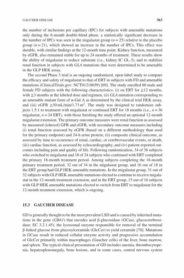
�
� �
�
GAUCHER DISEASE 363
the number of inclusions per capillary (IPC) for subjects with amenable mutationsonly during the 6-month double-blind phase, a statistically significant decrease inthe number of IPCs was seen in the migalastat group (n= 25) relative to the placebogroup (n= 21), which showed an increase in the number of IPCs. This effect wasdurable, with similar findings at the 12-month time point. Kidney function, measuredby eGFR, also remained stable for up to 24 months of treatment. These results showthe ability of migalastat to reduce substrate (i.e., kidney IC GL-3), and to stabilizerenal function in subjects with GLA mutations that were determined to be amenablein the GLP HEK assay.
The second Phase 3 trial is an ongoing randomized, open-label study to comparethe efficacy and safety of migalastat to that of ERT in subjects with FD and amenablemutations (ClinicalTrials.gov: NCT01218659) [69]. The study enrolled 60 male andfemale FD subjects with the following characteristics: (i) on ERT for ≥12 months,with ≥3 months at the labeled dose and regimen, (ii) GLA mutation corresponding toan amenable mutant form of α-Gal A as determined by the clinical trial HEK assay,and (iii) eGFR ≥30 mL/min/1.73 m2. The study was designed to randomize sub-jects 1.5:1 to treatment with migalastat or continued ERT for 18 months (i.e., n= 36migalastat, n= 24 ERT), with those finishing the study offered an optional 12-monthmigalastat extension. The primary outcome measures were renal function as assessedby measured (iohexol) GFR and eGFR, with secondary outcome measures including(i) renal function assessed by eGFR (based on a different methodology than usedfor the primary endpoint) and 24-h urine protein, (ii) composite clinical outcome, asassessed by time to occurrence of renal, cardiac, or cerebrovascular events, or death,(iii) cardiac function, as assessed by echocardiography, and (iv) patient-reported out-comes including pain and quality of life. Following randomization, 34 of 36 subjectswho switched to migalastat and 18 of 24 subjects who continued with ERT completedthe primary 18-month treatment period. Among subjects completing the 18-monthprimary treatment period, 32 out of 34 in the migalastat group, and 16 out of 18 inthe ERT group had GLP HEK amenable mutations. In the migalastat group, 31 out of32 subjects with GLP HEK amenable mutations elected to continue to receive migala-stat in the 12-month treatment extension, and in the ERT group, 15 out of 16 subjectswith GLP HEK amenable mutations elected to switch from ERT to migalastat for the12-month treatment extension, which is ongoing.
15.3 GAUCHER DISEASE
GD is generally thought to be the most prevalent LSD and is caused by inherited muta-tions in the gene (GBA1) that encodes acid β-glucosidase (GCase, glucocerebrosi-dase; EC 3.2.1.45), the lysosomal enzyme responsible for removal of the terminalβ-linked glucose from glucosylceramide (GlcCer) to yield ceramide [70]. Mutationsin GCase result in reduced cellular enzyme activity and progressive accumulationof GlcCer primarily within macrophages (Gaucher cells) of the liver, bone marrow,and spleen. The typical clinical presentation of GD includes anemia, thrombocytope-nia, hepatosplenomegaly, bone lesions, and in some cases, central nervous system

�
� �
�
364 PHARMACOLOGICAL CHAPERONES AS POTENTIAL THERAPEUTICS
(CNS) impairment [70]. GD without CNS involvement is classified as type I, whileneuronopathic GD is classified as type II (infantile acute) or type III (subacute). Over200 mutations in GBA1 have been identified, though the two most prevalent mis-sense mutant forms are N370S and L444P GCase [71]. The N370S mutant form hasapproximately 30% of the cellular enzymatic activity relative to cells that expresswild-type GCase [72]. Patients homozygous or heterozygous for N370S GCase typ-ically present with nonneuronopathic GD. In contrast, the L444P mutant form hassignificantly less residual activity (10–12% of wild type); consequently, individualshomozygous for the L444P mutation often present with CNS impairment that is asso-ciated with type III disease. More than 70% of Gaucher patients within the AshkenaziJewish population carry at least one N370S allele, while 38% of non-Jewish Gaucherpatients carry the L444P allele [71,73,74].
Currently, ERT and SRT are the only two approved treatment options for patientswith GD [75–79]. GD was the first LSD for which an ERT was available, withthe placental-derived Ceredase® (alglucerase) reaching clinical practice in 1988.Ceredase was replaced with a recombinant form, Cerezyme® (imiglucerase),in 2001; both ERTs are marketed by Genzyme. Several other ERTs have morerecently been approved for GD in various geographies, including velaglucerase alfa(VPRIV®, Shire), and taliglucerase alfa (ELELYSO™, Pfizer, New York, NY andProtalix, Carmiel, Israel). Treatment of GD with ERT is most effective against typeI disease, as well as the visceral manifestations of type II and III diseases, withsignificant reductions in spleen and liver weights, as well as increases in plateletcounts and hemoglobin levels, typically observed [80–83]. The CNS manifestationsof type II and III GD do not respond well to ERT due to the inability of the exogenousenzyme to cross the BBB [84].
As discussed, SRT drugs that are used to treat GD act as inhibitors of gluco-syltransferase, the enzyme responsible for the synthesis of GlcCer, thereby leadingto reduced cellular levels of this substrate. Zavesca® (N-butyl-1-deoxynojirimycin(NB-DNJ), miglustat) is currently the only approved SRT for mild-to-moderate typeI GD patients [78,79,85]. Unfortunately, many patients treated with Zavesca expe-rience side effects that include diarrhea, weight loss, tremor, and peripheral neu-ropathy [4]. Recently, another small molecule, orally available SRT drug, eliglustattartrate (Genzyme), has shown promise in Phase 2 and 3 studies for treatment of typeI GD [6]. As the therapeutic agents of SRT are small molecules, these drugs havethe potential for CNS access and treatment of the neurological aspects of GD. Tothis end, Zavesca has been evaluated as a treatment for neuronopathic GD, thoughno significant neurological benefit was seen in type III patients [86]. Unfortunately,eliglustat tartrate does access the CNS, but is also a PgP substrate, which transportseliglustat out of the brain, preventing significant drug levels from being achieved inthe CNS [87]. It is the hope that future drugs in this class may provide benefit topatients with neuronopathic forms of GD, though the long-term effects of inhibitingthis natural biosynthetic pathway are not known.
PCs also have been proposed as a potential therapy for GD [88–90]. Since noneof the currently approved therapies address the CNS manifestations of GD, there isalso a hope that small-molecule PCs may provide treatment of the neuronopathicforms of the disease. A wide variety of compounds that increase the cellular
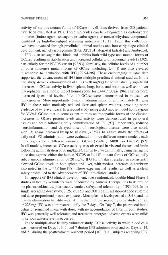
�
� �
�
GAUCHER DISEASE 365
activity of various mutant forms of GCase in cell lines derived from GD patientshave been evaluated as PCs. These molecules can be categorized as carbohydratemimetics (iminosugars, azasugars, or carbasugars), or noncarbohydrate compoundsidentified by high-throughput screening initiatives [10,11]. From this collection,two have advanced through preclinical animal studies and into early-stage clinicaldevelopment, namely isofagomine (IFG; AT2101; afegostat tartrate) and Ambroxol.
IFG is an azasugar that binds and inhibits both wild-type and mutant forms ofGCase, resulting in stabilization and increased cellular and lysosomal levels [91,92],particularly for the N370S variant [92,93]. Similarly, the cellular levels of a numberof other missense mutant forms of GCase, including L444P, are also elevatedin response to incubation with IFG [92,94–96]. These encouraging in vitro datasupported the advancement of IFG into multiple preclinical animal studies. In thefirst study, 4-week administration of IFG (3–30 mg/kg) led to statistically significantincreases in GCase activity in liver, spleen, lung, bone, and brain, as well as in livermacrophages, in a mouse model homozygous for L444P GCase [96]. Furthermore,increased lysosomal levels of L444P GCase were demonstrated in mouse liverhomogenates. More importantly, 6-month administration of approximately 8 mg/kgIFG to these mice modestly reduced liver and spleen weights, providing someevidence of in vivo efficacy. In a second study using a GD mouse model homozygousfor V394L GCase that to some extent mimics neuronopathic forms of the disease,increases in GCase protein levels and activity were demonstrated in peripheraltissues and brain following daily administration of IFG [97]. Importantly, reducedneuroinflammation and delayed onset of neurological disease were also noted,with life spans increased by up to 18 days (∼35%). In a third study, the effects ofdaily oral IFG administration were evaluated in three different mouse models, eachhomozygous for a different variant of GCase (V394L, D409H, or D409V) [92].In all models, increased GCase activity was observed in visceral tissues and brainfollowing administration of 30 mg/kg IFG for up to 8 weeks. Finally, using transgenicmice that express either the human N370S or L444P mutant forms of GCase, dailysubcutaneous administration of 20 mg/kg IFG for 14 days resulted in consistentlyelevated GCase levels in both spleen and liver, with modest increases in cerebrumalso noted in the L444P line [98]. These experimental results, as well as a cleansafety profile, led to the advancement of IFG into clinical studies.
In support of IFG clinical development, two randomized, double-blind Phase 1studies in healthy volunteers were conducted by Amicus Therapeutics to determinethe pharmacokinetics, pharmacodynamics, safety, and tolerability of IFG [99]. In thesingle ascending dose study, 8, 25, 75, 150, and 300 mg IFG all showed good systemicand dose-proportional plasma exposures. Mean plasma levels peaked at 3.4 h, and theplasma elimination half-life was 14 h. In the multiple ascending dose study, 25, 75,or 225 mg IFG was administered daily for 7 days. On Day 7, the pharmacokineticbehavior remained linear with dose, with no accumulation of IFG. In both studies,IFG was generally well tolerated and treatment-emergent adverse events were mild;no serious adverse events occurred.
In the multiple-dose healthy volunteer study, GCase activity in white blood cellswas measured on Days 1, 3, 5, and 7 during IFG administration and on Days 9, 14,and 21 during the posttreatment washout period [10]. In all subjects receiving IFG,

�
� �
�
366 PHARMACOLOGICAL CHAPERONES AS POTENTIAL THERAPEUTICS
there was a marked increase in GCase levels during the treatment period, followed bya decrease upon withdrawal of the drug, and a return to near baseline levels by Day21. The increase in enzyme level was dose related, reaching approximately 3.5-foldabove baseline levels at the highest dose level. These results for the safety, pharma-cokinetics, and preliminary pharmacodynamic effects in healthy volunteers supportedthe further evaluation of IFG in patients for the treatment of GD.
As such, two Phase 2 clinical studies with IFG were initiated by Amicus Thera-peutics. The first study evaluated the safety and pharmacodynamic effects of IFG intype I GD subjects who discontinued ERT for the 4-week duration of the study (Clin-icalTrials.gov: NCT00433147). Twenty-two women and eight men between the agesof 18 and 63, representing 12 different mutations (including N370S and L444P) and a9-year average duration of ERT treatment, were enrolled. IFG (25 and 150 mg daily,150 mg 3 days on/4 days off, or 7 days on/7 days off) was generally well toleratedat all dose levels evaluated, with no serious adverse events reported. GCase activ-ity as measured in white blood cells was increased in 20 of the 26 subjects. Five ofthe six subjects without a clear increase were in the lowest dose cohort or the cohortthat was administered IFG least frequently. As expected for a short-term study, thelevels of relevant markers of GD, including platelet counts, hemoglobin levels, Glc-Cer levels, chitotriosidase activity, and pulmonary activation-related chemokine wereunchanged. The second Phase 2 study was a 6-month study designed to evaluate thesafety of IFG and its effect on parameters that are commonly abnormal in GD (Clin-icalTrials.gov: NCT00446550). This study was also conducted in type I GD subjectswho had never received ERT. Two dose regimens of IFG (225 mg 3 days on/4 days offor 7 days on/7 days off) were studied. While all subjects experienced an increase inthe levels of GCase as measured in white blood cells, clinically meaningful improve-ments in key measures of disease were observed in just 1 of the 18 subjects whocompleted the study. Importantly, the white blood cell GCase levels attained in thisone subject following IFG treatment were close to those of healthy volunteers, whichwas substantially greater than the magnitude of increase in all other subjects. Whilethe results suggested that treatment with IFG was generally well tolerated, with noserious adverse events reported, Phase 3 studies were not pursued.
Ambroxol, an approved expectorant, was identified from a screen of 1040 FDA-approved drugs by its ability to stabilize wild-type GCase against thermal denatura-tion [95]. Ambroxol also showed pH-dependent affinity for GCase, with decreas-ing inhibition at lysosomal pH values. In cell-based assays using patient-derivedcell lines homozygous for N370S, Ambroxol increased cellular GCase activity upto twofold at a concentration of 60 μM, with evidence of increased lysosomal lev-els; similar cellular effects were seen in a heterozygous cell line expressing F213Iand L444P GCase. Evidence was also provided for modest reductions in GlcCer lev-els in patient-derived cells. In other in vitro studies, Ambroxol (at concentrationsup to 100 μM) elevated the cellular and lysosomal levels of multiple GCase mutantsin patient-derived cells expressing R120W, R131C, N188S, G193W, F213I, N370S,L444P, and/or P415R [100,101]. In wild-type mice, Ambroxol was generally welltolerated, with detrimental effects on water intake and body weight occurring inthe highest dose groups only [100]. Finally, N370S or L444P transgenic mice were

�
� �
�
GM2 GANGLIOSIDOSES (TAY–SACHS/SANDHOFF DISEASES) 367
subcutaneously administered 100 mg/kg Ambroxol daily for 14 days, resulting inconsistently elevated GCase levels in both spleen and liver [98]. These results ledto the advancement of Ambroxol into a physician-sponsored study in GD patients.
This pilot study assessed the tolerability and efficacy of 150 mg Ambroxol dailyin 12 subjects with symptomatic type I GD who were not on ERT, 11 of whomwere homozygous for the N370S allele [102]. Of the nine subjects that completedthe 6-month study, none showed deterioration in the measured GD-related param-eters (e.g., body weight, hemoglobin levels, platelet counts, liver and spleen vol-umes, and chitotriosidase activity), and three continued on therapy for an additional6 months. Over the 12-month course, these three subjects showed reductions in spleenvolumes (mean reduction approximately 15%) and chitotriosidase activity (meanreduction approximately 40%), with stabilized hemoglobin levels and liver volumes.Importantly, in one subject platelet counts also increased over 50%. Given thesepromising results, follow-up placebo-controlled studies that include larger numbersof GD subjects with more diverse genotypes, and potentially with CNS manifesta-tions, are warranted.
15.4 GM2 GANGLIOSIDOSES (TAY–SACHS/SANDHOFF DISEASES)
The terminal sugar N-acetylgalactosamine, which is present on several GSLs as wellas various other oligosaccharides and glycoproteins, can be enzymatically removed inthe lysosome by β-hexosaminidase (β-Hex; EC 3.2.1.52). β-Hex is composed of twosubunits and can exist as a heterodimer comprised of α- and β-subunits (β-Hex A), oras one of two homodimers, one containing two α-subunits (β-Hex S, which is excep-tionally rare and whose function is not clearly understood) and the other containingtwo β-subunits (β-Hex B). While most N-acetylgalactosamine-containing substratescan be processed by either the β-Hex A or β-Hex B isoform, the ganglioside GM2and several oligosaccharides can only be processed by β-Hex A. Mutations in eithergene result in GM2 gangliosidosis [103]. More specifically, mutations in the genethat encodes the β-subunit primarily lead to accumulation of GM2 and result in anLSD known as Tay–Sachs disease (TSD). In contrast, α-subunit mutations result indecreased activity of both β-Hex A and β-Hex B, and the accumulation of multipleGSLs. The resultant LSD is an equally or sometimes more severe form of GM2 gan-gliosidosis known as Sandhoff disease (SD). Progressive neurodegeneration is theprimary pathology associated with both forms of GM2 gangliosidosis. As with manyLSDs, the infantile or early-onset form of the disease has the most severe pathol-ogy with rapid neurodegeneration typically occurring at approximately 6 months ofage, with an overall lifespan of 4 years or less. The late-onset form typically tends topresent clinically in the second decade of life, again with progressive neurodegener-ation over the next two to three decades. An intermediate juvenile form has also beencharacterized, with life expectancies typically not beyond two decades. Currently,there are no effective treatment options for GM2 gangliosidosis patients.
Pyrimethamine (PYR), an approved antimalarial drug, was identified as a potentinhibitor of β-Hex from a screen of 1040 FDA-approved drugs [104]. In follow-up

�
� �
�
368 PHARMACOLOGICAL CHAPERONES AS POTENTIAL THERAPEUTICS
cell-based assays using 10 TSD and 7 SD patient-derived cell lines, PYR led tosignificant increases in cellular and lysosomal β-HEX levels and activity in one cellline with a β-subunit mutation (TSD) as well as all seven cell lines with α-subunitmutations (SD). Consequently, escalating doses of PYR (maximum: 100 mg/day)administered orally in a single daily dose over a 16-week period were evaluated ina Phase 1/2 open-label clinical study to determine tolerability and efficacy in thetreatment of late-onset GM2 gangliosidosis subjects [105]. A total of 11 subjectswere enrolled, 8 males and 3 females, aged 23–50 years. For the eight subjects thatcompleted the study, up to fourfold increases in β-Hex activity were observed inwhite blood cell lysates at doses of 50 mg per day or less, despite marked individualvariations in PYR’s pharmacokinetic profile. Unfortunately, significant side effects(increased ataxia, lack of coordination, and seizures) were observed in most subjectsat doses of 75 mg per day or higher and the trial was discontinued. These datasuggest that PYR can enhance β-Hex activity in peripheral cells of subjects withlate-onset GM2 gangliosidosis at doses lower than those that are associated withadverse effects. Future studies will be necessary to determine the clinical efficacy ofPYR in TSD and SD patients.
15.5 POMPE DISEASE
Pompe disease (acid maltase deficiency, glycogen storage disease type II) is anLSD caused by mutations in the gene (GAA) that encodes the lysosomal enzymeacid α-glucosidase (GAA; EC 3.2.1.20) [106,107], the enzyme that hydrolyzes theα1,4- and α1,6-glycosidic bonds of glycogen [108]. Deficiency in GAA activityresults in glycogen accumulation and deposition in the lysosomes of heart, skeletalmuscle, and other tissues [109]. GAA is synthesized as a 110 kDa immatureglycoprotein precursor in the ER and undergoes a series of proteolytic and N-glycanprocessing events to yield the 95 kDa intermediate and the 76 and 70 kDa matureisoforms [110–113]. This processing into the intermediate and mature forms occursin late endosomes and lysosomes, respectively [110], with the final mature isoformsshowing significantly increased affinity and activity toward glycogen compared tothe precursor forms [111,114]. Increasing the amount of mature, active GAA inlysosomes is an important step toward increasing glycogen hydrolysis and reducingsubstrate accumulation in this disease.
Pompe disease shows a broad phenotypic spectrum that ranges from the severeinfantile-onset form to more slowly progressing, later-onset forms. Infantile-onset Pompe disease patients have little or no GAA activity, present with hypotonia,cardiomegaly, and cardiorespiratory distress, and have a life expectancy of about2 years [115]. Late-onset forms of the disease typically show some detectableGAA activity, present in childhood or adulthood, and progress more slowly, withmusculoskeletal and pulmonary involvement leading to progressive weakness andrespiratory insufficiency [116]. Compared to an age- and gender-matched healthypopulation, untreated late-onset Pompe patients have higher mortality rates [117],while those treated with ERT show substantially reduced mortality [118].

�
� �
�
POMPE DISEASE 369
Currently, ERT is the only approved treatment for Pompe disease, administeredas a biweekly intravenous infusion of recombinant human GAA (rhGAA; alglucosi-dase alfa, marketed as Myozyme® and Lumizyme®, Genzyme-Sanofi). Treatmentwith rhGAA improves cardiac function, motor skills, and life span in infantile-onsetpatients [119–124] and leads to some improvements in motor and respiratory functionin late-onset patients [125,126]. Despite the clinical benefits of ERT, the efficacy ofrhGAA may be limited by insufficient targeting and uptake into key disease-relevanttissues, as well as poor tolerability due to sometimes severe immunogenic and ana-phylactic reactions to the exogenous enzyme [124,126–132]. Thus, a clear unmetmedical need still exists for many Pompe patients.
Small-molecule PCs have been proposed as a potential alternative to ERT forthe treatment of Pompe disease [133–136]. Approximately 150 GAA mutations arepredicted to produce a full-length protein containing a single amino acid substitution.However, these GAA mutants likely do not fold as efficiently or are less stablecompared to wild-type GAA and, therefore, are susceptible to premature degradationvia ERAD. It has been demonstrated that some mutant forms of GAA are responsiveto the PCs 1-deoxynojirimycin hydrochloride (DNJ, duvoglustat hydrochloride,AT2220) and NB-DNJ, as evidenced by increased trafficking, maturation, andactivity [133,135]. To this end, Flanagan et al. [136] characterized the effects ofDNJ on 76 different mutant forms of GAA. A homology model of GAA wasalso constructed using the crystal structure of a related eukaryotic α-glucosidase,maltase-glucoamylase, to gain conformational insights into GAA and to map thelocations of the residues of the mutant forms of GAA responsive to DNJ. Giventhe large number of pathogenic GAA mutants identified to date and the need todetermine which forms are amenable to PC therapy, this study provided additionalevidence and support for the continued evaluation of DNJ as a potential therapeuticfor Pompe disease. Mechanistically, DNJ has multiple modes of action during thesynthesis and maturation of mutant GAA, including increased specific activityprior to proteolytic processing in lysosomes, facilitated export from the ER withsubsequent trafficking and processing through the secretory pathway to lysosomes,and stabilization of the mature isoforms in lysosomes [12].
Furthermore, the in vivo effects of DNJ were tested in a new mouse model ofPompe disease [12]. This mouse model expresses the human mutant GAA trans-gene P545L on a Gaa knockout background (hP545L GAA Tg/KO) and shows lowGAA activity and elevated glycogen levels in disease-relevant tissues including heart,diaphragm, multiple skeletal muscles, and brain. Daily oral administration of DNJto hP545L GAA Tg/KO mice resulted in significant and dose-dependent increasesin GAA activity with concomitant reduction in tissue glycogen levels; less-frequentDNJ administration regimens (such as 4 days on drug followed by 3 days off, or 5days on drug followed by 2 days off) resulted in even greater glycogen reductioncompared to daily administration. Collectively, these results provided support for theevaluation of DNJ as a potential treatment for Pompe disease.
To date, five Phase 1 clinical studies have been completed in healthy adult vol-unteers by Amicus Therapeutics, with single ascending DNJ doses ranging from 50to 2500 mg, and repeat-administration (up to 14 days) ascending doses ranging from
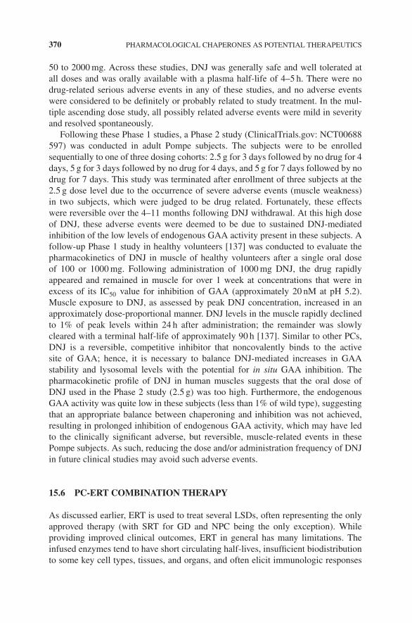
�
� �
�
370 PHARMACOLOGICAL CHAPERONES AS POTENTIAL THERAPEUTICS
50 to 2000 mg. Across these studies, DNJ was generally safe and well tolerated atall doses and was orally available with a plasma half-life of 4–5 h. There were nodrug-related serious adverse events in any of these studies, and no adverse eventswere considered to be definitely or probably related to study treatment. In the mul-tiple ascending dose study, all possibly related adverse events were mild in severityand resolved spontaneously.
Following these Phase 1 studies, a Phase 2 study (ClinicalTrials.gov: NCT00688597) was conducted in adult Pompe subjects. The subjects were to be enrolledsequentially to one of three dosing cohorts: 2.5 g for 3 days followed by no drug for 4days, 5 g for 3 days followed by no drug for 4 days, and 5 g for 7 days followed by nodrug for 7 days. This study was terminated after enrollment of three subjects at the2.5 g dose level due to the occurrence of severe adverse events (muscle weakness)in two subjects, which were judged to be drug related. Fortunately, these effectswere reversible over the 4–11 months following DNJ withdrawal. At this high doseof DNJ, these adverse events were deemed to be due to sustained DNJ-mediatedinhibition of the low levels of endogenous GAA activity present in these subjects. Afollow-up Phase 1 study in healthy volunteers [137] was conducted to evaluate thepharmacokinetics of DNJ in muscle of healthy volunteers after a single oral doseof 100 or 1000 mg. Following administration of 1000 mg DNJ, the drug rapidlyappeared and remained in muscle for over 1 week at concentrations that were inexcess of its IC50 value for inhibition of GAA (approximately 20 nM at pH 5.2).Muscle exposure to DNJ, as assessed by peak DNJ concentration, increased in anapproximately dose-proportional manner. DNJ levels in the muscle rapidly declinedto 1% of peak levels within 24 h after administration; the remainder was slowlycleared with a terminal half-life of approximately 90 h [137]. Similar to other PCs,DNJ is a reversible, competitive inhibitor that noncovalently binds to the activesite of GAA; hence, it is necessary to balance DNJ-mediated increases in GAAstability and lysosomal levels with the potential for in situ GAA inhibition. Thepharmacokinetic profile of DNJ in human muscles suggests that the oral dose ofDNJ used in the Phase 2 study (2.5 g) was too high. Furthermore, the endogenousGAA activity was quite low in these subjects (less than 1% of wild type), suggestingthat an appropriate balance between chaperoning and inhibition was not achieved,resulting in prolonged inhibition of endogenous GAA activity, which may have ledto the clinically significant adverse, but reversible, muscle-related events in thesePompe subjects. As such, reducing the dose and/or administration frequency of DNJin future clinical studies may avoid such adverse events.
15.6 PC-ERT COMBINATION THERAPY
As discussed earlier, ERT is used to treat several LSDs, often representing the onlyapproved therapy (with SRT for GD and NPC being the only exception). Whileproviding improved clinical outcomes, ERT in general has many limitations. Theinfused enzymes tend to have short circulating half-lives, insufficient biodistributionto some key cell types, tissues, and organs, and often elicit immunologic responses

�
� �
�
PC-ERT COMBINATION THERAPY 371
that adversely affect tolerability and efficacy. For instance, a recent study with Fabrypatients revealed that ERT provided only minimal improvement on cardiovascularparameters, again underscoring the significant unmet medical need in LSDs [138].
PCs offer another approach to the treatment of these diseases, with the poten-tial benefits of being noninvasive, well tolerated, and broadly biodistributed, includ-ing the CNS. At the same time, PC therapy also has some limitations, especiallyas PC monotherapy is only applicable for patients that express responsive mutantforms of the enzymes. However, it was recently hypothesized and demonstrated thatPCs can improve the biochemical and pharmacological properties of the exogenousenzymes used in ERTs through binding and stabilization, potentially opening up anew approach for LSD treatment: combination therapy using PCs and ERTs togetherfor improved therapeutic activity.
One of the first examples of PC-ERT combination therapy was described in thecontext of GD [139]. Incubation of recombinant human GCase (Cerezyme) with IFGsignificantly increased the stability of the recombinant enzyme and increased cellu-lar uptake into GD patient-derived cells to levels greater than those obtained withenzyme alone. Parenti and coworkers [140] extended the findings on combinationtherapy to the treatment of Pompe disease using the Gaa knockout mouse model.Animals receiving an oral dose of NB-DNJ (4.3 mg/kg) for 2 days prior to an intra-venous (IV) infusion of rhGAA showed a significant increase in enzyme activity inmultiple disease-relevant organs as compared to those receiving ERT only, includ-ing muscles that usually show little-to-no response to ERT alone. Furthermore, DNJprevented rhGAA denaturation and loss of activity at neutral pH and 37 ∘C in vitro.In rats, oral coadministration of DNJ (3 or 30 mg/kg), 60 min prior to intravenousinfusion of rhGAA, increased the circulating half-life and overall exposure of theenzyme by up to twofold. In Gaa knockout mice, four biweekly IV administrationsof rhGAA with oral coadministration of DNJ (30 mg/kg) prior to ERT resulted ina significant increase of up to 2.5-fold in GAA activity in disease-relevant tissues,including skeletal muscles, compared to ERT alone. More importantly, PC coad-ministration led to robust glycogen reduction in these same tissues, up to 2.6-foldgreater compared to ERT alone [141]. Similarly, increased enzyme stability and cel-lular uptake and improved substrate reduction in an FD mouse model were observedwith DGJ in combination with recombinant human α-Gal A [142].
These promising preclinical results led to the initiation of several Phase 2 PC/ERTcoadministration clinical studies. In a Pompe Phase 2 study conducted by AmicusTherapeutics (ClinicalTrials.gov: NCT01380743), subjects who had been on ERTas their standard of care were orally coadministered a single dose of DNJ at 50,100, 250, or 600 mg 1 h prior to ERT infusion; GAA activities in plasma and musclewere evaluated in comparison to administration of ERT alone. Similar to the findingsof the preclinical studies, the PC coadministration led to increased GAA activity inplasma as well as in muscle. Dose-dependent increases in plasma GAA exposure wereobserved for all subjects, attaining 1.5- to 2.8-fold greater exposures compared to ERTalone. In muscle biopsy samples taken on Day 3 or 7 after administration, increasesin total GAA activity were observed in 16 of 24 (67%) subjects with evaluable data.While all DNJ doses evaluated showed some increase in plasma GAA activity, the

�
� �
�
372 PHARMACOLOGICAL CHAPERONES AS POTENTIAL THERAPEUTICS
lowest dose (50 mg) was the least robust, while the highest dose (600 mg) showedthe greatest effect on both plasma and muscle GAA levels [143]. In addition, anotherclinical study based on the combination of NB-DNJ and rhGAA was initiated in Italy(Telethon foundation, trial GUP09017), and has similarly shown greater circulatingGAA levels in plasma with PC coadministration compared to ERT alone [144].
Finally, a Fabry Phase 2 study (ClinicalTrials.gov: NCT01196871) initiated byAmicus Therapeutics explored a single oral coadministered dose of 150 or 450 mgDGJ 2 h prior to the infusion of ERT. Again, increased plasma exposures of α-GalA activity of 1.2- to 5.0-fold compared to ERT alone were seen in 22 of 23 subjects.Both tested doses of DGJ led to greater total α-Gal A activity in Day 2 skin biopsiesin 19 of 23 (82.6%) subjects relative to ERT alone, with modest improvements stillnoted in some Day 7 skin biopsies. Of the two DGJ doses tested, 450 mg resulted ingreater increases of active α-Gal A enzyme levels in skin tissue [145].
These clinical studies extend preclinical observations and support the notion thatoral administration of a PC shortly before ERT infusion can stabilize the recombinantenzyme in the circulation, protecting the infused proteins from denaturing and leadingto greater exposure and improved cellular uptake. These proof-of-concept studies laythe foundation to further explore whether PC/ERT combination therapy can alter ERTbiodistribution and therapeutic efficacy in target tissues that respond poorly to ERTalone. Furthermore, the stabilization by the PC also may reduce the immunogenicityof the infused enzyme and thereby ameliorate the immune response elicited by manyERTs. This could improve tolerability and safety, and further enhance therapeuticoutcomes in some patients. The synergism of an ERT combined with a PC offers anew therapeutic option with promises for patients suffering with LSDs, especiallythose who are unable to use PC monotherapy, or who do not get sufficient benefitfrom ERT treatment alone.
REFERENCES
[1] Parkinson-Lawrence EJ, Shandala T, Prodoehl M, Plew R, Borlace GN, Brooks DA.Lysosomal storage disease: revealing lysosomal function and physiology. Physiology2010;25:102–115.
[2] Ron I, Horowitz M. ER retention and degradation as the molecular basis underlyingGaucher disease heterogeneity. Hum Mol Genet 2005;14:2387–2398.
[3] Wang D, El-Amouri SS, Dai M, Kuan C-Y, Hui DY, Brady RO, Pan D. Engineering alysosomal enzyme with a derivative of receptor-binding domain of apoE enables deliv-ery across the blood–brain barrier. Proc Natl Acad Sci U S A 2013;110:2999–3004.
[4] Zimran A, Elstein D. Gaucher disease and the clinical experience with substrate reduc-tion therapy. Philos Trans Roy Soc Lond Ser B Biol Sci 2003;358:961–966.
[5] Wraith JE, Vecchio D, Jacklin E, Abel L, Chadha-Boreham H, Luzy C, Giorgino R,Patterson MC. Miglustat in adult and juvenile patients with Niemann–Pick disease TypeC: long-term data from a clinical trial. Mol Genet Metab 2010;99:351–357.
[6] Cox TM. Eliglustat tartrate, an orally active glucocerebroside synthase inhibitor for thepotential treatment of Gaucher disease and other lysosomal storage diseases. Curr OpinInvestig Drugs 2010;11:1169–1181.

�
� �
�
REFERENCES 373
[7] Brady RO, Kanfer JN, Bradley RM, Shapiro D. Demonstration of a deficiencyof glucocerebroside-cleaving enzyme in Gaucher’s disease. J Clin Invest 1966;45:1112–1115.
[8] Germain DP, Fan JQ. Pharmacological chaperone therapy by active-site-specific chap-erones in Fabry disease: in vitro and preclinical studies. Int J Clin Pharmacol Ther2009;47:S111–S117.
[9] Bonifacino JS, Weissman AM. Ubiquitin and the control of protein fate in the secretoryand endocytic pathways. Annu Rev Cell Dev Biol 1998;14:19–57.
[10] Valenzano KJ, Khanna R, Powe AC, Boyd R, Lee G, Flanagan JJ, Benjamin ER. Iden-tification and characterization of pharmacological chaperones to correct enzyme defi-ciencies in lysosomal storage disorders. Assay Drug Develop Technol 2011;9:213–235.
[11] Boyd RE, Lee G, Rybczynski P, Benjamin ER, Khanna R, Wustman BA, ValenzanoKJ. Pharmacological chaperones as therapeutics for lysosomal storage diseases. J MedChem 2013;56:2705–2725.
[12] Khanna R, Powe AC, Lun Y, Soska R, Feng J, Dhulipala R, Frascella M, Garcia A,Pellegrino LJ, Xu S, Brignol N, Toth MJ, Do HV, Lockhart DJ, Wustman BA, Valen-zano KJ. The pharmacological chaperone AT2220 increases the specific activity andlysosomal delivery of mutant acid α-glucosidase, and promotes glycogen reduction in atransgenic mouse model of Pompe disease. PLoS ONE 2014;9:e102092.
[13] Khanna R, Soska R, Lun Y, Feng J, Frascella M, Young B, Brignol N, Pellegrino L,Sitaraman SA, Desnick RJ, Benjamin ER, Lockhart DJ, Valenzano KJ. The pharma-cological chaperone 1-deoxygalactonojirimycin reduces tissue globotriaosylceramidelevels in a mouse model of Fabry disease. Mol Ther 2010;18:23–33.
[14] Patnaik S, Zheng W, Choi JH, Motabar O, Southall N, Westbroek W, Lea WA, VelayatA, Goldin E, Sidransky E, Leister W, Marugan JJ. Discovery, SAR, and biological eval-uation of noninhibitory small molecule chaperones of glucocerebrosidase. J Med Chem2012;55:5734–5748.
[15] Perlmutter DH. Chemical chaperones: a pharmacological strategy for disorders of pro-tein folding and trafficking. Pediatr Res 2002;52:832–836.
[16] Desnick R, Ioannou Y, Eng C. a-Galactosidase A deficiency: Fabry disease. In: SriverCR, Beaudet AL, Sly WS, Valle D, editors. The Metabolic and Molecular Bases ofInherited Disease. New York: McGraw-Hill; 2001. p 3733–3774.
[17] Sweeley CC, Klionsky B. Fabry’s disease: classification as a sphingolipidosis and partialcharacterization of a novel glycolipid. J Biol Chem 1963;238:PC3148–PC3150.
[18] Brady OR, Gal AE, Bradley RM, Martensson E, Warshaw AL, Laster L. Enzymaticdefect in Fabry’s disease: ceramidetrihexosidase deficiency. N Engl J Med 1967;276:1163–1167.
[19] Askari H, Kaneski CR, Semino-Mora C, Desai P, Ang A, Kleiner DE, Perlee LT,Quezado M, Spollen LE, Wustman BA, Schiffmann R. Cellular and tissue localizationof globotriaosylceramide in Fabry disease. Virchows Arch 2007;451:823–834.
[20] Aerts JM, Groener JE, Kuiper S, Donker-Koopman WE, Strijland A, Ottenhoff R,van Roomen C, Mirzaian M, Wijburg FA, Linthorst GE, Vedder AC, Rombach SM,Cox-Brinkman J, Somerharju P, Boot RG, Hollak CE, Brady RO, Poorthuis BJ. Ele-vated globotriaosylsphingosine is a hallmark of Fabry disease. Proc Natl Acad Sci U SA 2008;105:2812–2817.
[21] Rombach SM, Dekker N, Bouwman MG, Linthorst GE, Zwinderman AH, Wijburg FA,Kuiper S, vd Bergh Weerman MA, Groener JEM, Poorthuis BJ, Hollak CEM, Aerts

�
� �
�
374 PHARMACOLOGICAL CHAPERONES AS POTENTIAL THERAPEUTICS
JMFG. Plasma globotriaosylsphingosine: diagnostic value and relation to clinical man-ifestations of Fabry disease. Biochim Biophys Acta 2010;1802:741–748.
[22] Togawa T, Kodama T, Suzuki T, Sugawara K, Tsukimura T, Ohashi T, Ishige N, SuzukiK, Kitagawa T, Sakuraba H. Plasma globotriaosylsphingosine as a biomarker of Fabrydisease. Mol Genet Metab 2010;100:257–261.
[23] Germain D. Fabry disease. Orphanet J Rare Dis 2010;5:30.[24] Wilcox WR, Oliveira JP, Hopkin RJ, Ortiz A, Banikazemi M, Feldt-Rasmussen U,
Sims K, Waldek S, Pastores GM, Lee P, Eng CM, Marodi L, Stanford KE, BreunigF, Wanner C, Warnock DG, Lemay RM, Germain DP. Females with Fabry diseasefrequently have major organ involvement: lessons from the Fabry registry. Mol GenetMetab 2008;93:112–128.
[25] von Scheidt W, Eng CM, Fitzmaurice TF, Erdmann E, Hubner G, Olsen EG, Christom-anou H, Kandolf R, Bishop DF, Desnick RJ. An atypical variant of Fabry’s disease withmanifestations confined to the myocardium. N Engl J Med 1991;324:395–399.
[26] Bekri S, Enica A, Ghafari T, Plaza G, Champenois I, Choukroun G, Unwin R, Jaeger P.Fabry disease in patients with end-stage renal failure: the potential benefits of screening.Nephron Clin Pract 2005;101:c33–38.
[27] Chimenti C, Pieroni M, Morgante E, Antuzzi D, Russo A, Russo MA, Maseri A, FrustaciA. Prevalence of Fabry disease in female patients with late-onset hypertrophic car-diomyopathy. Circulation 2004;110:1047–1053.
[28] Nakao S, Takenaka T, Maeda M, Kodama C, Tanaka A, Tahara M, Yoshida A, KuriyamaM, Hayashibe H, Sakuraba H, Tanaka H. An atypical variant of Fabry’s disease in menwith left ventricular hypertrophy. N Engl J Med 1995;333:288–293.
[29] Nakao S, Kodama C, Takenaka T, Tanaka A, Yasumoto Y, Yoshida A, Kanzaki T,Enriquez AL, Eng CM, Tanaka H, Tei C, Desnick RJ. Fabry disease: detection of undi-agnosed hemodialysis patients and identification of a “renal variant” phenotype. KidneyInt 2003;64:801–807.
[30] Rolfs A, Bottcher T, Zschiesche M, Morris P, Winchester B, Bauer P, Walter U,Mix E, Lohr M, Harzer K, Strauss U, Pahnke J, Grossmann A, Benecke R. Preva-lence of Fabry disease in patients with cryptogenic stroke: a prospective study. Lancet2005;366:1794–1796.
[31] Sachdev B, Takenaka T, Teraguchi H, Tei C, Lee P, McKenna WJ, Elliott PM. Preva-lence of Anderson–Fabry disease in male patients with late onset hypertrophic car-diomyopathy. Circulation 2002;105:1407–1411.
[32] Spada M, Pagliardini S, Yasuda M, Tukel T, Thiagarajan G, Sakuraba H, Ponzone A,Desnick RJ. High incidence of later-onset Fabry disease revealed by newborn screening.Am J Hum Genet 2006;79:31–40.
[33] Eng CM, Guffon N, Wilcox WR, Germain DP, Lee P, Waldek S, Caplan L, Linthorst GE,Desnick RJ. Safety and efficacy of recombinant human α-galactosidase A replacementtherapy in Fabry’s disease. N Engl J Med 2001;345:9–16.
[34] Hughes DA, Elliott PM, Shah J, Zuckerman J, Coghlan G, Brookes J, Mehta AB. Effectsof enzyme replacement therapy on the cardiomyopathy of Anderson–Fabry disease:a randomised, double-blind, placebo-controlled clinical trial of agalsidase alfa. Heart2008;94:153–158.
[35] Schiffmann R, Floeter MK, Dambrosia JM, Gupta S, Moore DF, Sharabi Y, KhuranaRK, Brady RO. Enzyme replacement therapy improves peripheral nerve and sweat func-tion in Fabry disease. Muscle Nerve 2003;28:703–710.

�
� �
�
REFERENCES 375
[36] Schiffmann R, Kopp JB, Austin Iii HA, Sabnis S, Moore DF, Weibel T, Balow JE, BradyRO. Enzyme replacement therapy in Fabry disease: a randomized controlled trial. JAMA2001;285:2743–2749.
[37] Warnock DG, Ortiz A, Mauer M, Linthorst GE, Oliveira JP, Serra AL, Maródi L,Mignani R, Vujkovac B, Beitner-Johnson D, Lemay R, Cole JA, Svarstad E, WaldekS, Germain DP, Wanner C, Registry obotF. Renal outcomes of agalsidase beta treat-ment for Fabry disease: role of proteinuria and timing of treatment initiation. NephrolDial Transplant 2012;27:1042–1049.
[38] West M, Nicholls K, Mehta A, Clarke JTR, Steiner R, Beck M, Barshop BA, RheadW, Mensah R, Ries M, Schiffmann R. Agalsidase alfa and kidney dysfunction in Fabrydisease. J Am Soc Nephrol 2009;20:1132–1139.
[39] Togawa T, Kawashima I, Kodama T, Tsukimura T, Suzuki T, Fukushige T, KanekuraT, Sakuraba H. Tissue and plasma globotriaosylsphingosine could be a biomarker forassessing enzyme replacement therapy for Fabry disease. Biochem Biophys Res Com-mun 2010;399:716–720.
[40] van Breemen MJ, Rombach SM, Dekker N, Poorthuis BJ, Linthorst GE, ZwindermanAH, Breunig F, Wanner C, Aerts JM, Hollak CE. Reduction of elevated plasma globo-triaosylsphingosine in patients with classic Fabry disease following enzyme replace-ment therapy. Biochim Biophys Acta 2011;1812:70–76.
[41] Rombach SM, Aerts JMFG, Poorthuis BJHM, Groener JEM, Donker-Koopman W,Hendriks E, Mirzaian M, Kuiper S, Wijburg FA, Hollak CEM, Linthorst GE. Long-termeffect of antibodies against infused alpha-galactosidase A in Fabry disease on plasmaand urinary (Lyso)Gb3 reduction and treatment outcome. PLoS One 2012;7:e47805.
[42] Yam GH, Bosshard N, Zuber C, Steinmann B, Roth J. Pharmacological chaperone cor-rects lysosomal storage in Fabry disease caused by trafficking-incompetent variants. AmJ Physiol Cell Physiol 2006;290:C1076–1082.
[43] Okumiya T, Ishii S, Takenaka T, Kase R, Kamei S, Sakuraba H, Suzuki Y. Galactose sta-bilizes various missense mutants of α-galactosidase in Fabry disease. Biochem BiophysRes Commun 1995;214:1219–1224.
[44] Fan J-Q, Nakao S, Kaneski CR, Brady RO, Desnick RJ, Suzuki S, Ishii S. Intracellularenhancement of α-galactosidase A activity in 31 Fabry lymphoblasts and fibroblasts by1-deoxy-galactonojirimycin. Am J Hum Genet 1999;65:A308.
[45] Fan J-Q. A contradictory treatment for lysosomal storage disorders: inhibitors enhancemutant enzyme activity. Trends Pharmacol Sci 2003;24:355–360.
[46] Fan J-Q, Ishii S, Asano N, Suzuki Y. Accelerated transport and maturation of lysoso-mal α-galactosidase A in Fabry lymphoblasts by an enzyme inhibitor. Nat Med 1999;5:112–115.
[47] Frustaci A, Chimenti C, Ricci R, Natale L, Russo MA, Pieroni M, Eng CM, Desnick RJ.Improvement in cardiac function in the cardiac variant of Fabry’s disease with galactose-infusion therapy. N Engl J Med 2001;345:25–32.
[48] Guce AI, Clark NE, Rogich JJ, Garman SC. The molecular basis of pharmacologicalchaperoning in human α-galactosidase. Chem Biol 2011;18:1521–1526.
[49] Benjamin E, Flanagan J, Schilling A, Chang H, Agarwal L, Katz E, Wu X, Pine C,Wustman B, Desnick R, Lockhart D, Valenzano K. The pharmacological chaperone1-deoxygalactonojirimycin increases α-galactosidase A levels in Fabry patient cell lines.J Inherit Metab Dis 2009;32:424–440.
�
� �
�
376 PHARMACOLOGICAL CHAPERONES AS POTENTIAL THERAPEUTICS
[50] Ishii S, Chang H-H, Yoshioka H, Shimada T, Mannen K, Higuchi Y, Taguchi A, Fan J-Q.Preclinical efficacy and safety of 1-deoxygalactonojirimycin in mice for Fabry disease.J Pharmacol Exp Therap 2009;328:723–731.
[51] Yam GH, Zuber C, Roth J. A synthetic chaperone corrects the trafficking defect anddisease phenotype in a protein misfolding disorder. FASEB J 2005;19:12–18.
[52] Ishii S, Yoshioka H, Mannen K, Kulkarni AB, Fan JQ. Transgenic mouse expressinghuman mutant α-galactosidase A in an endogenous enzyme deficient background: abiochemical animal model for studying active-site specific chaperone therapy for Fabrydisease. Biochim Biophys Acta 2004;1690:250–257.
[53] Germain DP, Poenaru L. Fabry disease: identification of novel α-galactosidase A muta-tions and molecular carrier detection by use of fluorescent chemical cleavage of mis-matches. Biochem Biophys Res Commun 1999;257:708–713.
[54] Ashton-Prolla P, Tong B, Shabbeer J, Astrin KH, Eng CM, Desnick RJ. Fabry disease:twenty-two novel mutations in the α-galactosidase A gene and genotype/phenotype cor-relations in severely and mildly affected hemizygotes and heterozygotes. J Invest Med2000;48:227–235.
[55] Ishii S, Sakuraba H, Suzuki Y. Point mutations in the upstream region of theα-galactosidase A gene exon 6 in an atypical variant of Fabry disease. Hum Genet1992;89:29–32.
[56] Wu X, Katz E, Valle CD, Mascioli K, Flanagan J, Castelli JP, Schiffmann R, BoudesP, Lockhart DJ, Valenzano KJ, Benjamin ER. A pharmacogenetic approach to identifymutant forms of α-galactosidase A that respond to a pharmacological chaperone forFabry disease. Hum Mutat 2011;32:965–977.
[57] Johnson FK, Mudd PN, Bragat A, Adera M, Boudes P. Pharmacokinetics and safety ofmigalastat HCl and effects on agalsidase activity in healthy volunteers. Clin PharmacolDrug Develop 2013;2:120–132.
[58] Germain D, Giugliani R, Hughes D, Mehta A, Nicholls K, Barisoni L, Jennette C, Bra-gat A, Castelli J, Sitaraman S, Lockhart D, Boudes P. Safety and pharmacodynamiceffects of a pharmacological chaperone on α-galactosidase A activity and globotriaosyl-ceramide clearance in Fabry disease: report from two phase 2 clinical studies. OrphanetJ Rare Dis 2012;7:91.
[59] Bekri S, Lidove O, Jaussaud R, Knebelmann B, Barbey F. The role of ceramide trihexo-side (globotriaosylceramide) in the diagnosis and follow-up of the efficacy of treatmentof Fabry disease: a review of the literature. Cardiovasc Hematol Agents Med Chem2006;4:289–297.
[60] Aerts J, Kallemeijn W, Wegdam W, Joao Ferraz M, van Breemen M, Dekker N, KramerG, Poorthuis B, Groener J, Cox-Brinkman J, Rombach S, Hollak C, Linthorst G, WitteM, Gold H, van der Marel G, Overkleeft H, Boot R. Biomarkers in the diagnosisof lysosomal storage disorders: proteins, lipids, and inhibodies. J Inherit Metab Dis2011;34:605–619.
[61] Young E, Mills K, Morris P, Vellodi A, Lee P, Waldek S, Winchester B. Is globotriao-sylceramide a useful biomarker in Fabry disease? Acta Paediatr Suppl 2005;94:51–54.
[62] Giugliani R, Waldek S, Germain DP, Nicholls K, Bichet DG, Simosky JK, Bragat AC,Castelli JP, Benjamin ER, Boudes PF. A phase 2 study of migalastat hydrochloridein females with Fabry disease: selection of population, safety and pharmacodynamiceffects. Mol Genet Metab 2013;109:86–92.

�
� �
�
REFERENCES 377
[63] Schiffmann R, Fernhoff P, Germain D, Hughes D, Mehta A, Nicholls K, Pastores G,Giugliani R, Waldek S, Wilcox W, Adera M.. Long-term safety, tolerability, and assess-ments of renal function in adult Fabry patients receiving treatment with AT1001 (migala-stat hydrochloride), a pharmacological chaperone. Presented at the 7th Annual WORLDSymposium. Poster 113, Las Vegas, NV; 2011.
[64] Hughes D, Adera M, Castelli J, Bragat A, Marsden D, Boudes P. Preliminary long-termsafety, tolerability, and assessments of renal function of adult Fabry patients receivingtreatment with AT1001 (migalastat hydrochloride), a pharmacological chaperone, forup to 3 years. J Inherit Metab Dis 2010;33:S148.
[65] Adera M, Overton C, Boudes P. A double-blind, randomized, placebo-controlled studyto evaluate the efficacy, safety and pharmacodynamics of AT1001 in patients with Fabrydisease and AT1001-responsive GLA mutations. Mol Genet Metab 2011;102:S4–S5.
[66] Barisoni L, Jennette JC, Colvin R, Sitaraman S, Bragat A, Castelli J, Walker D, BoudesP. Novel quantitative method to evaluate globotriaosylceramide inclusions in renal per-itubular capillaries by virtual microscopy in patients with Fabry disease. Arch PatholLab Med 2012;136:816–824.
[67] Nicholls K, Germain DP, Feliciani C, Shankar S, Ezgu F, Janmohamed SG, Laing SM,Schroyer RO, Bragat AC, Sitaraman S, Boudes PF. Phase 3 study of migalastat HCl forFabry disease: stage 1 results. Mol Genet Metab 2013;108:S70.
[68] Barlow C, Castelli J, Benjamin E, Yu J, France N, Ludington E, Lockhart DJ. Phase 3facets study of migalastat HCl for Fabry disease: post hoc GLA mutation-based identi-fication of subjects likely to show a drug effect. Mol Genet Metab 2014;111:S24.
[69] Adera M, Gladden A, Boudes P. A randomized, open-label study to compare the efficacyand safety of AT1001 and enzyme replacement therapy (ERT) in patients with Fabrydisease and AT1001-responsive GLA mutations, who were previously treated with ERT.Mol Genet Metab 2011;102:S4.
[70] Beutler E, Grabowski G. Gaucher disease. In: Scriver C, Beaudet A, Sly W, ValleD, editors. The Metabolic and Molecular Bases of Inherited Disease. New York:McGraw-Hill; 2001. p 3635–3668.
[71] Grabowski GA. Gaucher disease: gene frequencies and genotype/phenotype correla-tions. Genet Test 1997;1:5–12.
[72] Sawkar AR, Adamski-Werner SL, Cheng WC, Wong CH, Beutler E, Zimmer KP, KellyJW. Gaucher disease-associated glucocerebrosidases show mutation-dependent chemi-cal chaperoning profiles. Chem Biol 2005;12:1235–1244.
[73] Horowitz M, Pasmanik-Chor M, Borochowitz Z, Falik-Zaccai T, Heldmann K, CarmiR, Parvari R, Beit-Or H, Goldman B, Peleg L, Levy-Lahad E, Renbaum P, Legum S,Shomrat R, Yeger H, Benbenisti D, Navon R, Dror V, Shohat M, Magal N, Navot N,Eyal N. Prevalence of glucocerebrosidase mutations in the Israeli Ashkenazi Jewishpopulation. Hum Mutat 1998;12:240–244.
[74] Stenson PD, Ball EV, Mort M, Phillips AD, Shiel JA, Thomas NST, Abeysinghe S,Krawczak M, Cooper DN. Human gene mutation database (HGMD®): 2003 update.Hum Mutat 2003;21:577–581.
[75] Barton NW, Brady RO, Dambrosia JM, Di Bisceglie AM, Doppelt SH, Hill SC, MankinHJ, Murray GJ, Parker RI, Argoff CE, et al. Replacement therapy for inherited enzymedeficiency – macrophage-targeted glucocerebrosidase for Gaucher’s disease. N Engl JMed 1991;324:1464–1470.

�
� �
�
378 PHARMACOLOGICAL CHAPERONES AS POTENTIAL THERAPEUTICS
[76] Cox T. Gaucher disease: understanding the molecular pathogenesis of sphingolipidoses.J Inherit Metab Dis 2001;24:107–123.
[77] Figueroa ML, Rosenbloom BE, Kay AC, Garver P, Thurston DW, Koziol JA, GelbartT, Beutler E. A less costly regimen of alglucerase to treat Gaucher’s disease. N Engl JMed 1992;327:1632–1636.
[78] Platt FM, Jeyakumar M, Andersson U, Priestman DA, Dwek RA, Butters TD, CoxTM, Lachmann RH, Hollak C, Aerts JM, Van Weely S, Hrebicek M, Moyses C, GowI, Elstein D, Zimran A. Inhibition of substrate synthesis as a strategy for glycolipidlysosomal storage disease therapy. J Inherit Metab Dis 2001;24:275–290.
[79] Tifft CJ, Proia RL. Stemming the tide: glycosphingolipid synthesis inhibitors as therapyfor storage diseases. Glycobiology 2000;10:1249–1258.
[80] Barton NW, Furbish FS, Murray GJ, Garfield M, Brady RO. Therapeutic response tointravenous infusions of glucocerebrosidase in a patient with Gaucher disease. Proc NatlAcad Sci U S A 1990;87:1913–1916.
[81] Grabowski GA, Leslie N, Wenstrup R. Enzyme therapy for Gaucher disease: the first 5years. Blood Rev 1998;12:115–133.
[82] Lebel E, Dweck A, Foldes AJ, Golowa Y, Itzchaki M, Zimran A, Elstein D. Bonedensity changes with enzyme therapy for Gaucher disease. J Bone Mineral Metabol2004;22:597–601.
[83] Schiffmann R, Mankin H, Dambrosia JM, Xavier RJ, Kreps C, Hill SC, Barton NW,Rosenthal DI. Decreased bone density in splenectomized Gaucher patients receivingenzyme replacement therapy. Blood Cells Mol Dis 2002;28:288–296.
[84] Futerman AH, Sussman JL, Horowitz M, Silman I, Zimran A. New directions in thetreatment of Gaucher disease. Trends Pharmacol Sci 2004;25:147–151.
[85] Cox T, Lachmann R, Hollak C, Aerts J, van Weely S, Hrebicek M, Platt F, Butters T,Dwek R, Moyses C, Gow I, Elstein D, Zimran A. Novel oral treatment of Gaucher’sdisease with N-butyldeoxynojirimycin (OGT 918) to decrease substrate biosynthesis.Lancet 2000;355:1481–1485.
[86] Schiffmann R, FitzGibbon EJ, Harris C, DeVile C, Davies EH, Abel L, Schaik INV,Benko WS, Timmons M, Ries M, Vellodi A. Randomized, controlled trial of miglustatin Gaucher’s disease type 3. Ann Neurol 2008;64:514–522.
[87] Shayman JA. Eliglustat tartrate: glucosylceramide synthase inhibitor treatment of type1 Gaucher disease. Drugs Future 2010;35:613–620.
[88] Beck M. New therapeutic options for lysosomal storage disorders: enzyme replacement,small molecules and gene therapy. Hum Genet 2007;121:1–22.
[89] Sawkar AR, Cheng WC, Beutler E, Wong CH, Balch WE, Kelly JW. Chemical chap-erones increase the cellular activity of N370S β-glucosidase: a therapeutic strategy forGaucher disease. Proc Natl Acad Sci U S Am 2002;99:15428–15433.
[90] Sawkar AR, D’Haeze W, Kelly JW. Therapeutic strategies to ameliorate lysosomal stor-age disorders – a focus on Gaucher disease. Cell Mol Life Sci 2006;63:1179–1192.
[91] Lieberman RL, Wustman BA, Huertas P, Powe AC Jr, Pine CW, Khanna R, Schloss-macher MG, Ringe D, Petsko GA. Structure of acid β-glucosidase with pharmacologicalchaperone provides insight into Gaucher disease. Nat Chem Biol 2007;3:101–107.
[92] Sun Y, Liou B, Xu Y-H, Quinn B, Zhang W, Hamler R, Setchell KDR, Grabowski GA.Ex vivo and in vivo effects of isofagomine on acid b-glucosidase variants and substratelevels in Gaucher disease. J Biol Chem 2012;287:4275–4287.

�
� �
�
REFERENCES 379
[93] Steet RA, Chung S, Wustman B, Powe A, Do H, Kornfeld SA. The iminosugarisofagomine increases the activity of N370S mutant acid β-glucosidase in Gaucherfibroblasts by several mechanisms. Proc Natl Acad Sci U S A 2006;109:13813–13818.
[94] Yu Z, Sawkar AR, Whalen LJ, Wong CH, Kelly JW. Isofagomine- and 2,5-anhydro-2,5-imino-d-glucitol-based glucocerebrosidase pharmacological chaperones forGaucher disease intervention. J Med Chem 2007;50:94–100.
[95] Maegawa GHB, Tropak MB, Buttner JD, Rigat BA, Fuller M, Pandit D, Tang L,Kornhaber GJ, Hamuro Y, Clarke JTR, Mahuran DJ. Identification and characteriza-tion of ambroxol as an enzyme enhancement agent for Gaucher disease. J Biol Chem2009;284:23502–23516.
[96] Khanna R, Benjamin ER, Pellegrino L, Schilling A, Rigat BA, Soska R, Nafar H, RanesBE, Feng J, Lun Y, Powe AC, Palling DJ, Wustman BA, Schiffmann R, Mahuran DJ,Lockhart DJ, Valenzano KJ. The pharmacological chaperone isofagomine increasesthe activity of the Gaucher disease L444P mutant form of b-glucosidase. FEBS J2010;277:1618–1638.
[97] Sun Y, Ran H, Liou B, Quinn B, Zamzow M, Zhang W, Bielawski J, Kitatani K, SetchellKDR, Hannun YA, Grabowski GA. Isofagomine in vivo effects in a neuronopathicGaucher disease mouse. PLoS One 2011;6:e19037.
[98] Sanders A, Hemmelgarn H, Melrose HL, Hein L, Fuller M, Clarke LA. Transgenic miceexpressing human glucocerebrosidase variants: utility for the study of Gaucher disease.Blood Cells Mol Dis 2013;51:109–115.
[99] Palling DJ, Ludwig K, Casar M, Sitaraman S, Duke C, Marion AS, Stiles M, Duey S,Tsikos T, Goblot D, Jurima-Romet M., Roy M, DiMarco M, Demnati R. Phase 1 studiesof the safety, pharmacokinetics and pharmacodynamics of AT2101, a pharmacologicalchaperone for the treatment of Gaucher disease. Presented at the American College ofMedical Genetics Annual Meeting, Nashville, TN; 2007.
[100] Luan Z, Li L, Higaki K, Nanba E, Suzuki Y, Ohno K. The chaperone activity and toxicityof ambroxol on Gaucher cells and normal mice. Brain Develop 2013;35:317–322.
[101] Bendikov-Bar I, Maor G, Filocamo M, Horowitz M. Ambroxol as a pharmacologicalchaperone for mutant glucocerebrosidase. Blood Cells Mol Dis 2013;50:141–145.
[102] Zimran A, Altarescu G, Elstein D. Pilot study using ambroxol as a pharmacologicalchaperone in Type 1 Gaucher disease. Blood Cells Mol Dis 2013;50:134–137.
[103] Gravel R, Kaback M, Proia RL, Sandhoff K, Suzuki K, Suzuki K. The GM2 gangliosi-doses. In: Scriver C, Beaudet A, Sly W, Valle D, editors. The Metabolic and MolecularBases of Inherited Disease Online. New York: McGraw-Hill; 2001. p 3827–3876.
[104] Maegawa GHB, Tropak M, Buttner J, Stockley T, Kok F, Clarke JTR, Mahuran DJ.Pyrimethamine as a potential pharmacological chaperone for late-onset forms of GM2gangliosidosis. J BiolChem 2007;282:9150–9161.
[105] Clarke JTR, Mahuran DJ, Sathe S, Kolodny EH, Rigat BA, Raiman JA, Tropak MB.An open-label phase I/II clinical trial of pyrimethamine for the treatment of patientsaffected with chronic GM2 gangliosidosis (Tay-Sachs or Sandhoff variants). Mol GenetMetab 2011;102:6–12.
[106] Hirschhorn R, Reuser AJJ. Glycogen storage disease type II: acid a-glucosidase(acid maltase) deficiency. In: Scriver C, Beaudet A, Sly W, Valle D, editors. TheMetabolic and Molecular Bases of Inherited Disease. New York: McGraw-Hill; 2001.p 3389–3420.
[107] van der Ploeg AT, Reuser AJJ. Pompe’s disease. Lancet 2008;372:1342–1353.
�
� �
�
380 PHARMACOLOGICAL CHAPERONES AS POTENTIAL THERAPEUTICS
[108] Mancini R, Aebi M, Helenius A. Multiple endoplasmic reticulum-associated pathwaysdegrade mutant yeast carboxypeptidase Y in mammalian cells. J Biol Chem 2003;278:46895–46905.
[109] Raben N, Plotz P, Byrne BJ. Acid α-glucosidase deficiency (glycogenosis type II,Pompe disease). Curr Mol Med 2002;2:145–166.
[110] Moreland RJ, Jin X, Zhang XK, Decker RW, Albee KL, Lee KL, Cauthron RD, BrewerK, Edmunds T, Canfield WM. Lysosomal acid α-glucosidase consists of four differentpeptides processed from a single chain precursor. J Biol Chem 2005;280:6780–6791.
[111] Wisselaar HA, Kroos MA, Hermans MM, van Beeumen J, Reuser AJ. Structural andfunctional changes of lysosomal acid α-glucosidase during intracellular transport andmaturation. J Biol Chem 1993;268:2223–2231.
[112] Hasilik A, Klein U, Waheed A, Strecker G, von Figura K. Phosphorylatedoligosaccharides in lysosomal enzymes: identification of α-N-acetylglucosamine(1)phospho(6)mannose diester groups. Proc Natl Acad Sci U S A 1980;77:7074–7078.
[113] Hasilik A, Neufeld EF. Biosynthesis of lysosomal enzymes in fibroblasts. Synthesis asprecursors of higher molecular weight. J Biol Chem 1980;255:4937–4945.
[114] Bijvoet AG, Kroos MA, Pieper FR, Van der Vliet M, De Boer HA, Van der Ploeg AT,Verbeet MP, Reuser AJ. Recombinant human acid α-glucosidase: high level productionin mouse milk, biochemical characteristics, correction of enzyme deficiency in GSDIIKO mice. Hum Mol Genet 1998;7:1815–1824.
[115] Kishnani PS, Howell RR. Pompe disease in infants and children. Journal of Pediatrics2004;144:S35–S43.
[116] Hagemans ML, Winkel LP, Hop WC, Reuser AJ, Van Doorn PA, Van der Ploeg AT.Disease severity in children and adults with Pompe disease related to age and diseaseduration. Neurology 2005;64:2139–2141.
[117] Gungor D, de Vries J, Hop W, Reuser A, van Doorn P, van der Ploeg A, Hagemans M.Survival and associated factors in 268 adults with Pompe disease prior to treatment withenzyme replacement therapy. Orphanet J Rare Dis 2011;6:34.
[118] Gungor D, Kruijshaar M, Plug I, D’Agostino R, Hagemans M, van Doorn P, ReuserA, van der Ploeg A. Impact of enzyme replacement therapy on survival in adults withPompe disease: results from a prospective international observational study. OrphanetJ Rare Dis 2013;8:49.
[119] Van den Hout JM, Kamphoven JH, Winkel LP, Arts WF, De Klerk JB, Loonen MC,Vulto AG, Cromme-Dijkhuis A, Weisglas-Kuperus N, Hop W, Van Hirtum H, VanDiggelen OP, Boer M, Kroos MA, Van Doorn PA, Van der Voort E, Sibbles B, VanCorven EJ, Brakenhoff JP, Van Hove J, Smeitink JA, de Jong G, Reuser AJ, Van derPloeg AT. Long-term intravenous treatment of Pompe disease with recombinant humanα-glucosidase from milk. Pediatrics 2004;113:e448–e457.
[120] Nicolino M, Byrne B, Wraith JE, Leslie N, Mandel H, Freyer DR, Arnold GL, PivnickEK, Ottinger CJ, Robinson PH, Loo J-CA, Smitka M, Jardine P, Tatò L, Chabrol B,McCandless S, Kimura S, Mehta L, Bali D, Skrinar A, Morgan C, Rangachari L, CorzoD, Kishnani PS. Clinical outcomes after long-term treatment with alglucosidase alfa ininfants and children with advanced Pompe disease. Genet Med 2009;11:210–219.
[121] Chen L-R, Chen C-A, Chiu S-N, Chien Y-H, Lee N-C, Lin M-T, Hwu W-L, Wang J-K,Wu M-H. Reversal of cardiac dysfunction after enzyme replacement in patients withinfantile-onset Pompe disease. J Pediatr 2009;155:271–275.

�
� �
�
REFERENCES 381
[122] Kishnani PS, Hwu WL, Mandel H, Nicolino M, Yong F, Corzo D. A retrospective,multinational, multicenter study on the natural history of infantile-onset Pompe disease.J Pediatr 2006;148:671–676.
[123] Kishnani PS, Nicolino M, Voit T, Rogers RC, Tsai AC, Waterson J, Herman GE, Amal-fitano A, Thurberg BL, Richards S, Davison M, Corzo D, Chen YT. Chinese hamsterovary cell-derived recombinant human acid α-glucosidase in infantile-onset Pompe dis-ease. J Pediatr 2006;149:89–97.
[124] Kishnani PS, Steiner RD, Bali D, Berger K, Byrne BJ, Case LE, Crowley JF, Downs S,Howell RR, Kravitz RM, Mackey J, Marsden D, Martins AM, Millington DS, NicolinoM, O’Grady G, Patterson MC, Rapoport DM, Slonim A, Spencer CT, Tifft CJ, WatsonMS. Pompe disease diagnosis and management guideline. Genet Med 2006;8:267–288.
[125] Strothotte S, Strigl-Pill N, Grunert B, Kornblum C, Eger K, Wessig C, Deschauer M,Breunig F, Glocker F, Vielhaber S, Brejova A, Hilz M, Reiners K, Müller-Felber W,Mengel E, Spranger M, Schoser B. Enzyme replacement therapy with alglucosidasealfa in 44 patients with late-onset glycogen storage disease type 2: 12-month results ofan observational clinical trial. J Neurol 2010;257:91–97.
[126] van der Ploeg AT, Clemens PR, Corzo D, Escolar DM, Florence J, Groeneveld GJ,Herson S, Kishnani PS, Laforet P, Lake SL, Lange DJ, Leshner RT, Mayhew JE, MorganC, Nozaki K, Park DJ, Pestronk A, Rosenbloom B, Skrinar A, van Capelle CI, van derBeek NA, Wasserstein M, Zivkovic SA. A randomized study of alglucosidase alfa inlate-onset Pompe’s disease. N Engl J Med 2010;362:1396–1406.
[127] Raben N, Danon M, Gilbert AL, Dwivedi S, Collins B, Thurberg BL, Mattaliano RJ,Nagaraju K, Plotz PH. Enzyme replacement therapy in the mouse model of Pompe dis-ease. Mol Genet Metab 2003;80:159–169.
[128] Raben N, Nagaraju K, Lee A, Lu N, Rivera Y, Jatkar T, Hopwood JJ, Plotz PH. Inductionof tolerance to a recombinant human enzyme, acid α-glucosidase, in enzyme deficientknockout mice. Transgenic Res 2003;12:171–178.
[129] Kishnani PS, Goldenberg PC, DeArmey SL, Heller J, Benjamin D, Young S, BaliD, Smith SA, Li JS, Mandel H, Koeberl D, Rosenberg A, Chen YT. Cross-reactiveimmunologic material status affects treatment outcomes in Pompe disease infants. MolGenet Metab 2010;99:26–33.
[130] Cardone M, Porto C, Tarallo A, Vicinanza M, Rossi B, Polishchuk E, Donaudy F, AndriaG, De Matteis MA, Parenti G. Abnormal mannose-6-phosphate receptor traffickingimpairs recombinant α-glucosidase uptake in Pompe disease fibroblasts. Pathogenetics2008;1:6.
[131] de Vries JM, van der Beek NAME, Kroos MA, Özkan L, van Doorn PA, Richards SM,Sung CCC, Brugma J-DC, Zandbergen AAM, van der Ploeg AT, Reuser AJJ. Highantibody titer in an adult with Pompe disease affects treatment with alglucosidase alfa.Mol Genet Metab 2010;101:338–345.
[132] Fukuda T, Ahearn M, Roberts A, Mattaliano RJ, Zaal K, Ralston E, Plotz PH, Raben N.Autophagy and mistargeting of therapeutic enzyme in skeletal muscle in Pompe disease.Mol Ther 2006;14:831–839.
[133] Parenti G, Zuppaldi A, Gabriela Pittis M, Rosaria Tuzzi M, Annunziata I, Meroni G,Porto C, Donaudy F, Rossi B, Rossi M, Filocamo M, Donati A, Bembi B, Ballabio A,Andria G. Pharmacological enhancement of mutated α-glucosidase activity in fibrob-lasts from patients with Pompe disease. Mol Ther 2007;15:508–514.
�
� �
�
382 PHARMACOLOGICAL CHAPERONES AS POTENTIAL THERAPEUTICS
[134] Fan J-Q. A counterintuitive approach to treat enzyme deficiencies: use of enzymeinhibitors for restoring mutant enzyme activity. Biol Chem 2008;389:1–11.
[135] Okumiya T, Kroos MA, Vliet LV, Takeuchi H, Van der Ploeg AT, Reuser AJ. Chem-ical chaperones improve transport and enhance stability of mutant α-glucosidases inglycogen storage disease type II. Mol Genet Metab 2007;90:49–57.
[136] Flanagan JJ, Rossi B, Tang K, Wu X, Mascioli K, Donaudy F, Tuzzi MR, FontanaF, Cubellis MV, Porto C, Benjamin E, Lockhart DJ, Valenzano KJ, Andria G, ParentiG, Do HV. The pharmacological chaperone 1-deoxynojirimycin increases the activityand lysosomal trafficking of multiple mutant forms of acid α-glucosidase. Hum Mutat2009;30:1683–1692.
[137] Adera M, Boudes P, Bragat A, Sitaraman S, Lazauskas R, Valenzano K, Lankas G, StoltzR, Royalty J, Greene D. Pharmacokinetics and muscle distribution of AT2220, a phar-macological chaperone of acid-glucosidase, in healthy volunteers. Mol Genet Metab2011;102:S4.
[138] Koskenvuo J, Hartiala J, Nuutila P, Kalliokoski R, Viikari J, Engblom E, Penttinen M,Knuuti J, Mononen I, Kantola I. Twenty-four-month α-galactosidase A replacementtherapy in Fabry disease has only minimal effects on symptoms and cardiovascularparameters. J Inherit Metab Dis 2008;31:432–441.
[139] Shen J-S, Edwards NJ, Hong YB, Murray GJ. Isofagomine increases lysosomaldelivery of exogenous glucocerebrosidase. Biochem Biophys Res Commun 2008;369:1071–1075.
[140] Porto C, Cardone M, Fontana F, Rossi B, Tuzzi MR, Tarallo A, Barone MV, Andria G,Parenti G. The pharmacological chaperone N-butyldeoxynojirimycin enhances enzymereplacement therapy in Pompe disease fibroblasts. Mol Ther 2009;17:964–971.
[141] Khanna R, Flanagan JJ, Feng J, Soska R, Frascella M, Pellegrino LJ, Lun Y, Guillen D,Lockhart DJ, Valenzano KJ. The pharmacological chaperone AT2220 increases recom-binant human acid α-glucosidase uptake and glycogen reduction in a mouse model ofPompe disease. PLoS One 2012;7:e40776.
[142] Benjamin ER, Khanna R, Schilling A, Flanagan JJ, Pellegrino LJ, Brignol N, LunY, Guillen D, Ranes BE, Frascella M, Soska R, Feng J, Dungan L, Young B, Lock-hart DJ, Valenzano KJ. Co-administration with the pharmacological chaperone AT1001increases recombinant human α-galactosidase A tissue uptake and improves substratereduction in Fabry mice. Mol Ther 2012;20:717–726.
[143] Kishnani P, Tarnopolsky M, Sivakumar K, Roberts M, Byrne B, Goker-Alpan O, GuterK, Pervaiz MA, Dasouki M, Mozaffar T, Finanger E, Johnson F, Boudes P. A phase2A study to investigate drug–drug interactions between escalating doses of AT2220(Duvoglustat hydrochloride) and acid alfa-glucosidase in subjects with Pompe disease.Mol Genet Metab 2013;108:S54.
[144] Parenti G, Fecarotta S, Marca Gl, Rossi B, Ascione S, Donati MA, Morandi LO,Ravaglia S, Pichiecchio A, Ombrone D, Sacchini M, Pasanisi MB, Filippi PD, DanesinoC, Casa RD, Romano A, Mollica C, Rosa M, Agovino T, Nusco E, Porto C, Andria G.A chaperone enhances blood α-glucosidase activity in Pompe disease patients treatedwith enzyme replacement therapy. Mol Ther 2014;22:2004–2012.
[145] Warnock D, Bichet D, Holida M, Goker-Alpan O, Nicholls K, Thomas M, Eyskens F,Shankar S, Dasouki M, Johnson F, Boudes P. A phase 2A study to investigate the effectof a single dose of migalastat HCl, a pharmacological chaperone, on agalsidase activityin subjects with Fabry disease. Mol Genet Metab 2013;108:S96.

�
� �
�
16ENDOSOMAL ESCAPE PATHWAYSFOR DELIVERY OF BIOLOGICS
Philip L. LeopoldDepartment of Chemistry, Chemical Biology, and Biomedical Engineering, Stevens Instituteof Technology, Hoboken, NJ, USA
16.1 INTRODUCTION
The term “biologics” refers to a group of clinical therapies that employ biologicalmacromolecules, either as purified components, or in the context of biologicalpreparations, cells, and/or tissues that are intended to modify the health and well-being of patients based on the inherent biological structure and function of thetherapeutic. In the United States, the responsibility for approval and use of biologicslies with the United States Food and Drug Administration (FDA). The FDAprovides a list of classifications of biologics that includes Allergenics, Blood andBlood Products, Cellular and Gene Therapy Products, Tissue and Tissue Products,Vaccines, and Xenotransplantation (use of nonhuman biologics in humans). Amongthese major classes, only biologics related to Cellular and Gene Therapy Productsare likely to require endosomal escape for full activity. Of interest, as of January2015, 9 of the 10 approved products in this category included live human cellswhile the 10th is a live mycobacterium for use as a vaccine, none of which requireendosomal escape (US Food and Drug Administration Vaccines, Blood & Biologics:Cellular and Gene Therapy Products; http://www.fda.gov/biologicsbloodvaccines/cellulargenetherapyproducts/ApprovedProducts/default.htm; viewed on 01/30/15).Therefore, while a tremendous amount of research has gone into the developmentof intracellular therapies that will require endosomal escape, the benefits of that
Lysosomes: Biology, Diseases, and Therapeutics, First Edition.Edited by Frederick R. Maxfield, James M. Willard and Shuyan Lu.© 2016 John Wiley & Sons, Inc. Published 2016 by John Wiley & Sons, Inc.
383
�
� �
�
384 ENDOSOMAL ESCAPE PATHWAYS FOR DELIVERY OF BIOLOGICS
research remain to be realized, and the difficulties surrounding intracellular deliveryof large molecules can certainly be identified as one of the factors delaying thedevelopment of these therapies. Among the therapies in the pipeline, gene therapyholds tremendous promise for correction of genetic defects as well as in vivoproduction of therapeutic proteins. Despite the fact that the first approved humangene therapy trial was conducted in the United States in 1990 [1], there remainno FDA-approved gene therapies as of the writing of this chapter (US Food andDrug Administration Vaccines, Blood & Biologics: Cellular and Gene TherapyProducts; http://www.fda.gov/biologicsbloodvaccines/cellulargenetherapyproducts/default.htm; viewed on 01/30/15). Nonetheless, the role of intracellular therapeutics,involving nucleic acid-based drugs as well as other macromolecules, is expected togrow rapidly in coming years. In particular, the recent focus on the developmentof small interfering RNA (siRNA) technology has pushed the development of newsolutions to passing the plasma membrane [2–4].
Biologics, as a group of therapeutic agents, have unique challenges in terms ofpreparation and delivery. Compared with small molecules, biologics require specificformulations that preserve their complex molecular structure, are limited in termsof routes of administration due to tissue barriers, and have unique pharmacokineticand pharmacodynamics profiles based on their interactions with other biologicmolecules in the patient [5]. Thus, it is not surprising that delivery of biologics tothe cytosol of a target cell raises a new set of issues. While small molecules typicallyrely on organic phase partitioning or use of pumps or channels to enter the cytosol,the larger, more complex biologic drugs must find other ways to cross the membraneand gain access to the cytosol.
This chapter addresses common strategies for delivering biologics past the endo-somal membrane and into the cytosol. The major routes for biologic delivery fromthe endosome are summarized in Table 16.1. The majority, but not all, mechanismsfor leaving the endosome involve disruption of the endosomal membrane. Majormechanisms for disrupting the endosomal are summarized in Figure 16.1. Thisreview focuses on biologics that must escape from endosomes to be active. For somebiologics, the site of action is within the endosomal–lysosomal system and does notrequire passage across a membrane. For example, exogenous delivery of lysosomalenzymes to treat lysosomal storage diseases does not require crossing a membraneand is not addressed here. As noted here, the vast majority of biologics are either incellular form or constitute vaccines, and thus not relevant to this topic, vaccines thatare meant to enter the endosomal processing pathway of antigen-presenting cells butthat do not escape, or molecules that act primarily in the blood or on the cell surfacesuch as therapeutic antibodies (e.g., Herceptin or Avastin) or clotting factors thatact extracellularly and, thus, do not require endocytosis to reach their site of action.These classes of biologics are not addressed in this chapter.
16.2 ENDOSOME CHARACTERISTICS
The first step in developing strategies for endosomal escape during delivery ofbiologics is to understand the environment within organelles of the endosomal

�
� �
�
ENDOSOME CHARACTERISTICS 385
TABLE 16.1 Examples of Natural or Engineered Endosomal Escape for Delivery ofBiologics to the Cytosol
Mode of EndosomalEscape
Examples of Deliveryof Natural Biologics
Examples of Deliveryof Therapeutic Biologics
Endosomalmembrane rupture
Amphipathic helix
Influenza hemagglutinin HA2protein [6–8]
Liposomes/lipoplexes withpeptides derived from HA2[9–12]
HIV gp41 [13] A peptide derived from HIVgp41 retains endosomolyticactivity and enhancessiRNA and plasmid DNAdelivery [14,15]
Flock house virus capsid proteinautocleavage product [16]
Adenovirus protein VI [17]Bluetongue virus VP5 [18]Rotavirus VP7 [19]Rotavirus NSP4 [20]Poliovirus VP1a [21,22]Hepatitis A virus VP4 [23]Human rhinovirus VP1 [24] A peptide from the human
rhinovirus VP1 protein wasused to enhanceliposome-mediated genedelivery [24]
Listeria listeriolysin Ob [25–27] Liposomes/lipoplexes withpeptides derived fromlisteriolysin O [28–30]
Staphylococcus aureus PSMαtoxins [31,32]
Insertion of hydrophobic domains
Reovirus μN1 [33–35]Murine polyoma virus VP2 [36]Human rhinovirus VP4 [37]Poliovirus VP4a [38]
Acid-catalyzed phospholipase
Adeno-associated virus VP1 [39]Canine parvovirus VP1 [40,41]B19 VP1 [42]Porcine parvovirus VP1 [42]
(continued)

�
� �
�
386 ENDOSOMAL ESCAPE PATHWAYS FOR DELIVERY OF BIOLOGICS
TABLE 16.1 (Continued)
Mode of EndosomalEscape
Examples of Deliveryof Natural Biologics
Examples of Deliveryof Therapeutic Biologics
Phospholipase A2 fromhoney bee venom wascombined with a plasmid ina nanohydrogel-enhancedplasmid delivery [43]
Proton sponge effect Production of small pores that could admit K+ ions
Shigella IpaB and IpaCpore-forming proteinsb [44]
Francisella VgrG pore-formingproteinb [45]
Secondary amine- and imidazole-containing polymers
Polyethylenimine (PEI) [46]Polyamidoamine (PAA) [47]Branched polylysine [48]Polyhistidine [49–51]
Membrane fusion Peptide-induced membrane fusion
Dengue virus glycoprotein E [52]Ebola virus glycoprotein [53,54]SARS coronavirus [55] [56]
Fusion induced by high local positive charge
Cationic liposomes/lipoplexes with conicalzwitterionic colipids[57–59]
Conversion of zwitterionichead groups to positivehead groups by protonationof carboxyl groups [3]
Alternative endocytictrafficking
Retrograde secretory pathway trafficking
Pseudomonas exotoxin [60] Toxin conjugates foranticancer drugdelivery [61]
Diphtheria toxin [60]Cholera toxin [62]Ricin [63]Shiga toxin [64]
aPoliovirus appears to use more than one strategy for endosomal escape.bSince bacterial escape from phagosomes is complex, mechanistic contributions of bacterial proteins listedhere are proposed based on structure and comparison to other endosomal escape mechanisms.

�
� �
�
ENDOSOME CHARACTERISTICS 387
(a)
Proteolytic exposure of
membrane-disrupting
amphipathic helices
Phospholipase
activity
H+
H+
Figure 16.1 Endosomal escape pathways. Delivery of macromolecular biologic drugs to thecytosol requires passage across at least one membrane barrier. The endosome membrane rep-resents a convenient point of transit to the cytosol due to the fact that the characteristic, rapidacidification of the endosome provides a useful environmental trigger to deploy molecules thatmight otherwise be cytotoxic. At the same time, the endosome is a confined space where theenvironment can be manipulated and close apposition to the membrane is guaranteed. (a) Rup-ture of the endosomal membrane can be accomplished by membrane-lytic molecules such asamphipathic helices that display a series of positively charged R groups on one side of the helixfacilitating interaction with negatively charged lipid head groups. The resulting disruption tothe lipid bilayer can result in pore formation or larger fissures, sufficient to release endosomalcontents to the cytosol including viral particles with a diameter up to 80 nm and bacteria withshort-axis diameters up to 0.5 μm. The amphipathic helices are potentially cytotoxic and, thus,are stored in an inactive form as part of either the capsid (e.g., adenovirus), in the membraneof an enveloped virus (e.g., influenza virus), or within a bacterium (e.g., Listeria). Typically,the deployment of helices is linked to acidification of the endosome and activation of either aviral protease, such as the L3/p23 protease of adenovirus, or an endosomal protease as in thecase of influenza virus HA2 or listeriolysin O. When an enveloped virus utilizes a pH-triggeredinsertion of an amphipathic helix into the endosomal membrane, the insertion can be coupledwith a conformational change in the helix-bearing membrane protein to enhance the fusion ofthe viral membrane and the endosomal membrane. (b) Endosomal membrane rupture can alsobe accomplished by osmotic lysis via the proton sponge effect. The formation of a particle thatincludes a high concentration of functional groups that convert from neutral to proton-acceptingmoieties at slightly acidic pH, for example, secondary or tertiary amino groups, results in fix-ation of protons in association with weak bases inside of the endosome. As a result, far moreprotons are pumped into the endosome to achieve a drop in pH. The massive influx of protons isaccompanied by chloride counterions resulting in a locally high osmolarity. An influx of waterby osmosis then elevates the interior pressure in the endosome resulting in lysis of the mem-brane. The high transfection efficiency of polyethylenimine (PEI) has been attributed to theproton sponge effect. Other weak bases such as histidine residues can also generate osmoticlysis of endosomes. (c) Membrane fusion can also contribute to the delivery of biologics tothe cytosol across the endosomal membrane. Membrane fusion occurs when using cationicliposomes or lipoplexes due to the locally high number of positive charges in close apposi-tion to the inner leaflet of the endosomal membrane. While acidification is not required forfusion to occur, the close association of membranes that occurs within the endosome is likelyto favor fusion.

�
� �
�
388 ENDOSOMAL ESCAPE PATHWAYS FOR DELIVERY OF BIOLOGICS
High surface area charge
density from cationic
lipids (e.g., DOTAP)
Zwitterionic or polar
colipids (e.g., DOPE
or cholesterol)
++
+++
+ +
+++
+++
+++
+
++ +
++
+
++
++++
+++
++ ++
+
++ +
++ +
+++ +
++ ++
+
(c)
NN
NN
NN N
N
N
N N
N N
N
N N N
N
N
N
N
N
N
N
NN N
N
N
N
N
N
NN
NN
NNN
NN H+
H+
H+
H+
H+
H+
H+ H2O H2O
H2O
H2O
H2O
CI–
CI– CI–
CI–
CI–
CI–
(b)
N
N NN
N
Figure 16.1 (Continued)
and phagocytic trafficking pathways to lysosomes. Relevant parameters includecompartment size and pH as well as the kinetics of formation and maturation ofendosomal compartments, the dichotomy between the flow of membrane versus fluidcontents within the endosomal pathway, and the complement of lytic enzymes foundin these compartments. Several detailed reviews of these topics are available [65–67].
Several distinct pathways exist for entry into the endocytic or phagocyticpathways [65]. Endocytic pathways include both clathrin-mediated endocytosisand clathrin-independent endocytosis, both of which involve the invagination ofmembrane from the plasma membrane creating vesicles of approximately 100 nm indiameter. There do not appear to be any markers of nonclathrin-mediated endocytosisthat are distinct from the clathrin pathway, suggesting that clathrin-independentendocytosis may be simply a consequence of a particular set of conditions ratherthan a distinct pathway with unique molecular control. Both classes of endocyticvesicles rapidly (<3 min) acidify to a pH of approximately 6.0 and then fuse with alarger organelle, the sorting endosome. Sorting endosomes are larger than endocyticvesicles and feature a tightly controlled pH in the range from 5.9 to 6.0. Residencetime for components of the sorting endosome depends on whether the cargo adheresto the membrane or is present in the fluid phase. Ligands for cell surface receptorseither remain bound to their receptor or detach from their receptor as pH decreases.Membranous components including receptors and bound ligands return to the cellsurface while fluid-phase components including released ligands and other materialsthat may have entered endosomes by diffusion during endocytosis remain associatedwith the sorting endosome until the sorting endosome matures to become a late
�
� �
�
LESSONS ON ENDOSOMAL ESCAPE FROM PATHOGENS 389
endosome. Due to this dichotomy in routing at the sorting endosome, residence timedepends on fractionation. Membrane-bound components reside in sorting endo-somes for approximately 2.5 min, while fluid-phase components leave the sortingendosome with a half-life of 8 min. Membrane-bound components, in general, aretrafficked to recycling endosomes before returning the plasma membrane while lateendosomes ultimately fuse with primary lysosomes where the internal pH decreasesto 5.0–5.5 and a host of hydrolytic enzymes are activated. A subset of the hydrolyticenzymes are encountered earlier in the endocytic pathway, with cathepsins, type IItransmembrane serine proteases, furin, and other preprotein convertases having beenreported in sorting endosomes [68–70].
Caveolae, smaller invaginations of the plasma membrane having a characteristic“flask” shape, represent an important plasma membrane specialization that plays acritical role in cell signaling and appear to serve as an alternative site of internal-ization for a subset of ligands and receptors [71]. Caveolae have a distinct proteincoat, often including caveolin, and serve as sites for accumulation of aggregated,lipid-anchored proteins on the cell surface. Ligands that are internalized via caveolaeenter the endocytic sorting system and are likely to encounter similar conditions asthose internalized by endocytosis.
While phagocytosis accomplishes the same mission as endocytosis, that is, bring-ing a cargo into the cell with a surrounding envelope comprised of plasma mem-brane, the process is structurally and functionally distinct from endocytosis [72].Phagocytosis is normally restricted to a set of cells in the immune system includingmacrophages, dendritic cells, and neutrophils. These cells play key roles in internal-ization of bacteria and other pathogens, hydrolytic degradation of those pathogens,and antigen presentation [73]. The process of phagocytosis in macrophages and neu-trophils is not directly relevant to the general topic of endosomal or lysosomal escape,and thus is not discussed in detail here. However, a related process occurs in a largervariety of cells that normally do not exhibit endocytosis where a certain group ofpathogens induce phagocytic entry of the pathogen to the cytosol by escape fromphagocytic compartments to establish colonization of the cytosol [74]. These species(include the trypanosomes, which are eukaryotic protists, as well as several genusesof bacteria, including Shigella, Listeria, Rickettsia, and Francisella) are able to gainaccess to the cytosol by breaching a phagosome membrane and, thus, are of interestto a discussion of delivery of biologics to the cytosol.
In summary, the design of mechanisms to enable escape of biologics from endo-somes after internalization must be cognizant of the rate of acidification, size of thecompartment, presence of potentially catalytic and/or hydrolytic enzymes, and resi-dence time in that compartment prior to encountering a new set of conditions.
16.3 DELIVERY OF NATURE’S BIOLOGICS: LESSONS ONENDOSOMAL ESCAPE FROM PATHOGENS
Nature has been a consistent contributor as a starting point for engineering. Inseeking solutions to problems, a careful consideration of nature often points towarda reasonable, well-tested solution. In the case of endosomal escape, nature does

�
� �
�
390 ENDOSOMAL ESCAPE PATHWAYS FOR DELIVERY OF BIOLOGICS
not disappoint. Viruses, bacteria, and toxins have all been observed to escapefrom endosomes.
16.3.1 Viruses
Viruses provide, perhaps, the richest set of examples for molecular solutions tothe problem of endosomal escape. While some viruses are capable of fusing withthe plasma membrane, many viruses depend on endosomal escape mechanisms todeliver their genetic payload to the cytoplasm, with the ultimate target being eitherthe cytoplasm, for some RNA genome-containing viruses, or the nucleus, for DNAgenome-containing viruses or for those viruses that utilize reverse transcriptaseto create a DNA genome from an RNA genome. In both cases, the endosomalmembrane must be breached as an initial step in infection.
Viruses, in general, fall into two categories: enveloped and nonenveloped viruses.Enveloped viruses are formed by budding from an infected cell. Budding is an exo-cytic process during which viral proteins, host cellular proteins, and a small portionof host plasma membrane are collected around the viral core (nucleocapsid). In con-trast, nonenveloped viruses simply contain a protein shell, or capsid, that encases theirgenetic material. The encounter of enveloped viruses and nonenveloped viruses withthe endosomal membrane, while fundamentally different, can share similar molecularmechanisms for escape.
The most prevalent shared mechanism is the requirement for a pH trigger. Manyviruses that escape from endosomes require acidification of the endosome as partof the triggering mechanism for escape. Influenza virus hemagglutinin protein wasthe first protein for which the lytic activity was unambiguously assigned [6,7]. Thisrequirement has been experimentally documented by demonstrating that infection oftarget cells is inhibited in the presence of molecules that inhibit endosome acidifica-tion. Endosome acidification can be prevented through the use of weak bases such asammonium chloride or chloroquine that enter acidic compartments as neutral com-pounds before acting as weak bases and associating with protons at low pH where thecharged molecule becomes trapped [75]. Alternatively, ionophores such as monensinA1 can be used to prevent the acidification of endosomes by exchanging protons withother monovalent cations across the endosome membrane [76]. Finally, acidificationof endosomes can be blocked through the use of inhibitors of the vacuolar-type H+
ATPase, such as bafilomycin A1 [77]. In the presence of these agents, the infectionof many viruses is reduced or delayed.
Further investigation into the mechanism of the pH trigger has revealed severaldifferent models. The hemagglutinin 2 (HA2) protein from the influenza virus, anenveloped virus, was shown to undergo proteolytic cleavage mediated by ubiquitousendosomal proteases, such as furin [78] or preprotein convertase-6 [79]. Proteolyticmodification reveals an amphipathic helix that is capable of insertion into a biologicalmembrane. Upon association of a sufficient number of helices with the membrane,the endosomal membrane is effectively destabilized and the virus is able to escape tothe cytosol. Several models of membrane destabilization by amphipathic helices havebeen proposed [80]. Membrane destabilization results from the interaction of positive

�
� �
�
LESSONS ON ENDOSOMAL ESCAPE FROM PATHOGENS 391
amino acid side chains (largely lysine and histidine) with negatively charged headgroups of lipids. Two models, the barrel-stave model and the carpet model, describeinsertion of the amphipathic helices into membranes following binding to the lipidhead groups and can result in disruptions ranging from pore formation to membranesolubilization. Relevant to the role of amphipathic helices in endosomal escape, lowpH enhances the ability of amphipathic helices to disrupt membranes [81]. In the caseof enveloped viruses, membrane destabilization can lead to membrane fusion. Threemodels for membrane fusion exist: Class I, Class II, and Class III, which generallyinvolve peptide–peptide interactions leading to the formation of hairpins that drivethe fusion process [82].
Similar amphipathic peptides have also been implicated in endosomal escapemediated by other viral proteins [83]. In addition to influenza virus, proteins fromother enveloped viruses including HIV gp41 [13] and the γ fragment of Flockhouse virus capsid protein [16], as well as nonenveloped viruses such as adenovirusprotein VI [17], bluetongue virus VP5 [18], rotavirus VP7 [84], rotavirus NSP4 [20],poliovirus VP1 [21,22], and human rhinovirus VP1 [24] have also been implicatedin endosomal escape via an amphipathic alpha helix.
While the involvement of an amphipathic alpha helix is a common mechanismfound among viruses, other lytic systems are also employed. The reovirus μN1[33,34], murine polyomavirus VP2 [36], hepatitis A virus VP4 protein [23], andhuman rhinovirus VP4 protein [37] use the insertion of a myristoylated proteins intothe endosomal membrane to create pores that could permit translocation of viralRNA to the cytosol. Of interest, poliovirus also contains a myristoylated protein,VP4, suggesting that the poliovirus escape route might combine more than oneendosomal exit strategy [38].
In contrast, four parvoviruses including adeno-associated virus [39], canine par-vovirus [40,41], and B19 and porcine parvovirus [42] use a very different strategy.The VP1 proteins of these viruses contain a phospholipase A2 (PLA2) enzyme activ-ity. After entry into the endosome, the PLA2 activity is thought to destabilize theinner leaflet of the endosomal membrane leading to membrane rupture and capsidescape. Finally, flaviviruses such as dengue virus use the acidic environment of theendosome to induce a conformational change in an envelope protein (glycoprotein Ein dengue virus) exposing a series of hydrophobic peptide loops that can insert intothe membrane inducing fusion of the viral envelope and organelle [85].
Viruses may harbor additional mechanisms and/or control points for endosomalescape beyond those discussed above. For example, in the case of adenovirus, theacid dependence of the escape is mediated by the L3/p23 protease. Upon exposure toan acidic environment, L3/p23 cleaves a second protein, protein VI [17]. Protein VIis present in hundreds of copies just under the facets of the viral capsid [86]. A prote-olytic cleavage of protein VI reveals the amphipathic helix that is ultimately respon-sible for destabilizing the endosomal membrane [17,87]. Similar to adenovirus, themammalian reovirus is the beneficiary of a proteolytic trigger wherein the μ1 pro-tein is cleaved in the endosome to reveal the myristoylated μ1N fragment [33–35].In the case of dengue virus, the fusion loops of glycoprotein E are not active in theearly endosome where they are revealed. Instead, fusion is delayed until the virus

�
� �
�
392 ENDOSOMAL ESCAPE PATHWAYS FOR DELIVERY OF BIOLOGICS
encounters the late endosome where the loops interact with anionic lipids as cofac-tors for fusion [52]. Although the precise molecular events of endosomal escape byEbola virus, a flavivirus, are not known, they bear some resemblance to that of denguevirus. Both dengue virus and Ebola virus are activated by proteases during entry. ForEbola virus, members of the cathepsin cysteine protease family prime the glycopro-tein for interaction with cellular membranes [88,89]. Although Ebola virus infectiondoes not require low pH, cathepsin activity is enhanced at low pH, but the fusionevent, similar to that of dengue virus, does not occur in early endosomes. Instead,the cleaved Ebola virus glycoprotein binds to the Niemann–Pick C protein leading tomembrane fusion and late endosomal escape [53,54]. Finally, the SARS coronavirusshares a similar strategy to Ebola in that it employs a proteolytically activated fusionprotein with multiple, bundled alpha helices, but, like reovirus, a posttranslationallyadded lipid molecule helps to guide the fusion reaction [55,56].
16.3.2 Bacteria, Protozoa, and Fungi
Some bacteria and lower eukaryotic cells have developed mechanisms to escape fromthe endolysosomal system to avoid hydrolytic enzymes and to take safe harbor in thecytoplasm of host cells. The escape problem for these pathogens is literally magni-fied relative to viruses since viruses are typically less than 500 nm in their largestdimension, with all nonenveloped viruses being less than 100 nm in diameter [90],a property that enables uptake by endocytosis. In contrast, bacteria are hundreds tothousands of nanometers in diameter while protozoa and fungal cells are larger. Formost cells, engulfment by a eukaryotic phagosome leads to degradation and death,but a select group of pathogens live within eukaryotic host cells where they havedeveloped extraordinary means of avoiding their own demise.
Phagosomes are large, often on the order of microns in diameter, and requireactin-dependent extension of the plasma membrane, intracellular signaling, and inter-action with endosomes and lysosomes within the cell to engulf, internalize, acidify,and hydrolyze a cargo [91]. The maturation of the phagosome includes a periodduring which membrane can be exchanged with early endosomes and recycling endo-somes, although the contents of the phagosome remain in a single compartment.The phagosome first acquires the GTP-binding protein, rab5, which is known forits roles in directing membrane trafficking events in endocytic systems, but the regu-lation and, perhaps, the function of phagosomal rab5 appears to be distinct from thatof endosomal rab5, suggesting common functions but a clear delineation betweenthe trafficking pathways [92]. Eventually, rab5 is exchanged for rab7 and matura-tion concludes with accumulation of lysosomal markers such as lysosomal-associatedmembrane protein 1 (LAMP-1) and LAMP-2 in the compartment due to fusion withprimary lysosomes [92].
While some bacteria, protozoans, or fungi modify the phagosome to create a viableenvironment, others escape to the cytosol [74], and these methods of escape are rel-evant to this discussion. The mechanisms of phagosomal escape are as varied asobserved earlier for viral escape from endosomes. Species of Shigella, Listeria, Rick-ettsia, Francisella, Burkholderia, and Cryptococcus genuses have developed unique

�
� �
�
LESSONS ON ENDOSOMAL ESCAPE FROM PATHOGENS 393
solutions to this problem. In each case, the effectors of escape have been investigated.In some cases, the mechanisms are quite complex and involve multiple proteins oreven multiple protein complexes. Among these, the bacterial secretion systems areoften involved. Gram-negative bacteria are characterized by seven distinct secretionsystems that enable bacteria to facilitate delivery of large molecules across mem-branes [93]. In more than one case, elements of these systems do double duty enablingescape of the entire organism from the phagosome.
Recent data on the escape mechanism for Francisella tularensis illustrate the com-plexity with which bacteria escape from phagosomes. The ability of Francisella tocause disease is dependent upon escape from the endosome. Escape from the phago-some has been genetically mapped to a group of genes known as the Francisellapathogenicity island, which itself is a conserved part of type 6 secretion systems.Among the genes encoded in this domain, some gene products create a pore throughwhich other pathogenicity island proteins can be secreted into host cells. To give asense of the complexity in this system, some proteins, such as IglI, have been impli-cated in pathogenicity [94]. IglI is a protein that is secreted by Francisella into thecytoplasm of the host cell and requires the presence of VgrG to accomplish secre-tion. In contrast, VgrG, another protein that is secreted, does not require any otherprotein in the pathogenicity island to accomplish secretion [94]. VgrG appears to bea key protein since oligomers of VgrG can create holes in membranes [45]. Othermembers of the pathogenicity island are also required for phagosome escape, despitethe fact that their roles appear to be in secretion. DotU mutants do not escape fromphagosomes [45], and IglE, a palmitylated outer membrane protein, is also requiredfor both secretion and escape [95]. It is not clear where the line between secretionand endosomal escape is drawn.
Shigella flexneri is another Gram-negative human pathogen that begins its colo-nization by entry into macrophages or epithelial cells by phagocytosis followed byescape from the phagosome. Similar to several of the other pathogenic intracellularbacteria, Shigella enters cells and then spreads from cell to cell by a nonphago-cytic mechanism. This discussion focuses on the initial escape from the phagosome.Shigella relies on two proteins, IpaB and IpaC, to form a 2.5 nm pore, far too smallto permit escape of an intact bacterium [44]. These pores likely admit potassium ionsthat disrupt the phagosome membrane, since a separate model using IpaB alone is ableto cause endosomal disruption [96]. While IpaB and IpaC are integral membrane pro-teins, a soluble protein, IpaD facilitates their role in pore formation [97,98]. The sameproteins are involved in a type 2 secretion system that enables cell-to-cell spread ofthe bacteria. Separating the mechanical role of the Ipa proteins in phagosome escapecompared with intercellular spread is complex.
Listeria monocytogenes, a Gram-positive bacteria, is able to escape fromphagolysosomes in macrophages with expression of bacterial phospholipases andlisteriolysin O, a pore-forming protein of the cytolysin family that initially opensholes of 20–30 nm that appear to expand over time, eventually allowing release ofthe entire bacterium into the cytosol of the host cell. The initial activity requiresproximity triggers in a similar manner to many of the viruses described earlier.Activation of host calpain proteases by high Ca2+ ion concentrations, the presence

�
� �
�
394 ENDOSOMAL ESCAPE PATHWAYS FOR DELIVERY OF BIOLOGICS
of a pH-activated reductase activity, and host cell cholesterol are keys to initiationof Listeria escape [25,26,99]. The involvement of a host protease to initiate escapeis reminiscent of the pH trigger that initiates adenovirus or influenza virus escape.Recall that low pH triggers both endogenous furin to activate influenza virus hemag-glutinin protein and the adenovirus L3/p23 protease. Listeria escape also resemblesadenovirus and influenza virus in that the main actor, listeriolysin O, contains anamphipathic helix [27]. Of interest, Listeria fails to infect neutrophils, despite thephagocytic potential of that cell type. Arnett et al. [100] showed that neutrophils avoidinfection due to the fact that neutrophil metalloproteinase-8 lyses listeriolysin O.
Staphylococcus aureus is the organism responsible for the cytolytic, methicillin-resistant Staphylococcus aureus (MRSA) infections of skin. This same organismis also capable of escape from endosomes. In this case, the pathogen synthesizesa series of toxins that either individually or cooperatively have the ability to lysecholesterol-containing membranes [101,102].
The escape of Gram-negative Rickettsia species from phagosomes was initiallythought to be due to Rickettsial PL2, an enzyme that is required for infection of tar-get cells [103]. However, Walker et al. [104] later showed that under conditions thatinhibited the bacterial enzyme, there was no difference noted in the proportion of bac-teria that escaped from phagosomes by electron microscopic observation. Therefore,while it is tempting to propose that phospholipase enzyme plays a role in escape fromendosomes as it does in some parvoviruses, it appears that the enzyme plays a role ina different aspect of infection such as internalization from the cell surface.
Perhaps the most unusual escape route is shared by Cryptococcus neoformans,a fungal pathogen, and two species of yeast, Candida albicans and Candida krusei.C. neoformans use a nonlytic method of escape from the endosome, termed vomo-cytosis [105]. Vomocytosis involves secretion of phospholipase B1, [106], and whileit at first appears to use a comparable mechanism to the parvoviruses-PLA2 activityand Rickettsia secretion of PLA2 discussed previously, the end result of vomocy-tosis permits phagosome escape without causing lysis of the organelle. Vomocytosisalso exhibits a unique, inverted, two-stage pH dependence wherein acidification mustoccur, perhaps to trigger the process to begin, while inhibition of acidification ulti-mately enhances the end result [74].
16.3.3 Toxins
Organisms in every kingdom have evolved variety of toxic molecules that provideoffensive capability for overcoming prey or defensive properties that reduce the like-lihood of predation. Toxins can take the form of a number of different biologicalmolecules from small molecule metabolites to genetically encoded peptides. In thelatter case, the large size and relative hydrophilicity of the molecules require that thetoxin carry a mechanism for translocating across the plasma membrane in order toaccess the vulnerable cellular machinery in the cytoplasm. Pseudomonas exotoxin A,for example, is produced by the Pseudomonas bacterium and, upon entry to the cyto-plasm, poisons cells by interrupting the process of protein translation by catalyzingADP ribosylation of an elongation factor. In order to enter the cytoplasm, the exotoxin
�
� �
�
ENDOSOMAL ESCAPE USING ENGINEERED SYSTEMS 395
requires three functions: cell surface binding, translocation from the endosome to thecytosol, and enzymatic activity. The three functions are compartmentalized in thestructure of the exotoxin, with domain II enabling escape from endosomes [107].Domain II contains a series of positively charged arginine residues, and site-directedmutagenesis of either one of two critical Arg residues can knock out the translo-cation activity of Pseudomonas exotoxin [108]. The presence of the Arg residuesmight lead to the conclusion that the exotoxin utilizes a membrane disruption strat-egy similar to that described for cationic liposomes. However, replacement of theArg residues with other basic amino acids did not restore activity, and translocationof the exotoxin exhibited two other distinctions from a general membrane disruptionstrategy: exotoxin translocation was demonstrated to be a saturable process and wasdependent on the presence of Ca2+ ions suggesting that a defined molecular interac-tion rather than a general chemical reaction was occurring in the endosome [108,109].Subsequent characterization revealed that Pseudomonas exotoxin exemplified a classof molecules that escaped from endosomes without disrupting membranes. Instead,the exotoxin exhibited the ability to stay within membrane-bound organelles as itadopted a rather unorthodox intracellular trafficking pathway that takes the toxin fromendosomes to the trans-Golgi apparatus to the Golgi apparatus and finally to the endo-plasmic reticulum – a retrograde trip through the secretory pathway [60]. During thistranslocation, the exotoxin interacts with both protein disulfide isomerases (PDIs)and the endosomal protease furin, both of which change the structure of the toxinby releasing the C-terminal catalytic domain from the N-terminal targeting domain.Finally, by virtue of an endogenous RDEL peptide, the toxin rides from the Golgito the endoplasmic reticulum where it escapes to the cytosol via the Sec61p peptidetransporter, a protein complex designed to rid the endoplasmic reticulum of misfoldedproteins [110].
The example of Pseudomonas exotoxin A is but one of many naturally occurringcytotoxic molecules that have been commandeered due to their potential for inducingdeath of tumor cells in clinical settings. Gilabert-Oriol et al. [61] recently reviewedthe topic and include an exhaustive list of toxins that have been targeted for anti-cancer purposes. Notably, diphtheria toxin [60], cholera toxin [62], ricin [111], andShiga toxin [64] all use substantially the same route to escape the endosomes andgain access to the cytosol.
16.4 ENDOSOMAL ESCAPE USING ENGINEERED SYSTEMS
Much of what is known about engineered endosomal escape has been derived fromthe field of gene therapy in which the cargo is a large, hydrophilic macromoleculesuch as DNA or RNA, and the goal is delivery to the cytosol or even to the nucleusof the target cell. As a result, methods for breaching the barrier presented by theplasma membrane are in demand. In the case of nucleic acid delivery, designs forgene delivery vectors have often borrowed ideas from nature’s gene delivery vec-tors, namely, viruses. A description of engineered endosome escape systems will,therefore, intermittently introduce new chemical and material science applications

�
� �
�
396 ENDOSOMAL ESCAPE PATHWAYS FOR DELIVERY OF BIOLOGICS
interspersed with virus-inspired designs. In retrospect, the two are sometimes difficultto separate, but it should suffice to say that one of the principal tenets of gene deliv-ery was the requirement to form small condensed particles containing the polyanionicnucleic acid. Naturally, polycations were an early solution to this problem and wereintroduced in the form of polymeric polycations as well as cationic liposomes thatsimulated polycations by virtue of their sequestration into a lipid bilayer [112]. In bothcases, the clustered positive charges and clustered negative charges happily formedthe requisite small, <100 nm, condensed particles, and with assistance from targetingmolecules that conferred high-affinity binding to the cell surface, could lead to uptakeof those particles via endocytosis. Having gained entry into cells, the next stage ofdesign was to exit the endosome before the contents of the endosome encounteredthe hydrolytic environment of the lysosome. As it turned out, the very same polymersand lipids used for condensing nucleic acids were also amenable to modification toenhance endosomal escape.
16.4.1 Peptides and Polymers
Among the first polymers to be used for condensation of DNA was polyethylenamineor PEI, a very simple polymer containing a repeating structure that has secondaryamino groups separated by ethyl group [113]. PEI’s efficiency as a gene deliveryvehicle has been attributed to its ability to lyse endosomes. The proposed mechanismby which PEI lyses endosomes is known as the proton sponge effect [46]. This effectis predicated on the presence of functional groups that can accept protons only whenthe pH drops well below neutral. Secondary and tertiary amines are ideal for this rolesince they do not become proton acceptors until the pH falls to approximately 6, thepH found in endosomes. In contrast, primary amines would be fully protonated at aneutral pH as they entered the endosome, and thus, would have no further capacity toabsorb protons. Following this logic, the multiple secondary amino groups in PEI areavailable to bind protons that are pumped into the endosome by the vacuolar ATPase.However, far more protons are required to acidify the endosome due to the effectivebuffering provided by the PEI, so a second phenomenon begins to occur. Chlorideions, passively following the protons into the endosome down a charge gradient,begin to raise the local osmotic pressure inside of the endosome, finally resulting inrupture of the endosome and release of the contents. Some reports have questionedthe mechanism proposed by the proton sponge effect based on an absence of dis-cernable effect on lysosomes in PEI-transfected cells [114], but the efficiency of PEIalong with the relatively long time required for endocytic cargo to access lysosomessuggests that PEI complexes may accomplish their escape from endosomes by theproton sponge effect well before the encounter with lysosomes becomes an issue.Now, 20 years after it was introduced for the delivery of macromolecular biologicsto the cytosol of target cells, PEI remains an active area of research with well over100 PubMed-listed articles on PEI- or modified-PEI-mediated drug delivery havingbeen published in 2014 alone.
Using the same proton sponge logic, other polymers incorporating weak baseswith the potential to protonate at endosomal pH values have been incorporated into

�
� �
�
ENDOSOMAL ESCAPE USING ENGINEERED SYSTEMS 397
cationic polymers to accomplish DNA packaging and delivery. The proton spongeeffect helps to explain why a dendrimer made of branched polylysine is far moreeffective in gene delivery than a linear polylysine polymer [48]. Polyamidoamine(PAA), an early polymer that achieved success in gene transfer with low toxicity, isalso thought to utilize the proton sponge effect [47]. Another prominent entity that hasbeen incorporated into gene-transfer carriers to take advantage of the proton spongeeffect has been the amino acid, histidine, which contains a secondary amine in its imi-dazole ring. As in the case of secondary and tertiary amines in the other polymers,histidine remains uncharged at neutral pH but can become protonated below pH 6.The well-known biochemistry of amino acids and their polymerization into peptideshas facilitated the incorporation of histidine residues into a wide variety of moleculesranging from histidine-containing peptides [49] to histidylated polymers [115] to his-tidylated lipids [116]. A more complete description of histidine-containing peptideshas been provided by Martin and Rice [50].
Other peptides (amino acid polymers) have been utilized in endosomal escape sys-tems, but rather than relying on osmotic lysis of the endosome, some peptide-basedstrategies have utilized membrane fusion or membrane-lytic strategies, reminiscent ofthe solutions developed by viruses or bacteria using similar tools. Recall that severalviruses made use of amphipathic alpha helices to induce membrane disruption. Boththe HA2 protein from influenza virus and protein VI from adenovirus exhibited thisproperty [6,17]. Not surprisingly, the amphipathic helix from the HA2 protein hasbeen incorporated into engineered drug delivery vectors for accessing the cytosol.Both Subbarao et al. [9] and Wagner et al. [10] used the HA2 protein as a startingpoint for developing a set of potential fusogenic peptides to enhance the delivery ofDNA into cells. While the native HA2 peptide showed the ability to lyse membraneswith a gradient of increasing lytic activity as the pH decreased from 7 to 4.5 [10], theengineered peptides exhibited no lytic activity at pH 7 and maximal activity below pH5.5 [9,10]. By focusing the lytic activity only at acidic pH, the engineered peptideswere less likely to cause damage to target cells. This example is an elegant demon-stration of the principle that while nature has provided a wide array of biologicallyactive molecules for us to discover, we should continue to use our own knowledge toattempt to bend those molecules to address our needs.
Before leaving the topic of peptides, a note should be made about cell-penetratingpeptides. These peptides belong to a unique class of peptide that has a seemingly mag-ical ability to translocate across phospholipid bilayers, breaking many of the rules thatwe learn in introductory biology class [117,118]. Many of these peptides were dis-covered in natural settings such as the Tat protein of human immunodeficiency virus I[119] or the Antennapedia protein that was identified in studies of fruit fly develop-ment [120]. Not only can these peptides translocate themselves across membranes,but, when conjugated to other large, hydrophilic molecules, these small adducts candrag much larger hydrophilic molecules across membranes. Translocation of biolog-ics including siRNA and full biologically active proteins has been accomplished byconjugation of the bioactive molecule with cell-penetrating peptides [121,122]. How-ever, since there is no requirement for entry into endosomes, the topic is beyond thescope of this review.

�
� �
�
398 ENDOSOMAL ESCAPE PATHWAYS FOR DELIVERY OF BIOLOGICS
16.4.2 Lipids
Just as cationic polymers provide a very useful strategy for cytosolic delivery ofpolyanionic nucleic acids, cationic lipids have also been employed in nucleic aciddelivery. Lipids, however, have a feature that sets them apart from polymers, and thatis the natural formation of a container through the arrangement of the hydrophobictails into a lipid bilayer and, ultimately, into the bag-like liposome that has a naturalability to sequester a payload. As a result, liposomes can also be employed to deliverother biologics such as antibodies or enzymes [123,124]. But for any of these pay-loads, the critical problem remains delivery to the cytosol, so the same set of barriersand similar solutions are employed.
Liposomes have the added advantage that the investigator controls the compo-nents of the liposome since liposomes are typically synthesized from stock solutionsof lipids in organic solvents that are mixed to create the overall lipid formula, driedto a thin film under vacuum, and then hydrated to create multilamellar liposomes. Inorder to create unilamellar liposomes, a shear force is typically applied [124]. Whenhydrated in the presence of large molecular solutes, such as biologics, the solutesare often trapped within the layers of the liposome. Alternatively, the cargo can becombined with the liposome after the lipid bilayers have formed. The latter strategy iscommonly used to combine cationic liposomes with DNA, and the term for the result-ing complex structure is a “lipoplex” [125]. The structure and activity of lipoplexeshas been studied at great length, such that sophisticated structure–activity relation-ships can now be identified and associated with the commonly observed lamellar orhexagonal packing of DNA with cationic lipids [125]. The cationic liposomes includea mixture of cationic lipids and the so-called “colipids,” which are lipids with zwitte-rionic head groups that enhance the ability of liposome bilayer to fuse with endosomalor plasma membranes during delivery of cargo to the cytoplasm [57]. Recent evidencepoints to the geometry of the colipid (conical vs cylindrical) as well as the neutral netcharge as being determining factors in driving fusion in endosomes [58]. In fact, thecomposition of cationic lipids and colipids can be chosen to enhance fusion eventsin the endosome [59], just as peptide chemistry can be designed to favor membranelysis in a specified pH range as we saw earlier.
Toita et al. [43] employed a unique strategy for changing membrane lipid geometryafter internalization. By packaging plasmid DNA with PLA2 in a carbohydrate-basedcationic hydrogel, these authors were able to demonstrate enhanced delivery of DNAto target cells. The use of PLA2 in this context recalls the use of PLA2 by viruses toachieve endosomal disruption, as described earlier.
Liposomes, like polymers, are amenable to modification. Following nature’s lead,several groups have incorporated fusogenic or lysogenic molecules from pathogensinto lipid-based drug delivery systems. Provoda [28] incorporated variations on thelisteriolysin O protein to enhance liposome-mediated delivery of a tumor-killing toxinto cancer cells while Lorenzi and Lee [29] used listeriolysin O to enhance plasmiddelivery. In the same manner that the delivery properties of the HA2 proteins havebeen improved by amino acid substitutions, Walls et al. [30] employed site-directed
�
� �
�
CONCLUSION 399
mutagenesis to develop a highly efficient version of listeriolysin O that is effective infar smaller doses.
16.4.3 Other Chemical and Physical Strategies
The issue of delivery of biologics to the cytosol is such an important problem that ithas sparked the imagination of many scientists. While the major endosomal escapestrategies have been reviewed earlier, there remains an array of interesting strategiesthat may yet take center stage. Some of these strategies include ultrasound-induceddelivery of drugs to cells via induction of microbubbles, also known as sonopo-ration [126], photochemical internalization [127], photothermal endosome disrup-tion [128], and lipid emulsions [129], all of which have been used to deliver macro-molecules to the cytoplasm through an endocytic route. Perhaps the most excitingnew technology involves exosomes, which can either be naturally occurring plasmamembrane-bound vesicles released from cells that can carry bioactive molecules,such as micro-RNAs, to other cells and are being examined for potential roles inunderstanding and perhaps treating conditions related to oncology, infectious disease,regenerative medicine, and a host of other settings [130–132]. One key to utilizingexosomes for drug delivery may lie in virally engineered exosomes with a specificdrug delivery goal in mind [133]. Evidence is mounting that exosomes are taken intocells by endocytosis before they fuse with the endosomal membrane to escape [134].
16.5 CONCLUSION
Based on the discussion of natural and engineered endosomal escape systemsabove, it is clear that a variety of successful strategies exist for delivering largemacromolecules to the cytoplasm of target cells. More importantly, the fact thatreplication-deficient forms of these pathogens routinely employ a number of thesesystems during successful delivery of genes to cells without appreciable cytotoxicityimplies that the delivery can be accomplished without irreparable harm to the targetcell. Therefore, we can look forward to a time when a range of biologics includingsiRNA molecules, full-length genes, or even therapeutic proteins can be developedfor deployment to the cytosol of cells. However, a very practical consideration shouldnot be overlooked: many of the natural and/or engineered systems for delivery ofbiologics to the cytosol discussed above involve full-length foreign proteins, foreignpeptides, or foreign polymers. Use of foreign proteins is obviously a problem fordrug delivery since the administration of a foreign protein will almost certainlyinitiate an acquired immune response leading to humoral immunity against anytherapeutic agent bearing the foreign protein. As a result, readministration of thetherapy will be difficult if any foreign proteins are exposed on the surface of atherapeutic particle. While peptides, in general, are not as immunogenic as largerproteins, it should also be kept in mind that the use of peptides derived from foreignproteins as discussed above does not really constitute delivery of peptides per se.

�
� �
�
400 ENDOSOMAL ESCAPE PATHWAYS FOR DELIVERY OF BIOLOGICS
Rather, as envisioned in engineered systems, the peptides will be part of a largerparticle that combines a therapeutic cargo as well as the packaging for that cargo.Therefore, the peptides will not escape detection of the immune system but will beseen by the immune system as part of a pathogen-sized particle. Even polymersused in engineered systems run the risk of evoking an immune response. Recallthat the immune system can respond to antigens as small as a dinitrophenol haptenwhen conjugated with an appropriate carrier. In order to fool the immune system,it is likely that use of self-peptides will be preferred to the use of foreign peptidesand that a shielding system or transient immunosuppression will be required toprevent exposure of engineered particles during transit to the site of action. Theseare not simple problems to solve. The immune system has successfully thwarted thewidespread implementation of gene therapy strategies for more than two decades.
REFERENCES
[1] Culver K, Cornetta K, Morgan R, Morecki S, Aebersold P, et al. Lymphocytes as cellularvehicles for gene therapy in mouse and man. Proc Natl Acad Sci 1991;88:3155–3159.
[2] Miller AD. Delivery of RNAi therapeutics: work in progress. Expert Rev Med Devices2013;10:781–811.
[3] Li T, Wu M, Zhu YY, Chen J, Chen L. Development of RNA interference-based ther-apeutics and application of multi-target small interfering RNAs. Nucleic Acid Therap2014;24:302–312.
[4] Deng Y, Wang CC, Choy KW, Du Q, Chen J, et al. Therapeutic potentials of genesilencing by RNA interference: principles, challenges, and new strategies. Gene 2014;538:217–227.
[5] Skalko-Basnet N. Biologics: the role of delivery systems in improved therapy. Biologics2014;8:107–114.
[6] Huang RTC, Wahn K, Klenk HD, Rott R. Fusion between cell membrane and liposomescontaining the glycoproteins of influenza virus. Virology 1980;104:294–302.
[7] Gething MJ. Studies on the mechanism of membrane fusion: site-specific mutagenesisof the hemagglutinin of influenza virus. J Cell Biol 1986;102:11–23.
[8] Cross KJ, Langley WA, Russell RJ, Skehel JJ, Steinhauer DA. Composition and func-tions of the influenza fusion peptide. Protein Pept Lett 2009;16:766–778.
[9] Subbarao NK, Parente RA, Szoka FC, Nadasdi L, Pongracz K. The pH-dependentbilayer destabilization by an amphipathic peptide. Biochemistry 1987;26:2964–2972.
[10] Wagner E, Plank C, Zatloukal K, Cotten M, Birnstiel ML. Influenza virus hemag-glutinin HA-2 N-terminal fusogenic peptides augment gene transfer by transferrin-polylysine-DNA complexes: toward a synthetic virus-like gene-transfer vehicle. ProcNatl Acad Sci 1992;89:7934–7938.
[11] Liou JS, Liu BR, Martin AL, Huang YW, Chiang HJ, et al. Protein transduction inhuman cells is enhanced by cell-penetrating peptides fused with an endosomolytic HA2sequence. Peptides 2012;37:273–284.
[12] El-Sayed A, Masuda T, Khalil I, Akita H, Harashima H. Enhanced gene expressionby a novel stearylated INF7 peptide derivative through fusion independent endosomalescape. J Control Release 2009;138:160–167.

�
� �
�
REFERENCES 401
[13] Chernomordik L, Chanturiya AN, Suss-Toby E, Nora E, Zimmerberg J. An amphipathicpeptide from the C-terminal region of the human immunodeficiency virus envelopeglycoprotein causes pore formation in membranes. J Virol 1994;68:7115–7123.
[14] Kwon EJ, Bergen JM, Pun SH. Application of an HIV gp41-derived peptide forenhanced intracellular trafficking of synthetic gene and siRNA delivery vehicles. Bio-conjug Chem 2008;19:920–927.
[15] Kwon EJ, Liong S, Pun SH. A truncated HGP peptide sequence that retains endoso-molytic activity and improves gene delivery efficiencies. Mol Pharm 2010;7:1260–1265.
[16] Bong DT, Steinern C, Janshoff A, Johnson JE, Reza Ghadiri M. A highly membrane-active peptide in Flock House virus: implications for the mechanism of nodavirus infec-tion. Chem Biol 1999;6:473–481.
[17] Wiethoff CM, Wodrich H, Gerace L, Nemerow GR. Adenovirus protein VI mediatesmembrane disruption following capsid disassembly. J Virol 2005;79:1992–2000.
[18] Hassan SH, Wirblich C, Forzan M, Roy P. Expression and functional characteriza-tion of bluetongue virus VP5 protein: role in cellular permeabilization. J Virol 2001;75:8356–8367.
[19] Elaid S, Libersou S, Ouldali M, Morellet N, Desbat B, et al. A peptide derived from therotavirus outer capsid protein VP7 permeabilizes artificial membranes. Biochim Bio-phys Acta 2014;1838:2026–2035.
[20] Browne EP, Bellamy AR, Taylor JA. Membrane-destabilizing activity of rotavirusNSP4 is mediated by a membrane-proximal amphipathic domain. J Gen Virol 2000;81:1955–1959.
[21] Fricks CE, Hogle JM. Cell-induced conformational change in poliovirus: externaliza-tion of the amino terminus of VP1 is responsible for liposome binding. J Virol 1990;64:1934–1945.
[22] Roberts JA, Kuiper MJ, Thorley BR, Smooker PM, Hung A. Investigation of a predictedN-terminal amphipathic α-helix using atomistic molecular dynamics simulation of acomplete prototype poliovirus virion. J Mol Graph Model 2012;38:165–173.
[23] Shukla A, Padhi AK, Gomes J, Banerjee M. The VP4 peptide of hepatitis a virus rupturesmembranes through formation of discrete pores. J Virol 2014;88:12409–12421.
[24] Zauner W, Blaas D, Kuechler E, Wagner E. Rhinovirus-mediated endosomal release oftransfection complexes. J Virol 1995;69:1085–1092.
[25] Radtke AL, Anderson KL, Davis MJ, DiMagno MJ, Swanson JA, et al. Listeria monocy-togenes exploits cystic fibrosis transmembrane conductance regulator (CFTR) to escapethe phagosome. Proc Natl Acad Sci 2011;108:1633–1638.
[26] Lopez-Castejon G, Corbett D, Goldrick M, Roberts IS, Brough D. Inhibition of calpainblocks the phagosomal escape of Listeria monocytogenes. PLoS One 2012;7:e35936.
[27] Safley SA, Cluff CW, Marshall NE, Ziegler HK. Role of listeriolysin-O (LLO) in the Tlymphocyte response to infection with Listeria monocytogenes. Identification of T cellepitopes of LLO. J Immunol 1991;146:3604–3616.
[28] Provoda CJ. Tumor cell killing enabled by listeriolysin O-liposome-mediated deliveryof the protein toxin gelonin. J Biol Chem 2003;278:35102–35108.
[29] Lorenzi GL, Lee K-D. Enhanced plasmid DNA delivery using anionic LPDII by liste-riolysin O incorporation. J Gene Med 2005;7:1077–1085.
[30] Walls ZF, Goodell S, Andrews CD, Mathis J, Lee K-D. Mutants of listeriolysin O forenhanced liposomal delivery of macromolecules. J Biotechnol 2013;164:500–502.

�
� �
�
402 ENDOSOMAL ESCAPE PATHWAYS FOR DELIVERY OF BIOLOGICS
[31] Grosz M, Kolter J, Paprotka K, Winkler AC, Schafer D, et al. Cytoplasmic replication ofStaphylococcus aureus upon phagosomal escape triggered by phenol-soluble modulinalpha. Cell Microbiol 2014;16:451–465.
[32] Laabei M, Jamieson WD, Yang Y, van den Elsen J, Jenkins AT. Investigating the lyticactivity and structural properties of Staphylococcus aureus phenol soluble modulin(PSM) peptide toxins. Biochim Biophys Acta 2014;1838:3153–3161.
[33] Corcoran JA, Syvitski R, Top D, Epand RM, Epand RF, et al. Myristoylation, a protrud-ing loop, and structural plasticity are essential features of a nonenveloped virus fusionpeptide motif. J Biol Chem 2004;279:51386–51394.
[34] Odegard AL, Chandran K, Zhang X, Parker JSL, Baker TS, et al. Putative autocleavageof outer capsid protein 1, allowing release of myristoylated peptide 1N during particleuncoating, is critical for cell entry by reovirus. J Virol 2004;78:8732–8745.
[35] Nibert ML, Odegard AL, Agosto MA, Chandran K, Schiff LA. Putative autocleavageof reovirus μ1 protein in concert with outer-capsid disassembly and activation for mem-brane permeabilization. J Mol Biol 2005;345:461–474.
[36] Sahli R, Freund R, Dubensky T, Garcea R, Bronson R, et al. Defect in entry andaltered pathogenicity of a polyoma virus mutant blocked in VP2 myristylation. Virology1993;192:142–153.
[37] Panjwani A, Strauss M, Gold S, Wenham H, Jackson T, et al. Capsid protein VP4 ofhuman rhinovirus induces membrane permeability by the formation of a size-selectivemultimeric pore. PLoS Pathog 2014;10:e1004294.
[38] Tuthill TJ, Groppelli E, Hogle JM, Rowlands DJ. Picornaviruses. Cell Entry byNon-Enveloped Viruses. Springer Science + Business Media; 2010. p 43–89.
[39] Stahnke S, Lux K, Uhrig S, Kreppel F, Hösel M, et al. Intrinsic phospholipase A2 activ-ity of adeno-associated virus is involved in endosomal escape of incoming particles.Virology 2011;409:77–83.
[40] Zádori Z, Szelei J, Lacoste M-C, Li Y, Gariépy S, et al. A viral phospholipase A2 isrequired for parvovirus infectivity. Dev Cell 2001;1:291–302.
[41] Vihinen-Ranta M, Wang D, Weichert WS, Parrish CR. The VP1 N-terminal sequence ofcanine parvovirus affects nuclear transport of capsids and efficient cell infection. J Virol2002;76:1884–1891.
[42] Canaan S, Zadori Z, Ghomashchi F, Bollinger J, Sadilek M, et al. Interfacial enzymologyof parvovirus phospholipases A2. J Biol Chem 2004;279:14502–14508.
[43] Toita S, S-i S, Akiyoshi K. Polysaccharide nanogel gene delivery system withendosome-escaping function: co-delivery of plasmid DNA and phospholipase A2.J Control Release 2011;155:54–59.
[44] Blocker A. The tripartite type III secreton of Shigella flexneri inserts IpaB and IpaC intohost membranes. J Cell Biol 1999;147:683–693.
[45] Bröms JE, Meyer L, Lavander M, Larsson P, Sjöstedt A. DotU and VgrG, core com-ponents of Type VI secretion systems, are essential for francisella LVS pathogenicity.PLoS One 2012;7:e34639.
[46] Boussif O, Lezoualc’h F, Zanta MA, Mergny MD, Scherman D, et al. A versatile vectorfor gene and oligonucleotide transfer into cells in culture and in vivo: polyethylenimine.Proc Natl Acad Sci 1995;92:7297–7301.
[47] Haensler J, Szoka FC. Polyamidoamine cascade polymers mediate efficient transfectionof cells in culture. Bioconjugate Chem 1993;4:372–379.

�
� �
�
REFERENCES 403
[48] Yamagata M, Kawano T, Shiba K, Mori T, Katayama Y, et al. Structural advan-tage of dendritic poly(L-lysine) for gene delivery into cells. Bioorg Med Chem2007;15:526–532.
[49] Kichler A, Leborgne C, Danos O, Bechinger B. Characterization of the gene transferprocess mediated by histidine-rich peptides. J Mol Med 2006;85:191–201.
[50] Martin ME, Rice KG. Peptide-guided gene delivery. AAPS J 2007;9:E18–E29.
[51] Kichler A, Leborgne C, Danos O, Bechinger B. Characterization of the gene transferprocess mediated by histidine-rich peptides. J Mol Med (Berl) 2007;85:191–201.
[52] Zaitseva E, Yang S-T, Melikov K, Pourmal S, Chernomordik LV. Dengue virusensures its fusion in late endosomes using compartment-specific lipids. PLoS Pathog2010;6:e1001131.
[53] Carette JE, Raaben M, Wong AC, Herbert AS, Obernosterer G, et al. Ebola virus entryrequires the cholesterol transporter Niemann–Pick C1. Nature 2011;477:340–343.
[54] Miller EH, Obernosterer G, Raaben M, Herbert AS, Deffieu MS, et al. Ebola virusentry requires the host-programmed recognition of an intracellular receptor. EMBO J2012;31:1947–1960.
[55] Heald-Sargent T, Gallagher T. Ready, set, fuse! The coronavirus spike protein and acqui-sition of fusion competence. Viruses 2012;4:557–580.
[56] Simmons G, Zmora P, Gierer S, Heurich A, Pöhlmann S. Proteolytic activation ofthe SARS-coronavirus spike protein: cutting enzymes at the cutting edge of antiviralresearch. Antiviral Res 2013;100:605–614.
[57] Hafez IM, Cullis PR. Roles of lipid polymorphism in intracellular delivery. Adv DrugDeliv Rev 2001;47:139–148.
[58] Lonez C, Lensink MF, Kleiren E, Vanderwinden J-M, Ruysschaert J-M, et al. Fuso-genic activity of cationic lipids and lipid shape distribution. Cell Mol Life Sci2009;67:483–494.
[59] Hafez IM, Ansell S, Cullis PR. Tunable pH-sensitive liposomes composed of mixturesof cationic and anionic lipids. Biophys J 2000;79:1438–1446.
[60] Weldon JE, Pastan I. A guide to taming a toxin – recombinant immunotoxins con-structed from Pseudomonas exotoxin A for the treatment of cancer. FEBS J 2011;278:4683–4700.
[61] Gilabert-Oriol R, Weng A, Mallinckrodt B, Melzig M, Fuchs H, et al. Immunotoxinsconstructed with ribosome-inactivating proteins and their enhancers: a lethal cocktailwith tumor specific efficacy. Curr Pharm Des 2014;20:6584–6643.
[62] Lencer WI. Microbes and microbial Toxins: paradigms for microbial-mucosal toxins.V. Cholera: invasion of the intestinal epithelial barrier by a stably folded protein toxin.Am J Physiol Gastrointest Liver Physiol 2001;280:G781–786.
[63] Hartley MR, Lord JM. Cytotoxic ribosome-inactivating lectins from plants. BiochimBiophys Acta 2004;1701:1–14.
[64] Sandvig K, Bergan J, Dyve A-B, Skotland T, Torgersen ML. Endocytosis and retrogradetransport of Shiga toxin. Toxicon 2010;56:1181–1185.
[65] Maxfield FR, McGraw TE. Endocytic recycling. Nat Rev Mol Cell Biol 2004;5:121–132.
[66] Mukherjee S, Ghosh RN, Maxfield FR. Endocytosis. Physiol Rev 1997;77:759–803.
[67] Duncan R, Richardson SCW. Endocytosis and intracellular trafficking as gateways fornanomedicine delivery: opportunities and challenges. Mol Pharm 2012;9:2380–2402.

�
� �
�
404 ENDOSOMAL ESCAPE PATHWAYS FOR DELIVERY OF BIOLOGICS
[68] Pillay CS, Elliott E, Dennison C. Endolysosomal proteolysis and its regulation. BiochemJ 2002;363:417.
[69] Bugge TH, Antalis TM, Wu Q. Type II transmembrane serine proteases. J Biol Chem2009;284:23177–23181.
[70] Seidah NG. What lies ahead for the proprotein convertases? Ann N Y Acad Sci2011;1220:149–161.
[71] Parton RG, del Pozo MA. Caveolae as plasma membrane sensors, protectors and orga-nizers. Nat Rev Mol Cell Biol 2013;14:98–112.
[72] Huynh KK, Kay JG, Stow JL, Grinstein S. Fusion, fission, and secretion during phago-cytosis. Physiology 2007;22:366–372.
[73] Ramachandra L, Chu RS, Askew D, Noss EH, Canaday DH, et al. Phagocytic antigenprocessing and effects of microbial products on antigen processing and T-cell responses.Immunol Rev 1999;168:217–239.
[74] Smith Leanne M, May Robin C. Mechanisms of microbial escape from phagocytekilling. Biochem Soc Trans 2013;41:475–490.
[75] Titus EO. Recent developments in the understanding of the pharmacokinetics and mech-anism of action of chloroquine. Ther Drug Monit 1989;11:369–379.
[76] Tartakoff AM. Perturbation of vesicular traffic with the carboxylic ionophore monensin.Cell 1983;32:1026–1028.
[77] Crider BP, Xie XS, Stone DK. Bafilomycin inhibits proton flow through the H+ channelof vacuolar proton pumps. J Biol Chem 1994;269:17379–17381.
[78] Stieneke-Grober A, Vey M, Angliker H, Shaw E, Thomas G, et al. Influenza virushemagglutinin with multibasic cleavage site is activated by furin, a subtilisin-like endo-protease. EMBO J 1992;11:2407–2414.
[79] Horimoto T, Kawaoka Y. The hemagglutinin cleavability of a virulent avian influenzavirus by subtilisin-like endoproteases is influenced by the amino acid immediatelydownstream of the cleavage Site. Virology 1995;210:466–470.
[80] Shai Y. Mechanism of the binding, insertion and destabilization of phospholipid bilayermembranes by α-helical antimicrobial and cell non-selective membrane-lytic peptides.Biochim Biophys Acta Biomembr 1999;1462:55–70.
[81] Vogt TCB, Bechinger B. The interactions of histidine-containing amphipathic helicalpeptide antibiotics with Lipid Bilayers: the effects of charges and pH. J Biol Chem1999;274:29115–29121.
[82] Weissenhorn W, Hinz A, Gaudin Y. Virus membrane fusion. FEBS Lett 2007;581:2150–2155.
[83] Moyer CL, Wiethoff CM, Maier O, Smith JG, Nemerow GR. Functional genetic andbiophysical analyses of membrane disruption by human adenovirus. J Virol 2011;85:2631–2641.
[84] Elaid S, Libersou S, Ouldali M, Morellet N, Desbat B, et al. A peptide derived from therotavirus outer capsid protein VP7 permeabilizes artificial membranes. Biochim Bio-phys Acta Biomembr 2014;1838:2026–2035.
[85] Fuzo CA, Degrève L. The pH dependence of flavivirus envelope protein struc-ture: insights from molecular dynamics simulations. J Biomol Struct Dyn 2013;32:1563–1574.

�
� �
�
REFERENCES 405
[86] Liu H, Jin L, Koh SBS, Atanasov I, Schein S, et al. Atomic structure of humanadenovirus by cryo-EM reveals interactions among protein networks. Science 2010;329:1038–1043.
[87] Moyer CL, Nemerow GR. Viral weapons of membrane destruction: variable modes ofmembrane penetration by non-enveloped viruses. Curr Opin Virol 2011;1:44–49.
[88] Chandran K. Endosomal proteolysis of the ebola virus glycoprotein is necessary forinfection. Science 2005;308:1643–1645.
[89] Schornberg K, Matsuyama S, Kabsch K, Delos S, Bouton A, et al. Role of endo-somal cathepsins in entry mediated by the ebola virus glycoprotein. J Virol 2006;80:4174–4178.
[90] Leopold P. Microtubule-Dependent Motility during Intracellular Trafficking of VectorGenome to the Nucleus. Nanotechnology in Biology and Medicine: Informa UK Lim-ited; 2007.
[91] Swanson JA. Shaping cups into phagosomes and macropinosomes. Nat Rev Mol CellBiol 2008;9:639–649.
[92] Kinchen JM, Ravichandran KS. Phagosome maturation: going through the acid test. NatRev Mol Cell Biol 2008;9:781–795.
[93] Saier MH. Protein secretion and membrane insertion systems in Gram-negative bacteria.J Membr Biol 2006;214:75–90.
[94] Barker JR, Chong A, Wehrly TD, Yu J-J, Rodriguez SA, et al. The Francisella tularensispathogenicity island encodes a secretion system that is required for phagosome escapeand virulence. Mol Microbiol 2009;74:1459–1470.
[95] Robertson GT, Child R, Ingle C, Celli J, Norgard MV. IglE is an outer membrane-associated lipoprotein essential for intracellular survival and murine virulence of TypeA Francisella tularensis. Infect Immun 2013;81:4026–4040.
[96] Senerovic L, Tsunoda SP, Goosmann C, Brinkmann V, Zychlinsky A, et al. Spontaneousformation of IpaB ion channels in host cell membranes reveals how Shigella inducespyroptosis in macrophages. Cell Death Dis 2012;3:e384.
[97] Picking WL, Nishioka H, Hearn PD, Baxter MA, Harrington AT, et al. IpaD of Shigellaflexneri is independently required for regulation of ipa protein secretion and efficientinsertion of IpaB and IpaC into host membranes. Infect Immun 2005;73:1432–1440.
[98] Roehrich AD, Guillossou E, Blocker AJ, Martinez-Argudo I. Shigella IpaD has a dualrole: signal transduction from the type III secretion system needle tip and intracellularsecretion regulation. Mol Microbiol 2013;87:690–706.
[99] Parihar SP, Guler R, Lang DM, Suzuki H, Marais AD, et al. Simvastatin enhancesprotection against Listeria monocytogenes infection in mice by counteractingListeria-induced phagosomal escape. PLoS One 2013;8:e75490.
[100] Arnett E, Vadia S, Nackerman CC, Oghumu S, Satoskar AR, et al. The pore-formingtoxin listeriolysin O is degraded by neutrophil metalloproteinase-8 and fails to medi-ate Listeria monocytogenes intracellular survival in neutrophils. J Immun 2013;192:234–244.
[101] Grosz M, Kolter J, Paprotka K, Winkler A-C, Schäfer D, et al. Cytoplasmic replicationof Staphylococcus aureus upon phagosomal escape triggered by phenol-soluble modulinα. Cell Microbiol 2013;16:451–465.

�
� �
�
406 ENDOSOMAL ESCAPE PATHWAYS FOR DELIVERY OF BIOLOGICS
[102] Laabei M, Jamieson WD, Yang Y, van den Elsen J, Jenkins ATA. Investigating thelytic activity and structural properties of Staphylococcus aureus phenol soluble modulin(PSM) peptide toxins. Biochim Biophys Acta Biomembr 2014;1838:3153–3161.
[103] Silverman DJ. Oxidative Cell Injury and Spotted Fever Group Rickettsiae InfectiousAgents and Pathogenesis. Springer Science + Business Media; 2002. p 79–98.
[104] Walker DH, Feng HM, Popov VL. Rickettsial phospholipase A2 as a pathogenic mech-anism in a model of cell injury by typhus and spotted fever group rickettsiae. Am J TropMed Hyg 2001;65:936–942.
[105] Johnston SA, May RC. Cryptococcus interactions with macrophages: evasion andmanipulation of the phagosome by a fungal pathogen. Cell Microbiol 2012;15:403–411.
[106] Chayakulkeeree M, Johnston SA, Oei JB, Lev S, Williamson PR, et al. SEC14 is a spe-cific requirement for secretion of phospholipase B1 and pathogenicity of Cryptococcusneoformans. Mol Microbiol 2011;80:1088–1101.
[107] Pastan I, FitzGerald D. Pseudomonas exotoxin: chimeric toxins. J Biol Chem 1989;264:15157–15160.
[108] Jinno Y, Ogata M, Chaudhary VK, Willingham MC, Adhya S, et al. Domain IImutants of Pseudomonas exotoxin deficient in translocation. J Biol Chem 1989;264:15953–15959.
[109] Pirker R, FitzGerald DJ, Willingham MC, Pastan I. Enhancement of the activity ofimmunotoxins made with either ricin A chain or Pseudomonas exotoxin in human ovar-ian and epidermoid carcinoma cell lines. Cancer Res 1988;48:3919–3923.
[110] Plemper RK, Bohmler S, Bordallo J, Sommer T, Wolf DH. Mutant analysis linksthe translocon and BiP to retrograde protein transport for ER degradation. Nature1997;388:891–895.
[111] Hartley MR, Lord JM. Cytotoxic ribosome-inactivating lectins from plants. BiochimBiophys Acta Prot Proteom 2004;1701:1–14.
[112] Mintzer MA, Simanek EE. Nonviral vectors for Gene Delivery. Chem Rev 2009;109:259–302.
[113] Godbey WT, Wu KK, Mikos AG. Poly(ethylenimine) and its role in gene delivery. JControl Release 1999;60:149–160.
[114] Godbey WT, Barry MA, Saggau P, Wu KK, Mikos AG. Poly(ethylenimine)-mediatedtransfection: a new paradigm for gene delivery. J Biomed Mater Res 2000;51:321–328.
[115] Lächelt U, Kos P, Mickler FM, Herrmann A, Salcher EE, et al. Fine-tuning of pro-ton sponges by precise diaminoethanes and histidines in pDNA polyplexes. NanomedNanotechnol Biol Med 2014;10:35–44.
[116] Tarwadi JJA, Prankerd RJ, Pouton CW. Preparation and in vitro evaluation ofnovel lipopeptide transfection agents for efficient gene delivery. Bioconjugate Chem2008;19:940–950.
[117] Bechara C, Sagan S. Cell-penetrating peptides: 20years later, where do we stand? FEBSLett 2013;587:1693–1702.
[118] Copolovici DM, Langel K, Eriste E, Langel Ü. Cell-penetrating peptides: design, syn-thesis, and applications. ACS Nano 2014;8:1972–1994.
[119] Fawell S, Seery J, Daikh Y, Moore C, Chen LL, et al. Tat-mediated delivery of heterol-ogous proteins into cells. Proc Natl Acad Sci 1994;91:664–668.
[120] Derossi D, Chassaing G, Prochiantz A. Trojan peptides: the penetratin system for intra-cellular delivery. Trends Cell Biol 1998;8:84–87.

�
� �
�
REFERENCES 407
[121] van den Berg A, Dowdy SF. Protein transduction domain delivery of therapeutic macro-molecules. Curr Opin Biotechnol 2011;22:888–893.
[122] Presente A, Dowdy SF. PTD/CPP peptide-mediated delivery of siRNAs. Curr PharmDes 2013;19:2943–2947.
[123] Lim SB, Banerjee A, Önyüksel H. Improvement of drug safety by the use of lipid-basednanocarriers. J Control Release 2012;163:34–45.
[124] Akbarzadeh A, Rezaei-Sadabady R, Davaran S, Joo SW, Zarghami N, et al. Liposome:classification, preparation, and applications. Nanoscale Res Lett 2013;8:102.
[125] Ewert KK, Zidovska A, Ahmad A, Bouxsein NF, Evans HM, et al. Cationic liposome–nucleic acid complexes for gene delivery and silencing: pathways and mechanismsfor plasmid DNA and siRNA. Nucleic acid transfection. Top Curr Chem 2010;296:191–226.
[126] Lentacker I, De Cock I, Deckers R, De Smedt SC, Moonen CTW. Understanding ultra-sound induced sonoporation: definitions and underlying mechanisms. Adv Drug DelivRev 2014;72:49–64.
[127] Adigbli DK, MacRobert AJ. Photochemical internalisation: the journey from basicscientific concept to the threshold of clinical application. Curr Opin Pharmacol2012;12:434–438.
[128] Kim H, Kim WJ. Photothermally controlled gene delivery by reduced grapheneoxide-polyethylenimine nanocomposite. Small 2013;10:117–126.
[129] Nam HY, Park JH, Kim K, Kwon IC, Jeong SY. Lipid-based emulsion system asnon-viral gene carriers. Arch Pharm Res 2009;32:639–646.
[130] De Jong OG, Van Balkom BWM, Schiffelers RM, Bouten CVC, Verhaar MC. Extracel-lular vesicles: potential roles in regenerative medicine. Front Immunol 2014;5:608.
[131] Vella LJ. The emerging role of exosomes in epithelial mesenchymal-transition in cancer.Frontiers in Oncology 2014;4:361.
[132] Schorey JS, Cheng Y, Singh PP, Smith VL. Exosomes and other extracellular vesiclesin host-pathogen interactions. EMBO Rep 2014;16:24–43.
[133] Koppers-Lalic D, Hogenboom MM, Middeldorp JM, Pegtel DM. Virus-modified exo-somes for targeted RNA delivery: a new approach in nanomedicine. Adv Drug DelivRev 2013;65:348–356.
[134] Mulcahy LA, Pink RC, Carter DR. Routes and mechanisms of extracellular vesicleuptake. J Extracell Vesicles 2014;3:24641, http://dx.doi.org/10.3402/jev.v3.24641.

�
� �
�
17LYSOSOMES AND ANTIBODY–DRUGCONJUGATES
Michelle Mack, Jennifer Kahler, Boris Shor, MichaelRitchie, Maureen Dougher, Matthew Sung,and Puja SapraOncology Research Unit, World Wide Research and Development, Pfizer Inc., Pearl River,NY, USA
17.1 INTRODUCTION
Antibody–drug conjugates (ADCs) are emerging therapeutic modalities that utilizethe specificity of antibodies to deliver chemotherapeutic drugs directly to tumors withthe promise of maintaining antitumor efficacy while minimizing nontumor toxicities.With improved tumor targeting, ADCs are strategically designed to increase the ther-apeutic index of a given chemotherapeutic drug. The lysosome is a key organellecentral to the biology of ADCs. In this chapter, we discuss the cellular biology thatimpacts ADC efficacy and design a strategy as well as propose the lysosome as anantitumor target for novel ADC development.
With ever-increasing interest in precision medicine and targeted therapy, in thepast decade, there has been an explosion in the development of therapeutic biologics,including naked antibodies and ADCs. Many of the targets for these therapeuticentities are located on the plasma membrane with the target epitopes exposed tothe extracellular space. Therapeutic naked antibodies bind to their target antigenand presumptively inhibit their function (e.g., reduced ligand binding and partnerinteractions) [1–3]. For example, trastuzumab (Herceptin) is a therapeutic antibodytargeting the receptor tyrosine kinase human epidermal growth factor receptor 2(HER2) and is Food and Drug Administration (FDA)-approved to treat HER2+
Lysosomes: Biology, Diseases, and Therapeutics, First Edition.Edited by Frederick R. Maxfield, James M. Willard and Shuyan Lu.© 2016 John Wiley & Sons, Inc. Published 2016 by John Wiley & Sons, Inc.
409

�
� �
�
410 LYSOSOMES AND ANTIBODY–DRUG CONJUGATES
breast cancer. The antibody component of ADCs also binds extracellular epitopesof plasma-membrane-bound antigens. However, in order to achieve the therapeuticeffect of ADCs, the target antigen must also internalize into the cell. First, we discussthe mechanisms that mediate internalization of plasma-bound antigens.
17.2 RECEPTOR INTERNALIZATION
Molecules can be internalized through multiple mechanisms, including clathrin-mediated endocytosis (CME) or clathrin-independent mechanisms such as phago-cytosis, macropinocytosis, and caveolin-dependent endocytosis (Figure 17.1) [4,5].The internalization of cell surface receptors provides ADCs with a target-specificentry point [4,5]. CME is a strikingly complex process involving the recruitment ofa series of both adaptor and accessory proteins, along with a clathrin polymer latticeto both the proximal phospholipids of the membrane and the intracellular segmentof the receptor [6]. Adaptor proteins serve to target receptors for endocytosis.
Sequence motifs in the cytoplasmic domains of transmembrane proteins are knownto recruit different adaptor proteins; this allows segregation and packaging of theirinternalized cargo into coated vesicles. Similar motifs, including tyrosine-based anddileucine-based sorting signals, can mediate sorting of transmembrane proteins toendosomes and lysosomes and facilitate direct segregation of lysosomal proteins atthe trans-Golgi network (TGN) [7,8]. In addition, small RAB GTPases play a key rolein controlling the different sorting fates of cargoes in endosomal compartments [9].The best characterized adaptor protein is adaptor complex 2 (AP2), which bindsto short, linear tyrosine- and dileucine-based sequences on the cytoplasmic tails ofreceptors [10].
AP-2 plays a key role in clathrin-coated vesicle (CCV) formation and function,being responsible for the assembly of clathrin triskelia at the plasma membraneand selection of cargo receptors that are internalized by forming CCVs [11–13].Upon receptor internalization, clathrin begins to polymerize, causing membranerearrangement and the formation of intracellular vesicles [14]. This intracellularvesicle is released from the plasma membrane via GTP-dependent proteins, suchas the large GTPase, dynamin (Dyn), which hydrolyzes GTP to remove the vesiclefrom the plasma membrane [15–17]. After the internalized vesicles are releasedfrom the plasma membrane, they continue to fuse with each other in the cytoplasmto form the intracellular organelle known as the early endosome (EE) [18].
Fusion of these vesicles is crucial to the physiology of the endosome and regulatesthe vesicle maturation from the EE into the late endosome (LE). The EE is surroundedby a complex and compartmentalized system of proteins within the cytoplasm thatfunction to regulate the intracellular distribution of all internalized cargo. Within theEE, internalized receptors and their ligands are subjected to segregation into sepa-rate intracellular trafficking pathways, resulting in receptor recycling or degradation.In one scenario, internalized receptors discharge their ligands in EEs and recycle backto the cell surface. Examples of receptors that traverse this pathway are the transfer-rin receptor (TfR) [19] and HER2 [20]. Alternatively, cargo can be retained within

�
� �
�
RECEPTOR INTERNALIZATION 411
Clathrin-
coated
vesicle
Early
endosome
Late
endosome
Lysosome
Golgi
Nucleus
Caveosome
Endoplasmic
reticulum
Macropinosome
Caveolar
vesicle
Actin
filaments
(a) (b) (c)
Figure 17.1 Internalization pathways mediating ADC uptake. (a) Clathrin-mediated endo-cytosis (CME) originates with adaptor proteins targeting receptors for internalization byforming clathrin-coated vesicles (CCVs), followed by membrane rearrangement and theformation of intracellular vesicles. These intracellular vesicles are released and fuse toform the early endosome. (b) Caveolae-mediated endocytosis is initiated by caveolae, alipid raft containing sphingolipids, cholesterol, and caveolin proteins. Cargoes contained incaveolin-coated vesicles traffic to the caveosome, an intermediate compartment, en route tothe early endosome. (c) Macropinocytosis mediates nonspecific uptake of soluble antigens.Intake is an actin-dependent process mediated by plasma membrane projections that give riseto macropinosomes, large endocytic vesicles (>1 μm). (See color plate section for the colorrepresentation of this figure.)

�
� �
�
412 LYSOSOMES AND ANTIBODY–DRUG CONJUGATES
Early endosome
(pH 6)
Late endosome
(pH 5)
Lysosome
(pH 4)
5.
3.
4.
2.
1.
Figure 17.2 ADC receptor-mediated internalization. (1) The ADC binds to the antigenon the plasma membrane, then the complex internalizes into the early endosome. (2) TheADC/antigen complex navigates through vesicle maturation from the early endosome into theLE, where the pH is reduced from 6 to 5. (3) The ADC can then be delivered to the lysosome,where the pH is further reduced to 4, eventually targeted for degradation. (4) Alternatively, theADC can release its antigen and recycle back to the cell surface. (See color plate section forthe color representation of this figure.)
the maturing endosome and eventually be delivered to the lysosome for degradation(Figure 17.2). Instances of receptors that utilize this pathway include the epidermalgrowth factor receptor (EGFR) [4,21] and the gamma aminobutyric acid (GABAA)receptor [22]. The precise mechanisms governing the specificity of sorting and choiceof distinct trafficking paths for various cargoes remain unknown.
By the time the EE matures to become the LE, the lumen of the organelle becomeshighly acidic and the recycling of cargo back to the plasma membrane ceases. Afamily of proteins called the endosomal sorting complexes required for transport(ESCRT) facilitates the internalization of transmembrane proteins within the LEmembrane into the lumen to form multivesicular bodies (MVBs).
Mammalian ESCRTs have been implicated in diverse cellular processes includingMVB biogenesis, cytokinesis, viral budding, and autophagy. The primary function ofESCRT counterparts is to downregulate signaling receptors among other membraneproteins. Protein sorting into the internal vesicles of MVBs is regulated by the fourendosomal sorting complexes required for transport, ESCRT-0, -I, -II, and -III [23].Notably, the main function of complexes 0–II is to perform sorting by recognizingubiquitin-tagged membrane proteins in the endocytic pathway and mediate the mem-brane involution of those proteins into MVBs. The ESCRT-III and Vps4 complexesexpedite the completion of cargo sorting, deubiquitination, and induction of mem-brane curvature for vesicle formations [23]. Specifically, ESCRT-III complexes areonly transiently assembled on endosomes and facilitate recruitment of deubiquitina-tion machinery. Protein sorting in the MVB pathway is initiated by ESCRT-0, which
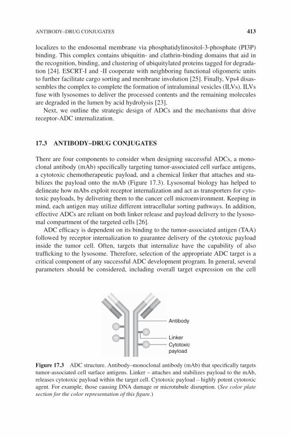
�
� �
�
ANTIBODY–DRUG CONJUGATES 413
localizes to the endosomal membrane via phosphatidylinositol-3-phosphate (PI3P)binding. This complex contains ubiquitin- and clathrin-binding domains that aid inthe recognition, binding, and clustering of ubiquitylated proteins tagged for degrada-tion [24]. ESCRT-I and -II cooperate with neighboring functional oligomeric unitsto further facilitate cargo sorting and membrane involution [25]. Finally, Vps4 disas-sembles the complex to complete the formation of intraluminal vesicles (ILVs). ILVsfuse with lysosomes to deliver the processed contents and the remaining moleculesare degraded in the lumen by acid hydrolysis [23].
Next, we outline the strategic design of ADCs and the mechanisms that drivereceptor-ADC internalization.
17.3 ANTIBODY–DRUG CONJUGATES
There are four components to consider when designing successful ADCs, a mono-clonal antibody (mAb) specifically targeting tumor-associated cell surface antigens,a cytotoxic chemotherapeutic payload, and a chemical linker that attaches and sta-bilizes the payload onto the mAb (Figure 17.3). Lysosomal biology has helped todelineate how mAbs exploit receptor internalization and act as transporters for cyto-toxic payloads, by delivering them to the cancer cell microenvironment. Keeping inmind, each antigen may utilize different intracellular sorting pathways. In addition,effective ADCs are reliant on both linker release and payload delivery to the lysoso-mal compartment of the targeted cells [26].
ADC efficacy is dependent on its binding to the tumor-associated antigen (TAA)followed by receptor internalization to guarantee delivery of the cytotoxic payloadinside the tumor cell. Often, targets that internalize have the capability of alsotrafficking to the lysosome. Therefore, selection of the appropriate ADC target is acritical component of any successful ADC development program. In general, severalparameters should be considered, including overall target expression on the cell
Antibody
Linker
Cytotoxic
payload
Figure 17.3 ADC structure. Antibody–monoclonal antibody (mAb) that specifically targetstumor-associated cell surface antigens. Linker – attaches and stabilizes payload to the mAb,releases cytotoxic payload within the target cell. Cytotoxic payload – highly potent cytotoxicagent. For example, those causing DNA damage or microtubule disruption. (See color platesection for the color representation of this figure.)
�
� �
�
414 LYSOSOMES AND ANTIBODY–DRUG CONJUGATES
membrane, relative levels of overexpression in malignant tissues compared withnormal counterparts, rates of antigen internalization, and the intracellular traffickingdynamics of internalized receptors. Interestingly, the level of TAA expression neededfor an efficacious ADC is dependent on the ADC payload. Programs that utilizeless potent payloads, such as microtubule inhibitors, should have TAA expressed atsufficiently high density to ensure delivery of cytotoxic payload to tumor cells. ADCswith more toxic DNA-damaging agents, for example, Mylotarg® (calicheamicinpayload) can be delivered with a relatively low TAA copy number [27,28].
Advances in antibody engineering have exploited differences between the envi-ronments of the extracellular and endosomal space. Despite the route that the targetantigen follows after internalization, antibody engineering can modify the intracellu-lar trafficking dynamics. Engineered antibodies can improve lysosomal localizationthereby ensuring the ADC/antigen complex is dissociated in the endosome, allow-ing the ADC to independently traverse endosomal sorting pathways regardless ofits receptor. In two separate studies, scientists have altered the intracellular route ofantibody trafficking by designing antibodies with high-affinity binding for the extra-cellular target at neutral pH and low-affinity binding for the receptor within the acidicEE [29,30]. Both studies utilized a histidine scanning approach, in which histidineresidues were introduced in critical areas of the antibody. This screen selected formutations that disrupt antibody binding at a low pH environment without affectingbinding at neutral pH. Histidine residues were prioritized based on the fact that theyshow pKa values of 6.0, and thus these histidine residues become protonated in pHenvironments below 6.0, causing disruption of antibody binding. In both cases, thisapproach was used to identify the antibody that can dissociate from its receptor withinthe EE and enter the recycling pathway while its target antigen underwent lysosomaldegradation. This yielded an antibody capable of escaping lysosomal degradation,leading to an increase in the serum half-life and efficacy of the therapeutic [29,30]. Inrecent studies, a similar method was used to generate anti-IL6 neutralizing antibod-ies with a range of pH dependencies for binding [31]. Following uptake into the cell,anti-IL-6 antibodies bind antigens with higher affinity at near-neutral pH (relative toacidic endosomal pH). After ADC uptake, the complex is expected to release antigenwithin the endosome, with concomitant antibody recycling and exocytosis occurringin FcRn-expressing cells. In the acidic pH of the endosome, the cytokine dissociatedfrom the engineered antibody and trafficked to the lysosome, thus decreasing its levelin the circulation, while the anti-IL-6 antibody was recycled out of the cell [31].
Linker-payloads are additional factors affecting ADC efficacy whereby theintracellular drug release from the antibody is accomplished through the disruptionof the chemical linker between the antibody and cytotoxic payload. Therefore,ADC efficacy can be directly impacted by linker choice. Early-generation ADCsrelied on the use of nonproteolytically cleavable linkers such as hydrazone, ordisulfide-based linker approaches [32]. Hydrazone linkers are stable at a neutralpH but are cleaved within the low pH environment of intracellular endosomeand lysosome compartments. Proteolytically cleavable linkers, such as the pep-tide linker valine-citrulline-p-aminobenzyl carbamate (vc), are designed to beselectively cleaved by lysosomal proteases, allowing for specific cleavage after

�
� �
�
ANTIBODY–DRUG CONJUGATES 415
internalization [33]. Noncleavable linkers, such as thioether- or amide-based linkers,have been utilized more recently and are intended to retain stability throughout theplasma and most of the intracellular space. Current evidence suggests that the earlyand LEs provide a highly reductive environment and contain sufficient proteases toprocess both cleavable types of linkers [34,35]. Nonetheless, one study has disputedthis notion and has shown that the endosomes are in fact highly oxidative in nature,thereby limiting the cleavage of disulfide-based linkers [36]. This finding suggestedthe possibility that using disulfide linkers may result in reduced antitumor activity insome situations. Interestingly, payloads attached through disulfide linkers can formlipophilic drug metabolites after cleavage in the lysosome [37]. These metaboliteshave the ability to cross membranes and contribute to the bystander killing effectwhen effluxed out of tumor cells. Such approaches may be beneficial when targetingtumors with heterogeneous antigen expression, but, at the same time, can result inhigher levels of undesirable toxicity. Another study has shown that the specific siteson which the linker-payload is conjugated onto the antibody can influence stabilityof a proteolytically cleavable linker [38].
One of the most common linkers across the ADCs in clinical trials is the aforemen-tioned vc-linker. Brentuximab vedotin (Adcetris®, SGN-35), a currently approvedADC, is an anti-CD30-auristatin conjugate that incorporates a peptide cleavablevc-linker conjugated to monomethyl auristatin E (MMAE) [39]. In Brentuximabvedotin, the vc-linker is conjugated via a cysteine sulfhydryl group on the antibodyto the maleimide functionality on the linker. Upon internalization of the ADC intothe endosomes and lysosomes, the amide bond between the citrulline residue and thep-aminobenzyl carbamate portion of the linker is cleaved by lysosomal proteases [33].Self-elimination of the p-aminobenzyl carbamate group, which is important to spa-tially separate the drug from the site of enzymatic cleavage, releases free MMAE inthe tumor cell. While ADCs with cleavable linkers tend to show good efficacy, thenature of a cleavable linker generates certain liability due to potential extracellularcleavage of the payload and the resultant off-target toxicity. As a consequence,some recent ADCs have been designed to possess linkers that are not cleavable byconventional mechanisms, such as thioether or amide bonds [40]. While this createsan ADC that is highly stable in the extracellular environment, the use of such a linkerrequires that the ADC be routed to the lysosomal compartment of the cell, wherethe antibody is completely degraded, liberating the linker-payload. The payload isthen released from the lysosome into the cytosol, where it elicits cytotoxic activity.However, some targets are not delivered to the lysosomal compartment at a sufficientconcentration and are instead recycled back to the plasma membrane, making itchallenging to develop ADCs with a noncleavable linker against such targets.
The development of novel cytotoxic payloads with optimized potency andappropriate linker compatibility remains an important goal in ADC development.Early-generation ADCs contained payloads that provided little antitumor activity dueto the lack of potency (e.g., methotrexate, doxorubicin, and vinblastine) [41–46]. Asa result, newer ADCs have incorporated highly potent cytotoxic agents that interferewith microtubule dynamics or cause irreversible disruptions in DNA integrity. Usageof highly potent microtubule-disrupting agents has become widely adopted due to

�
� �
�
416 LYSOSOMES AND ANTIBODY–DRUG CONJUGATES
the efficacy observed against rapidly dividing cells, which provides an additionalmechanism for tumor selectivity. Compared to conventional spindle poison cytotox-ics, DNA-damaging agents are less dependent on cell cycle progression to induceapoptosis, a property that may be advantageous when targeting malignant cells thatare not rapidly proliferating. Significant efforts are underway to develop new andmore tumor-selective linker-payloads.
Lysosomal sequestration should be considered when developing novel payloadsfor ADC platforms. The concept of pH partitioning has been well described forpayloads linked by amine residues [47]. When payloads exist in a pure basic form,they are capable of free diffusion across membranes. For example, when a smallmolecule exhibits a pKa value of 7–8, it exists predominantly in the free base formin the cytosol. However, if this molecule crosses into organelles with acidic lumens,such as endosomes and lysosomes, the molecule exists primarily in an ionized orprotonated form potentially producing a membrane impermeable molecule thatsubsequently traps the molecule inside the organelle. This is believed to be oneof the reasons why tumors develop resistance to drug therapy [47]. Lysosomalsequestration has been described for molecules such as sunitinib [48]. In addition, ithas been suggested that lysosomal sequestration of molecules can occur through thepresence of multidrug transporters, such as P-glycoprotein (Pgp), in the lysosomalmembrane [49]. In this case, cytotoxic agents are pumped out of the cytoplasm intothe lysosomal compartment by Pgp, where they are subsequently ionized within thelysosome, rendering them ineffective agents. This mode of sequestration has beendescribed for molecules such as doxorubicin, daunorubicin, and vinblastine [49].To date, lysosomal sequestration has not been reported for ADC payloads. Novelpayload development strategies should be considered to reduce potential issues oflysosomal sequestration. Therefore, developing payloads or agents that interferewith the integrity of the lysosomal compartment is an attractive option to considerin preclinical studies with future ADCs [50]. Disruption of lysosomes can lead toautophagic cell death, resulting in the release of detrimental lysosomal proteasesinto the cytoplasm. Bafilomycin A, salicylihalamide A, and concanamycin A areinteresting examples of natural products that disrupt the lysosomes. These agentsinhibit vacuolar ATPase (vATPase), which maintains the proton gradient across thelysosomal membrane [51–54]. Accordingly, inhibition of this vATPase impedes theability of lysosomes to acidify, which can result in cell death.
17.4 MECHANISMS OF RESISTANCE TO ADCs
The efficacy and duration of any chemotherapeutic regimen is dictated by the innateresistance of cancer cells to a therapy or the acquisition of resistance following drugexposure and resistant cell population selection. Multiple ADCs have entered clinicaldevelopment, yet little data is available that describes the molecular mechanismsof resistance that emerges within this drug class. Published clinical data for acutemyeloid leukemia (AML) suggests patients that initially responded but eventuallyfailed Mylotarg therapy had an enrichment of CD33+ cells with an upregulation of

�
� �
�
REFERENCES 417
Pgp or multidrug resistance (MDR1) [55,56]. As Mylotarg’s payload is a known sub-strate for MDR1, the mechanism of resistance in Mylotarg refractory patients couldbe due to efflux of payload out of leukemic cells [57,58]. However, published dataabout clinical resistance to other FDA-approved ADCs (i.e., Adcetris or Kadcyla®)are lacking. Recently, several studies have reported the acquisition of resistance totrastuzumab-based ADCs in in vitro models. The first study showed that chronictreatment of HER2+ cell lines with Kadcyla resulted in the emergence of resistantcell populations with varying mechanisms of resistance [59]. Mechanisms of resis-tance observed in this study included the compensatory expression of neuregulin-1,MDR1 protein overexpression, HER2 protein downregulation, and changes inmitogen-activated protein kinase (MAPK) family signaling. A second study revealedthat cyclical treatment of HER2+ cell lines with a trastuzumab-maytansinoid ADC,which is structurally similar to Kadcyla, yielded resistant cell lines. The observedresistance was mediated by payload drug efflux through overexpression of multidrugresistance protein 1 (MRP1) and HER2 protein downregulation [60]. Whether thesemechanisms of resistance translate from in vitro models to the clinic in HER2+breast cancer patients remain to be seen.
Interestingly, the aforementioned class of lysosomal-targeted payloads may be fur-ther attractive to develop given that some analogs do not appear to be substrates fordrug efflux proteins. The classical models of drug resistance mediated by MDR1- orMRP1-induced drug efflux use HL60/Vinc and HL60/Adr cell lines [61]. Marquardtand Center revealed a regulatory role for vATPase in the inhibition of drug efflux inthese cell models. Surprisingly, bafilomycin A, a vATPase inhibitor, appears not tobe a substrate for MDR1 [61]. This sets the stage for proposing the appealing com-bination of vATPase inhibitors with the payload classes of current clinical ADCs.
17.5 SUMMARY
With the promise of targeting chemotherapeutic drugs specifically to tumors, ADCseffectively increase the therapeutic index of conventional chemotherapeutic com-pounds by increasing efficacy while decreasing toxicities in many cases. ADCs utilizeTAAs, receptor internalization, and intracellular trafficking through the endolyso-somal pathway to deliver their cytotoxic payloads to tumor cells while protectingnontumor cells from drug exposure. Using antibody targeting as a platform, we canreconsider compounds previously thought to be too toxic, such as the lysosomalinhibitors described herein, as exciting new strategies for ADC development andpatient treatment regimens going forward.
REFERENCES
[1] Franklin MC, Carey KD, Vajdos FF, Leahy DJ, de Vos AM, et al. Insights into ErbBsignaling from the structure of the ErbB2-pertuzumab complex. Cancer Cell 2004;5:317–328.
�
� �
�
418 LYSOSOMES AND ANTIBODY–DRUG CONJUGATES
[2] Modjtahedi H, Komurasaki T, Toyoda H, Dean C. Anti-EGFR monoclonal antibodieswhich act as EGF, TGF alpha, HB-EGF and BTC antagonists block the binding of epireg-ulin to EGFR-expressing tumours. Int J Cancer 1998;75:310–316.
[3] Teramoto T, Onda M, Tokunaga A, Asano G. Inhibitory effect of anti-epidermal growthfactor receptor antibody on a human gastric cancer. Cancer 1996;77:1639–1645.
[4] Le Roy C, Wrana JL. Clathrin- and non-clathrin-mediated endocytic regulation of cellsignalling. Nat Rev Mol Cell Biol 2005;6:112–126.
[5] Mayor S, Pagano RE. Pathways of clathrin-independent endocytosis. Nat Rev Mol CellBiol 2007;8:603–612.
[6] Honing S, Ricotta D, Krauss M, Spate K, Spolaore B, et al. Phosphatidylinositol-(4,5)-bisphosphate regulates sorting signal recognition by the clathrin-associated adaptorcomplex AP2. Mol Cell 2005;18:519–531.
[7] Bonifacino JS, Traub LM. Signals for sorting of transmembrane proteins to endosomesand lysosomes. Annu Rev Biochem 2003;72:395–447.
[8] Trowbridge IS, Collawn JF, Hopkins CR. Signal-dependent membrane protein traffickingin the endocytic pathway. Annu Rev Cell Biol 1993;9:129–161.
[9] Stenmark H. Rab GTPases as coordinators of vesicle traffic. Nat Rev Mol Cell Biol2009;10:513–525.
[10] Owen DJ, Evans PR. A structural explanation for the recognition of tyrosine-based endo-cytotic signals. Science 1998;282:1327–1332.
[11] Conner SD, Schmid SL. Identification of an adaptor-associated kinase, AAK1, as a reg-ulator of clathrin-mediated endocytosis. J Cell Biol 2002;156:921–929.
[12] Fingerhut A, von Figura K, Honing S. Binding of AP2 to sorting signals is modulated byAP2 phosphorylation. J Biol Chem 2001;276:5476–5482.
[13] Semerdjieva S, Shortt B, Maxwell E, Singh S, Fonarev P, et al. Coordinated regula-tion of AP2 uncoating from clathrin-coated vesicles by rab5 and hRME-6. J Cell Biol2008;183:499–511.
[14] McMahon HT, Boucrot E. Molecular mechanism and physiological functions ofclathrin-mediated endocytosis. Nat Rev Mol Cell Biol 2011;12:517–533.
[15] Ferguson SM, Raimondi A, Paradise S, Shen H, Mesaki K, et al. Coordinated actions ofactin and BAR proteins upstream of dynamin at endocytic clathrin-coated pits. Dev Cell2009;17:811–822.
[16] Sundborger A, Soderblom C, Vorontsova O, Evergren E, Hinshaw JE, et al. Anendophilin-dynamin complex promotes budding of clathrin-coated vesicles during synap-tic vesicle recycling. J Cell Sci 2011;124:133–143.
[17] Wigge P, Kohler K, Vallis Y, Doyle CA, Owen D, et al. Amphiphysin heterodimers:potential role in clathrin-mediated endocytosis. Mol Biol Cell 1997;8:2003–2015.
[18] Helenius A, Marsh M. Endocytosis of enveloped animal viruses. Ciba Found Symp 1982;92:59–76.
[19] van Dam EM, Stoorvogel W. Dynamin-dependent transferrin receptor recycling byendosome-derived clathrin-coated vesicles. Mol Biol Cell 2002;13:169–182.
[20] Rubin I, Yarden Y. The basic biology of HER2. Ann Oncol 2001;12(Suppl 1):S3–8.
[21] Sirisaengtaksin N, Gireud M, Yan Q, Kubota Y, Meza D, et al. UBE4B protein cou-ples ubiquitination and sorting machineries to enable epidermal growth factor receptor(EGFR) degradation. J Biol Chem 2014;289:3026–3039.

�
� �
�
REFERENCES 419
[22] Arancibia-Carcamo IL, Yuen EY, Muir J, Lumb MJ, Michels G, et al. Ubiquitin-dependent lysosomal targeting of GABA(A) receptors regulates neuronal inhibition. ProcNatl Acad Sci U S A 2009;106:17552–17557.
[23] Schmidt O, Teis D. The ESCRT machinery. Curr Biol 2012;22:R116–120.
[24] Raiborg C, Stenmark H. The ESCRT machinery in endosomal sorting of ubiquitylatedmembrane proteins. Nature 2009;458:445–452.
[25] Henne WM, Buchkovich NJ, Emr SD. The ESCRT pathway. Dev Cell 2011;21:77–91.
[26] Beck A, Haeuw JF, Wurch T, Goetsch L, Bailly C, et al. The next generation ofantibody–drug conjugates comes of age. Discov Med 2010;10:329–339.
[27] DiJoseph JF, Armellino DC, Boghaert ER, Khandke K, Dougher MM, et al. Antibody-targeted chemotherapy with CMC-544: a CD22-targeted immunoconjugate of calicheam-icin for the treatment of B-lymphoid malignancies. Blood 2004;103:1807–1814.
[28] DiJoseph JF, Popplewell A, Tickle S, Ladyman H, Lawson A, et al. Antibody-targetedchemotherapy of B-cell lymphoma using calicheamicin conjugated to murine or human-ized antibody against CD22. Cancer Immunol Immunother 2005;54:11–24.
[29] Chaparro-Riggers J, Liang H, DeVay RM, Bai L, Sutton JE, et al. Increasing serumhalf-life and extending cholesterol lowering in vivo by engineering antibody withpH-sensitive binding to PCSK9. J Biol Chem 2012;287:11090–11097.
[30] Igawa T, Ishii S, Tachibana T, Maeda A, Higuchi Y, et al. Antibody recycling by engi-neered pH-dependent antigen binding improves the duration of antigen neutralization.Nat Biotechnol 2010;28:1203–1207.
[31] Devanaboyina SC, Lynch SM, Ober RJ, Ram S, Kim D, et al. The effect of pHdependence of antibody–antigen interactions on subcellular trafficking dynamics. MAbs2013;5:851–859.
[32] Ducry L, Stump B. Antibody–drug conjugates: linking cytotoxic payloads to monoclonalantibodies. Bioconjug Chem 2010;21:5–13.
[33] Doronina SO, Toki BE, Torgov MY, Mendelsohn BA, Cerveny CG, et al. Developmentof potent monoclonal antibody auristatin conjugates for cancer therapy. Nat Biotechnol2003;21:778–784.
[34] Lewis Phillips GD, Li G, Dugger DL, Crocker LM, Parsons KL, et al. Targeting HER2-positive breast cancer with trastuzumab-DM1, an antibody–cytotoxic drug conjugate.Cancer Res 2008;68:9280–9290.
[35] Tanaka M, Kano Y, Akutsu M, Tsunoda S, Izumi T, et al. The cytotoxic effects of gem-tuzumab ozogamicin (mylotarg) in combination with conventional antileukemic agentsby isobologram analysis in vitro. Anticancer Res 2009;29:4589–4596.
[36] Austin CD, Wen X, Gazzard L, Nelson C, Scheller RH, et al. Oxidizing potential ofendosomes and lysosomes limits intracellular cleavage of disulfide-based antibody–drugconjugates. Proc Natl Acad Sci U S A 2005;102:17987–17992.
[37] Erickson HK, Park PU, Widdison WC, Kovtun YV, Garrett LM, et al. Antibody–maytansinoid conjugates are activated in targeted cancer cells by lysosomal degradationand linker-dependent intracellular processing. Cancer Res 2006;66:4426–4433.
[38] Dorywalska M, Strop P, Melton-Witt JA, Hasa-Moreno A, Farias SE, et al. Effect ofattachment site on stability of cleavable antibody drug conjugates. Bioconjug Chem 2015;26:650–659.
[39] Gerber HP. Emerging immunotherapies targeting CD30 in Hodgkin’s lymphoma.Biochem Pharmacol 2010;79:1544–1552.
�
� �
�
420 LYSOSOMES AND ANTIBODY–DRUG CONJUGATES
[40] Kovtun YV, Audette CA, Ye Y, Xie H, Ruberti MF, et al. Antibody–drug conjugatesdesigned to eradicate tumors with homogeneous and heterogeneous expression of thetarget antigen. Cancer Res 2006;66:3214–3221.
[41] Friedman PN, Chace DF, Trail PA, Siegall CB. Antitumor activity of the single-chainimmunotoxin BR96 sFv-PE40 against established breast and lung tumor xenografts.J Immunol 1993;150:3054–3061.
[42] Ghose T, Ferrone S, Blair AH, Kralovec Y, Temponi M, et al. Regression of humanmelanoma xenografts in nude mice injected with methotrexate linked to monoclonalantibody 225.28 to human high molecular weight-melanoma associated antigen. CancerImmunol Immunother 1991;34:90–96.
[43] Schrappe M, Bumol TF, Apelgren LD, Briggs SL, Koppel GA, et al. Long-termgrowth suppression of human glioma xenografts by chemoimmunoconjugates of4-desacetylvinblastine-3-carboxyhydrazide and monoclonal antibody 9.2.27. Cancer Res1992;52:3838–3844.
[44] Smyth MJ, Pietersz GA, McKenzie IF. The in vitro and in vivo anti-tumour activity ofN-AcMEL-(Fab’)2 conjugates. Br J Cancer 1987;55:7–11.
[45] Trail PA, King HD, Dubowchik GM. Monoclonal antibody drug immunoconjugates fortargeted treatment of cancer. Cancer Immunol Immunother 2003;52:328–337.
[46] Yang HM, Reisfeld RA. Pharmacokinetics and mechanism of action of a doxorubicin-monoclonal antibody 9.2.27 conjugate directed to a human melanoma proteoglycan.J Natl Cancer Inst 1988;80:1154–1159.
[47] Raghunand N, Gillies RJ. pH and drug resistance in tumors. Drug Resist Updat 2000;3:39–47.
[48] Gotink KJ, Broxterman HJ, Labots M, de Haas RR, Dekker H, et al. Lysosomal seques-tration of sunitinib: a novel mechanism of drug resistance. Clin Cancer Res 2011;17:7337–7346.
[49] Yamagishi T, Sahni S, Sharp DM, Arvind A, Jansson PJ, et al. P-glycoprotein mediatesdrug resistance via a novel mechanism involving lysosomal sequestration. J Biol Chem2013;288:31761–31771.
[50] Gerber HP, Koehn FE, Abraham RT. The antibody–drug conjugate: an enabling modalityfor natural product-based cancer therapeutics. Nat Prod Rep 2013;30:625–639.
[51] Drose S, Altendorf K. Bafilomycins and concanamycins as inhibitors of V-ATPases andP-ATPases. J Exp Biol 1997;200:1–8.
[52] Forgac M. Vacuolar ATPases: rotary proton pumps in physiology and pathophysiology.Nat Rev Mol Cell Biol 2007;8:917–929.
[53] Xie XS, Padron D, Liao X, Wang J, Roth MG, et al. Salicylihalamide A inhibits the V0sector of the V-ATPase through a mechanism distinct from bafilomycin A1. J Biol Chem2004;279:19755–19763.
[54] Yoshimori T, Yamamoto A, Moriyama Y, Futai M, Tashiro Y. Bafilomycin A1, a specificinhibitor of vacuolar-type H(+)-ATPase, inhibits acidification and protein degradation inlysosomes of cultured cells. J Biol Chem 1991;266:17707–17712.
[55] Walter RB, Gooley TA, van der Velden VH, Loken MR, van Dongen JJ, et al. CD33expression and P-glycoprotein-mediated drug efflux inversely correlate and predict clin-ical outcome in patients with acute myeloid leukemia treated with gemtuzumab ozogam-icin monotherapy. Blood 2007;109:4168–4170.

�
� �
�
REFERENCES 421
[56] Linenberger ML, Hong T, Flowers D, Sievers EL, Gooley TA, et al. Multidrug-resistancephenotype and clinical responses to gemtuzumab ozogamicin. Blood 2001;98:988–994.
[57] Cianfriglia M. The biology of MDR1-P-glycoprotein (MDR1-Pgp) in designing func-tional antibody drug conjugates (ADCs): the experience of gemtuzumab ozogamicin. AnnIst Super Sanita 2013;49:150–168.
[58] Kung Sutherland MS, Walter RB, Jeffrey SC, Burke PJ, Yu C, et al. SGN-CD33A: a novelCD33-targeting antibody–drug conjugate using a pyrrolobenzodiazepine dimer is activein models of drug-resistant AML. Blood 2013;122:1455–1463.
[59] Phillips GD, Fields CT, Li G, Dowbenko D, Schaefer G, et al. Dual targeting of HER2-positive cancer with trastuzumab emtansine and pertuzumab: critical role for neureg-ulin blockade in antitumor response to combination therapy. Clin Cancer Res 2014;20:456–468.
[60] Loganzo F, Tan X, Sung M, Jin G, Myers JS, et al. Tumor cells chronically treated witha trastuzumab-maytansinoid antibody–drug conjugate develop varied resistance mecha-nisms but respond to alternate treatments. Mol Cancer Ther 2015;14(4):952–963.
[61] Marquardt D, Center MS. Involvement of vacuolar H(+)-adenosine triphosphatase activ-ity in multidrug resistance in HL60 cells. J Natl Cancer Inst 1991;83:1098–1102.

�
� �
�
18THE MECHANISMS ANDTHERAPEUTIC CONSEQUENCESOF AMINE-CONTAINING DRUGSEQUESTRATION IN LYSOSOMES
Nadia Hamid and Jeffrey P. KriseDepartment of Pharmaceutical Chemistry, The University of Kansas, Lawrence, KS, USA
18.1 INTRODUCTION
It is well known that certain low-molecular-weight, weakly basic drugs becomehighly concentrated in acidic intracellular organelles including lysosomes. Substratesfor this accumulation are routinely referred to as being lysosomotropic, and the mech-anism for accumulation is known as ion trapping or pH partitioning. Lysosomotropicdrugs have been shown to reach concentrations within lysosomes that are more than1000 times greater than in the cytosol or extracellular space at steady state. Thischapter reviews the mechanism by which lysosomal trapping occurs and the drug-and cell-related factors that influence the process. The chapter also examines some ofthe techniques used to assess lysosomal trapping in cultured cells. Subsequent discus-sions focus on the therapeutic implications associated with lysosomal drug seques-tration. As discussed, the lysosomal sequestration of drugs can impact the extent ofinteraction with intracellular targets. Toward this end, lysosomotropic behavior ofdrugs has been purposefully exploited in an effort to increase drug potency and selec-tivity. Moreover, the sequestration of drugs in lysosomes can have a profound impacton the pharmacokinetic properties of drugs. Specifically, lysosomotropic drugsoften have large volumes of distribution and, therefore, long elimination half-lives.
Lysosomes: Biology, Diseases, and Therapeutics, First Edition.Edited by Frederick R. Maxfield, James M. Willard and Shuyan Lu.© 2016 John Wiley & Sons, Inc. Published 2016 by John Wiley & Sons, Inc.
423

�
� �
�
424 CONSEQUENCES OF AMINE-CONTAINING DRUG SEQUESTRATION IN LYSOSOMES
Finally, the chapter reviews instances where it has been shown that one drug iscapable of influencing the lysosomal accumulation of a secondarily administereddrug, establishing the basis for a new type of pharmacokinetic drug–drug interaction.
18.2 LYSOSOMAL TRAPPING OVERVIEW
For over a century, light microscopy studies have revealed that certain low-molecular-weight dyes known as vital stains are able to selectively stain cellular compartmentsof living cells. Vital stains that were thought to specifically stain lysosomes were firstdescribed in the literature in the mid-19th century and originally included weaklybasic molecules such as neutral red and acridine orange (see Figure 18.1) [1,2].
In the mid-1970s, de Duve [3], a Nobel laureate, credited with the initial char-acterization of lysosomes, and his colleagues wrote an elegant theoretical commen-tary on the mechanism for weakly basic drug accumulation in lysosomes. In theircommentary, they referred to the substrates for lysosomal accumulation as lysoso-motropic agents. The authors argued that in order for ion trapping in lysosomes tooccur, a weakly basic drug must be relatively membrane permeable when it is union-ized and relatively membrane impermeable when ionized. Under these conditions,the driving force for lysosomal sequestration is based on the preferential ionizationof weakly basic drug that occurs inside the acidic lysosomes due to the low lysosomalpH (see Figure 18.2). This relatively low pH (approximately pH 4.5) is maintainedby the vacuolar proton ATPase [4]. The activity of vacuolar proton ATPase is there-fore central to the ion trapping phenomenon, and inhibitors of this enzyme have beenshown to abolish lysosomal ion trapping of drugs [5].
Neutral red
Acridine orange
N N
NN
N
N
H2N
Figure 18.1 Structures of neutral red and acridine orange, which are early examples oflysosomal vital stains.

�
� �
�
LYSOSOMAL TRAPPING OVERVIEW 425
Cytosol
(pH 7.4)
B
BH
B
Lysosome
(pH ˜4–5)
[lysosome]
[extracellular]
(α[H+]E + Ka)
(α[H+]L + Ka)([H+]
E + Ka)
=([H+]
L + Ka)
+ BH +
BH +
BH +
BH +
Figure 18.2 Diagram illustrating the pH partitioning-based mechanism for accumulation ofweakly basic drugs (B) in the acidic lysosomes. The equation at the bottom of the figure rep-resents the lysosome to extracellular space steady-state concentration ratio for a base. Thedissociation constant for the conjugate acid of the weak base is denoted as Ka and [H+] is theproton concentration (subscript E represents extracellular and L represents lysosomal). Theratio of permeabilities of the ionized base to that of the unionized base in the lysosomal lipidbilayer is denoted by the α term.
In their commentary, de Duve and his colleagues derive an equation relating thesteady-state concentration ratio of a weakly basic drug inside lysosomes compared tothe extracellular space (see Figure 18.2). According to this equation, the maximumpossible concentration ratio for a weakly basic substance in lysosomes (relative tothe extracellular space) is equal to the ratio of the hydrogen ion concentration inlysosomes relative to the fluid surrounding the cells. For example, if the extracellularpH was 7.4 and the pH of lysosomes was 4.4, the maximal lysosomal concentrationratio would be 1000. According to de Duve’s derivations, there are two drug-relatedterms that can theoretically influence the steady-state accumulation of weakly basicdrugs in lysosomes. The first variable is the weak base pKa, and the second is termedalpha (𝛼), which represents the ratio of permeabilities of the weakly basic drug acrossthe lysosomal lipid bilayer in its ionized and unionized forms.
The pKa of the weakly basic drug is predicted to impact the steady-state lysosomalaccumulation ratio. Molecules with pKa values of 6 or below were not consideredto be lysosomotropic because they would not extensively ionize in lysosomes. LowpKa molecules can never reach the maximal steady-state accumulation ratio of 1000.Molecules with pKa values of 8 or greater can theoretically achieve 1000-fold higherlevels in lysosomes relative to the extracellular space. However, molecules with veryhigh pKa values will start to become less lysosomotropic in a therapeutically relevanttimescale because they are predicted to take exceedingly long times to reach steadystate. Specifically, the permeation rate was predicted to decrease by a factor of 10for each 1-unit increase in drug pKa. Interestingly, de Duve’s theoretical calculations

�
� �
�
426 CONSEQUENCES OF AMINE-CONTAINING DRUG SEQUESTRATION IN LYSOSOMES
predict that the time to reach one-half of steady state for molecules with a pKa of 12would be over 11∕2 years. In contrast, bases with pKa values near 7 are predicted totake 10 min to reach one-half steady-state levels.
Duvvuri et al. [6] later quantitatively tested the theoretical predictions of de Duveand colleagues in cultured cells. Using a series of weakly basic structural isomersthat varied in pKa from 4 to 9, the authors experimentally investigated the influ-ence of pKa on lysosomal accumulation. Consistent with theoretical predictions, theauthors reported that molecules with pKa values below 6 were not appreciably lyso-somotropic. The molecule with a pKa value of 9 had the greatest degree of lysosomalaccumulation.
As a consequence of extensive entrapment of drugs, the lysosomes can take on avacuolated appearance. Using vacuolization as a readout for lysosomotropic behav-ior, early work by Ohkuma and Poole [7] investigated how weak base structure influ-ences lysosomal accumulation. Collectively, with few exceptions, both theoreticaland experimental approaches support the notion that most lysosomotropic agents willhave optimal pKa values between 7 and 10.
As previously mentioned, the term alpha (𝛼) refers to the ratio of the permeabilitycoefficients across the lysosomal lipid bilayer for the ionized base divided by that ofthe unionized base [3]. This term can theoretically vary from zero to one. Moleculeswith an alpha value of zero, meaning the ionized base is completely impermeable,will be the most lysosomotropic and will reach a theoretical maximal steady-stateratio of accumulation dictated by the pH differential. However, as the alpha parameterincreases in magnitude, the maximal steady-state accumulation ratio of a drug in thelysosomes significantly decreases. For example, a drug with an alpha value equalto 1 will have equal membrane permeability regardless of ionization. Under thesecircumstances, extensive protonation of the drug in lysosomes would not slow the rateof diffusion back to the cytosol. A theoretical relationship illustrating the combinedinfluence of pKa and alpha on lysosomal trapping is shown in Figure 18.3.
Duvvuri et al. [8] have experimentally estimated alpha values for a series of drugsand model compounds and correlated this value with the experimentally measuredlysosome-to-cytosol concentration ratio obtained in cell culture experiments. Asa proxy for lysosomal membrane permeability, which is difficult to measure, theauthors measured the octanol/water partition coefficients for drugs as a function ofpH. Estimates for the log D of the ionized and unionized species were obtained, andthe ratio of these values was used to obtain alpha. As anticipated, it was found thatcompounds with lower alpha values accumulated to a greater extent in lysosomesrelative to compounds with higher alpha values. The authors also examined howalpha correlated with mitochondrial versus lysosomal accumulation of weaklybasic drugs. They found that molecules with low alpha values accumulated almostexclusively in lysosomes, whereas molecules with high alpha values accumulatedwithin mitochondria. It was noted that the alpha parameter for a given drug appearedto correlate with the degree of charge delocalization present in the molecule.Weak bases with fixed localized positive charges tended to have very low alphavalues, whereas those with delocalized charges tended to have higher alpha valuesnear 1. The relationship between charge delocalization and alpha was attributed to

�
� �
�
TECHNIQUES TO ASSESS LYSOSOMAL TRAPPING 427
1200
1000α = 0.00001
α = 0.0001
α = 0.001
800
600
[lysosom
e] / [c
yto
sol]
400
200
4 6 8 10
pKa
12 140
Figure 18.3 Theoretical relationship between weak base pKa and the alpha permeabilityparameter. The relationships were derived using the equation shown in Figure 18.2, with indi-cated values for alpha.
differences in how tightly the molecules bind water, which subsequently relates tothe energy required to pass into a nonpolar lipid-like environment.
The relative lipophilicity (i.e., log P) does not influence the predicted steady-stateaccumulation ratios for weakly basic drugs. However, it will most definitely influ-ence the time to reach steady state and the propensity to exhibit lysosomal trap-ping in a therapeutically relevant timescale. As a consequence, most experimentallyobserved lysosomotropic substrates tend to be relatively lipophilic and have log Pvalues greater than 1 [9].
18.3 TECHNIQUES TO ASSESS LYSOSOMAL TRAPPING
For therapeutic reasons that are discussed below, there is a keen interest in under-standing how drugs localize and distribute within the cells and tissues of our body.Toward this end, a number of assays and approaches have been described over theyears. These approaches have been more extensively reviewed elsewhere [10,11],and the reader should refer to these examples for more details. The purpose of thissection is to give the reader a brief overview of the different techniques used and someof the associated strengths and weaknesses.
By far, the simplest approach is to utilize fluorescence microscopy on culturedcells that have been exposed to a drug that is intrinsically fluorescent. The anti-cancer drug daunorubicin is highly fluorescent, and, as a consequence, numerouspublications have examined the intracellular distribution of this drug in culturedcells [12–14]. Unfortunately, most drugs are not sufficiently fluorescent to allowfor this technique. However, extensive structure–localization relationship studieswith regard to lysosomal accumulation have been conducted using weakly basicfluorescent compounds that are not drugs per se. For example, Horobin and

�
� �
�
428 CONSEQUENCES OF AMINE-CONTAINING DRUG SEQUESTRATION IN LYSOSOMES
colleagues [15,16] have compiled correlations between intracellular distribution andphysical and structural properties of a variety of fluorophores. Similarly, Rosaniaand coworkers [17,18] have employed results from such studies to develop modelsand simulations that describe lysosomal accumulation of weak bases.
Microscopic detection methods that do not rely on fluorescence have also beeninvestigated. Recently, Fu et al. [19] described the use of hyperspectral stimulatedRaman scattering microscopy to examine the intracellular localization and concen-trations of tyrosine kinase inhibitors and chloroquine in cultured cells. The authorswere able to show that the weakly basic drug imatinib accumulated in lysosomes atconcentrations that were approximately 1000-fold higher than extracellular levels.The authors provided evidence that the drugs accumulated in lysosomes to an extentgreater than ion trapping theory would have predicted and that this was, for somedrugs, due to precipitation within lysosomes. The authors also used this method toshow that chloroquine administration decreases lysosomal accumulation of imatiniband that this could provide the basis for the positive therapeutic advantages of takingtwo drugs simultaneously. Although the current limit of detection for drugs in cellsdescribed in this manuscript was quite high (approximately 1–2 mM), it was com-mented that improvements in the instrumentation and techniques such as deuteriumlabeling of drugs can be expected to increase sensitivity.
It is also possible to isolate lysosomes from cultured cells or tissues in an effortto quantify lysosome and drug association. The purification can either occur beforeor after the cells have been exposed to drug. In the former scenario, the test drug isadded to the isolated lysosomes, and the degree of accumulation is assessed eitherdirectly or indirectly. For example, Ishizaki et al. [20] have used highly purified ratliver lysosomes to study the mechanism for imipramine accumulation and have alsostudied the ability of other basic molecules to compete with imipramine uptake.
Andrew et al. [21] developed an indirect approach to assess the permeability ofweakly basic drugs across the lysosomal lipid bilayer using an unpurified cell lysate.The assay was based on the measured ability of a test drug to protect lysosomes fromosmotic pressure-induced rupturing, which is directly related to the propensity for atest drug to permeate across the lipid bilayer of the isolated lysosomes. The readoutfor lysosomal rupturing involved testing for release of free lysosomal enzymes intothe cell lysate. The advantages of this approach are that lysosomes need not be puri-fied, and the test drug needs no special properties or tags. Accordingly, this approachis amenable to relatively high-throughput analysis, and the authors have used thismethod to demonstrate a strong correlation between hydrogen bonding capacity andlysosomal membrane permeability.
A unique analytical approach for evaluating the accumulation of fluorescent com-pounds into acidic organelles, such as lysosomes, was described by Arriaga andcoworkers [22]. The authors utilized capillary electrophoresis with laser-induced flu-orescence with dual-channel detection to identify lysosomes containing fluorescentnanospheres that were preloaded into lysosomes using an endocytic uptake-basedpulse-chase technique. Selecting organelles possessing both nanospheres and dox-orubicin fluorescence allowed them to specifically determine the content of the drugin lysosomes without potential interference from other organelles in which the drug

�
� �
�
INFLUENCE OF LYSOSOMOTROPISM ON DRUG ACTIVITY 429
could associate. The authors were able to identify heterogeneity among lysosomesin their doxorubicin content, an observation not readily achievable using alterna-tive techniques.
Duvvuri et al. [23] describe an approach whereby living cells are incubated, in apulse-chase manner, with magnetic dextran particles to allow them to be specificallylocalized in lysosomes. The cells were subsequently incubated with a test drug andthen lysed so that the lysosomes could be isolated using magnetic chromatography.The drug associated with the isolated lysosomes was then quantified usingconventional strategies including high-performance liquid chromatography-massspectrometry/mass spectrometry (HPLC-MS/MS). Similar to other studies, theauthors show that model drugs including quinacrine accumulated in lysosomes tolevels much greater than simple ion trapping theory would predict.
Lemieux et al. [9] have developed an approach for evaluating lysosomal sequestra-tion that is amenable to high-throughput screening formats. The main principle of themethod involves a competition between the test drug and the fluorescent basic amineprobe LysoTracker Red (LTR) in cultured cells. The authors show that drugs that arecationic and amphiphilic decrease LTR fluorescence in a dose-dependent manner,presumably by increasing lysosomal pH. The authors use this assay to establish thatlysosomotropism occurs more readily for drugs with a high log P. This general assayformat was further examined by Nadanaciva et al. [24], and they were able to estab-lish that lysosomotropic drugs identified in the assay generally had C log P> 2 and abasic pKa between 6.5 and 11. This same assay format was more recently applied toan immortalized hepatocyte cell line [25].
18.4 INFLUENCE OF LYSOSOMOTROPISM ON DRUG ACTIVITY
Mammalian cells are highly compartmentalized, and drug targets are typically local-ized in discrete intracellular organelles as opposed to being evenly distributed acrossall intracellular compartments. How a drug distributes within a human cell can have aprofound impact on its activity and side effect profile. Knowledge regarding the totalamount of drug associated with a cell cannot by itself allow one to accurately predictthe likelihood of experiencing a therapeutic effect. Instead, the most relevant pieceof information to know would be the free concentration of the drug that exists in thelocal microenvironment that immediately surrounds the intended drug target.
Hypothetically speaking, if a drug distributed evenly across the entire cell andall of its compartments, one would anticipate activity at some dose, but the propen-sity for off-target effects would also be high. This is due to the drug having a highdegree of interaction with unintended cellular components. However, if a scenariois considered where the drug has the propensity to specifically concentrate withinthe organelle containing the target, one would anticipate this causing an increase inthe potency. In addition, the propensity for unintended off-target interactions wouldbe minimized. The most unfavorable scenario would be realized if the drug were toexclusively concentrate in some discrete organelle that did not contain the target. Inthis scenario, the drug would have no activity, and off-target effects would be the
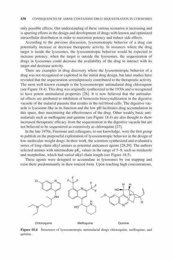
�
� �
�
430 CONSEQUENCES OF AMINE-CONTAINING DRUG SEQUESTRATION IN LYSOSOMES
only possible effects. Our understanding of these various scenarios is increasing andis spurring efforts in the design and development of drugs with known and optimizedintracellular distribution in order to maximize potency and reduce side effects.
According to the previous discussion, lysosomotropic behavior of a drug canpotentially increase or decrease therapeutic activity. In instances where the drugtarget is inside the lysosomes, the lysosomotropic behavior would be expected toincrease potency; when the target is outside the lysosomes, the sequestration ofdrugs in lysosomes could decrease the availability of the drug to interact with itstarget and decrease activity.
There are examples in drug discovery where the lysosomotropic behavior of adrug was not recognized or exploited in the initial drug design, but later studies haverevealed that the sequestration serendipitously contributed to the therapeutic activity.The most well-known example is the lysosomotropic antimalarial drug chloroquine(see Figure 18.4). This drug was originally synthesized in the 1930s and is recognizedto have potent antimalarial properties [26]. It is now believed that the antimalar-ial effects are attributed to inhibition of hemozoin biocrystallization in the digestivevacuole of the malarial parasite that resides in the red blood cells. The digestive vac-uole is lysosome-like in its function and the low pH facilitates drug accumulation inthis space, thus maximizing the effectiveness of the drug. Other weakly basic anti-malarials such as mefloquine and quinine (see Figure 18.4) are also thought to showincreased therapeutic efficacy from the sequestration in the digestive vacuole but arenot believed to be sequestered as extensively as chloroquine [27].
In the late 1970s, Firestone and colleagues, to our knowledge, were the first groupto publish on the purposeful exploitation of lysosomotropic behavior in the design oflow-molecular-weight drugs. In their work, the scientists synthesized and evaluated aseries of long-chain alkyl amines as potential anticancer agents [28,29]. The authorsselected amines with intermediate pKa values in the range of 5–8, such as imidazoleand morpholine, which had varied alkyl chain length (see Figure 18.5).
These agents were designed to accumulate in lysosomes by ion trapping andexist there predominantly in their ionized form. Upon reaching high concentrations,
NH
Cl N FF
FF
F
F
O
N
N
N
NH
H H
H
HO
Chloroquine Mefloquine Quinine
HO
N
Figure 18.4 Structures of lysosomotropic antimalarial drugs chloroquine, mefloquine, andquinine.

�
� �
�
INFLUENCE OF LYSOSOMOTROPISM ON DRUG ACTIVITY 431
H3CN
N
11
Figure 18.5 Example of lysosomotropic detergent investigated by Firestone and colleagues.
the agents would acquire surfactant properties that would lead to the selectivepermeabilization of the lysosomal membrane, which would precipitate the death ofthe cell. The authors provided several compelling pieces of evidence that the agentswere indeed acting as lysosomotropic detergents. First, cell fractionation studiesshowed that the drugs were specifically accumulating in fractions rich in lysosomes.Cellular cytotoxicity studies revealed a sharp sigmoidal dose–response relationship,which is consistent with the requirement for the formation of detergent micelles. Inaddition, the drugs were toxic only to cells that had lysosomes and were not toxic tothose that did not contain lysosomes (i.e., red blood cells). Finally, cell death waspreceded by lysosomal vacuolization, led to an increase in lysosomal pH, and wasassociated with the release of lysosomal contents.
In a later work, Dubowchick and colleagues also rationally incorporated lysoso-motropic features in a series of molecules that were designed to increase the luminalpH of endosomes and lysosomes. Such agents were hypothesized to have potentialutility in the reversal of anticancer multidrug resistance that was mediated throughincreased lysosomal ion trapping of weakly basic anticancer drugs [30,31]. In addi-tion, it was suggested that such agents could be effective against some viruses thatexploit the low pH of lysosomes to facilitate entry in the cell cytosol to allow forsubsequent replication [32]. The molecules were designed and optimized to be mem-brane active and capable of transporting protons across lipid bilayers resulting inthe dissipation of the lysosomal pH gradient (see Figure 18.6 for a depiction of thestrategy). The efficiency of proton transfer was optimized by designing hydrophobicnitrogenous bases that would have the tendency to remain embedded in the lysoso-mal lipid bilayer while mediating the proton transfer from the lumen of lysosomes tothe cytosol. Toward this end, the authors identified a lipophilic imidazole-containingmolecule that had the best ability to neutralize lysosomal pH in cells grown in cultureand had physicochemical properties evaluated in in vitro tests that were consistentwith the aforementioned mechanism of action in proton transfer (see Figure 18.6 forstructure). This molecule also showed activity in reversing anticancer drug resistanceand in the inhibition of influenza virus replication [33].
There are numerous examples whereby known lysosomotropic agents have beenshown to interfere with endosomal/lysosomal trafficking and function. This is animportant consideration from a potential toxicological point of view; however, thisis the focus of a separate chapter and is not further discussed here. Interestingly,these otherwise potentially negative effects have been proposed to have poten-tial relevance in the treatment of various diseases. For example, lysosomotropicagents have been shown to decrease the escape of viruses and other endocytosedmembrane-impermeable toxins from late endosomes and lysosomes [34–38].

�
� �
�
432 CONSEQUENCES OF AMINE-CONTAINING DRUG SEQUESTRATION IN LYSOSOMES
(a)
(b)
Cytosol
H+A–
NH+A–
NH+A– N
N
H+A–
pH = 5
pH = 7
Membrane
Lysosomal
compartment
HN
N
Figure 18.6 (a) Diagram of proposed lysosomal proton transfer mechanism. (b) Structure oflysosomotropic proton transfer reagent.
Similarly, there exists work that suggests that lysosomotropic amines can suppressmajor histocompatibility complex (MHC) class II antigen presentation [39] anddecrease iron release from lysosomes [40]. Lysosomotropic amines have also beenused to help decrease the exocytosis of anticancer drugs that are sequestered in lyso-somes as a means to reverse the anticancer drug-resistance mechanism, as was previ-ously described [41–44]. The mechanism by which lysosomotropic amines decreaselysosomal egress/function in the previous applications is not completely understood.One explanation is that the amines transiently increase the pH of lysosomes througha buffering effect. In this way, the drugs could decrease the activity of lysosomalhydrolases and this could lead to a buildup of undigested materials, which can inter-fere with lysosomal trafficking. Another possibility is that the lysosomotropic aminescould directly modify lysosomal membranes such that fusion and fission eventsrequired for the vesicle-mediated trafficking could become impaired. Consistent withthe latter notion, the weakly basic drug primaquine has been shown to impair buddingevents at the Golgi, which also has an acidic lumen relative to the cell cytosol [45].
It is well known that most anticancer agents can be classified as being weaklybasic and therefore presumably lysosomotropic. However, the therapeutic advantageof lysosomal trapping, if any, in the treatment of cancer is not immediately obviousbecause most anticancer drug targets are not localized within the lysosomes, and trap-ping in this space would be anticipated to decrease interactions with the drug targets,as previously discussed. Recently, it has been proposed that the weakly basic nature

�
� �
�
INFLUENCE OF LYSOSOMOTROPISM ON DRUG ACTIVITY 433
of anticancer drugs contributes to the selective toxicity of the drugs against cancercells. A number of studies have demonstrated that many cancer cells have defectiveacidification of lysosomes for reasons that have not been fully elucidated [46–49].Based on this difference in lysosomal pH between normal and many cancer cells, itwas hypothesized that lysosomotropic behavior of a cancer drug with extralysosomaltargets could enhance the selective toxicity of the drug toward a cancer cell comparedto a normal cell with normally acidified lysosomes. In other words, weakly basicanticancer agents that are exquisite substrates for lysosomal trapping would achievevery low concentrations in the cell cytosol and nucleus of normal cells and thus havereduced capacity to interact with targets and exert cytotoxic effects. Alternatively,when the lysosomotropic cancer drug is present in cancer cells with elevated lysoso-mal pH, the lysosomotropic drug would be relatively less concentrated in lysosomesand would exist to a greater degree in the cell cytosol and nucleus. According to thisproposed mechanism, the lysosomotropic anticancer agent would possess increasedselectivity against tumor cells compared to nonlysosomotropic anticancer agents (seeFigure 18.7 for an illustration of this concept).
NormalLow lysosomal pH
Target
Target Target
Lyso
som
otr
op
ic
dru
g
No
no
som
otr
op
ic
dru
g
CancerElevated lysosomal pH
Target
Figure 18.7 Overview of intracellular distribution-based anticancer drug targeting platform.(See color plate section for the color representation of this figure.)

�
� �
�
434 CONSEQUENCES OF AMINE-CONTAINING DRUG SEQUESTRATION IN LYSOSOMES
The initial evaluations of these hypotheses were conducted in cultured cells thathad variable lysosomal pH and with model anticancer agents that were derivativesof geldanamycin, a prototypic inhibitor of the molecular chaperone heat shock pro-tein 90 (Hsp90). Geldanamycin is amenable to diverse chemical derivatization at the17-position with little effect on binding to Hsp90 [50]. It was therefore possible tosynthesize inhibitors that had variations at the 17-position that made them eitherweakly basic (i.e., lysosomotropic) or neutral (nonlysosomotropic). The selectivityof the drugs was measured by taking the ratio of the IC50 cytotoxicity value for agiven drug in cells with low (normal) pH divided by the corresponding value in cellswith elevated lysosomal pH (i.e., cancer cells). These studies reveal that weakly basicinhibitors had additional selective toxicity toward cells with elevated lysosomal pH,a characteristic that was not the case for nonlysosomotropic inhibitors [51]. Furtherinvestigation of this hypothesis was completed in non-tumor-bearing mice with andwithout elevated lysosomal pH, again using lysosomotropic and nonlysosomotropicHsp90 inhibitors [52]. In this study, it was found that mice with elevated lysosomalpH were far more susceptible to the toxic effects of lysosomotropic Hsp90 inhibitors.As a control, nonlysosomotropic inhibitors had toxicities that did not vary with alter-ations in lysosomal pH. More recently, a more extensive series of Hsp90 derivativeswere synthesized with variable pKa in the 17-position, and it was shown that thedegree of selectivity toward tumor cells increased as the pKa was changed to valuesthat were most conducive to lysosomal trapping [53].
Kang et al. [54] have similarly exploited this difference in lysosomal pH betweennormal cells and cancer cells to aid in the development of fluorescent probes thatcould selectively identify cancer cells. The authors have developed a fluorophore(3,6-bis-(1-methyl-4-vinylpyridinium)carbazole diiodide (BMVC)), whose quantumyield increases by 100-fold when bound to DNA. The authors show that in normalcells these lysosomotropic fluorophores are not particularly fluorescent because theyare trapped in lysosomes and are unavailable to bind to DNA. However, in cancercells with elevated lysosomal pH, the fluorophores escape the lysosomes and bindwith mitochondrial and/or nuclear DNA and become highly fluorescent.
Prostate cancer cells, similar to many other cancer cell types, have alterations intheir ceramide metabolism that influence the ability of the cell to undergo apoptosis.This has led investigators to target this pathway in the development of a novel class ofanticancer agents. Ceramide metabolism occurs in the acidic lysosomes, and this hasprompted the evaluation of inhibitors with lysosomotropic properties [55–57]. Specif-ically, LCL204, a lysosomotropic analog of a nonlysosomotropic parent inhibitor(B13), was evaluated in cancer cells and was shown to have superior potency relativeto the parent drug. As expected, the lysosomotropic inhibitor also had greater cel-lular accumulation relative to the parent molecule. Evidence suggests that LCL204was able to transiently increase lysosomal pH immediately after exposure, but thisalone was not believed to be responsible for the activity. Instead, the authors pro-posed that the drug also needed to destabilize the lysosomal membrane such thatlysosomal enzymes were released into the cell cytosol, an event that is known toinduce apoptosis.

�
� �
�
INFLUENCE OF LYSOSOMAL TRAPPING ON PHARMACOKINETICS 435
Lysosomotropic properties have also been optimized in the development of drugsused to treat osteoporosis [58]. The lysosomal protease cathepsin K is involved in theprocess of bone reabsorption, and efforts are underway to develop potent and specificinhibitors against this enzyme. Falgueyret and colleagues have synthesized andevaluated inhibitors of cathepsin K with varying degrees of lysosomotropic proper-ties [58,59]. Interestingly, molecules with lysosomotropic properties had far greateractivity in whole cell enzyme occupancy assays compared to activity evaluationsconducted with isolated enzymes. Fortuitously, these compounds were inherentlyfluorescent, and their cellular distribution could be monitored using two-photonconfocal fluorescence microscopy. Microscopic evaluations demonstrated thatcompounds localized in lysosomes, as was confirmed with colocalization studieswith the lysosomal vital stain LTR. The lysosomotropic inhibitors of cathepsinK also had much improved pharmacokinetic properties in rats (see the followingsection for more details). The lysosomotropic inhibitors had high tissue-to-plasmaconcentration ratios in organs rich in lysosomes including liver, lungs, and kidneys.In addition, the half-life of lysosomotropic inhibitors was sevenfold longer than thatof nonlysosomotropic inhibitors. One noteworthy obstacle with this approach wasthat basic cathepsin K inhibitors were also concentrated in the lysosomes of cellsother than osteoclasts, and lysosomotropism tended to enhance their potential foroff-target interactions with other members of the cathepsin family in these cells (i.e.,cathepsin B, S, and L) [60].
The same principle in lysosomal targeting of cathepsin K inhibitors was addition-ally evaluated in a structure called the hemivacuole. The osteoclast resorbs bone byforming a ring in close contact with the bone surface within the confines of whichit secretes protons and lysosomal enzymes, thus forming an extracellular digestivehemivacuole. In this hemivacuole, the mineral component of bone is dissolvedby protons, and the organic component is digested by cathepsin K. Interestingly,lysosomotropism was shown to occur in the hemivacuole surrounding the osteo-clast [61]. This space was shown to be capable of trapping lysosomotropic drugs,thereby increasing their potency and prolonging their duration of action relative tononlysosomotropic inhibitors that had equal binding affinity to cathepsin K.
18.5 INFLUENCE OF LYSOSOMAL TRAPPINGON PHARMACOKINETICS
Shortly after lysosomotropic behavior of drugs grew to be appreciated in the litera-ture, it became apparent that it could provide the driving force for large tissue accu-mulation and, subsequently, a large volume of distribution and long half-life [62,63].In other words, lysosomotropic drugs had very large tissue-to-plasma concentrationratios, and this was particularly the case in tissues that had a high abundance of lyso-somes such as the liver, lungs, and kidneys.
In an attempt to quantitatively describe the contribution of lysosomal trapping onvolume of distribution, a hypothetical scenario will be employed (see Figure 18.8).

�
� �
�
436 CONSEQUENCES OF AMINE-CONTAINING DRUG SEQUESTRATION IN LYSOSOMES
Nonlysosomotropic drug
(fb = ft = 1)
VD~42 L
VD
~42L
Lysosomotropic drug
(fUB = fUT = 1)
Lysosomes
Lysosomes
Separate lysosomal compartment
from the body
Adjust volume of the lysosomes such that the
concentration in the body equals the
concentration in the lysosomes
Cpss=1 mg/L
Clysss=1,000 mg/L Clysss=1 mg/L
Cpss=1 mg/L
Cpss=1 mg/L
Cpss=1 mg/L
Clysss=1,000 mg/L
Vollys∼0.5 L
Volapp ∼500 L
VD~42 L
Figure 18.8 Hypothetical example illustrating the potential influence of lysosomotropism onthe apparent volume of distribution of a drug. See text for details.
In this example, the volume of distribution will be comparatively approximated for anonlysosomotropic drug and a lysosomotropic counterpart. For the lysosomotropicdrug, it will be assumed that its steady-state concentration in the lysosomes is 1000times greater than extracellular concentrations. We will assume that both drugs aredosed to achieve the same steady-state plasma concentration (i.e., 1 mg/L). Theequation for the volume of distribution (VD) is given by the following equation:
VD = VP +fUB
fUTVT
This equation states the VD is equal to the volume of the plasma (VP) plus thevolume of the tissues (VT) multiplied by the ratio of the free fraction of the drug inthe blood (fUB) and the free fraction of the drug in the tissues (fUT). For the sake ofsimplicity, we will assume that the drug does not physically bind to anything in thebody (i.e., free fraction in the blood and tissues are both equal to one). Under thesecircumstances, the total volume of distribution for the nonlysosomotropic drug wouldbe equal to the volume of plasma plus the volume of tissues, which is approximately42 L for an average 70 kg person.
In this scenario, the nonlysosomotropic drug will distribute evenly throughoutthe body, while the lysosomotropic drug will be concentrated within the lysosomes.Accordingly, it is clear that the volume of distribution will be greater for the lysoso-motropic drug relative to the nonlysosomotropic counterpart (i.e., 42 L). In order toquantitatively estimate the volume of distribution for the lysosomotropic drug, oneneeds estimates for both the volume of the lysosomes in the body and the concentra-tion of drug in the lysosomal compartment.
To estimate the volume of the lysosomes in the body, it is necessary to know thenumber of cells in the body, the average volume of a cell, and the percent of the cellvolume comprised by lysosomes. It will be assumed that the body has some 15 trillioncells with an average volume of 3.4× 10⁻⁹ cm3, a value which is the volume estimateof the average human liver cell. Using these values, we arrive at a total cell volume

�
� �
�
INFLUENCE OF LYSOSOMAL TRAPPING ON PHARMACOKINETICS 437
estimate of 51 L. Assuming that lysosomes take up 1% of the total volume of a cell,the volume of lysosomes in an average human body is approximately 0.51 L in total.This estimation can be rounded to 0.5 L.
In order for the physical volume of the lysosomes to be factored into the totalbody volume of distribution, the concentration of drug must be the same inside thelysosomes as it is in the plasma. To equalize concentrations, the apparent volumeof the lysosomes must be increased by 1000-fold. Accordingly, the apparent lyso-somal volume is now 500 L. Using these assumptions, one would anticipate that thetotal volume of distribution for the lysosomotropic drug would be 542 L, which isapproximately 10 times greater than the value obtained with an identical nonlysoso-motropic counterpart.
Such alterations in the volume of distribution of a drug will translate into changesin the elimination half-life for a drug. The half-life of a drug is referred to as a depen-dent pharmacokinetic parameter that is dictated by the independent pharmacokineticparameters of clearance and volume of distribution, as is shown in the followingequation:
t1∕2 =0.693 × Vd
Cl
According to this relationship, one would predict that a 10-fold increase in vol-ume of distribution would translate into a 10-fold increase in half-life, assuming nodifferences in clearance. Collectively, the aforementioned scenario is only hypothet-ical and relies on a number of assumptions; nevertheless, it illustrates the impact thatlysosomal trapping could have on a drug by influencing pharmacokinetic parametersincluding the volume of distribution and half-life.
In the preceding theoretical example, it was assumed that the concentration ofdrugs in lysosomes at steady state would be 1000 times greater than extracellularconcentrations. Assuming that lysosomes constitute 1% of the total cell volume andthat a drug does not bind with anything in the cell, one could predict that total cellu-lar concentrations of a lysosomotropic drug might be approximately 10 times greaterthan extracellular or plasma concentrations. Interestingly, when quantified throughexperimental means, Duvvuri and Krise [23] have shown that lysosomal trappingoccurs to a much larger extent than pure pH-partitioning theory would predict. Thisis further supported by the work of MacIntyre and Cutler [62]. Performing in vitroand in vivo studies in rats, and using typical therapeutic levels of chloroquine, theauthors found tissue-to-plasma accumulation ratios of approximately 800. The workgoes on to show that this sort of accumulation occurred as a result of lysosomal trap-ping. Furthermore, the accumulation ratio was constant at varying concentrations ofchloroquine, but this accumulation ratio decreased greatly when the concentration ofchloroquine was raised. This last observation likely occurs due to the tendency of highconcentrations of chloroquine to raise lysosomal pH [62]. Cramb’s [64] work showssimilar results. In this work, the lysosomotropic drug propranolol, when adminis-tered at therapeutically relevant concentrations, exhibits a concentration in culturedcells that is 1000-fold greater than in extracellular media. Cramb posits that lysoso-mal trapping could allow for a continuation of drug activity even after therapy ceases
�
� �
�
438 CONSEQUENCES OF AMINE-CONTAINING DRUG SEQUESTRATION IN LYSOSOMES
abruptly [64]. A fourth study by Ishizaki et al. [20] takes a look at the accumulation ofimipramine in isolated lysosomes and finds that accumulation is approximately 140times greater than would be predicted through pH-partitioning theory. The authorsattribute this finding to either the binding of the drug to lysosomal resident molecule,the aggregation of the drug, or a combination of both of these two scenarios. Thescientists conclude that these basic drugs, upon sequestration and binding within thelysosome, shift the equilibrium to favor the entry of further drug molecules [20].Taking these studies into consideration, it is plausible that lysosomal trapping mighthave even greater implications on volume of distribution and half-life of drugs thanwas contemplated in the previous theoretical example.
The lysosomal trapping phenomenon can occur unevenly in the various cells andtissues comprising the body. A study by Rodgers and colleagues examined the majorfactors governing the distribution of lysosomotropic β-blockers in vivo. These drugsare weakly basic, and the studies found that distribution into the lungs was moreprevalent than distribution into other tissues, especially with lipophilic β-blockers.Lysosomal trapping is given as the cause of this observation, since lung tissues containlarge numbers of lysosomes. In addition to lysosomal sequestration, the researcherssuggest phospholipid binding and pulmonary phospholipidosis, occurring when thebreakdown of phospholipids is impaired by the lysosomotropic drugs, as potentialreasons for this phenomenon [65].
Hung et al. [66] have examined and modeled the disposition kinetics of severalcationic drugs in isolated perfused rat liver preparations. The predictions of thesescientists hold that the unbound concentrations of drugs in tissues are over seventimes greater than the perfusate concentrations for lysosomotropic drugs such aspropranolol. Conversely, the ratio is one-to-one for a nonlysosomotropic drug suchas antipyrine.
A drug having a large volume of distribution can be viewed beneficially in thatit may have a long half-life, thereby allowing for a prolonged dosage interval in amultiple dose setting. However, it is also possible that lysosomal trapping could beviewed negatively from a drug development standpoint, particularly if the tissue bind-ing is strong enough to result in undetectable plasma concentrations. Along this line,Gong et al. [67] have demonstrated that lysosomotropic melanocortin receptor ago-nists were effectively trapped in the lysosomes of the liver after oral administrationand that this limited their detection in the plasma. Counterparts with less affinity forlysosomes were more easily detectable in the plasma. Ultimately, this work suggeststhat there needs to be some sort of balance of lysosomotropic properties to achievesuccess in drug development.
18.6 PHARMACOKINETIC DRUG–DRUG INTERACTIONS INVOLVINGLYSOSOMES
Given the aforementioned implications of lysosomal trapping on the pharmacokinet-ics of a drug, it is important to consider possible scenarios whereby the administra-tion of one drug (i.e., perpetrator of the interaction) to a patient could influence the
�
� �
�
PHARMACOKINETIC DRUG–DRUG INTERACTIONS INVOLVING LYSOSOMES 439
lysosomal accumulation of a second (i.e., victim of the interaction). Conceptually,such interactions could happen in one of three general ways. The first possibility isthat a perpetrator drug could in some way modify the steady-state accumulation ofa victim by either changing lysosomal pH or altering the binding of the victim withsome component of lysosomes. In a second scenario, the steady-state concentrationof a drug in the lysosomes remains constant, but a perpetrator drug, by some means,is able to modify the volume of the lysosomal compartment. Finally, it is possiblethat a perpetrator drug could modify the permeability characteristics of the lysosomalmembrane, and this could impact retention of the victim drug.
Daniel et al. [68,69] have investigated drug–drug interactions involving lyso-somes whereby one lysosomotropic drug could increase lysosomal pH and thereforedecrease the accumulation of a second lysosomotropic drug that was coadministeredor administered secondarily. Lysosomotropic antidepressant and antipsychotic drugswere the subject of the studies and were administered simultaneously either invivo or to isolated tissues in relatively high concentrations. Moreover, the authorsdemonstrate that the perpetrator drug caused a shift in the in vivo disposition ofvictim drugs. Specifically, the victim drugs were shown to decrease in concentrationin tissues high in lysosomes (i.e., lungs, liver, and kidneys) and increase in those thathave lower abundance of lysosomes (i.e., heart, skeletal muscle, and brain) [69]. Itis important to note that this type of interaction is unlikely to have profound clinicalimplications. Clinically, lysosomotropic drugs would not be administered at thedoses required to cause an elevation in lysosomal pH. If a drug-induced elevation inlysosomal pH were to occur, it would likely be very short lived.
More recently, alternative mechanisms for drug-drug interactions involving lyso-somes that may have more widespread therapeutic relevance have been considered.Such interactions are focused on the longer term effects that drugs can have onlysosome structure and function that would occur at therapeutically relevant drugconcentrations. As previously discussed, under most therapeutic settings most drugswould not be expected to significantly raise lysosomal pH. Funk and coworkershave shown that numerous weakly basic drugs from a variety of therapeutic classescan cause an appreciable increase (two- to threefold) in the apparent volume ofthe lysosomes in a concentration- and time-dependent manner, without impactinglysosomal pH [70]. In these conditions, the presence of the first drug can dramaticallyincrease the cellular accumulation of a secondarily administered lysosomotropicdrug. The authors investigated the mechanism for this effect and have shown it to bedue to changes in the vesicle-mediated trafficking associated with lysosomes [71].Specifically, the amine-containing drugs that perpetrate the interaction do so byincreasing the flux of membrane and volume into the lysosomes through theinduction of autophagy. In addition, the perpetrating amines have also been shown todecrease the efficiency of vesicle-mediated flux out of the lysosomes. More recently,Logan et al. [72] have examined the structure–activity relationship of drugs thatperpetrate this effect and have found that both the propensity to be lysosomotropicand having a capacity to intercalate within lipid bilayer membranes are important.So far, this drug interaction pathway has not been extensively studied in animals orhumans. However, it is postulated that future studies pertaining to this drug–drug

�
� �
�
440 CONSEQUENCES OF AMINE-CONTAINING DRUG SEQUESTRATION IN LYSOSOMES
interaction pathway involving lysosomes could shed light on a novel basis forvariability in drug pharmacokinetics.
Working with cultured cells, Kornhuber and colleagues have demonstrated thatweakly basic, lysosomotropic drugs such as amitriptyline do not alter the lysosomalpH or lysosomal volume but instead work to increase the permeability of the lipidmembrane of the lysosome. When lysosomotropic perpetrator drugs were adminis-tered concurrently with LTR, they caused a decrease in its fluorescence, suggestive ofa displacement of LTR from the lysosomes. This finding fits in with these researchers’prediction that the lysosomotropic drug has increased the permeability of the lysoso-mal membrane, allowing for increased diffusion of LTR out of the lysosome [73].
REFERENCES
[1] Allison AC, Mallucci L. Histochemical studies of lysosomes and lysosomal enzymes invirus-infected cell cultures. J Exp Med 1965;121:463–476.
[2] Allison AC, Young MR. Uptake of dyes and drugs by living cells in culture. Life Sci1964;3:1407–1414.
[3] de Duve C, de Barsy T, Poole B, Trouet A, Tulkens P, et al. Commentary. Lysosomotropicagents. Biochem Pharmacol 1974;23:2495–2531.
[4] Forgac M. Structure and function of vacuolar class of ATP-driven proton pumps. PhysiolRev 1989;69:765–796.
[5] Ouar Z, Bens M, Vignes C, Paulais M, Pringel C, et al. Inhibitors of vacuolar H+-ATPase impair the preferential accumulation of daunomycin in lysosomes and reversethe resistance to anthracyclines in drug-resistant renal epithelial cells. Biochem J2003;370:185–193.
[6] Duvvuri M, Konkar S, Funk RS, Krise JM, Krise JP. A chemical strategy to manip-ulate the intracellular localization of drugs in resistant cancer cells. Biochemistry2005;44:15743–15749.
[7] Ohkuma S, Poole B. Cytoplasmic vacuolation of mouse peritoneal macrophages and theuptake into lysosomes of weakly basic substances. J Cell Biol 1981;90:656–664.
[8] Duvvuri M, Gong Y, Chatterji D, Krise JP. Weak base permeability characteristics influ-ence the intracellular sequestration site in the multidrug-resistant human leukemic cellline HL-60. J Biol Chem 2004;279:32367–32372.
[9] Lemieux B, Percival MD, Falgueyret JP. Quantitation of the lysosomotropic charac-ter of cationic amphiphilic drugs using the fluorescent basic amine Red DND-99. AnalBiochem 2004;327:247–251.
[10] Kaufmann AM, Krise JP. Lysosomal sequestration of amine-containing drugs: analysisand therapeutic implications. J Pharm Sci 2007;96:729–746.
[11] Goldman SD, Funk RS, Rajewski RA, Krise JP. Mechanisms of amine accumulation in,and egress from, lysosomes. Bioanalysis 2009;1:1445–1459.
[12] Seynhaeve AL, Dicheva BM, Hoving S, Koning GA, ten Hagen TL. Intact Doxil is takenup intracellularly and released doxorubicin sequesters in the lysosome: evaluated by invitro/in vivo live cell imaging. J Control Release 2013;172:330–340.
[13] Noel G, Peterson C, Trouet A, Tulkens P. Uptake and subcellular localization of daunoru-bicin and adriamycin in cultured fibroblasts. Eur J Cancer 1978;14:363–368.

�
� �
�
REFERENCES 441
[14] Slapak CA, Lecerf JM, Daniel JC, Levy SB. Energy-dependent accumulation of daunoru-bicin into subcellular compartments of human leukemia cells and cytoplasts. J Biol Chem1992;267:10638–10644.
[15] Horobin RW, Stockert JC, Rashid-Doubell F. Fluorescent cationic probes for nuclei ofliving cells: why are they selective? A quantitative structure-activity relations analysis.Histochem Cell Biol 2006;126:165–175.
[16] Rashid F, Horobin RW, Williams MA. Predicting the behaviour and selectivity of fluores-cent probes for lysosomes and related structures by means of structure-activity models.Histochem J 1991;23:450–459.
[17] Zhang X, Zheng N, Rosania GR. Simulation-based cheminformatic analysis of organelle-targeted molecules: lysosomotropic monobasic amines. J Comput Aided Mol Des 2008;22:629–645.
[18] Trapp S, Rosania GR, Horobin RW, Kornhuber J. Quantitative modeling of selectivelysosomal targeting for drug design. Eur Biophys J 2008;37:1317–1328.
[19] Fu D, Zhou J, Zhu WS, Manley PW, Wang YK, et al. Imaging the intracellular distribu-tion of tyrosine kinase inhibitors in living cells with quantitative hyperspectral stimulatedRaman scattering. Nat Chem 2014;6:614–622.
[20] Ishizaki J, Yokogawa K, Ichimura F, Ohkuma S. Uptake of imipramine in rat liverlysosomes in vitro and its inhibition by basic drugs. J Pharmacol Exp Ther 2000;294:1088–1098.
[21] Andrew CL, Klemm AR, Lloyd JB. Lysosome membrane permeability to amines.Biochim Biophys Acta 1997;1330:71–82.
[22] Chen Y, Walsh RJ, Arriaga EA. Selective determination of the doxorubicin con-tent of individual acidic organelles in impure subcellular fractions. Anal Chem 2005;77:2281–2287.
[23] Duvvuri M, Krise JP. A novel assay reveals that weakly basic model compounds concen-trate in lysosomes to an extent greater than pH-partitioning theory would predict. MolPharm 2005;2:440–448.
[24] Nadanaciva S, Lu S, Gebhard DF, Jessen BA, Pennie WD, et al. A high content screeningassay for identifying lysosomotropic compounds. Toxicol in Vitro 2011;25:715–723.
[25] Kazmi F, Hensley T, Pope C, Funk RS, Loewen GJ, et al. Lysosomal sequestration(trapping) of lipophilic amine (cationic amphiphilic) drugs in immortalized human hep-atocytes (Fa2N-4 cells). Drug Metab Dispos 2013;41:897–905.
[26] Krafts K, Hempelmann E, Skorska-Stania A. From methylene blue to chloroquine: a briefreview of the development of an antimalarial therapy. Parasitol Res 2012;111:1–6.
[27] Foley M, Tilley L. Quinoline antimalarials: mechanisms of action and resistance andprospects for new agents. Pharmacol Ther 1998;79:55–87.
[28] Firestone RA, Pisano JM, Bonney RJ. Lysosomotropic agents. 1. Synthesis and cytotoxicaction of lysosomotropic detergents. J Med Chem 1979;22:1130–1133.
[29] Miller DK, Griffiths E, Lenard J, Firestone RA. Cell killing by lysosomotropic detergents.J Cell Biol 1983;97:1841–1851.
[30] Simon SM, Schindler M. Cell biological mechanisms of multidrug resistance in tumors.Proc Natl Acad Sci U S A 1994;91:3497–3504.
[31] Simon S, Roy D, Schindler M. Intracellular pH and the control of multidrug resistance.Proc Natl Acad Sci U S A 1994;91:1128–1132.
[32] Bullough PA, Hughson FM, Skehel JJ, Wiley DC. Structure of influenza haemagglutininat the pH of membrane fusion. Nature 1994;371:37–43.

�
� �
�
442 CONSEQUENCES OF AMINE-CONTAINING DRUG SEQUESTRATION IN LYSOSOMES
[33] Dubowchik GM, Padilla L, Edinger K, Firestone RA. Reversal of doxorubicin resistanceand catalytic neutralization of lysosomes by a lipophilic imidazole. Biochim BiophysActa 1994;1191:103–108.
[34] Helenius A, Marsh M, White J. Inhibition of Semliki forest virus penetration by lysoso-motropic weak bases. J Gen Virol 1982;58(Pt 1):47–61.
[35] Marnell MH, Stookey M, Draper RK. Monensin blocks the transport of diphtheria toxinto the cell cytoplasm. J Cell Biol 1982;93:57–62.
[36] Gupta DK, Gieselmann V, Hasilik A, von Figura K. Tilorone acts as a lysosomotropicagent in fibroblasts. Hoppe Seylers Z Physiol Chem 1984;365:859–866.
[37] Schwartz AL, Hollingdale MR. Primaquine and lysosomotropic amines inhibit malariasporozoite entry into human liver cells. Mol Biochem Parasitol 1985;14:305–311.
[38] Alcami A, Carrascosa AL, Vinuela E. The entry of African swine fever virus into Verocells. Virology 1989;171:68–75.
[39] Schultz KR, Gilman AL. The lysosomotropic amines, chloroquine and hydroxychloro-quine: a potentially novel therapy for graft-versus-host disease. Leuk Lymphoma 1997;24:201–210.
[40] Komarov AM, Hall JM, Chmielinska JJ, Weglicki WB. Iron uptake and release bymacrophages is sensitive to propranolol. Mol Cell Biochem 2006;288:213–217.
[41] Shiraishi N, Akiyama S, Kobayashi M, Kuwano M. Lysosomotropic agents reverse mul-tiple drug resistance in human cancer cells. Cancer Lett 1986;30:251–259.
[42] Klohs WD, Steinkampf RW. The effect of lysosomotropic agents and secretory inhibitorson anthracycline retention and activity in multiple drug-resistant cells. Mol Pharmacol1988;34:180–185.
[43] Zamora JM, Pearce HL, Beck WT. Physical-chemical properties shared by com-pounds that modulate multidrug resistance in human leukemic cells. Mol Pharmacol1988;33:454–462.
[44] Klugmann FB, Decorti G, Crivellato E, Candussio L, Mallardi F, et al. Effect of lysoso-motropic and membrane active substances on adriamycin uptake and histamine release.Anticancer Res 1990;10:1571–1577.
[45] Hiebsch RR, Raub TJ, Wattenberg BW. Primaquine blocks transport by inhibiting theformation of functional transport vesicles. Studies in a cell-free assay of protein transportthrough the Golgi apparatus. J Biol Chem 1991;266:20323–20328.
[46] Altan N, Chen Y, Schindler M, Simon SM. Defective acidification in human breast tumorcells and implications for chemotherapy. J Exp Med 1998;187:1583–1598.
[47] Kokkonen N, Rivinoja A, Kauppila A, Suokas M, Kellokumpu I, et al. Defective acidi-fication of intracellular organelles results in aberrant secretion of cathepsin D in cancercells. J Biol Chem 2004;279:39982–39988.
[48] Schindler M, Grabski S, Hoff E, Simon SM. Defective pH regulation of acidic compart-ments in human breast cancer cells (MCF-7) is normalized in adriamycin-resistant cells(MCF-7adr). Biochemistry 1996;35:2811–2817.
[49] Gong Y, Duvvuri M, Krise JP. Separate roles for the Golgi apparatus and lysosomes inthe sequestration of drugs in the multidrug-resistant human leukemic cell line HL-60. JBiol Chem 2003;278:50234–50239.
[50] Tian ZQ, Liu Y, Zhang D, Wang Z, Dong SD, et al. Synthesis and biological activities ofnovel 17-aminogeldanamycin derivatives. Bioorg Med Chem 2004;12:5317–5329.

�
� �
�
REFERENCES 443
[51] Duvvuri M, Konkar S, Hong KH, Blagg BS, Krise JP. A new approach for enhancingdifferential selectivity of drugs to cancer cells. ACS Chem Biol 2006;1:309–315.
[52] Ndolo RA, Forrest ML, Krise JP. The role of lysosomes in limiting drug toxicity in mice.J Pharmacol Exp Ther 2010;333:120–128.
[53] Ndolo RA, Luan Y, Duan S, Forrest ML, Krise JP. Lysosomotropic properties of weaklybasic anticancer agents promote cancer cell selectivity in vitro. PLoS One 2012;7:e49366.
[54] Kang CC, Huang WC, Kouh CW, Wang ZF, Cho CC, et al. Chemical principles for thedesign of a novel fluorescent probe with high cancer-targeting selectivity and sensitivity.Integrative Biol 2013;5:1217–1228.
[55] Szulc ZM, Mayroo N, Bai A, Bielawski J, Liu X, et al. Novel analogs of D-e-MAPP andB13. Part 1: synthesis and evaluation as potential anticancer agents. Bioorg Med Chem2008;16:1015–1031.
[56] Bielawska A, Bielawski J, Szulc ZM, Mayroo N, Liu X, et al. Novel analogs ofD-e-MAPP and B13. Part 2: signature effects on bioactive sphingolipids. Bioorg MedChem 2008;16:1032–1045.
[57] Szulc ZM, Bielawski J, Gracz H, Gustilo M, Mayroo N, et al. Tailoring structure-functionand targeting properties of ceramides by site-specific cationization. Bioorg Med Chem2006;14:7083–7104.
[58] Black WC, Percival MD. The consequences of lysosomotropism on the design of selec-tive cathepsin K inhibitors. Chembiochem 2006;7:1525–1535.
[59] Falgueyret JP, Desmarais S, Oballa R, Black WC, Cromlish W, et al. Lysosomotropism ofbasic cathepsin K inhibitors contributes to increased cellular potencies against off-targetcathepsins and reduced functional selectivity. J Med Chem 2005;48:7535–7543.
[60] Desmarais S, Black WC, Oballa R, Lamontagne S, Riendeau D, et al. Effect of cathepsink inhibitor basicity on in vivo off-target activities. Mol Pharmacol 2008;73:147–156.
[61] Fuller K, Lindstrom E, Edlund M, Henderson I, Grabowska U, et al. The resorptiveapparatus of osteoclasts supports lysosomotropism and increases potency of basic versusnon-basic inhibitors of cathepsin K. Bone 2010;46:1400–1407.
[62] MacIntyre AC, Cutler DJ. Role of lysosomes in hepatic accumulation of chloroquine. JPharm Sci 1988;77:196–199.
[63] MacIntyre AC, Cutler DJ. The potential role of lysosomes in tissue distribution of weakbases. Biopharm Drug Dispos 1988;9:513–526.
[64] Cramb G. Selective lysosomal uptake and accumulation of the beta-adrenergic antag-onist propranolol in cultured and isolated cell systems. Biochem Pharmacol 1986;35:1365–1372.
[65] Rodgers T, Leahy D, Rowland M. Tissue distribution of basic drugs: accounting for enan-tiomeric, compound and regional differences amongst beta-blocking drugs in rat. J PharmSci 2005;94:1237–1248.
[66] Hung DY, Chang P, Weiss M, Roberts MS. Structure-hepatic disposition relationships forcationic drugs in isolated perfused rat livers: transmembrane exchange and cytoplasmicbinding process. J Pharmacol Exp Ther 2001;297:780–789.
[67] Gong Y, Zhao Z, McConn DJ, Beaudet B, Tallman M, et al. Lysosomes contribute toanomalous pharmacokinetic behavior of melanocortin-4 receptor agonists. Pharm Res2007;24:1138–1144.
[68] Daniel WA, Wojcikowski J. Interactions between promazine and antidepressants at thelevel of cellular distribution. Pharmacol Toxicol 1997;81:259–264.

�
� �
�
444 CONSEQUENCES OF AMINE-CONTAINING DRUG SEQUESTRATION IN LYSOSOMES
[69] Daniel WA, Wojcikowski J, Palucha A. Intracellular distribution of psychotropic drugsin the grey and white matter of the brain: the role of lysosomal trapping. Br J Pharmacol2001;134:807–814.
[70] Funk RS, Krise JP. Cationic amphiphilic drugs cause a marked expansion of apparentlysosomal volume: implications for an intracellular distribution-based drug interaction.Mol Pharm 2012;9:1384–1395.
[71] Logan R, Kong A, Krise JP. Evaluating the roles of autophagy and lysosomal traffickingdefects in intracellular distribution-based drug–drug interactions involving lysosomes.J Pharm Sci 2013;102:4173–4180.
[72] Logan R, Kong AC, Axcell E, Krise JP. Amine-containing molecules and the induction ofan expanded lysosomal volume phenotype: a structure-activity relationship study. Journalof Pharmaceutical Sciences 2014;103:1572–1580.
[73] Kornhuber J, Henkel AW, Groemer TW, Stadtler S, Welzel O, et al. Lipophilic cationicdrugs increase the permeability of lysosomal membranes in a cell culture system. J CellPhysiol 2010;224:152–164.

�
� �
�
19LYSOSOME DYSFUNCTION: ANEMERGING MECHANISM OFXENOBIOTIC-INDUCED TOXICITY
Shuyan Lu and Bart JessenDrug Safety Research and Development, Pfizer Inc., San Diego, CA, USA
Yvonne WillDrug Safety Research and Development, Pfizer Inc., Groton, CT, USA
Greg StevensDrug Safety Research and Development, Pfizer Inc., San Diego, CA, USA
19.1 INTRODUCTION
All eukaryotic cells contain an elaborate array of membrane compartments includingthe endoplasmic reticulum (ER), Golgi cisternae, the trans-Golgi network (TGN),endosomes, lysosomes, mitochondria, various types of secretory vesicles, and theplasma membrane. The compartments are highly dynamic, and vesicular traffickingis an essential cellular process delivering cargo from one compartment to another.Lysosomes are single membrane-enclosed compartments filled with acid hydrolyticenzymes (e.g., cathepsins) that digest macromolecules and organelles from bothexternal and internal origins via endocytosis, phagocytosis, and autophagy degrada-tion pathways. For the optimal activity of the acid hydrolases, lysosomes require themaintenance of a low internal pH of about 4–5. Central to their role as metabolicregulators, recent compelling evidence has indicated that lysosomes play a critical
Lysosomes: Biology, Diseases, and Therapeutics, First Edition.Edited by Frederick R. Maxfield, James M. Willard and Shuyan Lu.© 2016 John Wiley & Sons, Inc. Published 2016 by John Wiley & Sons, Inc.
445

�
� �
�
446 LYSOSOME DYSFUNCTION
role in nutrient sensing and signaling pathways that are involved in cell metabolismand growth. For instance, the kinase complex mammalian target of rapamycincomplex 1 (mTORC1), a master controller of cell and organism growth, needs tobe translocated onto the lysosomal surface to exerts its activity [1]. In this review,we discuss how perturbation of lysosomes by pharmaceuticals and other chemicalsubstances can impact membrane trafficking and other biological processes thatpotentially can lead to cell injury and a variety of organ toxicities.
19.2 COMPOUNDS THAT IMPACT LYSOSOMAL FUNCTION
The list of compounds that can impact lysosomal function is steadily growing andcan be classified into two main categories. One is associated with lysosomal accu-mulation by either passive permeation or active transport, while the other involvespharmacological inhibition of certain targets required for lysosomal function and/orthe trafficking process. Collectively, these compounds are referred to as lysosomaltoxicants.
19.2.1 Lysosomotropic Compounds
For many decades, it has been known that weakly basic lipophilic (also known ascationic amphiphilic) compounds can accumulate in acidic organelles, includinglysosomes. Nobel laureate and discoverer of lysosomes, de Duve et al. [2] wrote anelegant commentary discussing the concept of lysosomotropism, a mechanism foraccumulation, and the potential link to toxic side effects of pharmaceuticals. Theacidic pH (≈4.5) in the lysosomal lumen is achieved by the vacuolar H+ ATPase,which uses the energy of ATP hydrolysis to pump H+ into the lysosome. The pHgradient between the lysosomal lumen and the cytosol (≈7.4) drives hyperaccumula-tion of basic lipophilic compounds via pH partitioning. Generally, the lipophilic freebases are believed to easily traverse lipid bilayers becoming trapped in the acidicenvironment of the lysosome due to ionization, which decreases the permeabilityof the compound. Large amounts of basic lipophilic compounds can accumulate inlysosomes. For instance, chloroquine, a well-known lysosomotropic compound, caneasily reach a concentration in excess of 20 mM inside lysosomes yielding a ratioseveral 100-fold higher than outside of the cells [2]. Similarly, after 2 h incubationwith amiodarone, the intracellular drug concentration can reach millimolar levels,which is 500-fold higher than the initial extracellular concentrations [3].
Two physicochemical properties, basic pKa (acid dissociation constant for theconjugated acid of the weak base) and clog P (partition coefficient between octanoland water, representing membrane permeability) affect the drug accumulation byinfluencing the extent of lysosomal trapping and regulating the kinetics of passivepermeation, respectively [2] (see Chapter 18). Since lysosomotropism is drivenby physicochemical properties, its occurrence will not be restricted to a specificchemical or drug class. The physicochemical property distribution of close to 1500
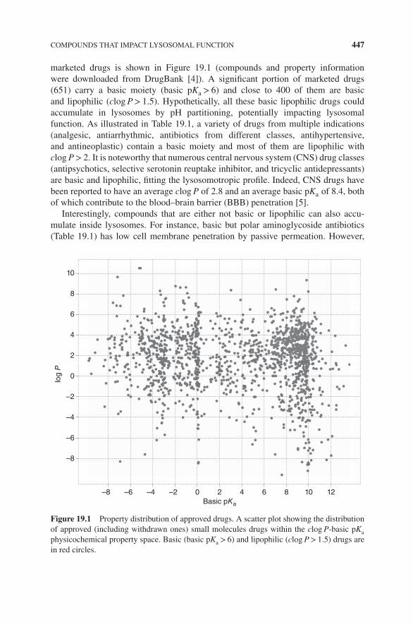
�
� �
�
COMPOUNDS THAT IMPACT LYSOSOMAL FUNCTION 447
marketed drugs is shown in Figure 19.1 (compounds and property informationwere downloaded from DrugBank [4]). A significant portion of marketed drugs(651) carry a basic moiety (basic pKa > 6) and close to 400 of them are basicand lipophilic (clog P> 1.5). Hypothetically, all these basic lipophilic drugs couldaccumulate in lysosomes by pH partitioning, potentially impacting lysosomalfunction. As illustrated in Table 19.1, a variety of drugs from multiple indications(analgesic, antiarrhythmic, antibiotics from different classes, antihypertensive,and antineoplastic) contain a basic moiety and most of them are lipophilic withclog P> 2. It is noteworthy that numerous central nervous system (CNS) drug classes(antipsychotics, selective serotonin reuptake inhibitor, and tricyclic antidepressants)are basic and lipophilic, fitting the lysosomotropic profile. Indeed, CNS drugs havebeen reported to have an average clog P of 2.8 and an average basic pKa of 8.4, bothof which contribute to the blood–brain barrier (BBB) penetration [5].
Interestingly, compounds that are either not basic or lipophilic can also accu-mulate inside lysosomes. For instance, basic but polar aminoglycoside antibiotics(Table 19.1) has low cell membrane penetration by passive permeation. However,
8
10
6
4
log P
2
0
–2
–4
–6
–8
–8 –6 –4 –2 0 2
Basic pKa
4 6 8 10 12
Figure 19.1 Property distribution of approved drugs. A scatter plot showing the distributionof approved (including withdrawn ones) small molecules drugs within the clog P-basic pKa
physicochemical property space. Basic (basic pKa > 6) and lipophilic (clog P> 1.5) drugs arein red circles.

�
� �
�
448 LYSOSOME DYSFUNCTION
TABLE 19.1 Basic Compounds in Multiple Drug Classes
Indication Class Drugs clog P Basic pKa
Analgesic Narcotic Levorphanol 2.9 9.66Levomethadyl
acetate4.88 9.87
Opioid Tapentadol 2.96 9.6Dezocine 3.23 9.67Dextromethorphan 3.49 9.85
Anesthetic Phenazopyridine 2.69 6.86Mepivacaine 3.19 7.25Lidocaine 2.84 7.75Bupivacaine 4.52 8Levobupivacaine 4.52 8
Antiallergic Antihistimine Chlorpheniramine 3.58 9.47Dexbrompheniramine 3.75 9.48Brompheniramine 3.75 9.48Phenindamine 3.62 9Promethazine 4.29 9.05Antazoline 2.88 9.24Loperamide 4.77 9.41Pheniramine 2.98 9.48
Antiarrhythmic Noncompetitivea- and b-adrenergicinhibitor
Amiodarone 7.64 8.47
Sodium channel blockers Encainide 4.49 9.48Mexiletine 2.46 9.52Flecainide 3.19 9.62Terfenadine 6.48 9.02
Antibiotic Aminoglycoside Kanamycin −7.1 9.75Amikacin −8.4 9.79Tobramycin −6.5 9.83Gentamicin −3.1 10.18Streptomycin −7.7 10.88Netilmicin −3.5 9.97
Fluoroquinolones Ciprofloxacin 8.68 −0.81Levofloxacin 6.2Moxifloxacin 9.42 −0.5Norfloxacin 8.68 −1Ofloxacin 6.2 −0.39Gemifloxacin 9.53 −0.92

�
� �
�
COMPOUNDS THAT IMPACT LYSOSOMAL FUNCTION 449
TABLE 19.1 (Continued)
Indication Class Drugs clog P Basic pKa
Ketolid Telithromycin 5.37 7.65Macrolide Troleandomycin 4.3 7.87
Josamycin 3.22 7.9Erythromycin 2.6 8.38Clarithromycin 3.24 8.38Azithromycin 2.44 9.57Rifapentine 3.56 7.98Quinupristin 2.18 8.28Rifabutin 4.19 8.62
Antifungal Ketoconazole 4.19 6.75Tioconazole 5.3 6.77Miconazole 5.96 6.77Econazole 5.35 6.77Sertaconazole 6.23 6.77Butoconazole 6.55 6.78
Anti-HIV Chemokine receptorantagonist
Maraviroc 3.63 9.38
Protease inhibitor Indinavir 2.81 7.37Nelfinavir 4.72 8.18Saquinavir 3.16 8.47
Antihypertensive Angiotensin II receptorantagonist
Eprosartan 3.8 6.93
Beta-1 adrenoceptorantagonist
Bevantolol 3.03 9.31
Calcium blocker Nicardipine 3.56 8.18Bepridil 5.49 9.16Lercanidipine 6.41 9.36Amlodipine 1.64 9.45Verapamil 5.04 9.68Perhexiline 5.53 10.58
Ganglionic blocker Mecamylamine 2.37 10.88Antimalarials Quinine 2.51 9.05
Hydroxychloroquine 2.89 9.76Lumefantrine 9.19 9.78Halofantrine 8.06 10.05Proguanil 1.89 10.12Primaquine 1.64 10.2Amodiaquine 3.76 10.23
(continued)

�
� �
�
450 LYSOSOME DYSFUNCTION
TABLE 19.1 (Continued)
Indication Class Drugs clog P Basic pKa
Antineoplastic Nonsteroidal selectiveestrogen receptormodulator
Toremifene 6.27 8.76
Tamoxifen 6.35 8.76Protein synthesis inhibitor Homoharringtonine 1.88 9.42Tyrosine kinase
inhibitorGefitinib 3.75 6.85
Vandetanib 4.54 9.13Nilotinib 4.41 6.3Lapatinib 4.64 7.2Dasatinib 3.82 7.22Ponatinib 4.97 8.03Imatinib 4.38 8.27Bosutinib 4.09 8.43Afatinib 3.76 8.81Sunitinib 2.93 9.04Daunorubicin 1.73 8.94
CNS Antipsychotics Promazine 3.93 9.2Quetiapine 2.81 7.06Chlorpromazine 4.54 9.2Chlorprothixene 5.07 9.76
Serotonin–norepinephrinereuptake inhibitor
Desvenlafaxine 2.29 8.87
Venlafaxine 2.74 8.91Selective serotonin
reuptake inhibitorFluvoxamine 2.8 9.16
Fluoxetine 4.17 9.8Sertraline 5.15 9.85
Tricyclic antidepressant Trimipramine 4.76 9.42Amitriptyline 4.81 9.76Amoxapine 3.08 8.83Imipramine 4.28 9.2Clomipramine 4.88 9.2Doxepin 3.84 9.76Desipramine 3.9 10.02Nortriptyline 4.43 10.47Protriptyline 4.5 10.54Maprotiline 4.37 10.54
lysosomal accumulation has been demonstrated for multiple aminoglycosideantibiotics [6], specifically in the kidney proximal tubule cells [7] and sensory haircells of the inner ear [8]. Rather than passive permeation, specialized uptake trans-porters and endocytosis is utilized by those polar molecules to transport themselvesinto lysosomes [9]. The major transport of aminoglycosides into proximal tubule

�
� �
�
COMPOUNDS THAT IMPACT LYSOSOMAL FUNCTION 451
cells involves interaction with acidic, negatively charged phospholipid-bindingsites at the level of the brush border membrane. The megalin receptor, a scavengerreceptor with broad tissue distribution, including the cochlea of the inner ear [10]and apical surface of polarized epithelial cell membranes facing the proximal tubule[11], has been shown to contribute to the aminoglycoside accumulation in thesetissues [12].
Another chemical class that is not associated with the basic lipophilic propertiesbut is lysosomotropic involves heavy metals such as arsenic, cadmium [13,14], andplatinum-containing compounds (e.g., cisplatin) [15]. Cadmium and arsenic areenvironmental toxicants, and cisplatin is one of the most widely used antitumordrugs. Similar to aminoglycoside antibiotics, various membrane transporters havebeen shown to be involved in heavy metal uptake. Cellular uptake of cisplatin ismediated, at least in part, by transport proteins including the copper transporters 1and 2 (Ctr1 and Ctr2), the P-type copper-transporting ATPases ATP7A and ATP7B,the organic cation transporter 2 (OCT2), and the multidrug extrusion transporter 1(MATE1) [16]. Similarly, arsenic is transported into mammalian cells via multiplepathways including the aquaglyceroporins (AQP3, AQP7, AQP9, AQP10), theglucose permeases (GLUT1, GLUT2, GLUT5), and the organic anion transportingpolypeptides (OATPB, OATPC) [17]. The major pathway for cadmium uptakeby kidney proximal tubule cells involves the endocytosis of cadmium complexesfollowed by binding to the high-affinity metal-binding protein metallothionein [18].Cadmium may also be transported into cells via zinc and/or copper transporters [19].Interestingly, various metal transporters are located on the lysosomes and play arole in metal import/export from lysosomes [20]. The proton-coupled divalent metaltransporter DMT1, for example, is expressed exclusively in endosomes/lysosomesand transports cadmium into lysosomes [21]. The exact mechanism of lysosomalsequestration of heavy metals and what lysosomal transporters are involved needs tobe further investigated.
19.2.2 Nonlysosomotropic Compounds
Lysosomal accumulation is not the only mechanism by which xenobiotics canimpact the lysosomal pathway. Lysosomal toxicants can also affect lysosomalfunction via pharmacological inhibition of molecular targets involved in lysosomalregulation. Bafilomycin A (Baf A) and concanamycin, for example, can blocklysosomal acidification through selective inhibition of the V-type ATPase [22]. Theionophores monensin and nigericin, which mediate the exchange of H+ and Na+, canalso increase the pH of lysosomes [23]. Evidently, inhibition of various cathepsinscan disrupt lysosomal proteolysis. Microtubule disruptors, such as paclitaxel andvincristine, can impair lysosomes by disrupting microtubule-dependent trafficking.Bacterial proteins such as staphylococcal a-toxin and anthrax toxin have been shownto impact lysosomal function by disrupting lysosomal membrane integrity [24,25].Recently, it was shown that botulinum neurotoxins (BoNTs, serotypes A–G) andtetanus neurotoxin (TeNT) induce perturbations of the fusogenic SNARE (solubleN-ethylmaleimide-sensitive factor attachment protein receptor) complex dynamics,

�
� �
�
452 LYSOSOME DYSFUNCTION
which could impact the fusion of lysosomes with other vesicles. In addition,tumor necrosis factor-related apoptosis-inducing ligand (TRAIL) was observed totrigger lysosomal change after binding to death receptor 5 via recruitment of theproapoptotic proteins Bim and Bax to the lysosomes [26].
19.3 CELLULAR CONSEQUENCES
Lysosomes are kinetic organelles and have an intimate link with the endocyticapparatus, phagocytosis, and autophagy, which deliver materials to lysosomes fordegradation and recycling. Lysosomes are not the only acidic organelles; earlyendosomes, late endosomes, phagosomes, and autolysosomes all have a differingdegrees of luminal acidity and thus are targets for accumulation of basic lipophilicxenobiotics. For simplicity, our discussions primarily focus on lysosomal changesand vesicular trafficking.
19.3.1 Effect of Drugs on pH and Lysosomal Volume
Accumulation of high concentrations of basic compounds in lysosomes can increasethe luminal pH. The lysosomotropic compounds, chloroquine and methylamine, wereshown to increase lysosomal pH drastically (0.5–2.0 pH units) after accumulationin vitro [27], and in the case of chloroquine, in vivo [28]. The LysoTracker probesconsist of a fluorophore linked to a weak base that is only partially protonated at neu-tral pH. This allows LysoTracker probes to freely permeate cell membranes enablingthem access to the luminal regions of acidic organelles in live cells. Similar to otherlysosomotropic compounds, low pH is required for the lysosomal accumulation andsubsequent detection of LysoTracker® probes. In a previous study using H9C2 cells,basic lipophilic compounds were shown to decrease the staining of LysoTracker RedDND-99 (LTR) after 4 h of treatment, indicating an increase in lysosomal pH [29].The drugs used in this study were from multiple drug classes and pharmacologi-cal actions including antidepressants, antipsychotics, and anticancer, indicating theincrease in pH is related to their physicochemical properties rather than specific phar-macological target inhibition. Indeed, clustering analysis demonstrated that com-pounds possessing a clog P> 2 and a basic pKa between 6.5 and 11 were associatedwith the increase in pH [29]. These data are consistent with the observation from adifferent publication using Fa2N-4 cells in which the decrease in LTR fluorescencewas associated with basic and lipophilic compounds [30]. An increase in pH would beexpected to decrease the lysosomal degradation capability since acidic pH is optimalfor lysosomal enzyme activity. In addition, an increase in pH can decrease the fusioncapability of lysosomes [31], and in fact, chloroquine has been shown to decrease thefusion between autophagosome and lysosome [32]. Besides basic lipophilic com-pounds, vacuole ATPase inhibitors, such as Baf A, can also inhibit the acidificationof lysosomes, resulting in decreased LTR staining [33].
Interestingly, pH increase by basic lipophilic compounds is only a short-termeffect. No significant change in lysosomal pH was observed after 24 h of imipramine

�
� �
�
CELLULAR CONSEQUENCES 453
exposure [34]. Consistent with a reversal of the pH change, prolonged exposure ofnumerous basic lipophilic drugs significantly increases the cellular accumulationof LTR [35,36], indicating an increase in lysosomal volume and normalizationof luminal pH. A fourfold increase in lysosomal volume was estimated based onboth the lysosomal pH and amount of LTR accumulated after 24 h exposure ofimipramine [34]. One plausible reason for the increase in lysosomal volume could bedue to an adaptive change by increasing lysosomal biogenesis with basic lipophiliccompounds. For example, increases in lysosome-associated membrane protein 2(LAMP-2) staining was observed in vivo with multiple basic lipophilic drugs [37]and marked lysosome-associated membrane protein 1 (LAMP-1) increase wasobserved in vitro with chloroquine treatment [38]. Transcription factor EB (TFEB)is a master transcription factor for lysosomal biogenesis [39] and TFEB nucleartranslocation was triggered by chloroquine treatment, which could explain in part theincrease in lysosomal volume [40]. TFEB also potentially plays a role in reversingthe pH change via upregulation of vATPase [39].
A second reason for an increase in lysosomal volume is accumulation ofundigested substrate in lysosomes. Lysosomal volume expansion by imipraminecan be reversed by exposing cells to hydroxypropyl-β-cyclodextrin, which reduceslysosomal cholesterol burden, suggesting elevation in lysosomal cholesterol contentcontributes to the lysosomal volume increase [34]. Similar lysosomal changeswere also observed for compounds that impact lysosomes via nonaccumulativeprocess. For instance, increases in the acidic compartment and LAMP-2-positivevesicles were observed following vincristine treatment [41]. Interestingly, cellsderived from patients with lysosomal storage diseases (LSDs) are often associatedwith an increase in LTR staining [34,42], implying the increase in LTR by compoundtreatment is indicative of lysosomal dysfunction. Hence, decreased LTR staining isa common approach as a phenotypic screen for LSDs drug discovery [42], whichfurther supports this notion.
19.3.2 Effects on Lysosomal Enzymes
Besides the pH change, lysosomotropic compounds have demonstrated effects onlysosomal enzyme activity, either directly or indirectly. Chlorpromazine and chloro-quine have demonstrated inhibition of lysosomal phospholipase A1 in vitro [43,44],as well as downregulation of acid ceramidase that was not caused by decreased tran-scription [45]. Furthermore, multiple lysosomotropic compounds have demonstratedthe ability to redistribute the mannose 6-phosphate (M6P) receptor from the TGN toendosomes and concomitantly increase the secretion of lysosomal enzymes resultingin a decline of mature intracellular lysosomal enzyme levels [46]. Since the majorityof enzymes delivered to lysosomes require the M6P receptor, the redistribution ofthe receptor by lysosomotropic compounds could dysregulate a range of lysosomalenzymes. Reduction of lysosomal enzyme activity (either due to alterations of pH, orby direct or indirect inhibition) could contribute to lysosomal dysfunction. Paradox-ically lysosomotropic cathepsin K inhibitors were shown to increase other cathep-sin activity [47]. In addition, upregulation of cathepsin D was also demonstrated in

�
� �
�
454 LYSOSOME DYSFUNCTION
Alzheimer’s disease (AD) presumably due to a compensatory protective response[48,49]. It is therefore conceivable that lysosomal toxicants can also trigger upregu-lation of lysosomal cathepsins to offset the lysosomal dysfunction. Long-term impactof lysosomal toxicants on lysosomal enzymes needs to be evaluated further.
19.3.3 Lysosomal Substrate Accumulation
One of the critical functions of lysosomes is to break down all types of biologicalpolymers including proteins, nucleic acids, carbohydrates, and lipids. Changes inpH or enzymatic activity by basic lipophilic compounds may decrease lysosomaldegradation capacity, triggering accumulation of various substrates. One of the bestcharacterized examples of this is the excessive accumulation of phospholipid, forexample, phospholipidosis (PLD), induced by lysosomotropic compounds. Thehallmark feature of PLD is the characteristic lamellar bodies that are visible bytransmission electron microscopy [50] (see Chapter 20). An in silico model forPLD prediction proposed by [51] describes a simple method based on both pKaof the most basic center and clog P value of the molecules. Interestingly, besidesbasic lipophilic compounds, polar aminoglycoside antibiotics, which carry a basicmoiety, are also associated with PLD [52]. Lysosomal hydrolysis contributesto the mobilization of lipid droplet-associated cholesterol [53] and cholesterolaccumulation is associated with lysosomal dysfunction. For instance, a mutation inNPC1 resulting in the accumulation of cholesterol in late endosomes or lysosomesleads to the autosomal recessive disease Niemann–Pick disease type C (NPC). Thebasic lipophilic compounds U18666A, clomiphene, and terconazole have been usedto produce the NPC phenotype (cholesterol accumulation) experimentally [54,55].Moreover, accumulation of lipofuscin, which is an intralysosomal waste materialconsisting of various components such as protein, lipid, carbohydrates, metals, andautofluorescent pigment [56] has been observed in postmitotic cells associated withaging, lysosomal protease inhibitors, and chloroquine treatment [57,58].
Excessive accumulation of various substrates triggers severe lysosomal dysfunc-tions exemplified by LSDs. While the majority of LSDs are a consequence of thedeficiency of a single enzyme required for the metabolism of lipids or glycoproteins,lysosomotropic compounds may affect multiple lysosomal activities. In addition,lysosomotropic agents including ammonium chloride, chloroquine, methylamine,doxorubicin, and the lysosomal enzyme inhibitor E64D, cause ferritin accumulation[59], which prevents the redistribution of iron to vital cellular processes and triggerscellular damage as a result.
19.3.4 Lysosomal Membrane Permeabilization (LMP) and Cell Death
Lysosomal membrane integrity, as measured by acridine orange leakage, can becompromised by excessive compound accumulation [60,61]. Subsequent leakageof large amounts of lysosomal acid hydrolases following lysosomal membranedisruption leads to cellular toxicity through apoptotic mechanisms [62–65]. Ingeneral, the magnitude of lysosomal rupture and, consequently, the amount of
�
� �
�
CELLULAR CONSEQUENCES 455
hydrolytic enzymes released into the cytosol, is dose dependent and may inducesublethal damage, apoptosis, or necrosis depending on the exposure level andduration [66]. This dose-related differential outcome has been demonstrated by thelysosomotropic agent O-methyl-serine dodecylamide hydrochloride (MSDH), inwhich apoptosis accompanied by caspase-3 activation occurred at <50 μM while at>75 μM necrosis occurred and was associated with more extensive lysosomal rupture[67]. Tamoxifen also exhibited a similar toxicity profile in which high concentration(10 μM) caused extensive necrosis while apoptotic-like cell death was observed atlower concentrations [68]. The close connection between lysosomal accumulationand cell death is further demonstrated by the observation that Baf A pretreatment ofcells can rescue the toxicity caused by certain basic lipophilic compounds [69].
The intimate relationship between lysosomes and mitochondria in the executionof apoptosis is emphasized in the lysosomal–mitochondria axis theory of apoptosis,where released lysosomal enzymes trigger mitochondria permeability transition andapoptosis via the Bcl-2 family (e.g., bax and BAD) [70] (see Chapter 8). Lysosomalmembrane permeabilization has also been reported to initiate a caspase-independentcell death pathway [71] as well as the release of iron that can catalyze the formationof highly reactive hydroxyl radicals leading to oxidative stress-induced apoptosis ornecrosis [72].
19.3.5 Membrane Trafficking Changes
Lysosomes are the central hub for membrane trafficking including endocytosis,autophagy, and phagocytosis. Lysosomal dysfunction from xenobiotic effects onpH, enzymatic activity, substrate accumulation, and/or impairment of membraneintegrity could lead to disruption of various trafficking pathways associated withlysosomes.
19.3.5.1 Autophagy Perturbation Autophagy refers to the catabolic process ineukaryotic cells that delivers cytoplasmic material to lysosomes for degradation.This highly conserved process is involved in the clearance of long-lived proteins anddamaged organelles. Vinblastine (a microtubule inhibitor), leupeptin (a lysosomalprotease inhibitor), and the lysosomotropic amines propylamine and chloro-quine are all capable of inhibiting autophagy in various model systems [73,74].Microtubule-associated protein light chain 3 (LC3) is an ubiquitin-like proteinrequired for the formation of autophagosomal membranes. During autophagy, acytosolic form of LC3 (LC3-I) is conjugated to phosphatidylethanolamine to form anLC3-phosphatidylethanolamine conjugate (LC3-II), which is recruited to autophago-somal membranes. Detection of LC3 by immunoblotting or immunofluorescencehas become a reliable method for monitoring autophagy. Several lysosomotropiccompounds have been shown to increase LC3 staining, indicating the modulationof autophagy [75]. The concentration-dependent increase in the abundance ofthe autophagy substrate, sequestosome 1 (SQSTM1/p62, hereafter referred to asp62) by these compounds supports the hypothesis that the increase in LC3 wasdue to autophagy inhibition rather than upstream enhancement. In addition, the

�
� �
�
456 LYSOSOME DYSFUNCTION
downregulation of extracellular matrix and cytoskeletal gene transcription by thesecompounds also indicate ineffective turnover of long-lived proteins, supporting thenotion of autophagy inhibition. Further evidence of this hypothesis is demonstratedby the finding that the lysosomotropic detergent siramesine decreases the colo-calization of LC3-positive vesicles with LAMP-2-positive vesicles, indicating theimpairment of autophagosomal fusion with lysosomes [76]. Conversely, many basiclipophilic compounds such as amiodarone [77], tamoxifen [78], amitriptyline [79],verapamil [80], and dimebon [81] have been proposed as autophagy stimulatorsbased on their ability to increase LC3. Because of their basic lipophilic properties,these compounds have the propensity to accumulate in lysosomes [29].
One common assay format employed to study autophagy flux is to evaluatethe effect on LC3-II abundance with test compound in the presence of proteaseinhibitors (e.g., E64d) or Baf A. An increase of LC3-II abundance with the combi-nation treatment relative to the test compound alone is indicative of increased fluxthrough the autophagy process. However, this assay only measures the initial stage ofautophagosome formation without the evaluation of the degradation step. Recently,it was shown that lysosomotropic compounds (e.g., chloroquine) induce lysosomalstress and, consequently, provokes TFEB nuclear translocation [40], which coulddrive expression of autophagy genes leading to the induction of autophagy. It ispossible that the increase of LC3-II caused by these basic lipophilic compounds isdue to the adaptive increase in autophagy by TFEB nuclear translocation. Ultimately,downstream lysosomal dysfunction inflicted by lysosomotropic compounds wouldtend to block autophagy [75]. Other types of membrane trafficking perturbationdiscussed later further supports the notion of global lysosomal dysfunction.
19.3.5.2 Impairment of Endocytosis/Phagocytosis Endocytosis is an energy-consuming process by which cells take up extracellular molecules (such as pro-teins) by engulfing them. There are various mechanisms of endocytosis such asclathrin-mediated, caveolae, and macropinocytosis. Phagocytosis is a special form ofendocytosis in which large particles are taken up via the phagosome. The endocyticpathway consists of distinct membrane compartments, which internalize moleculesfrom the plasma membrane and then either recycle them back to the surface, as inearly endosomes and recycling endosomes, or designate them for degradation, as inlate endosomes and lysosomes. Lysosomes are the last compartment of the endocyticpathway and their dysfunction by lysosomal toxicants can negatively impact theendocytic process and various endocytic compartments.
Indeed, numerous lysosomal toxicants are known to impact various stagesand compartments of endocytosis and are routinely used to study the endocyticprocess. For example, direct evidence for recycling came from the observation thatthe process can be inhibited by basic compounds, including chloroquine, NH4Cl,methylamine, and ionophores, such as monensin [82]. Many receptors are recycledand return to the plasma membrane after ligand dissociation. Lysosomal toxicantshave been shown to prevent recycling of receptors for LDL [83], transferrin [84],epidermal growth factor, and macroglobulin [85]. Instead of returning to the plasmamembrane they became trapped in endosomes. The mechanism could be related

�
� �
�
CELLULAR CONSEQUENCES 457
to the pH elevation of acidic compartments and inhibition of the dissociating ofthe ligand from the receptor [86] or perturbation of the clathrin lattice assembly.Indeed, chloroquine is recognized as a clathrin-dependent endocytosis inhibitor[87]. In addition, using an in vitro macrophage model, monensin and chloroquinewere shown to inhibit transfer to lysosomes of endocytosed macromolecules [88].Azithromycin, a lysosomotropic antibiotic, delayed sequestration of receptor-boundtransferrin and peroxidase–antiperoxidase immune complexes into cell-surfaceendocytic pits and vesicles [89].
Virus entry relies on either membrane fusion or endocytosis and, intriguingly, theviral entry has been shown to be impacted by lysosomotropic compounds. Ebolaviruses enter the cell by a macropinocytic-like process after attachment to the cellsurfaces [90], and multiple lysosomotropic compounds, including U18666A andclomiphene, were shown to inhibit Ebola virus entry and infection. Chloroquine andNH4Cl also demonstrated more than a 50% reduction of HCV infectivity. Recently,another lysosomotropic compound, amiodarone, has been shown to inhibit the entryof the hepatitis C virus [91]. Together these data strongly support the inhibition ofendocytosis by lysosomal toxicants.
19.3.5.3 Phagocytosis Basic lipophilic local anesthetics, dibucaine, tetracaine,and procaine, have been shown to exert a reversible inhibition of phagocytosisof opsonized sheep red blood cells using mouse peritoneal macrophages [92].Interestingly, the potencies of these local anesthetics for inhibition of phagocyticuptake were proportional to their log P, indicating the critical role of this physic-ochemical property in inhibiting phagocytosis. Retinal pigment epithelium (RPE)is one of the most phagocytotically active cell types. Chloroquine has been shownto decrease the uptake of rhodamine-labeled dextran in retinal pigment epithelialcells [38], suggesting disruption of the phagocytic pathway. The basic lipophiliccompounds tamoxifen, toremifene, and chloroquine were also shown to decrease theuptake of FITC-labeled rod outer segments [93] in retinal pigment epithelial cellsin vitro, further supporting the notion of inhibiting phagocytosis by lysosomotropicxenobiotics.
19.3.5.4 Exocytosis Exocytosis involves the transport of intracellular vesiclesto the plasma membrane of the cell where vesicular fusion results in the deliveryof membrane and protein to the cell surface, as well as secretion of the vesicularcontents. It is well established that increases in lysosomal pH result in lysosomalexocytosis, with enhanced secretion of preformed hydrolases [46,94–96]. Chloro-quine not only stimulated the release of porphyrins from yeast [97] but also enhancedthe exocytosis of MTT formazan, which accumulates in the endosomal/lysosomalcompartment [98]. Similarly, exocytosis of lysosomal contents was also observedfrom skeletal muscle in vivo following chloroquine treatment [99]. The associationof increased exocytosis with lysosomal dysfunction is further supported by the find-ings in lysosomal storage and aging-related diseases. Lysosomal storage materialshave been found in extracellular fluids, blood, and urine in some LSDs patients,providing direct evidence of lysosomal exocytosis [100,101]. The pathogenesis of
�
� �
�
458 LYSOSOME DYSFUNCTION
different neurodegenerative diseases, including AD, Parkinson’s disease (PD), andHuntington’s disease (HD), share one common feature: abnormal accumulationand aggregation of disease-specific proteins that form either intracellular inclusionsor extracellular aggregates, presumably by excessive lysosomal exocytosis. Forinstance the truncated, misfolded, and oxidatively modified α-synuclein formsappear to be enriched in the vesicular fraction and to be preferentially secreted [102].Chloroquine has been shown to increase nuclear translocation of TFEB duringlysosomal stress [40], promoting lysosomal exocytosis by increasing the pool oflysosomes in the proximity of, and promoting fusion with, the plasma membrane[103]. Besides the role of TFEB, Ca2+ release also contributes to the regulation ofexocytosis. The lysosomotropic agent glycyl-l-phenylalanine-naphthylamide (GPN)evoked robust increases in the intracellular Ca2+ concentration and triggered releaseof β-hexosaminidase within 5 min of exposure, demonstrating the characteristicruffling of the plasma membrane associated with exocytosis [104]. Ca2+ releasefrom the lysosome in this case is believed to play a role in the immediate increase inexocytosis.
19.3.6 Other Cellular Impacts
Due to the central role of lysosomes in membrane trafficking, organelle homeostasiscritically relies on effective lysosomal function. Membrane trafficking perturbationdue to lysosomal dysfunction subsequently impacts other organelles. In addition, theintriguing discovery that activation of mTORC1 requires lysosomal recruitment inresponse to amino acid concentrations [105] positions these degradation organelles asa new hub for signaling transduction. However, how xenobiotics affect cell signalingvia lysosomes is yet to be investigated.
19.3.6.1 Mitochondria The intimate relationship between lysosomes andmitochondria is twofold. First, as discussed earlier, released cathepsins can destabi-lize mitochondria, promote cytochrome c release, and ultimately induce apoptosis.Second, autophagic delivery to lysosomes is the major degradative pathway in mito-chondrial turnover, which is critical for mitochondrial quality control [106]. Thisspecial form of mitochondrial degradation by autophagy is referred to as mitophagy.Mitochondrial abnormalities would be an expected outcome of drug-induced lysoso-mal dysfunction and the persistence of dysfunctional mitochondria has indeed beenobserved with multiple LSDs [107]. Abnormal mitochondria were also observedin multiple autophagy knockout studies further strengthening this critical role ofautophagy [108]. The number of mitochondria and GFP-LC3 colocalized punctaincreased in the presence of lysosomal inhibitors such as chloroquine or E64 Dplus pepstatin A [109]. Accumulation of damaged mitochondria by lysosomotropiccompounds could certainly lead to cellular perturbations.
19.3.6.2 Endoplasmic Reticulum The ER has a role in folding and modifyingproteins, and secreting proteins via the secretory pathway. Elevated levels of mis-folded proteins causes ER stress, which is commonly observed in cells derived from

�
� �
�
CELLULAR CONSEQUENCES 459
LSD patients, indicating a role for lysosomes in maintaining of ER homeostasis[110]. The autophagy process has been shown to degrade sequestered ER withinautophagosomes [111], for instance, autophagy-deficient T lymphocytes demonstrateabnormal expansion of ER and impaired calcium influx [112]. Induction of autophagyduring ER stress [113,114] is believed to serve as a protective mechanism againstcell death. Presumably, an autophagy defect due to lysosomal dysfunction inducedby lysosomal toxicants could exacerbate the ER stress and enhance cytotoxicity.The lysosomotropic kinase inhibitors, imatinib and flavopiridol, have been associatedwith ER stress resulting from perturbation of autophagy and lysosomal membranepermeabilization [61] [115].
19.3.6.3 Oxidative Stress Several reports have associated reactive oxidativespecies (ROS) with lysosomotropic agents [116–119]. Interestingly, in situ imagingshowed ROS production was present within 1 h of exposure to the lysosomotropicantibiotic gentamicin [120]. The links between autophagy dysregulation andincreases in oxidative stress are evident by mouse lysosomal protein knockoutstudies and autophagy inhibitors [108]. These studies demonstrate the accumulationof oxidized ubiquitinated proteins and dysfunctional mitochondria. Mitochondriaare a major site of ROS generation from multiple sites along the respiratory chain[121], and accumulation of damaged mitochondria due to lysosomal functionimpairment likely contributes to the increase of oxidative stress. Interestingly,lysosomes are susceptible to oxidative stress and an increase in oxidative stress canact as an amplifying loop to further destabilize the lysosomal membrane [122,123].Antioxidant treatments have demonstrated that prevention of ROS production at leastpartially rescued cell death caused by lysosomal permeabilization [120]. Lysosomesare rich in redox-active iron due to degradation of iron-containing macromoleculesand the lysosomal iron has been shown to play a role in exacerbating the effectsof ROS via the Fenton reaction, in which an extremely reactive hydroxyl radical isformed during hydrogen peroxide oxidization of iron [124].
19.3.6.4 Signaling Modulation Besides membrane trafficking and degradation,lysosomes have a critical emerging role as a signaling hub, particularly for nutrientsensing and cellular metabolism. mTORC1 needs to be recruited to the lysosomal sur-face to exert its kinase activity [105]. Interestingly, it is the level of amino acids insidethe lysosomal lumen that controls mTORC1 docking on the lysosomal surface andsubsequent activation. Multiple protein complexes, including Rag GTPase, raptor andregulator, are involved in the mTORC1 translocation. p62, a substrate for autophagy,has been shown to bind to raptor, becoming an integral part of the mTORC1 complexand facilitating mTORC1 translocation to the lysosomal surface upon amino acidstimulation [125]. Furthermore, activation of mTORC1 inhibits both autophagy andlysosome biogenesis [126,127].
Lysosomal toxicants may affect signaling molecules via lysosomes in severalways. First of all, inadequate lysosomal degradation may decrease free aminoacid generation, impeding mTORC1 activation. Indeed, a decrease in mTORC1activity was not only observed in LSD models, such as in fibroblasts from

�
� �
�
460 LYSOSOME DYSFUNCTION
mucopolysaccharidosis type I, Fabry disease, and aspartylglucosaminuria [128],but also observed after treatment with several lysosomotropic compounds (e.g.,chloroquine [40], amiodarone [129], and imipramine [130]. In addition, p62 has beenshown to regulate various other signal transduction pathways (Nrf2, NF-κB, caspase8, and ERK1) through specific protein interactions [131]. As was discussed before,increases in p62 are typically associated with lysosomal dysfunction and autophagydeficiency, and this increase could modulate multiple signaling molecules. Anoncanonical mechanism of Nrf2 activation by autophagy deficiency due todirect interaction between Keap1 and p62 has recently been proposed [132,133].Essentially, p62 interacts with the Nrf2-binding site on Keap1, a component ofCullin-3-type ubiquitin ligase for Nrf2. When p62 accumulates due to autophagydeficiency, it competes with the interaction between Nrf2 and Keap1, resulting in sta-bilization of Nrf2 and transcriptional activation of Nrf2 target genes. Lysosomotropiccompounds were clearly shown to induce multiple Nrf2-regulated genes such asglutamate–cysteine ligase and glutathione-S-transferase, indicating Nrf2 activation[75]. Remarkably, arsenic inhibits autophagic flux and activates the Nrf2-Keap1 path-way in a p62-dependent manner since Nrf2 activation by arsenic is diminished whenp62 is knocked down [134]. In addition, activation of NF-κB has been observed in anautophagy-suppressed Atg7 knockout model accompanied by accumulation of p62[135], whereas knockout of p62 suppressed NF-κB signaling, indicating a critical roleof p62 in the regulation of NF-κB. The lysosomotropic compounds chloroquine andimatinib mesylate were indeed shown to induce NF-κB in astroglial cells and pan-creatic islet cells, respectively [136,137]. However, whether the increase in NF-κBis generic for all lysosomotropic compounds and whether the effect is dependent onp62 needs to be further investigated. p62 also promotes the full activation of caspase8 with subsequent triggering of apoptosis [138], and suppresses ERK1 signal duringadipogenesis [139]. More recent data showed autolysosomal sequestration of activeRhoA was via p62 and negative regulation of RhoA signaling activity by constitutiveautophagy [140]. Overall, lysosomal changes and p62 accumulation due to autophagydeficiency from lysosomal toxicant treatment could set off a chain of altered signalingmolecules, which in turn can further impact a variety of biological processes.
In conclusion, we propose a multifaceted model (Figure 19.2) in which lysosomaltoxicants, either by accumulation or disturbance of lysosome function, not onlyinduce cell death due to lysosomal membrane permeabilization but also impairlysosomal function. These functional changes can result in the perturbation ofmultiple membrane trafficking pathways including autophagy, endocytosis, phago-cytosis, and possibly exocytosis enhancement. Besides the lysosomal change, otherorganelles such as mitochondria and ER could be damaged due to deficiency ofautophagy, which subsequently leads to oxidative stress. Furthermore, multiplesignal transduction processes including mTORC1, ERK1, Nrf2, and NFκB aremodulated by lysosomal dysfunction due to the increase of p62 abundance resultingfrom autophagy deficiency. The pathophysiological consequences of modulatingthose signaling molecules by lysosomal toxicants needs to be further deciphered.Thus far, the focus has been on the impact of small molecules on lysosomes.However, nanotechnology is currently an area of intense scientific interest due to a

�
� �
�
IMPAIRED LYSOSOMAL FUNCTION AS A MECHANISM FOR ORGAN TOXICITY 461
Exosome
V-ATPase
B BH+
Toxicant
Endsosome
Autophagosome
SQSTM1/p62
mTOR
Nrf2
NfκB
Erk1
Phagosome
Mitochondria
Cell death
Lysosome
Mitochondriadysfunction
ERstress
Oxidativestress
H+
H+
H+ H+H+ H+
Figure 19.2 Biological impacts of the lysosomotropic compounds. Compounds can perturblysosome functions either by accumulation inside the lysosomes or direct inhibition of variouslysosomal functions (e.g., decrease of lysosomal enzyme activity). Lysosomal membrane per-meabilization can trigger cell death. In addition, lysosomal dysfunction can tamper membranetrafficking pathways including autophagy, endocytosis, phagocytosis, and exocytosis. Multiplesignal transduction processes including MTORC, ERK1, Nrf2, and NFκB can also be modu-lated by lysosomal dysfunction due to the increase of p62 abundance resulting from autophagydeficiency. (See color plate section for the color representation of this figure.)
wide variety of potential applications in biomedical, optical, and electronic fields,and various nanoparticles have been shown to be trapped inside the lysosomes anddisrupt lysosome function including LMP and inhibition of autophagy [141,142].Thus, lysosomal dysfunction could be an emerging toxicological mechanism forboth small molecules and nanomaterials.
19.4 IMPAIRED LYSOSOMAL FUNCTION AS A MECHANISM FORORGAN TOXICITY
A number of cellular processes can be affected by lysosomal dysfunction that canultimately lead to organ toxicity as illustrated in LSDs. Pathological manifestationsin LSDs as a result of lysosomal defects strongly indicate that a similar mechanismcould contribute to organ toxicity resulting from xenobiotics that perturb lysoso-mal function. In addition to the CNS, multiple peripheral organ systems including

�
� �
�
462 LYSOSOME DYSFUNCTION
cardiac, kidney, skeletal muscle, and hepatocyte are impacted by LSDs [107]. Aswill be discussed later, a number of organ toxicities have been associated with lyso-somal dysfunction. Obviously, many mechanisms discussed previously such as LMP,blocking of membrane trafficking, impacted organelle function, and modulation ofsignaling molecules elicited by lysosomal toxicants could contribute to target organtoxicity. Furthermore, one critical point that needs to be taken into consideration isthat many lysosomotropic compounds have higher volume of distribution [143] andtissue exposure in general is higher than systemic plasma exposure, especially in theorgans that are known to have abundant lysosomes such as liver, lung, and kidney.Indeed in animal studies, chloroquine concentrations in the liver, spleen, kidney, andlung were found to be 700–1600 times the concentration in plasma after 1 monthof administration at 40 mg/kg/day [144]. Even in the heart, which has low lysosomalcontent, chloroquine concentrations were 200 times over plasma levels. Higher tissuedrug exposure can certainly increase the risk of organ toxicity. Remarkably, simplylowering the pKa can result in decreased tissue exposure and lower liver toxicity [145]supporting the role of lysosomal accumulation in xenobiotic-induced toxicity.
19.4.1 Liver Toxicity
More than 900 drugs, toxins, and herbs have been reported to cause liver injury.Different mechanisms such as reactive metabolite formation, oxidative stress, andmitochondrial impairment have been proposed to contribute to the pathogenesis ofdrug-induced liver injury. The involvement of the lysosomal pathway in liver toxicityhas been suggested by numerous studies. Oral drugs are absorbed by the gastrointesti-nal system and enter the liver via the hepatic portal vein prior to systemic circulation.Thus, the liver typically receives the highest maximal drug exposures relative toother tissues. These high maximal exposures coupled with the lysosomal trapping ofbasic lipophilic drugs often leads to drug accumulation in the liver. For example, thelysosomotropic compounds amiodarone and chloroquine have been shown to havesignificantly higher levels in liver tissue compared to the plasma [146,147]. Manybasic lipophilic compounds, such as amiodarone, chloroquine, and perhexiline, areindeed associated with hepatic morphologic effects, which include marked lysosomalchanges described as lamellar lysosomal inclusions [148].
As discussed previously, nonbasic xenobiotics can affect the lysosomal pathwayvia a nonaccumulation mechanism. The cathepsin B–mitochondria apoptotic path-way has been demonstrated to play a critical role in hepatocyte apoptosis and liverinjury induced by TNFα [149,150]. Either genetic knockout or pharmacologicalinhibitors of cathepsin B can mitigate the liver damage. Interestingly, acetaminophen,overdose of which is the most frequent cause of drug-induced liver failure in theUnited States, can also increase the lysosomal membrane instability as demon-strated by the release of cathepsin B from lysosomes [151]. In addition, lysosomaliron mobilization into mitochondria has been shown as a contributing factor toacetaminophen-induced hepatotoxicity [152,153]. Hepatotoxicity is also one of theadverse reactions caused by diclofenac, an arylacetic nonsteroidal anti-inflammatorydrug (NSAID). Involvement of LMP and lysosomal/mitochondrial cross talk induced
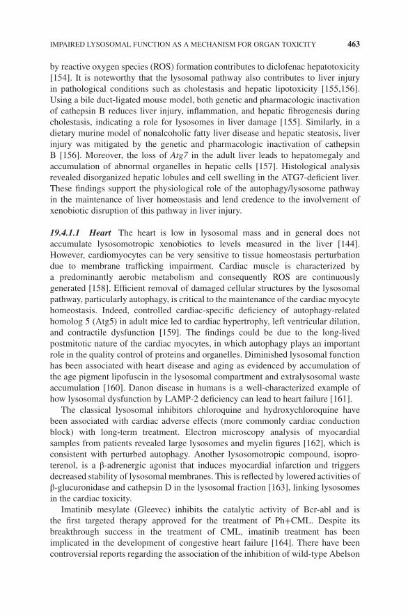
�
� �
�
IMPAIRED LYSOSOMAL FUNCTION AS A MECHANISM FOR ORGAN TOXICITY 463
by reactive oxygen species (ROS) formation contributes to diclofenac hepatotoxicity[154]. It is noteworthy that the lysosomal pathway also contributes to liver injuryin pathological conditions such as cholestasis and hepatic lipotoxicity [155,156].Using a bile duct-ligated mouse model, both genetic and pharmacologic inactivationof cathepsin B reduces liver injury, inflammation, and hepatic fibrogenesis duringcholestasis, indicating a role for lysosomes in liver damage [155]. Similarly, in adietary murine model of nonalcoholic fatty liver disease and hepatic steatosis, liverinjury was mitigated by the genetic and pharmacologic inactivation of cathepsinB [156]. Moreover, the loss of Atg7 in the adult liver leads to hepatomegaly andaccumulation of abnormal organelles in hepatic cells [157]. Histological analysisrevealed disorganized hepatic lobules and cell swelling in the ATG7-deficient liver.These findings support the physiological role of the autophagy/lysosome pathwayin the maintenance of liver homeostasis and lend credence to the involvement ofxenobiotic disruption of this pathway in liver injury.
19.4.1.1 Heart The heart is low in lysosomal mass and in general does notaccumulate lysosomotropic xenobiotics to levels measured in the liver [144].However, cardiomyocytes can be very sensitive to tissue homeostasis perturbationdue to membrane trafficking impairment. Cardiac muscle is characterized bya predominantly aerobic metabolism and consequently ROS are continuouslygenerated [158]. Efficient removal of damaged cellular structures by the lysosomalpathway, particularly autophagy, is critical to the maintenance of the cardiac myocytehomeostasis. Indeed, controlled cardiac-specific deficiency of autophagy-relatedhomolog 5 (Atg5) in adult mice led to cardiac hypertrophy, left ventricular dilation,and contractile dysfunction [159]. The findings could be due to the long-livedpostmitotic nature of the cardiac myocytes, in which autophagy plays an importantrole in the quality control of proteins and organelles. Diminished lysosomal functionhas been associated with heart disease and aging as evidenced by accumulation ofthe age pigment lipofuscin in the lysosomal compartment and extralysosomal wasteaccumulation [160]. Danon disease in humans is a well-characterized example ofhow lysosomal dysfunction by LAMP-2 deficiency can lead to heart failure [161].
The classical lysosomal inhibitors chloroquine and hydroxychloroquine havebeen associated with cardiac adverse effects (more commonly cardiac conductionblock) with long-term treatment. Electron microscopy analysis of myocardialsamples from patients revealed large lysosomes and myelin figures [162], which isconsistent with perturbed autophagy. Another lysosomotropic compound, isopro-terenol, is a β-adrenergic agonist that induces myocardial infarction and triggersdecreased stability of lysosomal membranes. This is reflected by lowered activities ofβ-glucuronidase and cathepsin D in the lysosomal fraction [163], linking lysosomesin the cardiac toxicity.
Imatinib mesylate (Gleevec) inhibits the catalytic activity of Bcr-abl and isthe first targeted therapy approved for the treatment of Ph+CML. Despite itsbreakthrough success in the treatment of CML, imatinib treatment has beenimplicated in the development of congestive heart failure [164]. There have beencontroversial reports regarding the association of the inhibition of wild-type Abelson

�
� �
�
464 LYSOSOME DYSFUNCTION
oncogene 1 (c-Abl) kinase and the observed cardiac findings. Through the use ofRNA interference technology and kinase-inactive chemical analogs of imatinib, alack of association between c-Abl inhibition and in vitro cardiomyocyte toxicitywas demonstrated [69]. Instead, the toxic effects of imatinib were related to thecompound’s physicochemical properties. Imatinib and its kinase-inactive analogs arebasic lipophilic compounds that accumulate in lysosomes and interrupt autophagy.The vacuolar-type H(+)-ATPase inhibitor Baf A inhibits the lysosomal acidification[165] and consequently prevents accumulation of basic compounds in lysosomes.The in vitro cytotoxicity of both imatinib and kinase-inactive analogs was shown tobe rescued by Baf A, further supporting the contribution of the lysosomal accumula-tion in imatinib-induced cardiac toxicity. The in vivo findings from imatinib-treatedspontaneous hypertensive rats further support this hypothesis. In the imatinib-treatedrat, aggregated lysosomes and nonfusion vacuoles were detected in the cytoplasm ofseverely atrophic myocytes, indicative of autophagy disruption [166]. Interestingly,myocyte lesions were more severe in spontaneous hypertensive rats than in normalSprague Dawley rat. Similarly, when cardiac-specific deficiency of Atg5 occursearly in cardiogenesis between embryonic day 7.5 and 8, no cardiac phenotypewas observed under baseline conditions. However, cardiac dysfunction and leftventricular dilatation developed 1 week after treatment with pressure overload [159],suggesting the role of autophagy in response to this type of cardiac stress. As drugtreatment gradually triggers lysosomal dysfunction and autophagy interruption,adaptive changes take place to maintain the basal function. This adaptive responsecan be compromised by the stress of pressure overload leading to greater sensitivityto autophagy disruption. Alternatively, autophagy disruption may limit the adaptiveresponse to the pressure overload, resulting in the same net effect on cardiac function.As a result, utilizing a stress model might increase the sensitivity for identificationof cardiac toxicants, especially those that trigger autophagy disruption.
The clinical use of the antineoplastic drug doxorubicin (DOX) is limited by itscumulative dose-dependent propensity to cause irreversible degenerative cardiomy-opathy and congestive heart failure [167,168]. The mechanism of cardiotoxicityby DOX involves oxidative stress, mitochondrial dysfunction, and apoptosis.Interestingly, DOX is a basic lipophilic compound and has been shown to becometrapped insides lysosomes [169] affecting lysosomal morphology and enzymeactivity [170]. Recently, dysregulation of autophagy has been shown to play acontributing role in DOX-induced cardiotoxicity [171]. Using GFP-LC3 transgenicmice, acute DOX cardiotoxicity is associated with impaired autophagic functionsdemonstrated by an increase of GFP-LC3 puncta, and accumulation of LC3-II andp62 [171]. Interestingly, starvation, which is the most extensively studied conditionthat induces autophagy, can at least partially restore autophagosome formation andis cardioprotective against DOX-induced cardiotoxicity.
19.4.2 Kidney Toxicity
Cisplatin, aminoglycoside antibiotics, and cadmium have been extensively studiedfor their nephrotoxic effects on proximal tubule epithelial cells [14,172,173].

�
� �
�
IMPAIRED LYSOSOMAL FUNCTION AS A MECHANISM FOR ORGAN TOXICITY 465
Lysosomal acid phosphatase staining has shown that proximal tubules are rich inlysosomes, while in distal tubules and glomerular cells, lysosomes are relativelysparse [174]. The local abundance of lysosomes in the proximal tubule could explainin part drug-induced kidney injury as a result of xenobiotic accumulation in thelysosomes. Functionally, proximal tubular epithelial cells have an extensive apicalendocytotic apparatus that is critical for the reabsorption and degradation of proteinsthat traverse the glomerular filtration barriers [175] and endosomal acidificationhas been shown to be critical for this physiological function of proximal tubulecells [176]. Therefore, lysosomal disruption could adversely affect proximal tubulefunction. It has been demonstrated that cisplatin [177], aminoglycosides [178], andcadmium [14] can accumulate in proximal tubule cells, localizing in lysosomalvacuoles. Ultrastructural alterations in proximal tubular cells following aminoglyco-side treatment span from enlargement of lysosomes and progressive deposition ofpolar lipids at low doses to lysosomal rupture and extensive mitochondrial swellingat higher doses [179]. Apoptosis induced by gentamicin in LLC-PK1 cells (pigkidney epithelial cells) involves the permeabilization of lysosomes followed bysubsequent activation of the mitochondrial apoptosis pathway [180], indicatingthe critical role of LMP in the mitochondrial change and cell death. In addition,gentamicin has been shown to decrease the activity of cathepsins B and L inproximal tubules after in vivo treatment, which could reduce renal protein catabolism[181]. Cisplatin has also been shown to increase lysosome fragility [182] andaccumulation of autophagosome-associated LC3 [173]. In addition, cisplatin alsoinhibits uptake of fluorescein-isothiocyanate (FITC)-albumin, a receptor-mediatedendocytosis marker [183]. Ulinastatin can protect the kidney from gentamicin andcisplatin-induced toxicity, possibly through its lysosomal membrane-stabilizingeffect [182].
Remarkably, both gentamicin and cisplatin are highly polar compounds withclog P values of −1.7 and −2.19, respectively. Compounds with this range of polaritytypically do not cross lipid membranes by passive diffusion. Electron microscopicanalysis demonstrated that aminoglycosides are localized in claritin-coated pits,endocytic compartments, and lysosomes in the proximal tubular cells, indicating theinvolvement of an endocytic pathway of accumulation [184]. Megalin, a multiligand,endocytic receptor abundantly expressed in the renal proximal tubule, has beenassociated with kidney tubular cell uptake of aminoglycosides [185], and renalaccumulation of gentamicin can be decreased by competition with another megalinligand [184]. This suggests an alternative route for basic compound accumulationin lysosomes besides permeation. It is noteworthy to mention that megalin is alsoexpressed in the marginal cells of the stria vascularis of cochlear duct of the ear. Bothaminoglycosides and cisplatin are known for their nephro- and ototoxicity, furtherhighlighting the role of megalin in the uptake of these polar molecules. In fact,nonsynonymous single-nucleotide polymorphisms of the megalin gene have beenshown to impact the individual susceptibility toward cisplatin-induced ototoxicity[186]. Clinically, the susceptibility of the kidney to these two compounds may beenhanced when administered by the IV route as this route bypasses absorptionbarriers and first-pass metabolism that an oral route would normally see. A relatively

�
� �
�
466 LYSOSOME DYSFUNCTION
large volume of blood flow to the kidney increases the accessibility of these drugs tothe kidney as well.
19.4.3 Retinal
The retina is a thin layer of light-sensitive neural tissue that lines the back of the eye.The retina is highly structured with multiple intricate layers responsible for convert-ing of visible light into the electrochemical signals interpreted by the brain as vision.The RPE is a specialized monolayer epithelium that forms the outermost layer ofthe retina and is positioned between the neuroretina and choroid. The RPE performsmultiple functions including nutritional transport, light absorption, and retinoidmetabolism [187,188]. RPE cells are differentiated with numerous long microvillion the apical side facing the photoreceptor outer segment. One RPE cell supports30–50 photoreceptors, which shed ∼5% of their outer segment mass daily [189]. Theshredded discs are phagocytosed into the RPE and digested within phagolysosomes.RPE cells are considered one of the most active phagocytic cell types, makingthem particularly susceptible to membrane trafficking perturbation due to lysosomaldysfunction. Not surprisingly, cathepsin D-deficient mice have membrane-boundcompartments containing granular osmiophilic deposits in the RPE, which leads toprogressive retinal layer thinning and loss of cone and rod cells [190]. The criticalrole of the lysosome in retinal homeostasis is also corroborated by the LSDs, suchas mucopolysaccharidoses (MPS). MPS is due to iduronate-2-sulfatase deficiencyand characterized by the defective breakdown of glycosaminoglycans (GAGs).GAG deposition within the retinal pigment epithelial cells and in the photoreceptormatrix leads to progressive photoreceptor loss, retinal degeneration, and dysfunctionof the remaining photoreceptors [191]. In addition, in the mouse model of Npc1(Niemann–Pick type C) deficiency, accumulation of lipofuscin in the RPE layerand degeneration of photoreceptors was observed [192]. Many lysosomotropiccompounds including amiodarone, imipramine, clomipramine, chlorphentermine,and chloroquine have been shown to induce retinal lipidosis [193]. In long-termtreatments with chloroquine (10 months) in rats, complete destruction of the outersegments of photoreceptors is indeed observed [194]. Recently, a correlation amongthe physicochemical properties, lysosomal dysfunction in vitro, and retinal lesion invivo was established, demonstrating the critical role of physicochemical propertiesin the xenobiotics-induced retinal toxicity [195].
19.4.4 Peripheral Neuropathy
Neurons are especially dependent on the membrane trafficking due to their complex,highly polarized, elongated structure, and active trafficking for synaptic activity.Anterograde axonal transport delivers cargo from the cell body toward distalterminals to maintain synaptic function, whereas retrograde axonal transport helpsclear toxic components from nerve terminals and deliver distal trophic signals tothe soma. LC3-positive autophagosomes have been shown to undergo exclusiveretrograde movement and rapidly obtain the endolysosomal markers Rab7 and

�
� �
�
IMPAIRED LYSOSOMAL FUNCTION AS A MECHANISM FOR ORGAN TOXICITY 467
LAMP1 [196]. In addition, signaling endosomes containing neurotrophic factorreceptor signaling complexes and their retrograde transport is critical to deliver thesignals from distal points to the cell body [197]. Axonal transport deficits haveemerged as a contributing factor in multiple neurodegenerative diseases such asAD and PD [198,199]. Recent data has suggested that impairment of membranetrafficking could be, in part, the cause for drug-induced peripheral neuropathy.Taxanes, including paclitaxel, docetaxel, and cabazitaxel, stabilize GDP-boundtubulin in the microtubule, whereas plant alkaloids, such as vinblastine, vincristine,and vinorelbine, prevent formation of microtubules. All of these drugs that perturbmicrotubule dynamics are associated with peripheral neuropathy [200], predomi-nately of sensory neurons. Microtubule-targeting drugs have demonstrated inhibitionof axonal transport [201], which translates into length-dependent neuropathiesafter cumulative exposure. In addition, peripheral neuropathy also develops duringplatinum drug treatment including cisplatin, carboplatin, and oxaliplatin [200] due toperturbed lysosomal function demonstrated with the disrupted aggresome-autophagypathway [202].
Lysosomotropic aminoglycosides have long been reported to cause peripheralneuropathy, and nerve biopsies have revealed an association with lysosomal abnor-malities [203,204]. Recently, a new peripheral neuropathy warning has been issuedby Food and Drug Administration (FDA) for the fluoroquinolone class of antibioticsincluding ciprofloxacin, levofloxacin, moxifloxacin, norfloxacin, ofloxacin, andgemifloxacin (http://www.fda.gov/Drugs/ DrugSafety/ucm365050.htm). Fluoro-quinolones are basic drugs for which lysosomal accumulation has been demonstrated.It is fairly sensible to assume that lysosomal dysfunction and membrane traffickingperturbation play a significant role in those peripheral neuropathies. However,additional studies are needed to further strengthen the link. Remarkably, platinumdrugs, aminoglycosides, and fluoroquinolones antibiotics are more hydrophilic thantypical lysosomotropic compounds [205]. The potential effects of hydrophilicity ontheir mechanism of toxicity warrant further investigation.
19.4.5 Muscle Toxicity
Similar to the other postmitotic tissues, skeletal muscle is also vulnerable todysfunctional organelles and accumulation of aggregation-prone proteins. There-fore, these degradation systems need to be carefully regulated in muscle cells. Indeed,excessive or defective activity of autophagic lysosomes or ubiquitin-proteasomesleads to detrimental effects on muscle homeostasis [206]. A growing numberof studies have linked abnormalities in the regulation of these two pathways tomyofiber degeneration and muscle weakness. The concept linking lysosomal failureor membrane trafficking perturbation to skeletal muscle toxicity is further supportedby evidence from genetic diseases and a range of mouse knockout studies. Danondisease, an X-linked dominant disorder resulting from a mutation in the lysosomalprotein LAMP-2 is characterized by cardiomyopathy and myopathy [207]. Extensiveaccumulation of autophagosomes in muscle was observed in LAMP-2-deficientmice and patients with Danon disease [208]. Conditional knockout of the Atg7 gene

�
� �
�
468 LYSOSOME DYSFUNCTION
in skeletal muscle of mice induces myofiber degeneration and muscle loss [209].In addition, autophagy-deficient muscles are extremely weak compared to musclesfrom control animals.
Multiple lysosomotropic drugs, such as chloroquine and hydroxychloroquine, areassociated with lysosomal storage myopathy, where storage of curvilinear bodieswithin the lysosomes and autophagic vacuolation with phospholipid inclusion hasbeen demonstrated [210]. Amiodarone has been associated with myopathy in humansand mouse models, demonstrating proximal muscle weakness with diffuse myalgia[211] and autophagic vacuolation and phospholipid inclusions, respectively [212].Antimicrotubule drugs such as colchicines and vincristine have also been reported tocause myopathy [213,214], which, in the case of colchicine, causes a vacuolar myopa-thy with accumulation of lysosomes and autophagic vacuoles without necrosis [215].
19.4.6 Tumorigenesis
Autophagy has paradoxical roles in the initiation and progression of cancer.In established tumors, autophagy might provide a survival advantage for tumorcells under metabolic stress. The emerging hypothesis is that autophagy can bea safe guardian of the genome and provide an antitumorgenesis function, at leastduring initiation. Hypothetically, lysosomal toxicants could block the autophagyprocess, subsequently triggering mitochondrial dysfunction leading to an increase inoxidative stress that consequently elicits DNA damage and genetic instability. Thus,lysosomal perturbation and autophagy inhibition could play a role in drug-inducedtumorigenesis. This notion was supported by a variety of autophagy gene deletionstudies. The first evidence to show a link between autophagy and tumorigenesis wasestablished in 1999, when the ATG gene beclin 1 was discovered as a candidatetumor suppressor [216]. A single copy loss of beclin-1 in the heterozygous knockoutmice increases the frequency of spontaneous malignancies in the lung and liverand accelerates the development of hepatitis B virus-induced premalignant lesions[217]. Besides beclin-1, other autophagy gene knockouts (e.g., Bif-1, atg5, and atg7)have also been reported to lead to tumors [135,218]. Nrf2, a transcription factorthat controls the expression of antioxidant and cytoprotective genes, was recentlysuggested to have an oncogenic function. In liver-specific autophagy-deficient mice,Nrf2 activation via p62 contributes to the development of hepatocellular carcinoma[219].
High incidences of cancer in various organs such as skin, lung, bladder, liver,and kidney have been associated with chronic exposure to inorganic arsenic fromcontaminated drinking water [220]. Multiple mechanisms including changes in DNAmethylation and generation of ROS [221,222] have been proposed for the mechanismof arsenic-related carcinogenicity. In a notable study, arsenic was shown to blockautophagy, resulting in accumulation of p62, sequestration of Keap1 in autophago-somes, and prolonged Nrf2 activation [134], similarly to autophagy-deficient mice.Interestingly, Nrf2 overexpression has been associated with many cancer types[223,224]. As a major regulator of cytoprotective responses, the Nrf2 pathwayhas also been demonstrated to be hijacked by cancer cells, resulting in intrinsic

�
� �
�
IMPAIRED LYSOSOMAL FUNCTION AS A MECHANISM FOR ORGAN TOXICITY 469
or acquired chemoresistance [225,226]. Although further studies are required, thenoncanonical activation of Nrf2 due to autophagy impairment could be part of anovel mechanism for arsenic carcinogenicity.
Besides arsenic, other heavy metals can potentially employ the same pathway forcarcinogenicity. Cadmium is classified as a human carcinogen [227] and can trig-ger a lysosomal membrane permeabilization-dependent necrosis pathway [228]. Itis possible that autophagy deficiency induced by cadmium can also activate Nrf2,leading to cancer. Zebrafish larvae exposed to cadmium indeed showed an increasein glutathione-S-transferase glutamate–cysteine ligase catalytic subunit, heme oxy-genase 1, and peroxiredoxin 1 mRNA levels indicative of Nrf2 activation [229].
19.4.7 General Considerations for Organ Toxicity
In this review, we only touch upon a limited set of organ toxicities but other organssuch as thyroid and the immune system can also be impacted by lysosomal toxicants.There are a number of factors that need to be considered when dealing with toxicityrelated to lysosomal dysfunction.
1. Although the lysosome was discovered more than 50 years ago and the conceptof lysosomotropism has been well established, the mechanistic link to toxic-ity has been overshadowed by the vast research effort focusing on PLD. Thelink between lysosomal dysfunction and organ toxicity discussed in this reviewis largely based on the information from LSDs, genetic modulation models,and lysosomal changes inflicted by xenobiotics. There are many gaps in theknowledge of the molecular pathways tying lysosomal perturbation to organtoxicity that requires further investigation. It is plausible that certain cells ororgans are more susceptible to the particular cellular change due to lysosomedysfunction. For instance, phagocytosis inhibition could be more detrimen-tal for retinal epithelial cells than liver cells, while cardiomyocytes may bemore susceptible to autophagy inhibition based on their respective specificphysiological functions.
2. Clearly not all lysosomal toxicants have the same in vivo toxicity profile.Dose and duration certainly can dictate the degree of lysosomal dysfunctionthus resulting in different toxicity manifestations. More importantly, the tissuedistribution could significantly contribute to the organs impacted. Althoughvarious analyses have been conducted to determine the correlation betweenphysicochemical properties with lysosomal accumulation in vitro, furtherinvestigations are required to understand the translation to tissue accumulationin vivo. Asymmetrical tissue accumulation are often observed with xenobioticsand it is reasonable to assume that orally administered compounds withhigher lipophilicity will result in higher relative liver concentrations due tothe first-pass effects and fast permeation. This scenario may lead to decreasedexposure in the other organs, while less lipophilic xenobiotics may have a bet-ter chance of accumulating in the other organs. Development of sophisticatedmathematic modeling with incorporation of pharmacokinetic parameters could

�
� �
�
470 LYSOSOME DYSFUNCTION
certainly help uncover the relationship between physicochemical propertiesand tissue distribution, in hopes of predicting organ toxicity. However, it ismore complex and challenging when considering the contribution of activetransport in the model. The other convoluting factor is that adaptive responseswill occur when cells or tissues are challenged with lysosomal toxicants.As was discussed earlier, lysosomal and autophagy genes can be upregulatedby lysosomal toxicants. The ultimate physiological outcome will be deter-mined by the net effects of the negative impact by the xenobiotic and adaptiveresponse within the cells. In other words, an adequate adaptive responsemay spare some tissues from detrimental consequences, while inadequateadaptation might result in the toxicity in others. The possibility that differenttissues are equipped with differing capabilities to respond to lysosomal insultswarrants further investigation.
3. The occurrence of organ toxicities are a function of duration and dose, and somemay only manifest following long-term exposure due to the protective adaptiveresponse and dependence on interactions with other stressors, as was observedwith cardiotoxicity from chloroquine. To some extent, the occurrence of organtoxicity mimics the progressive aging process. This presents a challenge forearly detection of toxicity in drug development since longer term studies arenot conducted until the later stages of development. The intriguing discoverythat atg5 knockout mice only showed rapid and dramatic declines in cardiacfunction with pressure overload [159] suggests the value of using stress modelsin toxicity screening. It is conceivable that, at basal conditions, cells or tissuescan maintain normal function with a certain degree of lysosomal dysfunction,particularly with some level of adaptive response, while additional stress cansway the delicate balance toward a pathophysiological perturbation. This con-cept can also offer some explanation for the occasional discrepancy betweenpreclinical studies and clinical outcome. In general, young healthy animals areused in the preclinical studies, while patients in the clinical setting may carryvarious levels of lysosomal dysfunction (disease- or age-related) prior to drugexposure. As a result, a greater incidence or severity of toxicity, or a differenttoxicity profile altogether may be revealed in the clinical situation. Alternativeanimal models, like those under particular stress or with preexisting lysosomaldysfunction, could offer additional value in predicting clinical toxicity.
4. Toxicities triggered by impairment of lysosomal functional are potentiallyreversible. For instance, PLD is reversible after cessation of fluoxetinetreatment and reversibility of fulminant amiodarone-induced hepatitis hasalso been shown for some cases [230]. While the degree of damage candetermine the potential for reversibility, it is possible that certain dosingregimens could prevent toxicity from occurring. Further investigation on thetissue pharmacokinetics during the dosing and recovery phases may shedlight on an innovative dosing regimen design that avoids toxicity. Growingevidence has demonstrated the benefit of various therapeutic approaches,such as antioxidants or compounds that increase the lysosomal membranestability. The antioxidant vitamin E was shown to play a role in ameliorating

�
� �
�
CONCLUDING REMARKS 471
amiodarone toxicity [231,232]. In addition, gallic acid and lycopene havebeen shown to ameliorate isoproterenol-induced cardiotoxicity by preservingthe integrity of the lysosomal membrane, presumably due to their free radicalscavenging and antioxidant properties [163,233]. The concept of increasingstability of the lysosomal membrane to ameliorate toxicity is further supportedby the finding that HSP70 can revert Niemann–Pick disease-associatedlysosomal pathology by stabilizing lysosomes through binding to anionicphospholipid bis(monoacylglycero)phosphate [234]. Recently, curcumin hasbeen shown to promote exosomes/microvesicles secretion that attenuateslysosomal cholesterol traffic impairment [235], indicating the potential ofcurcumin as a remedy for lysosomal dysfunction. However, curcumin wasalso reported to trigger LMP and apoptosis [236]. Further research in thisarea is needed to identify and characterize a genuine antidote. Appropriatecombination of drugs and antidotes may well attenuate or diminish certaintoxicity associated with lysosomal dysfunction.
5. Mechanisms of toxicity can be multifactorial. In this review, we focusedon how a compound’s impact on lysosomal function can lead to toxicity.However, other organelles (e.g., mitochondria) or biological processes can alsobe simultaneously disturbed by these same compounds further contributingto the manifestation of toxicity. Nefazodone, which was withdrawn from theUS market in 2004 due to hepatotoxicity, has been studied extensively. Asexpected with a basic pKa of 7.65 and clog P of 4.09, nefazodone has beenshown to increase lysosomal content staining [35]. It was also demonstratedthat nefazodone profoundly inhibited mitochondrial respiration in isolated ratliver mitochondria and in intact HepG2 cells [237]. Potentially, both mecha-nisms can contribute to the hepatotoxicity observed for nefazodone given thepotential for interaction between the two pathways. Lysosomal dysfunctioncan aggravate the mitochondria function by perturbing efficient mitochondriaturnover. This can lead to mitochondrial dysfunction-elicited ROS productionthat could further induce lysosomal membrane permeabilization. Compoundswith multiple impacts on organelles or biological processes may carry a higherrisk of toxicity.
19.5 CONCLUDING REMARKS
Growing evidence suggested that lysosomal dysfunction and membrane traffickingperturbation not only contribute to aging and aging-related diseases but also serveas unique emerging mechanisms for xenobiotic-induced toxicities. Lysosomal toxi-cants can disrupt lysosomal function either via accumulation, direct damage to thelysosome, or indirectly by vesicular trafficking perturbation. Lysosomal dysfunctionnot only impacts all membrane trafficking pathways that converge on lysosomesbut also modulates various signal transduction molecules, both of which can impairvarious cellular biological functions, consequently contributing to the developmentof a variety of organ toxicities. Although membrane trafficking, especially autophagy

�
� �
�
472 LYSOSOME DYSFUNCTION
research, has progressed noticeably, research efforts aimed at understanding the linkbetween the lysosomal pathway and toxicity are still in imperative need. A molecularcomparison among the toxicities arising from lysosomal toxicants, aging-relateddiseases, including neurodegeneration, and genetic lysosomal dysfunction couldserve as a valuable model in understanding disease pathology related to lysosomaldysfunction.
REFERENCES
[1] Sancak Y, Bar-Peled L, Zoncu R, Markhard AL, Nada S, et al. Ragulator-Rag complextargets mTORC1 to the lysosomal surface and is necessary for its activation by aminoacids. Cell 2010;141:290–303.
[2] de Duve C, de Barsy T, Poole B, Trouet A, Tulkens P, et al. Commentary. Lysoso-motropic agents. Biochem Pharmacol 1974;23:2495–2531.
[3] Honegger UE, Zuehlke RD, Scuntaro I, Schaefer MH, Toplak H, et al. Cellular accumu-lation of amiodarone and desethylamiodarone in cultured human cells. Consequencesof drug accumulation on cellular lipid metabolism and plasma membrane properties ofchronically exposed cells. Biochem Pharmacol 1993;45:349–356.
[4] Law V, Knox C, Djoumbou Y, Jewison T, Guo AC, et al. DrugBank 4.0: shedding newlight on drug metabolism. Nucleic Acids Res 2014;42:D1091–1097.
[5] Wager TT, Chandrasekaran RY, Hou X, Troutman MD, Verhoest PR, et al. Defin-ing desirable central nervous system drug space through the alignment of molecu-lar properties, in vitro ADME, and safety attributes. ACS Chem Neurosci 2010;1:420–434.
[6] Tulkens P, Trouet A. The uptake and intracellular accumulation of aminogly-coside antibiotics in lysosomes of cultured rat fibroblasts. Biochem Pharmacol1978;27:415–424.
[7] Beauchamp D, Gourde P, Bergeron MG. Subcellular distribution of gentamicin in prox-imal tubular cells, determined by immunogold labeling. Antimicrob Agents Chemother1991;35:2173–2179.
[8] Hashino E, Shero M, Salvi RJ. Lysosomal targeting and accumulation of aminoglyco-side antibiotics in sensory hair cells. Brain Res 1997;777:75–85.
[9] Nagai J, Takano M. Entry of aminoglycosides into renal tubular epithelial cells viaendocytosis-dependent and endocytosis-independent pathways. Biochem Pharmacol2014;90:331–337.
[10] Mizuta K, Saito A, Watanabe T, Nagura M, Arakawa M, et al. Ultrastructural localiza-tion of megalin in the rat cochlear duct. Hear Res 1999;129:83–91.
[11] Chatelet F, Brianti E, Ronco P, Roland J, Verroust P. Ultrastructural localization bymonoclonal antibodies of brush border antigens expressed by glomeruli. I. Renal distri-bution. Am J Pathol 1986;122:500–511.
[12] Dagil R, O’Shea C, Nykjaer A, Bonvin AM, Kragelund BB. Gentamicin binds tothe megalin receptor as a competitive inhibitor using the common ligand bindingmotif of complement type repeats: insight from the nmr structure of the 10th com-plement type repeat domain alone and in complex with gentamicin. J Biol Chem2013;288:4424–4435.

�
� �
�
REFERENCES 473
[13] Sokolova IM, Ringwood AH, Johnson C. Tissue-specific accumulation of cadmium insubcellular compartments of eastern oysters Crassostrea virginica Gmelin (Bivalvia:Ostreidae). Aquat Toxicol 2005;74:218–228.
[14] Chargui A, Zekri S, Jacquillet G, Rubera I, Ilie M, et al. Cadmium-induced autophagyin rat kidney: an early biomarker of subtoxic exposure. Toxicol Sci 2011;121:31–42.
[15] Safaei R, Larson BJ, Cheng TC, Gibson MA, Otani S, et al. Abnormal lysosomal traf-ficking and enhanced exosomal export of cisplatin in drug-resistant human ovariancarcinoma cells. Mol Cancer Ther 2005;4:1595–1604.
[16] Ciarimboli G. Membrane transporters as mediators of Cisplatin effects and side effects.Scientifica (Cairo) 2012;2012:473829.
[17] Maciaszczyk-Dziubinska E, Wawrzycka D, Wysocki R. Arsenic and antimonytransporters in eukaryotes. Int J Mol Sci 2012;13:3527–3548.
[18] Thevenod F. Nephrotoxicity and the proximal tubule. Insights from cadmium. NephronPhysiol 2003;93:87–93.
[19] Gachot B, Poujeol P. Effects of cadmium and copper on zinc transport kinetics by iso-lated renal proximal cells. Biol Trace Elem Res 1992;35:93–103.
[20] Lockwood TD. Lysosomal metal, redox and proton cycles influencing the CysHiscathepsin reaction. Metallomics 2013;5:110–124.
[21] Abouhamed M, Wolff NA, Lee WK, Smith CP, Thevenod F. Knockdown ofendosomal/lysosomal divalent metal transporter 1 by RNA interference preventscadmium-metallothionein-1 cytotoxicity in renal proximal tubule cells. Am J PhysiolRenal Physiol 2007;293:F705–712.
[22] Kane MD, Schwarz RD, St Pierre L, Watson MD, Emmerling MR, et al. Inhibitorsof V-type ATPases, bafilomycin A1 and concanamycin A, protectagainst beta-amyloid-mediated effects on 3-(4,5-dimethylthiazol-2-yl)-2,5-diphenyltetrazolium bromide (MTT) reduction. J Neurochem 1999;72:1939–1947.
[23] Reijngoud DJ, Oud PS, Tager JM. Effect of ionophores on intralysosomal pH. BiochimBiophys Acta 1976;448:303–313.
[24] Bernheimer AW, Schwartz LL. Lysosomal disruption by bacterial toxins. J Bacteriol1964;87:1100–1104.
[25] Averette KM, Pratt MR, Yang Y, Bassilian S, Whitelegge JP, et al. Anthrax lethaltoxin induced lysosomal membrane permeabilization and cytosolic cathepsin releaseis Nlrp1b/Nalp1b-dependent. PLoS One 2009;4:e7913.
[26] Werneburg NW, Bronk SF, Guicciardi ME, Thomas L, Dikeakos JD, et al. Tumornecrosis factor-related apoptosis-inducing ligand (TRAIL) protein-induced lysosomaltranslocation of proapoptotic effectors is mediated by phosphofurin acidic cluster sort-ing protein-2 (PACS-2). J Biol Chem 2012;287:24427–24437.
[27] Myers BM, Tietz PS, Tarara JE, LaRusso NF. Dynamic measurements of the acute andchronic effects of lysosomotropic agents on hepatocyte lysosomal pH using flow cytom-etry. Hepatology 1995;22:1519–1526.
[28] Ndolo RA, Forrest ML, Krise JP. The role of lysosomes in limiting drug toxicity inmice. J Pharmacol Exp Ther 2010;333:120–128.
[29] Nadanaciva S, Lu S, Gebhard DF, Jessen BA, Pennie WD, et al. A high content screeningassay for identifying lysosomotropic compounds. Toxicol In Vitro 2011;25:715–723.
[30] Kazmi F, Hensley T, Pope C, Funk RS, Loewen GJ, et al. Lysosomal sequestration(trapping) of lipophilic amine (cationic amphiphilic) drugs in immortalized humanhepatocytes (Fa2N-4 cells). Drug Metab Dispos 2013;41:897–905.

�
� �
�
474 LYSOSOME DYSFUNCTION
[31] Kawai A, Uchiyama H, Takano S, Nakamura N, Ohkuma S. Autophagosome-lysosomefusion depends on the pH in acidic compartments in CHO cells. Autophagy2007;3:154–157.
[32] Iwai-Kanai E, Yuan H, Huang C, Sayen MR, Perry-Garza CN, et al. A method tomeasure cardiac autophagic flux in vivo. Autophagy 2008;4:322–329.
[33] Hamacher-Brady A, Brady NR, Gottlieb RA. Enhancing macroautophagy pro-tects against ischemia/reperfusion injury in cardiac myocytes. J Biol Chem 2006;281:29776–29787.
[34] Funk RS, Krise JP. Cationic amphiphilic drugs cause a marked expansion of apparentlysosomal volume: implications for an intracellular distribution-based drug interaction.Mol Pharm 2012;9:1384–1395.
[35] Lu S, Jessen B, Strock C, Will Y. The contribution of physicochemical properties tomultiple in vitro cytotoxicity endpoints. Toxicol In Vitro 2012;26:613–620.
[36] Logan R, Kong AC, Axcell E, Krise JP. Amine-containing molecules and the inductionof an expanded lysosomal volume phenotype: a structure–activity relationship study. JPharm Sci 2014;103:1572–1580.
[37] Obert LA, Sobocinski GP, Bobrowski WF, Metz AL, Rolsma MD, et al. An immuno-histochemical approach to differentiate hepatic lipidosis from hepatic phospholipidosisin rats. Toxicol Pathol 2007;35:728–734.
[38] Chen PM, Gombart ZJ, Chen JW. Chloroquine treatment of ARPE-19 cells leads tolysosome dilation and intracellular lipid accumulation: possible implications of lysoso-mal dysfunction in macular degeneration. Cell Biosci 2011;1:10.
[39] Sardiello M, Palmieri M, di Ronza A, Medina DL, Valenza M, et al. A gene networkregulating lysosomal biogenesis and function. Science 2009;325:473–477.
[40] Settembre C, Zoncu R, Medina DL, Vetrini F, Erdin S, et al. A lysosome-to-nucleussignalling mechanism senses and regulates the lysosome via mTOR and TFEB. EMBOJ 2012;31:1095–1108.
[41] Groth-Pedersen L, Ostenfeld MS, Hoyer-Hansen M, Nylandsted J, Jaattela M.Vincristine induces dramatic lysosomal changes and sensitizes cancer cells tolysosome-destabilizing siramesine. Cancer Res 2007;67:2217–2225.
[42] Xu M, Liu K, Swaroop M, Sun W, Dehdashti SJ, et al. A phenotypic compound screen-ing assay for lysosomal storage diseases. J Biomol Screen 2014;19:168–175.
[43] Grabner R. Influence of cationic amphiphilic drugs on the phosphatidylcholine hydrol-ysis by phospholipase A2. Biochem Pharmacol 1987;36:1063–1067.
[44] Kubo M, Hostetler KY. Mechanism of cationic amphiphilic drug inhibition of purifiedlysosomal phospholipase A1. Biochemistry 1985;24:6515–6520.
[45] Elojeimy S, Holman DH, Liu X, El-Zawahry A, Villani M, et al. New insights on the useof desipramine as an inhibitor for acid ceramidase. FEBS Lett 2006;580:4751–4756.
[46] Ikeda K, Hirayama M, Hirota Y, Asa E, Seki J, et al. Drug-induced phospholipidosis iscaused by blockade of mannose 6-phosphate receptor-mediated targeting of lysosomalenzymes. Biochem Biophys Res Commun 2008;377:268–274.
[47] Falgueyret JP, Desmarais S, Oballa R, Black WC, Cromlish W, et al. Lysoso-motropism of basic cathepsin K inhibitors contributes to increased cellular poten-cies against off-target cathepsins and reduced functional selectivity. J Med Chem2005;48:7535–7543.

�
� �
�
REFERENCES 475
[48] Nixon RA, Cataldo AM. Lysosomal system pathways: genes to neurodegeneration inAlzheimer’s disease. J Alzheimers Dis 2006;9:277–289.
[49] Cataldo AM, Nixon RA. Enzymatically active lysosomal proteases are associated withamyloid deposits in Alzheimer brain. Proc Natl Acad Sci U S A 1990;87:3861–3865.
[50] Reasor MJ, Hastings KL, Ulrich RG. Drug-induced phospholipidosis: issues and futuredirections. Expert Opin Drug Saf 2006;5:567–583.
[51] Ploemen JP, Kelder J, Hafmans T, van de Sandt H, van Burgsteden JA, SaleminkiPJ, van Esch E. Use of physicochemical calculation of pKa and CLogP to predictphospholipidosis-inducing potential: a case study with structurally related piperazines.Exp Toxicol Pathol. 2004;55(5):347–355.
[52] Carlier MB, Laurent G, Claes PJ, Vanderhaeghe HJ, Tulkens PM. Inhibition of lyso-somal phospholipases by aminoglycoside antibiotics: in vitro comparative studies.Antimicrob Agents Chemother 1983;23:440–449.
[53] Ouimet M, Franklin V, Mak E, Liao X, Tabas I, et al. Autophagy regulates choles-terol efflux from macrophage foam cells via lysosomal acid lipase. Cell Metab2011;13:655–667.
[54] Ishibashi S, Yamazaki T, Okamoto K. Association of autophagy withcholesterol-accumulated compartments in Niemann–Pick disease type C cells. JClin Neurosci 2009;16:954–959.
[55] Shoemaker CJ, Schornberg KL, Delos SE, Scully C, Pajouhesh H, et al. Multiplecationic amphiphiles induce a Niemann–Pick C phenotype and inhibit Ebola virus entryand infection. PLoS One 2013;8:e56265.
[56] Brunk UT, Terman A. Lipofuscin: mechanisms of age-related accumulation andinfluence on cell function. Free Radic Biol Med 2002;33:611–619.
[57] Ivy GO, Kanai S, Ohta M, Smith G, Sato Y, et al. Lipofuscin-like substances accumulaterapidly in brain, retina and internal organs with cysteine protease inhibition. Adv ExpMed Biol 1989;266:31–45; discussion 45–37.
[58] Ivy GO, Schottler F, Wenzel J, Baudry M, Lynch G. Inhibitors of lysosomal enzymes:accumulation of lipofuscin-like dense bodies in the brain. Science 1984;226:985–987.
[59] Kwok JC, Richardson DR. Examination of the mechanism(s) involved indoxorubicin-mediated iron accumulation in ferritin: studies using metabolicinhibitors, protein synthesis inhibitors, and lysosomotropic agents. Mol Pharmacol2004;65:181–195.
[60] Olsson GM, Brunmark A, Brunk UT. Acridine orange-mediated photodamage ofmicrosomal- and lysosomal fractions. Virchows Arch B Cell Pathol Incl Mol Pathol1989;56:247–257.
[61] Erdal H, Berndtsson M, Castro J, Brunk U, Shoshan MC, et al. Induction of lysosomalmembrane permeabilization by compounds that activate p53-independent apoptosis.Proc Natl Acad Sci U S A 2005;102:192–197.
[62] Ostenfeld MS, Hoyer-Hansen M, Bastholm L, Fehrenbacher N, Olsen OD, et al.Anti-cancer agent siramesine is a lysosomotropic detergent that induces cytoprotectiveautophagosome accumulation. Autophagy 2008;4:487–499.
[63] Uchimoto T, Nohara H, Kamehara R, Iwamura M, Watanabe N, et al. Mechanismof apoptosis induced by a lysosomotropic agent, l-Leucyl-l-Leucine methyl ester.Apoptosis 1999;4:357–362.

�
� �
�
476 LYSOSOME DYSFUNCTION
[64] Holman DH, Turner LS, El-Zawahry A, Elojeimy S, Liu X, et al. Lysosomotropic acidceramidase inhibitor induces apoptosis in prostate cancer cells. Cancer Chemother Phar-macol 2008;61:231–242.
[65] van Nierop K, Muller FJ, Stap J, Van Noorden CJ, van Eijk M, et al. Lysosomaldestabilization contributes to apoptosis of germinal center B-lymphocytes. J HistochemCytochem 2006;54:1425–1435.
[66] Bursch W. The autophagosomal-lysosomal compartment in programmed cell death.Cell Death Differ 2001;8:569–581.
[67] Li W, Yuan X, Nordgren G, Dalen H, Dubowchik GM, et al. Induction of cell death bythe lysosomotropic detergent MSDH. FEBS Lett 2000;470:35–39.
[68] Bursch W, Ellinger A, Kienzl H, Torok L, Pandey S, et al. Active cell death inducedby the anti-estrogens tamoxifen and ICI 164 384 in human mammary carcinoma cells(MCF-7) in culture: the role of autophagy. Carcinogenesis 1996;17:1595–1607.
[69] Hu W, Lu S, McAlpine I, Jamieson JD, Lee DU, et al. Mechanistic investigation ofimatinib-induced cardiac toxicity and the involvement of c-Abl kinase. Toxicol Sci2012;129:188–199.
[70] Terman A, Kurz T, Gustafsson B, Brunk UT. Lysosomal labilization. IUBMB Life2006;58:531–539.
[71] Boya P, Kroemer G. Lysosomal membrane permeabilization in cell death. Oncogene2008;27:6434–6451.
[72] Terman A, Kurz T. Lysosomal iron, iron chelation, and cell death. Antioxid Redox Sig-nal 2013;18:888–898.
[73] Amaravadi RK, Yu D, Lum JJ, Bui T, Christophorou MA, et al. Autophagy inhibi-tion enhances therapy-induced apoptosis in a Myc-induced model of lymphoma. J ClinInvest 2007;117:326–336.
[74] Amaravadi RK, Lippincott-Schwartz J, Yin XM, Weiss WA, Takebe N, et al. Principlesand current strategies for targeting autophagy for cancer treatment. Clin Cancer Res2011;17:654–666.
[75] Ashoor R, Yafawi R, Jessen B, Lu S. The contribution of lysosomotropism to autophagyperturbation. PLoS One 2013;8:e82481.
[76] Cesen MH, Repnik U, Turk V, Turk B. Siramesine triggers cell death through destabil-isation of mitochondria, but not lysosomes. Cell Death Dis 2013;4:e818.
[77] Zhang L, Yu J, Pan H, Hu P, Hao Y, et al. Small molecule regulators of autophagyidentified by an image-based high-throughput screen. Proc Natl Acad Sci U S A2007;104:19023–19028.
[78] de Medina P, Silvente-Poirot S, Poirot M. Tamoxifen and AEBS ligands induced apop-tosis and autophagy in breast cancer cells through the stimulation of sterol accumulation.Autophagy 2009;5:1066–1067.
[79] Zschocke J, Zimmermann N, Berning B, Ganal V, Holsboer F, et al. Antidepressantdrugs diversely affect autophagy pathways in astrocytes and neurons – dissociation fromcholesterol homeostasis. Neuropsychopharmacology 2011;36:1754–1768.
[80] Renna M, Jimenez-Sanchez M, Sarkar S, Rubinsztein DC. Chemical inducers ofautophagy that enhance the clearance of mutant proteins in neurodegenerative diseases.J Biol Chem 2010;285:11061–11067.
[81] Bharadwaj PR, Verdile G, Barr RK, Gupta V, Steele JW, et al. Latrepirdine (Dime-bon) enhances autophagy and reduces intracellular GFP-Abeta42 levels in yeast. JAlzheimers Dis 2012;32:949–967.

�
� �
�
REFERENCES 477
[82] Brown MS, Anderson RG, Goldstein JL. Recycling receptors: the round-trip itineraryof migrant membrane proteins. Cell 1983;32:663–667.
[83] Wang LH, Rothberg KG, Anderson RG. Mis-assembly of clathrin lattices on endosomesreveals a regulatory switch for coated pit formation. J Cell Biol 1993;123:1107–1117.
[84] Hunt RC, Marshall-Carlson L. Internalization and recycling of transferrin and its recep-tor. Effect of trifluoperazine on recycling in human erythroleukemic cells. J Biol Chem1986;261:3681–3686.
[85] DiPaola M, Keith CH, Feldman D, Tycko B, Maxfield FR. Loss of alpha2-macroglobulin and epidermal growth factor surface binding induced by phenoth-iazines and naphthalene sulfonamides. J Cell Physiol 1984;118:193–202.
[86] Basu SK, Goldstein JL, Anderson RG, Brown MS. Monensin interrupts the recyclingof low density lipoprotein receptors in human fibroblasts. Cell 1981;24:493–502.
[87] Chen CL, Hou WH, Liu IH, Hsiao G, Huang SS, et al. Inhibitors of clathrin-dependentendocytosis enhance TGFbeta signaling and responses. J Cell Sci 2009;122:1863–1871.
[88] Stenseth K, Thyberg J. Monensin and chloroquine inhibit transfer to lysosomes of endo-cytosed macromolecules in cultured mouse peritoneal macrophages. Eur J Cell Biol1989;49:326–333.
[89] Tyteca D, Van Der Smissen P, Mettlen M, Van Bambeke F, Tulkens PM, et al.Azithromycin, a lysosomotropic antibiotic, has distinct effects on fluid-phase andreceptor-mediated endocytosis, but does not impair phagocytosis in J774 macrophages.Exp Cell Res 2002;281:86–100.
[90] Saeed MF, Kolokoltsov AA, Albrecht T, Davey RA. Cellular entry of ebola virusinvolves uptake by a macropinocytosis-like mechanism and subsequent traffickingthrough early and late endosomes. PLoS Pathog 2010;6:e1001110.
[91] Cheng YL, Lan KH, Lee WP, Tseng SH, Hung LR, et al. Amiodarone inhibits theentry and assembly steps of hepatitis C virus life cycle. Clin Sci (Lond) 2013;125:439–448.
[92] Fogler WE, Gersten DM, Fidler IJ. Inhibition by tertiary amine local anesthet-ics of phagocytosis in cultured mouse peritoneal macrophages. Biochem Pharmacol1978;27:2447–2453.
[93] Mannerstrom M, Maenpaa H, Toimela T, Salminen L, Tahti H. The phagocytosis of rodouter segments is inhibited by selected drugs in retinal pigment epithelial cell cultures.Pharmacol Toxicol 2001;88:27–33.
[94] Brown JA, Novak EK, Swank RT. Effects of ammonia on processing and secretion ofprecursor and mature lysosomal enzyme from macrophages of normal and pale ear mice:evidence for two distinct pathways. J Cell Biol 1985;100:1894–1904.
[95] Tapper H, Sundler R. Role of lysosomal and cytosolic pH in the regulation ofmacrophage lysosomal enzyme secretion. Biochem J 1990;272:407–414.
[96] Tapper H, Sundler R. Bafilomycin A1 inhibits lysosomal, phagosomal, and plasmamembrane H(+)-ATPase and induces lysosomal enzyme secretion in macrophages. JCell Physiol 1995;163:137–144.
[97] Kotal P, Kotyk A. Exocytosis as a possible mechanism for the secretion of copropor-phyrin from a chloroquine-treated yeast. Eur J Pharmacol 1988;155:301–303.
[98] Isobe I, Michikawa M, Yanagisawa K. Enhancement of MTT, a tetrazolium salt, exocy-tosis by amyloid beta-protein and chloroquine in cultured rat astrocytes. Neurosci Lett1999;266:129–132.

�
� �
�
478 LYSOSOME DYSFUNCTION
[99] Stauber WT, Trout JJ, Schottelius BA. Exocytosis of intact lysosomes from skeletalmuscle after chloroquine treatment. Exp Mol Pathol 1981;34:87–93.
[100] Meikle PJ, Fietz MJ, Hopwood JJ. Diagnosis of lysosomal storage disorders: currenttechniques and future directions. Expert Rev Mol Diagn 2004;4:677–691.
[101] Aerts JM, Kallemeijn WW, Wegdam W, Joao Ferraz M, van Breemen MJ, et al.Biomarkers in the diagnosis of lysosomal storage disorders: proteins, lipids, and inhi-bodies. J Inherit Metab Dis 2011;34:605–619.
[102] Jang A, Lee HJ, Suk JE, Jung JW, Kim KP, et al. Non-classical exocytosis ofalpha-synuclein is sensitive to folding states and promoted under stress conditions. JNeurochem 2010;113:1263–1274.
[103] Medina DL, Fraldi A, Bouche V, Annunziata F, Mansueto G, et al. Transcriptionalactivation of lysosomal exocytosis promotes cellular clearance. Dev Cell 2011;21:421–430.
[104] Sivaramakrishnan V, Bidula S, Campwala H, Katikaneni D, Fountain SJ. Constitutivelysosome exocytosis releases ATP and engages P2Y receptors in human monocytes. JCell Sci 2012;125:4567–4575.
[105] Zoncu R, Bar-Peled L, Efeyan A, Wang S, Sancak Y, et al. mTORC1 senses lysosomalamino acids through an inside-out mechanism that requires the vacuolar H(+)-ATPase.Science 2011;334:678–683.
[106] Ashrafi G, Schwarz TL. The pathways of mitophagy for quality control and clearanceof mitochondria. Cell Death Differ 2013;20:31–42.
[107] Platt FM, Boland B, van der Spoel AC. The cell biology of disease: lysosomal storagedisorders: the cellular impact of lysosomal dysfunction. J Cell Biol 2012;199:723–734.
[108] Lee J, Giordano S, Zhang J. Autophagy, mitochondria and oxidative stress: cross-talkand redox signalling. Biochem J 2012;441:523–540.
[109] Ding WX, Yin XM. Mitophagy: mechanisms, pathophysiological roles, and analysis.Biol Chem 2012;393:547–564.
[110] Wei H, Kim SJ, Zhang Z, Tsai PC, Wisniewski KE, et al. ER and oxidative stresses arecommon mediators of apoptosis in both neurodegenerative and non-neurodegenerativelysosomal storage disorders and are alleviated by chemical chaperones. Hum Mol Genet2008;17:469–477.
[111] Hamasaki M, Noda T, Baba M, Ohsumi Y. Starvation triggers the delivery of the endo-plasmic reticulum to the vacuole via autophagy in yeast. Traffic 2005;6:56–65.
[112] Jia W, Pua HH, Li QJ, He YW. Autophagy regulates endoplasmic reticulum homeostasisand calcium mobilization in T lymphocytes. J Immunol 2011;186:1564–1574.
[113] Yorimitsu T, Nair U, Yang Z, Klionsky DJ. Endoplasmic reticulum stress triggersautophagy. J Biol Chem 2006;281:30299–30304.
[114] Ciechomska IA, Gabrusiewicz K, Szczepankiewicz AA, Kaminska B. Endoplasmicreticulum stress triggers autophagy in malignant glioma cells undergoing cyclosporinea-induced cell death. Oncogene 2013;32:1518–1529.
[115] Mahoney E, Lucas DM, Gupta SV, Wagner AJ, Herman SE, et al. ER stress andautophagy: new discoveries in the mechanism of action and drug resistance of thecyclin-dependent kinase inhibitor flavopiridol. Blood 2012;120:1262–1273.
[116] Pivtoraiko VN, Stone SL, Roth KA, Shacka JJ. Oxidative stress and autophagyin the regulation of lysosome-dependent neuron death. Antioxid Redox Signal2009;11:481–496.

�
� �
�
REFERENCES 479
[117] Yu Z, Li W, Brunk UT. 3-Aminopropanal is a lysosomotropic aldehyde that causesoxidative stress and apoptosis by rupturing lysosomes. APMIS 2003;111:643–652.
[118] Ghosh M, Carlsson F, Laskar A, Yuan XM, Li W. Lysosomal membrane perme-abilization causes oxidative stress and ferritin induction in macrophages. FEBS Lett2011;585:623–629.
[119] Rouschop KM, Ramaekers CH, Schaaf MB, Keulers TG, Savelkouls KG, et al.Autophagy is required during cycling hypoxia to lower production of reactive oxygenspecies. Radiother Oncol 2009;92:411–416.
[120] Denamur S, Tyteca D, Marchand-Brynaert J, Van Bambeke F, Tulkens PM, et al. Roleof oxidative stress in lysosomal membrane permeabilization and apoptosis induced bygentamicin, an aminoglycoside antibiotic. Free Radic Biol Med 2011;51:1656–1665.
[121] Murphy MP. How mitochondria produce reactive oxygen species. Biochem J2009;417:1–13.
[122] Choi JH, Kim DH, Yun IJ, Chang JH, Chun BG, et al. Zaprinast inhibits hydrogenperoxide-induced lysosomal destabilization and cell death in astrocytes. Eur J Pharma-col 2007;571:106–115.
[123] Antunes F, Cadenas E, Brunk UT. Apoptosis induced by exposure to a lowsteady-state concentration of H2O2 is a consequence of lysosomal rupture. BiochemJ 2001;356:549–555.
[124] Kurz T, Terman A, Gustafsson B, Brunk UT. Lysosomes and oxidative stress in agingand apoptosis. Biochim Biophys Acta 2008;1780:1291–1303.
[125] Duran A, Amanchy R, Linares JF, Joshi J, Abu-Baker S, et al. p62 is a key regulator ofnutrient sensing in the mTORC1 pathway. Mol Cell 2011;44:134–146.
[126] Alers S, Loffler AS, Wesselborg S, Stork B. Role of AMPK-mTOR-Ulk1/2 in the regu-lation of autophagy: cross talk, shortcuts, and feedbacks. Mol Cell Biol 2012;32:2–11.
[127] Settembre C, Di Malta C, Polito VA, Garcia Arencibia M, Vetrini F, et al. TFEB linksautophagy to lysosomal biogenesis. Science 2011;332:1429–1433.
[128] Yu L, McPhee CK, Zheng L, Mardones GA, Rong Y, et al. Termination of autophagyand reformation of lysosomes regulated by mTOR. Nature 2010;465:942–946.
[129] Balgi AD, Fonseca BD, Donohue E, Tsang TC, Lajoie P, et al. Screen for chemicalmodulators of autophagy reveals novel therapeutic inhibitors of mTORC1 signaling.PLoS One 2009;4:e7124.
[130] Jeon SH, Kim SH, Kim Y, Kim YS, Lim Y, et al. The tricyclic antidepressantimipramine induces autophagic cell death in U-87MG glioma cells. Biochem BiophysRes Commun 2011;413:311–317.
[131] Komatsu M, Kageyama S, Ichimura Y. p62/SQSTM1/A170: physiology and pathology.Pharmacol Res 2012;66:457–462.
[132] Lau A, Wang XJ, Zhao F, Villeneuve NF, Wu T, et al. A noncanonical mechanism ofNrf2 activation by autophagy deficiency: direct interaction between Keap1 and p62. MolCell Biol 2010;30:3275–3285.
[133] Komatsu M, Kurokawa H, Waguri S, Taguchi K, Kobayashi A, et al. The selectiveautophagy substrate p62 activates the stress responsive transcription factor Nrf2 throughinactivation of Keap1. Nat Cell Biol 2010;12:213–223.
[134] Lau A, Zheng Y, Tao S, Wang H, Whitman SA, et al. Arsenic inhibits autophagicflux, activating the Nrf2-Keap1 pathway in a p62-dependent manner. Mol Cell Biol2013;33:2436–2446.

�
� �
�
480 LYSOSOME DYSFUNCTION
[135] Takamura A, Komatsu M, Hara T, Sakamoto A, Kishi C, et al. Autophagy-deficientmice develop multiple liver tumors. Genes Dev 2011;25:795–800.
[136] Park J, Kwon D, Choi C, Oh JW, Benveniste EN. Chloroquine induces activation ofnuclear factor-kappaB and subsequent expression of pro-inflammatory cytokines byhuman astroglial cells. J Neurochem 2003;84:1266–1274.
[137] Mokhtari D, Li T, Lu T, Welsh N. Effects of Imatinib Mesylate (Gleevec) on human isletNF-kappaB activation and chemokine production in vitro. PLoS One 2011;6:e24831.
[138] Jin Z, Li Y, Pitti R, Lawrence D, Pham VC, et al. Cullin3-based polyubiquitinationand p62-dependent aggregation of caspase-8 mediate extrinsic apoptosis signaling. Cell2009;137:721–735.
[139] Rodriguez A, Duran A, Selloum M, Champy MF, Diez-Guerra FJ, et al. Mature-onsetobesity and insulin resistance in mice deficient in the signaling adapter p62. Cell Metab2006;3:211–222.
[140] Belaid A, Ndiaye PD, Cerezo M, Cailleteau L, Brest P, et al. Autophagy and SQSTM1on the RHOA(d) again: emerging roles of autophagy in the degradation of signalingproteins. Autophagy 2014;10:201–208.
[141] Ma X, Wu Y, Jin S, Tian Y, Zhang X, et al. Gold nanoparticles induce autophagosomeaccumulation through size-dependent nanoparticle uptake and lysosome impairment.ACS Nano 2011;5:8629–8639.
[142] Stern ST, Adiseshaiah PP, Crist RM. Autophagy and lysosomal dysfunction as emergingmechanisms of nanomaterial toxicity. Part Fibre Toxicol 2012;9:20.
[143] Bickel MH, Graber BE, Moor M. Distribution of chlorpromazine and imipramine inadipose and other tissues of rats. Life Sci 1983;33:2025–2031.
[144] McChesney EW, Banks WF Jr, Fabian RJ. Tissue distribution of chloroquine,hydroxychloroquine, and desethylchloroquine in the rat. Toxicol Appl Pharmacol1967;10:501–513.
[145] Diaz D, Ford KA, Hartley DP, Harstad EB, Cain GR, et al. Pharmacokinetic driversof toxicity for basic molecules: strategy to lower pKa results in decreased tissueexposure and toxicity for a small molecule Met inhibitor. Toxicol Appl Pharmacol2013;266:86–94.
[146] MacIntyre AC, Cutler DJ. Role of lysosomes in hepatic accumulation of chloroquine. JPharm Sci 1988;77:196–199.
[147] Pirovino M, Honegger U, Muller O, Zysset T, Kupfer A, et al. Differences in hepaticdrug accumulation and enzyme induction after chronic amiodarone feeding of two ratstrains: role of the hydroxylator phenotype? Br J Pharmacol 1990;99:35–40.
[148] Schneider P, Korolenko TA, Busch U. A review of drug-induced lysosomal disorders ofthe liver in man and laboratory animals. Microsc Res Tech 1997;36:253–275.
[149] Guicciardi ME, Miyoshi H, Bronk SF, Gores GJ. Cathepsin B knockout miceare resistant to tumor necrosis factor-alpha-mediated hepatocyte apoptosis andliver injury: implications for therapeutic applications. Am J Pathol 2001;159:2045–2054.
[150] Gezginci S, Bolkent S. The effect of Z-FA.FMK on d-galactosamine/TNF-alpha-induced liver injury in mice. Cell Biochem Funct 2007;25:277–286.
[151] Woolbright BL, Ramachandran A, McGill MR, Yan HM, Bajt ML, et al. Lysosomalinstability and cathepsin B release during acetaminophen hepatotoxicity. Basic ClinPharmacol Toxicol 2012;111:417–425.

�
� �
�
REFERENCES 481
[152] Uchiyama A, Kim JS, Kon K, Jaeschke H, Ikejima K, et al. Translocation of iron fromlysosomes into mitochondria is a key event during oxidative stress-induced hepatocel-lular injury. Hepatology 2008;48:1644–1654.
[153] Kon K, Kim JS, Uchiyama A, Jaeschke H, Lemasters JJ. Lysosomal iron mobilizationand induction of the mitochondrial permeability transition in acetaminophen-inducedtoxicity to mouse hepatocytes. Toxicol Sci 2010;117:101–108.
[154] Pourahmad J, Mortada Y, Eskandari MR, Shahraki J. Involvement of Lysosomal Labil-isation and Lysosomal/mitochondrial Cross-Talk in Diclofenac Induced Hepatotoxicity.Iran J Pharm Res 2011;10:877–887.
[155] Canbay A, Guicciardi ME, Higuchi H, Feldstein A, Bronk SF, et al. Cathepsin Binactivation attenuates hepatic injury and fibrosis during cholestasis. J Clin Invest2003;112:152–159.
[156] Feldstein AE, Werneburg NW, Canbay A, Guicciardi ME, Bronk SF, et al. Free fattyacids promote hepatic lipotoxicity by stimulating TNF-alpha expression via a lysosomalpathway. Hepatology 2004;40:185–194.
[157] Komatsu M, Waguri S, Ueno T, Iwata J, Murata S, et al. Impairment ofstarvation-induced and constitutive autophagy in Atg7-deficient mice. J Cell Biol2005;169:425–434.
[158] Murray TV, Ahmad A, Brewer AC. Reactive oxygen at the heart of metabolism. TrendsCardiovasc Med 2014;24:113–120.
[159] Nakai A, Yamaguchi O, Takeda T, Higuchi Y, Hikoso S, et al. The role of autophagyin cardiomyocytes in the basal state and in response to hemodynamic stress. Nat Med2007;13:619–624.
[160] Terman A, Kurz T, Gustafsson B, Brunk UT. The involvement of lysosomes in myocar-dial aging and disease. Curr Cardiol Rev 2008;4:107–115.
[161] Nishino I, Fu J, Tanji K, Yamada T, Shimojo S, et al. Primary LAMP-2 deficiencycauses X-linked vacuolar cardiomyopathy and myopathy (Danon disease). Nature2000;406:906–910.
[162] Soong TR, Barouch LA, Champion HC, Wigley FM, Halushka MK. New clinical andultrastructural findings in hydroxychloroquine-induced cardiomyopathy – a report of 2cases. Hum Pathol 2007;38:1858–1863.
[163] Stanely Mainzen Prince P, Priscilla H, Devika PT. Gallic acid prevents lysoso-mal damage in isoproterenol induced cardiotoxicity in Wistar rats. Eur J Pharmacol2009;615:139–143.
[164] Kerkela R, Grazette L, Yacobi R, Iliescu C, Patten R, et al. Cardiotoxicity of the cancertherapeutic agent imatinib mesylate. Nat Med 2006;12:908–916.
[165] Yoshimori T, Yamamoto A, Moriyama Y, Futai M, Tashiro Y. Bafilomycin A1, aspecific inhibitor of vacuolar-type H(+)-ATPase, inhibits acidification and proteindegradation in lysosomes of cultured cells. J Biol Chem 1991;266:17707–17712.
[166] Herman EH, Knapton A, Rosen E, Thompson K, Rosenzweig B, et al. A mul-tifaceted evaluation of imatinib-induced cardiotoxicity in the rat. Toxicol Pathol2011;39:1091–1106.
[167] Bristow MR, Billingham ME, Mason JW, Daniels JR. Clinical spectrum of anthracy-cline antibiotic cardiotoxicity. Cancer Treat Rep 1978;62:873–879.
[168] Takemura G, Fujiwara H. Doxorubicin-induced cardiomyopathy from the cardiotoxicmechanisms to management. Prog Cardiovasc Dis 2007;49:330–352.

�
� �
�
482 LYSOSOME DYSFUNCTION
[169] Hurwitz SJ, Terashima M, Mizunuma N, Slapak CA. Vesicular anthracycline accu-mulation in doxorubicin-selected U-937 cells: participation of lysosomes. Blood1997;89:3745–3754.
[170] Singal PK, Segstro RJ, Singh RP, Kutryk MJ. Changes in lysosomal morphology andenzyme activities during the development of adriamycin-induced cardiomyopathy. CanJ Cardiol 1985;1:139–147.
[171] Kawaguchi T, Takemura G, Kanamori H, Takeyama T, Watanabe T, et al. Prior star-vation mitigates acute doxorubicin cardiotoxicity through restoration of autophagy inaffected cardiomyocytes. Cardiovasc Res 2012;96:456–465.
[172] Schmitz C, Hilpert J, Jacobsen C, Boensch C, Christensen EI, et al. Megalin defi-ciency offers protection from renal aminoglycoside accumulation. J Biol Chem2002;277:618–622.
[173] Kaushal GP, Kaushal V, Herzog C, Yang C. Autophagy delays apoptosis in renal tubularepithelial cells in cisplatin cytotoxicity. Autophagy 2008;4:710–712.
[174] Saftig P, editor. Lysosomes. Landes Biosciences. 193 p; 2005.
[175] Marshansky V, Bourgoin S, Londono I, Bendayan M, Maranda B, et al.Receptor-mediated endocytosis in kidney proximal tubules: recent advances andhypothesis. Electrophoresis 1997;18:2661–2676.
[176] Marshansky V, Ausiello DA, Brown D. Physiological importance of endosomal acid-ification: potential role in proximal tubulopathies. Curr Opin Nephrol Hypertens2002;11:527–537.
[177] Arany I, Safirstein RL. Cisplatin nephrotoxicity. Semin Nephrol 2003;23:460–464.
[178] Silverblatt FJ, Kuehn C. Autoradiography of gentamicin uptake by the rat proximaltubule cell. Kidney Int 1979;15:335–345.
[179] Mingeot-Leclercq MP, Tulkens PM. Aminoglycosides: nephrotoxicity. AntimicrobAgents Chemother 1999;43:1003–1012.
[180] Servais H, Van Der Smissen P, Thirion G, Van der Essen G, Van Bambeke F,et al. Gentamicin-induced apoptosis in LLC-PK1 cells: involvement of lysosomes andmitochondria. Toxicol Appl Pharmacol 2005;206:321–333.
[181] Olbricht CJ, Fink M, Gutjahr E. Alterations in lysosomal enzymes of the proximal tubulein gentamicin nephrotoxicity. Kidney Int 1991;39:639–646.
[182] Yamasaki F, Ishibashi M, Nakakuki M, Watanabe M, Shinkawa T, et al. Protective actionof ulinastatin against cisplatin nephrotoxicity in mice and its effect on the lysosomalfragility. Nephron 1996;74:158–167.
[183] Takano M, Nakanishi N, Kitahara Y, Sasaki Y, Murakami T, et al. Cisplatin-inducedinhibition of receptor-mediated endocytosis of protein in the kidney. Kidney Int2002;62:1707–1717.
[184] Nagai J, Saito M, Adachi Y, Yumoto R, Takano M. Inhibition of gentamicin bindingto rat renal brush-border membrane by megalin ligands and basic peptides. J ControlRelease 2006;112:43–50.
[185] Moestrup SK, Cui S, Vorum H, Bregengard C, Bjorn SE, et al. Evidence that epithe-lial glycoprotein 330/megalin mediates uptake of polybasic drugs. J Clin Invest1995;96:1404–1413.
[186] Riedemann L, Lanvers C, Deuster D, Peters U, Boos J, et al. Megalin genetic polymor-phisms and individual sensitivity to the ototoxic effect of cisplatin. PharmacogenomicsJ 2008;8:23–28.

�
� �
�
REFERENCES 483
[187] Bok D. The retinal pigment epithelium: a versatile partner in vision. J Cell Sci Suppl1993;17:189–195.
[188] Strauss O. The retinal pigment epithelium in visual function. Physiol Rev2005;85:845–881.
[189] Bonilha VL. Age and disease-related structural changes in the retinal pigment epithe-lium. Clin Ophthalmol 2008;2:413–424.
[190] Koike M, Nakanishi H, Saftig P, Ezaki J, Isahara K, et al. Cathepsin D deficiencyinduces lysosomal storage with ceroid lipofuscin in mouse CNS neurons. J Neurosci2000;20:6898–6906.
[191] Summers CG, Ashworth JL. Ocular manifestations as key features for diagnosingmucopolysaccharidoses. Rheumatology (Oxford) 2011;50(Suppl 5):v34–v40.
[192] Claudepierre T, Paques M, Simonutti M, Buard I, Sahel J, et al. Lack of Niemann–Picktype C1 induces age-related degeneration in the mouse retina. Mol Cell Neurosci2010;43:164–176.
[193] Mecklenburg L, Schraermeyer U. An overview on the toxic morphological changes inthe retinal pigment epithelium after systemic compound administration. Toxicol Pathol2007;35:252–267.
[194] Ivanina TA, Zueva MV, Lebedeva MN, Bogoslovsky AI, Bunin AJ. Ultrastructural alter-ations in rat and cat retina and pigment epithelium induced by chloroquine. Graefes ArchClin Exp Ophthalmol 1983;220:32–38.
[195] Khoh-Reiter S, Sokolowski SA, Jessen B, Evans M, Dalvie D, et al. Contribu-tion of membrane trafficking perturbation to retinal toxicity. Toxicol Sci 2015;145:383–395.
[196] Lee S, Sato Y, Nixon RA. Lysosomal proteolysis inhibition selectively disrupts axonaltransport of degradative organelles and causes an Alzheimer’s-like axonal dystrophy. JNeurosci 2011;31:7817–7830.
[197] Ibanez CF. Message in a bottle: long-range retrograde signaling in the nervous system.Trends Cell Biol 2007;17:519–528.
[198] Millecamps S, Julien JP. Axonal transport deficits and neurodegenerative diseases. NatRev Neurosci 2013;14:161–176.
[199] Neefjes J, van der Kant R. Stuck in traffic: an emerging theme in diseases of the nervoussystem. Trends Neurosci 2014;37:66–76.
[200] Grisold W, Cavaletti G, Windebank AJ. Peripheral neuropathies from chemother-apeutics and targeted agents: diagnosis, treatment, and prevention. Neuro Oncol2012;14(Suppl 4):iv45–iv54.
[201] LaPointe NE, Morfini G, Brady ST, Feinstein SC, Wilson L, et al. Effects of eribu-lin, vincristine, paclitaxel and ixabepilone on fast axonal transport and kinesin-1 drivenmicrotubule gliding: implications for chemotherapy-induced peripheral neuropathy.Neurotoxicology 2013;37:231–239.
[202] Watanabe T, Nagase K, Chosa M, Tobinai K. Schwann cell autophagy induced bySAHA, 17-AAG, or clonazepam can reduce bortezomib-induced peripheral neuropathy.Br J Cancer 2010;103:1580–1587.
[203] Bischoff A, Meier C, Roth F. Gentamicin neurotoxicity (polyneuropathy–encephalopathy). Schweiz Med Wochenschr 1977;107:3–8.
[204] Grill MF, Maganti RK. Neurotoxic effects associated with antibiotic use: managementconsiderations. Br J Clin Pharmacol 2011;72:381–393.

�
� �
�
484 LYSOSOME DYSFUNCTION
[205] Martorell C, Calpena AC, Escribano E, Poblet JM, Freixas J. Influence of the chromato-graphic capacity factor (log k′) as an index of lipophilicity in the antibacterial activityof a series of 6-fluoroquinolones. Relationship between physico-chemical and structuralproperties and their hydrophobicity. J Chromatogr A 1993;655:177–184.
[206] Sandri M, Coletto L, Grumati P, Bonaldo P. Misregulation of autophagy andprotein degradation systems in myopathies and muscular dystrophies. J Cell Sci2013;126:5325–5333.
[207] Yang Z, Vatta M. Danon disease as a cause of autophagic vacuolar myopathy. CongenitHeart Dis 2007;2:404–409.
[208] Tanaka Y, Guhde G, Suter A, Eskelinen EL, Hartmann D, et al. Accumulationof autophagic vacuoles and cardiomyopathy in LAMP-2-deficient mice. Nature2000;406:902–906.
[209] Masiero E, Agatea L, Mammucari C, Blaauw B, Loro E, et al. Autophagy is required tomaintain muscle mass. Cell Metab 2009;10:507–515.
[210] Guis S, Mattei JP, Liote F. Drug-induced and toxic myopathies. Best Pract Res ClinRheumatol 2003;17:877–907.
[211] Clouston PD, Donnelly PE. Acute necrotising myopathy associated with amiodaronetherapy. Aust N Z J Med 1989;19:483–485.
[212] Costa-Jussa FR, Guevara A, Brook GA, Duchen LW, Jacobs JM. Changes in denervatedskeletal muscle of amiodarone-fed mice. Muscle Nerve 1988;11:627–637.
[213] Choi SS, Chan KF, Ng HK, Mak WP. Colchicine-induced myopathy and neuropathy.Hong Kong Med J 1999;5:204–207.
[214] Bradley WG, Lassman LP, Pearce GW, Walton JN. The neuromyopathy of vin-cristine in man. Clinical, electrophysiological and pathological studies. J Neurol Sci1970;10:107–131.
[215] Kuncl RW, Duncan G, Watson D, Alderson K, Rogawski MA, et al. Colchicine myopa-thy and neuropathy. N Engl J Med 1987;316:1562–1568.
[216] Liang XH, Jackson S, Seaman M, Brown K, Kempkes B, et al. Induction of autophagyand inhibition of tumorigenesis by beclin 1. Nature 1999;402:672–676.
[217] Qu X, Yu J, Bhagat G, Furuya N, Hibshoosh H, et al. Promotion of tumorigenesisby heterozygous disruption of the beclin 1 autophagy gene. J Clin Invest 2003;112:1809–1820.
[218] Takahashi Y, Coppola D, Matsushita N, Cualing HD, Sun M, et al. Bif-1 interacts withBeclin 1 through UVRAG and regulates autophagy and tumorigenesis. Nat Cell Biol2007;9:1142–1151.
[219] Inami Y, Waguri S, Sakamoto A, Kouno T, Nakada K, et al. Persistent activation of Nrf2through p62 in hepatocellular carcinoma cells. J Cell Biol 2011;193:275–284.
[220] Platanias LC. Biological responses to arsenic compounds. J Biol Chem2009;284:18583–18587.
[221] Flora SJ. Arsenic-induced oxidative stress and its reversibility. Free Radic Biol Med2011;51:257–281.
[222] Hughes MF, Beck BD, Chen Y, Lewis AS, Thomas DJ. Arsenic exposure and toxicol-ogy: a historical perspective. Toxicol Sci 2011;123:305–332.
[223] Muscarella LA, Parrella P, D’Alessandro V, la Torre A, Barbano R, et al. Frequentepigenetics inactivation of KEAP1 gene in non-small cell lung cancer. Epigenetics2011;6:710–719.

�
� �
�
REFERENCES 485
[224] Jiang T, Chen N, Zhao F, Wang XJ, Kong B, et al. High levels of Nrf2 determinechemoresistance in type II endometrial cancer. Cancer Res 2010;70:5486–5496.
[225] Slocum SL, Kensler TW. Nrf2: control of sensitivity to carcinogens. Arch Toxicol2011;85:273–284.
[226] Shim GS, Manandhar S, Shin DH, Kim TH, Kwak MK. Acquisition of doxorubicinresistance in ovarian carcinoma cells accompanies activation of the NRF2 pathway. FreeRadic Biol Med 2009;47:1619–1631.
[227] Waalkes MP. Cadmium carcinogenesis. Mutat Res 2003;533:107–120.
[228] Messner B, Ploner C, Laufer G, Bernhard D. Cadmium activates a programmed,lysosomal membrane permeabilization-dependent necrosis pathway. Toxicol Lett2012;212:268–275.
[229] Wang L, Gallagher EP. Role of Nrf2 antioxidant defense in mitigating cadmium-inducedoxidative stress in the olfactory system of zebrafish. Toxicol Appl Pharmacol2013;266:177–186.
[230] Iliopoulou A, Giannakopoulos G, Mayrikakis M, Zafiris E, Stamatelopoulos S.Reversible fulminant hepatitis following intravenous amiodarone loading. Amiodaronehepatotoxicity. Int J Clin Pharmacol Ther 1999;37:312–313.
[231] Agoston M, Orsi F, Feher E, Hagymasi K, Orosz Z, et al. Silymarin and vita-min E reduce amiodarone-induced lysosomal phospholipidosis in rats. Toxicology2003;190:231–241.
[232] Zaki MS, Eid RA. Role of vitamin-E on rat liver-amiodarone: an ultrastructural study.Saudi J Gastroenterol 2009;15:104–110.
[233] Mohamadin AM, Elberry AA, Mariee AD, Morsy GM, Al-Abbasi FA. Lycopene attenu-ates oxidative stress and heart lysosomal damage in isoproterenol induced cardiotoxicityin rats: a biochemical study. Pathophysiology 2012;19:121–130.
[234] Kirkegaard T, Roth AG, Petersen NH, Mahalka AK, Olsen OD, et al. Hsp70 stabilizeslysosomes and reverts Niemann–Pick disease-associated lysosomal pathology. Nature2010;463:549–553.
[235] Canfran-Duque A, Pastor O, Quintana-Portillo R, Lerma M, de la Pena G, et al. Cur-cumin promotes exosomes/microvesicles secretion that attenuates lysosomal cholesteroltraffic impairment. Mol Nutr Food Res 2014;58:687–697.
[236] Chen QY, Shi JG, Yao QH, Jiao DM, Wang YY, et al. Lysosomal membrane permeabi-lization is involved in curcumin-induced apoptosis of A549 lung carcinoma cells. MolCell Biochem 2012;359:389–398.
[237] Dykens JA, Jamieson JD, Marroquin LD, Nadanaciva S, Xu JJ, et al. In vitro assessmentof mitochondrial dysfunction and cytotoxicity of nefazodone, trazodone, and buspirone.Toxicol Sci 2008;103:335–345.

�
� �
�
20LYSOSOMES ANDPHOSPHOLIPIDOSIS IN DRUGDEVELOPMENT AND REGULATION∗
James M. Willard and Albert De FeliceDivision of Cardiovascular and Renal Products, Office of Drug Evaluation I, Center forDrug Evaluation and Research, United States Food and Drug Administration, Silver Spring,MD, USA
20.1 INTRODUCTION
Drug development is inherently difficult, since most, if not all drugs carry some riskof an adverse event (AE). AEs vary in clinical importance, incidence, pathogenesis,and predictability. The AEs may result from pharmacodynamic activity, operation ofcollateral physiologic homeostatic systems, off-target direct cytotoxicity, or be dueto other biochemical lesions. Drug-induced phospholipidosis (DIPL) is essentially alysosomal storage disorder provoked by a variety of pharmacodynamically diversedrugs, usually cationic amphiphilic drugs (CADs). The lesion is characterized by theintracellular accumulation of tissue phospholipid–drug complexes. Not always dis-ruptive, the location and pathological significance of the deposits vary, reminiscent ofthe hereditary lysosomal storage diseases. These complexes form concentric lamellarmembranous vesicles that are diagnostic of DIPL [1–3]. The amphiphilic region of themolecule can enter the phospholipid bilayer of the plasma membrane, and the com-plex deposits in the lysosome during phagocytosis, pinocytosis, or plasma membrane
∗This chapter reflects the views of the authors and should not be construed to represent FDA’s views orpolicies.
Lysosomes: Biology, Diseases, and Therapeutics, First Edition.Edited by Frederick R. Maxfield, James M. Willard and Shuyan Lu.© 2016 John Wiley & Sons, Inc. Published 2016 by John Wiley & Sons, Inc.
487
�
� �
�
488 LYSOSOMES AND PHOSPHOLIPIDOSIS IN DRUG DEVELOPMENT AND REGULATION
recycling. The subsequent acidification of the lysosome traps the phospholipid–drugcomplex. Although the inclusions typically dissipate upon drug withdrawal, DIPLrepresents a concern in risk assessment, and biomarkers are being sought for routinephospholipidosis assessment.
20.2 FDA INVOLVEMENT
FDA deliberation of the safety implications of positive nonclinical toxicity findingof an investigational new drug (IND) escalated after 1962 when the Food and DrugAct was passed by the US Congress. It required the Food and Drug Administration(FDA) to evaluate and approve pharmaceuticals based on evidence of safety as wellas efficacy [4]. Since then, an increasing understanding of mechanisms of toxicologicpathology and organ dysfunction has afforded more reliable forecasting of risk of anAE in target patient populations. This can be attributed in part to the evolution andcontinued refinement of tests for toxicologic histopathology, safety pharmacology,and organ dysfunction that the FDA relies on to identify the safety margins and mini-mize the risk of an AE in a clinical trial [5], enabling increased safety of first-in-mantrials [6]. Nevertheless, the safety implications of some toxicologic lesions that areencountered in preclinical trials remain contentious. This includes DIPL, especiallywhen there is important collateral histopathology and organ dysfunction. However,DIPL, even when isolated, should be monitored, especially in drugs that will be pre-scribed to patients to long-term therapeutic use.
Toxicologic phospholipidosis was identified in the late 1960s as the cause of thelethal hepatotoxicity associated with use of coralgil and perhexiline [1], and it remainsproblematic. DIPL can be a regulatory issue even when encountered in IND trial atsuprapharmacodynamic dosages due to the absence of validated biomarkers and theuncertainty of the toxicological significance. In 2004, the Center for Drug Evalu-ation and Research (CDER) of the FDA established the Phospholipidosis WorkingGroup (PLWG) [7] after contentious findings of DIPL in nonclinical IND programs,and the absence of clinical information on its consequences, prevalence, and timecourse. There also had been no uniform regulatory review policy given the lack ofconsensus as to whether DIPL was an adaptive or toxic response [8] and, its safetyimplications, especially absent any other collateral histopathology. Clearly, this phe-nomenon required further study to enable informed and uniform regulatory responsesbecause it is not infrequently observed during IND development [9,10]. The PLWGhas considered the importance of the pathogenesis of DIPL primarily from a regula-tory perspective.
Until recently, DIPL has been poorly understood. A big step in clarifying thecollateral effects of DIPL occurred when Sawada et al. [11] reported that 12 phospho-lipidotic compounds altered the expression of genes controlling lysosomal phospho-lipase, lysosomal enzyme transport, and phospholipid and cholesterol biosynthesis.Real-time polymerase chain reaction analysis showed that expression of the 12 gene

�
� �
�
EARLY EXPERIENCE WITH LETHAL DIPL 489
markers covaried with lysosomal lamellar myelin body formation. The affected lyso-somes and/or endosomes accumulated phospholipids in a complex with the inducingdrug or metabolite [1,12–16], leading to the signature multilamellar vesicles and lead-ing to the cellular effects of DIPL.
20.3 AUTOPHAGY AND DIPL
Several drugs involved with DIPL have also been shown to be involved in autophagy,including rapamycin, for which the mammalian target of Rapamycin (mTOR)received its name. Conceivably, some consequences of DIPL likely depend onthe extent of disruption, if any, of pathways that control autophagy. Studies of theactivity of the lysosome, the primary target of DIPL, have elucidated autophagy asa homeostatic catabolic process in which the cell clears old organelles and dysfunc-tional proteins synthesized in the cell, thereby preserving cardiac, neuronal, andother cell function [17–23]. These studies have revealed signaling pathways cardinalto cell cycling that help determine whether cells are quiescent and differentiated orapoptotic. The process of autophagy is additionally regulated by cell conditions suchas starvation or stress. Multiple pathways control autophagy, and the processes ofinternalization of exogenous or plasma membrane-bound targets, and protein trafficwithin the cell [20] could be perturbed by lipidosis.
Many drugs that cause DIPL are useful for investigating the regulation ofautophagy and apoptosis. Compounds that regulate autophagy and apoptosis such asrapamycin, tamoxifen, and gentamicin also induce DIPL. The mTOR is an importantprotein in the autophagy cascade [18,24]. Binding of rapamycin, a CAD, to themTOR complex inhibits autophagy [24]. Rapamycin can block neurodegenerationthrough its actions on mTOR [25]. Other inducers of autophagy cause clearance ofmutant proteins such as β-amyloid and prevent neurodegeneration [18]. Tamoxifeninduces autophagy and causes cell death in cultured retinal pigment epithelial andphotoreceptor cells [26], while chloroquine inhibits autophagy in a cell line withretinal pigment epithelial properties and causes cell death [27]. The pathwayscontrolling autophagy should be studied further, and CADs are strong candidates formanipulating such pathways.
20.4 EARLY EXPERIENCE WITH LETHAL DIPL
In the late 1960s, multiple hepatic deaths and hospitalizations of patients in Japantreated with the coronary vasodilator coralgil (4,4′-diethylaminoethoxyhexestrol)brought DIPL to the attention of regulatory agencies [28–30]. Electron microscopyof liver tissue revealed the signature lamellar myeloid bodies in lysosomes [8,30].Bis(monoacylglycerol)phosphate, that is, lysobisphosphatidic acid (BMP) and acylphosphatidylglycerol were recovered from coralgil-treated patients. The lesions and

�
� �
�
490 LYSOSOMES AND PHOSPHOLIPIDOSIS IN DRUG DEVELOPMENT AND REGULATION
symptoms were noted to resemble those of patients with Niemann–Pick type C(NP-C), a heritable disorder of phospholipid metabolism [30]. Subsequently, use ofperhexiline maleate (a vasodilator prescribed for angina pectoris) and telithromycin(Ketek) were restricted because of the risk of liver toxicity and phospholipidosis[16,31]. DIPL may also underlie the toxicity of several other drugs. For example,the quinolone-type antimalarial drugs carry a risk of important neurotoxicity [32]and myotoxicity [33]. Amiodarone labeling has a boxed warning of the risk of lethalpulmonary toxicity.
20.5 CLINICAL AND NONCLINICAL EXPRESSIONS OF DIPL
20.5.1 Clinical
The clinical expression and context of DIPL may vary markedly. For example,although foamy alveolar macrophages and cytoplasmic lamellar bodies are promi-nent features of amiodarone-induced pneumonitis, their presence alone may notdistinguish the toxic from the asymptomatic patients [34,35], and their role in thepathogenesis of the pulmonary lesions in these cardiac patients remains uncertain.Some phospholipidotic compounds such as the antihistamine loratadine have fewtoxic manifestations other than the lipidosis, as in the case of the asymptomaticcardiac patients with amiodarone-induced DIPL. Obviously, such variations mayconfound the evaluation of phospholipidosis as a risk factor and cause difficultyin assessing the importance of DIPL. The genetic lysosomal storage diseases, forexample, NP-C disease also varies incidence and clinical importance of the liver andspleen lesions.
Response to a particular CAD is qualitatively and quantitatively unpredictable, andforecasting clinical or veterinary risk of organ dysfunction, or asymptomatic tissueinvolvement, remains tenuous. Currently, it is not possible to predict the major targetorgan for DIPL. In the importantly involved organs, compromised function can reflectbiochemical reactions beyond any concurrently altered phospholipid traffic. Forexample, a phospholipidotic molecule such as gentamicin may inhibit both proteinsynthesis and phospholipid degradation in a target tissue. Gentamicin is a nephrotoxicaminoglycoside that accumulates in lysosomes, the Golgi complex, and mitochon-dria in the renal cortex of the rat and disrupts function of these organelles early inthe time course of the nephrotoxicity [36]. As in the rat, gentamicin-associated DIPLmanifests in both the inner ear and kidney in humans, and deafness and kidney failurecan be caused by exposure to this antibiotic. In some cases, phospholipidosis doesnot obviously underlie clinical disease. In NP-C disease, a hereditary lysosomal lipidstorage disorder, respiratory distress is associated with depressed protein expressionin alveolar macrophages. Although foamy concentrically laminated macrophages canbe present in the lung, spleen, and liver of the NP-C patient, overt collateral pathology(alveolar proteinosis) can be confined to the lung at the time of biopsy [37]. Accord-ingly, for both DIPL and hereditary phospholipidosis, associated collateral pathologymay be unpredictable, or not expressed concurrently with the lipidotic tissue lesions.

�
� �
�
PHYSICAL CHEMISTRY 491
Drug accumulation and associated organ dysfunction in DIPL generally arereversible when drug treatment is discontinued, as with gentamicin nephrotoxicity.
20.5.2 Nonclinical
In nonclinical trials, DIPL is often multiorgan and not isolated, that is, it is associ-ated with excess nonphospholipidotic histopathology. In an analysis of 419 internaltoxicology reports of 46 proprietary phospholipogenic and 62 nonphospholipogeniccompounds tested in 1- to 4-week studies in the rat and dog, AstraZeneca (London,UK) reported that half of the phospholipogenic compounds targeted three or moreorgans [35]. Furthermore, the phospholipogenic drugs were more likely to be asso-ciated with other histopathology than the nonphospholipogenic compounds in allorgans, with significantly higher frequencies of liver necrosis, alveolitis, and pneu-monitis, and lymphocytolysis in the thymus, lymph nodes, and spleen [35]. Analysisof nonclinical studies submitted to the FDA CDER showed that the lowest observedadverse effect level (LOAEL) for phospholipidosis was highly correlated with theLOAEL for the appearance of nonlipidotic histopathological lesions [38]. Therefore,the appearance of phospholipidosis may be associated with other histopathologicalfindings in nonclinical studies.
20.6 PHYSICAL CHEMISTRY
Phospholipidotic activity is predictable based on physicochemical criteria [39],though the target organ is not. Most compounds that cause DIPL are CADs that havea positively charged primary, secondary, or tertiary amine group associated with anamphiphilic region that typically is incorporated into a ring structure [40]. DIPL maybe predicted from the acid dissociation constant (pKa) and logarithm of the partitioncoefficient between n-octanol and water (clog P), an index of hydrophobicity [40].Molecular modeling programs are used to calculate physicochemical properties fromstructural motifs [39]. The likelihood that a drug with pKa > 8 and clog P> 1 willconcentrate in the lysosomes by pH partitioning and induce DIPL in vivo is increasedwhen ([pKa]2 + [clog P]2)≥ 90 [39]. Sensitivity and specificity are 80% in severalmodels [39]. Modifications to the formula may increase predictive potential, andincorporation of Bayesian methods increases sensitivity to 93% [41] and specificityto 80%. Applying quantitative structure–activity relationship (QSAR) to the FDAdatabase of 385 compounds that provoked DIPL and the 358 compounds that didnot provoke DIPL yielded 5 false positives and 38 false negatives [42]. However, 6of the 38 false negative compounds were amphiphilic; therefore, in that data set, 32non-CADs provoked DIPL [42]. Based on this database and distribution of amines,more than 90% of the compounds that induced DIPL were CADs. Not surprisingly,metabolic transformation that appreciably affects cationic amphiphilic propertiesalso affects phospholipidotic activity [43].
CADs have physicochemical properties that promote access to the cell cyto-plasm [44], enabling access to the cell membrane, and promoting intracellularsequestering. The ionization pKa would enable these molecules to be charged,

�
� �
�
492 LYSOSOMES AND PHOSPHOLIPIDOSIS IN DRUG DEVELOPMENT AND REGULATION
and thus to be trapped, in the acidic interior of the lysosome [45]. One practicalapplication of this property is in the design of drugs for central nervous system(CNS) activity. Cationic amphiphilic compounds cross the blood–brain barrier, andare not extruded by transporters. CADs that are pharmacologically active could becandidates for neurologic and psychiatric therapies. Indeed, lysosomal sequesteringof CNS drugs is a well-known phenomenon, and not always accompanied by centrallipidosis [46]. Nevertheless, DIPL in the CNS is a major concern, especially whenirreversible, because DIPL occurring in nerves resembles the pathology of somesevere genetic sphingomyelin storage disorders such as NP-C [46]. Neurotoxicitycan be a problem with antimalarial drugs such as mefloquine that can achieve CNSconcentrations that are effective against resident parasites, but that may also causelipidosis and neurologic AEs [46].
20.7 QUANTITATIVE STRUCTURE–ACTIVITY RELATIONSHIP (QSAR)
QSAR screening is used to forecast toxicity as well as targeted pharmacodynamicactivity in drug development [47]. The QSAR programs for predicting DIPL com-plement those for carcinogenicity, reproductive toxicity, genetic toxicity, and QTprolongation, and, together, are a valuable recourse ancillary to FDA safety assess-ment [40]. QSAR involves algorithms that deconstruct the primary structure of com-pounds and identify compounds that fit a common pharmacophore. Phospholipidoticcompounds are especially amenable to QSAR modeling because they share relativelywell-defined and discrete structural and physicochemical features.
Model building for QSAR-based predictions of DIPL has been evolving at theFDA [9,42]. The FDA PLWG has created a large database of compounds that varyin phospholipidotic activity by using the FDA and literature databases, and by solic-iting FDA reviewers for candidate compounds [35,42]. This provided a compositedatabase of 750 compounds, including 385 that provoked DIPL in at least one species.From this robust data set, QSAR models were developed, using different commercialalgorithms that performed with 80% sensitivity and specificity. The early predic-tive models had high concordance with published or experimental results, compar-ing favorably to commercially available predictive tools. Modifications have sinceresulted in an increase in sensitivity and specificity to 84% and 80% [41].
Pharmaceutical companies have used QSAR models for DIPL activity and humanether-à-go-go-related gene (hERG) channel interaction to complement in vitro assaysystems in forecasting potential IND safety issues [47]. The utility of such mod-eling depends on accounting for factors that affect activity in vivo. For example, aQSAR analysis of CNS drugs that had cationic amphiphilic properties generated dis-proportionately more false positives, in part because the expected therapeutic plasmaconcentrations may not have been attained [41]. It has been suggested that perhexi-line, a phospholipidotic antianginal drug, be returned to use if patients are screenedfor deficiency in metabolic conversion [43,48,49]. Perhexiline can be problematic

�
� �
�
TOXICOGENOMICS 493
in patients with such a deficiency. However, loratadine and ketoconazole are morelikely to be phospholipidotic because metabolism leads to the usual oxidation to thephospholipidotic metabolite [43], making it less hydrophobic.
The continuing refinement of QSAR platforms for both targeted pharmacody-namic activity and lipidotic activity should help further both the synthesis and selec-tion of promising drug candidates devoid of lipidotic activity. This is aided by the factthat many physicochemical features of a drug that predispose to DIPL are indepen-dent of the pharmacophore that mediates pharmacodynamic activity, and capacity forlipidosis may be independent of pharmacologic activity. Lysosomotropic drugs, forexample, chloroquine, can be viewed as converging in the lysosome [3], but diverg-ing, markedly, in pharmacologic activity. Exceptions would include nanoparticles andnonorganic compounds without potent or specific pharmacodynamic activity that alsoaccumulate in the lysosome [44].
Many of the hERG channel blockers have a positive charge on one end of themolecule and hydrophobic regions on the other end [50,51], recalling CADs andtheir association with DIPL. The QSAR for predicting hERG channel interferenceis sophisticated and includes van der Waals surface forces and hydrogen bonding.However, hERG channel blockers and CADs have similar behavior. To block thehERG channel, a compound must readily cross the plasma membrane [3] to access theinterior of the cell, similar to CAD-like compounds. Therefore, the correspondencein physicochemical properties between compounds affecting lysosome behavior andhERG channel traffic, and the overlap between the hERG and DIPL pharmacophores[38,51] forecast the possibility of concurrent phospholipidosis and QT prolongation.This association is further probed below (see Section 20.12).
20.8 TOXICOGENOMICS
Changes in gene expression associated with lipid accumulation are being evaluatedas markers of DIPL. Gene chip microarray technologies have been used to examinesuch changes in response to a phospholipidotic challenge [11,52]. In a hepatic cellline (HepG2) exposed to 12 compounds provoking DIPL, alterations in gene expres-sion most correlated with lamellar myelin-like body formation changes were foundin those genes regulating phospholipid and cholesterol metabolism and lysosomalenzyme transport [11]. When combined with histopathology and measures of cell andorgan function, such a DNA microarray technique could identify early sensitive mark-ers of DIPL and determine whether DIPL is a primary pathogenic factor or an adaptiveresponse. From these studies, a profile of similarities between NP-C and phospholipi-dosis has been observed in regard to cholesterol transport that may define commonfeatures of disorders of lysosomal storage function. The early studies on DIPL inJapan noted a link between the increase in BMP levels in the liver and a concomitantincrease in cholesterol levels [28]. These studies have begun to clarify the processesand systems underlying the toxic lysosomal disruption induced by phospholipidosis.
�
� �
�
494 LYSOSOMES AND PHOSPHOLIPIDOSIS IN DRUG DEVELOPMENT AND REGULATION
20.9 FLUORESCENCE, DYE, AND IMMUNOHISTOCHEMICALMETHODS FOR SCREENING
Fluorescent compounds and dyes such as Nile red and nitrobenzoxadiazole (NBD)have been used in vitro for the rapid examination of cell cultures as indicators ofphospholipidosis, and these fluorophores may enable the rapid detection and quan-tification of DIPL [15,53–55]. The dye methods may enable the quantification ofsignal level but may be confounded by signal artifacts. In contrast, immunostainingmethods, for example, for lysosomal proteins, provide a specific signal, depending onthe choice of antibody target. Lysosomes have several specific surface markers thatenable identification and characterization of the organelle. The lysosome-associatedmembrane proteins 1 and 2 (LAMP-1 and LAMP-2) are specific to the lysosome andcomprise 50% of the surface membrane proteins of lysosomes [56]. There is func-tional overlap between LAMP-1 and LAMP-2, but knockouts of LAMP-1 have littleeffect. LAMP-2 knockouts cause accumulation of lysosomes and Danon disease, andthe double knockout is lethal to the embryo [56]. Use of LAMP-2 antibody immunos-taining and costaining with anti-adipophilin antibodies may enable the differentiationof phospholipidosis from lipidosis with nonphosphorylated lipids [57,58]. Therefore,immunostaining is a simple and effective method to determine the presence of phos-pholipidosis without recourse to electron microscopy.
20.10 FDA DATABASE AND QSAR MODELING
As mentioned, FDA pharmacologists have revisited the issue of DIPL, and its effecton drug development due to at least 50 INDs had caused the lesions [8]. A data setwas created from information in NDA and IND submissions, using published meth-ods [35]. At present, 384 compounds that caused DIPL in animals have afforded anexamination of the physicochemical characteristics of CADs and QSAR analyses ofsuch [41,42]. QSAR modeling is further described in the following sections.
20.11 LINKING PHOSPHOLIPIDOSIS AND OVERT TOXICITY
DIPL may be linked to symptomatic organ dysfunction such as arrhythmias. How-ever, a causal relation is difficult to confirm [37], in part because of the difficulty ofdiagnosing DIPL in humans. Absent validated biomarkers, electron microscopy isthe only recourse [8]. Therefore, evidence of DIPL linked with damage is typicallyobserved only in catastrophic occurrences, for example, hepatotoxicity of coralgil. Itsuse in Japan led to over 100 deaths and many more hospitalizations. This providedan early link between a drug and DIPL [13,29,30,37] and also raised the possibilityof BMP as a biomarker of liver damage [37]. Another coronary vasodilator indicatedfor angina pectoris, perhexiline, caused nonalcoholic steatohepatitis (NASH), simi-lar to coralgil [59], along with neuropathy. These occurrences raised much concern.That caution continues to this day with the recent limitations placed on the use oftelithromycin (Ketek) after the observation of DIPL and NASH [31].

�
� �
�
LINKING PHOSPHOLIPIDOSIS AND OVERT TOXICITY 495
Preclinical studies can be more forthcoming than clinical trials in probing fordose-related DIPL and its role in the pathogenesis of any histopathology beyondlipidosis, and any organ dysfunction. In IND toxicology studies, all animals dyingspontaneously or killed in extremis or at the study end are subjected to gross andmicroscopic examination of tissues. If evidence of DIPL such as foamy macrophagesis observed, further examination with electron microscopy can be done to confirmDIPL. An association has been observed between the appearance of DIPL in tissuesand histopathological findings such as necrosis, apoptosis, and/or hypertrophy [35].Tissues vary in susceptibility to DIPL, but there is enough evidence of colocalizationof such conditions to suggest that DIPL may have a pathogenic role in the expressionof other concurrent histopathology.
The lung, liver, and lymphoid tissue are the most susceptible to DIPL; basedon tissue distribution of phospholipidosis in the 134 rat and 42 dog studies [35]that comprise the FDA database (Figure 20.1). These organs may be sentinel organswhere the drug–lipid complex collects, but the lesions can present anywhere in thebody, and predicting the target tissue in animals or man is not possible. In dogs,the most frequent target organ is the liver followed by the lung and lymphoid tis-sue. In rats and dogs, coexpression of DIPL and nonlipidotic histopathology hasbeen investigated systematically [35]. Reevaluation of the behavior of phospholi-pogenic and nonphospholipogenic compounds (AstraZeneca) showed significantlyhigher frequencies of liver necrosis, alveolitis/pneumonitis, and lymphocytolysis inlymphoid tissue associated with the typically multiorgan systemic lipidosis, at a lower
Rat and dog DIPL target organs
25
Rat
Adre
na
l
gla
nd
Gl tr
act
Eye
Lib
ara
ry
syste
m
He
art
Kid
neys
Liv
er
Lu
ng
s
Lym
ph
oid
tissu
e
De
pro
du
ctive
syste
m
Ske
leta
l
mu
scle
Dog
20
15%
10
5
0
Figure 20.1 Comparison of dog (42 studies) and rat (134 studies) tissue distributions ofdrug-induced phospholipidosis from investigational new drug (IND) and new drug application(NDA) submissions to the USFDA from pharmaceutical companies [38]. GI, gastrointestinal;PLD, phospholipidosis. (See color plate section for the color representation of this figure.)

�
� �
�
496 LYSOSOMES AND PHOSPHOLIPIDOSIS IN DRUG DEVELOPMENT AND REGULATION
PLD presence versus toxicity
y = 0.9831x + 7.6178
R2 = 0.9639
1200
1000
800
0 200 400 600 800 1000 1200
600
Toxic
ity L
OE
L (
mg/k
g)
PLD LOEL (mg/kg)
400
200
0
Figure 20.2 Relation between lowest observed drug-induced phospholipidosis (DIPL) doseand the lowest observed adverse effect level (LOAEL) toxicity dose in nonclinical studiessubmitted to the USFDA by pharmaceutical companies. A total 53 different studies were usedto generate the graph [38]. LOEL, lowest observed effect level; PLD, phospholipidosis.
plasma exposure than in nonlipidotic animals. From the FDA DIPL database, therewas good correlation (R2 = 0.96) between the dosage that caused DIPL and the overtLOAEL for the drugs tested (Figure 20.2) [38]. These two results are evidence of alink between the appearance of DIPL and other histopathological damage.
20.12 PHOSPHOLIPIDOSIS AND QT INTERVAL PROLONGATION
Forecasting cardiovascular safety issues, including an important delay in cardiac ven-tricular repolarization, is an important goal in safety pharmacology testing of an IND.Prolonging the corrected QT (QTc) interval by altering hERG and/or other ion chan-nel trafficking may cause Torsades de pointes (TdP) and sudden cardiac death. There-fore, early testing for QTc interval prolongation is required because hERG channelblockade may be proarrhythmic in some patients [5]. Proarrhythmia has led to the

�
� �
�
PHOSPHOLIPIDOSIS AND QT INTERVAL PROLONGATION 497
postmarketing discontinuation of potentially beneficial drugs because cardiac riskoutweighed potential benefit.
The pharmacophore similarities in QSAR models for phospholipidosis and QTprolongation suggest that CADs that provoke DIPL may also carry risk of TdPalthough many drugs that caused phospholipidosis in preclinical animal toxicologystudies are not potent inhibitors of the hERG potassium current, at least in vitro.A review of the Arizona Center for Education and Research on Therapeuticsdatabase [60] showed that 44% of 28 drugs that increased QT interval and thusthe risk of TP were also associated with DIPL in animal studies (Table 20.1). Thiswas not unexpected because 69% of more than 100 approved drugs in our databasethat provoked DIPL also prolonged QT (Table 20.2) usually by blocking the hERG
TABLE 20.1 Selection from the Combined List of Drugs that Prolong QTand/or cause Torsades de Pointes (TDP) [54]
Generic Name Brand Name Phospholipidosis
Amiodarone Cordarone®, Pacerone® YesArsenic trioxide Trisenox® YesAstemizole Hismanal® YesBepridil Vascor® NoChloroquine Aralen® YesChlorpromazine Thorazine® YesCisapride Propulsid® NoClarithromycin Biaxin® NoDisopyramide Norpace® NoDofetilide Tikosyn® NoDomperidone Motilium® NoDroperidol Inapsine NoErythromycin Erythromycin, erythromycin
ethylsuccinateYes
Halofantrine Halfan® NoIbutilide Corvert® NoLevomethadyl Orlaam® NoMesoridazine Serentil® YesMethadone Dolophine®, Methadose® YesPentamidine Pentam®, Nebupent® YesPimozide Orap® NoProbucol Lorelco® NoProcainamide Pronestyl®, Procan® NoQuinidine Quinaglute®, Cardioquin®,
Quinidex®Yes
Sotalol Betapace® NoSparfloxacin Zagam® YesTerfenadine Seldane® NoThioridazine Mellaril® Yes
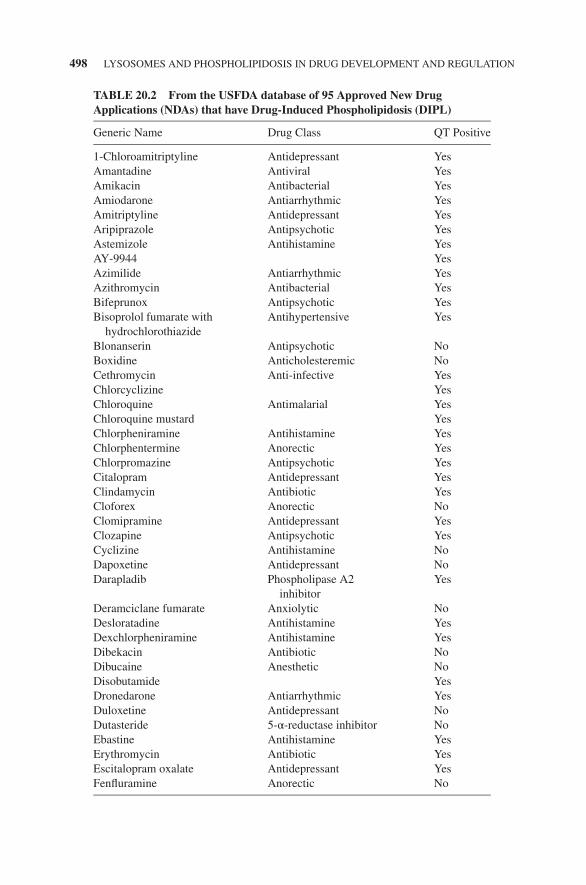
�
� �
�
498 LYSOSOMES AND PHOSPHOLIPIDOSIS IN DRUG DEVELOPMENT AND REGULATION
TABLE 20.2 From the USFDA database of 95 Approved New DrugApplications (NDAs) that have Drug-Induced Phospholipidosis (DIPL)
Generic Name Drug Class QT Positive
1-Chloroamitriptyline Antidepressant YesAmantadine Antiviral YesAmikacin Antibacterial YesAmiodarone Antiarrhythmic YesAmitriptyline Antidepressant YesAripiprazole Antipsychotic YesAstemizole Antihistamine YesAY-9944 YesAzimilide Antiarrhythmic YesAzithromycin Antibacterial YesBifeprunox Antipsychotic YesBisoprolol fumarate with
hydrochlorothiazideAntihypertensive Yes
Blonanserin Antipsychotic NoBoxidine Anticholesteremic NoCethromycin Anti-infective YesChlorcyclizine YesChloroquine Antimalarial YesChloroquine mustard YesChlorpheniramine Antihistamine YesChlorphentermine Anorectic YesChlorpromazine Antipsychotic YesCitalopram Antidepressant YesClindamycin Antibiotic YesCloforex Anorectic NoClomipramine Antidepressant YesClozapine Antipsychotic YesCyclizine Antihistamine NoDapoxetine Antidepressant NoDarapladib Phospholipase A2
inhibitorYes
Deramciclane fumarate Anxiolytic NoDesloratadine Antihistamine YesDexchlorpheniramine Antihistamine YesDibekacin Antibiotic NoDibucaine Anesthetic NoDisobutamide YesDronedarone Antiarrhythmic YesDuloxetine Antidepressant NoDutasteride 5-α-reductase inhibitor NoEbastine Antihistamine YesErythromycin Antibiotic YesEscitalopram oxalate Antidepressant YesFenfluramine Anorectic No

�
� �
�
PHOSPHOLIPIDOSIS AND QT INTERVAL PROLONGATION 499
TABLE 20.2 (Continued)
Generic Name Drug Class QT Positive
Fluoxetine Antidepressant YesGentamicin Antibiotic YesHaloperidol Antipsychotic YesHomochlorcyclizine Antidepressant YesHydroxyzine Antihistamine NoImipramine Antidepressant YesIndoramin Antiarrhythmic YesIprindole Antidepressant YesKetoconazole Antifungal YesLansoprazole Proton pump inhibitor NoLapatinib ditosylate Oncology NoMefloquine Antimalarial NoLoratadine Antihistamine YesMianserin Antihypertensive YesMibefradil
dihydrochlorideAntihypertensive Yes
Nebivolol Antihypertensive NoNilotinib Oncology YesNorchlorcyclizine Antihistamine YesNoxiptiline Antidepressant YesOrvepitant Neurokinin 1
antagonistNo
Perhexiline Antianginal YesPhentermine Antihistamine YesPosaconazole Antifungal YesPromazine Antipsychotic YesPropranolol β-blocker YesQuinacrine Antibiotic NoQuinidine Antiarrhythmic NoRemeron Sleep NoRilapladib Phospholipase A2
inhibitorNo
Rimantadine Antiviral YesRitonavir Antiviral YesRotigotine NoSatavaptan Diuretic YesSertraline Antidepressant YesSirolimus Immunosuppressant YesSpectinomycin Antibiotic NoTAK-032 Human
immunodeficiencyvirus treatment
Yes
Tamoxifen Antiestrogen receptor Yes
(continued)

�
� �
�
500 LYSOSOMES AND PHOSPHOLIPIDOSIS IN DRUG DEVELOPMENT AND REGULATION
TABLE 20.2 (Continued)
Generic Name Drug Class QT Positive
Tecastemizole/norastemizole Antihistamine YesTelithromycin Antibiotic YesTetracaine Anesthetic NoThioridazine Antipsychotic YesThiothixene Antiviral NoTiotropium bromide Muscarinic antagonist YesTobramycin Antibiotic YesTocamide Antiarrhythmic YesTriparanol NoTripelennamine Antihistamine NoValdecoxib Nonsteroidal
anti-inflammatorydrug
No
Varenicline tartrate Antismoking YesVerapamil Antihypertensive No
channel [38,51]. Some of these compounds may affect the hERG channel by othermechanisms, for example, inhibiting protein trafficking. For example, pentamidinedoes not block the hERG channel directly, but at 10 μM, a level achieved inantiparasitic therapy, it inhibits hERG channel trafficking [61,62]. In contrast, theDIPL-inducing and QTc-prolonging drug aclarubicin, an anthracycline antineoplas-tic drug, is an inhibitor of the vacuolar-type adenosine triphosphatase (V-ATPase)that is part of the proton pump mechanism in lysosomes [63]. Such activities mayfacilitate accumulation of the anthracycline and inhibit phospholipase activity in thelysosome by inhibiting lysosomal acidification. Accordingly, there are multiple waysto interfere with endosomal/lysosomal functions, which are important not only forclearing endocytosed materials coming from the cell surface but also for transportinghERG channels and other materials from the Golgi apparatus to the cell surfaceor other locations in the cell. Culprit drugs also might disrupt lysosomal recyclingof misfolded or incorrectly made proteins along with blocking cellular organellerecycling [23].
20.13 DIPL MECHANISMS
Multiple pathogenic mechanisms have been proposed for DIPL. Early studies onDIPL examined the role of lysosomal phospholipases in the pathogenesis of DIPL. Itwas hypothesized that the causative drug interfered with the activity of the phospho-lipases (sphingomyelin phosphodiesterase, lysosomal phospholipase A2 [LYPLA2],and lysosomal phospholipase A1) or that the CAD had increased cholesterol biosyn-thesis via the lanosterol synthase pathway, leading to accumulation of phospholipidsin the lysosomes [64,65]. Mice that were LYPLA2-deficient presented with foam

�
� �
�
DISCUSSION 501
cells, splenomegaly, and phospholipidosis [65]. However, it has been argued thatphospholipase inhibition is not a factor in DIPL as BMP is still excreted from cellsand is subsequently observed in the serum and urine [37]. Since BMP is produced byphospholipase A2 action, increased synthesis of the former would not be expectedif inhibition of phospholipase was a constant or important factor in phospholipi-dosis [37]. Evidence has been accumulating for the direct binding of the causativedrug to a phospholipid such as the phospholipids present in l-α-dipalmitoyl phos-phatidylcholine vesicles. Such binding distinguishes several CADs that cause DIPLfrom quinidine, which does not bind to phospholipids [66]. The phospholipidoticand antimalarial activities of chloroquine may occur from blocking the V-ATPaseand associated increase in pH in the parasitic and host cell organelle, with associ-ated indirect inhibition of acidic phospholipase and other activities requiring low pH[67]. Procainamide also blocks V-ATPase and raise lysosomal pH [24]. Pentamidineinhibits hERG protein trafficking in cells [61] but does not inhibit phospholipases.
20.14 TREATMENT
There are no approved treatments for DIPL, likely because it is usually reversibleupon drug withdrawal, and the need may not arise if DIPL is detected early. It is notclear how a clinical trial could be acceptably designed to test for reversal of DIPL,rather prevention might be more feasible. Gentamicin DIPL causes apoptosis of renaltubules [68] that can be blocked by polyaspartic acid [69]. Vitamin E can also inhibitapoptosis induced by 7-ketocholesterol, an inducer of phospholipidosis [70]. DIPLcaused by amiodarone is also reduced by vitamin E treatment [71], an antioxidantalso reported to inhibit the formation of DIPL and multilamellar vesicles associatedwith desipramine and other CADs [72]. These isolated reports are encouraging, but asystematic study of the effects of these compounds on DIPL is needed to determinewhether polyaspartic acid and vitamin E may be therapeutic or prophylactic in DIPL.
While it is not clear how DIPL would be treated other than recourse to promptdrug withdrawal, it is clear that CADs possess useful properties for developing drugsfor treating CNS or other indications. Some CADs are valuable pharmacotherapeu-tics that can readily cross plasma membranes to reach targets in the cell interior. Thisproperty also enables such compounds to cross the blood–brain barrier, which is oth-erwise a major obstacle to the development of new centrally acting drugs [46,73–76].The use of CADs for selectively promoting autophagy may afford new therapies foraging, neurodegeneration, and cancer [18–21,23,26,27,70,77].
20.15 DISCUSSION
Although the pathogenesis of DIPL is increasingly understood and the propensity forcausing these lesions is now relatively predictable [64], the forecasting of importantsequelae is not [9]. The safety implications remain obscure, and DIPL is not eas-ily addressed in the development and regulation of an IND. At the beginning of the

�
� �
�
502 LYSOSOMES AND PHOSPHOLIPIDOSIS IN DRUG DEVELOPMENT AND REGULATION
modern era of drug development in the late 1960s and early 1970s, regulators wereconfronted with deaths and severe hepatic injury associated with DIPL and elevatedlevels of circulating BMP [28–30,37]. There is also a concern that lipidotic drugsmight be associated with cardiac arrhythmias due to QTc prolongation, hERG pro-tein trafficking, or other effects related to endosome/lysosome complexes. At present,DIPL, when encountered in a toxicology study, cannot be dismissed without furtherinvestigation, especially if associated with cardiac arrhythmias, neuronal toxicity, orliver, lung, kidney, or cardiac parenchymal injury.
There is concern in the pharmaceutical industry when DIPL is encountered, orlikely to be encountered, given the problems in early detection and the potential clini-cal importance. Ongoing research addressing multiple aspects of DIPL, and an earlierand easier diagnosis, reflects the importance of DIPL to industry and to regulators[1,11,15,35,37,39,45,47,54,55,70,76,78–82]. The issue has driven QSAR and othermachine learning technologies for in silico prediction of phospholipidosis [35,42,45].Such forecasting is important considering, for example, the overlap between hERGchannel blockade and phospholipidosis [38,51]. Research has prompted develop-ment of rapid in vitro methods for cell culture and assays for phospholipid binding[15,53–55,66,74,78,81] and validation of their ability to predict DIPL. Toxicoge-nomic studies to evaluate responding genes [11], and immunohistochemical methodsof identifying DIPL definitively [28,57,58], have helped to identify the lipidosis with-out recourse to TEM. A reliable urine or blood test would provide an importantalternative to the definitive, but less convenient, current diagnostic procedure andwould resolve the present difficulty in recognizing DIPL in the clinic.
DIPL and hereditary lysosomal storage disease both vary widely in clinical impor-tance. Concern over DIPL is, of course, greater when there is additional nonphospho-lipidotic histopathology and important overt sequelae. The FDA phospholipidosisdatabase (Figure 20.2) and the AstraZeneca database, with its spectrum of histo-logic lesions from toxicology studies, shows a close association between DIPL andexcess nonphospholipidotic lesions [35]. However, DIPL provoked in animals bysome compounds such as loratadine may occur absent other concurrent histopathol-ogy. However, even the risk of isolated DIPL with loratadine depends on the extentof metabolic conversion to the less lipidotic desloratadine metabolite [43].
The lysosomal storage disease NP-C, and DIPL, not only share the hallmark mul-tilamellar vesicles and disrupted cholesterol transport, but both can be devastating intheir extreme expressions. Even patients with nonlethal NP-C and DIPL may presentwith important hepatotoxicity, neuropathy, and myopathy. Elevated serum and urinelevels of BMP are a feature of both NP-C and amiodarone-induced DIPL [37].Patients with DIPL present with variable organ damage, for example, chloroquinemyopathy, perhexiline neurogenic muscle atrophy, 4,4′-diethylaminoethoxyhexestrolhepatotoxicity, and gentamicin nephrotoxicity, evidence that DIPL and parenchymalinjury involve those organ(s) where there is a critical level of drug accumulation andlysosomal disruption.
There are several practical approaches to balancing the potential benefits versusrisks of CADs. Regulatory agencies, including the FDA, approve drug doses thatfavorably balance the risks of no-treatment (factoring in disease severity and natural

�
� �
�
DISCUSSION 503
history) versus adverse effects [83]. The initial doses for human phase 1 trials inhealthy subjects are based primarily on safety margins for positive findings in pre-clinical toxicity studies; the incidence and severity of AEs in clinical trials wouldlimit the maximum recommended human dose. Drugs occasionally must be adminis-tered based on milligram per kilogram or milligram per square meter when there is arelatively small therapeutic index. As with any condition that does not have a conve-nient validated biomarker, it may be difficult to establish the relation between DIPLand the study drug and whether organ dysfunction precedes or follows the appearanceof DIPL.
Covert off-target toxicities can be difficult to detect and evaluate unless toxicity isan expected extension of pharmacodynamic activity, for example, systemic toxicity ofcorticosteroids [73,75]. Although QSAR and physicochemical analysis of lipophilic-ity and amphiphilicity may be reassuring when they predict a low risk of DIPL, whenthe latter is observed in animal safety studies, it may be an unexpected off-target eventand imprudent to dismiss. The important lipophilicity and amphiphilicity compo-nents of the determinants of in vivo toxicity may be receptor-mediated, a consequenceof nonspecific accumulation, or a detergent effect [75]. Drug discovery projects areheavily populated with cationic amphiphilic amines, which are common pharma-cophores for histamine, muscarinic, and adrenal receptors. As exposure-toxicitydata sets and other lipidotic pharmacophore domains are accumulated, the risks ofdeveloping DIPL may become clarified, and DIPL may be avoided. Extensive DIPLand other histopathological lesions may not be colocated or obviously associated;therefore, these additional data sets and focused mechanistic studies may identifythe collateral pharmacodynamic activity that may cause collateral injury.
With the availability of QSAR and in vitro screening methods [80], the poten-tial risk of developing DIPL may be assessed early in drug development. However,safety margins cannot be determined until after the start of clinical studies and theestablishment of clinical dose ranges. When development of DIPL is predicted fromQSAR or in vitro testing, the risk should be verified in preclinical toxicology stud-ies that should determine propensity, safety margin, severity, and reversibility. Tis-sues that show DIPL should be monitored for signs of collateral histopathology,and the clinical relevance of histologic lesions such as liver or lung dysfunction orelectrocardiographic changes adjudicated. There may be a link between severity ofDIPL, AEs, and QTc interval prolongation by direct or indirect (protein trafficking)mechanisms.
Although BMP level may not be monitored in animal toxicity trials, except forcause, elevated levels would suggest its use in monitoring clinical studies. Early indi-cations of DIPL from in silico or in vitro assays would indicate whether BMP monitor-ing may be useful in animal or clinical trials. A preclinical assay of protein trafficking(hERG-Lite assay, ChanTest, Cleveland, OH) may be advisable for DIPL-positivecompounds. The possibility should be considered that metabolites may be more lipi-dotic than the parent compound, even though this may be uncommon [43]. It isimportant to evaluate lung, liver, brain, and lymph node parenchymal cells, residenttissue macrophages, and bile duct cells, and this evaluation may be a basis for classi-fying and monitoring DIPL according to organ systems most commonly involved. As

�
� �
�
504 LYSOSOMES AND PHOSPHOLIPIDOSIS IN DRUG DEVELOPMENT AND REGULATION
with any toxicologic pathology, targets may be specific to species, organ, and CAD,and a reliable biomarker is needed that would be species- and organ-independent.Monitoring in the clinic with BMP would be advisable as an early sentinel for the col-lateral tissue damage, especially when there is important tissue damage in nonclinicaltoxicology studies and elevated BMP level. This may be important especially whena small safety margin exists for phospholipidotic versus targeted pharmacodynamicactivity. Clinicians who oversee trials of CADs may benefit from having a biomarker,knowing the affected organ systems in animal studies and focusing on those systems.Recommendations are available for assessment of potential toxicity of DIPL and QTprolongation and protein trafficking [80] (Figure 20.3).
Although it may be difficult to develop a drug that may induce DIPL, thereis much potential for therapeutic benefit. CADs are increasingly recognized asinhibitors and inducers of autophagy [27] and processes linked to aging [19], cancer[77], Alzheimer’s disease [84], and neurodegenerative disease [21,22,25]. Thephysicochemical properties of CADs enable these drugs to access the interior ofthe cell and facilitate passage across the blood–brain barrier [3], which may not beachievable with other drugs. The multimodal study by Mesens et al. [85] provides anexample of additional ways to characterize a CAD compound. With the potential forthis group of compounds, Figure 20.4 offers a flowchart of steps and considerationsin developing a compound that causes phospholipidosis (see also Ref. [80]).
Protein trafficking inhibitor
QT prolongation Phospholipidosis
Gentamicin
Pentamidine
Arsenic trioxideChloroquine
Tunicamycin
FluoxetineDofetilide
Ibutilide
Sotalol Amiodarone
Haloperidol
Ketek
Meclizine
Lansoprazole
Amikacin
Memantine
Loratidine
Figure 20.3 Relation between phospholipidosis, QT prolongation, protein trafficking effects,and adverse events. The QT-only group had adverse events that were significant but on target(i.e., related to the mechanism of action of drugs). The phospholipidosis-only group had mildadverse events. The overlap groups had significant, off-target adverse events associated withtheir use [38]. (See color plate section for the color representation of this figure.)

�
� �
�
FUTURE DIRECTIONS AND RECOMMENDATIONS 505
Pre•In silico or in vitro test
results
•Look for signs of DIPL
•QT prolongation, hERG blockade, protein trafficking
•Histopathology
•Electron microscopy
•Bis(monoacy glycero) phosphate monitoring
•Immunostaining
•Is it reversible?
•Organ damage?
•Reversible
•Short-or long-term
therapy
•Severity of disease
•Severity of toxicity
Tox
Confirm
Study
Risk–
benefit
Figure 20.4 Flow chart for pharmaceutical development. When drug-induced phospholipi-dosis (DIPL) is predicted from in silico studies or in vitro assays, animal studies are used toevaluate DIPL. When DIPL is confirmed, it may be monitored with BMP and associated withtissue toxicity that may or may not be reversible. The BMP may be monitored clinically toassess potential drug toxicities.
All drugs have AEs that may or may not be dose related. Regulatory agenciesunderstand the potential for AEs and use risk/benefit profiles in decision-making.Together with sponsors, regulatory agencies implement a Risk Evaluation and Miti-gation Strategy (REMS) when it is necessary to ensure that the benefit of a drug orbiological product will justify the risk. Multiple reports suggest that vitamin E andpolyamino acids may decrease adverse symptoms of amiodarone [69–72], a phos-pholipidotic antiarrhythmic drug with major toxicity that restricts its usage. It wouldbe important to know whether vitamin E and polyamino acids improved the safety ofamiodarone without reducing its utility in atrial fibrillation.
20.16 FUTURE DIRECTIONS AND RECOMMENDATIONS
• Increase use of BMP as a biomarker in nonclinical and clinical trials of drugsthat induce phospholipidosis.
• Further develop strategies to treat or prevent phospholipidosis, for example,vitamin E and polyamino acids versus amiodarone lung toxicity.
• Increase awareness of the importance of metabolism, and genetic variations ofpatients, in the expression of phospholipidosis.

�
� �
�
506 LYSOSOMES AND PHOSPHOLIPIDOSIS IN DRUG DEVELOPMENT AND REGULATION
• Further investigate the association between phospholipidosis and autophagy/apoptosis.
• Develop new therapeutic uses for CADs.
REFERENCES
[1] Shikata T, Kanetaka T, Endo Y, Nagashima K. Drug-induced generalized phospholipi-dosis. Acta Pathol Jpn 1972;22(3):517–531.
[2] Nonoyama T, Fukuda R. Drug-induced phospholipidosis – pathological aspects and itsprediction. J Toxicol Pathol 2008;21(1):9–24.
[3] Vitovic P, Alakoskela JM, Kinnunen PK. Assessment of drug-lipid complex formation bya high-throughput Langmuir-balance and correlation to phospholipidosis. J Med Chem2008;51(6):1842–1848.
[4] Temple R. Policy developments in regulatory approval. Stat Med 2002;21(19):2939–2948.
[5] Bass AS, Vargas HM, Valentin JP, Kinter LB, Hammond T, Wallis R, Siegl PK,Yamamoto K. Safety pharmacology in 2010 and beyond: survey of significant eventsof the past 10 years and a roadmap to the immediate-, intermediate- and long-term futurein recognition of the tenth anniversary of the Safety Pharmacology Society. J PharmacolToxicol Methods 2011;64(1):7–15.
[6] Karsh LI. A clinical trial primer: historical perspective and modern implementation. UrolOncol 2012;30(4 suppl):S28–S32.
[7] Berridge BR, Chatman LA, Odin M, Schultze AE, Losco PE, Meehan JT, Peters T, Von-derfecht SL, Society of Scientific and Regulatory Policy Committee Working Group.Phospholipidosis in nonclinical toxicity studies. Toxicol Pathol 2007;35(2):325.
[8] Reasor MJ, Hastings KL, Ulrich RG. Drug-induced phospholipidosis: issues and futuredirections. Expert Opin Drug Saf 2006;5(4):567–583.
[9] Reasor MJ, Kacew S. Drug-induced phospholipidosis: are there functional conse-quences? Exp Biol Med 2001;226(9):825–830.
[10] Shayman JA, Abe A. Drug induced phospholipidosis: an acquired lysosomal storage dis-order. Biochim Biophys Acta 2013;1831(3):602–611.
[11] Sawada H, Takami K, Asahi S. A toxicogenomic approach to drug-induced phospholipi-dosis: analysis of its induction mechanism and establishment of a novel in vitro screeningsystem. Toxicol Sci 2005;83(2):282–292.
[12] Kodavanti UP, Mehendale HM. Cationic amphiphilic drugs and phospholipid storagedisorder. Pharmacol Rev 1990;42(4):327–354.
[13] Blohm TR. Drug-induced lysosomal lipidosis: biochemical interpretations. PharmacolRev 1978;30(4):593–603.
[14] Lüllmann H, Lüllmann-Rauch R, Wassermann O. Lipidosis induced by amphiphiliccationic drugs. Biochem Pharmacol 1978;27(8):1103–1108.
[15] Monteith DK, Morgan RE, Halstead B. In vitro assays and biomarkers for drug-inducedphospholipidosis. Expert Opin Drug Metab Toxicol 2006;2(5):687–696.
[16] Farrell GC. Drugs and steatohepatitis. Semin Liver Dis 2002;22(2):185–194.
[17] Levine B, Kroemer G. Autophagy in the pathogenesis of disease. Cell 2008;132(1):27–42.

�
� �
�
REFERENCES 507
[18] Gustafsson AB, Gottlieb RA. Recycle or die: the role of autophagy in cardioprotection.J Mol Cell Cardiol 2008;44(4):654–661.
[19] Rajawat YS, Hilioti Z, Bossis I. Aging: central role for autophagy and the lysosomaldegradative system. Ageing Res Rev 2009;8(3):199–213.
[20] Mehrpour M, Esclatine A, Beau I, Codogno P. Overview of macroautophagy regulationin mammalian cells. Cell Res 2010;20(7):748–762.
[21] Renna M, Jimenez-Sanchez M, Sarkar S, Rubinsztein DC. Chemical inducers ofautophagy that enhance the clearance of mutant proteins in neurodegenerative diseases.J Biol Chem 2010;285(15):11061–11067.
[22] Eskelinen EL, Illert AL, Tanaka Y, Schwarzmann G, Blanz J, Von Figura K, Saftig P.Role of LAMP-2 in lysosome biogenesis and autophagy. Mol Biol Cell 2002;13(9):3355–3368.
[23] Shanware NP, Bray K, Abraham RT. The PI3K, metabolic, and autophagy networks:interactive partners in cellular health and disease. Annu Rev Pharmacol Toxicol 2013;53:89–106.
[24] Pastor MD, García-Yébenes I, Fradejas N, Pérez-Ortiz JM, Mora-Lee S, Tranque P, MoroMA, Pende M, Calvo S. mTOR/S6 kinase pathway contributes to astrocyte survival dur-ing ischemia. J Biol Chem 2009;284(33):22067–22078.
[25] Bové J, Martinez-Vicente M, Vila M. Fighting neurodegeneration with rapamycin: mech-anistic insights. Nature Reviews. Neuroscience 2011;12(8):437–452.
[26] Cho KS, Yoon YH, Choi JA, Lee SJ, Koh JY. Induction of autophagy and cell death bytamoxifen in cultured retinal pigment epithelial and photoreceptor cells. Invest Ophthal-mol Vis Sci 2012;53(9):5344–5353.
[27] Yoon YH, Cho KS, Hwang JJ, Lee SJ, Choi JA, Koh JY. Induction of lysosomal dilata-tion, arrested autophagy, and cell death by chloroquine in cultured ARPE-19 cells. InvestOphthalmol Vis Sci 2010;51(11):6030–6037.
[28] Matsuzawa Y, Yamamoto A, Adachi S, Nishikawa M. Studies on drug-induced lipidosis.VIII Correlation between drug accumulation and acidic phospholipids. J Biochem 1977;82(5):1369–1377.
[29] Akeda S. A study on the lipidosis induced by a coronary vasodilator, 4,4’-diethylamino-ethoxyhexestrol dihydrochoride. Mie Med J 1972–1973;22:65–96.
[30] Kasama K, Yoshida K, Takeda S, Akeda S, Kawai K. Bis-(monoacylglyceryl)phosphateand acyl phosphatidylglycerol isolated from human livers of lipidosis induced by4,4’-diethylaminoethoxyhexestrol. Lipids 1974;9(4):235–243.
[31] Bolesta S, Roslund BP. Elevated hepatic transaminases associated with telithromycintherapy: a case report and literature review. Am J Health Syst Pharm 2008;65(1):37–41.
[32] Casado E, Gratacós J, Tolosa C, Martínez JM, Ojanguren I, Ariza A, Real J, Sanjuán A,Larrosa M. Antimalarial myopathy: an underdiagnosed complication? Prospective longi-tudinal study of 119 patients. Ann Rheum Dis 2006;65(3):385–390.
[33] Myers JL, Kennedy JI, Plumb VJ. Amiodarone lung: pathologic findings in clinicallytoxic patients. Hum Pathol 1987;18(4):349–354.
[34] Kennedy JI, Myers JL, Plumb VJ, Fulmer JD. Amiodarone pulmonary toxicity. Clinical,radiologic, and pathologic correlations. Arch Intern Med 1987;147(1):50–55.
[35] Barone LR, Boyer S, Damewood JR Jr, Fikes J, Ciaccio PJ. Phospholipogenic pharma-ceuticals are associated with a higher incidence of histological findings than nonphospho-lipogenic pharmaceuticals in preclinical toxicology studies. J Toxicol 2012;2012:308594.DOI: 10.1155/2012/308594.

�
� �
�
508 LYSOSOMES AND PHOSPHOLIPIDOSIS IN DRUG DEVELOPMENT AND REGULATION
[36] Sundin DP, Sandoval R, Molitoris BA. Gentamicin inhibits renal protein and phos-pholipid metabolism in rats: implications involving intracellular trafficking. J Am SocNephrol 2001;12(1):114–123.
[37] Tengstrand EA, Miwa GT, Hsieh FY. Bis(monoacylglycerol)phosphate as a non-invasivebiomarker to monitor the onset and time-course of phospholipidosis with drug-inducedtoxicities. Expert Opin Drug Metab Toxicol 2010;6(5):555–570.
[38] Willard JM. 2010. Phospholipidosis: data mining, modeling and laboratory research.United States Food and Drug Administration Web site. Available at http://www.fda.gov/downloads/AdvisoryCommittees/CommitteesMeetingMaterials/Drugs/AdvisoryCommitteeforPharmaceuticalScienceandClinicalPharmacology/UCM210798.pdf.Accessed 2015 June 29.
[39] Pelletier DJ, Gehlhaar D, Tilloy-Ellul A, Johnson TO, Greene N. Evaluation of a pub-lished in silico model and construction of a novel Bayesian model for predicting phos-pholipidosis inducing potential. J Chem Inf Model 2007;47(3):1196–1205.
[40] Kruhlak NL, Choi SS, Contrera JF, Weaver JL, Willard JM, Hastings KL, SancilioLF. Development of a phospholipidosis database and predictive quantitative structure–activity relationship (QSAR) models. Toxicol Mech Methods 2008;18(2–3):217–227.
[41] Choi SS, Kim JS, Valerio LG Jr, Sadrieh N. In silico modeling to predict drug-inducedphospholipidosis. Toxicol Appl Pharmacol 2013;269(2):195–204.
[42] Orogo AM, Choi SS, Minnier BL, Kruhlak NL. Construction and consensus performanceof (Q)SAR models for predicting phospholipidosis using a dataset of 743 compounds.Mol Inform 2012;31(10):725–739.
[43] Goracci L, Ceccarelli M, Bonelli D, Cruciani G. Modeling phospholipidosis induction:reliability and warnings. J Chem Inf Model 2013;53(6):1436–1446.
[44] Kaufmann AM, Krise JP. Lysosomal sequestration of amine-containing drugs: analysisand therapeutic implications. J Pharm Sci 2007;96(4):729–746.
[45] Zhang X, Zheng N, Rosania GR. Simulation-based cheminformatic analysis of organelle-targeted molecules: lysosomotropic monobasic amines. J Comput Aided Mol Des 2008;22(9):629–645.
[46] Mesens N, Steemans M, Hansen E, Verheyen GR, Van Goethem F, Van Gompel J.Screening for phospholipidosis induced by central nervous drugs: comparing the pre-dictivity of an in vitro assay to high throughput in silico assays. Toxicol In Vitro 2010;24(5):1417–1425.
[47] Valerio LG Jr. In silico toxicology models and databases as FDA Critical Path Initiativetoolkits. Hum Genomics 2011;5(3):200–207.
[48] Gardiner SJ, Begg EJ. Pharmacogenetic testing for drug metabolizing enzymes: is it hap-pening in practice? Pharmacogenet Genomics 2005;15(5):365–369.
[49] Inglis S, Stewart S. Metabolic therapeutics in angina pectoris: history revisited with per-hexilene. Eur J Cardiovasc Nurs 2006;5(2):175–184.
[50] Seierstad M, Agrafiotis DK. A QSAR model of hERG binding using a large, diverse, andinternally consistent training set. Chem Biol Drug Des 2006;67(4):284–296.
[51] Sun H, Xia M, Shahane SA, Jadhav A, Austin CP, Huang R. Are hERG channel blockersalso phospholipidosis inducers? Bioorg Med Chem Lett 2013;23(16):4587–4590.
[52] Cunningham ML, Lehman-McKeeman L. Applying toxicogenomics in mechanistic andpredictive toxicology. Toxicol Sci 2005;83(2):205–206.
�
� �
�
REFERENCES 509
[53] van de Water FM, Havinga J, Ravesloot WT, Horbach GJ, Schoonen WG. High con-tent screening analysis of phospholipidosis: validation of a 96-well assay with CHO-K1and HepG2 cells for the prediction of in vivo based phospholipidosis. Toxicol in Vitro2011;25(8):1870–1882.
[54] Kasahara T, Tomita K, Murano H, Harada T, Tsubakimoto K, Ogihara T, Ohnishi S,Kakinuma C. Establishment of an in vitro high-throughput screening assay for detectingphospholipidosis-inducing potential. Toxicol Sci 2006;90(1):133–141.
[55] Bhandari N, Figueroa DJ, Lawrence JW, Gerhold DL. Phospholipidosis assay in HepG2cells and rat or rhesus hepatocytes using phospholipid probe NBD-PE. Assay Drug DevTechnol 2008;6(3):407–419.
[56] Eskelinen EL. Roles of LAMP-1 and LAMP-2 in lysosome biogenesis and autophagy.Mol Aspects Med 2006;27(5–6):495–502.
[57] Obert LA, Sobocinski GP, Bobrowski WF, Metz AL, Rolsma MD, Altrogge DM, Dun-stan RW. An immunohistochemical approach to differentiate hepatic lipidosis from hep-atic phospholipidosis in rats. Toxicol Pathol 2007;35(5):728–734.
[58] Asaoka Y, Togashi Y, Imura N, Sai T, Miyoshi T, Miyamoto Y. Immunohistochemistryof LAMP-2 and adipophilin for phospholipidosis in liver and kidney in ketoconazole-treated mice. Exp Toxicol Pathol 2013;65(6):817–823.
[59] Larrain S, Rinella ME. A myriad of pathways to NASH. Clin Liver Dis 2012;16(3):525–548.
[60] Luo D. Ultracytochemical observation on lysosome of hair cells in kanamycin ototoxicguinea pigs [in Chinese]. Zhonghua Er Bi Yan Hou Ke Za Zhi 1990;25(5):281–282, 318.
[61] Dennis AT, Wang L, Wan H, Nassal D, Deschenes I, Ficker E. Molecular determi-nants of pentamidine-induced hERG trafficking inhibition. Mol Pharmacol 2012;81(2):198–209.
[62] Kuryshev YA, Ficker E, Wang L, Hawryluk P, Dennis AT, Wible BA, Brown AM, Kang J,Chen XL, Sawamura K, Reynolds W, Rampe D. Pentamidine-induced long QT syndromeand block of hERG trafficking. J Pharmacol Exp Therap 2005;312(1):316–323.
[63] Bravo-Cuellar A, Balercia G, Levesque JP, Liu XH, Osculati F, Orbach-Arbouys .Enhanced activity of peritoneal cells after aclacinomycin injection: effect of pretreat-ment with superoxide dismutase on aclacinomycin-induced cytological alterations andantitumoral activity. Cancer Res 1989;49(6):1578–1586.
[64] Lowe R, Mussa HY, Nigsch F, Glen RC, Mitchell JBO. Predicting the mechanism ofphospholipidosis. J Chemoinform 2012;4:2. DOI: 10.1186/1758-2946-4-2.
[65] Hiraoka M, Abe A, Lu Y, Yang K, Han X, Gross RW, Shayman JA. Lysosomal phospho-lipase A2 and phospholipidosis. Mol Cell Biol 2006;26(16):6139–6148.
[66] Joshi UM, Kodavanti PR, Coudert B, Dwyer TM, Mehendale HM. Types of interac-tion of amphiphilic drugs with phospholipid vesicles. J Pharmacol Experim Therap1988;246(1):150–157.
[67] Morissette G, Lodge R, Marceau F. Intense pseudotransport of a cationic drug mediatedby vacuolar ATPase: procainamide-induced autophagic cell vacuolization. Toxicol ApplPharmacol 2008;228(3):364–377.
[68] El Mouedden M, Laurent G, Mingeot-Leclercq MP, Taper HS, Cumps J, Tulkens PM.Apoptosis in renal proximal tubules of rats treated with low doses of aminoglycosides.Antimicrob Agents Chemother 2000;44(3):665–675.

�
� �
�
510 LYSOSOMES AND PHOSPHOLIPIDOSIS IN DRUG DEVELOPMENT AND REGULATION
[69] Kishore BK, Lambricht P, Laurent G, Maldague P, Wagner R, Tulkens PM. Mechanismof protection afforded by polyaspartic acid against gentamicin-induced phospholipidosis.II. Comparative in vitro and in vivo studies with poly-l-aspartic, poly-l-glutamic andpoly-d-glutamic acids. J Pharmacol Exp Therap 1990;255(2):875–885.
[70] Vejux A, Guyot S, Montange T, Riedinger JM, Kahn E, Lizard G. Phospholipido-sis and down-regulation of the PI3-K/PDK-1/Akt signalling pathway are vitamin Einhibitable events associated with 7-ketocholesterol-induced apoptosis. J Nutr Biochem2009;20(1):45–61.
[71] Card JW, Racz WJ, Brien JF, Massey TE. Attenuation of amiodarone-induced pul-monary fibrosis by vitamin E is associated with suppression of transforming growthfactor-beta1 gene expression but not prevention of mitochondrial dysfunction. J Phar-macol Exp Therap 2003;304(1):277–283.
[72] Scuntaro I, Kientsch U, Wiesmann UN, Honegger UE. Inhibition by vitamin E ofdrug accumulation and of phospholipidosis induced by desipramine and other cationicamphiphilic drugs in human cultured cells. Br J Pharmacol 1996;119(5):829–834.
[73] Lounkine E, Keiser MJ, Whitebread S, Mikhailov D, Hamon J, Jenkins JL, Lavan P,Weber E, Doak AK, Côté S, Shoichet BK, Urban L. Large-scale prediction and testingof drug activity on side-effect targets. Nature 2012;486(7403):361–367.
[74] Hanumegowda UM, Wenke G, Regueiro-Ren A, Yordanova R, Corradi JP, Adams SP.Phospholipidosis as a function of basicity, lipophilicity, and volume of distribution ofcompounds. Chem Res Toxicol 2010;23(4):749–755.
[75] Price DA, Blagg J, Jones L, Greene N, Wager T. Physicochemical drug properties asso-ciated with in vivo toxicological outcomes: a review. Expert Opin Drug Metab Toxicol2009;5(8):921–931.
[76] Ratcliffe AJ. Medicinal chemistry strategies to minimize phospholipidosis. Curr MedChem 2009;16(22):2816–2823.
[77] Mathew R, Karantza-Wadsworth V, White E. Role of autophagy in cancer. NatureReviews. Cancer 2007;7(12):961–967.
[78] Zhou L, Geraci G, Hess S, Yang L, Wang J, Argikar U. Predicting phospholipidosis:a fluorescence noncell based in vitro assay for the determination of drug-phospholipidcomplex formation in early drug discovery. Anal Chem 2011;83(18):6980–6987.
[79] Naisbitt DJ, Ruscoe JE, Williams D, O’Neill PM, Pirmohamed M, Park BK. Dis-position of amodiaquine and related antimalarial agents in human neutrophils: impli-cations for drug design. Journal of Pharmacology and Experimental Therapeutics1997;280(2):884–893.
[80] Chatman LA, Morton D, Johnson TO, Anway SD. A strategy for risk management ofdrug-induced phospholipidosis. Toxicol Pathol 2009;37(7):997–1005.
[81] Kuroda Y, Saito M. Prediction of phospholipidosis-inducing potential of drugs by in vitrobiochemical and physicochemical assays followed by multivariate analysis. Toxicol inVitro 2010;24(2):661–668.
[82] Smith DA, van de Waterbeemd H, Walker DK. Pharmocokinetics and metabolism indrug design. In: Mannhold R, Kubinyi H, Folkers G, editors. Methods And PrinciplesIn Medicinal Chemistry. Berlin: Wiley-VCH Verlag GmbH; 2001.

�
� �
�
REFERENCES 511
[83] United States Food and Drug Administration. 2005. Guidance for industry: premarket-ing risk assessment. Available at http://www.fda.gov/downloads/RegulatoryInformation/Guidances/UCM126958.pdf.
[84] Barnett A, Brewer GJ. Autophagy in aging and Alzheimer’s disease: pathologic or pro-tective? J Alzheimers Dis 2011;25(3):385–394.
[85] Mesens N, Desmidt M, Verheyen GR, Starckx S, Damsch S, De Vries R, VerhemeldonckM, Van Gompel J, Lampo A, Lammens L. Phospholipidosis in rats treated with amio-darone: serum biochemistry and whole genome micro-array analysis supporting the lipidtraffic jam hypothesis and the subsequent rise of the biomarker BMP. Toxicol Pathol2012;40(3):491–503.

�
� �
�
INDEX
acetaminophen, 462acid ceramidase (AC), 93, 183, 194, 195
deficiency, 210–213downregulation of, 453
acidic vesicular organelles (AVOs), 325acid sphingomyelinase (ASM), 90, 92, 93, 118,
184, 194–196, 213–215, 339acrosome, 75, 244acyl-coA-cholesterolacyltransferase (ACAT), 90,
91, 95acyl phosphatidylglycerol, 489adaptor complex 2 (AP2), 410adenomatous polyposis coli (APC), 38adverse event (AE), 487, 488, 503, 505age-related macular degeneration
(AMD), 143–144aging-related neurodegenerative diseases,
144–145Alzheimer’s disease see Alzheimer’s
disease (AD)amyotrophic lateral sclerosis, 144DLB, 155FTLD, 155–158Huntington’s disease, 144Parkinson’s disease and related disorders see
Parkinson’s disease (PD)
Lysosomes: Biology, Diseases, and Therapeutics, First Edition.Edited by Frederick R. Maxfield, James M. Willard and Shuyan Lu.© 2016 John Wiley & Sons, Inc. Published 2016 by John Wiley & Sons, Inc.
alpha-galactosidase A deficiency, 221–224Alzheimer’s disease (AD), 20, 109, 454, 467
amyloid-β and tau, 317APP, 145–149autosomal-dominant mutations, 145beta-amyloid peptide, 145–150hAPP mouse model, 149lysosomal dysfunction, 145–147mutations of PS1, 147pathogenesis of, 457–458
ambroxol, 365–367aminoglycoside antibiotics, 447, 450–451, 467amiodarone, 326, 329, 446, 456, 460, 462, 466,
468, 490, 501, 505amitriptyline, 440, 456β-amyloid, 489amyloid precursor protein (APP), 145–149amyotrophic lateral sclerosis (ALS), 20, 144, 317anthracycline antineoplastic drug, 500anthrax toxin, 451antibody–drug conjugates (ADCs), 417
brentuximab vedotin, 415cytotoxic payloads, 415–416efficacy, 413–414engineered antibodies, 414internalization pathways mediating, 410, 411
513

�
� �
�
514 INDEX
antibody–drug conjugates (ADCs) (Continued)linker-payloads, 414–415mechanisms of resistance, 416–417receptor-mediated internalization, 410–412structure, 413therapeutic effect of, 410
anticancer agents, 195, 196, 288, 295, 297, 302,430, 432–434
anticancer drugs, 184, 427, 431–433antihistamine loratadine, 490antimicrotubule drugs, 468antineoplastic drug doxorubicin (DOX), 464antioxidant vitamin E, 470–471antipyrine, 438α1-antitrypsin (ATZ), 110apoptosis, 116, 117, 121–125, 127
and DIPL, 489by gentamicin, 465vitamin E inhibits, 501
Arizona Center for Education and Research onTherapeutics database, 497
arsenic, 451, 469arylsulfatase A deficiency, 219–221aspartylglucosaminuria, 460ataxias, 20atherosclerosis, 67ATP31A2 gene, 20autolysosomes, 7, 13, 18–20, 54, 145, 151, 154,
282, 283, 330, 452autophagic lysosomal reformation (ALR), 8,
18–19, 154, 334–335autophagosomes, 138, 153, 154, 157
biogenesis, 14, 16electron micrograph, 13formation, 15fusion with LEs and Lys, 17–18structure, 7, 8
autophagyautophagy-related genes, 315–316canonical autophagy and noncanonical
autophagy, 280critical role, 280and DIPL, 489dual function in cancer see cancer therapy,
autophagy inhibition inelongation and maturation, 282–283enhancers screening see screening methods,
autophagosomesforms of, 315fusion and degradation, 283HDAC inhibition, 336impairment and diseases, 316–317induction, 283–285initiation, 282
literature reports, 317–324, 326lysosomal efficiency, 338–339and lysosomal lipid turnover, 94–95microautophagy, 279miRNAs, 339–340mTOR inhibition, 335–336nucleation, 282pathways, 12–13pH restoration, 337recycling and autophagic lysosome reformation,
283self-eating process, 315TFEB overexpression/activation, 338TRP activator, 337yeast genes and mammalian homologs, 280,
281autophagy-related diseases
cancer, 22Crohn’s disease, 20–21HSP, 22NBIA, 21–22Vici syndrome, 21
autophagy-related (Atg) proteins, 13–16, 22azithromycin, 333, 457
bacterial proteins, 451bafilomycin A1, 196bafilomycin A (Baf A), 417, 451, 452, 455, 456,
464bait loop, 125B/AKT pathway, 36BAMLET, 196BCL-2 proteins, 116beclin-1, 22, 56, 151, 282, 288, 468beta cyclodextrins (β-CDs), 143beta-galactosidase deficiency, 224–226beta-glucocerebrosidase deficiency, 215–217Bif-1 protein, 22biogenesis, LROs
CHS and GPS, 244, 246HPS see Hermansky–Pudlak syndrome (HPS)melanosome, 247model, 244, 245Weibel–Palade Body, 251–252
3,6-bis-(1-methyl-4-vinylpyridinium)carbazolediiodide (BMVC), 434
bis(monoacylglycero)phosphate (BMP), 89, 118BMT (bone marrow transplantation), 219botulinum neurotoxins (BoNTs), 451brentuximab vedotin, 415
cadmium, 451, 464, 469Caenorhabditis elegans, 110, 244

�
� �
�
INDEX 515
Caenorhabditis elegans epg-5, 21calcium, 52
channel blockers, 326-dependent exocytosis, 76
calpains, 339cancer
and autophagy, 22incidences of, 468
cancer-induced changes, in lysosomal functionand composition
altered lysosomal trafficking, 185altered lysosome membrane permeability, 184cathepsins see cathepsinsheparanase, 190–191increased lysosome size, 184lysosomal hydrolases, increased activity of,
182–183lysosomal membrane proteins LAMP1 and
LAMP2, 192sialidase 1/neuraminidase 1, 191–192sphingolipid metabolism, 193–195targeting lysosomal membrane integrity,
195–196V-ATPase, 192–193
cancer therapy, autophagy inhibition inclinical studies, 286, 288clinical trials for CQ/HCQ, 295–298future directions, 300–302inhibitors, 292–293, 298–300modulation, 288–290mouse model studies, 285–287multiple druggable targets, 291–292preclinical studies, 288–290, 292proximal autophagy inhibitor, 293quinolines, 293–295
canonical pathway, 41capillary electrophoresis, 428carbonilcyanide
p-trifluoromethoxyphenylhydrazon(FCCP), 56
carbonyl cyanide m-chlorophenyl hydrazone(CCCP), 56
cardiolipin, 94, 125cardiovascular disease, 142–144cargo adaptor proteins, 291casein kinase 1 (CK1), 38caspase-dependent apoptosis pathways, 195β-catenin, 38, 44cathepsin B, 183–185, 187–190, 193cathepsin K, 66, 189, 190, 435, 453cathepsins, 124–125, 327, 445cationic amphiphilic drugs (CADs), 489–494,
497, 500–502, 504, 506caveolae-mediated endocytosis, 410, 411
cell death signaling, 127LMP in see lysosomal membrane
permeabilization (LMP) in cell deathmodalities, 116–117
cellular clearance in human diseasecancer, 110–111LSDs, 107–109metabolic syndrome, 110neurodegenerative disorders, 109–110
cellular impacts, lysosomesdrugs effects on pH and lysosomal volume,
452–453LMP and cell death, 454–455on lysosomal enzyme activity, 453–454membrane trafficking changes see trafficking
pathwayssubstrate accumulation, 454
Center for Drug Evaluation and Research(CDER), 488
central nervous system (CNS), 89, 90, 109, 155,219, 316, 363, 447, 492
ceramide, 65, 90, 92, 93, 118, 194, 195, 209, 210,212, 213, 215, 230, 434
CeredaseⓇ (alglucerase), 364CerezymeⓇ (imiglucerase), 364chaperone-mediated autophagy (CMA), 8, 12,
137, 138, 293, 315Chediak–Higashi syndrome (CHS), 184, 244, 246chemical chaperones, 359chloroquine (CQ), 286, 293–295, 298, 299, 301,
327, 328, 332–334, 428, 430, 437, 446,452–458, 460, 462, 463, 466, 468, 470
chlorphentermine, 466chlorpromazine, 338cholesterol, 89, 95, 118, 119cholesteryl ester storage disease, 89chronic granulomatous disease (CGD), 255chylomicrons, 90ciprofloxacin, 121, 467cisplatin, 194, 195, 451, 464, 465clathrin-coated vesicle (CCV)
formation by AP-2, 410clathrin-independent transport route, 10clathrin-mediated endocytosis (CME), 410, 411clomipramine, 466Cochran–Mantel–Haenszel test, 362colchicines, 468concanamycin, 451concanamycin A, 416Coordinated Lysosomal Enhancement And
Regulation (CLEAR), 103C9orf72 mutation, 155, 157cristae, 52Crohn’s disease (CD), 20–21
�
� �
�
516 INDEX
Cryptococcus neoformans, 394Culprit drugs, 500β-cyclodextrins, 89cysteine cathepsins
cathepsin B, 187–188cathepsin D, 186cathepsin K, 189cathepsin L, 188–189deregulation, 190pharmacological targeting, 190
cytotoxic T lymphocytes (CTLs), 64–65, 239
Danon disease, 463, 467daunorubicin, 416, 427death-inducing signaling complex (DISC), 116Delta-like 4 (DLL-4), 42delta/serrate/Lag2 (DSL), 41dendrites, 66, 145, 224depolymerization
cortical actin, 69F-actin, 72
desipramine, 338diclofenac, 462differential centrifugation techniques, 1Diffuse Lewy Body Disease (DLB), 155dimebon, 456dimethylsulfoxide, 359dipeptide-repeat (DPR) proteins, 157dipeptidyl-β-naphthylamides, 121direct membrane lysis, 120disheveled (Dvl) proteins, 38disulfide linkers, 415DNA-damaging drugs, 195DNA microarray technique, 493N-dodecylimidazole, 120doxorubicin, 416, 428, 429Drosophila, 17, 18, 36Drosophila melanogaster, 21, 244drug-induced phospholipidosis (DIPL)
and autophagy, 489CADs, 491–492clinical expression and context of, 490–491in CNS, 492collateral effects of, 488–489comparison of dog and rat tissue distributions,
495–496FDA database and QSAR modeling, 494FDA involvement, 488–489lethal, 489–490linked with overt toxicity, 494–496lowest observed vs. toxicity LOEL, 496lysosomal storage disorder, 487mechanisms, 500–501nonclinical trials, 491
physicochemical properties, 491–492QSAR modeling, 492–493QT interval prolongation, 496–500regulatory issue, 488screening methods, 494toxicogenomics, 493treatments, 501USFDA database of approved NDAs, 497–500
dye methods, 494dysfunction, lysosme
cellular impacts see cellular impacts, lysosomeslysosmotropic compounds, 446–451mechnaism for organ toxicity see organ toxicitynonlysosomotropic compounds, 451–452
early endosomes (EEs), 7, 9, 11, 37, 101, 115,145, 245, 247, 248, 250, 257, 391, 392,410–412, 452, 456
E-cadherin, 187–189eliglustat tartrate (Genzyme), 364endocytic pathways, 10–12, 22, 63, 71, 115, 118,
119, 121, 124, 247, 251, 388, 389, 412, 456,465
endocytic recycling compartment (ERC), 89endogenous cysteine cathepsin inhibitors, 190endoplasmic reticulum (ER), 9, 10, 13, 54, 147,
209, 282, 284, 316, 358, 395, 445, 458–459endosomal escape pathways, biologics delivery,
383–384bacteria, 392–394endosome characteristics, 384, 388–389engineered endosomal escape, 385–386,
395–399fungi, 392–394macromolecular biologic drugs delivery, 384,
387protozoa, 392–394toxins, 394–395viruses, 390–392
endosomal sorting complex required for transport(ESCRT), 34–37, 39, 41, 42, 44, 89, 283, 412
engineered endosomal escapechemical and physical strategies, 399description of, 395–396lipids, 398–399peptides and polymers, 396–397
enzyme replacement therapy (ERT), 215, 217,219, 221, 223, 357
eosinophils, 75, 243epidermal growth factor receptor (EGFR), 11,
35–38, 412epigmentcell-specific premelanosome (PMEL)
protein, 247ER-associated degradation (ERAD) pathway, 358

�
� �
�
INDEX 517
esomeprazole, 193exocytosis, 457–458 see also lysosome exocytosisexosomes, 35, 39, 41–44, 253, 399
FabrazymeⓇ, 360Fabry disease (FD), 338, 460
alpha-galactosidase A deficiency, 221–224clinical manifestations, 3591-deoxygalactonojirimycin, 360–361ERT, 360female patient, 360GLP HEK assay, 362–363male patient, 359–360migalastat clinical trials, 361–362in vivo studies, 360–361
Fabry Phase 2 study, 372Farber disease, 210–213fast/direct recycling pathway, 11filamentous actin (F-actin), 66, 71, 72flaviviruses, 391fluorescence method, 494fluoroquinolone, 467fluoxetine, 329, 330Food and Drug Administration (FDA), 383, 384,
467, 488Francisella tularensis, 393free fatty acids (FFAs), 110Frizzled (Fz) receptors, 38, 40Frontotemporal Lobar Degeneration (FTLD)
C9orf72, 157genetic linkage, 157–158incidence, 155-TDP pathogenesis, 155–156TMEM106B, 156VCP mutation, 156–157
galactocerebrosidase beta-galactosidase (GALC)deficiency, 218–219
galactocerebrosidase deficiency, 218–219α-galactosidase A (α-Gal A), 359gallic acid, 471gamma aminobutyric acid (GABAA), 412gangliosides, 216, 224–227Gaucher disease (GD), 338
acid β-glucosidase, 363ambroxol, 366–367beta-glucocerebrosidase deficiency, 215–217classification, 364clinical presentation, 363–364ERT and SRT, 364isofagomine, 365–366PCs, 364–365side effect, 358
geldanamycin, 434gemifloxacin, 467gentamicin, 489–491, 501, 502globoid cell leukodystrophy (GLD), 218–219globotriaosylceramide (GL-3), 359globotriaosylsphingosine ( lyso-Gb3), 359glucocerebrosidase (GBA), 150, 151, 155glucosylceramide, 91, 93glucosylceramide-β-glucosidase, 93glucosylcerebroside (GC), 216glycerol, 110, 359glycerolipids, 91glycocalyx, 184, 192glycogen synthase kinase (GSK3), 35, 38, 39, 44glycoproteins, 357glycosaminoglycans (GAGs), 124, 357, 466glycosphingolipids (GSLs), 92, 93, 357Gly-Phe-β-naphthylamide, 121GM2-activator protein (GM2-AP), 92GM2 gangliosidoses, 226–229, 367–368GM1 gangliosidosis, 224–226GPI-anchored receptor (GPIHBP1), 89Gray Platelet syndrome (GPS), 244, 246Griscelli syndrome GS2 (RAB27A
deficiency), 252Griscelli syndrome GS3 (MLPH deficiency), 252growth hormone receptor (GHR), 11GTPase-activating protein (GAP), 106guanosine exchange factor (GEF), 11
half-life of drugs, 437, 438heat shock cognate 70 (HSC70), 279heat shock protein 70 (Hsp70), 339helix–loop–helix transcription factors, 103hemagglutinin 2 (HA2) protein, 390hemophagocytic lymphohistiocytosis (HLH), 252heparanase, 190–191hereditary spastic paraplegias (HSPs), 22, 154Hermansky–Pudlak syndrome (HPS)
BLOC-1,-2,-3 and AP-3, 250and melanosome maturation, 248–249neurosecretory granule biogenesis, 250–251RAB32/RAB38, 250
HEXA gene, 226–229hexosaminidase A and B deficiency, 226–229high-density lipoproteins (HDLs), 90high-performance liquid chromatography-mass
spectrometry/mass spectrometry(HPLC-MS/MS), 429
histone deacetylase 6 (HDAC6), 57histone deacetylase inhibitors (HDACi), 336human disease
and autophagy, 316

�
� �
�
518 INDEX
human disease (Continued)sphingolipid storage diseases see sphingolipid
storage diseasesTFEB and cellular clearance, 106–111
human ether-à-go-go-related gene (hERG)channel, 492, 493, 496, 497, 500–502
Huntington’s disease (HD), 109Huntingtin (htt), 317pathogenesis of, 457–458
hydroxychloroquine (HCQ), 125, 196, 295, 298,300, 301, 333–334, 468
4-hydroxynonenal (HNE), 144hypoxia-inducible factor-1 (HIF-1), 56
imatinib, 329, 428, 460, 463, 464imidazole, 397, 430imipramine, 329, 330, 333, 428, 438, 452,
460, 466immunohistochemical method, 494influenza virus, 3901,4,5-inositol trisphosphate (IP3), 316intracytosolic calcium (cAMP), 316intraluminal vesicles (ILVs), 8, 9, 11, 33, 34, 42,
44, 413 see also multivesicular bodies(MVBs), sorting onto ILVs
intralysosomal waste material, 454investigational new drug (IND), 361, 488, 495isofagomine (IFG), 365–366
‘‘kiss-and-run” exocytosis, 75Krabbe disease, 218–219Kufor-Rakeb syndrome, 153–154
late endosomes (LEs), 7, 9, 11, 20, 410late endosomes and lysosomes (LE/Ly), 1, 17, 87,
89, 91–94, 96LC3-interacting region (LIR), 16leucine-rich repeat kinase (LRRK2), 20, 150–155LeuLeuOMe, 121, 125, 196levofloxacin, 467ligand-independent Deltex-dependent pathway, 41lipid-bisretinoids (LBs), 143, 154lipids, 87
and autophagy, 94–95endocytic uptake of lipoproteins, 87–91engineered endosomal escape, 398–399hepatocytes and macrophages, 90hydrolysis in LE/Ly, 87, 91–93metabolic regulation, 95–96
lipofuscin, 120, 141–144, 155, 454, 466lipoprotein-related protein (LRP) coreceptors,
38, 40Listeria monocytogenes, 393–394
local anesthetics, 457low-density lipoprotein (LDL), 67
cholesterol, 91receptor, 88–89, 91
lowest observed adverse effect level (LOAEL)PLD nonclinical studies, 491toxicity vs. PLD LOEL, 496
LSDs see lysosomal storage disorders (LSDs)lycopene, 471lysobisphosphatidic acid (LBPA), 37, 89, 489lysosomal acid lipase, 89, 90, 94, 95lysosomal apoptotic pathway, 125–126lysosomal integral membrane protein 2
(LIMP2), 155lysosomal integral membrane proteins (LIMPs),
91, 118lysosomal membrane permeabilization (LMP),
142, 454–455lysosomal membrane permeabilization (LMP) in
cell death, 117–118characteristics, 118–119direct insult vs. molecular signaling, upstream
of, 121–124direct membrane lysis, 120mechanisms, 119minor destabilization, 117molecular events upstream and downstream
of, 121–122osmotic lysis, 120–121selective targeting, 119signaling downstream see signaling
downstream pathways, LMPlysosomal membrane proteins, 10, 101, 184,
192–193, 283lysosomal storage disorders (LSDs), 1, 316
ERT, 357–358Fabry disease, 359–363Gaucher disease see Gaucher
disease (GD)increased LTR staining, 453PC-ERT combination therapy, 370–372pharmacological chaperones, 358–359pompe disease, 368–370SRT, 358TFEB and cellular clearance, 106–109
lysosomal trappingassessment techniques, 427–429β-blockers in vivo distribution, 438in cultured cells, 423, 426drug–drug interactions, 438–440imipramine accumulation, 438long half-life, 437–438lysosomotropic behavior of drug see
lysosomotropism, drug activity

�
� �
�
INDEX 519
theoretical relationship between pKa and alpha,426–427
vital stains, 424volume of distribution, 423, 435–437weakly basic drug, 424–425
lysosome-associated membrane glycoprotein 2A(LAMP2A), 279
lysosome-associated membrane protein 1(LAMP1), 191–192
lysosome-associated membrane protein 2(LAMP2), 192
lysosome-associated membrane proteins(LAMPs), 91, 118
lysosome exocytosis, 104, 107–109, 182, 183,185, 191, 193, 197, 337, 338, 457, 458
calcium-dependent exocytosis, 76cortical actin in, 71–72extracellular degradation, 66–68fusion with plasma membrane, 75lysosome-related organelle, 64–65lysosome-related organelles maturation, 69–70membrane repair, 65–66occurrence, 63plasma membrane repair see plasma membraneproteins delivery into cell surfaces, 68role, 76schematic representation, 68–69source of membrane, 66
lysosome-related disorders (LSDs), 9, 18–20lysosome-related organelles (LROs), 239–240
azurophilic granules, 255biogenesis see biogenesis, LROscytolytic granules, 253–254defined, 239description, 244familial HLH and cytolytic granule secretion,
254–255IRF7-signaling and IFN induction, 256MIICs, 256–258motility, docking, and secretion, 252–253NADPH oxidase-containing, 254–256in nonhematopoietic cell types, 244perspectives, 260phagosomes and autophagosomes, 258–259physiological functions, 240–244vs. secretory granules, 240ultrastructure, 240, 243
lysosomesATG proteins, 14–16autophagy pathways, 12–13biosynthetic transport routes, 10cellular recycling centers, 181digestion, 1, 2, 88, 152endocytic pathways, 10–12
function in aging organisms, 139–142history, 2–4lipophilic compounds accumulation, 446morphology, 445neurodegeneration and links to autophagy, 20role in cellular processes, 101, 102role in nutrient sensing and signaling
pathways, 445–446lysosomotropic compounds
aminoglycoside antibiotics, 447, 450–451amiodarone, 446approved drugs, property distribution
of, 446–447autophagy screening, 327–338biological impacts of, 460, 461cadmium and arsenic, 451chloroquine, 446multiple drug classes, 447–450physicochemical properties, 446–447platinum-containing compound, 451
lysosomotropism, drug activityamines, 432antimalarial drugs, 430ceramide metabolism, 434detergent, 430, 431inhibitors of cathepsin K, 435intracellular distribution-based anticancer
drug, 433off-target effects, 429–430proton transfer mechanism, 431, 432selective against tumor cells, 433
LysoTracker staining, 300, 327, 333, 334, 429,440, 452
macroautophagy, 7, 9, 12, 13, 137, 138, 145, 151,153, 282, 284, 293, 315, 316 see alsoautophagy
macrolipophagy, 110macrophages, 65–66, 69, 76, 91, 94, 95, 117, 121,
142, 185, 187, 216–218, 256, 332, 334, 337,365, 389, 393, 457, 490, 495, 503
macropinocytosis, 42, 257, 333, 410, 411, 456magnetic chromatography, 429major histocompatibility complex class II
(MHCII), 68, 256–258malondialdehyde (MDA), 144mammalian target of rapamycin (mTOR)
inhibitors, 317, 326, 331–335, 489signaling pathways, 316
mammalshepatocytes, 90homologs, 281mitophagy mechanism, 55–57

�
� �
�
520 INDEX
mannose-6-phosphate (M6P) traffickingpathway, 102
mechanistic target or rapamycin complex 1(mTORC1), 95, 104–107
mediated by the calcium channel mucolipin-1(MCOLN1), 106
mefloquine, 196, 430, 492melanocytes, 64, 71, 72, 240, 247–249, 251, 252melanosomes, 35, 64, 70, 72, 247–249metabolic syndrome, 110metachromatic leukodystrophy (MLD), 219–2213-methyladenine (3-MA), 292, 293methylamine, 327, 452, 454, 4561-methyl-4-phenyl-1,2,3,6-tetrahydropyridine
(MPTP), 154MHC class II compartments (MIICs), 68,
256–258microautophagy, 8, 11, 12, 279, 315microphthalmia-associated transcription factor
(MiTF), 95, 329microphthalmia–transcription factor E
(MiT/TFE), 103microRNAs (miRNAs), 36, 42, 109, 339–340microscopic detection methods, 428microtubule, 291–292
-associated protein light chain 3, 56, 282dependent movement, 69, 71-destabilizing anticancer drug, 184, 193disruption, 413inhibitors, 414, 455in lysosome transport, 70–71stabilizing agents, 124targeting drugs, 195
microtubule-organizing center (MTOC), 70,71, 254
mitochondria, 282discovery, 51dysfunction, 52functions, 52fundamental role of, 51structure, 52
mitochondrial outer membrane (MOM), 116mitochondrial outer membrane permeabilization
(MOMP), 116, 127Bax/Bak-dependent, 125LMP upstream of, 123upstream and downstream of, 125, 126
mitochondrial permeability transition (MPT), 53mitochondrial-processing protease (MPP), 56mitogen-activated protein kinase (MAPK), 417mitophagy
history, 52–53in mammals, 55–57mechanism, 53–57
in yeast, 54–55monensin, 451, 456, 457monoclonal antibody (mAb), 413monomethyl auristatin E (MMAE), 415morpholine, 430moxifloxacin, 467mucopolysaccharidoses (MPS), 466mucopolysaccharidosis type I, 460multidrug resistance (MDR1), 417multiple sulfatase deficiency (MSD), 109multivesicular bodies (MVBs), 8, 11, 89,
283, 412multivesicular bodies (MVBs), sorting onto ILVs
downregulating EGFR signaling, 35–38electron micrograph, 33, 34ESCRT machinery, 34intercellular Notch signaling, 39–44maturation process, 33upregulating Wnt signaling, 38–40
Munc13– 4, 74, 245, 254muscle toxicity, 467–468Mycobacterium tuberculosis, 65–66, 76myeloid zinc finger transcription factor 1 (MZF1),
187MylotargⓇ, 414
α-N-acetyl-glucosaminidase (NAGLU) genemutations, 151
N-butyl-deoxynojirimycin (NB-DNJ), 358necroptosis, 116, 117, 121–123, 127nefazodone, 471neurodegeneration with Brain Iron Accumulation
(NBIA), 21–22neurodegenerative disease, 20, 109–110, 338 see
also Alzheimer’s disease (AD); Parkinson’sdisease (PD)
neutrophils, 64, 72, 74–76, 185, 244, 253, 255,256, 258, 259, 389, 394
new drug applications (NDAs), 495, 498niclosamide, 326, 333, 340Niemann–Pick disease, 2, 18, 339
type C, 89, 93, 143, 358, 454, 466, 490,492, 502
types A and B, 213–215Nile red dye, 494nitrobenzoxadiazole (NBD), 494NOD-like receptors (NLRs), 117nonalcoholic steatohepatitis (NASH), 494nonlysosomotropic compounds, 451–452nonlysosomotropic drug, 436, 438nonsteroidal anti-inflammatory drug
(NSAID), 462norfloxacin, 121, 467nortriptyline, 329, 330

�
� �
�
INDEX 521
Notch signaling, 41–43N-retinylidene-N-ethanolamine (A2E), 143
off-target effects, 340, 429ofloxacin, 467N-oleoylethanolamine, 195oligosaccharides, 11, 101, 224, 226, 227, 357, 367omeprazole, 193, 196O-methyl-serine dodecylamide hydrochloride
(MSDH), 120organ toxicity
factors, 469–471heart, 463–464kidney, 464–466liver, 462–463muscle, 467–468peripheral neuropathy, 466–467retinal, 466tumorigenesis, 468–469
osmotic lysis, 119–121, 123, 387, 397osteopenia, 66outer mitochondrial membrane (OMM) proteins,
54, 56, 57
paclitaxel, 195, 451, 467Parkinson’s disease (PD), 20, 109, 467
ATP13a2, 153–154autosomal-dominant form, 150death of neurons, 150disrupt by CMA, 151–152exposure of MPTP, 154lysosomal degradation, 151mutations of LRRK2, 152–153pathogenesis of, 457–458α-synuclein, 150–151, 317vacuolar protein sorting35, 154
perhexiline, 326, 329, 462, 488, 492–494perhexiline maleate, 490P-glycoprotein (Pgp), 416, 417phagocytosis, 1, 42, 66, 76, 102, 104, 115, 144,
182, 256–258, 331, 333–335, 389, 393, 410,445, 452, 455–457, 461, 487
phagophore, 7, 315biogenesis, 14electron micrograph of, 13elongation, 14formation, 15, 16, 316growing, 15
phagophore assembly site (PAS), 8phagosomes, 76, 243, 255–259, 335,
392–394, 452phosphatidylcholine, 91, 501phosphatidylethanolamine (PE), 15, 91, 282, 455
phosphatidylglycerol (PG), 94, 489phosphatidylinositol 4-kinases (PI4Ks), 19phosphatiylinositol-3-phosphate (PtdIns3P), 15,
22phosphoinositide 3,4,5 triphosphate (PIP3), 74, 75phospholipase A2 (PLA2) enzyme, 391phospholipidosis (PLD), 454
autophagy enhancers, 329drug development and regulation see
drug-induced phospholipidosis (DIPL)Phospholipidosis Working Group (PLWG), 488pH-partitioning theory, 437, 438pigment cell-specific premelanosome (PMEL)
protein, 247PI4KIIIβ knockdown cells, 19plasmacytoid DCs (pDCs), 256, 257plasma membrane, 10, 11, 13, 15, 33, 90, 95, 195,
209, 282, 316, 389, 487gangliosides, 224lysosome fusion with, 75repair mechanisms, 65–66, 76tethering of lysosomes, 72–75transport of lysosomes to, 70–72
Plasmodium falciparum, 294polyamidoamine (PAA), 397polyethylenamine (PEI), 396polyglutamine-expanded androgen receptor
(polyQ-AR), 109Pompe disease (PD), 18, 109, 338, 368–371preautophagosomal structure (PAS), 13presenilin-associated rhomboid-like protease
(PARL), 56propranolol, 437, 438proprotein convertase subtilisin/kexin type 9
(PCSK9), 91protolysosomes, 18, 19proton sponge effect, 121PSAP gene, 220, 229, 230Pseudomonas exotoxin, 394–395pyrimethamine (PYR), 367–368pyroptosis, 116, 117, 121–123, 127, 144
quantitative structure–activity relationship(QSAR), 491–494, 497, 502, 503
quinine, 430quinolone antibiotics, 121
Rab3, 72, 75Rab27, 72, 74, 75Rab27a, 42, 64, 72, 74, 245, 252–256Rab7-interacting lysosomal protein (RILP), 70, 71rapamycin, 94, 146, 317, 326, 332–335,
489
�
� �
�
522 INDEX
reactive oxidative species (ROS), 52, 56, 117, 119,121–124, 127, 157, 459, 463, 468, 471
real-time polymerase chain reaction analysis, 488receptor tyrosine kinases (RTKs), 33–36, 39,
44, 409recycling endosomes (REs), 7, 9, 16, 70, 245, 248,
389, 392, 456renal cell carcinoma (RCC), 110, 297, 334ReplagalⓇ, 223, 360retinal pigment epithelium (RPE), 72, 143, 247,
457, 466Rickettsia species, 394Risk Evaluation and Mitigation Strategy
(REMS), 505
salicylihalamide A, 416Sandhoff disease (SD), 226–229, 367screening methods, autophagosomes
Atg8/LC3, protein marker, 317autophagic flux assay, 329–331AVOs, 330GFP-LC3, 325–326immunohistochemical approach, 325lysosomotropic compounds, 327–329,
332–334membrane trafficking process, 331mTOR inhibitors, 331–335physicochemical properties, 327, 328steady-state accumulation, 317–324tandem mRFP/mCherry-GFP fluorescence
microscopy, 326tandem-tagged GFP-mCherry-LC3 probe
assay, 330TEM, 317
selective and potent autophagy inhibitor 1(Spautin-1), 292, 293
sequestosome 1 (SQSTM1/p62), 16. 157, 325,455, 459, 460, 464
serpin, 125, 188Shigella flexner, 393sialidase 1/neuraminidase 1, 191–192signaling downstream pathways, LMP
BID-independent apoptotic signalingpathways, 127
lysosomal apoptotic pathway, 125–126lysosomal cathepsins, 124–127
siramesine, 196, 329, 456SLC38A9, 106slow/indirect recycling pathway, 11small interfering RNA (siRNA) technology, 384Soluble NSF Attachment REceptor (SNARE)
proteins, 75sorting nexins (SNXs), 12spermatids, 75
sphingolipid activator proteins (SAPs), 92, 210,229–230
sphingolipids, 104, 118, 149, 209, 411catabolism, 93, 194in human disease see sphingolipid storage
diseasesmetabolism, cancer-associated changes,
193–195synthesis and breakdown of, 209
sphingolipid storage diseasesFabry disease, 221–224Farber disease, 210–213Gaucher disease, 215–217GLD, GALC deficiency, 218–219GM2 gangliosidoses, 226–229GM1 gangliosidosis, 224–226MLD, 219–221Types A and B NPD, ASM deficiency, 213–215
sphingomyelin (SM), 90, 92, 93, 118, 124, 184,194, 209, 214, 215, 230, 492, 500
sphingosine, 93, 118, 123, 184, 194–196, 209,210, 212–214
spinal muscular atrophy with myoclonic epilepsy(SMA-PME), 213
Spinster, 18, 283SREBP2, membrane protein, 90staphylococcal a-toxin, 451Staphylococcus aureus, 394Streptolysin-O, 65SU11652, anticancer remedy, 196substrate reduction therapy (SRT), 358, 364, 370sunitinib, 196, 416synaptotagmin-like protein 1 (Slp1), 72, 74–75α-synuclein (SNCA), 20, 109, 151–154, 317, 338,
339, 458
tamoxifen, 326–328, 333, 455–457, 489target SNAREs (t-SNAREs), 75taxanes, 292, 467Tay–Sachs disease (TSD), 2, 226–229, 367–368telithromycin (Ketek), 490tetanus neurotoxin (TeNT), 451therapeutic naked antibodies, 409thioether-or amide-based linkers, 415toll-like receptors (TLRs), 117, 256–259toremifene, 457Torsades de pointes (TdP), 496, 497toxicogenomics, 493trafficking pathways, 102, 245, 388
autophagy perturbation, 455–456endocytosis/phagocytosis impairment, 456–457endoplasmic reticulum, 458–459exocytosis, 457–458mitochondria, 458

�
� �
�
INDEX 523
oxidative stress, 459phagocytosis, 457signaling modulation, 459–461
transcription factor EB (TFEB), 16, 95, 139, 146,151, 154, 187, 284, 329, 330, 333, 335, 338,453, 456, 458
transcription factor EB (TFEB), cellular clearanceactivation on cellular mechanisms, 107, 108Ca2+-mediated regulation, 106, 107CLEAR network, 103–104diseases, 107–111function of, 107in human disease, 106–111LSDs, 107–109lysosome-to-nucleus signaling mechanism,
105–106metabolic syndrome, 110neurodegenerative disorders, 109–110overexpression, 104subcellular regulation by phosphorylation,
104–105transferrin receptor (TfR), 33, 410trans-Golgi network (TGN), 10, 11, 15, 245, 251,
410, 445, 453transient receptor potential calcium channel
MucoLipin subfamily member 1 (TRPML1),106, 148, 337
trastuzumab (Herceptin), 409Trypanosoma cruzi, 65, 72, 76tuberous sclerosis complex 2(TSC2), 335tubular lysosome, 71tumor-associated antigen (TAA), 413, 414, 417tumorigenesis, 181, 188, 190, 213, 285–288,
300–302, 468–469tumor necrosis factor-related apoptosis-inducing
ligand (TRAIL), 116, 123, 293,331, 452
tunicamycin, 331type-I interferon (IFN), 256, 257Types A and B Niemann–Pick disease (NPD),
213–215
ubiquitin-binding adaptor protein 62(p62/SQSTRM1), 57
ubiquitin proteasome system (UPS), 140, 141
UCH-L1 (ubiquitin carboxy-terminalhydrolase-L1 Parkin), 150, 151
ulinastatin, 465Uvrag, autophagy-associated protein, 22
vacuolar protein sorting35 (VPS35), 20, 154vacuolar-type adenosine triphosphatase
(V-ATPase), 138, 139, 147, 192–193, 195,335, 500, 501
valine-citrulline-p-aminobenzyl carbamate(vc), 414
valosin-containing protein (VCP), 156–157velaglucerase alfa (VPRIVⓇ), 364verapamil, 326, 328, 456very low-density lipoproteins (VLDLs), 88–89vesicular SNARE (v-SNARE) VAMP-7, 75Vici syndrome, 21vinblastine, 327, 416, 455, 467vincristine, 184, 195, 451, 453, 467, 468vinorelbine, 467viruses, endosomal escape
adenovirus, 391adenovirus protein VI, 391enveloped and nonenveloped viruses, 390flaviviruse, 391HIV gp41, 391influenza virus, 390–391parvoviruses, 391
vitamin Eantioxidant, 470safety of amiodarone, 505treatments for DIPL, 501
WIPI (WD-repeat protein interacting withphosphoinositides), 14, 15
Wnt signaling, 35, 38–40, 147Wollman disease, 89
yeastmitophagy mechanism, 54–55Saccharomyces cerevisiae, 248
ZavescaⓇ, 364

�
� �
�
Early
endosomeRecycling
endosome
PAS
PhagophoreAutophagosome
Amphisome
AutolysosomeProtolysosome
Endolysosomal pathways
Autophagy
Merging between endolysosomal pathways and autophagy
Microautophagy
Lysosome
ALR
CMA
Late
endosome/
MVB
ILV
ILV formation
Fast/direct
recycling
Slow/indirect
recycling
Figure 2.1 Schematic drawing depicting the endocytic and autophagy pathways to the lyso-somes. Note: Refer to page 8 for full caption.
Lysosomes: Biology, Diseases, and Therapeutics, First Edition.Edited by Frederick R. Maxfield, James M. Willard and Shuyan Lu.© 2016 John Wiley & Sons, Inc. Published 2016 by John Wiley & Sons, Inc.

�
� �
�
Lysosomes
“Late”
MVBs
“Early”
MVBs
Clathrin
EGF
P
P P P
Hrs
PTP1B
ESCRT
ESCRT
Ub
Ub
P
P
Hrs
EGFR
(a) (b)
Ub
Figure 3.2 Downregulation of EGFR signaling by sorting onto ILVs. Note: Refer to page 37for full caption.
Atg32
Atg11
p62
Ubiquitin
NIX
Binp3
FUNC1NIX
Parkin
PINK1
HDAC6Lysosomaldegradation
Fertilized egg
Red blood celldevelopment
Damagedmitochondria
Hypoxia
Mammalian
Yeast
Maternal
Paternal
Atg8
LC3/Atg8
Figure 4.1 Summary of mitophagy in yeast and mammalian cells. Note: Refer to page 55 forfull caption.
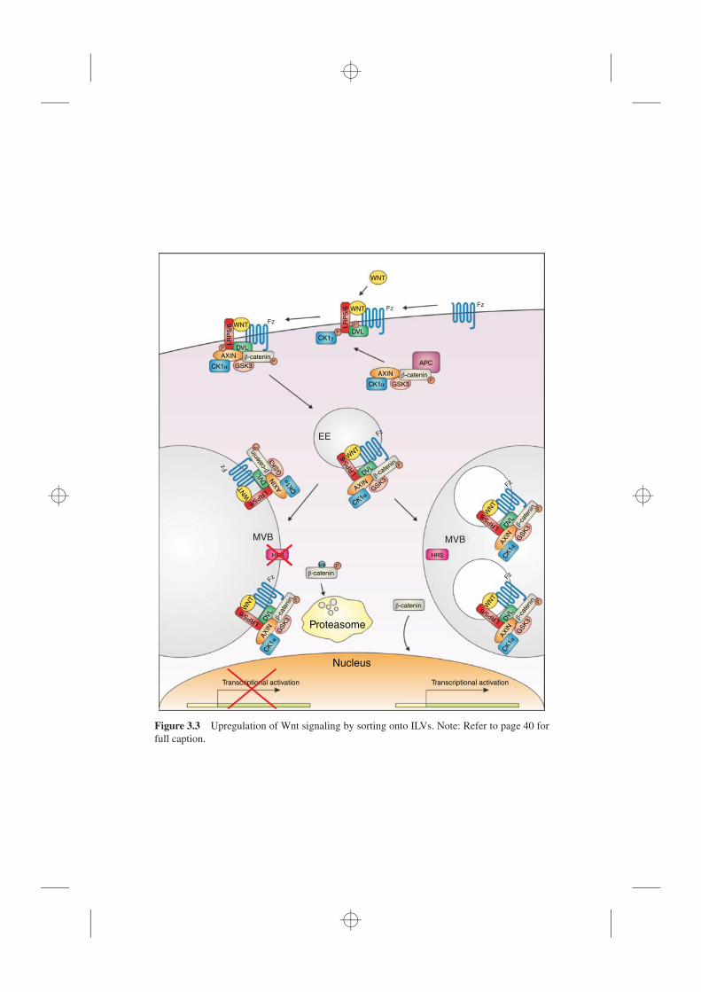
�
� �
�
WNT
WNT Fz
Fz
Fz
FzFzFz
Fz
Fz
WN
T
WNT
WN
T
WN
TW
NT
WNT
LR
P5/6 L
RP
5/6
LRP5/6
LRP5/
6
LRP5/6
LRP5/6LRP5/6
DVL
AXIN
CK1α GSK3
GSK3
GSK3
GSK3
GSK3
GSK3
GSK3
β-catenin
β-catenin
β-catenin
β-ca
teni
n
β-ca
teni
n
β-ca
teni
n
β-catenin
β-ca
tenin
β-ca
teni
n
CK1γ
CK1α
CK1α
CK1α
CK1α
CK1α
CK1α
P
P
P
HRS
Proteasome
Nucleus
Transcriptional activation Transcriptional activation
HRS
MVB
EE
MVB
Ub
P
P
P
P
P PP
P
AXIN
APC
AXIN
AXIN
AXIN
AXIN
AXIN
DVL
DVL
DVL
DVL
DVL
DVL
Figure 3.3 Upregulation of Wnt signaling by sorting onto ILVs. Note: Refer to page 40 forfull caption.

�
� �
�
Signal /
sending
cell
Signal /
receiving
cellNucleus
Delta
Notch
NICD
(i) (ii)(iii)
ILV
Nucleus
Signal /
receiving
cell
Figure 3.4 Intercellular Notch signaling mediated by ligand-bearing exosomes. Note: Referto page 43 for full caption.

�
� �
�
Lateendosome
Lysosomal hydrolases
Lysosome
Depolymerization
CorticalF-actin
Accessoryproteins
t-SNARE
Ca2+
v-SNAREM
icrotu
bule
s Exocytosis
Plasma membrane
Figure 5.2 Schematic of mechanisms involved in lysosome exocytosis. Note: Refer topage 69 for full caption.
Lipid droplet
Phagophore
Autophagosome
FA/cholesterol
Clathrin
LDLLDL receptor
Sorting endosome
pH ~ 6.2
ERC
pH 6.5
LAL
AutolysosomeLysosome
pH 4.5–5.0
Late endosome
pH 5.5
LE/Ly
Figure 6.1 Schematic diagram of lipoprotein and lipid droplet degradation by lysosomes.Note: Refer to page 88 for full caption.

�
� �
�
Endoplasmic reticulum
Nucleus
Autophagosome
Autophagy
Phagosome
Lysosome
Late endosome
Phagocytosis
Early endosome
Clathrin bud
Golgi
Clathrin bud
Endocytosis
Figure 7.1 Central role of lysosomes in key cellular processes.
Figure 7.2 Systems biology approach used to discover the CLEAR network. Note: Refer topage 103 for full caption.
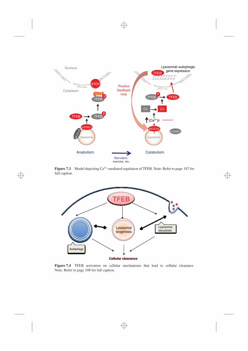
�
� �
�
Nucleus
Cytoplasm
STOPPositive
feedback
loopP
P
TFEB
TFEB
mTORC1MCOLN1
mTORC1
TFEB
TFEB
Lysosomal–autophagic
gene expression
TFEBP
Cn Cn
[Ca2+]c
lysosomelysosome
Anabolism Catabolism
Starvation,
exercise, etc.
MC
OLN
1
14-3-3
TFEB
Figure 7.3 Model depicting Ca2+-mediated regulation of TFEB. Note: Refer to page 107 forfull caption.
Lysosomal
excoytosisLysosomal
biogenesis
TFEB
Autophagy
Cellular clearanceCellular clearance
Figure 7.4 TFEB activation on cellular mechanisms that lead to cellular clearance.Note: Refer to page 108 for full caption.

�
� �
�
CTRL HDAd-TFEB
MSD
PDGlycogen Glycogen
HTT
PD+Ad-TFEB
HDAd-AFP HDAd-TFEB
Copper Sensor 3 Copper Sensor 3
Bacteria
Anti
fact
HLH-30
?
?
Cytoprotection
HLH-30
HLH-30
Epithelial cell
MSD + AAV-TFEB
Multiple sulfatase deficiency
Pompe disease
Diet-induced obesity
TFEB
Huntingron’s disease
GFP
α-sys+GFP α-sys+TFEB
TFEB
Alzheimer’s disease
Parkinson’s disease
Spinal-bulbar muscular atrophyBacterial infections
(in C.Elegans)
α-1-antitrypsin
deficiency
Wilson disease
Control HDAd-TFEB
exFLAG-TFEB
Figure 7.5 Diseases that respond to TFEB-mediated clearance. Note: Refer to page 108 forfull caption.
Macroautophagy
Endocytosis
Chaperone-mediated autophagy
Microautophagy
Expansion
Isolation membrane phagophore AutophagosomeMetabolite
effluxLysosome
Late endosome/MVB
Early endosome
Plasma membrane
Misfolded protein
KFERQ motif
Binding
Recognition
Receptor-mediated
Fluid phase
LAMP2a
Hsc70
Hsc70
LysoHsc70
hsp9
0
hsp90
hsp90
Completion Fusion
Unfolding Translocation
Figure 9.1 Major routes of substrate delivery to lysosomes. Note: Refer to page 138 for fullcaption.

�
� �
�
AD – App mutation/triplication,EE
LE
CMA
Microtubules
LysoA
L
Lyso
LysoHsc70
PD
LA
MP
2
KF
ER
O
AP
β-CTF
ALS – Alsin, C9orf72,
ALS – CHMP2B
Frontotemporal dementia – CHMP2B, p97/VCP
ALS, FTD – p97/VCP, p62, OPTN
Parkinson’ s Disease –
Pink, Parkin
PAS
PD, FTD
tau, α-synuclein,
UCH-L1, LRRK2
Hereditary spastic paraplegia – Spg4, Spartin
Charcot–Marie tooth type 2B – rab 7,
FIG4, and SH3TC2
ALS, MND, CMT – dynein, dynactin complex
Frontotemporal dementia –TMEM106B, GRN
Hereditary spastic paraplegia – Spg15, Spg11
LSDs (juvenile/adult forms): e.g.,
• Niemann–Pick Type C
• Mucopolysaccharidosis IIIB, IIB
• Gaucher’ s (GBA) Parkinson’ s
• Neuronal ceroid lipofuscinosis) PD (ATPase13a2), ?FTP (GRN)
• Chediak–Higashi (CHS/LYST) PD/dementia
AD – Presenilins 1, 2
Parkinson’ s Disease – ATPase 13A2, ATP6AP2
LRRK2, α-synuclein, GBA, VPS35
ApoE ε4 (high risk)
Figure 9.2 Genetic evidence strongly implicates the lysosomal network in the pathogenesisof neurodegenerative disease. Note: Refer to page 152 for full caption.
Cell growth
H+
H+
H+
H+
LMP
LCD
Lysosomal
leakage
Transformed cancer cell
Normal epithelial cell
ECM degradation
Acidification
Angiogenesis
Invasion
Figure 10.1 Cancer development induces changes in the lysosomal function. Cancer devel-opment and progression induces lysosomal biogenesis increasing the expression of variouslysosomal hydrolases. It alters the lysosomal membrane integrity, sensitizing them to LMPand lysosomal leakage that can lead to LCD. Note: Refer to page 183 for full caption.

�
� �
�
Changes in lysosomal components Cellular response
Invasive phenotype
Sialidase
Heparinase
Cysteine
cathepsins
Lysosomal
destabilization
H+
H+
H+
Transformation
Lysosomal
Hsp70Activation of ASM
LAMPS
ASMLMP
Drug resistance
Figure 10.2 Cancer induces changes in lysosomal composition. These include changes inthe expression levels and activity of several lysosomal hydrolases. Note: Refer to page 186 forfull caption.
mMel
Stage IV
Stage III
vWF
CD63Lysosome
NBEAL2
Munc
13-4
Munc
13-4
Rab27a
Rab27a
Rab11
α-Granules
iMel
Stage II
Tyr
PMEL
Amyloid
fibrils
TYRP1
OCA2
BLOC-1
BLOC-2
Microtubule
KIF13A
AP1 AP1
CD63
Granzymes
perforin
mLG
iLG
iLG
P-selectin
vWF polymers
iWPB
mWPB
AP3
AP3
BLOC-1
Early endosomesTGN
Golgi
Late endosome
MVB
Rab38/Rab32
BLOC-3
Figure 12.2 Model for biogenesis of four vertebrate LROs. Note: Refer to page 245 for fullcaption.
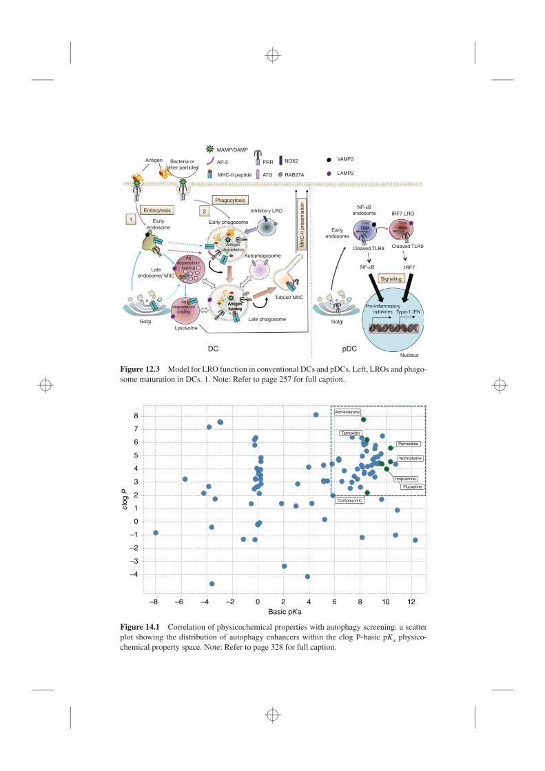
�
� �
�
Antigen
Endocytosis
1
2
Early
endosome
Phagocytosis
Early phagosome
Inhibitory LRO
Early
endosome
NF-κB
endosome IRF7 LRO
Cleaved TLR9Cleaved TLR9
Viral
DNA
Viral
DNA
Signaling
Pro-inflammatory
cytokines
GolgiLate phagosome
Tubular MIIC
Type-1 IFN
Nucleus
IRF7NF-κB
Autophagosome
MH
C-I
I pre
senta
tion
Antigen
degradation
Bacteria or
other particles
MHC-II:peptide
AP-3
MAMP/DAMP
PRR
ATG
NOX2
RAB27A
VAMP3
LAMP2
Antigen
loading
Ag
degradation/
loading
Ag
degradation/
loading
Late
endosome/ MIIC
Golgi
Lysosome
DC pDC
Figure 12.3 Model for LRO function in conventional DCs and pDCs. Left, LROs and phago-some maturation in DCs. 1. Note: Refer to page 257 for full caption.
8
7
6
5
4
clog P
3
2
1
0
–1
–2
–3
–4
–8 –6 –4 –2 0 2 4
Basic pKa
Amniodarone
Tamoxifen
Perhexiline
Nortriptyline
Fluoxetine
Compound C
6 8 10 12
Imipramine
Figure 14.1 Correlation of physicochemical properties with autophagy screening: a scatterplot showing the distribution of autophagy enhancers within the clog P-basic pKa physico-chemical property space. Note: Refer to page 328 for full caption.

�
� �
�
Clathrin-
coated
vesicle
Early
endosome
Late
endosome
Lysosome
Golgi
Nucleus
Caveosome
Endoplasmic
reticulum
Macropinosome
Caveolar
vesicle
Actin
filaments
(a) (b) (c)
Figure 17.1 Internalization pathways mediating ADC uptake. Note: Refer to page 411 forfull caption.

�
� �
�
Early endosome
(pH 6)
Late endosome
(pH 5)
Lysosome
(pH 4)
5.
3.
4.
2.
1.
Figure 17.2 ADC receptor-mediated internalization. Note: Refer to page 412 for full caption.
Antibody
Linker
Cytotoxic
payload
Figure 17.3 ADC structure. Note: Refer to page 413 for full caption.
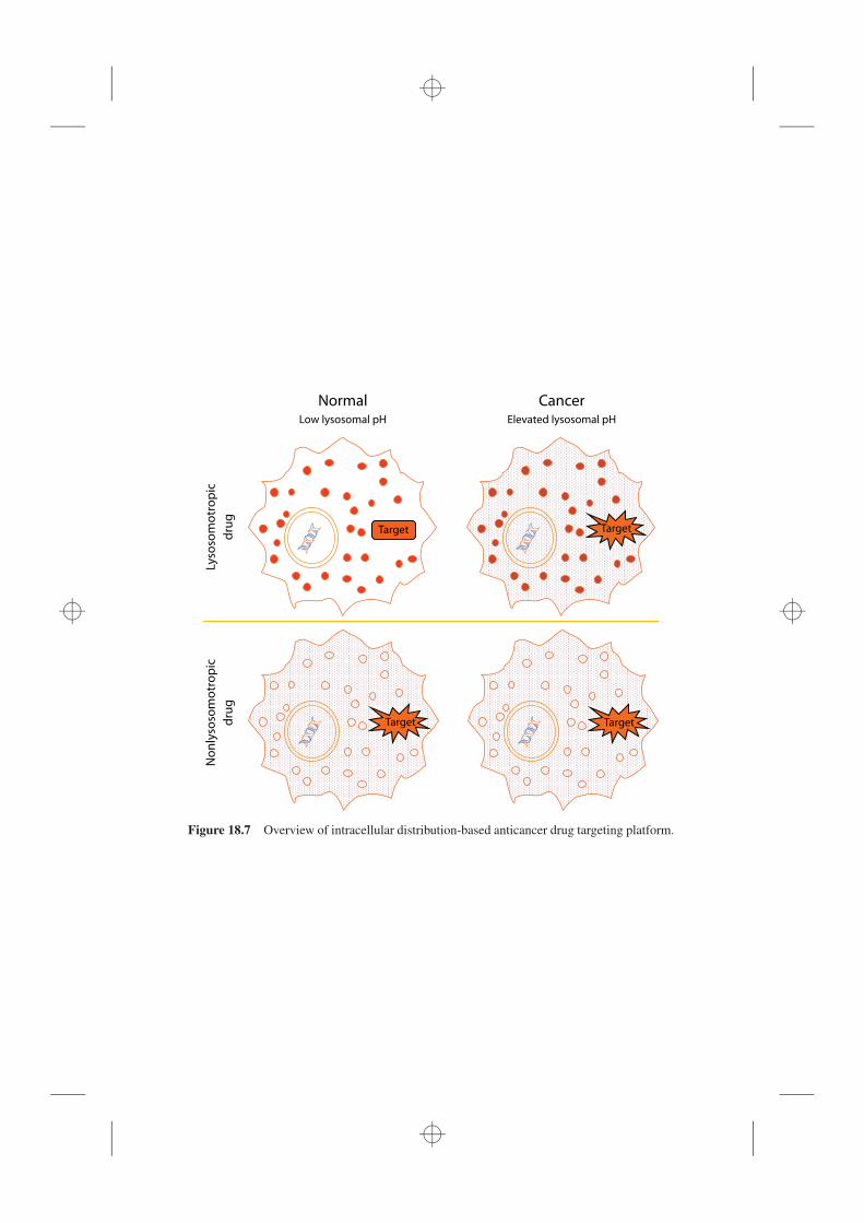
�
� �
�
NormalLow lysosomal pH
Target
Target Target
Lyso
som
otr
op
ic
dru
g
No
nly
soso
mo
tro
pic
dru
g
CancerElevated lysosomal pH
Target
Figure 18.7 Overview of intracellular distribution-based anticancer drug targeting platform.
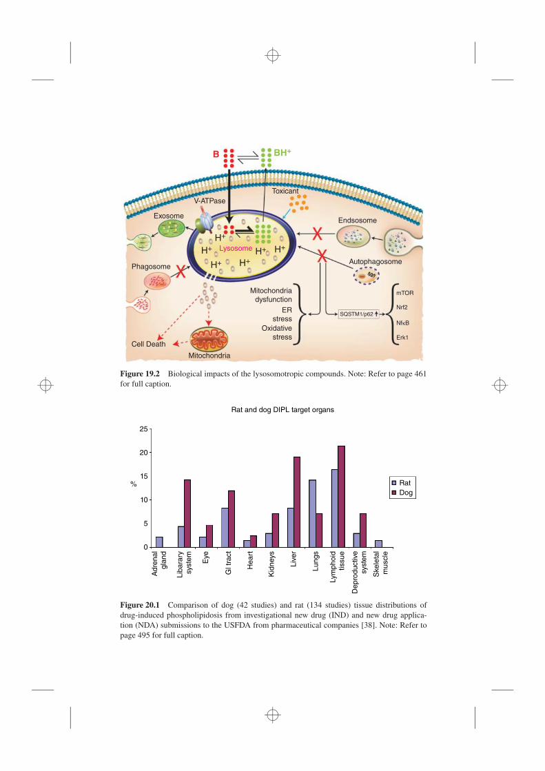
�
� �
�
Exosome
V-ATPase
B BH+
Toxicant
Endsosome
Autophagosome
SQSTM1/p62
mTOR
Nrf2
NfκB
Erk1
Phagosome
Mitochondria
Cell Death
Lysosome
Mitochondriadysfunction
ERstress
Oxidativestress
H+
H+
H+ H+H+ H+
Figure 19.2 Biological impacts of the lysosomotropic compounds. Note: Refer to page 461for full caption.
Rat and dog DIPL target organs
25
Rat
Adre
na
l
gla
nd
Gl tr
act
Eye
Lib
ara
ry
syste
m
He
art
Kid
neys
Liv
er
Lu
ng
s
Lym
ph
oid
tissu
e
De
pro
du
ctive
syste
m
Ske
leta
l
mu
scle
Dog
20
15%
10
5
0
Figure 20.1 Comparison of dog (42 studies) and rat (134 studies) tissue distributions ofdrug-induced phospholipidosis from investigational new drug (IND) and new drug applica-tion (NDA) submissions to the USFDA from pharmaceutical companies [38]. Note: Refer topage 495 for full caption.

�
� �
�
Protein trafficking inhibitor
QT prolongation Phospholipidosis
Gentamicin
Pentamidine
Arsenic trioxideChloroquine
Tunicamycin
FluoxetineDofetilide
Ibutilide
Sotalol Amiodarone
Haloperidol
Ketek
Meclizine
Lansoprazole
Amikacin
Memantine
Loratidine
Figure 20.3 Relation between phospholipidosis, QT prolongation, protein trafficking effects,and adverse events. Note: Refer to page 504 for full caption.