Mitochondrial biogenesis in health and disease. Molecular and therapeutic approaches
15
Send Orders for Reprints to [email protected] Current Pharmaceutical Design, 2014, 20, 000-000 1 1381-6128/14 $58.00+.00 © 2014 Bentham Science Publishers Mitochondrial Biogenesis in Health and Disease. Molecular and Therapeutic Approaches Sanchis-Gomar Fabian 1,3 , García-Giménez José Luis 1,2,3 , Gómez-Cabrera Mari Carmen 1,3 and Pallardó Federico V. 1,2,3,* 1 Department of Physiology. Faculty of Medicine and Dentistry. University of Valencia; 2 Center for Biomedical Network Research on Rare Diseases (CIBERER), 3 Fundación Investigación Hospital Clínico Universitario de Valencia/INCLIVA. Spain Abstract: Mitochondrial biogenesis (MB) is the essential mechanism by which cells control the number of mitochondria. Cells respond to different physiologic, metabolic, and pathologic changes by regulating this organelle with high morphological and functional adapt- ability. A considerable number of proteins, transcription factors, upstream regulatory proteins and secondary mechanisms are involved in MB and the stabilization of new mitochondrial DNA. These MB activators and regulators, including the main participating proteins (e.g. PGC-1 and mtTFA), are candidates for therapeutic intervention in diverse diseases, like neurodegenerative disorders, metabolic syn- drome, sarcopenia, cardiac pathophysiology and physiological processes like aging. In this review, we analyze the implication of MB in several diseases in which the MB pathway is affected. Furthermore, we describe therapeutical interventions on MB targets to boost MB for the prevention and treatment of human diseases. Furthermore, evidence based results and the knowledge gained during last years for some of these drugs aim us hypothesize about the value of a given treatment involved in the activation of MB. Keywords: Aging, sarcopenia, metabolic syndrome, renal diseases, cardiac diseases, neurodegenerative diseases. INTRODUCTION Mitochondria are the organelles that generate, via oxidative phosphorylation (OXPHOS), the cellular energy carrier ATP. They are involved in several biosynthetic processes, including choles- terol, heme, lipid, and nucleotide synthesis. Moreover, mitochon- dria are considered as the most important cellular sources and tar- gets of reactive oxygen and nitrogen species (RONS). Mitochondria signals via RONS and Ca +2 are critical regulators of cell cycle, pro- liferation, and apoptosis [1]. Thus, apart from their relevance in metabolism they have a central role in cell signaling pathways [2]. Mitochondria are highly malleable organelles capable of con- siderable morphological adaptations in response to different physiological or pathological changes [3]. Their function seems to be strongly dependent on mitochondrial morphology. It has been shown that changes in shape, number, and localization of the organelle can induce significant functional modifications [4]. Thus, mitochondria should not be considered as independent or isolated organelles. They comprise a dynamic network, a reticulum, and undergo movements, fission and, fusion processes, that affect their activity and cell function [3, 5]. These two events (fission and fusion) are coordinated and necessary for proper morphology and seem to play critical roles during development, cell division, and apoptosis [6]. The importance of fission and fusion homeostasis has been highlighted by a number of disease states linked to mutations involving shaping proteins. The mitochondrial fusion machinery, mitofusins, are putative downstream effectors in the MB program. Thus, it has been recently shown that there is a link between MB, fission, and fussion [7] (See Fig. 3). However, in this review, we are going to focus more specifically on the MB process. Mitochondrial biogenesis involves the orchestrated expression of the mitochondrial genome and the nuclear genes that encode mitochondrial proteins [8]. These organelles cannot be made de novo but replicate by a mechanism that recruits new proteins, which *Address correspondence to this author at the Department of Physiology. Faculty of Medicine and Dentistry, University of Valencia., Av. Blasco Ibañez, 15, Valencia, E46010 Spain; Tel: +(34) 96 386 46 41; Fax: +(34) 96 386 46 42; E-mail: [email protected] are added to pre-existing sub compartments and protein complexes. This process leads to a growth in size of the organelle that is subsequently divided by a fission process [3]. The major aim of this work is to review the recent advances in the knowledge of MB, its regulation, and the physiological and pathological situations that can induce relevant changes in the con- trol of this basic process. Finally, we will address the interventions of potential interest in pharmacology and drug design that can regu- late MB. In the first section of the review entitled “The mitochondrial biogenesis pathway” and in (Fig. 1), we aim to identify the main transcription factors and co activators involved in the cell signaling pathways that regulate MB. We briefly review the role of Perox- isome Proliferator–Activated Receptor (PPAR) coactivator-1 family (PGC-1), Nuclear Respiratory Factors (NRFs), Mitochon- drial Transcription Factor A (mtTFA), Estrogen-Related Receptors (ERRs), Peroxisome Proliferator–Activated Receptors (PPARs), YY1, cAMP Response Element–Binding Protein (CREB), c-myc, and Receptor-Interacting Protein 140 (RIP140) [9]. The second section of the review entitled “Regulation of mito- chondrial biogenesis” and (Fig. 2) are devoted to describing the upstream stimuli and signaling pathways involved in the regulation of PGC-1. We have focused in PGC-1 because it has emerged as a master regulator of MB and mitochondrial function, thus becom- ing a crucial metabolic node [10]. We also review the regulation of PGC-1 at the posttranslational level and more specifically, its phosphorylation [1], (de)acetylation [11], arginine methylation [12], and ubiquitination [13]. The role of RONS, and more specifically of Nitric Oxide (NO) in MB is highlighted in this section. The cell's energy supply is protected from conditions that damage mitochon- dria by an inducible transcriptional program of MB that mainly operates through redox signals involving the nitric oxide synthase (NOS) and the heme oxygenase-1/ carbon monoxide systems [14]. The third section of the review entitled "Mitochondrial bio- genesis dysregulation and disease" is focused on diseases in which the MB pathway seems to be altered [15]. The role of the mito- chondrial molecular changes and their consequences are reviewed in neurodegenerative, metabolic, renal, and musculoskeletal dis- eases.
-
Upload
independent -
Category
Documents
-
view
4 -
download
0
Transcript of Mitochondrial biogenesis in health and disease. Molecular and therapeutic approaches

Send Orders for Reprints to [email protected]
Current Pharmaceutical Design, 2014, 20, 000-000 1
1381-6128/14 $58.00+.00 © 2014 Bentham Science Publishers
Mitochondrial Biogenesis in Health and Disease. Molecular and Therapeutic Approaches
Sanchis-Gomar Fabian1,3
, García-Giménez José Luis1,2,3
, Gómez-Cabrera Mari Carmen1,3
and
Pallardó Federico V.1,2,3,*
1Department of Physiology. Faculty of Medicine and Dentistry. University of Valencia;
2Center for Biomedical Network Research on
Rare Diseases (CIBERER), 3Fundación Investigación Hospital Clínico Universitario de Valencia/INCLIVA. Spain
Abstract: Mitochondrial biogenesis (MB) is the essential mechanism by which cells control the number of mitochondria. Cells respond
to different physiologic, metabolic, and pathologic changes by regulating this organelle with high morphological and functional adapt-ability. A considerable number of proteins, transcription factors, upstream regulatory proteins and secondary mechanisms are involved in
MB and the stabilization of new mitochondrial DNA. These MB activators and regulators, including the main participating proteins (e.g. PGC-1 and mtTFA), are candidates for therapeutic intervention in diverse diseases, like neurodegenerative disorders, metabolic syn-
drome, sarcopenia, cardiac pathophysiology and physiological processes like aging. In this review, we analyze the implication of MB in several diseases in which the MB pathway is affected. Furthermore, we describe therapeutical interventions on MB targets to boost MB
for the prevention and treatment of human diseases. Furthermore, evidence based results and the knowledge gained during last years for some of these drugs aim us hypothesize about the value of a given treatment involved in the activation of MB.
Keywords: Aging, sarcopenia, metabolic syndrome, renal diseases, cardiac diseases, neurodegenerative diseases.
INTRODUCTION
Mitochondria are the organelles that generate, via oxidative phosphorylation (OXPHOS), the cellular energy carrier ATP. They are involved in several biosynthetic processes, including choles-terol, heme, lipid, and nucleotide synthesis. Moreover, mitochon-dria are considered as the most important cellular sources and tar-gets of reactive oxygen and nitrogen species (RONS). Mitochondria signals via RONS and Ca
+2 are critical regulators of cell cycle, pro-
liferation, and apoptosis [1]. Thus, apart from their relevance in metabolism they have a central role in cell signaling pathways [2].
Mitochondria are highly malleable organelles capable of con-siderable morphological adaptations in response to different physiological or pathological changes [3]. Their function seems to be strongly dependent on mitochondrial morphology. It has been shown that changes in shape, number, and localization of the organelle can induce significant functional modifications [4]. Thus, mitochondria should not be considered as independent or isolated organelles. They comprise a dynamic network, a reticulum, and undergo movements, fission and, fusion processes, that affect their activity and cell function [3, 5]. These two events (fission and fusion) are coordinated and necessary for proper morphology and seem to play critical roles during development, cell division, and apoptosis [6]. The importance of fission and fusion homeostasis has been highlighted by a number of disease states linked to mutations involving shaping proteins. The mitochondrial fusion machinery, mitofusins, are putative downstream effectors in the MB program. Thus, it has been recently shown that there is a link between MB, fission, and fussion [7] (See Fig. 3). However, in this review, we are going to focus more specifically on the MB process. Mitochondrial biogenesis involves the orchestrated expression of the mitochondrial genome and the nuclear genes that encode mitochondrial proteins [8]. These organelles cannot be made de novo but replicate by a mechanism that recruits new proteins, which
*Address correspondence to this author at the Department of Physiology. Faculty of Medicine and Dentistry, University of Valencia., Av. Blasco
Ibañez, 15, Valencia, E46010 Spain; Tel: +(34) 96 386 46 41; Fax: +(34) 96 386 46 42; E-mail: [email protected]
are added to pre-existing sub compartments and protein complexes. This process leads to a growth in size of the organelle that is subsequently divided by a fission process [3].
The major aim of this work is to review the recent advances in the knowledge of MB, its regulation, and the physiological and pathological situations that can induce relevant changes in the con-trol of this basic process. Finally, we will address the interventions of potential interest in pharmacology and drug design that can regu-late MB.
In the first section of the review entitled “The mitochondrial biogenesis pathway” and in (Fig. 1), we aim to identify the main transcription factors and co activators involved in the cell signaling pathways that regulate MB. We briefly review the role of Perox-isome Proliferator–Activated Receptor (PPAR ) coactivator-1 family (PGC-1), Nuclear Respiratory Factors (NRFs), Mitochon-drial Transcription Factor A (mtTFA), Estrogen-Related Receptors (ERRs), Peroxisome Proliferator–Activated Receptors (PPARs), YY1, cAMP Response Element–Binding Protein (CREB), c-myc, and Receptor-Interacting Protein 140 (RIP140) [9].
The second section of the review entitled “Regulation of mito-chondrial biogenesis” and (Fig. 2) are devoted to describing the upstream stimuli and signaling pathways involved in the regulation of PGC-1 . We have focused in PGC-1 because it has emerged as a master regulator of MB and mitochondrial function, thus becom-ing a crucial metabolic node [10]. We also review the regulation of PGC-1 at the posttranslational level and more specifically, its phosphorylation [1], (de)acetylation [11], arginine methylation [12], and ubiquitination [13]. The role of RONS, and more specifically of Nitric Oxide (NO) in MB is highlighted in this section. The cell's energy supply is protected from conditions that damage mitochon-dria by an inducible transcriptional program of MB that mainly operates through redox signals involving the nitric oxide synthase (NOS) and the heme oxygenase-1/ carbon monoxide systems [14].
The third section of the review entitled "Mitochondrial bio-genesis dysregulation and disease" is focused on diseases in which the MB pathway seems to be altered [15]. The role of the mito-chondrial molecular changes and their consequences are reviewed in neurodegenerative, metabolic, renal, and musculoskeletal dis-eases.
2 Current Pharmaceutical Design, 2014, Vol. 20, No. 00 Fabian et al.
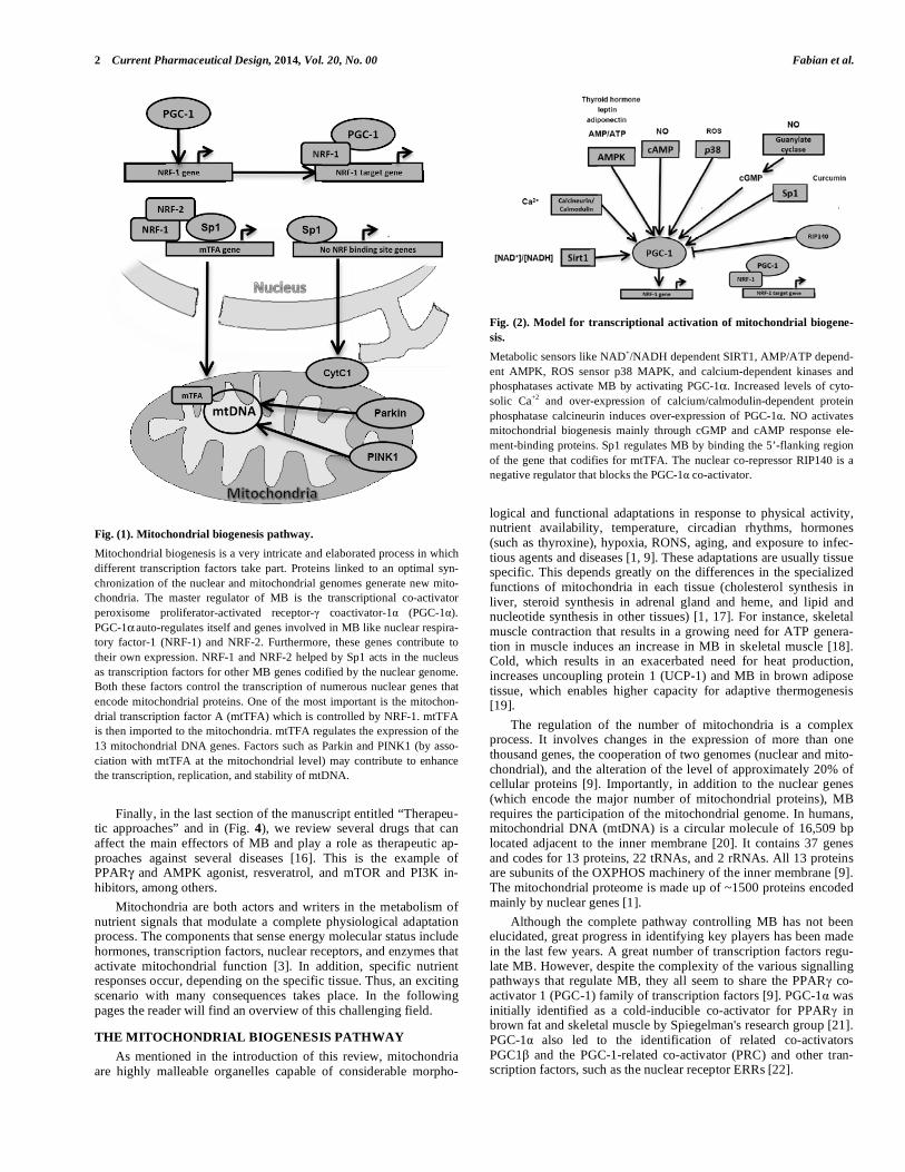
Fig. (1). Mitochondrial biogenesis pathway.
Mitochondrial biogenesis is a very intricate and elaborated process in which
different transcription factors take part. Proteins linked to an optimal syn-
chronization of the nuclear and mitochondrial genomes generate new mito-
chondria. The master regulator of MB is the transcriptional co-activator
peroxisome proliferator-activated receptor- coactivator-1 (PGC-1 ).
PGC-1 auto-regulates itself and genes involved in MB like nuclear respira-
tory factor-1 (NRF-1) and NRF-2. Furthermore, these genes contribute to
their own expression. NRF-1 and NRF-2 helped by Sp1 acts in the nucleus
as transcription factors for other MB genes codified by the nuclear genome.
Both these factors control the transcription of numerous nuclear genes that
encode mitochondrial proteins. One of the most important is the mitochon-
drial transcription factor A (mtTFA) which is controlled by NRF-1. mtTFA
is then imported to the mitochondria. mtTFA regulates the expression of the
13 mitochondrial DNA genes. Factors such as Parkin and PINK1 (by asso-
ciation with mtTFA at the mitochondrial level) may contribute to enhance
the transcription, replication, and stability of mtDNA.
Finally, in the last section of the manuscript entitled “Therapeu-tic approaches” and in (Fig. 4), we review several drugs that can affect the main effectors of MB and play a role as therapeutic ap-proaches against several diseases [16]. This is the example of PPAR and AMPK agonist, resveratrol, and mTOR and PI3K in-hibitors, among others.
Mitochondria are both actors and writers in the metabolism of nutrient signals that modulate a complete physiological adaptation process. The components that sense energy molecular status include hormones, transcription factors, nuclear receptors, and enzymes that activate mitochondrial function [3]. In addition, specific nutrient responses occur, depending on the specific tissue. Thus, an exciting scenario with many consequences takes place. In the following pages the reader will find an overview of this challenging field.
THE MITOCHONDRIAL BIOGENESIS PATHWAY
As mentioned in the introduction of this review, mitochondria are highly malleable organelles capable of considerable morpho-
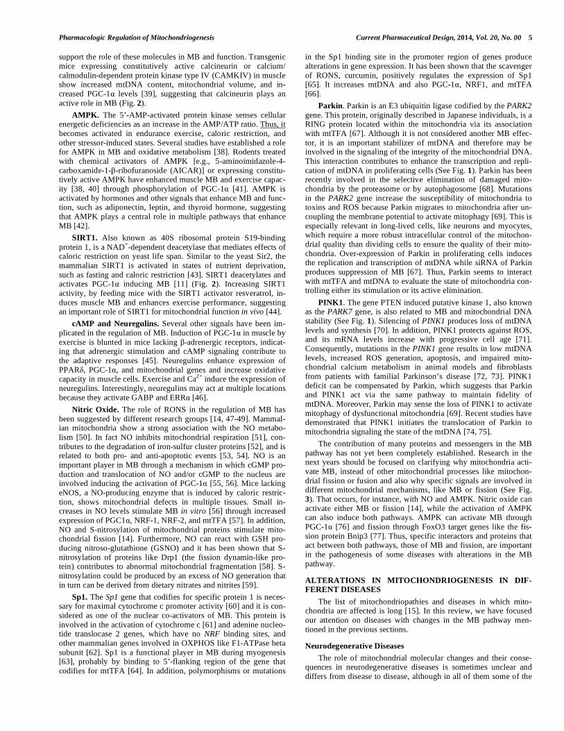
Fig. (2). Model for transcriptional activation of mitochondrial biogene-
sis.
Metabolic sensors like NAD+/NADH dependent SIRT1, AMP/ATP depend-
ent AMPK, ROS sensor p38 MAPK, and calcium-dependent kinases and
phosphatases activate MB by activating PGC-1 . Increased levels of cyto-
solic Ca+2 and over-expression of calcium/calmodulin-dependent protein
phosphatase calcineurin induces over-expression of PGC-1 . NO activates
mitochondrial biogenesis mainly through cGMP and cAMP response ele-
ment-binding proteins. Sp1 regulates MB by binding the 5’-flanking region
of the gene that codifies for mtTFA. The nuclear co-repressor RIP140 is a
negative regulator that blocks the PGC-1 co-activator.
logical and functional adaptations in response to physical activity, nutrient availability, temperature, circadian rhythms, hormones (such as thyroxine), hypoxia, RONS, aging, and exposure to infec-tious agents and diseases [1, 9]. These adaptations are usually tissue specific. This depends greatly on the differences in the specialized functions of mitochondria in each tissue (cholesterol synthesis in liver, steroid synthesis in adrenal gland and heme, and lipid and nucleotide synthesis in other tissues) [1, 17]. For instance, skeletal muscle contraction that results in a growing need for ATP genera-tion in muscle induces an increase in MB in skeletal muscle [18]. Cold, which results in an exacerbated need for heat production, increases uncoupling protein 1 (UCP-1) and MB in brown adipose tissue, which enables higher capacity for adaptive thermogenesis [19].
The regulation of the number of mitochondria is a complex process. It involves changes in the expression of more than one thousand genes, the cooperation of two genomes (nuclear and mito-chondrial), and the alteration of the level of approximately 20% of cellular proteins [9]. Importantly, in addition to the nuclear genes (which encode the major number of mitochondrial proteins), MB requires the participation of the mitochondrial genome. In humans, mitochondrial DNA (mtDNA) is a circular molecule of 16,509 bp located adjacent to the inner membrane [20]. It contains 37 genes and codes for 13 proteins, 22 tRNAs, and 2 rRNAs. All 13 proteins are subunits of the OXPHOS machinery of the inner membrane [9]. The mitochondrial proteome is made up of ~1500 proteins encoded mainly by nuclear genes [1].
Although the complete pathway controlling MB has not been elucidated, great progress in identifying key players has been made in the last few years. A great number of transcription factors regu-late MB. However, despite the complexity of the various signalling pathways that regulate MB, they all seem to share the PPAR co-activator 1 (PGC-1) family of transcription factors [9]. PGC-1 was initially identified as a cold-inducible co-activator for PPAR in brown fat and skeletal muscle by Spiegelman's research group [21]. PGC-1 also led to the identification of related co-activators PGC1 and the PGC-1-related co-activator (PRC) and other tran-scription factors, such as the nuclear receptor ERRs [22].

Pharmacologic Regulation of Mitochondriogenesis Current Pharmaceutical Design, 2014, Vol. 20, No. 00 3
PGC-1 appears to act as a master regulator of energy metabo-lism and MB by integrating and coordinating the activity of multi-ple transcription factors [21]. Pioneering studies by Scarpulla and co-workers identified the transcription factors that recognize the promoters of mitochondrial OXPHOS genes, leading to the identifi-cation of nuclear respiratory factor 1 (NRF-1) and GA-binding protein (GABP), also known as NRF-2 [23]. The coordination be-tween the mitochondrial and the nuclear genome is achieved by nuclear proteins, such as mtTFA [23]. mtTFA can be considered the most important mammalian transcription factor for mtDNA because it stimulates mitochondrial DNA transcription and replication (See Fig. 1). Apart from these, researchers have identified a number of additional transcription factors involved in MB: PPARs, YY1, CREB, c-myc, and RIP140 [9]. In this first section of our review we will summarise the role of the most relevant transcription factors in the MB pathway.
PPAR coactivator-1 family (PGC-1 , PGC-1 , and PRC). These three co-activators regulate the expression of a broad mito-chondrial gene set and promote MB [9]. PGC-1 was first identi-fied by Spiegelman and co-workers [8] as a protein interacting with PPAR , selectively expressed in brown adipose tissue (BAT), and skeletal muscle, and induced by exposure to cold. PGC-1 and PRC were identified on the basis of their similarity to PGC-1 [22]. PGC-1 links to transcription factors (NRF-1, GABP, PPARs, ERRs, and YY1) bound at their respective response elements and enable the recruitment of histone acetyltransferases enhancing transcrip-tion initiation and elongation [1]. Transgenic expression of PGC-1 and PGC-1 lead to an increase in mitochondrial content and in-creased expression of mitochondrial genes [23, 24]. PGC-1 and PGC-1 activate expression of distinct muscle contractile proteins (PGC-1 promotes type IIa and type I, and PGC-1 promotes type II fibers). Loss of function studies have shown that PGC-1 null animals have a significant decrease in expression of mitochondrial genes, decreased mitochondrial enzymatic activities, and pheno-types of mild to moderate mitochondrial dysfunction, such as a failure to defend body temperature when exposed to cold, reduced
capacity to sustain running, and energetic impairments in heart in response to -adrenergic stimulation or cardiac pressure overload [25]. Similarly, PGC-1 null mice show decreased mitochondrial gene expression and defects in thermogenesis and cardiac perform-ance [26]. The mild MB defects seen in mice lacking just PGC-1 or PGC-1 suggest that PGC-1 and PGC-1 compensate for each other’s loss in vivo.
Nuclear respiratory Factors. The nuclear respiratory factors, NRF-1 and NRF-2, contribute to the expression of respiratory subunits and mitochondrial transcription factors and thus have been implicated in nucleo-mitochondrial interactions. NRF-1 was identi-fied as a transcription factor binding to a conserved regulatory site of the cytochrome c promoter [24]. Accordingly, NRF-1 activates the expression of OXPHOS components, mitochondrial transport-ers, and mitochondrial ribosomal proteins. In addition, NRF-1 regu-lates the expression of the mtTFA and thereby coordinates the in-creased expression of nuclear mitochondrial genes with increases in mtDNA replication and expression [23]. Silencing NRF-1 leads to a significant suppression of mitochondrial target genes, suggesting that endogenous NRF-1 is constitutively active and important for the basal expression of mitochondrial targets [25]. In support of a role for NRF-1 as a critical transcription factor for expression of mitochondrial genes, NRF-1 null animals show early embryonic lethality [26]. However, although over-expression of NRF-1 in muscle increases the expression of select NRF-1 targets, it does not enhance respiratory capacity, suggesting that activation of parallel transcription pathways must complement NRF-1-induced muscle MB [27]. NRF-1 activity can be regulated by phosphorylation and/or interactions with PGC-1 , PGC-1 , PRC, and cyclin D1 [1]. Physical interactions of the PGC-1 family members with NRF-1 enhance NRF-1 dependent gene expression. However, when cyclin D1 is associated with NRF-1, its activity is repressed and MB is suppressed [1].
Regarding NRF-2, the group of Scarpulla identified it as an activator of the CoxIV promoter [28]. Functional NRF-2 binding
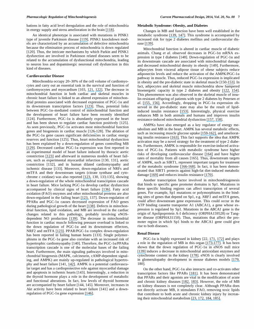
Fig. (3). Model for the relationship between mitochondrial biogenesis, fusion, and fission.
Mitochondrial biogenesis can be defined as the generation of new mitochondria from pre-existing mitochondria. However, dynamic changes in the morphol-
ogy of mitochondria are driven by fission and fusion. The ability to perform adequately these events enables mitochondria to divide and ensure proper organi-
zation of the mitochondrial network during mitochondrial biogenesis. Mfn, DRP1, Parkin, and PINK1 increase simultaneously with mitochondrial content,
suggesting that fusion/fission processes are an integral part of mitochondrial biogenesis. Thus, adequate MB, fusion and fission may take part in an orches-
trated fashion in order to generate the mitochondria.

4 Current Pharmaceutical Design, 2014, Vol. 20, No. 00 Fabian et al.
Fig. (4). Therapeutic approaches in aging and human diseases. There are
several strategies that target MB factors, and therefore, the MB pathway. It
is possible to act at the upstream level affecting metabolic sensors such as
SIRT1, using resveratrol or the agonist SRT1720. Moreover, AMPK can be
targeted with different drugs, such as metformin, AICAR or -GPA. An-
other level of targeting is the central player PGC-1 , which can be activated
by drugs such as bezafibrate, thiazolidinediones or azaleoyl, and it can even
be post-translationaly activated by deacetylation mediated by SIRT1. Parkin
activity can be modulated by rapamycin. LY294002 modulates mtTFA
stability through Parkin. Finally, the inhibition of the mTOR pathway by
rapamycin could affect the MB pathway at two different levels.
sites have been identified in the proximal promoters of many mito-chondrial genes, including those for OXPHOS components, mito-chondrial import, and mtTFA. Knockdown of NRF-2 expression in cells leads to the reduced expression of all ten nuclear-encoded COX genes and a 20% decrease in cellular COX activity. The NRF-2 promoter contains regulatory binding sites for the nuclear receptor ERR and for GABP itself, which enable transactivation of the promoter by PGC-1 . Thus, signals that activate PGC-1 and ERR are also likely to enhance NRF-2 expression [1].
Peroxisome proliferator-activated receptors (PPAR , PPAR , and PPAR ). PPARs are nuclear receptors that sense lipids and control lipid homeostasis. PPAR and PPAR are pri-marily regulators of lipid oxidation, whereas PPAR promotes lipid synthesis and storage [1]. PPARs act as heterodimers with retinoid X receptors (RXRs) to regulate a broad set of genes involved in lipid uptake, storage, and metabolism, including genes encoding mitochondria for fatty acid oxidation (FAO) enzymes [29]. Lipid uptake and metabolism provide substrates for mitochondrial oxida-tion and are thereby intimately related to mitochondrial function. Thus, in addition to their effects on lipid transport and metabolism, PPAR and PPAR promote MB in a cell type-specific manner [1].
Estrogen-related receptors (ERR , ERR , and ERR ).
ERRs are members of the nuclear receptor superfamily and the
most recent discoveries in the mitochondrial gene expression regu-latory network. Estrogens do not activate ERRs or estrogen-like molecules and can attain constitutively active ligand-binding do-main conformations in the absence of a ligand [30]. Consistent with a role in MB and function, ERR genes are expressed at high levels in tissues with high energetic demands [31]. Moreover, ERR , which is induced by PGC-1 via a positive auto regulatory loop, responds to signals central to the regulation of MB or function, such as upon exposure to cold, fasting, and physical activity [1].
cAMP response element-binding (CREB). Binding sites for CREB protein are present in the COX5A, COX8A, IDH3G, NNT, and UCP1 genes [23]. CREB contributes to mitochondrial function both directly, by acting in specific mitochondrial genes, as well as indirectly, by inducing PGC-1 expression [32].
c-Myc. Recent studies show the role of transcription factor c-Myc as a regulator of mitochondrial genes [23]. It has been shown that c-Myc binds to several mitochondrial genes, including the mi-tochondrial DNApolymerase [33]. Moreover, c-Myc may affect MB via its ability to activate the expression of PGC-1 [34].
YY1. This transcription factor has been implicated in both posi-tive and negative regulation of COX genes [23]. Recent studies show that YY1 in muscle is in a complex with PGC-1 , enhancing mitochondrial gene expression and cellular respiration. The interac-tion of YY1 with PGC-1 requires the activity of a mammalian target of rapamycin (mTOR), suggesting that YY1 integrates in-formation from two nutrient-sensing pathways: PGC-1 , which releases signals of low cellular energy state, and mTOR, which promotes cell growth in the presence of nutrients [1].
Receptor interacting protein 140 (RIP140). The nuclear re-ceptor co-repressor RIP140 functions as the antithesis of PGC-1 co-activators and acts as a transcriptional brake on mitochondrial bio-genesis (See Fig. 2). RIP140 docking to nuclear receptors recruits additional co-repressors, such as the C-terminal-binding protein (CtBP) and histone deacetylases (HDACs), and leads to suppression of gene transcription. Silencing of the RIP140 gene in 3T3L1 cells leads to increased expression of many mitochondrial genes, includ-ing those with roles in the tricarboxylic acid cycle, OXPHOS, FAO, and organelle biogenesis [35]. RIP140 null animals have increased oxygen consumption and expression of mitochondrial genes [36]. Muscle-specific deletion of RIP140 results in increased mitochon-drial volume and number of oxidative fibers, i.e., effects similar to those seen in mice over-expressing PGC-1 [37]. Interestingly, the same arginine methyltransferase, PRMT1, modifies RIP140 and PGC-1 . PRMT1-mediated methylation enhances the activity of PGC-1 and suppresses that of RIP140, suggesting that PRMT1 can act as a switch in the cellular balance of PGC-1 versus RIP140 [12]
REGULATION OF MITOCHONDRIAL BIOGENESIS
As mentioned before, PGC-1 has emerged as a master regulator of MB and mitochondrial function, thus becoming a crucial metabolic node [10]. One of the most interesting aspects of PGC-1 biology is the potential of this co-activator to sense signals of energetic or metabolic needs and to relay such signals to changes in gene expression. At the posttranslational level, PGC-1 activity is regulated via phosphorylation by Mitogen Activated Protein Kinase (MAPK) p38, Akt (Protein Kinase B), AMP-Activated Protein Kinase (AMPK), and Glycogen Synthase Kinase-3 (GSK-3) [1]; (de)acetylation by GCN5 and Sirtuin 1 (SIRT1) [11]; arginine methylation by PRMT1 [12]; and ubiquitination by SCFcdc4 [13]. The signaling pathways that regulate PGC-1 expression are dis-cussed in this section and shown in (Fig. 2).
Ca2+
-Induced Pathways. Skeletal muscle contraction leads to Ca
2+ bursts that signal to mitochondrial gene expression program
via the Ca2+
-dependent phosphatase calcineurin, Ca2+
/calmodulin-dependent kinases, and p38 MAPK [38]. Several lines of evidence
Pharmacologic Regulation of Mitochondriogenesis Current Pharmaceutical Design, 2014, Vol. 20, No. 00 5
support the role of these molecules in MB and function. Transgenic mice expressing constitutively active calcineurin or calcium/ calmodulin-dependent protein kinase type IV (CAMKIV) in muscle show increased mtDNA content, mitochondrial volume, and in-creased PGC-1 levels [39], suggesting that calcineurin plays an active role in MB (Fig. 2).
AMPK. The 5’-AMP-activated protein kinase senses cellular energetic deficiencies as an increase in the AMP/ATP ratio. Thus, it becomes activated in endurance exercise, caloric restriction, and other stressor-induced states. Several studies have established a role for AMPK in MB and oxidative metabolism [38]. Rodents treated with chemical activators of AMPK [e.g., 5-aminoimidazole-4-carboxamide-1- -ribofuranoside (AICAR)] or expressing constitu-tively active AMPK have enhanced muscle MB and exercise capac-ity [38, 40] through phosphorylation of PGC-1 [41]. AMPK is activated by hormones and other signals that enhance MB and func-tion, such as adiponectin, leptin, and thyroid hormone, suggesting that AMPK plays a central role in multiple pathways that enhance MB [42].
SIRT1. Also known as 40S ribosomal protein S19-binding protein 1, is a NAD
+-dependent deacetylase that mediates effects of
caloric restriction on yeast life span. Similar to the yeast Sir2, the mammalian SIRT1 is activated in states of nutrient deprivation, such as fasting and caloric restriction [43]. SIRT1 deacetylates and activates PGC-1 inducing MB [11] (Fig. 2). Increasing SIRT1 activity, by feeding mice with the SIRT1 activator resveratrol, in-duces muscle MB and enhances exercise performance, suggesting an important role of SIRT1 for mitochondrial function in vivo [44].
cAMP and Neuregulins. Several other signals have been im-plicated in the regulation of MB. Induction of PGC-1 in muscle by exercise is blunted in mice lacking -adrenergic receptors, indicat-ing that adrenergic stimulation and cAMP signaling contribute to the adaptive responses [45]. Neuregulins enhance expression of PPAR , PGC-1 , and mitochondrial genes and increase oxidative capacity in muscle cells. Exercise and Ca
2+ induce the expression of
neuregulins. Interestingly, neuregulins may act at multiple locations because they activate GABP and ERR [46].
Nitric Oxide. The role of RONS in the regulation of MB has been suggested by different research groups [14, 47-49]. Mammal-ian mitochondria show a strong association with the NO metabo-lism [50]. In fact NO inhibits mitochondrial respiration [51], con-tributes to the degradation of iron-sulfur cluster proteins [52], and is related to both pro- and anti-apoptotic events [53, 54]. NO is an important player in MB through a mechanism in which cGMP pro-duction and translocation of NO and/or cGMP to the nucleus are involved inducing the activation of PGC-1 [55, 56]. Mice lacking eNOS, a NO-producing enzyme that is induced by caloric restric-tion, shows mitochondrial defects in multiple tissues. Small in-creases in NO levels stimulate MB in vitro [56] through increased expression of PGC1 , NRF-1, NRF-2, and mtTFA [57]. In addition, NO and S-nitrosylation of mitochondrial proteins stimulate mito-chondrial fission [14]. Furthermore, NO can react with GSH pro-ducing nitroso-glutathione (GSNO) and it has been shown that S-nitrosylation of proteins like Drp1 (the fission dynamin-like pro-tein) contributes to abnormal mitochondrial fragmentation [58]. S-nitrosylation could be produced by an excess of NO generation that in turn can be derived from dietary nitrates and nitrites [59].
Sp1. The Sp1 gene that codifies for specific protein 1 is neces-sary for maximal cytochrome c promoter activity [60] and it is con-sidered as one of the nuclear co-activators of MB. This protein is involved in the activation of cytochrome c [61] and adenine nucleo-tide translocase 2 genes, which have no NRF binding sites, and other mammalian genes involved in OXPHOS like F1-ATPase beta subunit [62]. Sp1 is a functional player in MB during myogenesis [63], probably by binding to 5’-flanking region of the gene that codifies for mtTFA [64]. In addition, polymorphisms or mutations
in the Sp1 binding site in the promoter region of genes produce alterations in gene expression. It has been shown that the scavenger of RONS, curcumin, positively regulates the expression of Sp1 [65]. It increases mtDNA and also PGC-1 , NRF1, and mtTFA [66].
Parkin. Parkin is an E3 ubiquitin ligase codified by the PARK2 gene. This protein, originally described in Japanese individuals, is a RING protein located within the mitochondria via its association with mtTFA [67]. Although it is not considered another MB effec-tor, it is an important stabilizer of mtDNA and therefore may be involved in the signaling of the integrity of the mitochondrial DNA. This interaction contributes to enhance the transcription and repli-cation of mtDNA in proliferating cells (See Fig. 1). Parkin has been recently involved in the selective elimination of damaged mito-chondria by the proteasome or by autophagosome [68]. Mutations in the PARK2 gene increase the susceptibility of mitochondria to toxins and ROS because Parkin migrates to mitochondria after un-coupling the membrane potential to activate mitophagy [69]. This is especially relevant in long-lived cells, like neurons and myocytes, which require a more robust intracellular control of the mitochon-drial quality than dividing cells to ensure the quality of their mito-chondria. Over-expression of Parkin in proliferating cells induces the replication and transcription of mtDNA while siRNA of Parkin produces suppression of MB [67]. Thus, Parkin seems to interact with mtTFA and mtDNA to evaluate the state of mitochondria con-trolling either its stimulation or its active elimination.
PINK1. The gene PTEN induced putative kinase 1, also known as the PARK7 gene, is also related to MB and mitochondrial DNA stability (See Fig. 1). Silencing of PINK1 produces loss of mtDNA levels and synthesis [70]. In addition, PINK1 protects against ROS, and its mRNA levels increase with progressive cell age [71]. Consequently, mutations in the PINK1 gene results in low mtDNA levels, increased ROS generation, apoptosis, and impaired mito-chondrial calcium metabolism in animal models and fibroblasts from patients with familial Parkinson’s disease [72, 73]. PINK1 deficit can be compensated by Parkin, which suggests that Parkin and PINK1 act via the same pathway to maintain fidelity of mtDNA. Moreover, Parkin may sense the loss of PINK1 to activate mitophagy of dysfunctional mitochondria [69]. Recent studies have demonstrated that PINK1 initiates the translocation of Parkin to mitochondria signaling the state of the mtDNA [74, 75].
The contribution of many proteins and messengers in the MB pathway has not yet been completely established. Research in the next years should be focused on clarifying why mitochondria acti-vate MB, instead of other mitochondrial processes like mitochon-drial fission or fusion and also why specific signals are involved in different mitochondrial mechanisms, like MB or fission (See Fig. 3). That occurs, for instance, with NO and AMPK. Nitric oxide can activate either MB or fission [14], while the activation of AMPK can also induce both pathways. AMPK can activate MB through PGC-1 [76] and fission through FoxO3 target genes like the fis-sion protein Bnip3 [77]. Thus, specific interactors and proteins that act between both pathways, those of MB and fission, are important in the pathogenesis of some diseases with alterations in the MB pathway.
ALTERATIONS IN MITOCHONDRIOGENESIS IN DIF-FERENT DISEASES
The list of mitochondriopathies and diseases in which mito-chondria are affected is long [15]. In this review, we have focused our attention on diseases with changes in the MB pathway men-tioned in the previous sections.
Neurodegenerative Diseases
The role of mitochondrial molecular changes and their conse-quences in neurodegenerative diseases is sometimes unclear and differs from disease to disease, although in all of them some of the

6 Current Pharmaceutical Design, 2014, Vol. 20, No. 00 Fabian et al.
MB players seem to be altered. For instance, PGC-1 over-expression has been related to different pathological situations. Our group reported that the up-regulation of some MB factors such as, PGC-1 and mtTFA, could contribute to the pathogenic features in Friedreich’s ataxia, pointing to PGC-1 as a downstream effector of frataxin deficiency [78]. Friedreich's ataxia (FRDA) (ORPHA95) is a rare mitochondrial disease associated with a defect in the ex-pression of frataxin [79, 80]. The pathological consequences are degeneration of the nervous system structures, mainly sensory neu-ropathy in dorsal root ganglia accompanied by degeneration of posterior columns in the spinal cord and pyramidal tracts, and hy-pertrophic cardiomyopathy with necrosis and fibrosis [81]. Our research in FRDA fibroblasts showed that oxidative stress and low levels of ATP produce the over-expression of MB transcription factors in cells from adult patients but not in those from young pa-tients [76]. Coppola et al., whose results concurred with ours, re-ported low levels of PGC-1 in several tissues, but not in cardiac muscle from frataxin-deficient mice [82]. The hypothesis was that the induction of the proteins involved in MB could explain the heart disease in FRDA patients. Our results confirmed this hypothesis and suggested that an increase of ROS and defects in OXPHOS may be an early event in the cell pathophysiology of FRDA, whereas the increase in the MB response might be a late phenome-non associated with the patient's individual age and the natural his-tory of the disease [78].
Other disease in which PGC-1 function is compromised is Huntington's disease (HD) (ORPHA399). Huntington's disease is a rare inherited neurodegenerative disorder produced as a conse-quence of the expansion of a CAG repeat in the huntingtin HTT gene [83]. The age at onset of the disease is correlated with the length of the HTT CAG repeat and has been related to MB compo-nents [84]. Recent studies have shown genetic variations [85-87] and deregulation in PGC-1 [88-90], thus providing evidence for the dysfunction of MB in HD. In fact, modulation of PGC-1 levels and activity or other key factors that interfere in PGC-1 expres-sion in HD have been proposed as therapeutic strategies in HD pathology [91]. NRF-1, NRF-2 and mtTFA are downstream to PGC-1 in the MB pathway (See Fig. 1). These transcription fac-tors are also affected in HD. It has been shown that polymorphisms in NRF-1 and mtTFA influence the age of onset of HD which con-firms the implication of mitochondria metabolism in the HD patho-genesis [92]. Finally, numerous research groups have reported tran-scriptional deregulation in HD caused by association of Sp1 with the mutant protein huntingtin [93, 94]. Direct association of mutant huntingtin with mitochondria has been reported, which results in specific dysfunctions of respiratory chain complexes II and III and oxidative stress [95, 96].
As mentioned in the Introduction, it has recently been shown that there is a link between MB, fission, and fusion [7]. In addition to PGC-1 , PGC-1 is another positive inductor of MB [23, 24], producing a larger increase in mitochondrial volume and control-ling basal MB [97, 98]. In 2008, Liesa and coworkers [99] showed that mitochondrial fusion is increased by the nuclear co-activator PGC-1 . Mammals have two mitofusin isoforms, Mfn1 and Mfn2 [100]. PGC-1 increases Mfn2 promoter activity and transcription thereby increasing mitochondrial fusion. Mfn2 is a mutated protein in Charcot-Marie-Tooth (CMT) type 2A neuropathy (OR-PHA99946) [101]. This is the reason why these patients have low levels of Mfn2 in muscle [102]. PGC-1 expression is higher than PGC-1 in muscle cells under basal conditions [98] and depletion of one of them cannot be replaced by overexpression of the other. Thus, both PGC-1 and PGC-1 have decreased expression in the context of type 2 diabetes [103, 104]. It is interesting to note that in CMT there are very few descriptions of type 2 diabetes mellitus [105] although Mfn2 mutations have been related to diabetes [106]. It would, therefore, be very important to elucidate the mitochondrial events involved in CMT, in particular CMT2A, establish the mito-
chondrial function that occurs in the regulation of MB, fis-sion/fussion, and also clarify the function of insulin signaling and glucose homeostasis in vivo.
NRF-1 is also involved in the regulation of the expression of FMR1, the gene affected in fragile X mental retardation (FXS) (ORPHA908). This syndrome, which affects all carrier males and approximately 35% of carrier females, is a rare genetic disease associated with mild to severe intellectual affection linked with behavioral disorders (e.g. like mood instability, autism, affection of visuospatial abilities, and short-term memory) that evolve to sig-nificant intellectual deficit in adulthood. The genetic cause of this rare genetic disease is the expansion of the CGG trinucleotide re-peats in the 5’-untraslated region for the FMR1 gene (Xq27.3) that produces hypermethylation of the gene [107]. FMR1 codes for the FMRP protein, a RNA-binding protein that regulates protein syn-thesis in neuronal dendrites. Thus, down regulation in FMR1 can reduce synaptic plasticity. It has been found that hypermethylation of the FMR1 promoter blocks the binding of NRF-1 transcription factor [108]. Thus, the NRF-1 is an important player in genes that regulate cognitive development like MRF1 or others such as fragile X-related gene 2, FXR2 [109]. In consequence, alterations in the upstream activators of NRF1 or downstream factors may have seri-ous consequences in neuronal function.
Another interesting observation is that the NRF1 gene promoter contains an estrogen response element (ERE) that specifically binds to the estrogen receptors ER and ER in vitro [110, 111] and that supports the possibility of designing therapeutic strategies for the activation of the NRF1 gene.
NRF-2 has been shown to regulate the utrophin gene, one of the dystrophin-related protein family [112]. Utrophin is a ubiquitously transcribed gene that seems capable of molecularly substituting dystrophin and thus can improve muscle physiopathology.
A recent work by Yoshida et al. shows that over-expression of mtTFA correlates with low overall survival in patients with colorec-tal tumors. Thus, mtTFA can be used as a prognostic marker. This suggests that a therapy designed to down regulate mtTFA could be a strategy for patients with colorectal cancer treated with the cis-platinum analogous drug, oxaliplatin [113].
On the other hand, down regulation of mtTFA or a decreased transport of mtTFA to the mitochondria decreases retinal MB and the number of mitochondria contributing to the pathogenesis of diabetic retinopathy in a rat model [114]. This neuronal retina de-generation is not completely understood, and now MB seems to be contributing to the physiopathology of diabetic retinopathy. Inter-estingly, studies by Santos et al. showed that although a good gly-cemic control was achieved, diabetic retinopathy progressed if no action was taken to control MB [115].
As we have described in the previous section, Parkin is an in-teresting protein involved in mitochondrial replication and gene transcription via its association to mtTFA. It has been described that mutations in the PARK2 gene are a cause of Parkinson's disease type 2, also known as young adult-onset Parkinsonism (YOPD) (ORPHA2828), a neurodegenerative disorder with a prevalence of 1/10,000 characterized by bradykinesia, rigidity, postural instabil-ity, tremor, diurnal fluctuation of the symptoms, and loss of dopa-minergic neurons in the substantia nigra [116]. Although mutations in the PARK2 gene have been identified, the lack of understanding in the underlying mechanisms about genotype and phenotype corre-lations make us to consider the implication of other mechanisms that suggest the involvement of mitochondria. It has been shown that Parkin expression can be up regulated in dependence on ATF4, a transcription factor triggered by mitochondrial stress and the un-folded protein response (UPR) [117]. This mitochondrial control mediated by Parkin, under mitochondrial stress conditions, may be related to controlling fatty acid uptake [118]. Thus, one of the causes of Parkinson anticipation in YOPD may be linked to pertur-
Pharmacologic Regulation of Mitochondriogenesis Current Pharmaceutical Design, 2014, Vol. 20, No. 00 7
bations in fatty acid level deregulation and the role of mitochondria in energy supply and stress amelioration in the brain [118].
An identical phenotype is associated with mutations in PINK1 type of juvenile Parkinson disease [119]. PINK1 knockdown mod-els are characterized by an accumulation of defective mitochondria because the elimination process of mitochondria is down regulated [120]. Thus, the intricate mechanisms by which Parkin and PINK1 dysfunction are involved in Parkinson related diseases seem to be related to the accumulation of dysfunctional mitochondria, leading to neuron loss and dopaminergic neuronal cell dysfunction in this kind of diseases.
Cardiovascular Disease
Mitochondria occupy 20–30% of the cell volume of cardiomyo-cytes and carry out an essential task in the survival and function of cardiomyocytes and myocardium [103, 121, 122]. The decrease in mitochondrial function in both cardiac and skeletal muscles in chronic heart failure is linked to an altered expression of mitochon-drial proteins associated with decreased expression of PGC-1 and its downstream transcription factors [123]. Thus, potential links between PGC-1 –mediated control of mitochondrial function and the development of heart failure have been recently identified [124]. Furthermore, PGC-1 is abundantly expressed in the heart and has been shown to regulate cardiac function positively [125]. As seen previously, PGC-1 activates most mitochondrial function genes and biogenesis in cardiac muscle [126-128]. The ablation of the PGC-1 gene causes significant deficiencies in cardiac energy reserves and function [125]. Thus, heart mitochondrial dysfunction has been explained by a down-regulation of genes controlling MB [129]. Decreased cardiac PGC-1 expression was first reported in an experimental model of heart failure induced by thoracic aortic constriction [123] and afterward in numerous models of heart fail-ure, such as experimental myocardial infarction [130, 131], aortic constriction [132], and in human dilated cardiomyopathy and ischemic disease [133]. Moreover, down-regulation of NRFs and mtTFA and their downstream targets (citrate synthase and cyto-chrome c oxidase) was also reported [123, 130, 133-135], showing a down-regulation of the whole mitochondrial transcription cascade in heart failure. Mice lacking PGC-1 develop cardiac dysfunction accompanied by clinical signs of heart failure [136]. Fatty acid oxidation (FAO) enzymes and other mitochondrial proteins are also down-regulated in the failing heart [123, 137]. Down-regulation of PPAR and PGC-1 causes decreased expression of FAO genes during pathological growth of the heart [138]. Defects in mitochon-drial function, lipid oxidation, and MB are involved in the cardiac changes related to this pathology, probably involving eNOS-dependent NO production [139]. The decrease in mitochondrial function in cardiac muscle following pressure overload is linked to the down regulation of PGC-1 and its downstream effectors, NRF2 and mtTFA [123]. PPAR/PGC-1 complex down-regulation has been reported in failing human hearts [133]. Single polymor-phisms in the PGC-1 gene also correlate with an increased risk of hypertrophic cardiomyopathy [140]. Therefore, the PGC-1 /PPAR transcription cascade is one of the molecular bases of the failing heart. Furthermore, the main signaling pathways involved in mito-chondrial biogenesis (MAPK, calcineurin, cAMP-dependent signal-ing, and AMPK) are mainly up-regulated in pathological hypertro-phy and heart failure [141, 142]. AMPK is a potential cardiovascu-lar target and has a cardioprotective role against myocardial damage and apoptosis in ischemic hearts [143]. Interestingly, a reduction in the thyroid hormone plays a role in the development of metabolic and functional alterations. In fact, low levels of thyroid hormone are accompanied by heart failure [144, 145]. Moreover, increases in Akt activity have been related to heart failure [141] and a down-regulation of PGC-1 gene expression [146].
Metabolic Syndrome: Obesity, and Diabetes
Changes in MB and function have been well established in the metabolic syndrome [139, 147]. This syndrome is accompanied by increased risk for type 2 diabetes mellitus and cardiovascular dis-ease [139].
Mitochondrial function is altered in cardiac muscle of diabetic animals. Chang et al. observed decreases in PGC-1 mRNA ex-pression in type I diabetes [148]. Down-regulation of PGC-1 and its downstream cascade are associated with mitochondrial damage and decreased mitochondrial density in obesity [149]. Furthermore, adipocytes from visceral adipose tissue of obese subjects reduce adiponectin levels and reduce the activation of the AMPK/PGC-1 pathway in muscle. Thus, reduced PGC-1 expression is implicated in obesity and the pre-diabetic state in skeletal muscle [150-153]. In fact, adipocytes and skeletal muscle mitochondria show hampered bioenergetic capacity in type 2 diabetes and obesity [122, 154]. This phenomenon was also observed in the skeletal muscle of insu-lin-resistant offspring of patients with type 2 diabetes by Petersen et al. [155, 156]. Accordingly, dropping in PGC-1 expression ob-served in the pre-diabetic state may also be the result of lipid-induced insulin resistance [153]. Interestingly, physical exercise enhances MB in both animals and humans and improves insulin resistance-induced mitochondrial dysfunction [157, 158].
AMPKs have also emerged as a key regulator of energy me-tabolism and MB in the heart. AMPK has several metabolic effects, such as increasing muscle glucose uptake [159-162], and ameliorat-ing insulin resistance [163]. This fact supports the idea that AMPK activation may be a novel strategy for the treatment of type 2 diabe-tes. Furthermore, AMPK is responsible for exercise-induced activa-tion of PGC-1 . Patients with metabolic syndrome have higher risks of developing cardiovascular disease [164] and have higher rates of mortality from all causes [165]. Thus, downstream targets of AMPK, such as SIRT1, represent important targets for treatment of the metabolic syndrome [166-168]. In fact, it has been demon-strated that SIRT1 protects against high-fat diet-induced metabolic damage [169] and reduces insulin resistance [170].
Another transcription factor involved in mitochondriogenesis that binds to specific gene promoter domains is Sp1. Mutations in these specific binding regions can affect transcription of several genes. For example, Sp1 mutations or polymorphisms in the bind-ing sites of genes that depend on Sp1, e.g. NRF1 and mtTFA genes, could affect downstream gene expression. This could occur in the ATP binding cassette transporter A1 (ABCA1), a gene whose ex-pression is regulated by Sp1. Mutations in the ABCA1 gene is the origin of Apolipoprotein A-I deficiency (ORPHA159528) or Tang-ier disease (ORPHA31150). Thus, mutations that affect the pro-moter region in which Sp1 binds to the ABCA1 gene could give rise to both diseases.
Renal Disease
PGC-1 is highly expressed in kidney [22, 171, 172] and plays a role in the regulation of MB in this organ [173-177]. It has been shown that the down regulation of PGC-1 in eNOS null mice [139] induces a decrease in mitochondrial antioxidant enzymes and cytochrome content in the kidney [178]. eNOS is clearly involved in glomerulopathy development in mouse diabetes models [179, 180].
On the other hand, PGC-1 also interacts and co-activates other transcription factors like PPARs [181]. It has been demonstrated that PPARs and their agonists are vital in the modification of acute and chronic kidney diseases [182, 183]. However, the role of MB on kidney diseases is not completely clear. Although PPAR does not directly activate MB, it stimulates FAO, removing toxic lipids that contribute to both acute and chronic kidney injury by increas-ing their mitochondrial metabolism [23, 172, 184, 185].

8 Current Pharmaceutical Design, 2014, Vol. 20, No. 00 Fabian et al.
In addition, Portilla and collaborators noted a PPAR signaling impairment in induced acute kidney injury (AKI) [186-190]. These authors also reported that PPAR agonists produced benefits in in-duced AKI by increasing the expression of heme oxygenase (HO1), an important enzyme in the protection against renal damage [191]. In addition, overexpression of SIRT1 by cisplatin in S1 and S2 segment proximal tubules protects against AKI by retaining perox-isome number and function [192]. Thus, it has been suggested that SIRT1 effects are mediated through PPAR [172]. SIRT1 expres-sion is decreased in the aging kidneys and is prevented by calorie restriction [193]. Moreover, He et al. demonstrated that SIRT1 overexpression protects against oxidative stress and limits injury produced by ureteral obstruction by increasing interstitial cell ex-pression of cyclooxygenase-2 [194].
Although the current available information about the effects of MB on kidney pathogenesis is limited, MB in renal diseases will be subject to increasing attention given its ability to modify major pathogenic processes contributing to acute and chronic kidney dis-ease.
Aging and Sarcopenia
Harman first suggested that mitochondria are key organelles involved in aging [195]. Impaired MB has been postulated as a key factor in the age-related loss of muscle mass (sarcopenia) [196-198]. In addition, other factors such as programmed cell death, oxidative stress, lowering in the rate of protein synthesis, inflamma-tion, and lowering levels of anabolic hormones are also involved in sarcopenia [199, 200]. There is an association between the accumu-lation of mtDNA point mutations and deficiencies in mitochondrial OXPHOS in aging skeletal muscle fibers [201]. Thus, it is now clear that mitochondrial damage is associated with aging, and in particular with aging of the muscle cells.
We studied the MB pathway by measuring the expression of PGC-1 and NRF-1, and we estimated the mitochondrial content by determining the expression of cytochrome c in the hearts of young and old animals [202]. The expression of PGC-1 , NRF-1, and cytochrome c was significantly lower in old animals when com-pared with young animals. A possibility for the prevention and treatment of age-related disorders is, therefore, the modulation of PGC-1 levels in skeletal muscle [203]. On this regard, we have recently found an age-associated lack of induction of PGC-1 in response to exercise and other stimuli, such as cold exposure or thyroid hormone treatment [204].
Lack of p38 MAPK activation has been also reported in the skeletal muscle of old individuals following a resistance exercise bout [205]. In addition, it has been noted that aging diminishes protein synthesis in skeletal muscle [206, 207]. The Akt/mTOR pathway constitutes the main regulator pathway of protein synthesis via p70S6K, eIF4E, and 4EBP1 [208]. This pathway is undoubtedly altered in aged skeletal muscle [209, 210].
Overall, these results highlight the role of PGC-1 , p38 MAPK, and mTOR in the loss of MB associated with aging and emphasize its importance as targets for pharmacological interventions to pre-vent age-associated sarcopenia.
TERAPHEUTIC APPROACHES
Mitochondrial biogenesis is required for multiple cellular events and controls cellular homeostasis. In the previous section we have shown that MB alterations, in some of their multiple effectors, can lead to disease. Molecular studies are contributing to clarify the role of different players involved in this pathway and how they trigger complex interactions with other effectors. For this reason, designing strategies that affect MB at different levels could offer therapeutic approaches against metabolic disorders, neurodegenera-tive diseases, mitochondria-related diseases (including some rare mitochondrial disorders), and aging [16].
Therapeutic Strategies Against Neurodegenerative Disorders
Mitochondrial biogenesis dysfunction at different levels of the MB pathway has been related to different neurodegenerative disor-ders like, Friedreich’s ataxia, Huntington disease, and Parkin-sonism-related syndromes. Therefore, therapeutic approaches tar-geting mitochondrial factors involved in MB pathway have proved to be a great promise in neurodegenerative disease.
1. Friedreich’s Ataxia
Some drugs can affect the main effectors of MB. This is the example of thiazolidinediones (See Fig. 4). These drugs increase the expression of PGC-1 , and it has been shown experimentally that chronic overexpression of PGC-1 induces cardiomyopathy in hearts of transgenic mice [128]. We recently alerted about the fatal consequences of the use of thiazolidinediones in FRDA [211], be-cause a therapeutic intervention based in thiazolidinediones induces PGC-1 expression that, in turn, can increase the mitochondria mass in heart tissue. In addition they also decrease the production of prostacyclin from the endothelium, whereas production of thromboxane-A2 remains unaffected, giving rise to adverse cardio-vascular events one of the reasons for death in FRDA patients.
Interventions to modulate PGC-1 levels in muscle tissue in FRDA could be a therapeutic approach in this disease. However, thiazolidinediones are not the most appropriate therapeutic strategy due to the adverse effects that we have described above. However, thiazolidinediones could be promising drug candidates in other pathologies in which cardiac events are not critical for the disease, as occurs for FRDA.
Another candidate drug to stimulate PGC-1 in FRDA is the PPAR agonist Azelaoyl PAF (See Fig. 4), an alkyl phosphatidyl-choline with high affinity for PPAR . This drug significantly in-creased the levels of frataxin in neuroblastoma cell lines and fibro-blasts from FRDA patients [212]. This promising drug may have two beneficial effects 1) it may stimulate the expression of the fra-taxin gene (FXN), the molecular cause of Friedreich’s ataxia dis-ease and 2) it may up-regulate MB in several tissues in FRDA in which Coppola et al. observed down-regulation of PGC-1 in sev-eral models [82]. Obviously, an exhaustive control is necessary in order to avoid non-desirable effects at the cardiac levels in FRDA patients, like the ones we described for thiazolidinediones.
2. Huntington Disease
The importance of PGC-1 seems evident in the physiopathol-ogy of several diseases in which mitochondria occupy an essential position [213]. In that way, PGC-1 also appears as an important element in HD. Huntington disease represents one of the diseases in which overexpression of PGC-1 could result in amelioration of the neurological defects and several drugs can exert this role. For example, fibrates and thiazolidinediones (stimulators of PPAR), metformin and AICAR (agonist of AMPK) as well as resveratrol (SIRT1 activator) could result in the stimulation of the upstream effectors of MB (Fig. 4).
Drugs like bezafibrate that mimic the effect of PGC-1 over-expression may result in a valuable strategy in this type of diseases. In that regard, Wenz et al. described how bezafibrate administration in a mitochondrial myopathy mouse model mimicked the effects of transgenic PGC-1 expression [214]. However, the result that sug-gests that bezafibrate induces MB in mouse models seems conflict-ing, as PGC-1 has been reported to be increased [214] or normal [215]. More recent studies performed by Yatsuga and Suomalainen have shown that bezafibrate does not induce PGC-1 , although the use of bezafibrate improves mitochondrial myopathy features [216].
Another strategy to stimulate the MB pathway is affecting pharmacologically some of the upstream or downstream effectors such as SIRT1, AMPK, NRF-1, and mtTFA. Thus, this strategy may represent an alternative therapeutic approach.

Pharmacologic Regulation of Mitochondriogenesis Current Pharmaceutical Design, 2014, Vol. 20, No. 00 9
3. Parkinson Disease
Recently, it has been described how activation of NRF-1 and mtTFA, through continuous activation of Akt, protects neuronal cells with mitochondrial dysfunction in models for Parkinson dis-ease [217]. This is an interesting observation because it would al-low control of MB downstream of PGC-1 . However, more studies should be performed to clarify the magnitude of this regulation and for designing viable therapies.
Parkin and PINK1 protect mtDNA downstream in the MB pathway. Thus, stimulation of these proteins is an interesting thera-peutic approach against Parkinson related disorders. A strategy recently described for Parkin induction is the treatment of dopa-minergic neural cells with the mTOR inhibitor Rapamycin and the PI3K inhibitor LY294002 (Fig. 2). These drugs seem to increase the levels of the PARKIN gene [218]. This overexpression has bene-ficial effects in cells by decreasing apoptosis [219]. These results describe the possible pathways involved in Parkinsonism-related syndromes and offer relevant results about the signaling pathways involved in these diseases.
Therapeutic Strategies in Metabolic Syndromes
Colca suggests that insulin resistance is a physiological com-pensation for inappropriate oxidative metabolism that induces a metabolic inflammatory response, and that insulin sensitizers, such as thiazolidinediones (TZDs), exert their pharmacology through modifications of mitochondrial metabolism, preventing metabolic inflammation and allowing the up-regulation of MB [220]. TZDs, such as pioglitazone and rosiglitazone, increase PGC-1 expression and mitochondrial DNA copy number, and enhance the oxidative capacity of white adipose tissue, leading to insulin sensitization [221-223].
But it is possible to act on the mitochondriogenic pathway using other drugs. Forskolin, a labdane diterpene produced by the Indian Coleus plant, increases mitochondrial copy number and the expres-sion of genes involved in MB and fatty acid oxidation [224]. Met-formin, in human umbilical vein endothelial cells, inhibited hyper-glycaemia-induced intracellular and mitochondrial oxidant produc-tion, stimulated AMPK activity, increased the RNA expression of NRF1 and mtTFA genes, stimulated mitochondrial proliferation, and increased expression of PGC-1 , and SOD2 [225, 226] (Fig. 2). Adiponectin exerts anti-diabetic effects, possibly via the mecha-nism of increasing mitochondrial number and function [227]. How-ever, clinical evidence has shown that the TZDs, and specifically rosiglitazone, increase the risk of heart failure. Some of the adverse effects of these drugs are fluid retention, weight gain, and periph-eral edema [211]. In addition, TZDs causes an exaggerated throm-botic response to thrombotic stimuli, predisposing patients to ad-verse cardiovascular outcomes [211, 228]. Thus, these drugs may have negative effects in metabolic syndrome patients because they could complicate and accelerate cardiovascular problems. In this regard, we consider that metformin has fewer adverse effects than TZDs in MB interventions and, therefore, is more recommended for this purpose.
Therapeutic Strategies in Aging Associated Diseases and Sarco-penia
Mitochondrial biogenesis was induced in skeletal muscle by administration of bezafibrate (Fig. 4), a PPAR pan-agonist. This strategy stimulated residual respiratory capacity in muscle tissue. Mitochondrial proliferation resulted in an enhanced OXPHOS ca-pacity per muscle mass. As a consequence, ATP levels were con-served resulting in a delayed onset of the myopathy and a markedly prolonged life span. Thus, induction of MB through pharmacologi-cal or metabolic modulation of the PPAR/PGC-1 pathway prom-ises to be an effective therapeutic approach for mitochondrial disor-ders [214].
Rasbach and Schnellmann identified activity of multiple isofla-vones and 5-hydroxytryptamine receptor agonists that increase PGC-1 expression, biogenesis, and oxidative metabolism in rabbit proximal tubule cell primary culture [174, 177]. Funk et al. [173] have shown that treatment of primary cultures of rabbit proximal tubules with a SIRT1 activator, SRT1720, produces increased PGC-1 and MB that are dependent on SIRT1 deacetylase activity, but not apparently on AMPK (Fig. 4) [229]. Calorie restriction strongly increases SIRT1 expression in proximal tubules and attenuates cis-platin-induced functional changes and tubular cell injury [192]. Aging is also associated with kidney damage, and modified by calo-rie restriction and SIRT1 [193]. In addition, SIRT1 has been also suggested as a calorie restriction mimetic, and therefore, as a new therapeutic approach for type 2 diabetes mellitus and diabetic vas-cular complications [230]. In fact, resveratrol, a polyphenol in red wine that increases NAD
+ and the activity of SIRT1, has been re-
ported as a calorie restriction mimetic with potential anti-aging and anti-diabetogenic properties that ameliorates aging-related meta-bolic phenotypes [231]. Most recently, it has been demonstrated that Sirt3 responds to exercise and nutritional signals in skeletal muscle to coordinate downstream molecular responses [232].
Although the downstream targets of AMPK are not well estab-lished, AMPK activation has been shown to be involved in the up-regulation of nuclear-encoded mitochondrial genes. AMPK can be chronically activated by the administration of the drug AICAR (Fig. 4). After few weeks of this treatment, an up-regulation of various mitochondrial enzymes, as well as the GLUT-4 protein content within skeletal muscle occurred [233]. In addition, treatment with another pharmacological agent, -guanadinopropionic acid ( -GPA), causes phosphocreatine and ATP depletion, leading to AMPK activation (Fig. 4) [234]. After treatment with -GPA, there was an increase in cytochrome c content and augmented mitochon-drial density in rat skeletal muscle [234]. This effect of -GPA is abrogated in transgenic mice expressing a dominant negative mu-tant form of AMPK in muscle [235], confirming the involvement of the AMPK pathway in mitochondrial biogenesis. Taken together these results suggest that ATP turnover and AMPK activation are part of an important signaling pathway involved in mediating con-tractile activity-induced MB.
It is also tempting to speculate that muscle-specific agonists could be expected agents for the individuals with sedentary life style and reluctant to exercise. Narkar et al. [40] suggested that AMPK might be present in a transcriptional complex with PPAR- , where it could amplify receptor activity via direct protein-protein interaction and by phosphorylating co-activators such as PGC-1 . These findings underline a novel pharmacologic strategy to repro-gram muscle endurance by targeting AMPK-PPAR- signaling axis with orally active ligands [236]. Pharmacological activation of AMPK by AICAR, increases PGC-1 gene expression and mito-chondrial biogenesis in skeletal muscle (Fig. 2) [237], enhancing the running ability of mice even in the absence of exercise training [40]. Skeletal muscle targeted deletion of PGC-1 results in fiber-type switching with a reduction in slow twitch oxidative fibers and exercise intolerance [238]. However, the side effects of AICAR are contentious and a specific application for this compound has not been identified so far. The US Food and Drug Administration (FDA) will not approve any drug without proven clinical applica-tion and therefore these treatments remain under development [239].
Burks et al. [240] were interested in the beneficial impact on the muscle remodeling process of sarcopenic mice by losartan, an angiotensin II receptor antagonist (ARA) commonly used to treat high blood pressure. In their study, they showed that immobilized mice treated with losartan were protected against loss of muscle mass and this protective mechanism was mediated by an increased activation of the IGF-1)/Akt/mTOR pathway. Thus, blockade of the AT1 (angiotensin II type I) receptor improved muscle remodeling

10 Current Pharmaceutical Design, 2014, Vol. 20, No. 00 Fabian et al.
and protected against disuse atrophy. Moreover, another ARA, telmisartan, has been suggested to improve skeletal muscle function and to prevent adipogenesis and weight gain through activation of PPAR- -dependent pathways [241, 242]. Consequently, telmisartan may be used as a pharmacological intervention to treat sarcopenia [243, 244].
CONCLUDING REMARKS
Mitochondrial biogenesis is dynamically regulated in a tissue- and signal-specific manner to enable cellular adaptation to energetic and metabolic demands. The foregoing overview of the regulation of MB is largely derived from literature that has been developed from skeletal muscle and related data on adipocytes and hepato-cytes. The regulation of this pathway may vary in cell and tissue type. Although this may seem like an obstacle, because treatments with mitochondriogenic activators could alter diverse physiologic mechanisms, it actually represents a possibility for designing more tissue- specific therapeutic approaches after the complete under-standing of the MB pathway and its upstream and downstream players. A limited number of drugs, many of them reported here, are currently available for clinical use. However, the arsenal for the prevention and treatment still remains relatively poor. In the com-ing years, new substances will be developed which could provide a promising future in the treatment of diseases related to MB.
CONFLICT OF INTEREST
The authors confirm that this article content has no conflicts of interest.
ACKNOWLEDGEMENTS
We thank Mrs Marilyn Noyes for her kind help in reviewing the manuscript. Our work is supported by the Ministerio de Ciencia e Innovación (grant SAF2008-01338) and the Biomedical Network Research Center for Rare Diseases (CIBERER). CIBERER is an initiative of the ISCIII of Spain.
ABBREVIATIONS
AICAR = 5-aminoimidazole-4-carboxamide-1- -ribofuranoside
Akt = Protein Kinase B
AMPK = 5’-AMP-activated protein kinase
ARA = Angiotensin II receptor antagonist
AT1 = Angiotensin II type receptor
AKI = Acute kidney injury
BAT = Brown adipose tissue
-GPA = -guanidopropionic acid
cGMP = Cyclic guanosine monophosphate
COLIA1 = Type I collagen gene
COX = Cytochrome oxidase
CREB = cAMP response element–binding
eNOS = Endothelial nitric oxide synthase
ERE = Estrogen response element
ERRs = Estrogen-related receptor
FAO = Fatty acid oxidation
FDA = US Food and Drug Administration
FRDA = Friedreich's ataxia
FXS = Fragile X mental retardation
GAPB = GA-binding protein
GSH = Reduced glutathione
GSK-3 = Glycogen synthase kinase-3
GSNO = Nitroso-glutathione
HD = Huntington's disease
HDACs = Histone deacetylases
HF = Heart failure
HO = Heme oxygenase 1
MAPK = mitogen activated protein kinase
MB = Mitochondriogenesis or mitochondrial biogene-sis
mtDNA = Mitochondrial DNA
mtTFA = Mitochondrial transcription factor A
mTOR = Mammalian target of rapamycin
NO = Nitric oxide
NOS = Nitric oxide synthase
OXPHOS = Oxidative phosphorylation
PGC-1 = PPAR co-activator 1
PGC-1 = PPAR co-activator 1
PPAR = Peroxisome proliferator–activated receptors
PRC = PGC-1-related coactivator
PRMT1 = Arginine methyltransferase
RING = Really important new gene
RONS = Reactive oxygen and nitrogen species
rRNAs = Ribosomic RNA
RXRs = Retinoid X receptors
SP1 = Specific protein 1
SIRT = Sirtuin 1
tRNAs = Transfer RNA
TZDs = Thiazolidinediones
UCP-1 = Uncoupling protein 1
UPR = Unfolded protein response
YOPD = Young adult-onset Parkinsonism
REFERENCES
[1] Hock MB, Kralli A. Transcriptional control of mitochondrial bio-genesis and function. Annu Rev Physiol 2009; 71: 177-203.
[2] Tormos KV, Anso E, Hamanaka RB, et al. Mitochondrial complex III ROS regulate adipocyte differentiation. Cell Metab 2011; 14:
537-44. [3] Hood DA. Invited Review: contractile activity-induced mitochon-
drial biogenesis in skeletal muscle. J Appl Physiol 2001; 90: 1137-57.
[4] Osellame LD, Blacker TS, Duchen MR. Cellular and molecular mechanisms of mitochondrial function. Best Pract Res Clin Endo-
crinol Metab 2012; 26: 711-23. [5] Hales KG, Fuller MT. Developmentally regulated mitochondrial
fusion mediated by a conserved, novel, predicted GTPase. Cell 1997; 90: 121-9.
[6] Diaz F, Moraes CT. Mitochondrial biogenesis and turnover. Cell Calcium 2008; 44: 24-35.
[7] Papanicolaou KN, Phillippo MM, Walsh K. Mitofusins and the mitochondrial permeability transition: the potential downside of
mitochondrial fusion. Am J Physiol Heart Circ Physiol 2012; 303: H243-55.
[8] Wright DC, Han DH, Garcia-Roves PM, et al. Exercise-induced mitochondrial biogenesis begins before the increase in muscle
PGC-1alpha expression. J Biol Chem 2007; 282: 194-9. [9] Ryan MT, Hoogenraad NJ. Mitochondrial-nuclear communica-
tions. Annu Rev Biochem 2007; 76: 701-22. [10] Fernandez-Marcos PJ, Auwerx J. Regulation of PGC-1alpha, a
nodal regulator of mitochondrial biogenesis. Am J Clin Nutr 2011; 93: 884S-90.
[11] Gerhart-Hines Z, Rodgers JT, Bare O, et al. Metabolic control of muscle mitochondrial function and fatty acid oxidation through
SIRT1/PGC-1alpha. Embo J 2007; 26: 1913-23.

Pharmacologic Regulation of Mitochondriogenesis Current Pharmaceutical Design, 2014, Vol. 20, No. 00 11
[12] Teyssier C, Ma H, Emter R, Kralli A, Stallcup MR. Activation of
nuclear receptor coactivator PGC-1alpha by arginine methylation. Genes Dev 2005; 19: 1466-73.
[13] Olson BL, Hock MB, Ekholm-Reed S, et al. SCFCdc4 acts antago-nistically to the PGC-1alpha transcriptional coactivator by targeting
it for ubiquitin-mediated proteolysis. Genes Dev 2008; 22: 252-64. [14] Piantadosi CA. Regulation of mitochondrial processes by protein
S-nitrosylation. Biochim Biophys Acta 2012; 1820: 712-21. [15] Finsterer J. Mitochondriopathies. Eur J Neurol 2004; 11: 163-86.
[16] Stefano GB, Kim C, Mantione K, Casares F, Kream RM. Targeting mitochondrial biogenesis for promoting health. Med Sci Monit
2012; 18: SC1-3. [17] Hancock CR, Han DH, Higashida K, Kim SH, Holloszy JO. Does
calorie restriction induce mitochondrial biogenesis? A reevaluation. FASEB J 2011; 25: 785-91.
[18] Holloszy JO. Biochemical adaptations in muscle. Effects of exer-cise on mitochondrial oxygen uptake and respiratory enzyme activ-
ity in skeletal muscle. J Biol Chem 1967; 242: 2278-82. [19] Klingenspor M. Cold-induced recruitment of brown adipose tissue
thermogenesis. Exp Physiol 2003; 88: 141-8. [20] Legros F, Malka F, Frachon P, Lombes A, Rojo M. Organization
and dynamics of human mitochondrial DNA. J Cell Sci 2004; 117: 2653-62.
[21] Puigserver P, Wu Z, Park CW, et al. A cold-inducible coactivator of nuclear receptors linked to adaptive thermogenesis. Cell 1998;
92: 829-39. [22] Handschin C, Spiegelman BM. Peroxisome proliferator-activated
receptor gamma coactivator 1 coactivators, energy homeostasis, and metabolism. Endocr Rev 2006; 27: 728-35.
[23] Scarpulla RC. Transcriptional paradigms in mammalian mitochon-drial biogenesis and function. Physiol Rev 2008; 88: 611-38.
[24] Virbasius CA, Virbasius JV, Scarpulla RC. NRF-1, an activator involved in nuclear-mitochondrial interactions, utilizes a new
DNA-binding domain conserved in a family of developmental regulators. Genes Dev 1993; 7: 2431-45.
[25] Cam H, Balciunaite E, Blais A, et al. A common set of gene regu-latory networks links metabolism and growth inhibition. Mol Cell
2004; 16: 399-411. [26] Huo L, Scarpulla RC. Mitochondrial DNA instability and peri-
implantation lethality associated with targeted disruption of nuclear respiratory factor 1 in mice. Mol Cell Biol 2001; 21: 644-54.
[27] Baar K, Song Z, Semenkovich CF, et al. Skeletal muscle overex-pression of nuclear respiratory factor 1 increases glucose transport
capacity. Faseb J 2003; 17: 1666-73. [28] Virbasius JV, Virbasius CA, Scarpulla RC. Identity of GABP with
NRF-2, a multisubunit activator of cytochrome oxidase expression, reveals a cellular role for an ETS domain activator of viral promot-
ers. Genes Dev 1993; 7: 380-92. [29] Evans RM, Barish GD, Wang YX. PPARs and the complex jour-
ney to obesity. Nat Med 2004; 10: 355-61. [30] Kallen J, Schlaeppi JM, Bitsch F, et al. Evidence for ligand-
independent transcriptional activation of the human estrogen-related receptor alpha (ERRalpha): crystal structure of ERRalpha
ligand binding domain in complex with peroxisome proliferator-activated receptor coactivator-1alpha. J Biol Chem 2004; 279:
49330-7. [31] Giguere V. Transcriptional control of energy homeostasis by the
estrogen-related receptors. Endocr Rev 2008; 29: 677-96. [32] Herzig S, Long F, Jhala US, et al. CREB regulates hepatic glu-
coneogenesis through the coactivator PGC-1. Nature 2001; 413: 179-83.
[33] Khatib SY, Farah H, El-Migdadi F. Allopurinol enhances adenine nucleotide levels and improves myocardial function in isolated hy-
poxic rat heart. Biochemistry (Mosc) 2001; 66: 328-33.. [34] Zhang H, Gao P, Fukuda R, et al. HIF-1 inhibits mitochondrial
biogenesis and cellular respiration in VHL-deficient renal cell car-cinoma by repression of C-MYC activity. Cancer Cell 2007; 11:
407-20. [35] Powelka AM, Seth A, Virbasius JV, et al. Suppression of oxidative
metabolism and mitochondrial biogenesis by the transcriptional corepressor RIP140 in mouse adipocytes. J Clin Invest 2006; 116:
125-36. [36] Leonardsson G, Steel JH, Christian M, et al. Nuclear receptor
corepressor RIP140 regulates fat accumulation. Proc Natl Acad Sci U S A 2004; 101: 8437-42.
[37] Seth A, Steel JH, Nichol D, et al. The transcriptional corepressor
RIP140 regulates oxidative metabolism in skeletal muscle. Cell Metab 2007; 6: 236-45.
[38] Rockl KS, Witczak CA, Goodyear LJ. Signaling mechanisms in skeletal muscle: acute responses and chronic adaptations to exer-
cise. IUBMB Life 2008; 60: 145-53. [39] Wu H, Kanatous SB, Thurmond FA, et al. Regulation of mitochon-
drial biogenesis in skeletal muscle by CaMK. Science 2002; 296: 349-52.
[40] Narkar VA, Downes M, Yu RT, et al. AMPK and PPARdelta ago-nists are exercise mimetics. Cell 2008; 134: 405-15.
[41] Jager S, Handschin C, St-Pierre J, Spiegelman BM. AMP-activated protein kinase (AMPK) action in skeletal muscle via direct phos-
phorylation of PGC-1alpha. Proc Natl Acad Sci U S A 2007; 104: 12017-22.
[42] Irrcher I, Adhihetty PJ, Joseph AM, Ljubicic V, Hood DA. Regula-tion of mitochondrial biogenesis in muscle by endurance exercise.
Sports Med 2003; 33: 783-93. [43] Robinson DM, Ogilvie RW, Tullson PC, Terjung RL. Increased
peak oxygen consumption of trained muscle requires increased electron flux capacity. J Appl Physiol 1994; 77: 1941-52.
[44] Lagouge M, Argmann C, Gerhart-Hines Z, et al. Resveratrol im-proves mitochondrial function and protects against metabolic dis-
ease by activating SIRT1 and PGC-1alpha. Cell 2006; 127: 1109-22.
[45] Miura S, Kawanaka K, Kai Y, et al. An increase in murine skeletal muscle peroxisome proliferator-activated receptor-gamma coacti-
vator-1alpha (PGC-1alpha) mRNA in response to exercise is medi-ated by beta-adrenergic receptor activation. Endocrinology 2007;
148: 3441-8. [46] Ariazi EA, Kraus RJ, Farrell ML, Jordan VC, Mertz JE. Estrogen-
related receptor alpha1 transcriptional activities are regulated in part via the ErbB2/HER2 signaling pathway. Mol Cancer Res
2007; 5: 71-85. [47] Gomez-Cabrera MC, Domenech E, Romagnoli M, et al. Oral ad-
ministration of vitamin C decreases muscle mitochondrial biogene-sis and hampers training-induced adaptations in endurance per-
formance. Am J Clin Nutr 2008; 87: 142-9. [48] Ristow M, Zarse K, Oberbach A, et al. Antioxidants prevent
health-promoting effects of physical exercise in humans. Proc Natl Acad Sci U S A 2009; 106: 8665-70.
[49] Nisoli E, Tonello C, Cardile A, et al. Calorie restriction promotes mitochondrial biogenesis by inducing the expression of eNOS. Sci-
ence 2005; 310: 314-7. [50] Foster MW, Stamler JS. New insights into protein S-nitrosylation.
Mitochondria as a model system. J Biol Chem 2004; 279: 25891-7. [51] Brown GC. Regulation of mitochondrial respiration by nitric oxide
inhibition of cytochrome c oxidase. Biochim Biophys Acta 2001; 1504: 46-57.
[52] Drapier JC. Interplay between NO and [Fe-S] clusters: relevance to biological systems. Methods 1997; 11: 319-29.
[53] Ghafourifar P, Schenk U, Klein SD, Richter C. Mitochondrial nitric-oxide synthase stimulation causes cytochrome c release from
isolated mitochondria. Evidence for intramitochondrial peroxyni-trite formation. J Biol Chem 1999; 274: 31185-8.
[54] Mannick JB, Hausladen A, Liu L, et al. Fas-induced caspase deni-trosylation. Science 1999; 284: 651-4.
[55] Nisoli E, Carruba MO. Nitric oxide and mitochondrial biogenesis. J Cell Sci 2006; 119: 2855-62.
[56] Nisoli E, Clementi E, Paolucci C, et al. Mitochondrial biogenesis in mammals: the role of endogenous nitric oxide. Science 2003; 299:
896-9. [57] Kelly DP, Scarpulla RC. Transcriptional regulatory circuits con-
trolling mitochondrial biogenesis and function. Genes Dev 2004; 18: 357-68.
[58] Nakamura T, Lipton SA. S-nitrosylation of critical protein thiols mediates protein misfolding and mitochondrial dysfunction in neu-
rodegenerative diseases. Antioxid Redox Signal 2011; 14: 1479-92. [59] Hord NG. Dietary nitrates, nitrites, and cardiovascular disease.
Curr Atheroscler Rep 2011; 13: 484-92. [60] Evans MJ, Scarpulla RC. Interaction of nuclear factors with multi-
ple sites in the somatic cytochrome c promoter. Characterization of upstream NRF-1, ATF, and intron Sp1 recognition sequences. J
Biol Chem 1989; 264: 14361-8.

12 Current Pharmaceutical Design, 2014, Vol. 20, No. 00 Fabian et al.
[61] Li R, Luciakova K, Nelson BD. Expression of the human cyto-
chrome c1 gene is controlled through multiple Sp1-binding sites and an initiator region. Eur J Biochem 1996; 241: 649-56.
[62] Zaid A, Li R, Luciakova K, et al. On the role of the general tran-scription factor Sp1 in the activation and repression of diverse
mammalian oxidative phosphorylation genes. J Bioenerg Biomembr 1999; 31: 129-35.
[63] Kraft CS, LeMoine CM, Lyons CN, et al. Control of mitochondrial biogenesis during myogenesis. Am J Physiol Cell Physiol 2006;
290: C1119-27. [64] Choi YS, Lee HK, Pak YK. Characterization of the 5'-flanking
region of the rat gene for mitochondrial transcription factor A (Tfam). Biochim Biophys Acta 2002; 1574: 200-4.
[65] Chhunchha B, Fatma N, Bhargavan B, et al. Specificity protein, Sp1-mediated increased expression of Prdx6 as a curcumin-induced
antioxidant defense in lens epithelial cells against oxidative stress. Cell Death Dis 2011; 2: e234.
[66] Kuo JJ, Chang HH, Tsai TH, Lee TY. Positive effect of curcumin on inflammation and mitochondrial dysfunction in obese mice with
liver steatosis. Int J Mol Med 2012; 30: 673-9. [67] Kuroda Y, Mitsui T, Kunishige M, et al. Parkin enhances mito-
chondrial biogenesis in proliferating cells. Hum Mol Genet 2006; 15: 883-95.
[68] Tanaka A, Cleland MM, Xu S, et al. Proteasome and p97 mediate mitophagy and degradation of mitofusins induced by Parkin. J Cell
Biol 2010; 191: 1367-80. [69] Narendra D, Tanaka A, Suen DF, Youle RJ. Parkin is recruited
selectively to impaired mitochondria and promotes their autophagy. J Cell Biol 2008; 183: 795-803.
[70] Gegg ME, Cooper JM, Schapira AH, Taanman JW. Silencing of PINK1 expression affects mitochondrial DNA and oxidative phos-
phorylation in dopaminergic cells. PLoS One 2009; 4: e4756. [71] Mai S, Klinkenberg M, Auburger G, Bereiter-Hahn J, Jendrach M.
Decreased expression of Drp1 and Fis1 mediates mitochondrial elongation in senescent cells and enhances resistance to oxidative
stress through PINK1. J Cell Sci 2010; 123: 917-26. [72] Gandhi S, Wood-Kaczmar A, Yao Z, et al. PINK1-associated Park-
inson's disease is caused by neuronal vulnerability to calcium-induced cell death. Mol Cell 2009; 33: 627-38.
[73] Gispert S, Ricciardi F, Kurz A, et al. Parkinson phenotype in aged PINK1-deficient mice is accompanied by progressive mitochon-
drial dysfunction in absence of neurodegeneration. PLoS One 2009; 4: e5777.
[74] Narendra DP, Jin SM, Tanaka A, et al. PINK1 is selectively stabi-lized on impaired mitochondria to activate Parkin. PLoS Biol 2010;
8: e1000298. [75] Vives-Bauza C, Zhou C, Huang Y, et al. PINK1-dependent re-
cruitment of Parkin to mitochondria in mitophagy. Proc Natl Acad Sci U S A; 107: 378-83.
[76] Garcia-Gimenez JL, Gimeno A, Gonzalez-Cabo P, et al. Differen-tial expression of PGC-1alpha and metabolic sensors suggest age-
dependent induction of mitochondrial biogenesis in Friedreich ataxia fibroblasts. PLoS One 2012; 6: e20666.
[77] Romanello V, Guadagnin E, Gomes L, et al. Mitochondrial fission and remodelling contributes to muscle atrophy. EMBO J 2010; 29:
1774-85. [78] Garcia-Gimenez JL, Gimeno A, Gonzalez-Cabo P, et al. Differen-
tial expression of PGC-1alpha and metabolic sensors suggest age-dependent induction of mitochondrial biogenesis in Friedreich
ataxia fibroblasts. PLoS One 2011; 6: e20666. [79] Campuzano V, Montermini L, Molto MD, et al. Friedreich's ataxia:
autosomal recessive disease caused by an intronic GAA triplet re-peat expansion. Science 1996; 271: 1423-7.
[80] Campuzano V, Montermini L, Lutz Y, et al. Frataxin is reduced in Friedreich ataxia patients and is associated with mitochondrial
membranes. Hum Mol Genet 1997; 6: 1771-80. [81] Durr A, Cossee M, Agid Y, et al. Clinical and genetic abnormali-
ties in patients with Friedreich's ataxia. N Engl J Med 1996; 335: 1169-75.
[82] Coppola G, Marmolino D, Lu D, et al. Functional genomic analysis of frataxin deficiency reveals tissue-specific alterations and identi-
fies the PPARgamma pathway as a therapeutic target in Frie-dreich's ataxia. Hum Mol Genet 2009; 18: 2452-61.
[83] A novel gene containing a trinucleotide repeat that is expanded and unstable on Huntington's disease chromosomes. The Huntington's
Disease Collaborative Research Group. Cell 1993; 72: 971-83.
[84] Weydt P, Soyal SM, Gellera C, et al. The gene coding for PGC-
1alpha modifies age at onset in Huntington's Disease. Mol Neu-rodegener 2009; 4: 3.
[85] Taherzadeh-Fard E, Saft C, Andrich J, Wieczorek S, Arning L. PGC-1alpha as modifier of onset age in Huntington disease. Mol
Neurodegener 2009; 4: 10. [86] Arning L, Haghikia A, Taherzadeh-Fard E, et al. Mitochondrial
haplogroup H correlates with ATP levels and age at onset in Huntington disease. J Mol Med (Berl) 2010; 88: 431-6.
[87] Che HV, Metzger S, Portal E, et al. Localization of sequence varia-tions in PGC-1alpha influence their modifying effect in Huntington
disease. Mol Neurodegener 2011; 6: 1. [88] Cui L, Jeong H, Borovecki F, et al. Transcriptional repression of
PGC-1alpha by mutant huntingtin leads to mitochondrial dysfunc-tion and neurodegeneration. Cell 2006; 127: 59-69.
[89] Weydt P, Pineda VV, Torrence AE, et al. Thermoregulatory and metabolic defects in Huntington's disease transgenic mice implicate
PGC-1alpha in Huntington's disease neurodegeneration. Cell Metab 2006; 4: 349-62.
[90] Chaturvedi RK, Adhihetty P, Shukla S, et al. Impaired PGC-1alpha function in muscle in Huntington's disease. Hum Mol Genet 2009;
18: 3048-65. [91] Rona-Voros K, Weydt P. The Role of PGC-1alpha in the Patho-
genesis of Neurodegenerative Disorders. Curr Drug Targets 2010. [92] Taherzadeh-Fard E, Saft C, Akkad DA, et al. PGC-1alpha down-
stream transcription factors NRF-1 and TFAM are genetic modifi-ers of Huntington disease. Mol Neurodegener 2011; 6: 32.
[93] Dunah AW, Jeong H, Griffin A, et al. Sp1 and TAFII130 transcrip-tional activity disrupted in early Huntington's disease. Science
2002; 296: 2238-43. [94] Hodges A, Strand AD, Aragaki AK, et al. Regional and cellular
gene expression changes in human Huntington's disease brain. Hum Mol Genet 2006; 15: 965-77.
[95] Orr AL, Li S, Wang CE, et al. N-terminal mutant huntingtin asso-ciates with mitochondria and impairs mitochondrial trafficking. J
Neurosci 2008; 28: 2783-92. [96] Solans A, Zambrano A, Rodriguez M, Barrientos A. Cytotoxicity
of a mutant huntingtin fragment in yeast involves early alterations in mitochondrial OXPHOS complexes II and III. Hum Mol Genet
2006; 15: 3063-81. [97] St-Pierre J, Lin J, Krauss S, et al. Bioenergetic analysis of perox-
isome proliferator-activated receptor gamma coactivators 1alpha and 1beta (PGC-1alpha and PGC-1beta) in muscle cells. J Biol
Chem 2003; 278: 26597-603. [98] Staiger H, Stefan N, Machicao F, Fritsche A, Haring HU.
PPARGC1A mRNA levels of in vitro differentiated human skeletal muscle cells are negatively associated with the plasma oleate
concentrations of the donors. Diabetologia 2006; 49: 212-4. [99] Liesa M, Borda-d'Agua B, Medina-Gomez G, et al. Mitochondrial
fusion is increased by the nuclear coactivator PGC-1beta. PLoS One 2008; 3: e3613.
[100] Westermann B. Mitochondrial fusion and fission in cell life and death. Nat Rev Mol Cell Biol 2010; 11: 872-84.
[101] Zuchner S, Mersiyanova IV, Muglia M, et al. Mutations in the mitochondrial GTPase mitofusin 2 cause Charcot-Marie-Tooth
neuropathy type 2A. Nat Genet 2004; 36: 449-51. [102] Bach D, Naon D, Pich S, et al. Expression of Mfn2, the Charcot-
Marie-Tooth neuropathy type 2A gene, in human skeletal muscle: effects of type 2 diabetes, obesity, weight loss, and the regulatory
role of tumor necrosis factor alpha and interleukin-6. Diabetes 2005; 54: 2685-93.
[103] Mootha VK, Lindgren CM, Eriksson KF, et al. PGC-1alpha-responsive genes involved in oxidative phosphorylation are coordi-
nately downregulated in human diabetes. Nat Genet 2003; 34: 267-73.
[104] Patti ME, Butte AJ, Crunkhorn S, et al. Coordinated reduction of genes of oxidative metabolism in humans with insulin resistance
and diabetes: Potential role of PGC1 and NRF1. Proc Natl Acad Sci U S A 2003; 100: 8466-71.
[105] Win N, Needs M, Rahman S, Gold P, Ward S. An unusual case of an acute hemolytic transfusion reaction caused by an autoanti-I.
Immunohematology 2011; 27: 101-3. [106] Sebastian D, Hernandez-Alvarez MI, Segales J, et al. Mitofusin 2
(Mfn2) links mitochondrial and endoplasmic reticulum function with insulin signaling and is essential for normal glucose homeo-
stasis. Proc Natl Acad Sci U S A 2012; 109: 5523-8.

Pharmacologic Regulation of Mitochondriogenesis Current Pharmaceutical Design, 2014, Vol. 20, No. 00 13
[107] Peprah E. Fragile X syndrome: the FMR1 CGG repeat distribution
among world populations. Ann Hum Genet 2012; 76: 178-91. [108] Kumari D, Usdin K. Interaction of the transcription factors USF1,
USF2, and alpha -Pal/Nrf-1 with the FMR1 promoter. Implications for Fragile X mental retardation syndrome. J Biol Chem 2001; 276:
4357-64. [109] Mahishi L, Usdin K. NF-Y, AP2, Nrf1 and Sp1 regulate the fragile
X-related gene 2 (FXR2). Biochem J 2006; 400: 327-35. [110] Hsieh YC, Yang S, Choudhry MA, et al. PGC-1 upregulation via
estrogen receptors: a common mechanism of salutary effects of es-trogen and flutamide on heart function after trauma-hemorrhage.
Am J Physiol Heart Circ Physiol 2005; 289: H2665-72. [111] Mattingly KA, Ivanova MM, Riggs KA, et al. Estradiol stimulates
transcription of nuclear respiratory factor-1 and increases mito-chondrial biogenesis. Mol Endocrinol 2008; 22: 609-22.
[112] Angus LM, Chakkalakal JV, Mejat A, et al. Calcineurin-NFAT signaling, together with GABP and peroxisome PGC-1{alpha},
drives utrophin gene expression at the neuromuscular junction. Am J Physiol Cell Physiol 2005; 289: C908-17.
[113] Yoshida Y, Hasegawa J, Nezu R, et al. Clinical usefulness of mito-chondrial transcription factor A expression as a predictive marker
in colorectal cancer patients treated with FOLFOX. Cancer Sci 2011; 102: 578-82.
[114] Santos JM, Tewari S, Goldberg AF, Kowluru RA. Mitochondrial biogenesis and the development of diabetic retinopathy. Free Radic
Biol Med 2011; 51: 1849-60. [115] Santos JM, Kowluru RA. Role of mitochondria biogenesis in the
metabolic memory associated with the continued progression of diabetic retinopathy and its regulation by lipoic acid. Invest Oph-
thalmol Vis Sci 2011; 52: 8791-8. [116] Brice A, Durr A, Lucking C. Parkin Type of Juvenile Parkinson
Disease. 1993. [117] Bouman L, Schlierf A, Lutz AK, et al. Parkin is transcriptionally
regulated by ATF4: evidence for an interconnection between mito-chondrial stress and ER stress. Cell Death Differ 2011; 18: 769-82.
[118] Kim KY, Sack MN. Parkin in the regulation of fat uptake and mi-tochondrial biology: emerging links in the pathophysiology of
Parkinson's disease. Curr Opin Lipidol 2012; 23: 201-5. [119] Schneider SA, Klein C. PINK1 Type of Young-Onset Parkinson
Disease. 1993. [120] Grunewald A, Gegg ME, Taanman JW, et al. Differential effects of
PINK1 nonsense and missense mutations on mitochondrial func-tion and morphology. Exp Neurol 2009; 219: 266-73.
[121] Kim JA, Wei Y, Sowers JR. Role of mitochondrial dysfunction in insulin resistance. Circ Res 2008; 102: 401-14.
[122] Lowell BB, Shulman GI. Mitochondrial dysfunction and type 2 diabetes. Science 2005; 307: 384-7.
[123] Garnier A, Fortin D, Delomenie C, et al. Depressed mitochondrial transcription factors and oxidative capacity in rat failing cardiac
and skeletal muscles. J Physiol 2003; 551: 491-501. [124] Garnier A, Zoll J, Fortin D, et al. Control by circulating factors of
mitochondrial function and transcription cascade in heart failure: a role for endothelin-1 and angiotensin II. Circ Heart Fail 2009; 2:
342-50. [125] Arany Z, He H, Lin J, et al. Transcriptional coactivator PGC-1
alpha controls the energy state and contractile function of cardiac muscle. Cell Metab 2005; 1: 259-71.
[126] Huss JM, Kopp RP, Kelly DP. Peroxisome proliferator-activated receptor coactivator-1alpha (PGC-1alpha) coactivates the cardiac-
enriched nuclear receptors estrogen-related receptor-alpha and -gamma. Identification of novel leucine-rich interaction motif within
PGC-1alpha. J Biol Chem 2002; 277: 40265-74. [127] Lehman JJ, Barger PM, Kovacs A, et al. Peroxisome proliferator-
activated receptor gamma coactivator-1 promotes cardiac mito-chondrial biogenesis. J Clin Invest 2000; 106: 847-56.
[128] Russell LK, Mansfield CM, Lehman JJ, et al. Cardiac-specific induction of the transcriptional coactivator peroxisome proliferator-
activated receptor gamma coactivator-1alpha promotes mitochon-drial biogenesis and reversible cardiomyopathy in a developmental
stage-dependent manner. Circ Res 2004; 94: 525-33. [129] Rimbaud S, Garnier A, Ventura-Clapier R. Mitochondrial biogene-
sis in cardiac pathophysiology. Pharmacol Rep 2009; 61: 131-8. [130] Kemi OJ, Hoydal MA, Haram PM, et al. Exercise training restores
aerobic capacity and energy transfer systems in heart failure treated with losartan. Cardiovasc Res 2007; 76: 91-9.
[131] Sun CK, Chang LT, Sheu JJ, et al. Losartan preserves integrity of
cardiac gap junctions and PGC-1 alpha gene expression and pre-vents cellular apoptosis in remote area of left ventricular myocar-
dium following acute myocardial infarction. Int Heart J 2007; 48: 533-46.
[132] Witt H, Schubert C, Jaekel J, et al. Sex-specific pathways in early cardiac response to pressure overload in mice. J Mol Med (Berl)
2008; 86: 1013-24. [133] Sebastiani M, Giordano C, Nediani C, et al. Induction of mito-
chondrial biogenesis is a maladaptive mechanism in mitochondrial cardiomyopathies. J Am Coll Cardiol 2007; 50: 1362-9.
[134] Javadov S, Huang C, Kirshenbaum L, Karmazyn M. NHE-1 inhibi-tion improves impaired mitochondrial permeability transition and
respiratory function during postinfarction remodelling in the rat. J Mol Cell Cardiol 2005; 38: 135-43.
[135] Jullig M, Hickey AJ, Chai CC, et al. Is the failing heart out of fuel or a worn engine running rich? A study of mitochondria in old
spontaneously hypertensive rats. Proteomics 2008; 8: 2556-72. [136] Arany Z, Novikov M, Chin S, et al. Transverse aortic constriction
leads to accelerated heart failure in mice lacking PPAR-gamma co-activator 1alpha. Proc Natl Acad Sci U S A 2006; 103: 10086-91.
[137] Sack MN, Rader TA, Park S, et al. Fatty acid oxidation enzyme gene expression is downregulated in the failing heart. Circulation
1996; 94: 2837-42. [138] Lehman JJ, Kelly DP. Transcriptional activation of energy meta-
bolic switches in the developing and hypertrophied heart. Clin Exp Pharmacol Physiol 2002; 29: 339-45.
[139] Nisoli E, Clementi E, Carruba MO, Moncada S. Defective mito-chondrial biogenesis: a hallmark of the high cardiovascular risk in
the metabolic syndrome? Circ Res 2007; 100: 795-806. [140] Wang S, Fu C, Wang H, et al. Polymorphisms of the peroxisome
proliferator-activated receptor-gamma coactivator-1alpha gene are associated with hypertrophic cardiomyopathy and not with hyper-
tension hypertrophy. Clin Chem Lab Med 2007; 45: 962-7. [141] Haq S, Choukroun G, Lim H, et al. Differential activation of signal
transduction pathways in human hearts with hypertrophy versus advanced heart failure. Circulation 2001; 103: 670-7.
[142] Tian R, Musi N, D'Agostino J, Hirshman MF, Goodyear LJ. In-creased adenosine monophosphate-activated protein kinase activity
in rat hearts with pressure-overload hypertrophy. Circulation 2001; 104: 1664-9.
[143] Russell RR, 3rd, Li J, Coven DL, et al. AMP-activated protein kinase mediates ischemic glucose uptake and prevents
postischemic cardiac dysfunction, apoptosis, and injury. J Clin In-vest 2004; 114: 495-503.
[144] Kinugawa K, Yonekura K, Ribeiro RC, et al. Regulation of thyroid hormone receptor isoforms in physiological and pathological car-
diac hypertrophy. Circ Res 2001; 89: 591-8. [145] Wassen FW, Schiel AE, Kuiper GG, et al. Induction of thyroid
hormone-degrading deiodinase in cardiac hypertrophy and failure. Endocrinology 2002; 143: 2812-5.
[146] Cook SA, Matsui T, Li L, Rosenzweig A. Transcriptional effects of chronic Akt activation in the heart. J Biol Chem 2002; 277: 22528-
33. [147] Nishio Y, Kanazawa A, Nagai Y, Inagaki H, Kashiwagi A. Regula-
tion and role of the mitochondrial transcription factor in the dia-betic rat heart. Ann N Y Acad Sci 2004; 1011: 78-85.
[148] Chang LT, Sun CK, Wang CY, et al. Downregulation of peroxisme proliferator activated receptor gamma co-activator 1alpha in dia-
betic rats. Int Heart J 2006; 47: 901-10. [149] Reznick RM, Zong H, Li J, et al. Aging-associated reductions in
AMP-activated protein kinase activity and mitochondrial biogene-sis. Cell Metab 2007; 5: 151-6.
[150] Wu Z, Puigserver P, Andersson U, et al. Mechanisms controlling mitochondrial biogenesis and respiration through the thermogenic
coactivator PGC-1. Cell 1999; 98: 115-24. [151] Semple RK, Crowley VC, Sewter CP, et al. Expression of the
thermogenic nuclear hormone receptor coactivator PGC-1alpha is reduced in the adipose tissue of morbidly obese subjects. Int J Obes
Relat Metab Disord 2004; 28: 176-9. [152] Roden M. Muscle triglycerides and mitochondrial function: possi-
ble mechanisms for the development of type 2 diabetes. Int J Obes (Lond) 2005; 29 Suppl 2: S111-5.
[153] Hoeks J, Hesselink MK, Russell AP, et al. Peroxisome proliferator-activated receptor-gamma coactivator-1 and insulin resistance:
acute effect of fatty acids. Diabetologia 2006; 49: 2419-26.
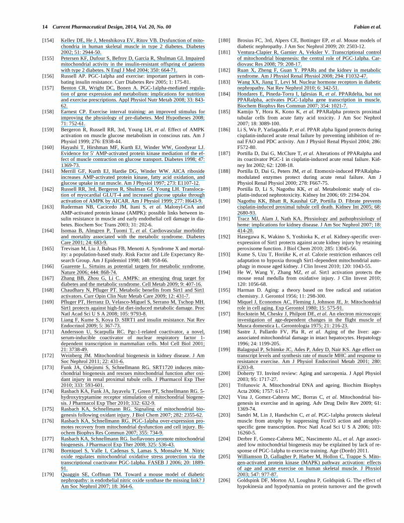
14 Current Pharmaceutical Design, 2014, Vol. 20, No. 00 Fabian et al.
[154] Kelley DE, He J, Menshikova EV, Ritov VB. Dysfunction of mito-
chondria in human skeletal muscle in type 2 diabetes. Diabetes 2002; 51: 2944-50.
[155] Petersen KF, Dufour S, Befroy D, Garcia R, Shulman GI. Impaired mitochondrial activity in the insulin-resistant offspring of patients
with type 2 diabetes. N Engl J Med 2004; 350: 664-71. [156] Russell AP. PGC-1alpha and exercise: important partners in com-
bating insulin resistance. Curr Diabetes Rev 2005; 1: 175-81. [157] Benton CR, Wright DC, Bonen A. PGC-1alpha-mediated regula-
tion of gene expression and metabolism: implications for nutrition and exercise prescriptions. Appl Physiol Nutr Metab 2008; 33: 843-
62. [158] Earnest CP. Exercise interval training: an improved stimulus for
improving the physiology of pre-diabetes. Med Hypotheses 2008; 71: 752-61.
[159] Bergeron R, Russell RR, 3rd, Young LH, et al. Effect of AMPK activation on muscle glucose metabolism in conscious rats. Am J
Physiol 1999; 276: E938-44. [160] Hayashi T, Hirshman MF, Kurth EJ, Winder WW, Goodyear LJ.
Evidence for 5' AMP-activated protein kinase mediation of the ef-fect of muscle contraction on glucose transport. Diabetes 1998; 47:
1369-73. [161] Merrill GF, Kurth EJ, Hardie DG, Winder WW. AICA riboside
increases AMP-activated protein kinase, fatty acid oxidation, and glucose uptake in rat muscle. Am J Physiol 1997; 273: E1107-12.
[162] Russell RR, 3rd, Bergeron R, Shulman GI, Young LH. Transloca-tion of myocardial GLUT-4 and increased glucose uptake through
activation of AMPK by AICAR. Am J Physiol 1999; 277: H643-9. [163] Ruderman NB, Cacicedo JM, Itani S, et al. Malonyl-CoA and
AMP-activated protein kinase (AMPK): possible links between in-sulin resistance in muscle and early endothelial cell damage in dia-
betes. Biochem Soc Trans 2003; 31: 202-6. [164] Isomaa B, Almgren P, Tuomi T, et al. Cardiovascular morbidity
and mortality associated with the metabolic syndrome. Diabetes Care 2001; 24: 683-9.
[165] Trevisan M, Liu J, Bahsas FB, Menotti A. Syndrome X and mortal-ity: a population-based study. Risk Factor and Life Expectancy Re-
search Group. Am J Epidemiol 1998; 148: 958-66. [166] Guarente L. Sirtuins as potential targets for metabolic syndrome.
Nature 2006; 444: 868-74. [167] Zhang BB, Zhou G, Li C. AMPK: an emerging drug target for
diabetes and the metabolic syndrome. Cell Metab 2009; 9: 407-16. [168] Chaudhary N, Pfluger PT. Metabolic benefits from Sirt1 and Sirt1
activators. Curr Opin Clin Nutr Metab Care 2009; 12: 431-7. [169] Pfluger PT, Herranz D, Velasco-Miguel S, Serrano M, Tschop MH.
Sirt1 protects against high-fat diet-induced metabolic damage. Proc Natl Acad Sci U S A 2008; 105: 9793-8.
[170] Liang F, Kume S, Koya D. SIRT1 and insulin resistance. Nat Rev Endocrinol 2009; 5: 367-73.
[171] Andersson U, Scarpulla RC. Pgc-1-related coactivator, a novel, serum-inducible coactivator of nuclear respiratory factor 1-
dependent transcription in mammalian cells. Mol Cell Biol 2001; 21: 3738-49.
[172] Weinberg JM. Mitochondrial biogenesis in kidney disease. J Am Soc Nephrol 2011; 22: 431-6.
[173] Funk JA, Odejinmi S, Schnellmann RG. SRT1720 induces mito-chondrial biogenesis and rescues mitochondrial function after oxi-
dant injury in renal proximal tubule cells. J Pharmacol Exp Ther 2010; 333: 593-601.
[174] Rasbach KA, Funk JA, Jayavelu T, Green PT, Schnellmann RG. 5-hydroxytryptamine receptor stimulation of mitochondrial biogene-
sis. J Pharmacol Exp Ther 2010; 332: 632-9. [175] Rasbach KA, Schnellmann RG. Signaling of mitochondrial bio-
genesis following oxidant injury. J Biol Chem 2007; 282: 2355-62. [176] Rasbach KA, Schnellmann RG. PGC-1alpha over-expression pro-
motes recovery from mitochondrial dysfunction and cell injury. Bi-ochem Biophys Res Commun 2007; 355: 734-9.
[177] Rasbach KA, Schnellmann RG. Isoflavones promote mitochondrial biogenesis. J Pharmacol Exp Ther 2008; 325: 536-43.
[178] Borniquel S, Valle I, Cadenas S, Lamas S, Monsalve M. Nitric oxide regulates mitochondrial oxidative stress protection via the
transcriptional coactivator PGC-1alpha. FASEB J 2006; 20: 1889-91.
[179] Quaggin SE, Coffman TM. Toward a mouse model of diabetic nephropathy: is endothelial nitric oxide synthase the missing link? J
Am Soc Nephrol 2007; 18: 364-6.
[180] Brosius FC, 3rd, Alpers CE, Bottinger EP, et al. Mouse models of
diabetic nephropathy. J Am Soc Nephrol 2009; 20: 2503-12. [181] Ventura-Clapier R, Garnier A, Veksler V. Transcriptional control
of mitochondrial biogenesis: the central role of PGC-1alpha. Car-diovasc Res 2008; 79: 208-17.
[182] Ruan X, Zheng F, Guan Y. PPARs and the kidney in metabolic syndrome. Am J Physiol Renal Physiol 2008; 294: F1032-47.
[183] Wang XX, Jiang T, Levi M. Nuclear hormone receptors in diabetic nephropathy. Nat Rev Nephrol 2010; 6: 342-51.
[184] Hondares E, Pineda-Torra I, Iglesias R, et al. PPARdelta, but not PPARalpha, activates PGC-1alpha gene transcription in muscle.
Biochem Biophys Res Commun 2007; 354: 1021-7. [185] Kamijo Y, Hora K, Kono K, et al. PPARalpha protects proximal
tubular cells from acute fatty acid toxicity. J Am Soc Nephrol 2007; 18: 3089-100.
[186] Li S, Wu P, Yarlagadda P, et al. PPAR alpha ligand protects during cisplatin-induced acute renal failure by preventing inhibition of re-
nal FAO and PDC activity. Am J Physiol Renal Physiol 2004; 286: F572-80.
[187] Portilla D, Dai G, McClure T, et al. Alterations of PPARalpha and its coactivator PGC-1 in cisplatin-induced acute renal failure. Kid-
ney Int 2002; 62: 1208-18. [188] Portilla D, Dai G, Peters JM, et al. Etomoxir-induced PPARalpha-
modulated enzymes protect during acute renal failure. Am J Physiol Renal Physiol 2000; 278: F667-75.
[189] Portilla D, Li S, Nagothu KK, et al. Metabolomic study of cis-platin-induced nephrotoxicity. Kidney Int 2006; 69: 2194-204.
[190] Nagothu KK, Bhatt R, Kaushal GP, Portilla D. Fibrate prevents cisplatin-induced proximal tubule cell death. Kidney Int 2005; 68:
2680-93. [191] Tracz MJ, Alam J, Nath KA. Physiology and pathophysiology of
heme: implications for kidney disease. J Am Soc Nephrol 2007; 18: 414-20.
[192] Hasegawa K, Wakino S, Yoshioka K, et al. Kidney-specific over-expression of Sirt1 protects against acute kidney injury by retaining
peroxisome function. J Biol Chem 2010; 285: 13045-56. [193] Kume S, Uzu T, Horiike K, et al. Calorie restriction enhances cell
adaptation to hypoxia through Sirt1-dependent mitochondrial auto-phagy in mouse aged kidney. J Clin Invest 2010; 120: 1043-55.
[194] He W, Wang Y, Zhang MZ, et al. Sirt1 activation protects the mouse renal medulla from oxidative injury. J Clin Invest 2010;
120: 1056-68. [195] Harman D. Aging: a theory based on free radical and ratiation
chemistry. J. Gerontol 1956; 11: 298-300. [196] Miquel J, Economos AC, Fleming J, Johnson JE, Jr. Mitochondrial
role in cell aging. Exp Gerontol 1980; 15: 575-91. [197] Rockstein M, Chesky J, Philpott DE, et al. An electron microscopic
investigation of age-dependent changes in the flight muscle of Musca domestica L. Gerontologia 1975; 21: 216-23.
[198] Sastre J, Pallardo FV, Pla R, et al. Aging of the liver: age-associated mitochondrial damage in intact hepatocytes. Hepatology
1996; 24: 1199-205. [199] Balagopal P, Schimke JC, Ades P, Adey D, Nair KS. Age effect on
transcript levels and synthesis rate of muscle MHC and response to resistance exercise. Am J Physiol Endocrinol Metab 2001; 280:
E203-8. [200] Doherty TJ. Invited review: Aging and sarcopenia. J Appl Physiol
2003; 95: 1717-27. [201] Trifunovic A. Mitochondrial DNA and ageing. Biochim Biophys
Acta 2006; 1757: 611-7. [202] Vina J, Gomez-Cabrera MC, Borras C, et al. Mitochondrial bio-
genesis in exercise and in ageing. Adv Drug Deliv Rev 2009; 61: 1369-74.
[203] Sandri M, Lin J, Handschin C, et al. PGC-1alpha protects skeletal muscle from atrophy by suppressing FoxO3 action and atrophy-
specific gene transcription. Proc Natl Acad Sci U S A 2006; 103: 16260-5.
[204] Derbre F, Gomez-Cabrera MC, Nascimento AL, et al. Age associ-ated low mitochondrial biogenesis may be explained by lack of re-
sponse of PGC-1alpha to exercise training. Age (Dordr) 2011. [205] Williamson D, Gallagher P, Harber M, Hollon C, Trappe S. Mito-
gen-activated protein kinase (MAPK) pathway activation: effects of age and acute exercise on human skeletal muscle. J Physiol
2003; 547: 977-87. [206] Goldspink DF, Morton AJ, Loughna P, Goldspink G. The effect of
hypokinesia and hypodynamia on protein turnover and the growth
Pharmacologic Regulation of Mitochondriogenesis Current Pharmaceutical Design, 2014, Vol. 20, No. 00 15
of four skeletal muscles of the rat. Pflugers Arch 1986; 407: 333-
40. [207] Marzani B, Balage M, Venien A, et al. Antioxidant supplementa-
tion restores defective leucine stimulation of protein synthesis in skeletal muscle from old rats. J Nutr 2008; 138: 2205-11.
[208] Spangenburg EE. Changes in muscle mass with mechanical load: possible cellular mechanisms. Appl Physiol Nutr Metab 2009; 34:
328-35. [209] Wu M, Liu H, Fannin J, et al. Acetaminophen improves protein
translational signaling in aged skeletal muscle. Rejuvenation Res 2010; 13: 571-9.
[210] Sanchis-Gomar F. Sestrins: Novel antioxidant and AMPK-modulating functions regulated by exercise? J Cell Physiol 2013.
[211] Garcia-Gimenez JL, Sanchis-Gomar F, Pallardo FV. Could thia-zolidinediones increase the risk of heart failure in Friedreich's
ataxia patients? Mov Disord 2011; 26: 769-71. [212] Marmolino D, Acquaviva F, Pinelli M, et al. PPAR-gamma agonist
Azelaoyl PAF increases frataxin protein and mRNA expression: new implications for the Friedreich's ataxia therapy. Cerebellum
2009; 8: 98-103. [213] Handschin C. The biology of PGC-1alpha and its therapeutic po-
tential. Trends Pharmacol Sci 2009; 30: 322-9. [214] Wenz T, Diaz F, Spiegelman BM, Moraes CT. Activation of the
PPAR/PGC-1alpha pathway prevents a bioenergetic deficit and ef-fectively improves a mitochondrial myopathy phenotype. Cell Me-
tab 2008; 8: 249-56. [215] Viscomi C, Bottani E, Civiletto G, et al. In vivo correction of COX
deficiency by activation of the AMPK/PGC-1alpha axis. Cell Me-tab 2011; 14: 80-90.
[216] Yatsuga S, Suomalainen A. Effect of bezafibrate treatment on late-onset mitochondrial myopathy in mice. Hum Mol Genet 2012; 21:
526-35. [217] Piao Y, Kim HG, Oh MS, Pak YK. Overexpression of TFAM,
NRF-1 and myr-AKT protects the MPP(+)-induced mitochondrial dysfunctions in neuronal cells. Biochim Biophys Acta 2012; 1820:
577-85. [218] Klinkenberg M, Gispert S, Dominguez-Bautista JA, et al. Restric-
tion of trophic factors and nutrients induces PARKIN expression. Neurogenetics 2012; 13: 9-21.
[219] Fitzgerald JC, Plun-Favreau H. Emerging pathways in genetic Parkinson's disease: autosomal-recessive genes in Parkinson's dis-
ease--a common pathway? FEBS J 2008; 275: 5758-66. [220] Colca JR. Insulin sensitizers may prevent metabolic inflammation.
Biochem Pharmacol 2006; 72: 125-31. [221] Wilson-Fritch L, Burkart A, Bell G, et al. Mitochondrial biogenesis
and remodeling during adipogenesis and in response to the insulin sensitizer rosiglitazone. Mol Cell Biol 2003; 23: 1085-94.
[222] Wilson-Fritch L, Nicoloro S, Chouinard M, et al. Mitochondrial remodeling in adipose tissue associated with obesity and treatment
with rosiglitazone. J Clin Invest 2004; 114: 1281-9. [223] Bogacka I, Xie H, Bray GA, Smith SR. Pioglitazone induces mito-
chondrial biogenesis in human subcutaneous adipose tissue in vivo. Diabetes 2005; 54: 1392-9.
[224] Bogacka I, Ukropcova B, McNeil M, Gimble JM, Smith SR. Struc-tural and functional consequences of mitochondrial biogenesis in
human adipocytes in vitro. J Clin Endocrinol Metab 2005; 90: 6650-6.
[225] Kukidome D, Nishikawa T, Sonoda K, et al. Activation of AMP-activated protein kinase reduces hyperglycemia-induced mitochon-
drial reactive oxygen species production and promotes mitochon-drial biogenesis in human umbilical vein endothelial cells. Diabetes
2006; 55: 120-7.
[226] Suwa M, Egashira T, Nakano H, Sasaki H, Kumagai S. Metformin
increases the PGC-1alpha protein and oxidative enzyme activities possibly via AMPK phosphorylation in skeletal muscle in vivo. J
Appl Physiol 2006; 101: 1685-92. [227] Civitarese AE, Ukropcova B, Carling S, et al. Role of adiponectin
in human skeletal muscle bioenergetics. Cell Metab 2006; 4: 75-87. [228] Konstantinopoulos PA, Karamouzis MV, Papavassiliou AG. Effect
of aspirin use on thiazolidinediones and cardiovascular events. JAMA 2008; 299: 1539; author reply 1539-40.
[229] Canto C, Gerhart-Hines Z, Feige JN, et al. AMPK regulates energy expenditure by modulating NAD+ metabolism and SIRT1 activity.
Nature 2009; 458: 1056-60. [230] Kume S, Uzu T, Kashiwagi A, Koya D. SIRT1, a calorie restriction
mimetic, in a new therapeutic approach for type 2 diabetes mellitus and diabetic vascular complications. Endocr Metab Immune Disord
Drug Targets 2010; 10: 16-24. [231] Park SJ, Ahmad F, Philp A, et al. Resveratrol ameliorates aging-
related metabolic phenotypes by inhibiting cAMP phosphodi-esterases. Cell 2012; 148: 421-33.
[232] Palacios OM, Carmona JJ, Michan S, et al. Diet and exercise sig-nals regulate SIRT3 and activate AMPK and PGC-1alpha in skele-
tal muscle. Aging (Albany NY) 2009; 1: 771-83. [233] Ojuka EO, Nolte LA, Holloszy JO. Increased expression of GLUT-
4 and hexokinase in rat epitrochlearis muscles exposed to AICAR in vitro. J Appl Physiol 2000; 88: 1072-5.
[234] Bergeron R, Ren JM, Cadman KS, et al. Chronic activation of AMP kinase results in NRF-1 activation and mitochondrial bio-
genesis. Am J Physiol Endocrinol Metab 2001; 281: E1340-6. [235] Zong H, Ren JM, Young LH, et al. AMP kinase is required for
mitochondrial biogenesis in skeletal muscle in response to chronic energy deprivation. Proc Natl Acad Sci U S A 2002; 99: 15983-7.
[236] Shi Y, Hon M, Evans RM. The peroxisome proliferator-activated receptor delta, an integrator of transcriptional repression and nu-
clear receptor signaling. Proc Natl Acad Sci U S A 2002; 99: 2613-8.
[237] Winder WW, Holmes BF, Rubink DS, et al. Activation of AMP-activated protein kinase increases mitochondrial enzymes in skele-
tal muscle. J Appl Physiol 2000; 88: 2219-26. [238] Handschin C, Chin S, Li P, et al. Skeletal muscle fiber-type switch-
ing, exercise intolerance, and myopathy in PGC-1alpha muscle-specific knock-out animals. J Biol Chem 2007; 282: 30014-21.
[239] Sanchis-Gomar F, Lippi G, Mayero S, Perez-Quilis C, Garcia-Gimenez JL. Irisin: a new potential hormonal target for the treat-
ment of obesity and type 2 diabetes. J Diabetes 2012; 4: 196. [240] Burks TN, Andres-Mateos E, Marx R, et al. Losartan restores
skeletal muscle remodeling and protects against disuse atrophy in sarcopenia. Sci Transl Med 2011; 3: 82ra37.
[241] Feng X, Luo Z, Ma L, et al. Angiotensin II receptor blocker telmisartan enhances running endurance of skeletal muscle through
activation of the PPAR delta/ AMPK pathway. J Cell Mol Med 2010.
[242] He H, Yang D, Ma L, et al. Telmisartan prevents weight gain and obesity through activation of peroxisome proliferator-activated re-
ceptor-delta-dependent pathways. Hypertension 2010; 55: 869-79. [243] Sanchis-Gomar F, Gomez-Cabrera MC, Vina J. The loss of muscle
mass and sarcopenia: non hormonal intervention. Exp Gerontol 2011; 46: 967-9.
[244] Sanchis-Gomar F, Lippi G. Telmisartan as metabolic modulator: a new perspective in sports doping? J Strength Cond Res 2012; 26:
608-10.
Received: December 18, 2013 Accepted: March 3, 2014