Branched-Chain Amino Acid Supplementation Promotes Survival and Supports Cardiac and Skeletal Muscle...
11
Cell Metabolism Article Branched-Chain Amino Acid Supplementation Promotes Survival and Supports Cardiac and Skeletal Muscle Mitochondrial Biogenesis in Middle-Aged Mice Giuseppe D’Antona, 1,2 Maurizio Ragni, 3 Annalisa Cardile, 3 Laura Tedesco, 3,4 Marta Dossena, 3,5 Flavia Bruttini, 1,2 Francesca Caliaro, 1,2 Giovanni Corsetti, 6 Roberto Bottinelli, 1,2 Michele O. Carruba, 3,4 Alessandra Valerio, 3,5 and Enzo Nisoli 3,4, * 1 Department of Physiology, Human Physiology Unit 2 Interuniversity Institute of Myology Pavia University, Pavia 27100, Italy 3 Center for Study and Research on Obesity, Department of Pharmacology, Chemotherapy and Medical Toxicology, School of Medicine, Milan University, Milan 20129, Italy 4 Istituto Auxologico Italiano, Milan 20145, Italy 5 Pharmacology Unit 6 Human Anatomy Unit Department of Biomedical Sciences and Biotechnologies, Brescia University, Brescia 25123, Italy *Correspondence: [email protected] DOI 10.1016/j.cmet.2010.08.016 SUMMARY Recent evidence points to a strong relationship between increased mitochondrial biogenesis and increased survival in eukaryotes. Branched-chain amino acids (BCAAs) have been shown to extend chronological life span in yeast. However, the role of these amino acids in mitochondrial biogenesis and longevity in mammals is unknown. Here, we show that a BCAA-enriched mixture (BCAAem) increased the average life span of mice. BCAAem supplementation increased mitochondrial biogen- esis and sirtuin 1 expression in primary cardiac and skeletal myocytes and in cardiac and skeletal muscle, but not in adipose tissue and liver of middle-aged mice, and this was accompanied by enhanced physical endurance. Moreover, the reac- tive oxygen species (ROS) defense system genes were upregulated, and ROS production was reduced by BCAAem supplementation. All of the BCAAem- mediated effects were strongly attenuated in endo- thelial nitric oxide synthase null mutant mice. These data reveal an important antiaging role of BCAAs mediated by mitochondrial biogenesis in mammals. INTRODUCTION Aging is a natural process that affects most biological functions and results in reduced resistance to stress, increased vulnera- bility to diseases (including cardiovascular disease, cancer, diabetes, sarcopenia, osteoporosis, and kidney disease), and increased probability of death. Among the plethora of biological phenomena affected by aging, the malfunction of mitochondria and the decrease of mitochondrial biogenesis, together with increased oxidative damage, seem to exert some of the most deleterious effects on the organism (Guarente, 2008; Lo ´ pez- Lluch et al., 2008). A variety of strategies that alleviate age- related deficits in mitochondrial biogenesis and activity, including calorie restriction (CR) and moderate physical exercise, promote survival in mammals. These interventions increase the expression of peroxisome proliferator-activated receptor g coactivator-1a (PGC-1a, a master regulator of mito- chondrial biogenesis and reactive oxygen species [ROS] defense system) and of sirtuin 1 (SIRT1, a member of the sirtuin family linked to life span extension, enhanced mitochondrial biogenesis, and decreased ROS production), thus reducing oxidative damage in metabolically active tissues of mice and humans (Civitarese et al., 2007; Nisoli et al., 2005; Ristow et al., 2009). The CR effects on mitochondrial biogenesis are due, at least in part, to induction of endothelial nitric oxide synthase (eNOS) expression (Nisoli et al., 2005). Indeed, eNOS null mutant (eNOS / ) mice are characterized by a reduced life span (Li et al., 2004), due to age-related diseases (Cook et al., 2003), and by a reduced mitochondrial biogenesis (Nisoli et al., 2003, 2004) and SIRT1 expression (Nisoli et al., 2005). Although CR has beneficial effects in humans (Heilbronn et al., 2006), such a dietary regimen is unlikely to be widely adopted in the elderly. As such, many researchers have focused on the development of CR mimetic compounds providing some of the benefits of dietary restriction without reduction in caloric intake (Ingram et al., 2004). Such attempts have been only partially successful in experimental models up to now and are not immi- nently feasible for humans. Recent intriguing results indicate that amino acids leucine, isoleucine, and valine extend chronological life span in Saccharo- myces cerevisiae (Alvers et al., 2009), thus identifying branched- chain amino acids (BCAAs) as potential candidates in promoting survival. We investigated whether long-term dietary supplemen- tation with a specific BCAA-enriched mixture (BCAAem) that improves age-related disorders in animals and humans (Pansar- asa et al., 2008; Solerte et al., 2008a) also promotes mice survival. 362 Cell Metabolism 12, 362–372, October 6, 2010 ª2010 Elsevier Inc.
-
Upload
independent -
Category
Documents
-
view
0 -
download
0
Transcript of Branched-Chain Amino Acid Supplementation Promotes Survival and Supports Cardiac and Skeletal Muscle...

Cell Metabolism
Article
Branched-ChainAminoAcidSupplementationPromotesSurvival and Supports Cardiac and Skeletal MuscleMitochondrial Biogenesis in Middle-AgedMiceGiuseppe D’Antona,1,2 Maurizio Ragni,3 Annalisa Cardile,3 Laura Tedesco,3,4 Marta Dossena,3,5 Flavia Bruttini,1,2
Francesca Caliaro,1,2 Giovanni Corsetti,6 Roberto Bottinelli,1,2 Michele O. Carruba,3,4 Alessandra Valerio,3,5
and Enzo Nisoli3,4,*1Department of Physiology, Human Physiology Unit2Interuniversity Institute of Myology
Pavia University, Pavia 27100, Italy3Center for Study and Research on Obesity, Department of Pharmacology, Chemotherapy and Medical Toxicology, School of Medicine,
Milan University, Milan 20129, Italy4Istituto Auxologico Italiano, Milan 20145, Italy5Pharmacology Unit6Human Anatomy Unit
Department of Biomedical Sciences and Biotechnologies, Brescia University, Brescia 25123, Italy*Correspondence: [email protected]
DOI 10.1016/j.cmet.2010.08.016
SUMMARY
Recent evidence points to a strong relationshipbetween increased mitochondrial biogenesis andincreased survival in eukaryotes. Branched-chainamino acids (BCAAs) have been shown to extendchronological life span in yeast. However, the roleof these amino acids in mitochondrial biogenesisand longevity in mammals is unknown. Here, weshow that a BCAA-enriched mixture (BCAAem)increased the average life span of mice. BCAAemsupplementation increased mitochondrial biogen-esis and sirtuin 1 expression in primary cardiac andskeletal myocytes and in cardiac and skeletalmuscle, but not in adipose tissue and liver ofmiddle-aged mice, and this was accompanied byenhanced physical endurance. Moreover, the reac-tive oxygen species (ROS) defense system geneswere upregulated, and ROS production was reducedby BCAAem supplementation. All of the BCAAem-mediated effects were strongly attenuated in endo-thelial nitric oxide synthase null mutant mice. Thesedata reveal an important antiaging role of BCAAsmediated by mitochondrial biogenesis in mammals.
INTRODUCTION
Aging is a natural process that affects most biological functions
and results in reduced resistance to stress, increased vulnera-
bility to diseases (including cardiovascular disease, cancer,
diabetes, sarcopenia, osteoporosis, and kidney disease), and
increased probability of death. Among the plethora of biological
phenomena affected by aging, the malfunction of mitochondria
and the decrease of mitochondrial biogenesis, together with
362 Cell Metabolism 12, 362–372, October 6, 2010 ª2010 Elsevier In
increased oxidative damage, seem to exert some of the most
deleterious effects on the organism (Guarente, 2008; Lopez-
Lluch et al., 2008). A variety of strategies that alleviate age-
related deficits in mitochondrial biogenesis and activity,
including calorie restriction (CR) and moderate physical
exercise, promote survival in mammals. These interventions
increase the expression of peroxisome proliferator-activated
receptor g coactivator-1a (PGC-1a, a master regulator of mito-
chondrial biogenesis and reactive oxygen species [ROS]
defense system) and of sirtuin 1 (SIRT1, a member of the sirtuin
family linked to life span extension, enhanced mitochondrial
biogenesis, and decreased ROS production), thus reducing
oxidative damage in metabolically active tissues of mice and
humans (Civitarese et al., 2007; Nisoli et al., 2005; Ristow
et al., 2009). The CR effects on mitochondrial biogenesis are
due, at least in part, to induction of endothelial nitric oxide
synthase (eNOS) expression (Nisoli et al., 2005). Indeed, eNOS
null mutant (eNOS�/�) mice are characterized by a reduced life
span (Li et al., 2004), due to age-related diseases (Cook et al.,
2003), and by a reduced mitochondrial biogenesis (Nisoli et al.,
2003, 2004) and SIRT1 expression (Nisoli et al., 2005).
Although CR has beneficial effects in humans (Heilbronn et al.,
2006), such a dietary regimen is unlikely to be widely adopted in
the elderly. As such, many researchers have focused on the
development of CR mimetic compounds providing some of the
benefits of dietary restriction without reduction in caloric intake
(Ingram et al., 2004). Such attempts have been only partially
successful in experimental models up to now and are not immi-
nently feasible for humans.
Recent intriguing results indicate that amino acids leucine,
isoleucine, and valine extend chronological life span inSaccharo-
myces cerevisiae (Alvers et al., 2009), thus identifying branched-
chain amino acids (BCAAs) as potential candidates in promoting
survival. We investigated whether long-term dietary supplemen-
tation with a specific BCAA-enriched mixture (BCAAem) that
improves age-related disorders in animals and humans (Pansar-
asa et al., 2008;Solerte et al., 2008a) alsopromotesmice survival.
c.
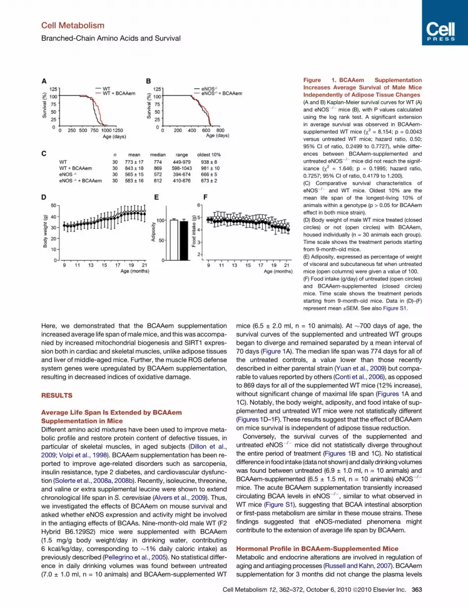
Figure 1. BCAAem Supplementation
Increases Average Survival of Male Mice
Independently of Adipose Tissue Changes
(A and B) Kaplan-Meier survival curves for WT (A)
and eNOS�/� mice (B), with P values calculated
using the log rank test. A significant extension
in average survival was observed in BCAAem-
supplemented WT mice (c2 = 8.154; p = 0.0043
versus untreated WT mice; hazard ratio, 0.50;
95% CI of ratio, 0.2499 to 0.7727), while differ-
ences between BCAAem-supplemented and
untreated eNOS�/� mice did not reach the signif-
icance (c2 = 1.646; p = 0.1995; hazard ratio,
0.7257; 95% CI of ratio, 0.4179 to 1.200).
(C) Comparative survival characteristics of
eNOS�/� and WT mice. Oldest 10% are the
mean life span of the longest-living 10% of
animals within a genotype (p > 0.05 for BCAAem
effect in both mice strain).
(D) Body weight of male WT mice treated (closed
circles) or not (open circles) with BCAAem,
housed individually (n = 30 animals each group).
Time scale shows the treatment periods starting
from 9-month-old mice.
(E) Adiposity, expressed as percentage of weight
of visceral and subcutaneous fat when untreated
mice (open columns) were given a value of 100.
(F) Food intake (g/day) of untreated (open circles)
and BCAAem-supplemented (closed circles)
mice. Time scale shows the treatment periods
starting from 9-month-old mice. Data in (D)–(F)
represent mean ±SEM. See also Figure S1.
Cell Metabolism
Branched-Chain Amino Acids and Survival
Here, we demonstrated that the BCAAem supplementation
increased average life span ofmalemice, and thiswas accompa-
nied by increased mitochondrial biogenesis and SIRT1 expres-
sion both in cardiac and skeletal muscles, unlike adipose tissues
and liver of middle-aged mice. Further, the muscle ROS defense
system genes were upregulated by BCAAem supplementation,
resulting in decreased indices of oxidative damage.
RESULTS
Average Life Span Is Extended by BCAAemSupplementation in MiceDifferent amino acid mixtures have been used to improve meta-
bolic profile and restore protein content of defective tissues, in
particular of skeletal muscles, in aged subjects (Dillon et al.,
2009; Volpi et al., 1998). BCAAem supplementation has been re-
ported to improve age-related disorders such as sarcopenia,
insulin resistance, type 2 diabetes, and cardiovascular dysfunc-
tion (Solerte et al., 2008a, 2008b). Recently, isoleucine, threonine,
and valine or extra supplemental leucine were shown to extend
chronological life span in S. cerevisiae (Alvers et al., 2009). Thus,
we investigated the effects of BCAAem on mouse survival and
asked whether eNOS expression and activity might be involved
in the antiaging effects of BCAAs. Nine-month-old male WT (F2
Hybrid B6.129S2) mice were supplemented with BCAAem
(1.5 mg/g body weight/day in drinking water, contributing
6 kcal/kg/day, corresponding to �1% daily caloric intake) as
previously described (Pellegrino et al., 2005). No statistical differ-
ence in daily drinking volumes was found between untreated
(7.0 ± 1.0 ml, n = 10 animals) and BCAAem-supplemented WT
Cell
mice (6.5 ± 2.0 ml, n = 10 animals). At �700 days of age, the
survival curves of the supplemented and untreated WT groups
began to diverge and remained separated by a mean interval of
70 days (Figure 1A). The median life span was 774 days for all of
the untreated controls, a value lower than those recently
described in either parental strain (Yuan et al., 2009) but compa-
rable to values reported by others (Conti et al., 2006), as opposed
to 869 days for all of the supplemented WT mice (12% increase),
without significant change of maximal life span (Figures 1A and
1C). Notably, the body weight, adiposity, and food intake of sup-
plemented and untreated WT mice were not statistically different
(Figures 1D–1F). These results suggest that the effect of BCAAem
on mice survival is independent of adipose tissue reduction.
Conversely, the survival curves of the supplemented and
untreated eNOS�/� mice did not statistically diverge throughout
the entire period of treatment (Figures 1B and 1C). No statistical
difference in food intake (datanotshown)anddailydrinkingvolumes
was found between untreated (6.9 ± 1.0 ml, n = 10 animals) and
BCAAem-supplemented (6.5 ± 1.5 ml, n = 10 animals) eNOS�/�
mice. The acute BCAAem supplementation transiently increased
circulating BCAA levels in eNOS�/�, similar to what observed in
WT mice (Figure S1), suggesting that BCAA intestinal absorption
or first-pass metabolism are similar in these mouse strains. These
findings suggested that eNOS-mediated phenomena might
contribute to the extension of average life span by BCAAem.
Hormonal Profile in BCAAem-Supplemented MiceMetabolic and endocrine alterations are involved in regulation of
aging and antiaging processes (Russell and Kahn, 2007). BCAAem
supplementation for 3 months did not change the plasma levels
Metabolism 12, 362–372, October 6, 2010 ª2010 Elsevier Inc. 363
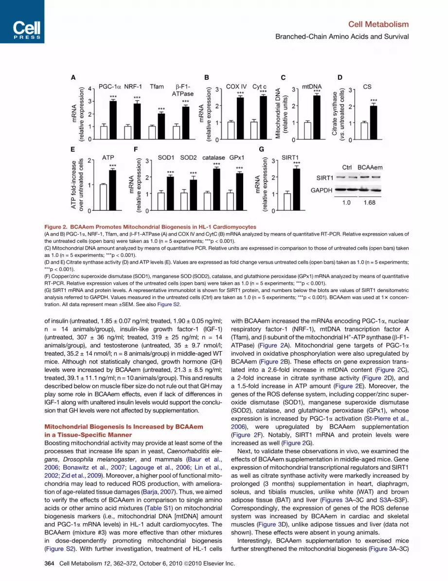
Figure 2. BCAAem Promotes Mitochondrial Biogenesis in HL-1 Cardiomyocytes(A and B) PGC-1a, NRF-1, Tfam, and b-F1-ATPase (A) and COX IV and CytC (B) mRNA analyzed bymeans of quantitative RT-PCR. Relative expression values of
the untreated cells (open bars) were taken as 1.0 (n = 5 experiments; ***p < 0.001).
(C) Mitochondrial DNA amount analyzed by means of quantitative PCR. Relative units are expressed in comparison to those of untreated cells (open bars) taken
as 1.0 (n = 5 experiments; ***p < 0.001).
(D and E) Citrate synthase activity (D) and ATP levels (E). Values are expressed as fold change versus untreated cells (open bars) taken as 1.0 (n = 5 experiments;
***p < 0.001).
(F) Copper/zinc superoxide dismutase (SOD1), manganese SOD (SOD2), catalase, and glutathione peroxidase (GPx1) mRNA analyzed by means of quantitative
RT-PCR. Relative expression values of the untreated cells (open bars) were taken as 1.0 (n = 5 experiments; ***p < 0.001).
(G) SIRT1 mRNA and protein levels. A representative immunoblot is shown for SIRT1 protein, and numbers below the blots are values of SIRT1 densitometric
analysis referred to GAPDH. Values measured in the untreated cells (Ctrl) are taken as 1.0 (n = 5 experiments; ***p < 0.001). BCAAem was used at 13 concen-
tration. All data represent mean ±SEM. See also Figure S2.
Cell Metabolism
Branched-Chain Amino Acids and Survival
of insulin (untreated, 1.85 ± 0.07 ng/ml; treated, 1.90 ± 0.05 ng/ml;
n = 14 animals/group), insulin-like growth factor-1 (IGF-1)
(untreated, 307 ± 36 ng/ml; treated, 319 ± 25 ng/ml; n = 14
animals/group), and testosterone (untreated, 35 ± 9.7 nmol/l;
treated, 35.2 ± 14 nmol/l; n = 8 animals/group) in middle-aged WT
mice. Although not statistically changed, growth hormone (GH)
levels were increased by BCAAem (untreated, 21.3 ± 8.5 ng/ml;
treated, 39.1± 11.1 ng/ml; n = 10animals/group). This and results
described below onmuscle fiber size do not rule out that GHmay
play some role in BCAAem effects, even if lack of differences in
IGF-1 along with unaltered insulin levels would support the conclu-
sion that GH levels were not affected by supplementation.
Mitochondrial Biogenesis Is Increased by BCAAemin a Tissue-Specific MannerBoosting mitochondrial activity may provide at least some of the
processes that increase life span in yeast, Caenorhabditis ele-
gans, Drosophila melanogaster, and mammals (Baur et al.,
2006; Bonawitz et al., 2007; Lagouge et al., 2006; Lin et al.,
2002; Zid et al., 2009). Moreover, a higher pool of functional mito-
chondria may lead to reduced ROS production, with ameliora-
tion of age-related tissue damages (Barja, 2007). Thus, we aimed
to verify the effects of BCAAem in comparison to single amino
acids or other amino acid mixtures (Table S1) on mitochondrial
biogenesis markers (i.e., mitochondrial DNA [mtDNA] amount
and PGC-1a mRNA levels) in HL-1 adult cardiomyocytes. The
BCAAem (mixture #3) was more effective than other mixtures
in dose-dependently promoting mitochondrial biogenesis
(Figure S2). With further investigation, treatment of HL-1 cells
364 Cell Metabolism 12, 362–372, October 6, 2010 ª2010 Elsevier In
with BCAAem increased the mRNAs encoding PGC-1a, nuclear
respiratory factor-1 (NRF-1), mtDNA transcription factor A
(Tfam), and b subunit of themitochondrial H+-ATP synthase (b-F1-
ATPase) (Figure 2A). Mitochondrial gene targets of PGC-1a
involved in oxidative phosphorylation were also upregulated by
BCAAem (Figure 2B). These effects on gene expression trans-
lated into a 2.6-fold increase in mtDNA content (Figure 2C),
a 2-fold increase in citrate synthase activity (Figure 2D), and
a 1.5-fold increase in ATP amount (Figure 2E). Moreover, the
genes of the ROS defense system, including copper/zinc super-
oxide dismutase (SOD1), manganese superoxide dismutase
(SOD2), catalase, and glutathione peroxidase (GPx1), whose
expression is increased by PGC-1a activation (St-Pierre et al.,
2006), were upregulated by BCAAem supplementation
(Figure 2F). Notably, SIRT1 mRNA and protein levels were
increased as well (Figure 2G).
Next, to validate these observations in vivo, we examined the
effects of BCAAem supplementation in middle-aged mice. Gene
expression of mitochondrial transcriptional regulators and SIRT1
as well as citrate synthase activity were markedly increased by
prolonged (3 months) supplementation in heart, diaphragm,
soleus, and tibialis muscles, unlike white (WAT) and brown
adipose tissue (BAT) and liver (Figures 3A–3C and S3A–S3F).
Correspondingly, the expression of genes of the ROS defense
system was increased by BCAAem in cardiac and skeletal
muscles (Figure 3D), unlike adipose tissues and liver (data not
shown). These effects were absent in young animals.
Interestingly, BCAAem supplementation to exercised mice
further strengthened the mitochondrial biogenesis (Figure 3A–3C)
c.
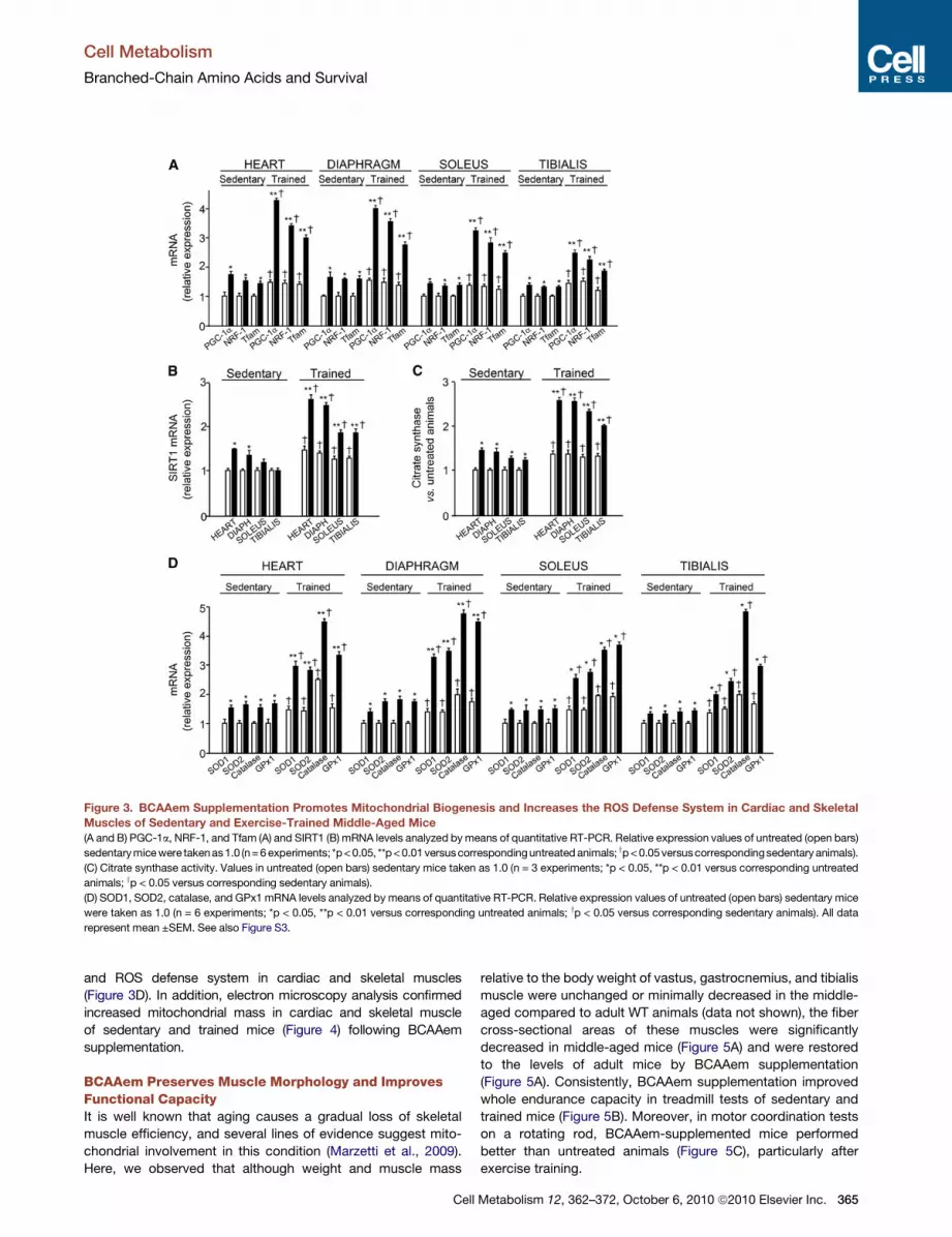
Figure 3. BCAAem Supplementation Promotes Mitochondrial Biogenesis and Increases the ROS Defense System in Cardiac and Skeletal
Muscles of Sedentary and Exercise-Trained Middle-Aged Mice(A and B) PGC-1a, NRF-1, and Tfam (A) and SIRT1 (B) mRNA levels analyzed bymeans of quantitative RT-PCR. Relative expression values of untreated (open bars)
sedentarymicewere takenas1.0 (n=6experiments; *p<0.05, **p<0.01versuscorrespondinguntreatedanimals; yp<0.05versuscorrespondingsedentary animals).
(C) Citrate synthase activity. Values in untreated (open bars) sedentary mice taken as 1.0 (n = 3 experiments; *p < 0.05, **p < 0.01 versus corresponding untreated
animals; yp < 0.05 versus corresponding sedentary animals).
(D) SOD1, SOD2, catalase, and GPx1 mRNA levels analyzed by means of quantitative RT-PCR. Relative expression values of untreated (open bars) sedentary mice
were taken as 1.0 (n = 6 experiments; *p < 0.05, **p < 0.01 versus corresponding untreated animals; yp < 0.05 versus corresponding sedentary animals). All data
represent mean ±SEM. See also Figure S3.
Cell Metabolism
Branched-Chain Amino Acids and Survival
and ROS defense system in cardiac and skeletal muscles
(Figure 3D). In addition, electron microscopy analysis confirmed
increased mitochondrial mass in cardiac and skeletal muscle
of sedentary and trained mice (Figure 4) following BCAAem
supplementation.
BCAAem Preserves Muscle Morphology and ImprovesFunctional CapacityIt is well known that aging causes a gradual loss of skeletal
muscle efficiency, and several lines of evidence suggest mito-
chondrial involvement in this condition (Marzetti et al., 2009).
Here, we observed that although weight and muscle mass
Cell
relative to the body weight of vastus, gastrocnemius, and tibialis
muscle were unchanged or minimally decreased in the middle-
aged compared to adult WT animals (data not shown), the fiber
cross-sectional areas of these muscles were significantly
decreased in middle-aged mice (Figure 5A) and were restored
to the levels of adult mice by BCAAem supplementation
(Figure 5A). Consistently, BCAAem supplementation improved
whole endurance capacity in treadmill tests of sedentary and
trained mice (Figure 5B). Moreover, in motor coordination tests
on a rotating rod, BCAAem-supplemented mice performed
better than untreated animals (Figure 5C), particularly after
exercise training.
Metabolism 12, 362–372, October 6, 2010 ª2010 Elsevier Inc. 365
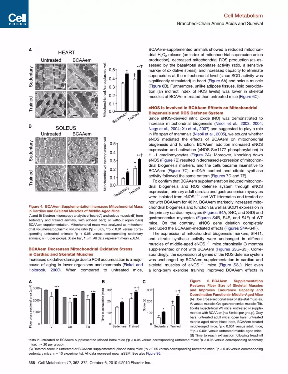
Figure 4. BCAAem Supplementation Increases Mitochondrial Mass
in Cardiac and Skeletal Muscles of Middle-Aged Mice
(A and B) Electron microscopy analysis of heart (A) and soleus muscle (B) from
sedentary and trained animals, with (closed bars) or without (open bars)
BCAAem supplementation. Mitochondrial mass was analyzed as mitochon-
drial volume/sarcoplasmic volume ratio (*p < 0.05, **p < 0.01 versus corre-
sponding untreated animals; yp < 0.05 versus corresponding sedentary
animals; n = 3 per group). Scale bar, 1 mm. All data represent mean ±SEM.
Cell Metabolism
Branched-Chain Amino Acids and Survival
BCAAem Decreases Mitochondrial Oxidative Stressin Cardiac and Skeletal MusclesIncreased oxidative damage due to ROS accumulation is amajor
cause of aging in lower organisms and mammals (Finkel and
Holbrook, 2000). When compared to untreated mice,
tests in untreated or BCAAem-supplemented (closed bars) mice (*p < 0.05 vers
mice; n = 20 per group).
(C) Rotarod score in untreated or BCAAem-supplemented (closed bars) mice (*p
sedentary mice; n = 10 experiments). All data represent mean ±SEM. See also F
366 Cell Metabolism 12, 362–372, October 6, 2010 ª2010 Elsevier In
BCAAem-supplemented animals showed a reduced mitochon-
drial H2O2 release (an index of mitochondrial superoxide anion
production), decreased mitochondrial ROS production (as as-
sessed by the basal/total aconitase activity ratio, a sensitive
marker of oxidative stress), and increased capacity to eliminate
superoxides at the mitochondrial level (since SOD activity was
significantly stimulated) in heart (Figure 6A) and soleus muscle
(Figure 6B). Furthermore, unlike adipose tissues, lipid peroxida-
tion (an indirect index of ROS levels) was lower in skeletal
muscles of BCAAem-treated than untreated mice (Figure 6C).
eNOS Is Involved in BCAAem Effects on MitochondrialBiogenesis and ROS Defense SystemSince eNOS-derived nitric oxide (NO) was demonstrated to
increase mitochondrial biogenesis (Nisoli et al., 2003, 2004;
Nagy et al., 2004; Xu et al., 2007) and suggested to play a role
in life span of mammals (Nisoli et al., 2005), we sought whether
eNOS mediated the effects of BCAAem on mitochondrial
biogenesis and function. BCAAem addition increased eNOS
expression and activation (eNOS-Ser1177 phosphorylation) in
HL-1 cardiomyocytes (Figure 7A). Moreover, knocking down
eNOS (Figure 7B) resulted in decreased expression of mitochon-
drial biogenesis markers, and the cells became insensitive to
BCAAem (Figure 7C). mtDNA content and citrate synthase
activity followed the same pattern (Figures 7D and 7E).
To confirm that BCAAem supplementation induced mitochon-
drial biogenesis and ROS defense system through eNOS
expression, primary adult cardiac and gastrocnemius myocytes
were isolated from eNOS�/� and WT littermates and treated or
nor with BCAAem for 48 hr. BCAAem markedly increased mito-
chondrial biogenesis and function as well as SOD1 expression in
the primary cardiac myocytes (Figures S4A, S4C, and S4D) and
gastrocnemius myocytes (Figures S4B, S4E, and S4F) of WT
mice. On the contrary, eNOS gene deletion completely
precluded the BCAAem-mediated effects (Figures S4A–S4F).
The expression of mitochondrial biogenesis markers, SIRT1,
and citrate synthase activity were unchanged in different
muscles of middle-aged eNOS�/� mice chronically (3 months)
supplemented or not with BCAAem (Figures S3G–S3I). Corre-
spondingly, the expression of genes of the ROS defense system
was unchanged by BCAAem supplementation in cardiac and
skeletal muscles of eNOS�/� mice (Figure S3J). Not even
a long-term exercise training improved BCAAem effects in
Figure 5. BCAAem Supplementation
Restores Fiber Size of Skeletal Muscles
and Improves Endurance Capacity and
Coordination Function inMiddle-AgedMice
(A) Fiber cross-sectional area of skeletal muscles.
V, vastus muscle; Gn, gastrocnemius muscle; Tib,
tibialismuscle fromWTmice, untreated or supple-
mentedwith BCAAem (n = 5mice per group). Gray
bars, untreated adult mice; open bars, untreated
middle-aged mice; black bars, BCAAem-treated
middle-aged mice. yp < 0.001 versus adult mice;
***p < 0.001 versus untreated middle-aged mice.
(B) Time to reach exhaustion following treadmill
us corresponding untreated mice; yp < 0.05 versus corresponding sedentary
< 0.05 versus corresponding untreated mice; yp < 0.05 versus corresponding
igure S6.
c.
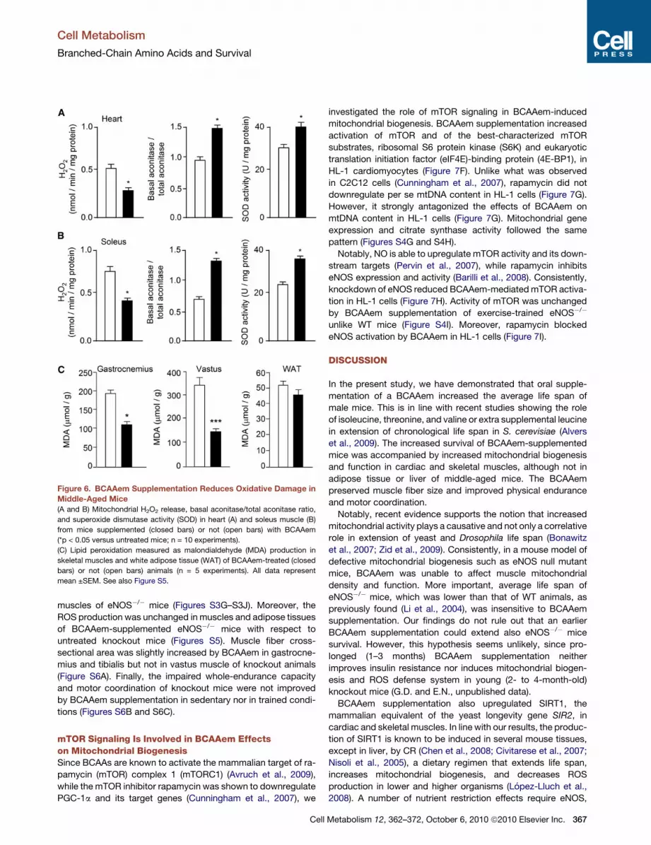
Figure 6. BCAAem Supplementation Reduces Oxidative Damage in
Middle-Aged Mice
(A and B) Mitochondrial H2O2 release, basal aconitase/total aconitase ratio,
and superoxide dismutase activity (SOD) in heart (A) and soleus muscle (B)
from mice supplemented (closed bars) or not (open bars) with BCAAem
(*p < 0.05 versus untreated mice; n = 10 experiments).
(C) Lipid peroxidation measured as malondialdehyde (MDA) production in
skeletal muscles and white adipose tissue (WAT) of BCAAem-treated (closed
bars) or not (open bars) animals (n = 5 experiments). All data represent
mean ±SEM. See also Figure S5.
Cell Metabolism
Branched-Chain Amino Acids and Survival
muscles of eNOS�/� mice (Figures S3G–S3J). Moreover, the
ROS production was unchanged in muscles and adipose tissues
of BCAAem-supplemented eNOS�/� mice with respect to
untreated knockout mice (Figures S5). Muscle fiber cross-
sectional area was slightly increased by BCAAem in gastrocne-
mius and tibialis but not in vastus muscle of knockout animals
(Figure S6A). Finally, the impaired whole-endurance capacity
and motor coordination of knockout mice were not improved
by BCAAem supplementation in sedentary nor in trained condi-
tions (Figures S6B and S6C).
mTOR Signaling Is Involved in BCAAem Effectson Mitochondrial BiogenesisSince BCAAs are known to activate the mammalian target of ra-
pamycin (mTOR) complex 1 (mTORC1) (Avruch et al., 2009),
while the mTOR inhibitor rapamycin was shown to downregulate
PGC-1a and its target genes (Cunningham et al., 2007), we
Cell
investigated the role of mTOR signaling in BCAAem-induced
mitochondrial biogenesis. BCAAem supplementation increased
activation of mTOR and of the best-characterized mTOR
substrates, ribosomal S6 protein kinase (S6K) and eukaryotic
translation initiation factor (eIF4E)-binding protein (4E-BP1), in
HL-1 cardiomyocytes (Figure 7F). Unlike what was observed
in C2C12 cells (Cunningham et al., 2007), rapamycin did not
downregulate per se mtDNA content in HL-1 cells (Figure 7G).
However, it strongly antagonized the effects of BCAAem on
mtDNA content in HL-1 cells (Figure 7G). Mitochondrial gene
expression and citrate synthase activity followed the same
pattern (Figures S4G and S4H).
Notably, NO is able to upregulate mTOR activity and its down-
stream targets (Pervin et al., 2007), while rapamycin inhibits
eNOS expression and activity (Barilli et al., 2008). Consistently,
knockdown of eNOS reduced BCAAem-mediated mTOR activa-
tion in HL-1 cells (Figure 7H). Activity of mTOR was unchanged
by BCAAem supplementation of exercise-trained eNOS�/�
unlike WT mice (Figure S4I). Moreover, rapamycin blocked
eNOS activation by BCAAem in HL-1 cells (Figure 7I).
DISCUSSION
In the present study, we have demonstrated that oral supple-
mentation of a BCAAem increased the average life span of
male mice. This is in line with recent studies showing the role
of isoleucine, threonine, and valine or extra supplemental leucine
in extension of chronological life span in S. cerevisiae (Alvers
et al., 2009). The increased survival of BCAAem-supplemented
mice was accompanied by increased mitochondrial biogenesis
and function in cardiac and skeletal muscles, although not in
adipose tissue or liver of middle-aged mice. The BCAAem
preserved muscle fiber size and improved physical endurance
and motor coordination.
Notably, recent evidence supports the notion that increased
mitochondrial activity plays a causative and not only a correlative
role in extension of yeast and Drosophila life span (Bonawitz
et al., 2007; Zid et al., 2009). Consistently, in a mouse model of
defective mitochondrial biogenesis such as eNOS null mutant
mice, BCAAem was unable to affect muscle mitochondrial
density and function. More important, average life span of
eNOS�/� mice, which was lower than that of WT animals, as
previously found (Li et al., 2004), was insensitive to BCAAem
supplementation. Our findings do not rule out that an earlier
BCAAem supplementation could extend also eNOS�/� mice
survival. However, this hypothesis seems unlikely, since pro-
longed (1–3 months) BCAAem supplementation neither
improves insulin resistance nor induces mitochondrial biogen-
esis and ROS defense system in young (2- to 4-month-old)
knockout mice (G.D. and E.N., unpublished data).
BCAAem supplementation also upregulated SIRT1, the
mammalian equivalent of the yeast longevity gene SIR2, in
cardiac and skeletal muscles. In line with our results, the produc-
tion of SIRT1 is known to be induced in several mouse tissues,
except in liver, by CR (Chen et al., 2008; Civitarese et al., 2007;
Nisoli et al., 2005), a dietary regimen that extends life span,
increases mitochondrial biogenesis, and decreases ROS
production in lower and higher organisms (Lopez-Lluch et al.,
2008). A number of nutrient restriction effects require eNOS,
Metabolism 12, 362–372, October 6, 2010 ª2010 Elsevier Inc. 367

Figure 7. BCAAem Exposure Increases Mitochondrial Biogenesis in HL-1 Cells through eNOS and mTOR Activation
(A) Upper panel: eNOS phosphorylation in untreated (Ctrl) and BCAAem-treated cells. Lower panel: eNOS expression measured after 48 hr treatment.
(B) eNOS expression in HL-1 cells transfected with either eNOS siRNA or nontargeting (NT) siRNA. Vehicle, transfection reagent.
(C) PGC-1a, NRF-1, and Tfam mRNA levels in untreated (open bars) or BCAAem-treated (closed bars) cells (***p < 0.001 versus corresponding BCAAem-
untreated cells, yp < 0.05 versus corresponding untransfected cells; n = 3).
(D and E) Mitochondrial DNA content (D) and citrate synthase activity (E) in untreated (open bars) or BCAAem-treated (closed bars) cells (***p < 0.001 versus
corresponding BCAAem-untreated cells, yp < 0.05 versus corresponding untransfected cells; n = 3).
(F) mTOR, S6K, and 4E-BP1 phosphorylation in untreated (Ctrl) and BCAAem-treated cells.
(G) The increase of mtDNA, induced by 48 hr BCAAem (closed bars), was antagonized by rapamycin (n = 3 experiments; *p < 0.05 and ***p < 0.001 versus
corresponding BCAAem-untreated cells; yp < 0.05 versus BCAAem-treated cells).
(H) mTOR activation in untreated or 30 min BCAAem-treated (closed bars) cells was decreased by eNOS silencing (n = 3 experiments; ***p < 0.001 versus
corresponding BCAAem-untreated cells; yp < 0.05 versus untransfected cells; #p < 0.05 versus untransfected, BCAAem-treated cells).
(I) Exposure to rapamycin (20 nM) prevented eNOS phosphorylation induced by 30 min BCAAem treatment. All data represent mean ±SEM. See also Figure S4.
Cell Metabolism
Branched-Chain Amino Acids and Survival
and NO is known to increase SIRT1 expression in cultured cells
(Nisoli et al., 2005). Further investigation is needed to assess the
role of SIRT1 induction in BCAAem effects on mice survival.
Of interest, the prolonged survival due to BCAAem supple-
mentation was associated with increased expression of genes
involved in antioxidant defense and marked reduction of ROS
368 Cell Metabolism 12, 362–372, October 6, 2010 ª2010 Elsevier In
production in cardiac and skeletal muscles of WT but not
eNOS�/� mice. Failure of eNOS null mutant mice to reduce
ROS in response to BCAAem supplementation may partly
explain the inability of this amino acid mixture to extend their
average life span. Notably, it has recently been reported that
NO-dependent mechanisms activate AMP-activated protein
c.
Cell Metabolism
Branched-Chain Amino Acids and Survival
kinase (AMPKa1), which increases the expression of genes
involved in antioxidant defense in endothelial cells (Colombo
and Moncada, 2009). These and the present results indicate
that NO plays a role in maintaining ‘‘antioxidant homeostasis.’’
Diverse reports point out that aging renders cells and tissues
increasingly vulnerable to oxidative stress (see for review Finkel
and Holbrook, 2000). Oxidative damage to mitochondria could
account for some of the age-related changes in their function.
Tissues from aged animals show mitochondrial dysfunctions with
increased superoxide andH2O2 generation and decreased energy
production (Sohal et al., 1994). Thus, the increased expression of
PGC-1a, apowerful transcription regulator ofROSdefensesystem
(St-Pierre et al., 2006), could play a relevant role in the extension of
average life spanevokedbyBCAAem inmiddle-agedmice.Other-
wise, hormonal changes, including insulin/IGF-1, testosterone, or
GH,whose role is not conclusivelydefinedboth inagingprocesses
and survival effects of nutrient restriction (Masoro, 2009), are
unlikely to take part in the BCAAem effects.
Exercise promotes survival and, in particular, extends average
although not maximal life span (Holloszy, 1993), specifically
improving insulin sensitivity (James et al., 1984) that is defective
in aging. This survival-promoting effect may be independent of
changes in body weight (Holloszy, 1993). Similarly, the
survival-extending property of BCAAem was not accompanied
by modification of food intake, body weight, or adiposity and
was independent of changes in mitochondrial biogenesis,
SIRT1, or antioxidant genes in adipose tissue and liver. A draw-
back of exercise is that it also increases mitochondrial ROS
production. Unexpectedly, Ristow et al. (2009) have recently
demonstrated that antioxidants such as vitamin C and vitamin
E abolished the health-promoting effects (i.e., increased param-
eters of insulin sensitivity, including plasma adiponectin) of exer-
cise in humans, since these compounds prevented the induction
of the ROS sensors PGC-1a/b and consequent activation of ROS
defense. Conversely, our present results demonstrate that
BCAAem supplementation increased expression of PGC-1a
and of antioxidant defense enzymes also in long-term exer-
cise-trained middle-aged mice. Moreover, the BCAAem has
been described to increase plasma adiponectin in slightly
trained elderly subjects (B. Solerte, personal communication).
Thus, the BCAAem could be a valid substitute for dietary supple-
mentation with antioxidants.
A relevant point is the molecular mechanism(s) involved in the
BCAAem-increased mitochondrial biogenesis and survival.
Interestingly, BCAAs have been reported to increase mTORC1
activity (Avruch et al., 2009), which correlates positively with
cell oxidative capacity (Schieke et al., 2006) and regulates
PGC-1a coactivation of its own promoter and mitochondrial
gene expression in muscle cells (Cunningham et al., 2007).
Moreover, rapamycin was shown to reduce eNOS expression
and NO production (Barilli et al., 2008). Based on our results,
one can hypothesize that BCAAem-activated mTOR signaling
could enhance mitochondrial biogenesis partly through
increasing the NO-generating system. In turn, our findings are
consistent with recent reports showing that NO upregulates
mTOR activity and its downstream targets (Pervin et al., 2007),
suggesting that a positive feedback mechanism between
eNOS and mTOR exists. Interestingly, selective knockout of
either mTOR or the mTORC1 component raptor in skeletal
Cell
muscle decreased oxidative capacity, mitochondrial gene
expression, and survival (Bentzinger et al., 2008; Risson et al.,
2009). In contrast, the absence of raptor in adipose tissue results
in lean mice with enhanced mitochondrial respiration (Polak
et al., 2008). Accordingly, BCAAem supplementation induces
mitochondrial biogenesis in muscle but not in fat or liver.
Several studies indicate that reduced TOR signaling underlies
life span extension by CR (see Stanfel et al., 2009 for review).
Mice fed rapamycin live longer than control mice fed unsupple-
mented chow, even when treatment begins in late life (Harrison
et al., 2009), and mice with deletion of S6K have increased life
span and resistance to age-related pathologies (Selman et al.,
2009). However, the reported prolongevity effects of chronic
rapamycin treatment in mice do not conclusively prove that
mTOR inhibition is the mechanism involved. Notably, rapamycin
has been recently shown to be ineffective to increase life span in
Drosophila (Harrison et al., 2010). Moreover, unlike CR, rapamy-
cin is more efficacious in female than in male mice (Harrison
et al., 2009), while S6K deletion increases life span in female
but not in male mice (Selman et al., 2009). Again, the role of
mTOR in CR is probably tissue specific, in that CR reduces
mTOR signaling in liver (Jiang et al., 2008) but increases it in
WAT and heart (Linford et al., 2007) and increases mitochondrial
function in different tissues (Nisoli et al., 2005), a finding that is
not consistent with reduced mTOR signaling. Although we did
not specifically investigate the contribution of mTOR in
BCAAem-mediated increase of survival, our findings support
the notion that the role of mTOR in CR mechanisms is complex
and not conclusively clarified at the moment (Anderson and
Weindruch, 2010).
Among drug strategies to extend life span, SIRT1 activators
(e.g., 3,5,4-trihydroxystilbene or resveratrol) are currently under
investigation in humans. Although able to improve health under
all dietary conditions, resveratrol promotes survival only in
high-fat diet (Baur et al., 2006) but not in standard-diet-fed
mice (Pearson et al., 2008). The BCAAem, besides increasing
average life span in normally fed mice, was found to promote
several healthy effects in humans, since it reduced sarcopenia
(Solerte et al., 2008b) and decreased inflammatory markers in
chronic heart failure patients (Kalantar-Zadeh et al., 2008).
In summary, we have provided evidence that an original BCAA
mixture increases average life span in male mice. This was likely
the consequence of increased mitochondrial biogenesis and
reduced oxidative stress in cardiac and skeletal muscles via
eNOS-mediated mechanisms. Our study offers a rationale for
deeply exploring the role of amino acids in prevention and
control of age-related disorders in humans.
EXPERIMENTAL PROCEDURES
Animals, Diets, and Treatments
Young (8-week-old; n = 20) and middle-aged (16-month-old) male WT (F2
Hybrid B6.129S2 obtained from crossing C57BL/6J and 129S1/SvImJ mice)
as well as middle-aged B6.129P2-Nos3tm1Unc/J (eNOS�/�) mice (Shesely
et al., 1996) (Jackson Laboratory), housed one per cage, were treated accord-
ing to the EU guidelines and with the approval of the Institutional Ethical
Committee. Animals were given unrestricted access to a standard diet
(4.3 kcal % fat, 18.8 kcal % protein, 76.9 kcal % carbohydrate, Laboratorio
Dottori Piccioni) and tap water. Both middle-aged WT and eNOS�/� mice
were divided into two groups, sedentary and exercise-trained groups, each
Metabolism 12, 362–372, October 6, 2010 ª2010 Elsevier Inc. 369

Cell Metabolism
Branched-Chain Amino Acids and Survival
further subdivided into untreated and BCAAem-supplemented groups (20
mice/group). BCAAem supplementation was performed for 3 months, while
the exercise training was performed during the third month of supplementa-
tion. BCAAem (1.5 mg/g body weight/day in drinking water with percent
composition detailed as mixture #3 in Table S1) (Pellegrino et al., 2005) was
dissolved in tap water by calculating average daily drinking 2 weeks before
starting treatment and stored at 4�C before daily administration. Drinking
volume was checked weekly. Mice were sacrificed by cervical dislocation.
Heart, diaphragm, posterior limbmuscles (soleus, tibialis, and gastrocnemius),
epididymal white fat, interscapular brown fat, and liver were dissected under
a stereomicroscope, frozen in liquid nitrogen, and stored at �80�C.
Study Design for Survival Analysis
For survival analysis, 9-month-oldWT (n = 60) and eNOS�/�mice (n = 60) were
randomly assigned to control and BCAAem-supplemented groups (BCAAem,
1.5mg/g bodyweight/day in drinking water) (Pellegrino et al., 2005). Mostmice
in the survival study died natural deaths; there were not accidental or external
causes. In particular, lymphomas, lung and liver tumors, kidney diseases, and
infection/septicemia were the main causes of death for WTmice (Haines et al.,
2001), while cardiovascular and metabolic disorders were mainly involved in
eNOS�/� mice death (Li et al., 2004). Moribund mice or those showing clear
signs of distress were euthanized to mitigate suffering. Survival curves were
plotted using the Kaplan-Meier method. Adiposity was measured as wet
weight of visceral and subcutaneous fat. Body weight and food intake were
monitored every other week for the time of the experiment.
Analytical Procedures
Glucose concentrations were determined using the Glucocard Gmeter (Me-
narini; Florence, Italy). ELISA kits were used to measure insulin (LINCO
Research Inc.; St. Charles, MO), IGF-1 and GH (Diagnostic Systems Laborato-
ries; Webster, TX), and testosterone (Diagnostic Products Corporation; Los
Angeles, CA). To study the BCAA absorption rate, plasma BCAA analysis
was performed. Details can be found in the Supplemental Experimental
Procedures.
HL-1 Cell Cultures and Treatments
HL-1 cardiomyocytes (a gift from W.C. Claycomb) were cultured as described
in the Supplemental Experimental Procedures. Cells were treated with single
amino acids or different amino acid mixtures, including BCAAem, with percent
composition and final concentrations indicated in Table S1 for 48 hr, except for
time- or dose-response experiments. For eNOS knockdown experiments,
HL-1 cells were transfectedwith 30 nMeNOS siRNASMARTpool (Dharmacon;
Lafayette, CO) or siGENOME nontargeting siRNA using Dharmafect 3
transfection reagent. Details can be found in the Supplemental Experimental
Procedures.
Cardiomyocyte and Myocyte Preparation and Treatment
Cardiac myocytes were isolated from left ventricle free wall of WT or eNOS�/�
mice, while satellite skeletal muscle cells were prepared from limbs of WT and
eNOS�/� postnatal mice. Mature cardiac and skeletal myocytes were treated
with BCAAem for 48 hr prior to cell harvesting. Details are in Supplemental
Experimental Procedures.
Gene Expression and Mitochondrial Biogenesis Methods
Quantitative RT-PCR reactions were performed as described (Tedesco et al.,
2008) and run with the iQ SybrGreenI SuperMix (Bio-Rad; Segrate, Italy) on an
iCycler iQ Real-Time PCR detection system (Bio-Rad). Calculations were per-
formed by a comparative method (2�DDCt) using 18S rRNA as an internal
control. For mtDNA analysis, total DNA was extracted with QIAamp DNA
extraction kit (QIAGEN). mtDNA was amplified using primers specific for the
mitochondrial cytochrome b (CytB) gene and normalized to genomic DNA
by amplification of the large ribosomal protein p0 (36B4) nuclear gene. Primers
were designed using Beacon Designer 2.6 software (Premier Biosoft Interna-
tional; Palo Alto, CA). Immunoblot analysis was performed as described
(Tedesco et al., 2008). The whole amount of ATP in cells was measured by
using the ATP determination kit from Molecular Probes (Eugene, OR) as
described (Tedesco et al., 2008). Citrate synthase activity was measured
spectrophotometrically in either tissue or whole-cell extracts and expressed
370 Cell Metabolism 12, 362–372, October 6, 2010 ª2010 Elsevier In
as nmol citrate produced/min/mg protein as described (Tedesco et al.,
2008). Electron microscopy studies were conducted on heart and soleus
muscle as previously described (Nisoli et al., 2004; Weibel, 1979). Details are
in Supplemental Experimental Procedures.
Mitochondrial Oxidative Stress
Mitochondria were isolated using the Qproteome Mitochondria Isolation Kit
(QIAGEN). Aconitase-specific activity was measured as previously described
(Lionetti et al., 2007). SOD activity was measured with the Superoxide Dismu-
tase Assay Kit (Calbiochem; San Diego, CA). Mitochondrial H2O2 release was
assayed by the Amplex Red Hydrogen Peroxide/Peroxidase Assay kit (Molec-
ular Probes). To assay lipid peroxidation (LPO), malondialdehyde (MDA)
derived from polyunsaturated fatty acid peroxides was evaluated by means
of the LPO-586 colorimetric assay kit (OxisResearch; Portland, OR). Details
are in Supplemental Experimental Procedures.
Training, Exercise Tolerance, and Motor Coordination Tests
Exercise training (5 days/week for 4 weeks as detailed in Supplemental Exper-
imental Procedures) and exercise exhaustion tests were performed on the belt
of a 6-lane motorized treadmill (Exer 3/6 Treadmill, Columbus Instruments;
Columbus, OH), supplied with shocker plates (electrical stimulus: 200 ms,
0.34 mA, 1 Hz) as previously described (Benchaouir et al., 2007). Constant
speed rotarod (47600 Model, Ugo Basile; Comerio, Italy) was used to measure
fore- and hindlimb motor coordination and balance in 12-month-old mice, as
detailed in Supplemental Experimental Procedures (Serradj and Jamon, 2007).
Muscle Fiber Size
Vastus, tibialis, and gastrocnemius muscles, obtained from adult (6-month-
old) or middle-aged male WT and eNOS�/� mice, supplemented or not with
BCAAem, were frozen in liquid nitrogen. Serial transverse sections (10 mm
thick) were cut with a LeicaCM 1850 cryostat and stained with hematoxylin
and eosin. To evaluate fiber cross-sectional area, morphometric analyses
were performed on �150 fibers per muscle (n = 5 mice per group) by using
an Image 1.63 software (Scion Corporation; Frederick, MD).
Statistical Analysis
Data were analyzed by two-tailed unpaired Student’s t test or by one-way
ANOVA with Tukey’s post hoc test in instances of multiple comparisons.
Two-way ANOVA was used to evaluate the supplementation and exercise
effects and their interaction. P values for survival analyses were calculated
using the log rank test. All data are reported as mean ±SEM unless otherwise
stated. Statistical analyses were performed using GraphPad Prism version 4.0
software.
SUPPLEMENTAL INFORMATION
Supplemental Information includes Supplemental Experimental Procedures,
Supplemental References, six figures, and two tables and can be found with
this article online at doi:10.1016/j.cmet.2010.08.016.
ACKNOWLEDGMENTS
This paper is dedicated to the memory of F. Conti. We are grateful to S. Mon-
cada for discussionandcritically reading themanuscript.We thankF.Dioguardi
for discussion, M. Sampaolesi and D. Galli for helpful advice with cardiac and
skeletalmyocyte preparations, P. Arcidiaco for analysis of BCAAplasma levels,
A. Mascaro for technical assistance, and S. Calza for statistical analysis. This
work was supported by the Ministero dell’Istruzione, dell’Universita e della
Ricerca grants 20075HJTHM_001 (to E.N.), 20075HJTHM_002 (to A.V.), and
2007BRR57M_002 (to M.O.C.); the Ministero della Salute (to E.N.); and the
Comune di Milano Flagship Project_2008 (to M.O.C.).
Received: July 17, 2009
Revised: December 17, 2009
Accepted: June 18, 2010
Published: October 5, 2010
c.
Cell Metabolism
Branched-Chain Amino Acids and Survival
REFERENCES
Alvers, A.L., Fishwick, L.K., Wood, M.S., Hu, D., Chung, H.S., Dunn, W.A., Jr.,
and Aris, J.P. (2009). Autophagy and amino acid homeostasis are required for
chronological longevity in Saccharomyces cerevisiae. Aging Cell 8, 353–369.
Anderson, R.M., and Weindruch, R. (2010). Metabolic reprogramming, caloric
restriction and aging. Trends Endocrinol. Metab. 21, 134–141.
Avruch, J., Long, X., Ortiz-Vega, S., Rapley, J., Papageorgiou, A., and Dai, N.
(2009). Amino acid regulation of TOR complex 1. Am. J. Physiol. Endocrinol.
Metab. 296, E592–E602.
Barilli, A., Visigalli, R., Sala, R., Gazzola, G.C., Parolari, A., Tremoli, E., Bono-
mini, S., Simon, A., Closs, E.I., Dall’Asta, V., and Bussolati, O. (2008). In human
endothelial cells rapamycin causes mTORC2 inhibition and impairs cell
viability and function. Cardiovasc. Res. 78, 563–571.
Barja, G. (2007). Mitochondrial oxygen consumption and reactive oxygen
species production are independently modulated: implications for aging
studies. Rejuvenation Res. 10, 215–224.
Baur, J.A., Pearson, K.J., Price, N.L., Jamieson, H.A., Lerin, C., Kalra, A.,
Prabhu, V.V., Allard, J.S., Lopez-Lluch, G., Lewis, K., et al. (2006). Resveratrol
improves health and survival of mice on a high-calorie diet. Nature 444,
337–342.
Benchaouir, R., Meregalli, M., Farini, A., D’Antona, G., Belicchi, M., Goyen-
valle, A., Battistelli, M., Bresolin, N., Bottinelli, R., Garcia, L., and Torrente, Y.
(2007). Restoration of human dystrophin following transplantation of exon-
skipping-engineered DMD patient stem cells into dystrophic mice. Cell Stem
Cell 1, 646–657.
Bentzinger, C.F., Romanino, K., Cloetta, D., Lin, S., Mascarenhas, J.B., Oliveri,
F., Xia, J., Casanova, E., Costa, C.F., Brink, M., et al. (2008). Skeletal muscle-
specific ablation of raptor, but not of rictor, causes metabolic changes and
results in muscle dystrophy. Cell Metab. 8, 411–424.
Bonawitz, N.D., Chatenay-Lapointe, M., Pan, Y., and Shadel, G.S. (2007).
Reduced TOR signaling extends chronological life span via increased respira-
tion and upregulation of mitochondrial gene expression. Cell Metab. 5,
265–277.
Chen, D., Bruno, J., Easlon, E., Lin, S.J., Cheng, H.L., Alt, F.W., and Guarente,
L. (2008). Tissue-specific regulation of SIRT1 by calorie restriction. Genes Dev.
22, 1753–1757.
Civitarese, A.E., Carling, S., Heilbronn, L.K., Hulver, M.H., Ukropcova, B.,
Deutsch, W.A., Smith, S.R., and Ravussin, E.; CALERIE Pennington Team.
(2007). Calorie restriction increases muscle mitochondrial biogenesis in
healthy humans. PLoS Med. 4, e76.
Colombo, S.L., and Moncada, S. (2009). AMPKalpha1 regulates the antioxi-
dant status of vascular endothelial cells. Biochem. J. 421, 163–169.
Conti, B., Sanchez-Alavez, M., Winsky-Sommerer, R., Morale, M.C., Lucero,
J., Brownell, S., Fabre, V., Huitron-Resendiz, S., Henriksen, S., Zorrilla, E.P.,
et al. (2006). Transgenic mice with a reduced core body temperature have
an increased life span. Science 314, 825–828.
Cook, S., Hugli, O., Egli, M., Vollenweider, P., Burcelin, R., Nicod, P., Thorens,
B., and Scherrer, U. (2003). Clustering of cardiovascular risk factors mimicking
the human metabolic syndrome X in eNOS null mice. Swiss Med. Wkly. 133,
360–363.
Cunningham, J.T., Rodgers, J.T., Arlow, D.H., Vazquez, F., Mootha, V.K., and
Puigserver, P. (2007). mTOR controls mitochondrial oxidative function through
a YY1-PGC-1a transcriptional complex. Nature 450, 736–740.
Dillon, E.L., Sheffield-Moore, M., Paddon-Jones, D., Gilkison, C., Sanford,
A.P., Casperson, S.L., Jiang, J., Chinkes, D.L., and Urban, R.J. (2009). Amino
acid supplementation increases lean body mass, basal muscle protein
synthesis, and insulin-like growth factor-I expression in older women.
J. Clin. Endocrinol. Metab. 94, 1630–1637.
Finkel, T., and Holbrook, N.J. (2000). Oxidants, oxidative stress and the
biology of ageing. Nature 408, 239–247.
Guarente, L. (2008). Mitochondria—a nexus for aging, calorie restriction, and
sirtuins? Cell 132, 171–176.
Cell
Haines, D.C., Chattopadhyay, S., and Ward, J.M. (2001). Pathology of aging
B6;129 mice. Toxicol. Pathol. 29, 653–661.
Harrison, D.E., Strong, R., Sharp, Z.D., Nelson, J.F., Astle, C.M., Flurkey, K.,
Nadon, N.L., Wilkinson, J.E., Frenkel, K., Carter, C.S., et al. (2009). Rapamycin
fed late in life extends lifespan in genetically heterogeneous mice. Nature 460,
392–395.
Harrison, B., Tran, T.T., Taylor, D., Lee, S.D., and Min, K.J. (2010). Effect of ra-
pamycin on lifespan in Drosophila. Geriatr. Gerontol. Int. 10, 110–112.
Heilbronn, L.K., de Jonge, L., Frisard, M.I., DeLany, J.P., Larson-Meyer, D.E.,
Rood, J., Nguyen, T., Martin, C.K., Volaufova, J., Most, M.M., et al; Pennington
CALERIE Team. (2006). Effect of 6-month calorie restriction on biomarkers of
longevity,metabolic adaptation, and oxidative stress in overweight individuals:
a randomized controlled trial. JAMA 295, 1539–1548.
Holloszy, J.O. (1993). Exercise increases average longevity of female rats
despite increased food intake and no growth retardation. J. Gerontol. 48,
B97–B100.
Ingram, D.K., Anson, R.M., de Cabo, R., Mamczarz, J., Zhu, M., Mattison, J.,
Lane, M.A., and Roth, G.S. (2004). Development of calorie restriction mimetics
as a prolongevity strategy. Ann. N. Y. Acad. Sci. 1019, 412–423.
James, D.E., Kraegen, E.W., and Chisholm, D.J. (1984). Effect of exercise
training on whole-body insulin sensitivity and responsiveness. J. Appl. Physiol.
56, 1217–1222.
Jiang, W., Zhu, Z., and Thompson, H.J. (2008). Dietary energy restriction
modulates the activity of AMP-activated protein kinase, Akt, and mammalian
target of rapamycin in mammary carcinomas, mammary gland, and liver.
Cancer Res. 68, 5492–5499.
Kalantar-Zadeh, K., Anker, S.D., Horwich, T.B., and Fonarow, G.C. (2008).
Nutritional and anti-inflammatory interventions in chronic heart failure. Am. J.
Cardiol. 101 (11A), 89E–103E.
Lagouge, M., Argmann, C., Gerhart-Hines, Z., Meziane, H., Lerin, C., Daussin,
F., Messadeq, N., Milne, J., Lambert, P., Elliott, P., et al. (2006). Resveratrol
improves mitochondrial function and protects against metabolic disease by
activating SIRT1 and PGC-1a. Cell 127, 1109–1122.
Li, W., Mital, S., Ojaimi, C., Csiszar, A., Kaley, G., and Hintze, T.H. (2004).
Premature death and age-related cardiac dysfunction in male eNOS-knockout
mice. J. Mol. Cell. Cardiol. 37, 671–680.
Lin, S.J., Kaeberlein, M., Andalis, A.A., Sturtz, L.A., Defossez, P.A., Culotta,
V.C., Fink, G.R., and Guarente, L. (2002). Calorie restriction extends Saccha-
romyces cerevisiae lifespan by increasing respiration. Nature 418, 344–348.
Linford, N.J., Beyer, R.P., Gollahon, K., Krajcik, R.A., Malloy, V.L., Demas, V.,
Burmer, G.C., and Rabinovitch, P.S. (2007). Transcriptional response to aging
and caloric restriction in heart and adipose tissue. Aging Cell 6, 673–688.
Lionetti, L., Mollica, M.P., Crescenzo, R., D’Andrea, E., Ferraro, M., Bianco, F.,
Liverini, G., and Iossa, S. (2007). Skeletal muscle subsarcolemmal mitochon-
drial dysfunction in high-fat fed rats exhibiting impaired glucose homeostasis.
Int. J. Obes. (Lond) 31, 1596–1604.
Lopez-Lluch, G., Irusta, P.M., Navas, P., and deCabo, R. (2008). Mitochondrial
biogenesis and healthy aging. Exp. Gerontol. 43, 813–819.
Marzetti, E., Lees, H.A., Wohlgemuth, S.E., and Leeuwenburgh, C. (2009). Sar-
copenia of aging: underlying cellular mechanisms and protection by calorie
restriction. Biofactors 35, 28–35.
Masoro, E.J. (2009). Caloric restriction-induced life extension of rats andmice:
a critique of proposedmechanisms. Biochim. Biophys. Acta 1790, 1040–1048.
Nagy, G., Barcza, M., Gonchoroff, N., Phillips, P.E., and Perl, A. (2004). Nitric
oxide-dependent mitochondrial biogenesis generates Ca2+ signaling profile of
lupus T cells. J. Immunol. 173, 3676–3683.
Nisoli, E., Clementi, E., Paolucci, C., Cozzi, V., Tonello, C., Sciorati, C., Bra-
cale, R., Valerio, A., Francolini, M., Moncada, S., and Carruba, M.O. (2003).
Mitochondrial biogenesis in mammals: the role of endogenous nitric oxide.
Science 299, 896–899.
Nisoli, E., Falcone, S., Tonello, C., Cozzi, V., Palomba, L., Fiorani, M., Pisconti,
A., Brunelli, S., Cardile, A., Francolini, M., et al. (2004). Mitochondrial biogen-
esis by NO yields functionally active mitochondria in mammals. Proc. Natl.
Acad. Sci. USA 101, 16507–16512.
Metabolism 12, 362–372, October 6, 2010 ª2010 Elsevier Inc. 371
Cell Metabolism
Branched-Chain Amino Acids and Survival
Nisoli, E., Tonello, C., Cardile, A., Cozzi, V., Bracale, R., Tedesco, L., Falcone,
S., Valerio, A., Cantoni, O., Clementi, E., et al. (2005). Calorie restriction
promotes mitochondrial biogenesis by inducing the expression of eNOS.
Science 310, 314–317.
Pansarasa, O., Flati, V., Corsetti, G., Brocca, L., Pasini, E., and D’Antona, G.
(2008). Oral amino acid supplementation counteracts age-induced sarcopenia
in elderly rats. Am. J. Cardiol. 101 (11A), 35E–41E.
Pearson, K.J., Baur, J.A., Lewis, K.N., Peshkin, L., Price, N.L., Labinskyy, N.,
Swindell, W.R., Kamara, D., Minor, R.K., Perez, E., et al. (2008). Resveratrol
delays age-related deterioration and mimics transcriptional aspects of dietary
restriction without extending life span. Cell Metab. 8, 157–168.
Pellegrino, M.A., Brocca, L., Dioguardi, F.S., Bottinelli, R., and D’Antona, G.
(2005). Effects of voluntary wheel running and amino acid supplementation
on skeletal muscle of mice. Eur. J. Appl. Physiol. 93, 655–664.
Pervin, S., Singh, R., Hernandez, E., Wu, G., and Chaudhuri, G. (2007). Nitric
oxide in physiologic concentrations targets the translational machinery to
increase the proliferation of human breast cancer cells: involvement of
mammalian target of rapamycin/eIF4E pathway. Cancer Res. 67, 289–299.
Polak, P., Cybulski, N., Feige, J.N., Auwerx, J., Ruegg, M.A., and Hall, M.N.
(2008). Adipose-specific knockout of raptor results in leanmice with enhanced
mitochondrial respiration. Cell Metab. 8, 399–410.
Risson, V., Mazelin, L., Roceri, M., Sanchez, H., Moncollin, V., Corneloup, C.,
Richard-Bulteau, H., Vignaud, A., Baas, D., Defour, A., et al. (2009). Muscle
inactivation of mTOR causes metabolic and dystrophin defects leading to
severe myopathy. J. Cell Biol. 187, 859–874.
Ristow, M., Zarse, K., Oberbach, A., Kloting, N., Birringer, M., Kiehntopf, M.,
Stumvoll, M., Kahn, C.R., and Bluher, M. (2009). Antioxidants prevent
health-promoting effects of physical exercise in humans. Proc. Natl. Acad.
Sci. USA 106, 8665–8670.
Russell, S.J., and Kahn, C.R. (2007). Endocrine regulation of ageing. Nat. Rev.
Mol. Cell Biol. 8, 681–691.
Schieke, S.M., Phillips, D., McCoy, J.P., Jr., Aponte, A.M., Shen, R.F., Bala-
ban, R.S., and Finkel, T. (2006). The mammalian target of rapamycin (mTOR)
pathway regulates mitochondrial oxygen consumption and oxidative capacity.
J. Biol. Chem. 281, 27643–27652.
Selman, C., Tullet, J.M., Wieser, D., Irvine, E., Lingard, S.J., Choudhury, A.I.,
Claret, M., Al-Qassab, H., Carmignac, D., Ramadani, F., et al. (2009). Ribo-
somal protein S6 kinase 1 signaling regulates mammalian life span. Science
326, 140–144.
Serradj, N., and Jamon, M. (2007). Age-related changes in the motricity of the
inbred mice strains 129/sv and C57BL/6j. Behav. Brain Res. 177, 80–89.
Shesely, E.G., Maeda, N., Kim, H.S., Desai, K.M., Krege, J.H., Laubach, V.E.,
Sherman, P.A., Sessa, W.C., Smithies, O., and Smithies, O. (1996). Elevated
372 Cell Metabolism 12, 362–372, October 6, 2010 ª2010 Elsevier In
blood pressures in mice lacking endothelial nitric oxide synthase. Proc. Natl.
Acad. Sci. USA 93, 13176–13181.
Sohal, R.S., Ku, H.H., Agarwal, S., Forster, M.J., and Lal, H. (1994). Oxidative
damage, mitochondrial oxidant generation and antioxidant defenses during
aging and in response to food restriction in the mouse. Mech. Ageing Dev.
74, 121–133.
Solerte, S.B., Fioravanti, M., Locatelli, E., Bonacasa, R., Zamboni, M., Basso,
C., Mazzoleni, A., Mansi, V., Geroutis, N., and Gazzaruso, C. (2008a). Improve-
ment of blood glucose control and insulin sensitivity during a long-term
(60 weeks) randomized study with amino acid dietary supplements in elderly
subjects with type 2 diabetes mellitus. Am. J. Cardiol. 101 (11A), 82E–88E.
Solerte, S.B., Gazzaruso, C., Bonacasa, R., Rondanelli, M., Zamboni, M.,
Basso, C., Locatelli, E., Schifino, N., Giustina, A., and Fioravanti, M. (2008b).
Nutritional supplements with oral amino acid mixtures increases whole-body
lean mass and insulin sensitivity in elderly subjects with sarcopenia. Am. J.
Cardiol. 101 (11A), 69E–77E.
St-Pierre, J., Drori, S., Uldry, M., Silvaggi, J.M., Rhee, J., Jager, S., Handschin,
C., Zheng, K., Lin, J., Yang, W., et al. (2006). Suppression of reactive oxygen
species and neurodegeneration by the PGC-1 transcriptional coactivators.
Cell 127, 397–408.
Stanfel, M.N., Shamieh, L.S., Kaeberlein, M., and Kennedy, B.K. (2009). The
TOR pathway comes of age. Biochim. Biophys. Acta 1790, 1067–1074.
Tedesco, L., Valerio, A., Cervino, C., Cardile, A., Pagano, C., Vettor, R., Pas-
quali, R., Carruba, M.O., Marsicano, G., Lutz, B., et al. (2008). Cannabinoid
type 1 receptor blockade promotes mitochondrial biogenesis through endo-
thelial nitric oxide synthase expression in white adipocytes. Diabetes 57,
2028–2036.
Volpi, E., Ferrando, A.A., Yeckel, C.W., Tipton, K.D., and Wolfe, R.R. (1998).
Exogenous amino acids stimulate net muscle protein synthesis in the elderly.
J. Clin. Invest. 101, 2000–2007.
Weibel, E.R. (1979). Practical methods for biological morphometry. In Stereo-
logical Methods, Volume 1. (London: Academic Press), pp. 40–116.
Xu, W., Koeck, T., Lara, A.R., Neumann, D., DiFilippo, F.P., Koo, M., Janocha,
A.J., Masri, F.A., Arroliga, A.C., Jennings, C., et al. (2007). Alterations of cellular
bioenergetics in pulmonary artery endothelial cells. Proc. Natl. Acad. Sci. USA
104, 1342–1347.
Yuan, R., Tsaih, S.W., Petkova, S.B., Marin de Evsikova, C., Xing, S., Marion,
M.A., Bogue, M.A., Mills, K.D., Peters, L.L., Bult, C.J., et al. (2009). Aging in
inbred strains of mice: study design and interim report on median lifespans
and circulating IGF1 levels. Aging Cell 8, 277–287.
Zid, B.M., Rogers, A.N., Katewa, S.D., Vargas, M.A., Kolipinski, M.C., Lu, T.A.,
Benzer, S., and Kapahi, P. (2009). 4E-BP extends lifespan upon dietary restric-
tion by enhancing mitochondrial activity in Drosophila. Cell 139, 149–160.
c.