University of Groningen Lysosome Biogenesis and Autophagy ...
Uteroplacental insufficiency and reducing litter size alters skeletal muscle mitochondrial
biogenesis in a sex specific manner in the adult rat
Glenn D. Wadley1, Andrew L. Siebel1, Greg J. Cooney2, Glenn K. McConell1, Mary E. Wlodek1
and Julie A. Owens3.
1Department of Physiology, The University of Melbourne, Parkville, Victoria, Australia, 3010.
2Garvan Institute of Medical Research, Sydney, New South Wales, Australia, 2010. 3School of
Paediatrics and Reproductive Health Discipline of Obstetrics and Gynaecology Faculty of Health
Sciences Adelaide University, Australia 5005
Running Head: Altered growth, skeletal muscle and mitochondria
Address for correspondence:
Dr. Glenn Wadley,
Department of Physiology,
The University of Melbourne,
Parkville, 3010, Australia.
Ph: +61-3-8344-8503,
Fax: +61-3-8344-5818,
email: [email protected]
Page 1 of 31Articles in PresS. Am J Physiol Endocrinol Metab (March 4, 2008). doi:10.1152/ajpendo.00037.2008
Copyright © 2008 by the American Physiological Society.

2
ABSTRACT
Objective: Uteroplacental insufficiency has been shown to impair insulin action and glucose
homeostasis in adult offspring and may act in part via altered mitochondrial biogenesis and lipid
balance in skeletal muscle. Research design and methods: Bilateral uterine vessel ligation to induce
uteroplacental insufficiency in the offspring (Restricted) or sham surgery was performed on day 18
of gestation in rats. To match the litter size of Restricted offspring, a separate cohort of sham litters
had litter size reduced to 5 at birth (Reduced Litter), which also restricted postnatal growth.
Remaining litters from sham mothers were unaltered (Control). Offspring were studied at 6 months
of age. Results: In males, both Restricted and Reduced Litter offspring had reduced gastrocnemius
PGC-1α mRNA and protein, and mtTFA and COX III mRNA (P<0.05), while only Restricted had
reduced skeletal muscle COX IV mRNA and protein and glycogen (P<0.05), despite unaltered
glucose tolerance, HOMA and intramuscular triglycerides. In females, only gastrocnemius mtTFA
mRNA was lower in Reduced Litter offspring (P<0.05). Furthermore, glucose tolerance was not
altered in any female offspring, although HOMA and intramuscular triglycerides increased in
Restricted offspring (P<0.05). Conclusions: Restriction of growth due to uteroplacental
insufficiency alters skeletal muscle mitochondrial biogenesis and metabolic characteristics, such as
glycogen and lipid levels, in a sex specific manner in the adult rat, in the absence of impaired
glucose tolerance. Furthermore, an adverse postnatal environment induced by reducing litter size
also restricts growth and alters skeletal muscle mitochondrial biogenesis and metabolic
characteristics in the adult rat.
Keywords: fetal programming, glucose metabolism, IUGR
Page 2 of 31

3
INTRODUCTION
In humans, uteroplacental insufficiency is relatively common and a major cause of fetal growth
restriction, which is characterized by increased perinatal morbidity and mortality and an increased
predisposition to adult disease (3, 9, 19). In particular, low birthweight and fetal growth restriction in
humans is associated with whole body, peripheral and skeletal muscle insulin resistance in the young
adult and the subsequent onset of diabetes (19). The mechanisms and defects underlying this prenatally
induced insulin resistance are poorly understood, but candidates include impaired mitochondrial
biogenesis and altered intramuscular lipid balance (17, 23, 26).
Mitochondria are the primary controllers of cellular metabolism and several studies suggest that
mitochondrial dysfunction and the associated impairment in the ability of key insulin sensitive
tissues, such as skeletal muscle, to produce ATP or to oxidize fats can ultimately lead to insulin
resistance, impaired glucose tolerance and type 2 diabetes (10, 17, 18, 23). Recently, Boushel et al.
(4) have shown that the reduced mitochondrial function in skeletal muscle of people with type 2
diabetes can be attributed to the reduced mitochondrial volume. Some studies (11, 17, 23), but not
all (18), report reduced mitochondrial biogenesis, in insulin resistant skeletal muscle, although it is
still unclear if it is a cause of insulin resistance or a consequence (2, 42). One consequence of this,
accumulated intracellular lipids, is thought to inhibit insulin signaling via activation of insulin-
receptor substrate (IRS)-1 serine phosphorylation (18, 26).
The role of such defects in the insulin resistance and impaired glucose tolerance associated with
uteroplacental insufficiency has been investigated by uterine vessel ligation in the rat (29, 30, 35) and
carunclectomy in the sheep (7, 21). These models restrict placental delivery of substrates to the fetus
and also impair mammary development and lactation (20), restricting substrate supply to offspring.
This restriction of prenatal and postnatal substrate induces insulin resistance, impaired glucose
Page 3 of 31
4
tolerance and subsequently diabetes in adult offspring (29, 30). However, the impact of uteroplacental
insufficiency on skeletal muscle lipid accumulation, mitochondrial biogenesis and how this relates to
changes in adult insulin action in animal models is limited and equivocal (12, 13, 29). Uteroplacental
insufficiency increases skeletal muscle triglycerides in the juvenile rat, but this coincides with elevated
enzyme activity and mRNA levels of several mitochondrial β-oxidation enzymes, consistent with
increased fatty acid oxidation (12). Furthermore, uteroplacental insufficiency increases the protein
expression of a major regulator of mitochondrial biogenesis, peroxisome proliferator-activated
receptor (PPAR)-γ coactivator-1α (PCG-1α) in hind limb skeletal muscle from juvenile rats (13). This
suggests improved mitochondrial biogenesis and fatty acid oxidation, inconsistent with elevated
triglycerides and later onset of insulin resistance.
In the one study in the adult rat to date, uteroplacental insufficiency did lead to insulin resistance,
diabetes and reduced muscle glycogen levels, but did not alter mitochondrial number, morphology and
distribution in skeletal muscle of adult offspring, suggesting mitochondrial biogenesis was unaffected
(29). However, uteroplacental insufficiency did impair the state 3 oxygen consumption of isolated
mitochondria from skeletal muscle, although fatty acid metabolism was not altered (29).
A major aim of the present study was to therefore determine the effect of uteroplacental insufficiency
on mitochondrial biogenesis markers and lipid and glycogen levels in a key insulin sensitive tissue,
skeletal muscle, and on glucose tolerance and circulating insulin in adult offspring. In previous studies
examining these in the rat, control litters were typically culled to reduce the litter size to that of
uteroplacental insufficient litters (12, 13, 29). We have now shown however that reductions in litter
size impact adversely on mammary function and on postnatal growth of the offspring (20) and may
itself independently program later adverse outcomes. Thus, the present study aimed to compare
offspring of sham operated controls of normal litter size and a separate group of reduced litter size,
Page 4 of 31

5
which underwent sham surgery (termed Reduced Litter), to litters exposed to uteroplacental
insufficiency (termed Restricted). To minimize stress associated with metabolic assessment in vivo,
animals were chronically catheterized. Further, both male and female offspring were studied separately
as marked sex differences in a range of outcomes following uteroplacental insufficiency and other
challenges have been reported, notably in insulin action and glucose tolerance and their molecular
determinants (13, 21). Based on the association between reduced mitochondrial biogenesis, lipid
accumulation and insulin resistance in skeletal muscle (11, 17, 18, 23, 26), we hypothesized that
mitochondrial biogenesis markers (i.e. PGC-1α, mitochondrial transcription factor A (mtTFA) and
cytochrome oxidase (COX)) would be reduced and triglyceride levels increased in the skeletal
muscle of adult (6 month old) rats that were exposed to uteroplacental insufficiency.
RESEARCH DESIGN AND METHODS
Animals
All experiments were approved by The University of Melbourne Animal Experimentation Sub-
Committee. Wistar Kyoto rats (9-13 weeks of age) were obtained from the Australian Resource
Centre (Murdoch, Western Australia) at least 1 week prior to mating. Animals were housed in an
environmentally controlled room (temperature 22 oC) with 12 hour light/dark cycle, and had access
to food and tap water ad libitum.
A vaginal impedence reader (Model MK-10B, MuKomachi Kikai Co. Ltd., Osaka, Tokyo) was used
to determine if females were in the appropriate stage of the estrous cycle for mating, as described
previously (20, 40, 41). The following morning, presence of sperm in vaginal smears was taken as
day 1 of pregnancy. On day 18 of gestation, pregnant rats were randomly allocated into placentally
Page 5 of 31

6
restricted or sham surgery groups. The placentally restricted group underwent bilateral uterine
vessel (artery and vein) ligation to induce uteroplacental insufficiency, as described previously and
the sham surgery was identical except the uterine vessels were not ligated (20, 40, 41). At birth, half
of the litters from the sham surgery group (litter size 10-14 pups) had their litter size randomly
reduced to five pups (Reduced Litter) to match the litter size of pups born to uteroplacentally
restricted mothers (Restricted) (20, 40). The remaining half of the litters from the sham surgery
were left with a normal litter size of 10-14 pups (Control). All pups were handled at similar times
and in the same manner and remained with their own mothers until weaning at 5 weeks of age. For
all groups, 7-8 offspring of each sex were studied, each arising from different mothers.
Catheterization surgery and intra-arterial glucose tolerance test
At 6 months of age, offspring were weighed and given a single subcutaneous dose of analgesic
(Temgesic: 0.05mg.kg-1 body weight) and anaesthetic (ketamine 50 mg.kg-1, xylazine 10 mg.kg-1).
The carotid artery was then catheterised and the catheter was exteriorized between the shoulder
blades. Catheter patency was maintained by flushing daily with 0.5 ml of saline.
Intra-arterial glucose tolerance tests (IAGTT) were performed two days after the catheterization
surgery, following an overnight fast (12-16 hours). Extension lines were attached to the
polyethylene catheters, with animals remaining conscious and unrestrained in their cage throughout
the experiment. Two blood samples were taken 10 and 5 minutes before administration of glucose.
A bolus injection of glucose (0.5 g.kg-1 body weight) was administered through the carotid artery
catheter over a 2 minute period, followed immediately by 0.2ml of saline. Arterial blood samples
(150-200µl) were collected 1, 2, 3, 5, 10, 20, 30, 40, 60 and 90 minutes after injection of the intra-
arterial bolus of glucose. Blood removed (maximum of 8% blood volume) was subsequently
Page 6 of 31

7
replaced with a similar volume of saline. Plasma was stored at –20 ˚C until analysis. At completion
of the IAGTT experiments, the animals were allowed access to food and water ad libitum.
Plasma analysis
Plasma glucose was measured using the Roche Glucose HK kit (Roche Diagnostics, NSW,
Australia). Plasma non-esterified fatty acids (NEFA) were assessed by an enzymatic colorimetric
procedure (NEFA-C test, Wako, Osaka, Japan). Plasma insulin was measured using a commercially
available radioimmunoassay kit (Linco Research, Inc, St. Charles, MO). Homeostasis model
assessment (HOMA) measures of insulin action were calculated from fasting glucose and insulin
values obtained prior to the glucose bolus. HOMA = fasting plasma insulin (µU.ml-1) × fasting
plasma glucose (mmol.l-1) ÷ 22.5 (15). Area under the glucose and insulin curves were calculated
using the trapezoidal model (1, 16) and the insulin to glucose ratio was calculated by dividing the
areas under insulin and glucose curves, respectively.
Preparation of rat tissue
Three days after the IAGTT experiment, rats were killed with an intraperitoneal injection of
xylazine (30 mg.kg-1) and ketamine (225 mg.kg-1). Dorsal, visceral, retroperitoneal, perirenal and
for males, epididymal fat, was dissected, pooled and weighed. The gastrocnemius was excised,
weighed, frozen in liquid N2 and stored at -80°C. Total RNA was extracted from frozen rat
gastrocnemius using the Micro-to-Midi Total RNA Purification System kit (Invitrogen, Carlsbad,
CA). For immunoblotting and enzyme activity, frozen muscle (10µl of buffer per mg of muscle)
was homogenized as previously described (36) in freshly prepared ice-cold buffer (50mM Tris at pH
7.5 containing 1mM EDTA, 10% v/v glycerol, 1% v/v Triton X-100, 50mM NaF, 5mM Na4P2O7,
1mM DTT, 1mM PMSF and 5µl.ml-1 Protease Inhibitor Cocktail (P8340, Sigma, St. Louis, MO)).
Page 7 of 31

8
Tissue lysates were incubated on ice for 20 min and then spun at 16,000 × g for 20 min at 40C.
Protein concentration was determined using a bicinchoninic acid protein assay (Pierce, Rockford, Il)
with BSA as the standard.
Intramuscular triglycerides and glycogen
Intramuscular triglycerides were extracted and measured as previously described (8). Briefly, total
neutral lipid was extracted in CHCl3-MeOH and the phases separated with NaCl. The organic
extracts were assayed by measuring the glycerol liberated after enzymatic hydrolysis of triglycerides
(GPO-PAP kit, Boehringer Mannheim, Mannheim, Germany). Muscle glycogen was extracted by
incubating the sample in HCl, then NaOH, and then analyzed for glucose units using an enzymatic
fluorometric method (22).
Gene expression
RNA concentration was determined by spectrophotometric analysis. First strand cDNA was
generated from 0.5 µg RNA using AMV Reverse Transcriptase (Promega, Madison, WI) (37). The
primer sequences, as previously described (36), were obtained from gene sequences from GenBank
(PGC-1α: AY237127, mtTFA: AB014089, COX III: AF504920, COX IV: J05425). Primer
sequences for β-actin were 5’-GACAGGATGCAGAAGGAGATTACT-3’ and 5’-
TGATCCACATCTGCTGGAAGGT-3’; for PGC-1α were 5’-
ACCCACAGGATCAGAACAAACC-3’ and 5’-GACAAATGCTCTTTGCTTTATTGC-3’; for
mtTFA were 5’-AGCCATGTGGAGGGAGCTT-3’ and 5’-
TTGTACACCTTCCACTCAGCTTTAA-3’; for COXIII were 5’-
GACGGAATTTACGGCTCAACAT-3’ and 5’-AATTAGGAAAGTTGAGCCAATAATTACG-3’;
for COX IV were 5’-GTGCTGATCTGGGAGAAGAGCTA-3’ and 5’-
Page 8 of 31

9
GGTTGACCTTCATGTCCAGCAT-3’. Real-time PCR using SYBR® Green chemistry was
performed as previously described (36) using the sequence detector software (Rotor-Gene v6,
Corbett Research, Sydney, Australia). Samples were subjected to a heat dissociation protocol after
the final cycle of PCR to ensure that only one product was detected. Relative quantification of gene
expression was performed by the comparative CT (∆∆CT) method with β-actin as the endogenous
control.
Immunoblotting
Total lysates for determination of PGC-1α and COX IV were solubilised in Laemmli sample buffer.
Equal amounts of total protein were separated by SDS-PAGE and electrotransfer of proteins from
the gel to PVDF membranes. Blots were probed with anti-PGC-1α rabbit polyclonal (Chemicon,
Temecula, CA) and anti-COX IV mouse monoclonal (Molecular Probes, Eugene, OR) antibodies as
previously described (36). Binding was detected with IRDye™ 800-conjugated anti-rabbit IgG
(Rockland, Gilbertsville, PA) or IRDye™ 680-conjugated anti-mouse IgG (Molecular Probes,
Eugene, OR) secondary antibodies. All data was expressed as integrated intensity following infrared
detection (Odyssey Imaging system, LI-COR Biosciences, Lincoln, NE).
Enzyme activities
All enzyme activities were measured spectrophotometrically at room temperature using the muscle
homogenates and expressed in µmol.min-1.g-1 of total protein (36). β-hydroxyacyl CoA
dehydrogenase (βHAD; β-oxidation of fatty acids) activity was measured at 340nm by following the
disappearance of NADH. Citrate synthase (Krebs cycle) activity was measured at 412nm by
following the increase in 5,5’-dithiobis-2-nitrobenzoate (DTNB) (31).
Page 9 of 31

10
Statistical analyses
Glucose tolerance and insulin secretion data from the IAGTT were analyzed using two-way analysis
of variance (ANOVA; time (within factor) × treatment (between factor)). All other results were
analyzed using one-way ANOVA, with Newman-Keuls post-hoc analysis, where appropriate. All
data are presented as mean ± SE. The level of significance was set at P < 0.05.
RESULTS
Effect of uteroplacental insufficiency and reduced litter size on body, fat and gastrocnemius muscle
weights
Reducing the litter size in mothers exposed to sham surgery reduced body weight of the remaining
male and female pups at 3 days of age (Reduced Litter), compared with pups born to sham operated
mothers with unchanged litter size (Control, P<0.05, Table 1). The effect of reducing litter size on
growth was still evident at 6 months of age in the female (but not male) rats, which had reduced
body weight compared to Controls (P<0.05, Table 1, Reduced Litter vs. Control). Uteroplacental
insufficiency reduced body weight of male and female offspring at 3 days and 6 months of age
compared with Controls (P<0.05, Table 1, Restricted vs. Control). Uteroplacental insufficiency also
reduced body weight of adult male (but not female) offspring compared with Reduced Litter
(P<0.05, Table 1). Despite the reduced body weights in the Restricted and Reduced Litter groups,
total fat mass in absolute or relative terms was not different compared to Controls (Table 1). Both
Restricted and Reduced Litter groups had reduced absolute gastrocnemius weight in male and
female offspring at 6 months of age compared with Controls, but not relative to body weight
(P<0.05, Table 1).
Page 10 of 31

11
Plasma glucose, insulin, HOMA and free fatty acids
Uteroplacental insufficiency or reductions in litter size did not alter fasting plasma glucose, insulin
and free fatty acids (Fig 1 and Table 2, respectively) or glucose tolerance and insulin secretion
following IAGTT in male (Fig 1 A and B, respectively) or female offspring (Fig 1 C and D,
respectively) at 6 months of age. Furthermore, no treatment altered the area under the glucose or
insulin curves or the insulin to glucose ratio during IAGTT of male or female offspring (data not
shown). In adult female offspring, uteroplacental insufficiency increased HOMA compared to
Control and Reduced Litter rats (P<0.05, Table 2). HOMA was not altered by any treatment in adult
male offspring.
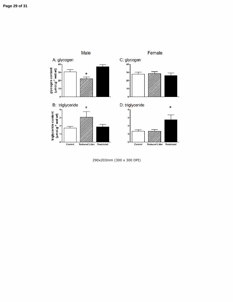
Muscle glycogen and triglyceride levels
In males, Reduced Litter offspring had lower skeletal muscle glycogen compared to Control and
Restricted offspring (P<0.05, Fig 2 A). In females, skeletal muscle glycogen was not altered by
treatment (Fig 2 C). Reduced Litter male offspring tended to have increased skeletal muscle
triglycerides compared to Control and Restricted offspring (P=0.06, Fig 2 B). In Restricted female
offspring, skeletal muscle triglyceride levels were increased compared to Control and Reduced
Litter offspring (P<0.05, Fig 2 D).
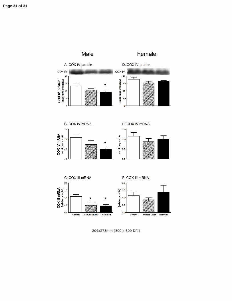
Muscle mitochondrial biogenesis markers
In males, skeletal muscle, PGC-1α protein, PGC-1α mRNA and mtTFA mRNA levels were lower
in both Restricted and Reduced Litter offspring compared Controls (P<0.05, Fig 3 A-C,
respectively). Similarly, skeletal muscle COX IV protein and mRNA levels were lower in Restricted
male offspring compared with Controls (P<0.05, Fig 4 A and B, respectively) and COX III mRNA
levels were lower in both Restricted and Reduced Litter offspring compared with Controls (P<0.05,
Fig 4 C).
Page 11 of 31
12
In females, Reduced Litter offspring had lower skeletal muscle mtTFA levels (P<0.05, Fig 3 F) and
tended to have lower PGC-1α mRNA levels (P=0.11, Fig 3 E). Treatment did not alter skeletal
muscle PGC-1α protein (Fig 3 D), COX IV protein, mRNA and COX III mRNA in female offspring
however (Fig 4 D – F, respectively).
Treatment did not alter skeletal muscle citrate synthase and βHAD activities in male or female
offspring (data not shown).
DISCUSSION
A major finding of the present study was that the early life perturbations of both uteroplacental
insufficiency and restriction of postnatal growth via reductions in litter size, reduced the expression
of skeletal muscle mitochondrial biogenesis markers in the adult male rat, while only reducing litter
size and therefore postnatal growth induced similar trends in female offspring. One of the expected
metabolic consequences of lower skeletal muscle mitochondrial protein levels would be increased
lipid accumulation and impaired glucose utilization, resulting in insulin resistance (10, 18, 26).
Therefore, a surprising finding from the present study was that these early life interventions caused
a dissociation between elevated intramuscular triglycerides (Fig 2) and the reduced mitochondrial
biogenesis markers (PGC-1α mRNA and protein, mtTFA mRNA) and mitochondrial protein (COX
IV mRNA and protein and COX III mRNA) in male and female offspring (Fig 3 and 4).
Furthermore, in contrast to previous reports (30, 35) uteroplacental insufficiency did not alter
glucose tolerance or adiposity in adult offspring.
Page 12 of 31
13
We clearly show that our model of uteroplacental insufficiency in the Wistar Kyoto rat does not
impair fasting glucose or insulin or glucose tolerance (or the insulin response). This contrasts with
earlier reports that uteroplacental insufficiency in the Sprague Dawley rat impairs glucose tolerance
as early as one week after birth in offspring, with a clear diabetic phenotype present at 6 months of
age (29, 30). One factor that may contribute to these contrasting findings may be strain differences
between the Wistar Kyoto rat used in the present study, which has reduced body weight following
uteroplacental insufficiency (Table 1) and the Sprague Dawley rat used in previous studies that
instead gains body weight and fat and subsequently develops diabetes in adulthood (29, 30). Apart
from differences in strain, experimental differences between the present and previous studies were
also evident that may account for these very different findings in development of obesity and
glucose intolerance. These potentially include fostering placentally restricted offspring onto
unoperated mothers following birth (30) and minor reductions in litter size, that in addition to
placental restriction (29), may have induced additional stress or nutritional differences that
exacerbated prenatal programming of obesity and glucose tolerance. Future studies utilizing
different rat strains in the impact of uteroplacental insufficiency in the programming of insulin
action and glucose control and body composition may be highly informative and complement
studies of gene and early environment interactions in humans. Furthermore, future uteroplacental
insufficiency experiments utilizing both rat strains under identical experimental conditions may
provide novel insights into the relationship between the programming of insulin action and glucose
control in response to body and fat weight gain. The present study also found that uteroplacental
insufficiency in the rat increased HOMA in female, but not male adult offspring. This could perhaps
be indicative of onset of hepatic insulin resistance as reported in a previous study (35), which has
yet to affect whole-body insulin resistance or glucose tolerance. Although the use of HOMA as a
measure of insulin resistance in rodent studies is quite common (14, 27), it is also controversial (38)
and more suited to larger cohorts. Nevertheless, our finding of elevated HOMA in the Restricted
Page 13 of 31

14
female rats suggests that alterations in insulin sensitivity could be occurring in our model of
restriction that might be more readily detected by more sensitive measures such as a euglycemic,
hyperinsulinemic clamp. Also, future studies incorporating additional groups of insulin-stimulated
rats could examine skeletal muscle insulin signaling, thus clarifying the potential impact of growth
restriction on tissue metabolism in our model of placental insufficiency.
The uncoupling observed in the present study between intramuscular triglyceride levels and skeletal
muscle mitochondrial biogenesis markers and mitochondrial proteins suggests that this relationship
is not necessarily causative. Several studies have reported a strong association between either
reduced mitochondrial biogenesis, impaired mitochondrial function or lower mitochondrial volume
and elevated lipid content in skeletal muscle that is insulin resistant or from people with type 2
diabetes (10, 17, 23-25). Recent studies in obese rodent models (34) and insulin infusion into
humans (2) suggest however that mitochondrial dysfunction and reduced expression of
mitochondrial proteins in skeletal muscle may be a secondary defect in response to insulin
resistance and lipid accumulation. Genetic models of obesity and insulin resistance, such as the
obese Zucker rat and db/db mice have elevated intramuscular triglycerides, but higher PGC-1α
protein and skeletal muscle mitochondrial enzyme activities and increased fatty acid oxidative
capacity (34). High fat feeding in rats and mice also induces a similar phenotype (34). Also, if
mitochondrial dysfunction causes skeletal muscle insulin resistance then reducing mitochondrial
function should result in insulin resistance. However, skeletal muscle specific mtTFA knockout
mice have reduced oxidative capacity but increased insulin-stimulated glucose uptake (42).
Another major finding of the present study was the influence of reducing litter size on postnatal
growth and its subsequent effects on skeletal muscle characteristics in adulthood. Modestly reducing
the litter size of pups born to sham-operated mothers reduced the body weight of the remaining pups
Page 14 of 31

15
at 3 days of age. This resulted in changes to adult male skeletal muscle, including lower glycogen, a
tendency for higher triglycerides and reduced expression of markers of mitochondrial biogenesis
and mitochondrial proteins. Restricting postnatal growth of female offspring by reducing litter size
also reduced mtTFA mRNA and tended to reduce that of other mitochondrial biogenesis markers in
skeletal muscle. These findings have important implications for the design and interpretation of
studies of prenatal and postnatal perturbations in the rat and highlight the need to include controls
with unaltered litter size (12, 13, 29). The use of a sham-operated group with reduced litter size as
the only control in the current study would have obscured and changed a number of our findings.
Indeed, if we had only used the Reduced Litter group as our control group we would have observed
no alterations in mitochondrial biogenesis markers in the males and higher mtTFA mRNA in the
females (compared with the Restricted group).
Although severe reductions in litter size have been used as a model of overfeeding and obesity (39),
we have previously shown that more modest reductions in litter size, as a control for
uteroplacentally restricted pups, reduces milk quality and quantity and impairs mammary function,
possibly by a reduced suckling stimulus, resulting in slowed postnatal growth (20). Also, the impact
of reducing litter size on postnatal growth and other long term outcomes observed here is also in
agreement with the outcomes of our recent cross-fostering studies related to hypertension (40).
Furthermore, we observed differences in metabolic characteristics, such as skeletal muscle
triglycerides and glycogen levels between the male Restricted and Reduced litter groups. This is not
surprising given that these groups experienced nutrient perturbations at different stages of
development. Indeed, the metabolic milieu of these animals is quite nutritionally different, with
Restricted animals exposed to restriction of substrates and oxygen in utero and also postnatal
lactational restraint (20), whilst the Reduced litter animals were exposed to lactational restraint only
in the postnatal period. Our data highlight that an adverse postnatal environment can program later
Page 15 of 31

16
metabolic function. Further studies may identify specific nutrient deficiencies in growth restricted
offspring to further define the mechanistic pathways leading to metabolic dysfunction. Thus,
reduced postnatal growth as a consequence of a reduction in litter size appears to exert its own
influence on the metabolic characteristics of adult skeletal muscle, independently of uteroplacental
insufficiency and warrants further study.
The present study also found that restriction of postnatal growth via reductions in litter size and
uteroplacental insufficiency reduced mitochondrial biogenesis markers to a greater extent in adult
male than female rats. In support of our findings, Lane et al. (13) found PGC-1α mRNA levels were
altered to a greater extent in male compared to female juvenile rats following uteroplacental
insufficiency. It is possible that estrogen may be playing a protective role in terms of minimizing the
impact of restriction on skeletal muscle mitochondrial biogenesis. Estrogen treatment of female
ovariectomized rats increases the mRNA and enzyme activity of lipid oxidative enzymes (5, 6) in
skeletal muscle and mitochondrial biogenesis in blood vessels (32) and PGC-1α is a known
activator of the estrogen receptor (33). However, our finding of lower mtTFA mRNA and a
tendency for lower PGC-1α mRNA following reductions in litter size suggests that skeletal muscle
mitochondria from adult females is vulnerable to particular insults, at least in early postnatal life,
although the mechanisms for this remain unclear. Furthermore, it is not clear what other sex-specific
factors could explain the differences in the present study, as the effects of sex hormones on the
regulation of mitochondrial biogenesis in skeletal muscle have not been studied in detail. The
effects of testosterone on mitochondrial biogenesis in skeletal muscle are not known, although it
does inhibit mitochondrial biogenesis in brown fat cells in culture. However in this model, estrogen
has no effect on PGC-1α mRNA and down regulates other mitochondrial biogenesis markers (28).
Thus, further study is required into the influence of sex hormones on, and the mechanistic pathways
that may alter, mitochondrial biogenesis in a sex-specific manner. Nevertheless, it appears that the
Page 16 of 31

17
skeletal muscle mitochondria from males are more vulnerable to early life nutrient restriction
overall than females.
Because of clear reductions in skeletal muscle PGC-1α mRNA and protein and mtTFA mRNA in
male Restricted and Reduced Litter rats it was somewhat surprising that the maximal citrate
synthase and β-HAD enzyme activities were also not reduced. Since the maximal in vitro activity of
mitochondrial enzymes largely reflects the abundance of the enzyme, these findings suggest that
certain components of the mitochondrial respiratory chain (i.e. Krebs cycle and β-oxidation) were
not altered in these rats despite reduced mitochondrial biogenesis markers. However, other
components of the respiratory chain, such as the electron transport chain were altered, as evidenced
by reduced COX III (mitochondrial encoded) mRNA and COX IV (nuclear encoded) mRNA and
protein in the male Restricted and Reduced Litter rats. Future studies are now required to determine
the functional consequences of altered mitochondrial biogenesis markers and proteins in the skeletal
muscles of this model of uteroplacental insufficiency.
In conclusion, the present study shows that uteroplacental insufficiency and restriction of postnatal
growth via reductions in litter size differentially alter skeletal muscle mitochondrial biogenesis and
metabolic characteristics (i.e. intramuscular triglycerides and glycogen) in a sex specific manner in
the adult rat. Furthermore these early life interventions alter skeletal muscle characteristics in the
absence of overt changes in plasma glucose, glucose tolerance, insulin secretion or adiposity.
Notably, reducing litter size exposes pups born of normal birthweight to reduced postnatal growth
and alterations in skeletal muscle markers of mitochondrial biogenesis in adult male offspring,
which are less evident in female offspring. The longer term impact of such alterations in
mitochondrial biogenesis in peripheral tissues on adult health that are induced in early life and in
other paradigms require further investigation.
Page 17 of 31

18
ACKNOWLEDGEMENTS
The authors would like to thank Fran Leone, Melissa Walker, Olivia Wyss, Kerryn Westcott, Nicole
Ferguson, Tyler Munk, Jessica Savage and Georgina Strinavic for their technical assistance. This
work was supported by grants from the National Health and Medical Research Council of Australia
(MEW 208948 and GKM, MEW and GDW 454570), Channel 7 Children’s Foundation of South
Australia (JAO and MEW), National Heart Foundation (MEW and JAO) and Diabetes Australia
Research Trust (MEW, JAO and GC).
Page 18 of 31

19
REFERENCES
1. Allison DB, Paultre F, Maggio C, Mezzitis N, and Pi-Sunyer FX. The use of areas under
curves in diabetes research. Diabetes Care 18: 245-250, 1995.
2. Asmann YW, Stump CS, Short KR, Coenen-Schimke JM, Guo Z, Bigelow ML, and Nair
KS. Skeletal muscle mitochondrial functions, mitochondrial DNA copy numbers, and gene
transcript profiles in type 2 diabetic and nondiabetic subjects at equal levels of low or high
insulin and euglycemia. Diabetes 55: 3309-3319, 2006.
3. Barker DJ, Osmond C, Golding J, Kuh D, and Wadsworth ME. Growth in utero, blood
pressure in childhood and adult life, and mortality from cardiovascular disease. BMJ 298: 564-
567, 1989.
4. Boushel R, Gnaiger E, Schjerling P, Skovbro M, Kraunsoe R, and Dela F. Patients with
type 2 diabetes have normal mitochondrial function in skeletal muscle. Diabetologia 50: 790-
796, 2007.
5. Campbell SE and Febbraio MA. Effect of ovarian hormones on mitochondrial enzyme
activity in the fat oxidation pathway of skeletal muscle. Am J Physiol 281: E803-E808, 2001.
6. Campbell SE, Mehan KA, Tunstall RJ, Febbraio MA, and Cameron-Smith D. 17beta-
estradiol upregulates the expression of peroxisome proliferator-activated receptor alpha and
lipid oxidative genes in skeletal muscle. J Mol Endocrinol 31: 37-45, 2003.
7. De Blasio MJ, Gatford KL, McMillen IC, Robinson JS, and Owens JA. Placental
restriction of fetal growth increases insulin action, growth, and adiposity in the young lamb.
Endocrinology 148: 1350-1358, 2007.
8. Ellis BA, Poynten A, Lowy AJ, Furler SM, Chisholm DJ, Kraegen EW, and Cooney GJ.
Long-chain acyl-CoA esters as indicators of lipid metabolism and insulin sensitivity in rat and
human muscle. Am J Physiol 279: E554-E560, 2000.
Page 19 of 31

20
9. Eriksson J, Forsen T, Tuomilehto J, Osmond C, and Barker D. Fetal and childhood growth
and hypertension in adult life. Hypertension 36: 790-794, 2000.
10. Kelley DE, He J, Menshikova EV, and Ritov VB. Dysfunction of mitochondria in human
skeletal muscle in type 2 diabetes. Diabetes 51: 2944-2950, 2002.
11. Koves TR, Li P, An J, Akimoto T, Slentz D, Ilkayeva O, Dohm GL, Yan Z, Newgard CB,
and Muoio DM. Peroxisome proliferator-activated receptor-gamma co-activator 1alpha-
mediated metabolic remodeling of skeletal myocytes mimics exercise training and reverses
lipid-induced mitochondrial inefficiency. J Biol Chem 280: 33588-33598, 2005.
12. Lane RH, Kelley DE, Ritov VH, Tsirka AE, and Gruetzmacher EM. Altered expression
and function of mitochondrial beta-oxidation enzymes in juvenile intrauterine growth retarded
rat skeletal muscle. Pediatr Res 50: 83-90, 2001.
13. Lane RH, Maclennan NK, Daood MJ, Hsu JL, Janke SM, Pham TD, Puri AR, and
Watchko JF. IUGR alters postnatal rat skeletal muscle peroxisome proliferator-activated
receptor-gamma coactivator-1 gene expression in a fiber specific manner. Pediatr Res 53: 994-
1000, 2003.
14. Li PP, Shan S, Chen YT, Ning ZQ, Sun SJ, Liu Q, Lu XP, Xie MZ, and Shen ZF. The
PPARalpha/gamma dual agonist chiglitazar improves insulin resistance and dyslipidemia in
MSG obese rats. Br J Pharmacol 148: 610-618, 2006.
15. Matthews DR, Hosker JP, Rudenski AS, Naylor BA, Treacher DF, and Turner RC.
Homeostasis model assessment: insulin resistance and beta-cell function from fasting plasma
glucose and insulin concentrations in man. Diabetologia 28: 412-419, 1985.
16. Matthews JN, Altman DG, Campbell MJ, and Royston P. Analysis of serial measurements
in medical research. Bmj 300: 230-235, 1990.
17. Mootha VK, Lindgren CM, Eriksson KF, Subramanian A, Sihag S, Lehar J, Puigserver
P, Carlsson E, Ridderstrale M, Laurila E, Houstis N, Daly MJ, Patterson N, Mesirov JP,
Page 20 of 31

21
Golub TR, Tamayo P, Spiegelman B, Lander ES, Hirschhorn JN, Altshuler D, and Groop
LC. PGC-1alpha-responsive genes involved in oxidative phosphorylation are coordinately
downregulated in human diabetes. Nat Genet 34: 267-273, 2003.
18. Morino K, Petersen KF, Dufour S, Befroy D, Frattini J, Shatzkes N, Neschen S, White
MF, Bilz S, Sono S, Pypaert M, and Shulman GI. Reduced mitochondrial density and
increased IRS-1 serine phosphorylation in muscle of insulin-resistant offspring of type 2
diabetic parents. J Clin Invest 115: 3587-3593, 2005.
19. Newsome CA, Shiell AW, Fall CH, Phillips DI, Shier R, and Law CM. Is birth weight
related to later glucose and insulin metabolism? A systematic review. Diabet Med 20: 339-348,
2003.
20. O'Dowd R, Kent J, Moseley J, and Wlodek M. The effects of uteroplacental insufficiency
and reducing litter size on maternal mammary function and postnatal offspring growth. Am J
Physiol Regul Integr Comp Physiol published 12 December, 10.1152/ajpregu.00628, 2007.
21. Owens JA, Thavaneswaran P, De Blasio MJ, McMillen IC, Robinson JS, and Gatford
KL. Sex-specific effects of placental restriction on components of the metabolic syndrome in
young adult sheep. Am J Physiol 292: E1879-E1889, 2007.
22. Passonneau JV and Lauderdale VR. A comparison of three methods of glycogen
measurement in tissues. Anal Biochem 60: 405-412, 1974.
23. Patti ME, Butte AJ, Crunkhorn S, Cusi K, Berria R, Kashyap S, Miyazaki Y, Kohane I,
Costello M, Saccone R, Landaker EJ, Goldfine AB, Mun E, DeFronzo R, Finlayson J,
Kahn CR, and Mandarino LJ. Coordinated reduction of genes of oxidative metabolism in
humans with insulin resistance and diabetes: Potential role of PGC1 and NRF1. PNAS 100:
8466-8471, 2003.
Page 21 of 31

22
24. Petersen KF, Befroy D, Dufour S, Dziura J, Ariyan C, Rothman DL, DiPietro L, Cline
GW, and Shulman GI. Mitochondrial dysfunction in the elderly: possible role in insulin
resistance. Science 300: 1140-1142, 2003.
25. Petersen KF, Dufour S, Befroy D, Garcia R, and Shulman GI. Impaired mitochondrial
activity in the insulin-resistant offspring of patients with type 2 diabetes. N Engl J Med 350:
664-671, 2004.
26. Petersen KF and Shulman GI. Etiology of insulin resistance. Am J Med 119: S10-16, 2006.
27. Pickavance LC, Brand CL, Wassermann K, and Wilding JP. The dual PPARalpha/gamma
agonist, ragaglitazar, improves insulin sensitivity and metabolic profile equally with
pioglitazone in diabetic and dietary obese ZDF rats. Br J Pharmacol 144: 308-316, 2005.
28. Rodriguez-Cuenca S, Monjo M, Gianotti M, Proenza AM, and Roca P. Expression of
mitochondrial biogenesis-signaling factors in brown adipocytes is influenced specifically by
17beta-estradiol, testosterone, and progesterone. Am J Physiol Endocrinol Metab 292: E340-
346, 2007.
29. Selak MA, Storey BT, Peterside I, and Simmons RA. Impaired oxidative phosphorylation in
skeletal muscle of intrauterine growth-retarded rats. Am J Physiol 285: E130-E137, 2003.
30. Simmons RA, Templeton LJ, and Gertz SJ. Intrauterine growth retardation leads to the
development of type 2 diabetes in the rat. Diabetes 50: 2279-2286, 2001.
31. Srere P. Citrate synthase. New York: Academic Press, 1969.
32. Stirone C, Duckles SP, Krause DN, and Procaccio V. Estrogen increases mitochondrial
efficiency and reduces oxidative stress in cerebral blood vessels. Mol Pharmacol 68: 959-965,
2005.
33. Tcherepanova I, Puigserver P, Norris JD, Spiegelman BM, and McDonnell DP.
Modulation of estrogen receptor-alpha transcriptional activity by the coactivator PGC-1. J Biol
Chem 275: 16302-16308, 2000.
Page 22 of 31
23
34. Turner N, Bruce CR, Beale SM, Hoehn KL, So T, Rolph MS, and Cooney GJ. Excess lipid
availability increases mitochondrial fatty acid oxidative capacity in muscle: evidence against a
role for reduced fatty acid oxidation in lipid-induced insulin resistance in rodents. Diabetes 56:
2085-2092, 2007.
35. Vuguin P, Raab E, Liu B, Barzilai N, and Simmons R. Hepatic insulin resistance precedes
the development of diabetes in a model of intrauterine growth retardation. Diabetes 53: 2617-
2622, 2004.
36. Wadley GD and McConell GK. Effect of nitric oxide synthase inhibition on mitochondrial
biogenesis in rat skeletal muscle. J Appl Physiol 102: 314-320, 2007.
37. Wadley GD, Tunstall RJ, Sanigorski A, Collier GR, Hargreaves M, and Cameron-Smith
D. Differential effects of exercise on insulin-signaling gene expression in human skeletal
muscle. J Appl Physiol 90: 436-440, 2001.
38. Wallace TM, Levy JC, and Matthews DR. Use and abuse of HOMA modeling. Diabetes
Care 27: 1487-1495, 2004.
39. Widdowson EM and Mc CR. Some effects of accelerating growth. I. General somatic
development. Proceedings of the Royal Society of London Series B, Containing papers of a
Biological character 152: 188-206, 1960.
40. Wlodek ME, Mibus A, Tan A, Siebel AL, Owens JA, and Moritz KM. Normal lactational
environment restores nephron endowment and prevents hypertension after placental restriction
in the rat. J Am Soc Nephrol 18: 1688-1696, 2007.
41. Wlodek ME, Westcott KT, O'Dowd R, Serruto A, Wassef L, Moritz KM, and Moseley
JM. Uteroplacental restriction in the rat impairs fetal growth in association with alterations in
placental growth factors including PTHrP. Am J Physiol 288: R1620-R1627, 2005.
Page 23 of 31

24
42. Wredenberg A, Freyer C, Sandstrom ME, Katz A, Wibom R, Westerblad H, and Larsson
NG. Respiratory chain dysfunction in skeletal muscle does not cause insulin resistance.
Biochem Biophys Res Commun 350: 202-207, 2006.
Page 24 of 31

25
Table 1: The effect of uteroplacental insufficiency (Restricted) and reducing litter size (Reduced
Litter) on body, fat and gastrocnemius weight
Control Reduced Litter Restricted
MALE BWT: Day 3 (g) 6.01 ± 0.38 5.29 ± 0.15 * 4.41 ± 0.24 * †
BWT: 6 Month (g) 366.6 ± 7.1 352.4 ± 5.0 329.0 ± 8.5 * †
Fat weight: 6 Month (g) 17.6 ± 1.0 16.8 ± 0.8 14.5 ± 1.3
Fat weight: 6 Month (% BWT) 4.8 ± 0.3 4.8 ± 0.2 4.4 ± 0.4
Gastrocnemius: 6 Month (g) 1.77 ± 0.05 1.60 ± 0.08 * 1.55 ± 0.05*
Gastrocnemius: 6 Month (% BWT) 0.48 ± 0.01 0.41 ± 0.04 0.47 ± 0.01
FEMALE BWT: Day 3 (g) 5.57 ± 0.23 4.97 ± 0.19 * 4.35 ± 0.20 *
BWT: 6 Month (g) 231.2 ± 3.9 214.5 ± 5.8 * 213.0 ± 3.3 *
Fat weight: 6 Month (g) 8.2 ± 0.6 7.3 ± 0.6 8.0± 0.6
Fat weight: 6 Month (% BWT) 3.5 ± 0.2 3.3 ± 0.2 3.7 ± 0.3
Gastrocnemius: 6 month (g) 1.18 ± 0.03 1.04 ± 0.03 * 1.00 ± 0.03 *
Gastrocnemius: 6 Month (% BWT) 0.51 ± 0.02 0.49 ± 0.01 0.47 ± 0.01
Body weight (BWT) measured at postnatal Day 3 and at post mortem (6 months of age) in male and female
rats (in grams). Fat weight is the sum of dorsal, visceral, retroperitoneal, perirenal and for males, epididymal
fat (in grams and as a percentage of body weight). Gastrocnemius muscle weight measured at post mortem
in male and female rats (in grams and as a percentage of body weight). Values are mean ± SE. * P<0.05 vs.
Control; † P<0.05 vs. Reduced Litter (one-way ANOVA).
Page 25 of 31

26
Table 2: The effect of uteroplacental insufficiency (Restricted) and reducing litter size (Reduced
Litter) on HOMA and free fatty acid levels in 6 month old male and female rats
Control Reduced Litter Restricted
MALE HOMA 9.3 ± 1.8 11.7 ± 2.0 12.8 ± 4.0
FFA (mmol.l-1) 1.0 ± 0.1 1.4 ± 0.1 1.5 ± 0.3
FEMALE HOMA 10.5 ± 1.7 8.9 ± 1.7 17.1 ± 3.1* †
FFA (mmol.l-1) 1.2 ± 0.1 0.9 ± 0.2 1.3 ± 0.1
FFA: free fatty acids, HOMA: homeostasis model assessment. Values are mean ± SE. * P<0.05 vs. Control;
† P<0.05 vs. Reduced Litter (one-way ANOVA).
Page 26 of 31

27
Figure Legends
Figure 1. The effect of uteroplacental insufficiency (Restricted) and reducing litter size (Reduced Litter) on
plasma glucose and insulin measured during an IAGTT in adult male and female offspring. Values are mean
± SE with n = 7 for all groups.
Figure 2. The effect of uteroplacental insufficiency (Restricted) and reducing litter size (Reduced Litter) on
gastrocnemius muscle glycogen (A and C) and triglyceride (B and D) content in adult male and female
offspring. Values are mean ± SE with n = 8 for all groups except n = 7 for female Restricted group. *
P<0.05 vs. Control, # P=0.06 vs. Control (one-way ANOVA).
Figure 3. The effect of uteroplacental insufficiency (Restricted) and reducing litter size (Reduced Litter) on
gastrocnemius muscle on PGC-1α protein and mRNA and mtTFA mRNA in adult male and female
offspring. Values are mean ± SE. Western blots are representative from one rat from each treatment group. n
= 8 for all groups except n = 7 for female and male Restricted groups. * P<0.05 vs. Control (one-way
ANOVA).
Figure 4. The effect of uteroplacental insufficiency (Restricted) and reducing litter size (Reduced Litter) on
gastrocnemius muscle COX IV protein and mRNA and COX III mRNA in adult male and female offspring.
Values are mean ± SE. Western blots are representative from one rat from each treatment group. n = 8 for all
groups except n = 7 for female and male Restricted groups. * P<0.05 vs. Control (one-way ANOVA).
Page 27 of 31

285x203mm (300 x 300 DPI)
Page 28 of 31
290x203mm (300 x 300 DPI)
Page 29 of 31

205x269mm (300 x 300 DPI)
Page 30 of 31
204x273mm (300 x 300 DPI)
Page 31 of 31
Copyright © 2022 FDOKUMEN