A phosphorelay system controls stalk biogenesis during cell cycle progression in Caulobacter...
16
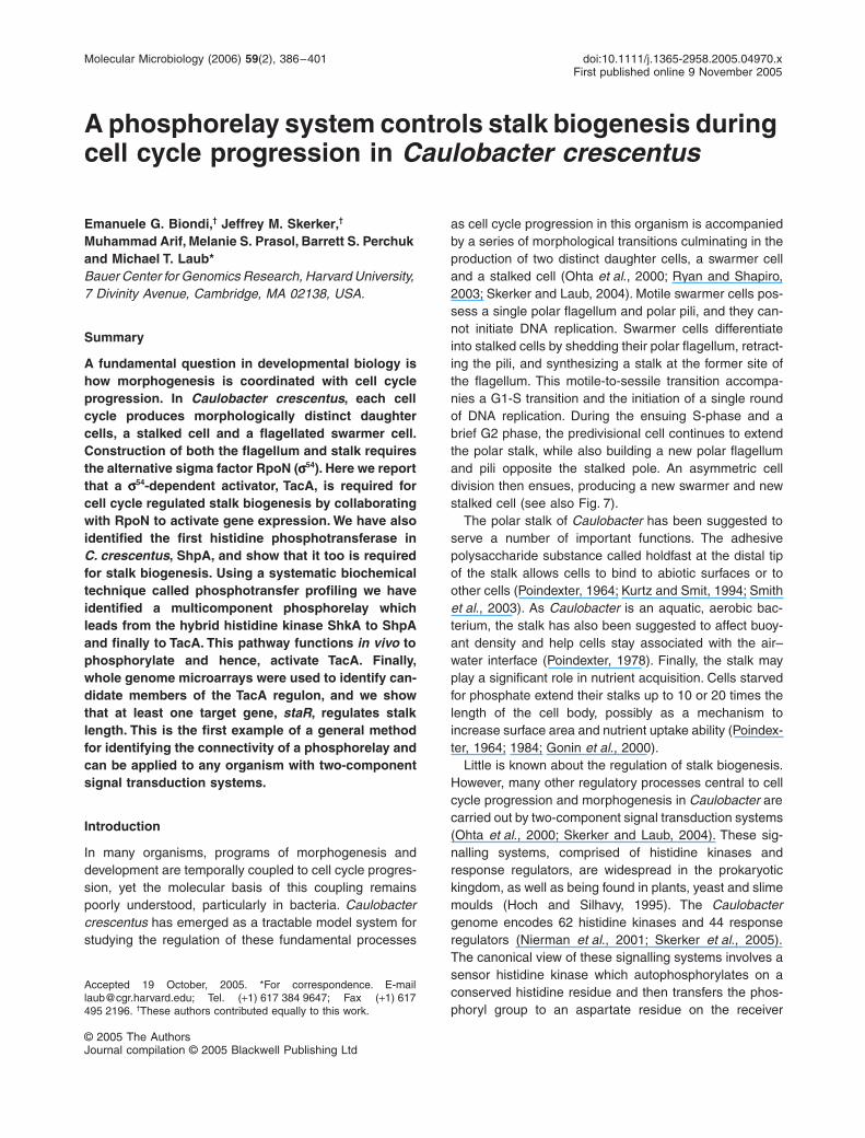
Molecular Microbiology (2006) 59(2), 386–401 doi:10.1111/j.1365-2958.2005.04970.x First published online 9 November 2005 © 2005 The Authors Journal compilation © 2005 Blackwell Publishing Ltd Blackwell Science, LtdOxford, UKMMIMolecular Microbiology0950-382X© 2005 The Authors; Journal compilation © 2005 Blackwell Publishing Ltd ? 2005592386401Original ArticleRegulation of stalk biogenesis in C. crescentusE. G. Biondi et al. Accepted 19 October, 2005. *For correspondence. E-mail [email protected]; Tel. (+1) 617 384 9647; Fax (+1) 617 495 2196. † These authors contributed equally to this work. A phosphorelay system controls stalk biogenesis during cell cycle progression in Caulobacter crescentus Emanuele G. Biondi, † Jeffrey M. Skerker, † Muhammad Arif, Melanie S. Prasol, Barrett S. Perchuk and Michael T. Laub* Bauer Center for Genomics Research, Harvard University, 7 Divinity Avenue, Cambridge, MA 02138, USA. Summary A fundamental question in developmental biology is how morphogenesis is coordinated with cell cycle progression. In Caulobacter crescentus , each cell cycle produces morphologically distinct daughter cells, a stalked cell and a flagellated swarmer cell. Construction of both the flagellum and stalk requires the alternative sigma factor RpoN ( σ 54 ). Here we report that a σ 54 -dependent activator, TacA, is required for cell cycle regulated stalk biogenesis by collaborating with RpoN to activate gene expression. We have also identified the first histidine phosphotransferase in C. crescentus , ShpA, and show that it too is required for stalk biogenesis. Using a systematic biochemical technique called phosphotransfer profiling we have identified a multicomponent phosphorelay which leads from the hybrid histidine kinase ShkA to ShpA and finally to TacA. This pathway functions in vivo to phosphorylate and hence, activate TacA. Finally, whole genome microarrays were used to identify can- didate members of the TacA regulon, and we show that at least one target gene, staR , regulates stalk length. This is the first example of a general method for identifying the connectivity of a phosphorelay and can be applied to any organism with two-component signal transduction systems. Introduction In many organisms, programs of morphogenesis and development are temporally coupled to cell cycle progres- sion, yet the molecular basis of this coupling remains poorly understood, particularly in bacteria. Caulobacter crescentus has emerged as a tractable model system for studying the regulation of these fundamental processes as cell cycle progression in this organism is accompanied by a series of morphological transitions culminating in the production of two distinct daughter cells, a swarmer cell and a stalked cell (Ohta et al ., 2000; Ryan and Shapiro, 2003; Skerker and Laub, 2004). Motile swarmer cells pos- sess a single polar flagellum and polar pili, and they can- not initiate DNA replication. Swarmer cells differentiate into stalked cells by shedding their polar flagellum, retract- ing the pili, and synthesizing a stalk at the former site of the flagellum. This motile-to-sessile transition accompa- nies a G1-S transition and the initiation of a single round of DNA replication. During the ensuing S-phase and a brief G2 phase, the predivisional cell continues to extend the polar stalk, while also building a new polar flagellum and pili opposite the stalked pole. An asymmetric cell division then ensues, producing a new swarmer and new stalked cell (see also Fig. 7). The polar stalk of Caulobacter has been suggested to serve a number of important functions. The adhesive polysaccharide substance called holdfast at the distal tip of the stalk allows cells to bind to abiotic surfaces or to other cells (Poindexter, 1964; Kurtz and Smit, 1994; Smith et al ., 2003). As Caulobacter is an aquatic, aerobic bac- terium, the stalk has also been suggested to affect buoy- ant density and help cells stay associated with the air– water interface (Poindexter, 1978). Finally, the stalk may play a significant role in nutrient acquisition. Cells starved for phosphate extend their stalks up to 10 or 20 times the length of the cell body, possibly as a mechanism to increase surface area and nutrient uptake ability (Poindex- ter, 1964; 1984; Gonin et al ., 2000). Little is known about the regulation of stalk biogenesis. However, many other regulatory processes central to cell cycle progression and morphogenesis in Caulobacter are carried out by two-component signal transduction systems (Ohta et al ., 2000; Skerker and Laub, 2004). These sig- nalling systems, comprised of histidine kinases and response regulators, are widespread in the prokaryotic kingdom, as well as being found in plants, yeast and slime moulds (Hoch and Silhavy, 1995). The Caulobacter genome encodes 62 histidine kinases and 44 response regulators (Nierman et al ., 2001; Skerker et al ., 2005). The canonical view of these signalling systems involves a sensor histidine kinase which autophosphorylates on a conserved histidine residue and then transfers the phos- phoryl group to an aspartate residue on the receiver
Transcript of A phosphorelay system controls stalk biogenesis during cell cycle progression in Caulobacter...
Molecular Microbiology (2006)
59
(2), 386–401 doi:10.1111/j.1365-2958.2005.04970.xFirst published online 9 November 2005
© 2005 The AuthorsJournal compilation © 2005 Blackwell Publishing Ltd
Blackwell Science, LtdOxford, UKMMIMolecular Microbiology0950-382X© 2005 The Authors; Journal compilation © 2005 Blackwell Publishing Ltd
? 2005
59
2386401
Original Article
Regulation of stalk biogenesis in C. crescentusE. G. Biondi
et al.
Accepted 19 October, 2005. *For correspondence. [email protected]; Tel. (
+
1) 617 384 9647; Fax (
+
1) 617495 2196.
†
These authors contributed equally to this work.
A phosphorelay system controls stalk biogenesis during cell cycle progression in
Caulobacter crescentus
Emanuele G. Biondi,
†
Jeffrey M. Skerker,
†
Muhammad Arif, Melanie S. Prasol, Barrett S. Perchuk and Michael T. Laub*
Bauer Center for Genomics Research, Harvard University, 7 Divinity Avenue, Cambridge, MA 02138, USA.
Summary
A fundamental question in developmental biology ishow morphogenesis is coordinated with cell cycleprogression. In
Caulobacter crescentus
, each cellcycle produces morphologically distinct daughtercells, a stalked cell and a flagellated swarmer cell.Construction of both the flagellum and stalk requiresthe alternative sigma factor RpoN (
σσσσ
54
). Here we reportthat a
σσσσ
54
-dependent activator, TacA, is required forcell cycle regulated stalk biogenesis by collaboratingwith RpoN to activate gene expression. We have alsoidentified the first histidine phosphotransferase in
C. crescentus
, ShpA, and show that it too is requiredfor stalk biogenesis. Using a systematic biochemicaltechnique called phosphotransfer profiling we haveidentified a multicomponent phosphorelay whichleads from the hybrid histidine kinase ShkA to ShpAand finally to TacA. This pathway functions
in vivo
tophosphorylate and hence, activate TacA. Finally,whole genome microarrays were used to identify can-didate members of the TacA regulon, and we showthat at least one target gene,
staR
, regulates stalklength. This is the first example of a general methodfor identifying the connectivity of a phosphorelay andcan be applied to any organism with two-componentsignal transduction systems.
Introduction
In many organisms, programs of morphogenesis anddevelopment are temporally coupled to cell cycle progres-sion, yet the molecular basis of this coupling remainspoorly understood, particularly in bacteria.
Caulobactercrescentus
has emerged as a tractable model system forstudying the regulation of these fundamental processes
as cell cycle progression in this organism is accompaniedby a series of morphological transitions culminating in theproduction of two distinct daughter cells, a swarmer celland a stalked cell (Ohta
et al
., 2000; Ryan and Shapiro,2003; Skerker and Laub, 2004). Motile swarmer cells pos-sess a single polar flagellum and polar pili, and they can-not initiate DNA replication. Swarmer cells differentiateinto stalked cells by shedding their polar flagellum, retract-ing the pili, and synthesizing a stalk at the former site ofthe flagellum. This motile-to-sessile transition accompa-nies a G1-S transition and the initiation of a single roundof DNA replication. During the ensuing S-phase and abrief G2 phase, the predivisional cell continues to extendthe polar stalk, while also building a new polar flagellumand pili opposite the stalked pole. An asymmetric celldivision then ensues, producing a new swarmer and newstalked cell (see also Fig. 7).
The polar stalk of
Caulobacter
has been suggested toserve a number of important functions. The adhesivepolysaccharide substance called holdfast at the distal tipof the stalk allows cells to bind to abiotic surfaces or toother cells (Poindexter, 1964; Kurtz and Smit, 1994; Smith
et al
., 2003). As
Caulobacter
is an aquatic, aerobic bac-terium, the stalk has also been suggested to affect buoy-ant density and help cells stay associated with the air–water interface (Poindexter, 1978). Finally, the stalk mayplay a significant role in nutrient acquisition. Cells starvedfor phosphate extend their stalks up to 10 or 20 times thelength of the cell body, possibly as a mechanism toincrease surface area and nutrient uptake ability (Poindex-ter, 1964; 1984; Gonin
et al
., 2000).Little is known about the regulation of stalk biogenesis.
However, many other regulatory processes central to cellcycle progression and morphogenesis in
Caulobacter
arecarried out by two-component signal transduction systems(Ohta
et al
., 2000; Skerker and Laub, 2004). These sig-nalling systems, comprised of histidine kinases andresponse regulators, are widespread in the prokaryotickingdom, as well as being found in plants, yeast and slimemoulds (Hoch and Silhavy, 1995). The
Caulobacter
genome encodes 62 histidine kinases and 44 responseregulators (Nierman
et al
., 2001; Skerker
et al
., 2005).The canonical view of these signalling systems involves asensor histidine kinase which autophosphorylates on aconserved histidine residue and then transfers the phos-phoryl group to an aspartate residue on the receiver

Regulation of stalk biogenesis in
C. crescentus 387
© 2005 The AuthorsJournal compilation © 2005 Blackwell Publishing Ltd,
Molecular Microbiology
,
59
, 386–401
domain of a response regulator (Fig. 1A). Phosphorylationof the receiver domain activates the output domain, whichoften has the ability to bind DNA, thereby coupling signal-ling pathways to changes in gene expression and theexecution of various cellular events.
Complicating this simple paradigm for two-componentsignalling are hybrid histidine kinases in which a histidinekinase contains a response regulator receiver domain atits C-terminal end. These hybrid histidine kinases oftenparticipate in more complex pathways called phosphore-lays (Fig. 1A). In such pathways, hybrid kinases first auto-phosphorylate on a conserved histidine residue in thekinase domain and then transfer the phosphoryl groupintramolecularly to a conserved aspartate in its receiverdomain. The phosphoryl group is then transferred toanother conserved histidine on a protein called a histidinephosphotransferase, which can in turn transfer its phos-
phoryl group to an aspartate on a diffusible responseregulator. Unlike histidine kinases and response regula-tors, histidine phosphotransferases show little sequenceidentity to one another, with conservation of only a fewcritical residues and a four-helix bundle structure (Kato
et al
., 1997; Song
et al
., 1999; Xu and West, 1999).Previous genetic screens as well as a systematic dele-
tion of each of the 106 two-component genes in
Caulo-bacter
have identified 39 genes which are required fornormal cell cycle progression or morphogenesis (Skerker
et al
., 2005). Of these, 19 were impaired in motility andnine had defects in stalk biogenesis. Many of thoseinvolved in motility had been previously identified as reg-ulators of flagellar biogenesis, the best understood aspectof morphogenesis in
Caulobacter
. The assembly of thepolar flagellum in predivisional cells is controlled by a four-tiered transcriptional cascade (Ramakrishnan
et al
., 1994;
Fig. 1.
Identification of a putative histidine phosphotransferase in
C. crescentus
.A. Schematic of a canonical two-component signal transduction pathway versus a multicomponent phosphorelay.B. CC1114 amino acid sequence was aligned against the phosphotransferase domain of ArcB (gi48994928, fragment 655–778 aa), the P1 domain of CheA (gi16129840, fragment 1–140 aa), LuxU (gi39721593) and Ypd1 (gi6319966), using 3D-Coffee. Alpha helices are indicated (grey bars) according to the secondary structure of ArcB.C. Homology model of CC1114 using ArcB as template.

388
E. G. Biondi
et al.
© 2005 The AuthorsJournal compilation © 2005 Blackwell Publishing Ltd,
Molecular Microbiology
,
59
, 386–401
Gober and England, 2000). This cascade is set in motionby the DNA-binding response regulator CtrA (Quon
et al
.,1996). The phosphorylation and activity of CtrA are underextensive cell cycle control, coupling flagellar biogenesisto cell cycle progression (Quon
et al
., 1996; Domian
et al
.,1997). CtrA directly regulates the expression of at least95 genes, including
rpoN
, which encodes the
σ
54
subunitof RNA polymerase (Laub
et al
., 2002).
rpoN
mutants arenon-motile and stalkless, suggesting that
σ
54
regulatesgenes involved in flagellum and stalk biogenesis (Brunand Shapiro, 1992).
σ
54
-dependent gene expression typically requires aphosphorylated transcriptional activator of the NtrC-familyof response regulators (Kustu
et al
., 1989). In
Caulo-bacter
,
flbD
encodes a
σ
54
-dependent activator which isinduced at approximately the same time as
rpoN
, andtogether with RpoN, activates expression of genesrequired late in flagellar assembly (Ramakrishnan
et al
.,1991). FlbD also represses genes required early in flagel-lar assembly, and thus coordinates the timing of multiplesteps in flagellum assembly (Wingrove
et al
., 1993; Ben-son
et al
., 1994; Wingrove and Gober, 1994). However,unlike
rpoN
,
flbD
mutants are not impaired in stalk syn-thesis, suggesting that another NtrC-like response regu-lator functions with
rpoN
to regulate stalk biogenesis.In a systematic deletion of two-component signalling
genes the NtrC-like regulator
tacA
was found to lack stalks(Skerker
et al
., 2005), suggesting it regulates stalk bio-genesis. In that study and previous genetic screens, delet-ing the histidine kinase
pleC
was also found to producestalkless cells (Sommer and Newton, 1989; Skerker
et al
.,2005). PleC, however, exclusively phosphorylates theresponse regulators DivK and PleD, and not TacA(Skerker
et al
., 2005). In the systematic deletion study noother deletions of canonical histidine kinases produced astalkless phenotype. However, the hybrid kinase CC0138was found to be stalkless, suggesting that a phosphorelaycontrols phosphorylation of the
σ
54
-activator TacA. Here,using genetics and a systematic biochemical methodcalled phosphotransfer profiling, we describe the identifi-cation of ShpA, the first histidine phosphotransferase in
Caulobacter
, and demonstrate that it is central to a phos-phorelay which regulates stalk biogenesis by controllingthe activity of TacA. Using whole-genome DNA microar-rays, we identify the likely target genes of TacA. Ourresults provide evidence for a transcriptional cascade thatregulates stalk biogenesis and suggest a mechanism forthe cell cycle timing of stalk synthesis.
Results
Identification of
C. crescentus
histidine phosphotransferases
A systematic study of all
C. crescentus
two-component
signalling genes identified two genes whose deletion pro-duced stalkless cells, the gene CC0138 which encodes ahybrid histidine kinase and CC3315 which encodes a
σ
54
-dependent response regulator named TacA (Skerker
et al
., 2005). The discovery of these mutants suggestedthat a phosphorelay may control stalk formation in
C.crescentus
. However, phosphorelays require histidinephosphotransferases to shuttle phosphoryl groups fromhybrid kinases to response regulators (Fig. 1A), and nonewere identified in the initial annotation of the
C. crescentus
genome based on sequence homology methods (Nier-man
et al
., 2001). To identify putative phosphotrans-ferases in the
C. crescentus
genome, we searched forgenes predicted to encode proteins bearing other com-mon features. Specifically, we sought proteins with (i)fewer than 250 amino acids, (ii) greater than 70% pre-dicted alpha helical secondary structure, and (iii) the con-served motif HXXKG within a predicted alpha helix. Twogenes satisfied these criteria, CC1114 and CC1220. Thisreport focuses on the gene CC1114 which is predicted toencode a protein of 112 amino acids with high alphahelical content (Fig. 1B). The primary sequence ofCC1114 was successfully threaded onto the known four-helix bundle structures of three monomeric histidine phos-photransferases: Ypd1 from
Saccharomyces cerevisiae
(Xu and West, 1999), LuxU from
Vibrio harveyi
(Ulrich
et al
., 2005), and the phosphotransferase domain of ArcBfrom
Escherichia coli
(Kato
et al
., 1997) (Fig. 1C). Thesethreaded structural alignments further predicted that His-56 of CC1114 lies within the middle of an alpha helix andis solvent exposed. Homologues of CC1114 are found inthe genomes of closely related alpha-proteobacteria(Fig. S1). In sum, these bioinformatic analyses suggestedthat CC1114 may encode a histidine phosphotransferase.
CC1114 has histidine phosphotransferase activity
in vitro
To determine whether the CC1114 protein exhibits phos-photransferase activity, we tagged CC1114 with a His
6
-tagand purified the protein by affinity chromatography togreater than 90% homogeneity. To determine whetherCC1114 can receive a phosphoryl group from a hybridhistidine kinase, we incubated CC1114 with the purifiedkinase and receiver domains from each of four hybridkinases which possess high
in vitro
activity: CC0138,CckA, CC3102 and CC3219. We used separate kinaseand receiver domains for each hybrid kinase because full-length constructs had little to no activity (data not shown).Reactions were initiated by the addition of [
γ
-
32
P]-ATP,incubated for 30 min, and then resolved by SDS-PAGE(Fig. 2A). In each reaction, radiolabel was incorporatedinto the kinase domain by autophosphorylation and sub-sequently transferred to the corresponding receiverdomain. For the reactions with CC0138 and CC3102,

Regulation of stalk biogenesis in
C. crescentus 389
© 2005 The AuthorsJournal compilation © 2005 Blackwell Publishing Ltd,
Molecular Microbiology
,
59
, 386–401
Fig. 2.
Biochemical identification of the TacA phosphorelay.A. CC1114 can be phosphorylated by
Caulobacter
hybrid kinases. The kinase (HY-HK) and receiver domains (HY-RD) for four different hybrid kinases were each purified and tested
in vitro
for their ability to phosphorylate CC1114. The gene name or CC number is indicated for each kinase.B. Phosphorylation of CC1114 is dependent on the presence of a receiver domain. Pluses and minuses indicate presence or absence of reaction components: purified kinase domain of CC0138 (CC0138-HK), its receiver domain (CC0138-RD), and CC1114.C and D. Identification of a CC1114-dependent phosphorelay using phosphotransfer profiling. Phosphorylated CC1114 was profiled against all 44 purified
Caulobacter
response regulators (C) and against all 27 purified receiver domains of
Caulobacter
hybrid kinases (D). Each phospho-transfer reaction was incubated for 2 min at room temperature.E. CC1114 exhibits a kinetic preference for CC0138 and TacA-RD. Phosphotransfer from CC1114 to the receiver domains of CC0138, CC0921, CckA, CC3102, CC3219 and TacA were tested at a 10 s time point. Open arrowheads in panels C–E indicate lanes with evidence of phospho-transfer, manifested as loss of radiolabel from the CC1114 band and/or incorporation of label on a receiver domain.F. Schematic of the phosphorelay between the hybrid kinase CC0138, the histidine phosphotransferase CC1114 and the response regulator TacA.G. Reconstitution of the phosphorelay
in vitro
using purified components. Pluses and minuses indicate the presence or absence of purified CC0138-HK, CC0138-RD, CC1114 and TacA-RD.

390
E. G. Biondi
et al.
© 2005 The AuthorsJournal compilation © 2005 Blackwell Publishing Ltd,
Molecular Microbiology
,
59
, 386–401
significant accumulation of label was also found at a posi-tion corresponding to CC1114. Incubation of CC1114 withthe CC0138 kinase domain in the absence of the CC0138receiver domain was indistinguishable from incubating theCC0138 kinase domain alone (Fig. 2B, lane 1 vs. 4) indi-cating that (i) the receiver domain is required for phospho-transfer from the kinase domain to CC1114, and (ii)CC1114 does not have autophosphorylation activity.Together these observations support the conclusion thatCC1114 is a histidine phosphotransferase.
CC1114 participates in a phosphorelay culminating in the phosphorylation of TacA
We previously developed an assay called phosphotransferprofiling which allows the rapid, systematic identificationof response regulator targets of a given histidine kinase(Skerker
et al
., 2005). In this assay, one histidine kinaseis systematically tested
in vitro
for its ability to transfer aphosphoryl group to each purified response regulatorencoded in the genome. As histidine kinases exhibit alarge kinetic preference
in vitro
for their
in vivo
cognatesubstrates, kinase-regulator pairs are easily identified bytesting phosphotransfer at multiple time points and iden-tifying the kinetically preferred substrate(s) (Skerker
et al
.,2005). Here, we adapted this assay (also see Fig. S2) toexamine the substrate specificity, and hence probable
invivo
partners, of CC1114. CC1114 was first phosphory-lated (producing CC1114
∼
P) and then split and incubatedindividually for 2 min with each of the 44 purified, full-length response regulators encoded in the
C. crescentus
genome (Skerker
et al
., 2005). Phosphorimaging demon-strated a single preferred target of CC1114
∼
P, theresponse regulator TacA (Fig. 2C).
Next, we wished to identify the hybrid kinase whichphosphorylates CC1114.
In vivo
hybrid histidine kinasescan pass a phosphoryl group to a phosphotransferase orvice versa, with the directionality typically driven by mass-action (Uhl and Miller, 1996; Georgellis
et al
., 1998). Wetook advantage of this reversibility to identify the hybridkinases which interact with CC1114, by profiling phospho-transfer from CC1114
∼
P to the purified receiver domainsof each of the 27 hybrid kinases encoded in
C. crescentus
(Fig. S3). With phosphotransfer reaction times of 2 min,two receiver domains, from the hybrid kinases CC0138and CC0921, were efficiently phosphorylated, resulting indepletion of radiolabel from CC1114
∼P (Fig. 2D). How-ever, with a shorter reaction time (Fig. 2E), CC1114 exhib-ited a kinetic preference for phosphotransfer to theCC0138 receiver domain. Furthermore, the rate of trans-fer to the CC0138 and TacA receiver domains was approx-imately equal, suggesting that these are the kineticallypreferred and hence most likely in vivo partners forCC1114 (Fig. 2E). Finally, as a control, we examined the
phosphotransfer profile of the CC0138 kinase domain withrespect to each of the 44 soluble, full-length C. crescentusresponse regulators. This experiment verified thatCC0138 does not directly phosphorylate TacA or anyresponse regulator other than its own receiver domain(data not shown).
Taken together, the phosphotransfer profiles suggestthat CC1114 mediates a phosphorelay between the hybridkinase CC0138 and the response regulator TacA(Fig. 2F). To verify the complete biochemical pathway, wemixed all components with [γ-32P]-ATP and incubatedreactions at 30°C (Fig. 2G). Phosphorimaging showedthat label accumulated mostly in TacA (Fig. 2G, lane 4).Excluding the CC0138 receiver domain (Fig. 2G, lane 2)or CC1114 (Fig. 2G, lane 3) from the reaction eliminatedphosphorylation of TacA. These data support the conclu-sion that autophosphorylation of CC0138 leads to a phos-phorelay through its own receiver domain and CC1114,culminating in the phosphorylation of TacA.
CC0138, CC1114 and TacA control stalk biogenesis and cell division
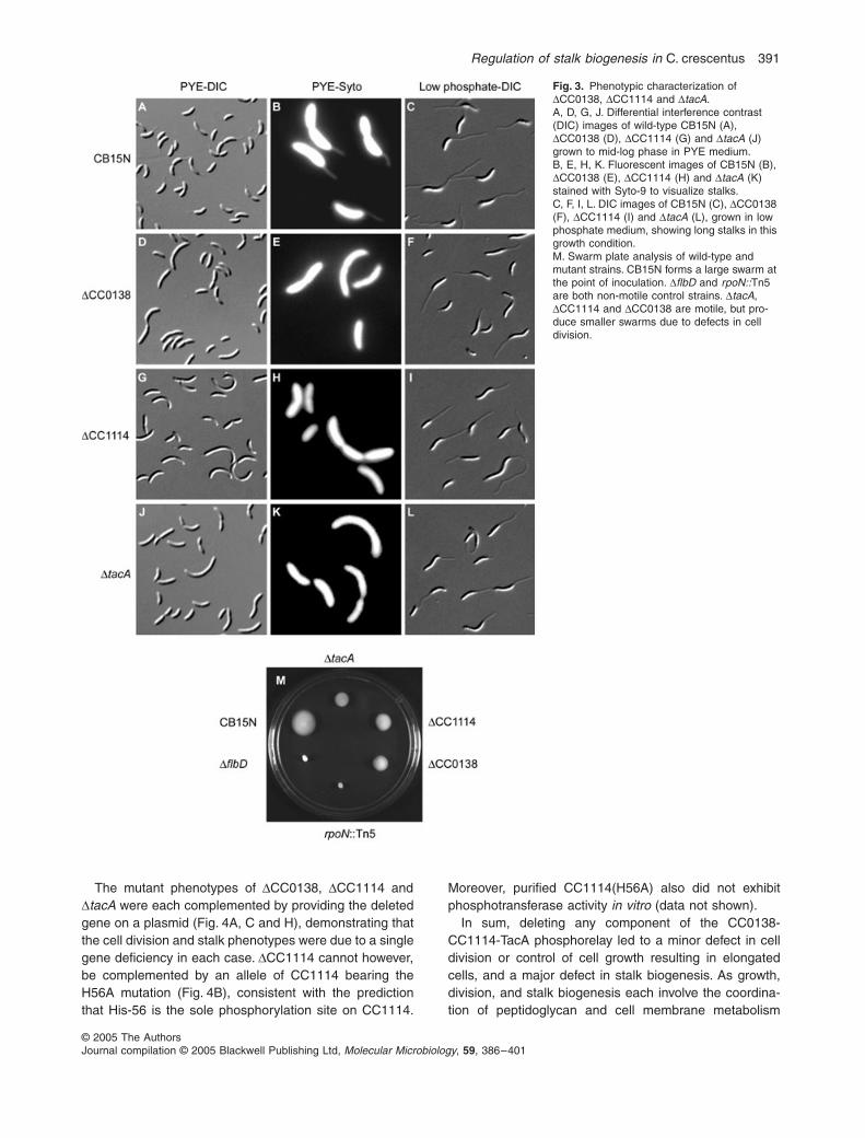
The phosphotransfer experiments suggested thatCC1114 mediates an exclusive phosphorelay which leadsto phosphorylation of TacA. As deletion strains for bothtacA and CC0138 were found previously to lack stalks(Skerker et al., 2005), we predicted that a CC1114 dele-tion should also be stalkless. We therefore constructed astrain in which almost the entire coding region of CC1114was replaced with a tetracycline resistance cassette.Examination of this strain by light microscopy confirmedthat ∆CC1114, like ∆CC0138 and ∆tacA, is stalkless inrich (Fig. 3A, D, G and J) and minimal media (data notshown). However, deletion strains for each phosphorelaycomponent were able to synthesize stalks when grown inlow phosphate medium, a condition known to triggerextensive stalk growth (Poindexter, 1964; 1984; Goninet al., 2000), suggesting that these strains are impaired inthe regulation of stalk biogenesis, not stalk synthesis perse (Fig. 3C, F, I and L). Each deletion strain also appearedmildly filamentous with at least 20% of cells showing obvi-ous cellular elongation relative to wild type, suggesting adefect in cell division or control of cell growth. Motile cellsof ∆CC0138, ∆CC1114 and ∆tacA were easily observedby microscopy, but due to elongation cells did not swimas rapidly as wild type. Consistent with this observation,each phosphorelay mutant showed partial defects in aswarm plate motility assay (Fig. 3M). ∆CC0138, ∆CC1114and ∆tacA each exhibited a swarm size intermediatebetween that of the wild-type CB15N and the non-motilestrains ∆flbD and rpoN::Tn5, presumably due to the inabil-ity of the filamentous, mutant cells to swim efficientlythrough the agar.
Regulation of stalk biogenesis in C. crescentus 391
© 2005 The AuthorsJournal compilation © 2005 Blackwell Publishing Ltd, Molecular Microbiology, 59, 386–401
The mutant phenotypes of ∆CC0138, ∆CC1114 and∆tacA were each complemented by providing the deletedgene on a plasmid (Fig. 4A, C and H), demonstrating thatthe cell division and stalk phenotypes were due to a singlegene deficiency in each case. ∆CC1114 cannot however,be complemented by an allele of CC1114 bearing theH56A mutation (Fig. 4B), consistent with the predictionthat His-56 is the sole phosphorylation site on CC1114.
Moreover, purified CC1114(H56A) also did not exhibitphosphotransferase activity in vitro (data not shown).
In sum, deleting any component of the CC0138-CC1114-TacA phosphorelay led to a minor defect in celldivision or control of cell growth resulting in elongatedcells, and a major defect in stalk biogenesis. As growth,division, and stalk biogenesis each involve the coordina-tion of peptidoglycan and cell membrane metabolism
Fig. 3. Phenotypic characterization of ∆CC0138, ∆CC1114 and ∆tacA.A, D, G, J. Differential interference contrast (DIC) images of wild-type CB15N (A), ∆CC0138 (D), ∆CC1114 (G) and ∆tacA (J) grown to mid-log phase in PYE medium.B, E, H, K. Fluorescent images of CB15N (B), ∆CC0138 (E), ∆CC1114 (H) and ∆tacA (K) stained with Syto-9 to visualize stalks.C, F, I, L. DIC images of CB15N (C), ∆CC0138 (F), ∆CC1114 (I) and ∆tacA (L), grown in low phosphate medium, showing long stalks in this growth condition.M. Swarm plate analysis of wild-type and mutant strains. CB15N forms a large swarm at the point of inoculation. ∆flbD and rpoN::Tn5 are both non-motile control strains. ∆tacA, ∆CC1114 and ∆CC0138 are motile, but pro-duce smaller swarms due to defects in cell division.

392 E. G. Biondi et al.
© 2005 The AuthorsJournal compilation © 2005 Blackwell Publishing Ltd, Molecular Microbiology, 59, 386–401
(Wagner et al., 2005), a single deficiency may underlie allof the morphological phenotypes of ∆CC0138, ∆CC1114and ∆tacA. Moreover, the similarity of mutant phenotypesfor CC0138, CC1114 and tacA supports the notion thatthey act in the same signalling pathway in vivo, as sug-gested by the in vitro results demonstrating a phosphore-lay comprised of these proteins. As the major defect ofthe deletion strains is in stalk biogenesis we have namedCC0138 shkA for stalk biogenesis histidine kinase A andnamed CC1114 shpA for stalk biogenesis histidine phos-photransferase A.
The constitutively active allele tacA(D54E) rescues ∆shkA, ∆shpA and ∆tacA
The data presented thus far suggest that ShkA andShpA function in vivo to phosphorylate TacA. We there-fore predicted that expression of a constitutively activeform of tacA, tacA(D54E), should rescue deletion ofshkA or shpA. In the NtrC family of response regulators,which includes TacA, changing the active site aspartateto a glutamate often mimics phosphorylation of theresponse regulator and hence renders its activation
Fig. 4. Genetic analysis of the TacA phospho-relay. CC0138 = shkA, CC1114 = shpA.A. Deletion of CC1114 results in a stalkless phenotype (Fig. 3G), which is complemented by providing a copy of the CC1114 gene in trans on the plasmid pHXM-CC1114 (ML691). The presence of stalks is indicated by white arrowheads.B. CC1114 bearing a mutation of the active site histidine to an alanine (H56A) is unable to complement stalk formation in vivo.A, C, H. Phosphorelay mutants can be comple-mented by providing the gene in trans on a xylose-inducible plasmid. (A) ML691 (∆shpA + pHXM-shpA), (C) ML690 (∆shkA + pHXM-shkA), (H) ML692 (∆tacA + pHXM-tacA). All strains were grown in PYE + glucose, which provides a low level of expression of the gene being tested. Stalks are indicated by white arrowheads. DIC images of strains grown to mid-log phase.D–I. Expression of tacA(D54E) but not tacA, is able to rescue the stalk defect of both ∆shkA (D vs. E) and ∆shpA (F vs. G). (H–I) Either tacA or tacA(D54E) can rescue a ∆tacA mutant.

Regulation of stalk biogenesis in C. crescentus 393
© 2005 The AuthorsJournal compilation © 2005 Blackwell Publishing Ltd, Molecular Microbiology, 59, 386–401
independent of upstream kinases or phosphorelays(Klose et al., 1993). We therefore placed a tacA(D54E)allele on the vector pJS71 under control of the xylose-inducible promoter PxylX (Meisenzahl et al., 1997) andtransformed this plasmid into the strains ∆shkA, ∆shpAand ∆tacA. Each strain was grown in rich medium sup-plemented with glucose. As the pJS71 vector is a rela-tively high copy vector, growth in the presence of therepressor glucose leads to a low level of expression fromPxylX but prevents overexpression. For ∆shkA, ∆shpA and∆tacA, expression of tacA(D54E) rescued both the cellu-lar elongation and stalk defects of the mutant strains(Fig. 4E, G and I). In contrast, expression of wild-typetacA from the same plasmid rescued ∆tacA (Fig. 4H),but did not rescue the mutant phenotypes of ∆shkA or∆shpA (Fig. 4D and F). Even overexpression of wild-typetacA, by growth in 0.1% xylose, did not rescue the∆shkA and ∆shpA mutants (data not shown). These datasupport the conclusion that the primary in vivo functionof ShkA and ShpA is to phosphorylate, and hence acti-vate, TacA.
Next, we examined the effect of overproducingTacA(D54E). Wild-type cells harbouring the PxylX-tacA(D54E) allele on pJS71 were grown in the presenceof either glucose or xylose. In glucose, these cellsappeared wild type in terms of morphology (Fig. 5A) andgrowth rate (data not shown). However, in xylose the cellsgrew almost twice as slowly as in glucose and rapidlybecame filamentous. Cells had abnormal stalks that wereshorter or longer than wild type and in some casesappeared only as a small bud or protrusion from the cellpole (Fig. 5B). These growth and stalk defects could bethe result of high levels of constitutively active TacA and/or the synthesis of active TacA at the wrong time duringcell cycle progression. Together, the overexpression anddeletion analyses indicate that TacA activity must betightly regulated during the Caulobacter cell cycle toensure proper stalk biogenesis.
Identifying the TacA regulon with DNA microarrays
TacA is a member of the NtrC family of response regu-
Fig. 5. staR (CC2251) is involved in stalk formation.A–D. Overexpression of tacA(D54E) in wild-type Caulobacter (CB15N) leads to cell filamen-tation, and defective stalk formation (A vs. B). Similarly, overexpression of CC2251 leads to filamentation and long stalks (C vs. D). White arrowheads indicate long stalks and open arrowheads indicate defective stalks.E–G. StaR regulates stalk length. DIC images of wild-type CB15N (E) ∆CC2251 (F) and ML760 (CB15N + pLXM-CC2251) in PYE xylose (G) for comparison of stalk length. The deletion mutant ∆CC2251 has stalks approxi-mately 1/2 the length of wild type, whereas overexpression of CC2251 results in stalks which are about twice the length of wild type (see text for details).

394 E. G. Biondi et al.
© 2005 The AuthorsJournal compilation © 2005 Blackwell Publishing Ltd, Molecular Microbiology, 59, 386–401
lators, also known as enhancer-binding proteins (EBPs),which typically function to activate σ54-dependent geneexpression. σ54 is encoded in Caulobacter by the generpoN and is required for both flagellar and stalk biogen-esis (Brun and Shapiro, 1992). FlbD was identified asthe EBP response regulator which works with σ54 in pre-divisional cells to regulate the expression of flagellargenes (Ramakrishnan et al., 1991; Wingrove et al.,1993; Benson et al., 1994; Wingrove and Gober, 1994).Based on our results, we hypothesize that TacA collabo-rates with σ54 to control genes involved in regulatingstalk biogenesis. To identify possible target genes, weused whole genome DNA microarrays to interrogate theexpression patterns of TacA phosphorelay mutants(∆shkA, ∆shpA, ∆tacA) and a rpoN disruption mutantrpoN::Tn5 (Fig. 6). To partition the σ54 regulon into FlbD-
and TacA-dependent genes, we also examined a ∆flbDstrain (Fig. 6).
Strains were grown to mid-log phase (OD600 ∼0.4) in richmedium, then RNA was harvested and used to probewhole-genome DNA microarrays. Each mutant strain wascompared with the wild-type CB15N and each comparisondone in duplicate or triplicate with results averaged.Genes whose expression level changed at least twofoldin rpoN::Tn5 and in any one of the phosphorelay or flbDmutants were selected for further analysis (Fig. 6). Visualinspection of expression profiles for each mutant relativeto wild type, clearly demonstrated the similarity of ∆shkA,∆shpA, ∆tacA and rpoN::Tn5 (Fig. 6). Correlation coeffi-cients for each pairwise comparison among these mutantswere greater than 0.73. Similarly high correlation coeffi-cients have been seen for mutants of other two-compo-
Fig. 6. Expression profiles of ∆shkA, ∆shpA, ∆tacA, rpoN::Tn5 and ∆flbD. Colours, as indicated in the legend, represent upregulation (yellow) or downregulation (blue) in comparison with expression in wild-type cells.A. Correlation between expression profiles of ∆shkA, ∆shpA, ∆tacA (see text for details). Columns, corresponding to ∆shkA, ∆shpA, ∆tacA, rpoN mutant and ∆flbD, show only genes upregulated or downregulated more than twofold in at least one of the mutants. For complete data, see Table S1.B. Genes consistently downregulated in phosphorelay mutants and rpoN form a putative TacA regulon, and genes consistently downregulated in rpoN and ∆flbD the FlbD regulon.

Regulation of stalk biogenesis in C. crescentus 395
© 2005 The AuthorsJournal compilation © 2005 Blackwell Publishing Ltd, Molecular Microbiology, 59, 386–401
nent signalling genes which function in the same pathway(Jacobs et al., 2003). These results thus add further sup-port to the biochemical and genetic data demonstratingthat ShkA-ShpA-TacA constitutes a phosphorelay whichcontrols gene expression through phosphorylation ofTacA.
We predicted that the TacA target genes involved instalk biogenesis would be similarly affected in ∆shkA,∆shpA, ∆tacA and rpoN::Tn5, each of which lacks stalks,but unchanged in the ∆flbD mutant which is non-motile,but retains stalks. In total, 30 genes satisfied these criteria,two of which are consistently downregulated in each stalk-less mutant and 28 upregulated in each. The two down-regulated genes contained σ54 binding sites (TGGCCC-N5-TTGC) (Wu et al., 1995) in their upstream regulatory
regions, suggesting that each is a direct target of TacAand σ54. CC3218 has no predicted function based onsequence homology but is transcriptionally cell cycle-reg-ulated (Laub et al., 2000) with expression highest instalked cells and again in late predivisional cells. The otherputative TacA target, CC2251, is predicted to encode atranscription factor of the Cro/CI family. During wild-typecell cycle progression CC2251 mRNA levels peak in pre-divisional cells, shortly after tacA expression peaks, sug-gesting that these regulators may form a transcriptionalcascade which controls stalk biogenesis (Fig. 7).
Although EBP response regulators such as TacA typi-cally activate gene expression, some can directly repressgenes, independent of σ54 (Wingrove et al., 1993; Bensonet al., 1994; Wingrove and Gober, 1994). Hence to identify
Fig. 7. Model of the genetic circuitry regulating polar morphogenesis in C. crescentus.A. Transcriptional regulation of stalk and flagellar genes. Phosphorylated CtrA activates transcription of rpoN (σ54) and tacA. σ54 and FlbD activate transcription of flagellar genes whereas σ54 and phosphorylated TacA activate transcription of stalk biogenesis genes including staR. A phospho-relay, composed of ShkA (CC0138) and ShpA (CC1114), phosphorylates TacA.B. Cell cycle-regulated transcription of genes involved in regulation of stalk and flagellum biogenesis (see text for details).

396 E. G. Biondi et al.
© 2005 The AuthorsJournal compilation © 2005 Blackwell Publishing Ltd, Molecular Microbiology, 59, 386–401
genes which may be directly repressed by TacA, wesearched for genes whose expression was significantlyupregulated in ∆tacA, but unaffected in the rpoN mutant(Table S1). The only gene fitting these criteria wasCC3314, which is annotated as a ‘conserved hypothetical’and reads divergently from tacA itself. All other genes aresimilarly upregulated in rpoN::Tn5 and each of the phos-phorelay mutants, suggesting that they are regulated indi-rectly by TacA, perhaps through the transcription factorCC2251. The vast majority of these genes have no anno-tated function, but their roles, if any, in stalk biogenesiscan now be explored.
staR (CC2251) expression is regulated by the TacA phosphorelay and regulates stalk biogenesis
Microarray analysis identified at least two genes, CC2251and CC3218, as probable direct targets of TacA. To deter-mine their roles, if any, in stalk biogenesis, we constructeddeletion and overexpression strains for each gene. Dele-tion and overexpression of CC3218 had no obvious phe-notype (data not shown). Deletion of CC2251 did noteliminate stalks, but the average stalk length for ∆CC2251cells (0.68 µm ± 0.24, Fig. 5F) was significantly shorterthan wild type (1.26 µm ± 0.32, Fig. 5E). Conversely, over-expression of CC2251 from a xylose-inducible promoteron the low-copy vector pMR20 resulted in cells with stalksthat were highly variable and longer on average(1.95 µm ± 0.89, Fig. 5G) than wild type. Overexpressionof CC2251 from a high-copy (pJS71) vector also producedlonger stalks in addition to a growth defect and filamen-tous cells (Fig. 5D). This growth defect and filamentationwas similar to the phenotype of TacA(D54E) overexpres-sion (Fig. 5B), consistent with CC2251 being a direct tar-get of TacA. Based on these observations we have namedCC2251 staR for stalk biogenesis regulator. We notethough, that because ∆CC2251 does not precisely phe-nocopy ∆tacA, there must be other TacA target geneswhich also help to control stalk biogenesis. Candidatesfrom the DNA microarray data will need to be examinedto fully account for the stalkless phenotype of tacA, shkAand shpA mutants.
Discussion
Stalk biogenesis in Caulobacter
For Caulobacter, a developmental program is tightly cou-pled to cell cycle progression. This development includesthree major morphogenetic events, biogenesis of a flagel-lum, polar pili and a stalk. While the regulation of flagellumassembly and, to a lesser extent, pili biogenesis havebeen worked out in mechanistic detail (Gober andEngland, 2000; Skerker and Shapiro, 2000; Viollier et al.,
2002a,b), the regulation of stalk biogenesis has remainedpoorly understood. Here, we have identified a key signaltransduction pathway responsible for stalk formation dur-ing the cell cycle and have begun outlining the transcrip-tional pathways involved. This genetic circuitry issummarized in Fig. 7A.
At the heart of this model is σ54 which is necessary forboth flagellum and stalk biogenesis. As in other bacteria,σ54-dependent gene expression in Caulobacter requiresan activator of the NtrC-family of response regulators, alsoknown as enhancer-binding-proteins, or EBPs. Typically,phosphorylation of an EBP enables it to stimulate theclosed-to-open complex transition of a promoter bound byRNA polymerase containing a σ54 subunit, thereby cou-pling activation of the signalling pathway to changes ingene expression (Kustu et al., 1989). The EBP responseregulator FlbD is induced at the same time as RpoN, andtogether they regulate genes involved in flagellar biogen-esis (Fig. 7B) (Wingrove et al., 1993; Benson et al., 1994;Wingrove and Gober, 1994). We have now identified TacAas the EBP which collaborates with RpoN to regulategenes involved in stalk biogenesis. Furthermore, we iden-tified a phosphorelay which regulates stalk biogenesis bymodulating the phosphorylation state of TacA: ShkA, ahybrid histidine kinase, first autophosphorylates and theninitiates a phosphorelay in which a phosphoryl group ispassed to its own receiver domain, then to the histidinephosphotransferase ShpA, and finally to the transcrip-tional regulator TacA (Fig. 7).
DNA microarray analysis identified two genes controlledby TacA which may help regulate stalk biogenesis:CC2251 (staR) and CC3218. The mRNAs for both staRand CC3218 are cell cycle-regulated with induction in latepredivisional cells, immediately after the induction of tacA(M. T. Laub, unpubl. data) (Fig. 7B). StaR appears to playa role in controlling stalk length, as the deletion strain hasstalks shorter than wild type, and overexpression leads tolonger stalks (Fig. 5). However, as staR mutants do notcompletely lack stalks or exhibit cellular elongation liketacA mutants, other TacA targets remain to be identifiedand characterized. The microarray data presented herecan now be explored to find these additional targets.
The coupling of stalk biogenesis to cell cycle progres-sion is accomplished in large part by CtrA, the master cellcycle regulator. CtrA is regulated at multiple levels duringcell cycle progression such that activity drops during theG1-S transition and then rapidly returns to peak levels asS phase proceeds (Domian et al., 1997). CtrA triggersboth flagellum and stalk biogenesis by directly activatingrpoN (σ54) and tacA (Laub et al., 2002). Taken together,we propose that stalk biogenesis, like flagellum biogene-sis, is regulated by a transcriptional cascade: CtrA isinduced after DNA replication initiation and directly acti-vates synthesis of σ54 and TacA, which together activate

Regulation of stalk biogenesis in C. crescentus 397
© 2005 The AuthorsJournal compilation © 2005 Blackwell Publishing Ltd, Molecular Microbiology, 59, 386–401
the expression of stalk regulatory genes, including thetranscription factor staR, which in turn probably controlsthe expression of yet other genes required for stalkbiogenesis.
Precisely how PleC, another histidine kinase requiredfor stalk biogenesis, feeds into the stalk regulatory circuitremains unknown. However, suppressor analyses placeCtrA downstream of PleC, suggesting that the effect maybe through regulation of CtrA activity (Ohta et al., 1992;Wu et al., 1998).
Stalk biogenesis is a cell cycle-regulated event andinitially occurs in only one of the two daughter cells pro-duced by cell division, raising the possibility that TacAactivity is controlled spatially as well as temporally. FlbD,for example, is phosphorylated and active only in thenascent swarmer cell, where it activates expression of lateflagellar genes and represses early flagellar genes (Goberand England, 2000). In the nascent stalked cell, FlbD ispresent but inactive; this pool of FlbD will be activated asthe stalked cell develops, initiating early flagellar geneexpression in the predivisional cell. Whether TacA activityis also asymmetrically regulated in a manner that restrictsstalk biogenesis to a single daughter cell, the stalked cell,remains to be explored.
What signals lead to activation of TacA?
Why does Caulobacter use a phosphorelay to controlstalk biogenesis? It has been suggested that phosphore-lays allow the integration of multiple signals by providingnumerous points of control. In Bacillus subtilis, a numberof signals impinge on the phosphorelay controlling sporu-lation initiation by affecting the phosphorylation of differ-ent pathway components (Perego et al., 1994). Stalkbiogenesis is a major developmental event in the life cycleof Caulobacter, and hence may also integrate a widerange of signals. However, the nature of the signal(s)controlling the ShkA-ShpA-TacA phosphorelay is not yetknown. ShkA is predicted to be a soluble, cytoplasmickinase, suggesting that the phosphorelay may respond toan internal cell cycle cue. Caulobacter thus appears tohave at least two independent pathways for regulatingstalk biogenesis, one dependent on an external cue,phosphate starvation, and one linked to cell cycleprogression.
Identification of a histidine phosphotransferase
ShkA-ShpA-TacA is the first phosphorelay identified inC. crescentus as ShpA is the first histidine phosphotrans-ferase identified in this organism. The C. crescentusgenome likely encodes other histidine phosphotrans-ferases. CC1220, identified here in the same computa-tional screen as ShpA, is one candidate, but others almost
certainly exist, particularly given that the Caulobactergenome encodes 27 hybrid histidine kinases. Hybridkinases often function in phosphorelays like the onedescribed here; however, in some cases the C-terminalreceiver domain of a hybrid kinase may function as anauto-inhibitory domain. Intramolecular phosphorylationmay relieve this inhibition, allowing the kinase to phospho-rylate another, diffusible response regulator, as with theAgrobacterium tumefaciens hybrid kinase VirA (Changet al., 1996).
Rapid, systematic mapping of complex phosphorylation cascades
To identify the probable in vivo cognate substrates forShpA, we adapted a technique called phosphotransferprofiling (Skerker et al., 2005). By examining phospho-transfer from a kinase to each response regulator in par-allel, and at multiple time points, kinetic preference isrevealed and identifies the probable in vivo targets of thekinase (Skerker et al., 2005). Here, we hypothesized thathistidine phosphotransferases would similarly exhibit akinetic preference with respect to both soluble responseregulators and the receiver domains of hybrid histidinekinases. A previous smaller scale study of two phospho-relays also suggested that histidine phosphotransferasesexhibit kinetic preference in vitro (Perraud et al., 1998).Our phosphotransfer profiling experiments indeed dem-onstrated a kinetic preference for ShpA to transfer phos-phoryl groups to or from the receiver domains of ShkAand TacA. Our genetic analyses (Figs 3 and 4) and DNAmicroarray results (Fig. 6) confirmed the in vivo rele-vance of these interactions. We suggest that histidinephosphotransferases in general may exhibit a globalkinetic preference for interaction with their cognate sub-strates, similar to what has been observed for histidinekinases.
The global kinetic preference of histidine kinases fortheir cognate response regulator substrates means thatthe specificity of two-component signal transduction path-ways is determined largely at a biochemical level; otherfactors probably function primarily to enhance this inher-ent specificity. Our results with ShpA suggest that theseobservations extend to histidine phosphotransferasesand are general properties of two-component signallingpathways. We thus anticipate that our phosphotransferprofiling technique will be generally applicable to themapping of phosphorelays, both in Caulobacter as wellas in other organisms. Two-component signalling sys-tems are prevalent throughout the bacterial kingdom aswell as being present in fungi and plants (Stock et al.,2000), and the mapping of pathway connectivity amonglarge numbers of signalling components remains a majorchallenge.

398 E. G. Biondi et al.
© 2005 The AuthorsJournal compilation © 2005 Blackwell Publishing Ltd, Molecular Microbiology, 59, 386–401
Concluding remarks
Many organisms couple developmental programs andmorphogenesis to cell cycle progression. In the buddingyeast S. cerevisiae, mating requires a morphologicalchange called shmooing which prepares haploid cells forfusion. This morphological event is tied to cell cycle pro-gression and can only occur during G1 phase. ForB. subtilis the initiation of sporulation, a complex develop-mental program, is also tightly coupled to cell cycle events(Burkholder et al., 2001). The morphological events offlagellar, pili and stalk biogenesis in Caulobacter are sim-ilarly dependent on cell cycle progression. The identifica-tion of a complete signalling pathway controlling stalkbiogenesis now opens the way to a more complete under-standing of how this key morphological event is regulatedin space and time during the Caulobacter cell cycle.
Experimental procedures
Bacterial strains, plasmids and growth conditions
Caulobacter crescentus strains were grown in peptone-yeastextract (PYE, rich medium) or M2G (minimal medium) at30°C (Ely, 1991), supplemented with 3% sucrose, tetracy-cline (1 µg ml−1), kanamycin (25 µg ml−1), spectinomycin(25 µg ml−1), 0.1% glucose, or 0.1% xylose, as required. Low-phosphate growth experiments were performed as previouslydescribed (Quon et al., 1996). E. coli strains were grown at30°C or 37°C in Luria–Bertani broth supplemented withcarbenicillin (100 µg ml−1), chloramphenicol (30 µg ml−1), tet-racycline (10 µg ml−1), kanamycin (50 µg ml−1) or spectinomy-cin (50 µg ml−1) as necessary. PYE swarm plates contained0.3% bacto agar. Plasmids were transformed intoC. crescentus by electroporation. Tetracycline marked dele-tion strains for CC1114, CC2251 and CC3218 were gener-ated by a two-step recombination protocol (Skerker et al.,2005). pENTR clones of the 27 hybrid kinase receiverdomains were constructed as previously described (Skerkeret al., 2005). For in vivo expression of Caulobacter genes,pENTR plasmids for CC0138, CC1114, CC1114 (H56A),TacA, TacA(D54E), CC2251 and CC3218 were recombinedwith either a high-copy destination vector pHXM-DEST or alow-copy destination vector pLXM-DEST (Table S2), as nec-essary, to generate xylose-inducible constructs (Table S2).For overexpression experiments in Caulobacter, usingpHXM-based vectors, saturated overnight cultures grown inPYE plus glucose were washed twice in PYE medium andthen diluted to OD600 = 0.03 in either PYE plus glucose orxylose. For experiments using the low copy vector pLXM-CC2251 samples were grown overnight in PYE plus xyloseprior to analysis.
Strains and plasmids generated are listed in Table S2.Primers and plasmids used to generate deletion strains,pENTR clones and point mutations are listed in Table S3.Site-directed mutagenesis was performed using theQuikChange protocol (Stratagene), with pENTR clones astemplates. Morphology of mid-log phase cultures wasimaged as described previously using DIC microscopy
(Skerker et al., 2005). Stalks were visualized by staining with5 µM Syto-9 (Molecular Probes).
Sequence analysis and homology modelling
Alpha-helical content was estimated using the Garnier algo-rithm within EMBOSS (http://emboss.sourceforge.net). Astructure-based multiple sequence alignment of CC1114against other known phosphotransferases was constructedusing 3D-Coffee (Poirot et al., 2004). This alignment wasused as input to Swiss-Model (Schwede et al., 2003) to pre-dict the structure of CC1114 using the following templates:ArcB (PDB: 1FR0), Ypd1 (PDB: 1QSP) and LuxU (PDB:1Y6D). ArcB provided the best alignment and the predictedCC1114 structure was analysed and displayed using UCSFChimera (Pettersen et al., 2004).
Gateway cloning and protein purification
Escherichia coli thioredoxin (TRX)-His6 expression plasmidsfor all 44 response regulators and 27 hybrid kinase receiverdomains were generated using recombinational cloning andpurified as described previously (Skerker et al., 2005).CC1114 and CC1114(H56A) were expressed and purified asN-terminal His6-tagged proteins using the destination vector,pHIS-DEST. To construct pHIS-DEST, pET15b (Novagen)was digested with NdeI, blunted with T4 DNA polymerase,and the RfC Gateway vector conversion cassette (Invitrogen)was inserted. A His6-version of the TacA receiver domain(TacA-RD) was also constructed and purified, using pHIS-DEST. TacA-RD yielded a cleaner purification, and actedidentical to full-length TacA in our assays, so we substitutedthis protein as necessary. Entry clones of the kinase domainsof CckA, CC0138, CC3102 were recombined into pTRX-HIS-DEST or pHIS-MBP-DEST (for CC3219), and purified aseither TRX-His6 or His6-MBP fusions. All purified proteinswere stored in HKEDG buffer (10 mM HEPES-KOH, pH 8.0,50 mM KCl, 10% glycerol, 0.1 mM EDTA, 1 mM DTT) andconcentrations were normalized by densitometry.
Phosphotransfer profiling and phosphorelay biochemistry
We used a modified phosphotransfer profiling method(Skerker et al., 2005) to identify the cognate substrates forCC1114 (Fig. 2C and D, Fig. S2). All reactions and dilutionswere performed in HKEDG buffer plus 5 mM MgCl2. First, wegenerated CC1114∼P by incubating 10 µM His6-CC1114 with1 µM TRX-His6-CC0138-HK and 1 µM TRX-His6-CC0138-RDplus 1 µCi µl−1 [γ-32P]ATP (∼6000 Ci mmol−1, Amersham Bio-sciences) for 30 min at 30°C. This reaction was then diluted10-fold, and 5 µl of the mixture was added to 5 µl of bufferalone (as a control) or 5 µl of each purified response regula-tor or hybrid receiver domain (diluted to 5 µM) with incuba-tions for 2 min at room temperature. The final concentrationswere 0.5 µM for CC1114, and 2.5 µM for the phosphotransfersubstrates. To demonstrate phosphorylation of CC1114 byvarious hybrid kinases (Fig. 2A), the following four autophos-phorylation reactions (30 µl total volume) were performed: (i)TRX-His6-CckA (6 µM), (ii) TRX-His6-CC0138-HK (0.5 µM),(iii) TRX-His6-CC3102-HK (4.5 µM), or (iv) His6-MBP-

Regulation of stalk biogenesis in C. crescentus 399
© 2005 The AuthorsJournal compilation © 2005 Blackwell Publishing Ltd, Molecular Microbiology, 59, 386–401
CC3219-HK (20 µM) were incubated for 30 min at 30°C afteraddition of 30 µCi [γ-32P]ATP. Then, 1 µl of reaction A, 1 µl ofreaction B, 4 µl of reaction C, and 9 µl of reaction D weremixed with 1 µM of the corresponding receiver domains ofeach histidine kinase, and, as necessary, with 10 µM of His6-CC1114 for 2 or 5 min, in a total volume of 10 µl. For analysisof CC1114 activity (Fig. 2B), reactions were performed thesame as for phosphotransfer profiling except that variouscomponents were omitted before incubating 30 min at 30°C.For analysis of the complete TacA phosphorelay and kinetics(Fig. 2E and G), CC1114∼P was prepared as above, diluted,and then mixed with 5 µl of 5 µM His6-TacA-RD or otherhybrid kinase receiver domains. All phosphotransfer reac-tions were stopped with 3.5 µl of 4 × SDS-PAGE samplebuffer and analysed using 12% or 15% Tris-HCl gels asdescribed previously (Skerker et al., 2005).
Microarray analysis of the TacA regulon
Strains were grown in PYE and harvested at OD600 = 0.4.RNA preparation was performed using the RNA-easy Kit(Qiagen). DNA arrays contained 50mer probes for each genein the Caulobacter genome (see Table S1 for sequences).Three or more independent cultures were used for eachexperiment and each sample was labelled either with Cy5-dCTP or Cy3-dCTP (Amersham Biosciences). Hybridiza-tions, scanning and data processing were performed asdescribed previously (Laub et al., 2002) except that hybrid-izations contained 30% formamide and were incubated at44°C for 6 h.
Acknowledgements
We gratefully acknowledge support from the Office of Sci-ence (BER), US Department of Energy, Grants No. DE-FG03-01ER63219 and DE-FG02-04ER63922. Support alsoprovided in part by a National Institutes of Health grant toM.T.L. and the Bauer Center for Genomics Research atHarvard University.
References
Benson, A.K., Ramakrishnan, G., Ohta, N., Feng, J., Ninfa,A.J., and Newton, A. (1994) The Caulobacter crescentusFlbD protein acts at ftr sequence elements both to activateand to repress transcription of cell cycle-regulated flagellargenes. Proc Natl Acad Sci USA 91: 4989–4993.
Brun, Y.V., and Shapiro, L. (1992) A temporally controlledsigma-factor is required for polar morphogenesis and nor-mal cell division in Caulobacter. Genes Dev 6: 2395–2408.
Burkholder, W.F., Kurtser, I., and Grossman, A.D. (2001)Replication initiation proteins regulate a developmentalcheckpoint in Bacillus subtilis. Cell 104: 269–279.
Chang, C.H., Zhu, J., and Winans, S.C. (1996) Pleiotropicphenotypes caused by genetic ablation of the receivermodule of the Agrobacterium tumefaciens VirA protein. JBacteriol 178: 4710–4716.
Domian, I.J., Quon, K.C., and Shapiro, L. (1997) Cell type-specific phosphorylation and proteolysis of a transcrip-
tional regulator controls the G1-to-S transition in a bacterialcell cycle. Cell 90: 415–424.
Ely, B. (1991) Genetics of Caulobacter crescentus. MethodsEnzymol 204: 372–384.
Georgellis, D., Kwon, O., De Wulf, P., and Lin, E.C. (1998)Signal decay through a reverse phosphorelay in the Arctwo-component signal transduction system. J Biol Chem273: 32864–32869.
Gober, J.W., and England, J.C. (2000) Regulation of flagel-lum biosynthesis and motility in caulobacter. In ProkaryoticDevelopment. Brun, Y.V., and Shimkets, L.J. (eds). Wash-ington, DC: American Society for Microbiology Press, pp.319–339.
Gonin, M., Quardokus, E.M., O’Donnol, D., Maddock, J., andBrun, Y.V. (2000) Regulation of stalk elongation by phos-phate in Caulobacter crescentus. J Bacteriol 182: 337–347.
Hoch, J.A., and Silhavy, T.J. (eds). (1995) Two-ComponentSignal Transduction. Washington, DC: American Societyfor Microbiology Press.
Jacobs, C., Ausmees, N., Cordwell, S.J., Shapiro, L., andLaub, M.T. (2003) Functions of the CckA histidine kinasein Caulobacter cell cycle control. Mol Microbiol 47: 1279–1290.
Kato, M., Mizuno, T., Shimizu, T., and Hakoshima, T. (1997)Insights into multistep phosphorelay from the crystal struc-ture of the C-terminal HPt domain of ArcB. Cell 88: 717–723.
Klose, K.E., Weiss, D.S., and Kustu, S. (1993) Glutamate atthe site of phosphorylation of nitrogen-regulatory proteinNTRC mimics aspartyl-phosphate and activates the pro-tein. J Mol Biol 232: 67–78.
Kurtz, H.D., Jr, and Smit, J. (1994) The Caulobacter crescen-tus holdfast: identification of holdfast attachment complexgenes. FEMS Microbiol Lett 116: 175–182.
Kustu, S., Santero, E., Keener, J., Popham, D., and Weiss,D. (1989) Expression of sigma 54 (ntrA)-dependent genesis probably united by a common mechanism. Microbiol Rev53: 367–376.
Laub, M.T., McAdams, H.H., Feldblyum, T., Fraser, C.M.,and Shapiro, L. (2000) Global analysis of the genetic net-work controlling a bacterial cell cycle. Science 290: 2144–2148.
Laub, M.T., Chen, S.L., Shapiro, L., and McAdams, H.H.(2002) Genes directly controlled by CtrA, a master regula-tor of the Caulobacter cell cycle. Proc Natl Acad Sci USA99: 4632–4637.
Meisenzahl, A.C., Shapiro, L., and Jenal, U. (1997) Isolationand characterization of a xylose-dependent promoter fromCaulobacter crescentus. J Bacteriol 179: 592–600.
Nierman, W.C., Feldblyum, T.V., Laub, M.T., Paulsen, I.T.,Nelson, K.E., Eisen, J., et al. (2001) Complete genomesequence of Caulobacter crescentus. Proc Natl Acad SciUSA 98: 4136–4141.
Ohta, N., Lane, T., Ninfa, E.G., Sommer, J.M., and Newton,A. (1992) A histidine protein kinase homologue requiredfor regulation of bacterial cell division and differentiation.Proc Natl Acad Sci USA 89: 10297–10301.
Ohta, N., Grebe, T.W., and Newton, A. (2000) Signal trans-duction and cell cycle checkpoints in developmental regu-lation of Caulobacter. In Prokaryotic Development. Brun,

400 E. G. Biondi et al.
© 2005 The AuthorsJournal compilation © 2005 Blackwell Publishing Ltd, Molecular Microbiology, 59, 386–401
Y.V., and Shimkets, L.J. (eds). Washington, DC: AmericanSociety for Microbiology Press, pp. 341–359.
Perego, M., Hanstein, C., Welsh, K.M., Djavakhishvili, T.,Glaser, P., and Hoch, J.A. (1994) Multiple protein-aspar-tate phosphatases provide a mechanism for the integrationof diverse signals in the control of development in B. sub-tilis. Cell 79: 1047–1055.
Perraud, A.L., Kimmel, B., Weiss, V., and Gross, R. (1998)Specificity of the BvgAS and EvgAS phosphorelay is medi-ated by the C-terminal HPt domains of the sensor proteins.Mol Microbiol 27: 875–887.
Pettersen, E.F., Goddard, T.D., Huang, C.C., Couch, G.S.,Greenblatt, D.M., Meng, E.C., and Ferrin, T.E. (2004)UCSF Chimera – a visualization system for exploratoryresearch and analysis. J Comput Chem 25: 1605–1612.
Poindexter, J.S. (1964) Biological properties and classifica-tion of the Caulobacter group. Bacteriol Rev 28: 231–295.
Poindexter, J.S. (1978) Selection for nonbuoyant morpholog-ical mutants of Caulobacter crescentus. J Bacteriol 135:1141–1145.
Poindexter, J.S. (1984) The role of calcium in stalk develop-ment and in phosphate acquisition in Caulobacter crescen-tus. Arch Microbiol 138: 140–152.
Poirot, O., Suhre, K., Abergel, C., O’Toole, E., and Notre-dame, C. (2004) 3DCoffee@igs: a web server for combin-ing sequences and structures into a multiple sequencealignment. Nucleic Acids Res 32: W37–W40.
Quon, K.C., Marczynski, G.T., and Shapiro, L. (1996) Cellcycle control by an essential bacterial two-component sig-nal transduction protein. Cell 84: 83–93.
Ramakrishnan, G., Zhao, J.L., and Newton, A. (1991) Thecell cycle-regulated flagellar gene flbF of Caulobacter cres-centus is homologous to a virulence locus (lcrD) of Yersiniapestis. J Bacteriol 173: 7283–7292.
Ramakrishnan, G., Zhao, J.L., and Newton, A. (1994) Multi-ple structural proteins are required for both transcriptionalactivation and negative autoregulation of Caulobacter cres-centus flagellar genes. J Bacteriol 176: 7587–7600.
Ryan, K.R., and Shapiro, L. (2003) Temporal and spatialregulation in prokaryotic cell cycle progression and devel-opment. Annu Rev Biochem 72: 367–394.
Schwede, T., Kopp, J., Guex, N., and Peitsch, M.C. (2003)SWISS-MODEL: an automated protein homology-model-ing server. Nucleic Acids Res 31: 3381–3385.
Skerker, J.M., and Shapiro, L. (2000) Identification and cellcycle control of a novel pilus system in Caulobacter cres-centus. EMBO J 19: 3223–3234.
Skerker, J.M., and Laub, M.T. (2004) Cell-cycle progressionand the generation of asymmetry in Caulobacter crescen-tus. Nat Rev Microbiol 2: 325–337.
Skerker, J.M., Prasol, M.S., Perchuk, B.S., Biondi, E.G., andLaub, M.T. (2005) Two-component signal transductionpathways regulating growth and cell cycle progression in abacterium: a system-level analysis. PLoS Biol 3: e334.
Smith, C.S., Hinz, A., Bodenmiller, D., Larson, D.E., andBrun, Y.V. (2003) Identification of genes required for syn-thesis of the adhesive holdfast in Caulobacter crescentus.J Bacteriol 185: 1432–1442.
Sommer, J.M., and Newton, A. (1989) Turning off flagellumrotation requires the pleiotropic gene pleD: pleA, pleC, and
pleD define two morphogenic pathways in Caulobactercrescentus. J Bacteriol 171: 392–401.
Song, H.K., Lee, J.Y., Lee, M.G., Moon, J., Min, K., Yang,J.K., and Suh, S.W. (1999) Insights into eukaryotic multi-step phosphorelay signal transduction revealed by thecrystal structure of Ypd1p from Saccharomyces cerevisiae.J Mol Biol 293: 753–761.
Stock, A.M., Robinson, V.L., and Goudreau, P.N. (2000) Two-component signal transduction. Annu Rev Biochem 69:183–215.
Uhl, M.A., and Miller, J.F. (1996) Central role of the BvgSreceiver as a phosphorylated intermediate in a complextwo-component phosphorelay. J Biol Chem 271: 33176–33180.
Ulrich, D.L., Kojetin, D., Bassler, B.L., Cavanagh, J., andLoria, J.P. (2005) Solution structure and dynamics of LuxUfrom Vibrio harveyi, a phosphotransferase protein involvedin bacterial quorum sensing. J Mol Biol 347: 297–307.
Viollier, P.H., Sternheim, N., and Shapiro, L. (2002a) Identi-fication of a localization factor for the polar positioning ofbacterial structural and regulatory proteins. Proc Natl AcadSci USA 99: 13831–13836.
Viollier, P.H., Sternheim, N., and Shapiro, L. (2002b) Adynamically localized histidine kinase controls the asym-metric distribution of polar pili proteins. EMBO J 21: 4420–4428.
Wagner, J.K., Galvani, C.D., and Brun, Y.V. (2005) Caulo-bacter crescentus requires RodA and MreB for stalk syn-thesis and prevention of ectopic pole formation. J Bacteriol187: 544–553.
Wingrove, J.A., and Gober, J.W. (1994) A sigma 54 transcrip-tional activator also functions as a pole-specific repressorin Caulobacter. Genes Dev 8: 1839–1852.
Wingrove, J.A., Mangan, E.K., and Gober, J.W. (1993) Spa-tial and temporal phosphorylation of a transcriptionalactivator regulates pole-specific gene expression in Cau-lobacter. Genes Dev 7: 1979–1992.
Wu, J., Benson, A.K., and Newton, A. (1995) Global regula-tion of a sigma 54-dependent flagellar gene family in Cau-lobacter crescentus by the transcriptional activator FlbD. JBacteriol 177: 3241–3250.
Wu, J., Ohta, N., and Newton, A. (1998) An essential, multi-component signal transduction pathway required for cellcycle regulation in Caulobacter. Proc Natl Acad Sci USA95: 1443–1448.
Xu, Q., and West, A.H. (1999) Conservation of structure andfunction among histidine-containing phosphotransfer (HPt)domains as revealed by the crystal structure of YPD1. JMol Biol 292: 1039–1050.
Supplementary material
The following supplementary material is available for thisarticle online:Fig. S1. Alignment of putative CC1114 orthologues from var-ious alpha-proteobacteria. Putative CC1114 orthologueswere identified by reciprocal best BLAST analysis. A multiplesequence alignment was performed using T-COFFEE anddrawn using BOXSHADE. Conserved residues are shadedblack and marked with an asterisk. Residues which are sim-ilar are shaded grey and marked by a period. The sequences

Regulation of stalk biogenesis in C. crescentus 401
© 2005 The AuthorsJournal compilation © 2005 Blackwell Publishing Ltd, Molecular Microbiology, 59, 386–401
were obtained from the following genomes: Agrobacteriumtumefaciens C58 (gi17935249), Sinorhizobium meliloti 1021(gi15074749), Bradyrhizobium japonicum USDA 110(gi27380997), Rhodopseudomonas palustris CGA009(gi39937018) and Caulobacter crescentus CB15(gi25399659). Sequences GenBank accession numbers arein parenthesis.Fig. S2. Phosphotransfer profiling method applied to histi-dine phosphotransferases.A. Reactions involved in phosphotransfer profiling.B. Schematic of phosphotransfer profiling method as appliedto histidine phosphotransferases (HPTs).Fig. S3. Purification of the receiver domains from all 27hybrid kinases encoded in the Caulobacter genome SDS-PAGE analysis of purified TRX-His6-TEV fusions to each ofthe 27 receiver domains derived from hybrid kinases (see
Experimental procedures and Table S2). A molecular weightladder is marked with sizes in kDa. Approximately 500 ng ofpurified receiver domain was analysed and normalizedagainst an equivalent amount of BSA. Gene names are indi-cated by their CC number and the predicted molecular weightof each fusion is indicated in Daltons. Six of the 27 receiverdomains purified poorly (CC0723, CC0921, CC2521,CC2632, CC3191, CC3560), although a band correspondingto the correct molecular weight is still present in each case.Table S1. DNA microarray data and probes.Table S2. Strains and plasmids.Table S3. Primers for generating pENTR clones, deletionstrains and site-directed mutants.
This material is available as part of the online article fromhttp://www.blackwell-synergy.com