Retinal ganglion cell-derived sonic hedgehog signaling is required for optic disc and stalk...
14
INTRODUCTION Two distinct inductive events govern vertebrate eye development: an initial morphogenetic process results in regional specification of the various eye structures, and cells in these defined anatomic domains then differentiate to acquire their respective fates and functions in the mature eye (Pei and Rhodin, 1970). In mice, the morphogenetic phase begins at about embryonic day 8.5 (E8.5) with the lateral outgrowth of the prosencephalon to form the optic vesicles. By mid- gestation, the optic vesicle contacts and induces the formation of a lens placode from the overlying surface ectoderm, and simultaneously invaginates to form the bilayered optic cup, which is connected to the diencephalon by the optic stalk. The invagination of the optic vesicle extends proximally to include the ventral portion of the distal optic stalk, creating a transient opening (optic fissure) through which the hyaloid vessels gain access into the retina. All the cells of the optic stalk express the homeobox transcription factor Pax2, and some of these cells protrude into the retina and persist into late embryogenesis as a cuff of cells that form an annulus around exiting RGC axons. These cells separate the axons from the retinal neuroepithelium (Otteson et al., 1998) and the potential subretinal space (Rhodes, 1982). Subsequent differentiation of neuroepithelial cells in the optic vesicle depends on its interaction with other neural and non-neural tissues. For example, neuroretinal differentiation requires FGF signaling (Pittack et al., 1997; Hyer et al., 1998; Nguyen and Arnheiter, 2000; Zhao et al., 2001) from the surface ectoderm, whereas pigment epithelial specification depends on sustained expression of the microphthalmia-associated transcription factor Mitf, which is maintained by activin-like signaling from the extra-ocular mesenchyme (Fuhrmann et al., 2000). In addition, the induction of Pax2 expression in neuroepithelial cells in the optic disc and stalk is necessary for their specification as glial cells (Nornes et al., 1990; Torres et al., 1996). Optic stalk neuroepithelial cell development as astroglia requires their interaction with RGC axons (Juurlink and Fedoroff, 1980; Huxlin et al., 1992). Classical embryological studies demonstrate that RGC axon invasion of the optic stalk is associated with increased neuroepithelial cell proliferation, and survival and transformation into glial lineage precursor cells (Ulshafer and Clavert, 1979; Navascues et al., 1985). Moreover, the failure of axons to invade the optic stalk, as observed in ZRDCT-An mice with inherited optic nerve aplasia (Silver and Hughes, 1974; Silver et al., 1984) and ocular retardation mutant mice (Silver and Robb, 1979), as well as in Pax6 –/– (Grindley et al., 1995) and Math5 –/– (Brown et al., 2001) mutant mice, results in abortive neuroepithelial cell 2967 Development 130, 2967-2980 © 2003 The Company of Biologists Ltd doi:10.1242/dev.00515 The development of optic stalk neuroepithelial cells depends on Hedgehog (Hh) signaling, yet the source(s) of Hh protein in the optic stalk is unknown. We provide genetic evidence that sonic hedgehog (Shh) from retinal ganglion cells (RGCs) promotes the development of optic disc and stalk neuroepithelial cells. We demonstrate that RGCs express Shh soon after differentiation, and cells at the optic disc in close proximity to the Shh-expressing RGCs upregulate Hh target genes, which suggests they are responding to RGC-derived Shh signaling. Conditional ablation of Shh in RGCs caused a complete loss of optic disc astrocyte precursor cells, resulting in defective axon guidance in the retina, as well as conversion of the neuroepithelial cells in the optic stalk to pigmented cells. We further show that Shh signaling modulates the size of the Pax2 + astrocyte precursor cell population at the optic disc in vitro. Together, these data provide a novel insight into the source of Hh that promotes neuroepithelial cell development in the mammalian optic disc and stalk. Key words: Retinal ganglion cells, Shh, Ihh, Pax2, Optic disc/stalk, Neuroepithelial cells, Astrocyte precursor cells, Development, Mouse SUMMARY Retinal ganglion cell-derived sonic hedgehog signaling is required for optic disc and stalk neuroepithelial cell development Gabriel D. Dakubo 1 , Ya Ping Wang 1 , Chantal Mazerolle 1 , Katrina Campsall 1 , Andrew P. McMahon 2 and Valerie A. Wallace 1,3, * 1 Molecular Medicine Program, Ottawa Health Research Institute, 501 Smyth Road, Ottawa, ON K1H 8L6, Canada 2 Department of Molecular and Cellular Biology, The Biolabs, Harvard University, 16 Divinity Avenue, Cambridge, MA 02138, USA 3 Eye Institute, Center for Neuromuscular Disease, and Department of Biochemistry, Microbiology and Immunology, University of Ottawa, 451 Smyth Road, Ottawa, ON K1H 8M5, Canada *Author for correspondence (e-mail: [email protected]) Accepted 31 March 2003
-
Upload
independent -
Category
Documents
-
view
0 -
download
0
Transcript of Retinal ganglion cell-derived sonic hedgehog signaling is required for optic disc and stalk...

INTRODUCTION
Two distinct inductive events govern vertebrate eyedevelopment: an initial morphogenetic process results inregional specification of the various eye structures, and cells inthese defined anatomic domains then differentiate to acquiretheir respective fates and functions in the mature eye (Pei andRhodin, 1970). In mice, the morphogenetic phase begins atabout embryonic day 8.5 (E8.5) with the lateral outgrowth ofthe prosencephalon to form the optic vesicles. By mid-gestation, the optic vesicle contacts and induces the formationof a lens placode from the overlying surface ectoderm, andsimultaneously invaginates to form the bilayered optic cup,which is connected to the diencephalon by the optic stalk. Theinvagination of the optic vesicle extends proximally to includethe ventral portion of the distal optic stalk, creating a transientopening (optic fissure) through which the hyaloid vessels gainaccess into the retina. All the cells of the optic stalk expressthe homeobox transcription factor Pax2, and some of thesecells protrude into the retina and persist into lateembryogenesis as a cuff of cells that form an annulus aroundexiting RGC axons. These cells separate the axons from theretinal neuroepithelium (Otteson et al., 1998) and the potentialsubretinal space (Rhodes, 1982). Subsequent differentiationof neuroepithelial cells in the optic vesicle depends on its
interaction with other neural and non-neural tissues. Forexample, neuroretinal differentiation requires FGF signaling(Pittack et al., 1997; Hyer et al., 1998; Nguyen and Arnheiter,2000; Zhao et al., 2001) from the surface ectoderm, whereaspigment epithelial specification depends on sustainedexpression of the microphthalmia-associated transcriptionfactor Mitf, which is maintained by activin-like signaling fromthe extra-ocular mesenchyme (Fuhrmann et al., 2000). Inaddition, the induction of Pax2 expression in neuroepithelialcells in the optic disc and stalk is necessary for theirspecification as glial cells (Nornes et al., 1990; Torres et al.,1996).
Optic stalk neuroepithelial cell development as astrogliarequires their interaction with RGC axons (Juurlink andFedoroff, 1980; Huxlin et al., 1992). Classical embryologicalstudies demonstrate that RGC axon invasion of the optic stalkis associated with increased neuroepithelial cell proliferation,and survival and transformation into glial lineage precursorcells (Ulshafer and Clavert, 1979; Navascues et al., 1985).Moreover, the failure of axons to invade the optic stalk, asobserved in ZRDCT-An mice with inherited optic nerve aplasia(Silver and Hughes, 1974; Silver et al., 1984) and ocularretardation mutant mice (Silver and Robb, 1979), as well as inPax6–/– (Grindley et al., 1995) and Math5–/– (Brown et al.,2001) mutant mice, results in abortive neuroepithelial cell
2967Development 130, 2967-2980 © 2003 The Company of Biologists Ltddoi:10.1242/dev.00515
The development of optic stalk neuroepithelial cellsdepends on Hedgehog (Hh) signaling, yet the source(s) ofHh protein in the optic stalk is unknown. We providegenetic evidence that sonic hedgehog (Shh) from retinalganglion cells (RGCs) promotes the development of opticdisc and stalk neuroepithelial cells. We demonstrate thatRGCs express Shhsoon after differentiation, and cells atthe optic disc in close proximity to the Shh-expressingRGCs upregulate Hh target genes, which suggests they areresponding to RGC-derived Shh signaling. Conditionalablation of Shh in RGCs caused a complete loss of opticdisc astrocyte precursor cells, resulting in defective axon
guidance in the retina, as well as conversion of theneuroepithelial cells in the optic stalk to pigmented cells.We further show that Shh signaling modulates the size ofthe Pax2+ astrocyte precursor cell population at the opticdisc in vitro. Together, these data provide a novel insightinto the source of Hh that promotes neuroepithelial celldevelopment in the mammalian optic disc and stalk.
Key words: Retinal ganglion cells, Shh, Ihh, Pax2, Optic disc/stalk,Neuroepithelial cells, Astrocyte precursor cells, Development,Mouse
SUMMARY
Retinal ganglion cell-derived sonic hedgehog signaling is required for optic
disc and stalk neuroepithelial cell development
Gabriel D. Dakubo 1, Ya Ping Wang 1, Chantal Mazerolle 1, Katrina Campsall 1, Andrew P. McMahon 2 andValerie A. Wallace 1,3,*1Molecular Medicine Program, Ottawa Health Research Institute, 501 Smyth Road, Ottawa, ON K1H 8L6, Canada 2Department of Molecular and Cellular Biology, The Biolabs, Harvard University, 16 Divinity Avenue, Cambridge, MA 02138, USA3Eye Institute, Center for Neuromuscular Disease, and Department of Biochemistry, Microbiology and Immunology, University ofOttawa, 451 Smyth Road, Ottawa, ON K1H 8M5, Canada*Author for correspondence (e-mail: [email protected])
Accepted 31 March 2003

2968
development in the optic stalk. These studies emphasize thecritical requirement of RGC axons in the induction andmaintenance of gliogenesis in the optic stalk. The growth conesof RGCs have been shown to contain clusters of smallaxoplasmic vesicles (Kuwabara, 1975), which might containfactors that signal to cells in the optic disc and stalk, with whichthey make tight contacts en route to the brain (Horsburgh andSefton, 1986). Although it is well established that RGC axonsare necessary for the normal development of optic disc andstalk cells, the signals that mediate this RGC axon-to-neuroepithelial cell interaction are unknown.
We have investigated the role of Shh from RGCs in opticdisc and stalk neuroepithelial cell development. The Hh genefamily encodes secretory glycoproteins that are required forembryonic tissue patterning and organogenesis (Ingham andMcMahon, 2001; McMahon et al., 2003). The threemammalian Hh genes, sonic hedgehog (Shh), Indian hedgehog(Ihh) and desert hedgehog (Dhh), share the same signalingpathway components. The Hh receptor patched (Ptch) and oneof the transcriptional adaptors of the pathway, Gli, are directtargets of Hh signaling, such that transcript levels of Ptch(Goodrich et al., 1996; Marigo and Tabin, 1996) and Gli(Marigo et al., 1996; Litingtung and Chiang, 2000; Bai et al.,2002) are upregulated in Hh responsive cells and vice versa.Hence, Ptchand Gli are established molecular readouts of Hhsignal reception in several tissues.
Hh genes are important regulators of ocular morphogenesisand cellular diversification in several species examined. Inthe early somite stage mammalian embryo, Shhfrom theprechordal plate, and subsequently from the ventral forebrainneuroepithelium (Marti et al., 1995), patterns ventral forebrainstructures including the hypothalamus and optic vesicles(Chiang et al., 1996; Rubenstein and Beachy, 1998). At laterdevelopmental stages, Shh and Ihhare expressed in anoverlapping temporal fashion but in distinct spatial domains ofthe rodent eye (Levine et al., 1997; Wallace and Raff, 1999)(present study). Although a group of peri-ocular mesenchymalcells express Ihh at about E12, Shhis expressed in the emergingRGC layer (present study). Shh signaling from RGCs regulatesthe proliferation, differentiation and organization of retinalneuroblasts (Jensen and Wallace, 1997; Levine et al., 1997;Stenkamp et al., 2000; Zhang and Yang, 2001; Wang et al.,2002), and, in zebrafish, also drives neurogenesis across theretina (Neumann and Nuesslein-Volhard, 2000). Dhh isundetectable, by in situ hybridization, in or around tissues ofthe developing rodent eye.
Hh proteins are also axon-associated molecules in the visualsystems of both invertebrate and vertebrate species (Kunes,2000). In the fly, Hh transmitted along retinal axons inducesneurogenesis and synaptic cartridge organization in the brain(Huang and Kunes, 1996; Huang and Kunes, 1998), whereasShh from RGCs regulates astrocyte proliferation in the rodentoptic nerve (Wallace and Raff, 1999). Recent biochemicalanalysis of adult hamster ocular and brain tissues providesfurther support for a possible anterograde transport of Shh inthe mammalian visual system (Traiffort et al., 2001). At aboutE12 of mouse development, neuroepithelial cells in the opticstalk express Ptchand Gli in the absence of Hh mRNAexpression (Wallace and Raff, 1999) (present study). Thesource of Hh in the optic nerve at this developmental stage isunclear. However, given that Hh proteins may be axonally
transported, it is not inconceivable that Shh may be associatedwith the growth cones or axolema of RGCs and madeaccessible to neuroepithelial cells in the optic stalk. In addition,optic disc neuroepithelial cells express Hh target geneswhereas differentiated RGCs express Shh, which suggests thatRGC-derived Shh could signal to neuroepithelial cells at theoptic disc. To investigate these two possibilities, we used aconditional gene ablation approach because Shh-knockoutmice exhibit severe midline patterning defects and cyclopia(Chiang et al., 1996). By successfully disrupting the Shhallele in regions of the CNS, including retinal precursor cells,prior to RGC differentiation, we provide genetic evidencefor a requirement of RGC-derived Shh signaling in thedifferentiation of optic disc and stalk neuroepithelial cells.
MATERIALS AND METHODS
MiceThe generation and characterization of Thy1-Cremice has beenreported elsewhere (Campsall et al., 2002). Cre recombinase geneexpression in these mice is under the control of the regulatoryelements of the murine Thy-1.2gene. Shhconditional(Shhc) mice (Lewiset al., 2001) have exon 2 of the Shh allele flanked by loxP sites. Asexon 2 of the Shh gene encodes about half of the N-terminal activeShh protein, Cre-mediated recombination at the Shhc locus generatesan Shhnull (Shhn) allele. The breeding strategy to generate Thy1-Cre;Shhn/c (hereafter referred to as ThyCreShhn/c) embryos has beenreported (Wang et al., 2002). Briefly, the Thy1-Cre mice were firstcrossed with heterozygous Shh mice and progeny that wereheterozygous for both the Cre and Shhalleles were then crossed withShhc/c mice to generate ThyCreShhn/c-mutant embryos. We obtainedIhh–/– embryos by crosses between heterozygous Ihh mice (St-Jacqueset al., 1999). Genotyping was done by PCR as described previously(Campsall et al., 2002; Lewis et al., 2001; St-Jacques et al., 1999).All mice were maintained on a mixed genetic background, and mutantmice were analyzed in comparison to their wild-type littermates.Embryonic ages of mice were assessed from the day of observedvaginal plug that was designated as E0.
Histology, RNA in situ hybridization andimmunohistochemistryTissues for histology, in situ hybridization and immunohistochemistrywere dissected in PBS and fixed overnight in 4% paraformaldehydein 0.1 M PBS (pH 7.4). After overnight protection in 30% sucrose/PBS, tissues were embedded in sucrose:OCT (Tissue-Tek) and storedat –80°C until the day of an experiment, when 10-14 µm cryosectionswere cut. In situ hybridization was performed according to Wallaceand Raff (Wallace and Raff, 1999). Briefly, the sections were air-driedfor at least 4 hours before overnight hybridization at 65°C in a moistchamber with a specific riboprobe (diluted 1:1000). Following theusual stringency washes and an alkaline phosphatase-conjugated anti-digoxigenin antibody treatment, staining in nitro blue tetrazolium/5-bromo-4-chloro-3-indoylphosphate revealed the blue color indicativeof regions of specific in situ gene expression. Templates of full-lengthShh(Shhfl), Shh exon 2 (Shhexon2),Ptch, Gli, Pax2, Pax6, Netrin 1(Ntn1), Mitf, Vax2, Pdgfra and Nkx2-1were in vitro transcribed togenerate the respective digoxigenin-labeled antisense riboprobes.
Anti-neurofilament-associated protein immunohistochemistry wasperformed essentially according to Jensen and Wallace (Jensen andWallace, 1997), with a monoclonal antibody-3A10 (DevelopmentalStudies Hybridoma Bank). For collagen type IV immunoreactivity,sections were fixed in -20°C acetone, treated with 0.3% H2O2, andblocked in 20% sheep serum in 0.5% Triton X-100 before incubationfor at least 1 hour at room temperature with polyclonal anti-collagen
G. D. Dakubo and others

2969Retinal ganglion cell Shh in optic stalk development
type IV antibody (1:3000 Biogenesis). Using diaminobenzidine(DAB) as a substrate, conjugated antibodies were detected withthe Vectorstain ABC Elite avidin/biotin/peroxidase kit (VectorLaboratories, Burlingame, California).
Retinal Explant CultureOptic cups of E12 and E14 C57BL/6 embryos were dissected inMEM-HEPES (ICN) and cultured on 13 mm polycarbonate filters(pore size: 0.8 µm; Nucleopore) in serum-free conditions as describedpreviously (Wang et al., 2002). The culture medium was composed of1:1 DMEM/F12, insulin (10 µg/ml), transferrin (100 mg/ml), BSAFraction V (100 mg/ml), progesterone (60 ng/ml), putrescine (16µg/ml), sodium selenite (40 ng/ml) and gentamycin (25 µg/ml).Except for untreated controls, the eyecups were cultured in thepresence of a recombinant myristoylated N-terminal active fragmentof Shh (Shh-N) at 2 µg/ml, an anti-Hh antibody (5E1) at 30 µg/ml(Ericson et al., 1996) or an isotype-matched antibody (1E6) at 30µg/ml. After 48 hours in culture, tissues were processed for in situhybridization, and serial sections cut through the entire eyecup andanalyzed for Pax2expression. All stained sections were examined ona Zeiss Axioplan microscope and digital images were captured withthe Axio Vision 2.05 (Zeiss) camera and processed with AdobePhotoshop, version 7.
RESULTS
Temporal and spatial expression patterns of Shhand Hh target genes during ocular morphogenesis To obtain an indication of the probable source(s) and roles ofHh signaling in the developing optic stalk we analyzed, by insitu hybridization, the temporal and spatial expression of Shh,Ptch and Gli in the developing optic primodia prior to, andduring, RGC differentiation. At about E8.5, Shhexpression inthe prechordal plate resolves the single visual field into twoseparate optic primodia (Marti et al., 1995). By E9.0, Shh isexpressed in the basal forebrain neuroepithelium (Fig. 1A), inresponse to which Ptchis upregulated in the ventral forebrainneuroepithelium and the surrounding cephalic mesenchymeclose to the Shh-expressing cells at the midline (Fig. 1E).However, by E11 Ptchexpression is graded in the ventraldiencephalon (Fig. 1F). The anterior hypothalamicneuroepithelial cells closest to the midline source of Shh (Fig.1B) express higher levels of Ptchcompared with cells in moredistal regions of the optic stalk (Fig. 1; compare B and F). Thisexpression pattern of Ptchis consistent with the presence of agraded Shh activity from the ventral midline (Fig. 1B).Interestingly, from E12 onwards, Hh target gene expression israther uniform along the entire length of the optic stalk andnerve (Fig. 1G,H). It is noteworthy that the period from E12 toE14 is associated with rapid glial cell development in the opticstalk (Kuwabara, 1975). The dynamic expression patterns ofShhand its target genes in the developing optic primodia, inconjunction with the demonstration that Shh is an axon-associated molecule in the visual system of perinatal and adultmammals (Wallace and Raff, 1999; Triaffort et al., 2001),suggest Shh from early-born retinal neurons (Fig. 1C,D) maybe transported into the optic stalk to promote neuroepithelialcell development.
Neuroepithelial cells that originate from the optic stalk forma cuff around RGC axons at the optic disc. At a laterdevelopmental stage, these cells differentiate and migrate intothe retina as retinal astrocytes (we will refer to these cells as
optic disc astrocyte precursor cells). Prior to E12, cells in theregion of the prospective optic disc, and at the lips of the opticfissure, express Pax2and Ntn1(Dressler et al., 1990; Norneset al., 1990; Deiner et al., 1997; Otteson et al., 1998). However,following closure of the optic fissure, these genes aredownregulated in the ventral optic cup, but persist in optic discand stalk neuroepithelial cells (Fig. 1K,L). Thus, optic discastrocyte precursor cells can be identified by the expression ofgenes, such as Pax2and Ntn1, that are not expressed byadjacent retinal neuroblasts. We also demonstrate that the opticdisc astrocyte precursor cells are early targets of Hh signaling.RGC differentiation begins and spreads from the central to theperipheral retina. Soon after differentiation, RGCs express Shh(Fig. 1C,D,I), and the highest levels of Hh target geneexpression in the retina at E12 are in optic disc astrocyteprecursor cells that are adjacent to the Shh-expressing RGCs(Fig. 1J). The high Hh target gene expression by optic discastrocyte precursor cells compared with adjacent retinalneuroblasts probably indicates that these cells are among thefirst cells to respond to RGC-derived Shh signal; the rest of theretina catches up in terms of Gli intensity at laterdevelopmental stages (Fig. 1N; data not shown). Although wedid not perform co-localization experiments, examination ofserial sections through the optic discs of E12 to E14 embryosfor Gli, Pax2and Ntn1 transcripts suggests the same cells areexpressing these genes at the optic disc (Fig. 1J-L; data notshown). The differentiation program of optic disc astrocyteprecursor cells involves the downregulation of Hhresponsiveness, as evidenced by decreased Gli expression inPax2+/ Pdgfrα+ retinal astrocyte precursor cells at the disc andin those migrating into the retina (Fig. 1N-P; data not shown).Again, it is of interest to note that the period from E12 to E14is coincident with rapid RGC differentiation and axon routinginto the optic stalk, and that this window corresponds to theperiod of maximal response of optic disc astrocyte precursorcells to Hh signaling.
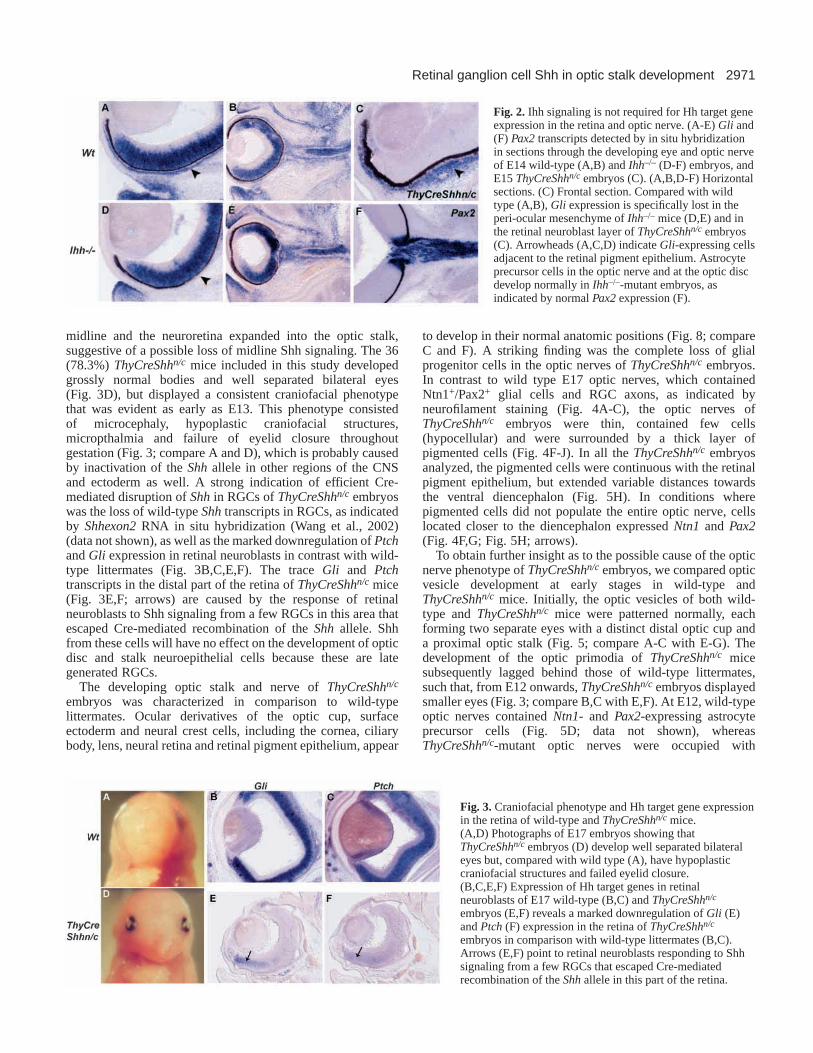
Ihh signaling is not required for Gli and Ptchexpression in the retina and optic nerveAnother mammalian hedgehog homolog, Ihh, is expressed bya group of mesenchymal cells located outside the retinalpigment epithelium (Wallace and Raff, 1999) (data not shown),raising the possibility that Ihhcould signal to cells in the retina,optic disc and stalk. To delineate the relative roles of the twoHh genes in ocular tissue patterning, we examined eyedevelopment and Hh target gene expression in ocular tissuesof Ihh–/–-mutant mice at various developmental stages. The eyesizes of Ihh mutants were comparable to their wild-typelittermates (Fig. 2B,E). However, in contrast to wild type, Gliand Ptch expression were markedly downregulated in a layerof peri-ocular mesenchymal cells surrounding the retinalpigment epithelium (Fig. 2; arrowheads in A,D; data notshown). However, Gliexpression in the neuroretina and opticnerve of Ihh–/– mice was not different from wild-typelittermates (Fig. 2A,B,D,E). Astrocyte precursor cells at theoptic disc and in the optic nerve also developed normally inIhh–/– mice, as indicated by normal Pax2expression (Fig. 2F).The loss of Gliexpression in the peri-ocular mesenchyme ofIhh–/– mice (Fig. 2D) indicates that RGC-derived Shh does notsignal in the peri-ocular tissue. Likewise, Ihh signaling fromthe peri-ocular mesenchyme does not induce Gliexpression in

2970
the retinal neuroblasts of ThyCreShhn/c mice with conditionalablation of Shhin RGCs (Fig. 2C). Taken together, thesefindings suggest that Ihh signaling is received by nearbymesenchymal cells, and is not required for Hh target geneexpression in the retina, optic disc and nerve.
Optic nerves of ThyCreShh n/c embryos are‘hypoplastic’, hypocellular and pigmented Our expression analyses suggest that Shh from RGCs maysignal to optic disc and stalk neuroepithelial cells at the peak
of their transformation into glial progenitors. To directlyaddress this question, we used a conditional gene ablationapproach to disrupt Shhin RGCs (see Materials and Methods).The Thy1-Creline 703 mice used for this study expressed Crerecombinase in the developing CNS, including the optic cup,prior to RGC differentiation (Campsall et al., 2002).ThyCreShhn/c embryos were recovered at the expectedMendelian ratio. A total of 46 ThyCreShhn/c-mutant embryoswere generated for this study, of which 10 (21.7%) wereexcluded from the analysis because the eyes were closer to the
G. D. Dakubo and others
Fig. 1.Expression analysis suggests that Shh from RGCs signals to cells in the retinal neuroblast, optic disc and optic stalk. (A,E) Sectionsthrough the optic vesicle of an E9 embryo hybridized with Shh(A) and Ptch(E) riboprobes demonstrate the upregulation of Ptchexpression inthe neuroepitheliun and cephalic mesenchyme adjacent to the Shh-expressing cells. (B,F) Frontal sections through the developing eye of an E11embryo reveals a graded Ptchexpression (F) in the ventral forebrain, with high levels in the anterior hypothalamic neuroepithelium (ahn) andlow levels in the optic stalk (os), consistent with an established morphogen gradient of Shh (B) from the ventral midline. Ptch(arrows in F)expression in the diencephalic neuroepithelium is due to Shhfrom the zona limitans intrathalamica (not shown). The period from E12 to E14 iswhen most neuroepithelial cells of the optic stalk transform into astrocyte progenitor cells (Kuwabara, 1975), and this period is coincident withthe rapid RGC differentiation and expression of Shh(C,D), as well as uniform Ptch(G,H) expression in the optic nerve. (I-P) The response ofoptic disc astrocyte precursor cells (odap) to RGC-derived Shh signaling. As RGCs differentiate in the central retina and express Shh(I), opticdisc astrocyte precursor cells and retinal neuroblasts in close proximity to the Shh-expressing cells respond by upregulating the Hh target gene,Gli (J). It is likely that the Gli-expressing cells at the disc are the same cells that express Pax2(K) and netrin 1 (Ntn1; L). Although RGCscontinue to express Shh(M) into late embryogenesis and the underlying neuroblasts respond to this by expressing Gli (N), the Pax2- (O) andPdgfra-(P) expressing retinal astrocyte precursor cells migrating into the retina (arrowheads in N,O,P), and those at the optic disc (arrows inN,O,P), downregulate their Hh responsiveness. ov, optic vesicle; tv, telencephalic vesicle; oc, optic cup; os, optic stalk; ahn, anteriorhypothalamic neuroepithelium; odap, optic disc astocyte precursor cells.
2971Retinal ganglion cell Shh in optic stalk development
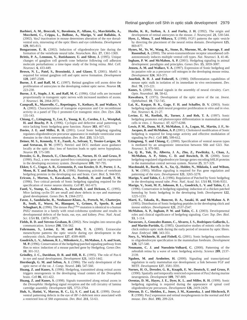
midline and the neuroretina expanded into the optic stalk,suggestive of a possible loss of midline Shh signaling. The 36(78.3%) ThyCreShhn/c mice included in this study developedgrossly normal bodies and well separated bilateral eyes(Fig. 3D), but displayed a consistent craniofacial phenotypethat was evident as early as E13. This phenotype consistedof microcephaly, hypoplastic craniofacial structures,micropthalmia and failure of eyelid closure throughoutgestation (Fig. 3; compare A and D), which is probably causedby inactivation of the Shhallele in other regions of the CNSand ectoderm as well. A strong indication of efficient Cre-mediated disruption of Shhin RGCs of ThyCreShhn/c embryoswas the loss of wild-type Shhtranscripts in RGCs, as indicatedby Shhexon2RNA in situ hybridization (Wang et al., 2002)(data not shown), as well as the marked downregulation of Ptchand Gli expression in retinal neuroblasts in contrast with wild-type littermates (Fig. 3B,C,E,F). The trace Gli and Ptchtranscripts in the distal part of the retina of ThyCreShhn/c mice(Fig. 3E,F; arrows) are caused by the response of retinalneuroblasts to Shh signaling from a few RGCs in this area thatescaped Cre-mediated recombination of the Shh allele. Shhfrom these cells will have no effect on the development of opticdisc and stalk neuroepithelial cells because these are lategenerated RGCs.
The developing optic stalk and nerve of ThyCreShhn/c
embryos was characterized in comparison to wild-typelittermates. Ocular derivatives of the optic cup, surfaceectoderm and neural crest cells, including the cornea, ciliarybody, lens, neural retina and retinal pigment epithelium, appear
to develop in their normal anatomic positions (Fig. 8; compareC and F). A striking finding was the complete loss of glialprogenitor cells in the optic nerves of ThyCreShhn/c embryos.In contrast to wild type E17 optic nerves, which containedNtn1+/Pax2+ glial cells and RGC axons, as indicated byneurofilament staining (Fig. 4A-C), the optic nerves ofThyCreShhn/c embryos were thin, contained few cells(hypocellular) and were surrounded by a thick layer ofpigmented cells (Fig. 4F-J). In all the ThyCreShhn/c embryosanalyzed, the pigmented cells were continuous with the retinalpigment epithelium, but extended variable distances towardsthe ventral diencephalon (Fig. 5H). In conditions wherepigmented cells did not populate the entire optic nerve, cellslocated closer to the diencephalon expressed Ntn1 and Pax2(Fig. 4F,G; Fig. 5H; arrows).
To obtain further insight as to the possible cause of the opticnerve phenotype of ThyCreShhn/c embryos, we compared opticvesicle development at early stages in wild-type andThyCreShhn/c mice. Initially, the optic vesicles of both wild-type and ThyCreShhn/c mice were patterned normally, eachforming two separate eyes with a distinct distal optic cup anda proximal optic stalk (Fig. 5; compare A-C with E-G). Thedevelopment of the optic primodia of ThyCreShhn/c micesubsequently lagged behind those of wild-type littermates,such that, from E12 onwards, ThyCreShhn/c embryos displayedsmaller eyes (Fig. 3; compare B,C with E,F). At E12, wild-typeoptic nerves contained Ntn1- andPax2-expressing astrocyteprecursor cells (Fig. 5D; data not shown), whereasThyCreShhn/c-mutant optic nerves were occupied with
Fig. 2. Ihh signaling is not required for Hh target geneexpression in the retina and optic nerve. (A-E) Gli and(F) Pax2transcripts detected by in situ hybridizationin sections through the developing eye and optic nerveof E14 wild-type (A,B) and Ihh–/– (D-F) embryos, andE15 ThyCreShhn/c embryos (C). (A,B,D-F) Horizontalsections. (C) Frontal section. Compared with wildtype (A,B), Gliexpression is specifically lost in theperi-ocular mesenchyme of Ihh–/– mice (D,E) and inthe retinal neuroblast layer of ThyCreShhn/c embryos(C). Arrowheads (A,C,D) indicate Gli-expressing cellsadjacent to the retinal pigment epithelium. Astrocyteprecursor cells in the optic nerve and at the optic discdevelop normally in Ihh–/–-mutant embryos, asindicated by normal Pax2 expression (F).
Fig. 3.Craniofacial phenotype and Hh target gene expressionin the retina of wild-type and ThyCreShhn/c mice.(A,D) Photographs of E17 embryos showing thatThyCreShhn/c embryos (D) develop well separated bilateraleyes but, compared with wild type (A), have hypoplasticcraniofacial structures and failed eyelid closure.(B,C,E,F) Expression of Hh target genes in retinalneuroblasts of E17 wild-type (B,C) and ThyCreShhn/c
embryos (E,F) reveals a marked downregulation of Gli(E)and Ptch(F) expression in the retina of ThyCreShhn/c
embryos in comparison with wild-type littermates (B,C).Arrows (E,F) point to retinal neuroblasts responding to Shhsignaling from a few RGCs that escaped Cre-mediatedrecombination of the Shhallele in this part of the retina.

2972 G. D. Dakubo and others
Fig. 4.The optic nerves of ThyCreShhn/c embryos are hypocellular and covered by a thick layer of melanotic cells. (A,B,F,G) RNA in situhybridization for Ntn1 (A,F) and Pax2(B,G) expression in sections through the eye and optic nerve of E16 wild-type (A,B) and ThyCreShhn/c
(F,G) embryos. Note the expression ofNtn1and Pax2(arrows in F,G) in only proximal optic stalks of ThyCreShhn/c mice. Although RGC axonsinvaded the optic nerves of ThyCreShhn/c mice, as evidenced by anti-neurofilament-3A10 immunostaining (compare C and H), neuroepithelialcells in the nerve failed to differentiate as glial cells, resulting in the hypocellularity of the optic nerves (compare D,E with I,J). (E,J) Highmagnifications of the boxed areas in D and I. Dashed lines (E,J) outline the optic nerves, and arrowheads (I) indicate nuclei of some cells inoptic nerves of ThyCreShhn/c mice.
Fig. 5. Normal optic vesiclepatterning and abnormal geneexpression in the optic nervesof ThyCreShhn/c mice.(A-C,E-G) Pax2 expressiondetected by in situhybridization in sectionsthrough the developing opticvesicle of E10 wild-type (A-C) and ThyCreShhn/c (E-G)embryos. Pax2 expressionand optic vesicle morphologyappear normal inThyCreShhn/c when comparedto wild-type littermates. Notethat wild-type (C) andThyCreShhn/c (G) embryosections were not exactlythrough the same frontalplane. By E12, wild-type (D)optic nerves contained Ntn1-
expressing astrocyte lineage cells, but theneuroepithelial cells in the optic stalk ofThyCreShhn/c embryos (H) were pigmented. Arrowindicates proximal optic stack with normal Ntn1expression. (I-N) Cross sections through the opticnerve of E13 wild-type (I-K) and ThyCreShhn/c (L-N) embryos analyzed for Pax2(I,L), Pax6(J,M)and Mitf(K,N) expression. Compared with wild-type, almost all the neuroepithelial cells in the opticnerves of ThyCreShhn/c mice were pigmented andexpressed Pax6(M) and Mitf(N), but not Pax2(L),and the pigmented cells were already separating tothe periphery of the axons. (I-N) dorsal, top;ventral, bottom.

2973Retinal ganglion cell Shh in optic stalk development
pigmented cells (Fig. 5H) interspersed with RGC axons. A daylater, the pigmented cells appeared to have moved to theperiphery of the optic nerve (Fig. 5L-N), which is consistentwith the idea that pigmented cells and axons are repellant(Silver and Sapiro, 1981). This observation raised the questionof a possible abnormal transcriptional control ofneuroepithelial cell development in the optic stalk. Weaddressed this issue by examining a number of transcriptionfactors shown to be necessary for the normal differentiation ofthe optic stalk and retinal pigment epithelium. Pax2and Pax6,for example,undergo dynamic regulation in the optic stalk andnerve, with the gradual repression of Pax6by Pax2(Schwarzet al., 2000) such that by E14 all the cells in the optic nerveexpress Pax2and not Pax6. Sagittal sections of E13 wild-typebrains revealed high levels of Pax2and low levels of Pax6expression in the optic nerve (Fig. 5I,J). Also at thisdevelopmental stage, Mitfwas expressed only in the dorsalregions of the distal optic nerve in wild-type mice (Fig. 5K).By contrast, the distal two thirds of the optic nerves ofThyCreShhn/c embryos contained no Pax2-expressing glialcells. Instead, almost all the neuroepithelial cells in the opticstalk were pigmented, and expressed Pax6and Mitf (Fig. 5L-N), which indicates abnormal transcriptional regulation of thedifferentiation program of optic stalk neuroepithelial cells inthese mutant mice.
ThyCreShh n/c embryos have RGC axon guidancedefects in the retina We showed previously that RGC development is inducedacross the retina of ThyCreShhn/c
mice, and that it may even beincreased (Wang et al., 2002)(Y.P.W., unpublished). Hence, theoptic nerve ‘hypoplasia’ ofThyCreShhn/c mice suggested thatnot all the axons exited the retina.We therefore stained retinal sectionsof perinatal ThyCreShhn/c micewith an anti-neurofilament-3A10antibody, which revealed aremarkable axon guidance defect inthe retina that was very similar tothat observed in Ntn1–/– mutantmice (Fig. 6C-F) (Deiner et al.,1997). Compared with wild-typemice (Fig. 6A,B), RGC axonsof ThyCreShhn/c embryos weremisrouted to sub-retinal spaces inseveral regions of the retina and atthe optic disc (Fig. 6C-F). Of theaxons that reached the disc, somefailed to exit into the optic nerve,and instead coiled in the sub-retinalspace (Fig. 6; white asterisks inC and D). The similarity of theretinal phenotype of ThyCreShhn/c
embryos to that of Ntn1–/– mutants,and the observation that optic discastrocyte precursor cells are earlytargets of Hh signaling, suggestedthat netrin signaling was disrupted
in optic disc astrocyte precursor cells. We therefore examinedthe development of the optic disc astrocyte precursor cells inThyCreShhn/c embryos. We were unable to detect anyNtn1/Pax2-expressing cells at the optic discs of E12ThyCreShhn/c embryos by in situ hybridization analysis (Fig.7A,D; data not shown). From E15 onwards, the optic discastrocyte precursor cells express Pdgfra as they migrate intothe retina over RGC axons, such that they are easily detectedin sections at, or within the vicinity of, the optic nerve head(Fig. 7B,C). In contrast to wild-type littermates (Fig. 7B,C),three ThyCreShhn/c embryos examined at this developmentalstage consistently demonstrated absence of Pax2,Pdgfra andNtn1 expression in the optic disc region (Fig. 7E,F; data notshown).
We needed to rule out confounding factors that might alsocontribute to the eye phenotype of ThyCreShhn/c embryos.First, Shh is a mitogen for retinal neuroblasts (Jensen andWallace 1997; Levine et al., 1997; Stenkamp et al., 2000) andperinatal optic nerve astrocytes (Wallace and Raff, 1999).Thus the absence of retinal astrocyte precursor cells at theoptic nerve head of ThyCreShhn/c embryos could haveresulted from failure to stimulate the proliferation of thesecells in the absence of RGC-derived Shh signaling. Weaddressed this issue by examining the retinas of cyclin D1–/–
mice that are highly defective in cellular proliferation in theretina (Fantl et al., 1995; Sicinski et al., 1995). Althoughcyclin D1–/– mice at E18 displayed thinner retina than wild-type littermates, Pax2andPdgfrawere expressed in the opticdisc and retina, and there was no RGC axon misrouting (Fig.
Fig. 6.RGC axon guidance defects in ThyCreShhn/c mice. (A,D) Hematoxylin and Eosin stainingof sections through the optic disc of E12 wild-type (A) and ThyCreShhn/c (D) embryos. Note thatin the wild type, the eosinophilic RGC axons are separated from the potential subretinal space(black asterisk in A) by the optic disc astrocyte precursor cells (white arrows in A), and that thesecells are missing in ThyCreShhn/c mutants (D) thereby exposing the subretinal space (blackasterisk in D) to invasion by RGC axons (white asterisk in D). Anti-neurofilament-associatedprotein immunostaining (B,C,E,F) of E17 retinal sections of wild-type (B) and ThyCreShhn/c
embryos (C,E,F) reveals the retinal axon guidance defects of ThyCreShhn/c embryos. Comparedwith wild type (B), ThyCreShhn/c embryos display axon coiling in the subretinal space at the opticdisc (white asterisks in C and D), and axon misrouting into the peripheral retina (open whiteasterisks in E,F). Note the severe axon misguidance in (F) that creates what looks like multipleoptic discs (white arrows in F).

2974
7G-J; data not shown). Second, the failure of RGC axons toexit the retina ofThyCreShhn/c mice could have resulted fromabnormal formation or faulty closure of the optic fissure, asobserved in the inherited optic nerve aplastic strain, ZRDCT-An (Silver et al., 1984), Chx10or (Smith et al., 2002) andBst/+ (Rice et al., 1997) mutant mice. To determine whetherthe eye phenotype of ThyCreShhn/c mice was caused byabnormalities in optic fissure closure, we examined sagittalsections through the eye at E13, a time at which the opticfissure should normally be closed. In both wild-type andThyCreShhn/c embryos, the optic fissures were completelyclosed, and the expression of Vax2, a homeobox transcriptionfactor that is important for this process (Barbieri et al., 2002),was localized to the ventral retina (Fig. 8A,D). In addition,the expression of Pax6 and other neuroretinal genes in theretina of ThyCreShhn/c embryos was comparable to wild-typelittermates (Fig. 8B,E; data not shown). The extension of thechoroid fissure into the distal optic stalk is also necessary forthe hyaloid vessels to enter the optic cup and establish thevascular tunic of the lens (Smith et al., 2002). Collagen typeIV immunostaining for retinal blood vessels of wild-type andThyCreShhn/c embryos revealed normal hyaloid vasculaturein the posterior chamber of the eye (Fig. 8C,F), againsuggestive of normal formation and closure of the opticfissure in ThyCreShhn/c mice.
The ocular phenotype of ThyCreShh n/c mice is not aconsequence of loss of midline Shh early during eyedevelopmentCre-recombinase activity in line 703 Thy1-Cremice used forthis study was heterogeneous and not restricted to the retina,as some of the ThyCreShhn/c embryos we recovered hadholoprosencephaly, which indicates that midline Shhexpression was affected in these mice (these mice were notincluded in our analysis). Given the importance of midline-derived Shh signaling in proximal-distal patterning of theoptic vesicle, we were concerned that the eye phenotype weobserved could have resulted from loss of Shh at the midline.To address this issue, we first examined the expression ofShhexon2, Ptch and Gli in the developing hypothalamus by insitu hybridization. These transcripts were detectable inThyCreShhn/c embryos at comparable levels to their wild-typelittermates (Fig. 9A,D; data not shown). We next examinedsome markers of the diencephalon such as Nkx2-1 (Titf1 –Mouse Genome Informatics) and Ntn1, whose expression maybe regulated by Shh signaling (Sussel et al., 1999; Pabst et al.,2000; Hynes et al., 2000). Analysis of Nkx2-1 and Ntn1expression in E13, E16 and E17 ThyCreShhn/c embryosshowed comparable expression patterns to wild-typelittermates (Fig. 9B,C,E,F; data not shown), which indicatesthe hypothalamus was present in ThyCreShhn/c mice, and its
ventral molecular identity was preservedas well. Based on the above findings, andthe presence of bilateral eyes inThyCreShhn/c embryos, it is unlikely theeye and optic nerve defects are due toloss of Shhexpression at the ventralmidline.
Shh modulates optic discastrocyte precursor cellpopulation in vitro Our in vivo analysis is consistent with amodel whereby Shh from RGCs actsboth early and transiently to promote thedevelopment of astrocyte precursor cellsat the optic disc. To test whether Hhsignaling can modulate the size of thePax2+ optic disc astrocyte precursor cellpopulation, we cultured optic cups fromE12 C57BL/6 embryos for 48 hours inthe presence or absence of recombinantmyristoylated N-terminal activefragment of Shh (Shh-N), an anti-Hhantibody-5E1 (Ericson et al., 1996) or a5E1 isotype-matched antibody-1E6, andperformed in situ hybridization on serialsections through the optic disc for Pax2expression. Compared with controls,Shh-N markedly increased, whereas5E1 decreased, the size of the Pax2+ cellpopulation at the optic disc (Fig. 10A-C). Treatment with 1E6 was similar tountreated controls (data not shown). Theeffect of anti-Hh antibody 5E1 wasdue to blockage of endogenous Shhproduced by RGCs, which is present in
G. D. Dakubo and others
Fig. 7. Loss of optic disc astrocyte precursor cells in ThyCreShhn/c mice is not caused by aglobal defect in proliferation. Ntn1(A,D), Pax2(B,E) and Pdgfra(C,F) transcript expressiondetected by in situ hybridization in sections through the optic disc of wild-type (A-C) andThyCreShhn/c (D-F) embryos, at E12 (A,D) and E17 (B,C,E,F). Compared with wild-typemice (A-C), there was no Ntn1(D), Pax2(E) or Pdgfra(F) expression in optic discs ofThyCreShhn/c-mutant mice. Pax2(G,H) and Pdgfra(I,J) expression is comparable betweenE18 wild-type (G,I) and cyclin D1–/– (H,J) mice.

2975Retinal ganglion cell Shh in optic stalk development
the cultures during the first 24 hours (Wang et al., 2002). Asthe entire disc region of each explanted eyecup was coveredin three sections irrespective of the treatment, the changein size of the Pax2+ cells at the optic disc wasquantitative. However, by E14 Shh-N treatment had noeffect on the size of the Pax2+ population, nor was itassociated with an increase in Gliexpression in optic discastrocyte precursor cells after two days in culture (Fig. 10;
compare E and F, and D and H). Thus the timing of Hhresponsiveness of optic disc astrocytes is maintained in vitroand probably represents a cell intrinsic property of thispopulation. However, anti-Hh treatment of E14 explantsreduced the size of the Pax2+ cell population (Fig. 10G),which indicates that the size of this population may be underthe control of Hh signaling at least at early time points inthese explants.
DISCUSSION
Three conclusions can be drawn from the presentstudy: (1) Shh signaling is required for Hh targetgene expression in the developing retina, whereasIhh signaling is required for Hh target geneexpression in the peri-ocular mesenchyme; (2) Shhexpression in RGCs is required for glial lineagespecification from optic stalk neuroepithelial cells;and (3) Shh expression in RGCs is required to inducethe development of astrocyte precursor cells at theoptic disc. The last two conclusions are based on theobservation that conditional ablation of Shhinretinal neuroepithelial cells prior to RGCdifferentiation resulted in failure of optic discastrocyte precursor cell development, and theconversion of optic stalk neuroepithelial cells intopigmented cells. In addition, modulating the levelsof Shh signaling in retinal explants in vitro alteredthe size of the optic disc astrocyte precursor cellpopulation. As Ntn1 expression in optic discastrocyte precursor cells is required for local axonguidance at the optic disc (Deiner et al., 1997),netrin signaling was perturbed in the conditionalmutant mice, which resulted in severe intraretinalRGC axon guidance defects. These observationssupport a model whereby RGCs, the first neurons todifferentiate in the retina, signal through Shh to
Fig. 8.Dorsal ventral patterning and opticfissure closure are normal in ThyCreShhn/c
embryos. (A,B,D,E) Sagittal sectionsthrough the eyes of E13 wild-type (A,B)and ThyCreShhn/c (D,E) embryoshybridized with Vax2(A,D) and Pax6(B,E) riboprobes. Note the absence ofcoloboma (D,E), and the normal expressionof Vax2 (D) in the ventral retina, and Pax6(E) in the retina, lens, and cornealepithelium of ThyCreShhn/c embryos incomparison with wild-type littermates.(C,F) Collagen type IV immunostainingreveals the presence of normal vasculartunics in the posterior chamber of E17ThyCreShhn/c eyes (compare hyaloidvessels in C and F). ac, anterior chamber; l,lens; hv, hyaloid vessels; cb, ciliary body.
Fig. 9.Normal development and expression of ventral markers in thehypothalamus of ThyCreShhn/c embryos. (A-F) Frontal sections through thehypothalamus hybridized with Shhexon2(A,D), Nkx2-1(B,E) and Ntn1(C,F) riboprobes reveal comparable expression of functional Shh (A,D), Nkx2-1(B,E) and Ntn1(C,F) in wild-type (A-C) and ThyCreShhn/c (D-F) mice. ah,anterior hypothalamus; v3, third ventricle.

2976
setup their axon guidance system and promote thedevelopment of glial support cells along their path.
RGC-derived Shh and peri-ocular mesenchymal Ihhsignal in distinct domains of the developing eyeThere is an overlap in the timing of expression of Shhin theneuroretina and Ihh in the peri-ocular mesenchyme, whichraises the possibility that either Shh or Ihh, or both, couldinduce Hh target gene expression in the retina, optic stalk orperi-ocular mesenchyme. Indeed, there are several exampleswhere more than one Hh member is responsible for tissuepatterning and/or where one Hh family member compensatesfor the loss of another (Pathi et al., 2001; Zhang et al., 2001;Wijerde et al., 2002). As there is currently no blockingantibody available that discriminates between the two Hhproteins, we employed a genetic approach to delineate therelative roles of Shh and Ihh signaling in eye development.Evidence from our analysis conclusively demonstrates that Ihhsignaling is not required for Ptch or Gliexpression in the retinaand optic nerve, and probably does not contribute to thedevelopment of these structures. However, Ihh appears to playa role in the development of peri-ocular structures, as Ihh–/–
mice have ocular phenotypes suggestive of abnormal peri-ocular mesenchymal tissue development (G.D.D.,unpublished). Further evidence for the lack of Ihh signaling inthe retina and optic nerve was accrued from analysis ofThyCreShhn/c mice. In these conditional mutant mice, Ihhsignaling could induce Gli expression in the peri-ocularmesenchyme, but not in the retina or optic nerve. Whateverlimits the spread of Ihh into the retina or optic nerve, and RGC-derived Shh into the peri-ocular environment, is unclear at thistime, but may be related to the tight junctions formed betweenretinal pigment epithelial cells.
Shh signaling from RGCs is required for Hh signalreception in the optic stalkThere is ample evidence that axon-derived factors promote
glial cell development (Fields and Stevens-Graham, 2002).Indeed, several experiments conclusively demonstrate that glialcell numbers in the optic nerve are regulated by RGC axons.Optic nerve astrocyte proliferation was increased in Bcl2transgenic mice, which have more RGC axons than their wild-type littermates (Burne et al., 1996). Furthermore, when RGCaxons of wild-type and Wallerian-degeneration-deficientmutant mice were severed behind the eye, astrocyteproliferation decreased, which suggests that factors from theRGC body are involved in the induction of astrocyteproliferation in the optic nerve. Subsequent intra-ocularcolchicine and tetrodotoxin injection experiments revealed thatastrocyte proliferation in the rodent optic nerve depends on fastaxonal transport (Burne and Raff, 1997). However, the axonalfactors that mediate astrocyte proliferation in the optic nervehave remained elusive. We showed previously that Shh protein,but not the mRNA was present in the perinatal optic nerve, andRGC axotomy and functional anti-Hh antibody blockageabrogated Ptch expression, as well as astrocyte proliferation,in the perinatal optic nerve, which is consistent with a modelwhereby Shh is transported in an anterograde fashion into theoptic nerve (Wallace and Raff, 1999). We have also observeda marked downregulation of Gli, Ptchand cyclin D1 expressionin postnatal optic nerves of mice with conditional disruption ofShh in parts of the retina (G.D.D., unpublished), whichprovides further evidence that Hh signaling in the perinataloptic nerve is dependent on Shh expression in the retina.Recently, a role for anterograde transport of Shh has beendemonstrated in the visual system of the adult hamster(Triaffort et al., 2001), and in the fornix of the adult ratforebrain (Lai et al., 2003), suggesting that axon-associatedShh signaling occurs in other parts of the adult CNS as well.The failure of optic stalk neuroepithelial cells ofThyCreShhn/c
mice to differentiate as glial progenitors at E12 is consistentwith an even earlier requirement of Shh signaling from RGCaxons in optic stalk neuroepithelial cell development. In thefly, in-growing retinal axons contain Hh that induces patched
G. D. Dakubo and others
Fig. 10.Modulation of the Pax2-positive astrocyte precursor cellpopulation at the optic disc by Shhsignaling. Optic cups of E12 (A-C)and E14 (D-H) C57BL/6 embryoswere cultured for 48 hours, untreated(A,E) or treated with recombinantShh-N (B,D,F,H) or the anti-Shhantibody 5E1 (C,G), and sectionedfor in situ hybridization for Pax2(A-G) and Gli(H) expression.Compared with control (A), the sizeof the Pax2-positive cell populationat the optic disc was markedlyincreased by Shh-N treatment (B),whereas 5E1 treatment (C) resultedin an almost complete loss of thesecells. (D,H) The kinetics of Hhresponsiveness by optic disc cells ismaintained in vitro, as the Pax2-positive cells (arrow in D) of the E14Shh-N treated explants downregulate Gli(arrow in H) expression by the second day in culture. Consistent with this observation, the size of thePax2-positive cell population at the optic disc did not differ much between controls (E) and Shh-N treated (F) cultures, but was much reducedin the 5E1 (G) treated explants at E14.

2977Retinal ganglion cell Shh in optic stalk development
expression in glial cells along their trajectory and in the brain,and also triggers neurogenesis in the visual ganglia (Huangand Kunes, 1996). Thus, anterograde axonal transport of Hhproteins may be a conserved phenomenon from flies tomammals.
Given that ThyCreShhn/c mice develop smaller brains thantheir wild-type littermates, it could be argued that the opticnerve phenotype is caused by insufficient signaling from Shhat the midline, which results in proximal-distal defects in opticvesicle patterning. However, evidence from our analysis andother studies strongly disputes this possibility. ThyCreShhn/c
mice express Shhand Shh-dependent markers at the ventraldiencephalon. In addition, it is unlikely that Shh diffusion fromthe midline can account for Hh target gene expression in theoptic nerve by E12, and thereafter, as the furthest distance Shhseems to travel extracellularly is in the range of 20-30 celldiameters (Lewis et al., 2001, Wijerde et al., 2002), and fromE12 onwards there is no graded Ptch or Gli expression in theoptic stalk to suggest graded Shh signaling from the ventralmidline. The ocular phenotypes associated with loss of midlineShh, such as cyclopia in Shh–/– mice (Chiang et al., 1996), andthe failure of the optic vesicle to transit from vesicle to cupstage, as in BF1–/–mice (Huh et al., 1999), were never observedin ThyCreShhn/c mice. As cholesterol modified Shh moleculesare associated with the lipid raft machinery in the optic nerve(Traiffort et al., 2001), it is very likely that RGC-derived Shhis associated with their growth cones or axons in lipid rafts,and signals to cells in the optic disc and stalk. To directlyaddress the possibility that cholesterol modification is essentialfor axonal transport of Shh proteins in the visual system mayrequire, for example, the generation and analysis of conditionalmice that express functional Shh without the cholesterolmoiety in RGCs. Our prediction is that such mice will developsimilar optic nerve phenotype as those observed inThyCreShhn/c mice.
Regulation of Pax2 expression and gliogenesis inthe optic stalk requires RGC-derived Shh signalingThe homeobox transcription factor Pax2plays an importantrole in normal development of the proximal optic vesicle,where it is expressed early in embryogenesis (Favor et al.,1996; Torres et al., 1996; Otteson et al., 1998). It also appearsthat Pax2is required to suppress pigment cell formation in theoptic disc and stalk, as Pax2expression is never observed inpigmented cells and Pax2–/– mice develop a pigmented opticnerve (Torres et al., 1996). Indeed the pigmented opticnerve phenotype of ThyCreShhn/c mice is histologicallyindistinguishable from that ofPax2–/–-mutant mice (Torres etal., 1996). In an experiment where RGC axons were preventedfrom entering the optic stalk, the neuroepithelial cells becameheavily pigmented with increased apoptosis (Ulshafer et al.,1979). This finding, together with the pigmented optic nervephenotype of Pax2–/– mice, suggests that factors from retinalaxons may signal to optic stalk neuroepithelial cells to maintainPax2expression, thereby preventing them from differentiatingas pigmented cells. Hh signaling regulates Pax2 expression(Macdonald et al., 1995; Ekker et al., 1996), but it is not knownwhether this effect is direct or indirect. Misexpression of shhor twhh in the zebrafish embryo results in the expansion of thepax2 expression domain at the expense of pax6 in the opticprimodium, with a reduction in eye pigmentation (Macdonald
et al., 1995; Ekker et al., 1996). Thus, the similarities in theoptic nerve phenotypes of ThyCreShhn/c and Pax2–/– miceprovides strong support for a direct link between RGC-derivedShh signaling in the maintenance of Pax2 expression in theoptic nerve.
Optic disc astrocyte precursor cell developmentdepends on RGC-derived Shh signaling Optic disc astrocyte precursor cells are first targets of Hhsignaling in the retina, and our findings suggest that RGC-derived Shh signaling promotes their development. First weshow that there is a temporal and spatial correlation in thepattern of Shhand Gli expression in the central retina,consistent with local Shh signaling from RGCs. Next weprovide genetic evidence that optic disc astrocyte precursorcells fail to develop in mice with targeted disruption of ShhinRGCs. Finally, we demonstrate that Shh signaling modulatesthe size of the optic disc astrocyte precursor cell population invitro. It is probable that the initial requirement for RGC-derived Shh signaling is to maintain Pax2expression in opticdisc astrocyte precursors, which is consistent with our in vitrodata where we show that functional blockage of RGC-derivedShh signaling results in a remarkable decrease in the size ofthe optic disc Pax2+ cell population. Later, Shh signaling mayalso be associated with the induction of Pdgfra expression inoptic disc astrocyte precursor cells, as the peak of Gliexpression by the Pax2+ optic disc cells coincides with theirinitial expression of Pdgfra, and Gli has been shown to directlyactivate the expression and phosphorylation of Pdgfra in celllines (Xie et al., 2001). The maintenance ofPdgfraexpressionin more differentiated retinal astrocyte precursor cells isunlikely to be dependent upon sustained Shh signaling, as theexpression of Pdgfrain astrocyte precursor cells at the disc isfollowed by the downregulation of Gli expression.
There is probably a cell intrinsic program that regulates theresponse of optic disc cells to Hh signaling. In contrast toastrocytes in the optic nerve that continue to respond to Hhsignaling into the postnatal period (Wallace and Raff, 1999),the response of optic disc astrocyte precursor cells is transient,averaging 4-5 days. One possible explanation for thisdifference is that optic disc astrocyte precursors are respondingto Shh released from the growth cones of RGCs, such that onceRGC axons have finished invading the optic disc, Shh becomeslimiting to optic disc cells. But this explanation is unlikelygiven that astrocytes in the optic nerve are able to receive axon-associated Hh signals right into the perinatal period (Wallaceand Raff, 1999), and that the addition of recombinant Shh-Nto retinal explants in vitro did not restore Gliexpression in cellsat the optic disc. Another explanation for the downregulationof Hh responsiveness in optic disc astrocyte precursor cellscould be an alteration in the levels of their extra-cellular matrixcomponents in this region of the retina, as extracellular matrixhas been shown to have positive and negative influences on Hhsignaling (Pons et al., 2001).
Regulation of gliogenesis by Shh signalingGlial cell specification from uncommitted neuroepithelial cellsrequires Shh signaling (Pringle et al., 1996). For example,oligodendrocytes are generated in specific domains of theventral neuroepithelium under the influence of Shh signaling(reviewed by Bongarzone, 2002). However, in the optic stalk,

2978
which is part of the ventral neuroepithelium, Shh signalingrather induces specification of astroglial lineage cells fromuncommitted neural stem cells. Upon induction by Shh in theventral forebrain, oligodendrocyte precursor cells migrate intothe perinatal optic nerve where they differentiate into matureoligodendrocytes. How neuroepithelial cells interpret Shhsignals appears to depend on the history and spatial location ofa given group of cells. For example, in the ventral spinal cord(Orentas et al., 1999; Lu et al., 2000), metencephalon (Daviesand Miller, 2001) and telencephalon (Nery et al., 2001; Tekki-Kessaris et al., 2001), Shh signaling has been shown to regulatethe development of oligodendrocytes by inducing theexpression of oligodendrocyte fate specification genes such asOlig1 and Olig2 in neuroepithelial cells (Sussman et al., 2002).However, in the optic stalk, Shh signaling rather induces andmaintains the expression of Pax2that is required for astrocyte,but not oligodendrocyte, lineage commitment.
Shh may also be involved in the development andmaintenance of the integrity of glial cells in other regions ofthe adult CNS. For instance, the proliferation and organizationof Müller glia in the developing and adult retina (Jensen andWallace, 1997; Wang et al., 2002) require Shh signaling,whereas in the adult cerebellum, Bergman glia respond to Shhsignaling from cerebellar Purkinje cells (Traiffort et al., 1998).
Misguidance of retinal axons in ThyCreShh n/c
embryos is due to the loss of Ntn1-expressing opticdisc astrocyte precursor cellsNtn1 from optic disc cells is required for the proper guidanceof RGC axons into the optic stalk (Deiner et al., 1997; Shewanet al., 2002). The identical retinal axon guidance defects ofThyCreShhn/c- and Ntn1–/–-mutant mice suggests that Shh isrequired for the development of the Ntn1-positive cells, or forthe induction of Ntn1expression in optic disc astrocyteprecursor cells or both. However, the failure to observe anyPax2 or Pdgfra expression at the optic disc in ThyCreShhn/c
mice implies these cells were unspecified in the mutant mice.It is likely that the loss of optic disc astrocyte precursors thatultimately results in a failure of Ntn1expression at the opticdisc could have resulted in the axon guidance problems.However, it should also be noted that the loss of optic disc cellsin ThyCreShhn/c mice could lead to abnormal optic discformation, as well as exposure of the potential subretinal spaceto exiting RGC axons, both of which could exacerbate the axonmisguidance.
Unlike Ntn1–/– mice, the eyes of ThyCreShhn/c embryos aresmall and the retinal layers are disrupted (Wang et al., 2002),which could have caused the axon misrouting. However, twolines of evidence indicate that disorganized retinal morphologywas not responsible for the axon misrouting. First, retinalrosettes were not observed until E17, but RGC axon coiling insubretinal spaces at the optic disc was evident as early as E12.Second, misguided RGC axons were not restricted to spaces inbetween retinal rosettes, as axons were seen misrouted intoperipheral retina in the absence of lamination defects, andsome misguided RGC axons were observed going throughrosettes. The smaller eye size of ThyCreShhn/c mutants isconsistent with the role of Shh in ocular growth andproliferation. However, it is unlikely that the micropthalmia isresponsible for the axon misrouting, as micropthalmia per sedoes not result in RGC axon misguidance in the retina.
Conceivably, the misrouted axons in between the retinalpigment epithelium and the neuroretina could obstructcommunication between these retinal layers thereby leading tothe rosettes observed in ThyCreShhn/c-mutant retina; however,evidence from our studies and others do not support thisconclusion. We observed regions of normal retinal layeringwith misrouted axons in subretinal spaces and, moreover, theretinal rosettes we observed in ThyCreShhn/c-mutant retinawere largely not associated with misrouted axons in thesubretinal space. Furthermore, the axon misrouting in Ntn1–/–-mutant mice did not result in any retinal laminationabnormalities (Deiner et al., 1997).
As in Ntn1–/–-mutant mice, RGC axons were still able to exitthe retina of ThyCreShhn/c mice. The earliest age at whichRGC-derived Shh signaling appears to act on optic discastrocyte precursor cells is E12, but Ntn1is expressed in theoptic fissure and stalk prior to this age (Deiner et al., 1997).As RGC differentiation begins some hours earlier than E12, itis possible normal axon guidance could have occurred at theoptic disc prior to the loss of Ntn1-expressing cells. Thus, someof the later generated RGC axons would then respond toguidance cues, such as N-CAM (Brittis et al., 1995), from theearly axons and follow them into the optic nerve.
RGC-derived Shh signaling in eye developmentShh signaling from RGCs plays an important role in thedevelopment of the vertebrate visual system. RGC-derived Shhregulates the proliferation and differentiation of retinalneuroblasts, thereby controlling the number and organizationof their synaptic connections (Jensen and Wallace, 1997;Levine et al., 1997; Stenkamp et al., 2000; Neumann andNuesslein-Volhard, 2000; Zhang and Yang, 2001). Bypromoting the organization and structural integrity of Müllerglia in the retina, RGC-derived Shh signaling also helpsmaintain the normal layering of the retina (Wang et al., 2002).In addition, the proliferation of perinatal optic nerve astrocytesis regulated in part by RGC axon-associated Shh signaling(Wallace and Raff, 1999). We now provide evidence that Shhfrom early-born retinal neurons is required for optic disc andstalk neuroepithelial cell development.
We thank Drs R. Kothary, D. Picketts and M. Furimsky for readingthe manuscript, and thank the following for the gift of probes andreagents: Drs L. V. Goodrich (Ptch), A. Joyner (Gli), M. Tessier-Lavigne (Netrin 1), W. D. Richardson (Pdgfra), G. R. Dressler (Pax2),N. Pringle (Pax6), C. Cekpo (Vax2), H. Arnheiter (Mitf), J. L. R.Rubenstein (Nkx2-1), V. Fantl (cyclin D1–/– mice) and Curis, Inc(recombinant Shh-N protein). We also thank the Animal Care andVeterinary Services Department of the University of Ottawa foranimal husbandry. This work was supported by grants from theNational Cancer Institute of Canada and the Canadian Institutes ofHealth Research (CIHR) to V.A.W. Work in the laboratory of A.P.M.was supported by grant NS 33642 from the National Institutes ofHealth. V.A.W. is a recipient of a CIHR New Investigator award andG.D.D. is a recipient of the Multiple Sclerosis Society of CanadaPostdoctoral Fellowship award.
REFERENCES
Bai, C. B., Auerbach, W., Lee, J. S., Stephen, D. and Joyner, A. L. (2002).Gli2, but not Gli1, is required for initial Shh signaling and ectopic activationof the Shh pathway. Development129, 4753-4761.
G. D. Dakubo and others
2979Retinal ganglion cell Shh in optic stalk development
Barbieri, A. M., Broccoli, V., Bovolenta, P., Alfano, G., Marchitiello, A.,Mocchetti, C., Crippa, L., Bulfone, A., Marigo, V. and Ballabio, A.(2002). Vax2 inactivation in mouse determines alteration of the eye dorsal-ventral axis, misrouting of the optic fibers and eye coloboma. Development129, 805-813.
Bongarzone, E. R.(2002). Induction of oligodendrocyte fate during theformation of the vertebrate neural tube. Neurochem. Res. 27, 1361-1369.
Brittis, P. A., Lemmon, V., Rutishauser, U. and Silver, J. (1995). Uniquechanges of ganglion cell growth cone behavior following cell adhesionmolecule perturbations: a time-lapse study of the living retina. Mol. Cell.Neurosci. 6, 433-449.
Brown, N. L., Patel, S., Brzezinski, J. and Glaser, T.(2001). Math5isrequired for retinal ganglion cell and optic nerve formation. Development128, 2497-2508.
Burne, J. F. and Raff, M. C. (1997). Retinal ganglion cell axons drive theproliferation of astrocytes in the developing rodent optic nerve. Neuron 18,223-230.
Burne, J. F., Staple, J. K. and Raff, M. C. (1996). Glial cells are increasedproportionally in transgenic optic nerves with increased numbers of axons.J. Neurosci. 16, 2064-2073.
Campsall, K., Mazerolle, C., Repetingny, Y., Kothary, R. and Wallace, V.A. (2002). Characterization of transgene expression and Cre recombinaseactivity in a panel of Thy-1 promoter-Cre transgenic mice. Dev. Dyn.224,135-143.
Chiang, C., Litingtung, Y., Lee, E., Young, K. E., Corden, J. L., Westphal,H. and Beachy, P. A. (1996). Cyclopia and defective axial patterning inmice lacking Sonic hedgehog gene function. Nature383, 407-413.
Davies, J. E. and Miller, R. H. (2001). Local Sonic hedgehog signalingregulates oligodendrocyte precursor appearance in multiple ventricular zonedomains in the chick metencephalon. Dev. Biol. 233, 513-525.
Deiner, M. S., Kennedy, T. E., Fazeli, A., Sefrani, T., Tessier-Lavigne, M.and Sretavan, D. W. (1997). Netrin1 and DCC mediate axon guidancelocally at the optic disc: loss of function leads to optic nerve hypoplasia.Neuron19, 575-589.
Dressler, G. R., Deutsch, U., Chowdhury, K., Nornes, H. O. and Gruss, P.(1990). Pax2, a new murine paired-box-containing gene and its expressionin the developing excretory system.Development 109, 787-795.
Ekker, S. C., Ungar, A. R., Greenstein, P., von Kessler, D. P., Porter, J. A.,Moon, R. T. and Beachy, P. A.(1996). Patterning activities of vertebratehedgehog proteins in the developing eye and brain. Curr. Biol. 5, 944-955.
Ericson, J., Morton, S., Kawakami, A., Roelink, H. and Jessell, T. M.(1996). Two critical periods of Sonic hedgehog signaling required for thespecification of motor neuron identity. Cell 87, 661-673.
Fantl, V., Stamp, G., Andrews, A., Rosewell, I. and Dickson, C.(1995).Mice lacking cyclin D1 are small and show defects in eye and mammarygland development. Genes Dev.9, 2364-2372.
Favor, J., Sandulache, R., Neuhauser-Klaus, A., Pretsch, W., Chatterjee,B., Senft, E., Wurst, W., Blanquet, V., Grimes, P., Sporle, R. andSchughart, K. (1996). The mouse Pax21Neumutation is identical to a humanPAX2 mutation in a family with renal-coloboma syndrome and results indevelopmental defects of the brain, ear, eye, and kidney. Proc. Natl. Acad.Sci. USA 93, 13870-13875.
Fields, R. D. and Stevens-Graham, B. (2002). New insights into neuron-gliacommunication. Science298, 556-562.
Fuhrmann, S., Levine, E. M. and Reh, T. A. (2000). Extraocularmesenchyme patterns the optic vesicle during eye development in theembryonic chick. Development 127, 4599-4609.
Goodrich, L. V., Johnson, R. L., Milenkovic, L., McMahon, J. A. and Scott,M. P. (1996). Conservation of the hedgehog/patched signaling pathway fromflies to mice: induction of a mouse patched gene by Hedgehog. Genes Dev.10, 301-312.
Grindley, J. C., Davidson, D. R. and Hill, R. E.(1995). The role of Pax-6in eye and nasal development. Development121, 1433-1442.
Horsburgh, G. M. and Sefton, A. J.(1986). The early development of theoptic nerve of the rat. J. Comp. Neurol. 243, 547-560.
Huang, Z. and Kunes, S. (1996). Hedgehog, transmitted along retinal axonstriggers neurogenesis in the developing visual centers of the Drosophilabrain. Cell 86, 411-422.
Huang, Z. and Kunes, S. (1998). Signals transmitted along retinal axons inthe Drosophila: Hedgehog signal reception and the cell circuitry of laminacartridge assembly. Development125, 3753-3764.
Huh, S., Hatini, V., Marcus, R. C., Li, S. C. and Lai, E.(1999). Dorsal-ventral patterning defects in the eye of BF-1-deficient mice associated witha restricted loss of Shhexpression. Dev. Biol.211, 53-63.
Huxlin, K. R., Stefton, A. J. and Furby, J. H. (1992). The origin anddevelopment of retinal astrocytes in the mouse. J. Neurocytol.21, 530-544.
Hyer, J., Mima, T. and Mikawa, T. (1998). FGF1 patterns the optic vesicleby directing the placement of the neural retina domain. Development125,869-877.
Hynes, M., Ye, W., Wang, K., Stone, D., Murone, M., de Sauvage, F. andRosenthal, A.(2000). The seven-transmembrane receptor smoothened cell-autonomously induces multiple ventral cell types. Nat. Neurosci.3, 41-46.
Ingham, P. W. and McMahon, A. P.(2001). Hedgehog signaling in animaldevelopment: paradigms and principles. Genes Dev.15, 3059-3087.
Jensen, A. M. and Wallace, V. A. (1997). Expression of Sonic hedgehog andits putative role as a precursor cell mitogen in the developing mouse retina.Development124, 363-371.
Juurlink, B. H. J. and Fedoroff, S. (1980). Differentiation capabilities ofmouse optic stalk in isolation of its immediate in vivo environment. Dev.Biol. 78, 215-221.
Kunes, S. (2000). Axonal signals in the assembly of neural circuitry. Curr.Opin. Neurobiol. 10, 58-62.
Kuwabara, T. (1975). Development of the optic nerve of the rat. Invest.Ophthalmol. 10, 732-745.
Lai, K., Kaspar, B. K., Gage, F. H. and Schaffer, D. V. (2003). Sonichedgehog regulates adult neural progenitor proliferation in vitro and in vivo.Nat. Neurosci. 6, 21-27.
Levine, E. M., Roelink, H., Turner, J. and Reh, T. A. (1997). Sonichedgehog promotes rod photoreceptor differentiation in mammalian retinalcells in vitro. J. Neurosci.17, 6277-6288.
Lewis, P. M., Dunn, M. P., McMahon, J. A., Logan, M., Martin, J. F., St-Jacques, B. and McMahon, A. P. (2001). Cholesterol modification of Sonichedgehog is required for long-range activity and effective modulation ofsignaling by Ptc1. Cell105, 599-612.
Litingtung, Y. and Chiang, C. (2000). Specification of ventral neuron typesis mediated by an antagonistic interaction between Shh and Gli3. Nat.Neurosci.3, 979-985.
Lu, Q. R., Yuk, D., Alberta, J. A., Zhu, Z., Pawlitzky, I., Chan, J.,McMahon, A. P., Stites, C. D. and Rowitch, D. H. (2000). Sonichedgehog-regulated oligodendrocyte lineage genes encoding bHLH proteinsin the mammalian central nervous system. Neuron25, 317-329.
Macdonald, R., Barth, K. A., Xu, Q., Holder, N., Mikkola, I. and Wilson,S. W. (1995). Midline signaling is required for Pax gene regulation andpatterning of the eyes. Development121, 3265-3278.
Marigo, V. and Tabin, C. J. (1996). Regulation of Patched by Sonic hedgehogin the developing neural tube. Proc. Natl. Acad. Sci. USA93, 9346-9351.
Marigo, V., Scott, M. P., Johnson, R. L., Goodrich, L. V. and Tabin, C. J.(1996). Conservation in hedgehog signaling: induction of a chicken patchedhomolog by Sonic hedgehog in the developing limb. Development 122,1225-1233.
Marti, E., Takada, R., Buncrot, D. A., Sasaki, H. and McMahon A. P.(1995). Distribution of Sonic hedgehog peptides in the developing chick andmouse embryo. Development121, 2537-2547.
McMahon, A. P., Ingham, P. W. and Tabin, C. J. (2003). Developmentalroles and clinical significance of hedgehog signaling. Curr. Top. Dev. Biol.53, 1-114.
Navascues, J., Gonzalez-Ramos, C., Alvarez, I. S., Rodriquez-Gallardo, L.and Martin-Partido, G. (1985). Quantitative studies of mitotic cells in thechick embryo optic stalk during the early period of invasion by optic fibres.Anat. Embryol.180, 343-351.
Nery, S., Wichterle, H. and Fishell, G. (2001). Sonic hedgehog contributesto oligodendrocyte specification in the mammalian forebrain. Development128, 527-540.
Neumann, C. J. and Nuesslein-Volhard, C. (2000). Patterning of thezebrafish retina by a wave of sonic hedgehog activity. Science289, 2137-2139.
Nguhen, M. and Arnheiter, H. (2000). Signaling and transcriptionalregulation in early mammalian eye development: a link between FGF andMITF. Development127, 3581-3591.
Nornes, H. O., Dressler, G. R., Knapik, E. W., Deutsch, U. and Gruss, P.(1990). Spatially and temporally restricted expression of Pax2 during murineneurogenesis. Development109, 797-809.
Orentas, D. M., Hayes, J. E., Dyer, K. L. and Miller, R. H. (1999). Sonichedgehog signaling is required during the appearance of spinal cordoligodendrocyte precursors. Development126, 2419-2429.
Otteson, D. C., Shelden, E., Jones, J. M., Kameoka, J. and Hitchcock, P.F. (1998). Pax2 expression and retinal morphogenesis in the normal and Krdmouse. Dev. Biol.193, 209-224.

2980
Pathi, S., Pagan-Westphal, S., Baker, D. P., Garber, E. A., Rayhorn, P.,Buncrot, D., Tabin, C. J., Blake Pepinsky, R. and Williams, K. P. (2001).Comparative biologic response to human Sonic, Indian, and Deserthedgehog. Mech. Dev. 106, 107-117.
Pabst, O., Herbrand, H., Takuma, N. and Arnold, H. H. (2000). NKX2gene expression in neuroectoderm but not in mesendodemally derivedstructures depends on Sonic hedgehog in mouse embryos. Dev. Genes Evol.210, 47-50.
Pei, Y. F. and Rhodin, J. A. G. (1970). The prenatal development of the mouseeye. Anat. Rec. 168, 105-126.
Pittack, C., Grunwald, G. B. and Reh, T. A. (1997). Fibroblast growthfactors are necessary for neural retina but not pigmented epitheliumdifferentiation in chick embryos. Development 124, 805-816.
Pons, S., Trejo, J. L., Martinez-Morales, J. R. and Marti, E. (2001).Vitronectin regulates Sonic hedgehog activity during cerebellumdevelopment through CREB phosphorylation. Development128, 1481-1492.
Pringle, N. P., Yu, W. P., Guthrie, S., Roelink, H., Lumsden, A., Peterson,A. C. and Richardson, W. D.(1996). Determination of neuroepithelial cellfate: induction of oligodendrocyte lineage by ventral midline cells and sonichedgehog. Dev. Biol.177, 30-42.
Rhodes, R. H. (1982). Development of the optic nerve. In Ocular anatomy,embryology and teratology(ed. F. A. Jakobiec), pp. 601-638. Philadelphia:Harper and Row.
Rice, D. S., Tang, Q., Williams, R. W., Harris, B. S., Davidson, M. T. andGoldowitz, D. (1997). Decreased retinal ganglion cell number andmisdirected axon growth associated with defects in Bst/+mutant mice.Invest. Opthtalmol. Vis. Sci.38, 2112-2124.
Rubenstein, J. L. R. and Beachy, P. A. (1998). Patterning of the embryonicforebrain. Curr. Opin. Neurobiol. 8, 18-26.
Schwarz, M., Cecconi, F., Bernier, G., Andrejewski, N., Kammandel, B.,Wagner, M. and Gruss, P.(2000). Spatial specification of mammalian eyeterritories by reciprocal transcriptional repression of Pax2and Pax5.Development127, 4325-4334.
Shewan, D., Dwivedy, A., Anderson, R. and Holt, C. E. (2002). Age-relatedchanges underlie switch in netrin-1 responsiveness as growth cones advancealong visual pathway. Nat. Neurosci.5, 955-962.
Sicinski, P., Donaher, J. L., Parker, S. B., Li, T., Fazeli, A., Gardner, H.,Haslam, S. Z., Bronson, R. T., Elledge, S. J. and Weinberg, R. A.(1995).Cyclin D1 provides a link between development and oncogenesis in theretina and breast. Cell 82, 621-630.
Silver, J. and Hughes, A. F. (1974). The relationship between morphogeneticcell death and the development of congenital anophthalmia. J. Comp.Neurol. 157, 281-301.
Silver, J. and Robb, R. M. (1979). Studies on the development of the eyecupand optic nerve in normal mice and in mutants with congenital optic nerveapalasia. Dev. Biol. 68, 175-190.
Silver, J. and Sapiro, J.(1981). Axonal guidance during development of theoptic nerve: the role of pigment epithelia and other extrinsic factors. J.Comp. Neurol.202, 521-538.
Silver, J., Puck, S. M. and Albert, D. M. (1984). Development and aging ofthe eye in mice with inherited optic nerve aplasia: histopathological studies.Exp. Eye. Res. 38, 257-266.
Smith, R. S., John, S. W. M. and Sundberg, J. P. (2002). Optic nerve andorbit. In Systematic Evaluation of the Mouse Eye: Anatomy, Pathology andBiomethods, pp. 228-229. Boca Raton, FL: CRC Press.
Stenkamp, D. L., Frey, R. A., Prabhudesai, S. N. and Raymond, P. A.(2000). Function for hedgehog genes in zebrafish retinal development. Dev.Biol. 220, 238-252.
St-Jacques, B., Hammerschmidt, M. and McMahon, A. P. (1999). Indianhedgehog signaling regulates proliferation and differentiation ofchondrocytes and is essential for bone formation. Genes Dev.13, 2072-2086.
Sussel, L., Marin, O., Kimura, S. and Rubenstein, J. L. R. (1999). Loss ofNkx2-1homeobox gene results in a ventral to dorsal respecification of thepallidum into the striatum. Development126, 3359-3370.
Sussman, C. R., Davies, J. E. and Miller, R. H. (2002). Extracellular andintracellular regulation of oligodendrocyte development: roles of Sonichedgehog and expression of E proteins. Glia 40, 55-64.
Tekki-Kessarris, N., Woodruff, R., Hall, A. C., Gaffield, W., Kimura, S.,Stiles, C. D., Rowitch, D. H. and Richardson, W. D. (2001). Hedgehog-dependent oligodendrocyte lineage specification in the telencephalon.Development 128, 2545-2554.
Torres, M., Gomez-Pardo, E. and Gruss, P. (1996). Pax2 contributes to innerear patterning and optic nerve trajectory. Development122, 3381-3391.
Traiffort, E., Charytoniuk, D. A., Faure, H. and Ruat, M. (1998). Regionaldistribution of Sonic hedgehog, Patched and Smoothened mRNA in the adultrat brain. J. Neurochem.70, 1327-1330.
Traiffort, E., Moya, K. L., Faure, H., Hassig, R. and Ruat, M. (2001). Highexpression and anterograde axonal transport of aminoterminal sonichedgehog in the adult hamster brain. Eur. J. Neurosci.14, 839-850.
Ulshafer, R. J. and Clavert, A. (1979). Cell death and optic fiber penetrationin the optic stalk of the chick. J. Morphol. 162, 67-76.
Wallace, V. A. and Raff, M. C.(1999). A role for Sonic hedgehog in axon-to-astrocyte signaling in the rodent optic nerve. Development126, 2901-2909.
Wang, Y.-P., Dakubo, G., Howley, P., Mazorelle, C., Campsall, K., Shiga,S., Lewis, P., McMahon, A. P. and Wallace, V. A. (2002). Developmentof normal retinal organization depends on Sonic hedgehog signaling fromganglion cells. Nat. Neurosci. 5, 831-832.
Wijgerde, M., McMahon, J. A., Rule, M. and McMahon, A. P.(2002). Adirect requirement for Hedgehog signaling for normal specification of allventral progenitor domains in the presumptive mammalian spinal cord.Genes Dev.16, 2849-2864.
Xie, J., Aszterbaum, M., Zhang, X., Bonifas, J. M., Zachary, C., Epstein,E. and McCormick, F. (2001). A role of PDGFRα in basal cell carcinomaproliferation. Proc. Natl. Acad. Sci. USA98, 9255-9259.
Zhang, X.-M. and Yang, X.-J. (2001). Regulation of retinal ganglion cellproduction by Sonic hedgehog. Development 128, 943-957.
Zhang, X.-M., Ramalho-Santos, M. and McMahon, A. P. (2001).Smoothenedmutants reveal redundant roles for Shh and Ihh signalingincluding regulation of L/R asymmetry by the mouse node. Cell 105, 781-792.
Zhao, S., Hung, F.-C., Colvin, J. S., White, A., Dai, W., Lovicu, F. J.,Ornitz, D. M. and Overbeek, P. A. (2001). Patterning the opticneuroepithelium by FGF signaling and Ras activation. Development 128,5051-5060.
G. D. Dakubo and others


![Accuracy of distinguishing between dysembryoplastic neuroepithelial tumors and other epileptogenic brain neoplasms with [11C]methionine PET](https://static.fdokumen.com/doc/165x107/63360da5cd4bf2402c0b568c/accuracy-of-distinguishing-between-dysembryoplastic-neuroepithelial-tumors-and-other.jpg)