Localization of Retinal Proteins in the Stalk and Dorsal Eyes of ...
10
2002 Zoological Society of Japan ZOOLOGICAL SCIENCE 19 : 1231–1240 (2002) Localization of Retinal Proteins in the Stalk and Dorsal Eyes of the Marine Gastropod, Onchidium Nobuko Katagiri 1 * , Tatsuo Suzuki 2 , Yuichi Shimatani 3 and Yasuo Katagiri 4 1 Medical Research Institute, Tokyo Women’s Medical University, Tokyo 162-8666, Japan 2 Department of Pharmacology, Hyogo College of Medicine, Hyogo 663-8501, Japan 3 Department of Physiology, Tokyo Women’s Medical University School of Medicine, Tokyo 162-8666, Japan 4 Section of Basic Science, Tokyo Women’s Medical University School of Nursing, Daito-cho 437-1434, Japan ABSTRACT — Onchidium possesses stalk eye (SE) and dorsal eye (DE) which comprise part of a unique multiple photoreceptive system. The retina of SE consists of rhabdomeric-type visual cells, whereas the DE contains two types of photoreceptor cells; ciliary-type cells in the retina and rhabdomeric-type cells in the lens. High-performance liquid chromatography (HPLC) analyses revealed the presence of 11- cis- retinal as well as all- trans- retinal in both eyes. The amount of retinal of one DE (0.17 pmol) is far less than that in one SE (0.41 pmol) in the dark-adapted Onchidium . In the dark-adapted SE, the amount of all- trans- retinal was higher than that of 11- cis -retinal. This finding is consistent with the presence of photic vesicles, including retinochrome, in rhabdomeric-type visual cells. In contrast, a higher amount of 11- cis -retinal than all -trans- retinal was present in dark-adapted DE, although this was decreased in light-adapted DE. Upon UV irradiation following treatment with sodium borohydride (NaBH 4 ), the fluorescence (derived from retino- chrome) was observed in the somatic layer of SE. Additional fluorescence (due to rhodopsin) was observed in the villous layer upon treatment with NaBH 4 after denaturation. However, only weak, obscure fluorescence of retinyl proteins was observed in the DE, not in a specific but an indefinite area on treatment with NaBH 4 with or without denaturation. With fluorescence histochemistry, the localization of rhodopsin and retinochrome was confirmed at specific regions in the retina of the SE, whereas no distinct localization of these photopigments in DE was demonstrated. The amount of retinal to detect the fluorescence may be too low in the DE, or photopigments of DE may differ in chemical nature from those of SE. Key words : stalk eye, dorsal eye, HPLC, retinal protein, Onchidium. INTRODUCTION Rhodopsin localizes to the photoreceptive sites of pho- toreceptor cells in both vertebrate and invertebrate eyes. Visual cells in Cephalopods comprise two kinds of photopig- ments. Rhodopsin is localized in the microvilli of the outer segment, while retinochrome is present in myeloid bodies in the inner segment of visual cells (Robles et al. , 1987, 1995; Hara and Hara, 1991; Hara et al ., 1992, 1995). Rhodopsin and retinochrome function cooperatively to mutually regen- erate photopigments (Terakita et al. , 1989). Upon illumination, rhodopsin is converted to metarhodopsin, and retinochrome to metaretinochrome. The 11- cis -retinal in rhodopsin is pho- toisomerized to the all- trans configuration. Conversely, all- trans -retinal in retinochrome is photoisomerized to its 11- cis - isomer. The 11- cis -retinal is required for rhodopsin formation. In the dark, rhodopsin and retinochrome are regenerated from these two isomers by chromophore exchange. Terakita et al . (1989) demonstrated that the turnover of 11- cis- and all- trans -retinal chromophores is mediated by retinal-binding protein, and proposed a conjugate rhodopsin-retinochrome system in visual cells of the squid eye. Using antibodies against squid retinal proteins, Katagiri et al . (2001) demon- strated the localization of three retinal proteins (rhodopsin, retinochrome and retinal-binding protein), and consequently the presence of a rhodopsin-retinochrome system in the stalk eye (SE) of the gastropod Onchidium . Retinochrome is readily reduced by sodium borohydride (NaBH 4 ) into N-retinyl protein that emits visible fluorescence upon UV irradiation. Rhodopsin is similarly converted to a fluorescent product, but only upon reduction following denaturation (Bownds and * Corresponding author: Tel. +81-3-5269-7364; FAX. +81-3-5269-7364. E-mail: [email protected]
-
Upload
khangminh22 -
Category
Documents
-
view
0 -
download
0
Transcript of Localization of Retinal Proteins in the Stalk and Dorsal Eyes of ...

2002 Zoological Society of JapanZOOLOGICAL SCIENCE
19
: 1231–1240 (2002)
Localization of Retinal Proteins in the Stalk and Dorsal Eyesof the Marine Gastropod,
Onchidium
Nobuko Katagiri
1
*, Tatsuo Suzuki
2
, Yuichi Shimatani
3
and Yasuo Katagiri
4
1
Medical Research Institute, Tokyo Women’s Medical University, Tokyo 162-8666, Japan
2
Department of Pharmacology, Hyogo College of Medicine, Hyogo 663-8501, Japan
3
Department of Physiology, Tokyo Women’s Medical University School of Medicine,Tokyo 162-8666, Japan
4
Section of Basic Science, Tokyo Women’s Medical University School of Nursing,Daito-cho 437-1434, Japan
ABSTRACT
—
Onchidium
possesses stalk eye (SE) and dorsal eye (DE) which comprise part of a uniquemultiple photoreceptive system. The retina of SE consists of rhabdomeric-type visual cells, whereas theDE contains two types of photoreceptor cells; ciliary-type cells in the retina and rhabdomeric-type cells inthe lens. High-performance liquid chromatography (HPLC) analyses revealed the presence of 11-
cis-
retinalas well as all-
trans-
retinal in both eyes. The amount of retinal of one DE (0.17 pmol) is far less than thatin one SE (0.41 pmol) in the dark-adapted
Onchidium
. In the dark-adapted SE, the amount of all-
trans-
retinal was higher than that of 11-
cis
-retinal. This finding is consistent with the presence of photic vesicles,including retinochrome, in rhabdomeric-type visual cells. In contrast, a higher amount of 11-
cis
-retinal thanall
-trans-
retinal was present in dark-adapted DE, although this was decreased in light-adapted DE. UponUV irradiation following treatment with sodium borohydride (NaBH
4
), the fluorescence (derived from retino-chrome) was observed in the somatic layer of SE. Additional fluorescence (due to rhodopsin) wasobserved in the villous layer upon treatment with NaBH
4
after denaturation. However, only weak, obscurefluorescence of retinyl proteins was observed in the DE, not in a specific but an indefinite area on treatmentwith NaBH
4
with or without denaturation. With fluorescence histochemistry, the localization of rhodopsinand retinochrome was confirmed at specific regions in the retina of the SE, whereas no distinct localizationof these photopigments in DE was demonstrated. The amount of retinal to detect the fluorescence maybe too low in the DE, or photopigments of DE may differ in chemical nature from those of SE.
Key words
: stalk eye, dorsal eye, HPLC, retinal protein,
Onchidium.
INTRODUCTION
Rhodopsin localizes to the photoreceptive sites of pho-toreceptor cells in both vertebrate and invertebrate eyes.Visual cells in Cephalopods comprise two kinds of photopig-ments. Rhodopsin is localized in the microvilli of the outersegment, while retinochrome is present in myeloid bodies inthe inner segment of visual cells (Robles
et al.
, 1987, 1995;Hara and Hara, 1991; Hara
et al
., 1992, 1995). Rhodopsinand retinochrome function cooperatively to mutually regen-erate photopigments (Terakita
et al.
, 1989). Upon illumination,rhodopsin is converted to metarhodopsin, and retinochrometo metaretinochrome. The 11-
cis
-retinal in rhodopsin is pho-toisomerized to the all-
trans
configuration. Conversely, all-
trans
-retinal in retinochrome is photoisomerized to its 11-
cis
-isomer. The 11-
cis
-retinal is required for rhodopsin formation.In the dark, rhodopsin and retinochrome are regeneratedfrom these two isomers by chromophore exchange. Terakita
et al
. (1989) demonstrated that the turnover of 11-
cis-
andall-
trans
-retinal chromophores is mediated by retinal-bindingprotein, and proposed a conjugate rhodopsin-retinochromesystem in visual cells of the squid eye. Using antibodiesagainst squid retinal proteins, Katagiri
et al
. (2001) demon-strated the localization of three retinal proteins (rhodopsin,retinochrome and retinal-binding protein), and consequentlythe presence of a rhodopsin-retinochrome system in thestalk eye (SE) of the gastropod
Onchidium
. Retinochrome isreadily reduced by sodium borohydride (NaBH
4
) into N-retinylprotein that emits visible fluorescence upon UV irradiation.Rhodopsin is similarly converted to a fluorescent product,but only upon reduction following denaturation (Bownds and
* Corresponding author: Tel. +81-3-5269-7364;FAX. +81-3-5269-7364.E-mail: [email protected]

Katagiri
et al
.1232
Wald, 1965; Hara and Hara, 1973). Based on these differentfluorescent products, Ozaki
et al
. (1983) successfully identi-fied rhodopsin in the microvilli and retinochrome in the cyto-plasm of visual cells in
Octopus
and
Limax
eyes. Katagiri
etal.
(1995) further identified rhodopsin and retinochrome inthe villous and somatic layers of
Onchidium
SE, using themethod described by Ozaki
et al
. (1983) and reported thatfluorescence from reduced retinal proteins disappears rap-idly.
Onchidium
has a unique multiple photoreceptive sys-tem comprising SE, dorsal eyes (DE), extraocular dermalphotoreceptors and photosensitive ganglion cells in the CNS(Hisano
et al
., 1972, Katagiri
et al.
, 1985, 1990). This multi-ple photoreceptive system contains one ciliary-type andthree kinds of rhabdomeric-type photoreceptor cells. Theretina of the SE contains rhabdomeric-type visual cells char-acterized by microvilli and photic vesicles in the cytoplasm.DE is designated ‘vertebrate-type eye’, due to similar mor-phology to the vertebrate eye (Stantschinsky, 1908).However, previous studies by Katagiri
et al
. (1983, 1985)demonstrate that the DE exhibits some unique structuresand responses to light. The DE contains two different typesof photoreceptor cells, specifically, ciliary-type photoreceptorcells in the retina and rhabdomeric-type lens cells in thelens. The ciliary-type photoreceptor cell is believed to be theintrinsic photoreceptor since the retina contains no other celltypes. The lens cell may dually function as the dioptric appa-ratus and photoreceptor (Katagiri
et al
., 1983, 1985, 1998).To examine the function of SE and DE and their rela-
tionship in the
Onchidium
multiple photoreceptive system, itis of considerable importance to compare the photopig-ments of SE and DE in addition to elucidating the structureand photoresponse. As both eyes of
Onchidium
are toominute for biochemical study, it is difficult to generateantibodies against
Onchidium
photopigments. Immunohis-tochemical analyses using anti-squid retinal protein antibod-ies were therefore employed to determine the localization ofrhodopsin and retinochrome in the SE (Katagiri
et al
., 2001).In the present investigation, photopigments in both SE andDE of
Onchidium
were examined by HPLC and fluorescencehistochemistry. The localization of rhodopsin and retino-chrome in the SE is in concurrence with earlier immunohis-tochemical results (Katagiri
et al
., 2001) but photopigmentsof DE did not show the specific localization.
MATERIALS AND METHODS
Animals
Onchidium sp
. (Gastropoda, Mollusca, 3–5 g body weight) werecollected on the seashore of Tateyama (Chiba Prefecture) betweenJune to August and maintained in a tank of aerated artificial sea-water (Tetra Marine) under natural light/dark and an additional 12hr-light/12hr-dark cycle by a submergible NS-fluorescent lamp (Nissei,15W) at 20–23
°
C. The intensity of illumination on the water surfaceat the center of the tank was about 600–1000 lux but varied depend-ing on the weather. Animals inhabit under the stones in the artificialseawater or clung to the corners of the tank except for feeding time.
HPLC analyses
Onchidium
were dark-adapted overnight before use. SEs andDEs were excised under deep red light with the scissors under aWild stereoscopic microscope and frozen immediately. Sampleswere maintained at –80
°
C in the dark until use. Light-adapted ani-mals were picked up from the tank at noon, then, DEs were excisedfrom the dorsal mantle under a ring-type fluorescent lamp (SZ-FLR,10W, about 1500–2000 lux) of a binocular microscope (Olympus SZ4045) and frozen, similar to the dark-adapted samples. Retinalswere extracted and analyzed according to the method of Makino-Tasaka and Suzuki (1986). Seventeen dark-adapted SEs, 50 dark-adapted DEs and 50 light-adapted DEs were homogenized in 0.2ml phosphate buffer (0.06 M, pH 6.8) and 0.1 ml 1M hydroxylamine(neutralized with NaOH). Methanol (1ml) was added to the homo-genate to convert retinals to retinaloximes. Oximes were extractedthree times with 1 ml dichloromethane and 2 ml n-hexane. Extractswere evaporated under reduced pressure, dissolved in 100
µ
l elu-tion solvent and subjected to HPLC, using the JASCO systemequipped with a Zorbax SIL column (2.1
×
250mm, Shimadzu). Theelution solvent used was 7% ether in n-hexane containing 0.075%ethanol, at a flow rate of 0.6 ml/min. Absorbance at 360 nm wasmonitored with a UV monitor (JASCO UVIDEC 100-III). Peak areaswere determined by integrating the absorbance at 360 nm with aChromatopac E-1A integrator (Shimadzu). The 11-
cis
and all-
trans
-retinal isomers were quantified from peak areas and standardcurves determined with authentic compounds.
Fluorescence histochemistry
SEs and DEs were isolated from animals dark-adapted over-night, embedded in Tissue Tek OCT compound (Sakura Finetech-nical Co.) and frozen immediately by immersion in liquid nitrogenunder red light. Cryosections of both eyes (10
µ
m thickness) weremounted onto the same 3-aminopropyltriethoxy-silan-coated glassslides and simultaneously treated. Sections were air-dried for 3 hrat room temperature. Some specimens were immersed in 0.2%NaBH
4
for 1 sec at 4
°
C to induce fluorescence by conversion of reti-nochrome to N-retinyl protein, and rinsed gently with water. Otherspecimens were treated with a 20% formaldehyde aqueous solutionfor 3 min at room temperature to induce rhodopsin fluorescence,immersed in methanol for 2 min and 0.2% NaBH
4
for 5 sec at 4
°
C,and rinsed with water after each step. Fluorescence was observedwith an epifluorescence microscope (Nikon Optiphoto EFD) througha L-420 filter to exclude reflected light upon excitation. To excitesamples, UV light (334-365 nm) from a 100W high-pressuremercury lamp was isolated by combining a UG-1 filter and DM-400dichromatic mirror. Since the fluorescence from reduced retinalproteins faded very quickly, it was difficult to record data from
Onchidium
samples with a conventional camera attached to anepifluorescent microscope.
For recording a drastic change in faintfluorescence, the image observed with an epifluorescent micro-scope was amplified using a highly sensitive image intensifier(Hamamatsu Photonics, V1366P) and recorded with a high-speedVTR system (nac, MHS-200). Still pictures were subsequently pho-tographed from VTR. Each fluorescent micrograph (Figs. 4, 5, 7and 8) was printed from the same film under similar conditions tocompare fluorescence
RESULTS
I. HPLC analysesStalk eye:
Chromophores of photopigments in the 17 dark-adapted SEs were extracted as retinaloximes. HPLCanalyses revealed the presence of both 11-
cis
-retinal andall-
trans
-retinal, but no 3-dehydroretinal in SE (Fig. 1). Thetotal amount of retinal was calculated as 6.90 pmol (average
Retinal proteins in the stalk and dorsal eyes of
Onchidium
1233
Fig. 1
.
High–performance liquid chromatogram of stalk eye extract(
a
)
and standard oximes (
b
). (
a
) Seventeen dark-adapted stalk eyes(SE) contained 6.90 pmol retinal. The proportions of 11-
cis-
and all-
trans
-retinal were 41.3% and 58.7%, respectively. The averageamount of 11-
cis
-retinal was 0.17 pmol, while that of all-
trans-
retinalwas 0.24 pmol per SE. (
b
) Standard oximes (5 pmol each). 11
1
, 11-
cis-
retinaloxime; 11
2
, 11-
cis
-3-dehydroretinaloxime; All
1
, all-
trans
-retinaloxime; All
2
, all-
trans
-3-dehydroretinaloxime.
0.002
0 10 20Time(min)�
Abs
orba
nce
at 3
60nm
Syn Anti
111
111
111
111
112
112
All1
All1
All1
All1
All2
All2
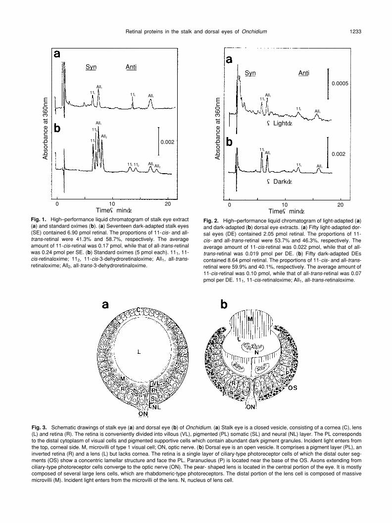
Fig. 2.
High–performance liquid chromatogram of light-adapted (
a
)and dark-adapted (
b
) dorsal eye extracts. (
a
) Fifty light-adapted dor-sal eyes (DE) contained 2.05 pmol retinal. The proportions of 11-
cis-
and all-
trans
-retinal were 53.7% and 46.3%, respectively. Theaverage amount of 11-
cis
-retinal was 0.022 pmol, while that of all-
trans
-retinal was 0.019 pmol per DE. (
b
) Fifty dark-adapted DEscontained 8.64 pmol retinal. The proportions of 11-
cis-
and all-
trans
-retinal were 59.9% and 40.1%, respectively. The average amount of11-
cis
-retinal was 0.10 pmol, while that of all-
trans
-retinal was 0.07pmol per DE. 11
1
, 11-
cis
-retinaloxime; All
1
, all-
trans
-retinaloxime.
0.002
0.0005
(Light)�
(Dark)�
Abs
orba
nce
at 3
60nm
Syn Anti
0 10 20Time(min)�
111
111
111
111
All1
All1
All1
All1
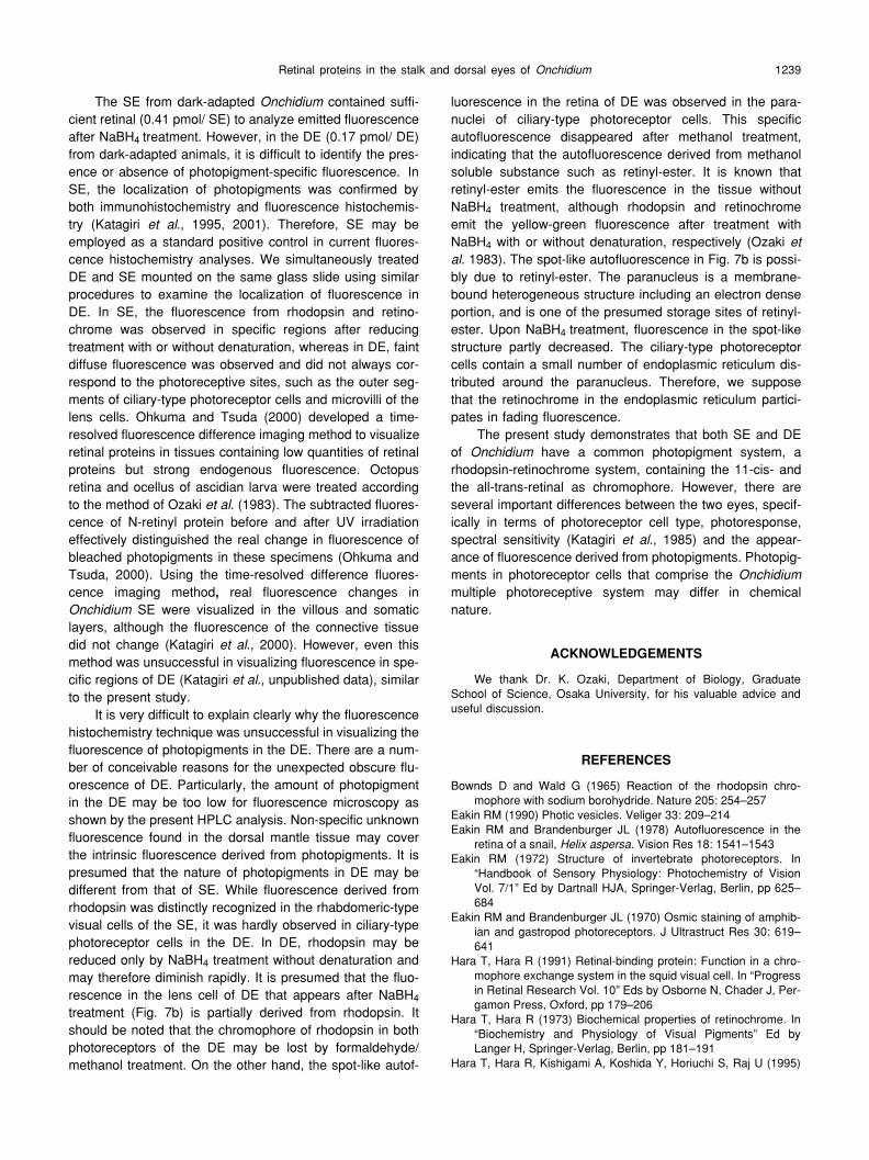
Fig. 3.
Schematic drawings of stalk eye (
a
) and dorsal eye (
b
) of
Onchidium
. (
a
) Stalk eye is a closed vesicle, consisting of a cornea (C), lens(L) and retina (R). The retina is conveniently divided into villous (VL), pigmented (PL) somatic (SL) and neural (NL) layer. The PL correspondsto the distal cytoplasm of visual cells and pigmented supportive cells which contain abundant dark pigment granules. Incident light enters fromthe top, corneal side. M, microvilli of type 1 visual cell; ON, optic nerve. (
b
) Dorsal eye is an open vesicle. It comprises a pigment layer (PL), aninverted retina (R) and a lens (L) but lacks cornea. The retina is a single layer of ciliary-type photoreceptor cells of which the distal outer seg-ments (OS) show a concentric lamellar structure and face the PL. Paranucleus (P) is located near the base of the OS. Axons extending fromciliary-type photoreceptor cells converge to the optic nerve (ON). The pear- shaped lens is located in the central portion of the eye. It is mostlycomposed of several large lens cells, which are rhabdomeric-type photoreceptors. The distal portion of the lens cell is composed of massivemicrovilli (M). Incident light enters from the microvilli of the lens. N, nucleus of lens cell.

Katagiri
et al.1234

Retinal proteins in the stalk and dorsal eyes of Onchidium 1235
of 0.41 pmol/ SE), of which 11-cis- retinal comprised 2.85pmol (average of 0.17 pmol/ SE) and all-trans-retinal, 4.05pmol (average of 0.24 pmol/ SE). The proportion of all-trans-retinal was greater (58.7%) than that of 11-cis-retinal (41.3%).Dorsal eye: Chromophores of photopigments wereextracted as retinaloximes from the 50 dark-adapted and 50light-adapted DEs. HPLC analyses similarly revealed 11-cis-retinal and all-trans-retinal, but no 3-dehydroretinal in DE(Fig. 2). In light-adapted DE, the total amount of retinal wascalculated as 2.05 pmol (average of 0.04 pmol/ DE), ofwhich 11-cis-retinal was 1.10 pmol (average of 0.022 pmol/DE) and all-trans-retinal was 0.95 pmol (average of 0.019pmol/DE). The relative proportions of 11-cis-retinal and all-trans-retinal were 53.7% and 46.3%, respectively (Fig. 2a).In the dark-adapted DE, the total amount of retinal was 8.64pmol (average of 0.17 pmol/ DE), of which 11-cis-retinalcomprised 5.18 pmol (average of 0.10 pmol/ DE) and all-trans-retinal, 3.46 pmol (average of 0.07 pmol/DE). In thedark-adapted DE, the proportions of 11-cis-retinal was greater(59.9%) than that of all-trans-retinal (40.1%) (Fig. 2b).
In dark-adapted eyes, the amount of retinal of one DEwas far less (two-fifth) than that of one SE. Furthermore, inlight-adapted DE, the total amount of retinal was far less(one-fourth), with a slightly lower proportion of 11-cis to all-trans-retinal, than in dark-adapted DE (Figs. 2a, 2b).
II. Fluorescence histochemistryStalk eye: The SE is an ellipsoidal vesicle of about 200 µmin diameter, comprising a cornea, lens and retina (Figs. 3a,4a, 5a). The fine structure of the SE has been described indetail elsewhere (Katagiri et al., 1995). The rhabdomeric-type photoreceptor cell extending well-developed microvilliin the SE is designated the ‘type 1 visual cell’. The retina isa pigmented columnar epithelium divided into villous, pig-mented, somatic and neural layers (Figs. 3a, 4a, 5a). Thevillous layer consists of an enormous tuft of microvilli of type1 visual cells. The dark pigmented layer corresponds to thedistal heavily pigmented cytoplasm of visual cells and pig-mented supportive cells, while the somatic layer containsthe nuclei and cytoplasm of visual and pigmented supportivecells. In the neural layer, axons of visual cells converge to
form the optic nerve.Upon immersion in water or formaldehyde solution,
autofluorescence was observed in the epidermis and its sur-face and in unknown cells in the connective tissue of thetentacle. The SE does not show autofluorescence except forthe lens. The particularly strong autofluorescence of the lensinterfered with the intrinsic fluorescence of the villous layerin the retina when it was treated with NaBH4 with or withoutdenaturation. When cryosections of the SE were treatedwith NaBH4, yellow-green fluorescence was observed in thesomatic layer, which faded rapidly and disappeared com-pletely within 60 sec after UV irradiation (Figs. 4b–d). UponNaBH4 treatment after denaturation, additional fluorescencewas observed in the villous layer (Fig. 5b). Fluorescence in
Fig. 6. Time-course of the optical density that corresponds to fluo-rescent intensity in two portions of the stalk eye (SE) and connectivetissue (CT). Three circled portions were examined; the villous (v)and somatic (s) layers, and the CT (c) in Fig. 5b. The lifetime ofdecay of fluorescence in the SE differs from that of CT. Fluores-cence from the villous (○ ) and somatic (● ) layers declined within 15sec to about 35%, but that from CT (x) did not change considerablyduring irradiation. The time constants (
τ
) of the villous and somaticlayers were 13 and 14 sec respectively, while that of CT was 104sec.
Time(sec)�
Opt
ical
Den
sity(
%)�
00
50
100
5 10
SLVLCT
15
τ=13
τ=14
τ=104 sec
Fig. 4.
(
a
) Differential interference light micrograph of the stalk eye (SE) was taken from the same section as that in Fig. 4b–d after VTRrecording. The pigmented layer (PL, lines) is located between the inner villous (*) and outer somatic layer (SL, ▲ ). To identify the location offluorescence in the SE, the height of the PL is drawn with the lines and the outline of the SE is depicted with dots. CT, connective tissue. (
b
)–
(
d
) Fluorescent micrographs of the stalk eye (SE) treated with sodium borohydride. (
b
) Immediately after irradiation, a yellow-green fluorescentband was observed in the SL (▲ ) under the PL. The lens (L) displayed strong autofluorescence. ( c ) The fluorescence image of the same spec- imen (Fig. 4b) at 10 sec after UV irradiation. Fluorescence in SL (▲ ) diminished rapidly. ( d ) After 30 sec irradiation, fluorescence derived fromretinochrome in the SL (▲ ) disappeared. Fig. 5. ( a ) Differential interference light micrograph of the stalk eye was taken from the same sec- tion as that in Fig. 5b-d after VTR recording. See abbreviations of Fig. 4a. (
b
)–(
d
) Fluorescence micrographs of the stalk eye (SE) treated with
sodium borohydride after denaturation with formaldehyde and methanol. (
b
) Upon
UV irradiation, yellow-green fluorescence of the retino-chrome was visualized in the SL (▲ ) under the PL. Additional fluorescence was observed in the VL (*) along the internal side of PL. Fluores- cence in the SL was stronger than that in VL. The optic density of three circled areas, the villous layer (v), somatic layer (s) and connectivetissue (c), were examined. The lens displayed intense autofluorescence. (
c
) At 10 seconds after irradiation, fluorescence of the retinochromeconsiderably faded in the SL (▲ ), but that of rhodopsin in the VL (*) disappeared. Autofluorescence of the lens was observed throughout. ( d )At 30 seconds after irradiation, the fluorescence of the SL (▲ ) remained faintly, although endogeneous fluorescence of the lens and connec- tive tissue remained.

Katagiri
et al
.1236

Retinal proteins in the stalk and dorsal eyes of
Onchidium
1237
the villous layer was weaker than that in the somatic layer.At 10 sec after irradiation, the yellow-green fluorescence inthe villous layer had almost disappeared, but remainedweakly in the somatic layer (Fig. 5c). Fluorescence in bothlayers faded considerably at 30 sec (Fig. 5d), disappearingcompletely within 60 sec after UV irradiation, although theendogeneous fluorescence of the lens and connective tis-sue was observed throughout.
A typical case of quick decay of fluorescence derivedfrom photopigments is shown in Fig. 6. Time courses of opti-cal density corresponding to a change in fluorescence wereanalyzed at three spots in a sample of Fig. 5, the villous andsomatic layers in SE and connective tissue, that was treatedwith NaBH
4
treatment after denaturation with formaldehydeand methanol. The lifetime of decay of fluorescence inten-sity in both villous and somatic layers differed from that inconnective tissue. The optical density of fluorescence fromthe villous and somatic layers decreased to 30% (
τ
-13)
and35% (
τ
-14) after 15 sec, while that from connective tissuewas maintained at 85% (
τ
-104) during exposure to light (Fig.6).
Dorsal eye:
The DE is spherical in shape (not a closed ves-icle) and about 200 µm in diameter. The DE comprises acup-shaped pigment layer, inverted retina and lens, butlacks cornea (Figs. 3b, 7a, 8a). The retina is composed of asingle columnar epithelium of only ciliary-type photoreceptorcells. The distal portion (outer segment) of the ciliary-typephotoreceptor cell shows a concentric lamellar structure andfaces the pigmented cells of the pigment layer. The supra-nuclear region contains rough and smooth endoplasmicreticulum, several paranuclei (dense inclusions) and otherorganelles. A biconvex pear-shaped transparent lens is sit-uated in the center of DE. It consists of several large lenscells. Each lens cell contains massive microvilli in the distalportion, numerous lamellar bodies and a network of endo-plasmic reticulum in the proximal cytoplasm. Based on finestructure and photoresponse data, the lens cell is identifiedas a rhabdomeric-type photoreceptor cell (Katagiri et al.,1983, 1985, 1998).
In control sections that were immersed in water or form-aldehyde solution, autofluorescence was observed in theepidermis, its surface and in cuboidal cells in the connective
tissue of the dorsal mantle, which did not disappear withreducing treatment. When non-specific fluorescence wasevident in the surrounding connective tissue, weak, obscurefluorescence overspread diffusely the DE (not shown). Theparanuclei of the ciliary-type photoreceptor cell in the retinaemitted strong autofluorescence that faded slightly in thecontrol sections and rapidly after NaBH4 treatment (Fig. 7b–7d) and was not observed after methanol treatment (Fig. 8b)upon UV irradiation.
Fluorescence in DE after treatment with NaBH4 with orwithout denaturation was observed in samples that showedless autofluorescence in connective tissue. Upon NaBH4
treatment, unclear fluorescence was observed in indefiniteand vague areas in individual DE at various intensities anddifferent fading times. Faint fluorescence observed in theretina and the distal microvilli of the lens faded within 30 sec(Figs. 7b–7d). Upon NaBH4 treatment after denaturationwith formaldehyde and methanol, vague fluorescence wasobserved (Fig. 8b). However, additional fluorescence waseither not present or very faint in both the lens and retina.Non-specific fluorescence persisted in the epidermis andconnective tissue around the DE (Figs. 7c–d, 8c–8d). Thedrastic fading of fluorescence within 30sec under UV irradi-ation characteristic of reduced photopigments was notdetected in the DE, despite recording with a high-speedVTR system (Figs. 8c–8d).
DISCUSSION
I. Chromophores in the stalk and dorsal eyes of Onchi-dium
HPLC analyses in the present study revealed 11-cis-and all-trans-retinal in the SE of dark-adapted Onchidiumand the amount of all-trans-retinal was higher than that of11-cis-retinal. The all-trans-retinal is considered to be achromophore of retinochrome (Hara and Hara, 1973; Ozakiet al., 1983, 1986). The rhabdomeric-type photoreceptorcells of gastropoda eyes, including Onchidium SE, containabundantly photic vesicles in the cytoplasm (Eakin, 1972,1990; Kataoka, 1975, Herman and Strumwasser, 1984;Katagiri et al, 1995). As photic vesicles contain retino-chrome (Hara and Hara, 1973; Ozaki et al., 1986), the all-
Fig. 7. (a) Differential interference light micrograph of the dorsal eye corresponds to Figs. 7b-d. The dorsal eye (DE) consists of the pig-mented layer (PL, lines), retina (R) and lens (L). To identify the location of fluorescence in the DE, the height of the PL is drawn with the linesand the outline of the lens is depicted with dots. The outer segment (*) of ciliary-type photoreceptor cell faces to the PL. An arrow shows para-nuclei of ciliary-type photoreceptor cell. CT, connective tissue; E, epidermis; L, lens cell; M, microvilli of lens cell; ON, optic nerve. (b)–(d) Flu-orescence micrographs of the dorsal eye treated with sodium borohydride. (b) Immediately after irradiation, faint, obscure yellow-greenfluorescence was observed in the entire dorsal eye, including the retina and lens. Strong spotty autofluorescence (arrow) was frequentlydetected in the supranuclear regions of ciliary-type photoreceptor cells in the retina. (c) After 10 sec and (d) 30 sec irradiation, almost all fluo-rescence disappeared in the DE, except for non-specific fluorescence in the epidermis (E). Fig. 8. (a) Differential interference light micro-graph of the dorsal eye corresponds to Figs. 8b–d. See abbreviations in Fig.7a. (b)–(d) Fluorescence micrographs of the dorsal eye (DE)treated with sodium borohydride after denaturation with formaldehyde and methanol. (b) Upon UV irradiation, the yellow-green fluorescence ofN-retinyl proteins was observed indistinctly in the DE. Specific fluorescence derived from rhodopsin did not appear in the retina or microvilli (M)of lens cells. (c) After 10 sec and (d) 30 sec irradiation, non-specific fluorescence remained in the DE. Diminishing of specific fluorescencederived from photopigments was not detected in the DE, although endogeneous fluorescence remained in the epidermis (E) and connectivetissue.

Katagiri et al.1238
trans-retinal detected in SE by the present HPLC analysesis possibly derived from the retinochrome in photic vesiclesof type 1 visual cells in the retina. Furthermore, 11-cis-retinalin the dark-adapted SE may be the chromophore of rhodop-sin of type 1 visual cells of which the microvilli occupyalmost all the villous layer (Katagiri et al., 1995). NumerousSEs were required for HPLC analyses due to their minutesize. However, we could not obtain sufficient amounts of SE,and therefore could not examine the chromophores of thelight-adapted SE. In the eye of the marine gastropoda,Conomulex, 11-cis- and all-trans-retinal were also detectedby HPLC analyses (Ozaki et al., 1986). The 11-cis-retinalwas dominant in extracts from the microvillar fraction, whileall-trans-retinal was prevalent in extracts from the photicvesicle-containing fraction. These results indicate that thephotopigment is rhodopsin in the former fraction and retino-chrome in the latter. The amount of all-trans-retinal in theConomulex eye is several times greater than that of 11-cis-retinal. The observed ratio of retinal isomers in chro-mophores of Onchidium SE is similar to that in Conomulexeye (Ozaki et al., 1986).
On the other hand, in the dark-adapted Onchidium, thetotal amount of retinal in one SE was 2.4 times higher thanthat in one DE. A pair of SEs is situated in the tentacles. Incontrast, more than 50 DEs are distributed on the dorsalmantle surface (Katagiri et al., 1985). Therefore, DEappears to be superior in terms of total numbers and con-sequently, total amount of retinal in an individual Onchidium.However, the SE is regarded as the principal eye within themultiple photoreceptive system (Hisano et al., 1972, Katagiriet al., 1985, 1990) on the basis of the presence of sufficientretinal per SE, existence of a rhodopsin–retinochrome sys-tem (Katagiri et al., 2001) and the typical structure of thegastropoda eye (Eakin, 1972, 1990; Kataoka, 1975; Jackletand Colquhoun, 1983; Herman and Strumwasser, 1984;Katagiri et al., 1995).
The present study is the first significant report showingthat the Onchidium DE contains 11-cis- and all-trans-retinalas well as SE. The DE comprises two different types of pho-toreceptor cells, specifically, ciliary-type in the retina andrhabdomeric-type in the lens. Since it is very difficult toseparate the retina and lens in the DE, HPLC analysis wasperformed on all photopigments derived from both ciliary-and rhabdomeric-type photoreceptor cells. It is proposedthat as the retina is the essential receptive site in DE, theHPLC data mainly represent the chromophores of photopig-ments in ciliary-type photoreceptor cells. The proportion of11-cis-retinal (59.5%) to total retinal in dark-adapted DE wascharacteristically greater than that in dark-adapted SE(41.3%). Light-adapted DE contained a decreased propor-tion of 11-cis-retinal to all-trans-retinal compared to dark-adapted DE, suggesting the recycling of retinals in the DE.Notably, total retinal in light-adapted DE was far less (one-fourth) than that in dark-adapted DE. This decreasedamount of total retinal could be explained on the assumptionthat predominant ciliary-type photoreceptor cells in DE con-
tain vertebrate-type visual pigments. In vertebrate eyes con-taining ciliary-type photoreceptors, all-trans-retinal isreleased from the protein opsin during light adaptation,which is converted into retinol and subsequently stored asretinylesters in pigment epithelial cells (Saari, 1994). It ispossible that all-trans-retinal in the ciliary-type photoreceptorcells of light-adapted DE is released from protein and storedas retinylesters in adjacent pigmented cells that directly facethe outer segments of ciliary-type cells. At the same time,retinylester might be stored in the paranuclei of ciliary-typephotoreceptor cells, similar to the oil droplets reported as thestorage site of retinylester in crayfish retina (Suzuki et al.,1988). Unfortunately, our HPLC analyses did not encom-pass data on retinylesters, since these are eluted at the sol-vent front and therefore not quantified with the presentexperimental conditions (Suzuki et al., 1988). Retinalsdetected in the light-adapted DE may be derived mainlyfrom the photopigments (rhodopsin and retinochrome) ofrhabdomeric-type lens cells in the DE. Rhabdomeric-typelens cells have massive microvilli, and contain no photicvesicles but abundant endoplasmic reticulum, which is thestorage site of retinochrome (Katagiri et al., 1998). Thephotic vesicle is a special form of endoplasmic reticulum(Whittle, 1976). Photic vesicle and endoplasmic reticulumwere impregnated by prolonged osmification that demon-strates the presence of lipids, such as vitamin A (Eakin andBrandenburger, 1970; Pourcho and Bernstein, 1975; Kata-giri 1984, 1998). In the Onchidium DE, several coordinatingsystems for photopigment recycling may be present, suchas those in ciliary-type and rhabdomeric-type photoreceptorcells. The rhodopsin-retinochrome system was reported inthe SE (Katagiri et al., 2001). Since 11-cis and all-trans-reti-nal were detected in both SE and DE, the rhodopsin-retino-chrome system may be present in the DE similarly.
II. Fluorescence histochemistryThe localization of retinal protein in the DE was exam-
ined by fluorescence histochemistry and compared with thatof SE. Fluorescence histochemistry is a convenient andvaluable method for detecting photopigments, since themethod only requires treatment with reducing agent and notantibodies (Ozaki et al., 1983). Fluorescence of N-retinylprotein that was reduced by borane dimethylamine wasreported formerly in the snail eye (Eakin and Branden-burger, 1978). Ozaki et al. (1983, 1986) and Hara et al.(1992) demonstrated the localization of rhodopsin andretinochrome, based on differences in their reactivity toNaBH4 in several invertebrate eyes. In Onchidium SE, fluo-rescence derived from rhodopsin and retinochrome wasobserved in villous and somatic layers of the retina. This flu-orescence faded rapidly, in concurrence with earlier obser-vations on the retinas of several gastropods (Ozaki et al.,1983, 1986). The stronger fluorescence in the somatic layerof Onchidium SE than that in the villous layer confirmed thatthe amount of retinochrome is greater than rhodopsin, asshown by HPLC analyses.
Retinal proteins in the stalk and dorsal eyes of Onchidium 1239
The SE from dark-adapted Onchidium contained suffi-cient retinal (0.41 pmol/ SE) to analyze emitted fluorescenceafter NaBH4 treatment. However, in the DE (0.17 pmol/ DE)from dark-adapted animals, it is difficult to identify the pres-ence or absence of photopigment-specific fluorescence. InSE, the localization of photopigments was confirmed byboth immunohistochemistry and fluorescence histochemis-try (Katagiri et al., 1995, 2001). Therefore, SE may beemployed as a standard positive control in current fluores-cence histochemistry analyses. We simultaneously treatedDE and SE mounted on the same glass slide using similarprocedures to examine the localization of fluorescence inDE. In SE, the fluorescence from rhodopsin and retino-chrome was observed in specific regions after reducingtreatment with or without denaturation, whereas in DE, faintdiffuse fluorescence was observed and did not always cor-respond to the photoreceptive sites, such as the outer seg-ments of ciliary-type photoreceptor cells and microvilli of thelens cells. Ohkuma and Tsuda (2000) developed a time-resolved fluorescence difference imaging method to visualizeretinal proteins in tissues containing low quantities of retinalproteins but strong endogenous fluorescence. Octopusretina and ocellus of ascidian larva were treated accordingto the method of Ozaki et al. (1983). The subtracted fluores-cence of N-retinyl protein before and after UV irradiationeffectively distinguished the real change in fluorescence ofbleached photopigments in these specimens (Ohkuma andTsuda, 2000). Using the time-resolved difference fluores-cence imaging method, real fluorescence changes inOnchidium SE were visualized in the villous and somaticlayers, although the fluorescence of the connective tissuedid not change (Katagiri et al., 2000). However, even thismethod was unsuccessful in visualizing fluorescence in spe-cific regions of DE (Katagiri et al., unpublished data), similarto the present study.
It is very difficult to explain clearly why the fluorescencehistochemistry technique was unsuccessful in visualizing thefluorescence of photopigments in the DE. There are a num-ber of conceivable reasons for the unexpected obscure flu-orescence of DE. Particularly, the amount of photopigmentin the DE may be too low for fluorescence microscopy asshown by the present HPLC analysis. Non-specific unknownfluorescence found in the dorsal mantle tissue may coverthe intrinsic fluorescence derived from photopigments. It ispresumed that the nature of photopigments in DE may bedifferent from that of SE. While fluorescence derived fromrhodopsin was distinctly recognized in the rhabdomeric-typevisual cells of the SE, it was hardly observed in ciliary-typephotoreceptor cells in the DE. In DE, rhodopsin may bereduced only by NaBH4 treatment without denaturation andmay therefore diminish rapidly. It is presumed that the fluo-rescence in the lens cell of DE that appears after NaBH4
treatment (Fig. 7b) is partially derived from rhodopsin. Itshould be noted that the chromophore of rhodopsin in bothphotoreceptors of the DE may be lost by formaldehyde/methanol treatment. On the other hand, the spot-like autof-
luorescence in the retina of DE was observed in the para-nuclei of ciliary-type photoreceptor cells. This specificautofluorescence disappeared after methanol treatment,indicating that the autofluorescence derived from methanolsoluble substance such as retinyl-ester. It is known thatretinyl-ester emits the fluorescence in the tissue withoutNaBH4 treatment, although rhodopsin and retinochromeemit the yellow-green fluorescence after treatment withNaBH4 with or without denaturation, respectively (Ozaki etal. 1983). The spot-like autofluorescence in Fig. 7b is possi-bly due to retinyl-ester. The paranucleus is a membrane-bound heterogeneous structure including an electron denseportion, and is one of the presumed storage sites of retinyl-ester. Upon NaBH4 treatment, fluorescence in the spot-likestructure partly decreased. The ciliary-type photoreceptorcells contain a small number of endoplasmic reticulum dis-tributed around the paranucleus. Therefore, we supposethat the retinochrome in the endoplasmic reticulum partici-pates in fading fluorescence.
The present study demonstrates that both SE and DEof Onchidium have a common photopigment system, arhodopsin-retinochrome system, containing the 11-cis- andthe all-trans-retinal as chromophore. However, there areseveral important differences between the two eyes, specif-ically in terms of photoreceptor cell type, photoresponse,spectral sensitivity (Katagiri et al., 1985) and the appear-ance of fluorescence derived from photopigments. Photopig-ments in photoreceptor cells that comprise the Onchidiummultiple photoreceptive system may differ in chemicalnature.
ACKNOWLEDGEMENTS
We thank Dr. K. Ozaki, Department of Biology, GraduateSchool of Science, Osaka University, for his valuable advice anduseful discussion.
REFERENCES
Bownds D and Wald G (1965) Reaction of the rhodopsin chro-mophore with sodium borohydride. Nature 205: 254–257
Eakin RM (1990) Photic vesicles. Veliger 33: 209–214Eakin RM and Brandenburger JL (1978) Autofluorescence in the
retina of a snail, Helix aspersa. Vision Res 18: 1541–1543Eakin RM (1972) Structure of invertebrate photoreceptors. In
“Handbook of Sensory Physiology: Photochemistry of VisionVol. 7/1” Ed by Dartnall HJA, Springer-Verlag, Berlin, pp 625–684
Eakin RM and Brandenburger JL (1970) Osmic staining of amphib-ian and gastropod photoreceptors. J Ultrastruct Res 30: 619–641
Hara T, Hara R (1991) Retinal-binding protein: Function in a chro-mophore exchange system in the squid visual cell. In “Progressin Retinal Research Vol. 10” Eds by Osborne N, Chader J, Per-gamon Press, Oxford, pp 179–206
Hara T, Hara R (1973) Biochemical properties of retinochrome. In“Biochemistry and Physiology of Visual Pigments” Ed byLanger H, Springer-Verlag, Berlin, pp 181–191
Hara T, Hara R, Kishigami A, Koshida Y, Horiuchi S, Raj U (1995)

Katagiri et al.1240
Rhodopsin and retinochrome in the retina of a TetrabranchiateCephalopod, Nautilus pompilus. Zool Sci 12: 195–201
Hara T, Hara R, Kishigami A, Koshida Y, Horiuchi S, Yoshida M,Yamamoto M, Goto T, Raj U (1992) Localization of retin-ochrome in the retina of a Tetrabranchiate Cephalopod, Nauti-lus pompilius. Zool Sci 9: 211–217
Herman KG, Strumwasser F (1984) Regional specializations in theeye of Aplysia, a neuronal circadian oscillator. J Comp Neurol230: 593–613
Hisano N, Tateda H. Kuwabara M (1972) Photosensitive neuronesin the marine pulmonate mollusc Onchidium verruculatum. JExp Biol 57: 651–660
Jacklet JW, Colquhoun W (1983) Ultrastructure of photoreceptorsand circadian pacemaker neurons in the eye of a gastropod,Bulla. J Neurocytol 12: 673–696
Katagiri N, Terakita A, Shichida Y and Katagiri Y (2001) Demonstra-tion of a rhodopsin-retinochrome system in the stalk eye of amarine gastropod, Onchidium, by immunohistochemistry. JComp Neurol 433: 380–389
Katagiri N, Katagiri Y, Shimatani Y, Hashimoto Y (1998) ‘Lens cell’in the dorsal eye of Onchidium is a rhabdomeric-type photore-ceptor cell that depolarizes in response to light. Invest Ophtha-lomol Vis Sci 39: S1059
Katagiri N, Katagiri Y, Shimatani Y, Hashimoto Y (1995) Cell typeand fine structure of the retina of Onchidium stalk-eye. J Elec-tron Microsc 44: 219–230
Katagiri N, Hama K, Katagiri Y, Shimatani Y, Hashimoto Y, AikawaE (1990) High-voltage electron microscopic study on the axonof the dermal photoreceptor cell in the dorsal mantle of Onchi-dium verruculatum. J Electron Microsc 39: 363–371
Katagiri N (1984) Cytoplasmic characteristics of three different rhab-domeric photoreceptor cells in a marine gastropod, Onchidiumverruculatum. J Electron Microsc 33: 142–150
Katagiri N, Fujimoto K, Katagiri Y (1983) Cellular composition andphotoresponse of the lens in the Onchidium dorsal eye. ZoolMag 92: 199–206
Katagiri Y, Katagiri N, Shimatani Y, Terakita A, Ohkuma M, andTsuda M. (2000) A rhodopsin-retinochrome system in theOnchidium stalk eye. Invest Ophthalomol Vis Sci 41: S599
Katagiri Y, Katagiri N, Fujimoto K (1985) Morphological and electro-physiological studies of a multiple photoreceptive system in amarine gastropod, Onchidium. Neurosci Res 2 (suppl): S1–S15
Kataoka S (1975) Fine structure of the retina of a slug, Limax flavusL. Vision Res 15: 681–686
Makino-Tasaka, M and Suzuki, T (1986) Quantitative analysis ofretinal and 3-dehydroretinal by high-pressure liquid chromatog-raphy. Method Enzymol 123: 53–61
Ohkuma M, Tsuda M (2000) Visualization of retinal proteins in thecerebral ganglion of Ascidian, Halocynthia roretzi. Zool Sci 17:161–170
Ozaki K, Terakita A, Hara R, Hara T (1986) Rhodopsin and retino-chrome in the retina of a marine gastropod, Conomulex luhua-nus. Vision Res 26: 691–705
Ozaki K, Hara R, Hara T (1983) Histochemical localization of retino-chrome and rhodopsin studied by fluorescence microscopy.Cell Tissue Res 233: 335–345
Pourcho RG, Bernstein MH (1975) Light dependence of osmiumreactivity in mouse photoreceptor cells. Am J Anat 143: 371–386
Robles LJ, Camacho JL, Torres SC, Flores A, Fariss RN, Matsu-moto B (1995) Retinoid cycling proteins redistribute in light-/dark-adapted Octopus retinas. J Comp Neurol 358: 605–614
Robles LJ, Watanabe A, Kremer NE, Wong F, Bok D (1987) Immu-nocytochemical localization of photopigments in cephalopodretinae. J Nerurocytol 16: 403–415
Saari JC (1994) Retinoids in photosensitive systems. In “The retino-ids: Biology, Chemistry and Medicine 2nd ed”, Eds by SpornMB, Roberts AB, Goodman DS, Raven Press, New York,pp351–385
Stantschinsky W (1908) Über den Bau der Rückenaugen und dieHistologie der Rückenregion der Oncidien. Zeit wiss Zool 90:137–178
Suzuki T, Maeda Y, Toh Y, Eguchi E (1988) Retinyl and 3-dehy-droretinyl esters in the crayfish retina. Vision Res 28: 1061–1070
Terakita A, Hara R, Hara T (1989) Retinal-binding protein as a shut-tle for retinal in the rhodopsin-retinochrome system of the squidvisual cells. Vision Res 29: 639–652
Whittle AC (1976) Reticular specializations in photoreceptors: Areview. Zool Scr 5: 191–206
(Received June 3, 2002 / Accepted September 11, 2002)