Cell-cell interfaces as specialized compartments directing cell ...
28
1 Nature Reviews Molecular Cell Biology Cell-cell interfaces as specialized compartments directing cell function Brian Belardi 1 , Sungmin Son 1 , James H. Felce 2 , Michael L. Dustin 2 , and Daniel A. Fletcher 1,3,4 1 Department of Bioengineering & Biophysics Program, UC Berkeley, CA, 94720 USA 2 Kennedy Institute, University of Oxford, Oxford UK 3 Division of Biological Systems & Engineering, Lawrence Berkeley National Laboratory, Berkeley, CA, 94720 USA 4 Chan Zuckerberg Biohub, San Francisco, CA 94158 Correspondence should be addressed to: D.A.F [email protected] Author contributions The authors contributed equally to all aspects of the article. Competing interests The authors declare no competing interests. Abstract Cell-cell interfaces are found throughout multicellular organisms, from transient interactions between motile immune cells to long-lived cell-cell contacts in epithelia. In this review, we summarize recent findings that support the emerging view of cell-cell interfaces as specialized compartments that biophysically constrain the arrangement and activity of their protein, lipid, and glycan components. Studies of immune cell interactions, epithelial cell barriers, neuronal contacts, and sites of cell-cell fusion have identified a core set of features shared by cell-cell interfaces that critically control their function. Data from diverse cell types show that cells actively and passively regulate the localization, strength, duration, and cytoskeletal coupling of receptor interactions governing cell-cell signaling and physical connections between cells. We review how these biophysical features of cell-cell interfaces, which drive unique membrane organization from local molecular and cellular mechanics, allow cells to respond selectivity and sensitivity to multiple inputs, serving as the basis for wide-ranging cellular function and as opportunities for therapeutic intervention.
-
Upload
khangminh22 -
Category
Documents
-
view
0 -
download
0
Transcript of Cell-cell interfaces as specialized compartments directing cell ...
1
Nature Reviews Molecular Cell Biology
Cell-cell interfaces as specialized compartments directing cell function
Brian Belardi1, Sungmin Son1, James H. Felce2, Michael L. Dustin2, and Daniel A. Fletcher1,3,4
1Department of Bioengineering & Biophysics Program, UC Berkeley, CA, 94720 USA 2Kennedy Institute, University of Oxford, Oxford UK
3Division of Biological Systems & Engineering, Lawrence Berkeley National Laboratory, Berkeley, CA, 94720 USA
4Chan Zuckerberg Biohub, San Francisco, CA 94158 Correspondence should be addressed to: D.A.F [email protected]
Author contributions The authors contributed equally to all aspects of the article.
Competing interests The authors declare no competing interests. Abstract Cell-cell interfaces are found throughout multicellular organisms, from transient interactions between motile immune cells to long-lived cell-cell contacts in epithelia. In this review, we summarize recent findings that support the emerging view of cell-cell interfaces as specialized compartments that biophysically constrain the arrangement and activity of their protein, lipid, and glycan components. Studies of immune cell interactions, epithelial cell barriers, neuronal contacts, and sites of cell-cell fusion have identified a core set of features shared by cell-cell interfaces that critically control their function. Data from diverse cell types show that cells actively and passively regulate the localization, strength, duration, and cytoskeletal coupling of receptor interactions governing cell-cell signaling and physical connections between cells. We review how these biophysical features of cell-cell interfaces, which drive unique membrane organization from local molecular and cellular mechanics, allow cells to respond selectivity and sensitivity to multiple inputs, serving as the basis for wide-ranging cellular function and as opportunities for therapeutic intervention.
2
Introduction Cell-cell interfaces have been the subject of intense interest since the origin of cell theory in the mid-1800s. Today, detailed studies of a variety of interfaces have shown these junctions to be sites of intercellular recognition, communication, and both long-term and short-term adhesion. However, due to the distinct sets of molecules involved at cell-cell interfaces in different tissues and contexts, there has been limited appreciation for the common biophysical and biochemical properties of these contacts that make them powerful and unique zones of information processing and transmission. Efforts to understand the properties and function of cell-cell interfaces span more than a century (see Fig.1 and Supplemental Timeline). The past few decades have also seen incredible progress in elucidating the molecular structure of adhesion proteins through the use of x-ray crystallography and cryo-electron microscopy1,2. Today, membrane interfaces are often depicted as two phospholipid bilayers with a few receptors binding the two together, usually with the goal of emphasizing the role of specific receptor-ligand interactions. While some aspects of this simple abstraction still hold, cell-cell interfaces are now being recognized as local environments that are surprisingly distinct and more complex from those in which bulk behavior of molecules is usually studied. These specialized compartments are responsible for forming both homotypic and heterotypic cell-cell contacts and, through them, directing immune cell recognition, epithelial sheet formation, nerve cell signal propagation, and myoblast fusion (Fig. 2 and Supplemental Information 1). What allows cell-cell interfaces to dictate cell function? Recent work has uncovered that interfaces are organized, physically, in such a way that they carry out two key tasks: i) integrating multiple inputs from a cell-cell contact and ii) generating sensitive and selective responses for function. For example, macrophages are tasked with deciding in a short period of time (mins) whether a target cell or particle is healthy, diseased or foreign and, when appropriate, initiating a phagocytic response. To accomplish this, the interface must assess the density, strength, and mobility of multiple activating and inhibitory ligands on the target and does so by creating a unique environment defined by specific molecules, biophysical properties, and reaction kinetics to perform this task. Cell-cell interfaces from diverse tissues use common components tuned in different ways to direct specific cellular functions (Supplemental Information 2). For instance, interface durations range in timescale from short, transient contacts observed between immune cells to long, persistent contacts between neurons in the brain. They also differ in terms of their length-scales, with the small protrusive interfaces – 100s of nm in scale – of some fusogenic cells dwarfed by the lateral contacts – extending over multiple micrometers – between neighboring epithelia. Even the average interfacial gap distance between the plasma membranes of two cells varies across interface type. Tight junctions between epithelial cells, for example, have a gap size of ~2 nm, whereas presynaptic-postsynaptic membranes in the nervous system are separated by 15-25 nm. These unique traits are central to the execution of specific tasks – T cells and macrophages need

3
to surveil host cells and invaders in short periods of time, while epithelia need to restrict transport of large material between cells. In this review, we summarize findings over the last decade that highlight key biophysical features of the cell-cell interfacial compartment, which organize the interface for cellular decision-making and function. Intracellular membrane contacts between organelles, vesicles, and the plasma membrane have been extensively reviewed elsewhere3–6, so our focus here will be on cell-cell interfaces. Throughout the review, we underscore the unique physical boundary conditions that give rise to membrane organization and activity at seemingly disparate interfaces. We also cover the ways in which cell-cell interfaces couple energy consumption with molecular and cellular mechanics and with signaling networks that converge at interfacial hubs. Since cell surfaces and cell-cell interfaces have long been the target of therapeutic molecules, most recently immunotherapeutics, a fundamental understanding of the molecular mechanisms influencing signaling at cell-cell interfaces has never been more important. Biophysical Features of Cell-Cell Interfaces Below, we discuss five notable biophysical features of cell-cell interfaces that enable these compartments to perform physiologically critical tasks across many different cell types. By creating local molecular environments that differ from those of either cell’s plasma membrane, cell-cell interfaces are sensitive to both the biophysical and biochemical properties of cell surface receptors and ligands, along with their collective interactions and spatial organization. This ability to simultaneously probe for molecular affinity, mobility, size, dynamics, and mechanics at cell-cell interfaces enables multiple layers of rapid communication and regulation between cells that give rise to the remarkably complex and diverse functional consequences of cell-cell interactions. Modern experimental techniques are now allowing researchers to uncover the mechanistic details that underpin cell-cell interface function (Box 1 and Supplementary Information 3), though much more remains to be discovered. As further work helps to illuminate how cells contact and communicate with one another, we expect the list below to grow. 1. Altered Diffusion at Interfaces The fluid mosaic model of the plasma membrane, proposed by Singer and Nicolson in 19727, in which orientated transmembrane receptors and transporters diffuse through a sea of lipids, has led some to assume that all parts of a cell’s plasma membrane – interfacial and non-interfacial alike – are equally accessible and mobile. However, measurements at cell-cell interfaces show dramatically reduced mobility of molecules, due in part to adhesive interactions, membrane ordering and assembly of cytoskeletal structures that obstruct mobility (Fig. 3A). Adhesive and lipid constraints. A membrane protein’s mobility is characterized by the 2D diffusion coefficient for the protein in a lipid environment, typically between 1 - 10 μm2/s in bare model membranes8,9. Membrane interfaces, on the other hand, create regions with diffusive properties that are significantly reduced. In one study, the mobility of an adhesive protein between a supported lipid bilayer (SLB) and a GUV was reduced by a factor of 9.3 from that in a single
4
membrane10, while, in cells, the trans-interacting E-cadherin’s (E-cad’s) diffusion coefficient was estimated to be 0.28 µm2/s11 on free cell membranes and reduced to between 0.5x10⁻² and 6x10-
2 μm2/s at cell-cell interfaces12. These values likely reflect multiple trans interactions across cells at the interface, emphasizing that low mobility is the rule at interfaces, not the exception. Another mechanism that influences mobility at interfaces is association of membrane proteins with specific lipid environments. Both occludin and ZO-1 of the tight junction of epithelial cells have been found to reside in detergent-resistant, cholesterol-rich fractions13 affecting their lateral mobility14, and a similar phenomenon has been observed for desmoglein-1 at desmosomes in epithelia15. Cytoskeletal constraints. Cytoskeletal structures assembled in response to receptor engagement at cell-cell interfaces can form a lateral picket fence that has significant effects on free diffusion of membrane components. For example, at macrophage interfaces, a diffusion barrier is established by an FcγR-IgG interface recruiting integrins, which initiate Arp2/3-mediated actin assembly, ultimately blocking single molecule trajectories of CD45, an inhibitory phosphatase16. If integrin activation or actin assembly was disrupted, then CD45 exclusion was less pronounced and phagocytosis of the pathogen S. typhimurium was disrupted. In another example, the membrane protein CD44 was found to couple to intracellular formin-derived actin structures through ezrin and ankyrin17 to alter diffusion as well as to hyaluronic acid, a type of proteoglycan, through its Link domain in the extracellular space. The latter creates an additional pericellular picket fence that also restricted receptor mobility. The diffusion of phagocytic receptors and glycoprotein pickets are extensively reviewed in18. The first biophysical feature of cell-cell interfaces – regulation of membrane protein mobility by reducing diffusive character – is used by cells to probe and signal to opposing cell surfaces. This regulation is achieved through a variety of physical means, including accumulation of ligated receptors, association of membrane proteins with specific lipid compositions, tethering receptors to the cytoskeleton, and constructing diffusion barriers – within the membrane, below the membrane, and above the membrane – to restrict mobility. By doing so, cells can sample their local neighbors with select receptors and activate specific pathways, such as phagocytosis, when necessary. 2. Size-Dependent Sorting at Interfaces In addition to changes in mobility of proteins at cell-cell interfaces, the presence of a second membrane imposes a physical constraint on the interface that sterically excludes molecules that do not fit and enhances the binding of receptors that do (Fig. 3B). Typically, this means that ligated receptors with small molecular dimensions, such as short heights, form an interfacial space that excludes proteins with large molecular dimensions and imposes physical constraints on protein-protein association. Size-dependent exclusion. Exclusion of membrane proteins from cell-cell interfaces based on their height creates a unique environment for signaling components, such as immunoreceptor tyrosine-based activation/inhibition motifs, ITAMs and ITIMs. In this environment, kinase enzymes tethered to the inner leaflet of the membrane, such as Lck and Lyn, can act freely on ITAM motifs,
5
giving rise to a phosphorylation-rich region that is intimately connected to the membrane interface. An illustrative example comes from the field of immunology, where T cells, macrophages, and natural killer cells all rely on the size-dependent exclusion of CD45, a large mucin containing a cytoplasmic phosphatase domain, from the interfaces they form with other cells19–23. In macrophages, phagocytosis was found to scale with the size of the IgG antibody-antigen complex compared to the size of CD45, with the most efficient eating seen for antigens that were <10 nm in size19. Similar results were obtained for IgE FcR triggering of a mast cell-like cell line20. Based on in vitro experiments, protein height differences of only 5 nm can drive switch-like exclusion of proteins from interfaces24, which may help explain the near complete exclusion of CD45 (28-53 nm) from pMHC-TCR interfaces (13 nm). Size-dependent organization. In early work on epithelial junctions, electron microscopy of polarized epithelia from guinea pig gall bladders demonstrated segregation of the basal-most desmosome from the adherens junction from the apical-most tight junction in epithelial cells25. Decades later, it was found that these distinct interfaces consisted of proteins with molecular scale differences in height, beginning with the desmosomal cadherins, desmoglein and desmocollin (tallest), followed next by the classical cadherins and nectin, and finally by occludin, JAMs, and claudins (shortest)26. Since positioning the different junctions appropriately along the lateral surface of the epithelium is necessary for polarization and adhesion, size-dependent processes in epithelial cells in addition to the known cytoskeletal regulatory elements, e.g. intermediate filaments, catenins, and ZO-126, may play, in our opinion, a significant and to date unrecognized role in barrier formation and tissue cohesion. With the advancement of correlative super-resolution and electron microscopy, visualizing molecular localizations with ultrastructural detail is now within reach, as has recently been shown for the TJ membrane protein, JAM-C27, which opens up the possibility of observing size-dependent processes at cell-cell interfaces. Size effects on binding. The large size of some non-adherent proteins can also modulate binding kinetics of membrane receptors by presenting a physical barrier that can prevent binding (kon) of short receptors and by introducing tension in short receptor-ligand interactions after binding (koff). This latter effect arises when the membrane deforms the bond or when tall molecules exert pressure either by being excluded from an interface or compressed at the binding interface. For receptors that form either an ideal bond, where koff is independent of force, or slip bond, where koff increases with force, tall proteins on a cell surface will simply slow down their binding to cognate ligands28. When receptors form a catch bond, where koff decreases with force (see Mechanosensitivity at Interfaces), however, the physical barrier presented by tall, immobile proteins can promote clustering of receptors29. This is because once a receptor successfully binds to its ligand, the surrounding glycocalyx that is locally compressed can exert tension on the bond, thus enhancing the bond affinity. Consequently, the rate of new bond formation is significantly enhanced near the existing bond, which creates the effect of a ‘kinetic trap’ (see next section on Cooperative Binding and Clustering). This mechanism of receptor clustering at interfaces is particularly relevant in cancer, where cells upregulate tall glycoproteins such as mucin-130–32 and create a bulky glycocalyx. In breast cancer, the bulky glycocalyx of cancer cells has been shown to promote clustering of bound integrins, which leads to the signaling necessary for cell survival33. A similar mechanism was also demonstrated to promote cancer metastasis34.

6
The second biophysical feature of cell-cell interfaces arises from the molecular dimensions of cell surface molecules, an often-overlooked aspect of extracellular proteins and glycans, and the limited gap size between membranes at interfaces. As demonstrated in the above examples, the height of proteins can determine whether they have access to the interface, whether they form spatially separated adhesions, and whether their cell-cell interactions are promoted and strengthened. Ectodomain size can, therefore, have significant ramifications on cancer progression, immune cell activation, and epithelial tissue cohesion. 3. Cooperative Binding and Clustering Ligand-receptor binding at cell-cell interfaces can change both the apparent affinity of adhesive interactions, via cooperative binding, and their spatial organization, via lateral clustering. These phenomena are governed by cooperative effects of physical fluctuations in the membrane and the dimensionality of receptor binding (Fig. 3C). Membrane fluctuations and binding. Membrane bending and thermal fluctuations are known to play a significant role in shaping adhesion and interface formation28. Formation of an adhesion imposes a mechanical constraint perpendicular to the membrane that dampens fluctuations of bilayer membranes, while adhesions that expand the interface can increase tension parallel to the membrane. Due to the competition between the entropy and enthalpy when receptors and ligands bind between fluctuating membranes, membrane adhesions form in spatially segregated zones as demonstrated at the GUV-SLB interface35. This mechanism of spatial segregation of protein binding is thought to contribute to pattern formation at interfaces, for example at the T-cell immunological synapse36. More recently, similar principles have been shown to play a role at other interfaces. For example, a number of different adhesion morphologies were observed at the GUV-SLB interface formed by E-cadherins depending on the average membrane fluctuation amplitude37. As well, dampened membrane fluctuations near the binding interface between the ‘marker of self’ CD47 on a GUV surface and SIRPα immobilized on a glass surface resulted in cooperative binding38. In addition to protein binding at cell-cell interfaces, curved membrane architectures in live cells, like actin-driven filopodia extensions and actin-rich microvilli, have also been shown to play a role in adhesion nucleation in epithelial cells39 and ligand detection in T cells40.
Dimensionality and affinity. The interplay between enthalpy and entropy of molecular binding is fundamentally different between 3D (solution) and 2D (membrane-bound) due to the influences of geometry and mechanical tension across the bond in 2D configurations. Although not trivial to determine the 2D binding affinity of a receptor and ligand whose affinity is measured in 3D, it has been proposed that the 2D dissociation constant (2D Kd) is roughly equal to the 3D (solution) Kd divided by the confinement length, which is dependent on membrane fluctuations and molecular tilting41. In addition, physical forces on the scale of picoNewtons that result from membrane fluctuations, repulsive interactions at the interface, and active cellular processes can further change the binding affinities. Early studies of the interface between a T cell and a supported lipid bilayer (SLB) found that the 2D Kd of the CD2-CD58 interaction is on the order of 1-10
7
molecule/µm2, compared to the 3D Kd on the order of 1 µM42. However, 2D Kd may depend on the specific binding interaction as the 2D Kd between an activating peptide-MHC and T-cell receptor was found to be ~100-200 molecule/µm2 43. A more detailed understanding of 2D binding has been achieved by computational simulations of membrane adhesion. Monte Carlo simulations of E/N-cad interactions of epithelia showed that cis-interactions compensate for the entropic penalty of 2D cadherin trans-interactions to promote formation of a stable membrane interface44. The structure and dynamic properties of proteins are also critically related to the 2D binding affinity. For SYG-1 and SYG-2 proteins – the immunoglobulin superfamily cell adhesion molecules that participate in synapse formation and kidney filtration barrier formation – it was found that the rigidity between domains of SYG-1 as well as the binding angle between SYG-1 and SYG-2 critically affect their binding strength45. Similarly, cadherins require calcium-dependent stiffening of their ectodomains for proper trans binding46. Recent work using x-ray crystallography and cryo-EM has emphasized the profound impact of interface dimensionality in neurons. Protocadherin repertoires are responsible for self-avoidance in the nervous system. In solution, protocadherins form dimer-of-dimers. However, at 2D membrane interfaces, these proteins organize into extended zipper-like lattices along the membrane that rely on alternating orthogonal trans and cis associations47. In the case of a mismatched cell surface repertoire, chain termination of the extended protocadherin lattice occurs because of the lack of a single trans ligation, leading to signaling that results in non-self interaction. Dimensionality and signaling. At membrane interfaces, simply recruiting cytosolic proteins to the plasma membrane creates a region where binding is converted from a 3D to a 2D problem, often with the impact of increasing the effective affinity. This is a fundamental principle in the triggering of many cell surface receptors, particularly in the case of transient, activation-driven contacts such as those of the immune system. In such instances, removal of the degree of freedom provided by a third dimension makes a substantial difference to enzymatic rate constants, meaning previously sub-threshold activity is now sufficient to drive signaling, such as the phosphorylation of surface-recruited ZAP70 by Lck in T cells48. This has the immediate effect of activating ligand-bound surface receptors, but also of restricting signaling to the contact and hence driving cell polarization. Coupled with the observation of sub-domain level organization in signaling microclusters on the order of 10s or 100s49, this highlights the importance of nanoscale organization within contacts for the regulation of signaling. Cooperativity and dynamics. Lateral organization of proteins within contacts serves an important role in the regulation and integration of signaling from multiple sources. This is again most striking in highly transient contacts. During the formation of the T cell immunological synapse, TCR molecules phosphorylated in response to pMHC binding rapidly coalesce into sub-micron scale microclusters which act as the sites of active TCR signaling23. The distribution of signals across the immunological synapse is markedly uneven, since signaling microclusters form at the periphery before translocating towards the center where their associated complexes are
8
disassembled and they are either internalized or packaged into vesicles for extracellular release50. This is achieved in part through restriction of the active form of the TCR-activating kinase Lck to the periphery51, which in turn may be reinforced by the size-dependent exclusion of CD4552. Indeed, the balance of CD45’s activatory effects on Lck and its inhibitory effects on the TCR appears to regulate the antigen threshold for activation53; dampening signaling from weak or non-specific antigen, whilst permitting graded signaling from stronger antigens. This again is highly sensitive to the lateral spatial organization of the various signaling components, since disruption of normal CD45 segregation undermines not just T cell activation but also the graded nature of the response to antigen54,55. Moreover, clustering of the TCR may be a consequence of local binding cooperativity, as an increase in intermolecular kon directly adjacent to ligated TCRs has recently been shown in T cell-SLB experiments56. Longer lifetimes of ligated TCR or higher density of ligands increase the likelihood of subsequent reactions taking place in the T cell, in this case ligation and reversible clustering. Clustering, even by a few receptors, can then segregate CD45 and allow other time-dependent reactions to take place, like phosphorylation of ITAMs by Lck, followed by ZAP70 association and phosphorylation, eventually leading to T cell activation43. Clustering of receptors and downstream effectors, the third biophysical feature of interfaces, can serve as a checkpoint for cell activation. Interfaces make use of their 2D environment to promote clustering given the proper conditions – if membrane fluctuations are dampened, if repeating trans and cis interaction can form, if high 2D concentration of enzymes can reach thresholds for signaling, and if ligation events augment the surrounding binding kinetics. This provides cells a critical layer of regulation for subsequent signaling in immune cells, neurons, and during development. 4. Mechanosensitivity at Interfaces The biophysical signatures of cell-cell interfaces described so far – altered diffusion, size-dependent sorting, and cooperative binding – are all passive phenomena that can occur in the absence of energy consumption. Membrane interfaces, however, are a highly dynamic, energy-consuming compartment on account of the membrane’s close association with the cell cortex. The energy source for interfaces is mainly derived from one ubiquitous process, the hydrolysis of ATP (~20 kT). This chemical transformation is usually accomplished by filamentous actin which consumes approximately 50% of the total cellular ATP57. While actin is constantly being turned over and remodeled right below the membrane, other cytoskeletal motor proteins are contributing to the energy-consuming nature of interfaces too, by aligning and exerting force on filaments through ATP hydrolysis. Myosin-associated actin structures, so called actomyosin, can collectively contract filaments, achieving a pulling force on membrane interfaces (~1-5 pN per myosin per powerstroke58). Coupling of energy consumption to mechanical perturbation can have a profound impact on individual transmembrane proteins at the interface, including force-sensing and downstream signaling (Fig. 4A, B). Force-sensing proteins at interfaces. Recently, TCRs have been shown to bind to matched peptide-MHCs through a catch bond but bind to mismatched peptide-MHCs as a slip bond, the characteristics of which can lead to robust ligand discrimination - a 15-fold change in differential
9
recognition and T cell activation when under force59. The maximal catch bond for TCR was shown to be at 10 pN of applied force, which can easily be accounted for by the pulling force from retrograde actin flow and myosin-driven contraction, shown for example by Murugesan et al.60 among others, right underneath the TCR-pMHC interface. Intriguingly, affinity does not seem to compensate for the lack of force-sensitivity as a number of high affinity (2D and 3D) pMHC-TCR complexes failed to induce T cell activation61. However, the origin of forces that triggers catch bond formation at immune cell interfaces remains an open question and an active area of research (Box 2). Furthermore, in T cells, it has been proposed that pMHC-TCR force sensitivity can be related to the way force is applied at T cells, i.e. whether force is applied at a constant force vs. a constant velocity62. Other receptors also display similar behavior. Notch interfaces, e.g. Notch1-Jag2, which play a significant role in organismal development, also exhibit catch bond behavior63, which suggests that coupling of mechanics to interfacial energetics is likely widespread. The question of how the adherens junction is connected to F-actin belts, a prerequisite for developing polarized epithelial tissue, has been evaluated by examining the force sensitivity of the E-cadherin-β-catenin-α-catenin-F-actin complex. In an optical trap experiment, α-catenin was found to form a catch bond towards F-actin when in complex with E-cadherin and β-catenin, requiring a force of ~5 pN to induce the strongly bound state. This force is physiologically relevant as E-cadherin in epithelial cells was found to be under a similar magnitude of tension by protein tension sensor measurements64. Moreover, α-catenin is known to partially unfold under this amount of force, exposing a cryptic binding site for vinculin, which provides another link between the adherens junction and the cytoskeleton65. Vinculin, itself, has recently been shown to exhibit catch bond behavior toward F-actin66. The catch-bond behavior of vinculin, though, appeared to depend on the direction of applied force – revealing a possible mechanism for polarizing actin structures and organizing the adherens junction under load. Of note, α-catenin and vinculin are not the only proteins of the adherens junction that display force responsiveness. Atomic Force Microscopy (AFM) and single molecule experiments have been used to demonstrate that E-cadherin trans association is force-responsive67. E-cadherin-E-cadherin binding is known to adopt two distinct conformations, the X-dimer and the strand-swap dimer. The X-dimer conformation forms catch-bonds in the presence of calcium, while the strand-swap dimer forms slip bonds68,69 – effectively allowing E-cadherin to increase affinity in vivo above the weak trans interaction observed in solution. These two conformations can be interconverted by achieving an intermediate state that displays ideal bond, force-insensitive, behavior70. E-cadherin appears capable of transitioning to different conformations under varying loads, which regulates the initiation and eventual stability and stiffness of epithelial contacts in response to stress. Membrane deformability and cortical tension. Suspended, spherical cells typically exhibit high cortical tension due to contractile acto-myosin networks associated with the intracellular side of the cell membrane71. In order for interfaces to form, sites of cell-cell contact must relieve cortical tension locally to flatten the membrane along the entire length of the adhesion72 – effectively building a tension differential parallel to the membrane axis. For example, cadherin-based contacts are shown to recruit the Elmo-Dock complex, which in turn causes actin reorganization and actin cortex dissolution through modulating RhoGTPase activity, namely Rac activity, locally73. This is in contrast to the rest of the plasma membrane where the actin cortex bundle
10
remains dense and continuous. Thus, cadherin-based interfaces, through Elmo-Dock, are able to create a mechanical differential between the interface and the rest of the cell membrane, which allows cells to adhere and maintain large contacts for the lifetime of the tissue. The cortex underlying the plasma membrane of a cell-cell interfaces need not behave in mechanically the same way, thereby allowing even more diverse functions of interfaces. In fact, inhomogenous actin structures linked to adherens junctions have recently been observed in epithelial cells. Actin turnover at the tip of an actin bundle, which is in close proximity to E-cadherin, is faster than turnover of the stalk region of the bundle away from the membrane74. Another interesting example comes from the myoblast fusion field. Myoblast cells fuse their membranes at sites of membrane interfaces to form long, multinucleated muscle cells. This is accomplished by first initiating a fusogenic contact, which can direct localized cytoskeleton assembly on either side of the membrane, and then by forming a fusion pore. Although mechanics have been implicated in overcoming the energy barrier associated with membrane fusion, it is difficult to imagine how fusion takes place if one cell is pushing against another’s compliant, deformable membrane. The fusogenic interface consists of two main adhesion proteins, sticks and stones (Sns) and dumbfounded (Duf), which organized actin into podosome-like protrusive structures and thin sheaths of actin beneath the membrane, respectively75. Surprisingly, these two very different actin-derived mechanical environments can exist simultaneously at the fusogenic interface. The “attacking” Sns-expressing cell makes use of protrusive forces from the cytoskeleton, and in response, the “receiving” founder cell increases cortical tension at the interface by accumulating myosin II76. These two forces work together to compress the “attacking” cell and “receiving” cell membranes, thereby promoting cell-cell fusion. The dynamic nature of interfaces, through ATP consumption, can thus lead actin to take on many different mechanical forms, even across a single interface. The mechanism by which the T cell immunological synapse is typically believed to function involves the nucleation of signaling in focal adhesions by transmembrane receptors undergoing long-lived interactions with ligands on the opposing cell. This provides a stable network of interactions reaching from the cytoskeleton of one cell to that of the other, and hence spatiotemporal organization of the foci as a whole can be achieved through actin remodeling on one or both sides. In the case of TCR-containing microclusters, signaling TCR complexes drive the polymerization of local F-actin foci through the recruitment of WASP and the Arp2/3 complex independently of formins77, in contrast to the formin-dependent polarization of microtubule-associated structures into the immunological synapse78. The fourth biophyiscal feature of interfaces centers on the generation of localized mechanical forces. By coupling active mechanical perturbation to interfaces, cells test bonds, deform membranes, reinforce assemblies, and drive otherwise intractable processes. The interface is uniquely poised for this activity because of the coupling of mechanics across cells and is used in diverse physiological examples, from tissue development and maintenance to cell-cell fusion. 5. Temporal Coordination at Interfaces
11
The assembly of spatially-defined signaling domains, both dynamic and long-lived, opens the possibility of temporal control of signaling through incorporation of rate-limiting recruitment events. While each of the biophysical signatures of cell-cell interfaces described above involves time as a component – diffusion and residence time at an interface, cooperative binding and cluster size increase, mechanosensitivity and bond lifetime – an emerging feature of the interfacial compartment is orchestration of multiple time-dependent events into a temporally-coordinated process. For instance, recent work on tight junctions suggests that the kinetics of alignment between dynamic claudin strands in the membrane and the stiff actin cytoskeleton might play an outsized role in the time-dependence of epithelial permeability79. In other examples, temporally coordinated processes are achieved through stepwise assembly of signaling clusters, biomolecular condensates, and, in some cases, regulation of gene expression. Timing of microcluster assembly and organization. In T cells, microclusters assemble in a step-wise manner defined by the sequential recruitment of TCR, ZAP70, LAT, and LAT-associated effectors49, with moderate time lags between steps on the order of tens of seconds. Temporal regulation in this manner may impact TCR discrimination between pMHC complexes of varying affinities, as it imposes a defined minimum lifetime of TCR retention within the contact necessary for active microcluster generation, which is in turn dependent on TCR-pMHC interaction lifetime. Similar temporal thresholds may exist in other systems, particularly those involving step-wise assembly of signaling complexes. The most obvious consequence of such tight spatiotemporal organization is to limit the lifetime of any one signaling TCR complex, and hence rapidly downregulate TCR activity to prevent excessive T cell responses. Just as important, however, is the integration of signals from multiple receptors at the same site. Since the initiation and maintenance of such signals are the product of enzymatic cascades, the concentration of enzymes and potential protein substrates into a localized region has a substantial impact on the extent and nature of emergent signals. In the most basic scenario, concentration of TCR with the adaptor protein LAT promotes LAT phosphorylation by TCR-bound ZAP70, thereby acting as simple amplification of an otherwise weaker signal. More complex regulation can emerge through the incorporation of various activating or inhibitory co-receptors into the same microclusters, whereupon associated modulators can locally influence signaling. The best characterized case is for the co-receptor CD28, which is recruited to TCR-containing microclusters in a ligand (CD80 or CD86)-dependent manner80, where it recruits the kinase PKCθ and drives activation of PI3K. Biomolecular condensates and low-affinity interactions at membranes. The high density of adaptor proteins and their multivalent ligands, such as phosphorylated receptors, at cell-cell interfaces make these subcellular compartments poised for biomolecular condensates (or liquid-liquid phase separates). Condensation of membrane proteins and their downstream adaptors has been of intense interest lately in a broad range of fields, as reviewed in81. However, it’s important to note that the concentration threshold for phase separation is drastically reduced in 2D, by ~30-fold82. Thus, cell-cell interfaces are potentially rich sources and sites for condensation of proteins into cross-linked networks, further excluding other proteins. This was recently demonstrated for the TCR signaling cascade83, where purified, phosphorylated LAT of T cells can form a phase
12
separated cluster in the presence Grb2 and Sos1, promoting MAPK signaling in cells. Interestingly, phase separation can also lead to the recruitment of the ZAP70 kinase and the exclusion of the phosphatase CD45 in reconstitution experiments, offering an orthogonal means of CD45 segregation to kinetic segregation at T cell interfaces. 2D biomolecular condensates also appear instrumental in assembling actin structures and increasing the specific activity of actin nucleators and polymerases82–85. Profoundly, biomolecular condensation may serve a role in kinetic proofreading during T cell activation86. The GEF domain of SOS requires long activation times (~50 s), and a biomolecular condensate, like that formed between LAT-Grb2-SOS in 2D, extends the dwell times of SOS to release its autoinhibition, which allows SOS to achieve full activation of its GEF activity for Ras – a necessary step in T cell activation (Fig. 4C). Signaling domains formed through multiple low affinity, short-lived interactions, where the domain is constant but the proteins within remain dynamic, has apparent functional advantages at cell-cell interfaces. The resultant fluidity of the domain at a single-protein level means that it does not form fixed cytoskeletal foci, yet can still act as an active signaling domain and as a site for modulating the activity of other signaling molecules that may themselves be undergoing actin-driven migration. An example of such a domain is that formed by the T cell adhesion protein CD2, which binds only weakly to its ligand CD58 and has a relatively rapid off rate87. Nonetheless, CD58-binding is sufficient to induce accumulation of CD2 within the T cell immunological synapse, wherein its distribution is highly dependent on the density of CD2 recruited88. Importantly, since fluid domains such as those of CD2 are dependent on transient ligation rather than receptor activation (unlike TCR microclusters and other similar structures), they can persist even when signaling is locally inhibited (e.g. by PD1) and hence can serve as modulatory domains under a wide range of both activating and inhibitory conditions. Though the contribution of such dynamic domains to cell-cell interactions is not well defined, several comparable low-affinity interactions are known to contribute to a range of contacts in different systems. A prominent example is the neural cell adhesion molecule (NCAM) which mediates neural cell-cell interactions and neurite outgrowth89. NCAM exists as a high-affinity dimer in cis but forms low-affinity homophilic and heterophilic interactions in trans90, the dynamic nature of which strongly contributes to neural plasticity91,92. NCAM on developing NK cells controls their motility on but not adhesion to stromal cells93, while homophilic interactions between NCAM on mature NK cells and targets cells has been linked to increased cytotoxicity94. The function of NCAM in such processes is only partially linked to its role as an adhesion molecule, and is strongly dependent on its intracellular signaling processes, such as interaction and activation of growth factor receptors and Src kinases95. These observations are consistent with the potential for dynamic NCAM domains in the manner of CD2; acting partially as anchor points between cells but also as active signaling foci emerging from the ensemble behavior of many proteins rather than the stable localization of a small number. It remains to be seen how commonplace such domains are across interface biology. Molecular assemblies are also found in the postsynaptic density (PSD) within the postsynaptic membrane at neuronal synapses96. The PSD of all vertebrate species share a highly complex proteome with ∼1000 conserved proteins97, and super-resolution microscopy has revealed that
13
the sub-synaptic proteins are concentrated into nanodomains (∼80 nm diameter)98,99. It has also been shown that the membrane-associated guanylate kinase (MAGUK) scaffold proteins, such as post synaptic density protein 95 (PSD-95), are responsible for the lateral patterning of PSD components, as reviewed in97,100. Interestingly, recent in vitro reconstitution of purified fragments of PSD95 and SynGAP suggested a liquid-liquid phase separation of postsynaptic proteins into nanodomains101. Finally, membrane condensates appear in epithelial cells as well. Phase separation of purified ZO-1102 and of non-junctional pools of ZO-1103 has recently been documented, pointing to a possible widespread phenomenon across interface types. Localization of gene expression. In the last few years, cell-cell interfaces have emerged, unexpectedly, as sites of gene regulation. Most interfaces are spatially distant from gene expression programs occurring within the nucleus, but several novel mechanisms have been found to operate at cell-cell interfaces due to their unique biochemical and biophysical properties. As one example, epithelial interfaces are long-lived and stable structures, and cells appear to take advantage of this temporal feature by localizing RNA interference machinery to adherens junctions104. There, certain microRNAs are enriched along with the interference machinery to silence drivers of pluripotency, such as SOX2 and MYC, preventing the dedifferentiation of epithelial cells within a developed tissue. Thus, cells use the presence of the interface compartment as an indicator of cell health and state. Integration of multiple inputs remains one of the most intriguing aspects of cell-cell interfaces. In the fifth biophysical feature of cell-cell interfaces, temporal coordination helps to partly explain how cells rely on these membrane hubs to integrate a multitude of factors from their local environment and direct cell function. Across diverse interfaces, long time-lags in enzymatic activation are used to sample and read-out the numerous ligation and organizing events at the membrane, including upstream adhesions and conversions, critical concentrations for phase separation, and recruitment of RNA interference machinery. Conclusions and perspectives The wealth of new data on cell-cell interfaces from diverse biological systems, only some of which we have been able to reference in this review, provide a window into the physical interactions that govern cell-cell communication and organization. Those interactions, we are learning, is at times slowed by local obstructions, constrained by interface geometry, corralled into clusters, strained by mechanical forces, and sequenced into carefully timed steps. The cell-cell interfaces that are home to these biophysical features constitute a unique compartment within cells, one that may serve as a global center of transcriptional and metabolic activity for the entire cell – something we are only beginning to understand in mechanistic detail. Yet, a number of open questions surrounding cell-cell interfaces remain. How does the diversity of cell surface biomolecular size and the extent of glycosylation shape interfaces and influence function? What is the precise relationship between lipid composition, organization, and protein mobility? Since interfaces can adopt several possible molecular states, how does the cell tune the contributions from driving forces that are equilibrium-based and those that are energy-consuming to select a final state for function? Is it possible to engineer novel synthetic cell-cell interfaces in vivo? Can new therapies
14
be devised that target the organization of interfaces for intervention (see Box 3)? The increasing array of molecular, biophysical, and imaging tools now available to interrogate cell-cell interfaces promises new insights and surprises in the near future, providing a guide to address a multitude of diseases that depend on proper functioning of cell-cell interfaces. Acknowledgments We thank Eva Schmid for her work on the topic of this review and the Fletcher and Dustin labs for helpful discussions. B.B. was supported by the NIH Ruth L. Kirschstein NRSA fellowship from the NIH (1F32GM115091). S.S. was supported by Life Science Research Foundation. J. F. and M. L. D. were supported by Wellcome Trust. This work was supported in part by the NIH R01 GM114671 (DAF), the Immunotherapeutics and Vaccine Research Initiative at UC Berkeley (DAF), the Miller Institute for Basic Research (DAF), the NSF Center for Cellular Construction (DBI-1548297) and the Chan Zuckerberg Biohub (DAF).
15
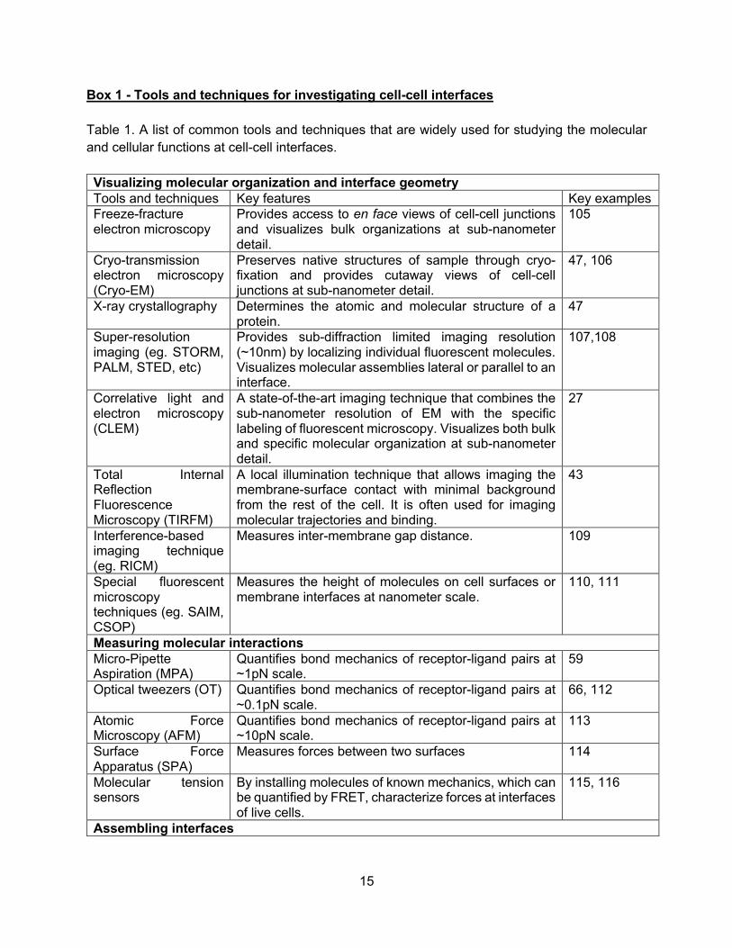
Box 1 - Tools and techniques for investigating cell-cell interfaces Table 1. A list of common tools and techniques that are widely used for studying the molecular and cellular functions at cell-cell interfaces. Visualizing molecular organization and interface geometry Tools and techniques Key features Key examples Freeze-fracture electron microscopy
Provides access to en face views of cell-cell junctions and visualizes bulk organizations at sub-nanometer detail.
105
Cryo-transmission electron microscopy (Cryo-EM)
Preserves native structures of sample through cryo-fixation and provides cutaway views of cell-cell junctions at sub-nanometer detail.
47, 106
X-ray crystallography Determines the atomic and molecular structure of a protein.
47
Super-resolution imaging (eg. STORM, PALM, STED, etc)
Provides sub-diffraction limited imaging resolution (~10nm) by localizing individual fluorescent molecules. Visualizes molecular assemblies lateral or parallel to an interface.
107,108
Correlative light and electron microscopy (CLEM)
A state-of-the-art imaging technique that combines the sub-nanometer resolution of EM with the specific labeling of fluorescent microscopy. Visualizes both bulk and specific molecular organization at sub-nanometer detail.
27
Total Internal Reflection Fluorescence Microscopy (TIRFM)
A local illumination technique that allows imaging the membrane-surface contact with minimal background from the rest of the cell. It is often used for imaging molecular trajectories and binding.
43
Interference-based imaging technique (eg. RICM)
Measures inter-membrane gap distance. 109
Special fluorescent microscopy techniques (eg. SAIM, CSOP)
Measures the height of molecules on cell surfaces or membrane interfaces at nanometer scale.
110, 111
Measuring molecular interactions Micro-Pipette Aspiration (MPA)
Quantifies bond mechanics of receptor-ligand pairs at ~1pN scale.
59
Optical tweezers (OT) Quantifies bond mechanics of receptor-ligand pairs at ~0.1pN scale.
66, 112
Atomic Force Microscopy (AFM)
Quantifies bond mechanics of receptor-ligand pairs at ~10pN scale.
113
Surface Force Apparatus (SPA)
Measures forces between two surfaces 114
Molecular tension sensors
By installing molecules of known mechanics, which can be quantified by FRET, characterize forces at interfaces of live cells.
115, 116
Assembling interfaces
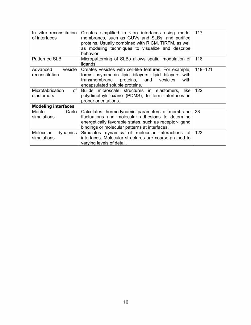
16
In vitro reconstitution of interfaces
Creates simplified in vitro interfaces using model membranes, such as GUVs and SLBs, and purified proteins. Usually combined with RICM, TIRFM, as well as modeling techniques to visualize and describe behavior.
117
Patterned SLB Micropatterning of SLBs allows spatial modulation of ligands.
118
Advanced vesicle reconstitution
Creates vesicles with cell-like features. For example, forms asymmetric lipid bilayers, lipid bilayers with transmembrane proteins, and vesicles with encapsulated soluble proteins.
119–121
Microfabrication of elastomers
Builds microscale structures in elastomers, like polydimethylsiloxane (PDMS), to form interfaces in proper orientations.
122
Modeling interfaces Monte Carlo simulations
Calculates thermodynamic parameters of membrane fluctuations and molecular adhesions to determine energetically favorable states, such as receptor-ligand bindings or molecular patterns at interfaces.
28
Molecular dynamics simulations
Simulates dynamics of molecular interactions at interfaces. Molecular structures are coarse-grained to varying levels of detail.
123
17
Box 2 - Which forces drive mechanosensitive signaling at T cell interfaces? Although mechanosensitivity has emerged as a key player in many areas of interfacial signaling, the precise source of mechanical force driving these processes is often poorly defined. Several models of force-generation have been proposed, however it is not always clear which is the major contributor in each case. Of these, the most prominent models are those of cytoskeletal-driven lateral force, forces derived from membrane fluctuations, and glycocalyx compression-driven axial force. Unquestionably, the strongest active forces that can be accounted for under these models emerge from the contractile movements of actomyosin complexes in the cytoskeleton, as discussed above. The ensemble contributions of large numbers of pN powerstrokes58 generate nN-scale forces that can link to large protein assemblies or domains, however it is not clear if this is also able to manifest as constant pN-level forces on individual bonds, such as those proposed to define TCR catch bond affinity59. This necessitates that the force per bond remains roughly constant during the lifetime of the TCR-actomyosin interaction, which transitions substantially from single complex-level interactions in probing microvilli to large ensemble interactions in translocating microclusters. It is not clear how such consistency is maintained, nor how early TCR-actomyosin interactions would generate constant force given the cyclical nature of the actomyosin powerstroke. Moreover, if a TCR-pMHC catch bond drives peptide discrimination, the force acting on this bond must be present during initial TCR-pMHC interaction but before T cell activation. The lack of substantial TCR-actin cross-linking prior to activation, and the propensity of actomyosin to generate lateral rather than axial force, raise further questions about its ability to deliver bond-specific forces to TCR complexes in a meaningful manner. Conversely, force derived from membrane fluctuations seems likely to be more consistent and less sensitive to the molecular context of protein-protein interactions. However, whilst it has been proposed that lateral segregation of proteins may be driven by transient membrane fluctuations (James and Vale, 2012), it is not clear whether they would be capable of generating forces on the pN scale required for TCR-pMHC catch bond activation. Resistance to compression by the glycocalyx may also be able to achieve relatively uniform force distribution across a whole contact, however in this case it does appear to reach pN levels as seen by the demonstration that such an effect can activate integrin catch bonds29. This would therefore seem to be a promising model for the uniform maintenance of TCR catch bonds throughout the lifetime of the T cell-APC contact. Which glycocalyx components may contribute to force generation in the T cell-APC interface is unclear, however a strong candidate must be the highly-abundant CD45. CD45 was included in GUVs used to discriminate alternately activated slip and catch TCR-pMHC bonds by Garcia and colleagues61, however dense CD45 on the T cell side may also provide resistance to compression. Similarly, receptor-ligand complexes with axial dimensions larger than that of the TCR-pMHC, such as LFA1-ICAM1, could also promote a localized glycocalyx effect capable to providing constant axial force to the TCR124. One issue, though, is that small adhesion molecules like CD2-CD58, which are generally seen as supporting TCR triggering, might shield TCR-pMHC interactions from force and prevent the testing of catch bonds125. In all, each model of force generation at the TCR has its own caveats, many of which apply to mechanosensitivity in other systems.
18
Box 3 - Implications for immunotherapy How might this new picture of cell-cell interfaces as compartments be taken advantage of for therapeutic purposes? One contemporaneous development in biomedicine over the past two decades has been the increased use of cell-based immunotherapy in the clinic. Immunotherapy can come in different forms, but antibody-dependent cellular cytotoxicity (ADCC) and ex vivo T cell engineering, e.g. chimera antigen receptor (CAR) – T expressing cells (CAR-T), are some of the most prominent examples being used in patients today. These non-native interfaces are subject to the same organizing forces discussed above, providing new tools that could help to design desired immune responses. Below, we briefly outline means by which cell-based immunotherapies could make use of the biophysical signatures of cell-cell interfaces to potentially improve efficacy in the clinic (See Box Fig.). i) Engineer interface size: Molecular sizes of adhesive interactions control the intermembrane gap distance, thereby excluding large inhibitory glycoproteins, like CD45, from immune synapses. To maximize this organizing force, antibodies involved in ADCC and CAR-T receptors should target antigens that are small in molecular dimensions or that are in close proximity to the target cell’s membrane – in effect, shortening the cell-cell interface19. ii) Engineer 2D affinity: Cell-cell interfaces are defined by a high density of proteins at the site of adhesion. By increasing the 2D affinity between adhesive proteins, the interface becomes denser, in turn driving cell activation. For therapeutic antibodies, multiple approaches have been pioneered to increase 2D Fc affinity, such as engineering the N-linked glycosylation on the Fc portion of the antibody and mutating the Fc region to modulate affinity126. Another, less explored option may be to tailor the flexibility of the Fc domain, since conformational flexibility plays a more important role at 2D interfaces. iii) Engineer mechanosensitivity: Active processes shape and organize cell-cell interfaces and control the downstream output of junction formation. Designing CAR-T receptors that are sensitive to a tumor’s mechanics may prove therapeutic advantageous, for example. This type of strategy could be characteristic of an AND logic gate, where a CAR T targets a tumor antigen, but only forms a robust response in the presence of proper interfacial mechanics. One could imagine adapting SynNotch receptor technology to be force sensitive127. As well, an orthogonal approach would be target the interfacial mechanics themselves. By altering tumor cell cortical tension or interfacial tension, cell-cell interfaces with longer-lived states would be accessible, leading to cell activation and cell killing.128,129 iv) Engineer composition and stoichiometry: Perhaps the most brute force approach to improve cell-based immunotherapy is by changing the biochemical composition at the cell-cell interface. Since there are many inhibitory co-receptors at immune cell interfaces, expression-modifying methods, such as siRNA silencing, and gene-editing with CRISPR techniques may prove effective for biochemical remodeling of interfaces. Other approaches already in practice are based on antibodies that block specific trans interactions between inhibitory receptors, e.g. anti-
19
PD-1 and anti-PD-L1 antibodies, at the interface. Direct biochemical remodeling of an interface has also been achieved through therapeutic enzyme constructs130.

20
Glossary • Physical boundary conditions: set of constraints that define a closed physical system • Tight junctions: cell-cell junction that seals adjacent epithelial cells together, preventing the
passage of most dissolved molecules from one side of the epithelial sheet to the other. • Adherens junctions: Cell junction in which the cytoplasmic face of the plasma membrane is
attached to actin filaments. Examples include the adhesion belts linking adjacent epithelial cells.
• Hydrophobic mismatch: for a lipid bilayer, when the height of a hydrophobic domain differs in length from another
• Antigen-presenting cell: Highly specialized cells that can process antigens and display their peptide fragments on the cell surface together with other, co-stimulatory, proteins required for activating naïve T cells.
• Desmosome: Type of anchoring cell-cell junction, usually formed between two epithelial cells, characterized by dense plaques of protein into which intermediate filaments in the two adjoining cells insert.
• slip bond, ideal bond, catch bond: slip bond refers to the shortening of bond lifetime under force; an ideal bond refers to a bond lifetime that is not influenced by force; catch bond refers to bonds that increase their bond lifetimes under force
• Glycocalyx: Carbohydrate-rich layer that forms the outer coat of a eukaryotic cell. Composed of the oligosaccarides linked to intrinsic plasma membrane glycoproteins and glyolipids, as well as glycoproteins and proteoglycans that have been secreted and reabsorbed onto the cell surface.
• Anti-PD1 immunotherapy: cancer therapy that blocks the inhibitory interaction between PD-1 on immune cells and PD-L1 on cancer cells
• Liquid-liquid phase separation: the demixing of a fluid into two distinct liquid phases • α-catenin: adaptor protein of the adherens junction that is part of E-cadherin-catenin
complex and can bind to the actin cytoskeleton • Vinculin: adaptor protein of both the adherens junction and focal adhesions that re-inforces
connections to the actin cytoskeleton • WASP: nucleation promoting factor of the actin cytoskeleton that acts on the Arp2/3
complex • Arp2/3 complex: protein complex that nucleates the assembly of branched actin filament
networks • Focal adhesion: A type of anchoring cell junction, forming a small region on the surface of a
fibroblast or other cell that is anchored to the extracellular matrix. Attachment is mediated by transmembrane proteins such as integrins, which are linked, through other proteins, to actin filaments in the cytoplasm.
• Yap1: transcription factor that translocates to the nucleus in a mechanics-dependent manner
• Antibody-dependent cellular cytotoxicity (ADCC): The killing of antibody-coated target cells by cells with Fc receptors that recognize the constant region of the bound antibody. Most ADCC is mediated by NK cells that have the Fc receptor FcγRIII on their surface.
21
• Chimera antigen receptor (CAR): Engineered fusion proteins composed of extracellular antigen-specific receptors (e.g., single-chain antibody) and intracellular signaling domains that activate and co-stimulate, expressed in T cells for use in cancer immunotherapy.
22
References 1. Honig, B. & Shapiro, L. Adhesion Protein Structure, Molecular Affinities, and Principles of
Cell-Cell Recognition. Cell 181, 520–535 (2020). 2. Rozbesky, D. & Jones, E. Y. Cell guidance ligands, receptors and complexes -
orchestrating signalling in time and space. Curr. Opin. Struct. Biol. 61, 79–85 (2020). 3. Helle, S. C. J. et al. Organization and function of membrane contact sites. Biochim.
Biophys. Acta BBA - Mol. Cell Res. 1833, 2526–2541 (2013). 4. Phillips, M. J. & Voeltz, G. K. Structure and function of ER membrane contact sites with
other organelles. Nat. Rev. Mol. Cell Biol. 17, 69–82 (2016). 5. Prinz, W. A. Bridging the gap: Membrane contact sites in signaling, metabolism, and
organelle dynamics. J. Cell Biol. 205, 759–769 (2014). 6. Prinz, W. A., Toulmay, A. & Balla, T. The functional universe of membrane contact sites.
Nat. Rev. Mol. Cell Biol. 21, 7–24 (2020). 7. Singer, S. J. & Nicolson, G. L. The fluid mosaic model of the structure of cell membranes.
Science 175, 720–731 (1972). 8. Aimon, S. et al. Membrane shape modulates transmembrane protein distribution. Dev. Cell
28, 212–218 (2014). 9. Domanov, Y. A. et al. Mobility in geometrically confined membranes. Proc. Natl. Acad. Sci.
U. S. A. 108, 12605–12610 (2011). 10. Fenz, S. F., Merkel, R. & Sengupta, K. Diffusion and intermembrane distance: case study
of avidin and E-cadherin mediated adhesion. Langmuir ACS J. Surf. Colloids 25, 1074–1085 (2009).
11. Thoumine, O., Lambert, M., Mège, R.-M. & Choquet, D. Regulation of N-cadherin dynamics at neuronal contacts by ligand binding and cytoskeletal coupling. Mol. Biol. Cell 17, 862–875 (2006).
12. Cavey, M., Rauzi, M., Lenne, P.-F. & Lecuit, T. A two-tiered mechanism for stabilization and immobilization of E-cadherin. Nature 453, 751–756 (2008).
13. Nusrat, A. et al. Tight junctions are membrane microdomains. J. Cell Sci. 113 ( Pt 10), 1771–1781 (2000).
14. Shigetomi, K., Ono, Y., Inai, T. & Ikenouchi, J. Adherens junctions influence tight junction formation via changes in membrane lipid composition. J. Cell Biol. 217, 2373–2381 (2018).
15. Lewis, J. D. et al. The desmosome is a mesoscale lipid raft-like membrane domain. Mol. Biol. Cell 30, 1390–1405 (2019).
16. Freeman, S. A. et al. Integrins Form an Expanding Diffusional Barrier that Coordinates Phagocytosis. Cell 164, 128–140 (2016).
17. Freeman, S. A. et al. Transmembrane Pickets Connect Cyto- and Pericellular Skeletons Forming Barriers to Receptor Engagement. Cell 172, 305-317.e10 (2018).
18. Ostrowski, P. P., Grinstein, S. & Freeman, S. A. Diffusion Barriers, Mechanical Forces, and the Biophysics of Phagocytosis. Dev. Cell 38, 135–146 (2016).
19. Bakalar, M. H. et al. Size-Dependent Segregation Controls Macrophage Phagocytosis of Antibody-Opsonized Targets. Cell 174, 131-142.e13 (2018).
20. Felce, J. H. et al. CD45 exclusion– and cross-linking–based receptor signaling together broaden FcεRI reactivity. Sci. Signal. 11, eaat0756 (2018).
23
21. Goodridge, H. S. et al. Activation of the innate immune receptor Dectin-1 upon formation of a ‘phagocytic synapse’. Nature 472, 471–475 (2011).
22. James, J. R. & Vale, R. D. Biophysical mechanism of T-cell receptor triggering in a reconstituted system. Nature 487, 64–69 (2012).
23. Varma, R., Campi, G., Yokosuka, T., Saito, T. & Dustin, M. L. T Cell Receptor-Proximal Signals Are Sustained in Peripheral Microclusters and Terminated in the Central Supramolecular Activation Cluster. Immunity 25, 117–127 (2006).
24. Schmid, E. M. et al. Size-dependent protein segregation at membrane interfaces. Nat. Phys. 12, 704–711 (2016).
25. Farquhar, M. G. & Palade, G. E. Junctional complexes in various epithelia. J. Cell Biol. 17, 375–412 (1963).
26. Franke, W. W. Discovering the molecular components of intercellular junctions--a historical view. Cold Spring Harb. Perspect. Biol. 1, a003061 (2009).
27. Hoffman, D. P. et al. Correlative three-dimensional super-resolution and block face electron microscopy of whole vitreously frozen cells. bioRxiv 773986 (2019) doi:10.1101/773986.
28. Weikl, T. R. & Lipowsky, R. Chapter 4 Membrane Adhesion and Domain Formation. in Advances in Planar Lipid Bilayers and Liposomes (ed. Leitmannova Liu, A.) vol. 5 63–127 (Academic Press, 2006).
29. Paszek, M., Boettiger, D., Weaver, V. & Hammer, D. Integrin Clustering Is Driven by Mechanical Resistance from the Glycocalyx and the Substrate. PLOS Comput. Biol. 5, e1000604 (2009).
30. Hakomori, S. Tumor malignancy defined by aberrant glycosylation and sphingo(glyco)lipid metabolism. Cancer Res. 56, 5309–5318 (1996).
31. Hollingsworth, M. A. & Swanson, B. J. Mucins in cancer: protection and control of the cell surface. Nat. Rev. Cancer 4, 45–60 (2004).
32. Horm, T. M. & Schroeder, J. A. MUC1 and metastatic cancer. Cell Adhes. Migr. 7, 187–198 (2013).
33. Paszek, M. J. et al. The cancer glycocalyx mechanically primes integrin-mediated growth and survival. Nature 511, 319–325 (2014).
34. Woods, E. C. et al. A bulky glycocalyx fosters metastasis formation by promoting G1 cell cycle progression. eLife 6, e25752 (2017).
35. Franziska Fenz, S., Smith, A.-S., Merkel, R. & Sengupta, K. Inter-membrane adhesion mediated by mobile linkers: Effect of receptor shortage. Soft Matter 7, 952–962 (2011).
36. Qi, S. Y., Groves, J. T. & Chakraborty, A. K. Synaptic pattern formation during cellular recognition. Proc. Natl. Acad. Sci. 98, 6548–6553 (2001).
37. Fenz, S. F. et al. Membrane fluctuations mediate lateral interaction between cadherin bonds. Nat. Phys. advance online publication, (2017).
38. Steinkühler, J. et al. Membrane fluctuations and acidosis regulate cooperative binding of ‘marker of self’ protein CD47 with the macrophage checkpoint receptor SIRPα. J. Cell Sci. 132, (2019).
39. Biswas, K. H. et al. E-cadherin junction formation involves an active kinetic nucleation process. Proc. Natl. Acad. Sci. U. S. A. 112, 10932–10937 (2015).

24
40. Cai, E. et al. Visualizing dynamic microvillar search and stabilization during ligand detection by T cells. Science 356, (2017).
41. Bell, G. I. Models for the specific adhesion of cells to cells. Science 200, 618–627 (1978). 42. Dustin, M. L. Adhesive bond dynamics in contacts between T lymphocytes and glass-
supported planar bilayers reconstituted with the immunoglobulin-related adhesion molecule CD58. J. Biol. Chem. 272, 15782–15788 (1997).
43. Pielak, R. M. et al. Early T cell receptor signals globally modulate ligand:receptor affinities during antigen discrimination. Proc. Natl. Acad. Sci. U. S. A. 114, 12190–12195 (2017).
44. Wu, Y., Vendome, J., Shapiro, L., Ben-Shaul, A. & Honig, B. Transforming binding affinities from three dimensions to two with application to cadherin clustering. Nature 475, 510–513 (2011).
45. Özkan, E. et al. Extracellular Architecture of the SYG-1/SYG-2 Adhesion Complex Instructs Synaptogenesis. Cell 156, 482–494 (2014).
46. Shapiro, L. & Weis, W. I. Structure and Biochemistry of Cadherins and Catenins. Cold Spring Harb. Perspect. Biol. 1, a003053 (2009).
47. Brasch, J. et al. Visualization of clustered protocadherin neuronal self-recognition complexes. Nature 569, 280–283 (2019).
48. Chan, A. C. et al. Activation of ZAP-70 kinase activity by phosphorylation of tyrosine 493 is required for lymphocyte antigen receptor function. EMBO J. 14, 2499–2508 (1995).
49. Yi, J., Balagopalan, L., Nguyen, T., McIntire, K. M. & Samelson, L. E. TCR microclusters form spatially segregated domains and sequentially assemble in calcium-dependent kinetic steps. Nat. Commun. 10, 1–13 (2019).
50. Choudhuri, K. et al. Polarized release of T-cell-receptor-enriched microvesicles at the immunological synapse. Nature 507, 118–123 (2014).
51. Campi, G., Varma, R. & Dustin, M. L. Actin and agonist MHC–peptide complex–dependent T cell receptor microclusters as scaffolds for signaling. J. Exp. Med. 202, 1031–1036 (2005).
52. Saunders, A. E. & Johnson, P. Modulation of immune cell signalling by the leukocyte common tyrosine phosphatase, CD45. Cell. Signal. 22, 339–348 (2010).
53. Courtney, A. H. et al. CD45 functions as a signaling gatekeeper in T cells. Sci. Signal. 12, (2019).
54. Cai, H. et al. Full control of ligand positioning reveals spatial thresholds for T cell receptor triggering. Nat. Nanotechnol. 13, 610–617 (2018).
55. Chang, V. T. et al. Initiation of T cell signaling by CD45 segregation at ‘close contacts’. Nat. Immunol. 17, 574–582 (2016).
56. Taylor, M. J., Husain, K., Gartner, Z. J., Mayor, S. & Vale, R. D. A DNA-Based T Cell Receptor Reveals a Role for Receptor Clustering in Ligand Discrimination. Cell 169, 108-119.e20 (2017).
57. Bays, J. L., Campbell, H. K., Heidema, C., Sebbagh, M. & DeMali, K. A. Linking E-cadherin mechanotransduction to cell metabolism through force-mediated activation of AMPK. Nat. Cell Biol. 19, 724–731 (2017).
58. Finer, J. T., Simmons, R. M. & Spudich, J. A. Single myosin molecule mechanics: piconewton forces and nanometre steps. Nature 368, 113–119 (1994).
25
59. Liu, B., Chen, W., Evavold, B. D. & Zhu, C. Accumulation of Dynamic Catch Bonds between TCR and Agonist Peptide-MHC Triggers T Cell Signaling. Cell 157, 357–368 (2014).
60. Murugesan, S. et al. Formin-generated actomyosin arcs propel T cell receptor microcluster movement at the immune synapse. J. Cell Biol. 215, 383–399 (2016).
61. Sibener, L. V. et al. Isolation of a Structural Mechanism for Uncoupling T Cell Receptor Signaling from Peptide-MHC Binding. Cell 174, 672-687.e27 (2018).
62. Huppa, J. B. et al. TCR-peptide-MHC interactions in situ show accelerated kinetics and increased affinity. Nature 463, 963–967 (2010).
63. Luca, V. C. et al. Notch-Jagged complex structure implicates a catch bond in tuning ligand sensitivity. Science 355, 1320–1324 (2017).
64. Borghi, N. et al. E-cadherin is under constitutive actomyosin-generated tension that is increased at cell-cell contacts upon externally applied stretch. Proc. Natl. Acad. Sci. U. S. A. 109, 12568–12573 (2012).
65. Yao, M. et al. Force-dependent conformational switch of α-catenin controls vinculin binding. Nat. Commun. 5, 4525 (2014).
66. Huang, D. L., Bax, N. A., Buckley, C. D., Weis, W. I. & Dunn, A. R. Vinculin forms a directionally asymmetric catch bond with F-actin. Science 357, 703–706 (2017).
67. Zhang, Y., Sivasankar, S., Nelson, W. J. & Chu, S. Resolving cadherin interactions and binding cooperativity at the single-molecule level. Proc. Natl. Acad. Sci. U. S. A. 106, 109–114 (2009).
68. Manibog, K., Li, H., Rakshit, S. & Sivasankar, S. Resolving the molecular mechanism of cadherin catch bond formation. Nat. Commun. 5, 1–11 (2014).
69. Rakshit, S., Zhang, Y., Manibog, K., Shafraz, O. & Sivasankar, S. Ideal, catch, and slip bonds in cadherin adhesion. Proc. Natl. Acad. Sci. 109, 18815–18820 (2012).
70. Manibog, K. et al. Molecular determinants of cadherin ideal bond formation: Conformation-dependent unbinding on a multidimensional landscape. Proc. Natl. Acad. Sci. 113, E5711–E5720 (2016).
71. Chugh, P. et al. Actin cortex architecture regulates cell surface tension. Nat. Cell Biol. 19, 689–697 (2017).
72. Manning, M. L., Foty, R. A., Steinberg, M. S. & Schoetz, E.-M. Coaction of intercellular adhesion and cortical tension specifies tissue surface tension. Proc. Natl. Acad. Sci. U. S. A. 107, 12517–12522 (2010).
73. Toret, C. P., Collins, C. & Nelson, W. J. An Elmo-Dock complex locally controls Rho GTPases and actin remodeling during cadherin-mediated adhesion. J. Cell Biol. 207, 577–587 (2014).
74. Indra, I., Troyanovsky, R. B., Shapiro, L., Honig, B. & Troyanovsky, S. M. Sensing Actin Dynamics through Adherens Junctions. Cell Rep. 30, 2820-2833.e3 (2020).
75. Shilagardi, K. et al. Actin-propelled invasive membrane protrusions promote fusogenic protein engagement during cell-cell fusion. Science 340, 359–363 (2013).
76. Kim, J. H. et al. Mechanical Tension Drives Cell Membrane Fusion. Dev. Cell 32, 561–573 (2015).
77. Kumari, S. et al. Actin foci facilitate activation of the phospholipase C-γ in primary T lymphocytes via the WASP pathway. eLife 4, (2015).
26
78. Gomez, T. S. et al. Formins regulate the actin-related protein 2/3 complex-independent polarization of the centrosome to the immunological synapse. Immunity 26, 177–190 (2007).
79. Belardi, B., Hamkins-Indik, T., Harris, A. R. & Fletcher, D. A. A weak link with actin organizes tight junctions to control epithelial permeability. bioRxiv 805689 (2019) doi:10.1101/805689.
80. Yokosuka, T. & Saito, T. Dynamic regulation of T-cell costimulation through TCR-CD28 microclusters. Immunol. Rev. 229, 27–40 (2009).
81. Case, L. B., Ditlev, J. A. & Rosen, M. K. Regulation of Transmembrane Signaling by Phase Separation. Annu. Rev. Biophys. 48, 465–494 (2019).
82. Banjade, S. & Rosen, M. K. Phase transitions of multivalent proteins can promote clustering of membrane receptors. eLife 3, e04123 (2014).
83. Su, X. et al. Phase separation of signaling molecules promotes T cell receptor signal transduction. Science 352, 595–599 (2016).
84. Case, L. B., Zhang, X., Ditlev, J. A. & Rosen, M. K. Stoichiometry controls activity of phase-separated clusters of actin signaling proteins. Science 363, 1093–1097 (2019).
85. Ditlev, J. A. et al. A composition-dependent molecular clutch between T cell signaling condensates and actin. eLife 8, (2019).
86. Huang, W. Y. C. et al. A molecular assembly phase transition and kinetic proofreading modulate Ras activation by SOS. Science 363, 1098–1103 (2019).
87. Davis, S. J., Ikemizu, S., Wild, M. K. & Merwe, P. A. van der. CD2 and the nature of protein interactions mediating cell-cell recognition. Immunol. Rev. 163, 217–236 (1998).
88. Demetriou, P. et al. CD2 expression acts as a quantitative checkpoint for immunological synapse structure and T-cell activation. bioRxiv 589440 (2019) doi:10.1101/589440.
89. Weledji, E. P. & Assob, J. C. The ubiquitous neural cell adhesion molecule (N-CAM). Ann. Med. Surg. 3, 77–81 (2014).
90. Kiselyov, V. V., Soroka, V., Berezin, V. & Bock, E. Structural biology of NCAM homophilic binding and activation of FGFR. J. Neurochem. 94, 1169–1179 (2005).
91. Doherty, P., Fazeli, M. S. & Walsh, F. S. The neural cell adhesion molecule and synaptic plasticity. J. Neurobiol. 26, 437–446 (1995).
92. Rønn, L. C., Berezin, V. & Bock, E. The neural cell adhesion molecule in synaptic plasticity and ageing. Int. J. Dev. Neurosci. Off. J. Int. Soc. Dev. Neurosci. 18, 193–199 (2000).
93. Mace, E. M., Gunesch, J. T., Dixon, A. & Orange, J. S. Human NK cell development requires CD56-mediated motility and formation of the developmental synapse. Nat. Commun. 7, (2016).
94. Taouk, G. et al. CD56 expression in breast cancer induces sensitivity to natural killer-mediated cytotoxicity by enhancing the formation of cytotoxic immunological synapse. Sci. Rep. 9, 8756 (2019).
95. Ditlevsen, D. K., Povlsen, G. K., Berezin, V. & Bock, E. NCAM-induced intracellular signaling revisited. J. Neurosci. Res. 86, 727–743 (2008).
96. Missler, M., Südhof, T. C. & Biederer, T. Synaptic Cell Adhesion. Cold Spring Harb. Perspect. Biol. 4, a005694 (2012).
97. Frank, R. A. & Grant, S. G. Supramolecular organization of NMDA receptors and the postsynaptic density. Curr. Opin. Neurobiol. 45, 139–147 (2017).

27
98. Broadhead, M. J. et al. PSD95 nanoclusters are postsynaptic building blocks in hippocampus circuits. Sci. Rep. 6, 1–14 (2016).
99. MacGillavry, H. D., Song, Y., Raghavachari, S. & Blanpied, T. A. Nanoscale Scaffolding Domains within the Postsynaptic Density Concentrate Synaptic AMPA Receptors. Neuron 78, 615–622 (2013).
100. Biederer, T., Kaeser, P. S. & Blanpied, T. A. Transcellular Nanoalignment of Synaptic Function. Neuron 96, 680–696 (2017).
101. Zeng, M. et al. Phase Transition in Postsynaptic Densities Underlies Formation of Synaptic Complexes and Synaptic Plasticity. Cell 166, 1163-1175.e12 (2016).
102. Beutel, O., Maraspini, R., Pombo-García, K., Martin-Lemaitre, C. & Honigmann, A. Phase Separation of Zonula Occludens Proteins Drives Formation of Tight Junctions. Cell 179, 923-936.e11 (2019).
103. Schwayer, C. et al. Mechanosensation of Tight Junctions Depends on ZO-1 Phase Separation and Flow. Cell 179, 937-952.e18 (2019).
104. Kourtidis, A. et al. Cadherin complexes recruit mRNAs and RISC to regulate epithelial cell signaling. J. Cell Biol. 216, 3073–3085 (2017).
105. Alberts, B. et al. Cell Junctions. Mol. Biol. Cell 4th Ed. (2002). 106. Kim, J. H. & Chen, E. H. The fusogenic synapse at a glance. J. Cell Sci. 132, (2019). 107. Bertocchi, C. et al. Nanoscale architecture of cadherin-based cell adhesions. Nat. Cell Biol.
19, 28–37 (2017). 108. Mangeol, P., Massey-Harroche, D., Bivic, A. L. & Lenne, P.-F. Nectins rather than E-
cadherin anchor the actin belts at cell-cell junctions of epithelia. bioRxiv 809343 (2019) doi:10.1101/809343.
109. Parthasarathy, R. & Groves, J. T. Optical techniques for imaging membrane topography. Cell Biochem. Biophys. 41, 391–414 (2004).
110. Paszek, M. J. et al. Scanning angle interference microscopy reveals cell dynamics at the nanoscale. Nat. Methods 9, 825–827 (2012).
111. Son, S. et al. Molecular height measurement by cell surface optical profilometry (CSOP). Proc. Natl. Acad. Sci. 117, 14209–14219 (2020).
112. Buckley, C. D. et al. Cell adhesion. The minimal cadherin-catenin complex binds to actin filaments under force. Science 346, 1254211 (2014).
113. Priest, A. V., Shafraz, O. & Sivasankar, S. Biophysical basis of cadherin mediated cell-cell adhesion. Exp. Cell Res. 358, 10–13 (2017).
114. Sivasankar, S., Brieher, W., Lavrik, N., Gumbiner, B. & Leckband, D. Direct molecular force measurements of multiple adhesive interactions between cadherin ectodomains. Proc. Natl. Acad. Sci. 96, 11820–11824 (1999).
115. Freikamp, A., Cost, A.-L. & Grashoff, C. The Piconewton Force Awakens: Quantifying Mechanics in Cells. Trends Cell Biol. 26, 838–847 (2016).
116. Ma, V. P.-Y. & Salaita, K. DNA Nanotechnology as an Emerging Tool to Study Mechanotransduction in Living Systems. Small 15, 1900961 (2019).
117. Schmid, E. M., Richmond, D. L. & Fletcher, D. A. Reconstitution of proteins on electroformed giant unilamellar vesicles. Methods Cell Biol. 128, 319–338 (2015).
118. Biswas, K. H. & Groves, J. T. Hybrid Live Cell–Supported Membrane Interfaces for Signaling Studies. Annu. Rev. Biophys. 48, 537–562 (2019).

28
119. Stachowiak, J. C. et al. Unilamellar vesicle formation and encapsulation by microfluidic jetting. Proc. Natl. Acad. Sci. U. S. A. 105, 4697–4702 (2008).
120. Dürre, K. et al. Capping protein-controlled actin polymerization shapes lipid membranes. Nat. Commun. 9, 1–11 (2018).
121. Lemière, J., Carvalho, K. & Sykes, C. Cell-sized liposomes that mimic cell motility and the cell cortex. Methods Cell Biol. 128, 271–285 (2015).
122. Cohen, D. J., Gloerich, M. & Nelson, W. J. Epithelial self-healing is recapitulated by a 3D biomimetic E-cadherin junction. Proc. Natl. Acad. Sci. 113, 14698–14703 (2016).
123. Smirnova, Y. G., Risselada, H. J. & Müller, M. Thermodynamically reversible paths of the first fusion intermediate reveal an important role for membrane anchors of fusion proteins. Proc. Natl. Acad. Sci. 116, 2571–2576 (2019).
124. Ma, Z., Janmey, P. A. & Finkel, T. H. The receptor deformation model of TCR triggering. FASEB J. Off. Publ. Fed. Am. Soc. Exp. Biol. 22, 1002–1008 (2008).
125. Kaizuka, Y., Douglass, A. D., Vardhana, S., Dustin, M. L. & Vale, R. D. The coreceptor CD2 uses plasma membrane microdomains to transduce signals in T cells. J. Cell Biol. 185, 521–534 (2009).
126. Kang, T. H. & Jung, S. T. Boosting therapeutic potency of antibodies by taming Fc domain functions. Exp. Mol. Med. 51, 138 (2019).
127. Morsut, L. et al. Engineering Customized Cell Sensing and Response Behaviors Using Synthetic Notch Receptors. Cell 164, 780–791 (2016).
128. Al Absi, A. et al. Actin Cytoskeleton Remodeling Drives Breast Cancer Cell Escape from Natural Killer-Mediated Cytotoxicity. Cancer Res. 78, 5631–5643 (2018).
129. Ito, S. et al. Induced cortical tension restores functional junctions in adhesion-defective carcinoma cells. Nat. Commun. 8, 1–16 (2017).
130. Xiao, H., Woods, E. C., Vukojicic, P. & Bertozzi, C. R. Precision glycocalyx editing as a strategy for cancer immunotherapy. Proc. Natl. Acad. Sci. U. S. A. 113, 10304–10309 (2016).