Abnormal adhesion and cell-cell interactions in sickle cell disease
Upload
khangminh22Category
view
3download
0
Faculty of Health Sciences Department of Medical Biology
Merkel Cell Polyomavirus and Merkel Cell
Carcinoma
—
Kashif Rasheed
A dissertation for the degree of Philosophiae Doctor – September 2020

Merkel Cell Polyomavirus and Merkel Cell
Carcinoma
Kashif Rasheed
A dissertation for the degree of Philosophiae Doctor
Molecular Inflammation research Group (MIRG)
Department of Medical Biology
Faculty of Health Science
UiT- The Arctic University of Norway
September 2020

With faith, discipline and selfless devotion to duty, there is nothing worthwhile that you
cannot achieve.
Founder of Pakistan - Muhammad Ali Jinnah (1876-1948)
Science is part of the reality of living; it is the what, the how, and the why of everything in
our experience.
Rachel Carson (2011).
“Lost Woods: The Discovered Writing of Rachel Carson”, p.91, Beacon Press

I
Table of Contents ACKNOWLEDGEMENTS ................................................................................................................. III
LIST OF PAPERS ................................................................................................................................. V
ABBREVIATIONS ............................................................................................................................. VII
ABSTRACT ........................................................................................................................................... X
1. INTRODUCTION ............................................................................................................................. 1
1.1. Epidemiology ............................................................................................................................... 1
1.1.1. Incidence ............................................................................................................................... 1
1.1.2. Risk Factors ........................................................................................................................... 3
1.2. Merkel Cell Carcinoma Pathogenesis .......................................................................................... 5
1.2.1. Merkel Cell Polyomavirus positive (VP) MCC .................................................................... 6
1.2.1.2. Merkel Cell Polyomavirus negative (VN) MCC .............................................................. 10
1.2.2. Merkel cell polyomavirus and non‐MCC tumors .................................................................... 11
1.3. Inflammation .............................................................................................................................. 12
1.3.1. Immune System ................................................................................................................... 13
1.3.2. Cancer Immunology ............................................................................................................ 13
1.3.3. Inflammatory Mediators ...................................................................................................... 14
1.3.4. Immunogenicity of Merkel cell Carcinoma ......................................................................... 18
1.3.5. Immune Evasion in MCC .................................................................................................... 19
1.4. Current treatment options for MCC ........................................................................................... 23
1.5. Limitations of Immunotherapy ................................................................................................... 26
2. METHODOLOGICAL CONSIDERATIONS ............................................................................. 28
2.1. Biological material ..................................................................................................................... 29
2.1.1. Cell lines .............................................................................................................................. 29
2.1.2. Human tissue and plasma samples. ..................................................................................... 29
2.2. Promoter luciferase assay ........................................................................................................... 29
2.3. Gene expression studies ............................................................................................................. 30
2.3.1. RT2 Profiler PCR array ........................................................................................................ 30
2.3.2. RT-qPCR ............................................................................................................................. 31
2.4. Protein detection ......................................................................................................................... 32
2.4.1. Western blot ........................................................................................................................ 32
2.4.2. Immunohistochemistry ........................................................................................................ 33
2.4.3. ELISA .................................................................................................................................. 33
2.5. Regulation of cell signaling pathways ........................................................................................ 34
2.5.1Phosphospecific western blots ............................................................................................... 34

II
2.6. MTT cell viability assay ............................................................................................................. 35
3. SUMMARY OF MAIN RESULTS ................................................................................................ 59
3.1. PAPER I: Promoter activity of Merkel cell Polyomavirus variants in human dermal fibroblasts
and a Merkel cell carcinoma cell line ................................................................................................ 59
3.2. PAPER II: CCL17/TARC and CCR4 expression in Merkel cell carcinoma .............................. 60
3.3. PAPER III: The Merkel cell polyomavirus T-antigens and IL33/ST2-IL1RAcP axis: Role in
Merkel cell carcinoma. ...................................................................................................................... 61
3.4. PAPER IV: Merkel cell polyomavirus large T antigen and small t antigen increase the
expression of high-risk human papillomaviruses 16 and 18 E6 and E7 in cervical cancer cells ...... 62
4. GENERAL DISCUSSION .............................................................................................................. 63
4.1. Transcriptional activity of NCCR of different MCPyV variants................................................ 63
4.2. Merkel cell polyomavirus T antigens alter inflammatory cytokine gene expression ................. 65
4.3. Role of Merkel cell polyomavirus in non-Merkel cell carcinomas ............................................ 73
5. CONCLUSION ................................................................................................................................ 76
6. REFERENCES ................................................................................................................................ 77

III
ACKNOWLEDGEMENTS
This work has been carried out at the Molecular Inflammation Research Group (MIRG),
Department of Medical Biology, Faculty of Health Sciences, The Arctic University of Norway
with funding from the University of Tromsø and financial support from the Erna and Olav
Aakre Foundation for Cancer Research.
This Ph.D. has been a long and exciting journey with lot of things to learn, meeting with nice
people, and working in different research conditions in collaboration with other groups. I am
deeply grateful to everyone who has been involved and contributed to any aspect in this process.
First and foremost, I would like to express my sincerest thank to my main supervisor Prof.
Baldur Sveinbjørnsson for introducing me to this fascinating field of research in Merkel cell
carcinoma. I express my gratitude for giving me the opportunity with continuous guidance;
numerous discussion and giving me time for meeting even when you were very busy. I really
appreciate all of your support during my entire Ph.D.
My special thanks go to my co-supervisor Prof. Ugo Lionel Moens for sharing his knowledge
in virology with me. I appreciate all of your contributions of time, scientific advices and help
in experimentation throughout different projects, which elevated my research experience during
my Ph.D. Thanks for believing in me and encouraging me to explore the new area.
My profound gratitude goes to Silje Fismen† and Øystein Grimstad from University Hospital
North Norway for providing clinical samples. Silje Fismen, you was such a nice person who
always ready to help. It’s really a big loss and I will always appreciate your contribution in my
research work.
After joining MIRG, I shared office with very nice people especially, Ketil, Liv Maria and
Conny who gave me confidence and helped me settle in lab. Ketil and Liv Maria, both of you
guys motivated me to learn Norwegian by giving some basic lessons and sharing recipes of
FISH and PONNY pizza . How it is possible to forget Conny, you are such a nice colleague.
I always found you very hard working and keeping yourself busy not only in her own work but
also always ready to help others as well. You always encourage me to learn new things except
feeding your fishes. I would also like to thank my past (Gianina, Dag, Ibrahim, Igor, Ida
Sofie, Julia, Nannan, Brynjar, Aelita) and present colleagues (Maria Ludvigsen, Balint, Diana,
Marianne, Benedetta) of Molecular Inflammation research group for creating such a nice,
supportive and helpful environment with amazing cakes and good humor. From past colleagues
special thanks goes to Gianina who is always ready to help and motivating me with some
deadlines set to publish paper. Thank you Maria Ludvigsen for keeping the lab going and all
your support and help throughout my Ph. D. journey.
Thanks to lab members of RNA and Molecular Pathology (RAMP), Annica, Erik, Mohammad,
Ismail, Molecular Cancer Research Group (MCRG), Zambarlal, Yakubu and Host Microbe
Interaction (HMI) research group members, Adriana, Clement, Jessin, Mushtaq, Kenneth and
Bishnu. Thank you Annica for allowing me to use your lab for experimentation and Erik for
providing me help to get access in lab especially during off-hours and Zambarlal for your
guidance. If I need anything in urgent then Yakubu is always present even during weekend to
help me. I would also take opportunity to thank Roy from Advanced Microscopy Core Facility
(AMCF) for helping me in flow cytometry.

IV
During Ph. D. I got opportunity to work as Utenlandsstipendient with our collaborators at
Translational Skin Cancer Research, German Cancer Research center (DKFZ), Essen Germany
and Karolinska University Hospital (KI), Stockholm Sweden. I would like to thank Prof. Jurgen
C. Becker for allowing me to work at DKFZ. It was really a good experience where I learned
new things and worked together with really nice people. I also extend my appreciation to Prof.
Catharina Larsson and Associate Prof. Weng-Onn Lui for accepting me to work in critical
situation with COVID-19 pandemic. Unfortunately, we could not finish things we planned but
that was a nice experience. I would like to thank all colleagues from DKFZ (Kaiji, Cathrin,
Ivelina, Shakhlo, Jan and Vishwanath) and KI (Hao, Patrick and Jiwei) for helping me during
my stay.
I would like to express my utmost and sincere gratitude to my co-authors; Baldur, Ugo, Jurgen,
Ibrahim, Silje, Øystein, Conny, Hao, Benedetta, Balint, Bernhard, Kaiji, Jan, Cathrin, Thilo,
Masahiro and David for all of your work on the papers. It has been a great pleasure to be part
of your research work.
Furthermore, I would like to thank Norsk Biokjemisk selskap (NBS) Norway, European
Molecular Biology Laboratory (EMBL), Heidelberg Germany and European Bioinformatics
Institute (EMBL-EBI), Cambridge UK for financial support that allowed me to attend
extremely valueable conferences and courses.
Outside of lab, I would also like to thank all of my friends in Tromsø or who moved including,
Rizwan Mohyuddin, Sohail, Munawar, Najeeb, Hassan UiT, Saeed, Tanveer, Zeeshan, Azeem,
Intisab and Asad. I really enjoyed your company and especially parties.
I would like to thank my Father† and Mother, who always motivated me in my life. I will always
remember all of your sacrifices you gave for me. I would also take opportunity to thank my
brothers and sisters especially to my eldest brother, Sohail for helping me throughout my studies
even without his support I don’t even think that was possible and elder brother for continuous
discussion and guidance throughout my studies. I would also like to thank my in-laws for giving
me moral support in my life.
Last but most important to my wonderful wife Falak and my little lovely angels Eesha and
Kiswa for your sacrifice, love and understanding that helped to made it possible. I really feel
nice that I am blessed with a nice family who supported during Ph.D. so that I could focus more
on my work. Thank you so much and I really appreciate that.
Kashif Rasheed

V
LIST OF PAPERS
Paper I
Abdulsalam I, Rasheed K, Sveinbjørnsson B, Ehlers B and Moens U. Promoter activity of
Merkel cell Polyomavirus variants in human dermal fibroblasts and a Merkel cell
carcinoma cell line. Virology Journal (2020) 17(54). https://doi.org/10.1186/s12985-020-
01317-x
Reprinted under the Creative Common Attribution license (CC BY 4.0)
Paper II
Rasheed K, Abdulsalam I, Fismen S, Grimstad Ø, Sveinbjørnsson B and Moens U.
CCL17/TARC and CCR4 expression in Merkel cell carcinoma. Oncotarget. (2018)
9:31432-31447. https://doi.org/10.18632/oncotarget.25836
Reprinted under the Creative Common Attribution license (CC BY 3.0)
Paper III
Rasheed K, Shi H, Tummler C. Policastro B, Fismen S, Johnsen JI, Lui WO, Moens U and
Sveinbjørnsson B. The Merkel cell polyomavirus T-antigens and IL-33/ST2-IL1RAcP axis:
Role in Merkel cell carcinoma.
Manuscript
Paper IV
Rasheed K, Sveinbjørnsson B and Moens U. Merkel cell polyomavirus large T antigen and
small t antigen increase the expression of high-risk human papillomaviruses 16 and 18 E6
and E7 in cervical cancer cells.
Manuscript

VI
Additional Manuscripts Published During Ph.D But Not Included
In The Thesis
Paper SI
Fan K, Gravemeyer J, Ritter C, Rasheed K, Gambichler T, Moens U, Shuda M, Schrama D,
Becker JC. MCPyV Large T antigen induced atonal homolog 1 (ATOH1) is a lineage-
dependent oncogene in Merkel cell carcinoma. Journal of Investigative Dermatology (2019)
140(1). https://doi.org/10.1016/j.jid.2019.06.135
Paper S2
Moens U, Rasheed K, Abdulsalam I, Sveinbjørnsson B. The role of Merkel cell polyomavirus
and other human polyomaviruses in emerging hallmarks of cancer. Viruses (2015) Volume
7(4). https://doi.org/10.3390/v7041871
Paper S3
Csoboz B, Rasheed K, Sveinbjørnsson B, Moens U. Merkel cell polyomavirus and non-
Merkel cell carcinomas: Guilty or circumstantial evidence. Journal of Pathology
Microbiology and Immunology (2020) 128. https://doi.org/10.1111/apm.13019

VII
ABBREVIATIONS
ALTO Alternative large Tumorigenic antigen open reading frame
APC Antigen presentation cell
B2M β2-microglobulin
BKPyV BK polyomavirus
C/EBP CCAAT/enhancer-binding protein
CCL17 Chemokine ligand-17
CD Cluster of differentiation
CI Cancer incidence
CLA Cutanious lymphocyte antigen
CTLA-4 Cytotoxic T-lymphocyte antigen-4
CR1 Conserved region 1
DAMP damage associated molecular pattern
DDR DNA damage repair
EM Electron microscopy
ERE Estrogen responsive element
Fbw7 F-box and WD repeat domain-containing 7
FLT MCPyV full-length large T-antigen
GATA1/2 GATA-binding factor 1/2
H2A Histone 2 A
H2B Histone 2 B
HDAC Histone deacetylase-3
HPyV12 Human polyomavirus 12
HPyV6 Human polyomavirus 6
HPyV7 Human polyomavirus 7
HPyV9 Human polyomavirus 9
HR-HPV High risk human papilloma virus
hTERT Human telomerase reverse transcriptase
ICI Immune checkpoint inhibitor
IgG Immunoglobuline
IL Interleukine
IL1RAcP IL-1 receptor accessory protein
IL1RL1 IL-1 receptor ligand 1
IL-33R IL-33 receptors
INF Interferon
iNKT Invariant natural killer t cells
JCPyV JC polyomavirus
KIPyV Karolinska Institute polyomavirus
KNSTRN Kinetochore Localized Astrin (SPAG5) Binding Protein
LIPyV IARC-Lyon polyomavirus
LSD Large T-antigen stabilization domain
LT Large tumor antigen

VIII
MAPK mitogen-activated protein kinase
MCC Merkel cell carcinoma
MCPyV Merkel cell polyomavirus
MHC Major histocompatibility complex
MICA MHC class I chain-related protein A
MICB MHC class I chain-related protein B
miRNA micro RNA
mTOR Mammalian target of the rapamycin
MUR-1 MCPyV unique region 1
MUR-2 MCPyV unique region 2
MWPyV Malawi polyomavirus
NCCR Non-coding control region
NEMO NFkB-essential modulator
NF-HEV Nuclear factors from high endothelial venule
NJPyV New Jersey polyomavirus
NK Natural killer cells
NKG2D Natural killer group 2D
NKT Natural killer T cells
NLS Nuclear localization signals
NOTCH1 Notch (Drosophila) Homolog 1
PD-1 Programmed death receptor 1
PD-L1 PD ligand 1
PI3K phosphatidyl-3-kinase
PML Progressive multifocal leukoencephalopathy
PP1 Protein phosphate 1
PP2A Protein phosphate 2A
PP2Cα Protein phosphate catalytic subunit alpha
PP2Cβ Protein phosphate catalytic subunit beta
PP4C Protein phosphatase 4C
PP4R1 Protein phosphatase 4 regulatory subunit 1
PRUNE2 Prune Homolog 2 With BCH Domain
PyV Polyomavirus
QPyV Quebec polyomavirus
RB1 Retinoblastoma protein 1
RNOS Reactive nitrogen oxygen species
SCFFbw7 Skp1-Cul1-F-box protein
SEER Serveillance, epidemiology, and end results
SIR Standardized incidence rate ratio
sST2 soluble ST2
sT Small tumor antigen
ST2 Suppression of tumoerigenicity 2
ST2L ST2 ligand (membrane bound)
ST2LV ST2 ligand variant
ST2V ST2 variant
IX
STLPyV St Louis polyomavirus
TAAs Tumor associated antigens
T-ag Tumor antigen
TARC Thymus and activation-regulated chemokine
TCR T cell receptor
TGF Tumor growth factor
Th2 T helper cells 2
TIM-3 T-cell immunoglobulin and mucin-domain containing-3
TLR9 Toll-like receptor 9
TME Tumor microenvironment
TNF Tumor necrotic factor
TP53 Tumor suppressor 53
TRAE Treatment related adverse event
Treg Regulatory T cells
tLT MCPyV truncated Large T-antigen
TSPyV Trichodysplasia spinuolsa associated polyomavirus
UV Ultra violet
VN Virus negative
VP Merkel cell polyomavirus positive
VP Viral capsid protein
WHO Wolrd health organization
WUPyV Washington University polyomavirus
4E-BP1 4E-binding protein 1

X
ABSTRACT
Merkel cell carcinoma (MCC) is a rare, highly aggressive neuroendocrine skin cancer. Merkel
cell polyomavirus (MCPyV) is the major aetiology with almost 80% of the examined MCC
tumors contain integrated viral DNA in their genome, while the remaining 20% of the MCC
tumors are virus negative. MCC is particularly linked to immune suppression as compared to
other tumors but can be immunogenic. Immunotherapy is rapidly becoming a preferred
systemic therapy in several cancer types, especially because responses to immunotherapy (when
they occur) are generally long-lasting.
This thesis aims to identify novel inflammatory mediators and pathways in MCC to contribute
to a better understanding of MCC biology, a prerequisite for novel therapeutic approaches.
Several MCPyV variants with polymorphism in their promoter region have been isolated, but
it is not known whether these differences affect the biological properties of the virus. In first
study, we have found that full-length large T-antigen (FLT) inhibited early and late promoter
activities while truncated large T-antigen (tLT), which is expressed in MCPyV-positive (VP)
MCCs, stimulated the activity of its cognate promoter in both MCC-13 and human dermal
fibroblast cell lines.
Previous studies have shown altered cytokine expression in MCC. In the second and third study,
by performing RT2 profile PCR array for inflammatory cytokines and receptors, we compared
the cytokine expression pattern in VP with MCPyV-negative (VN) MCC cells and examined
the role of the viral oncoproteins, LT and sT on cytokine expression. The second and third
studies demonstrated an increased expression of CCL17/TARC and IL-33 in the VP cell lines
compared to the VN cell lines. Furthermore, recombinant CCL17/TARC and IL-33 proteins
activated both the mitogen-activated protein (MAP) kinase and the nuclear factor-κB (NF-κB)
pathways. Finally, immunohistochemical staining on human MCC tissues showed a strong
staining of CCL17/TARC, CCR4, IL-33, ST2/IL1RL1 and IL1RAcP in both VP- and VN-
MCC. So, targeting CCL17/CCR4 and/or IL-33/ST2 complex could be an option to treat MCC.
Recent findings reported the co-detection of MCPyV DNA in high-risk human papilloma virus
(HR-HPV) -positive cervical cancers, though a role for MCPyV in cervical carcinogenesis has
not been proven. The fourth study demonstrated that MCPyV LT and sT stimulated the
promoter activity of the HR- HPVs HPV16 and HPV18, and induced the expression of their E6
and E7 oncoproteins. These results indicate that the co-infection of MCPyV may act as a co-
factor in the initiation and/or progression of HPV-induced cervical cancer, or in other HPV-
associated cancers.
1
1. INTRODUCTION
Cancer is a group of diseases that involves abnormal cell division and growth, where cells have
the potential to invade and spread to other parts of the body [1]. It can develop almost anywhere
in the body [2]. Cancer is the second leading cause of death worldwide after cardiovascular
diseases with 16.23%. The World Health Organization (WHO) reported that in 2018 worldwide
more than 18 million new cases of cancer occurred and that approximately 9.6 million people
died of cancer [3]. However, in high-income countries, deaths by cancer are leading among
other causes [4].
Merkel cell carcinoma (MCC) is rare, highly aggressive neuroendocrine skin cancer [5]. MCC
is a relatively recently described entity, although the Merkel cell was identified more than 100
years ago. In 1875, human Merkel cells were first described by Friedrich S. Merkel (1845-
1919). He named these cells Tastzellen (touch cells) assuming that they had a sensory touch
function within the skin because of their association with nerves [6]. MCC was first described
as “trabecular carcinoma of the skin” in 1972 by Cyril Toker [7]. Six years later, in 1978, Tang
and Toker found dense granules in tumors by electron microscopy (EM) [8]. Merkel cells are
the only cells in the skin that have dense granules. This fact led to a hypothesis that trabecular
carcinoma of the skin arises from Merkel cells, hence named as MCC [9].
1.1. Epidemiology
1.1.1. Incidence
The most common cancer is skin cancer, including melanoma and non-melanoma skin cancers
such as basal cell and squamous cell carcinoma (Figure 1; [10]). The incidence of skin cancer
is increasing among newly diagnosed cancers with one out of three cases being skin cancer
[11]. Melanoma is the 19th most common cancer in men and women and WHO estimates
around 3 million new cases [12] and almost 1 million of non-melanoma cases as 5th most
commonly occurring worldwide in 2018 [1]. The incidence of both melanoma and non-
melanoma skin cancers may exceed 4.6 and 2 million respectively by 2040 [13]. Basal cell
carcinoma and squamous cell carcinoma have an incidence of ~70% and ~17% respectively,
whereas melanoma accounts for ~10% of the skin cancers. MCC is relatively rare with ˂2% of
all skin cancers (Figure 1). Basal cell and squamous cell skin cancers grow more slowly than

2
Basal cell carcinoma (70%)
Squamous cell carcinoma and
other (17%)
Melanoma (10%)
Merkel cell carcinoma (˂2%)
Dermatofibrosarcoma protuberans
(˂1%)
melanomas and are easier to treat, whereas melanoma and MCC are highly aggressive and
metastatic and are responsible for ~75% of all deaths caused by skin cancer. The 5-year survival
rate for melanoma ranges from 95% (localized) to 25% (distant spreading), while it is ~50%
for MCC with distal spreading [14, 15].
In the US, the standardized incidence rate of all solid cancers decreased from 2000 to 2013, but
the rate of aggressive skin cancer increased significantly. Surveillance, Epidemiology, and End
Results 18 (SEER18) database reported a decline from 429 cases/100,000 to 379 cases/100,000
during 2000-2013 respectively. In contrast, for the most aggressive skin cancers, MCC,
incidence rates significantly increased from 0.5 cases per 100,000 in 2000 (95% CI 0.4–0.5) to
0.7 per 100,000 in 2013 (95% CI 0.7–0.8). The total number of MCC cases reported annually
to the SEER-18 database also increased almost 95.2% (from 334 cases captured by SEER in
2000 to 652 in 2013). Furthermore, SEER also predicted that the incidence of MCC increased
from 2835 cases per year in 2020 to 3284 cases in 2025 [16]. In other populations, the incidence
rate also increased over time. The incidences were higher among some populations (men:
Australia, New Zealand, and Israel; women: New Zealand, Australia, Ireland, and the
Netherlands) while the number of cases remained relatively stable among some of the
populations (men: U.S. Black population, Japan, Norway, Denmark; women: Denmark,
Norway, Sweden) [17]. SEER-18 registry also reported a 10-fold increase in incidence in MCC
between the ages 40-44, 60-64 and > 84 with 0.1 to 1 and 9.8 cases per 100,000 persons per
year [16].
Figure 1: Incidence of different types of skin cancer [18].

3
1.1.2. Risk Factors
There are several risk factors associated with MCC, including heavily exposure to UV/ sunlight
(Figure 2), light skin, history of other cancers, advanced age, weakened immune system and
immunosuppression (Figure 3) [19].
Figure 2: Most MCCs occur at sun-exposed sites with 81% on heavily, 14% on partially and
less than 5% on protected areas from sun exposure. For almost 86% of MCC patients, the
primary site of the tumor is the skin while 14% have shown nodal metastasis with unknown
primary lesion [5]. Modified from [20] copyrights © The McGraw-Hill Companies, Inc. All
rights reserved.
MCC is more common in white population as compared to non-white populations with 94.9%
versus 4.1% [21]. Similarly, heavily exposure to sunlight and UV light cause DNA mutations
(C[C>T]N and N[C>T]C), which also increases the incidence of MCC in VN-MCC [7].
Another risk factor for MCC is the human polyomavirus Merkel cell polyomavirus (MCPyV).
Most of the MCC patients have genomic integration of MCPyV in the tumor. However, this
percentage varies according to region, as in Australia and New Zealand, there is a higher
incidence of VN- as compared to VP-MCC with 18-24% (Australia) and 23% (New Zealand)
VP-MCC compared to 80% in northern Europe [22, 23]. The risk of getting MCC augments as
people get old. The mean age of diagnosis is 74 years for men and 76 years for women [5].
MCC has been diagnosed in patients at young age but this is frequently related to
immunosuppression due to organ transplant, HIV-infection or B cell malignancies [24-30]. The

4
United States Scientific Registry of Transplant Recipients database and 15 population-based
cancer registries reveal that transplant recipients have a 10-fold higher risk of MCC than
immunocompetent patients [27]. Moreover, males are affected more commonly as compared to
females (61% male vs. 39% female) [31]. The risk of MCC is significantly increased in patients
with other malignancies. SEER database study of over 2 million patients showed that the risk
of developing MCC significantly increased with multiple myeloma, chronic lymphocytic
leukemia and malignant melanoma with standardized incidence ratio (SIR) of 3.7, 6.9, and 3.1,
respectively [32]. Likewise, after the first year of MCC diagnosis, the incidence of other cancer
of salivary gland, biliary tract, and non-Hodgkin lymphoma also increase (SIR 11.6, 7.2, and
2.6, respectively) [26]. Data from the Danish National Health and Population Register show
that the MCC incidence rate more than 1 year after the diagnosis of another cancer was 2.6
times higher (women: SIR = 1.8, and in men SIR = 4.0) than expected (2.2 cases/ million) based
on the MCC incidence in the general Danish population. There was significant elevated risk of
being diagnosed with MCC more than 1 year after a diagnosis of any skin cancer (SIR = 2.6),
basal cell carcinoma (SIR = 4.3), squamous cell carcinoma of the skin (SIR = 14.6), cutaneous
malignant melanoma (SIR = 3.3), chronic lymphocytic leukemia (SIR = 12.0), Hodgkin
lymphoma (SIR = 17.6), or non-Hodgkin lymphoma (SIR = 5.6) [33].
Figure 3: Factors that may increase risk of Merkel cell carcinoma.
5
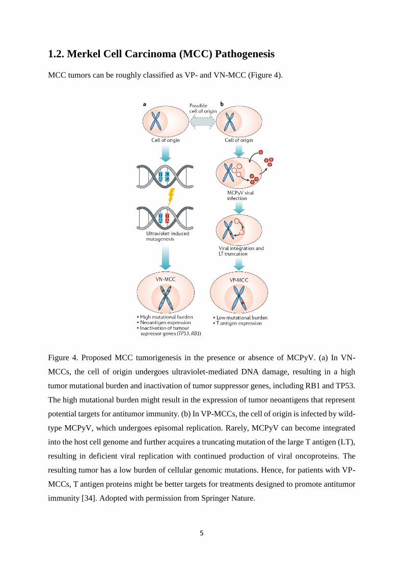
1.2. Merkel Cell Carcinoma (MCC) Pathogenesis
MCC tumors can be roughly classified as VP- and VN-MCC (Figure 4).
Figure 4. Proposed MCC tumorigenesis in the presence or absence of MCPyV. (a) In VN-
MCCs, the cell of origin undergoes ultraviolet-mediated DNA damage, resulting in a high
tumor mutational burden and inactivation of tumor suppressor genes, including RB1 and TP53.
The high mutational burden might result in the expression of tumor neoantigens that represent
potential targets for antitumor immunity. (b) In VP-MCCs, the cell of origin is infected by wild-
type MCPyV, which undergoes episomal replication. Rarely, MCPyV can become integrated
into the host cell genome and further acquires a truncating mutation of the large T antigen (LT),
resulting in deficient viral replication with continued production of viral oncoproteins. The
resulting tumor has a low burden of cellular genomic mutations. Hence, for patients with VP-
MCCs, T antigen proteins might be better targets for treatments designed to promote antitumor
immunity [34]. Adopted with permission from Springer Nature.

6
1.2.1. Merkel Cell Polyomavirus positive (VP) MCC
1.2.1.1. Merkel Cell Polyomavirus
For nearly 40 years, BK polyomavirus (BKPyV) and JC polyomavirus (JCPyV) have been the
only known human polyomaviruses (PyVs). During the last decade, 11 new human PyVs,
including Karolinska Institute polyomavirus (KIPyV), Washington University polyomavirus
(WUPyV), Merkel cell polyomavirus (MCPyV), Human polyomavirus 6 (HPyV6), Human
polyomavirus 7 (HPyV7), Human polyomavirus 9 (HPyV9), New Jersey Polyomavirus
(NJPyV), Trichodysplasia spinuolsa associated polyomavirus (TSPyV), Malawi polyomavirus
(MWPyV), HPyV12, and St Louis polyomavirus (STLPyV) have been discovered [35]. To add
to this list, a putative human PyV named IARC-Lyon PyV (LIPyV) was recently isolated from
human skin [36] and Quebec polyomavirus (QPyV) isolated from 85-years old male in Canada
[37]. Although there is little information known about the pathogenesis of these novel human
PyVs, some of them have been linked to human diseases: BKPyV-associated nephropathy,
JCPyV-associated progressive multifocal leukoencephalopathy (PML), WU-PyV-associated
bronchitis, HPyV6/HPyV7-associated dermatosis and TSPyV-associated trichodysplasia
spinulosa [38, 39].
HPyV infection is common in the human population. Serological studies have shown a
seroprevalence ranging from ~5% for HPyV12, NJPyV, and LIPyV, ~20% for HPyV9 and
≥60% for the other HPyV in the healthy adult population. Moreover, each individual is infected
with several HPyVs [40, 41]. Primary infection occurs in early childhood, after which the virus
establishes a life-long and sub-clinical co-existence with its host [42]. The molecular detection
of HPyV genomes has been complemented by important serological evidence of infection using
HPyV VP1 capsid-specific IgG antibodies. The results indicated that HPyV infections
frequently occur during childhood, reaching high seroprevalence rates of 40% to 90% in the
general adult population, with average coexposure rates of 6 to 7 HPyVs [41, 43]. Despite this
high rate, clinical symptoms or signs of primary HPyV infection have not been identified. In
fact, only 5 HPyVs have been consistently linked to disease.
Evidence for persistent infection by a specific polyomavirus is reflected in serum antibodies
against the corresponding polyomavirus coat protein VP1 [44]. Based on the VP1 serology
assay, different studies have investigated prevalence of seven most important human
polyomavirus in health individuals with different age groups [41].

7
MCC is associated with the MCPyV, with studies showing up to 80% presence in MCC tumors
[45-47]. Although evidence suggests a causative link between MCPyV and MCC [48], further
research is needed to evaluate the absolute risk of cancer. Infection with this virus is common
during childhood and is usually asymptomatic. MCPyV antibodies are present in 35% of 13-
year-olds [49] and increase to 80% at age 50 years [50].
HPyV diseases occur almost exclusively in patients with inherited, acquired, or therapeutic
immunodeficiency states such as transplantation, HIV-AIDS, autoimmune disease, and
cancer/chemotherapy [51, 52]. Evidence of HPyV disease is emerging for KIPyV [53], WUPyV
[54, 55], HPyV6 [56, 57], HPyV10 [58], and NJPyV-2013 [59] due to dedicated studies
correlating histopathology and virus infection by specific immunohistochemistry.
Figure 5: (A) Circular map and (B) linear maps of the MCPyV early genes [7]. MCPyV is a
non-enveloped, double-stranded DNA (dsDNA) virus with genome of ~5kb and belongs to the
family Polyomaviridae [36]. The viral genome is divided into three major regions: the non-
coding control region (NCCR), early region and late region. The NCCR contains the origin of
replication and transcriptional regulatory elements. The early region also called functional
region encodes large T (LT) antigen, small T (sT) antigen, 57kT antigen and a protein called

8
alternative LT open reading frame (ALTO) [60-63]. The late region also called structural region
encodes the viral capsid proteins VP1 and VP2 [64]. The polyomavirus genome is maintained
episomal during normal life cycle. MCPyV has been found to encode a single microRNA
(miRNA) precursor, which can produce a mature miRNAs, termed mcv-miR-M1. mcv-miR-
M1 have the ability to negatively regulate expression of viral gene products required for viral
DNA replication [65]. Adopted with permission from Springer Nature.
The original report from the Moore and Chang’s lab outlined several important features about
VP-MCC. Approximately, 80% of all MCC contain clonally integrated copies of the virus [45].
A vital question in VP-MCC is that how do MCPyV-encoded proteins contribute to viral
activities and MCC development. Most of our knowledge of MCPyV oncogenic mechanisms
was gained from studies of the viral oncogenes, LT and sT antigens, both of which have been
implicated in MCPyV-induced tumorigenesis, at least in cell cultures and animal models [66].
An important feature of VP-MCC is that the tumor maintains expression of LT and sT antigen
[45]. The functions of the 57 kT and ALTO proteins remain unknown, but mutant MCPyV virus
lacking expressing of ALTO replicated similar to wild-type virus [63].
1.2.1.1A. small T antigen (sT antigen)
The primary MCPyV oncogenes are thought to be the sT and LT antigens. The sT antigen is
expressed from two of four alternative spliced mRNA of the MCPyV virome [65]. MCPyV LT
and sT antigen share exon 1 of T antigen locus with the DnaJ, CR1, and Hsc70 domains [67]
(Figure 5). MCPyV sT was found to bind the catalytic subunit of PP1 [68]. The inhibition of
PP1 prevents dephosphorylation of retinoblastoma and ensures cell cycle progression [69]. It is
known that HPyV drive cells into the S phase that may be (partially) achieved by sT-mediated
inhibition of PP1 thus facilitate viral genome replication [70]. This may be (partially) achieved
by sT-mediated inhibition of PP1, which results in hyperphosphorylation of retinoblastoma
[71].
In many PyVs, sT antigen binds protein phosphatase 2A (PP2A) and this interaction is mediated
by the N-terminal J domain and the C-terminal zinc binding motif of sT [72]. PP2A is a
phosphoserine/threonine phosphatase that exists as a heterotrimer composed of a structural
subunit A, a regulatory subunit B, and a catalytic C subunit [73]. Different studies showed the
interaction between MCPyV sT and PP2A Aβ, and weakly with PP2A Aα, but also with the
catalytic subunits PP2Cα and Cβ. The binding of sT to PP2A reduced the catalytic activity of

9
the enzyme [68, 74, 75]. The biological implication of the sT:PP2A interaction is not known
because mutations that abrogate PP2A binding had no effect on sT’s transforming activity [76].
Interestingly, MCPyV sT targets the NF-κB regulator NEMO by binding of a complex formed
by protein phosphatase 4C (PP4C) and protein phosphatase 4 regulatory subunit 1 (PP4R1)
leading to reduced NF-κB translocation and transcriptional activity. Regulation of PP4C and
PP4R1 by the sT could be a mechanism by which MCPyV modulates host anti-viral response
or autoimmunity [74].
sT also has the large T stabilization domain (LSD) located between amino acids 91–95 that
helps in transformation. The LSD binds and inhibits E3 ubiquitin ligase (SCFFbw7). MCPyV
LT is a target of Fbw7-mediated ubiquitination to induce proteasomal degradation; therefore
sT increases the half-life of LT protein. Furthermore, inhibition of Fbw7 also increases the
levels of other Fbw7 target proteins of the cell cycle such as c-Myc and cyclin E [77]. The
MCPyV sT LSD also increases hyperphosphorylated 4E-binding protein 1 (4E-BP1), a
regulator of cap-dependent translation, thereby increasing protein translation [76].
Taken together, MCPyV sT increases protein translation, reduces proteasomal degradation, and
thereby promotes tumor cell survival via increased levels of c-Myc, cyclin E, and MCPyV LT
antigen.
1.2.1.1B. Large T antigen (LT antigen)
In all cases reported to date, VP-MCCs express a truncated form of LT (tLT) lacking the C-
terminal of the protein due to premature stop codon. The tLT, however, preserves the N-
terminal J domain and LXCXE motif but lacks the DNA binding and helicase domains. In
addition, the C-terminal domain also contains around 100 amino acid residues in exon 3 with
growth inhibitory activity that is not expressed in VP-MCC. In some of VP-MCC, LTs retained
the nuclear localizations signal (NLS) while others lost it [78, 79].
MCPyV LT also plays specific fundamental roles in oncogenesis. LT contains a helicase
domain at the C-terminal that helps in initiating viral DNA replication [67, 80]. Importantly,
mutations in the C-terminal domain are essential for preventing initiation of replication within
integrated virus genome. LT helps to prevent host cell death from DNA damage response to
host cells [62, 67, 77, 80-82]. Studies have shown that when FLT was expressed in VP-MCC
cell lines, a specific DNA-damage response was observed [62, 82].
10
MCPyV LT has different domains that inhibit tumor suppressors and activate oncoproteins.
Most VP-MCC express a tLT with an intact retinoblastoma suppressor gene (RB1) binding
domain (LxCxE motif; amino acids 211–217) [83, 84]. LT binding inhibits Rb activity thus,
preventing Rb-mediated suppression of transcriptional activity of E2F. Release of E2F
repression allows transcription of genes involve in cell cycle G1 to S-phase transitions, thus
promoting tumor growth [78, 83]. Rb binding to LT domain also helps in upregulation of the
anti-apoptotic oncoprotein, survivin [85]. Tumor suppressor p53 is another target of LT.
Although MCPyV LT is not known to bind p53 directly, but higher LT expression leads to
reduced p53 transactivation activity [86]. Therefore, in short, the preservation of N-terminal
and mutation of C-terminal LT in tumors suggests importance for host cell transformation. LT
also interacts with hVam6p, a protein involved in lysosomal trafficking, but is unlikely to
contribute to cellular transformation and tumorigenesis [87]. LT has two unique domains,
MUR-1 and MUR-2, with minimal contributions to transformation [88].
Taken together, integrated MCPyV employs multiple sT and LT-mediated mechanisms to
promote tumor development and growth.
1.2.1.2. Merkel Cell Polyomavirus negative (VN) MCC
About 20% of MCC are VN-MCC and occurs due to excessive sun exposure (Figure 4) [45].
The mechanisms of oncogenesis leading to MCC is not completely understood. Next-
generation sequencing analysis of both VN- and VP-MCC showed an overall higher mutational
burden in VN-tumors compared with VP-tumors [89].
VN-MCCs contain an exceptionally high somatic mutational burden (50 mutations/Mb in VN-
as compared to <1 mutation/Mb in VP-MCC) [90], enriched for ultraviolet signature C > T
transitions throughout the genome [84, 91, 92]. RB1 and TP53 genes are two most commonly
mutated genes in VN-MCC. Interestingly both of tumor suppressor proteins (p53 indirect) are
also target for MCPyV LT [84-86]. Most of RB1 mutations result in genome deletion or
epigenetic hypermethylation [84, 89, 91, 93], which ultimately leads to increased E2F activity
and thus increased tumor growth [78]. Likewise, TP53 mutations in VN-MCC results in
inactivation of p53. These p53 inactivating mutations result in downregulation of p53 targets,
thereby preventing tumor cell senescence, cell cycle arrest, DNA damage repair, and apoptosis
[84, 85, 91].

11
Different studies have shown activating oncogenic mutations in HRAS, PIK3CA, KNSTRN,
PREX2 and RAC1 in the majority of VN- (6 out of 8) as compared to VP-MCC (2 out of 8). In
addition to that, recurrent mutations in other tumor suppressor genes, including NOTCH1, and
PRUNE2, were found in VN-MCC. All of these mutations suggest that genetic aberrations
independent of MCPyV infection are involved in the pathogenesis of VN-MCCs [84, 89, 91].
Viruses are intracellular pathogens that not only replicate in host cell but also transform infected
cell into a tumor cell. This is done by hijacking the host machinery and reprogram it through
interfering signaling pathways [71]. Previous studies have shown different signaling pathways
targeted in MCC. The main targets include phosphatidyl-3-kinase/AKT/mammalian target of
the rapamycin (PI3K/AKT/mTOR) pathway [94-96], Mitogen-Activated Protein Kinase
(MAPK) Pathways [97], Notch Signaling Pathway [98, 99] hedgehog signaling pathway [100,
101], DNA damage repair (DDR) pathways [82, 102, 103], Retinoblastoma-E2F Pathway [62,
86, 104], tumor suppressor p53 [104], Programmed cell death or apoptosis pathways [105, 106]
and ubiquitination-proteasomal degradation pathway [77, 107-109].
Taken together for MCC pathogenesis, loss of tumor suppressive function of both Rb and p53
seems to be a consistent feature of both VP- and VN-MCC. In addition to that, additional
oncogenic processes are consistently activated by viral oncogenes in VP-MCC.
1.2.2. Merkel cell polyomavirus and non‐MCC tumors
Many studies have detected MCPyV DNA in various non‐cancerous tissues of the body like
the skin, adrenal gland, spleen, bone marrow, stomach, gallbladder, pancreas, heart, and aorta,
although with a relatively low viral load between 0.00026 and 0.22 copies per cell [110]. But,
the copy number of MCPyV genome was 60 times higher in MCC tissues as compared to
healthy tissues. A higher presence of MCPyV among different tissues are digestive system,
saliva, and in the upper digestive tract [111]. Since the role of MCPyV in the development of
MCC is critical, so widespread prevalence of the virus encouraged researchers to explore a
possible role of MCPyV in non‐MCC cancers. Different studies have found MCPyV DNA,
transcripts, and proteins presence in malignant tissues, but the integration state and the
truncations of LT have rarely been examined. Moreover, the viral copy numbers are <1
copy/cell, adjacent non-malignent tissue was not always examined and the number of samples
was often low (reviewed in [112]). Therefore, a possible role of MCPyV in other tumors
remains elusive.

12
There are few reports in which MCPyV DNA was found in reproductive system‐related tumors;
for example in prostate cancer [111, 113], breast cancer [114], and cervical cancer [115]. In one
study with testicular cancer, a relatively high viral load with 0.934 copies per cell was detected,
but LT protein expression was not assessed [111]. Other studies of MCPyV‐positive prostate
cancers and cervical cancer showed that these samples exhibited a lower viral copy number
with 0.002 [111] and 0.00003055 and 0.0015 [115, 116] copies per cell respectively. A small
subset of breast cancer were MCPyV DNA positive [114, 117], but viral DNA could not be
detected in ovarian and vulva cancer [48, 117]. Even though, MCPyV may not be the
perpetrator, its oncoproteins LT and sT may have an effect on the expression of oncoproteins
expressed by other co-infecting oncovirus. One example is high-risk human papilloma virus
(HR-HPV) associated cervical cancer. Different studies have shown the presence of MCPyV in
HR-HPV associated cervical cancer [118-120].
Papillomaviridae is a family of nonenveloped, double-stranded DNA viruses that can cause
cancer in humans [121]. HR-HPV are mainly associated with cervical cancer, but also with
penile, anal, vulvar, vaginal and oropharyngeal cancers [122, 123]. Among different HR-HPV
strains, HPV-16 and HPV-18 are responsible for almost 70% of cervical cancers worldwide.
There is viral genome integration into host genome and HR-HPV is considered one of the most
important risk factors for cervical cancer development [124-126]. The oncogenic potentials of
HR-HPV is mainly associated with the viral proteins E6 and E7. These proteins can bind p53,
mTOR, hTERT and pRb family members, respectively. Additionally both E6 and E7 can also
interfere with other hallmarks of viral oncogenesis [127] as seen by MCPyV LT in MCC. So
co-expression of LT, sT, E6 and E7 may collaborate in the neoplastic processes by these viruses.
Co-infection with MCPyV and the oncoviruses Epstein-Barr virus and human herpesvirus-8 in
respectively chronic lymphocytic leukemia, Kaposi’s sarcoma has been described [35], but
whether MCPyV plays a causal role in these tumors has not been established.
1.3. Inflammation
Inflammation is described as a series of events induced by the body in response to stimuli that
are potentially harmful to the body, such as infection or injury. The inflammatory response is
characterized by the rapid accumulation of neutrophils, macrophages, immune cells and later
on production of soluble mediators with the ultimate aim of protecting the organism from
foreign invaders and initiating healing processes [128]. The process of inflammatory response
depends on the nature of the initial stimulus and the site in the body. They all share common

13
mechanisms with: 1) detrimental stimuli recognition by cell surface pattern receptors; 2)
activation of inflammatory pathways; 3) release of inflammatory mediators; and finally 4)
recruitment of inflammatory cells [129].
1.3.1. Immune System
The human immune system categorized as the innate immune response that is fast but not
specific and the adaptive immune response that is slow but involving specificity and memory.
The innate immune system provides the first barrier against any type of infections and consists
of granulocytes, macrophages, mast cells, dendritic cells (DCs), natural killer cells and humoral
part such as mucosal barrier, complement system, lysozyme. In the adaptive immune response,
immune cells (B- and T-cells) specifically target invaders in an antigen-antibody specific
manner. There are also some other cells that function at the interface between the innate and
adaptive immune response and consists of Natural killer T cells (NKT) cells and γδ T cells. The
immune system not only protects host against pathogens like viruses and bacteria, but also plays
an important role in the surveillance of cancer [130].
1.3.2. Cancer Immunology
A role for inflammation in cancer development was proposed in 1893, when Rudolf Virchow
observed the presence of immune cells in neoplastic tissues [131]. In 2011, after more than 100
years of its original proposal by Virchow, it was formally acknowledged with the inclusion of
immune system evasion and inflammation as additional hallmarks of cancer [132, 133].
Cancer immunology is the study of interactions between the immune system and cancer cells.
In cancer immunology, the immune response is of particular interest with most important work
as cancer-specific antigens recognition known as tumor-associated antigens (TAAs). The
immune system can recognize the antigenic changes and further develop specific T-cells and
antibodies to recognized neo-antigen [134-136]. These anti-TAAs that are associated with
cancer, might be considered as reporters from immune system to identify antigenic cellular
proteins changes involved in transformation process [137]. These anti-TAAs autoantibodies
have advantage over antigens with persistence and stability in serum samples of cancer patients
[138]. In recent years, anti-TAAs autoantibody have been explored as early cancer biomarker
as well as indicators of disease prognosis [139].
14
1.3.3. Inflammatory Mediators
Various intrinsic (reactive nitrogen oxygen species RNOS, oncogenic events) and extrinsic
(infection and other inflammatory conditions) factors trigger the recruitment of the
inflammatory cells to the site of inflammation [140]. This results in the activation of several
molecular signaling cascades. These signaling cascades are associated with increased
production of inflammatory cytokines and hence the establishment of inflammation [140].
Cytokines are central mediators between cells in the inflammatory tumor microenvironment
(TME) [141]. They also have normal function in body as messengers between the cells. They
are released in response to a diverse range of cellular stresses, including carcinogen-induced
injury, infection and inflammation. In these settings, cytokines function to stimulate a host
response that is aimed at controlling the cellular stress and minimizing cellular damage [130].
They are usually classified into two classes: anti-inflammatory cytokines, such as IL-4, IL-10,
IL-13, IFN-α, and TGF-β, and pro-inflammatory cytokines, such as IL-1β, IL-6, IL-15, IL-17,
IL-23, and TNF-α [140]. The anti- and pro-tumorigenic function depends on cellular contents
and concentrations in TME. Various therapeutic approaches have been developed to either
target tumor-supporting inflammatory mediators or promote anti-tumorigenic inflammation
and immune responses, with response to reeducate the TME and promote anti-cancer function
[142-144].
1.3.3.1. CCL17/TARC
The human cytokines system consists of 50 distinct proteins that act via distinct receptors of
almost 20 different types [145, 146]. Chemokines are a family of small (8-12KDa), structurally
related, secreted proteins that regulate leukocyte trafficking in immunity and inflammation
[147]. Based on the first two of the four conserved cysteine positions, chemokines are divided
into four subfamilies: CXC, CC, XC, and CX3C [148, 149]. Based on physiological features,
chemokines are divided into two categories, inflammatory and homeostatic [150]. Upon
stimulation by pro-inflammatory cytokines, inflammatory chemokines are expressed in
inflamed tissues. This type of chemokines is specialized for recruitment of effector cells,
including granulocytes, monocytes, and effector T cells. Homeostatic chemokines, on the other
hand, are produced in discrete microenvironments within lymphoid and non-lymphoid tissues
such as the skin and mucosa. These chemokines maintain physiological traffic and positioning
of cells during immune surveillance [151].

15
TARC (thymus and activation-regulated chemokine/CCL17) is a member of CC chemokine
group expressed in the thymus and also expressed by keratinocytes, endothelial cells, dendritic
cells, bronchial epithelial cells and fibroblasts (Figure 6). It is assumed that TARC/CCL17
works both as a inflammatory and homeostatic chemokine [148].
Figure 6: CCL17/TARC expressing, target cells and signaling pathway.
1.3.3.2. CCR4
All chemokine receptors are class A G-protein-coupled seven-transmembrane receptors that
induce signal transduction via G proteins [152]. Based on the chemokine subfamily they bind,
chemokines receptors are classified as CXCR1-5, CCR1-9, CXCR1 and CX3CR1. The specific
functional high affinity receptor for TARC/CCL17 is CC chemokine receptor 4 (CCR4). CCR4
is predominantly expressed on CD4+ T cells [153], but also on a variety of functionally distinct
thymocytes including skin-homing T cells, CD25+ T suppressor cells and T helper (Th) 2 cells.
Since their discoveries, numerous studies have explored the expression and function of CCR4
and its ligands in skin diseases, such as atopic dermatitis [154], bullous pemphigoid [155],
mycosis fungoides [156] and several other allergic diseases including allergic asthma [157],
allergic rhinitis [158] and allergic contact dermatitis [159]. The chemokine receptor CCR4 is
expressed in the TME of several cancers and is associated with poor prognosis. New findings
show that CCR4 is highly expressed in human renal cell carcinoma (RCC) [160], lung cancer
[161, 162], melanoma brain metastasis [163] many hematologic malignancies such as Adult T-
16
cell leukemia (ATL) [164, 165] relapsed peripheral T cell lymphoma (PTLC), and Cutaneous
T-cell lymphoma (CTL) [156].
1.3.3.3. IL-33
The IL-1 family (IL-1α, IL-1β, IL-18) of cytokines plays a major role in inflammatory,
infectious, and autoimmune diseases [166, 167]. Interleukin-33 (IL-33), previously known as
nuclear factor from high endothelial venules or NF-HEV also belongs to IL-1 cytokine family
[168]. This cytokine is produced by various types of immune cells, such as mast cells,
macrophages, and dendritic cells, and also by nonimmune cells, such as endothelial cells,
epithelial cells, smooth muscle cells, and fibroblasts [169]. IL-33 is a 30-kDa protein that
functions both as a transcription factor and a cytokine. Full length IL-33 (FL-IL-33) is divided
into three major domains, a nuclear domain (N terminal), an activation domain and an IL-1 like
cytokine (C terminal) domain (Figure 7). The nuclear domain contains a nuclear localization
signal, a DNA-binding homeodomain-like helix-turn-helix motif, and a chromatin-binding
domain [168, 170]. Upon IL-33 synthesis, it is targeted to the nucleus, where it binds to
chromatin and is thought to regulate gene expression. It can bind to histones H2A and H2B
[168, 171] and can activate histone deacetylase-3 (HDAC) activity [172] thereby affecting gene
expression by remodeling chromatin structure and by epigenetic mechanisms. It has been
shown to interact with the N-terminal domain of the p65 subunit of nuclear factor κB (NF-κB)
and to repress the expression of NF-κB-regulated genes that are necessary for pro-inflammatory
signaling [173]. In response to cellular damage, tissue injury or viral infection, IL-33 is quickly
released from the nucleus of necrotic cells and secreted into extracellular space where it can
bind to the membrane-bound suppression of tumorigenicity 2 receptor (ST2) through its
cytokine domain [174, 175]. Binding to its receptor triggers an inflammatory cascade, thus IL-
33 acts as an “alarmin” and is considered a damage-associated molecular pattern (DAMP)
[176]. The nuclear and cytokine functions of IL-33 are tightly regulated through its localization
[177].
1.3.3.4. IL-33 receptors
The IL-33 receptor (IL-33R) is a heterodimeric complex of ST2 and IL-1 receptor accessory
protein (IL-1RAcP) [178]. The ST2 receptor had been extensively studied and was known as
an orphan receptor for >10 years before the discovery of IL-33 [179]. ST2 was first identified
in murine fibroblasts as an oncogene-induced gene [180, 181]. It is encoded by IL1RL1 that
produces four isoforms through alternative splicing: ST2L (ligand), sST2 (soluble), ST2V

17
(variant), and ST2LV (ligand variant). ST2L is a membrane-embedded receptor that is highly
homologous to IL-1 type-1 receptors and harbors three Ig-like extracellular domains, a
transmembrane spanning region, and an ILI-R1-like intracellular domain [182, 183].
Figure 7: IL-33 producing cells, functional domains and signaling.
Of the four isoforms, ST2L and its decoy receptor sST2 have been most studied, while not much
is known about ST2V and ST2LV [184]. Expression of the IL1RL1 gene is regulated by
GATA1/2, and estrogen-response elements (EREs) that are found on the distal and proximal
promoters that regulate ST2L and sST2 expression [185-187]. While both isoforms can be
transcribed from both promoters, the cell type appears to govern gene expression from either
proximal or distal promoters [186]. ST2L is expressed in fibroblasts, mast cells, eosinophils,
Th2 lymphocytes, dendritic cells, basophils, invariant natural killer cells (iNKT) cells, and
macrophages, while sST2 is predominantly expressed by fibroblasts and epithelial cells [188].
IL-1RAcP is a co-receptor for IL-33 signaling, and this molecule acts as a shared co-receptor
for other IL-1 family members [178, 189, 190]. ST2L creates a heterodimeric transmembrane
receptor complex with the IL1-RAcP [191], and hetero-dimerization brings together the
intracellular domains of the two transmembrane proteins, and its assembly initiates the
recruitment of adaptor molecules through which the IL-33 signal is transduced [170, 192].

18
1.3.4. Immunogenicity of MCC
Immunogenicity is an important inherent feature of tumor cells. This feature is determined by
the tumor cell itself, and is also influenced by the TME [193]. Fundamental determinants of
tumor immunogenicity include tumor antigenicity, and antigen processing and presenting
efficiency [194].
MCC is particularly linked to immune suppression as compared to other tumors. Indeed, almost
8 % of MCC patients are immune suppressed [5]. There are different groups of individuals who
are at high risk of getting MCC. Among those, are HIV infected patients with an 8-fold higher
risk [30], solid organ transplant recipients with a 24-fold increased risk [28], chronic
lymphocytic leukemia and other hematologic malignancies with a 34 to 48-fold increased risk
[5] and also patients with auto-immune diseases, treated with immunosuppressive medication
are at higher risk to develop a MCC compared to immunocompetent individuals [195]. This
suggests that defective cellular immunity predisposes individuals to not only developing MCC,
but also to poorly controlling their disease.
In MCC, both VP- and VN-MCC can be immunogenic, as revealed by the finding of robust
intratumoral infiltration of CD8 T cells into MCC tumors with 100% survival, independent of
the stage of the cancer [196]. Among VP-MCCs, immunogenicity can be assessed with MCPyV
oncoprotein–specific antibodies and T-cell responses, which are quite prevalent. Whereas VN-
MCCs do not express viral oncoproteins, these tumors often express extremely high numbers
of tumor neoantigens which may provide tumor-specific targets for immune recognition [197].
There are also different finding about MCCs regression following withdrawal of immune
suppressive treatment [198, 199]. MCCs with either partial regression (PR), complete
spontaneous regression (CSR) or regression after recurrence and/or metastasis have been
reported in the literature. CSR in MCC is much more common (1.3 per 1,000 cases) than in
other malignancies (1 in 60,000-100,000 cases) [200]. Until 2016, a total of 32 cases with CSR
have been reported. The exact mechanism is still unknown. However previous literature
revealed a female tendency (24F:6M), with the most common site being on the head and neck
region (26/30, 87%), with regression occurring most often after the initial biopsy. This supports
the hypothesis that the biopsy procedure may induce an antitumoral, T-cell–mediated immune
response [200, 201]. Among the 32 CSR patients, MCPyV status was evaluated in only four
studies with three were VP and only one was VN MCC patient [202]. The CSR has also been

19
reported after local recurrence and/or metastasis in total of 16 MCC cases. In this cohort of
MCC patients, there was slight male predominance (7F:9M), with the most common site of the
primary MCC on the head and neck region (14/15, 93%). The average time interval from the
detection of the recurrence and/or metastatic disease to regression was 6.02 months (11 days–
30 months) [202]. Additional mechanisms that attribute to the elevated proportion of regression
include a higher rate of apoptotic activity [203], dense lymphocytic infiltration around the tumor
cells [204] and antiretroviral therapy [205].
1.3.5. Immune Evasion in MCC
In 2000, Hanahan and Weinberg initially postulated the six hallmarks of cancer [133], but in
their update in 2011, inflammation and immune evasion was added as an important feature of
cancer cells [132]. Despite of immune surveillance, it had become apparent that tumors are still
able to develop, even in hosts with fully functioning immune system [206].
During the last two decades, conceptual developments have demonstrated that the immune
system can paradoxically constrain and promote tumor development and progression. This
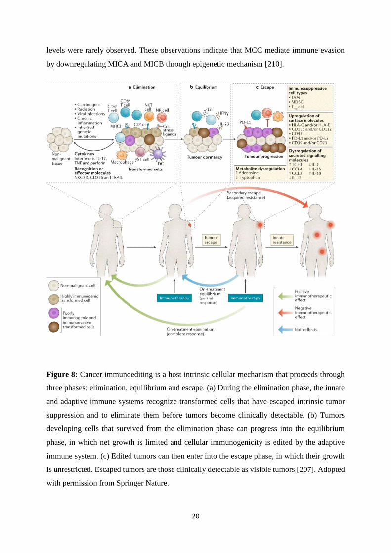
process is referred to as cancer immunoediting and proceeds through three phases, termed
elimination, equilibrium and escape [207]. During first phase of (1) eliminations, also called
immunesurveillance, the immune system reacts to tumor and tumor cells are eliminated; and in
(2) equilibrium phase, the immune system does react, but is not able to eliminate the tumor cells
to a full extent. In the last phase of (3) escape, surviving immune resistant cancer cells can form
tumors that are evading the immune response (Figure 8) [206]. For VP-MCC, immune evasion
is difficult to accomplish despite continuous expression of viral T-antigens. While MCPyV
specific T cells are present in the majority of MCC patients, they rarely infiltrate into MCC
tumors [208]. This fact suggests that MCC cells and cells of the MCC TME create an
immunosuppressive environment that inactivates MCC specific T cells and prevents them from
infiltrating, thus facilitating the immune escape of MCCs.
Indeed, during the last years several studies accumulated evidences that MCC cells employ a
variety of immune escape strategies. During viral infection and/or cellular transformation,
natural killer group 2D (NKG2D) ligands, major histocompatibility complex (MHC) class I
chain-related protein A and B (MICA/MICB) undergo upregulations. The NKG2D and
MICA/MICB interaction stimulates proliferation and cytotoxic potential of NK cells [209]. In
a study of MCC tumors and cell lines, MICA and MICB mRNA levels were low and protein
20
levels were rarely observed. These observations indicate that MCC mediate immune evasion
by downregulating MICA and MICB through epigenetic mechanism [210].
Figure 8: Cancer immunoediting is a host intrinsic cellular mechanism that proceeds through
three phases: elimination, equilibrium and escape. (a) During the elimination phase, the innate
and adaptive immune systems recognize transformed cells that have escaped intrinsic tumor
suppression and to eliminate them before tumors become clinically detectable. (b) Tumors
developing cells that survived from the elimination phase can progress into the equilibrium
phase, in which net growth is limited and cellular immunogenicity is edited by the adaptive
immune system. (c) Edited tumors can then enter into the escape phase, in which their growth
is unrestricted. Escaped tumors are those clinically detectable as visible tumors [207]. Adopted
with permission from Springer Nature.

21
During active immune system, CCAAT/enhancer-binding protein (C/EBP) transcription factor,
a positive regulator of the Toll-like receptor 9 (TLR9) promoter, acts as an important mediator
of pro-inflammatory immune responses. TLR9 activation results in activation of NF-κB–
mediated transcription, which produces pro-inflammatory cytokines and type 1 interferons
(IFNs) that are important for clearing virus-infected cells and promoting further immune
activation [211]. In VP-MCC, T-antigen mediated inhibition of C/EBP leads to reduced TLR9
expression thus inhibiting NF-κB-mediated transcription that ultimately results in reduced
expression of proinflammatory cytokines and IFNs [212, 213]. However, TLR’s involvement
is not well understood in VN-MCC.
Previous studies also have shown macrophage infiltration in MCC and the level of infiltrate
was higher in VP- as compared to VN-MCC [214]. Surprisingly, a portion of the macrophage
infiltrate was found to express CD163, a marker of M2 phenotype. M2 phenotype is linked to
tumor growth and survival through secretion of immune suppressive cytokines, rather than M1
that is pro-inflammatory phenotype [214]. However, the level of CD163+ macrophages was not
associated with the status of MCPyV [214]. Taken together, NKG2D and MICA/MICB
inhibition interaction, downregulation of TLR9 and M2 macrophage infiltrate in tumor
contributes to innate immunity evasion of MCC.
The effectiveness of immune responses may decrease due to inability of all activated T cells to
properly home tumor tissue in MCC. For T cells to migrate to the area of inflammation,
interactions must occur between T cells and endothelium. E-selectin, a receptor present on
endothelium, has been proposed to be critical for T lymphocyte migration [215]. A subset of T
cells expresses a ligand for E‐selectin, the cutaneous lymphocyte antigen (CLA) [215, 216]. In
a study of 56 MCC samples, 52% of MCC samples displayed downregulation of E-selectin
expression in intratumoral vasculature [217]. A study by Dowlatshahi et al., demonstrated
correlation between CLA expression and T cell infiltration into tumors. The authors found that
3/3 MCC tumors with high CLA expression were infiltrated by an increased number of T cells
present within tumor nests. They also found 4/4 MCC tumors with deceased T cells CLA
expression, peritumoral pattern of T cells without penetration into tumor nest [218]. These
above two events, downregulation of E-selectin and lacking CLA contributes to the lack of T
cell migration into sites of MCC tumors.
Previous studies have shown that optimal T cell activation requires two signals: an antigen-
specific signal (mediated by antigen presentation) through the T cell receptor (TCR) and a

22
costimulatory signal [219, 220]. Antigen presentation is essential for identification and
successful eradication of tumor cells by CD8+ T cells. Dysregulation of antigen presentation,
by loss of MHC-I and β2-microglobulin (B2M) is a common mechanism of immune evasion
by various types of cancers [221]. In a study of 114 MCC patient samples, 84% showed reduced
MHC-I expression as compared with surrounding tissues with 51% had poor or undetectable
MHC-I expression. The authors of this study also found that VP-MCC showed lower MHC-I
expression as compared to VN-MCC [222].
In addition to the downregulation of immune cell recognition receptors, inhibitory receptors are
upregulated on tumor-targeted immune cells. Negative regulatory pathways in the immune
system help in successful clearing pathogens and malignant cells thus limiting
immunopathology [223]. Programmed death receptor 1 (PD1) has been identified as critical
negative regulator of T cell activity [224]. PD1-mediated T cell inhibition is an important
mechanism to prevent autoimmunity, cancer and chronic infectious diseases [225]. Chronic
antigen stimulation from viral infections and tumor cells upregulates inhibitory receptors on
active T cells that results in T cells functionality loss over time and leads to T cell exhaustion
[226, 227]. Increased expression of T cell receptors, PD-1 and TIM-3 results in immune
dysfunction and prevents CD8+ T-cell mediated clearance of virus infected and malignant cells
[228]. MCPyV-specific T-cells in blood and MCC tumors show simultaneous PD-1 and TIM-
3 expression [229]. Another study of effector T-cells isolated from VP-MCC have shown lower
levels of activation markers (CD25 and CD69) and higher levels of PD-1 compared with normal
skin T cells [218]. As tumor-specific epitopes are necessary for T-cells isolation, VN-MCC-
specific T-cells have not been identified. So, it is still unclear whether T-cell exhaustion markers
are present on tumor-specific effector T-cells present in TME of VN-MCC [230]. In case of
melanoma, tumor-specific CD8+ T –cells also upregulate PD-1 and TIM-3, indicating that
somatic mutations induced by UV exposure contribute to T-cell dysfunction; this may also
occur in UV exposure–mediated MCC [231]. Additionally MCC can also evade the immune
system by overexpressing PD-L1 receptor present on tumor cells, thus inactivating active CD8+
T cells [232]. A study of 49 MCC patient tissues analyzed PD-L1 expression on tumor cells
and infiltrating lymphocytes. This study showed that 50% of the VP-MCC samples showed PD-
L1 expression and had a moderate-severe immune infiltrate, as compared to 0% of the VN-
MCC tissue samples [232]. However, another study demonstrated that PD-L1 overexpression
in VN-MCC correlated with increased mutational burden [91].

23
Additionally, Tregs cells recruitment to the area of inflammation also results in immune
suppression. Tregs can inactivate CD8+ T-cells and antigen presenting cells (APCs), thus
leading to disease progression in response to UV radiation exposure and viral infection [233].
Different studies have shown high Tregs infiltration in MCC as compared to normal skin
(reviewed in [218]). While comparing the role of Tregs in MCC as compared to other cancers,
a study of 116 MCC patients with Tregs presence is associated with longer survival. This
possibly indicating that association between MCC and viral infection results in distinct profile
of T-cell response [214]. In another study of MCC patients, CD4+ and CD8+ Tregs were found
within MCC tumors, but their presence was not associated with overall survival [218]. Thus,
role of Tregs in establishment and progression of MCC is unclear.
1.4. Current treatment options for MCC
For MCC, there are different options for treatment including standard (currently used options)
to some therapies that are being tested in clinical trials. There are four different types of standard
treatment options being used in clinics. These standard options include surgery, radiation
therapy, chemotherapy and immunotherapy. All of these options are used alone or in
combinations for better recovery. The local recurrence chances after surgical treatment are still
higher as compared to other most common skin cancers (basal cell carcinoma, squamous cell
carcinoma and even melanoma) [234, 235]. Radiation therapy can markedly lower the risk of
recurrence of MCC in irradiated areas. Most of time radiation therapy is used in combination
with local surgical excision. Radiotherapy decreases local recurrence about 3.7 time when used
in combination [236]. Chemotherapy is also another line for treating MCC that leads to
significant shrinkage of tumors, but chemotherapy is not very effective as tumors often get
resistant and recur within 90 days. The reason for this failure is either immune suppression by
chemotherapy and/or MCC tumor cells acquire resistance towards chemotherapy [237]. A study
of standard chemotherapy in metastatic MCC patients showed initial shrinkage of the tumors
in the majority of patients, but no durable effect. It was found that half of the patients after 3
months and more than 90% of the patients after 10 months had recurrent growth of their tumors
[238]. For patients who do not have problems with their immune system for example, no
autoimmune disease, it is typically recommended to first try an immune stimulating therapy
also called immunotherapy. The main idea of using cancer immunotherapy is to stimulate the
host immune defense system against specific type of diseases. This can be done by using

24
monoclonal antibodies [239], vaccines [240], cytokines [241], oncolytic viruses [242], chimeric
antigen receptors (CAR) T-cell therapy [243] and immune checkpoint inhibitors (ICIs) [244].
Immunotherapy is rapidly becoming a preferred systemic therapy in several cancer types,
especially because responses to immunotherapy (when they occur) are generally long-lasting.
The durability of immunotherapy responses places this approach in stark contrast to
chemotherapy, which was previously considered the standard option for patients with
metastatic MCC [238]. Immune cells, such as T cells, and some cancer cells have certain
proteins, called checkpoint proteins, on their surface that keep immune responses in check. The
development of ICIs is a revolutionary milestone in the field of immuno-oncology. Tumor cells
evade immunosurveillance and progress through different mechanisms, including activation of
immune checkpoint signaling that suppress antitumor immune responses.
Two types of ICIs therapies are currently being used, targeting programed cell death-1 (PD-1)
(Figure 9) and cytotoxic T-lymphocyte antigen-4 (CTLA-4) (Figure 10). PD-1 is a protein
expressed on the T-cells. When PD-1 attaches to another protein called PD-L1 on a cancer cell
or myeloid cells (DCs, TAM), it stops the T cell from killing the cancer cell. PD-1 inhibitors
attach to PD-L1 and allow the T cells to kill cancer cells. Avelumab (antibody against PD-L1)
[245] and pembrolizumab (antibody against PD-1) [246] are used to treat advanced Merkel cell
carcinoma. Nivolumab (targets PD-1) [247] is being studied to treat advanced Merkel cell
carcinoma. CTLA-4 is another immune checkpoint protein expressed on the surface of Tregs
cells that helps keep the body’s immune responses in check. When CTLA-4 attaches to another
protein called B7 on a APCs. Ipilimumab is a type of CTLA-4 inhibitor being used to treat
advanced Merkel cell carcinoma [248, 249]. There are still a number of clinical trials are going
on different phases to target ICIs.

25
Figure 9: Immune checkpoint inhibition. Checkpoint proteins, such as PD-L1 on tumor cells
and PD-1 on T cells, help keep immune responses in check. The binding of PD-L1 to PD-1
keeps T cells from killing tumor cells in the body (left panel). Blocking the binding of PD-L1
to PD-1 with an immune checkpoint inhibitor (anti-PD-L1 or anti-PD-1) allows the T cells to
kill tumor cells (right panel) [250]. The figure is adopted with permission from the National
Cancer Institute © (2015) Terese Winslow LLC, U.S. Govt. has certain rights.
Despite the success of anti-CTLA-4 and anti-PD-1/PD-L1 therapies, only a fraction of patients
benefit from ICIs. Antitumor immunity, regulated through complex factors in the TME, could
create variable immune responses [244]. Immunotherapeutic approaches other than targeting
ICIs are also being investigated in clinical studies (table). These approaches involve either
stimulations of NK cells, macrophages, T-cells (NCT02054884), infusion of activated NK-92
cells (NCT02465957), IL-12 gene therapy (NCT01440816), targeting CD47 (NCT02890368)
and TLR4 agonist (NCT02035657).

26
Figure 10: Immune checkpoint inhibition. Checkpoint proteins, such as B7-1/B7-2 on APCs
and CTLA-4 on T cells, help keep the body’s immune responses in check. When the TCR binds
to antigen and MHC proteins on the APC and CD28 binds to B7-1/B7-2 on the APC, the T cell
can be activated. However, the binding of B7-1/B7-2 to CTLA-4 keeps the T cells in the
inactive state so they are not able to kill tumor cells in the body (left panel). Blocking the
binding of B7-1/B7-2 to CTLA-4 with an immune checkpoint inhibitor (anti-CTLA-4 antibody)
allows the T cells to be active and to kill tumor cells (right panel) [250]. The figure is adopted
with permission from the National Cancer Institute © (2019) Terese Winslow LLC, U.S. Govt.
has certain rights.
1.5. Limitations of Immunotherapy
However, not all patients experience durable response to immune-based therapies. Furthermore,
patients requiring immunosuppression in the setting of solid organ transplants or those with
autoimmune disease may not be optimal candidates for immune-based therapy. Therefore,
27
predicting and improving response to immunotherapy, and identifying alternative therapies for
cases in which immunotherapy is contraindicated or ineffective, represent current research
priorities in MCC [34].
A clinical trials with MCC patients using pembrolizumab had shown 56% objective response
with 16% complete and 40% shown partial response with a median follow-up of 33 weeks (7-
53 weeks). The response duration ranged from 2.2 months to at least 9.7 months. The
progression-free survival rate was 67% in 6 months. The response rate in VP- and VN-MCC
was 62% and 44% respectively. In the same study, the authors observed treatment-related
adverse event (TRAE) of any stage including adrenal insufficiency, colitis, hepatitis,
myocarditis, nephritis, pneumonitis, thyroiditis, and transaminitis [246]. In another clinical trial
with avelumab, the investigators found objective response in 33% of patients with 65.5% of
whom had partial response. Among patients who have shown objective response, 93% had
durable response ≥ 6 months, 71% had ≥ 12 months and 29% had at 18 months [251].
Considering evaluable safety, 71.8% had TRAE with 15.4% grade-1 immune-related adverse
events and only 20.5% grade 3 TRAE but no grade 4 TRAE or treatment related deaths
occurred. The patients also had shown some adverse effects i.e. cholangitis, elevated aspartate
aminotransferase and alanine aminotransferase levels, paraneoplastic syndrome, gait
disturbance, paraneoplastic encephalomyelitis and polyneuropathy and infusion-related
reaction [252]. It became apparent that cancer patients respond very different to immunotherapy
and so far, there are almost no predictive markers, which patient will benefit from which
immunotherapy. In addition, it became obvious that combination of different drugs will be the
key to success for immunotherapy.
28
2. AIMS OF THE THESIS
The overall aim of this thesis was to investigate the effect of Merkel cell polyomavirus effect
in merkel cell carcinoma and other cancers.
The specific aims were:
Paper I: To compare the transcriptional activity of NCCR of different MCPyV variants isolated
from virus-positive MCC and non-MCC samples in a MCC cell line, and in human dermal
fibroblasts.
Paper II: To study effect of MCPyV LT on CCL17/TARC expression and impact on viral
expression.
Paper III: To study effect of MCPyV T-ag on IL-33 expression and role in viral expression.
Paper IV: To study effect of MCPyV oncoproteins LT and sT on expression of HR-HPV
oncoproteins E6 and E7, thereby accelerating the initiation and/or progression of cervical
cancer.
29
3. METHODOLOGICAL CONSIDERATIONS
3.1. Biological material
Cancer cell lines are an extremely useful tool to study diverse biological processes and the
efficacy of therapeutic agents in basic science research. A broad variety of MCC cell lines are
available from different patients and represent various aspects of MCC biology, such as
presence or absence of MCPyV. MCC cell lines are a convenient tool as their handling is not
complicated, allowing extensive experimental set ups. However, we should keep in mind
unavoidably changes in the cells due to passages of cell line. Therefore, we should considered
a useful model that can however not replace patient material and in vivo studies.
3.1.1. Cell lines
To confirm the identity of the cell lines used in this work STR (short tandem repeat) profiling
was performed at the Centre of Forensic Genetics, University of Tromsø. The cell lines were
routinely grown without antibiotics as it has been demonstrated that antibiotics can affect the
proliferation and gene regulation of cultured cells [253, 254]. Furthermore, the use of antibiotics
can hide low level contaminations and mycoplasma infections [255]. Mycoplasma tests were
performed regularly.
3.1.2. Human tissue and plasma samples.
MCC tumor tissues were obtained in accordance with the ethical approval during 2000-2015
from the St. Olavs University Hospital Trondheim, Norway according to the ethical approval
from the Regional Ethical Committee (REK NORD application number 2016/988). MCC
plasma samples were obtained in accordance with approved by the Ethics Committee of
Karolinska Institutet (2010/1092-31/3).
3.2. Promoter luciferase assay
This method makes use of luciferase as a reporter gene to test and compare promoter(s)
activities. One of the commonly used reporter genes used in monitoring promoter strength of
interest is the luciferase gene of the firefly Photimus pyralis. The firefly luciferase gene placed
under the control of the promoter of interest was used in this thesis. Through transient
transfection, the plasmid containing the promoter of interest controlling the transcription of the
luciferase gene is introduced into the desired cells. Luciferase protein is then expressed by the

30
transfected cells and the amount of luciferase produced is proportional to the strength of the
promoter. The strengths of the various promoters can be determined by measuring the amount
of luciferase produced. This indicates that a strong promoter produces more luciferase mRNA
and therefore synthesizes more luciferase protein while weak promoter produces less luciferase
mRNA and therefore expresses less luciferase protein.
Luciferase buffer used contains luciferin, a substrate of the produced luciferase protein
(enzyme) which it oxidizes with the use of ATP to oxyluciferin with a concomitant emission of
light, detectable in the blue range of the visible spectrum, 440-479nm [256]. The intensity of
light emitted corresponds to the amount of oxidized luciferin and hence the promoter’s strength
[256]. A luminometer, which is a special instrument is used for a fast and effective measurement
of the emitted light. This method is an excellent method because it is very sensitive, highly
reproducible, fast, quantitative, and relatively cheap.
Although luciferase assay is a reproducible, quantative, high-throughput, nonradioactive,
nontoxic, relatively low cost, and easily adaptable to automation method. The primary
disadvantages of luciferase are the requirement for exogenous substrates and the 4–6 h delay
from stimulus to response to allow transcription to occur. The method measures product (=post-
transcriptional) and not transcripts (= promoter activity). A major disadvantage of this method
is that the promoter is not studied in its natural context. Moreover, transfection can introduce
>>2copies of the plasmid and thus the promoter, which more than a normal diploid cell.
Transcriptional activator or repressors present in low concentration may therefore become
limited for the tested promoter. These limitations may result in a measured promoter activity
that does not necessary reflects its bona fide activity.
3.3. Gene expression studies
3.3.1. RT2 Profiler PCR array
RT2 Profiler PCR Array is a reliable tool for analyzing the expression of a focused panel of
genes. Each 96-well plate includes SYBR® Green-optimized primer assays for a thoroughly
researched panel of relevant, pathway- or disease-focused genes. The high-quality primer
design and RT2 SYBR® Green qPCR Mastermix formulation enable the PCR array to amplify
different gene-specific products simultaneously under uniform cycling conditions. This
combination provides the RT2 Profiler PCR Array with the specificity and the high
amplification efficiencies required for accurate real-time SYBR® Green results. PCR arrays

31
are easy to use in any research laboratory. RT2 Profiler PCR Arrays are sensitive enough for
use with RNA prepared from regular samples (0.1–5 µg RNA), FFPE samples, and small
samples (1–100 ng RNA). Each RT2 Profiler PCR Array also includes control elements for Data
normalization, Genomic DNA contamination detection, RNA sample quality and General PCR
performance.
Data can be analyzed using an easy-to-use Excel-based data analysis template or Web-based
software. Data analysis is based on the ΔΔCT method with normalization of the raw data to
either housekeeping genes.
3.3.2. RT-qPCR
Quantitative PCR (qPCR) is used to detect, characterize and quantify nucleic acids for
numerous applications. Commonly, in RT-qPCR, RNA transcripts are quantified by reverse
transcribing them into cDNA first and then qPCR is subsequently carried out. As in standard
PCR, DNA is amplified by 3 repeating steps: denaturation, annealing and elongation. However,
in qPCR, fluorescent labeling enables the collection of data as PCR progresses. This technique
has many benefits due to a range of methods and chemistries available.
In dye-based qPCR (typically SYBR green), fluorescent labeling allows the quantification of
the amplified DNA molecules by employing the use of a dsDNA binding dye. During each
cycle, the fluorescence is measured. The fluorescence signal increases proportionally to the
amount of replicated DNA and hence the DNA is quantified in “real time”. The disadvantages
to dye-based qPCR are that only one target can be examined at a time and that the dye will bind
to any ds-DNA present in the sample.
In probe-based qPCR, many targets can be detected simultaneously in each sample but this
requires optimization and design of a target specific probe(s), used in addition to primers. There
are several types of probe designs available, but the most common type is a hydrolysis probe,
which incorporates the use of a fluorophore and quencher. Fluorescence resonance energy
transfer (FRET) prevents the emission of the fluorophore via the quencher while the probe is
intact. However, during the PCR reaction, the probe is hydrolyzed during primer extension and
amplification of the specific sequence it is bound to. The cleavage of the probe separates the
fluorophore from the quencher and results in an amplification-dependent increase in
fluorescence. Thus, the fluorescence signal from a probe-based qPCR reaction is proportional

32
to the amount of the probe target sequence present in the sample. Because probe-based qPCR
is more specific than dye-based qPCR, it is often the technology used in qPCR diagnostic
assays. Different from classical northern blot, which allows detects of full length transcripts
(and possible splice variants), qPCR amplifies only a short fragment of the transcript.
3.4. Protein detection
3.4.1. Western blot
Western blotting (immunoblotting, protein blotting) is a sensitive immunological method for
detecting electrophoretically separated proteins. The technique was developed independently
by several groups in 1979 and was later termed ‘western blotting’ because of its analogy to
Southern (to detect DNA; named after its inventor Edwin Southern) and northern blotting (to
detect RNA). Its characteristic feature is the transfer of a pattern resolved by slab to a
membrane, which serves as a carrier for all subsequent immunochemical reactions. Major fields
of application in basic research are determination of molecular weights of protein antigens,
detection of antigens in crude mixtures of proteins.
Western blot is a common method used to detect specific proteins in cell or tissue lysates.
Briefly, protein lysates are separated according to their size by SDS PAGE (sodium dodecyl
sulfate–polyacrylamide gel electrophoresis) and transferred to a membrane. Unspecific binding
sites are subsequently blocked and the membranes are incubated in the primary antibody
solution, with the primary antibody being directed against the protein of interest. Following
washing steps to remove unbound antibodies, the membranes are incubated in a secondary
antibody solution containing antibodies directed against the species of the primary antibody.
The secondary antibody is conjugated with an enzyme, for example horseradish peroxidase
(HRP), or a fluorescent dye. If an enzyme-conjugated secondary antibody is used, the addition
of, for example a chemoluminescent substrate, leads to substrate conversion and a
chemoluminescent signal that can be detected. A marker containing proteins of a known size
allows for size estimation of the obtained signal/protein band. This is a major advantage of
western blotting because information can be gained concerning antibody specificity based on
the number of visible bands and size estimations. Western blotting is a semi-quantitative
method allowing comparisons of band strength, corresponding to protein amounts, between
samples on the same gel/membrane. The amount of the protein of interest should be normalized
to the amount of a housekeeping protein (ideally in the same size range) to ensure equal sample

33
input and even protein transfer and blocking Western blot analyses were used in this work to
determine the presence of different proteins in Merkel cell lysates and to evaluate the specificity
of antibodies. A disadvantage of western blotting (immunoblot) is that it is time-consuming
(compared to ELISA) and has a high demand in terms of experience of the experimenter.
Additionally, it requires optimizing the experimental conditions (i.e. protein isolation, buffers,
type of separation, gel concentration, etc.), it is not as sensitive as qPCR and requires highly
specific antibodies [257, 258].
3.4.2. Immunohistochemistry
Immunohistochemistry (IHC) is method that determines the localization of proteins in tissues.
The use of different fluorescent dyes or chromogens allows for multiplexing and colocalization
studies. Either primary (direct detection) or secondary antibodies (indirect detection) can be
conjugated to a fluorescent dye or chromogen. Indirect detection has the advantage of greater
sensitivity since multiple secondary antibodies can bind to a primary antibody leading to an
amplification of the signal. The use of isotype control antibodies (same species as the primary
antibody) instead of primary antibodies is important to control for unspecific binding. A crucial
step in IHC is the use of the optimal antigen retrieval approach (heat induced or enzymatic) to
unmask epitopes, as cross-linking of proteins during fixation can mask antigens. ICC and IHC
are commonly used to determine the subcellular location of a specific protein and to study
protein expression pattern in different cell types and areas of tissues (IHC). A prerequisite for
successful ICC and IHC is the availability of high quality antibodies to avoid false positive
staining (lack of specificity) or false negative staining (lack of sensitivity). In this work, ICC
and IHC were used to determine the presence/absence and localization of specific proteins in
Merkel cell lines and tissues. An alternative method to detect the subcellular localization of a
protein in a cell is by making an expression vector that encodes a fusion protein consisting of
the protein of interest and a fluorescent protein (e.g. GFP, RFP, etc). Confocal microscopy of
cells transfected with this plasmid can then be used to detect the subcellular localization of the
fusion protein. However, the results should be interpreted with care as the GPF tag may alter
the subcellular localization or interfere with expression of the protein of interest.
3.4.3. ELISA
Enzyme-linked immunosorbent assay (ELISA) is a method to quantify proteins in cell
supernatants, plasma, serum, and other body fluids, and tissue lysates. In this work,

34
commercially available, validated sandwich ELISAs were used to determine the concentration
of IL-33, ST2 and IL1RAcP in plasma of blood. In typical sandwich ELISAs, plates are coated
with an antibody directed against the protein of interest. Unspecific binding sites are blocked
followed by addition of the samples. An additional antibody specific to the protein of interest
is added that is either directly enzyme-conjugated (e.g. HRP) or tagged (e.g. biotin). If a tagged
antibody is used, an enzyme-conjugated antibody targeting the tag is added. Thorough washing
between each step is essential to remove unbound sample and antibodies. Finally, the
appropriate substrate, such as TMB (3,3′,5,5′-tetramethylbenzidine), for HPR is added and after
sufficient color development, the reaction is stopped. Absorbance measurements at the
appropriate wavelength (e.g. 450nm for TMB) are performed to quantify the amounts of the
reaction product. A dilution series of known protein concentrations is included in each run to
draw a standard curve, allowing for the extrapolation of the concentration of the protein of
interest in the samples. One major drawback of ELISA is high possibility of false positive or
negative results because of insufficient blocking of the surface of microtiter plate immobilized
with antigen [259].
3.5. Regulation of cell signaling pathways
3.5.1Phosphospecific western blots
Phospho-specific western blots were used in paper I and II to determine the activation or
inhibition of specific cell signaling pathways in response to specific stimuli or inhibitors. Many
components of signaling pathways are often activated after phosphorylation. Hence examining
the phosphorylated form of such a component reflects activation [260]. Normalization to the
corresponding total protein and/or a house keeping protein are important to ensure equal loading
and consequently that the differences observed are in fact due to stimulation or inhibition and
not a result of unequal protein input. During our work in paper II, we experienced difficulties
to completely remove phospho-antibodies prior to re-probing with the total-protein antibodies.
This can lead to signals from the remaining phospho-antibodies or can impair the binding of
the total protein antibodies. Incomplete antibody removal is a well-recognized problem for
relative phospho-protein quantification [261]. We, therefore, chose an approach to circumvent
this problem, which was running separate gels for phospho and total protein and to normalize
each to its loading control before calculating phospho/total protein ratios.

35
3.6. MTT cell viability assay
The MTT assay was used to determine cell viability in response to stimuli, inhibitors or drugs.
The cell viability was determined in comparison to control cells that were set as 100% viable
cells. There are a variety of colorimetric or fluorescence based cell viability assays. We chose
the MTT assay because it is inexpensive, widely used, and a well-established method in our
lab. MTT is tetrazolium dye that is reduced to a purple insoluble formazan by nicotinamide
adenine dinucleotide phosphate (NADPH)-dependent cellular oxidoreductase enzymes (mainly
mitochondrial dehydrogenases present in living cells) [262]. Therefore, the production of
formazan is a measure of metabolic activity/cell viability. The MTT assay can also be used as
a proliferation assay as an increase in cell numbers correlates with an overall increase in cellular
activity. However, it is important to note that more specific assays are available to determine
proliferation directly, such as the BrdU assay or the 3H-thymidine incorporation assay, whose
measures of DNA synthesis are more specific to determining proliferation.


o NCCR (Early and Late promoter)
o Early Region (LT, sT, 57kT,
ALTO, miRNA)
o Late Region (VP1, VP2)
4. SUMMARY OF MAIN RESULTS
4.1. PAPER I: Promoter activity of Merkel cell Polyomavirus
variants in human dermal fibroblasts and a Merkel cell carcinoma
cell line
The promoter activities of all MCPyV variants tested was stronger in human dermal fibroblasts,
a cell line that supports viral replication better than MCC13 cells, which are not permissive for
MCPyV. tLT, but not FLT stimulated viral promoter activity. Whether, the difference in
promoter strength and regulation by large T-antigen may affect the replication and tumorigenic
properties of the virus remains to be determined.
LT
MCPyV
NCCR

L E
CCL17/TARC
NCCR
4.2. PAPER II: CCL17/TARC and CCR4 expression in Merkel cell
carcinoma
We demonstrate that MCPyV LT is linked with an altered expression of CCL17/TARC. The
ectopic expression of CCL17/TARC upregulated MCPyV early and late promoter activities in
MCC13 cells. The exogenous stimulation of MCC-13 cells with recombinant CCL17/TARC
activated both the mitogen-activated protein kinase and the NF-κB pathways. Finally,
immunohistochemical staining on human MCC tissues showed a strong staining of
CCL17/TARC and its receptor CCR4 in MCC tissue samples. The expression of CCL17/TARC
and CCR4 may constitute an autocrine or paracrine survival loop which contributes to the
growth and survival of the tumor, and also mediates immune suppression through the
recruitment of regulatory T cells. Thus, strategies based on the selective targeting of the
CCL17/CCR4 axis, either by monoclonal antibodies or specific receptor antagonists, could be
a therapeutic interventions for patients with MCC.
MCPyV-LT

NCCR
L E
FL-IL-33
CyD-IL-33
ST2/IL1RL1 IL-33 IL1RAcP
sST2
ST2.L
sIL1RAcP
IL1RAcP.L
4.3. PAPER III: The Merkel cell polyomavirus T-antigens and
IL33/ST2-IL1RAcP axis: Role in Merkel cell carcinoma.
We demonstrated that MCPyV T-ag is linked with an altered expression of IL-33 and its
receptors, ST2/IL1RL1 and IL1RAcP. The FL-IL-33 showed inhibitory while CyD-IL-33
showed stimulatory effect on both early and late promoter activity. The soluble form of
ST2/IL1RL1 also activate both early and late promoter activity while soluble form of IL1RAcP
and membrane bound forms of both ST2/IL1RL1 and IL1RAcP have inhibitory effect. Finally,
immunohistochemical staining on human MCC tissues showed a strong staining of IL-33 and
its receptors, ST2/IL1RL1 and IL1RAcP in MCC tissue samples. However, either patient- or
disease-specific therapy based on the selective targeting of the IL-33/ST2-IL1RAcP axis, by
monoclonal antibodies or specific receptor antagonists could be therapeutic interventions for
patients with MCC.

HPV16/18 (LCR)
4.4. PAPER IV: Merkel cell polyomavirus large T antigen and
small t antigen increase the expression of high-risk human
papillomaviruses 16 and 18 E6 and E7 in cervical cancer cells
The tumorigenic human polyomavirus MCPyV can occasionally be found in cervical cancers.
We found that its oncoproteins LT and sT can stimulate the activity of the HR-HPV16 and 18
promoters, and therefore increase the transcript and protein levels of the oncoproteins E6 and
E7 in cervical cancer cell lines. This suggests that MCPyV may collaborate to transform
cervical tissue. Further studies are required to prove a possible role of MCPyV in HR-HPV-
induced cervical carcinoma.
!
!
HPV16/18 MCPyV
FLT, MKL-1, MKL2, sT
Increase HPV16/18
promoter activity
Increase HPV16/18
E6/7 expression
Cervical cancer

5. GENERAL DISCUSSION
5.1. Transcriptional activity of NCCR of different MCPyV variants
Human PyV DNA genomes contain three functional regions denoted the early region, encoding
the regulatory T-antigens and one microRNA (only BKPyV, JCPyV and MCPyV), the late
region, encoding the structural capsid proteins (VP), and the noncoding control region (NCCR).
The NCCR harbors the origin of viral genome replication and bidirectional promoter/enhancer
functions governing early and late region expression on opposite DNA strands. Despite
principal similarities, HPyV NCCRs differ in length, sequence, and architecture [263].
Mutations in the non-coding control region (NCCR) of human polyomaviruses like BKPyV,
JCPyV, KIPyV, HPyV7, HPyV9 and HPyV12 have an impact on the transcriptional activity of
the promoter, and may affect the virulence of the virus [263-272]. A previous study by Moens
et al. compared promoter (both early and late) activity of at that time 13 known HPyVs in 10
different human cells lines derived from brain, colon, kidney, liver, lung, the oral cavity and
skin. They found that the BKPyV, MCPyV, TSPyV and HPyV12 early and late promoters
displayed the strongest activity in most cell lines tested as compared to other HPyVs. These
findings suggest which cell lines may be suitable for virus propagation and may give an
indication of the cell tropism of the HPyVs [264].
A vital question in MCPyV research has been how MCPyV-encoded proteins contribute to viral
activities and MCC development. Most of our knowledge of MCPyV oncogenic mechanisms
was gained from studies of the viral oncogenes, LT and sT antigens, both of which have been
implicated in MCPyV-induced tumorigenesis (See [66, 104, 273] for more detailed discussion
on this subject). Sequence analysis of the MCPyV NCCR of different virus isolates revealed
genetic variability, but the biological implications in the viral life cycle and the development of
MCC have not been studied.
In paper I, we studied whether changes in NCCR of MCPyV had effect on promoter activity,
as this may have pathogenic consequences during MCC because the promoter strength
determines the expression levels of the oncoproteins LT and sT. We used promoters from
MCPyVs isolated from MCC patients and non-MCC samples. We classified MCPyVs NCCR
into seven different groups based on changes in NCCRs either point mutations, larger deletion
or insertions. The basal activity of both early (almost 4-folds) and late (almost 3-folds) promoter
from each seven groups was higher in human dermal fibroblast (HDF) as compared to MCC-

13 cells. This may indicate that the MCPyV promoter is more adaptive to HDF and suggests
this cell type as a natural host cell for the virus, while the infection of Merkel cells and
subsequent transformation of these cells could be seen as an accidental and unfortunate event.
In paper I, we also checked the effect of LT on early and late promoter variants from MCPyVs.
We found that LT has an inhibitory effect on the activity of the early and late promoter of all
variants we tested in both MCC-13 and HDF cell lines. A dose-dependent inhibitory effect was
observed for all variants in MCC-13 and HDF except the late promoter from both MS-1 and
HUN MCPyV variants in MCC-13 cell line. Both these variants were isolated from VP-MCCs
[274, 275]. This effect was also observed by Kwun et al. by using MCPyV isolate MCC339 in
HEK293 cells, but they did not studied the effect of LT in MCC-13 or HDF [108]. There is
also one study by Ajuh et al. that showed results contrary to both our and Kwun’s work. This
study reported that LT trans-activated MCPyV R17b (consensus) early and late promoter
activity in HEK293T cells [263]. This discrepancy could be because they used HEK293T cells
constitutively expressing SV40 LT and sT but not MCPyV LT. Moreover, no dose-dependent
studies were performed. Additionally, possible co-stimulatory effect of MCPyV LT and sT was
not studied in our work. Ajuh et al. also used bidirectional reporter plasmid, thus allowing
simultaneous monitoring of both early and late promoter activities. While, in our study we used
early and late promoters independently that better reflects the situation during infection,
because during viral infection, both early and late promoters are activated in time-dependent
manner with the early promoter active in the initial phase of infection, while the late promoter
is active after viral DNA genome replication has initiated [70].
Because VP-MCC express tLT, we also tested the effect of tLTs in initiating MCC and further
development. We found that tLT, MKL-1 and MS-1 stimulated both their cognate early and
late promoter activity in MCC-13 cells. We also found that both tLTs also upregulate late
promoter activity in HDF but showed inhibitory effect on early promoter activity. This cell
specific effect of tLT is unknown but MCPyV LT interacts with different cellular factors, so
this interaction pattern may determine activity in different cells. The observation that tLT
stimulates the early promoter suggest a positive feedback loop in which LT promotes
expression of LT and sT and this may accelerate MCC tumorigenesis.
A previous study by Velásquez et al. showed that even though both CVG-1 and MKL-1
contains seven copies of integrated genome per diploid cell. Although these promoters differ
by only one nucleotide change, early mRNA expression is almost 2.5X higher in CVG-1 as

compared to MKL-1 [276]. An explanation is that the chromatin structure or/and DNA
methylation pattern may affect the activity of the integrated viral promoter.
Taken together, our studies showed that MCPyV NCCR variants possess different promoter
activity, and the promoter is much stronger in HDF as compared to MCC-13. tLT also stimulate
viral promoter activity. The question whether difference in promoter strength and regulation by
LT may affect the viral replication and tumorigenic properties of virus is still undetermined.
5.2. Merkel cell polyomavirus T antigens alter inflammatory
cytokine gene expression
Cancer and inflammation are connected by the intrinsic and extrinsic pathway. The intrinsic
pathway is activated by genetic events for example, the activation of various types of oncogene
by mutation, chromosomal rearrangement or amplification, and the inactivation of tumor-
suppressor genes. The transformed cells produce inflammatory mediators, thereby generating
an inflammatory microenvironment in tumors. On the other side, in the extrinsic pathway is
driven by inflammatory conditions that increase cancer risk, such as inflammatory bowel
disease increase risk for the development of colorectal cancer [277].
Inflammation has a dual role in activating either pro- or anti-tumor effect. This dual role solely
depends on plasticity of mostly tumor and stromal cells, their phenotype in TME and their
secretory factors such as cytokines, chemokines, growth factors and proteolytic enzymes [278].
These cells produce inflammatory mediators that activate transcriptional factors and
downstream signaling pathways. This immune-mediated activation leads to tumor invasion,
migration and vascularization [279].
The anti-tumor effect of immune cells requires not only the generation of tumor-specific T cells
but additionally, also the ability of these T cells to penetrate within the tumor as well [280]. The
T cells are printed with the expression of tissue-specific adhesion molecules (addressins) and
specially migrate to the peripheral tissue in which they first encountered antigen [281, 282].
Similarly, skin-specific T cells should express the skin-homing addressin CLA. MCC are
cutaneous tumors and skin-draining lymph nodes are the first place where T cells first encounter
antigen. Previous studies have shown decreased numbers of skin-homing CLA+ T cells in a
subset of MCCs. In a number of human cancers, including malignant melanoma, cutaneous
squamous cell carcinoma, breast, gastric, cervical, and lung cancers impaired T-cell homing
was demonstrated because of decrease in vascular addressin expression [283-286]. However, a

study with MCC tumors that showed CLA+ skin-homing T cells infiltration did not have a
markedly improved survival, suggests that other immunosuppressive mechanisms are also at
work [218].
MCC is a neuroendocrine skin cancer with old age, immunosuppression, polyomavirus
infection, and exposure to UV radiation as identified risk factors [45, 287, 288]. Although 80%
of MCCs have genomic integration of MCPyV and produce viral proteins including sT and LT
antigens which are targets for the immune system, these tumors are still highly malignant in
immunocompetent individuals [45, 47, 78, 289, 290]. Approximately 92% of MCC patients are
immunocompetent individuals, and mortality is 30%, making MCC a more fatal cancer than
malignant melanoma [5]. The highly malignant nature of this virally mediated cancer suggests
that MCC has potent strategies for evading immune response and that neutralization of these
strategies may enhance anti-tumor immunity.
The TME has lately emerged as a major contributor to tumorigenesis, by providing either
proliferative or inhibitory signals to the malignant cells [291]. This is especially true for cancers
whose progression is substantially affected by cytokines and inflammatory mediators released
in the TME [277, 292].
As with other virally associated cancers, T cell immunity plays a critical role in the
susceptibility and immune responses to MCC. Incidence is markedly increased in
immunosuppressed individuals and withdrawal of iatrogenic immunosuppression or biopsy
itself has induced regression of MCC [5, 198, 199, 293, 294]. T cells specific for MCPyV
oncoproteins are present in the blood and tumors of patients with MCC and these patients have
levels of circulating antibodies specific for MCPyV oncoproteins that fluctuate with disease
activity [295, 296]. However, these immune responses are insufficient in most cases to control
growth of the cancer, suggesting that MCC tumors have potent immune evasion strategies
[218].
In view of the limited number of treatment options for MCC patients, there is an urgent need
for the discovery of tumor-specific pathways and possible therapeutic targets [31, 297]. It has
been shown that virus-driven human cancers have developed immune escape strategies, and
therapeutic intervention of immune checkpoint pathways has gained considerable interest in
MCC [211].
Paper II and III are results from an initial study where we examined a panel of 84 inflammatory
cytokines and receptors for differential expression between VP-MCC and VC-MCC cell lines.
Previous studies have shown expression of different inflammatory mediators including,
CCL20, CCL24, CXCL1, CXCL5, CXCR1, CXCR2, IL1A, IL1R2, IL1RL1, IL1RL2,
IL20RA, IL24, IL26G, IL36RN [298] in both VP- and VN-MCC.
As LT (FLT and tLT) and sT are the two main oncoproteins expressed in VP infection and
MCC development, we also investigated how differential regulation of inflammatory cytokines
and receptors is regulated by MCPyV oncoproteins in paper II and III. Previous studies have
reported that MCPyV sT downregulates IL2, IL-8, CCL20 and CXCL9 expression in MCC-13
cells [75], while MCPyV FLT, tLT (LT339) and sT upregulate IL-1β, IL-6, IL-8, CXCL1 and
CXCL6 in hTERT-immortalized BJ human foreskin fibroblasts [299].
In paper II and III, we demonstrate that among 84 different inflammatory cytokines and
receptors, Chemokine (C-C motif) ligand 17/thymus and activation-regulated (CCL17/TARC)
and Interleukine-33 (IL-33) expression were upregulated >2-fold in VP-MCC cell lines as
compared to VN-MCC cell lines.
CCL17/TARC is constitutively expressed in thymus and by other cells including dendritic cells,
endothelial cells, keratinocytes and fibroblasts [300]. In paper II, we investigated the effect of
MCPyV LT and sT on CCL17/TARC expression. We found that MCPyV FLT and tLTs, but
not sT stimulated CCL17/TARC promoter activity in transient transfection studies with a
luciferase reporter plasmid. CCL17/TARC promoter sequence analysis showed that there are
no predicative binding sites of LT (GRGGC), indication an indirect effect of LT. This is
underscored by the finding that tLT lacking its DNA binding domain, still enhanced
CCL17/TARC promoter activity. MCPyV LT can interact with different transcription factors
including, Brd4, the E2F family members 2 and 3, MED14/CRSP2, PRTF1, SALL2 and DP1
[71, 301]. The FLT and tLT (MKL-1, MKL-2 and MS-1) contain the pRb binding motif LxCxE
and usurping pRb by LT leads to release of pRb-mediated inhibition of E2F. The CCL17/TARC
promoter contains the putative GGCGGCA E2F binding site at position -1085/-1079 [302]. In
paper II, we used three different CCL17/TARC promoter variants, -2535/+40, -1084/+40 and -
375/+40. Thus two of our large promoter fragments, -2535/+40 and -1084/+40 contain, whereas
the shortest fragment -375/+40 lack E2F putative binding site. In paper II, all of CCL17/TARC
promoter fragments showed LT-mediated promoter activation. So, LT-mediated
CCL17/TARC17 promoter activation seems to be E2F-independent. The exact mechanism by
which LT transactivates the CCL17/TARC promoter remains to be determined. We found,
however, that sT had no effect on the CCL17/TARC promoter in MCC13 cells.

We next investigated the effect of CCL17/TARC on both early and late promoter activity. Our
results showed that this autocrine release of CCL17/TARC results in an increase of both early
and late promoter activity of MCPyV. These results indicate that CCL17/TARC not only helps
in MCPyV replication but also promotes MCC development. So, this MCPyV LT-mediated
increased expression of CCL17/TARC helps in positive feedback mechanism. Previous studies
in different cell types also reported increased expression of CCL17/TARC by the latent
membrane protein-1 and the nuclear antigen-leader protein of Epstein-Barr virus in B-cells
[303, 304], after infection of lung cells by respiratory syncytial virus [305] or human bocavirus
[306], and hepatitis C virus infection of Huh7 cells [307]. Hence, induced expression of
CCL17/TARC may be a general strategy used by viruses, probably as a mechanism to evade
the immune system by recruiting Treg cells at the site of inflammation.
We further checked expression of both CCL17/TARC and its receptor CCR4 in MCC tumor
tissues by immunohistochemistry. A total of 23 tissues were stained with CK-20 (a MCC
marker), LT, CCL17/TARC and CCR4. We did not observed any difference in expression of
both CCL17/TARC and CCR4 expression between VP- and -VN MCC tumor tissues. As
immunohistochemistry is more qualitative analysis tool, difference in expression between both
groups may not be visible.
The CCR4-CCL17/TARC axis has been shown to activate the MAP kinase and NF-κB
signaling pathways [298]. Additionally, CCR4-mediated MMP13 activity in colorectal cancer
cells requires NF-κB [308]. Chemokine-like factor (CKLF1), which also uses the CCR4
receptor, can activate the NF-κB pathway [309], and NF-κB signaling is significantly
downregulated in CCR4-/- macrophages [310].We therefore monitored the effect of
CCL17/TARC on these pathways in MCC-13 cells. Stimulation of MCC-13 cells with
exogenous rhCCL17/TARC showed increase in phosphorylation of both MAP kinases ERK1/2
and NF-κB p65 and p105 proteins, indicating activation of these signaling pathways. This
CCL17/TARC-mediated induction of these pathways was confirmed by blocking the CCR4
receptor with the CCR4 receptor antagonist C021. Activation of the NF-κB pathway by
CCL17/TARC was further confirmed by using a luciferase reporter plasmid containing a NF-
κB responsive promoter. Recombinant CCL17 stimulated the NF-κB responsive promoter in a
CCR4-dependent manner.
CCL17/TARC acts as a chemoattractant, which helps in the recruitment of CD4+ Treg cells,
Th2 and Th17 cells [311, 312]. CCL17/TARC exert its effect by CCR4 receptor [313, 314].
Treg cells are attracted by TMEs created by cancer cells. The natural function of Treg cells is

suppression of excessive activity of the immune system that is supposedly linked to an increased
risk of cancer. It is clearly shown that Treg cells are associated with acceleration of existing
tumors [315]. The Treg cells suppress the activity of the main subsets of immune cells involved
in immune surveillance against tumors and this regulation is very much localized and precise.
A local mode of the suppression exerted by Treg cells is of special importance in immune
regulation. The studies on graft rejection in animals showed that these cells mainly accumulate
and activate in the graft and in the local lymphoid tissues [316]. In addition, Treg cells regulate
cytotoxic subsets, such as CD8+ T cells and NK cells, mainly through direct cell-to-cell
contacts. Both CD8+ T cells and NK cells show reduced cytotoxicity and lower production of
IFN-γ and perforin when co-cultured with Treg cells [317]. Interestingly, some studies also
observed protective effects of Treg cells against some types of malignancy. Increased levels of
Treg cells infiltrates in affected lymphoid tissues can be a positive predictor of patients survival
in follicular lymphoma and aggressive forms of diffuse large B-cell lymphoma (DLBCL) have
shown reduction in Treg cells [318]. There has been also reported that large number of Treg
cells are associated with lower metastasis in colorectal cancer (CRC) [319]. This effect probably
depended on the inhibition of Th17 response by Treg cells. Ex vivo studies showed that Treg
cells have the ability to inhibit the secretion of pro-inflammatory cytokines (IL-17 and IL-22)
by Th17 cells in this environment [320]. To be fair, this may be a unique feature of this
particular kind of tumor, in which the inflammation promotes metastases. CD4+ helper T (Th)
cells mainly participate in tumor immunology and are functionally divided into different
subsets, Th1, Th2, and Th17 cells [321]. Th1 cells secrete IL-1β, IL-2, IL-12, TNF-α, and IFN-
γ cytokines and are associated with good prognosis, whereas Th2 cells produce IL-4, IL-5, and
IL-10 cytokines and are related to tumor growth or metastasis [277, 322]. Although there is
uncertainty about the role of Th17 cells secreting IL-17, IL-21, and IL-22, there is evidence of
Th17 cells contributing to HCC progression [323, 324].
Chemokines play pleiotropic roles in promoting tumor invasion, migration, and vascularization
[279, 325]. Because CCR4 is expressed by Tregs and recently we demonstrated high CCL17
expression by MCC, this chemokine is thus thought to play a role in immune evasion by
attracting Tregs to the TME [326]. So targeting either CCL17 or CCR4 with monoclonal
antibodies or antagonist could be a novel strategy for MCC, especially for the treatment of VP
tumors because the virus enhances CCL17/TARC expression. CCR4 is under consideration as
a target molecule for the therapy of some hematologic malignancies that express CCR4. In
particular, a humanized anti-CCR4 mAb, mogamulizumab, is undergoing clinical testing

(NCT00355472) in Adult T-cell leukemia/lymphoma (ATLL) patients, and these trials have
been showing favorable results [327]. In our study, we also found CCR4 expression by MCC
cells. Thus, it is possible that an anti-CCR4 mAb therapy could help by not only kill the CCR4-
expressing MCC cells directly, but also inhibit the suppressive function of CCR4-expressing
Tregs in the TME. Additionally, combinational therapy along with targeting ICIs could be an
option for treating MCC. There are different clinical trials undergoing by targeting CCR4
receptor along with other inhibitors in different types of malignancies. We believe that
molecular-targeted therapy and the blockade of CCL17/CCR4 pathways using anti-CCR4 mAb
may be a potential approach for adjuvant MCC treatment.
Interleukin-33 (IL-33) is member of IL-1 family cytokines and considered as an “alarmin”
released after cellular damage [141]. IL-33 is expressed in multiple organs and cell types in
humans and mice and is the ligand for the suppression of tumorigenicity 2 L receptor (ST2) and
IL-1 receptor accessory protein (IL1RAcP) [184]. It was initially believed that IL-33 gets
mature by caspase-1 [170] but later on, different studies have shown that FL-IL-33 is
biologically active and processing by caspases-1 resulted in IL-33 inactivation [175, 328-330].
Upon cell stress and damage, IL-33 is released and cleaved by different inflammatory proteases
from neutrophils (proteinase 3, elastase, and cathepsin G) [331] and mast cells (chymase,
tryptase, and granzyme B) [332] into shorter mature forms (18–21 kDa) with 10- to 30-fold
higher activity [333].
IL-33 was first described as a potent initiator of type 2 immune responses through the activation
of many cell types, including the Th2 cells, type 2 innate lymphoid cells (ILC2s), mast cells,
basophils, eosinophils, and myeloid cells such as myeloid-derived antigen-presenting cells
including macrophages and dendritic cells (DCs) [170, 178, 334-340]. Moreover, IL-33 was
shown to play either important pro- or anti-tumorigenic functions in several cancers [341].
Currently, IL-33 is a possible inducer and prognostic marker of cancer development with a
direct effect on tumor cells promoting tumorigenesis, proliferation, survival, and metastasis
[341]. IL-33 helps in remodeling of TME thus promotes tumor growth, metastasis and inducing
angiogenesis [342]. Induction of M2 macrophage polarization, tumor infiltration and activation
of immunosuppressive cells such as myeloid-derived suppressor cells (MDSC) or Treg cells by
IL-33 favors tumor progression [343, 344].
Alongside its pro-tumorigenic role, IL-33 can also behave as a tumor suppressor by in vitro
inhibition of proliferation and induction of apoptosis of MIA PaCa-2, a pancreatic cancer cell
line [345]. IL-33 has a significant role in cancer immune-surveillance in primary prostate and

lung tumors, which can be lost during the metastatic transition inducing immune escape. The
down-regulation of IL-33 during the metastatic process ultimately decreases the functionality
of HLA-I and reduces immune-surveillance favoring tumor development [346]. It has been
shown that IFN-γ-producing cells present in tumors associated with an IL-33 antitumor effect,
were CD8+ T cells and NK cells. Indeed, IL-33 expression in several cancers affects the number
of CD8+ T cells and NK cells in tumor tissues and the production of IFN-γ/TNF-α, thereby
favoring tumor eradication through tumor cell cytolysis [347, 348]. Finally, the ILC2s which
support type 2 immune responses by producing IL-5 and IL-13 in response to IL-33 could also
have an antitumor function. Indeed, their tissue-repair function can induce cholangiocarcinoma
and liver metastasis [349]. ILC2s can also be mobilized from the lung and other tissues thanks
to IL-33, to penetrate tumors, mediate immune-surveillance with DC, and promote adaptive
cytolytic T cell responses and attraction [350, 351]. The anti-tumorigenic function of IL-33 is
carried out by recruiting and activating CD8+T lymphocytes, natural killer (NK) cells and by
promoting second type immune response by the type 2 innate lymphoid cells (ILC2) [348, 351-
353].
Paper III described upregulated expression of IL-33 in VP-MCC cell lines compared to VN-
MCC cell lines. We demonstrated that MCPyV both sT and LT (FLT and tLT) significantly
upregulate IL-33 promoter activity. IL-33 promoter sequence analysis predicts two putative
binding sites for LT (-821/-817 and -199/-195). We could not find any altered effect of MCPyV
T-ag by mutating these binding sites. Additionally MKL-2 LT lack nuclear localization signals
among FLT and tLT variants, MKL-1 and MS-1. We still see IL-33 promoter activation by
MKL-2 LT. This suggest that MCPyV T-ag may have indirect effect by interacting with other
transcription factors or bind with some other protein that not only helps in nuclear localization
but also activation of the IL-33 promoter. This induction of promoter activity was also
confirmed by checking IL-33 protein expression after transiently transfecting MCC-13 cells
with MCPyV T-ags. An increased expression of IL-33 was observed in different malignancies
including, breast cancer [347, 354], colorectal cancer [355-357], gastric cancer [358],
hepatocellular carcinoma [359, 360], pancreatic cancer [361, 362], lung cancer [363, 364],
prostate and kidney cancer [346], skin cancer different hematological malignancies [365], as
well as in inflammatory diseases [366].
We also found that MCPyV upregulates promoter activity of IL-33 heterodimeric receptors,
ST2/IL1RL1 and IL1RAcP. Previously it has been reported that the human and mouse
ST2/IL1RL1 gene have two alternative promoter regions, the distal and proximal promoters,

followed by noncoding first exons, E1a and E1b [186]. Transcription from the distal promoter
generates the mRNA for the membrane bound ST2L/IL1RL1L receptor protein, while the
proximal promoter is used to produce the transcript for the soluble form of sST2/sIL1RL1
receptor [185].
Further, we checked the ectopic effect of IL-33 on both MCPyV early and late promoters
activity. We found that FL-IL-33 downregulates while CyD-IL-33 upregulates both MCPyV
early and late promoter activity in a dose-dependent manner. IL-33 has three domain: nuclear,
central and cytokine domain. IL-33 expressing the nuclear domain localizes in nucleus while
IL-33 expressing only the cytokine domain resides in the cytoplasm. We have found this
inhibitory effect by only FL-IL-33. This suggests that the nuclear domain of FL-IL-33 has either
direct or indirect inhibitory effect on early and late promoter activity. The mechanism by which
FL-IL-33 represses the MCPyV promoter remains elusive.
As both of receptors for IL-33 are also found in soluble forms, we investigated effect of both
the membrane-bound and the soluble forms of both ST2/IL1RL1 and IL1RAcP receptors on
MCPyV both early and late promoter activity. We demonstrated that sST2 increase both early
and late promoter activity, indicating that during disease, sST2 acts as positive feedback
mechanism by upregulating MCPyV T-ag expression and contributes in MCPyV induced
development of MCC. Additionally, we also found that IL1RAcP has inhibitory effect on
MCPyV both early and late promoter activity.
We also studied the effect of IL-33, ST2/IL1RL1 and IL1RAcP proteins on IL-33/ST2-
IL1RAcP complex promoter activity. We found that sIL1RAcP upregulated sST2/IL1RL1
promoter activity. We also observed increased IL1RAcP promoter activity by both FL-IL-33
and CyD-IL-33, and increased IL1RAcP promoter activation by sST2/IL1RL1. Furthermore,
immunohistochemistry confirmed that MCC tumors expressed IL-33 and its receptors
ST2/IL1RL1 and IL1RAcP.
Given the fact that MCC produces IL-33 and expresses ST2/IL1RL1 and IL1RAcP, in paper
III, we found that exogenously added recombinant hIL-33 (CyD-rhIL-33) resulted in ERK1/2,
p38 and JNK phosphorylation in MCC-13 cells. Previous studies have shown that IL-33
induced phosphorylation of p38 MAPK in breast cancer positive cells [367], ERK1/2 and JNK
in gastric cancer cells [368, 369] and JNK in renal cell carcinoma cells [370]. We also
demonstrated that IL-33 triggered NF-κB pathway activation in MCC-13 cells. This is in
agreement with a previous study that reported IL-33/ST2 dependent NF-κB activation in
different cancers including, glioblastoma [371] and colorectal cancer [371]. Additionally, the
nuclear domain of IL-33 also interacts with the p65 subunit of NF-κB and represses expression
of NF-κB regulated genes necessary for pro-inflammatory signaling [177, 372].
Given their central roles in the regulation of immune responses, cytokines are appealing targets
for therapeutic intervention. In the past 2 decades, scientists have made remarkable progress in
the development of IL-33/ST2 blocking tools. IL-33 and ST2 have been drug targets in
preclinical studies and pharmaceutical pipelines. There are 3 major therapeutic strategies for
directly blocking the binding of IL-33 to ST2: (1) IL-33 neutralizing antibodies; (2) soluble
decoy receptors; and (3) anti-ST2 receptor antibodies. IL-33/ST2 signaling has diverse cellular
targets and functions and its functions both in physiological and pathological conditions are not
fully understood and needs additional research exploration. Using genetically modified IL-33
mice will allow us to better understand its role and mechanisms involving IL-33/ST2 signaling.
This will facilitate us to develop novel therapeutic strategies targeting IL-33/ST2 pathway.
Monoclonal antibodies against IL-33 or ST2 are under development by pharmaceutical
companies and in phase I–II clinical trials for the treatment of allergic diseases (NCT03546907,
NCT03615040). The combination therapy targeting IL-33 with other therapeutics could be an
option for the efficacious treatment. However, the key to best therapy is to determine the various
components of combination. It is currently too early to answer this question because the
scientific evidence is insufficient. Further studies comparing the efficacies of different
therapeutic combinations will help answer these questions.
5.3. Role of Merkel cell polyomavirus in non-Merkel cell
carcinomas
MCPyV is the major causative factor of MCC with characteristics of host genome integration
and expression of sT and tLT proteins, viral oncoproteins. This strong association urged
scientists to explore the role of MCPyV in other cancers. Previous studies have shown presence
of MCPyV in non-MCC cancers (reviewed [112]). On average, the MCPyV viral genome copy
number was 60 times lower in healthy tissues as compared to MCC samples across the body
[111]. However, these studies have not investigated a possible role in those non-MCC tumors.
The route of MCPyV infection is still unknown, though sexual transmission has been suggested
as one of possible route of transmission. Previous studies have detected MCPyV DNA among
31% (37/120) of HIV positive men and 72% (86/120) of HR-HPV positive angiogenital
mucosal samples [373]. Also another study of 120 Iranian cervical carcinoma positive women

showed presence of HR-HPV in 40% (45/120) of cervical samples and 35% (14/45) were also
positive for MCPyV infection [115]. In another study of cervical samples with HR-HPV
detected, 124/140 (88.6%) HIV positive African women and 24/50 (48%) French women
showed coexistence of MCPyV in 81/148 (55%) samples [374]. These studies have shown
coexistence of MCPyV and HR-HPV in genital tissues from both benign and malignant cervical
tissues but the relationship have not been classified yet. The frequent concomitant occurrence
of MCPyV and HR-HPV in cervical cancer and the transactivating properties of LT and sT
suggest that MCPyV may promoter HPV-associated cervical cancer. So, all of previous findings
prompted us to investigate the effect of MCPyV oncoproteins LT and sT on HR-HPV. Since
more than 70% of cervical carcinoma are associated with two types of HPV, HPV16 and
HPV18, we used them in our studies.
In paper IV, we found that the basal activity of HPV18 long control region (LCR) promoter is
30-50X stronger as compared to HPV16 in C33A cells, a non-HPV cervical carcinoma cell line.
Previous studies also showed higher HPV18 LCR basal activity in Hela cells (3X times higher)
and T47D breast cancer cells (3.2X) [375], C33A (20X) and HeLa cells (7X) [376].
As both MCPyV and HR-HPV exist as coinfection in angiogenital mucosal tissues, we
investigated the effect of MCPyV oncoproteins, both FLT and tLT (isolated from MKL-1 and
MKL-2 VP-MCC cells) and sT independently or expressed together on both HPV16 and
HPV18 LCR promoter activity. We found that both FLT, tLT and sT, independently and
together, upregulate both HPV16 and HPV18 LCR promoter activity. Interestingly, we also
found that higher concentrations of both FLT and sT have inhibitory effect on both HPV16 and
HPV18 LCR promoter activity.
E6 and E7 are the two major oncoproteins involved in tumorigenesis by HR-HPV [375, 377-
379]. In paper IV, transcriptional activity regulated by LCR region of both E6 and E7
HPV16/HPV18 genes also confirmed both at mRNA and protein levels. We found HPV16 E6
and E7 mRNA stimulatory effect with above 1.5 fold changes by MCPyV FLT and tLT (MKL-
1 and MKL-2) alone or in combination with sT in HPV16 positive SiHa cells transfected for 48
hrs. Similarly, increased HPV18 E6 and E7 mRNA levels were also observed in HVP18
positive HeLa cells transiently transfected with MCPyV LT or/and sT oncoproteins.
Furthermore, we found above 3-fold stimulatory effect of HPV18 E6 and E7 by MCPyV FLT
and tLT (MKL-1 and MKL-2) alone or in combination with sT. Interestingly, sT alone had no
or an inhibitory effect (30% reduced) on HPV18 E6 and E7 expression, respectively. We have
also observed that sT is able to stimulate HPV18 LCR activity but did not showed same results

while studying HPV18 E6 and E7 at mRNA level. We do not know the exact reason but this
could be either use of different methods used to study. C33A cells were transiently
cotransfected with HPV18 E6 and E7 and HPV18 LCR reporter plasmid, while in HeLa cells,
the HPV18 genome is integrated and the chromatin structure or/and DNA methylation of LCR
could affect transcriptional activity.
Both HR-HPV E6 and E7 are synthesized from a bicistronic mRNA with proximal and distal
ORF for E6 and E7 respectively. During translation, the ribosomal read-through of E7 ORF is
25-30% of E6 protein level [380]. So, because of difference in translation efficiency of the E6
and E7 ORFs, we examined E6 and E7 protein levels in both SiHa (HPV16) and HeLa (HPV18)
cells.
We found that sT, FLT and tLT (MKL-1 and MKL-2) increased protein levels of HPV18 E6
~1.5-fold both after 24 and 48 hrs, while co-expression of sT along with FLT and tLT did not
showed any further effect. Similarly, we also observed 2- to 3.5-fold increase in HPV18 E7
expression in HeLa cells 24 hrs after transfected with sT, FLT and tLT (MKL-1 and MKL-2).
We did not find any additive effect of sT co-expression with LT on HPV18 E7 expression. We
observed that sT, FLT and tLT (MKL-1 and MKL-2) up-regulated HPV16 E6 protein
expression 1.5-3 fold after 24 and 48 hrs. tLT was more potent than FLT on HPV16 E6
expression after 24 hrs. We also observed almost 2-fold increase in HPV16 E7 protein
expression when SiHa cells were transfected with sT, FLT and tLT (MKL-1 and MKL-2),
whereas co-expression of sT and FLT and tLT (MKL-1 and MKL2) did not showed any
additional effect.
The biological significance and hence the indirect role of MCPyV in HPV-induced cervical
cancer can be discussed. However, all human oncoviruses have a very long incubation time
[122]. Therefore, the long-term moderate MCPyV-induced increased expression of E6 and E7,
two potent oncogenes that can transform cells in vitro and induce tumors in animal models [381,
382], may enhance the tumorigenic process by these HPV oncoproteins in human cervical
cancer. HR-HPV or MCPyV have also been detected in oropharyngeal cancers [383, 384], and
concomitant infections have been described [118, 385, 386]. Whether MCPyV is a co-factor in
HPV-induced cancers may be worth investigating.

6. CONCLUSION
The fact that MCC is either VP- or VN-MCC presents both unique opportunities and a
therapeutic conundrum. At this time, MCC viral status does not help stratify patients into those
who are more or less likely to respond to any specific therapy, given the present state of our
knowledge and the available therapeutic options.
Although there is a long road ahead to achieving successful treatment of MCCs, the past decade
has marked a turning point in the understanding, care, and prognosis of this rare skin cancer.
Multiple clinical trials on MCC are ongoing or enrolling at the present time and many of them
focus on novel or combination treatments. We will likely witness changes to the treatment
guidelines as the literature continues to grow. Nonetheless, when compared with the dismal
outlook that accompanied a diagnosis of MCC 10 years ago, the future is looking brighter for
patients with MCC. With this work, I hope to contribute some small pieces to the puzzle of
MCC biology and in the development of potential novel and improved treatment.

7. REFERENCES
1. World Health Organization. (2018). Cancer.
2. (MD) B. (2007). Understanding Cancer. In: (US) NIoH, ed. National Institutes of Health (US);
Biological Sciences Curriculum Study NIH Curriculum Supplement Series [Internet]
3. Organization WH. (2018, September 12). Cancer.
4. Dagenais GR, Leong DP, Rangarajan S, Lanas F, Lopez-Jaramillo P, Gupta R, Diaz R, Avezum
A, Oliveira GBF, Wielgosz A, Parambath SR, Mony P, Alhabib KF, et al. Variations in common
diseases, hospital admissions, and deaths in middle-aged adults in 21 countries from five
continents (PURE): a prospective cohort study. Lancet. 2019. doi: 10.1016/S0140-
6736(19)32007-0.
5. Heath M, Jaimes N, Lemos B, Mostaghimi A, Wang LC, Penas PF, Nghiem P. Clinical
characteristics of Merkel cell carcinoma at diagnosis in 195 patients: the AEIOU features. J Am
Acad Dermatol. 2008; 58: 375-81. doi: 10.1016/j.jaad.2007.11.020.
6. Westerveld DR, Hall DJ, Richards WT. Merkel Cell Carcinoma of the Hand: A Case Report
and Review of the Literature. Hand (N Y). 2016; 11: NP24-NP9. doi:
10.1177/1558944715616098.
7. Becker JC, Stang A, DeCaprio JA, Cerroni L, Lebbe C, Veness M, Nghiem P. Merkel cell
carcinoma. Nat Rev Dis Primers. 2017; 3: 17077. doi: 10.1038/nrdp.2017.77.
8. Tang CK, Toker C. Trabecular carcinoma of the skin: an ultrastructural study. Cancer. 1978;
42: 2311-21. doi: 10.1002/1097-0142(197811)42:5<2311::aid-cncr2820420531>3.0.co;2-l.
9. Moll R, Lowe A, Laufer J, Franke WW. Cytokeratin 20 in human carcinomas. A new
histodiagnostic marker detected by monoclonal antibodies. Am J Pathol. 1992; 140: 427-47.
doi:
10. Tsao H. Genetics of nonmelanoma skin cancer. Arch Dermatol. 2001; 137: 1486-92. doi:
10.1001/archderm.137.11.1486.
11. Leiter U, Garbe C. Epidemiology of melanoma and nonmelanoma skin cancer--the role of
sunlight. Adv Exp Med Biol. 2008; 624: 89-103. doi: 10.1007/978-0-387-77574-6_8.
12. World Cancer Research Fund. (2018). Worldwide cancer data, Global cancer statistics for the
most common cancers.
13. Organization WH. (2018). Cancer Tomorrow.
14. Society AC. MERKEL CELL SKIN CANCER. 2018, October 9. doi:
15. UK CR. Merkel cell carcinoma (MCC). 2019, July 23. doi:
16. Paulson KG, Park SY, Vandeven NA, Lachance K, Thomas H, Chapuis AG, Harms KL,
Thompson JA, Bhatia S, Stang A, Nghiem P. Merkel cell carcinoma: Current US incidence and
projected increases based on changing demographics. J Am Acad Dermatol. 2018; 78: 457-63
e2. doi: 10.1016/j.jaad.2017.10.028.
17. Stang A, Becker JC, Nghiem P, Ferlay J. The association between geographic location and
incidence of Merkel cell carcinoma in comparison to melanoma: An international assessment.
Eur J Cancer. 2018; 94: 47-60. doi: 10.1016/j.ejca.2018.02.003.
18. Laikova KV, Oberemok VV, Krasnodubets AM, Gal'chinsky NV, Useinov RZ, Novikov IA,
Temirova ZZ, Gorlov MV, Shved NA, Kumeiko VV, Makalish TP, Bessalova EY, Fomochkina,
II, et al. Advances in the Understanding of Skin Cancer: Ultraviolet Radiation, Mutations, and
Antisense Oligonucleotides as Anticancer Drugs. Molecules. 2019; 24. doi:
10.3390/molecules24081516.
19. Liu W, MacDonald M, You J. Merkel cell polyomavirus infection and Merkel cell carcinoma.
Curr Opin Virol. 2016; 20: 20-7. doi: 10.1016/j.coviro.2016.07.011.
20. Lowell A. Goldsmith SIK, Barbara A. Gilchrest, Amy S. Paller, David J. Leffell, Klaus Wolff.
Fitzpatrick's Dermatology in General Medicine, 8e: The McGraw-Hill Companies, Inc.).
21. Albores-Saavedra J, Batich K, Chable-Montero F, Sagy N, Schwartz AM, Henson DE. Merkel
cell carcinoma demographics, morphology, and survival based on 3870 cases: a population
based study. J Cutan Pathol. 2010; 37: 20-7. doi: 10.1111/j.1600-0560.2009.01370.x.
22. Lee Y, Chao P, Coomarasamy C, Mathy JA. Epidemiology and survival of Merkel cell

carcinoma in New Zealand: A population-based study between 2000 and 2015 with international
comparison. Australas J Dermatol. 2019. doi: 10.1111/ajd.13023.
23. Schrama D, Peitsch WK, Zapatka M, Kneitz H, Houben R, Eib S, Haferkamp S, Moore PS,
Shuda M, Thompson JF, Trefzer U, Pfohler C, Scolyer RA, et al. Merkel cell polyomavirus
status is not associated with clinical course of Merkel cell carcinoma. J Invest Dermatol. 2011;
131: 1631-8. doi: 10.1038/jid.2011.115.
24. Lanoy E, Costagliola D, Engels EA. Skin cancers associated with HIV infection and solid-organ
transplantation among elderly adults. Int J Cancer. 2010; 126: 1724-31. doi: 10.1002/ijc.24931.
25. Mittal A, Colegio OR. Skin Cancers in Organ Transplant Recipients. Am J Transplant. 2017;
17: 2509-30. doi: 10.1111/ajt.14382.
26. Howard RA, Dores GM, Curtis RE, Anderson WF, Travis LB. Merkel cell carcinoma and
multiple primary cancers. Cancer Epidemiol Biomarkers Prev. 2006; 15: 1545-9. doi:
10.1158/1055-9965.EPI-05-0895.
27. Clarke CA, Robbins HA, Tatalovich Z, Lynch CF, Pawlish KS, Finch JL, Hernandez BY,
Fraumeni JF, Jr., Madeleine MM, Engels EA. Risk of merkel cell carcinoma after solid organ
transplantation. J Natl Cancer Inst. 2015; 107. doi: 10.1093/jnci/dju382.
28. Penn I, First MR. Merkel's cell carcinoma in organ recipients: report of 41 cases.
Transplantation. 1999; 68: 1717-21. doi: 10.1097/00007890-199912150-00015.
29. Tadmor T, Aviv A, Polliack A. Merkel cell carcinoma, chronic lymphocytic leukemia and other
lymphoproliferative disorders: an old bond with possible new viral ties. Ann Oncol. 2011; 22:
250-6. doi: 10.1093/annonc/mdq308.
30. Engels EA, Frisch M, Goedert JJ, Biggar RJ, Miller RW. Merkel cell carcinoma and HIV
infection. Lancet. 2002; 359: 497-8. doi: 10.1016/S0140-6736(02)07668-7.
31. Lemos BD, Storer BE, Iyer JG, Phillips JL, Bichakjian CK, Fang LC, Johnson TM, Liegeois-
Kwon NJ, Otley CC, Paulson KG, Ross MI, Yu SS, Zeitouni NC, et al. Pathologic nodal
evaluation improves prognostic accuracy in Merkel cell carcinoma: analysis of 5823 cases as
the basis of the first consensus staging system. J Am Acad Dermatol. 2010; 63: 751-61. doi:
10.1016/j.jaad.2010.02.056.
32. Kim BS, Siracusa MC, Saenz SA, Noti M, Monticelli LA, Sonnenberg GF, Hepworth MR, Van
Voorhees AS, Comeau MR, Artis D. TSLP elicits IL-33-independent innate lymphoid cell
responses to promote skin inflammation. Sci Transl Med. 2013; 5: 170ra16. doi:
10.1126/scitranslmed.3005374.
33. Kaae J, Hansen AV, Biggar RJ, Boyd HA, Moore PS, Wohlfahrt J, Melbye M. Merkel cell
carcinoma: incidence, mortality, and risk of other cancers. J Natl Cancer Inst. 2010; 102: 793-
801. doi: 10.1093/jnci/djq120.
34. Harms PW, Harms KL, Moore PS, DeCaprio JA, Nghiem P, Wong MKK, Brownell I,
International Workshop on Merkel Cell Carcinoma Research Working G. The biology and
treatment of Merkel cell carcinoma: current understanding and research priorities. Nat Rev Clin
Oncol. 2018; 15: 763-76. doi: 10.1038/s41571-018-0103-2.
35. Moens U, Van Ghelue M, Ehlers B. Are human polyomaviruses co-factors for cancers induced
by other oncoviruses? Rev Med Virol. 2014; 24: 343-60. doi: 10.1002/rmv.1798.
36. Gheit T, Dutta S, Oliver J, Robitaille A, Hampras S, Combes JD, McKay-Chopin S, Le Calvez-
Kelm F, Fenske N, Cherpelis B, Giuliano AR, Franceschi S, McKay J, et al. Isolation and
characterization of a novel putative human polyomavirus. Virology. 2017; 506: 45-54. doi:
10.1016/j.virol.2017.03.007.
37. Ondov BD, Starrett GJ, Sappington A, Kostic A, Koren S, Buck CB, Phillippy AM. Mash
Screen: high-throughput sequence containment estimation for genome discovery. Genome Biol.
2019; 20: 232. doi: 10.1186/s13059-019-1841-x.
38. Lemos B, Nghiem P. Merkel cell carcinoma: more deaths but still no pathway to blame. J Invest
Dermatol. 2007; 127: 2100-3. doi: 10.1038/sj.jid.5700925.
39. Chang Y, Moore PS. Merkel cell carcinoma: a virus-induced human cancer. Annu Rev Pathol.
2012; 7: 123-44. doi: 10.1146/annurev-pathol-011110-130227.
40. Kamminga S, van der Meijden E, Feltkamp MCW, Zaaijer HL. Seroprevalence of fourteen
human polyomaviruses determined in blood donors. PLoS One. 2018; 13: e0206273. doi:
10.1371/journal.pone.0206273.

41. Kean JM, Rao S, Wang M, Garcea RL. Seroepidemiology of human polyomaviruses. PLoS
Pathog. 2009; 5: e1000363. doi: 10.1371/journal.ppat.1000363.
42. DeCaprio JA, Garcea RL. A cornucopia of human polyomaviruses. Nat Rev Microbiol. 2013;
11: 264-76. doi: 10.1038/nrmicro2992.
43. Gossai A, Waterboer T, Nelson HH, Michel A, Willhauck-Fleckenstein M, Farzan SF, Hoen
AG, Christensen BC, Kelsey KT, Marsit CJ, Pawlita M, Karagas MR. Seroepidemiology of
Human Polyomaviruses in a US Population. Am J Epidemiol. 2016; 183: 61-9. doi:
10.1093/aje/kwv155.
44. Hurdiss DL, Morgan EL, Thompson RF, Prescott EL, Panou MM, Macdonald A, Ranson NA.
New Structural Insights into the Genome and Minor Capsid Proteins of BK Polyomavirus using
Cryo-Electron Microscopy. Structure. 2016; 24: 528-36. doi: 10.1016/j.str.2016.02.008.
45. Feng H, Shuda M, Chang Y, Moore PS. Clonal integration of a polyomavirus in human Merkel
cell carcinoma. Science. 2008; 319: 1096-100. doi: 10.1126/science.1152586.
46. Kassem A, Schopflin A, Diaz C, Weyers W, Stickeler E, Werner M, Zur Hausen A. Frequent
detection of Merkel cell polyomavirus in human Merkel cell carcinomas and identification of a
unique deletion in the VP1 gene. Cancer Res. 2008; 68: 5009-13. doi: 10.1158/0008-5472.CAN-
08-0949.
47. Becker JC, Houben R, Ugurel S, Trefzer U, Pfohler C, Schrama D. MC polyomavirus is
frequently present in Merkel cell carcinoma of European patients. J Invest Dermatol. 2009; 129:
248-50. doi: 10.1038/jid.2008.198.
48. Sastre-Garau X, Peter M, Avril MF, Laude H, Couturier J, Rozenberg F, Almeida A, Boitier F,
Carlotti A, Couturaud B, Dupin N. Merkel cell carcinoma of the skin: pathological and
molecular evidence for a causative role of MCV in oncogenesis. J Pathol. 2009; 218: 48-56. doi:
10.1002/path.2532.
49. Chen T, Hedman L, Mattila PS, Jartti T, Ruuskanen O, Soderlund-Venermo M, Hedman K.
Serological evidence of Merkel cell polyomavirus primary infections in childhood. J Clin Virol.
2011; 50: 125-9. doi: 10.1016/j.jcv.2010.10.015.
50. Tolstov YL, Pastrana DV, Feng H, Becker JC, Jenkins FJ, Moschos S, Chang Y, Buck CB,
Moore PS. Human Merkel cell polyomavirus infection II. MCV is a common human infection
that can be detected by conformational capsid epitope immunoassays. Int J Cancer. 2009; 125:
1250-6. doi: 10.1002/ijc.24509.
51. Hirsch HH, Babel N, Comoli P, Friman V, Ginevri F, Jardine A, Lautenschlager I, Legendre C,
Midtvedt K, Munoz P, Randhawa P, Rinaldo CH, Wieszek A, et al. European perspective on
human polyomavirus infection, replication and disease in solid organ transplantation. Clin
Microbiol Infect. 2014; 20 Suppl 7: 74-88. doi: 10.1111/1469-0691.12538.
52. White MK, Gordon J, Khalili K. The rapidly expanding family of human polyomaviruses: recent
developments in understanding their life cycle and role in human pathology. PLoS Pathog. 2013;
9: e1003206. doi: 10.1371/journal.ppat.1003206.
53. Siebrasse EA, Nguyen NL, Smith C, Simmonds P, Wang D. Immunohistochemical detection of
KI polyomavirus in lung and spleen. Virology. 2014; 468-470: 178-84. doi:
10.1016/j.virol.2014.08.005.
54. Siebrasse EA, Nguyen NL, Willby MJ, Erdman DD, Menegus MA, Wang D. Multiorgan WU
Polyomavirus Infection in Bone Marrow Transplant Recipient. Emerg Infect Dis. 2016; 22: 24-
31. doi: 10.3201/eid2201.151384.
55. Siebrasse EA, Pastrana DV, Nguyen NL, Wang A, Roth MJ, Holland SM, Freeman AF, McDyer
J, Buck CB, Wang D. WU polyomavirus in respiratory epithelial cells from lung transplant
patient with Job syndrome. Emerg Infect Dis. 2015; 21: 103-6. doi: 10.3201/eid2101.140855.
56. Schrama D, Groesser L, Ugurel S, Hafner C, Pastrana DV, Buck CB, Cerroni L, Theiler A,
Becker JC. Presence of human polyomavirus 6 in mutation-specific BRAF inhibitor-induced
epithelial proliferations. JAMA Dermatol. 2014; 150: 1180-6. doi:
10.1001/jamadermatol.2014.1116.
57. Nguyen KD, Lee EE, Yue Y, Stork J, Pock L, North JP, Vandergriff T, Cockerell C, Hosler GA,
Pastrana DV, Buck CB, Wang RC. Human polyomavirus 6 and 7 are associated with pruritic
and dyskeratotic dermatoses. J Am Acad Dermatol. 2017; 76: 932-40 e3. doi:
10.1016/j.jaad.2016.11.035.

58. Siebrasse EA, Reyes A, Lim ES, Zhao G, Mkakosya RS, Manary MJ, Gordon JI, Wang D.
Identification of MW polyomavirus, a novel polyomavirus in human stool. J Virol. 2012; 86:
10321-6. doi: 10.1128/JVI.01210-12.
59. Mishra N, Pereira M, Rhodes RH, An P, Pipas JM, Jain K, Kapoor A, Briese T, Faust PL, Lipkin
WI. Identification of a novel polyomavirus in a pancreatic transplant recipient with retinal
blindness and vasculitic myopathy. J Infect Dis. 2014; 210: 1595-9. doi: 10.1093/infdis/jiu250.
60. Gjoerup O, Chang Y. Update on human polyomaviruses and cancer. Adv Cancer Res. 2010;
106: 1-51. doi: 10.1016/S0065-230X(10)06001-X.
61. Johne R, Buck CB, Allander T, Atwood WJ, Garcea RL, Imperiale MJ, Major EO, Ramqvist T,
Norkin LC. Taxonomical developments in the family Polyomaviridae. Arch Virol. 2011; 156:
1627-34. doi: 10.1007/s00705-011-1008-x.
62. Shuda M, Feng H, Kwun HJ, Rosen ST, Gjoerup O, Moore PS, Chang Y. T antigen mutations
are a human tumor-specific signature for Merkel cell polyomavirus. Proc Natl Acad Sci U S A.
2008; 105: 16272-7. doi: 10.1073/pnas.0806526105.
63. Carter JJ, Daugherty MD, Qi X, Bheda-Malge A, Wipf GC, Robinson K, Roman A, Malik HS,
Galloway DA. Identification of an overprinting gene in Merkel cell polyomavirus provides
evolutionary insight into the birth of viral genes. Proc Natl Acad Sci U S A. 2013; 110: 12744-
9. doi: 10.1073/pnas.1303526110.
64. Schowalter RM, Buck CB. The Merkel cell polyomavirus minor capsid protein. PLoS Pathog.
2013; 9: e1003558. doi: 10.1371/journal.ppat.1003558.
65. Theiss JM, Gunther T, Alawi M, Neumann F, Tessmer U, Fischer N, Grundhoff A. A
Comprehensive Analysis of Replicating Merkel Cell Polyomavirus Genomes Delineates the
Viral Transcription Program and Suggests a Role for mcv-miR-M1 in Episomal Persistence.
PLoS Pathog. 2015; 11: e1004974. doi: 10.1371/journal.ppat.1004974.
66. Grundhoff A, Fischer N. Merkel cell polyomavirus, a highly prevalent virus with tumorigenic
potential. Curr Opin Virol. 2015; 14: 129-37. doi: 10.1016/j.coviro.2015.08.010.
67. Kwun HJ, Guastafierro A, Shuda M, Meinke G, Bohm A, Moore PS, Chang Y. The minimum
replication origin of merkel cell polyomavirus has a unique large T-antigen loading architecture
and requires small T-antigen expression for optimal replication. J Virol. 2009; 83: 12118-28.
doi: 10.1128/JVI.01336-09.
68. Kwun HJ, Shuda M, Camacho CJ, Gamper AM, Thant M, Chang Y, Moore PS. Restricted
protein phosphatase 2A targeting by Merkel cell polyomavirus small T antigen. J Virol. 2015;
89: 4191-200. doi: 10.1128/JVI.00157-15.
69. Kolupaeva V, Janssens V. PP1 and PP2A phosphatases--cooperating partners in modulating
retinoblastoma protein activation. FEBS J. 2013; 280: 627-43. doi: 10.1111/j.1742-
4658.2012.08511.x.
70. Moens U, Krumbholz A, Ehlers B, Zell R, Johne R, Calvignac-Spencer S, Lauber C. Biology,
evolution, and medical importance of polyomaviruses: An update. Infect Genet Evol. 2017; 54:
18-38. doi: 10.1016/j.meegid.2017.06.011.
71. Moens U, Macdonald A. Effect of the Large and Small T-Antigens of Human Polyomaviruses
on Signaling Pathways. Int J Mol Sci. 2019; 20. doi: 10.3390/ijms20163914.
72. Cho US, Morrone S, Sablina AA, Arroyo JD, Hahn WC, Xu W. Structural basis of PP2A
inhibition by small t antigen. PLoS Biol. 2007; 5: e202. doi: 10.1371/journal.pbio.0050202.
73. Janssens V, Longin S, Goris J. PP2A holoenzyme assembly: in cauda venenum (the sting is in
the tail). Trends Biochem Sci. 2008; 33: 113-21. doi: 10.1016/j.tibs.2007.12.004.
74. Abdul-Sada H, Muller M, Mehta R, Toth R, Arthur JSC, Whitehouse A, Macdonald A. The
PP4R1 sub-unit of protein phosphatase PP4 is essential for inhibition of NF-kappaB by merkel
polyomavirus small tumour antigen. Oncotarget. 2017; 8: 25418-32. doi:
10.18632/oncotarget.15836.
75. Griffiths DA, Abdul-Sada H, Knight LM, Jackson BR, Richards K, Prescott EL, Peach AH,
Blair GE, Macdonald A, Whitehouse A. Merkel cell polyomavirus small T antigen targets the
NEMO adaptor protein to disrupt inflammatory signaling. J Virol. 2013; 87: 13853-67. doi:
10.1128/JVI.02159-13.
76. Shuda M, Kwun HJ, Feng H, Chang Y, Moore PS. Human Merkel cell polyomavirus small T
antigen is an oncoprotein targeting the 4E-BP1 translation regulator. J Clin Invest. 2011; 121:

3623-34. doi: 10.1172/JCI46323.
77. Kwun HJ, Shuda M, Feng H, Camacho CJ, Moore PS, Chang Y. Merkel cell polyomavirus small
T antigen controls viral replication and oncoprotein expression by targeting the cellular ubiquitin
ligase SCFFbw7. Cell Host Microbe. 2013; 14: 125-35. doi: 10.1016/j.chom.2013.06.008.
78. Nakamura T, Sato Y, Watanabe D, Ito H, Shimonohara N, Tsuji T, Nakajima N, Suzuki Y,
Matsuo K, Nakagawa H, Sata T, Katano H. Nuclear localization of Merkel cell polyomavirus
large T antigen in Merkel cell carcinoma. Virology. 2010; 398: 273-9. doi:
10.1016/j.virol.2009.12.024.
79. Laude HC, Jonchere B, Maubec E, Carlotti A, Marinho E, Couturaud B, Peter M, Sastre-Garau
X, Avril MF, Dupin N, Rozenberg F. Distinct merkel cell polyomavirus molecular features in
tumour and non tumour specimens from patients with merkel cell carcinoma. PLoS Pathog.
2010; 6: e1001076. doi: 10.1371/journal.ppat.1001076.
80. Feng H, Kwun HJ, Liu X, Gjoerup O, Stolz DB, Chang Y, Moore PS. Cellular and viral factors
regulating Merkel cell polyomavirus replication. PLoS One. 2011; 6: e22468. doi:
10.1371/journal.pone.0022468.
81. Fischer N, Brandner J, Fuchs F, Moll I, Grundhoff A. Detection of Merkel cell polyomavirus
(MCPyV) in Merkel cell carcinoma cell lines: cell morphology and growth phenotype do not
reflect presence of the virus. Int J Cancer. 2010; 126: 2133-42. doi: 10.1002/ijc.24877.
82. Li J, Wang X, Diaz J, Tsang SH, Buck CB, You J. Merkel cell polyomavirus large T antigen
disrupts host genomic integrity and inhibits cellular proliferation. J Virol. 2013; 87: 9173-88.
doi: 10.1128/JVI.01216-13.
83. Hesbacher S, Pfitzer L, Wiedorfer K, Angermeyer S, Borst A, Haferkamp S, Scholz CJ, Wobser
M, Schrama D, Houben R. RB1 is the crucial target of the Merkel cell polyomavirus Large T
antigen in Merkel cell carcinoma cells. Oncotarget. 2016; 7: 32956-68. doi:
10.18632/oncotarget.8793.
84. Harms PW, Collie AM, Hovelson DH, Cani AK, Verhaegen ME, Patel RM, Fullen DR, Omata
K, Dlugosz AA, Tomlins SA, Billings SD. Next generation sequencing of Cytokeratin 20-
negative Merkel cell carcinoma reveals ultraviolet-signature mutations and recurrent TP53 and
RB1 inactivation. Mod Pathol. 2016; 29: 240-8. doi: 10.1038/modpathol.2015.154.
85. Dresang LR, Guastafierro A, Arora R, Normolle D, Chang Y, Moore PS. Response of Merkel
cell polyomavirus-positive merkel cell carcinoma xenografts to a survivin inhibitor. PLoS One.
2013; 8: e80543. doi: 10.1371/journal.pone.0080543.
86. Borchert S, Czech-Sioli M, Neumann F, Schmidt C, Wimmer P, Dobner T, Grundhoff A,
Fischer N. High-affinity Rb binding, p53 inhibition, subcellular localization, and transformation
by wild-type or tumor-derived shortened Merkel cell polyomavirus large T antigens. J Virol.
2014; 88: 3144-60. doi: 10.1128/JVI.02916-13.
87. Liu X, Hein J, Richardson SC, Basse PH, Toptan T, Moore PS, Gjoerup OV, Chang Y. Merkel
cell polyomavirus large T antigen disrupts lysosome clustering by translocating human Vam6p
from the cytoplasm to the nucleus. J Biol Chem. 2011; 286: 17079-90. doi:
10.1074/jbc.M110.192856.
88. Houben R, Angermeyer S, Haferkamp S, Aue A, Goebeler M, Schrama D, Hesbacher S.
Characterization of functional domains in the Merkel cell polyoma virus Large T antigen. Int J
Cancer. 2015; 136: E290-300. doi: 10.1002/ijc.29200.
89. Harms PW, Vats P, Verhaegen ME, Robinson DR, Wu YM, Dhanasekaran SM, Palanisamy N,
Siddiqui J, Cao X, Su F, Wang R, Xiao H, Kunju LP, et al. The Distinctive Mutational Spectra
of Polyomavirus-Negative Merkel Cell Carcinoma. Cancer Res. 2015; 75: 3720-7. doi:
10.1158/0008-5472.CAN-15-0702.
90. Sunshine JC, Jahchan NS, Sage J, Choi J. Are there multiple cells of origin of Merkel cell
carcinoma? Oncogene. 2018; 37: 1409-16. doi: 10.1038/s41388-017-0073-3.
91. Wong SQ, Waldeck K, Vergara IA, Schroder J, Madore J, Wilmott JS, Colebatch AJ, De Paoli-
Iseppi R, Li J, Lupat R, Semple T, Arnau GM, Fellowes A, et al. UV-Associated Mutations
Underlie the Etiology of MCV-Negative Merkel Cell Carcinomas. Cancer Res. 2015; 75: 5228-
34. doi: 10.1158/0008-5472.CAN-15-1877.
92. Pulitzer MP, Brannon AR, Berger MF, Louis P, Scott SN, Jungbluth AA, Coit DG, Brownell I,
Busam KJ. Cutaneous squamous and neuroendocrine carcinoma: genetically and

immunohistochemically different from Merkel cell carcinoma. Mod Pathol. 2015; 28: 1023-32.
doi: 10.1038/modpathol.2015.60.
93. Sahi H, Savola S, Sihto H, Koljonen V, Bohling T, Knuutila S. RB1 gene in Merkel cell
carcinoma: hypermethylation in all tumors and concurrent heterozygous deletions in the
polyomavirus-negative subgroup. APMIS. 2014; 122: 1157-66. doi: 10.1111/apm.12274.
94. Hafner C, Houben R, Baeurle A, Ritter C, Schrama D, Landthaler M, Becker JC. Activation of
the PI3K/AKT pathway in Merkel cell carcinoma. PLoS One. 2012; 7: e31255. doi:
10.1371/journal.pone.0031255.
95. Carpten JD, Faber AL, Horn C, Donoho GP, Briggs SL, Robbins CM, Hostetter G, Boguslawski
S, Moses TY, Savage S, Uhlik M, Lin A, Du J, et al. A transforming mutation in the pleckstrin
homology domain of AKT1 in cancer. Nature. 2007; 448: 439-44. doi: 10.1038/nature05933.
96. Parikh C, Janakiraman V, Wu WI, Foo CK, Kljavin NM, Chaudhuri S, Stawiski E, Lee B, Lin
J, Li H, Lorenzo MN, Yuan W, Guillory J, et al. Disruption of PH-kinase domain interactions
leads to oncogenic activation of AKT in human cancers. Proc Natl Acad Sci U S A. 2012; 109:
19368-73. doi: 10.1073/pnas.1204384109.
97. Lasithiotaki I, Antoniou KM, Derdas SP, Sarchianaki E, Symvoulakis EK, Psaraki A, Spandidos
DA, Stathopoulos EN, Siafakas NM, Sourvinos G. The presence of Merkel cell polyomavirus
is associated with deregulated expression of BRAF and Bcl-2 genes in non-small cell lung
cancer. Int J Cancer. 2013; 133: 604-11. doi: 10.1002/ijc.28062.
98. Wardhani LO, Matsushita M, Kuwamoto S, Nonaka D, Nagata K, Kato M, Kitamura Y, Hayashi
K. Expression of Notch 3 and Jagged 1 Is Associated With Merkel Cell Polyomavirus Status
and Prognosis in Merkel Cell Carcinoma. Anticancer Res. 2019; 39: 319-29. doi:
10.21873/anticanres.13114.
99. Rozenblatt-Rosen O, Deo RC, Padi M, Adelmant G, Calderwood MA, Rolland T, Grace M,
Dricot A, Askenazi M, Tavares M, Pevzner SJ, Abderazzaq F, Byrdsong D, et al. Interpreting
cancer genomes using systematic host network perturbations by tumour virus proteins. Nature.
2012; 487: 491-5. doi: 10.1038/nature11288.
100. Kuromi T, Matsushita M, Iwasaki T, Nonaka D, Kuwamoto S, Nagata K, Kato M, Akizuki G,
Kitamura Y, Hayashi K. Association of expression of the hedgehog signal with Merkel cell
polyomavirus infection and prognosis of Merkel cell carcinoma. Hum Pathol. 2017; 69: 8-14.
doi: 10.1016/j.humpath.2017.05.011.
101. Brunner M, Thurnher D, Pammer J, Heiduschka G, Petzelbauer P, Schmid C, Schneider S,
Erovic BM. Expression of hedgehog signaling molecules in Merkel cell carcinoma. Head Neck.
2010; 32: 333-40. doi: 10.1002/hed.21191.
102. Tsang SH, Wang X, Li J, Buck CB, You J. Host DNA damage response factors localize to
merkel cell polyomavirus DNA replication sites to support efficient viral DNA replication. J
Virol. 2014; 88: 3285-97. doi: 10.1128/JVI.03656-13.
103. Li J, Diaz J, Wang X, Tsang SH, You J. Phosphorylation of Merkel cell polyomavirus large
tumor antigen at serine 816 by ATM kinase induces apoptosis in host cells. J Biol Chem. 2015;
290: 1874-84. doi: 10.1074/jbc.M114.594895.
104. Cheng J, Rozenblatt-Rosen O, Paulson KG, Nghiem P, DeCaprio JA. Merkel cell polyomavirus
large T antigen has growth-promoting and inhibitory activities. J Virol. 2013; 87: 6118-26. doi:
10.1128/JVI.00385-13.
105. Sahi H, Koljonen V, Kavola H, Haglund C, Tukiainen E, Sihto H, Bohling T. Bcl-2 expression
indicates better prognosis of Merkel cell carcinoma regardless of the presence of Merkel cell
polyomavirus. Virchows Arch. 2012; 461: 553-9. doi: 10.1007/s00428-012-1310-3.
106. Arora R, Shuda M, Guastafierro A, Feng H, Toptan T, Tolstov Y, Normolle D, Vollmer LL,
Vogt A, Domling A, Brodsky JL, Chang Y, Moore PS. Survivin is a therapeutic target in Merkel
cell carcinoma. Sci Transl Med. 2012; 4: 133ra56. doi: 10.1126/scitranslmed.3003713.
107. Kwun HJ, Wendzicki JA, Shuda Y, Moore PS, Chang Y. Merkel cell polyomavirus small T
antigen induces genome instability by E3 ubiquitin ligase targeting. Oncogene. 2017; 36: 6784-
92. doi: 10.1038/onc.2017.277.
108. Kwun HJ, Chang Y, Moore PS. Protein-mediated viral latency is a novel mechanism for Merkel
cell polyomavirus persistence. Proc Natl Acad Sci U S A. 2017; 114: E4040-E7. doi:
10.1073/pnas.1703879114.
109. Dye KN, Welcker M, Clurman BE, Roman A, Galloway DA. Merkel cell polyomavirus Tumor
antigens expressed in Merkel cell carcinoma function independently of the ubiquitin ligases
Fbw7 and beta-TrCP. PLoS Pathog. 2019; 15: e1007543. doi: 10.1371/journal.ppat.1007543.
110. Matsushita M, Kuwamoto S, Iwasaki T, Higaki-Mori H, Yashima S, Kato M, Murakami I, Horie
Y, Kitamura Y, Hayashi K. Detection of Merkel cell polyomavirus in the human tissues from
41 Japanese autopsy cases using polymerase chain reaction. Intervirology. 2013; 56: 1-5. doi:
10.1159/000338620.
111. Loyo M, Guerrero-Preston R, Brait M, Hoque MO, Chuang A, Kim MS, Sharma R, Liegeois
NJ, Koch WM, Califano JA, Westra WH, Sidransky D. Quantitative detection of Merkel cell
virus in human tissues and possible mode of transmission. Int J Cancer. 2010; 126: 2991-6. doi:
10.1002/ijc.24737.
112. Csoboz B, Rasheed K, Sveinbjornsson B, Moens U. Merkel cell polyomavirus and non-Merkel
cell carcinomas: guilty or circumstantial evidence? APMIS. 2020. doi: 10.1111/apm.13019.
113. Smelov V, Bzhalava D, Arroyo Muhr LS, Eklund C, Komyakov B, Gorelov A, Dillner J, Hultin
E. Detection of DNA viruses in prostate cancer. Sci Rep. 2016; 6: 25235. doi:
10.1038/srep25235.
114. Peng J, Wang T, Zhu H, Guo J, Li K, Yao Q, Lv Y, Zhang J, He C, Chen J, Wang L, Jin Q.
Multiplex PCR/mass spectrometry screening of biological carcinogenic agents in human
mammary tumors. J Clin Virol. 2014; 61: 255-9. doi: 10.1016/j.jcv.2014.07.010.
115. Salehi-Vaziri M, Sadeghi F, Alamsi-Hashiani A, Haeri H, Monavari SH, Keyvani H. Merkel
cell polyomavirus and human papillomavirus infections in cervical disease in Iranian women.
Arch Virol. 2015; 160: 1181-7. doi: 10.1007/s00705-015-2368-4.
116. Imajoh M, Hashida Y, Nemoto Y, Oguri H, Maeda N, Furihata M, Fukaya T, Daibata M.
Detection of Merkel cell polyomavirus in cervical squamous cell carcinomas and
adenocarcinomas from Japanese patients. Virol J. 2012; 9: 154. doi: 10.1186/1743-422X-9-154.
117. Jung SE, Lee JM, Kang CS, Cho YH. Sclerosing lipogranuloma of the scrotum: sonographic
findings and pathologic correlation. J Ultrasound Med. 2007; 26: 1231-3. doi:
10.7863/jum.2007.26.9.1231.
118. Vazquez-Guillen JM, Palacios-Saucedo GC, Rivera-Morales LG, Alonzo-Morado MV,
Burciaga-Bernal SB, Montufar-Martinez M, Ortiz-Lopez R, Gonzalez-Villasana V, Martinez-
Torres AC, Serna-Hernandez JC, Hernandez-Martinez SJ, Castelan-Maldonado EE, Zavala-
Pompa A, et al. Infection and coinfection by human papillomavirus, Epstein-Barr virus and
Merkel cell polyomavirus in patients with squamous cell carcinoma of the larynx: a
retrospective study. PeerJ. 2018; 6: e5834. doi: 10.7717/peerj.5834.
119. Joh J, Jenson AB, Moore GD, Rezazedeh A, Slone SP, Ghim SJ, Kloecker GH. Human
papillomavirus (HPV) and Merkel cell polyomavirus (MCPyV) in non small cell lung cancer.
Exp Mol Pathol. 2010; 89: 222-6. doi: 10.1016/j.yexmp.2010.08.001.
120. Baez CF, Goncalves MTV, da Rocha WM, Magalhaes de Souza L, Savassi-Ribas F, de Oliveira
Almeida NK, Delbue S, Guimaraes M, Cavalcanti SMB, Luz FB, Varella RB. Investigation of
three oncogenic epitheliotropic viruses shows human papillomavirus in association with non-
melanoma skin cancer. Eur J Clin Microbiol Infect Dis. 2019; 38: 1129-33. doi:
10.1007/s10096-019-03508-z.
121. Haley CT, Mui UN, Vangipuram R, Rady PL, Tyring SK. Human oncoviruses: Mucocutaneous
manifestations, pathogenesis, therapeutics, and prevention: Papillomaviruses and Merkel cell
polyomavirus. J Am Acad Dermatol. 2019; 81: 1-21. doi: 10.1016/j.jaad.2018.09.062.
122. Mui UN, Haley CT, Tyring SK. Viral Oncology: Molecular Biology and Pathogenesis. J Clin
Med. 2017; 6. doi: 10.3390/jcm6120111.
123. Mui UN, Haley CT, Vangipuram R, Tyring SK. Human oncoviruses: Mucocutaneous
manifestations, pathogenesis, therapeutics, and prevention: Hepatitis viruses, human T-cell
leukemia viruses, herpesviruses, and Epstein-Barr virus. J Am Acad Dermatol. 2019; 81: 23-41.
doi: 10.1016/j.jaad.2018.10.072.
124. Doolittle-Hall JM, Cunningham Glasspoole DL, Seaman WT, Webster-Cyriaque J. Meta-
Analysis of DNA Tumor-Viral Integration Site Selection Indicates a Role for Repeats, Gene
Expression and Epigenetics. Cancers (Basel). 2015; 7: 2217-35. doi: 10.3390/cancers7040887.
125. Woodman CB, Collins SI, Young LS. The natural history of cervical HPV infection: unresolved

issues. Nat Rev Cancer. 2007; 7: 11-22. doi: 10.1038/nrc2050.
126. Hu Z, Zhu D, Wang W, Li W, Jia W, Zeng X, Ding W, Yu L, Wang X, Wang L, Shen H, Zhang
C, Liu H, et al. Genome-wide profiling of HPV integration in cervical cancer identifies clustered
genomic hot spots and a potential microhomology-mediated integration mechanism. Nat Genet.
2015; 47: 158-63. doi: 10.1038/ng.3178.
127. Mesri EA, Feitelson MA, Munger K. Human viral oncogenesis: a cancer hallmarks analysis.
Cell Host Microbe. 2014; 15: 266-82. doi: 10.1016/j.chom.2014.02.011.
128. Helming L. Inflammation: cell recruitment versus local proliferation. Curr Biol. 2011; 21: R548-
50. doi: 10.1016/j.cub.2011.06.005.
129. Chen L, Deng H, Cui H, Fang J, Zuo Z, Deng J, Li Y, Wang X, Zhao L. Inflammatory responses
and inflammation-associated diseases in organs. Oncotarget. 2018; 9: 7204-18. doi:
10.18632/oncotarget.23208.
130. Dranoff G. Cytokines in cancer pathogenesis and cancer therapy. Nat Rev Cancer. 2004; 4: 11-
22. doi: 10.1038/nrc1252.
131. Balkwill F, Mantovani A. Inflammation and cancer: back to Virchow? Lancet. 2001; 357: 539-
45. doi: 10.1016/S0140-6736(00)04046-0.
132. Hanahan D, Weinberg RA. Hallmarks of cancer: the next generation. Cell. 2011; 144: 646-74.
doi: 10.1016/j.cell.2011.02.013.
133. Hanahan D, Weinberg RA. The hallmarks of cancer. Cell. 2000; 100: 57-70. doi:
10.1016/s0092-8674(00)81683-9.
134. Zhang JY, Megliorino R, Peng XX, Tan EM, Chen Y, Chan EK. Antibody detection using
tumor-associated antigen mini-array in immunodiagnosing human hepatocellular carcinoma. J
Hepatol. 2007; 46: 107-14. doi: 10.1016/j.jhep.2006.08.010.
135. Zhang JY, Chan EK, Peng XX, Tan EM. A novel cytoplasmic protein with RNA-binding motifs
is an autoantigen in human hepatocellular carcinoma. J Exp Med. 1999; 189: 1101-10. doi:
10.1084/jem.189.7.1101.
136. Stockert E, Jager E, Chen YT, Scanlan MJ, Gout I, Karbach J, Arand M, Knuth A, Old LJ. A
survey of the humoral immune response of cancer patients to a panel of human tumor antigens.
J Exp Med. 1998; 187: 1349-54. doi: 10.1084/jem.187.8.1349.
137. Zhang JY, Tan EM. Autoantibodies to tumor-associated antigens as diagnostic biomarkers in
hepatocellular carcinoma and other solid tumors. Expert Rev Mol Diagn. 2010; 10: 321-8. doi:
10.1586/erm.10.12.
138. Anderson KS, LaBaer J. The sentinel within: exploiting the immune system for cancer
biomarkers. J Proteome Res. 2005; 4: 1123-33. doi: 10.1021/pr0500814.
139. Zhang J, Han S, Zhang B, Zhang Y. Cancer immunology and cancer immunodiagnosis. J
Immunol Res. 2014; 2014: 725691. doi: 10.1155/2014/725691.
140. Qu X, Tang Y, Hua S. Immunological Approaches Towards Cancer and Inflammation: A Cross
Talk. Front Immunol. 2018; 9: 563. doi: 10.3389/fimmu.2018.00563.
141. Shen JX, Liu J, Zhang GJ. Interleukin-33 in Malignancies: Friends or Foes? Front Immunol.
2018; 9: 3051. doi: 10.3389/fimmu.2018.03051.
142. Mazzarella L, Duso BA, Trapani D, Belli C, D'Amico P, Ferraro E, Viale G, Curigliano G. The
evolving landscape of 'next-generation' immune checkpoint inhibitors: A review. Eur J Cancer.
2019; 117: 14-31. doi: 10.1016/j.ejca.2019.04.035.
143. Nakamura K, Smyth MJ. Targeting cancer-related inflammation in the era of immunotherapy.
Immunol Cell Biol. 2017; 95: 325-32. doi: 10.1038/icb.2016.126.
144. Todoric J, Antonucci L, Karin M. Targeting Inflammation in Cancer Prevention and Therapy.
Cancer Prev Res (Phila). 2016; 9: 895-905. doi: 10.1158/1940-6207.CAPR-16-0209.
145. Riis JL, Johansen C, Vestergaard C, Bech R, Kragballe K, Iversen L. Kinetics and differential
expression of the skin-related chemokines CCL27 and CCL17 in psoriasis, atopic dermatitis and
allergic contact dermatitis. Exp Dermatol. 2011; 20: 789-94. doi: 10.1111/j.1600-
0625.2011.01323.x.
146. Mariani M, Lang R, Binda E, Panina-Bordignon P, D'Ambrosio D. Dominance of CCL22 over
CCL17 in induction of chemokine receptor CCR4 desensitization and internalization on human
Th2 cells. Eur J Immunol. 2004; 34: 231-40. doi: 10.1002/eji.200324429.
147. Rossi D, Zlotnik A. The biology of chemokines and their receptors. Annu Rev Immunol. 2000;

18: 217-42. doi: 10.1146/annurev.immunol.18.1.217.
148. Saeki H, Tamaki K. Thymus and activation regulated chemokine (TARC)/CCL17 and skin
diseases. J Dermatol Sci. 2006; 43: 75-84. doi: 10.1016/j.jdermsci.2006.06.002.
149. Hughes CE, Nibbs RJB. A guide to chemokines and their receptors. FEBS J. 2018; 285: 2944-
71. doi: 10.1111/febs.14466.
150. Moser B, Loetscher P. Lymphocyte traffic control by chemokines. Nat Immunol. 2001; 2: 123-
8. doi: 10.1038/84219.
151. Marti RM, Estrach T, Reverter JC, Mascaro JM. Prognostic clinicopathologic factors in
cutaneous T-cell lymphoma. Arch Dermatol. 1991; 127: 1511-6. doi:
10.1001/archderm.1991.01680090075007.
152. Wu D, LaRosa GJ, Simon MI. G protein-coupled signal transduction pathways for interleukin-
8. Science. 1993; 261: 101-3. doi: 10.1126/science.8316840.
153. Wirnsberger G, Hebenstreit D, Posselt G, Horejs-Hoeck J, Duschl A. IL-4 induces expression
of TARC/CCL17 via two STAT6 binding sites. Eur J Immunol. 2006; 36: 1882-91. doi:
10.1002/eji.200635972.
154. Vestergaard C, Yoneyama H, Murai M, Nakamura K, Tamaki K, Terashima Y, Imai T, Yoshie
O, Irimura T, Mizutani H, Matsushima K. Overproduction of Th2-specific chemokines in
NC/Nga mice exhibiting atopic dermatitis-like lesions. J Clin Invest. 1999; 104: 1097-105. doi:
10.1172/JCI7613.
155. Kakinuma T, Wakugawa M, Nakamura K, Hino H, Matsushima K, Tamaki K. High level of
thymus and activation-regulated chemokine in blister fluid and sera of patients with bullous
pemphigoid. Br J Dermatol. 2003; 148: 203-10. doi: 10.1046/j.1365-2133.2003.05066.x.
156. Ferenczi K, Fuhlbrigge RC, Pinkus J, Pinkus GS, Kupper TS. Increased CCR4 expression in
cutaneous T cell lymphoma. J Invest Dermatol. 2002; 119: 1405-10. doi: 10.1046/j.1523-
1747.2002.19610.x.
157. Berin MC. The Role of TARC in the Pathogenesis of Allergic Asthma. Drug News Perspect.
2002; 15: 10-6. doi: 10.1358/dnp.2002.15.1.660501.
158. Terada N, Nomura T, Kim WJ, Otsuka Y, Takahashi R, Kishi H, Yamashita T, Sugawara N,
Fukuda S, Ikeda-Ito T, Konno A. Expression of C-C chemokine TARC in human nasal mucosa
and its regulation by cytokines. Clin Exp Allergy. 2001; 31: 1923-31. doi: 10.1046/j.1365-
2222.2001.01152.x.
159. Goebeler M, Trautmann A, Voss A, Brocker EV, Toksoy A, Gillitzer R. Differential and
sequential expression of multiple chemokines during elicitation of allergic contact
hypersensitivity. Am J Pathol. 2001; 158: 431-40. doi: 10.1016/s0002-9440(10)63986-7.
160. Allison SJ. Kidney cancer: CCR4: a new target for RCC. Nat Rev Nephrol. 2017; 13: 192. doi:
10.1038/nrneph.2017.14.
161. Karasaki T, Qiang G, Anraku M, Sun Y, Shinozaki-Ushiku A, Sato E, Kashiwabara K,
Nagayama K, Nitadori JI, Sato M, Murakawa T, Kakimi K, Fukayama M, et al. High CCR4
expression in the tumor microenvironment is a poor prognostic indicator in lung
adenocarcinoma. J Thorac Dis. 2018; 10: 4741-50. doi: 10.21037/jtd.2018.07.45.
162. Liu W, Wei X, Li L, Wu X, Yan J, Yang H, Song F. CCR4 mediated chemotaxis of regulatory
T cells suppress the activation of T cells and NK cells via TGF-beta pathway in human non-
small cell lung cancer. Biochem Biophys Res Commun. 2017; 488: 196-203. doi:
10.1016/j.bbrc.2017.05.034.
163. Klein A, Sagi-Assif O, Meshel T, Telerman A, Izraely S, Ben-Menachem S, Bayry J, Marzese
DM, Ohe S, Hoon DSB, Erez N, Witz IP. CCR4 is a determinant of melanoma brain metastasis.
Oncotarget. 2017; 8: 31079-91. doi: 10.18632/oncotarget.16076.
164. Tobinai K, Takahashi T, Akinaga S. Targeting chemokine receptor CCR4 in adult T-cell
leukemia-lymphoma and other T-cell lymphomas. Curr Hematol Malig Rep. 2012; 7: 235-40.
doi: 10.1007/s11899-012-0124-3.
165. Yoshie O. Expression of CCR4 in adult T-cell leukemia. Leuk Lymphoma. 2005; 46: 185-90.
doi: 10.1080/10428190400007607.
166. Dinarello CA. Immunological and inflammatory functions of the interleukin-1 family. Annu
Rev Immunol. 2009; 27: 519-50. doi: 10.1146/annurev.immunol.021908.132612.
167. Dinarello CA. Interleukin-1 in the pathogenesis and treatment of inflammatory diseases. Blood.

2011; 117: 3720-32. doi: 10.1182/blood-2010-07-273417.
168. Carriere V, Roussel L, Ortega N, Lacorre DA, Americh L, Aguilar L, Bouche G, Girard JP. IL-
33, the IL-1-like cytokine ligand for ST2 receptor, is a chromatin-associated nuclear factor in
vivo. Proc Natl Acad Sci U S A. 2007; 104: 282-7. doi: 10.1073/pnas.0606854104.
169. Oboki K, Ohno T, Kajiwara N, Saito H, Nakae S. IL-33 and IL-33 receptors in host defense and
diseases. Allergol Int. 2010; 59: 143-60. doi: 10.2332/allergolint.10-RAI-0186.
170. Schmitz J, Owyang A, Oldham E, Song Y, Murphy E, McClanahan TK, Zurawski G, Moshrefi
M, Qin J, Li X, Gorman DM, Bazan JF, Kastelein RA. IL-33, an interleukin-1-like cytokine that
signals via the IL-1 receptor-related protein ST2 and induces T helper type 2-associated
cytokines. Immunity. 2005; 23: 479-90. doi: 10.1016/j.immuni.2005.09.015.
171. Roussel L, Erard M, Cayrol C, Girard JP. Molecular mimicry between IL-33 and KSHV for
attachment to chromatin through the H2A-H2B acidic pocket. EMBO Rep. 2008; 9: 1006-12.
doi: 10.1038/embor.2008.145.
172. Zhang F, Tossberg JT, Spurlock CF, Yao SY, Aune TM, Sriram S. Expression of IL-33 and its
epigenetic regulation in Multiple Sclerosis. Ann Clin Transl Neurol. 2014; 1: 307-18. doi:
10.1002/acn3.47.
173. Ali S, Mohs A, Thomas M, Klare J, Ross R, Schmitz ML, Martin MU. The dual function
cytokine IL-33 interacts with the transcription factor NF-kappaB to dampen NF-kappaB-
stimulated gene transcription. J Immunol. 2011; 187: 1609-16. doi: 10.4049/jimmunol.1003080.
174. Zhao W, Hu Z. The enigmatic processing and secretion of interleukin-33. Cell Mol Immunol.
2010; 7: 260-2. doi: 10.1038/cmi.2010.3.
175. Cayrol C, Girard JP. The IL-1-like cytokine IL-33 is inactivated after maturation by caspase-1.
Proc Natl Acad Sci U S A. 2009; 106: 9021-6. doi: 10.1073/pnas.0812690106.
176. Cayrol C, Girard JP. IL-33: an alarmin cytokine with crucial roles in innate immunity,
inflammation and allergy. Curr Opin Immunol. 2014; 31: 31-7. doi: 10.1016/j.coi.2014.09.004.
177. Liew FY, Girard JP, Turnquist HR. Interleukin-33 in health and disease. Nat Rev Immunol.
2016; 16: 676-89. doi: 10.1038/nri.2016.95.
178. Ali S, Huber M, Kollewe C, Bischoff SC, Falk W, Martin MU. IL-1 receptor accessory protein
is essential for IL-33-induced activation of T lymphocytes and mast cells. Proc Natl Acad Sci
U S A. 2007; 104: 18660-5. doi: 10.1073/pnas.0705939104.
179. Trajkovic V, Sweet MJ, Xu D. T1/ST2--an IL-1 receptor-like modulator of immune responses.
Cytokine Growth Factor Rev. 2004; 15: 87-95. doi: 10.1016/j.cytogfr.2004.02.004.
180. Tominaga S. A putative protein of a growth specific cDNA from BALB/c-3T3 cells is highly
similar to the extracellular portion of mouse interleukin 1 receptor. FEBS Lett. 1989; 258: 301-
4. doi: 10.1016/0014-5793(89)81679-5.
181. Tominaga S, Yokota T, Yanagisawa K, Tsukamoto T, Takagi T, Tetsuka T. Nucleotide
sequence of a complementary DNA for human ST2. Biochim Biophys Acta. 1992; 1171: 215-
8. doi: 10.1016/0167-4781(92)90125-j.
182. Yanagisawa K, Takagi T, Tsukamoto T, Tetsuka T, Tominaga S. Presence of a novel primary
response gene ST2L, encoding a product highly similar to the interleukin 1 receptor type 1.
FEBS Lett. 1993; 318: 83-7. doi: 10.1016/0014-5793(93)81333-u.
183. Hardman C, Ogg G. Interleukin-33, friend and foe in type-2 immune responses. Curr Opin
Immunol. 2016; 42: 16-24. doi: 10.1016/j.coi.2016.05.004.
184. Larsen KM, Minaya MK, Vaish V, Pena MMO. The Role of IL-33/ST2 Pathway in
Tumorigenesis. Int J Mol Sci. 2018; 19. doi: 10.3390/ijms19092676.
185. Bergers G, Reikerstorfer A, Braselmann S, Graninger P, Busslinger M. Alternative promoter
usage of the Fos-responsive gene Fit-1 generates mRNA isoforms coding for either secreted or
membrane-bound proteins related to the IL-1 receptor. EMBO J. 1994; 13: 1176-88. doi:
186. Iwahana H, Yanagisawa K, Ito-Kosaka A, Kuroiwa K, Tago K, Komatsu N, Katashima R,
Itakura M, Tominaga S. Different promoter usage and multiple transcription initiation sites of
the interleukin-1 receptor-related human ST2 gene in UT-7 and TM12 cells. Eur J Biochem.
1999; 264: 397-406. doi: 10.1046/j.1432-1327.1999.00615.x.
187. Baba Y, Maeda K, Yashiro T, Inage E, Kasakura K, Suzuki R, Niyonsaba F, Hara M, Tanabe
A, Ogawa H, Okumura K, Ohtsuka Y, Shimizu T, et al. GATA2 is a critical transactivator for
the human IL1RL1/ST2 promoter in mast cells/basophils: opposing roles for GATA2 and

GATA1 in human IL1RL1/ST2 gene expression. J Biol Chem. 2012; 287: 32689-96. doi:
10.1074/jbc.M112.374876.
188. Akimoto M, Takenaga K. Role of the IL-33/ST2L axis in colorectal cancer progression. Cell
Immunol. 2019; 343: 103740. doi: 10.1016/j.cellimm.2017.12.014.
189. Chackerian AA, Oldham ER, Murphy EE, Schmitz J, Pflanz S, Kastelein RA. IL-1 receptor
accessory protein and ST2 comprise the IL-33 receptor complex. J Immunol. 2007; 179: 2551-
5. doi: 10.4049/jimmunol.179.4.2551.
190. Palmer G, Lipsky BP, Smithgall MD, Meininger D, Siu S, Talabot-Ayer D, Gabay C, Smith
DE. The IL-1 receptor accessory protein (AcP) is required for IL-33 signaling and soluble AcP
enhances the ability of soluble ST2 to inhibit IL-33. Cytokine. 2008; 42: 358-64. doi:
10.1016/j.cyto.2008.03.008.
191. Oshikawa K, Yanagisawa K, Tominaga S, Sugiyama Y. Expression and function of the ST2
gene in a murine model of allergic airway inflammation. Clin Exp Allergy. 2002; 32: 1520-6.
doi: 10.1046/j.1365-2745.2002.01494.x.
192. Funakoshi-Tago M, Tago K, Hayakawa M, Tominaga S, Ohshio T, Sonoda Y, Kasahara T.
TRAF6 is a critical signal transducer in IL-33 signaling pathway. Cell Signal. 2008; 20: 1679-
86. doi: 10.1016/j.cellsig.2008.05.013.
193. Wang S, He Z, Wang X, Li H, Liu XS. Antigen presentation and tumor immunogenicity in
cancer immunotherapy response prediction. Elife. 2019; 8. doi: 10.7554/eLife.49020.
194. Blankenstein T, Coulie PG, Gilboa E, Jaffee EM. The determinants of tumour immunogenicity.
Nat Rev Cancer. 2012; 12: 307-13. doi: 10.1038/nrc3246.
195. Hemminki K, Liu X, Ji J, Sundquist J, Sundquist K. Kaposi sarcoma and Merkel cell carcinoma
after autoimmune disease. Int J Cancer. 2012; 131: E326-8. doi: 10.1002/ijc.27376.
196. Vandeven N, Nghiem P. Rationale for immune-based therapies in Merkel polyomavirus-
positive and -negative Merkel cell carcinomas. Immunotherapy. 2016; 8: 907-21. doi:
10.2217/imt-2016-0009.
197. Goh G, Walradt T, Markarov V, Blom A, Riaz N, Doumani R, Stafstrom K, Moshiri A,
Yelistratova L, Levinsohn J, Chan TA, Nghiem P, Lifton RP, et al. Mutational landscape of
MCPyV-positive and MCPyV-negative Merkel cell carcinomas with implications for
immunotherapy. Oncotarget. 2016; 7: 3403-15. doi: 10.18632/oncotarget.6494.
198. Muirhead R, Ritchie DM. Partial regression of Merkel cell carcinoma in response to withdrawal
of azathioprine in an immunosuppression-induced case of metastatic Merkel cell carcinoma.
Clin Oncol (R Coll Radiol). 2007; 19: 96. doi: 10.1016/j.clon.2006.10.001.
199. Friedlaender MM, Rubinger D, Rosenbaum E, Amir G, Siguencia E. Temporary regression of
Merkel cell carcinoma metastases after cessation of cyclosporine. Transplantation. 2002; 73:
1849-50. doi: 10.1097/00007890-200206150-00028.
200. Pang C, Sharma D, Sankar T. Spontaneous regression of Merkel cell carcinoma: A case report
and review of the literature. Int J Surg Case Rep. 2015; 7C: 104-8. doi:
10.1016/j.ijscr.2014.11.027.
201. Sugamata A, Goya K, Yoshizawa N. A case of complete spontaneous regression of extremely
advanced Merkel cell carcinoma. J Surg Case Rep. 2011; 2011: 7. doi: 10.1093/jscr/2011.10.7.
202. Ahmadi Moghaddam P, Cornejo KM, Hutchinson L, Tomaszewicz K, Dresser K, Deng A,
O'Donnell P. Complete Spontaneous Regression of Merkel Cell Carcinoma After Biopsy: A
Case Report and Review of the Literature. Am J Dermatopathol. 2016; 38: e154-e8. doi:
10.1097/DAD.0000000000000614.
203. Kubo H, Matsushita S, Fukushige T, Kanzaki T, Kanekura T. Spontaneous regression of
recurrent and metastatic Merkel cell carcinoma. J Dermatol. 2007; 34: 773-7. doi:
10.1111/j.1346-8138.2007.00382.x.
204. Inoue T, Yoneda K, Manabe M, Demitsu T. Spontaneous regression of merkel cell carcinoma:
a comparative study of TUNEL index and tumor-infiltrating lymphocytes between spontaneous
regression and non-regression group. J Dermatol Sci. 2000; 24: 203-11. doi: 10.1016/s0923-
1811(00)00103-1.
205. Burack J, Altschuler EL. Sustained remission of metastatic Merkel cell carcinoma with
treatment of HIV infection. J R Soc Med. 2003; 96: 238-9. doi: 10.1258/jrsm.96.5.238.
206. Dunn GP, Bruce AT, Ikeda H, Old LJ, Schreiber RD. Cancer immunoediting: from

immunosurveillance to tumor escape. Nat Immunol. 2002; 3: 991-8. doi: 10.1038/ni1102-991.
207. O'Donnell JS, Teng MWL, Smyth MJ. Cancer immunoediting and resistance to T cell-based
immunotherapy. Nat Rev Clin Oncol. 2019; 16: 151-67. doi: 10.1038/s41571-018-0142-8.
208. Stetsenko GY, Malekirad J, Paulson KG, Iyer JG, Thibodeau RM, Nagase K, Schmidt M, Storer
BE, Argenyi ZB, Nghiem P. p63 expression in Merkel cell carcinoma predicts poorer survival
yet may have limited clinical utility. Am J Clin Pathol. 2013; 140: 838-44. doi:
10.1309/AJCPE4PK6CTBNQJY.
209. Bauer S, Groh V, Wu J, Steinle A, Phillips JH, Lanier LL, Spies T. Activation of NK cells and
T cells by NKG2D, a receptor for stress-inducible MICA. Science. 1999; 285: 727-9. doi:
10.1126/science.285.5428.727.
210. Ritter C, Fan K, Paulson KG, Nghiem P, Schrama D, Becker JC. Reversal of epigenetic
silencing of MHC class I chain-related protein A and B improves immune recognition of Merkel
cell carcinoma. Sci Rep. 2016; 6: 21678. doi: 10.1038/srep21678.
211. Schadendorf D, Nghiem P, Bhatia S, Hauschild A, Saiag P, Mahnke L, Hariharan S, Kaufman
HL. Immune evasion mechanisms and immune checkpoint inhibition in advanced merkel cell
carcinoma. Oncoimmunology. 2017; 6: e1338237. doi: 10.1080/2162402X.2017.1338237.
212. Akira S, Uematsu S, Takeuchi O. Pathogen recognition and innate immunity. Cell. 2006; 124:
783-801. doi: 10.1016/j.cell.2006.02.015.
213. Shahzad N, Shuda M, Gheit T, Kwun HJ, Cornet I, Saidj D, Zannetti C, Hasan U, Chang Y,
Moore PS, Accardi R, Tommasino M. The T antigen locus of Merkel cell polyomavirus
downregulates human Toll-like receptor 9 expression. J Virol. 2013; 87: 13009-19. doi:
10.1128/JVI.01786-13.
214. Sihto H, Bohling T, Kavola H, Koljonen V, Salmi M, Jalkanen S, Joensuu H. Tumor infiltrating
immune cells and outcome of Merkel cell carcinoma: a population-based study. Clin Cancer
Res. 2012; 18: 2872-81. doi: 10.1158/1078-0432.CCR-11-3020.
215. Picker LJ, Kishimoto TK, Smith CW, Warnock RA, Butcher EC. ELAM-1 is an adhesion
molecule for skin-homing T cells. Nature. 1991; 349: 796-9. doi: 10.1038/349796a0.
216. Berg EL, Yoshino T, Rott LS, Robinson MK, Warnock RA, Kishimoto TK, Picker LJ, Butcher
EC. The cutaneous lymphocyte antigen is a skin lymphocyte homing receptor for the vascular
lectin endothelial cell-leukocyte adhesion molecule 1. J Exp Med. 1991; 174: 1461-6. doi:
10.1084/jem.174.6.1461.
217. Afanasiev OK, Nagase K, Simonson W, Vandeven N, Blom A, Koelle DM, Clark R, Nghiem
P. Vascular E-selectin expression correlates with CD8 lymphocyte infiltration and improved
outcome in Merkel cell carcinoma. J Invest Dermatol. 2013; 133: 2065-73. doi:
10.1038/jid.2013.36.
218. Dowlatshahi M, Huang V, Gehad AE, Jiang Y, Calarese A, Teague JE, Dorosario AA, Cheng
J, Nghiem P, Schanbacher CF, Thakuria M, Schmults CD, Wang LC, et al. Tumor-specific T
cells in human Merkel cell carcinomas: a possible role for Tregs and T-cell exhaustion in
reducing T-cell responses. J Invest Dermatol. 2013; 133: 1879-89. doi: 10.1038/jid.2013.75.
219. Bretscher PA. A two-step, two-signal model for the primary activation of precursor helper T
cells. Proc Natl Acad Sci U S A. 1999; 96: 185-90. doi: 10.1073/pnas.96.1.185.
220. Schwartz RH. Costimulation of T lymphocytes: the role of CD28, CTLA-4, and B7/BB1 in
interleukin-2 production and immunotherapy. Cell. 1992; 71: 1065-8. doi: 10.1016/s0092-
8674(05)80055-8.
221. Leone P, Shin EC, Perosa F, Vacca A, Dammacco F, Racanelli V. MHC class I antigen
processing and presenting machinery: organization, function, and defects in tumor cells. J Natl
Cancer Inst. 2013; 105: 1172-87. doi: 10.1093/jnci/djt184.
222. Paulson KG, Tegeder A, Willmes C, Iyer JG, Afanasiev OK, Schrama D, Koba S, Thibodeau
R, Nagase K, Simonson WT, Seo A, Koelle DM, Madeleine M, et al. Downregulation of MHC-
I expression is prevalent but reversible in Merkel cell carcinoma. Cancer Immunol Res. 2014;
2: 1071-9. doi: 10.1158/2326-6066.CIR-14-0005.
223. Odorizzi PM, Wherry EJ. Inhibitory receptors on lymphocytes: insights from infections. J
Immunol. 2012; 188: 2957-65. doi: 10.4049/jimmunol.1100038.
224. Latchman Y, Wood CR, Chernova T, Chaudhary D, Borde M, Chernova I, Iwai Y, Long AJ,
Brown JA, Nunes R, Greenfield EA, Bourque K, Boussiotis VA, et al. PD-L2 is a second ligand

for PD-1 and inhibits T cell activation. Nat Immunol. 2001; 2: 261-8. doi: 10.1038/85330.
225. Mellman I, Coukos G, Dranoff G. Cancer immunotherapy comes of age. Nature. 2011; 480:
480-9. doi: 10.1038/nature10673.
226. Jin HT, Anderson AC, Tan WG, West EE, Ha SJ, Araki K, Freeman GJ, Kuchroo VK, Ahmed
R. Cooperation of Tim-3 and PD-1 in CD8 T-cell exhaustion during chronic viral infection. Proc
Natl Acad Sci U S A. 2010; 107: 14733-8. doi: 10.1073/pnas.1009731107.
227. Wherry EJ, Kurachi M. Molecular and cellular insights into T cell exhaustion. Nat Rev
Immunol. 2015; 15: 486-99. doi: 10.1038/nri3862.
228. Sakuishi K, Apetoh L, Sullivan JM, Blazar BR, Kuchroo VK, Anderson AC. Targeting Tim-3
and PD-1 pathways to reverse T cell exhaustion and restore anti-tumor immunity. J Exp Med.
2010; 207: 2187-94. doi: 10.1084/jem.20100643.
229. Afanasiev OK, Yelistratova L, Miller N, Nagase K, Paulson K, Iyer JG, Ibrani D, Koelle DM,
Nghiem P. Merkel polyomavirus-specific T cells fluctuate with merkel cell carcinoma burden
and express therapeutically targetable PD-1 and Tim-3 exhaustion markers. Clin Cancer Res.
2013; 19: 5351-60. doi: 10.1158/1078-0432.CCR-13-0035.
230. Keir ME, Freeman GJ, Sharpe AH. PD-1 regulates self-reactive CD8+ T cell responses to
antigen in lymph nodes and tissues. J Immunol. 2007; 179: 5064-70. doi:
10.4049/jimmunol.179.8.5064.
231. Fourcade J, Sun Z, Benallaoua M, Guillaume P, Luescher IF, Sander C, Kirkwood JM, Kuchroo
V, Zarour HM. Upregulation of Tim-3 and PD-1 expression is associated with tumor antigen-
specific CD8+ T cell dysfunction in melanoma patients. J Exp Med. 2010; 207: 2175-86. doi:
10.1084/jem.20100637.
232. Lipson EJ, Vincent JG, Loyo M, Kagohara LT, Luber BS, Wang H, Xu H, Nayar SK, Wang
TS, Sidransky D, Anders RA, Topalian SL, Taube JM. PD-L1 expression in the Merkel cell
carcinoma microenvironment: association with inflammation, Merkel cell polyomavirus and
overall survival. Cancer Immunol Res. 2013; 1: 54-63. doi: 10.1158/2326-6066.CIR-13-0034.
233. Beissert S, Schwarz A, Schwarz T. Regulatory T cells. J Invest Dermatol. 2006; 126: 15-24.
doi: 10.1038/sj.jid.5700004.
234. Fields RC, Busam KJ, Chou JF, Panageas KS, Pulitzer MP, Allen PJ, Kraus DH, Brady MS,
Coit DG. Recurrence after complete resection and selective use of adjuvant therapy for stage I
through III Merkel cell carcinoma. Cancer. 2012; 118: 3311-20. doi: 10.1002/cncr.26626.
235. Frohm ML, Griffith KA, Harms KL, Hayman JA, Fullen DR, Nelson CC, Wong SL, Schwartz
JL, Bichakjian CK. Recurrence and Survival in Patients With Merkel Cell Carcinoma
Undergoing Surgery Without Adjuvant Radiation Therapy to the Primary Site. JAMA Dermatol.
2016; 152: 1001-7. doi: 10.1001/jamadermatol.2016.1428.
236. Bhatia S, Storer BE, Iyer JG, Moshiri A, Parvathaneni U, Byrd D, Sober AJ, Sondak VK,
Gershenwald JE, Nghiem P. Adjuvant Radiation Therapy and Chemotherapy in Merkel Cell
Carcinoma: Survival Analyses of 6908 Cases From the National Cancer Data Base. J Natl
Cancer Inst. 2016; 108. doi: 10.1093/jnci/djw042.
237. Poulsen M, Rischin D, Walpole E, Harvey J, Macintosh J, Ainslie J, Hamilton C, Keller J,
Tripcony L. Analysis of toxicity of Merkel cell carcinoma of the skin treated with synchronous
carboplatin/etoposide and radiation: a Trans-Tasman Radiation Oncology Group study. Int J
Radiat Oncol Biol Phys. 2001; 51: 156-63. doi: 10.1016/s0360-3016(01)01577-2.
238. Iyer JG, Blom A, Doumani R, Lewis C, Tarabadkar ES, Anderson A, Ma C, Bestick A,
Parvathaneni U, Bhatia S, Nghiem P. Response rates and durability of chemotherapy among 62
patients with metastatic Merkel cell carcinoma. Cancer Med. 2016; 5: 2294-301. doi:
10.1002/cam4.815.
239. Hong D, Sloane DE. Hypersensitivity to monoclonal antibodies used for cancer and
inflammatory or connective tissue diseases. Ann Allergy Asthma Immunol. 2019; 123: 35-41.
doi: 10.1016/j.anai.2019.04.015.
240. Coventry BJ. Therapeutic vaccination immunomodulation: forming the basis of all cancer
immunotherapy. Ther Adv Vaccines Immunother. 2019; 7: 2515135519862234. doi:
10.1177/2515135519862234.
241. Rivero-Lezcano OM. Cytokines as immunomodulators in tuberculosis therapy. Recent Pat
Antiinfect Drug Discov. 2008; 3: 168-76. doi: 10.2174/157489108786242332.

242. Russell SJ, Barber GN. Oncolytic Viruses as Antigen-Agnostic Cancer Vaccines. Cancer Cell.
2018; 33: 599-605. doi: 10.1016/j.ccell.2018.03.011.
243. Miliotou AN, Papadopoulou LC. CAR T-cell Therapy: A New Era in Cancer Immunotherapy.
Curr Pharm Biotechnol. 2018; 19: 5-18. doi: 10.2174/1389201019666180418095526.
244. Darvin P, Toor SM, Sasidharan Nair V, Elkord E. Immune checkpoint inhibitors: recent
progress and potential biomarkers. Exp Mol Med. 2018; 50: 165. doi: 10.1038/s12276-018-
0191-1.
245. Kaufman HL, Russell J, Hamid O, Bhatia S, Terheyden P, D'Angelo SP, Shih KC, Lebbe C,
Linette GP, Milella M, Brownell I, Lewis KD, Lorch JH, et al. Avelumab in patients with
chemotherapy-refractory metastatic Merkel cell carcinoma: a multicentre, single-group, open-
label, phase 2 trial. Lancet Oncol. 2016; 17: 1374-85. doi: 10.1016/S1470-2045(16)30364-3.
246. Nghiem PT, Bhatia S, Lipson EJ, Kudchadkar RR, Miller NJ, Annamalai L, Berry S, Chartash
EK, Daud A, Fling SP, Friedlander PA, Kluger HM, Kohrt HE, et al. PD-1 Blockade with
Pembrolizumab in Advanced Merkel-Cell Carcinoma. N Engl J Med. 2016; 374: 2542-52. doi:
10.1056/NEJMoa1603702.
247. LoPiccolo J, Schollenberger MD, Dakhil S, Rosner S, Ali O, Sharfman WH, Silk AW, Bhatia
S, Lipson EJ. Rescue therapy for patients with anti-PD-1-refractory Merkel cell carcinoma: a
multicenter, retrospective case series. J Immunother Cancer. 2019; 7: 170. doi: 10.1186/s40425-
019-0661-6.
248. Williams BA HM, Hutchins LF, Shalin S, Gao L. A Case of Merkel Cell Carcinoma Treated
with Anti-CTLA-4 Antibody (Ipilimumab). Journal of Clinical Case Reports. 2015; 5: 3. doi:
10.4172/2165-7920.1000659.
249. Winkler JK, Dimitrakopoulou-Strauss A, Sachpekidis C, Enk A, Hassel JC. Ipilimumab has
efficacy in metastatic Merkel cell carcinoma: a case series of five patients. J Eur Acad Dermatol
Venereol. 2017; 31: e389-e91. doi: 10.1111/jdv.14193.
250. Institute NC. (2019, March 23). Merkel Cell Carcinoma Treatment (PDQ®).
251. D'Angelo SP, Hunger M, Brohl AS, Nghiem P, Bhatia S, Hamid O, Mehnert JM, Terheyden P,
Shih KC, Brownell I, Lebbe C, Lewis KD, Linette GP, et al. Early objective response to
avelumab treatment is associated with improved overall survival in patients with metastatic
Merkel cell carcinoma. Cancer Immunol Immunother. 2019; 68: 609-18. doi: 10.1007/s00262-
018-02295-4.
252. D'Angelo SP, Russell J, Lebbe C, Chmielowski B, Gambichler T, Grob JJ, Kiecker F,
Rabinowits G, Terheyden P, Zwiener I, Bajars M, Hennessy M, Kaufman HL. Efficacy and
Safety of First-line Avelumab Treatment in Patients With Stage IV Metastatic Merkel Cell
Carcinoma: A Preplanned Interim Analysis of a Clinical Trial. JAMA Oncol. 2018; 4: e180077.
doi: 10.1001/jamaoncol.2018.0077.
253. Kuhlmann I. The prophylactic use of antibiotics in cell culture. Cytotechnology. 1995; 19: 95-
105. doi: 10.1007/BF00749764.
254. Ryu AH, Eckalbar WL, Kreimer A, Yosef N, Ahituv N. Use antibiotics in cell culture with
caution: genome-wide identification of antibiotic-induced changes in gene expression and
regulation. Sci Rep. 2017; 7: 7533. doi: 10.1038/s41598-017-07757-w.
255. Inc. TFS. (2016). Cell Culture Basics Handbook
256. Fan F, Wood KV. Bioluminescent assays for high-throughput screening. Assay Drug Dev
Technol. 2007; 5: 127-36. doi: 10.1089/adt.2006.053.
257. Gorr TA, Vogel J. Western blotting revisited: critical perusal of underappreciated technical
issues. Proteomics Clin Appl. 2015; 9: 396-405. doi: 10.1002/prca.201400118.
258. Bass JJ, Wilkinson DJ, Rankin D, Phillips BE, Szewczyk NJ, Smith K, Atherton PJ. An
overview of technical considerations for Western blotting applications to physiological research.
Scand J Med Sci Sports. 2017; 27: 4-25. doi: 10.1111/sms.12702.
259. Sakamoto S, Putalun W, Vimolmangkang S, Phoolcharoen W, Shoyama Y, Tanaka H,
Morimoto S. Enzyme-linked immunosorbent assay for the quantitative/qualitative analysis of
plant secondary metabolites. J Nat Med. 2018; 72: 32-42. doi: 10.1007/s11418-017-1144-z.
260. Ardito F, Giuliani M, Perrone D, Troiano G, Lo Muzio L. The crucial role of protein
phosphorylation in cell signaling and its use as targeted therapy (Review). Int J Mol Med. 2017;
40: 271-80. doi: 10.3892/ijmm.2017.3036.

261. Janes KA. An analysis of critical factors for quantitative immunoblotting. Sci Signal. 2015; 8:
rs2. doi: 10.1126/scisignal.2005966.
262. Mosmann T. Rapid colorimetric assay for cellular growth and survival: application to
proliferation and cytotoxicity assays. J Immunol Methods. 1983; 65: 55-63. doi: 10.1016/0022-
1759(83)90303-4.
263. Ajuh ET, Wu Z, Kraus E, Weissbach FH, Bethge T, Gosert R, Fischer N, Hirsch HH. Novel
Human Polyomavirus Noncoding Control Regions Differ in Bidirectional Gene Expression
according to Host Cell, Large T-Antigen Expression, and Clinically Occurring Rearrangements.
J Virol. 2018; 92. doi: 10.1128/JVI.02231-17.
264. Moens U, Van Ghelue M, Ludvigsen M, Korup-Schulz S, Ehlers B. Early and late promoters of
BK polyomavirus, Merkel cell polyomavirus, Trichodysplasia spinulosa-associated
polyomavirus and human polyomavirus 12 are among the strongest of all known human
polyomaviruses in 10 different cell lines. J Gen Virol. 2015; 96: 2293-303. doi:
10.1099/vir.0.000181.
265. Moens U, Song X, Van Ghelue M, Lednicky JA, Ehlers B. A Role of Sp1 Binding Motifs in
Basal and Large T-Antigen-Induced Promoter Activities of Human Polyomavirus HPyV9 and
Its Variant UF-1. Int J Mol Sci. 2017; 18. doi: 10.3390/ijms18112414.
266. Song X, Van Ghelue M, Ludvigsen M, Nordbo SA, Ehlers B, Moens U. Characterization of the
non-coding control region of polyomavirus KI isolated from nasopharyngeal samples from
patients with respiratory symptoms or infection and from blood from healthy blood donors in
Norway. J Gen Virol. 2016; 97: 1647-57. doi: 10.1099/jgv.0.000473.
267. Gosert R, Rinaldo CH, Funk GA, Egli A, Ramos E, Drachenberg CB, Hirsch HH. Polyomavirus
BK with rearranged noncoding control region emerge in vivo in renal transplant patients and
increase viral replication and cytopathology. J Exp Med. 2008; 205: 841-52. doi:
10.1084/jem.20072097.
268. Gosert R, Kardas P, Major EO, Hirsch HH. Rearranged JC virus noncoding control regions
found in progressive multifocal leukoencephalopathy patient samples increase virus early gene
expression and replication rate. J Virol. 2010; 84: 10448-56. doi: 10.1128/JVI.00614-10.
269. Barcena-Panero A, Echevarria JE, Van Ghelue M, Fedele G, Royuela E, Gerits N, Moens U.
BK polyomavirus with archetypal and rearranged non-coding control regions is present in
cerebrospinal fluids from patients with neurological complications. J Gen Virol. 2012; 93: 1780-
94. doi: 10.1099/vir.0.042143-0.
270. Van Loy T, Thys K, Ryschkewitsch C, Lagatie O, Monaco MC, Major EO, Tritsmans L, Stuyver
LJ. JC virus quasispecies analysis reveals a complex viral population underlying progressive
multifocal leukoencephalopathy and supports viral dissemination via the hematogenous route. J
Virol. 2015; 89: 1340-7. doi: 10.1128/JVI.02565-14.
271. L'Honneur AS, Leh H, Laurent-Tchenio F, Hazan U, Rozenberg F, Bury-Mone S. Exploring the
role of NCCR variation on JC polyomavirus expression from dual reporter minicircles. PLoS
One. 2018; 13: e0199171. doi: 10.1371/journal.pone.0199171.
272. Humbert T, Keriel C, Batlle DM, Luu-Duc C, Comet M, Cuchet P. Intramyocardial fate of 15-
p-iodophenyl-beta-methylpentadecanoic acid (IMPPA): is it a good tracer of fatty acid
myocardial uptake? Mol Cell Biochem. 1989; 88: 195-200. doi: 10.1007/BF00223444.
273. Spurgeon ME, Lambert PF. Merkel cell polyomavirus: a newly discovered human virus with
oncogenic potential. Virology. 2013; 435: 118-30. doi: 10.1016/j.virol.2012.09.029.
274. Guastafierro A, Feng H, Thant M, Kirkwood JM, Chang Y, Moore PS, Shuda M.
Characterization of an early passage Merkel cell polyomavirus-positive Merkel cell carcinoma
cell line, MS-1, and its growth in NOD scid gamma mice. J Virol Methods. 2013; 187: 6-14.
doi: 10.1016/j.jviromet.2012.10.001.
275. Horvath KB, Pankovics P, Battyani Z, Kalman E, Reuter G. [A probable etiological role of
Merkel cell polyomavirus in the development of Merkel cell carcinoma]. Orv Hetil. 2013; 154:
102-12. doi: 10.1556/OH.2013.29525.
276. Velasquez C, Amako Y, Harold A, Toptan T, Chang Y, Shuda M. Characterization of a Merkel
Cell Polyomavirus-Positive Merkel Cell Carcinoma Cell Line CVG-1. Front Microbiol. 2018;
9: 713. doi: 10.3389/fmicb.2018.00713.
277. Mantovani A, Allavena P, Sica A, Balkwill F. Cancer-related inflammation. Nature. 2008; 454:

436-44. doi: 10.1038/nature07205.
278. Shrihari TG. Dual role of inflammatory mediators in cancer. Ecancermedicalscience. 2017; 11:
721. doi: 10.3332/ecancer.2017.721.
279. Sarvaiya PJ, Guo D, Ulasov I, Gabikian P, Lesniak MS. Chemokines in tumor progression and
metastasis. Oncotarget. 2013; 4: 2171-85. doi: 10.18632/oncotarget.1426.
280. Gajewski TF. Failure at the effector phase: immune barriers at the level of the melanoma tumor
microenvironment. Clin Cancer Res. 2007; 13: 5256-61. doi: 10.1158/1078-0432.CCR-07-
0892.
281. Kupper TS, Fuhlbrigge RC. Immune surveillance in the skin: mechanisms and clinical
consequences. Nat Rev Immunol. 2004; 4: 211-22. doi: 10.1038/nri1310.
282. Campbell DJ, Butcher EC. Rapid acquisition of tissue-specific homing phenotypes by CD4(+)
T cells activated in cutaneous or mucosal lymphoid tissues. J Exp Med. 2002; 195: 135-41. doi:
10.1084/jem.20011502.
283. Clark RA, Huang SJ, Murphy GF, Mollet IG, Hijnen D, Muthukuru M, Schanbacher CF,
Edwards V, Miller DM, Kim JE, Lambert J, Kupper TS. Human squamous cell carcinomas
evade the immune response by down-regulation of vascular E-selectin and recruitment of
regulatory T cells. J Exp Med. 2008; 205: 2221-34. doi: 10.1084/jem.20071190.
284. Madhavan M, Srinivas P, Abraham E, Ahmed I, Vijayalekshmi NR, Balaram P. Down
regulation of endothelial adhesion molecules in node positive breast cancer: possible failure of
host defence mechanism. Pathol Oncol Res. 2002; 8: 125-8. doi: 10.1007/BF03033721.
285. Piali L, Fichtel A, Terpe HJ, Imhof BA, Gisler RH. Endothelial vascular cell adhesion molecule
1 expression is suppressed by melanoma and carcinoma. J Exp Med. 1995; 181: 811-6. doi:
10.1084/jem.181.2.811.
286. Weishaupt C, Munoz KN, Buzney E, Kupper TS, Fuhlbrigge RC. T-cell distribution and
adhesion receptor expression in metastatic melanoma. Clin Cancer Res. 2007; 13: 2549-56. doi:
10.1158/1078-0432.CCR-06-2450.
287. Del Marmol V, Lebbe C. New perspectives in Merkel cell carcinoma. Curr Opin Oncol. 2019;
31: 72-83. doi: 10.1097/CCO.0000000000000508.
288. Rodig SJ, Cheng J, Wardzala J, DoRosario A, Scanlon JJ, Laga AC, Martinez-Fernandez A,
Barletta JA, Bellizzi AM, Sadasivam S, Holloway DT, Cooper DJ, Kupper TS, et al. Improved
detection suggests all Merkel cell carcinomas harbor Merkel polyomavirus. J Clin Invest. 2012;
122: 4645-53. doi: 10.1172/JCI64116.
289. Foulongne V, Kluger N, Dereure O, Brieu N, Guillot B, Segondy M. Merkel cell polyomavirus
and Merkel cell carcinoma, France. Emerg Infect Dis. 2008; 14: 1491-3. doi:
10.3201/eid1409.080651.
290. Shuda M, Arora R, Kwun HJ, Feng H, Sarid R, Fernandez-Figueras MT, Tolstov Y, Gjoerup O,
Mansukhani MM, Swerdlow SH, Chaudhary PM, Kirkwood JM, Nalesnik MA, et al. Human
Merkel cell polyomavirus infection I. MCV T antigen expression in Merkel cell carcinoma,
lymphoid tissues and lymphoid tumors. Int J Cancer. 2009; 125: 1243-9. doi: 10.1002/ijc.24510.
291. Quail DF, Joyce JA. Microenvironmental regulation of tumor progression and metastasis. Nat
Med. 2013; 19: 1423-37. doi: 10.1038/nm.3394.
292. Mager LF, Wasmer MH, Rau TT, Krebs P. Cytokine-Induced Modulation of Colorectal Cancer.
Front Oncol. 2016; 6: 96. doi: 10.3389/fonc.2016.00096.
293. Val-Bernal JF, Garcia-Castano A, Garcia-Barredo R, Landeras R, De Juan A, Garijo MF.
Spontaneous complete regression in merkel cell carcinoma after biopsy. Adv Anat Pathol. 2011;
18: 174-7; author reply 7. doi: 10.1097/PAP.0b013e31820ce11f.
294. Wooff JC, Trites JR, Walsh NM, Bullock MJ. Complete spontaneous regression of metastatic
merkel cell carcinoma: a case report and review of the literature. Am J Dermatopathol. 2010;
32: 614-7. doi: 10.1097/DAD.0b013e3181cd3158.
295. Paulson KG, Carter JJ, Johnson LG, Cahill KW, Iyer JG, Schrama D, Becker JC, Madeleine
MM, Nghiem P, Galloway DA. Antibodies to merkel cell polyomavirus T antigen oncoproteins
reflect tumor burden in merkel cell carcinoma patients. Cancer Res. 2010; 70: 8388-97. doi:
10.1158/0008-5472.CAN-10-2128.
296. Iyer JG, Afanasiev OK, McClurkan C, Paulson K, Nagase K, Jing L, Marshak JO, Dong L,
Carter J, Lai I, Farrar E, Byrd D, Galloway D, et al. Merkel cell polyomavirus-specific CD8(+)

and CD4(+) T-cell responses identified in Merkel cell carcinomas and blood. Clin Cancer Res.
2011; 17: 6671-80. doi: 10.1158/1078-0432.CCR-11-1513.
297. Coit DG. Merkel cell carcinoma. Ann Surg Oncol. 2001; 8: 99S-102S. doi:
298. Starrett GJ, Marcelus C, Cantalupo PG, Katz JP, Cheng J, Akagi K, Thakuria M, Rabinowits G,
Wang LC, Symer DE, Pipas JM, Harris RS, DeCaprio JA. Merkel Cell Polyomavirus Exhibits
Dominant Control of the Tumor Genome and Transcriptome in Virus-Associated Merkel Cell
Carcinoma. mBio. 2017; 8. doi: 10.1128/mBio.02079-16.
299. Richards KF, Guastafierro A, Shuda M, Toptan T, Moore PS, Chang Y. Merkel cell
polyomavirus T antigens promote cell proliferation and inflammatory cytokine gene expression.
J Gen Virol. 2015; 96: 3532-44. doi: 10.1099/jgv.0.000287.
300. Kumai T, Nagato T, Kobayashi H, Komabayashi Y, Ueda S, Kishibe K, Ohkuri T, Takahara M,
Celis E, Harabuchi Y. CCL17 and CCL22/CCR4 signaling is a strong candidate for novel
targeted therapy against nasal natural killer/T-cell lymphoma. Cancer Immunol Immunother.
2015; 64: 697-705. doi: 10.1007/s00262-015-1675-7.
301. Moens U, Calvignac-Spencer S, Lauber C, Ramqvist T, Feltkamp MCW, Daugherty MD,
Verschoor EJ, Ehlers B, Ictv Report C. ICTV Virus Taxonomy Profile: Polyomaviridae. J Gen
Virol. 2017; 98: 1159-60. doi: 10.1099/jgv.0.000839.
302. Messeguer X, Escudero R, Farre D, Nunez O, Martinez J, Alba MM. PROMO: detection of
known transcription regulatory elements using species-tailored searches. Bioinformatics. 2002;
18: 333-4. doi: 10.1093/bioinformatics/18.2.333.
303. Nakayama T, Hieshima K, Nagakubo D, Sato E, Nakayama M, Kawa K, Yoshie O. Selective
induction of Th2-attracting chemokines CCL17 and CCL22 in human B cells by latent
membrane protein 1 of Epstein-Barr virus. J Virol. 2004; 78: 1665-74. doi:
10.1128/jvi.78.4.1665-1674.2004.
304. Kanamori M, Watanabe S, Honma R, Kuroda M, Imai S, Takada K, Yamamoto N, Nishiyama
Y, Kawaguchi Y. Epstein-Barr virus nuclear antigen leader protein induces expression of
thymus- and activation-regulated chemokine in B cells. J Virol. 2004; 78: 3984-93. doi:
10.1128/jvi.78.8.3984-3993.2004.
305. Monick MM, Powers LS, Hassan I, Groskreutz D, Yarovinsky TO, Barrett CW, Castilow EM,
Tifrea D, Varga SM, Hunninghake GW. Respiratory syncytial virus synergizes with Th2
cytokines to induce optimal levels of TARC/CCL17. J Immunol. 2007; 179: 1648-58. doi:
10.4049/jimmunol.179.3.1648.
306. Khalfaoui S, Eichhorn V, Karagiannidis C, Bayh I, Brockmann M, Pieper M, Windisch W,
Schildgen O, Schildgen V. Lung Infection by Human Bocavirus Induces the Release of
Profibrotic Mediator Cytokines In Vivo and In Vitro. PLoS One. 2016; 11: e0147010. doi:
10.1371/journal.pone.0147010.
307. Riezu-Boj JI, Larrea E, Aldabe R, Guembe L, Casares N, Galeano E, Echeverria I, Sarobe P,
Herrero I, Sangro B, Prieto J, Lasarte JJ. Hepatitis C virus induces the expression of CCL17 and
CCL22 chemokines that attract regulatory T cells to the site of infection. J Hepatol. 2011; 54:
422-31. doi: 10.1016/j.jhep.2010.07.014.
308. Ou B, Zhao J, Guan S, Feng H, Wangpu X, Zhu C, Zong Y, Ma J, Sun J, Shen X, Zheng M, Lu
A. CCR4 promotes metastasis via ERK/NF-kappaB/MMP13 pathway and acts downstream of
TNF-alpha in colorectal cancer. Oncotarget. 2016; 7: 47637-49. doi:
10.18632/oncotarget.10256.
309. Li G, Li GY, Wang ZZ, Ji HJ, Wang DM, Hu JF, Yuan YH, Liu G, Chen NH. The chemokine-
like factor 1 induces asthmatic pathological change by activating nuclear factor-kappaB
signaling pathway. Int Immunopharmacol. 2014; 20: 81-8. doi: 10.1016/j.intimp.2014.02.014.
310. Ness TL, Ewing JL, Hogaboam CM, Kunkel SL. CCR4 is a key modulator of innate immune
responses. J Immunol. 2006; 177: 7531-9. doi: 10.4049/jimmunol.177.11.7531.
311. Kubo T, Ichimiya S, Tonooka A, Nagashima T, Kikuchi T, Sato N. p63 induces CD4+ T-cell
chemoattractant TARC/CCL17 in human epithelial cells. J Interferon Cytokine Res. 2008; 28:
725-32. doi: 10.1089/jir.2008.0035.
312. Gilet J, Chang Y, Chenivesse C, Legendre B, Vorng H, Duez C, Wallaert B, Porte H, Senechal
S, Tsicopoulos A. Role of CCL17 in the generation of cutaneous inflammatory reactions in Hu-
PBMC-SCID mice grafted with human skin. J Invest Dermatol. 2009; 129: 879-90. doi:

10.1038/jid.2008.333.
313. Hirahara K, Liu L, Clark RA, Yamanaka K, Fuhlbrigge RC, Kupper TS. The majority of human
peripheral blood CD4+CD25highFoxp3+ regulatory T cells bear functional skin-homing
receptors. J Immunol. 2006; 177: 4488-94. doi: 10.4049/jimmunol.177.7.4488.
314. Lim HW, Lee J, Hillsamer P, Kim CH. Human Th17 cells share major trafficking receptors with
both polarized effector T cells and FOXP3+ regulatory T cells. J Immunol. 2008; 180: 122-9.
doi: 10.4049/jimmunol.180.1.122.
315. Gliwinski M, Piotrowska M, Iwaszkiewicz-Grzes D, Urban-Wojciuk Z, Trzonkowski P.
Therapy with CD4(+)CD25(+) T regulatory cells - should we be afraid of cancer? Contemp
Oncol (Pozn). 2019; 23: 1-6. doi: 10.5114/wo.2019.84110.
316. Carvalho-Gaspar M, Jones ND, Luo S, Martin L, Brook MO, Wood KJ. Location and time-
dependent control of rejection by regulatory T cells culminates in a failure to generate memory
T cells. J Immunol. 2008; 180: 6640-8. doi: 10.4049/jimmunol.180.10.6640.
317. Trzonkowski P, Szmit E, Mysliwska J, Dobyszuk A, Mysliwski A. CD4+CD25+ T regulatory
cells inhibit cytotoxic activity of T CD8+ and NK lymphocytes in the direct cell-to-cell
interaction. Clin Immunol. 2004; 112: 258-67. doi: 10.1016/j.clim.2004.04.003.
318. Carreras J, Lopez-Guillermo A, Fox BC, Colomo L, Martinez A, Roncador G, Montserrat E,
Campo E, Banham AH. High numbers of tumor-infiltrating FOXP3-positive regulatory T cells
are associated with improved overall survival in follicular lymphoma. Blood. 2006; 108: 2957-
64. doi: 10.1182/blood-2006-04-018218.
319. Ladoire S, Martin F, Ghiringhelli F. Prognostic role of FOXP3+ regulatory T cells infiltrating
human carcinomas: the paradox of colorectal cancer. Cancer Immunol Immunother. 2011; 60:
909-18. doi: 10.1007/s00262-011-1046-y.
320. Crome SQ, Nguyen LT, Lopez-Verges S, Yang SY, Martin B, Yam JY, Johnson DJ, Nie J,
Pniak M, Yen PH, Milea A, Sowamber R, Katz SR, et al. A distinct innate lymphoid cell
population regulates tumor-associated T cells. Nat Med. 2017; 23: 368-75. doi:
10.1038/nm.4278.
321. Zhou L, Chong MM, Littman DR. Plasticity of CD4+ T cell lineage differentiation. Immunity.
2009; 30: 646-55. doi: 10.1016/j.immuni.2009.05.001.
322. Budhu A, Forgues M, Ye QH, Jia HL, He P, Zanetti KA, Kammula US, Chen Y, Qin LX, Tang
ZY, Wang XW. Prediction of venous metastases, recurrence, and prognosis in hepatocellular
carcinoma based on a unique immune response signature of the liver microenvironment. Cancer
Cell. 2006; 10: 99-111. doi: 10.1016/j.ccr.2006.06.016.
323. Greten TF, Zhao F, Gamrekelashvili J, Korangy F. Human Th17 cells in patients with cancer:
Friends or foe? Oncoimmunology. 2012; 1: 1438-9. doi: 10.4161/onci.21245.
324. Zhang JP, Yan J, Xu J, Pang XH, Chen MS, Li L, Wu C, Li SP, Zheng L. Increased intratumoral
IL-17-producing cells correlate with poor survival in hepatocellular carcinoma patients. J
Hepatol. 2009; 50: 980-9. doi: 10.1016/j.jhep.2008.12.033.
325. Wang JM, Deng X, Gong W, Su S. Chemokines and their role in tumor growth and metastasis.
J Immunol Methods. 1998; 220: 1-17. doi: 10.1016/s0022-1759(98)00128-8.
326. Rasheed K, Abdulsalam I, Fismen S, Grimstad O, Sveinbjornsson B, Moens U. CCL17/TARC
and CCR4 expression in Merkel cell carcinoma. Oncotarget. 2018; 9: 31432-47. doi:
10.18632/oncotarget.25836.
327. Yamamoto K, Utsunomiya A, Tobinai K, Tsukasaki K, Uike N, Uozumi K, Yamaguchi K,
Yamada Y, Hanada S, Tamura K, Nakamura S, Inagaki H, Ohshima K, et al. Phase I study of
KW-0761, a defucosylated humanized anti-CCR4 antibody, in relapsed patients with adult T-
cell leukemia-lymphoma and peripheral T-cell lymphoma. J Clin Oncol. 2010; 28: 1591-8. doi:
10.1200/JCO.2009.25.3575.
328. Ali S, Nguyen DQ, Falk W, Martin MU. Caspase 3 inactivates biologically active full length
interleukin-33 as a classical cytokine but does not prohibit nuclear translocation. Biochem
Biophys Res Commun. 2010; 391: 1512-6. doi: 10.1016/j.bbrc.2009.12.107.
329. Talabot-Ayer D, Lamacchia C, Gabay C, Palmer G. Interleukin-33 is biologically active
independently of caspase-1 cleavage. J Biol Chem. 2009; 284: 19420-6. doi:
10.1074/jbc.M901744200.
330. Luthi AU, Cullen SP, McNeela EA, Duriez PJ, Afonina IS, Sheridan C, Brumatti G, Taylor RC,

Kersse K, Vandenabeele P, Lavelle EC, Martin SJ. Suppression of interleukin-33 bioactivity
through proteolysis by apoptotic caspases. Immunity. 2009; 31: 84-98. doi:
10.1016/j.immuni.2009.05.007.
331. Lefrancais E, Roga S, Gautier V, Gonzalez-de-Peredo A, Monsarrat B, Girard JP, Cayrol C. IL-
33 is processed into mature bioactive forms by neutrophil elastase and cathepsin G. Proc Natl
Acad Sci U S A. 2012; 109: 1673-8. doi: 10.1073/pnas.1115884109.
332. Lefrancais E, Duval A, Mirey E, Roga S, Espinosa E, Cayrol C, Girard JP. Central domain of
IL-33 is cleaved by mast cell proteases for potent activation of group-2 innate lymphoid cells.
Proc Natl Acad Sci U S A. 2014; 111: 15502-7. doi: 10.1073/pnas.1410700111.
333. Smithgall MD, Comeau MR, Yoon BR, Kaufman D, Armitage R, Smith DE. IL-33 amplifies
both Th1- and Th2-type responses through its activity on human basophils, allergen-reactive
Th2 cells, iNKT and NK cells. Int Immunol. 2008; 20: 1019-30. doi: 10.1093/intimm/dxn060.
334. Espinassous Q, Garcia-de-Paco E, Garcia-Verdugo I, Synguelakis M, von Aulock S, Sallenave
JM, McKenzie AN, Kanellopoulos J. IL-33 enhances lipopolysaccharide-induced inflammatory
cytokine production from mouse macrophages by regulating lipopolysaccharide receptor
complex. J Immunol. 2009; 183: 1446-55. doi: 10.4049/jimmunol.0803067.
335. Pecaric-Petkovic T, Didichenko SA, Kaempfer S, Spiegl N, Dahinden CA. Human basophils
and eosinophils are the direct target leukocytes of the novel IL-1 family member IL-33. Blood.
2009; 113: 1526-34. doi: 10.1182/blood-2008-05-157818.
336. Neill DR, Wong SH, Bellosi A, Flynn RJ, Daly M, Langford TK, Bucks C, Kane CM, Fallon
PG, Pannell R, Jolin HE, McKenzie AN. Nuocytes represent a new innate effector leukocyte
that mediates type-2 immunity. Nature. 2010; 464: 1367-70. doi: 10.1038/nature08900.
337. Chang YJ, Kim HY, Albacker LA, Baumgarth N, McKenzie AN, Smith DE, Dekruyff RH,
Umetsu DT. Innate lymphoid cells mediate influenza-induced airway hyper-reactivity
independently of adaptive immunity. Nat Immunol. 2011; 12: 631-8. doi: 10.1038/ni.2045.
338. Kurowska-Stolarska M, Stolarski B, Kewin P, Murphy G, Corrigan CJ, Ying S, Pitman N,
Mirchandani A, Rana B, van Rooijen N, Shepherd M, McSharry C, McInnes IB, et al. IL-33
amplifies the polarization of alternatively activated macrophages that contribute to airway
inflammation. J Immunol. 2009; 183: 6469-77. doi: 10.4049/jimmunol.0901575.
339. Turnquist HR, Sumpter TL, Tsung A, Zahorchak AF, Nakao A, Nau GJ, Liew FY, Geller DA,
Thomson AW. IL-1beta-driven ST2L expression promotes maturation resistance in rapamycin-
conditioned dendritic cells. J Immunol. 2008; 181: 62-72. doi: 10.4049/jimmunol.181.1.62.
340. Rank MA, Kobayashi T, Kozaki H, Bartemes KR, Squillace DL, Kita H. IL-33-activated
dendritic cells induce an atypical TH2-type response. J Allergy Clin Immunol. 2009; 123: 1047-
54. doi: 10.1016/j.jaci.2009.02.026.
341. Fournie JJ, Poupot M. The Pro-tumorigenic IL-33 Involved in Antitumor Immunity: A Yin and
Yang Cytokine. Front Immunol. 2018; 9: 2506. doi: 10.3389/fimmu.2018.02506.
342. Akimoto M, Maruyama R, Takamaru H, Ochiya T, Takenaga K. Soluble IL-33 receptor sST2
inhibits colorectal cancer malignant growth by modifying the tumour microenvironment. Nat
Commun. 2016; 7: 13589. doi: 10.1038/ncomms13589.
343. Lim HX, Choi S, Cho D, Kim TS. IL-33 inhibits the differentiation and immunosuppressive
activity of granulocytic myeloid-derived suppressor cells in tumor-bearing mice. Immunol Cell
Biol. 2017; 95: 99-107. doi: 10.1038/icb.2016.72.
344. Huang JR, Tsai YC, Chang YJ, Wu JC, Hung JT, Lin KH, Wong CH, Yu AL. alpha-
Galactosylceramide but not phenyl-glycolipids induced NKT cell anergy and IL-33-mediated
myeloid-derived suppressor cell accumulation via upregulation of egr2/3. J Immunol. 2014;
192: 1972-81. doi: 10.4049/jimmunol.1302623.
345. Vitale G, van Eijck CH, van Koetsveld Ing PM, Erdmann JI, Speel EJ, van der Wansem Ing K,
Mooij DM, Colao A, Lombardi G, Croze E, Lamberts SW, Hofland LJ. Type I interferons in
the treatment of pancreatic cancer: mechanisms of action and role of related receptors. Ann
Surg. 2007; 246: 259-68. doi: 10.1097/01.sla.0000261460.07110.f2.
346. Saranchova I, Han J, Huang H, Fenninger F, Choi KB, Munro L, Pfeifer C, Welch I, Wyatt AW,
Fazli L, Gleave ME, Jefferies WA. Discovery of a Metastatic Immune Escape Mechanism
Initiated by the Loss of Expression of the Tumour Biomarker Interleukin-33. Sci Rep. 2016; 6:
30555. doi: 10.1038/srep30555.

347. Jovanovic I, Radosavljevic G, Mitrovic M, Juranic VL, McKenzie AN, Arsenijevic N, Jonjic S,
Lukic ML. ST2 deletion enhances innate and acquired immunity to murine mammary
carcinoma. Eur J Immunol. 2011; 41: 1902-12. doi: 10.1002/eji.201141417.
348. Gao X, Wang X, Yang Q, Zhao X, Wen W, Li G, Lu J, Qin W, Qi Y, Xie F, Jiang J, Wu C,
Zhang X, et al. Tumoral expression of IL-33 inhibits tumor growth and modifies the tumor
microenvironment through CD8+ T and NK cells. J Immunol. 2015; 194: 438-45. doi:
10.4049/jimmunol.1401344.
349. Li J, Razumilava N, Gores GJ, Walters S, Mizuochi T, Mourya R, Bessho K, Wang YH, Glaser
SS, Shivakumar P, Bezerra JA. Biliary repair and carcinogenesis are mediated by IL-33-
dependent cholangiocyte proliferation. J Clin Invest. 2014; 124: 3241-51. doi:
10.1172/JCI73742.
350. Saranchova I, Han J, Zaman R, Arora H, Huang H, Fenninger F, Choi KB, Munro L, Pfeifer
CG, Welch I, Takei F, Jefferies WA. Type 2 Innate Lymphocytes Actuate Immunity Against
Tumours and Limit Cancer Metastasis. Sci Rep. 2018; 8: 2924. doi: 10.1038/s41598-018-
20608-6.
351. Kim J, Kim W, Moon UJ, Kim HJ, Choi HJ, Sin JI, Park NH, Cho HR, Kwon B. Intratumorally
Establishing Type 2 Innate Lymphoid Cells Blocks Tumor Growth. J Immunol. 2016; 196:
2410-23. doi: 10.4049/jimmunol.1501730.
352. Kouzaki H, Iijima K, Kobayashi T, O'Grady SM, Kita H. The danger signal, extracellular ATP,
is a sensor for an airborne allergen and triggers IL-33 release and innate Th2-type responses. J
Immunol. 2011; 186: 4375-87. doi: 10.4049/jimmunol.1003020.
353. Gao K, Li X, Zhang L, Bai L, Dong W, Gao K, Shi G, Xia X, Wu L, Zhang L. Transgenic
expression of IL-33 activates CD8(+) T cells and NK cells and inhibits tumor growth and
metastasis in mice. Cancer Lett. 2013; 335: 463-71. doi: 10.1016/j.canlet.2013.03.002.
354. Jovanovic IP, Pejnovic NN, Radosavljevic GD, Arsenijevic NN, Lukic ML. IL-33/ST2 axis in
innate and acquired immunity to tumors. Oncoimmunology. 2012; 1: 229-31. doi:
10.4161/onci.1.2.18131.
355. Zhang Y, Davis C, Shah S, Hughes D, Ryan JC, Altomare D, Pena MM. IL-33 promotes growth
and liver metastasis of colorectal cancer in mice by remodeling the tumor microenvironment
and inducing angiogenesis. Mol Carcinog. 2017; 56: 272-87. doi: 10.1002/mc.22491.
356. Liu X, Zhu L, Lu X, Bian H, Wu X, Yang W, Qin Q. IL-33/ST2 pathway contributes to
metastasis of human colorectal cancer. Biochem Biophys Res Commun. 2014; 453: 486-92. doi:
10.1016/j.bbrc.2014.09.106.
357. Cui G, Qi H, Gundersen MD, Yang H, Christiansen I, Sorbye SW, Goll R, Florholmen J.
Dynamics of the IL-33/ST2 network in the progression of human colorectal adenoma to sporadic
colorectal cancer. Cancer Immunol Immunother. 2015; 64: 181-90. doi: 10.1007/s00262-014-
1624-x.
358. Petersen CP, Meyer AR, De Salvo C, Choi E, Schlegel C, Petersen A, Engevik AC, Prasad N,
Levy SE, Peebles RS, Pizarro TT, Goldenring JR. A signalling cascade of IL-33 to IL-13
regulates metaplasia in the mouse stomach. Gut. 2018; 67: 805-17. doi: 10.1136/gutjnl-2016-
312779.
359. Zhang P, Liu XK, Chu Z, Ye JC, Li KL, Zhuang WL, Yang DJ, Jiang YF. Detection of
interleukin-33 in serum and carcinoma tissue from patients with hepatocellular carcinoma and
its clinical implications. J Int Med Res. 2012; 40: 1654-61. doi: 10.1177/030006051204000504.
360. Brunner SM, Rubner C, Kesselring R, Martin M, Griesshammer E, Ruemmele P, Stempfl T,
Teufel A, Schlitt HJ, Fichtner-Feigl S. Tumor-infiltrating, interleukin-33-producing effector-
memory CD8(+) T cells in resected hepatocellular carcinoma prolong patient survival.
Hepatology. 2015; 61: 1957-67. doi: 10.1002/hep.27728.
361. Schmieder A, Multhoff G, Radons J. Interleukin-33 acts as a pro-inflammatory cytokine and
modulates its receptor gene expression in highly metastatic human pancreatic carcinoma cells.
Cytokine. 2012; 60: 514-21. doi: 10.1016/j.cyto.2012.06.286.
362. Masamune A, Watanabe T, Kikuta K, Satoh K, Kanno A, Shimosegawa T. Nuclear expression
of interleukin-33 in pancreatic stellate cells. Am J Physiol Gastrointest Liver Physiol. 2010;
299: G821-32. doi: 10.1152/ajpgi.00178.2010.
363. Hu LA, Fu Y, Zhang DN, Zhang J. Serum IL-33 as a diagnostic and prognostic marker in non-

small cell lung cancer. Asian Pac J Cancer Prev. 2013; 14: 2563-6. doi:
10.7314/apjcp.2013.14.4.2563.
364. Naumnik W, Naumnik B, Niewiarowska K, Ossolinska M, Chyczewska E. Novel cytokines:
IL-27, IL-29, IL-31 and IL-33. Can they be useful in clinical practice at the time diagnosis of
lung cancer? Exp Oncol. 2012; 34: 348-53. doi:
365. Allegra A, Innao V, Tartarisco G, Pioggia G, Casciaro M, Musolino C, Gangemi S. The
ST2/Interleukin-33 Axis in Hematologic Malignancies: The IL-33 Paradox. Int J Mol Sci. 2019;
20. doi: 10.3390/ijms20205226.
366. Byrne SN, Beaugie C, O'Sullivan C, Leighton S, Halliday GM. The immune-modulating
cytokine and endogenous Alarmin interleukin-33 is upregulated in skin exposed to
inflammatory UVB radiation. Am J Pathol. 2011; 179: 211-22. doi:
10.1016/j.ajpath.2011.03.010.
367. Kim JY, Lim SC, Kim G, Yun HJ, Ahn SG, Choi HS. Interleukin-33/ST2 axis promotes
epithelial cell transformation and breast tumorigenesis via upregulation of COT activity.
Oncogene. 2015; 34: 4928-38. doi: 10.1038/onc.2014.418.
368. Yu XX, Hu Z, Shen X, Dong LY, Zhou WZ, Hu WH. IL-33 Promotes Gastric Cancer Cell
Invasion and Migration Via ST2-ERK1/2 Pathway. Dig Dis Sci. 2015; 60: 1265-72. doi:
10.1007/s10620-014-3463-1.
369. Ye XL, Zhao YR, Weng GB, Chen YC, Wei XN, Shao JP, Ji H. IL-33-induced JNK pathway
activation confers gastric cancer chemotherapy resistance. Oncol Rep. 2015; 33: 2746-52. doi:
10.3892/or.2015.3898.
370. Wu CW, Wu YG, Cheng C, Hong ZD, Shi ZM, Lin SQ, Li J, He XY, Zhu AY. Interleukin-33
Predicts Poor Prognosis and Promotes Renal Cell Carcinoma Cell Growth Through its Receptor
ST2 and the JNK Signaling Pathway. Cell Physiol Biochem. 2018; 47: 191-200. doi:
10.1159/000489766.
371. Zhang JF, Tao T, Wang K, Zhang GX, Yan Y, Lin HR, Li Y, Guan MW, Yu JJ, Wang XD. IL-
33/ST2 axis promotes glioblastoma cell invasion by accumulating tenascin-C. Sci Rep. 2019;
9: 20276. doi: 10.1038/s41598-019-56696-1.
372. Travers J, Rochman M, Miracle CE, Habel JE, Brusilovsky M, Caldwell JM, Rymer JK,
Rothenberg ME. Chromatin regulates IL-33 release and extracellular cytokine activity. Nat
Commun. 2018; 9: 3244. doi: 10.1038/s41467-018-05485-x.
373. Wieland U, Mauch C, Kreuter A, Krieg T, Pfister H. Merkel cell polyomavirus DNA in persons
without merkel cell carcinoma. Emerg Infect Dis. 2009; 15: 1496-8. doi:
10.3201/eid1509.081575.
374. Kolia-Diafouka P, Foulongne V, Boulle N, Ngou J, Kelly H, Sawadogo B, Delany-Moretlwe S,
Mayaud P, Segondy M, Group HS. Detection of four human polyomaviruses (MCPyV, HPyV6,
HPyV7 and TSPyV) in cervical specimens from HIV-infected and HIV-uninfected women. Sex
Transm Infect. 2016; 92: 492-4. doi: 10.1136/sextrans-2015-052430.
375. Taghizadeh E, Jahangiri S, Rostami D, Taheri F, Renani PG, Taghizadeh H, Gheibi Hayat SM.
Roles of E6 and E7 Human Papillomavirus Proteins in Molecular Pathogenesis of Cervical
Cancer. Curr Protein Pept Sci. 2019; 20: 926-34. doi: 10.2174/1389203720666190618101441.
376. Ottinger M, Smith JA, Schweiger MR, Robbins D, Powell ML, You J, Howley PM. Cell-type
specific transcriptional activities among different papillomavirus long control regions and their
regulation by E2. Virology. 2009; 395: 161-71. doi: 10.1016/j.virol.2009.09.027.
377. Estevao D, Costa NR, Gil da Costa RM, Medeiros R. Hallmarks of HPV carcinogenesis: The
role of E6, E7 and E5 oncoproteins in cellular malignancy. Biochim Biophys Acta Gene Regul
Mech. 2019; 1862: 153-62. doi: 10.1016/j.bbagrm.2019.01.001.
378. Yeo-Teh NSL, Ito Y, Jha S. High-Risk Human Papillomaviral Oncogenes E6 and E7 Target
Key Cellular Pathways to Achieve Oncogenesis. Int J Mol Sci. 2018; 19. doi:
10.3390/ijms19061706.
379. Hoppe-Seyler K, Bossler F, Braun JA, Herrmann AL, Hoppe-Seyler F. The HPV E6/E7
Oncogenes: Key Factors for Viral Carcinogenesis and Therapeutic Targets. Trends Microbiol.
2018; 26: 158-68. doi: 10.1016/j.tim.2017.07.007.
380. Tan TM, Gloss B, Bernard HU, Ting RC. Mechanism of translation of the bicistronic mRNA
encoding human papillomavirus type 16 E6-E7 genes. J Gen Virol. 1994; 75 ( Pt 10): 2663-70.
doi: 10.1099/0022-1317-75-10-2663.
381. Roman A, Munger K. The papillomavirus E7 proteins. Virology. 2013; 445: 138-68. doi:
10.1016/j.virol.2013.04.013.
382. Vande Pol SB, Klingelhutz AJ. Papillomavirus E6 oncoproteins. Virology. 2013; 445: 115-37.
doi: 10.1016/j.virol.2013.04.026.
383. Pytynia KB, Dahlstrom KR, Sturgis EM. Epidemiology of HPV-associated oropharyngeal
cancer. Oral Oncol. 2014; 50: 380-6. doi: 10.1016/j.oraloncology.2013.12.019.
384. Dickinson A, Xu M, Silen S, Wang Y, Fu Y, Sadeghi M, Toppinen M, Carpen T, Hedman K,
Makitie A, Soderlund-Venermo M. Newly detected DNA viruses in juvenile nasopharyngeal
angiofibroma (JNA) and oral and oropharyngeal squamous cell carcinoma (OSCC/OPSCC).
Eur Arch Otorhinolaryngol. 2019; 276: 613-7. doi: 10.1007/s00405-018-5250-7.
385. Yahyapour Y, Sadeghi F, Alizadeh A, Rajabnia R, Siadati S. Detection of Merkel Cell
Polyomavirus and Human Papillomavirus in Esophageal Squamous Cell Carcinomas and Non-
Cancerous Esophageal Samples in Northern Iran. Pathol Oncol Res. 2016; 22: 667-72. doi:
10.1007/s12253-016-0048-7.
386. Yahyapour Y, Rahmani R, Alipour M, Alizadeh A, Khademian A, Sadeghi F. Prevalence and
association of human papillomavirus, Epstein-Barr virus and Merkel Cell polyomavirus with
neoplastic esophageal lesions in northern Iran. Caspian J Intern Med. 2018; 9: 353-60. doi:
10.22088/cjim.9.4.353.

PAPER I


RESEARCH Open Access
Promoter activity of Merkel cellPolyomavirus variants in human dermalfibroblasts and a Merkel cell carcinoma celllineIbrahim Abdulsalam1,2, Kashif Rasheed1, Baldur Sveinbjørnsson1, Bernhard Ehlers3 and Ugo Moens1*
Abstract
Background: Merkel cell polyomavirus (MCPyV) is a human polyomavirus that establishes a life-long harmlessinfection in most individuals, with dermal fibroblasts believed to be the natural host cell. However, this virus is themajor cause of Merkel cell carcinoma (MCC), an aggressive skin cancer. Several MCPyV variants with polymorphismin their promoter region have been isolated, but it is not known whether these differences affect the biologicalproperties of the virus.
Methods: Using transient transfection studies in human dermal fibroblasts and the MCC cell line MCC13, wecompared the transcription activity of the early and late promoters of the most commonly described non-codingcontrol region MCPyV variant and six other isolates containing specific mutation patterns.
Results: Both the early and late promoters were significantly stronger in human dermal fibroblasts compared withMCC13 cells, and a different promoter strength between the MCPyV variants was observed. The expression of full-length large T-antigen, a viral protein that regulates early and late promoter activity, inhibited early and latepromoter activities in both cell lines. Nonetheless, a truncated large T-antigen, which is expressed in virus-positiveMCCs, stimulated the activity of its cognate promoter.
Conclusion: The promoter activities of all MCPyV variants tested was stronger in human dermal fibroblasts, a cellline that supports viral replication, than in MCC13 cells, which are not permissive for MCPyV. Truncated large T-antigen, but not full-length large T-antigen stimulated viral promoter activity. Whether, the difference in promoterstrength and regulation by large T-antigen may affect the replication and tumorigenic properties of the virusremains to be determined.
Keywords: Non-coding control region, Large T-antigen, Luciferase assay, MCC13 cells, MCPyV, Mutations
© The Author(s). 2020 Open Access This article is licensed under a Creative Commons Attribution 4.0 International License,which permits use, sharing, adaptation, distribution and reproduction in any medium or format, as long as you giveappropriate credit to the original author(s) and the source, provide a link to the Creative Commons licence, and indicate ifchanges were made. The images or other third party material in this article are included in the article's Creative Commonslicence, unless indicated otherwise in a credit line to the material. If material is not included in the article's Creative Commonslicence and your intended use is not permitted by statutory regulation or exceeds the permitted use, you will need to obtainpermission directly from the copyright holder. To view a copy of this licence, visit http://creativecommons.org/licenses/by/4.0/.The Creative Commons Public Domain Dedication waiver (http://creativecommons.org/publicdomain/zero/1.0/) applies to thedata made available in this article, unless otherwise stated in a credit line to the data.
* Correspondence: [email protected] Inflammation Research Group, Department of Medical Biology,Faculty of Health Sciences, University of Tromsø, The Arctic University ofNorway, Tromsø, NorwayFull list of author information is available at the end of the article
Abdulsalam et al. Virology Journal (2020) 17:54 https://doi.org/10.1186/s12985-020-01317-x

BackgroundIn 2008, a new human polyomavirus was isolated, whichrekindled the field of polyomavirus research [1]. This viruswas isolated from Merkel cell carcinoma (MCC), a rare butaggressive skin cancer. Accordingly, this virus was namedMerkel cell polyomavirus (MCPyV). The original studyshowed that 8 out of the 10 examined MCC samples con-tained MCPyV DNA [1]. Numerous studies by differentgroups worldwide have confirmed that approximately 80%of MCCs are positive for this virus [2–5]. Because cell cul-ture and transgenic mice studies have shown that MCPyVhas an oncogenic potential that can be attributed to its viralproteins large T-antigen (LT) and small t-antigen (sT) ([6–9]), and the association of the virus with MCC, MCPyV isconsidered an etiological factor in MCC and is classified asprobably carcinogenic to humans [10]. Two hallmarks ofMCPyV-positive MCCs are the integration of the viral gen-ome in the host chromosome and expression of a truncatedversion of LT [5, 11]. Integration disrupts the late region sothat no infectious particles are generated in MCCs, whilethe truncation of LT results in a non-DNA binding variantthat retains the ability to bind the tumor suppressor retino-blastoma protein, but not p53 [12].Serological studies demonstrated that seroprevalence against
MCPyV increases with age, and reaches up to ~80% inhealthy individuals [13–18]. Little is known about the route ofinfection, transmission and the cell tropism of MCPyV. Der-mal fibroblasts are a genuine host cell for MCPyV [19], andthe virus seems to persist in the skin [20–22]. However, PCR-based analyses detected MCPyV DNA in other sites in thebody, both in healthy individuals and patients (SupplementaryTable S1), as well as in sewage water and environmental sur-faces (Supplementary Table S2). The implication of MCPyVin cancers other than MCC remains unknown, although viralDNA, RNA and proteins can be detected in some cases ofother malignancies [23]. Sequence analysis of the MCPyV LT,sT and VP1 genes of different virus isolates revealed geneticvariability, but the biological implications in the viral life cycleand the development of MCC have not been studied.Mutations in the non-coding control region (NCCR) of
human polyomaviruses like BKPyV, JCPyV, KIPyV, HPyV7,HPyV9 and HPyV12 have an impact on the transcriptionalactivity of the promoter, and may affect the virulence of thevirus [24–33]. Whether changes in the NCCR of MCPyVhave an effect on the promoter activity, and have pathogenicconsequences, has not been investigated. Here, we comparethe transcriptional activity of NCCR of different MCPyV var-iants isolated from virus-positive MCC and non-MCC sam-ples in a MCC cell line, and in human dermal fibroblasts.
MethodsCellsThe MCPyV-negative MCC13 cell line was kindly pro-vided by Dr. Baki Akgül (University of Cologne, Germany)
and was grown in RPMI-1640 (Sigma Life Science, St.Louis, MO. USA; cat. no. R8758) with 10% fetal bovineserum (Gibco, Life Technologies Limited, Pailey, UK) inthe presence of 100 µg/ml streptomycin and 100 units/mlpenicillin. Immortalized human dermal fibroblasts fHDF/TERT166 were purchased from Evercyte (Vienna, Austria)and kept in DMEM/Ham’s F12 (1:1) (Biochrom, Berlin,Germany; cat. no. F4815), 10% fetal bovine serum, 2mMGlutaMaxTM-I (Giboco; cat. no. 35050–038) and 100 µg/ml G418 (Santa Cruz Biotechnology, Dallas, TX, USA; cat.no. sc-29,065). Cells were kept in a humidified CO2 incu-bator at 37 °C.
PlasmidsThe luciferase reporter plasmids with the consensus NCCRMCPyV in early (pGL3-cons-E) or late (pGL3-cons-L)orientation have been previously described [24]. The lucif-erase reporter plasmids containing the NCCR of the vari-ants 10b, 15a, 16b, HUN, MKL-1, MS-1 were generated byGenScript (Piscataway, NJ, USA). Each NCCR was clonedin both early (NCCR-E) and late (NCCR-L) orientation, re-spectively. The luciferase plasmids with the NCCR contain-ing the 25 bp duplication described by Hasida et al. [34]was generated by site-directed mutagenesis using the plas-mid pGL3-cons-E (pGL3-cons-L, respectively) containingthe consensus NCCR and the complementary primers 5′-GGCCGGAGGCTTTTTTTTCTCTTACAAAGGGAG-GAGGACATTTCTCTTACAAAGGG-3′ and 5′-CCCTTTGTAAGAGAAATGTCCTCCTCCCTTTGTAAGAGAAAAAAAAGCCTCCGGCC-3′. The emptyexpression vector pcDNA3.1(+) was purchased from Invi-trogen (ThermoFisher Scientific, Oslo, Norway). TheMCPyV expression vectors for full-length and truncated LThave been previously described [35], and all plasmids wereverified by sequencing. Expression of full-length and trun-cated LT was confirmed by western blotting using antibodyCM2B4 from Santa Cruz Biotechnology (Dallas, TX, USA;cat. no. sc-136,172; results not shown).
Transfection and luciferase assayCells were seeded out in 12-well culture plates. At thetime of transfection, the cells were approximately 70%confluent, with a total of 1 µg luciferase reporter plasmidDNA used per well and polyethylenimine (PEI linearMW25000; transfection grade, cat. no. 23966–1, Poly-sciences, Warrington, PA, USA). DNA was mixed with150 mM NaCl, and a mixture of PEI:150 mM NaCl wasthen added to the DNA. The ratio DNA:PEI used was 1:2. This mixture was incubated for 15 min at roomtemperature, and then carefully added to the cells. Themedium containing the transfection mixture was re-placed 4 h later. Cells were harvested 24 h after transfec-tion in a 100 µl Tropix lysis buffer per well with 0.5 mMDTT freshly added. Cells were centrifuged for 3 min at
Abdulsalam et al. Virology Journal (2020) 17:54 Page 2 of 10
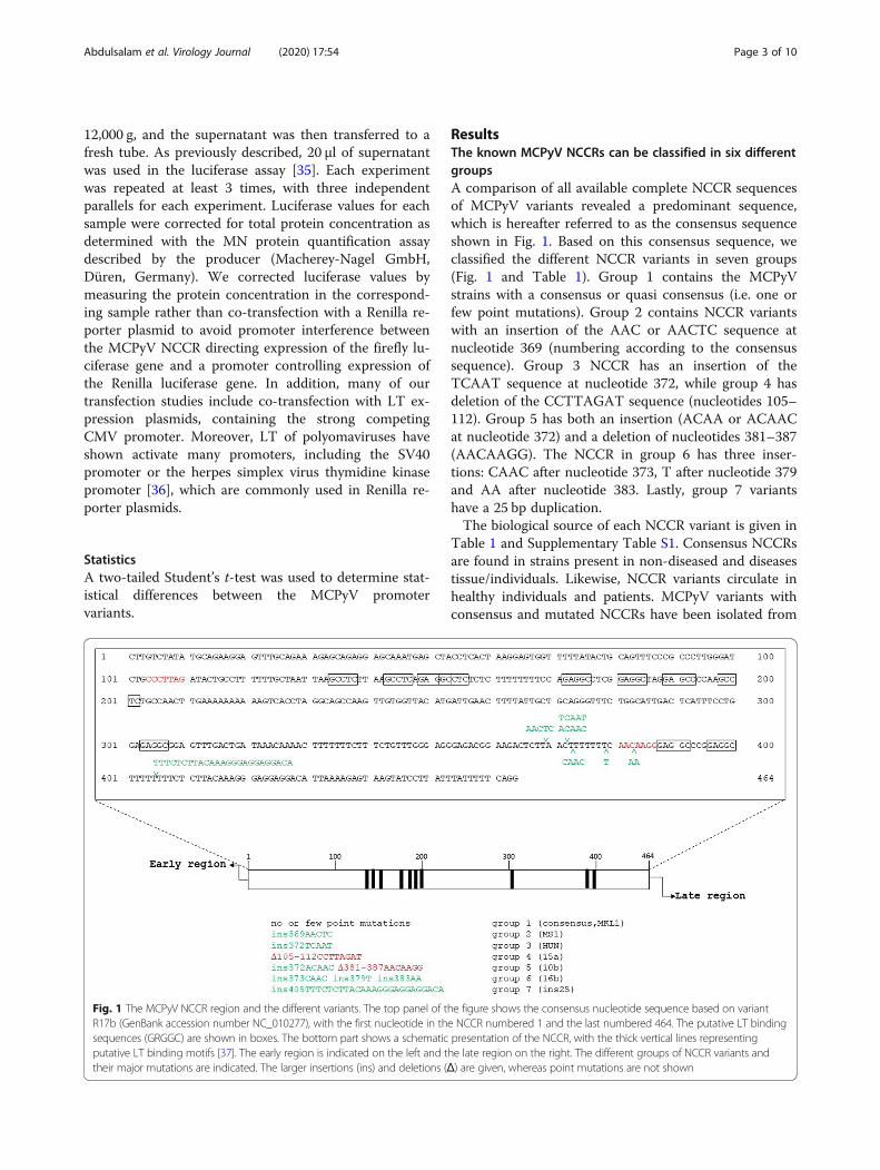
12,000 g, and the supernatant was then transferred to afresh tube. As previously described, 20 µl of supernatantwas used in the luciferase assay [35]. Each experimentwas repeated at least 3 times, with three independentparallels for each experiment. Luciferase values for eachsample were corrected for total protein concentration asdetermined with the MN protein quantification assaydescribed by the producer (Macherey-Nagel GmbH,Düren, Germany). We corrected luciferase values bymeasuring the protein concentration in the correspond-ing sample rather than co-transfection with a Renilla re-porter plasmid to avoid promoter interference betweenthe MCPyV NCCR directing expression of the firefly lu-ciferase gene and a promoter controlling expression ofthe Renilla luciferase gene. In addition, many of ourtransfection studies include co-transfection with LT ex-pression plasmids, containing the strong competingCMV promoter. Moreover, LT of polyomaviruses haveshown activate many promoters, including the SV40promoter or the herpes simplex virus thymidine kinasepromoter [36], which are commonly used in Renilla re-porter plasmids.
StatisticsA two-tailed Student’s t-test was used to determine stat-istical differences between the MCPyV promotervariants.
ResultsThe known MCPyV NCCRs can be classified in six differentgroupsA comparison of all available complete NCCR sequencesof MCPyV variants revealed a predominant sequence,which is hereafter referred to as the consensus sequenceshown in Fig. 1. Based on this consensus sequence, weclassified the different NCCR variants in seven groups(Fig. 1 and Table 1). Group 1 contains the MCPyVstrains with a consensus or quasi consensus (i.e. one orfew point mutations). Group 2 contains NCCR variantswith an insertion of the AAC or AACTC sequence atnucleotide 369 (numbering according to the consensussequence). Group 3 NCCR has an insertion of theTCAAT sequence at nucleotide 372, while group 4 hasdeletion of the CCTTAGAT sequence (nucleotides 105–112). Group 5 has both an insertion (ACAA or ACAACat nucleotide 372) and a deletion of nucleotides 381–387(AACAAGG). The NCCR in group 6 has three inser-tions: CAAC after nucleotide 373, T after nucleotide 379and AA after nucleotide 383. Lastly, group 7 variantshave a 25 bp duplication.The biological source of each NCCR variant is given in
Table 1 and Supplementary Table S1. Consensus NCCRsare found in strains present in non-diseased and diseasestissue/individuals. Likewise, NCCR variants circulate inhealthy individuals and patients. MCPyV variants withconsensus and mutated NCCRs have been isolated from
Fig. 1 The MCPyV NCCR region and the different variants. The top panel of the figure shows the consensus nucleotide sequence based on variantR17b (GenBank accession number NC_010277), with the first nucleotide in the NCCR numbered 1 and the last numbered 464. The putative LT bindingsequences (GRGGC) are shown in boxes. The bottom part shows a schematic presentation of the NCCR, with the thick vertical lines representingputative LT binding motifs [37]. The early region is indicated on the left and the late region on the right. The different groups of NCCR variants andtheir major mutations are indicated. The larger insertions (ins) and deletions (Δ) are given, whereas point mutations are not shown
Abdulsalam et al. Virology Journal (2020) 17:54 Page 3 of 10
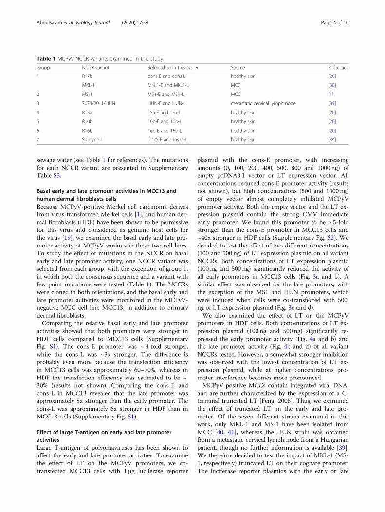
sewage water (see Table 1 for references). The mutationsfor each NCCR variant are presented in SupplementaryTable S3.
Basal early and late promoter activities in MCC13 andhuman dermal fibroblasts cellsBecause MCPyV-positive Merkel cell carcinoma derivesfrom virus-transformed Merkel cells [1], and human der-mal fibroblasts (HDF) have been shown to be permissivefor this virus and considered as genuine host cells forthe virus [19], we examined the basal early and late pro-moter activity of MCPyV variants in these two cell lines.To study the effect of mutations in the NCCR on basalearly and late promoter activity, one NCCR variant wasselected from each group, with the exception of group 1,in which both the consensus sequence and a variant withfew point mutations were tested (Table 1). The NCCRswere cloned in both orientations, and the basal early andlate promoter activities were monitored in the MCPyV-negative MCC cell line MCC13, in addition to primarydermal fibroblasts.Comparing the relative basal early and late promoter
activities showed that both promoters were stronger inHDF cells compared to MCC13 cells (SupplementaryFig. S1). The cons-E promoter was ~ 4-fold stronger,while the cons-L was ~3x stronger. The difference isprobably even more because the transfection efficiencyin MCC13 cells was approximately 60–70%, whereas inHDF the transfection efficiency was estimated to be ~30% (results not shown). Comparing the cons-E andcons-L in MCC13 revealed that the late promoter wasapproximately 8x stronger than the early promoter. Thecons-L was approximately 6x stronger in HDF than inMCC13 cells (Supplementary Fig. S1).
Effect of large T-antigen on early and late promoteractivitiesLarge T-antigen of polyomaviruses has been shown toaffect the early and late promoter activities. To examinethe effect of LT on the MCPyV promoters, we co-transfected MCC13 cells with 1 µg luciferase reporter
plasmid with the cons-E promoter, with increasingamounts (0, 100, 200, 400, 500, 800 and 1000 ng) ofempty pcDNA3.1 vector or LT expression vector. Allconcentrations reduced cons-E promoter activity (resultsnot shown), but high concentrations (800 and 1000 ng)of empty vector almost completely inhibited MCPyVpromoter activity. Both the empty vector and the LT ex-pression plasmid contain the strong CMV immediateearly promoter. We found this promoter to be > 5-foldstronger than the cons-E promoter in MCC13 cells and~40x stronger in HDF cells (Supplementary Fig. S2). Wedecided to test the effect of two different concentrations(100 and 500 ng) of LT expression plasmid on all variantNCCRs. Both concentrations of LT expression plasmid(100 ng and 500 ng) significantly reduced the activity ofall early promoters in MCC13 cells (Fig. 3a and b). Asimilar effect was observed for the late promoters, withthe exception of the MS1 and HUN promoters, whichwere induced when cells were co-transfected with 500ng of LT expression plasmid (Fig. 3c and d).We also examined the effect of LT on the MCPyV
promoters in HDF cells. Both concentrations of LT ex-pression plasmid (100 ng and 500 ng) significantly re-pressed the early promoter activity (Fig. 4a and b) andthe late promoter activity (Fig. 4c and d) of all variantNCCRs tested. However, a somewhat stronger inhibitionwas observed with the lowest concentration of LT ex-pression plasmid, while at higher concentrations pro-moter interference becomes more pronounced.MCPyV-positive MCCs contain integrated viral DNA,
and are further characterized by the expression of a C-terminal truncated LT [Feng, 2008]. Thus, we examinedthe effect of truncated LT on the early and late pro-moter. Of the seven different strains examined in thiswork, only MKL-1 and MS-1 have been isolated fromMCC [40, 41], whereas the HUN strain was obtainedfrom a metastatic cervical lymph node from a Hungarianpatient, though no further information is available [39].We therefore decided to test the impact of MKL-1 (MS-1, respectively) truncated LT on their cognate promoter.The luciferase reporter plasmids with the early or late
Table 1 MCPyV NCCR variants examined in this study
Group NCCR variant Referred to in this paper Source Reference
1 R17b cons-E and cons-L healthy skin [20]
MKL-1 MKL1-E and MKL1-L MCC [38]
2 MS-1 MS1-E and MS1-L MCC [1]
3 7673/2011/HUN HUN-E and HUN-L metastatic cervical lymph node [39]
4 R15a 15a-E and 15a-L healthy skin [20]
5 R10b 10b-E and 10b-L healthy skin [20]
6 R16b 16b-E and 16b-L healthy skin [20]
7 Subtype I Ins25-E and ins25-L healthy skin [34]
Abdulsalam et al. Virology Journal (2020) 17:54 Page 4 of 10
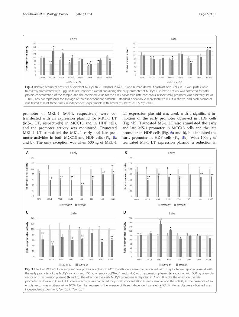
promoter of MKL-1 (MS-1, respectively) were co-transfected with an expression plasmid for MKL-1 LT(MS-1 LT, respectively) in MCC13 and in HDF cells,and the promoter activity was monitored. TruncatedMKL-1 LT stimulated the MKL-1 early and late pro-moter activities in both MCC13 and HDF cells (Fig. 5aand b). The only exception was when 500 ng of MKL-1
LT expression plasmid was used, with a significant in-hibition of the early promoter observed in HDF cells(Fig. 5b). Truncated MS-1 LT also stimulated the earlyand late MS-1 promoter in MCC13 cells and the latepromoter in HDF cells (Fig. 5a and b), but inhibited theearly promoter in HDF cells (Fig. 5b). With 100 ng oftruncated MS-1 LT expression plasmid, a reduction in
Fig. 2 Relative promoter activities of different MCPyV NCCR variants in MCC13 and human dermal fibroblast cells. Cells in 12-well plates weretransiently transfected with 1 μg luciferase reporter plasmid containing the early promoter of MCPyV. Luciferase activity was corrected for totalprotein concentration of the sample, and the corrected value for the early consensus (late consensus, respectively) promoter was arbitrarily set as100%. Each bar represents the average of three independent parallels + standard deviation. A representative result is shown, and each promoterwas tested at least three times in independent experiments with similar results; *p < 0.05, **p < 0.01
Fig. 3 Effect of MCPyV LT on early and late promoter activity in MCC13 cells. Cells were co-transfected with 1 μg luciferase reporter plasmid withthe early promoter of the MCPyV variants and 100 ng of empty pcDNA3.1 vector (EV) or LT expression plasmid (a and c), or with 500 ng of emptyvector or LT expression plasmid (b and d). The effect on the early MCPyV promoters is depicted in A and B, while the effect on the latepromoters is shown in C and D. Luciferase activity was corrected for protein concentration in each sample, and the activity in the presence of anempty vector was arbitrary set as 100%. Each bar represents the average of three independent parallels + SD. Similar results were obtained in anindependent experiment; *p < 0.05; **p < 0.01
Abdulsalam et al. Virology Journal (2020) 17:54 Page 5 of 10

promoter activity of 15–24% compared to the controlwas observed. However, this decrease was not significant(p-values between 0.0553 and 0.0632 in the differentexperiments).
DiscussionMCPyV has a seroprevalence of approximately 80% inthe healthy, adult population [13–18]. MCPyV is chron-ically shed from healthy skin [20], but can also cause anaggressive skin cancer known as Merkel cell carcinoma[1]. Several MCPyV variants have been described withmutations in their NCCR (Supplementary Table S3).These variants have been isolated from both healthy tis-sue and tumors, but so far, no typical strain seems to beassociated with MCC (Supplementary Table S3). Themutations described in the known MCPyV variantscould be classified in seven groups with group 1 contain-ing the most common NCCR, which was referred to asthe consensus sequence in our study and variants withone or a few point mutations. Groups 2–7 contain inser-tions and/or deletions in their NCCR. Significant differ-ences in basal early and late promoter activities in HDFand MCC13 cells were observed. The basal early-, aswell as late promoter activity, of all variants tested washigher in HDF cells than in MCC13 cells despite a lowertransfection efficiency (Fig. 6). This may indicate thatthe MCPyV promoter is more adapted to the former cell
type. HDF have been suggested as natural host cells forthe virus and are permissive for the virus, while the in-fection of Merkel cells and the subsequent transform-ation of these cells could be seen as an accidental andunfortunate event [19].Co-transfection with a 100 ng of full-length LT ex-
pression plasmid resulted in reduced early and late pro-moter activity of all variants in both MCC13 and HDFcells (Fig. 6). However, 500 ng of LT expression plasmidreduced early and late promoter activities in HDF andearly promoter activity in MCC13 cells, but had only aslight or no effect, but significantly stimulated the HUNand MS1 (a 20 and 40% increase, respectively) late pro-moters. Kwun et al. found that MCPyV LT repressedearly and late promoter activity of MCPyV isolateMCC339 in HEK293 cells, but they did not examinethe effect of LT in MCC13 or HDF cells [42]. Yet, inanother study by Ajuh and co-workers, the authorsshowed that LT trans-activated the MCPyV R17b (=consensus) early and late promoter in HEK293T cells[29]. The discrepancy between their results and thefindings by Kwun et al. and us can be explained by theuse of different LT. HEK293T cells express both SV40LT and sT, but not MCPyV LT [29]. Moreover, thepossible contribution of sT in the trans-activation ofthe early and late MCPyV promoters in these cells wasnot investigated. Furthermore, Ajuh et al. studied the
Fig. 4 Effect of MCPyV LT on early and late promoter activity in HDF cells. For details, see legend of Fig. 3
Abdulsalam et al. Virology Journal (2020) 17:54 Page 6 of 10
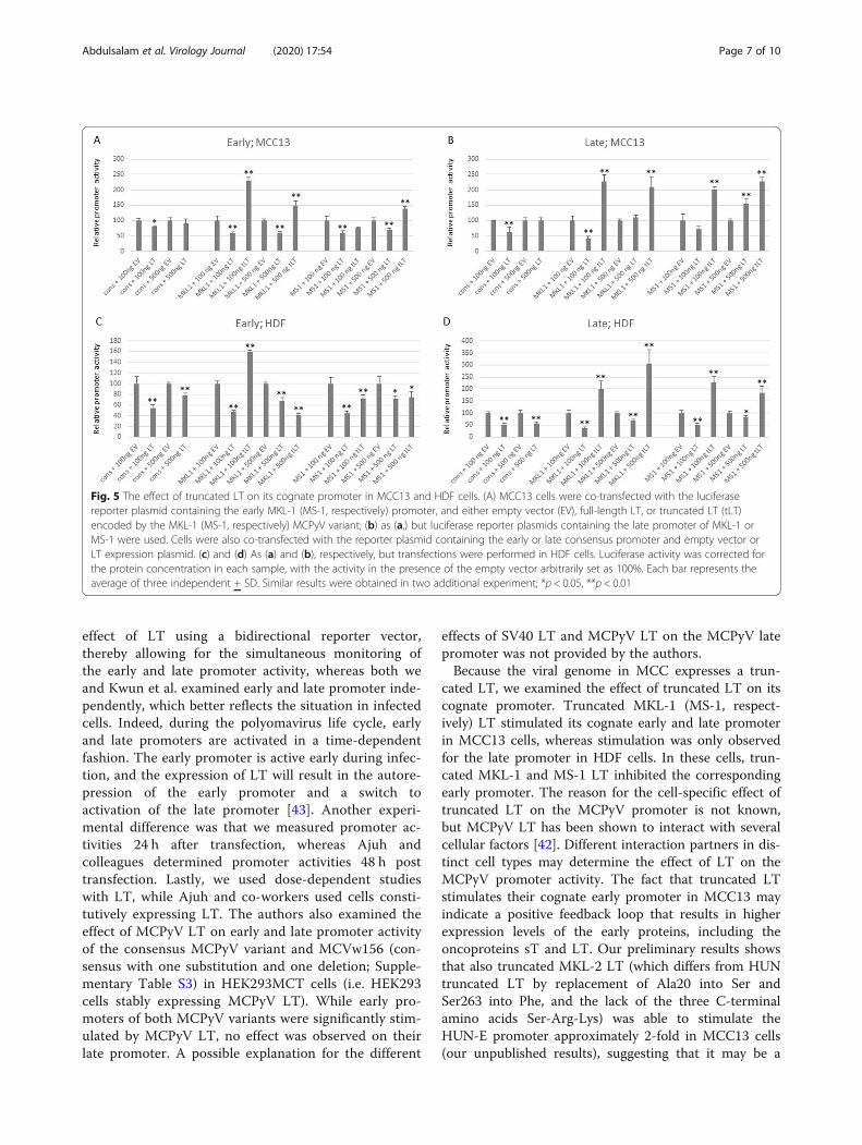
effect of LT using a bidirectional reporter vector,thereby allowing for the simultaneous monitoring ofthe early and late promoter activity, whereas both weand Kwun et al. examined early and late promoter inde-pendently, which better reflects the situation in infectedcells. Indeed, during the polyomavirus life cycle, earlyand late promoters are activated in a time-dependentfashion. The early promoter is active early during infec-tion, and the expression of LT will result in the autore-pression of the early promoter and a switch toactivation of the late promoter [43]. Another experi-mental difference was that we measured promoter ac-tivities 24 h after transfection, whereas Ajuh andcolleagues determined promoter activities 48 h posttransfection. Lastly, we used dose-dependent studieswith LT, while Ajuh and co-workers used cells consti-tutively expressing LT. The authors also examined theeffect of MCPyV LT on early and late promoter activityof the consensus MCPyV variant and MCVw156 (con-sensus with one substitution and one deletion; Supple-mentary Table S3) in HEK293MCT cells (i.e. HEK293cells stably expressing MCPyV LT). While early pro-moters of both MCPyV variants were significantly stim-ulated by MCPyV LT, no effect was observed on theirlate promoter. A possible explanation for the different
effects of SV40 LT and MCPyV LT on the MCPyV latepromoter was not provided by the authors.Because the viral genome in MCC expresses a trun-
cated LT, we examined the effect of truncated LT on itscognate promoter. Truncated MKL-1 (MS-1, respect-ively) LT stimulated its cognate early and late promoterin MCC13 cells, whereas stimulation was only observedfor the late promoter in HDF cells. In these cells, trun-cated MKL-1 and MS-1 LT inhibited the correspondingearly promoter. The reason for the cell-specific effect oftruncated LT on the MCPyV promoter is not known,but MCPyV LT has been shown to interact with severalcellular factors [42]. Different interaction partners in dis-tinct cell types may determine the effect of LT on theMCPyV promoter activity. The fact that truncated LTstimulates their cognate early promoter in MCC13 mayindicate a positive feedback loop that results in higherexpression levels of the early proteins, including theoncoproteins sT and LT. Our preliminary results showsthat also truncated MKL-2 LT (which differs from HUNtruncated LT by replacement of Ala20 into Ser andSer263 into Phe, and the lack of the three C-terminalamino acids Ser-Arg-Lys) was able to stimulate theHUN-E promoter approximately 2-fold in MCC13 cells(our unpublished results), suggesting that it may be a
Fig. 5 The effect of truncated LT on its cognate promoter in MCC13 and HDF cells. (A) MCC13 cells were co-transfected with the luciferasereporter plasmid containing the early MKL-1 (MS-1, respectively) promoter, and either empty vector (EV), full-length LT, or truncated LT (tLT)encoded by the MKL-1 (MS-1, respectively) MCPyV variant; (b) as (a,) but luciferase reporter plasmids containing the late promoter of MKL-1 orMS-1 were used. Cells were also co-transfected with the reporter plasmid containing the early or late consensus promoter and empty vector orLT expression plasmid. (c) and (d) As (a) and (b), respectively, but transfections were performed in HDF cells. Luciferase activity was corrected forthe protein concentration in each sample, with the activity in the presence of the empty vector arbitrarily set as 100%. Each bar represents theaverage of three independent + SD. Similar results were obtained in two additional experiment; *p < 0.05, **p < 0.01
Abdulsalam et al. Virology Journal (2020) 17:54 Page 7 of 10

common feature of truncated LT to autostimulate its ex-pression. This positive autoregulation of LT and sT couldbe potentially important for tumorigenesis. Since full-length LT had an inhibitory effect on the promoters, itwould be interested to test whether full-length LT reversesthe activity of truncated LT. However, to be of biologicalrelevance, both full-length and truncated LT must be co-expressed in MCPyV infected cells. To our best know-ledge, only truncated LT is expressed in virus-positiveMCCs. We are aware of only two studies were co-expression of full-length and truncated LT was observed.One case of non small cell lung cancer (a squamous cellcarcinoma) with both episomal and integrated viral DNAand both full-length and truncated LT protein was de-scribed by Hashida and co-workers [44]. In another study,mRNAs for truncated and full- length LT were confirmedby highly sensitive qRT-PCR in two cases of chroniclymphocytic leukemia, but expression of truncated andfull-length LT at protein level was not investigated [45].The genome copy per chronic lymphocytic leukemia cellwas 3 to 4 logs lower than MCPyV-positive MCCs, sug-gesting that very low levels of LT/truncated LT arepresent in these cells. It remains to be determined whethersuch low levels have any biological relevance for the virallife cycle or tumorigenesis. Because integration of the
MCPyV genome interrupts the late region, no late pro-teins are expressed and no viral particles are produced.The biological implication of enhanced late promoter ac-tivity by truncated LT in MCC remains elusive.Our transient transfection studies showed that the
MCPyV NCCR variants possess different promoter activ-ity, and can lead to different expression levels of the viralproteins. This has been confirmed by in situ studies. TheCVG-1 and MKL-1 MCC cell lines both contain sevencopies of the integrated virus genome per diploid cell [46].The CVG-1 cell line contains the consensus NCCR se-quence, while MKL-1 contains one single point mutation(T52C; Supplementary Table S2). Quantitative real-timePCR demonstrated that the total LT and sT mRNA ex-pression levels were approximately 2.5 times higher inCVG-1 cells compared to MKL-1 cells, thus indicatingthat the former promoter is 2.5x stronger than the latter.Our transient transfection study in MCC13 cells con-firmed that the MKL-1 early promoter was weaker thanthe consensus early promoter (Fig. 2). The expressionlevels of early proteins in MCC not only depend on thestrength of the early promoter, but the number of inte-grated viral genomes and the integration site (hetero- ver-sus euchromatin) may also influence the promoterstrength. Velásquez et al. determined that MKL-2 and
Fig. 6 Heatmap showing the relative promoter activities of eight MCPyV NCCR variants in MCC13 and human dermal fibroblasts (HDF) in theabsence and presence of large T antigen (LT). The activity of the consensus NCCR (cons) was arbitrary set as 100 and the activities of the otherpromoters were related to this
Abdulsalam et al. Virology Journal (2020) 17:54 Page 8 of 10

MS-1 MCC cells contain two and four genome copies perdiploid cells, respectively [46]. The LT transcript levelswere approximately 2-fold higher in MS-1 cells comparedto the MKL-2 cells, while the ST mRNA levels were 4xhigher in MS-1 cells than in MKL-2 cells. The completeNCCR sequence of the MKL-2 variant has not been deter-mined, but the 240 nucleotides downstream of the LTstart codon are identical with the consensus sequence.The MCPyV NCCR contains a plethora of putative tran-
scription factor binding motifs (see Supplementary Fig. 3and Supplementary Table S4), but the binding of the corre-sponding transcription factor has not been confirmed.Whether the mutations found in these sites in the NCCRvariants we investigated abolished binding of the transcrip-tion factor, has not been tested. The 25 bp insertion gener-ates putative binding motifs for the transcription factorsFOXO3a, SRY, Elk-1 and p300, but their possible role inregulating the promoter activity remains to be investigated.
ConclusionsOur study shows that the promoters of different MCPyVisolates possess unlike transcriptional activity, and that full-length LT and MCC-associated truncated LT have a dis-tinct impact on the promoter. Whether these differences inpromoter activity contribute to the replication and trans-formation properties of the virus remains to be determined.
Supplementary informationSupplementary information accompanies this paper at https://doi.org/10.1186/s12985-020-01317-x.
Additional file 1 Supplementary Fig. S1. Early and late promoteractivity of seven MCPyV NCCR variants in MCC13 and human dermalfibroblasts. Supplementary Fig. S2. Relative promoter activity of theimmediately early cytomegalovirus and the consensus early MCPyVpromoter in MCC13 and human dermal fibroblasts. Supplementary Fig.S3. Putative transcription factor binding sites in the consensus MCPyVNCCR. Supplementary Table S1. Detection of MCPyV in benign anddiseased human tissues other than Merkel cell carcinoma.Supplementary Table S2. MCPyV isolated in sewage water andenvironmental surfaces. Supplementary Table S3. MCPyV NCCRvariants and mutations compared to the consensus sequence.Supplementary Table S4. Putative transcription factor binding sites inthe consensus MCPyV NCCR.
AbbreviationsHDF: Human dermal fibroblasts; LT: Large T antigen; MCC: Merkel cellcarcinoma; MCPyV: Merkel cell polyomavirus; NCCR: Non-coding controlregion; sT: Small T antigen; tLT: Truncated large T antigen
AcknowledgmentsThe authors wish to thank Dr. Baki Akgül from the University of Cologne,Germany for providing MCC13 cells.
Authors’ contributionsConceptualization, U.M. and B.E.; experiments, I.A., K.R., and U.M.; validation,B.E., U.M.; writing—original draft preparation, U.M. and B.E.; writing—reviewand editing, I.A., K. R, B.S., E. B, and U.M.; supervision, U.M. and B.S.; projectadministration, U.M.; funding acquisition, U.M. and B.S. The author (s) readand approved the final manuscript.
FundingThis research was funded by “Helse-Nord Forskningsmidler” project 23226.The APC was funded by the UiT, The Arctic University of Norway.
Availability of data and materialsThe datasets used and/or analysed during the current study are availablefrom the corresponding author on reasonable request.
Ethics approval and consent to participateNot applicable.
Consent for publicationNot applicable.
Competing interestsThe authors declare that they have no competing interests.
Author details1Molecular Inflammation Research Group, Department of Medical Biology,Faculty of Health Sciences, University of Tromsø, The Arctic University ofNorway, Tromsø, Norway. 2Present address: Tumor Biology Research Group,Department of Medical Biology, Faculty of Health Sciences, University ofTromsø, The Arctic University of Norway, Tromsø, Norway. 3Division 12Measles, Mumps, Rubella and Viruses Affecting ImmunocompromisedPatients, Robert Koch Institute, Berlin, Germany.
Received: 19 December 2019 Accepted: 10 March 2020
References1. Feng H, Shuda M, Chang Y, Moore PS. Clonal integration of a polyomavirus
in human Merkel cell carcinoma. Science. 2008;319:1096–100. https://doi.org/10.1126/science.1152586.
2. Kassem A, Schöpflin A, Diaz C, Weyers W, Stickler E, Werner M, Zur HA.Frequent detection of Merkel cell polyomavirus in human Merkel cellcarcinomas and identification of a unique deletion in the VP1 gene. CancerRes. 2008;68:5009–13. https://doi.org/10.1158/0008-5472.CAN-08-0949.
3. Foulongne V, Kluger N, Dereure O, Brieu N, Guillot B, Segondy M. Merkelcell polyomavirus and Merkel cell carcinoma. France Emerg Dis. 2008;14:1491–3. https://doi.org/10.3201/eid1409.080651.
4. Becker JC, Houben R, Ugurel S, Trefzer U, Pföhler C, Schrama D. MC polyomavirusis frequently present in Merkel cell carcinoma of European patients. J InvestDermatol. 2009;129:248–50. https://doi.org/10.1038/jid.2008.198.
5. Van Ghelue M, Moens U. Merkel cell polyomaviurs: a causal factor in Merkel cellcarcinoma. In: La Porta CAM, editor. Skin cancers-risk factors, prevention andtherapy. Croatiia: IntechOpen; 2011. p. 109–42. https://doi.org/10.5772/25675.
6. Wendzicki JA, Moore PS, Chang Y. Large T and small T antigens of Merkelcell polyomavirus. Curr Opin Virol. 2015;11:38–43. https://doi.org/10.1016/j.coviro.2015.01.009.
7. Shuda M, Guastafierro A, Geng X, Shuda Y, Ostrowski SM, Lukianov S,Jenkins FJ, Honda K, Maricich SM, Moore PS, Chang Y. Merkel cellPolyomavirus small T antigen induces Cancer and embryonic Merkel cellproliferation in a transgenic mouse model. PLoS One. e01423292015;10.https://doi.org/10.1371/journal.pone.0142329.
8. Verhaegen ME, Mangelberger D, Harms PW, Vozheiko TD, Weick JW, Wilbert DM,Saunders TL, Ermilov AN, Bichakjian CK, Johnson TM, Imperiale MJ, Dlugosz AA.Merkel cell polyomavirus small T antigen is oncogenic in transgenic mice. J InvestDermatol. 2015;135:1415–24. https://doi.org/10.1038/jid.2014.446.
9. Spurgeon ME, Cheng J, Bronson RT, Lambert PF, DeCaprio, J.A. Tumorigenicactivity of merkel cell polyomavirus T antigens expressed in the stratifiedepithelium of mice. Cancer Res 2015;75:1068–1079. doi: https://doi.org/10.1158/0008-5472.CAN-14-2425.
10. Bouvard V, Baan RA, Grosse Y, Lauby-Secretan B, El Ghissassi F, Benbrahim-Tallaa L, Guha N, Straif K. Carcinogenicity of malaria and of somepolyomaviruses. Lancet Oncol. 2012;13:339–40.
11. Stakaityte G, Wood JJ, Knight LM, Abdul-Sada H, Adzahar NS, Nwogu N,Macdonald A, Whitehouse A. Merkel cell polyomavirus: molecular insightsinto the most recently discovered human tumour virus. Cancers. 2014;6:1267–97. https://doi.org/10.3390/cancers6031267.
12. Borchert S, Czech-Sioli M, Neumann F, Schmidt C, Wimmer P, Dobner T,Grundhoff A, Fischer N. High-affinity Rb binding, p53 inhibition, subcellular
Abdulsalam et al. Virology Journal (2020) 17:54 Page 9 of 10

localization, and transformation by wild-type or tumor-derived shortenedMerkel cell polyomavirus large T antigens. J Virol. 2014;88:3144–60. https://doi.org/10.1128/JVI.02916-13.
13. Viscidi RP, Rollison DE, Sondak VK, Silver B, Messina JL, Giuliano AR, Fulp W,Ajidahun A, Rivanera D. Age-specific seroprevalence of Merkel cellpolyomavirus, BK virus, and JC virus. Clin Vaccine Immunol. 2011;18:1737–43.https://doi.org/10.1128/CVI.05175-11.
14. Kean JM, Rao S, Wang M, Garcea RL. Seroepidemiology of humanpolyomaviruses. PLoS Pathog. 2009;5:e1000363. https://doi.org/10.1371/journal.ppat.1000363.
15. Zhang C, Liu F, He Z, Deng Q, Pan Y, Liu Y, Zhang C, Ning T, Guo C, LiangY, Xu R, Zhang L, Cai H, Ke Y. Seroprevalence of Merkel cell polyomavirus inthe general rural population of Anyang. China PLoS One. 2014;9:e106430.https://doi.org/10.1371/journal.pone.0106430.
16. Martel-Jantin C, Pedergnana V, Nicol JT, Leblond V, Tregouet DA, Tortevoye P,Plancoulaine S, Coursaget P, Touze A, Abel L. Gessain, A. Merkel cell polyomavirusinfection occurs during early childhood and is transmitted between siblings. JClin Virol 2013;58:288–291. doi: https://doi.org/10.1016/j.jcv.2013.06.004.
17. Nicol JT, Robinot R, Carpentier A, Carandina G, Mazzoni E, Tognon M, Touze A,Coursaget P. Age-specific seroprevalences of merkel cell polyomavirus, humanpolyomaviruses 6, 7, and 9, and trichodysplasia spinulosa-associated polyomavirus.Clin Vaccine Immunol. 2013;20:363–8. https://doi.org/10.1128/CVI.00438-12.
18. Kamminga S. E van der Meijden E, Feltkamp MCW. Zaaijer HL Seroprevalenceof fourteen human polyomaviruses determined in blood donors PLoS One.2018;13:e0206273. https://doi.org/10.1371/journal.pone.0206273.
19. Liu W, Yang R, Payne AS, Schowalter RM, Spurgeon ME, Lambert PF, Xu X,Buck CB, You J. Identifying the target cells and mechanisms of Merkel cellPolyomavirus infection. Cell Host Microbe. 2016;19:775–87. https://doi.org/10.1016/j.chom.2016.04.024.
20. RM RS, Pastrana DV, Pumphrey KA, Moyer AL, Buck CB. Merkel cellpolyomavirus and two previously unknown polyomaviruses are chronicallyshed from human skin. Cell Host Microbe. 2010;7:509–15. https://doi.org/10.1016/j.chom.2010.05.006.
21. Foulongne V, Sauvage V, Hebert C, Dereure O, Cheval J, Gouilh MA, ParienteK, Segondy M, Burguiere A, Manuguerra JC, Caro V, Eloit M. Human skinmicrobiota: high diversity of DNA viruses identified on the human skin byhigh throughput sequencing. PLoS One. 2012;7:e38499. https://doi.org/10.1371/journal.pone.0038499.
22. Hashida Y, Kamioka M, Tanaka M, Hosokawa S, Murakami M, Nakajima K,Kikuchi H, Fujieda M, Sano S, Daibata M. Ecology of Merkel cellPolyomavirus in healthy skin among individuals in an Asian cohort. J InfectDis. 2016;213:1708–16. https://doi.org/10.1093/infdis/jiw040.
23. Csoboz B, Rasheed K, Sveinbjørnsson U. Moens U. Merkel cell polyomavirusand non-Merkel cell carcinomas: guilty or circumstantial evidence? APMIS.2020; [Online ahead of print]. https://doi.org/10.1111/apm.13019.
24. Moens U, Van Ghelue M, Ludvigsen M, Korup-Schulz S, Ehlers B. The earlyand late promoters of BKPyV, MCPyV, TSPyV, and HPyV12 are among thestrongest of all known human polyomaviruses in 10 different cell lines. JGen Virol. 2015;96:2293–303. https://doi.org/10.1099/vir.0.000181.
25. Moens U, Song X, Van Ghelue M, Lednicky JA, Ehlers, B. A Role of Sp1Binding Motifs in Basal and Large T-Antigen-Induced Promoter Activities ofHuman Polyomavirus HPyV9 and Its Variant UF-1. Int J Mol Sci. 2017;18:pii:E2414. doi: https://doi.org/10.3390/ijms18112414.
26. Song X, Van Ghelue M, Ludvigsen M, Nordbo SA, Ehlers B, Moens U.Characterization of the non-coding control region of polyomavirus KIisolated from nasopharyngeal samples from patients with respiratorysymptoms or infection and from blood from healthy blood donors inNorway. J Gen Virol. 2016;97:1647–57. https://doi.org/10.1099/jgv.0.000473.
27. Gosert R, Rinaldo CH, Funk GA, Egli A, Ramos E, Drachenberg CB, Hirsch HH.Polyomavirus BK with rearranged noncoding control region emerge in vivoin renal transplant patients and increase viral replication and cytopathology.J Exp Med. 2008;205:841–52. https://doi.org/10.1084/jem.20072097.
28. Gosert R, Kardas P, Major EO, Hirsch HH. Rearranged JC virus noncodingcontrol regions found in progressive multifocal leukoencephalopathypatient samples increase virus early gene expression and replication rate. JVirol. 2010;84:10448–56. https://doi.org/10.1128/JVI.00614-10.
29. Ajuh ET, Wu Z, Kraus E, Weissbach FH, Bethge T, Gosert R, Fisher N, HirschHH. Novel human polyomavirus noncoding control region differ inbidirectional gene expression according to host cell, large T-antigenexpression, and clinically occurring rearrangements. J Virol. 2018;92:e02231–17. https://doi.org/10.1128/JVI.02231-17.
30. Barcena-Panero A, Echevarria JE, Van Ghelue M, Fedele G, Royuela E, GeritsN, Moens U. BK polyomavirus with archetypal and rearranged non-codingcontrol regions is present in cerebrospinal fluids from patients withneurological complications. J Gen Virol. 2012;93:1780–94. https://doi.org/10.1099/vir.0.042143-0.
31. Van Loy T, Thys K, Ryschkewitsch C, Lagatie O, Monaco MC, Major EO,Tritsmans L, Stuyver LJ. JC virus quasispecies analysis reveals a complex viralpopulation underlying progressive multifocal leukoencephalopathy andsupports viral dissemination via the hematogenous route. J Virol. 2015;89:1340–7. https://doi.org/10.1128/JVI.02565-14.
32. L'Honneur AS, Leh H, Laurent-Tchenio F, Hazan U, Rozenberg F, Bury-MoneS. Exploring the role of NCCR variation on JC polyomavirus expression fromdual reporter minicircles. PLoS One. 2018;13:e0199171. https://doi.org/10.1371/journal.pone.0199171.
33. Takahashi K, Sekizuka T, Fukumoto H, Nakamichi K, Suzuki T, Sato Y,Hasegawa H, Kuroda M, Katano H. Deep-Sequence Identification and Role inVirus Replication of a JC Virus Quasispecies in Patients with ProgressiveMultifocal Leukoencephalopathy. J Virol. 2016;91:pii: e01335–e01316. doi:https://doi.org/10.1128/JVI.01335-16.
34. Hashida Y, Higuchi T, Matsui K, Shibata Y, Nakajima K, Sano S, Daibata M.Genetic variability of the noncoding control region of cutaneous Merkel cellPolyomavirus: identification of geographically related genotypes. J InfectDis. 2018;217:1601–11. https://doi.org/10.1093/infdis/jiy070.
35. Rasheed K, Abdulsalam I, Fismen S, Grimstad Ø, Sveinbjornsson B, Moens U.CCL17/TARC and CCR4 expression in Merkel cell carcinoma. Oncotarget.2018;9:31432–47. https://doi.org/10.18632/oncotarget.25836.
36. Pannuti A, Pascucci A, La Mantia G, Fisher-Fantuzzi L, Vesco C, Lania L.Trans-activation of cellular and viral promoters by a transformingnonkaryophilic simian virus 40 large T antigen. J Virol. 1987;61:1296–9.
37. Harrison C, Jiang T, Banerjee P, Meinke G, D'Abramo CM, Schaffhausen B,Bohm A. Polyomavirus large T antigen binds symmetrical repeats at theviral origin in an asymmetrical manner. J Virol. 2013;87:13751–9. https://doi.org/10.1128/JVI.01740-13.
38. Shuda M, Feng H, Kwun HJ, Rosen ST, Gjoerup O, Moore PS, Chang Y. Tantigen mutations are a human tumor-specific signature for Merkel cellpolyomavirus. Proc Natl Acad Sci U S A. 2008;105:16272–7. https://doi.org/10.1073/pnas.0806526105.
39. Horvath KB, Pankovics P, Battyani Z, Kalman E, Reuter G. A probable etiologicalrole of Merkel cell polyomavirus in the development of Merkel cell carcinoma.Orv Hetil. 2013;154:102–12. https://doi.org/10.1556/OH.2013.29525.
40. Rosen ST, Gould VE, Salwen HR, Herst CV, Le Beau MM, Lee I, Bauer K,Marder RJ, Andersen R, Kies MS, et al. Establishment and characterization ofa neuroendocrine skin carcinoma cell line. Lab Investig. 1987;56:302–12.
41. Guastafierro A, Feng H, Thant M, Kirkwood JM, Chang Y, Moore PS, Shuda M.Characterization of an early passage Merkel cell polyomavirus-positive Merkelcell carcinoma cell line, MS-1, and its growth in NOD scid gamma mice. J VirolMethods. 2013;187:6–14. https://doi.org/10.1016/j.jviromet.2012.10.001.
42. Kwun HJ, Chang Y, Moore PS. Protein-mediated viral latency is a novelmechanism for Merkel cell polyomavirus persistence. Proc Natl Acad Sci U SA. 2017;114:e4040–7. https://doi.org/10.1073/pnas.1703879114.
43. Moens U, Krumbholz A, Ehlers B, Zell R, Johne R, Calvignac-Spencer S,Lauber C. Biology, evolution, and medical importance of polyomaviruses: anupdate. Infect Genet Evol. 2017;54:18–38. https://doi.org/10.1016/j.meegid.2017.06.011.
44. Hashida Y, Imajoh M, Nemoto Y, Kamioka M, Taniguchi A, Taguchi T, KumeM, Orihashi K, Daibata M. Detection of Merkel cell polyomavirus with atumour-specific signature in non-small cell lung cancer. Br J Cancer. 2013;108:629–37. https://doi.org/10.1038/bjc.2012.567.
45. Pantulu ND, Pallasch CP, Kurz AK, Kassem A, Frenzel L, Sodenkamp S, KvasnickaHM, Wendtner CM, Zur Hausen A. Detection of a novel truncating Merkel cellpolyomavirus large T antigen deletion in chronic lymphocytic leukemia cells.Blood. 2010;116:5280–4. https://doi.org/10.1182/blood-2010-02-269829.
46. Velasquez C, Amako Y, Harold A, Toptan T, Chang Y, Shuda M.Characterization of a Merkel cell Polyomavirus-positive Merkel cellcarcinoma cell line CVG-1. Front Microbiol. 2018;9:713. https://doi.org/10.3389/fmicb.2018.00713.
Publisher’s NoteSpringer Nature remains neutral with regard to jurisdictional claims inpublished maps and institutional affiliations.
Abdulsalam et al. Virology Journal (2020) 17:54 Page 10 of 10

PAPER II


Oncotarget31432www.oncotarget.com
CCL17/TARC and CCR4 expression in Merkel cell carcinoma
Kashif Rasheed1, Ibrahim Abdulsalam1, Silje Fismen2, Øystein Grimstad3, Baldur Sveinbjørnsson1 and Ugo Moens1
1Molecular Inflammation Research Group, Department of Medical Biology, Faculty of Health Sciences, University of Tromsø, N-9037, Tromsø, Norway
2Department of Pathology, University Hospital of Northern Norway, N-9038, Tromsø, Norway3Department of Dermatology, University Hospital of Northern Norway, N-9038, Tromsø, Norway
Correspondence to: Kashif Rasheed, email: [email protected]: Merkel cell carcinoma; inflammation; cytokines; CCL17/TARC; CCR4Received: January 29, 2018 Accepted: July 12, 2018 Published: July 31, 2018Copyright: Rasheed et al. This is an open-access article distributed under the terms of the Creative Commons Attribution License 3.0 (CC BY 3.0), which permits unrestricted use, distribution, and reproduction in any medium, provided the original author and source are credited.
ABSTRACT
Merkel cell carcinoma (MCC) is a rare, highly aggressive neuroendocrine skin cancer. In more than 80% of the cases, Merkel cell polyomavirus (MCPyV) is a causal factor. The oncogenic potential of MCPyV is mediated through its viral oncoproteins, large T antigen (LT) and small t antigen (sT). To investigate the role of cytokines in MCC, a PCR array analysis for genes encoding inflammatory cytokines and receptors was performed on MCPyV-negative and MCPyV-positive MCC cell lines, respectively. We detected an increased expression of CCL17/TARC in the MCPyV-positive MKL2 cell line compared to the MCPyV-negative MCC13 cell line. Transfection studies in MCC13 cells with LT expression plasmid, and a luciferase reporter plasmid containing the CCL17/TARC promoter, exhibited stimulated promoter activity. Interestingly, the ectopic expression of CCL17/TARC upregulated MCPyV early and late promoter activities in MCC13 cells. Furthermore, recombinant CCL17/TARC activated both the mitogen-activated protein kinase and the NF-κB pathways. Finally, immunohistochemical staining on human MCC tissues showed a strong staining of CCL17/TARC and its receptor CCR4 in both LT-positive and -negative MCC. Taken together, CCL17/TARC and CCR4 may be a potential target in MCC therapy providing MCC patients with a better overall survival outcome.
INTRODUCTION
Merkel cell carcinoma (MCC) is a rare, highly aggressive neuroendocrine skin cancer [1, 2]. In 2008, using digital transcriptome subtraction, a new virus belonging to the family of Polyomaviruses, and hence named Merkel cell polyomavirus (MCPyV), was identified in MCC. Worldwide studies have shown that approximately 80% of all examined MCC contain clonal integrated MCPyV DNA [3], thus indicating that MCPyV is associated with the etiology of MCC [4, 5]. MCPyV has a circular, double-stranded DNA genome of approximately 5.4 kb [6]. The viral genome possesses the typical polyomavirus tripartite organization, with
an early region encoding the regulatory proteins large T (LT) antigen and small T (sT) antigen [7], the late region encoding the capsid proteins [8], and a non-coding control region encompassing the origin of replication and transcription regulatory elements [9]. In addition, the early region encodes the 57kT antigen and a protein called alternative LT ORF (ALTO), although their function remains unknown [7]. All MCPyV-positive (MCPyV+) MCC express a C-terminal truncated form of LT antigen that has lost its DNA binding and p53 interaction domains, but retains the ability to interact with retinoblastoma protein pRb [10]. Both sT and full-length LT, as well as truncated LT, have been shown to possess oncogenic potential in both cell culture and
www.oncotarget.com Oncotarget, 2018, Vol. 9, (No. 59), pp: 31432-31447
Research Paper
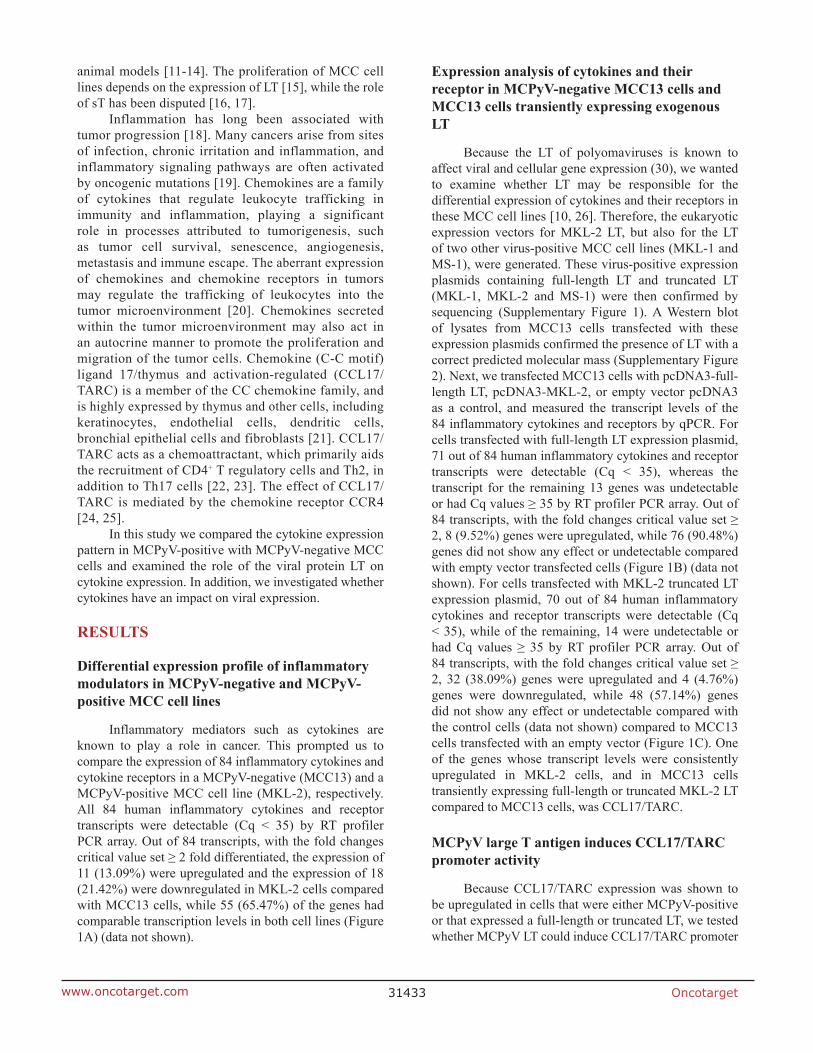
Oncotarget31433www.oncotarget.com
animal models [11-14]. The proliferation of MCC cell lines depends on the expression of LT [15], while the role of sT has been disputed [16, 17].
Inflammation has long been associated with tumor progression [18]. Many cancers arise from sites of infection, chronic irritation and inflammation, and inflammatory signaling pathways are often activated by oncogenic mutations [19]. Chemokines are a family of cytokines that regulate leukocyte trafficking in immunity and inflammation, playing a significant role in processes attributed to tumorigenesis, such as tumor cell survival, senescence, angiogenesis, metastasis and immune escape. The aberrant expression of chemokines and chemokine receptors in tumors may regulate the trafficking of leukocytes into the tumor microenvironment [20]. Chemokines secreted within the tumor microenvironment may also act in an autocrine manner to promote the proliferation and migration of the tumor cells. Chemokine (C-C motif) ligand 17/thymus and activation-regulated (CCL17/TARC) is a member of the CC chemokine family, and is highly expressed by thymus and other cells, including keratinocytes, endothelial cells, dendritic cells, bronchial epithelial cells and fibroblasts [21]. CCL17/TARC acts as a chemoattractant, which primarily aids the recruitment of CD4+ T regulatory cells and Th2, in addition to Th17 cells [22, 23]. The effect of CCL17/TARC is mediated by the chemokine receptor CCR4 [24, 25].
In this study we compared the cytokine expression pattern in MCPyV-positive with MCPyV-negative MCC cells and examined the role of the viral protein LT on cytokine expression. In addition, we investigated whether cytokines have an impact on viral expression.
RESULTS
Differential expression profile of inflammatory modulators in MCPyV-negative and MCPyV-positive MCC cell lines
Inflammatory mediators such as cytokines are known to play a role in cancer. This prompted us to compare the expression of 84 inflammatory cytokines and cytokine receptors in a MCPyV-negative (MCC13) and a MCPyV-positive MCC cell line (MKL-2), respectively. All 84 human inflammatory cytokines and receptor transcripts were detectable (Cq < 35) by RT profiler PCR array. Out of 84 transcripts, with the fold changes critical value set ≥ 2 fold differentiated, the expression of 11 (13.09%) were upregulated and the expression of 18 (21.42%) were downregulated in MKL-2 cells compared with MCC13 cells, while 55 (65.47%) of the genes had comparable transcription levels in both cell lines (Figure 1A) (data not shown).
Expression analysis of cytokines and their receptor in MCPyV-negative MCC13 cells and MCC13 cells transiently expressing exogenous LT
Because the LT of polyomaviruses is known to affect viral and cellular gene expression (30), we wanted to examine whether LT may be responsible for the differential expression of cytokines and their receptors in these MCC cell lines [10, 26]. Therefore, the eukaryotic expression vectors for MKL-2 LT, but also for the LT of two other virus-positive MCC cell lines (MKL-1 and MS-1), were generated. These virus-positive expression plasmids containing full-length LT and truncated LT (MKL-1, MKL-2 and MS-1) were then confirmed by sequencing (Supplementary Figure 1). A Western blot of lysates from MCC13 cells transfected with these expression plasmids confirmed the presence of LT with a correct predicted molecular mass (Supplementary Figure 2). Next, we transfected MCC13 cells with pcDNA3-full-length LT, pcDNA3-MKL-2, or empty vector pcDNA3 as a control, and measured the transcript levels of the 84 inflammatory cytokines and receptors by qPCR. For cells transfected with full-length LT expression plasmid, 71 out of 84 human inflammatory cytokines and receptor transcripts were detectable (Cq < 35), whereas the transcript for the remaining 13 genes was undetectable or had Cq values ≥ 35 by RT profiler PCR array. Out of 84 transcripts, with the fold changes critical value set ≥ 2, 8 (9.52%) genes were upregulated, while 76 (90.48%) genes did not show any effect or undetectable compared with empty vector transfected cells (Figure 1B) (data not shown). For cells transfected with MKL-2 truncated LT expression plasmid, 70 out of 84 human inflammatory cytokines and receptor transcripts were detectable (Cq < 35), while of the remaining, 14 were undetectable or had Cq values ≥ 35 by RT profiler PCR array. Out of 84 transcripts, with the fold changes critical value set ≥ 2, 32 (38.09%) genes were upregulated and 4 (4.76%) genes were downregulated, while 48 (57.14%) genes did not show any effect or undetectable compared with the control cells (data not shown) compared to MCC13 cells transfected with an empty vector (Figure 1C). One of the genes whose transcript levels were consistently upregulated in MKL-2 cells, and in MCC13 cells transiently expressing full-length or truncated MKL-2 LT compared to MCC13 cells, was CCL17/TARC.
MCPyV large T antigen induces CCL17/TARC promoter activity
Because CCL17/TARC expression was shown to be upregulated in cells that were either MCPyV-positive or that expressed a full-length or truncated LT, we tested whether MCPyV LT could induce CCL17/TARC promoter

Oncotarget31434www.oncotarget.com
activity. For this purpose, MCC13 cells were transiently co-transfected with a plasmid encoding a full-length or truncated LT (MKL-1, MKL-2 and MS-1), and with a luciferase reporter driven by different fragments of the CCL17/TARC promoter (fragment -2535/+ 40, -1084/+ 40 or -378/+40, respectively). The empty vector pcDNA3 was used as a control. Full-length, as well as the truncated LT versions MKL-1 and MS-1 significantly stimulated the CCL17/TARC promoter activity. The largest CCL17/TARC promoter fragment (-2535/+40) was more potently activated than the shorter promoter fragments (-1084/+40 and -375/+40, respectively) by LT MKL-1 and MS-1,
while comparable full-length LT-mediated transactivation the three different CCL17/TARC promoter fragments. MKL-2 LT significantly stimulated the activity of the CCL17/TARC promoter encompassing nucleotides -2535/+40 and -375/+40, but induced only slightly, but statistically insignificant the CCL17/TARC -1080/+40 promoter sequence (Figure 2). These results confirm the qPCR data showing that CCL17/TARC expression is higher in LT expressing MCC13 compared to MCC13 cells. The enhanced CCL17/TARC transcript levels in MKL-2 cells, compared to the virus-negative MCC13 cells, may at least be partially triggered by LT. In contrast,
Figure 1: Relative expression comparison of 84 inflammatory cytokines and receptors genes between MCPyV-associated and non-associated Merkel cell carcinoma. The figures depict a log transformation plot of the relative expression level of each gene (2-ΔCt) between (A) MCC13 cells vs. MKL-2 cells, (B) full-length LT vs. empty vector transfection in MCC13 cell line and (C) MKL-2 truncated LT vs. empty vector transfection in MCC13 cell line. The dotted lines indicate a two-fold change in gene expression threshold.

Oncotarget31435www.oncotarget.com
sT did not stimulate the activity of any of the CCL17 promoter fragments or increase sT protein levels when expressed in MCC13 cells (results not shown).
CCL17/TARC ectopic effect on MCPyV early and promoter activity
Cytokines such as IL-1β, TGF β and TNF-α have been shown to stimulate the activity of human polyomavirus promoters [27-30]. Therefore, we investigated whether CCL17/TARC could exert an effect on the MCPyV early and late promoter activity. MCC13 cells were transfected with a luciferase reporter plasmid containing, either the early or late MCPyV promoter, and cells were either co-transfected with a CCL17/TARC expression plasmid or treated with recombinant human CCL17/TARC protein. Both an ectopic expression of CCL17/TARC and administering of recombinant CCL17/TARC to cells resulted in a significant upregulation of both the early and late MCPyV promoter activity (Figure 3).
Expression of CCL17/TARC and CCR4 in MCC cells
To confirm the stimulating effect of full-length and truncated LT on CCL17/TARC promoter activity, we first evaluated the mRNA expression of CCL17/TARC by qPCR in MCC13 cells transfected with expression vector for full-length LT or truncated LT variants. Full-length and MKL-1 LT significantly increased CCL17/TARC expression in the MCC13 cell line (P < 0.01), while MKL-2 and MS-1 LT variants, modestly, but significantly increased CCL17/TARC mRNA levels (Figure 4A). Western blot analysis with anti-CCL17/TARC antibodies confirmed that CCL17/TARC protein levels were increased in MCC13 cells expressing either full-length or truncated LT compared to MCC13 cells (Figure 4B and 4D). In our screening experiments, we also found a slight upregulation of CCR4 mRNA by exogenous expression of both pcDNA3-FLTA and pcDNA3-MKL-2 plasmids. So, to check at the protein level, we conducted a Western blot.
Figure 2: Effect of full-length and truncated (MKL-1, MKL-2 and MS-1) LT on CCL17/TARC promoter activity in MCC13 cells. Cells were transfected with a luciferase reporter vector driven by CCL17/TARC promoter fragments spanning nucleotides -2535/+ 40, -1084/+ 40 or -375/+40, respectively. Expression plasmid for full-length LT (FLTA), MKL-1, MKL-2 or MS-1 LT, or pcDNA3 control, were co-transfected. Luciferase activity was assessed after an overnight cultivation of transfected cells. The promoter activity in presence of pcDNA3 was arbitrary set as 1.0 and the promoter activity measured in the presence of LT was related to this. Each bar represents the average of three independent parallels +SD. The experiment was repeated two more times, and similar results were obtained. Luciferase values were normalized with a total protein in each sample. P* ≤ 0.05, P** ≤ 0.01, P*** ≤ 0.001 and P**** ≤ 0.0001.

Oncotarget31436www.oncotarget.com
We did not find expression at a significant level, but only a slight upregulation of CCR4 in MCC13 cells with an exogenous expression of MCPyV LT (Figure 4C and 4D).
CCL17/TARC activates the mitogen-activated protein kinase (MAP kinase) and NF-κB pathways in MCC cells
Both CCL17/TARC and ERK1/2 have been shown to be involved in skin inflammation [31-33], thereby suggesting that CCL17/TARC may activate the MEK1/2-ERK1/2 mitogen-activated protein kinase pathway. To help investigate the effect of CCL17/TARC on MEK1/2-ERK1/2 activation in MCPyV-associated MCC, we stimulated MCC13 cells with recombinant human CCL17/TARC. The cells were stimulated with different concentrations (2.5 ng/ml to 15 ng/ml) and time periods (5 min to 60 min) and we monitored the phosphorylation of ERK1/2 using phospho-specific antibodies and western blotting. Activation of ERK1/2 by rhCCL17/TARC was shown to be concentration dependent (Figure 5). The ERK1/2 phosphorylation activity was inhibited by using a specific CCR4 antagonist (C021 dihydrochloride) (Figure 6).
Previous studies have indicated that NF-κB is a target downstream of CCL17/TARC. We found that CCL17/TARC increased the phosphorylation of NF-κB/p65 activity in a concentration-dependent manner (Figure 7A, 7B), which was inhibited by using a specific CCR4 antagonist (Figure 7D, 7E). Furthermore, NF-κB promoter assay confirmed an increased luciferase activity of NF-κB when cells were co-transfected with pCMV2-CCL17 and NF-κB-luc reporter plasmids (Figure 7C).
CCL17/TARC stimulates cell proliferation of MCC13 cells
Next, we examined the effect of rhCCL17/TARC on MCC13 proliferation. A dose-dependent increase in cell proliferation was observed (Figure 7F). The CCR4 antagonist C021 inhibited CCL17/TARC-induced cell proliferation (Figure 7G).
CCL17/TARC and its receptor are expressed in MCC tissue samples
A total of 23 primary cutaneous MCCs were immunohistochemically stained for LT, CK20, CCL17/TARC and CCR4, respectively. Fifteen out of 23 (65.2%) demonstrated an intranuclear positivity consistent with a positivity for LT. All of the 15 LT-positive tumors demonstrated a uniform positivity for CK20 (dot-like cytoplasmic) and CCL17/TARC (dot-like cytoplasmic). With regard to CCR4, 12 out of 15 (80%) of the LT-positive tumors demonstrated cytoplasmic, membranous positivity for CCR4. Of the remaining eight (34.8%) LT -negative tumors, all of these demonstrated a uniform positivity for CK20, CCL17/TARC and CCR4, respectively (Figure 8).
DISCUSSION
There is increasing evidence that inflammatory mediators such as chemokines and chemokine receptors are involved in promoting tumor invasion, migration and vascularization [34]. Previous studies have demonstrated the involvement of chemokines, such as CXCL1, CXCL5,
Figure 3: CCL17/TARC stimulates MCPyV early and late promoter activity. (A) MCC13 cells were seeded, and after 24 hrs the cells were serum starved. After serum starvation for 24 hrs, the cells were co-transfected with either CCL17/TARC expression plasmid or empty expression vector pcDNA3.1, and luciferase reporter plasmid containing either the MCPyV early or the late promoter. Luciferase activity was measured 24 hrs after transfection. The activity of the MCPyV early (respectively late) promoter in the presence of pcDNA3 was arbitrary set as 1.0 and the activity in the presence of CCL17 was related to this. (B) Cells were transfected as in (A), and were then exposed to rhCCL17/TARC (12ng/ml) or vehicle (PBS) for 4 hrs with a subsequent analysis of luciferase activity. Each bar represents the average of three independent parallels +SD*P < 0.05, ***P < 0.001.

Oncotarget31437www.oncotarget.com
CXXC4 and IL20RA in MCPyV-associated MCC [35]. In the current study, we performed the screening of different inflammatory cytokines and receptors, and compared their expression in MCPyV LT-positive and -negative MCC, respectively. Other groups have previously reported that LT and sT have an impact on cytokine expression. Ectopic sT expression in MCC13 cells resulted in decreased IL2, IL-8, CCL20 and CXCL9 expression [36]. Also, expression of full-length LT, truncated LT339, and truncated LT339 plus sT in hTERT-immortalized BJ human foreskin fibroblasts increased the expression of IL-1β, IL-6, IL-8, CXCL1 and CXCL6 [37].
Here we demonstrate that one of the cytokine member whose expression was shown to be significantly enhanced in MCPyV LT-positive cells was CCL17/TARC. CCL17/TARC is a member of CC-motif chemokine family, and is constitutively expressed in thymus and by dendritic cells, endothelial cells, keratinocytes and fibroblasts [38]. An enhanced expression of CCL17/
TARC has been reported in several human malignancies, such as Hodgkin’s and B cell lymphoma [39, 40]. Previous studies have revealed that CCL17/TARC possesses several important effects attributed to tumor growth, such as the proliferation [41-43], migration and recruitment of regulatory T-cells [44-46].
We detected a higher expression of CCL17/TARC in MCPyV-positive MCC compared to MCPyV-negative MCC cells at both the RNA and protein level. Furthermore, by co-transfecting MCC cells with MCPyV full LT, MKL-1 LT, MKL-2 LT and MS-1 LT expression plasmids, as well as CCL17/TARC promoter, resulted in an upregulation of CCL17/TARC promoter activity (Figure 2), which in turn increased the expression of CCL17/TARC (Figure 4). At present, we do not know why the different LT variants activate the CCL17 promoter at different levels. MCPyV full-length LT can interact with several transcription factors including Brd4, the E2F family members 2 and 3, MED14/CRSP2, PRTF1,
Figure 4: Transient expression of full-length or truncated MCPyV LT increases the transcript and protein levels of CCL17/TARC. MCC13 cells were transfected with an empty vector or expression plasmid for MCPyV full-length LT (FLTA), or truncated MKL-1, MKL-2, or MS-1 LT. (A) qRT-PCR analysis shows CCL17/TARC mRNA levels normalized with eukaryotic 18S rRNA levels. (B) CCL17/TARC protein levels analyzed by Western blotting. The uttermost right lane represents baseline expression of CCL17/TARC by MCC13 cells. (C) CCR4 protein levels analyzed by Western blotting. A Western blot with ERK2 antibodies was used as a loading control. (D) Representative figures of CCL17/TARC (B) and CCR4 (C) Western blots. The lane most to the left in the lower part contains the protein molecular mass marker (in kDa). Bars in (B) and (C) shows a densitometric scanning of the Western blot signals. P* ≤ 0.05 and P*** ≤ 0.001.

Oncotarget31438www.oncotarget.com
SALL2 and DP1 (see supplementary data in Moens et al. [47]). However, no putative binding sites are present in the CCL17 promoter fragments used in our study. The truncated MCPyV LT variants MKL-1, MKL-2 and MS-1 retain the pRb binding motif and may thus relieve the pRb-mediated inhibition of E2F by using pRb. The CCL17/TARC promoter contains the putative GGCGGCA E2F binding site at position -1085/-1079 [48]. This means that our luciferase reporter plasmids containing
the CCL17/TARC promoter fragments -2535/+40 and-1084/+40 contain this putative E2F binding site. LT may thus activate the CCL17/TARC promoter fragments with an E2F motif by usurping pRb and releasing the repressive effect on the E2F site. However, the shorter promoter fragment (-375/+40) which lacks the putative E2F binding site is still trans-activated by LT, indicating that LT-mediated activation seems to be independently of the E2F site. The autocrine release of CCL17/TARC
Figure 5: CCL17/TARC upregulates ERK1/2 activity. (A) PhosphoERK1/2 (pERK1/2) expression levels were determined by Western blot analysis using a phospho-specific antibody detecting Thr202/Tyr204 phosphorylation and compared to total ERK1/2 (tERK1/2) levels using an anti-pan-ERK1/2 antibody. MCC13 (1x10⁵) cells were seeded in a 6-well plate. Cells were then serum-starved for 24 hrs, and thereafter stimulated with either PBS (control or C) or with 2.5, 5, 7.5, 10 and 15 ng/ml of rhCCL17/TARC for 45 min. (B) Densitometry scanning represent the expression of pERK1 (respectively pERK2) protein relative to GAPDH.

Oncotarget31439www.oncotarget.com
results in an increase of MCPyV, both in early and late promoter activity, as shown by the overexpression of CCL17 with pCMV2-CCL17-flag plasmid and stimulation with recombinant CCL17/TARC (Figure 3). These data above indicated that MCPyV LT increases CCL17/TARC expression, which results in the replication of MCPyV and the release of viral oncoproteins, which in turn promotes MCC development. In line with our data, the virus-mediated expression of CCL17/TARC has been reported in different types of cells. The Epstein-Barr infection of B cells induces the expression of CCL17/TARC [49], whereas the respiratory syncytial virus infection of Balb/c mice results in an increased CCL17/TARC production in the lung [50]. Hence, the virulent properties of viruses may depend on their ability to stimulate the expression of CCL17/TARC.
The role of CCR4 in tumor growth and survival has previously been reported. CCR4 is expressed in T-cell leukemia [51], non-lymphoid solid tumors, such as breast cancer, lung cancer, colorectal cancer, gastric and hepatocellular carcinoma [42, 43, 52-56], where it may contribute to the proliferation of tumor cells and chemotaxis of regulatory T cells [43, 46, 53, 57]. Interestingly, an elevated expression of CCR4 in different types of human cancers has been related to a poor prognosis [52, 58-61].
By immunohistochemistry, we detected CCL17/TARC and its receptor CCR4 in the tumor cells of all MCC tissue samples analyzed. The normal epidermis of the skin was also shown to express CCL17/TARC and CCR4, and has been reported earlier [62-65]. We did not observed any difference in the protein expression
Figure 6: The CCR4 receptor antagonist C021 reduces CCL17/TARC-induced phosphorylation of ERK1/2. (A) MCC13 (1x10⁵) cells were seeded in a 6-well plate and were serum starved for 24 hrs. The cells were either pre-exposed to PBS, C021 dihydrochloride (+), a specific CCR4 receptor antagonist, or left untreated (-) for 15 min (15’) or 30 min (30’) as indicated. The cells were subsequently incubated with rhCCL17/TARC (15ng/ml) or left untreated (-) for another 45 min. Cell lysates were prepared and phosphoERK1/2 and total ERK1/2 levels were monitored. GAPDH levels were used as loading control. Densitometry represents the expression of pERK1 (respectively pERK2) protein relative to (B) total ERK (tERK1/2) and (C) GAPDH.

Oncotarget31440www.oncotarget.com
Figure 7: CCL17/TARC upregulated NFκB activity. (A) NFκB-p65 activation was determined by monitoring phosphorylation of p65 and p105. MCC13 cells (1x10⁵) were seeded in a 6-well plate. Cells were serum starved for 24 hrs, and then stimulated with 2.5, 5, 7.5, 10 and 15 ng/ml of rhCCL17/TARC for 45 min. PBS was used as a control. Relative phospho p65 and phospho p105 were determined by western blot with phosphospecific antibodies. (B) Shows densitometry bars of western blot. (C) NFκB activity was measured by using a luciferase reporter plasmid containing a NFκB-responsive promoter. Cells were stimulated with 12ng/ml or 15ng/ml of rhCCL17/TARC for 4 hrs. Luciferase values were normalized with total protein. (D) The CCR4 receptor antagonist interferes with CCL17/TARC-induced activation of NFκB. Phospop65 and phosphor p105 levels were determined by western blot. MCC13 (1x10⁵) cells were seeded in a 6-well plate and were serum starved for 24 hrs. The cells were pre-incubated with C021 dihydrochloride (0.3μM) for 30 min. The cells were then stimulated with rhCCL17/TARC (15ng/ml) in the presence or without CCR4 receptor antagonist for 45 min. DMSO was used as a control. (E) C021 ablates CCL17/TARC-induced activation of an NFκB-responsive promoter. Cells were transfected with the luciferase reporter plasmid with an NFκB responsive promoter and exposed to rhCCL17/TARC (15ng/ml) in the presence or without CCR4 receptor antagonist C012 (0.1 or 1 μM) for 45 min. DMSO was used as a control. Each bar represent the average of three independent parallels. Luciferase values were corrected for protein concentration of the samples. P* ≤ 0.05 and P** ≤ 0.01. (F) CCL17/TARC stimulates proliferation of MCC13 cells. Cell were exposed to PBS or increasing concentrations of rhCCL17/TARC /12-100 ng/ml) and cell proliferation was measured after 24 and 48 hrs. Each bar represents the average of three independent parallels. (G) Cells were incubated for 45 min with 0.1 or 1 0μM C021 and rhCCL17/TARC (12 or 25 ng/ml) was subsequently added. Proliferation was monitored 24 hrs later. P* ≤ 0.05 and P** ≤ 0.01.

Oncotarget31441www.oncotarget.com
of CCR4 and CCL17/TARC between MCPyV-negative and MCPyV-positive primary cutaneous MCCs. Since immunohistochemistry is more qualitative rather than a quantitative analysis therefore, increase in CCL17/TARC levels between MCPyV-positive and -negative samples may not be visible by IHC.
Given the fact that MCC produces CCL17/TARC and expresses CCR4, we decided to investigate the effect of exogenously added CCL17/TARC on intracellular signaling pathways. Recombinant hCCL17/TARC induced the proliferation in MCPyV-negative MCC cells, which was abolished in the presence of the CCR4 receptor antagonist. The addition of CCL17/TARC resulted in ERK1/2 phosphorylation in MCC13 cells (Figure 6). Previous studies have shown that CCL17/TARC induced chemotaxis of the mouse T-cell lymphoma cell line EL4 in a MEK1/2-ERK1/2-dependent manner [66].
We also demonstrate that CCL17/TARC activates the NF-κB pathway in MCC13 cells. Previous studies have shown that CCL17/TARC is a NF-κB target gene [67, 68], but it has not been shown that CCL17/TARC can itself activate NF-κB. CCR4-mediated MMP13 activity in colorectal cancer cells requires NF-κB [52]. Chemokine-like factor (CKLF1), which also uses the CCR4 receptor, can activate the NF-κB pathway [69], and NF-κB signaling is significantly downregulated in CCR4-
/- macrophages [70].In the tumor microenvironment, a high expression
of CCL17/TARC and CCR4 by MCC cells may contribute to the activation of inflammatory pathways and the
promotion of tumor growth and immune suppression. We found that CCL17/TARC stimulated proliferation of MCC13 cells, whereas several studies have reported that CCL17/TARC or CCL22-associated CD4+CD25+Foxp3+ increases the population in tumor-infiltrating lymphocytes (TILs), with peripheral blood lymphocytes (PBLs) being one of the reasons for impaired anti-tumor immunity in both gastric and esophageal squamous cell carcinoma [44, 45]. It is postulated that the preferential attraction of CCR4-bearing Th2 lymphocytes may cause a shift towards a Th2-dominated cytokine microenvironment, thereby hampering the cytotoxic immune response and providing a mechanism by which neoplastic cells are able to escape from the immune system [71]. Targeting CCR4 is an emerging strategy for immunotherapy for cancer [57, 72]. Gain-of-function mutations in adult T cell lymphoma have been reported [73] and recently, Mogalizumab, a monoclonal antibody targeting CCR4 receptor has shown promising results in the treatment of relapsed adult T cell lymphoma patients [74-78].
Taken together, we demonstrate that MCPyV LT is linked with an altered expression of CCL17/CCR4. The expression of CCL17/TARC and CCR4 may constitute an autocrine or paracrine survival loop which contributes to the growth and survival of the tumor, and also mediates immune suppression through the recruitment of regulatory T cells. Thus, strategies based on the selective targeting of the CCL17/CCR4 axis, either by monoclonal antibodies or specific receptor antagonists, could be a therapeutic interventions for patients with MCC.
Figure 8: Immunoperoxidase staining of MCPyV-associated MCC primary tumors. (A) HE, (B) CCL17/TARC, (C) CCR4, (D) CK20, (E) LTA and (F) Isotype control. The displayed images are representative stainings from a panel of MCC primary tumors (Scale bar= 500 μm).

Oncotarget31442www.oncotarget.com
MATERIALS AND METHODS
Materials
In our study, the following primary antibodies were used: i.e. monoclonal mouse MCPyV LTA (Sc-136172, Santa Cruz Biotechnologies), polyclonal rabbit CCL17/TARC (Ab-182793, Abcam), polyclonal rabbit anti-CCR4 (cat.#PA1516, Boster, USA), polyclonal rabbit ERK2 (Sc-136172, Santa Cruz Biotechnologies, Dallas, TX, USA), monoclonal rabbit keratin 20 (cat.# 13063, Cell Signaling, Danvers, MA, USA), monoclonal rabbit Phospho-p44/42 MAPK (Erk1/2) (Thr202/Tyr204) (cat.#4370 Cell Signaling), monoclonal rabbit p44/42 MAPK (Erk1/2) (cat.#4695S, Cell Signaling), Phospho-NF-κB p105 (Ser933) (18E6) (cat.#4806S, Cell Signaling) rabbit anti-GAPDH (cat.# G9545, Sigma Aldrich, St. Louis, MO, USA) and CCR4 chemokine receptor antagonist (C 021 dihydrochloride) (cat. #3581, Tocris Bioscience Minneapolis, MN, USA)).
Plasmids
The empty expression plasmid pcDNA3.1(+) was purchased from Invitrogen. pcDNA3-full large T-antigen (pcDNA6.MCV.cLT206.V5_CM2B4) was purchased from Addgene (Cambridge, MA, USA), while pcDNA3-MKL-1 large T-antigen, pcDNA3-MKL-2 large T-antigen and pcDNA3-MS-1 large T-antigen were constructed by site-directed mutagenesis using the original pcDNA6.MCV.cLT206.V5_CM2B4 plasmid. pCMV2-CCL17-flag (cat.# HG10233-M-F) expression plasmid was purchased from SinoBiological (Beijing, China). The plasmids pCCL17-2532+40-LUC, pCCL17-1080/+40-LUC, and pCCL17-375/+40-LUC containing CCL17 promoter fragments in
the luciferase reporter plasmid pGL3-basic (Promega) were a kind gift from Dr. Daniel Hebenstreit [79].
Cell lines and human tissue samples
MCC13 and MKL-2 cell lines were kindly provided by Dr. Baki Akgül (University of Cologne, Germany). MCC13 is a MCPyV-negative MCC cell line, whereas MKL-2 is a MCPyV-positive MCC cell line [58, 59]. MCC13 cells were grown in RPMI-1640 with 10% FBS in the presence of 100 μg/ml streptomycin and 100 units/ml of penicillin, while MKL-2 cells were grown in RPMI-1640 with 20% FBS in the presence of 100 μg/ml streptomycin and 100 units/ml penicillin. Cells were kept in a humidified CO2 incubator at 37oC. Human MCC tissue were obtained during 2000-2015 from the St. Olavs University Hospital Trondheim, Norway according to the ethical approval from the Regional Ethical Committee (REK NORD application number 2016/988).
PCR-based site-directed mutagenesis of MCPyV truncated LT encoding plasmid
To generate expression plasmids encoding truncated variants of MCPyV LT expressed in the virus-positive MKL-1, MKL-2 and MS-1 MCC cell lines, an oligonucleotide-directed mutagenesis was performed using the QuickChange site-directed mutagenesis kit from Stratagene (cat. no. 200518; Stratagene La Jolla, CA, USA). Plasmid pcDNA6.MCV.cLT206.V5 was used as a template to generate the plasmids pcDNA3-MKL-1 LT, pcDNA3-MKL-2 LT, pcDNA3-MS-1 LT. Table 1 shows different primers to generate truncated MKL-1, MKL-2 and MS-1 sequences from full-length LT.
Table 1: Primer sequences for full-length LT and generating truncated Large Tag transcripts
MCPyV full-length LT sequence primers
Full_LT.F 5′-TACAAGCACTCCACCAAAGC-3′
Full_LT.R 5′-TCCAATTACAGCTGGCCTCT-3′
Site-directed mutagenesis primers to make MKL-1 LT
MKL-1_LTstop.F 5′-GCCATGCTGTGTACAAGTTTTAAACAGTCTCCTGTTTTGC-3′
MKL-1_LTstop.R 5′-GCAAAACAGGAGACTGTTTAAAACTTGTACACAGCATGGC-3′
Site-directed mutagenesis primers to make MKL-2 LT
MKL-2_LTstop.F 5′-GAAGACCCCTCCTCCATAGTCAAGAAAGCG-3′
MKL-2_LTstop.R 5′-CGCTTTCTTGACTATGGAGGAGGGGTCTTC-3′
Site-directed mutagenesis primers to make MS-1 LT
MS-1_LTstop.F 5′-GCCACTGCTAAATTAGGAATTTCAAGAAAAAG-3′
MS-1_LTstop.R 5′-CTTTTTCTTGAAATTCCTAATTTAGCAGTGGC-3′
Oncotarget31443www.oncotarget.com
Transfection
Cells were seeded out in 6- and 12-well cell culture plates with a total number of 1.5x10⁵ and 2x10⁵, respectively. At the time of transfections, the cells were approximately 60-70% confluent. jetPRIME (Polyplus-transfection SA, Illkirch, France) was used to transfect all plasmids according to the manufacturer’s instructions. A total of 2-μg per well in a 6-well plate and 800 ng per well in a 12-well plate DNA was used to transfect cells. All experiments were performed 24 hrs after transfection.
RNA extraction
RNA extraction was done by using RNeasy® Plus Mini kit (Qiagen, Hilden, Germany) according to the manufacturer’s protocol. For RNA yield and quality, A260/
A280 and A260/A230 ratios were analyzed with Nano-Drop® ND-2000 spectrophotometer (NanoDrop Technologies, ThermoFisher Scientific, Waltham, MA, USA).
cDNA construction and quality control
iScript™ cDNA Synthesis Kit (BioRad, Hercules, CA, USA) was used to make cDNA. A total of 1-2 μg RNA was used to generate cDNA according to the manufacturer‘s instructions. PCR with the housekeeping APRT primers (5’- CCCGAGGCTTCCTCTTTGGC-3’ and 5’-CTCCCTGCCCTTAAGCGAGG-3’) were used to check for genomic DNA contamination in the cDNA prep. An 800 bp fragment was obtained with genomic DNA as a template, while a 300 bp amplicon was obtained with cDNA (76). PCR products were visualized on a 1% agarose gel stained with GelredTM Nucleic Acid Gel Stain gel red stain (Biotium, Cambridge Bioscience, Bar Hill, UK).
RT2 Profiler PCR array
A human cytokines and receptor genes transcription was measured using the human RT2 Profiler PCR Inflammatory Cytokines and Receptors Array (PAHS-011ZA, SABiosciences, Qiagen). Twenty μl cDNA were diluted to 111 μl by adding 91 μl of RNase-free water. One hundred and two μl were added in a 1350 μl 2x RT2 SYBR Green Mastermix according to the manufacturer’s protocol. Twenty-five μl PCR components mix was added to each well of a 96-well plate. A two-step real-time PCR was initiated at 95oC (10 min) for one cycle, and followed by alteration of 95oC (15 sec) and 60oC (1 min) for 45 cycles by using Light Cycler 96 (Roche Diagnostics, Indianapolis, IN, USA). All data was collected from the PCR machine by Light Cycler 96 SW 1.1 software (Provided by manufacturer), and analyzed by SA Bioscience’s Gene Glob PCR Array Data Analysis Web Portal. For considering a gene differentially expressed, we used a differential cut-off of 2-fold (up- or downregulated).
Luciferase assays
For luciferase assays, approximately 24 hrs after transfection, cells were lysed in a 100 μl Luciferase Assay Tropix Lysis solution (ThermoFisher Scientific), with 0.5 mM DTT. Cells were scraped, transferred to Eppendorf tubes and then centrifuged for 3 minutes at 12,000 g. Twenty μl of the supernatant was used in a 96-well microtiter plate, and a 50 μl luciferase buffer (Promega, Madison, WI, USA) was added. A Luminometer (Labsystem, Luminoscan RT) used to measure lights units. Each experiment was repeated three times with three independent parallels for each experiment, and luciferase values were corrected for protein content in each sample. The total protein concentration was measured using the MN protein quantification assay (Macherey-Nagel GmbH, Düren, Germany).
Quantitative real-time PCR
The gene expression level of CCL17/TARC was measured by real-time quantitative RT-PCR using an ABI PRISM® 7300 Sequence Detection System (Applied Biosystems, Foster City, CA, USA). The expression level was measured by using a FAM-labeled TaqMan gene expression assay CCL17/TARC probe/primer (Cat. # Hs00171074_m1), and the expression level was normalized by using a VIC/MGB probe/primer Eukaryotic 18S rRNA Endogenous Control (Cat. #4319413E, Applied Biosystems). PCR reactions were prepared in a total volume of 25 μl, with a final concentration of 1X TaqMan® Universal Master Mix and cDNA from 1 μg total RNA.
Immunoblotting
Western blot was done by running samples in 4-12% of NuPAGE Bis-Tris Mini Gels (Invitrogen Life Technologies, Carlsbad, CA, USA) according to the manufacturer’s protocol and blotted onto a 0.45 μm PVDF membrane (Millipore, Billerica, MA, USA). Membrane blocking was performed by using TBS-T (TBS with 0.1% Tween-20; Sigma Aldrich) containing 5% (w/v) dried skimmed milk for 1 hour. The protein was probed by using an appropriate primary antibody overnight at 4°C. After washing the membrane 3 times with TBS-T, an appropriate secondary antibody was added for 1 hour at room temperature. After 2 washes with TBS-T and 2 washings with washing buffer, antigen-antibody complex was visualized by using SuperSignal™ West Pico Chemiluminescent Substrate (Cat.#34080 Thermo Fisher Scientific, Rockford, IL, USA). Magic-Mark™ Western standard from Invitrogen Life Technologies was used to estimate the molecular mass of the detected proteins.
Immunohistochemistry
Formalin-fixed and paraffin-embedded tissue sections were deparaffinized in xylene and graded alcohols, hydrated and washed in PBS. After antigen retrieval in

Oncotarget31444www.oncotarget.com
a sodium citrate buffer (pH 6) in a microwave oven, the endogenous peroxidase was blocked by 0.3% H2O2 for 15 min. Sections were incubated overnight at 4°C with the primary antibody CCR4 (Abcam Cat.#ab1699, Cambridge, UK), CCL17/TARC (Abcam Cat.#ab182793), MCPyV-LT (Santa Cruz Biotechnology Cat.#sc-136172) and CK20 (Roche Cat.#790-4431). As a secondary antibody, the anti-rabbit-HRP SuperPicTure Polymer detection kit (87-9663, Zymed-Invitrogen, San Francisco, CA, USA) or anti-mouse EnVision-HRP (Dako, Agilent Technologies, Inc., Santa Clara, CA, USA) was used. A matched isotype control was used as a control for nonspecific background staining.
MTT assay
To measure cell proliferation, the colorimetric MTT (3-(4,5-dimethylthiazol-2-yl)-2,5-diphenyltetrazodium bromide)-assay was used [80].
Statistical analysis
The RT2 Profiler PCR Array data analysis version 3.5 (http://dataanalysis.sabiosciences.com/pcr/arrayanalysis.php) was used for inflammatory cytokines and receptor data analysis. For the analysis, the significant values were considered with a fold change/fold regulation ≥2 and a p-value less than 0.05. GraphPad software was used for the statistical analysis and graphs. The sample t-test was used to compare differences between the experimental and control group and a p-value set <0.05. The densitometry analysis of Western blot was done by using imageJ.
CONFLICTS OF INTEREST
The authors declare no conflicts of interest.
FUNDING
This study was funded with grants from the University of Tromsø, Erna and Olav Aakre Foundation for Cancer Research Tromsø, The Olav Raagholt og Gerd Meidel Raagholt Research Foundation, Norway and Odd Fellow Research foundation, Norway.
REFERENCES
1. Heath M, Jaimes N, Lemos B, Mostaghimi A, Wang LC, Penas PF, Nghiem P. Clinical characteristics of Merkel cell carcinoma at diagnosis in 195 patients: the AEIOU features. J Am Acad Dermatol. 2008; 58:375-81. https://doi.org/10.1016/j.jaad.2007.11.020.
2. Poulsen M. Merkel-cell carcinoma of the skin. Lancet Oncol. 2004; 5:593-9. https://doi.org/10.1016/S1470-2045(04)01593-1.
3. Feng H, Shuda M, Chang Y, Moore PS. Clonal integration of a polyomavirus in human Merkel cell carcinoma. Science. 2008; 319:1096-100. https://doi.org/10.1126/science.1152586.
4. Chang Y, Moore PS. Merkel cell carcinoma: a virus-induced human cancer. Annu Rev Pathol. 2012; 7:123-44. https://doi.org/10.1146/annurev-pathol-011110-130227.
5. Samimi M, Touze A. Merkel cell carcinoma: the first human cancer shown to be associated with a polyomavirus. Presse Med. 2014; 43:e405-11. https://doi.org/10.1016/j.lpm.2014.09.008.
6. Gjoerup O, Chang Y. Update on human polyomaviruses and cancer. Adv Cancer Res. 2010; 106:1-51. https://doi.org/10.1016/S0065-230X(10)06001-X.
7. Carter JJ, Daugherty MD, Qi X, Bheda-Malge A, Wipf GC, Robinson K, Roman A, Malik HS, Galloway DA. Identification of an overprinting gene in Merkel cell polyomavirus provides evolutionary insight into the birth of viral genes. Proc Natl Acad Sci U S A. 2013; 110:12744-9. https://doi.org/10.1073/pnas.1303526110.
8. Tolstov YL, Pastrana DV, Feng H, Becker JC, Jenkins FJ, Moschos S, Chang Y, Buck CB, Moore PS. Human Merkel cell polyomavirus infection II. MCV is a common human infection that can be detected by conformational capsid epitope immunoassays. Int J Cancer. 2009; 125:1250-6. https://doi.org/10.1002/ijc.24509.
9. Feng H, Kwun HJ, Liu X, Gjoerup O, Stolz DB, Chang Y, Moore PS. Cellular and viral factors regulating Merkel cell polyomavirus replication. PLoS One. 2011; 6:e22468. https://doi.org/10.1371/journal.pone.0022468.
10. Wendzicki JA, Moore PS, Chang Y. Large T and small T antigens of Merkel cell polyomavirus. Curr Opin Virol. 2015; 11:38-43. https://doi.org/10.1016/j.coviro.2015.01.009.
11. Shuda M, Guastafierro A, Geng X, Shuda Y, Ostrowski SM, Lukianov S, Jenkins FJ, Honda K, Maricich SM, Moore PS, Chang Y. Merkel cell polyomavirus small T antigen induces cancer and embryonic merkel cell proliferation in a transgenic mouse model. PLoS One. 2015; 10:e0142329. https://doi.org/10.1371/journal.pone.0142329.
12. Verhaegen ME, Mangelberger D, Harms PW, Vozheiko TD, Weick JW, Wilbert DM, Saunders TL, Ermilov AN, Bichakjian CK, Johnson TM, Imperiale MJ, Dlugosz AA. Merkel cell polyomavirus small T antigen is oncogenic in transgenic mice. J Invest Dermatol. 2015; 135:1415-24. https://doi.org/10.1038/jid.2014.446.
13. Spurgeon ME, Cheng J, Bronson RT, Lambert PF, DeCaprio JA. Tumorigenic activity of merkel cell polyomavirus T antigens expressed in the stratified epithelium of mice. Cancer Res. 2015; 75:1068-79. https://doi.org/10.1158/0008-5472.CAN-14-2425.
14. Verhaegen ME, Mangelberger D, Harms PW, Eberl M, Wilbert DM, Meireles J, Bichakjian CK, Saunders TL, Wong SY, Dlugosz AA. Merkel cell polyomavirus small T antigen initiates merkel cell carcinoma-like tumor development in mice. Cancer Res. 2017; 77:3151-7. https://doi.org/10.1158/0008-5472.CAN-17-0035.
15. Houben R, Adam C, Baeurle A, Hesbacher S, Grimm J, Angermeyer S, Henzel K, Hauser S, Elling R, Brocker

Oncotarget31445www.oncotarget.com
EB, Gaubatz S, Becker JC, Schrama D. An intact retinoblastoma protein-binding site in Merkel cell polyomavirus large T antigen is required for promoting growth of Merkel cell carcinoma cells. Int J Cancer. 2012; 130:847-56. https://doi.org/10.1002/ijc.26076.
16. Angermeyer S, Hesbacher S, Becker JC, Schrama D, Houben R. Merkel cell polyomavirus-positive Merkel cell carcinoma cells do not require expression of the viral small T antigen. J Invest Dermatol. 2013; 133:2059-64. https://doi.org/10.1038/jid.2013.82.
17. Shuda M, Chang Y, Moore PS. Merkel cell polyomavirus-positive Merkel cell carcinoma requires viral small T-antigen for cell proliferation. J Invest Dermatol. 2014; 134:1479-81. https://doi.org/10.1038/jid.2013.483.
18. Nagarsheth N, Wicha MS, Zou W. Chemokines in the cancer microenvironment and their relevance in cancer immunotherapy. Nat Rev Immunol. 2017; 17:559-72. https://doi.org/10.1038/nri.2017.49.
19. Zamarron BF, Chen W. Dual roles of immune cells and their factors in cancer development and progression. Int J Biol Sci. 2011; 7:651-8.
20. Grivennikov SI, Greten FR, Karin M. Immunity, inflammation, and cancer. Cell. 2010; 140:883-99. https://doi.org/10.1016/j.cell.2010.01.025.
21. Saeki H, Tamaki K. Thymus and activation regulated chemokine (TARC)/CCL17 and skin diseases. J Dermatol Sci. 2006; 43:75-84. https://doi.org/10.1016/j.jdermsci.2006.06.002.
22. Kubo T, Ichimiya S, Tonooka A, Nagashima T, Kikuchi T, Sato N. p63 induces CD4+ T-cell chemoattractant TARC/CCL17 in human epithelial cells. J Interferon Cytokine Res. 2008; 28:725-32. https://doi.org/10.1089/jir.2008.0035.
23. Gilet J, Chang Y, Chenivesse C, Legendre B, Vorng H, Duez C, Wallaert B, Porte H, Senechal S, Tsicopoulos A. Role of CCL17 in the generation of cutaneous inflammatory reactions in Hu-PBMC-SCID mice grafted with human skin. J Invest Dermatol. 2009; 129:879-90. https://doi.org/10.1038/jid.2008.333.
24. Hirahara K, Liu L, Clark RA, Yamanaka K, Fuhlbrigge RC, Kupper TS. The majority of human peripheral blood CD4+CD25highFoxp3+ regulatory T cells bear functional skin-homing receptors. J Immunol. 2006; 177:4488-94.
25. Lim HW, Lee J, Hillsamer P, Kim CH. Human Th17 cells share major trafficking receptors with both polarized effector T cells and FOXP3+ regulatory T cells. J Immunol. 2008; 180:122-9.
26. Moens U, Seternes OM, Johansen B, Rekvig OP. Mechanisms of transcriptional regulation of cellular genes by SV40 large T- and small T-antigens. Virus Genes. 1997; 15:135-54.
27. Atwood WJ, Wang L, Durham LC, Amemiya K, Traub RG, Major EO. Evaluation of the role of cytokine activation in the multiplication of JC virus (JCV) in human fetal glial cells. J Neurovirol. 1995; 1:40-9.
28. Kim SY, Choi EC, Woo Jo Y, Henson JW, Kim HS. Transcriptional activation of JC virus early promoter by phorbol ester and interleukin-1beta: critical role of nuclear factor-1. Virology. 2004; 327:60-9. https://doi.org/10.1016/j.virol.2004.06.021.
29. Enam S, Sweet TM, Amini S, Khalili K, Del Valle L. Evidence for involvement of transforming growth factor beta1 signaling pathway in activation of JC virus in human immunodeficiency virus 1-associated progressive multifocal leukoencephalopathy. Arch Pathol Lab Med. 2004; 128:282-91. https://doi.org/10.1043/1543-2165(2004)128<282:EFIOTG>2.0.CO;2.
30. Abend JR, Imperiale MJ. Transforming growth factor-beta-mediated regulation of BK virus gene expression. Virology. 2008; 378:6-12. https://doi.org/10.1016/j.virol.2008.05.009.
31. Reiss Y, Proudfoot AE, Power CA, Campbell JJ, Butcher EC. CC chemokine receptor (CCR)4 and the CCR10 ligand cutaneous T cell-attracting chemokine (CTACK) in lymphocyte trafficking to inflamed skin. J Exp Med. 2001; 194:1541-7.
32. Vestergaard C, Johansen C, Christensen U, Just H, Hohwy T, Deleuran M. TARC augments TNF-alpha-induced CTACK production in keratinocytes. Exp Dermatol. 2004; 13:551-7. https://doi.org/10.1111/j.0906-6705.2004.00202.x.
33. Johansen C, Kragballe K, Westergaard M, Henningsen J, Kristiansen K, Iversen L. The mitogen-activated protein kinases p38 and ERK1/2 are increased in lesional psoriatic skin. Br J Dermatol. 2005; 152:37-42. https://doi.org/10.1111/j.1365-2133.2004.06304.x.
34. Sarvaiya PJ, Guo D, Ulasov I, Gabikian P, Lesniak MS. Chemokines in tumor progression and metastasis. Oncotarget. 2013; 4:2171-85. https://doi.org/10.18632/oncotarget.1426.
35. Starrett GJ, Marcelus C, Cantalupo PG, Katz JP, Cheng J, Akagi K, Thakuria M, Rabinowits G, Wang LC, Symer DE, Pipas JM, Harris RS, DeCaprio JA. Merkel cell polyomavirus exhibits dominant control of the tumor genome and transcriptome in virus-associated merkel cell carcinoma. MBio. 2017; 8:e02079-16. https://doi.org/10.1128/mBio.02079-16.
36. Griffiths DA, Abdul-Sada H, Knight LM, Jackson BR, Richards K, Prescott EL, Peach AH, Blair GE, Macdonald A, Whitehouse A. Merkel cell polyomavirus small T antigen targets the NEMO adaptor protein to disrupt inflammatory signaling. J Virol. 2013; 87:13853-67. https://doi.org/10.1128/JVI.02159-13.
37. Richards KF, Guastafierro A, Shuda M, Toptan T, Moore PS, Chang Y. Merkel cell polyomavirus T antigens promote cell proliferation and inflammatory cytokine gene expression. J Gen Virol. 2015; 96:3532-44. https://doi.org/10.1099/jgv.0.000287.
38. Kumai T, Nagato T, Kobayashi H, Komabayashi Y, Ueda S, Kishibe K, Ohkuri T, Takahara M, Celis E, Harabuchi Y. CCL17 and CCL22/CCR4 signaling is a strong candidate for novel targeted therapy against nasal natural killer/T-cell lymphoma. Cancer Immunol Immunother. 2015; 64:697-705. https://doi.org/10.1007/s00262-015-1675-7.

Oncotarget31446www.oncotarget.com
39. Niens M, Visser L, Nolte IM, van der Steege G, Diepstra A, Cordano P, Jarrett RF, Te Meerman GJ, Poppema S, van den Berg A. Serum chemokine levels in Hodgkin lymphoma patients: highly increased levels of CCL17 and CCL22. Br J Haematol. 2008; 140:527-36. https://doi.org/10.1111/j.1365-2141.2007.06964.x.
40. Takegawa S, Jin Z, Nakayama T, Oyama T, Hieshima K, Nagakubo D, Shirakawa AK, Tsuzuki T, Nakamura S, Yoshie O. Expression of CCL17 and CCL22 by latent membrane protein 1-positive tumor cells in age-related Epstein-Barr virus-associated B-cell lymphoproliferative disorder. Cancer Sci. 2008; 99:296-302. https://doi.org/10.1111/j.1349-7006.2007.00687.x.
41. Liu LB, Xie F, Chang KK, Shang WQ, Meng YH, Yu JJ, Li H, Sun Q, Yuan MM, Jin LP, Li DJ, Li MQ. Chemokine CCL17 induced by hypoxia promotes the proliferation of cervical cancer cell. Am J Cancer Res. 2015; 5:3072-84.
42. Al-haidari AA, Syk I, Jirstrom K, Thorlacius H. CCR4 mediates CCL17 (TARC)-induced migration of human colon cancer cells via RhoA/Rho-kinase signaling. Int J Colorectal Dis. 2013; 28:1479-87. https://doi.org/10.1007/s00384-013-1712-y.
43. Zhu F, Li X, Chen S, Zeng Q, Zhao Y, Luo F. Tumor-associated macrophage or chemokine ligand CCL17 positively regulates the tumorigenesis of hepatocellular carcinoma. Med Oncol. 2016; 33:17. https://doi.org/10.1007/s12032-016-0729-9.
44. Mizukami Y, Kono K, Kawaguchi Y, Akaike H, Kamimura K, Sugai H, Fujii H. CCL17 and CCL22 chemokines within tumor microenvironment are related to accumulation of Foxp3+ regulatory T cells in gastric cancer. Int J Cancer. 2008; 122:2286-93. https://doi.org/10.1002/ijc.23392.
45. Maruyama T, Kono K, Izawa S, Mizukami Y, Kawaguchi Y, Mimura K, Watanabe M, Fujii H. CCL17 and CCL22 chemokines within tumor microenvironment are related to infiltration of regulatory T cells in esophageal squamous cell carcinoma. Dis Esophagus. 2010; 23:422-9. https://doi.org/10.1111/j.1442-2050.2009.01029.x.
46. Liu W, Wei X, Li L, Wu X, Yan J, Yang H, Song F. CCR4 mediated chemotaxis of regulatory T cells suppress the activation of T cells and NK cells via TGF-beta pathway in human non-small cell lung cancer. Biochem Biophys Res Commun. 2017; 488:196-203. https://doi.org/10.1016/j.bbrc.2017.05.034.
47. Moens U, Calvignac-Spencer S, Lauber C, Ramqvist T, Feltkamp MCW, Daugherty MD, Verschoor EJ, Ehlers B, Ictv Report C. ICTV virus taxonomy profile: polyomaviridae. J Gen Virol. 2017; 98:1159-60. https://doi.org/10.1099/jgv.0.000839.
48. Messeguer X, Escudero R, Farre D, Nunez O, Martinez J, Alba MM. PROMO: detection of known transcription regulatory elements using species-tailored searches. Bioinformatics. 2002; 18:333-4.
49. Nakayama T, Hieshima K, Nagakubo D, Sato E, Nakayama M, Kawa K, Yoshie O. Selective induction of Th2-attracting
chemokines CCL17 and CCL22 in human B cells by latent membrane protein 1 of Epstein-Barr virus. J Virol. 2004; 78:1665-74.
50. Monick MM, Powers LS, Hassan I, Groskreutz D, Yarovinsky TO, Barrett CW, Castilow EM, Tifrea D, Varga SM, Hunninghake GW. Respiratory syncytial virus synergizes with Th2 cytokines to induce optimal levels of TARC/CCL17. J Immunol. 2007; 179:1648-58.
51. Ueda R. Clinical Application of Anti-CCR4 Monoclonal Antibody. Oncology. 2015; 89:16-21. https://doi.org/10.1159/000431059.
52. Ou B, Zhao J, Guan S, Feng H, Wangpu X, Zhu C, Zong Y, Ma J, Sun J, Shen X, Zheng M, Lu A. CCR4 promotes metastasis via ERK/NF-kappaB/MMP13 pathway and acts downstream of TNF-alpha in colorectal cancer. Oncotarget. 2016; 7:47637-49. https://doi.org/10.18632/oncotarget.10256.
53. Li JY, Ou ZL, Yu SJ, Gu XL, Yang C, Chen AX, Di GH, Shen ZZ, Shao ZM. The chemokine receptor CCR4 promotes tumor growth and lung metastasis in breast cancer. Breast Cancer Res Treat. 2012; 131:837-48. https://doi.org/10.1007/s10549-011-1502-6.
54. Olkhanud PB, Baatar D, Bodogai M, Hakim F, Gress R, Anderson RL, Deng J, Xu M, Briest S, Biragyn A. Breast cancer lung metastasis requires expression of chemokine receptor CCR4 and regulatory T cells. Cancer Res. 2009; 69:5996-6004. https://doi.org/10.1158/0008-5472.CAN-08-4619.
55. Nakamura ES, Koizumi K, Kobayashi M, Saitoh Y, Arita Y, Nakayama T, Sakurai H, Yoshie O, Saiki I. RANKL-induced CCL22/macrophage-derived chemokine produced from osteoclasts potentially promotes the bone metastasis of lung cancer expressing its receptor CCR4. Clin Exp Metastasis. 2006; 23:9-18. https://doi.org/10.1007/s10585-006-9006-1.
56. Lee JH, Cho YS, Lee JY, Kook MC, Park JW, Nam BH, Bae JM. The chemokine receptor CCR4 is expressed and associated with a poor prognosis in patients with gastric cancer. Ann Surg. 2009; 249:933-41. https://doi.org/10.1097/SLA.0b013e3181a77ccc.
57. Ishida T, Ueda R. CCR4 as a novel molecular target for immunotherapy of cancer. Cancer Sci. 2006; 97:1139-46. https://doi.org/10.1111/j.1349-7006.2006.00307.x.
58. Leonard JH, Ramsay JR, Kearsley JH, Birrell GW. Radiation sensitivity of Merkel cell carcinoma cell lines. Int J Radiat Oncol Biol Phys. 1995; 32:1401-7. https://doi.org/10.1016/0360-3016(94)00610-W.
59. Shuda M, Feng H, Kwun HJ, Rosen ST, Gjoerup O, Moore PS, Chang Y. T antigen mutations are a human tumor-specific signature for Merkel cell polyomavirus. Proc Natl Acad Sci U S A. 2008; 105:16272-7. https://doi.org/10.1073/pnas.0806526105.
60. Figenschau Y, Knutsen G, Shahazeydi S, Johansen O, Sveinbjornsson B. Human articular chondrocytes express

Oncotarget31447www.oncotarget.com
functional leptin receptors. Biochem Biophys Res Commun. 2001; 287:190-7. https://doi.org/10.1006/bbrc.2001.5543.
61. Klein A, Sagi-Assif O, Meshel T, Telerman A, Izraely S, Ben-Menachem S, Bayry J, Marzese DM, Ohe S, Hoon DSB, Erez N, Witz IP. CCR4 is a determinant of melanoma brain metastasis. Oncotarget. 2017; 8:31079-91. https://doi.org/10.18632/oncotarget.16076.
62. Shoda T, Futamura K, Kobayashi F, Saito H, Matsumoto K, Matsuda A. Expression of thymus and activation-regulated chemokine (TARC) by human dermal cells, but not epidermal keratinocytes. J Dermatol Sci. 2014; 76:90-5. https://doi.org/10.1016/j.jdermsci.2014.08.009.
63. McCully ML, Moser B. The human cutaneous chemokine system. Front Immunol. 2011; 2:33. https://doi.org/10.3389/fimmu.2011.00033.
64. Fujimoto S, Uratsuji H, Saeki H, Kagami S, Tsunemi Y, Komine M, Tamaki K. CCR4 and CCR10 are expressed on epidermal keratinocytes and are involved in cutaneous immune reaction. Cytokine. 2008; 44:172-8. https://doi.org/10.1016/j.cyto.2008.07.472.
65. Stutte S, Quast T, Gerbitzki N, Savinko T, Novak N, Reifenberger J, Homey B, Kolanus W, Alenius H, Forster I. Requirement of CCL17 for CCR7- and CXCR4-dependent migration of cutaneous dendritic cells. Proc Natl Acad Sci U S A. 2010; 107:8736-41. https://doi.org/10.1073/pnas.0906126107.
66. Moroi Y, Yu B, Urabe K, Koga T, Nakahara T, Dainichi T, Uchi H, Furue M. Effects of MAPK inhibitors on CCR4-mediated chemotaxis against thymus and activation-regulated chemokine (TARC/CCL17). J Dermatol Sci. 2004; 36:186-8. https://doi.org/10.1016/j.jdermsci.2004.08.013.
67. Berin MC, Eckmann L, Broide DH, Kagnoff MF. Regulated production of the T helper 2-type T-cell chemoattractant TARC by human bronchial epithelial cells in vitro and in human lung xenografts. Am J Respir Cell Mol Biol. 2001; 24:382-9. https://doi.org/10.1165/ajrcmb.24.4.4360.
68. Bouffi C, Rochman M, Zust CB, Stucke EM, Kartashov A, Fulkerson PC, Barski A, Rothenberg ME. IL-33 markedly activates murine eosinophils by an NF-kappaB-dependent mechanism differentially dependent upon an IL-4-driven autoinflammatory loop. J Immunol. 2013; 191:4317-25. https://doi.org/10.4049/jimmunol.1301465.
69. Li G, Li GY, Wang ZZ, Ji HJ, Wang DM, Hu JF, Yuan YH, Liu G, Chen NH. The chemokine-like factor 1 induces asthmatic pathological change by activating nuclear factor-kappaB signaling pathway. Int Immunopharmacol. 2014; 20:81-8. https://doi.org/10.1016/j.intimp.2014.02.014.
70. Ness TL, Ewing JL, Hogaboam CM, Kunkel SL. CCR4 is a key modulator of innate immune responses. J Immunol. 2006; 177:7531-9.
71. Vermeer MH, Dukers DF, ten Berge RL, Bloemena E, Wu L, Vos W, de Vries E, Tensen CP, Meijer CJ,
Willemze R. Differential expression of thymus and activation regulated chemokine and its receptor CCR4 in nodal and cutaneous anaplastic large-cell lymphomas and Hodgkin’s disease. Mod Pathol. 2002; 15:838-44. https://doi.org/10.1097/01.MP.0000021006.53593.B0.
72. Bayry J, Tartour E, Tough DF. Targeting CCR4 as an emerging strategy for cancer therapy and vaccines. Trends Pharmacol Sci. 2014; 35:163-5. https://doi.org/10.1016/j.tips.2014.02.003.
73. Nakagawa M, Schmitz R, Xiao W, Goldman CK, Xu W, Yang Y, Yu X, Waldmann TA, Staudt LM. Gain-of-function CCR4 mutations in adult T cell leukemia/lymphoma. J Exp Med. 2014; 211:2497-505. https://doi.org/10.1084/jem.20140987.
74. Yamamoto K, Utsunomiya A, Tobinai K, Tsukasaki K, Uike N, Uozumi K, Yamaguchi K, Yamada Y, Hanada S, Tamura K, Nakamura S, Inagaki H, Ohshima K, et al. Phase I study of KW-0761, a defucosylated humanized anti-CCR4 antibody, in relapsed patients with adult T-cell leukemia-lymphoma and peripheral T-cell lymphoma. J Clin Oncol. 2010; 28:1591-8. https://doi.org/10.1200/JCO.2009.25.3575.
75. Ishida T, Joh T, Uike N, Yamamoto K, Utsunomiya A, Yoshida S, Saburi Y, Miyamoto T, Takemoto S, Suzushima H, Tsukasaki K, Nosaka K, Fujiwara H, et al. Defucosylated anti-CCR4 monoclonal antibody (KW-0761) for relapsed adult T-cell leukemia-lymphoma: a multicenter phase II study. J Clin Oncol. 2012; 30:837-42. https://doi.org/10.1200/JCO.2011.37.3472.
76. Ogura M, Ishida T, Hatake K, Taniwaki M, Ando K, Tobinai K, Fujimoto K, Yamamoto K, Miyamoto T, Uike N, Tanimoto M, Tsukasaki K, Ishizawa K, et al. Multicenter phase II study of mogamulizumab (KW-0761), a defucosylated anti-cc chemokine receptor 4 antibody, in patients with relapsed peripheral T-cell lymphoma and cutaneous T-cell lymphoma. J Clin Oncol. 2014; 32:1157-63. https://doi.org/10.1200/JCO.2013.52.0924.
77. Solari R, Pease JE. Targeting chemokine receptors in disease--a case study of CCR4. Eur J Pharmacol. 2015; 763:169-77. https://doi.org/10.1016/j.ejphar.2015.05.018.
78. Tobinai K, Takahashi T, Akinaga S. Targeting chemokine receptor CCR4 in adult T-cell leukemia-lymphoma and other T-cell lymphomas. Curr Hematol Malig Rep. 2012; 7:235-40. https://doi.org/10.1007/s11899-012-0124-3.
79. Maier E, Wirnsberger G, Horejs-Hoeck J, Duschl A, Hebenstreit D. Identification of a distal tandem STAT6 element within the CCL17 locus. Hum Immunol. 2007; 68:986-92. https://doi.org/10.1016/j.humimm.2007.10.012.
80. Mosmann T. Rapid colorimetric assay for cellular growth and survival: application to proliferation and cytotoxicity assays. J Immunol Methods. 1983; 65:55-63.

www.oncotarget.com Oncotarget, Supplementary Materials
CCL17/TARC and CCR4 expression in Merkel cell carcinoma
SUPPLEMENTARY MATERIALS
Supplementary Figure 1: Amino acids sequence of full-length MCPyV LT. The amino acids marked in red represent the last amino acid of the truncated LT in MS-1 (N), MKL-1 (Y) and MKL-2 (F). Green amino acid are residues that differ in MKL-2 LT compared to MKL-1 and MS-1 LT.
Supplementary Figure 2: Western blot detection of MCPyV full-length and truncated (MKL-1, MKL-2 and MS-1) LT in MCC13 cells. Cells were transfected with an empty control vector (pcDNA3) or expression plasmid for full-length (FLTA) and truncated (MKL-1, MKL-2 and MS-1) LT. Lysate from full-length LT showed a band at approximately 100KDa, and lysates from cells transfected with an expression plasmid for truncated MKL-1, MKL-2 and MS-1 LT showed a band at approximately 50KDa, 45KDa and 60KDa, respectively. The molecular mass marker is shown in the utmost right lane.


PAPER III


1
The Merkel cell polyomavirus T-antigens and IL33/ST2-IL1RAcP
axis: Role in Merkel cell carcinoma
Kashif Rasheed1*, Ugo Moens1, Silje Fismen2†, Conny Tümmler3, Hao Shi4, Benedetta
Policastro1, Jene Imstad1, Weng-Onn Lui4, John Inge Johnsen3, Baldur Sveinbjørnsson1,3.
1Molecular Inflammation Research Group, Department of Medical Biology, Faculty of Health
Sciences, University of Tromsø, N-9037, Tromsø, Norway.
2Department of Pathology, University Hospital of Northern Norway, N-9038, Tromsø, Norway.
3Childhood Cancer Research Unit, Department of Women’s and Children’s Health, Karolinska
Institutet, Tomtebodav 18A, 17177 Stockholm, Sweden.
4Department of Oncology-Pathology, Karolinska Institutet, Karolinska University Hospital,
Stockholm, Sweden.
Corresponding Author*: [email protected]
Abstract
Merkel Cell polyomavirus (MCPyV) is a causal factor in Merkel cell carcinoma (MCC). The
oncogenic potential is mediated through its viral oncoproteins large T antigen (LT) and small
T antigen (sT). Using the human RT2 Profiler PCR Inflammatory Cytokines and Receptors
Array, we performed a PCR array analysis of 84 different human cytokine and receptor genes
in virus positive (VP) and virus negative (VN) MCC cell lines. We observed an increased
expression of IL-33 in VP MKL-1, MKL-2, MS-1 and WaGa cell lines compared with VN-
MCC-13 cells. Transfection studies demonstrated that MCPyV LT and sT stimulate IL-33,
ST2/IL1RL1 and IL1RAcP promoter activity in MCC-13 cells. Furthermore, promoter
luciferase assays were used to study the effect of IL-33, ST2/IL1RL and IL1RAcP on MCPyV
early and late promoter activity. We determined that the cytokine domain of IL-33 and the
membrane-bound and soluble forms of ST2/IL1RL1 - stimulated early and late promoter
activity while full-length IL-33 and IL1RAcP (membrane and soluble form) inhibited early and
late promoter activity in MCC-13 cells. An addition, induction of IL-33 expression was
confirmed by transfecting MCC-13 cells with MCPyV LT. Furthermore, recombinant human
cytokine domain IL-33 activated both MAP kinase and NF-κB signaling pathways and
blockade of the ST2 receptor abolished IL-33-induced MAP kinase and NF-B activation.
Moreover, significantly higher IL-33 and IL1RAcP protein levels were observed in MCC
patient plasma compared to plasma from healthy controls. Immunohistochemical analysis
demonstrated a significantly stronger IL-33, ST2 and IL1RAcP expression in MCC tissues
compared to normal skin.
Our study revealed a T–antigens-dependent IL-33 function in MCC. Therefore, neutralizing the
IL-33/ST2 axis may present a novel therapeutic approach for MCC patients.
Keywords: Merkel cell carcinoma; inflammation; cytokines; IL-33; ST2/IL1RL1; IL1RAcP
2
Introduction
Merkel cell carcinoma (MCC) is a rare, highly aggressive neuroendocrine skin cancer [1]. MCC
is a relatively recently described entity, although the Merkel cells were identified more than
100 years ago by Friedrich S. Merkel (1845-1919) in 1875 [2]. MCC was originally described
as “trabecular carcinoma of the skin” in 1972 by Cyril Toker [3]. Six years later, in 1978, Tang
and Toker found dense granules in tumors using electron microscopy (EM) with similarities to
Merkel cells [4], hence renaming this tumor as Merkel cell carcinoma [5]. Despite common
phenotypic features, MCC is not believed to originate directly from Merkel cells in the skin.
Dermal, epidermal, and pre/pro- B cell markers, such as CK14, SOX2, CD56, PAX5, and TdT
are commonly expressed in MCC [6, 7]. Consequently, various other candidates, including
epithelial, fibroblastic and neuronal precursors or pre/pro-B-cells have been suggested as the
cell(s) of origin [6-9].
MCPyV is a non-enveloped, double-stranded DNA virus with a circular genome of ~5kb [10].
The viral genome is divided into three major regions: the non-coding control region (NCCR),
the early region and the late region. The NCCR contains the origin of replication and
transcriptional regulatory elements. The early region encodes large T antigen (LT), small T
antigen (sT), 57 kT and a protein called alternative LT open reading frame (ALTO) [11-14].
While the oncogenic potential of LT and sT are well documented, the roles of 57 kT and ALTO
in the viral life cycle and MCC tumorigenesis remain elusive [15]. The late region encodes for
the viral capsid proteins VP1 and VP2 [16]. The MCPyV genome is maintained as episomal
circular dsDNA during the normal life cycle but is integrated in virus positive (VP) MCCs [17].
Characteristic for VP-MCC is the expression of a C-terminal truncated LT (tLT) resulting from
mutations or reading frame shift which introduce premature start codons [18].
Cytokines are central mediators in an inflammatory tumor microenvironment. Interleukin-33
(IL-33) is considered as an “alarmin” (endogenous danger signal) released following cellular
damage [19] and belongs to the IL-1 family of cytokines that are expressed in multiple organs
and cell types in both humans and mice. It is the ligand for the “suppression of tumorigenicity
2 L receptor” (ST2) and “IL-1 receptor accessory protein” (IL1RAcP) [20].
IL-33 induces cytokine and chemokine expression in various immune cells, including mast
cells, basophils, eosinophils, Th2 lymphocytes, invariant natural killer T and natural killer cells
[21]. In cancer, IL-33 function have been linked to tumor growth, metastasis, neo-angiogenesis
and evasion of programmed cell death [19]. Furthermore, IL-33 affects the tumor
microenvironment (TME) through immune cells, such as myeloid-derived suppressor cells
(MDSCs), dendritic cells (DCs), and regulatory T cells (Tregs) [19].
Despite important advances in understanding the biological role of IL-33, very little is known
about mechanisms regulating its activity. Full-length IL-33 is a 270aa protein localized in the
nucleus of blood vessel endothelial cells and epithelial barrier tissues [22, 23]. It binds
chromatin [23] and histones through a short chromatin binding motif located in the N-terminal
part (40-58 amino acids) [24]. Upon cellular damage and necrotic cell death, IL-33 is released
in the extracellular space [25, 26], thus acting as an alarmin, alerting the immune system to
tissue injury following infection or trauma [25-27]. Proteases secreted by different
inflammatory cells regulate IL-33 activity [26]. Full-length IL-33 is biologically active and
these proteases process IL-33 into its mature form containing an intact IL-1-like cytokine

3
domain with increased biological activity [28]. In the current study, we examined the effect of
MCPyV T-antigens (T-ags) on IL-33 expression and its pro- or anti-tumorigenic role in MCC.
Material and Methods
Materials
Primary antibodies used in this study are displayed in Supplementary Table 1. The recombinant
proteins include recombinant human full-length IL-33 (FL-rhIL-33) (cat.#TP760633, Origen)
and recombinant human cytokine-domain IL-33 (CyD-rhIL-33) (cat.# 3625-IL, R&D systems).
Plasmids
The empty expression plasmid pcDNA3.1(+) was purchased from Invitrogen. pcDNA3-full
length LT (pcDNA6.MCV.cLT206.V5_CM2B4) was purchased from Addgene (Cambridge,
MA, USA), while pcDNA3-MKL-1 LT, pcDNA3-MKL-2 LT and pcDNA3-MS-1 LT were
constructed by site-directed mutagenesis using the original
pcDNA6.MCV.cLT206.V5_CM2B4 plasmid and have been previously described [29]. pNF-
BLUC plasmid was obtained from Clontech (Takara Bio, Mountain View, CA, USA) and
MCPyV sT-FLAG was a kind gift from Andrew Macdonald [30]. IL-33, ST2 (IL1RL1) and
IL1RAcP expression plasmids were generated by cloning RT-PCR amplified cDNA into
pCMV3_His(N)_FLAG(C). Supplementary Table 2 displays the primers used to construct
expression plasmids. The plasmids pGL3_IL33(-1050/+50), ST2/IL1RL1 distal promoter
pGL3_ST2L(-100/+82), ST2/IL1RL1 proximal promoter pGL3_sST2(-499/+100),
pGL3_IL1RAP(-1397/+182) and pGL3_IL1RAP(-517/+182) were amplified by PCR of
promoter sequence from genomic DNA purified from blood and cloned into the luciferase
reporter plasmid pGL3-basic (Promega). Luciferase reporter plasmids containing truncated
versions of the IL-33 promoter were generated by site directed mutagenesis using
complementary primers (see Supplementary table 3) containing a KpnI restriction enzyme
motif, and additional KpnI site was introduced in the pGL_IL33(-1050/+50) vector. The
plasmid was then digested with KpnI and re-ligated. Site-directed mutagenesis was also applied
to destroy each of the two putative LT motifs in the IL-33 promoter. A double IL-33 promoter
mutant in which both LT motifs were destroyed was also generated. The different primers used
are shown in supplementary Table 3. All constructs were sequenced to verify the presence of
the mutations.
Cell lines and human tissue samples
MCC-13, MCC-26, UISO, MKL-1, MKL-2 and MS-1 cell lines were kindly provided by Dr.
Baki Akgül (University of Cologne, Germany). The WaGa cell line was a kind gift from Dr.
Weng-Onn Lui (Karolinska Institute, Sweden). MCC-13, MCC-26 [31] and UISO [32] are VN
MCC cell lines, whereas MKL-1 [33], MKL-2, WaGa [34, 35] and MS-1 [36] are VP MCC cell
lines. Both VP and VN cells were grown in RPMI-1640 containing 10% FBS, streptomycin
(100 μg/ml) and penicillin (100 U/ml). The cells were kept in a humidified CO2 incubator at
37°C. Human MCC tissues were obtained during 2000-2015 from the St. Olavs University
Hospital Trondheim, Norway in accordance with the ethical approval from the Regional Ethical
Committee (REK NORD application number 2016/988).

4
Transfection
Cells were seeded out in 6- and 12-well cell culture plates with a total number of 2x10⁵ and
1.0x10⁵, respectively. At the time of transfections, the cells were approximately 80-90%
confluent and jetPRIME (Polyplus-transfection SA, Illkirch, France) was used to transfect all
plasmids for RNA and protein analysis according to the manufacturer’s instructions. For gene
promoter analysis, the cells were transfected with polyethylenimine (PEI linear MW25000;
transfection grade, cat.# 3966–1, Polysciences, Warrington, PA, USA). DNA was mixed with
150mM NaCl, and a mixture of PEI:150mM NaCl was then added to the DNA at a ratio
DNA:PEI of 1:4. This mixture was incubated for 15 min at room temperature and then carefully
added to the cells. The medium containing the transfection mixture was replaced 4 hrs later. A
total of 2-μg DNA per well in a 6-well plate and 1-μg DNA per well in a 12-well plate was used
to transfect cells. All experiments were performed 24 hrs after transfection.
RNA extraction
RNA extraction was performed using the RNeasy® Plus Mini kit (Qiagen, Hilden, Germany)
according to the manufacturer’s protocol. For RNA yield and quality, A260/A280 and
A260/A230 ratios were analyzed with the Nano-Drop® ND-2000 spectrophotometer
(NanoDrop Technologies, ThermoFisher Scientific, Waltham, MA, USA).
cDNA construction and quality control
The iScript™ cDNA Synthesis Kit (BioRad, Hercules, CA, USA) was used to generate cDNA.
A total of 1-2 μg RNA was converted to cDNA according to the manufacturer‘s instructions.
To determine the presence of genomic DNA contamination PCR with the housekeeping APRT
primers (5’- CCCGAGGCTTCCTCTTTGGC-3’ and 5’-CTCCCTGCCCTTAAGCGAGG-3’)
was performed. While an 800 bp fragment is obtained with genomic DNA as a template, a
cDNA template results in an 300 bp amplicon [37]. PCR products were visualized on a 1%
agarose gel stained with GelRed™ Nucleic Acid Gel Stain (Biotium, Cambridge Bioscience,
Bar Hill, UK).
RT2 Profile PCR array
Human cytokine and receptor gene transcription was measured using the human RT2 Profiler
PCR Inflammatory Cytokines and Receptors Array (PAHS-011ZA, SABiosciences, Qiagen).
The cDNA was diluted by adding 20 μl cDNA to 91 μl of RNase-free water and 102 μl of the
diluted cDNA were added into 1350 μl 2x RT2 SYBR Green Mastermix according to the
manufacturer’s protocol. Afterwards, 25 μl PCR mix were added to each well of a 96-well plate.
A two-step real-time PCR was initiated at 95°C (10 min) for one cycle, followed by alteration
of 95°C (15 sec) and 60°C (1 min) for 45 cycles using the Light Cycler 96 (Roche Diagnostics,
Indianapolis, IN, USA). All data was collected from the PCR machine by Light Cycler 96 SW
1.1 software and analyzed by SA Bioscience’s Gene Glob PCR Array Data Analysis Web
Portal. For considering a gene differentially expressed, we used a differential cut-off of 2-fold
(up- or downregulated).
Luciferase assays
For luciferase assays, approximately 24 hrs after transfection, cells were lysed in 100 μl
Luciferase Assay Tropix Lysis solution (ThermoFisher Scientific) with freshly added 0.5 mM
DTT. Cells were scraped, transferred to Eppendorf tubes and then centrifuged for 3 min at

5
12,000xg. Twenty μl of the supernatant were added to a 96-well microtiter plate containing 50
μl luciferase buffer (Promega, Madison, WI, USA). The CLARIOstar Plus Microplate reader
(BMG Labtech, Ortenberg, Germany) was used to measure relative luciferase units (RLU).
Each experiment was repeated three times with three parallel samples for each experiment.
Luciferase values were corrected for the total protein concentration, which was measured using
the MN protein quantification assay (Macherey-Nagel GmbH, Düren, Germany).
Immunoblotting
Western blot was performed by separating protein samples on 4-12% NuPAGE Bis-Tris Mini
Gels (Invitrogen Life Technologies, Carlsbad, CA, USA) according to the manufacturer’s
protocol. Proteins were blotted onto a 0.45 μm PVDF membrane (Millipore, Billerica, MA,
USA) and blocking was performed using TBS-T (TBS with 0.1% Tween-20; Sigma Aldrich)
containing 5% (w/v) dried skimmed milk for 1 hour. The protein was probed by incubating the
membrane with the primary antibody overnight at 4°C. After washing the membrane 3 times
with TBS-T, an appropriate secondary antibody was added for 1 hour at room temperature.
After 2 washes with TBS-T and 2 washes with washing buffer, antigen-antibody complex was
visualized using SuperSignal™ West Pico Chemiluminescent Substrate (Cat.#34080 Thermo
Fisher Scientific, Rockford, IL, USA). The Magic-Mark™ Western standard from Invitrogen
Life Technologies was used to estimate the molecular mass of the detected proteins.
Immunohistochemistry
Formalin-fixed and paraffin-embedded tissue sections were deparaffinized in xylene and
graded alcohols, hydrated and washed in PBS. After antigen retrieval in a sodium citrate buffer
(pH 6) in a microwave oven, the endogenous peroxidase was blocked by 0.3% H2O2 for 15 min.
Sections were incubated overnight at 4°C with the primary antibodies against MCPyV-LT, IL-
33, ST2/IL1RL1, IL1RAcP and CK20 (Supplementary Table 1). As a secondary antibody, the
anti-rabbit-HRP Signalstain (R) DAB Substrate kit (Cat.# 8059S cell signaling) or anti-mouse
EnVision-HRP (Dako, Agilent Technologies, Inc., Santa Clara, CA, USA) was used. A
matched isotype control was used as a control for nonspecific background staining.
Plasma levels of IL33, ST2/IL1RL1 and IL1RAcP in MCC patients and control
Patient plasma samples were obtained with consent and in accordance with the ethical approval
from the Ethics Committee of Karolinska Institutet, Sweden (2010/1092-31/3). Plasma
concentrations of IL-33, ST2IL1RL1 and IL1RAcP were measured by enzyme-linked
immunosorbent assay (ELISA) using Human ST2/IL-33R DuoSet ELISA, Human IL-1
RAcP/IL-1 R3 DuoSet ELISA and Human IL-33 Quantikine ELISA Kit (R&D Systems
Minneapolis, MN, USA) according to manufacturer’s protocol. The assay ranges for IL-33,
ST2/IL1RL1 and IL1RAcP were 3.1 - 200 pg/mL, 31.2 - 2,000 pg/mL and 31.2 - 2,000 pg/mL,
respectively.
Statistical analysis
The RT2 Profiler PCR Array data analysis version 3.5
(http://dataanalysis.sabiosciences.com/pcr/arrayanalysis.php) was used for inflammatory
cytokines and receptor data analysis. For the analysis, values with a fold change/fold regulation
≥2 and a p-value less than 0.05 were considered significant. GraphPad Prism 6 software was
used for statistical analysis and graph design. The sample t-test was used to compare differences

6
between the experimental and control group and a p-value <0.05 was considered statistically
significant. Densitometric analysis of Western blots was performed by using imageJ.
Results
Differential Expression of Inflammatory cytokines and receptors in MCC cell lines
RT2 profiler PCR array was used to access expression pattern of different inflammatory
cytokines and receptors in VP (MKL-1, MKL-2, MS-1 and WaGa) and VN (MCC-13) MCC
cell lines. Gene expression of 84 different genes was determined and a gene was considered as
differentially expressed when the expression level exceeded two-fold in all of the cell lines
examined. Most of the genes examined were detectable (MCC-13 82/84, MKL-1 83/84, MKL-
2 83/84, MS-1 80/84, WaGa 81/84) with Cq values ˂35.
The inflammatory cytokines and receptor genes regulation were compared among VP cell lines
(MKL-1, MKL-2, MS-1 and WaGa cells) and the VN MCC-13 cell line. Compared to MCC-
13 cells the number of differentially expressed genes were 65 for MKL-1 (13 upregulated, 52
downregulated), 71 for MKL-2 (60 upregulated, 11 downregulated), 63 for MS-1 (18
upregulated, 45 downregulated) and 70 for WaGa (19 upregulated, 51 downregulated) (Figure
1A-D). Overall the data demonstrated that 22 different inflammatory cytokines and receptors
were differentially expressed (12 upregulated, 10 downregulated) between all VP cell lines
andthe VN MCC-13 cell line (figure 1E).
IL-33 expression in MCC cell lines
IL-33 was among genes that were differentially expressed in VP cell lines compared to the VN
cell line. To confirm this, western blot was performed on lysates from VP and VN cell lines
using a IL-33-specific antibody. Western blot analysis shows that two- to three-fold higher IL-
33 levels were present in the VP cell lines (MKL-1, MKL-2, MS-1 and WaGa) in comparison
to the VN cell lines (MCC-13, MCC-26 and UISO) and hTERT-immortalized human dermal
fibroblasts (Figure 2).
MCPyV T-ag stimulates IL-33 promoter activity and hence increases IL-33 expression
Since VP-MCC cell lines express sT and tLT, we investigated if these proteins could induce
the expression of IL-33. Therefore, MCC-13 cells were transiently co-transfected with a
luciferase reporter plasmid driven by the IL-33 promoter and an expression plasmid for either
MCPyV full-length LT (FLT), MKL-1 LT, MKL-2 LT, MS-1 LT, or sT. The pcDNA3 empty
vector (EV) was used as a control plasmid. MCPyV sT, FLT, truncated MKL-1, MKL-2 and
MS-1 LT significantly upregulated IL-33 promoter activity (Figure 3A). Analysis of the IL-33
promoter sequence demonstrated that there are two predicted binding sites for MCPyV LT at
position -821/-817 and -199/-195. Consequently we deleted one or both LT motifs in the IL-33
promoter. MCC-13 cells were transiently co-transfected with pGL3_IL-33 (-1050/+50),
pGL3_IL-33 (-460/+50) or pGL3_IL-33 (-160/+50) luciferase plasmids with either of FLT,
MKL-1 or sT expression plasmids. The truncated promoter (-160/+50) had a significantly
higher basal activity than the (-1050/+50) and (-460/+50) IL-33 promoter sequences. However,
the activity of all three promoter fragments was still induced by LT, MKL-1 LT and sT (Figure
4A-C). Mutation of either LT motif or both did not abrogate stimulation of the IL-33 promoter
activity by LT or tLT (Figure 4D-E).

7
Furthermore, the stimulatory effect of FLT, tLT (MKL-1, MKL-2 and MS-1) and sT on IL33
promoter was evaluated at the IL-33 protein level. The MCC-13 cells were transfected with
MCPyV T-ag and pcDNA3.1 as control. MCPyV T-ag upregulated IL-33 protein in MCC-13
cells 24 hrs post transfection (Figure 3B).
MCPyV T-ags stimulate ST2/IL1RL1 and IL1RAcP promoter activity
Human and mouse ST2/IL1RL1 genes have two alternative promoter regions, the distal and
proximal promoters, followed by the noncoding first exons, E1a and E1b [38]. Transcription
from the distal promoter generates the mRNA for the membrane bound ST2L/IL1RL1L
receptor protein, while the proximal promoter produces the transcript for the soluble form of
sST2/sIL1RL1 receptor [39]. To study the effect of the MCPyV T-ags on ST2/IL1RL1
promoter regulation, we cloned the distal (pGL3_ST2L-100/+82) and the proximal promoter
(pGL3_sST2-499/+100) into pGL3-basic plasmid. MCC-13 cells were co-transfected with
MCPyV FLT, tLT, or sT and luciferase reporter plasmid with either the distal or the proximal
promoter. The results demonstrate a moderate, but significant upregulation of the distal
promoter activity. While a more pronounced stimulation of the proximal promoter activity was
observed for all LT, a slight, but not significant, increase was detected for sT. Interestingly,
truncated variants of LT more potently stimulated the distal ST2 promoter than full-length LT
(Figure 5A).
We also evaulated the effect of MCPyV T-ags on the IL1RAcP promoter activity. MCC-13
cells were transiently co-transfected with FLT, MKL-1, MKL-2, or MS-1 tLT, or sT and either
of pGL3_IL1RAcP (-1397/+184) and pGL3_IL1RAcP (-517/+184) luciferase reporter
plasmids. MCPyV T-ags significantly upregulated the IL1RAcP promoter activity (Figure 5B)
and, moreover, MKL-1 and MS-1 LT-ag provoked higher IL1RAcP promoter activity
compared to FLT, MKL-2 and sT.
Effect of IL-33, ST2 and IL1RAcP on activity of the MCPyV early and late promoter
Previous studies have demonstrated that different cytokines can modulate human polyomavirus
promoter activity [29, 40-43]. Therefore, we examined the effect of IL-33, ST2/IL1RL1 and
IL1RAcP (both the membrane-bound and soluble forms of the receptors) on MCPyV early and
late promoter activity. Co-transfection with increased concentrations of plasmid encoding full-
length IL-331-270aa (FL-IL-33) increased both early and late promoter activity. Interestingly, we
also found that increased plasmid concentrations showed an inhibitory effect on both early and
late promoter activity (Figure 6A). Stimulation of transfected cells with recombinant cytokine
domain of hIL-33 (CyD-IL-33) alone increased both early and late promoter activity in a dose-
dependent manner (Figure 6A). Recombinant human FL-IL-33 and CyD-IL33 expressing
protein also stimulated the MCPyV late promoter activity, whereas no significant effect was
observed on the early promoter, except for 12.5 ng/ml full-length rhIL-33 (Figure 6B). To
characterize which domain(s) of IL-33 mediated induction of the MCPyV promoter activity,
we generated expression plasmids for separate or a combination of IL-33 domains. We
determined that IL-33 expression plasmids containing the nuclear domain either alone (aa1-65)
or in combination with the activation domain (aa1-111) and/or the IL-1-like cytokine domain
aa1-65,112-270) downregulated both early and late MCPyV promoter activity while IL-1-like
domain without the nuclear domain (aa112-270) activated both MCPyV early and late promoter
activity (Figure 6 C-D).

8
Furthermore, we evaluated the effect of ST2, both the membrane-bound (ST2L) and the soluble
form (sST2) of the protein, on MCPyV early and late promoter activity. We demonstrated that
ST2L decreased both MCPyV early and late promoter activity, while sST2 increased the
activity of both promoters in a dose-dependent manner (Figure 7A). Furthermore, we observed
that sST2 had a more robust effect on late promoter compared to early promoter activity.
Moreover, we investigated the effect of both the membrane-bound and the soluble form of
IL1RAcP on the MCPyV early and late promoter activity. Interestingly, we found that low
concentrations of both membrane-bound and soluble forms of IL1RAcP resulted in both early
and late promoter activation while an inhibition was observed with an increase in expression
plasmid concentration (Figure 7B).
Effect of IL-33, ST2/Il1RL1 and IL1RAcP on IL-33/ST2-IL1RAcP complex promoters
Next, we investigated the effect of IL-33, ST2/IL1Rl1 or IL1RAcP on IL-33/ST2-IL1RAcP
complex promoter activity. We observed that that both FL-IL-33 and CyD-IL-33 activated
IL1RAcP promoter activity (Figure 8A). We also demonstrated IL1RAcP promoter activation
or inhibition by sST2/IL1RL1 and ST2.L, respectively (Figure 8B). Moreover, sIL1RAcP
upregulated sST2/IL1RL1 promoter activity without displaying an effect on the ST2.L
promoter (Figure 8C). We did not observe an effect on IL-33 promoter activity by either
ST2/IL1RL1 or IL1RAcP and no significant effect of FL-IL-33 or CyD-IL-33 on the membrane
bound nor soluble forms of ST2 receptor was seen.
IL-33 activates mitogen-activated protein kinase (MAP kinase) and NF-κB signaling
pathways in MCC cells
IL-33 activates multiple signaling pathways in different cellular systems, including NF-κB and
MAPK signaling cascades (ERK, JNK and p38) (Figure 8) [44-49]. The effect of recombinant
IL-33 was investigated by stimulating VN MCC-13 cells with CyD-IL-33 (1 ng/ml) for
different time periods (5-60 min). Phosphorylation of ERK1/2, JNK and p38 was assessed by
western blotting using different phospho-specific antibodies (Figure 10A). An increase in of
ERK1/2, p38 and JNK phosphorylation was observed, although with somewhat different
kinetics. ERK1/2 phosphorylation was already seen 5 min after stimulation, while p38 and JNK
phosphorylation was noticed after 45 min and 30 min, respectively. Pre-blocking the IL-33
receptor with ST2L receptor specific antibody abrogated rhIL-33-induced phosphorylation of
ERK1/2 (Figure 10B).
NF-κB is an inducible transcription factor that respond to different inflammatory mediators and
is regulated through several mechanisms both in the nucleus and cytoplasm [50]. To evaluate
the effect of IL-33 on NF-κB activation, we stimulated MCC-13 cells with 1 ng/ml CyD-rhIL-
33 protein determining the phosphorylation status of p65 and p105 at different time points (5-
60 min). An increase in p65 and p-105 phosphorylation was observed following stimulation
with CyD-rh-IL33 (Figure 11A). Furthermore, pre-incubation with an ST2L receptor-specific
antibody inhibited IL-33-induced phosphorylation of p65 (Figure 11C).
To support our western blot findings, we performed a NF-κB promoter reporter assay. The
MCC-13 cells were co-transfected with the CyD-IL-33 expression plasmid and a luciferase
reporter plasmid with an NF-κB responsive promoter. We determined that CyD-rhIL-33
activates p65 promoter activity with increasing CyD-IL-33 plasmid concentration (figure 11B)
and is inhibited by blocking ST2l receptor using an anti-ST2L antibody (Figure 11D).

9
IL-33, sST2 and sIL1RAcP measurement in human patient plasma samples
A total of 24 plasma samples were examined for IL-33, sST2/IL1RL1 and sIL1RAcP with 12
samples being obtained from healthy controls and 12 samples from MCC patients. The
minimum level of 3.1 pg/mL, 31.3 pg/mL and 31.2 pg/mL was set as detectable for IL-33, sST2
and sIL1RAcP respectively. The mean IL-33 plasma values were 26.14±5.53 pg/mL and
37.81±4.64 pg/mL for control and MCC patients, respectively (Figure 12A). The mean
sST2/IL1RL1 plasma values were 39.36±14.32 pg/mL and 41.92±14.96 pg/mL for control and
MCC patients respectively (Figure 12B). Similarly, the mean sIL1RAcP plasma values were
189.20±139.288 pg/mL and 463.45±110.28 pg/mL for healthy controls and MCC patients,
respectively (Figure 12C).
IL-33 and its receptors, ST2/IL1RL1 and IL1RAcP are expressed in MCC tissue
samples
A total of 23 primary cutaneous MCCs were immunohistochemically stained for LT, CK20,
IL-33, ST2/IL1RL1 and IL1RAcP. (Figure A-J). Fifteen of 23 (65.2%) demonstrated an
intranuclear positivity for LT. All of 23 MCC tissue samples displayed a uniform positivity for
CK20 (dot-like cytoplasmic), IL-33 (strong nuclear and weaker cytoplasmic), ST2/IL1RL1
(membranous positivity) and IL1RAcP (membranous positivity) (13A-J).
Discussion
In the present study, we investigated differential expression of 84 inflammatory mediators
between VP and VN MCC cell lines. Furthermore, we also examined if MCPyV T-ags affected
the regulation of those differentially expressed inflammatory mediators. Previous studies
reported that MCPyV sT downregulates IL2, IL-8, CCL20 and CXCL9 expression in MCC-13
cells [51], while MCPyV FLT, tLT (LT339) and sT upregulate IL-1β, IL-6, IL-8, CXCL1 and
CXCL6 in hTERT-immortalized BJ human foreskin fibroblasts [52]. Additionally, a previous
study by our group found that MCPyV FLT and tLT upregulates CCL17/TARC expression in
a VP MCC (MKL-2) cell line as compared to a VN MCC (MCC-13) cell line [29].
In this work, we demonstrated that expression of IL-33 is upregulated in the VP MCC cell lines
MKL-1, MKL-2, MS-1 and WaGa cells compared to VN MCC cell lines MCC-13, MCC-26
and UISO. IL-33 is a chromatin-associated nuclear cytokine that binds to the acidic pocket
formed by the histone heterodimer H2A–H2B on the surface of the nucleosome. The IL-33
nuclear domain contains nuclear localization signals and was originally described as a nuclear
protein designated 'nuclear factor from high endothelial venules' (NF-HEV). Furthermore, the
nuclear domain also interacts with the p65 subunit of NFB and represses the expression of
NFB-regulated genes that are necessary for pro-inflammatory signaling. The activation
domain of IL-33 has different protease cleavage site and the IL-1-like cytokine domain exerts
its cytokine activity [53, 54].
Furthermore, we found that MCPyV T-ags stimulated the promoter activity of the IL-33
receptors, ST2/IL1RL1 and IL1RAcP and IL-33 protein expression was increased in MCC-13
cells transiently transfected with MCPyV T-ags. Previous studies have demonstrated enhanced
expression of IL-33 in different malignancies including, breast cancer [55, 56], colorectal
cancer [57-59], gastric cancer [60], hepatocellular carcinoma [61, 62], pancreatic cancer [63,
64], lung cancer [65, 66], prostate and kidney cancer [67]. The functions of IL-33 in cancer

10
include involvement in Th2 immune responses [68-70], regulatory T cell (Treg) development
in intestinal tissue [71], and viral-specific CD8+ T cell functions [72].
It was initially believed that IL-33 matures by caspase-1 cleavage, as has been described for IL-
1β and IL-18 [68]. However, different studies have shown FL-IL-33 as the biologically active
form and that processing by caspases-1 results in IL-33 inactivation [25, 26, 73, 74]. IL-33 is
released in response to cell stress or damage and inflammatory proteases from neutrophils
(proteinase 3, elastase, and cathepsin G) [28] and mast cells (chymase, tryptase, and granzyme
B) [21] can process full-length IL-33 into shorter mature forms (18–21 kDa) with 10- to 30-
fold higher activity [75, 76]. Based on the diverse activity of IL-33, we studied a possible
autocrine effect on MCPyV early and late promoter activity. We observed that while FL-IL-33
downregulated both MCPyV early and late promoter activity in a dose-dependent manner CyD-
IL-33 upregulated promoter activity. We also found that the inhibitory effect of FL-IL-33 was
mediated by its nuclear domain.
Since both IL-33 receptors occur in soluble form, we examined if these afffected MCPyV
promoter activity. We observed that sST2 increased both early and late promoter activity,
indicating that during disease, sST2 also may upregulate MCPyV T-ag expression and
contribute to MCPyV induced development of MCC. Additionally, we found an inhibitory
effect of sIL1RAcP on MCPyV early and late promoter activity.
Using immunohistochemistry, we detected the presence of IL-33 and its receptors ST2/IL1RL1
and IL1RAcP in the tumor cells of all analyzed MCC tissues. Moreover, IL-33 was also present
in the healthy epidermis of the skin. Unfortunately, quantification of IL-33 protein levels in VP
and VN tumor samples was not possible with the IHC protocol used in this study.
Our work demonstrates that MCC produces IL-33 and expresses ST2/IL1RL1 and IL1RAcP,
therefore, we decided to investigate the effect of exogenous IL-33 on intracellular signaling
pathways. Stimulation with CyD-rhIL-33 resulted in phosphorylation of ERK1/2, p38 and JNK
in MCC-13 cells (Figure 9). Previous studies have demonstrated that IL-33 induced
phosphorylation of p38 MAPK in breast cancer cells [77], ERK1/2 and JNK in gastric cancer
cells [44, 78] and JNK in renal cell carcinoma [47]. Furthermore, we observed that IL-33
triggered NF-κB pathway activation in MCC-13 cells (Figure 10). IL-33/ST2 dependent NF-
B activation has previously been described in other cancers including, glioblastoma [79] and
colorectal cancer [80]. Additionally, it has been demonstrated that the nuclear domain of IL-33
can interact with the p65 subunit of NF-B and repressing the expression of NF-B regulated
genes related to pro-inflammatory signaling [53, 54].
The TME serves as a central operating system for tumor progression, local invasion, and
metastasis [81]. Various studies using patient samples, in vitro experiments, and in vivo mouse
models describe a versatile role of the IL-33-ST2 pathway in the tumor microenvironment with
respect to tumor initiation, development and resistance to therapy. In different cancers pro-
tumorigenic functions of IL-33 have been demonstrated, however, anti-tumorigenic functions
have also been described [82]. As an alarmin, IL-33 amplifies innate immune responses that
can contribute to different types of inflammatory disorders as well as to the modulation of
tumorigenesis [53, 83, 84]. A xenograft model using the breast cancer cell line 4T1 showed
significantly reduced metastasis following injection with ST2−/− compared to ST2+/+ cells.
Furthermore, in breast cancer patients IL-33 expression correlated to cancer progression.
Moreover, patients with estrogen receptor-positive breast cancer showed increased IL-33 and

11
sST2 serum levels that correlated with elevated levels of the angiogenetic factors [85], matrix
metalloproteinase-11 (MMP-11) and platelet derived growth factor-c (PDGF-C) [86]. In
addition, overexpression of IL-33 in SW620 human colon cancer cells increased tumor growth,
migration, and colony formation in vitro and enhanced tumor growth and lung metastasis in
vivo, while inhibition of IL-33 had the opposite effect [58].
Taken together, we demonstrated that MCPyV T-ag is linked to an altered expression of IL-33
and its receptors, ST2/IL1RL1 and IL1RAcP. The expression of IL-33 and its receptor
ST2/IL1RL1 may constitute an autocrine or paracrine survival loop, which contributes to the
growth and survival of the tumor. Further research, including in vivo studies, is necessary to
fully understand the functions of IL-33 and its receptors in MCC TME. However, either patient-
or disease-specific therapy based on the selective targeting of the IL-33/ST2-IL1RAcP axis, by
monoclonal antibodies or specific receptor antagonists may be a therapeutic approach for
patients with MCC.

12
References:
1. Heath M, Jaimes N, Lemos B, Mostaghimi A, Wang LC, Penas PF, Nghiem P. Clinical characteristics of Merkel cell carcinoma at diagnosis in 195 patients: the AEIOU features. J Am Acad Dermatol. 2008; 58: 375-81. doi: 10.1016/j.jaad.2007.11.020.
2. Westerveld DR, Hall DJ, Richards WT. Merkel Cell Carcinoma of the Hand: A Case Report and Review of the Literature. Hand (N Y). 2016; 11: NP24-NP9. doi: 10.1177/1558944715616098.
3. Toker C. Trabecular carcinoma of the skin. Arch Dermatol. 1972; 105: 107-10. doi: 4. Tang CK, Toker C. Trabecular carcinoma of the skin: an ultrastructural study. Cancer. 1978; 42:
2311-21. doi: 10.1002/1097-0142(197811)42:5<2311::aid-cncr2820420531>3.0.co;2-l. 5. Moll R, Lowe A, Laufer J, Franke WW. Cytokeratin 20 in human carcinomas. A new
histodiagnostic marker detected by monoclonal antibodies. Am J Pathol. 1992; 140: 427-47. doi:
6. Sunshine JC, Jahchan NS, Sage J, Choi J. Are there multiple cells of origin of Merkel cell carcinoma? Oncogene. 2018; 37: 1409-16. doi: 10.1038/s41388-017-0073-3.
7. Sauer CM, Haugg AM, Chteinberg E, Rennspiess D, Winnepenninckx V, Speel EJ, Becker JC, Kurz AK, Zur Hausen A. Reviewing the current evidence supporting early B-cells as the cellular origin of Merkel cell carcinoma. Crit Rev Oncol Hematol. 2017; 116: 99-105. doi: 10.1016/j.critrevonc.2017.05.009.
8. Tilling T, Moll I. Which are the cells of origin in merkel cell carcinoma? J Skin Cancer. 2012; 2012: 680410. doi: 10.1155/2012/680410.
9. Kervarrec T, Samimi M, Guyetant S, Sarma B, Cheret J, Blanchard E, Berthon P, Schrama D, Houben R, Touze A. Histogenesis of Merkel Cell Carcinoma: A Comprehensive Review. Front Oncol. 2019; 9: 451. doi: 10.3389/fonc.2019.00451.
10. Gheit T, Dutta S, Oliver J, Robitaille A, Hampras S, Combes JD, McKay-Chopin S, Le Calvez-Kelm F, Fenske N, Cherpelis B, Giuliano AR, Franceschi S, McKay J, et al. Isolation and characterization of a novel putative human polyomavirus. Virology. 2017; 506: 45-54. doi: 10.1016/j.virol.2017.03.007.
11. Gjoerup O, Chang Y. Update on human polyomaviruses and cancer. Adv Cancer Res. 2010; 106: 1-51. doi: 10.1016/S0065-230X(10)06001-X.
12. Johne R, Buck CB, Allander T, Atwood WJ, Garcea RL, Imperiale MJ, Major EO, Ramqvist T, Norkin LC. Taxonomical developments in the family Polyomaviridae. Arch Virol. 2011; 156: 1627-34. doi: 10.1007/s00705-011-1008-x.
13. Shuda M, Feng H, Kwun HJ, Rosen ST, Gjoerup O, Moore PS, Chang Y. T antigen mutations are a human tumor-specific signature for Merkel cell polyomavirus. Proc Natl Acad Sci U S A. 2008; 105: 16272-7. doi: 10.1073/pnas.0806526105.
14. Carter JJ, Daugherty MD, Qi X, Bheda-Malge A, Wipf GC, Robinson K, Roman A, Malik HS, Galloway DA. Identification of an overprinting gene in Merkel cell polyomavirus provides evolutionary insight into the birth of viral genes. Proc Natl Acad Sci U S A. 2013; 110: 12744-9. doi: 10.1073/pnas.1303526110.
15. Wendzicki JA, Moore PS, Chang Y. Large T and small T antigens of Merkel cell polyomavirus. Curr Opin Virol. 2015; 11: 38-43. doi: 10.1016/j.coviro.2015.01.009.
16. Schowalter RM, Buck CB. The Merkel cell polyomavirus minor capsid protein. PLoS Pathog. 2013; 9: e1003558. doi: 10.1371/journal.ppat.1003558.
17. Liu W, MacDonald M, You J. Merkel cell polyomavirus infection and Merkel cell carcinoma. Curr Opin Virol. 2016; 20: 20-7. doi: 10.1016/j.coviro.2016.07.011.
18. Feng H, Shuda M, Chang Y, Moore PS. Clonal integration of a polyomavirus in human Merkel cell carcinoma. Science. 2008; 319: 1096-100. doi: 10.1126/science.1152586.
19. Shen JX, Liu J, Zhang GJ. Interleukin-33 in Malignancies: Friends or Foes? Front Immunol. 2018; 9: 3051. doi: 10.3389/fimmu.2018.03051.
20. Larsen KM, Minaya MK, Vaish V, Pena MMO. The Role of IL-33/ST2 Pathway in Tumorigenesis. Int J Mol Sci. 2018; 19. doi: 10.3390/ijms19092676.

13
21. Lefrancais E, Duval A, Mirey E, Roga S, Espinosa E, Cayrol C, Girard JP. Central domain of IL-33 is cleaved by mast cell proteases for potent activation of group-2 innate lymphoid cells. Proc Natl Acad Sci U S A. 2014; 111: 15502-7. doi: 10.1073/pnas.1410700111.
22. Baekkevold ES, Roussigne M, Yamanaka T, Johansen FE, Jahnsen FL, Amalric F, Brandtzaeg P, Erard M, Haraldsen G, Girard JP. Molecular characterization of NF-HEV, a nuclear factor preferentially expressed in human high endothelial venules. Am J Pathol. 2003; 163: 69-79. doi: 10.1016/S0002-9440(10)63631-0.
23. Carriere V, Roussel L, Ortega N, Lacorre DA, Americh L, Aguilar L, Bouche G, Girard JP. IL-33, the IL-1-like cytokine ligand for ST2 receptor, is a chromatin-associated nuclear factor in vivo. Proc Natl Acad Sci U S A. 2007; 104: 282-7. doi: 10.1073/pnas.0606854104.
24. Roussel L, Erard M, Cayrol C, Girard JP. Molecular mimicry between IL-33 and KSHV for attachment to chromatin through the H2A-H2B acidic pocket. EMBO Rep. 2008; 9: 1006-12. doi: 10.1038/embor.2008.145.
25. Cayrol C, Girard JP. The IL-1-like cytokine IL-33 is inactivated after maturation by caspase-1. Proc Natl Acad Sci U S A. 2009; 106: 9021-6. doi: 10.1073/pnas.0812690106.
26. Luthi AU, Cullen SP, McNeela EA, Duriez PJ, Afonina IS, Sheridan C, Brumatti G, Taylor RC, Kersse K, Vandenabeele P, Lavelle EC, Martin SJ. Suppression of interleukin-33 bioactivity through proteolysis by apoptotic caspases. Immunity. 2009; 31: 84-98. doi: 10.1016/j.immuni.2009.05.007.
27. Moussion C, Ortega N, Girard JP. The IL-1-like cytokine IL-33 is constitutively expressed in the nucleus of endothelial cells and epithelial cells in vivo: a novel 'alarmin'? PLoS One. 2008; 3: e3331. doi: 10.1371/journal.pone.0003331.
28. Lefrancais E, Roga S, Gautier V, Gonzalez-de-Peredo A, Monsarrat B, Girard JP, Cayrol C. IL-33 is processed into mature bioactive forms by neutrophil elastase and cathepsin G. Proc Natl Acad Sci U S A. 2012; 109: 1673-8. doi: 10.1073/pnas.1115884109.
29. Rasheed K, Abdulsalam I, Fismen S, Grimstad O, Sveinbjornsson B, Moens U. CCL17/TARC and CCR4 expression in Merkel cell carcinoma. Oncotarget. 2018; 9: 31432-47. doi: 10.18632/oncotarget.25836.
30. Knight LM, Stakaityte G, Wood JJ, Abdul-Sada H, Griffiths DA, Howell GJ, Wheat R, Blair GE, Steven NM, Macdonald A, Blackbourn DJ, Whitehouse A. Merkel cell polyomavirus small T antigen mediates microtubule destabilization to promote cell motility and migration. J Virol. 2015; 89: 35-47. doi: 10.1128/JVI.02317-14.
31. Leonard JH, Dash P, Holland P, Kearsley JH, Bell JR. Characterisation of four Merkel cell carcinoma adherent cell lines. Int J Cancer. 1995; 60: 100-7. doi: 10.1002/ijc.2910600115.
32. Ronan SG, Green AD, Shilkaitis A, Huang TS, Das Gupta TK. Merkel cell carcinoma: in vitro and in vivo characteristics of a new cell line. J Am Acad Dermatol. 1993; 29: 715-22. doi: 10.1016/0190-9622(93)70236-m.
33. Rosen ST, Gould VE, Salwen HR, Herst CV, Le Beau MM, Lee I, Bauer K, Marder RJ, Andersen R, Kies MS, et al. Establishment and characterization of a neuroendocrine skin carcinoma cell line. Lab Invest. 1987; 56: 302-12. doi:
34. Houben R, Grimm J, Willmes C, Weinkam R, Becker JC, Schrama D. Merkel cell carcinoma and Merkel cell polyomavirus: evidence for hit-and-run oncogenesis. J Invest Dermatol. 2012; 132: 254-6. doi: 10.1038/jid.2011.260.
35. Houben R, Shuda M, Weinkam R, Schrama D, Feng H, Chang Y, Moore PS, Becker JC. Merkel cell polyomavirus-infected Merkel cell carcinoma cells require expression of viral T antigens. J Virol. 2010; 84: 7064-72. doi: 10.1128/JVI.02400-09.
36. Guastafierro A, Feng H, Thant M, Kirkwood JM, Chang Y, Moore PS, Shuda M. Characterization of an early passage Merkel cell polyomavirus-positive Merkel cell carcinoma cell line, MS-1, and its growth in NOD scid gamma mice. J Virol Methods. 2013; 187: 6-14. doi: 10.1016/j.jviromet.2012.10.001.
37. Ogura M, Ishida T, Hatake K, Taniwaki M, Ando K, Tobinai K, Fujimoto K, Yamamoto K, Miyamoto T, Uike N, Tanimoto M, Tsukasaki K, Ishizawa K, et al. Multicenter phase II study of
14
mogamulizumab (KW-0761), a defucosylated anti-cc chemokine receptor 4 antibody, in patients with relapsed peripheral T-cell lymphoma and cutaneous T-cell lymphoma. J Clin Oncol. 2014; 32: 1157-63. doi: 10.1200/JCO.2013.52.0924.
38. Iwahana H, Yanagisawa K, Ito-Kosaka A, Kuroiwa K, Tago K, Komatsu N, Katashima R, Itakura M, Tominaga S. Different promoter usage and multiple transcription initiation sites of the interleukin-1 receptor-related human ST2 gene in UT-7 and TM12 cells. Eur J Biochem. 1999; 264: 397-406. doi: 10.1046/j.1432-1327.1999.00615.x.
39. Bergers G, Reikerstorfer A, Braselmann S, Graninger P, Busslinger M. Alternative promoter usage of the Fos-responsive gene Fit-1 generates mRNA isoforms coding for either secreted or membrane-bound proteins related to the IL-1 receptor. EMBO J. 1994; 13: 1176-88. doi:
40. Abend JR, Imperiale MJ. Transforming growth factor-beta-mediated regulation of BK virus gene expression. Virology. 2008; 378: 6-12. doi: 10.1016/j.virol.2008.05.009.
41. Enam S, Sweet TM, Amini S, Khalili K, Del Valle L. Evidence for involvement of transforming growth factor beta1 signaling pathway in activation of JC virus in human immunodeficiency virus 1-associated progressive multifocal leukoencephalopathy. Arch Pathol Lab Med. 2004; 128: 282-91. doi: 10.1043/1543-2165(2004)128<282:EFIOTG>2.0.CO;2.
42. Kim SY, Choi EC, Woo Jo Y, Henson JW, Kim HS. Transcriptional activation of JC virus early promoter by phorbol ester and interleukin-1beta: critical role of nuclear factor-1. Virology. 2004; 327: 60-9. doi: 10.1016/j.virol.2004.06.021.
43. Atwood WJ, Wang L, Durham LC, Amemiya K, Traub RG, Major EO. Evaluation of the role of cytokine activation in the multiplication of JC virus (JCV) in human fetal glial cells. J Neurovirol. 1995; 1: 40-9. doi: 10.3109/13550289509111009.
44. Ye XL, Zhao YR, Weng GB, Chen YC, Wei XN, Shao JP, Ji H. IL-33-induced JNK pathway activation confers gastric cancer chemotherapy resistance. Oncol Rep. 2015; 33: 2746-52. doi: 10.3892/or.2015.3898.
45. Buckley ML, Williams JO, Chan YH, Laubertova L, Gallagher H, Moss JWE, Ramji DP. The interleukin-33-mediated inhibition of expression of two key genes implicated in atherosclerosis in human macrophages requires MAP kinase, phosphoinositide 3-kinase and nuclear factor-kappaB signaling pathways. Sci Rep. 2019; 9: 11317. doi: 10.1038/s41598-019-47620-8.
46. Mine Y, Makihira S, Yamaguchi Y, Tanaka H, Nikawa H. Involvement of ERK and p38 MAPK pathways on Interleukin-33-induced RANKL expression in osteoblastic cells. Cell Biol Int. 2014; 38: 655-62. doi: 10.1002/cbin.10249.
47. Wu CW, Wu YG, Cheng C, Hong ZD, Shi ZM, Lin SQ, Li J, He XY, Zhu AY. Interleukin-33 Predicts Poor Prognosis and Promotes Renal Cell Carcinoma Cell Growth Through its Receptor ST2 and the JNK Signaling Pathway. Cell Physiol Biochem. 2018; 47: 191-200. doi: 10.1159/000489766.
48. Fang M, Li Y, Huang K, Qi S, Zhang J, Zgodzinski W, Majewski M, Wallner G, Gozdz S, Macek P, Kowalik A, Pasiarski M, Grywalska E, et al. IL33 Promotes Colon Cancer Cell Stemness via JNK Activation and Macrophage Recruitment. Cancer Res. 2017; 77: 2735-45. doi: 10.1158/0008-5472.CAN-16-1602.
49. Choi YS, Park JA, Kim J, Rho SS, Park H, Kim YM, Kwon YG. Nuclear IL-33 is a transcriptional regulator of NF-kappaB p65 and induces endothelial cell activation. Biochem Biophys Res Commun. 2012; 421: 305-11. doi: 10.1016/j.bbrc.2012.04.005.
50. Liu T, Zhang L, Joo D, Sun SC. NF-kappaB signaling in inflammation. Signal Transduct Target Ther. 2017; 2. doi: 10.1038/sigtrans.2017.23.
51. Griffiths DA, Abdul-Sada H, Knight LM, Jackson BR, Richards K, Prescott EL, Peach AH, Blair GE, Macdonald A, Whitehouse A. Merkel cell polyomavirus small T antigen targets the NEMO adaptor protein to disrupt inflammatory signaling. J Virol. 2013; 87: 13853-67. doi: 10.1128/JVI.02159-13.
52. Richards KF, Guastafierro A, Shuda M, Toptan T, Moore PS, Chang Y. Merkel cell polyomavirus T antigens promote cell proliferation and inflammatory cytokine gene expression. J Gen Virol. 2015; 96: 3532-44. doi: 10.1099/jgv.0.000287.

15
53. Liew FY, Girard JP, Turnquist HR. Interleukin-33 in health and disease. Nat Rev Immunol. 2016; 16: 676-89. doi: 10.1038/nri.2016.95.
54. Travers J, Rochman M, Miracle CE, Habel JE, Brusilovsky M, Caldwell JM, Rymer JK, Rothenberg ME. Chromatin regulates IL-33 release and extracellular cytokine activity. Nat Commun. 2018; 9: 3244. doi: 10.1038/s41467-018-05485-x.
55. Jovanovic IP, Pejnovic NN, Radosavljevic GD, Arsenijevic NN, Lukic ML. IL-33/ST2 axis in innate and acquired immunity to tumors. Oncoimmunology. 2012; 1: 229-31. doi: 10.4161/onci.1.2.18131.
56. Jovanovic I, Radosavljevic G, Mitrovic M, Juranic VL, McKenzie AN, Arsenijevic N, Jonjic S, Lukic ML. ST2 deletion enhances innate and acquired immunity to murine mammary carcinoma. Eur J Immunol. 2011; 41: 1902-12. doi: 10.1002/eji.201141417.
57. Zhang Y, Davis C, Shah S, Hughes D, Ryan JC, Altomare D, Pena MM. IL-33 promotes growth and liver metastasis of colorectal cancer in mice by remodeling the tumor microenvironment and inducing angiogenesis. Mol Carcinog. 2017; 56: 272-87. doi: 10.1002/mc.22491.
58. Liu X, Zhu L, Lu X, Bian H, Wu X, Yang W, Qin Q. IL-33/ST2 pathway contributes to metastasis of human colorectal cancer. Biochem Biophys Res Commun. 2014; 453: 486-92. doi: 10.1016/j.bbrc.2014.09.106.
59. Cui G, Qi H, Gundersen MD, Yang H, Christiansen I, Sorbye SW, Goll R, Florholmen J. Dynamics of the IL-33/ST2 network in the progression of human colorectal adenoma to sporadic colorectal cancer. Cancer Immunol Immunother. 2015; 64: 181-90. doi: 10.1007/s00262-014-1624-x.
60. Petersen CP, Meyer AR, De Salvo C, Choi E, Schlegel C, Petersen A, Engevik AC, Prasad N, Levy SE, Peebles RS, Pizarro TT, Goldenring JR. A signalling cascade of IL-33 to IL-13 regulates metaplasia in the mouse stomach. Gut. 2018; 67: 805-17. doi: 10.1136/gutjnl-2016-312779.
61. Zhang P, Liu XK, Chu Z, Ye JC, Li KL, Zhuang WL, Yang DJ, Jiang YF. Detection of interleukin-33 in serum and carcinoma tissue from patients with hepatocellular carcinoma and its clinical implications. J Int Med Res. 2012; 40: 1654-61. doi: 10.1177/030006051204000504.
62. Brunner SM, Rubner C, Kesselring R, Martin M, Griesshammer E, Ruemmele P, Stempfl T, Teufel A, Schlitt HJ, Fichtner-Feigl S. Tumor-infiltrating, interleukin-33-producing effector-memory CD8(+) T cells in resected hepatocellular carcinoma prolong patient survival. Hepatology. 2015; 61: 1957-67. doi: 10.1002/hep.27728.
63. Schmieder A, Multhoff G, Radons J. Interleukin-33 acts as a pro-inflammatory cytokine and modulates its receptor gene expression in highly metastatic human pancreatic carcinoma cells. Cytokine. 2012; 60: 514-21. doi: 10.1016/j.cyto.2012.06.286.
64. Masamune A, Watanabe T, Kikuta K, Satoh K, Kanno A, Shimosegawa T. Nuclear expression of interleukin-33 in pancreatic stellate cells. Am J Physiol Gastrointest Liver Physiol. 2010; 299: G821-32. doi: 10.1152/ajpgi.00178.2010.
65. Hu LA, Fu Y, Zhang DN, Zhang J. Serum IL-33 as a diagnostic and prognostic marker in non- small cell lung cancer. Asian Pac J Cancer Prev. 2013; 14: 2563-6. doi: 10.7314/apjcp.2013.14.4.2563.
66. Naumnik W, Naumnik B, Niewiarowska K, Ossolinska M, Chyczewska E. Novel cytokines: IL-27, IL-29, IL-31 and IL-33. Can they be useful in clinical practice at the time diagnosis of lung cancer? Exp Oncol. 2012; 34: 348-53. doi:
67. Saranchova I, Han J, Huang H, Fenninger F, Choi KB, Munro L, Pfeifer C, Welch I, Wyatt AW, Fazli L, Gleave ME, Jefferies WA. Discovery of a Metastatic Immune Escape Mechanism Initiated by the Loss of Expression of the Tumour Biomarker Interleukin-33. Sci Rep. 2016; 6: 30555. doi: 10.1038/srep30555.
68. Schmitz J, Owyang A, Oldham E, Song Y, Murphy E, McClanahan TK, Zurawski G, Moshrefi M, Qin J, Li X, Gorman DM, Bazan JF, Kastelein RA. IL-33, an interleukin-1-like cytokine that signals via the IL-1 receptor-related protein ST2 and induces T helper type 2-associated cytokines. Immunity. 2005; 23: 479-90. doi: 10.1016/j.immuni.2005.09.015.

16
69. Maywald RL, Doerner SK, Pastorelli L, De Salvo C, Benton SM, Dawson EP, Lanza DG, Berger NA, Markowitz SD, Lenz HJ, Nadeau JH, Pizarro TT, Heaney JD. IL-33 activates tumor stroma to promote intestinal polyposis. Proc Natl Acad Sci U S A. 2015; 112: E2487-96. doi: 10.1073/pnas.1422445112.
70. Hu WT, Li MQ, Liu W, Jin LP, Li DJ, Zhu XY. IL-33 enhances proliferation and invasiveness of decidual stromal cells by up-regulation of CCL2/CCR2 via NF-kappaB and ERK1/2 signaling. Mol Hum Reprod. 2014; 20: 358-72. doi: 10.1093/molehr/gat094.
71. Yamada D, Rizvi S, Razumilava N, Bronk SF, Davila JI, Champion MD, Borad MJ, Bezerra JA, Chen X, Gores GJ. IL-33 facilitates oncogene-induced cholangiocarcinoma in mice by an interleukin-6-sensitive mechanism. Hepatology. 2015; 61: 1627-42. doi: 10.1002/hep.27687.
72. Kurowska-Stolarska M, Kewin P, Murphy G, Russo RC, Stolarski B, Garcia CC, Komai-Koma M, Pitman N, Li Y, Niedbala W, McKenzie AN, Teixeira MM, Liew FY, et al. IL-33 induces antigen-specific IL-5+ T cells and promotes allergic-induced airway inflammation independent of IL-4. J Immunol. 2008; 181: 4780-90. doi: 10.4049/jimmunol.181.7.4780.
73. Talabot-Ayer D, Lamacchia C, Gabay C, Palmer G. Interleukin-33 is biologically active independently of caspase-1 cleavage. J Biol Chem. 2009; 284: 19420-6. doi: 10.1074/jbc.M901744200.
74. Ali S, Nguyen DQ, Falk W, Martin MU. Caspase 3 inactivates biologically active full length interleukin-33 as a classical cytokine but does not prohibit nuclear translocation. Biochem Biophys Res Commun. 2010; 391: 1512-6. doi: 10.1016/j.bbrc.2009.12.107.
75. Smithgall MD, Comeau MR, Yoon BR, Kaufman D, Armitage R, Smith DE. IL-33 amplifies both Th1- and Th2-type responses through its activity on human basophils, allergen-reactive Th2 cells, iNKT and NK cells. Int Immunol. 2008; 20: 1019-30. doi: 10.1093/intimm/dxn060.
76. Waldman JD, Swensson RE. Therapeutic cardiac catheterization in children. West J Med. 1990; 153: 288-95. doi:
77. Kim JY, Lim SC, Kim G, Yun HJ, Ahn SG, Choi HS. Interleukin-33/ST2 axis promotes epithelial cell transformation and breast tumorigenesis via upregulation of COT activity. Oncogene. 2015; 34: 4928-38. doi: 10.1038/onc.2014.418.
78. Yu XX, Hu Z, Shen X, Dong LY, Zhou WZ, Hu WH. IL-33 Promotes Gastric Cancer Cell Invasion and Migration Via ST2-ERK1/2 Pathway. Dig Dis Sci. 2015; 60: 1265-72. doi: 10.1007/s10620-014-3463-1.
79. Zhang JF, Tao T, Wang K, Zhang GX, Yan Y, Lin HR, Li Y, Guan MW, Yu JJ, Wang XD. IL-33/ST2 axis promotes glioblastoma cell invasion by accumulating tenascin-C. Sci Rep. 2019; 9: 20276. doi: 10.1038/s41598-019-56696-1.
80. Li Y, Shi J, Qi S, Zhang J, Peng D, Chen Z, Wang G, Wang Z, Wang L. IL-33 facilitates proliferation of colorectal cancer dependent on COX2/PGE2. J Exp Clin Cancer Res. 2018; 37: 196. doi: 10.1186/s13046-018-0839-7.
81. Hanahan D, Coussens LM. Accessories to the crime: functions of cells recruited to the tumor microenvironment. Cancer Cell. 2012; 21: 309-22. doi: 10.1016/j.ccr.2012.02.022.
82. Wasmer MH, Krebs P. The Role of IL-33-Dependent Inflammation in the Tumor Microenvironment. Front Immunol. 2016; 7: 682. doi: 10.3389/fimmu.2016.00682.
83. Oboki K, Ohno T, Kajiwara N, Saito H, Nakae S. IL-33 and IL-33 receptors in host defense and diseases. Allergol Int. 2010; 59: 143-60. doi: 10.2332/allergolint.10-RAI-0186.
84. Lu B, Yang M, Wang Q. Interleukin-33 in tumorigenesis, tumor immune evasion, and cancer immunotherapy. J Mol Med (Berl). 2016; 94: 535-43. doi: 10.1007/s00109-016-1397-0.
85. Lu DP, Zhou XY, Yao LT, Liu CG, Ma W, Jin F, Wu YF. Serum soluble ST2 is associated with ER-positive breast cancer. BMC Cancer. 2014; 14: 198. doi: 10.1186/1471-2407-14-198.
86. Yang ZP, Ling DY, Xie YH, Wu WX, Li JR, Jiang J, Zheng JL, Fan YH, Zhang Y. The Association of Serum IL-33 and sST2 with Breast Cancer. Dis Markers. 2015; 2015: 516895. doi: 10.1155/2015/516895.

17
Figure 1: Relative expression comparison of 84 inflammatory cytokines and receptors
genes between MCPyV-positive and negative Merkel cell carcinoma cell lines
The figure depicts a log transformation plot of the relative expression level differences for each
gene (2-ΔCt) between (A) MCC13 cells vs. MKL-1 cells, (B) MCC13 cells vs. MKL-2
cells, (C) MCC13 cells vs. MS-1 cells, (D) MCC13 cells vs. WaGa cells. The lines indicate a
two-fold change in gene expression threshold. (E) Venn diagram showing the number of
differentially expressed cytokines and receptors between the groups A-D with (+) indicating
upregulated and (–) indicating downregulated genes.

18
Figure 2: IL-33 expression in different MCC cell lines.
Top panel: Western blot analysis of IL-33 expression in MCPyV-positive and negative cell
lines. Immortalized human dermal fibroblasts (HDF) were used as control cells and GAPDH
was used as a loading control. Bottom panel: densitometry of the signals obtained with IL-33
and GAPDH antibodies.

19
Figure 3: Effect of MCPyV T-ag on IL-33 regulation in MCC-13 cells.
(A) Cells were transfected with a luciferase reporter vector driven by the IL-33 promoter
fragment spanning nucleotides −1050/+50. Expression plasmid for full-length LT (FLTA),
MKL-1, MKL-2 or MS-1 LT, sT or pcDNA3 (empty vector EV) control, were co-transfected.
Luciferase activity was assessed after overnight cultivation of transfected cells. Each bar
represents the average of three independent parallels +SD. Luciferase values were normalized
for total protein in each sample. Comparable results were obtained for three independent
experiments. (B) MCC13 cells were transfected with expression plasmids for full-length or
truncated MCPyV LT-ag, sT or empty vector and protein expression was measured 24 hrs after
transfection. IL-33 expression was normalized with GAPDH. The image is representative for
three independent experiments. p** ≤ 0.01 and p*** ≤ 0.001.

20
Figure 4. MCPyV large and small T-antigens stimulate IL-33 promoter activity. (A) MCC 13 cells were co-transfected with a luciferase
reporter plasmid containing the human IL-33 promoter (sequences -1050/+50 or truncated version) and either an empty expression vector
pcDNA3.1 or an expression plasmid encoding MCPyV full-length LT. (B) as in (A) but co-expression of MKL2 truncated LT. (C) as in (A) but
co-transfection with an expression plasmid for MCPyV sT. (D) MCC13 cells were co-transfected with a luciferase reporter plasmid containing IL-
33 promoter sequences -1050/+50 without mutations of the putative LT binding motifs or with mutation of either one or both potential LT binding
sites and with empty vector or LT expression vector. (E) as in (D) but co-transfection with MKL2 truncated LT. Luciferase values were normalized
the total protein concentration in the sample. Each bar represents the average+SD of three independent parallels. Each experiment was performed
2 to 4 times and similar results were obtained. *p<0.05, **p<0.01; ***p<0.005.
A B C
D E

21
Figure 5: Effect of MCPyV T-ag on ST2/IL1RL1 and IL1RAcP promoter activity in
MCC-13 cells.
Cells were transfected with a luciferase reporter vector driven by ST2/IL1RL1 and IL1RAcP
promoter sequences. The promoter fragments spanning nucleotides for (A) ST2/IL1RL1 and
(B) IL1RAcP were -100/+82, -499/+100, and -1397/+184, -517/+184, respectively. Expression
plasmid for full-length LT (FLTA), MKL-1, MKL-2 or MS-1 LT, sT or empty vector (EV)
control, were co-transfected. Luciferase activity was assessed following overnight cultivation
of transfected cells. Each bar represents the average of three independent parallels +SD. The
experiment was repeated 3 times, and similar results were obtained. Luciferase values were
normalized to the total protein in each sample. p* ≤ 0.05, p** ≤ 0.01, p*** ≤ 0.001 and p****
≤ 0.0001.
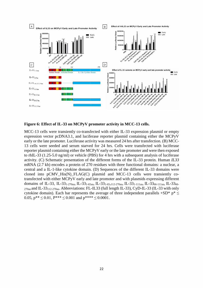
22
Figure 6: Effect of IL-33 on MCPyV promoter activity in MCC-13 cells.
MCC-13 cells were transiently co-transfected with either IL-33 expression plasmid or empty
expression vector pcDNA3.1, and luciferase reporter plasmid containing either the MCPyV
early or the late promoter. Luciferase activity was measured 24 hrs after transfection. (B) MCC-
13 cells were seeded and serum starved for 24 hrs. Cells were transfected with luciferase
reporter plasmid containing either the MCPyV early or the late promoter and were then exposed
to rhIL-33 (1.25-5.0 ng/ml) or vehicle (PBS) for 4 hrs with a subsequent analysis of luciferase
activity. (C) Schematic presentation of the different forms of the IL-33 protein. Human IL33
mRNA (2.7 kb) encodes a protein of 270 residues with three functional domains: a nuclear, a
central and a IL-1-like cytokine domain. (D) Sequences of the different IL-33 domains were
cloned into pCMV_His(N)_FLAG(C) plasmid and MCC-13 cells were transiently co-
transfected with either MCPyV early and late promoter and with plasmids expressing different
domains of IL-33, IL-331-270aa, IL-331-65aa, IL-331-65,112-270aa, IL-331-111aa, IL-3366-111aa, IL-3366-
270aa and IL-33112-270aa. Abbreviations: FL-IL33 (full length IL-33), CyD-IL-33 (IL-33 with only
cytokine domain). Each bar represents the average of three independent parallels +SD* p* ≤
0.05, p** ≤ 0.01, P*** ≤ 0.001 and p**** ≤ 0.0001.

23
Figure 7. Effect of ST2/IL1RL1 and IL1RAcP on MCPyV promoter activity in MCC-13
cells.
MCC-13 cells were transiently co-transfected with luciferase reporter plasmid and either the
early or the late luciferase promoter of MCPyV and (A) ST2/IL1RL1 and (B) IL1RAcP
expression plasmids, respectively. pcDNA3.1 was used as empty expression vector (EV).
Luciferase activity was measured 24 hrs after transfection. Abbreviations: ST2L (membrane
bound ST2/IL1RL1), sST2 (soluble form of ST2/IL1RL1), IL1RAcP.L (membrane bound
IL1RAcP), s IL1RAcP (soluble form of IL1RAcP). Each bar represents the average of three
independent parallels +SD* p* ≤ 0.05, p** ≤ 0.01, p*** ≤ 0.001 and p**** ≤ 0.0001.

24
Figure 8: Effect of IL-33, ST2/Il1RL1 and IL1RAcP on IL-33/ST2-IL1RAcP complex
promoters
Cells were transiently co-transfected with IL-33, ST2/IL1RL1, IL1RAcP or empty vector (EV)
as control expression plasmids. The promoter fragments spanning nucleotides for (A), (B)
ILIL1RAcP and (C) sST2/sIL1RL1 were -517/+100, and -499/+100, respectively. Each bar
represents the average of three independent parallels +SD. The experiment was repeated total
3 times, and similar results were obtained. Luciferase values were normalized with a total
protein in each sample. p* ≤ 0.05, p** ≤ 0.01 and p*** ≤ 0.001.

25
Figure 9: IL-33 signaling pathway
IL-33 binds to ST2L causing a conformational change that leads to recruitment of IL1-RAcP. Heterodimerization of the transmembrane proteins
results in interaction between their intracellular C-terminal domains that facilitates recruitment of adaptor molecules including MyD88, IRAK1,
IRAK4, and TRAF6. Subsequent activation of signaling pathways NF-κB, JNK, ERK, and p38 leads to expression of genes encoding cytokines,
chemokines, and growth factors.
26
Figure 10: IL-33 upregulates MAP kinase activity
(A) Phosphorylation levels of ERK1/2, p38 and JNK were determined by Western blot analysis
using phospho-specific antibody detecting Thr202/Tyr204 (ERK1/2), Thr183/Tyr185 (JNK)
and Thr180/Tyr182 (p38) phosphorylation and compared to GAPDH. MCC13 cells were
serum-starved for 24 hrs, and thereafter stimulated with either vehicle (PBS) or 1 ng/ml of CyD-
rhIL-33 for different time points (5´, 15´, 30´, 45´ and 60´). (B) The blockade of the ST2
receptor interferes with IL-33-induced activation of ERK1/2 activity. Phospho ERK1/2 levels
were determined by western blot. MCC13 cells were serum starved for 24 hrs. The cells were
pre-blocked with goat anti-ST2 antibody (2 ng/mL) for varying time duration 5-60 min or with
polyclonal normal goat IgG for 5 or 60 min. The cells were then stimulated with CyD-rhIL-33
(1 ng/ml) in the presence or without ST2 receptor antibody for 60 min. PBS was used as a
vehicle control.

27
Figure 11: IL-33 upregulates NF-κB activity
(A) NFκB-p65 activation was determined by monitoring phosphorylation of p65 and p105.
MCC-13 cells were serum starved for 24 hrs, and then stimulated with 1 ng/ml of CyD-rhIL-
33 for varying times 5-60 min. PBS was used as a vahicle control. Relative phospho-p65 and
phospho-p105 were determined by western blot using phospho-specific antibodies. (B) NF-κB
activity was measured by using a luciferase reporter plasmid containing a NF-κB-responsive
promoter. Cells were stimulated with 0.24, 0.48, 0.96 ng/ml for 4 hrs. Luciferase values were
normalized to total protein. (C) The blocking of the ST2 receptor interferes with IL-33-induced
activation of NF-κB. Phospho p65 levels were determined by western blot. MCC13 were serum
starved for 24 hrs and then pre-blocked with goat anti-ST2 antibody (2 ng/mL) or normal goat
IgG for varying time duration (5-60 min). The cells were then stimulated with CyD-rhIL-33 (1
ng/ml) in the presence or absence of ST2 receptor antibody for 60 min. PBS was used as a
control. (D) anti-ST2 antibody ablates IL-33-induced activation of an NF-κB-responsive
promoter. Cells were transfected with the luciferase reporter plasmid with an NF-κB responsive
promoter and exposed to CyD-rhIL-33 (1 ng/ml) in the presence or without anti-ST2 antibody
(0.5 or 1 μg/mL) for 4 hrs. Goat IgG was used as a control. Each bar represents the average of
three independent parallels. Luciferase values were corrected for protein concentration of the
samples. P* ≤ 0.05 P*** ≤ 0.001.

28
Figure 12: Correlation between IL-33, ST2/IL1RL1 and IL1RAcP levels among MCC
patients and control plasma samples.

29
Figure 13: Immunoperoxidase staining of MCPyV-associated MCC primary tumors
(A) HE, (B) LT, (C) CK20, (D) Isotype, (E,F) IL-33, (G,H) ST2/IL1RL1 and (I,J) IL1RAcP.
The displayed images are representative stainings from a panel of MCC primary tumors (Scale
bar= 500 μm for A, B, C, D; 100 μm for E, G, I and 20 μm for F, G, J).

30

PAPER IV


1
Merkel cell polyomavirus large T antigen and small t antigen
increase the expression of high-risk human papillomaviruses 16
and 18 E6 and E7 in cervical cancer cells
Kashif Rasheed1, Baldur Sveinbjørnsson1 and Ugo Moens1;*
1Molecular Inflammation Research Group, Department of Medical Biology, Faculty of Health
Sciences, University of Tromsø, The Arctic University of Norway, NO-9037 Tromsø, Norway;
[email protected]; [email protected]; [email protected].
* Correspondence: [email protected]; phone: +47-77644622
Abstract
A causal role of high-risk papillomaviruses (HR-HPV) in anogenital cancers is well-
established, while Merkel cell polyomavirus (MCPyV) seems to be an etiological factor in
Merkel cell carcinoma. Recent findings reported the co-detection of MCPyV DNA in HR-HPV-
positive cervical cancers, though a role for MCPyV in cervical carcinogenesis has not been
proven. We investigated whether the MCPyV early proteins large T-antigen (LT) and small
antigen (sT) could increase the expression of the HR-HPV E6 and E7 oncoproteins.
Transactivation of the HPV16 and the HPV 18 promoters by MCPyV LT and sT was monitored
by transient transfection studies in the HPV-negative cervical cancer cell line C33A. The effect
of these MCPyV proteins on HPV16 and HPV18 E6 and E7 expression was examined by real-
time PCR and western blot in SiHa and HeLa cells, respectively. MCPyV LT and sT stimulated
the transcriptional activity of the HPV16 and HPV18 promoters, and induced the expression of
HPV E6 and E7 oncoproteins. These results indicate that the co-infection of MCPyV may act
as a co-factor in the initiation and/or progression of HPV-induced cervical cancer, or in other
HPV-associated cancers.
Keywords: co-infection; promoter; tumor, luciferase assay, cervical cancer

2
Introduction
Papillomaviridae and Polyomaviridae are two families of naked, double-stranded DNA viruses
that can cause cancer in humans [1]. High-risk human papillomaviruses (HR-HPV) are
predominantly associated with cervical cancer, but also with penile, anal, vulvar, vaginal and
oropharyngeal cancers [1, 2]. HPV-16 and HPV-18 are responsible for almost 70% of cervical
cancers worldwide, and integration of the viral genome is considered one of the most important
risk factors for cervical cancer development [3-5]. The oncogenic potentials of HR-HPV mainly
depend on the viral proteins E6 and E7. These proteins can bind p53 and pRb family members,
respectively, but can also interfere with other hallmarks of viral oncogenesis (reviewed in [6]).
Merkel cell polyomavirus is an etiological factor for Merkel cell carcinoma (MCC), which is
an aggressive skin cancer [7]. Approximately 80% of all MCC are positive for MCPyV DNA
[8-10], and similarly to HR-HPV in cervical cancer, the MCPyV genome is integrated into all
virus-positive MCC examined thus far [3]. The viral oncoproteins of MCPyV are denoted as
large tumor antigen (LT) and small tumor antigen (sT) [11]. Equivalently, as seen in HPV,
LTAg can interfere with the activity of p53 and pRb proteins [12]. This similar effect also
suggests that E6, E7 and LT can act together to augment the transformative properties of HR-
HPV.
The route of MCPyV infection is not known, though sexual transmission has been suggested
since viral DNA has been detected in 37/120 (31%) anogenital mucosa samples of HIV-positive
men [13]. Interestingly, 86/120 (72%) of the anogenital mucosa samples were also positive for
HR-HPV. These results indicate that MCPyV and HR-HPV may coexist in genital tissue. The
co-presence of HR-HPV and MCPyV DNA in both benign and malignant cervical tissue has
also been confirmed by other studies. For instance, MCPyV DNA could be amplified in 9/48
(19%) of cervical squamous cell carcinomas, and in 4/16 (25%) of cervical adenocarcinomas.
All MCPyV positive specimens were also positive for HR-HPV DNA [14]. Furthermore, a
study on 112 cervical samples of Iranian women showed that 40% of the samples were HR-
HPV positive. Of these, 14 (35%) were also MCPyV positive [15]. Even so, the authors did not
find a significant association between concomitant infection and the grade of cervical lesions,
with less than one copy number/cell of the MCPyV genome found. Normal cervical tissue was
not examined for the presence of MCPyV. The coexistence of HR-HPV and MCPyV in
precancerous and cancerous cervical lesions of women with or without HIV has also been
reported [16]. The authors detected HR-HPV in 124 out of 140 (88.6%) cervical samples from
HIV-positive African women, as well as in 24 out of 50 (48%) samples from HIV-negative

3
French women. Of the 148 HR-HPV samples, 81 (55%) were also positive for MCPyV, but
there was no significant difference with HR-HPV negative samples, of which 24/42 (57%) were
MCPyV positive.
Although the relationship between MCPyV and cervical cancer has not yet been clarified, the
frequent detection of MCPyV DNA in HR-HPV positive cervical tumor specimens suggests
that MCPyV may promote HPV-induced cervical cancer. This prompted us to investigate
whether the MCPyV oncoproteins LT and sT could stimulate the expression of the major HR-
HPV oncoproteins E6 and E7, thereby accelerating the initiation and progression of HR-HPV-
induced oncogenesis.
Materials and methods
Cell lines
The human cell lines C33A (human papilloma virus-negative cervical cancer) and HeLa (HR-
HPV18-positive cervical cancer) were obtained from the American Type Culture Collection
(cat. nos. CCL-2 and HTB-31, respectively). SiHa cells (HPV16-positive) were a kind gift from
Moe Bjørn Thorvald Greve (University Hospital of Northern Norway). Hela cells were
maintained in Dulbecco’s Modified Eagle’s Medium (Sigma D5796) with 10% foetal bovine
serum (Life Technologies, cat. no. 10500-064), and SiHa cells were cultivated in RPMI1604
medium with 10% foetal bovine serum. Cells were kept at 37oC in a humidified CO2 incubator.
Plasmids
The HPV16 luciferase reporter plasmid (pGL-LCR-HPV16-LCR (nt 7000-100-luciferase) and
the HPV18 luciferase reporter plasmid (pGL4.20 HPV18-LCR luciferase) were obtained from
Addgene (cat. no. #32888 and #22859, respectively). The luciferase reporter plasmid with the
HPV16 long control region (LCR) contains nucleotides 7000 to 100 (GenBank accession
NC_0015264.4; [17]), whereas the luciferase reporter plasmid with the HPV18 LCR
encompasses nucleotides 6943 to 105 (GenBank accession X05015; [18]). Using these
plasmids, we analyzed luciferase activity driven by these LCR. The empty expression vector
pcDNA3.1(+) was purchased from Thermo Fisher Scientific (Waltham, MA, USA). The
MCPyV LT expression vector was obtained from Addgene, while the MCPyV sT expression
plasmid was a kind gift of Dr. Andrew Macdonald [19]. The expression vectors for MKL1 and
MKL2 LT have been previously described [20].

4
Transfection
Cells were seeded out in 12-well cell culture plates so that they were approximately 70%
confluent the next day when they were transfected. JetPrime (Polyplus-transfection, Illkirch,
France) was used as the transfection reagent, with cells transfected with 400 ng luciferase
plasmid according to the manufacturer’s instructions. In the experiments in which the effect of
LT or sT on promoter activity was monitored, co-transfection was performed with increasing
amounts of LT and/or sT expression plasmid being used. The amount of DNA in the different
experiments was kept constant by adding the appropriate amount of empty vector
pcDNA3.1(+).
Firefly luciferase assay
Cells were lysed 24 h post-transfection in 100 l of Luciferase Assay Tropix Lysis solution
(Applied Biosystems, Foster City, CA, USA), with 0.5 mM DTT (Sigma-Aldrich Norway AS)
freshly added. Cells were scraped and transferred to Eppendorf tubes, followed by 3 min of
centrifugation at 12,000 g. Twenty l of supernatant were transferred to a 96-well microtiter
plate, and luciferase buffer (Promega, Madison, WI, USA) was added. Light units were
measured using the CLARIONstar (BMG Labtech GmbH, Ortenberg, Germany).
RNA isolation and cDNA synthesis
RNA extraction was done by using an RNeasy® Plus Mini kit (Qiagen, Hilden, Germany)
according to the manufacturer’s protocol. For RNA yield and quality, A260/A280 and
A260/A230 ratios were analyzed with a Nano-Drop® ND-2000 spectrophotometer (NanoDrop
Technologies, ThermoFisher Scientific, Waltham, MA, USA). An iScript™ cDNA Synthesis
Kit (BioRad, Hercules, CA, USA) was used to make cDNA. A total of 100 ng of RNA was
used to generate cDNA according to the manufacturer‘s instructions. PCR with the
housekeeping APRT primers (5’-CCCGAGGCTTCCTCTTTGGC-3’ and 5’-
CTCCCTGCCCTTAAGCGAGG-3’) were used to check for genomic DNA contamination in
the cDNA prep. An 800 base-pair fragment was obtained with genomic DNA as a template,
while a 300 base-pair amplicon was obtained with cDNA [21]. PCR products were visualized
on a 1% agarose gel stained with a GelRed® Nucleic Acid Gel Stain (Biotium, Cambridge
Bioscience, Bar Hill, UK).

5
Real-time quantitative PCR
HeLa (SiHa, respectively) cells were transfected with 500 ng LT or/and sT expression plasmid.
The total amount of transfected DNA was kept constant at 1 µg by adding the appropriate
amount of empty vector DNA. The transcript levels of HPV18E6/E7 and HPV16E6/E7 were
measured by real-time quantitative RT-PCR, using an ABI PRISM® 7300 Sequence Detection
System (Applied Biosystems, Foster City, CA, USA). The expression level was measured by
using a JOE/FAM-labeled TaqMan gene expression assay HPV18E6/E7 and HPV16E6/E7
probe/primer (Table 1), and the expression level was normalized by using a VIC/MGB
probe/primer eukaryotic 18S rRNA endogenous control (cat. no. #4319413E, Applied
Biosystems). PCR reactions were prepared in a total volume of 25 µl, with a final concentration
of 1X TaqMan™ Fast Advanced Master Mix (cat. no. #4444557, Applied Biosystems) and
cDNA from 100 ng total RNA.
Protein concentration assay
The luciferase values of each sample were corrected for protein concentration, as measured by
the Protein Quantification Assay from Macherey-Nagel (Düren, Germany) according to the
instructions of the manufacturer. OD570 was measured using the CLARIOstar Plus Microplate
Reader (Biogen Cientifica Madrid Spain). We corrected luciferase values by measuring the
protein concentration in the corresponding sample rather than co-transfection with a Renilla
reporter plasmid to avoid promoter interference between the HPV promoter directing
expression of the firefly luciferase gene and a promoter controlling expression of the Renilla
luciferase gene. In addition, many of our transfection studies include co-transfection with LT
and/or sT expression plasmids, containing the strong competing CMV promoter. Moreover, LT
of polyomaviruses have shown activate many promoters, including the SV40 and the herpes
simplex virus thymidine kinase promoters, which are used in Renilla reporter plasmids [22].
Western blotting
Cells were plated out in 6-well plates and transfected the next day with 2 µg of DNA (either
empty vector pcDNA3.1 or LT and/or sT expression plasmid) using JetPrime. Twenty-four
hours after transfection, the cells were briefly washed with phosphate-buffered saline (PBS,
Biochrom GmbH), and harvested in NuPage LDS sample buffer (Invitrogen, Carlsbad, CA,
USA) with 100mM of DTT. The samples were then sonicated and heated for 10 min at 70oC.
Proteins were separated on NuPAGE™ Novex™ 4-12% Bis-Tris Protein Gels (Thermo Fisher

6
Scientific Inc.), and transferred onto a 0.45μm PVDF Membrane (Merck Life Science AS, Oslo,
Norway). The membrane was blocked in TBST (Tris-buffered saline with 0.1% Tween-20;
Sigma-Aldrich Norway AS) containing 5% (w/v) skimmed milk powder. Incubation with the
primary HPV16 E6 antibody (Santa Cruz Biotechnology Inc., Dallas TX, USA; cat. no. sc-
460), HPV18 E6 antibody (Santa Cruz; cat. no. sc-365089), HPV16 E7 (Santa Cruz; cat. no.
sc-65711) and HPV18 E7 antibody (Santa Cruz; cat. no. sc-365035) was done overnight at 4°C
in a blocking buffer. Following three washes in TBST, the membrane was incubated with the
polyclonal swine anti-rabbit secondary antibody conjugated with an alkaline phosphatase
(D0306, Dako, Santa Clara, CA, USA) solution for 1h at room temperature. After four washes,
detection and visualization were performed using CDP-Star chemiluminiscent (C0712, Sigma-
Aldrich) and the ImageQuant LAS 4000 imager (GE Healthcare, Oslo, Norway). MagicMark™
XP Western Protein Standard (Thermo Fisher Scientific Inc.) was used to estimate the
molecular mass of the detected proteins, with ERK2 levels serving as loading control.
Subsequently, a western blot was performed with anti-ERK2 antibody (SC-154) as described
above.
Statistical analysis
The t-test was employed to determine statistical differences between the promoter activities.

7
Results
MCPyV LT and sT increase the HPV16 and HPV18 promoter activity
MCPyV DNA can be co-detected in HR-HPV-positive cervical carcinoma samples [14-16],
and the LT and sT of polyomaviruses can affect heterologous promoters [23]. Hence, we wanted
to investigate whether these polyomavirus proteins had an effect on the activity of HR-HPV
promoters. First, the relative promoter strength of HPV16 and HPV18, the two most common
HPVs in cervical cancer [24], was compared in the HPV-negative cell line C33A. The HPV18
promoter was 30-50x stronger than the HPV16 promoter in these cells (Figure 1).
Next, we tested the effect of MCPyV LT and/or sT on the HPV16 and HPV18 promoter activity
in this cell line. LT as well as sT were able to trans-activate the HPV18 promoter (Figure 2A).
A moderate (~2-fold) but highly significant induction of the promoters was measured. The
simultaneous expression of LT and sT did not result in an additive effect. High concentrations
(1200 ng of expression plasmid per transfection) of LT or sT resulted in a reduction of the
HPV18 promoter activity. A similar increase (~2-fold) in HPV16 promoter activity as with the
HPV18 promoter was measured in the presence of LT or of sT (Figure 2B). Higher LT or sT
concentrations also inhibited the HPV16 promoter activity.
All examined MCPyV-positive MCCs express a truncated version of LT due to a nonsense
mutation in the gene [25-31]. We therefore tested the effect of two C-terminal truncated LT
versions identified in the MCC cell line MKL1 and MKL2 on the HPV16 and HPV18 promoter.
The truncated LT variants lack their DNA binding domain [25, 32]. Similar to full-length LT,
truncated MKL-1 and MKL-2 LT variants stimulated the HPV16 and HPV18 promoters with
comparable levels. While no significant differences were observed for full-length LT, MKL-1,
and MKL-2 on the HPV16 promoter, full-length LT was a stronger trans-activator (p<0.05)
than MKL-1 and MKL-2 of the HPV18 promoter (Figure 3).
MCPyV LT and sT increase the protein levels of E6 and E7
E6 and E7 are the two major oncoproteins involved in tumorigenesis by HR-HPV [33-36]. They
are translated from a bicistronic mRNA with the E6 open reading frame (ORF) proximal and
the E7 ORF distal. Ribosomal readthrough of the E6 ORF allows the E7 protein to be
synthesized at a rate of approximately 25-30% of the E6 protein level [37]. Forty-eight hrs after
transfection of SiHa cells with expression plasmids encoding sT, full-length LT, truncated
MKL-1 or MKL-2 LT, or a combination of sT and LT, HPV16 E6 and E7 mRNA levels

8
significantly increased 1.5- to 2-fold (except for MKL2 LT plus ST where a 4- to 6-fold increase
was measured) compared with empty vector. HPV18 E6 and E7 mRNA levels in HeLa cells
were also significantly induced (3- to 5-fold) by all three LT variants alone or in combination
with sT, except for sT alone, which had no effect on E6 mRNA levels and reduced E7 mRNA
levels by 30% (Figure 4).
Because of the different translation efficiency of the E6 and E7 ORFs and because the
oncogenic activities are attributed to the proteins rather than to their mRNA, we examined the
E6 and E7 protein levels in non-transfected SiHa (HPV16-positive) and HeLa (HPV18-
positive) cells, and in cells transiently transfected with an expression plasmid for MCPyV LT
and/or sT. Because MCPyV expresses C-terminal truncated LT in MCC [8], we also tested the
effect of the truncated LT variants MKL1 and MKL2 on E6 and E7 levels in HeLa and SiHa
cells. Western blot analysis showed that the expression of full-length LT or truncated versions
of LT increased HPV18 E6 levels ~1.5-fold 24 and 48 h after transfection, whereas sT
stimulated HPV E6 levels approximately 1.5-fold 48 h after transfection (Figure 5A). Co-
expression of LT (truncated or full length) plus sT did not cause a further increase in E6 levels
compared to LT or sT alone. For HPV18 E7 levels, a 2- to 3.5-fold increase was observed in
the presence of the LT (full-length and MKL-1 and MKL-2 truncated variants) and/or sT but
only 24 h after transfection. No increase in E7 levels were detected 48 h after transfection
(Figure 5B). Again, co-expression of LT and ST did not further enhance HPV18 E7 protein
levels. The expression of full-length LT (resp. sT) in SiHA cells resulted in a 1.5-fold (resp. 2-
fold increase in HPV16 E6 protein levels 24 h post transfection) and a 3-fold (resp. 2-fold)
increase 48 h after transfection (Figure 5C). Again, no additional stimulation was observed
when LT and sT were co-expressed. The truncated LTs were more potent than full length LT
to stimulate the protein levels of HPV16 E6 (Figure 5C). The levels of HPV16 E6 in the
presence of MCPyV early proteins were lower 48 h after transfection compared to 24 h post
transfection, but still higher than the control cells transfected with empty vector. An
approximately 2-fold increase in HPV16 E7 levels was measured in the presence of LT (full
length and truncated) or sT 24h and 48 h after transfection. Co-expression of LT and sT did not
further enhance the E7 protein levels (Figure 5D).

9
Dscussion
HR-HPV16 and HPV18 are the two most common HPVs in cervical cancer [24]. Because
MCPyV DNA is frequently found in samples of cervical cancers [14-16], we investigated
whether MCPyV could affect the promoter activity of HPV16 and HPV18 in cervical cancer
cell lines. Comparing the basal transcriptional activity of the HPV16 and HPV18 LCR in the
HPV-negative cervical cancer cell line C33A showed that the activity of the HPV18 promoter
was 30-50 times stronger than the HPV16 promoter, which is in agreement with other studies.
Chen et al. also found that the basal transcriptional activity of the HPV18 LCR was 3x higher
than that of HPV16 LCR in HeLa cells, and 3.2x in the breast cancer T47D cells [38]. Ottinger
et al. compared the basal activities of the HPV18 and HPV16 LCR in six different cell lines,
including C33A and HeLa and demonstrated that the HPV18 LCR activity was 20-fold stronger
in C33A and 7-fold stronger in HeLa cells [17]. Moreover, SiHa cells have 1-2 copy numbers
of HPV16, whereas HeLa cells contain 17-35 copy numbers of HPV18 [39]. Transient
expression of MCPyV ST, full-length LT, truncated MKL-1 or MKL-2 LT, or a combination
of ST and LT resulted in a significant increase in HPV16 and HPV18 E6 and E7 mRNA levels,
which is in accordance with the ability of these proteins to stimulate the transcriptional activity
of the HPV16 and HPV18 LCR. In the luciferase reporter assay, sT was able to potentiate the
HPV18 LCR activity, yet sT had no or minor effect on HPV18 E6 and E7 mRNA level (Figure
4). We do not know the reason for this discrepancy, but could be due to the different method.
In the luciferase assay, enzyme activity in form of light units produced by the conversion of
luciferin into oxyluciferin are measured, while the qRT-PCR determines E6/E7 mRNA copy
numbers. Although the E6 and E7 ORFs are on the same mRNA, differences in relative E6 and
E7 mRNA levels were measured. This is in accordance with other studies (e.g. [40-42]).
Despite the weaker promoter activity and the lower number of HPV16 genome copies, we found
comparable E6 and E7 proteins levels in the HR-HPV18-positive HeLa cells and the HR-
HPV16-positive SiHa cells. This has also been confirmed in other studies [39, 43-48]. The
reason for this may be that the comparison of the HPV16 and HPV18 LCR transcriptional
activity was done with an isolated promoter in a transient transfection assay in C33A cells,
while in HeLa and SiHa cells, respectively, the promoter is integrated and the conformation of
the chromatin (tightly packed or more open) and methylation pattern may have affected the
transcriptional activity of the promoter and hence the expression of the E6 and E7 genes.
Nevertheless, we cannot exclude that the use of different antibodies may have had an effect on

10
the sensitivity of detecting target proteins (i.e. HVP16 E6 and E7 and HPV18 E6 and E7), a
well-known phenomenon [49-51].
Our results demonstrate that both MCPyV LT and MCPyV sT stimulated the transcriptional
activity of the HPV16 and HPV18 promoter in a dose-dependent manner. A moderate increase
(2- to 3.5-fold) was observed. Previous studies with the HPV16 and HPV18 promoter in other
cell lines showed a variable stimulating effect of LT and sT of the polyomavirus SV40. SV40
LT increased HPV18 promoter activity 13-fold in HeLa cells, ~3-fold in SW13 (human
adrenocortical carcinoma) and in 3T6 (mouse fibroblasts) cells, but had no effect in monkey
kidney CV-1 cells [52]. In human keratinocytes, a 9-fold increase in HPV18 promoter strength
was observed in the simultaneous presence of LT and sT [53]. In human embryonal fibroblasts,
the HPV16 promoter activity was stimulated 20- to 30-fold by SV40 sT, while the effect of LT
was 5- to 6-fold weaker than sT [54]. These experiments with SV40 LT and sT lacked dose-
dependent studies. We found that increasing the concentration of the LT and sT expression
plasmids eventually resulted in the inhibition of HPV16 and HPV18 promoter activity. This
reduction in promoter activity occurred at lower concentrations of LT and sT expression
plasmids for the HPV16 promoter compared to the HPV18 promoter (Figure 2). The difference
in HPV16 and HPV18 promoter strength in C33A cells depends on the transcription factors that
regulate the promoter activity. The HPV16 and HPV18 promoters do not shown significant
sequence homology [55] and they possess exclusive binding sites for different transcription
factors [56]. One possible explanation could be that the CMV promoter, which directs the
expression of LT and sT (see Materials and Methods) and has a stronger activity than the
HPV16 and HPV18 promoters [57], usurps mutual transcription factors that regulate the activity
of the HPV16 (respectively HPV18) and CMV promoters. The CMV promoter may sequester
more transcription factors from the HPV16 promoter compared to the HPV18 promoter,
resulting in an inhibition of the HPV16 promoter activity at lower concentrations of LT/sT
expression plasmids.
MCPyV early proteins moderately (~2-fold) induced the E6 and E7 protein levels of HR-HPVs
16 and 18 (Figure 4). It should be noticed that, although both proteins are translated from the
same transcript, differences in E6 and E7 protein levels were monitored both in the absence and
presence of LT and/or sT. This has also be observed in other studies [48, 58-62]. The difference
could have a technical explanation because the antibodies directed against respectively E6 and
E7 may have different affinity. Another explanation may be the differences in half-life of the
E6 and E7 proteins [63-65]. Another possibility is that LT and/or sT have a different impact on
the stability of E6 and E7 proteins. It is not known whether LT or sT have an impact on the

11
stability of E6 and E7 proteins, but sT of MCPyV has been shown to prevent degradation of
MCPyV LT through inhibition of the ubiquitin ligase complex SCFFbw7 [66]. HPV16 E7 has
been shown to be a substrate of the ubiquitin ligase complex SCF [67].
The biological importance of this restricted increase in E6 and E7 levels, and hence the indirect
role of MCPyV in HPV-induced cervical cancer can be discussed. However, all cancers induced
by human oncoviruses have a very long incubation time [2]. Therefore, the long-term moderate
MCPyV-induced increased expression of E6 and E7, two potent oncogenes that can transform
cells in vitro and induce tumors in animal models [68, 69], may enhance the tumorigenic
process by these HPV oncoproteins in human cervical cancer. The co-infection of cervical
cancer with HPV and other viruses has been reported [70, 71], but the effect of viral proteins
on HPV E6 and E7 has not always been studied. Infection of the SiHa or CaSki cells (both
HPV16 positive) with Kaposi’s sarcoma herpesvirus led to a 50-60% reduction in HPV16 E6
and E7 levels [72, 73], while HIV Tat protein increased HPV16 E6 protein levels 1.35-fold in
SiHa cells, but did not upregulate E7 protein levels [74]. A 3-fold increase in E6 mRNA levels
was observed in HSV-2 infected CaSki (HPV16 positive) cells compared to uninfected cells
[75], whereas the infection of HeLa cells with HSV-1 resulted in a 50-fold reduction of E6
mRNA [76].
The mechanism by which MCPyV LT and sT stimulates the expression of HPV18 E6 and E7
is unknown. LT can activate its own promoter and heterogeneous promoters by binding to
adjacently repeated GA/GGGC motifs [23]. Yet, a sequence analysis of the HPV16 and HPV18
promoters did not reveal repeated LT binding motifs. Moreover, the C-terminal truncated LT
variants MKL1 and MKL2, which lack the DNA binding domain still activated the HPV16 and
HPV18 promoters (Figure 3) and enhanced the E6 and E7 protein levels (Figure 4). LT and sT
of MCPyV have been shown to modulate the activity of transcription factors (see
supplementary Tables S2 and S3 in [23]), and may hence stimulate the HPV16 and HPV18
promoter activity indirectly without binding to DNA.
The co-infection of MCPyV and HR-HPV has been detected in other malignancies, including
squamous cell carcinoma patients [77-81], tonsillar carcinoma [82], epidermodysplasia
verruciformis [83], epithelial proliferations [84] and actinic cheilitis [85]. Whether MCPyV
affects the expression of HPV oncoproteins and plays a role in these cancers, remains unknown.
To unambiguously establish a role of MCPyV in HPV-induced cervical and other cancers, the
state and sequence of genomes of MCPyV, and LT and sT expression in these tumors should
be determined. MCPyV DNA is found to be integrated and a truncated LT is expressed in MCCs
[7, 8], but it is not known whether MCPyV DNA is integrated and C-terminal truncated LTAg

12
is expressed in HPV-positive cancers. It should also be examined whether more pathogenic
MCPyV variants with e.g. changes in the promoter region, LT or ST are present in HPV positive
cancers. Furthermore, the absence of MCPyV in the majority of cervical and other cancers
examined so far does not rule out the involvement of this virus as a co-factor in HPV-positive
cancers because MCPyV may be required for the initiation of the tumorigenic process. Indeed,
a hit-and-run mechanism for MCPyV triggered tumorigenesis has been suggested [86].
Two other human polyomaviruses have been linked to cancer. JCPyV has been associated with
colorectal cancer and the viral genome was integrated in the genome of the tumor cells and LT,
but not late proteins were expressed [87]. An oncogenic role for BKPyV in renal carcinomas
and cancers of the urogenital tract has been suggested [88-90]. For BKPyV-positive tumors,
integrated, episomal viral DNA or both were observed and LT was expressed. The virus was
replication deficient because late proteins were absent, dysfunctional or incomplete expressed.
Expression of the late genes encoding the capsid proteins was impaired due to deletions in the
non-coding control region [91]. Whether MCPyV on its own can induce cervical cancer remains
to be proven. In analogy with JCPyV and BKPyV-induced tumors, the expression of LT and
late proteins should be examined, the state of the viral genome should be determined and the
non-coding region should be sequenced. In the few cases of cervical cancer where MCPyV
DNA was present in a few, LT protein was not always detected and late gene expression was
been monitored (10.1186/1743-422X-9-154; 10.1007/s00705-015-2368-4; 10.1136/sextrans-2015-052430).
Moreover, the viral genome copy number per tumor cell (0.00008 to 0.0021) was several logs
lower than the viral genome copy number in MCCs, which have an average of 5.2 (range 0.8-
14.3) copies per cell [92]. The low copy number may suggest that the viral genome is not
integrated in MCPyV-positive cervical cancers, but this has not been investigated. The non-
coding region of MCPyV in virus-positive cervical tumors has not been sequenced. Finally,
PCR was used to detect the co-presence of MCPyV DNA in HPV-positive cervical tumors, but
it has not been examined whether the viruses actually co-infect the tumor cells or whether
MCPyV DNA was present in other cells of the tumor microenvironment.
In conclusion, the tumorigenic human polyomavirus MCPyV can occasionally be found in
cervical cancers. Its oncoproteins LT and sT can stimulate the activity of the HR-HPV16 and
18 promoters, and therefore increase the transcript and protein levels of the oncoproteins E6
and E7 in cervical cancer cell lines. This suggests that MCPyV may collaborate to transform
cervical tissue. Further studies are warranted to prove a possible role of MCPyV in HR-HPV-
induced cervical carcinoma.

13
Authors and contributions
Conceptualization, B.S. and U.M.; methodology, K.R. and U.M.; validation, K.R., B.S. and
U.M.; investigation, K.R. and U.M.; data curation, K.R., B.S. and U.M.; writing—original draft
preparation, U.M.; writing—review and editing, K.R., B.S. and U.M.; visualization, K.R. and
U.M.; supervision, B.S. and U.M.; project administration, U.M.; funding acquisition, B.S. and
U.M.
Conflicts of interest
The authors declare that there are no conflict of interest.
Ethical approval
Not applicable
Acknowledgments
The authors thank Dr. Andrew Macdonald (University of Leeds, UK) for kindly providing the
MCPyV sT expression plasmid and Moe Bjørn Thorvald Greve (University Hospital of
Northern Norway) for the kind gift of SiHa cells. Research was funded by grants from the
Northern Norway Regional Health Authority and the University Hospital of North Norway
(HNF1441-18), in addition to the Erna and Olav Aakre Foundation against cancer (A65242).
References
1. Haley CT, Mui UN, Vangipuram R, Rady PL, Tyring SK. Human oncoviruses:
Mucocutaneous manifestations, pathogenesis, therapeutics, and prevention:
Papillomaviruses and Merkel cell polyomavirus. J Am Acad Dermatol. 2019; 81: 1-21.
doi: 10.1016/j.jaad.2018.09.062.
2. Mui UN, Haley CT, Tyring SK. Viral Oncology: Molecular Biology and Pathogenesis.
J Clin Med. 2017; 6. doi: 10.3390/jcm6120111.
3. Doolittle-Hall JM, Cunningham Glasspoole DL, Seaman WT, Webster-Cyriaque J.
Meta-Analysis of DNA Tumor-Viral Integration Site Selection Indicates a Role for
Repeats, Gene Expression and Epigenetics. Cancers (Basel). 2015; 7: 2217-35. doi:
10.3390/cancers7040887.
4. Woodman CB, Collins SI, Young LS. The natural history of cervical HPV infection:
unresolved issues. Nat Rev Cancer. 2007; 7: 11-22. doi: 10.1038/nrc2050.
5. Hu Z, Zhu D, Wang W, Li W, Jia W, Zeng X, Ding W, Yu L, Wang X, Wang L, Shen
H, Zhang C, Liu H, et al. Genome-wide profiling of HPV integration in cervical cancer

14
identifies clustered genomic hot spots and a potential microhomology-mediated
integration mechanism. Nat Genet. 2015; 47: 158-63. doi: 10.1038/ng.3178.
6. Mesri EA, Feitelson MA, Munger K. Human viral oncogenesis: a cancer hallmarks
analysis. Cell Host Microbe. 2014; 15: 266-82. doi: 10.1016/j.chom.2014.02.011.
7. Chang Y, Moore PS. Merkel cell carcinoma: a virus-induced human cancer. Annu Rev
Pathol. 2012; 7: 123-44. doi: 10.1146/annurev-pathol-011110-130227.
8. Feng H, Shuda M, Chang Y, Moore PS. Clonal integration of a polyomavirus in human
Merkel cell carcinoma. Science. 2008; 319: 1096-100. doi: 10.1126/science.1152586.
9. Laude HC, Jonchere B, Maubec E, Carlotti A, Marinho E, Couturaud B, Peter M, Sastre-
Garau X, Avril MF, Dupin N, Rozenberg F. Distinct merkel cell polyomavirus
molecular features in tumour and non tumour specimens from patients with merkel cell
carcinoma. PLoS Pathog. 2010; 6: e1001076. doi: 10.1371/journal.ppat.1001076.
10. Martel-Jantin C, Filippone C, Cassar O, Peter M, Tomasic G, Vielh P, Briere J, Petrella
T, Aubriot-Lorton MH, Mortier L, Jouvion G, Sastre-Garau X, Robert C, et al. Genetic
variability and integration of Merkel cell polyomavirus in Merkel cell carcinoma.
Virology. 2012; 426: 134-42. doi: 10.1016/j.virol.2012.01.018.
11. Wendzicki JA, Moore PS, Chang Y. Large T and small T antigens of Merkel cell
polyomavirus. Curr Opin Virol. 2015; 11: 38-43. doi: 10.1016/j.coviro.2015.01.009.
12. Borchert S, Czech-Sioli M, Neumann F, Schmidt C, Wimmer P, Dobner T, Grundhoff
A, Fischer N. High-affinity Rb binding, p53 inhibition, subcellular localization, and
transformation by wild-type or tumor-derived shortened Merkel cell polyomavirus large
T antigens. J Virol. 2014; 88: 3144-60. doi: 10.1128/jvi.02916-13.
13. Wieland U, Mauch C, Kreuter A, Krieg T, Pfister H. Merkel cell polyomavirus DNA in
persons without merkel cell carcinoma. Emerg Infect Dis. 2009; 15: 1496-8. doi:
10.3201/eid1509.081575.
14. Imajoh M, Hashida Y, Nemoto Y, Oguri H, Maeda N, Furihata M, Fukaya T, Daibata
M. Detection of Merkel cell polyomavirus in cervical squamous cell carcinomas and
adenocarcinomas from Japanese patients. Virol J. 2012; 9: 154. doi: 10.1186/1743-
422x-9-154.
15. Salehi-Vaziri M, Sadeghi F, Alamsi-Hashiani A, Haeri H, Monavari SH, Keyvani H.
Merkel cell polyomavirus and human papillomavirus infections in cervical disease in
Iranian women. Arch Virol. 2015; 160: 1181-7. doi: 10.1007/s00705-015-2368-4.
16. Kolia-Diafouka P, Foulongne V, Boulle N, Ngou J, Kelly H, Sawadogo B, Delany-
Moretlwe S, Mayaud P, Segondy M. Detection of four human polyomaviruses (MCPyV,
HPyV6, HPyV7 and TSPyV) in cervical specimens from HIV-infected and HIV-
uninfected women. Sex Transm Infect. 2016. doi: 10.1136/sextrans-2015-052430.
17. Ottinger M, Smith JA, Schweiger MR, Robbins D, Powell ML, You J, Howley PM.
Cell-type specific transcriptional activities among different papillomavirus long control
regions and their regulation by E2. Virology. 2009; 395: 161-71. doi:
10.1016/j.virol.2009.09.027.
18. Schweiger MR, Ottinger M, You J, Howley PM. Brd4-independent transcriptional
repression function of the papillomavirus e2 proteins. J Virol. 2007; 81: 9612-22. doi:
10.1128/jvi.00447-07.
19. Griffiths DA, Abdul-Sada H, Knight LM, Jackson BR, Richards K, Prescott EL, Peach
AH, Blair GE, Macdonald A, Whitehouse A. Merkel cell polyomavirus small T antigen
targets the NEMO adaptor protein to disrupt inflammatory signaling. J Virol. 2013; 87:
13853-67. doi: 10.1128/jvi.02159-13.
20. Rasheed K, Abdulsalam I, Fismen S, Grimstad O, Sveinbjornsson B, Moens U.
CCL17/TARC and CCR4 expression in Merkel cell carcinoma. Oncotarget. 2018; 9:
31432-47. doi: 10.18632/oncotarget.25836.

15
21. Tummler C, Dumitriu G, Wickstrom M, Coopman P, Valkov A, Kogner P, Johnsen JI,
Moens U, Sveinbjornsson B. SYK Inhibition Potentiates the Effect of Chemotherapeutic
Drugs on Neuroblastoma Cells in Vitro. Cancers (Basel). 2019; 11. doi:
10.3390/cancers11020202.
22. Pannuti A, Pascucci A, La Mantia G, Fisher-Fantuzzi L, Vesco C, Lania L. trans-
activation of cellular and viral promoters by a transforming nonkaryophilic simian virus
40 large T antigen. J Virol. 1987; 61: 1296-9. doi:
23. Moens U, Krumbholz A, Ehlers B, Zell R, Johne R, Calvignac-Spencer S, Lauber C.
Biology, evolution, and medical importance of polyomaviruses: An update. Infect Genet
Evol. 2017; 54: 18-38. doi: 10.1016/j.meegid.2017.06.011.
24. Doorbar J, Egawa N, Griffin H, Kranjec C, Murakami I. Human papillomavirus
molecular biology and disease association. Rev Med Virol. 2015; 25 Suppl 1: 2-23. doi:
10.1002/rmv.1822.
25. Shuda M, Feng H, Kwun HJ, Rosen ST, Gjoerup O, Moore PS, Chang Y. T antigen
mutations are a human tumor-specific signature for Merkel cell polyomavirus. Proc Natl
Acad Sci U S A. 2008; 105: 16272-7. doi: 10.1073/pnas.0806526105.
26. Sastre-Garau X, Peter M, Avril MF, Laude H, Couturier J, Rozenberg F, Almeida A,
Boitier F, Carlotti A, Couturaud B, Dupin N. Merkel cell carcinoma of the skin:
pathological and molecular evidence for a causative role of MCV in oncogenesis. J
Pathol. 2009; 218: 48-56. doi: 10.1002/path.2532.
27. Fischer N, Brandner J, Fuchs F, Moll I, Grundhoff A. Detection of Merkel cell
polyomavirus (MCPyV) in Merkel cell carcinoma cell lines: cell morphology and
growth phenotype do not reflect presence of the virus. Int J Cancer. 2010; 126: 2133-
42. doi: 10.1002/ijc.24877.
28. Houben R, Shuda M, Weinkam R, Schrama D, Feng H, Chang Y, Moore PS, Becker
JC. Merkel cell polyomavirus-infected Merkel cell carcinoma cells require expression
of viral T antigens. J Virol. 2010; 84: 7064-72. doi: 10.1128/jvi.02400-09.
29. Rodig SJ, Cheng J, Wardzala J, DoRosario A, Scanlon JJ, Laga AC, Martinez-
Fernandez A, Barletta JA, Bellizzi AM, Sadasivam S, Holloway DT, Cooper DJ,
Kupper TS, et al. Improved detection suggests all Merkel cell carcinomas harbor Merkel
polyomavirus. J Clin Invest. 2012; 122: 4645-53. doi: 10.1172/jci64116.
30. Houben R, Dreher C, Angermeyer S, Borst A, Utikal J, Haferkamp S, Peitsch WK,
Schrama D, Hesbacher S. Mechanisms of p53 restriction in Merkel cell carcinoma cells
are independent of the Merkel cell polyoma virus T antigens. J Invest Dermatol. 2013;
133: 2453-60. doi: 10.1038/jid.2013.169.
31. Schrama D, Sarosi EM, Adam C, Ritter C, Kaemmerer U, Klopocki E, Konig EM,
Utikal J, Becker JC, Houben R. Characterization of six Merkel cell polyomavirus-
positive Merkel cell carcinoma cell lines: Integration pattern suggest that large T antigen
truncating events occur before or during integration. Int J Cancer. 2019; 145: 1020-32.
doi: 10.1002/ijc.32280.
32. Guastafierro A, Feng H, Thant M, Kirkwood JM, Chang Y, Moore PS, Shuda M.
Characterization of an early passage Merkel cell polyomavirus-positive Merkel cell
carcinoma cell line, MS-1, and its growth in NOD scid gamma mice. J Virol Methods.
2013; 187: 6-14. doi: 10.1016/j.jviromet.2012.10.001.
33. Hoppe-Seyler K, Bossler F, Braun JA, Herrmann AL, Hoppe-Seyler F. The HPV E6/E7
Oncogenes: Key Factors for Viral Carcinogenesis and Therapeutic Targets. Trends
Microbiol. 2018; 26: 158-68. doi: 10.1016/j.tim.2017.07.007.
34. Yeo-Teh NSL, Ito Y, Jha S. High-Risk Human Papillomaviral Oncogenes E6 and E7
Target Key Cellular Pathways to Achieve Oncogenesis. Int J Mol Sci. 2018; 19. doi:
10.3390/ijms19061706.

16
35. Estevao D, Costa NR, Gil da Costa RM, Medeiros R. Hallmarks of HPV carcinogenesis:
The role of E6, E7 and E5 oncoproteins in cellular malignancy. Biochim Biophys Acta
Gene Regul Mech. 2019; 1862: 153-62. doi: 10.1016/j.bbagrm.2019.01.001.
36. Taghizadeh E, Jahangiri S, Rostami D, Taheri F, Renani PG, Taghizadeh H, Gheibi
Hayat SM. Role of E6 and E7 Human Papillomavirus Proteins in Molecular
Pathogenesis of Cervical Cancer. Curr Protein Pept Sci. 2019. doi:
10.2174/1389203720666190618101441.
37. Tan TM, Gloss B, Bernard HU, Ting RC. Mechanism of translation of the bicistronic
mRNA encoding human papillomavirus type 16 E6-E7 genes. The Journal of general
virology. 1994; 75 ( Pt 10): 2663-70. doi: 10.1099/0022-1317-75-10-2663.
38. Chen YH, Huang LH, Chen TM. Differential effects of progestins and estrogens on long
control regions of human papillomavirus types 16 and 18. Biochem Biophys Res
Commun. 1996; 224: 651-9. doi: 10.1006/bbrc.1996.1080.
39. Hu Z, Ding W, Zhu D, Yu L, Jiang X, Wang X, Zhang C, Wang L, Ji T, Liu D, He D,
Xia X, Zhu T, et al. TALEN-mediated targeting of HPV oncogenes ameliorates HPV-
related cervical malignancy. J Clin Invest. 2015; 125: 425-36. doi: 10.1172/JCI78206.
40. Bellone S, El-Sahwi K, Cocco E, Casagrande F, Cargnelutti M, Palmieri M, Bignotti E,
Romani C, Silasi D-A, Azodi M, Schwartz PE, Rutherford TJ, Pecorelli S, et al. Human
papillomavirus type 16 (HPV-16) virus-like particle L1-specific CD8+ cytotoxic T
lymphocytes (CTLs) are equally effective as E7-specific CD8+ CTLs in killing
autologous HPV-16-positive tumor cells in cervical cancer patients: implications for L1
dendritic cell-based therapeutic vaccines. Journal of virology. 2009; 83: 6779-89. doi:
10.1128/JVI.02443-08.
41. Walline HM, Komarck CM, McHugh JB, Bellile EL, Brenner JC, Prince ME, McKean
EL, Chepeha DB, Wolf GT, Worden FP, Bradford CR, Carey TE. Genomic Integration
of High-Risk HPV Alters Gene Expression in Oropharyngeal Squamous Cell
Carcinoma. Molecular cancer research : MCR. 2016; 14: 941-52. doi: 10.1158/1541-
7786.MCR-16-0105.
42. Ham S, Kim KH, Kwon TH, Bak Y, Lee DH, Song YS, Park S-H, Park YS, Kim MS,
Kang JW, Hong JT, Yoon D-Y. Luteolin induces intrinsic apoptosis via inhibition of
E6/E7 oncogenes and activation of extrinsic and intrinsic signaling pathways in HPV-
18-associated cells. Oncology reports. 2014; 31: 2683-91. doi: 10.3892/or.2014.3157.
43. Singh M, Singh N. Curcumin counteracts the proliferative effect of estradiol and induces
apoptosis in cervical cancer cells. Mol Cell Biochem. 2011; 347: 1-11. doi:
10.1007/s11010-010-0606-3.
44. Choudhari AS, Suryavanshi SA, Kaul-Ghanekar R. The aqueous extract of Ficus
religiosa induces cell cycle arrest in human cervical cancer cell lines SiHa (HPV-16
Positive) and apoptosis in HeLa (HPV-18 positive). PLoS One. 2013; 8: e70127. doi:
10.1371/journal.pone.0070127.
45. Jing K, Shin S, Jeong S, Kim S, Song KS, Park JH, Heo JY, Seo KS, Park SK, Kweon
GR, Wu T, Park JI, Lim K. Docosahexaenoic acid induces the degradation of HPV
E6/E7 oncoproteins by activating the ubiquitin-proteasome system. Cell Death Dis.
2014; 5: e1524. doi: 10.1038/cddis.2014.477.
46. Jung HS, Rajasekaran N, Song SY, Kim YD, Hong S, Choi HJ, Kim YS, Choi JS, Choi
YL, Shin YK. Human Papillomavirus E6/E7-Specific siRNA Potentiates the Effect of
Radiotherapy for Cervical Cancer in Vitro and in Vivo. Int J Mol Sci. 2015; 16: 12243-
60. doi: 10.3390/ijms160612243.
47. Deshpande R, Mansara P, Kaul-Ghanekar R. Alpha-linolenic acid regulates
Cox2/VEGF/MAP kinase pathway and decreases the expression of HPV oncoproteins

17
E6/E7 through restoration of p53 and Rb expression in human cervical cancer cell lines.
Tumour Biol. 2016; 37: 3295-305. doi: 10.1007/s13277-015-4170-z.
48. Gao R, Wu X, Huang Z, Wang B, Li F, Xu H, Ran L. Anti-tumor effect of aloe-emodin
on cervical cancer cells was associated with human papillomavirus E6/E7 and glucose
metabolism. Onco Targets Ther. 2019; 12: 3713-21. doi: 10.2147/OTT.S182405.
49. Bordeaux J, Welsh A, Agarwal S, Killiam E, Baquero M, Hanna J, Anagnostou V,
Rimm D. Antibody validation. Biotechniques. 2010; 48: 197-209. doi:
10.2144/000113382.
50. Gilda JE, Ghosh R, Cheah JX, West TM, Bodine SC, Gomes AV. Western Blotting
Inaccuracies with Unverified Antibodies: Need for a Western Blotting Minimal
Reporting Standard (WBMRS). PLoS One. 2015; 10: e0135392. doi:
10.1371/journal.pone.0135392.
51. Acharya P, Quinlan A, Neumeister V. The ABCs of finding a good antibody: How to
find a good antibody, validate it, and publish meaningful data. F1000Res. 2017; 6: 851.
doi: 10.12688/f1000research.11774.1.
52. Thierry F, Heard JM, Dartmann K, Yaniv M. Characterization of a transcriptional
promoter of human papillomavirus 18 and modulation of its expression by simian virus
40 and adenovirus early antigens. J Virol. 1987; 61: 134-42. doi:
53. Bernard BA, Bailly C, Lenoir MC, Darmon MY. Modulation of HPV18 and BPV1
transcription in human keratinocytes by simian virus 40 large T antigen and adenovirus
type 5 E1A antigen. J Cell Biochem. 1990; 42: 101-10. doi: 10.1002/jcb.240420206.
54. Smits PH, de Ronde A, Smits HL, Minnaar RP, van der Noordaa J, ter Schegget J.
Modulation of the human papillomavirus type 16 induced transformation and
transcription by deletion of loci on the short arm of human chromosome 11 can be
mimicked by SV40 small t. Virology. 1992; 190: 40-4. doi: 10.1016/0042-
6822(92)91190-6.
55. Chan SY, Bernard HU, Ong CK, Chan SP, Hofmann B, Delius H. Phylogenetic analysis
of 48 papillomavirus types and 28 subtypes and variants: a showcase for the molecular
evolution of DNA viruses. J Virol. 1992; 66: 5714-25. doi:
56. Sichero L, Sobrinho JS, Villa LL. Identification of novel cellular transcription factors
that regulate early promoters of human papillomavirus types 18 and 16. The Journal of
infectious diseases. 2012; 206: 867-74. doi: 10.1093/infdis/jis430.
57. Shillitoe EJ, Noonan S. Strength and specificity of different gene promoters in oral
cancer cells. Oral oncology. 2000; 36: 214-20. doi: 10.1016/s1368-8375(99)00064-0.
58. Caberg JH, Hubert PM, Begon DY, Herfs MF, Roncarati PJ, Boniver JJ, Delvenne PO.
Silencing of E7 oncogene restores functional E-cadherin expression in human
papillomavirus 16-transformed keratinocytes. Carcinogenesis. 2008; 29: 1441-7. doi:
10.1093/carcin/bgn145.
59. Kim YW, Chaturvedi PK, Chun SN, Lee YG, Ahn WS. Honeybee venom possesses
anticancer and antiviral effects by differential inhibition of HPV E6 and E7 expression
on cervical cancer cell line. Oncol Rep. 2015; 33: 1675-82. doi: 10.3892/or.2015.3760.
60. Kim M, Kim YS, Kim H, Kang MY, Park J, Lee DH, Roh GS, Kim HJ, Kang SS, Cho
GJ, Park JK, Cho JW, Shin JK, et al. O-linked N-acetylglucosamine transferase
promotes cervical cancer tumorigenesis through human papillomaviruses E6 and E7
oncogenes. Oncotarget. 2016; 7: 44596-607. doi: 10.18632/oncotarget.10112.
61. He H, Lai Y, Hao Y, Liu Y, Zhang Z, Liu X, Guo C, Zhang M, Zhou H, Wang N, Luo
XG, Huo L, Ma W, et al. Selective p300 inhibitor C646 inhibited HPV E6-E7 genes,
altered glucose metabolism and induced apoptosis in cervical cancer cells. Eur J
Pharmacol. 2017; 812: 206-15. doi: 10.1016/j.ejphar.2017.06.005.

18
62. Javadi H, Lotfi AS, Hosseinkhani S, Mehrani H, Amani J, Soheili ZS, Hojati Z, Kamali
M. The combinational effect of E6/E7 siRNA and anti-miR-182 on apoptosis induction
in HPV16-positive cervical cells. Artif Cells Nanomed Biotechnol. 2018; 46: 727-36.
doi: 10.1080/21691401.2018.1468770.
63. Selvey LA, Dunn LA, Tindle RW, Park DS, Frazer IH. Human papillomavirus (HPV)
type 18 E7 protein is a short-lived steroid-inducible phosphoprotein in HPV-
transformed cell lines. J Gen Virol. 1994; 75 ( Pt 7): 1647-53. doi: 10.1099/0022-1317-
75-7-1647.
64. Tomaic V, Pim D, Banks L. The stability of the human papillomavirus E6 oncoprotein
is E6AP dependent. Virology. 2009; 393: 7-10. doi: 10.1016/j.virol.2009.07.029.
65. Ajiro M, Zheng ZM. E6^E7, a novel splice isoform protein of human papillomavirus
16, stabilizes viral E6 and E7 oncoproteins via HSP90 and GRP78. mBio. 2015; 6:
e02068-14. doi: 10.1128/mBio.02068-14.
66. Kwun HJ, Shuda M, Feng H, Camacho CJ, Moore PS, Chang Y. Merkel cell
polyomavirus small T antigen controls viral replication and oncoprotein expression by
targeting the cellular ubiquitin ligase SCFFbw7. Cell Host Microbe. 2013; 14: 125-35.
doi: 10.1016/j.chom.2013.06.008.
67. Oh KJ, Kalinina A, Wang J, Nakayama K, Nakayama KI, Bagchi S. The papillomavirus
E7 oncoprotein is ubiquitinated by UbcH7 and Cullin 1- and Skp2-containing E3 ligase.
J Virol. 2004; 78: 5338-46. doi: 10.1128/jvi.78.10.5338-5346.2004.
68. Vande Pol SB, Klingelhutz AJ. Papillomavirus E6 oncoproteins. Virology. 2013; 445:
115-37. doi: 10.1016/j.virol.2013.04.026.
69. Roman A, Munger K. The papillomavirus E7 proteins. Virology. 2013; 445: 138-68.
doi: 10.1016/j.virol.2013.04.013.
70. Guidry JT, Scott RS. The interaction between human papillomavirus and other viruses.
Virus Res. 2017; 231: 139-47. doi: 10.1016/j.virusres.2016.11.002.
71. Cantalupo PG, Katz JP, Pipas JM. Viral sequences in human cancer. Virology. 2018;
513: 208-16. doi: 10.1016/j.virol.2017.10.017.
72. Dai L, Qiao J, Del Valle L, Qin Z. KSHV co-infection regulates HPV16+ cervical cancer
cells pathogenesis in vitro and in vivo. Am J Cancer Res. 2018; 8: 708-14. doi:
73. Dai L, Cao Y, Jiang W, Zabaleta J, Liu Z, Qiao J, Qin Z. KSHV co-infection down-
regulates HPV16 E6 and E7 from cervical cancer cells. Oncotarget. 2017; 8: 35792-803.
doi: 10.18632/oncotarget.16207.
74. Barillari G, Palladino C, Bacigalupo I, Leone P, Falchi M, Ensoli B. Entrance of the Tat
protein of HIV-1 into human uterine cervical carcinoma cells causes upregulation of
HPV-E6 expression and a decrease in p53 protein levels. Oncol Lett. 2016; 12: 2389-
94. doi: 10.3892/ol.2016.4921.
75. Pisani S, Imperi M, Seganti L, Superti F, Tinari A, Bucci M, Degener AM. Effect of
HSV-2 infection on the expression of HPV 16 genes in CaSki cells. Int J Immunopathol
Pharmacol. 2004; 17: 65-70. doi: 10.1177/039463200401700109.
76. Karlen S, Offord EA, Beard P. Herpes simplex virions interfere with the expression of
human papillomavirus type 18 genes. J Gen Virol. 1993; 74 ( Pt 6): 965-73. doi:
10.1099/0022-1317-74-6-965.
77. Mitteldorf C, Mertz KD, Fernandez-Figueras MT, Schmid M, Tronnier M, Kempf W.
Detection of Merkel cell polyomavirus and human papillomaviruses in Merkel cell
carcinoma combined with squamous cell carcinoma in immunocompetent European
patients. Am J Dermatopathol. 2012; 34: 506-10. doi:
10.1097/DAD.0b013e31823b9b4e.
78. Falchook GS, Rady P, Hymes S, Nguyen HP, Tyring SK, Prieto VG, Hong DS,
Kurzrock R. Merkel cell polyomavirus and HPV-17 associated with cutaneous

19
squamous cell carcinoma arising in a patient with melanoma treated with the BRAF
inhibitor dabrafenib. JAMA Dermatol. 2013; 149: 322-6. doi:
10.1001/jamadermatol.2013.2023.
79. Dworkin AM, Tseng SY, Allain DC, Iwenofu OH, Peters SB, Toland AE. Merkel cell
polyomavirus in cutaneous squamous cell carcinoma of immunocompetent individuals.
J Invest Dermatol. 2009; 129: 2868-74. doi: 10.1038/jid.2009.183.
80. Vazquez-Guillen JM, Palacios-Saucedo GC, Rivera-Morales LG, Alonzo-Morado MV,
Burciaga-Bernal SB, Montufar-Martinez M, Ortiz-Lopez R, Gonzalez-Villasana V,
Martinez-Torres AC, Serna-Hernandez JC, Hernandez-Martinez SJ, Castelan-
Maldonado EE, Zavala-Pompa A, et al. Infection and coinfection by human
papillomavirus, Epstein-Barr virus and Merkel cell polyomavirus in patients with
squamous cell carcinoma of the larynx: a retrospective study. PeerJ. 2018; 6: e5834.
doi: 10.7717/peerj.5834.
81. Yahyapour Y, Rahmani R, Alipour M, Alizadeh A, Khademian A, Sadeghi F.
Prevalence and association of human papillomavirus, Epstein-Barr virus and Merkel
Cell polyomavirus with neoplastic esophageal lesions in northern Iran. Caspian J Intern
Med. 2018; 9: 353-60. doi: 10.22088/cjim.9.4.353.
82. Herberhold S, Hellmich M, Panning M, Bartok E, Silling S, Akgul B, Wieland U.
Human polyomavirus and human papillomavirus prevalence and viral load in non-
malignant tonsillar tissue and tonsillar carcinoma. Med Microbiol Immunol. 2016. doi:
10.1007/s00430-016-0486-6.
83. Mertz KD, Schmid M, Burger B, Itin P, Palmedo G, Scharer L, Kutzner H, Fernandez
Figueras MT, Cribier B, Pfaltz M, Kempf W. Detection of Merkel cell polyomavirus in
epidermodysplasia-verruciformis-associated skin neoplasms. Dermatology. 2011; 222:
87-92. doi: 10.1159/000321880.
84. Schrama D, Groesser L, Ugurel S, Hafner C, Pastrana DV, Buck CB, Cerroni L, Theiler
A, Becker JC. Presence of human polyomavirus 6 in mutation-specific BRAF inhibitor-
induced epithelial proliferations. JAMA Dermatol. 2014; 150: 1180-6. doi:
10.1001/jamadermatol.2014.1116.
85. Neto Pimentel DR, Montosa Nunes E, Termini L, Almeida Lima Nunes R, Mendoza
Lopez RV, Ferreira S, Boccardo E, Mann Prado JC, Enokihara M, Sichero L, Tomimori
J. Detection of human papillomaviruses and human polyomaviruses in
immunosuppressed and immunocompetent individuals with actinic cheilitis: a case
series. J Eur Acad Dermatol Venereol. 2019. doi: 10.1111/jdv.15779.
86. Houben R, Grimm J, Willmes C, Weinkam R, Becker JC, Schrama D. Merkel cell
carcinoma and Merkel cell polyomavirus: evidence for hit-and-run oncogenesis. J Invest
Dermatol. 2012; 132: 254-6. doi: 10.1038/jid.2011.260.
87. Hori R, Murai Y, Tsuneyama K, Abdel-Aziz HO, Nomoto K, Takahashi H, Cheng CM,
Kuchina T, Harman BV, Takano Y. Detection of JC virus DNA sequences in colorectal
cancers in Japan. Virchows Arch. 2005; 447: 723-30. doi: 10.1007/s00428-005-0014-3.
88. Kenan DJ, Mieczkowski PA, Burger-Calderon R, Singh HK, Nickeleit V. The
oncogenic potential of BK-polyomavirus is linked to viral integration into the human
genome. J Pathol. 2015; 237: 379-89. doi: 10.1002/path.4584.
89. Kenan DJ, Mieczkowski PA, Latulippe E, Cote I, Singh HK, Nickeleit V. BK
Polyomavirus Genomic Integration and Large T Antigen Expression: Evolving
Paradigms in Human Oncogenesis. Am J Transplant. 2017; 17: 1674-80. doi:
10.1111/ajt.14191.
90. Muller DC, Ramo M, Naegele K, Ribi S, Wetterauer C, Perrina V, Quagliata L, Vlajnic
T, Ruiz C, Balitzki B, Grobholz R, Gosert R, Ajuh ET, et al. Donor-derived, metastatic

20
urothelial cancer after kidney transplantation associated with a potentially oncogenic
BK polyomavirus. J Pathol. 2018; 244: 265-70. doi: 10.1002/path.5012.
91. Nickeleit V, Singh HK, Kenan DJ, Mieczkowski PA. The two-faced nature of BK
polyomavirus: lytic infection or non-lytic large-T-positive carcinoma. The Journal of
pathology. 2018; 246: 7-11. doi: 10.1002/path.5127.
92. Shuda M, Arora R, Kwun HJ, Feng H, Sarid R, Fernandez-Figueras MT, Tolstov Y,
Gjoerup O, Mansukhani MM, Swerdlow SH, Chaudhary PM, Kirkwood JM, Nalesnik
MA, et al. Human Merkel cell polyomavirus infection I. MCV T antigen expression in
Merkel cell carcinoma, lymphoid tissues and lymphoid tumors. Int J Cancer. 2009; 125:
1243-9. doi: 10.1002/ijc.24510.

Figure 1. The basal HPV18 promoter is stronger than the HPV16 promoter in C33A cells.
HPV-negative C33A cells were transiently transfected with 400 ng/well of a luciferase reporter
plasmid containing the long control region (LCR) of HPV16 or HPV18. Cells were harvested
24 h after transfection and luciferase activity was determined. The luciferase values were
corrected for the protein content in each sample, and are expressed as relative luciferase units
(RLU). The bars represent the average of three independent parallel+SD (***p<0.001). Similar
results were obtained in two additional experiments. RLU = relative luciferase units; EV=
empty vector (pcDNA3.1).

Figure 2. MCPyV LT and ST stimulate the HPV16 and HPV18 promoter activity. (A)
C33A cells were co-transfected with a reporter plasmid containing the luciferase gene under
control of the HPV18 promoter and increasing amounts of MCPyV LTAg and/or STAg
expression plasmid. The amount of DNA in each sample was kept constant by adding an
appropriate concentration of empty vector pcDNA3.1(+). Each bar represents the average+SD
of 3-5 separate experiments performed with three independent parallels. Luciferase activity was
corrected for the protein content in each sample. The corrected luciferase activity in the
presence of an empty vector was arbitrarily set as 1.0, with the promoter activity in the presence
of LTAg and/or STAg shown as fold induction. (B) as in (A) but the effect of MCPyV LTAg
and STAg was tested on the HPV16 promoter. For each well, cells were co-transfected with
400 ng of luciferase plasmid and different concentrations of expression plasmid of LTAg or/and
STAg. The total amount of DNA in each sample was kept constant by adding empty vector
DNA. EV= empty vector (pcDNA3.1). *p<0.05; **p<0.01.

Figure 3. Full-length and truncated LTAg activate the HPV16 and HPV18 promoters.
Each well with C33A cells was co-transfected with 400 ng of luciferase reporter plasmid with
either the HPV16 or the HPV18 promoter and with 200 ng of empty vector (EV) or expression
plasmid for full-length LTAg (LTAg) or truncated versions MKL1 or MKL2. Luciferase
activities were standardized to the protein content in the sample and the promoter activity in the
presence of empty vector was then arbitrarily set as 1.0. Each bar represents the average of three
independent parallels+SD (*p<0.05; **p<0.01).

Figure 4. MCPyV LT and ST increase HPV16 and HPV18 E6 and E7 mRNA. (A) HeLa
cells were transfected with expression plasmids for full-length LT (FLTA), sT, truncated LT
MKL1 or MKL2 or empty expression vector pcDNA3.1 (EV). Total RNA was isolated 48 hrs
after transfection and cDNA was generated. qPCR was performed with specific primers for E6
and E7 and 18S rRNA was used to standardize. Relative mRNA levels in cells transfected with
empty vector was arbitrarily set as 1. Each bar represent the average of three parallels+SD.
*p<0.05; **p<0.01; ***p<0.001. (B) as in (A) but SiHa cells were transfected and relative
HPV16 E6 and E7 mRNA levels were determined.

Figure 5. Expression of MCPyV LT and ST augment HPV16 and HPV18 E6 and E7
protein levels. HeLa cells (panels A and B) or SiHa cells (panels C and D) were transfected
with the empty expression vector (EV) pcDNA3.1(+) or with an expression plasmid for full-
length LT (FL-LT), truncated MKL-1 LT, truncated MKL-2 LT, sT, or a combination of LT
plus sT. Five hundred ng of DNA of the expression plasmids for LTAg and sTAg were used
per well. The total amount of plasmid DNA in each well was kept constant at 1 µg by adjusting
with EV DNA. Levels of E6 (panels A and C) and E7 (panels B and D) were monitored by
western blot 24 h and 48 h after transfection. Densitometry was used to determine the relative
amounts of E6 and E7, with GAPDH levels used for standardization. The relative expression
level of E6 (resp. E7) in the presence of empty vector was arbitrarily set as 1. Similar results
were obtained in an independent experiment

Table 1: HPV18E6/E7 primer/probe sequence labelled with specific fluorophore
Sense Primer Anti-Sense Primer Labeled Probe
ORF Sequence (5'-3') Conc
(nM)
Sequence (5'-3') Conc
(nM)
Fluorophore - Sequence (5'-3') Conc
(nM)
HPV18 E6 AGCTGGGCACTAT
AGAGGC
50 TGTGTTTCTCTGCGTCGT
TGG
200 JOE-CCATTCGTGCTGCAACCGAGCACGAC-
BHQ1
50
HPV18 E7 GAACCACAACGTC
ACACAATG
100 CAGAAACAGCTGCTGGA
ATG
50 FAM-
AGTAGAAAGCTCAGCAGACGACCTTCG-
BHQ1
50
HPV16 E6 GCGACCCAGAAAG
TTACCACAG
100 CCATCTCTATATACTAT
GCATAAATCCCG
100 JOE-
TACCTCACGTCGCAGTAACTGTTGCTTG
175
HPV16
E7
AGAACCGGACAGA
GCCCATTAC
100 GCCCATTAACAGGTCTT
CCAAAG
100 FAM-
CGCACAACCGAAGCGTAGAGTCACACTT
50
Copyright © 2022 FDOKUMEN