Capillary condensation of a binary mixture in slit-like pores
Upload
khangminh22Category
view
1download
0
Citation: Eugenin, E.; Camporesi, E.;
Peracchia, C. Direct Cell-Cell
Communication via Membrane Pores,
Gap Junction Channels, and
Tunneling Nanotubes: Medical
Relevance of Mitochondrial
Exchange. Int. J. Mol. Sci. 2022, 23,
6133. https://doi.org/10.3390/
ijms23116133
Academic Editor: Diego Guidolin
Received: 30 April 2022
Accepted: 28 May 2022
Published: 30 May 2022
Publisher’s Note: MDPI stays neutral
with regard to jurisdictional claims in
published maps and institutional affil-
iations.
Copyright: © 2022 by the authors.
Licensee MDPI, Basel, Switzerland.
This article is an open access article
distributed under the terms and
conditions of the Creative Commons
Attribution (CC BY) license (https://
creativecommons.org/licenses/by/
4.0/).
International Journal of
Molecular Sciences
Review
Direct Cell-Cell Communication via Membrane Pores, GapJunction Channels, and Tunneling Nanotubes: MedicalRelevance of Mitochondrial ExchangeEliseo Eugenin 1,* , Enrico Camporesi 2 and Camillo Peracchia 3
1 Department of Neuroscience, Cell Biology, and Anatomy, University of Texas Medical Branch (UTMB),105 11th Street, Galveston, TX 77555, USA
2 Department of Surgery and TEAM Health Anesthesia, University of South Florida, 2 Tampa General Circle,Tampa, FL 33606, USA; [email protected]
3 Department of Pharmacology and Physiology, School of Medicine and Dentistry, University Rochester,601 Elmwood Avenue, Rochester, NY 14642, USA; [email protected]
* Correspondence: [email protected]
Abstract: The history of direct cell-cell communication has evolved in several small steps. Firstdiscovered in the 1930s in invertebrate nervous systems, it was thought at first to be an exception tothe “cell theory”, restricted to invertebrates. Surprisingly, however, in the 1950s, electrical cell-cellcommunication was also reported in vertebrates. Once more, it was thought to be an exceptionrestricted to excitable cells. In contrast, in the mid-1960s, two startling publications proved thatvirtually all cells freely exchange small neutral and charged molecules. Soon after, cell-cell communi-cation by gap junction channels was reported. While gap junctions are the major means of cell-cellcommunication, in the early 1980s, evidence surfaced that some cells might also communicate viamembrane pores. Questions were raised about the possible artifactual nature of the pores. However,early in this century, we learned that communication via membrane pores exists and plays a majorrole in medicine, as the structures involved, “tunneling nanotubes”, can rescue diseased cells bydirectly transferring healthy mitochondria into compromised cells and tissues. On the other hand,pathogens/cancer could also use these communication systems to amplify pathogenesis. Here, wedescribe the evolution of the discovery of these new communication systems and the potentialtherapeutic impact on several uncurable diseases.
Keywords: gap junctions; TNTs; communication; cancer; HIV
1. The Identification of Membrane Channels
The “cell theory,” proposed by Matthias J. Schleiden and Theodor Schwann in theearly 19th century, stated that plant and animal tissue are both made of independentunits (later called “cells”) [1,2]; rev. in [3]. This theory implied the existence of a wall-like structure (now known as the plasma membrane) functioning as a barrier to preventmolecules from spreading into neighboring cells and the extracellular compartment orvice versa. However, although this theory did not consider that molecules might moveacross the plasma membrane, we learned that certain molecules freely cross the plasmamembrane soon after publication. Indeed, evidence of the existence of membrane channelswas finally reported in the mid-20th century primarily by the studies of Alan L. Hodgkinand coworkers [4–8]; rev. in [9,10].
2. Direct Cell-Cell Communication via Gap Junctions
Unequivocal evidence of membrane channels directly mediates cell-to-cell commu-nication in virtually all cells, but circulating cells emerged in the 1960s [11–13]. However,knowledge of ionic cell-cell communication in some excitable cells was reported earlier ininvertebrate nervous systems [14,15].
Int. J. Mol. Sci. 2022, 23, 6133. https://doi.org/10.3390/ijms23116133 https://www.mdpi.com/journal/ijms
Int. J. Mol. Sci. 2022, 23, 6133 2 of 24
In the late 1800s, zoologists discovered that invertebrates such as annelids (earth-worms), crustaceans (crayfish), and cephalopods (squid) have large tube-like structuresthat extend from rostral to caudal ends of the body. Most relevant was the early report ofthis structure in the squid [16], a structure that would become fundamentally important forstudying the ionic basis of the action potential and the function of ion channels in general.Most scientists attributed different functions to these tube-like structures, but Leydig, in1864, was the first to propose their nervous function [17]. However, his interpretation wasignored for almost a century until John Z. Young eventually proved it right in 1938 [18].
Early in the 20th century, scientists realized that some of these giant structures (ax-ons) were not continuous. The first to report it was George E. Johnson, who discovereddiscontinuities in giant crustacean axons (Cambarus and Palaemonetes) [19]. Two years later,these discontinuities were named “septa” in earthworm axons by Howard B. Stough [14]—earthworms have three giant axons: a median and two lateral, all segmented.
In his 1930′s study [15], Stough found that when the median axon is cut, only theanterior part of the worm contracts; in contrast, when just the lateral axons are cut, posteriorstimulation causes the posterior portion of the worm to contract up to the cut area. Thisinduced him to conclude that: “ . . . the median giant fiber conducts antero-posteriorly andthe lateral giant fibers conduct postero-anteriorly”. This, however, was proven wrong byJohn C. Eccles and coworkers who recorded electrical impulses in isolated nerve cordselicited either in the head or the tail of the earthworm [20]; in their words: “ . . . thetransverse membranes do not influence the conduction of impulses, although the separation ofthe segments utilizing these transverse membranes appears to be as complete as that existing atvertebrate synaptic junctions”. Several research teams confirmed their findings a decadelater [21–24]. Stough, nonetheless, deserves credit for demonstrating for the first timethe direct cell-cell communication across septal barriers [14,15]. Three decades later, thedetailed ultrastructure of earthworm junctions was described by Kiyoshi Hama as a closemembrane apposition ~200 Å thick [25].
By noticing the apparent polarization of giant earthworm axons, Stough inadvertentlydiscovered the capacity of injured cells to become independent from healthy cells [15].This property, now known as cell-cell uncoupling, is mediated by the gating mechanismof gap junction channels. The unpolarized transmission of electrical impulses acrosssepta was also reported in lateral giant axons of crayfish [26] and was later confirmed byintracellular recording [27–30]. Evidence of electrical cell-cell coupling was reported inother invertebrates, such as lobster cardiac ganglion [27–30] and muscle fibers [31], andleech segmental ganglia [32,33].
3. Direct Electrical Communication between Mammalian Cardiac Fibers
In the early 1950s, Silvio Weidmann made a major discovery while measuring byintracellular recording the myoplasm’s electrical resistance of kid heart’s Purkinje fibers [34];in his words: “ . . . The relatively low value of the specific d.c. resistance of myoplasm (twice thatof Tyrode solution) suggests (i) that the smaller units making up the Purkinje fiber, the Purkinjecells, are not surrounded by ionic barriers of any importance, (ii) that transverse membranes donot subdivide Purkinje fibers, etc., and (iii) that most of the intracellular ions must be free to moveunder the influence of an electric field” [34].
By proving the electrical communication of Purkinje cells, Weidmann’s study providedthe earliest example of direct cell-cell communication in vertebrates. In addition, and moreimportantly, it further proved the “healing-over” ability of these cells, a phenomenon firstdescribed by Theodor W. Engelmann [35] and later confirmed by Karl E. Rothschuh [36].This phenomenon, now called cell-cell uncoupling, is mediated by the gating mechanismof gap junction channels.
4. Cell-to-Cell Communication in the Vertebrate Nervous System
Aside from Weidmann’s study on the mammalian heart [34], in the late 1950s, evidenceof direct cell-cell communication was only reported in electrically excitable invertebrate

Int. J. Mol. Sci. 2022, 23, 6133 3 of 24
cells. Therefore, the 1959′s report on electrical coupling in supra-medullary neurons of avertebrate made the news [37]. This important discovery proved that electrical couplingis not restricted to excitable invertebrate cells. Soon after, electrical coupling was alsodemonstrated in two other vertebrate cells: the caliciform synapses of chick’s ciliaryganglion [38,39] and goldfish Mauthner neurons [40].
5. Direct Cell-to-Cell Communication in Unexcitable Cells
All examples of cell-cell coupling mentioned above were in electrically excitable cells,so no one expected that unexcitable cells would communicate directly. This was provenwrong in 1964 when two major discoveries demonstrated that all cells, but circulating cells,are coupled. Two independent studies published evidence of direct cell-cell communi-cation in virtually all cells: one on insect glands [11,12] and the other on leech glia [13].Interestingly, these discoveries were accidental.
Indeed, Yoshinobu Kanno and Werner R. Loewenstein were studying the electricalproperties of the nuclear envelope in gland cells of Drosophila flavorepleta larvae and wereunaware that adjacent gland cells might directly communicate with each other. Much totheir surprise, with electrical current injection into the nucleus of one cell, the membranepotential changed not only in the injected cell but also in the neighboring cells [11,12].Stephen W. Kuffler and David D. Potter also accidentally discovered glial electrical coupling,as their study was trying to determine whether neurons interact with glial cells [13]. Thesetwo studies opened an exciting new chapter in the history of cell biology because, afterthat, no one questioned whether cells were independent units of tissue.
Gap Junction Structure
Direct cell-cell communication is mediated by channels grouped at cell contact do-mains known as gap junctions (Figure 1). Each channel is made of two hemichannels(connexons in vertebrates and innexons in invertebrates) that form a hydrophilic pathwayspanning two apposed plasma membranes and a narrow extracellular space (gap). In turn,each hemichannel is formed by the radial interaction of six proteins known as connexins invertebrates and innexins in invertebrates (Figure 1). Connexins/innexins are intramem-brane proteins that cross the membrane thickness four times (trans-membrane chains) andcontain two extracellular loops and three cytoplasmic domains: a short NH2-terminus,a cytoplasmic loop, and a COOH-terminus domain of various lengths (Figure 2). Forchannels functioning as cell-to-cell pathways well insulated from the extracellular medium,their frameworks (innexons/connexons) must be in register with each other and bind toeach other to bridge the extracellular gap (Figure 1).
Figure 1. Gap junction model. Each channel comprises two hemichannels that form a hydrophilicpathway spanning two apposed plasma membranes and a narrow extracellular space (gap). Eachhemichannel is formed by the radial interaction of six proteins (connexins/innexins).

Int. J. Mol. Sci. 2022, 23, 6133 4 of 24
Figure 2. Model of connexin 32 (Cx32) topology. The connexin spans the bilayer four times (M1–M4)and has both NH2- and COOH-termini at the cytoplasmic side of the membrane, forming twoextracellular loops (E1, E2) and one cytoplasmic loop (CL).
6. Chemical Gating of Gap Junction Channels
Gap junction channels are chemically gated by a rise in cytosolic calcium or hydro-gen ion concentrations ([Ca2+]i and [H+]i, respectively). We have reported that cytosolicacidification activates the gating mechanism via a secondary [Ca2+]i increase in the highnanomolar to the low micromolar range [41,42]. Over the years, we have proposed adirect role of Ca-calmodulin (Ca-CaM) via a cork-like pore-plugging mechanism, probablyinvolving conformational changes in connexins as well (Figure 3); rev. in [43–46].
Figure 3. Cork-gating model. Both the positively charged channel’s mouth and the negativelycharged CaM lobes are ~35 Å in diameter (A). Therefore, a CaM lobe could fit well in the channel’smouth (vestibule) and electrostatically interact with it. In (B), the channel is split along its lengthto display the pore diameter (light blue area) along the channel’s length. Both CaM and connexonimages (B) were provided by Dr. Francesco Zonta (Venetian Institute of Molecular Medicine, VIMM,University of Padua, Italy).

Int. J. Mol. Sci. 2022, 23, 6133 5 of 24
The model proposes two types of CaM-driven gating: “Ca-CaM-Cork” and “CaM-Cork”. In the first, gating involves Ca2+-induced CaM-activation. In the second, gatingtakes place without [Ca2+]i rise. The Ca-CaM-Cork gating is only reversed by a return of[Ca2+]i to resting values, while the CaM-Cork gating is reversed by trans-junctional voltage(Vj) positive at the gated side [45,47].
7. Direct Cell-Cell Communication via Membrane Pores
In the spring of 1968, while we were working on the ultrastructure of gap junctionsbetween lateral giant axons of crayfish located at the ganglia of the ventral nerve cord,we were startled to see areas of cell contact between axons and sheath glial cells in whichthe two plasma membranes appeared to fuse forming clear images of membrane pores(Figures 4–6). Several pores were usually grouped in the same region (Figure 4). In theseregions, there were often aggregates of endoplasmic reticulum (ER) tubules, mitochondria,and other membrane structures (Figure 5). The pores were generally 15–20 nm in size(Figure 4), but larger pores were also seen (Figures 5 and 6). Pores of this size are expectedto allow wide communication between axoplasm and glial cell cytoplasm. It seemedunlikely that such structures are artifacts of the preparative procedure because thesevascular perfused samples’ membranes and other cellular structures were always extremelywell preserved (Figures 4–6). Indeed, pores between axons and sheath glial cells seemedreasonable from a theoretical viewpoint because only passageways of this size could allowthe exchange of molecules such as RNA and proteins as heavy as 200,000 molecular weight(MW) [48,49]. The pores may form transiently but are unlikely to be a preparation artifactbecause the radius of membrane fusion at the pores (Figure 4, inset “b”) is consistent withthe minimum curvature radius of biological membranes.
Figure 4. Thin section of an axon of a crayfish abdominal ganglion. Axonal and glial cell plasmamembranes fuse, forming two pores (P). Inset “a” shows a fluorescence micrograph of a median giantaxon intracellularly injected with Lucifer Yellow CH; note that the nuclei of the adaxonal (sheath) glialcells are stained by the fluorescent dye (Inset “a,” red arrows). Inset “b” is an enlargement of the poreregion. A, axon; G, glial cell; C, Adjacent glial connective tissue sheath, non-cellular. The fluorescencemicrograph of inset “a” (provided by Dr. Terry A. Viancour) represents a crayfish median giant axon.The electron micrograph is an original, unpublished image taken by one of the authors (C. Peracchia).

Int. J. Mol. Sci. 2022, 23, 6133 6 of 24
Figure 5. Thin section of an axon of a crayfish abdominal ganglion. The arrows point to a largepore area. Note the accumulation of cytoplasmic organelles near the pore region. A, axon; G, glialsatellite cell; C, Adjacent Glial connective tissue sheath, non-cellular; M, mitochondrion; TL, tubular lattice(Peracchia and Robertson, J. Cell Biol. 51, 223–239, 1971). The electron micrograph is an original,unpublished image taken by one of the authors (C. Peracchia).
Figure 6. Thin section of an axon of a crayfish abdominal ganglion. The arrows point to a large porearea. A, axon; G, glial cell; C, Adjacent Glial connective tissue sheath, non-cellular; M, mitochondrion.The electron micrograph is an original unpublish image taken by one of the authors (C. Peracchia).

Int. J. Mol. Sci. 2022, 23, 6133 7 of 24
If indeed pores existed, it would have meant that axons and sheath glial cells couldexchange larger molecules than those exchanged by gap junctions. In those years, evi-dence for the transfer of molecules as large as proteins from glial cells to axons had beenreported [50,51]. Still, most of the data supported the transfer from glial cells to axons only,and mechanisms different from cell-cell pores were proposed. We felt that the pores wereprobably a fixation artifact and decided not to publish these data.
However, the findings of the Viancour’s team contacted us a decade later, asking if wehad ever seen gap junctions between crayfish axons and their adjacent sheath glial cells.Viancour asked this question because his team had found that when crayfish median giantaxons were intracellularly injected with the fluorescent dye Lucifer Yellow CH, the dyerapidly diffused from the axoplasm to the cytoplasm of the adjacent glial cells (Grossfeld,R.M. et al. Soc. Neurosci. Abstr. 6, 385, 1980). The images he sent us were startling asthey showed that the nuclei of the glial cells bordering the giant axon were stained bythe fluorescent dye (Figure 4, inset “a”). In our answer, we mentioned that while we hadreported specialized regions in which axon and glial cell surface membranes were regularlycurved and projected into the axoplasm [52], the membranes of these regions of axon-gliaapposition were separated by a gap of 130–140 Å, which is much wider than the typical 20 Ågap of gap junctions. Moreover, in freeze-fracture replicas, the structures at the projectionsdid not bridge the extracellular space, so they could not contain cell-cell channels [52],answering the Viancour team’s claim that no gap junction existed between axons and glia.
But, Viancour’s findings prompted us to reconsider our earlier interpretation of thepores and immediately decided to publish the data in Nature [53]. Incidentally, our Nature’spublication, accompanied by our picture on the front page (Figure 7), caused a big uproar.The principal investigator of gap junctions in the nervous system wrote a rebuttal to Naturesuggesting the artifactual nature of the pores. However, Nature accepted our answer to hiscomments and refused to publish the rebuttal in “Matters Arising.”
Figure 7. Nature’s front page shows one of our pore images.

Int. J. Mol. Sci. 2022, 23, 6133 8 of 24
Soon after our Nature’s publication, Viancour’s team published their data in Nature aswell [54]. Indeed, since our original observation of the late 1960s, evidence for the exchangeof molecules much larger than gap junction permeants between axons and sheath glialcells of crayfish and squid had surfaced [48,49,55–57], indicating that in some tissues, andexceptional circumstances, cell-cell communication could also be mediated by membranepores. In the four decades that followed our 1981’s publication [53], only one study oncrayfish stretch receptors confirmed the presence of membrane pores [58]. Therefore, forseveral decades the possibility that some cells might be able under special conditions todirectly communicate via membrane pores was by and large ignored.
In 2004, however, Rustom and coworkers [59] reported the presence of nanotubularstructures between cells. They proposed a novel biological means of cell-to-cell interac-tion based on membrane continuity and intercellular transfer of organelles. Followingthis provocative report [59], several publications confirmed that some cells are capable ofinteracting directly with other cells via tubular membranous structures named “tunnelingnanotubes” (TNT), capable of creating large membranous pores by fusing with the plasmamembrane of the receiving cell and transferring cytoplasmic organelles and large as mito-chondria; rev. in [60–64] among others. Evidence of tunneling nanotubes finally confirmedour data suggesting that some neighboring cells can communicate directly with each othervia membrane pores [53].
8. TNT or Cytonemes, a New Concept for a Novel Long-Range8.1. Cell-to-Cell Communication System
Both TNTs and cytonemes are thin actin-based long processes that communicate twoor more cells. Two different kinds of TNTs or cytonemes have been described as open-ended(a long-range pore that communicates the cytoplasm of 2 or more cells) and a synaptic typeof TNT/cytoneme connection like neuronal synapses. The work of Dr. Thomas Kornbergin Drosophila [65,66], as well as the early description of cell-to-cell pores [53], openeda new system of communication that had multiple benefits, including, first, a targeteddelivery of a ligand into a targeted cell preventing the diffusion of the ligand into theextracellular space. Second, delivering a high concentration of a factor or receptor at along distance, and third, establishing a syncytium that can extend up to 500 µm in tissues.Third, a complex transfer of organelles and associated second messengers can change thetargeted cell’s metabolism and differentiation. Thus, TNTs/cytoneme/pores provide a newdimension to the cell-to-cell communication that can expand for millimeters depending onthe extent of the processes and type of connections. Description of these kinds of cell-to-cellcommunications has been described in Drosophila wing imaginal disc [65]; abdominalhistoblastnest [67], gonadal stem cell niche [68], zebrafish neural plate [69], Chick limbbud [70], and human embryonic stem cells [71]. However, as described below also, severalpathogens use these developmental pathways to amplify inflammation and infection.
Early studies were limited to electron microscopy analysis. Several new approacheshave confirmed these processes’ essential role in the development of several pathologicalconditions such as bacterial, viral, and cancer types, as well as acute conditions such asstroke and tissue recovery/resolution. In the developmental examples, most of the Hedge-hog (Hh), decapentaplegic (Dpp), wingless (Wg/Wnt), and branchless (Bnl) growth factorsare dependent on cytonemes formation and contact with the targeted cell. Preventing theselong-range specific signaling compromises development [66,72]. In addition, reevaluationof pioneer data in cell-to-cell communication can be further proven and explained. Thus, anew area or dimension of cell-to-cell communication was born.
8.2. Tunneling Nanotubes (TNT)
The development of live-cell imaging and better technologies to fix, identify andcharacterize the structure and function of these thin processes was essential for obtainingreliable data. TNT have been described relatively recently (early 2000), which may be dueto their small diameters and fragility upon fixation. TNTs are long membrane extensions

Int. J. Mol. Sci. 2022, 23, 6133 9 of 24
that allow the exchange of several small molecules, vesicles, mitochondria, and pathogeniccomponents, including bacteria, viruses, and pathogenic proteins. TNTs are around 50 to200 nm in diameter and several µm in length with no contact with the substrate. Works ofseveral laboratories demonstrated that TNT is positive for f-actin, myosin Va, and myosinX, GAP-43, 14-3-3γ, but mostly tubulin negative [60,73–76]. However, depending on thecell type, differentiation and treatment examined, significant differences in size, diameter,composition (actin and tubulin), transport capacity (transport of molecules), permeability(cut off of the exchanged molecules or components or electrical signals), stability (second,minutes or hours), and regulation has been described [77–79].
Several groups have suggested, especially using cell lines, that TNTs are formed fromfilopodium [59,76,80]. However, our experiments in primary cells indicate that TNT andfilopodium are formed in different cellular areas and play different roles (see details below).In conclusion, it is essential to clarify and standardize the terminology or classification ofthese processes and define a TNT process versus other processes and cell-to-cell pores.
Experiments in several cell lines indicate that TNT has heterogeneous morphology,composition, lengths, and thickness and could form by different mechanisms. TNTs aretransient structures with variable lifetimes ranging from a few minutes to hours (see a com-pressive review [81]. Depending on the TNT analyzed, TNT transfer organelles, ER-relatedvesicles, endosomal/lysosomal vesicles, mitochondria, and electric and small second mes-sengers, including small proteins, microRNAs, calcium, IP3, and several other molecules.The exact formation and transfer mechanism are unknown, but the recent developments incell-to-cell communication and inter-organelle interactions further developed these researchareas [61,82–84].
Overall, TNT differs from filopodia in several ways. First, TNT structures establishphysical contact between connected cells, while filopodia or long processes do not. Second,the length of the TNT differs from filopodium but can be similar to the non-connectingprocess that looks like TNT. Filopodia are 2.0 to 6 µm long, while TNT can reach 150 µm.In contrast, both have diameters between 50 to 200 nm. However, this is upon debate, aswill be discussed below. These differences have been described using light and confocalmicroscopy, but better resolution shows multiple processes per TNT. Third, TNT communi-cation requires exchanging cellular or pathogenic components between communicated cells,including second messengers, electrical signals, cytoplasm, vesicles, mitochondria, andpathogenic agents. Fourth, these processes are formed from a different cellular area thanfilopodium in primary cells. Filopodium and other TNT-like structures require interactionwith substrates; TNT does not. Fifth, identifying at least two different kinds of TNTs,open-ended and synaptic, opens the possibility of having pores at long range with thesubsequent consequences of sharing organelles and large molecules.
8.3. Major Differences between Gap Junctions, Pores, and TNT
The main differences between these communication systems are the distance betweenconnected cells and the size of the molecules exchanged between connected cells. In thecase of gap junction channels and cellular pores, the main limitation of these commu-nication systems is the dilution factor of the cytoplasmic molecules exchanged betweencommunicated cells. Most molecules exchanged by gap junctions/pores are small secondmessengers up to 1.5 kDa, including cAMP, cGMP, IP3, ATP, calcium, glutamate, aspartate,and small peptides and RNA, but no organelles or big protein complexes can be trans-ferred (see [85]). Despite that, diffusion of these second messengers is a major limitation;auto regenerative long-range calcium waves going through gap junctions have been de-scribed [86,87]. An example of this long-distance communication system mediated by gapjunctions has been observed in the liver but depends on cell-to-cell contact. Hepatocytesexpressing vasopressin or glucagon receptors are only localized in the pericentral area ofthe acini. However, most hepatocytes that respond to vasopressin and glucagon stimulationby degrading glycogen are in the periportal area, at least 100 µm behind the blood flow.Despite this odd localization, upon binding vasopressin or glucagon to their receptors in
Int. J. Mol. Sci. 2022, 23, 6133 10 of 24
the pericentral area, retrograde calcium waves are generated and transmitted through gapjunctions by an intracellular pathway against the blood flow to reach specialized hepa-tocytes in the periportal area to release glucose [86,87]. Thus, even though diffusion is alimitation between gap junctions in communicated cells, several systems evolved to amplifythese signals to other communicated cells. In the cell-to-cell pores, the possibilities aremultiple, including the transfer of cytoplasmic soluble components, organelles, complexesof organelles, and even genetic material due to the large pore size observed by electronmicroscopy, 130–140 Å [53,58] (Figures 4–6).
TNTs, due to a targeted transmission, diffusion of second messengers or pathogensis minimal. Also, to exchange second messengers, TNT allows the exchange of cellularmaterials, including cellular cargos and second messengers such as vesicles, MCH classI molecules, mitochondria, IP3 receptors, and endoplasmic reticulum (ER), calcium andelectrical signals [60,88–91]. Most of the transport through TNT is actin-dependent becauseblocking actin reduces the transfer of these cellular components [59,92].
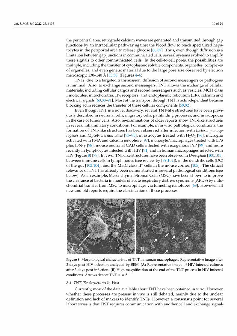
Even though TNT is a novel discovery, several TNT-like structures have been previ-ously described in neuronal cells, migratory cells, pathfinding processes, and invadopodiain the case of tumor cells. Also, re-examinations of older reports show TNT-like structuresin several inflammatory conditions. For example, in in vitro pathological conditions, theformation of TNT-like structures has been observed after infection with Listeria monocy-togenes and Mycobacterium bovis [93–95]; in astrocytes treated with H2O2 [96], microgliaactivated with PMA and calcium ionophore [97], monocyte/macrophages treated with LPSplus IFN-γ [98], mouse neuronal CAD cells infected with exogenous PrP [99] and morerecently in lymphocytes infected with HIV [91] and in human macrophages infected withHIV (Figure 8) [79]. In vivo, TNT-like structures have been observed in Drosophila [100,101],between immune cells in lymph nodes (see review by [89,102]), in the dendritic cells (DC)of the gut [103,104], and the MHC class II+ cells in the mouse cornea [105]. The clinicalrelevance of TNT has already been demonstrated in several pathological conditions (seebelow). As an example, Mesenchymal Stromal Cells (MSC) have been shown to improvethe clearance of bacteria in models of acute respiratory distress syndrome (ARDS) by mito-chondrial transfer from MSC to macrophages via tunneling nanotubes [63]. However, allnew and old reports require the classification of these processes.
Figure 8. Morphological characteristic of TNT in human macrophages. Representative image after3 days post HIV infection analyzed by SEM. (A) Representative image of HIV-infected culturesafter 3 days post-infection. (B) High magnification of the end of the TNT process in HIV-infectedconditions. Arrows denote TNT. n = 5.
8.4. TNT-like Structures In Vivo
Currently, most of the data available about TNT have been obtained in vitro. However,whether these processes are present in vivo is still debated, mainly due to the uncleardefinition and lack of makers to identify TNTs. However, a consensus point for severallaboratories is that TNT requires communication with another cell and exchange signal-

Int. J. Mol. Sci. 2022, 23, 6133 11 of 24
ing, cargo, or organelles components. So far, in vivo, these two components have notbeen proven.
Currently, several suggestive publications show TNT-like structures in MHC class IIdendritic cells in the corneal stroma [105], non-neural ectoderm cells in the midbrain [106],trophectoderm cells in the neural crest [107], epiblast cells in the blastula [108] and theectoderm [109]. Also, several groups have suggested that neuronal processes may be similarto TNT. However, we will exclude from this review this possibility because dendrites are aclear formation, transport, and stability and lack many components of TNT.
One of the best examples of TNT in vivo has been described in flies. Only recently,the role of TNT or cytoneme has been well described for several groups, indicating thecritical role of these processes in the signaling and delivery of developmental proteins suchas Dpp, fibroblast growth factor (FGF), Hedgehog, Wingless (in flies), sonic Hedgehold(chick limbs), and wnt proteins (in developing zebrafish) directly into sites of cell to cellcontact without dilution of these factors into the extracellular space [67,68,110–113]. TNT isa changing paradigm in the area because it was assumed that most of these proteins werereleased into the extracellular space. Roy et al. [114] describe that Dpp and FGF signalingis required for signal-producing wing disc cells, and several of the genes activated bythese factors are required for TNT (cytonemes) [114,115] or synapses formation [114–116].Most of the evidence seems to indicate that other factors important in the development offlies are TNT-mediated, such as fibroblast growth factor branchless [117], Dpp [113,114],EGF, Hh [67], SHh [70], and Notch [114–116,118]. Most of these factors are also present inhumans, but the role of TNTs is unknown. Thus, it may be possible to extrapolate some ofthese data into human development.
In chicken embryos, the filopodia-like protrusion (or TNT-like processes) span thesub-ectodermal space to establish contact with the ectoderm. These actin and tubulinpositive processes require Rac1 to form and participate in the retrograde trafficking of thetransmembrane Wnt-receptor Frizzled-7. Another great example of TNT-like processeshas been described in zebrafish embryos; several manuscripts describe their key role incontrolling the spread of Wnt morphogens such as wnt8a that activates Wnt/β-cateninsignaling pathways upon TNT contact. The formation and transfer of these molecules werecdc42/N-wasp dependent [119]. However, experiments in chicks suggest that shh-positivetransfer and spread are dependent on long TNT-like processes (hundreds of micrometers)that are cdc42 negative [70]. Thus, in these experiments, using actin blockers such aslatrunculin B, cytochalasin D, or overexpression of IRSp53 reduces filopodium formation,suggesting that actin assembling and f-actin-bundling are necessary for filopodium for-mation [69]. In these experiments, it has been suggested that the formation of filopodia(no-TNT) can be initiated in response to morphogenic factors such as FGF by the mDia3Cmechanism [120]. Kondo’s laboratory (Osaka University, Japan) is a pioneer in identify-ing TNT-like structures in controlling zebrafish pigmentation [121,122]. This transfer ofinformation between xanthophores extends processes (or dendrites) to melanophores, andcontact with these processes induces the melanophores to migrate away, changing thepigmentation pattern [123].
In heart development, TNT-like processes participate in the cardiac differentiation ofprogenitor cells that give rise to the heart tube. In this case, filopodial extensions from thedorsal pericardial wall are directed into the endoderm and adjacent mesenchymal cellsto coordinate signaling events related to the hedgehog, Notch, BMT, and non-canonicalwnt pathways and to maintain cardiac progenitor status during heart tube extension [124].Genetic deletion of tbx (22q11.2 deletion syndrome gene) and aPKCζ activator resultedin the loss of filopodia and decreased FGF signaling, reduced proliferation, and ectopicdifferentiation [124,125].
8.5. TNT Expression under Pathological Conditions
The data discussed above clearly indicate that TNT-like processes are expressed todeliver and spread in a specific manner wnt and notch-related morphogenic molecules
Int. J. Mol. Sci. 2022, 23, 6133 12 of 24
during development. Thus, are pathogens that induce TNT for spread and inflammationusing these mechanisms?
Interestingly, viruses, such as African swine fever, Ebola, Herpes Simplex, Marburgfiloviruses, SARS-CoV-2, and Poxvirus Vaccinia encode viral factors or alter cell activationto induce the formation of filopodia structures to allow viral trafficking between theextracellular matrix or environment into cells [126–132], suggesting that viruses are adaptedto use filopodia and TNT-like structures to improve viral spread.
Cytonemes (also called TNT, membrane tubulovesicular extensions, or TNT-like struc-tures) have been described in neutrophils directly contacting yeast or bacteria. The role ofTNT is to provide a directed delivery of anti-pathogen molecules at the site of infection.This group using human neutrophils describes that the mechanism of formation of theseprocesses is GTPase dynamin or actin-dependent [133]. However, most of these reportslack an accurate characterization of these processes. These data indicate that pores and/orTNT can be present in activated immune cells.
8.6. Molecular Mechanism of TNT Formation
Despite the multiple questions in the TNT area, several groups using cell lines ofdifferent origins have described increased TNT formation in several stress conditions. Still,how they are formed and the signal(s) involved in directing formation and stability isunknown. Several reviews and primary communications indicate that M-sec, part of sec6family proteins, and p53 activation are the key master regulator of TNT formation in severalcell lines [134,135]. The mechanisms by which M-sec activates Ras-like small GTPase ral-Apromote the binding to filamin. This protein cross-links actin filaments and promotesfilopodia formation, as well as cdc42 [134,135]. Also, H2O2 treatment or serum starvationin neuronal cell lines is dependent on p53 and subsequent activation of epidermal growthfactor (EGF) receptor, Akt1, PI3K, and mTOR [136]. Despite the beauty of this signalingsystem in cell lines, our data in primary human cells are confusing about the role of theseproteins in TNT formation. Clearly, there is a missing link between primary cells andimmortalized cell lines, where most of them have affected p53 expression and signaling.
Our data, using human primary macrophages or T cells, indicate that blocking M-secexpression using siRNA or treatment of primary cells with p53 activators such as CGK733,p53-stabilizing agents such as CP31398, reactivators of P53 mutant such as NSC319726, orMIRO-1, a proapoptotic p53 related protein, does not alter the formation of TNT in basal orin HIV-infected conditions or the form of TNT including fused open-ended TNT or synaptickind of TNTs. Interestingly, several TNTs behave like pores (described above—direct cell tocell communication), though some were also sensitive to gap junction blockers suggestingthat the synaptic kind of TNTs contain gap junctions (GJ). In addition, in different systems,functional GJ channels increase the invasion and dissemination of Shigella in epithelialcells [137] and increase toxicity between connected HIV-infected cells [138–140]. Suggestingthe possibility that particular infectious agents, such as Shigella and HIV, can use GJ chan-nels to sensitize uninfected cells and spread infection/toxicity to healthy cells, but whetherTNT or TNT-like structures play a role in these infectious diseases is still under investi-gation. Alternatively, GJ benefits the host immune system by mediating a cross-antigenpresentation phenomenon. This enables coupled cells to share viral peptides (antigens) andtrigger a response in CTL cells, even when some cells were never directly exposed to thepathogen [141]. GJ-mediated immune coupling suggests the possibility that GJ expressedby monocytes/macrophages in inflammatory conditions cross-present antigens to lympho-cytes and other inflammatory cells to maintain an immune memory in cells never exposeddirectly to specific antigen [141]. In the agreement, Cx43 is recruited to the immunologicalsynapses during T cell priming, suggesting that GJ and hemichannels also participate inantigen presentation [142]. Thus, some pathogens such as HIV may potentially use GJ andmaybe TNT (see below) to spread toxicity and its normal role in enhancing the immuneresponse. However, most viral and bacterial infections down-regulate expression and GJfunction, probably to reduce damage. Thus, pathogens require hijacking this communica-

Int. J. Mol. Sci. 2022, 23, 6133 13 of 24
tion system to spread toxicity and damage. We propose at least three kinds of direct cell tocell communication: GJ, pore mediated (close interactions), and TNT mediated (pore andsynaptic type, long-range combination of both communication systems).
Several pioneer studies using T cells described the potential role of TNT in the propaga-tion of Fas ligand-related cell death signals between communicated T lymphocytes [143,144].Also, TNT has been proposed to provide an exchange pathway between injured cardiomy-oblasts or endothelial cells by mesenchymal stem cells (MSC) through transferred mito-chondria [145,146]. The direct transfer of mitochondria between connected cells, pores, orTNT provides metabolic advantages for metabolic distress cells, such as during stroke [147].However, tumor cells utilize mitochondrial transfer in several types of cancers to adapt thetissue to the metabolic tumor environment [61,148–152]. However, whether these cells alsoshow direct pore communication is unknown.
8.7. Cell-Cell Mitochondrial Transfer via Tunneling Nanotubes
Overall, TNTs are minimally expressed under physiological conditions [84,153]. How-ever, they proliferate under stress and in response to several pathogens such as HIV, as wellas pathogenic conditions such as cancer, Alzheimer’s, Parkinson’s, and other neurodegener-ative diseases. The proposed function of TNT in pathogenic conditions is to amplify toxicity,apoptosis, and pathogenesis between connected cells. TNTs are generated in the “sick” cellsinto healthy surrounding cells to maximize efficiency in these conditions. In contrast, TNTsare also induced by stress conditions, including stroke, spinal cord, and traumatic braininjury. In these cases, TNTs are generated from compromised cells (cells in the penumbraarea) into healthy areas. In these cases, it has been proposed that compromised cells “re-quest” healthy factors and organelles to survive the damage in these conditions. These twomechanisms of TNT-induced formation, pathogenic and recovery, could be therapeuticallytargeted to regulate TNT formation and associated transport. In addition, several groupsdescribed exosomes containing mitochondria elements [154,155]. However, we will not discussthis type of mitochondrial element transfer for two reasons; first, released exosomes diffuse into theextracellular space losing the advantage provided by GJ, pores, and TNTs by maintaining a closemitochondrial transfer. This is essential to transfer enough organelles or their DNA to change themetabolism of the targeted cell. Second, there is a lack of proper cell-to-cell selectivity. This despitethat exosomes had potential receptors to provide selectivity. Even though the exosomal mediatedmitochondrial transfer or some of its elements can be exciting, whether the exosomal transfer caneffectively change the phenotype of the targeted cell is still unclear.
Several groups suggested that TNT-transmitted organelles such as lysosomes andmitochondria could alter the fate of the recipient cells. Mitochondria are interesting TNT-transmitted organelles. In addition to their traditional role as cell powerhouses, mitochon-dria also play a key role in calcium buffering, ER interactions, ROS generation, apoptosis,ER stress response activation, and a broad variety of mitochondrial dysfunction. Fur-thermore, mitochondrial genetic alterations have been associated with metabolic diseases,including Alzheimer’s disease (AD) and Parkinson’s disease (PD), muscular dystrophy,and the process of normal aging [156]. Thus, TNT exchange of compromised mitochondriacould result in accelerated disease.
The mitochondrial TNT-mediated exchange has been proposed as a general princi-ple to rescue damaged cells and genetic diseases [157–160]. Mitochondrial transfer canrescue aerobic respiration in stem cells [161], mitochondrial function even if mtDNA isdamaged [162], and epithelial function if mitochondria are transferred from stem cells [163].However, few studies demonstrated that TNT could be the mechanism of mitochondrialtransfer and, more importantly, the transferred mitochondria could change the TNT-targeted cell’s metabolism or function. For example, microinjection of intact mitochondriainto oocytes prevented apoptosis [164]. These data indicate that these communicationsystems can change the metabolism of the targeted cell, probably due to the highly concen-trated organelle transfer in a cell-to-cell specific manner.

Int. J. Mol. Sci. 2022, 23, 6133 14 of 24
Our data demonstrated that TNT generated in response to heterogeneous interactionbetween tumor cells and stromal cells promotes the transfer of a unique type of mitochon-dria from tumor cells into surrounding astrocytes resulting in their metabolic and hypoxicadaptation [61]. Currently, it is unknown whether the TNT-transferred mitochondria workindependently in the targeted astrocytes, fused with the existing ones, or replace the mi-tochondrial system of non-cancer cells. However, blocking TNT mitochondrial transferprevents healthy astrocytes’ adaptation to tumor-related conditions such as ischemia. Moreconcerning is that radiation alone could induce TNT formation and transfer, suggesting thatanti-cancer treatment may contribute to the tumor’s adaptation to subsequent treatmentor tumor survival. In contrast, UV treatment induces the formation of TNT to enable thetransfer of healthy mitochondria from healthy cells to rescue cell apoptosis with compro-mised DNA [165]. Both conditions indicate that DNA damage or compromise activatesTNT formation and associated mitochondrial transfer.
Our data indicate that mitochondrial transfer enables “healthy cells,” such as primaryastrocytes, to become tumor-like cells from a metabolic point of view. Furthermore, weidentified a critical “fingerprint” of the TNT-transferred mitochondria in glioblastoma. Mi-tochondria expressed few genes and depended on nuclear transcription for the synthesis ofall proteins for the organelle; however, we identified critical mutations in the mitochondrialDNA that favor the use of glutamine/glutamate instead of glucose and lipids to produceenergy, a critical phenotype on aggressive tumors [166,167]. Glutamine use is a marker ofaggressive glioblastoma [168]. Glutamine is a nitrogen source to synthesize nucleotides,amino acids, and ATP and is a major anaplerotic precursor for the TCA cycle. Glutaminedependency in different cancers promotes invasion and tumor aggressiveness [169]. Also,glutamine dependency can downregulate glycolysis reducing glucose uptake and lactateproduction, contributing to the Warburg effects observed in ischemic tumors. We identifiedthat TNT formation also protects astrocytes from hypoxic conditions. Tumor persistenceand growth relay in the survival of cancer stem-like cells promoted by a hypoxic microen-vironment [170–172]. It is believed that low oxygen levels prevent ROS formation andDNA damage, nuclear and mitochondrial. Our data support both ideas that mitochondrialtransfer changes the metabolism and contributes to adaptation to hypoxic conditions—bothessential components to promote tumor growth.
Interestingly, TNTs are the only communication system that enables targeted andselected transfer of pathogenic material, including mitochondria. Thus, blocking TNTformation and associated communication could provide additional treatment to reducetumor adaptation and prevent chemotherapeutic resistance [157,173–177]. Also, becauseTNTs are not expressed in healthy adult tissues, the expected toxic effects may be reduced.
We recently demonstrated TNT communication between heterogeneous glioblastomatumor cells based on their resistance to TMZ and radiation therapy. First, surprisingly weidentify that anti-tumor treatment promotes TNT formation and transport. Second, TNTconcentrates the anti-apoptotic enzyme MGMT and distributes it among cells with insuffi-cient MGMT expression to avoid apoptosis in response to TMZ and radiation treatment.Blocking TNTs prevented MGMT diffusion and survival of cells with minimal MGMTexpression. Third, we identify that tumor TNTs are selective because MGMT protein, butnot its mRNA, was transferred in vitro and in vivo in glioblastoma and breast cancer.
In agreement with the data in glioblastoma, in HIV-infected conditions, the mitochon-drial transfer is essential to spread infection and associated inflammation. In immunecells, it has been demonstrated that TNT can propagate cell death induced by Fas ligandamong T lymphocytes [143,144]; however, how amplification of disease and their protectivemechanism remain unsolved. Our data on HIV indicate that viral spread is associated withmitochondrial function, not only to provide a formidable amount of ATP to induce theTNT formation and associated transfer but also because some of these organelles nursesome of the infectious HIV material transferred by TNTs. However, the contribution ofthese organelles is still unknown.

Int. J. Mol. Sci. 2022, 23, 6133 15 of 24
Several manuscripts identified specific molecules involved in mitochondrial transport,such as the protein Miro. Miro 1 and 2 are mitochondrial outer membrane-associatedGTPases that regulate mitochondrial trafficking and distribution by coordinating actin-and microtubule-dependent mitochondrial movement due to interactions with TRAK andKIF5 [178]. It was reported that Miro1 knockout significantly inhibited microtubule-dependentmitochondrial motion, whereas Miro2 deficiency showed no effect on microtubule-dependentmitochondrial movement, suggesting that Miro1 is important for microtubule-dependentmitochondrial trafficking [179].
Several stimuli of TNT formation, such as the Fas-ligand receptor in the immunesystem or M-sec, a protein associated with the component Sec6 of the exocyst complexrequired for the docking of exocytic vesicles on the plasma membrane [143], have beendiscovered. Involved molecules in the TNT formation machinery are Rho and Ras smallGTPases families such as Cdc42, Ral or the exocyst effector, myosin V/X, and the transmem-brane MHC class III protein LST1. Also, Dephosphorylated β-Ca2+-Calmodulin-dependentprotein kinase II (βCaMKII) has been shown to stabilize TNT formation [180]. βCaMKIInot only stabilizes F-actin and binds to G-actin, preventing its nucleation, and in TNT, theprotein is localized at the base of the TNT. These findings suggest that the TNT formationmight be like filopodia and lamellipodia regulation mechanisms.
First, the interaction of M-Sec with RalA is necessary for the formation of TNT. Further,TNT formation has been associated with M-Sec expression. Also, RalA has been shown tointeract with filamin to promote filopodia formation; however, whether these interactionsplay a role in TNT formation, stability, and associated transport is unknown.
Overall, identifying TNT as a new mechanism of long-range communication duringdevelopment and pathological conditions opens several potential innovative therapeuticpossibilities. First, TNTs are minimally expressed in healthy individuals; Second, TNTsproliferate upon pathogenesis; Third, pathogens such as viruses and bacteria use them toamplify inflammation and infection; and Fourth, traumatic conditions use TNT to rescuecompromised cells. Thus, identifying potential blockers or inducers of TNT and the cargoassociated with its formation can provide novel medicine and therapeutic approaches forlife-threatening conditions.
8.8. Clinical Relevance of Tunneling Nanotubes
Currently, there are few examples of TNT in vivo, including in HIV animal models [77]and glioblastoma tumors [61,82,181]. However, observation of TNT processes in clinicalsamples has been proposed but not examined [182,183], though several laboratories areworking on similar microtentacle (well-defined tubulin content) processes. Microtentaclesare microtubule-based protrusions with high actin and vimentin content, as originallyobserved in detached breast cancer cells [184]. The main proposal is that detached cancercells may influence the metastatic and chemoresistance profile of metastatic cells. Boththese are also critical functions of TNT. However, a critical difference with TNT is thatthey do not communicate with cells until they establish contact with endothelial cells ortissues to invade. Thus, these microtentacles could establish TNT-mediated communicationwith other cells at one point. The growth of the microtentacles is regulated by kinesinsand microtubule-associated proteins, including tau [185]. Also, cdc42 acts as molecularswitches by alternating their active GTP-bound form and inactive GDP-bound form in cells.Aberrant cdc42 activity is implicated in tumor metastasis by promoting transendothelialmigration [185,186]. Currently, several clinical trials block and/or promote microtentacleextension or attachment [185,187–189]; however, whether these drugs alter TNT formationor associated communication is unknown.
Interestingly, tau induces the formation of microtentacles and reduces the effects orbinding of the anti-cancer drug paclitaxel [190]. However, these results had significantimplications for cancer, Alzheimer’s disease, and other tau-related pathologies [191–194].
The identification of the mechanism of TNT formation (as well as other types of longcommunication processes) and associated function in cancer and other pathologies is an es-

Int. J. Mol. Sci. 2022, 23, 6133 16 of 24
sential step to move forward in the TNT field from a mostly in vitro system into in vivo andthe use of potential drugs to block or promote TNT formation. Block to prevent the spreadof pathogenesis (cancer, Alzheimer’s disease, and other related diseases). In this case, TNTformation spread toxic organelles and second messengers into neighboring healthy cells.However, it also promotes TNT formation to rescue damaged cells from ischemic eventssuch as stroke and traumatic events. In this case, healthy cells send healthy mitochondriaand second messengers to compromised cells. These mechanisms denote a high impact ofTNT formation and associated transport on human diseases that have not been exploiteduntil now. In addition, there is no formation of TNTs in healthy individuals, increasing thepossibility of not having side effects. Thus, the clinical and translational possibilities arehighly significant for several incurable diseases and provide an alternative treatment forreducing brain/spinal cord physical damage or preventing/recovering ischemic eventssuch as heart attacks and stroke.
A recent commentary [64] updated the present trends in mitochondrial research tothree areas: (1) the causal role of mitochondrial dysfunction in several neurodegenerativeailments, such as Parkinson’s and Alzheimer’s disease; (2) the control of Ca2+ milIeu inselected intracellular micro-domains, changing intracellular responses; and (3) microvesicle(mV) or TNT-mediated transfer of mitochondria within different cells and tissues, of healthyor of degenerated mitochondria, as a mechanism of functional renewal in diseased cells, orfostering propagation of cell distress or pathology, such as in metastatic cancer evolution.
Until recently, mitochondria were considered to remain housed intracellularly for thelife duration of each cell. The current review builds on the evidence that the intercellularexchange of mitochondria (and other intracellular organelles) continues during normaltissue growth and evolution. This exchange between cells appears robust during normaldevelopment. It continues in various pathological conditions, as demonstrated separately,especially in the lungs, kidneys, and the myocardium, especially in the ischemic brain’srepair. The earliest paper [195] demonstrated that bone marrow-derived mesenchymal stemcells (MSC) reduce acute lung injury in animals challenged by bacterial infection. They alsosuggested that MSC has a crucial role in preventing VILI (volume-induced lung injury) inhealthy rats subjected to high volume ventilation. A potential therapeutic tool for patientsemerged from this early paper, suggesting several clinical trials. In a later publication,Chen et al. [196] summarized in a table the several clinical application of mitochondrialtransplantation by detailing a register of ongoing human clinical trials. This paper alsodiscusses how mitochondrial transfer to damaged cells can help revive cells energetics inrecipient cells.
Jackson et al. [63] demonstrated that mesenchymal stromal cell MSCs exert their an-timicrobial effect via macrophage phagocytic activity, which can be mediated throughmitochondrial transfer. In this model, fluorescent imaging and flow cytometry demon-strated extensive mitochondrial transfer from MSC to macrophages, which occurred at leastpartially through TNT structures. Finally, in a different opinion paper and a more recentreview [197,198], we can appreciate the effect of mitochondrial transfer as a therapeuticstrategy against ischemic stroke. These comments discuss how healthy mitochondria canbe transferred to stroke cells during ischemic stroke.
Lippert and Borlongan [199] extend these concepts and elucidate the prophylactic useof hyperbaric oxygen treatment. This novel paper describes the mechanism of action ofpreventive treatments by hyperbaric oxygen exposure. For the first time, it demonstrateshow a treatment preceding the exposure to a noxa could transfer the resistance mechanismto a tissue. Since oxygen levels inside a tissue are fleeting and can only last for a short time,the authors visualized directly mitochondrial transfer between cells induced by hyperbaricoxygen pretreatment. The process results in the transfer of healthy mitochondria andinduces increased resistance of the receiving cells to the inflammatory response typicalof ischemia and traumatic brain injury. These results are robust, with preconditioningdoubling the salvage rate of the cells subjected to the experimental trauma, and explain

Int. J. Mol. Sci. 2022, 23, 6133 17 of 24
the long-term effect of this technique which is clinically used in several surgical andmedical patients.
A comprehensive review of mesenchymal stem cells and their ability to providemitochondrial transfer has been organized by Cheng et al., 2019 [200]. In this review,the authors underline the improvement in clinical outcomes obtained in animal modelsof alveolar cells and pulmonary infection in improving the function of cardiomyocytesand the transfer of mitochondria via astrocyte into ischemic brain cells. However, it alsodemonstrates that cancer cells may enhance their ability to produce metastases by shiftingdamaged mitochondria and inducing chemoresistance. Because of this, the title of thisreview contains the definition: of a double-edged sword. The highlight of his article is asummary of different mechanisms of mitochondrial transfer via TNT and cell fusion orthrough GJ. The transfer mechanism is reviewed in this paper. It illustrates the formation ofTNT between the recipient cell and the donor cell, or the organization of GJ, via the help ofthe connexin-43 or direct transfer of mitochondrial DNA from MSC cells via extracellularvesicles (eV), and also the possibility of selective loss of donor nuclei after complete cellfusion. They mediate the transfer of chemoresistance in a tumor and can be utilized as aclinical tool to provide pharmaceutical blockage sites and restoration of healthy tissue.
A recent review by Crew et al. [201] underlines how inter-organ transport of Mito-chondria can be generated from the abundant supply provided by adipocytes. They canreadily donate eV-encapsulated mitochondria from oxidatively damaged adipocytes. Theseare injected into the circulation and can be uptaken in cardiac tissue. This up-regulation ofcardiac tissue results in resistance to cardiac ischemia, a paradoxical observation in obesepatients, repeatedly reported.
Finally, in a recent review article Liu et al. [202] provide specific tables summarizingintercellular mitochondrial transfer in physiological conditions between cells and betweendifferent tissues and an extensive list of dozens of papers providing transfer in animalmodels and the various tissues studied so far. This comprehensive review also discussesall the mechanisms of transfer and especially the stimuli necessary to evoke the beginningof the intercellular transfer. They learn the means of organization and cell connection topropagate and initiate intercellular communication and transfer of organelles betweencells. The evolution of this novel field will provide clinical novelty for maintaining organssuffering from aging and ischemic cells. And hopefully offer a new mechanism to supportchemotherapy and limit cancer expansion.
Therefore, mitochondrial transfer is a frequent phenomenon associated with variousbodily physiological and pathological activities. It may result in the restoration of normalphysiological functions, and may help in the recovery from disease in animal models, asit already has in a few patient cases. Meanwhile, the mechanism of inhibiting transferfrom stromal cells to cancer may develop the potential therapeutic target of a new anti-tumoral agent.
Author Contributions: All authors contributed to performing the experiments, analyzing the data, andwriting the manuscript. All authors have read and agreed to the published version of the manuscript.
Funding: This work was funded by grants from the National Institute of Mental Health, MH128082,the National Institute of Neurological Disorders and Stroke, NS105584, the UTMB Sealy Institute forVaccine Sciences, and the UTMB Institute for Human Infection & Immunity (to EAE).
Institutional Review Board Statement: No applicable.
Informed Consent Statement: No applicable.
Data Availability Statement: No applicable.
Conflicts of Interest: The authors declare no conflict of interest.
Int. J. Mol. Sci. 2022, 23, 6133 18 of 24
References1. Schleiden, M.J. Beiträge zur Phytogenesis. Arch. Anat. Physiol. Wiss. Med. 1839, 13, 137–176.2. Schwann, T. Mikroskopische Untersuchungen über die Uebereinstimmung in der Struktur und dem Wachsthum der Thiere und Pflanzen;
Sander’schen Buchhandlung: Berlin, Germany, 1839.3. Turner, W. The Cell Theory, Past and Present. J. Anat. Physiol. 1890, 24, 253–287. [PubMed]4. Hodgkin, A.L.; Huxley, A.F. Propagation of electrical signals along giant nerve fibers. Proc. R. Soc. Lond. B Biol. Sci. 1952,
140, 177–183. [PubMed]5. Hodgkin, A.L.; Huxley, A.F. Currents carried by sodium and potassium ions through the membrane of the giant axon of Loligo. J.
Physiol. 1952, 116, 449–472. [CrossRef]6. Hodgkin, A.L.; Huxley, A.F.; Katz, B. Measurement of current-voltage relations in the membrane of the giant axon of Loligo. J.
Physiol. 1952, 116, 424–448. [CrossRef]7. Hodgkin, A.L.; Huxley, A.F.; Katz, B. Ionic currents underlying activiti in the giant axon of the squid. Arch. Sci. Physiol. 1949,
3, 129–150.8. Hodgkin, A.L.; Katz, B. The effect of sodium ions on the electrical activity of giant axon of the squid. J. Physiol. 1949, 108, 37–77.
[CrossRef]9. Hille, B. Ion Channels of Excitable Membranes, 2nd ed.; Sinauer Associates, Inc.: Sunderland, MA, USA, 1992.10. Peracchia, C. (Ed.) Handbook of Membrane Channels. In Molecular and Cellular Physiology; Academic Press Inc.: San Diego, CA,
USA, 1994.11. Kanno, Y.; Loewenstein, W.R. Low-resistance coupling between gland cells. Some observations on intercellular contact membranes
and intercellular space. Nature 1964, 201, 194–195. [CrossRef]12. Loewenstein, W.R.; Kanno, Y. Studies on an epithelial (gland) cell junction. I. Modifications of surface membrane permeability. J.
Cell Biol. 1964, 22, 565–586. [CrossRef]13. Kuffler, S.W.; Potter, D.D. Glia in the leech central nervous system: Physiological properties and neuron-glia relationship. J.
Neurophysiol. 1964, 27, 290–320. [CrossRef]14. Stough, H.B. Giant nerve fibers of the earthworn. J. Comp. Neurol. 1926, 40, 409–463. [CrossRef]15. Stough, H.B. Polarization of the giant nerve fibers of the earthworm. J. Comp. Neurol. 1930, 50, 217–229. [CrossRef]16. Williams, L.W. Anatomy of the Common Squid; American Museum of Natural History: New York, NY, USA, 1909.17. Leydig, F. Zum Nervensystem und den Sinnesorganen der Würmer und Gleiderfüssler. In Tafeln zur Vergleichen Anatomie;
H. Laupp: Tubingen, Germany, 1864.18. Young, J.Z. The functioning of the giant nerve fibres of the squid. J. Exp. Biol. 1938, 15, 170–185. [CrossRef]19. Johnson, G.E. Giant nerve fibers in crustaceans, with special reference to Cambarus and Palaemonetes. J. Comp. Neurol. 1924,
36, 323–375. [CrossRef]20. Eccles, J.C.; Granit, R.; Young, J.Z. Impulses in the giant nerve fibers of earthworm. J. Physiol. 1932, 77, 23P–25P.21. Bullock, T.H. Functional organization of the giant fiber system of Lumbricus. J. Neurophysiol. 1945, 8, 55–71. [CrossRef]22. Rushton, W.A.H. Action potentials from the isolated nerve cord of the earthworm. Proc. Roy. Soc. B 1945, 132, 423–437.23. Kao, C.Y.; Grundfest, H. Postsynaptic electrogenesis in septate giant axons. I. Earthworm median giant axon. J. Neurophysiol.
1957, 20, 553–573. [CrossRef]24. Wilson, D.M. The connections between the lateral giant fibers of earthworms. Comp. Biochem. Physiol. 1961, 3, 274–284. [CrossRef]25. Hama, K. Some observations on the fine structure of the giant nerve fibers of the earthworm, Eisenia foetida. J. Biophys. Biochem.
Cytol. 1959, 6, 61–66. [CrossRef]26. Wiersma, C.A.G. Giant nerve fiber system of the crayfish. A contribution to comparative physiology of synapse. J. Neurophysiol.
1947, 10, 23–38. [CrossRef] [PubMed]27. Watanabe, A. The interaction of electrical activity among neurons of lobster cardiac ganglion. Jap. J. Physiol. 1958, 8, 305–318.
[CrossRef] [PubMed]28. Hagiwara, S.; Watanabe, A.; Saito, N. Potential changes in syncytial neurons of lobster cardiac ganglion. J. Neurophysiol. 1959,
22, 554–572. [CrossRef] [PubMed]29. Watanabe, A.; Bullock, T.H. Modulation of activity of one neuron by subthreshold slow potentials in another in lobster cardiac
ganglion. J. Gen. Physiol. 1960, 43, 1031–1045. [CrossRef]30. Watanabe, A.; Grundfest, H. Impulse propagation at the septal and commissural junctions of crayfish lateral giant axons. J. Gen.
Physiol. 1961, 45, 267–308. [CrossRef]31. Reuben, J.P. Electrotonic connections between lobster muscle fibers. Biol. Bull. Woods Hole 1960, 49, 334.32. Hagiwara, S.; Morita, H. Electrotonic transmission between two nerve cells in leech ganglion. J. Neurophysiol. 1962, 25, 721–731.
[CrossRef]33. Eckert, R. Electrical Interaction of Paired Ganglion Cells in the Leech. J. Gen. Physiol. 1963, 46, 573–587. [CrossRef]34. Weidmann, S. The electrical constants of Purkinje fibres. J. Physiol. 1952, 118, 348–360. [CrossRef]35. Engelmann, T.W. Vergleichende Untersuchungen zur Lehre von der Muskel und Nervenelektricität. Pflügers Arch. 1877,
15, 116–148. [CrossRef]36. Rothschuh, K.E. Über den funktionellen aufbaudes herzens aus elektrophysiologischen Elementen und üiber den mechanismus
der erregungsleitung im herzen. Pflüg. Arch. 1951, 253, 238–251. [CrossRef]

Int. J. Mol. Sci. 2022, 23, 6133 19 of 24
37. Bennett, M.V.; Crain, S.M.; Grundfest, H. Electrophysiology of supramedullary neurons in Spheroides maculatus. III. Organizationof the supramedullary neurons. J. Gen. Physiol. 1959, 43, 221–250. [CrossRef]
38. Martin, A.R.; Pilar, G. Dual mode of synaptic transmission in the avian ciliary ganglion. J. Physiol. 1963, 168, 443–463. [CrossRef]39. Martin, A.R.; Pilar, G. Transmission through the ciliary ganglion of the chick. J. Physiol. 1963, 168, 464–475. [CrossRef]40. Furukawa, T.; Furshpan, E.J. Two inhibitory mechanisms in the Mauthner neurons of goldfish. J. Neurophysiol. 1963, 26, 140–176.
[CrossRef]41. Peracchia, C. Increase in gap junction resistance with acidification in crayfish septate axons is closely related to changes in
intracellular calcium but not hydrogen ion concentration. J. Membr. Biol. 1990, 113, 75–92. [CrossRef]42. Lazrak, A.; Peracchia, C. Gap junction gating sensitivity to physiological internal calcium regardless of pH in Novikoff hepatoma
cells. Biophys. J. 1993, 65, 2002–2012. [CrossRef]43. Peracchia, C.; Sotkis, A.; Wang, X.G.; Peracchia, L.L.; Persechini, A. Calmodulin directly gates gap junction channels. J. Biol. Chem.
2000, 275, 26220–26224. [CrossRef]44. Sotkis, A.; Wang, X.G.; Yasumura, T.; Peracchia, L.L.; Persechini, A.; Rash, J.E.; Peracchia, C. Calmodulin colocalizes with
connexins and plays a direct role in gap junction channel gating. Cell Commun. Adhes. 2001, 8, 277–281. [CrossRef]45. Peracchia, C. Calmodulin-cork model of gap junction channel gating-one molecule, two mechanisms. Int. J. Mol. Sci. 2020,
21, 4938. [CrossRef]46. Peracchia, C. Gap junction stucture and chemical regulation. In Direct Calmodulin Role in Cell-To-Cell Channel Gating; Academic
Press: London, UK, 2019.47. Peracchia, C.; Leverone Peracchia, L.M. Calmodulin-connexin partnership in gap junction channel regulation-Calmodulin-cork
gating model. Int. J. Mol. Sci. 2021, 22, 13055. [CrossRef]48. Lasek, R.J.; Gainer, H.; Przybylski, R.J. Transfer of newly synthesized proteins from Schwann cells to the squid giant axon. Proc.
Natl. Acad. Sci. USA 1974, 71, 1188–1192. [CrossRef]49. Lasek, R.J.; Gainer, H.; Barker, J.L. Cell-to-cell transfer of glial proteins to the squid giant axon. The glia-neuron protein trnasfer
hypothesis. J. Cell Biol. 1977, 74, 501–523. [CrossRef]50. Singer, M.; Green, M.R. Autoradiographic studies of uridine incorporation in peripheral nerve of the newt, Triturus. J. Morphol.
1968, 124, 321–344. [CrossRef]51. Singer, M.; Salpeter, M.M. The transport of 3H-l-histidine through the Schwann and myelin sheath into the axon, including a
reevaluation of myelin function. J. Morphol. 1966, 120, 281–315. [CrossRef]52. Peracchia, C. Excitable membrane ultrastructure. I. Freeze fracture of crayfish axons. J. Cell Biol. 1974, 61, 107–122. [CrossRef]53. Peracchia, C. Direct communication between axons and sheath glial cells in crayfish. Nature 1981, 290, 597–598. [CrossRef]54. Viancour, T.A.; Bittner, G.D.; Ballinger, M.L. Selective transfer of Lucifer yellow CH from axoplasm to adaxonal glia. Nature 1981,
293, 65–67. [CrossRef]55. Andersson, E.; Edstrom, A.; Jarlstedt, J. Properties of RNA from giant axons of the crayfish. Acta Physiol. Scand. 1970, 78, 491–502.
[CrossRef]56. Gainer, H.; Tasaki, I.; Lasek, R.J. Evidence for the glia-neuron protein transfer hypothesis from intracellular perfusion studies of
squid giant axons. J. Cell Biol. 1977, 74, 524–530. [CrossRef]57. Meyer, M.R.; Bittner, G.D. Histological studies of trophic dependencies in crayfish giant axons. Brain Res. 1978, 143, 195–211.
[CrossRef]58. Fedorenko, G.; Neginskaya, M.; Fedorenko, A.; Uzdensky, A. The paired neuroglial and interglial membranes in the crayfish
stretch receptor and their local disorganization. J. Neurosci. Res. 2015, 93, 707–713. [CrossRef] [PubMed]59. Rustom, A.; Saffrich, R.; Markovic, I.; Walther, P.; Gerdes, H.H. Nanotubular highways for intercellular organelle transport.
Science 2004, 303, 1007–1010. [CrossRef] [PubMed]60. Abounit, S.; Zurzolo, C. Wiring through tunneling nanotubes—From electrical signals to organelle transfer. J. Cell Sci. 2012,
125, 1089–1098. [CrossRef]61. Valdebenito, S.; Malik, S.; Luu, R.; Loudig, O.; Mitchell, M.; Okafo, G.; Bhat, K.; Prideaux, B.; Eugenin, E.A. Tunneling
nanotubes, TNT, communicate glioblastoma with surrounding non-tumor astrocytes to adapt them to hypoxic and metabolictumor conditions. Sci. Rep. 2021, 11, 14556. [CrossRef]
62. Eugenin, E.A.; Gaskill, P.J.; Berman, J.W. Tunneling nanotubes (TNT): A potential mechanism for intercellular HIV trafficking.Commun. Integr. Biol. 2009, 2, 243–244. [CrossRef]
63. Jackson, M.V.; Morrison, T.J.; Doherty, D.F.; McAuley, D.F.; Matthay, M.A.; Kissenpfennig, A.; O’Kane, C.M.; Krasnodembskaya, A.D.Mitochondrial Transfer via Tunneling Nanotubes is an Important Mechanism by Which Mesenchymal Stem Cells EnhanceMacrophage Phagocytosis in the in Vitro and in Vivo Models of ARDS. Stem. Cells 2016, 34, 2210–2223. [CrossRef]
64. Pizzo, P.; Pozzan, T. Mitochondrialand: What will come next? Function 2021, 3, 1–2. [CrossRef]65. Chen, W.; Huang, H.; Hatori, R.; Kornberg, T.B. Essential basal cytonemes take up Hedgehog in the Drosophila wing imaginal
disc. Development 2017, 144, 3134–3144. [CrossRef]66. Kornberg, T.B. Distributing signaling proteins in space and time: The province of cytonemes. Curr. Opin. Genet. Dev. 2017,
45, 22–27. [CrossRef]
Int. J. Mol. Sci. 2022, 23, 6133 20 of 24
67. Bischoff, M.; Gradilla, A.C.; Seijo, I.; Andres, G.; Rodriguez-Navas, C.; Gonzalez-Mendez, L.; Guerrero, I. Cytonemes are requiredfor the establishment of a normal Hedgehog morphogen gradient in Drosophila epithelia. Nat. Cell Biol. 2013, 15, 1269–1281.[CrossRef]
68. Rojas-Rios, P.; Guerrero, I.; Gonzalez-Reyes, A. Cytoneme-mediated delivery of hedgehog regulates the expression of bonemorphogenetic proteins to maintain germline stem cells in Drosophila. PLoS Biol. 2012, 10, e1001298. [CrossRef]
69. Stanganello, E.; Hagemann, A.I.; Mattes, B.; Sinner, C.; Meyen, D.; Weber, S.; Schug, A.; Raz, E.; Scholpp, S. Filopodia-based Wnttransport during vertebrate tissue patterning. Nat. Commun. 2015, 6, 5846. [CrossRef]
70. Sanders, T.A.; Llagostera, E.; Barna, M. Specialized filopodia direct long-range transport of SHH during vertebrate tissuepatterning. Nature 2013, 497, 628–632. [CrossRef]
71. Junyent, S.; Garcin, C.L.; Szczerkowski, J.L.A.; Trieu, T.J.; Reeves, J.; Habib, S.J. Specialized cytonemes induce self-organization ofstem cells. Proc. Natl. Acad. Sci. USA 2020, 117, 7236–7244. [CrossRef]
72. Wood, B.M.; Baena, V.; Huang, H.; Jorgens, D.M.; Terasaki, M.; Kornberg, T.B. Cytonemes with complex geometries andcomposition extend into invaginations of target cells. J. Cell Biol. 2021, 220, e202101116. [CrossRef]
73. Las, G.; Shirihai, O.S. Miro1: New wheels for transferring mitochondria. Embo. J. 2014, 33, 939–941. [CrossRef]74. Austefjord, M.W.; Gerdes, H.H.; Wang, X. Tunneling nanotubes: Diversity in morphology and structure. Commun. Integr. Biol.
2014, 7, e27934. [CrossRef]75. Schiller, C.; Diakopoulos, K.N.; Rohwedder, I.; Kremmer, E.; von Toerne, C.; Ueffing, M.; Weidle, U.H.; Ohno, H.; Weiss, E.H. LST1
promotes the assembly of a molecular machinery responsible for tunneling nanotube formation. J. Cell Sci. 2013, 126, 767–777.[CrossRef]
76. Gerdes, H.H.; Rustom, A.; Wang, X. Tunneling nanotubes, an emerging intercellular communication route in development. Mech.Dev. 2013, 130, 381–387. [CrossRef]
77. Okafo, G.; Prevedel, L.; Eugenin, E. Tunneling nanotubes (TNT) mediate long-range gap junctional communication: Implicationsfor HIV cell to cell spread. Sci. Rep. 2017, 7, 16660. [CrossRef]
78. Malik, S.; Eugenin, E.A. Mechanisms of HIV Neuropathogenesis: Role of Cellular Communication Systems. Curr. HIV Res. 2016,14, 400–411. [CrossRef]
79. Eugenin, E.A.; Gaskill, P.J.; Berman, J.W. Tunneling nanotubes (TNT) are induced by HIV-infection of macrophages: A potentialmechanism for intercellular HIV trafficking. Cell Immunol. 2009, 254, 142–148. [CrossRef]
80. Wang, X.; Gerdes, H.H. Long-distance electrical coupling via tunneling nanotubes. Biochim. Biophys. Acta 2012, 1818, 2082–2086.[CrossRef]
81. Sisakhtnezhad, S.; Khosravi, L. Emerging physiological and pathological implications of tunneling nanotubes formation betweencells. Eur. J. Cell Biol. 2015, 94, 429–443. [CrossRef]
82. Valdebenito, S.; Audia, A.; Bhat, K.P.L.; Okafo, G.; Eugenin, E.A. Tunneling Nanotubes Mediate Adaptation of Glioblastoma Cellsto Temozolomide and Ionizing Radiation Treatment. iScience 2020, 23, 101450. [CrossRef]
83. Eugenin, E.A. Role of cell-to-cell communication in cancer: New features, insights, and directions. Cancer Rep. (Hoboken) 2019,2, e1228. [CrossRef]
84. Valdebenito, S.; Lou, E.; Baldoni, J.; Okafo, G.; Eugenin, E. The Novel Roles of Connexin Channels and Tunneling Nanotubes inCancer Pathogenesis. Int. J. Mol. Sci. 2018, 19, 1270. [CrossRef]
85. Harris, A.L. Connexin specificity of second messenger permeation: Real numbers at last. J. Gen. Physiol. 2008, 131, 287–292.[CrossRef]
86. Eugenin, E.A.; Gonzalez, H.; Saez, C.G.; Saez, J.C. Gap junctional communication coordinates vasopressin-induced glycogenolysisin rat hepatocytes. Am. J. Physiol. 1998, 274, G1109–G1116. [CrossRef]
87. Nathanson, M.H.; Burgstahler, A.D.; Mennone, A.; Fallon, M.B.; Gonzalez, C.B.; Saez, J.C. Ca2+ waves are organized amonghepatocytes in the intact organ. Am. J. Physiol. 1995, 269, G167–G171. [CrossRef] [PubMed]
88. Onfelt, B.; Davis, D.M. Can membrane nanotubes facilitate communication between immune cells? Biochem. Soc. Trans. 2004,32, 676–678. [CrossRef] [PubMed]
89. Onfelt, B.; Nedvetzki, S.; Yanagi, K.; Davis, D.M. Cutting edge: Membrane nanotubes connect immune cells. J. Immunol. 2004,173, 1511–1513. [CrossRef] [PubMed]
90. Gerdes, H.H.; Carvalho, R.N. Intercellular transfer mediated by tunneling nanotubes. Curr. Opin. Cell Biol. 2008, 20, 470–475.[CrossRef] [PubMed]
91. Sowinski, S.; Jolly, C.; Berninghausen, O.; Purbhoo, M.A.; Chauveau, A.; Kohler, K.; Oddos, S.; Eissmann, P.; Brodsky, F.M.;Hopkins, C.; et al. Membrane nanotubes physically connect T cells over long distances presenting a novel route for HIV-1transmission. Nat. Cell Biol. 2008, 10, 211–219. [CrossRef] [PubMed]
92. Gurke, S.; Barroso, J.F.; Gerdes, H.H. The art of cellular communication: Tunneling nanotubes bridge the divide. Histochem. CellBiol. 2008, 129, 539–550. [CrossRef]
93. Dramsi, S.; Cossart, P. Intracellular pathogens and the actin cytoskeleton. Annu. Rev. Cell Dev. Biol. 1998, 14, 137–166. [CrossRef]94. Onfelt, B.; Nedvetzki, S.; Benninger, R.K.; Purbhoo, M.A.; Sowinski, S.; Hume, A.N.; Seabra, M.C.; Neil, M.A.; French, P.M.;
Davis, D.M. Structurally distinct membrane nanotubes between human macrophages support long-distance vesicular traffic orsurfing of bacteria. J. Immunol. 2006, 177, 8476–8483. [CrossRef]
95. Wehland, J.; Carl, U.D. The sophisticated survival strategies of the pathogen Listeria monocytogenes. Int. Microbiol. 1998, 1, 11–18.

Int. J. Mol. Sci. 2022, 23, 6133 21 of 24
96. Zhu, D.; Tan, K.S.; Zhang, X.; Sun, A.Y.; Sun, G.Y.; Lee, J.C. Hydrogen peroxide alters membrane and cytoskeleton properties andincreases intercellular connections in astrocytes. J. Cell Sci. 2005, 118, 3695–3703. [CrossRef]
97. Martinez, A.D.; Eugenin, E.A.; Branes, M.C.; Bennett, M.V.; Saez, J.C. Identification of second messengers that induce expressionof functional gap junctions in microglia cultured from newborn rats. Brain Res. 2002, 943, 191–201. [CrossRef]
98. Eugenin, E.A.; Branes, M.C.; Berman, J.W.; Saez, J.C. TNF-alpha plus IFN-gamma induce connexin43 expression and formation ofgap junctions between human monocytes/macrophages that enhance physiological responses. J. Immunol. 2003, 170, 1320–1328.[CrossRef]
99. Gousset, K.; Schiff, E.; Langevin, C.; Marijanovic, Z.; Caputo, A.; Browman, D.T.; Chenouard, N.; de Chaumont, F.; Martino, A.;Enninga, J.; et al. Prions hijack tunnelling nanotubes for intercellular spread. Nat. Cell Biol. 2009, 11, 328–336. [CrossRef]
100. Hsiung, F.; Ramirez-Weber, F.A.; Iwaki, D.D.; Kornberg, T.B. Dependence of Drosophila wing imaginal disc cytonemes onDecapentaplegic. Nature 2005, 437, 560–563. [CrossRef]
101. Kornberg, T. Pictures in cell biology. Cytonemes. Trends Cell Biol. 1999, 9, 434. [CrossRef]102. Gerdes, H.H.; Bukoreshtliev, N.V.; Barroso, J.F. Tunneling nanotubes: A new route for the exchange of components between
animal cells. FEBS Lett. 2007, 581, 2194–2201. [CrossRef]103. Niess, J.H.; Brand, S.; Gu, X.; Landsman, L.; Jung, S.; McCormick, B.A.; Vyas, J.M.; Boes, M.; Ploegh, H.L.; Fox, J.G.; et al.
CX3CR1-mediated dendritic cell access to the intestinal lumen and bacterial clearance. Science 2005, 307, 254–258. [CrossRef]104. Rescigno, M.; Urbano, M.; Valzasina, B.; Francolini, M.; Rotta, G.; Bonasio, R.; Granucci, F.; Kraehenbuhl, J.P.; Ricciardi-Castagnoli, P.
Dendritic cells express tight junction proteins and penetrate gut epithelial monolayers to sample bacteria. Nat. Immunol. 2001,2, 361–367. [CrossRef]
105. Chinnery, H.R.; Pearlman, E.; McMenamin, P.G. Cutting edge: Membrane nanotubes in vivo: A feature of MHC class II+ cells inthe mouse cornea. J. Immunol. 2008, 180, 5779–5783. [CrossRef]
106. Pyrgaki, C.; Trainor, P.; Hadjantonakis, A.K.; Niswander, L. Dynamic imaging of mammalian neural tube closure. Dev. Biol. 2010,344, 941–947. [CrossRef]
107. Teddy, J.M.; Kulesa, P.M. In vivo evidence for short- and long-range cell communication in cranial neural crest cells. Development2004, 131, 6141–6151. [CrossRef]
108. Caneparo, L.; Pantazis, P.; Dempsey, W.; Fraser, S.E. Intercellular bridges in vertebrate gastrulation. PLoS ONE 2011, 6, e20230.[CrossRef]
109. Miller, J.; Fraser, S.E.; McClay, D. Dynamics of thin filopodia during sea urchin gastrulation. Development 1995, 121, 2501–2511.[CrossRef]
110. Bilioni, A.; Sanchez-Hernandez, D.; Callejo, A.; Gradilla, A.C.; Ibanez, C.; Mollica, E.; Carmen Rodriguez-Navas, M.; Simon, E.;Guerrero, I. Balancing Hedgehog, a retention and release equilibrium given by Dally, Ihog, Boi and shifted/DmWif. Dev. Biol.2013, 376, 198–212. [CrossRef]
111. Gradilla, A.C.; Gonzalez, E.; Seijo, I.; Andres, G.; Bischoff, M.; Gonzalez-Mendez, L.; Sanchez, V.; Callejo, A.; Ibanez, C.;Guerra, M.; et al. Exosomes as Hedgehog carriers in cytoneme-mediated transport and secretion. Nat. Commun. 2014, 5, 5649.[CrossRef]
112. Kornberg, T.B. The contrasting roles of primary cilia and cytonemes in Hh signaling. Dev. Biol. 2014, 394, 1–5. [CrossRef]113. Roy, S.; Hsiung, F.; Kornberg, T.B. Specificity of Drosophila cytonemes for distinct signaling pathways. Science 2011, 332, 354–358.
[CrossRef]114. Roy, S.; Huang, H.; Liu, S.; Kornberg, T.B. Cytoneme-mediated contact-dependent transport of the Drosophila decapentaplegic
signaling protein. Science 2014, 343, 1244624. [CrossRef]115. Roy, S.; Kornberg, T.B. Paracrine signaling mediated at cell-cell contacts. Bioessays 2015, 37, 25–33. [CrossRef]116. Kornberg, T.B.; Roy, S. Cytonemes as specialized signaling filopodia. Development 2014, 141, 729–736. [CrossRef]117. Sato, M.; Kornberg, T.B. FGF is an essential mitogen and chemoattractant for the air sacs of the drosophila tracheal system. Dev.
Cell. 2002, 3, 195–207. [CrossRef]118. Callejo, A.; Bilioni, A.; Mollica, E.; Gorfinkiel, N.; Andres, G.; Ibanez, C.; Torroja, C.; Doglio, L.; Sierra, J.; Guerrero, I. Dispatched
mediates Hedgehog basolateral release to form the long-range morphogenetic gradient in the Drosophila wing disk epithelium.Proc. Natl. Acad. Sci. USA 2011, 108, 12591–12598. [CrossRef] [PubMed]
119. Kesavan, G.; Lieven, O.; Mamidi, A.; Ohlin, Z.L.; Johansson, J.K.; Li, W.C.; Lommel, S.; Greiner, T.U.; Semb, H. Cdc42/N-WASPsignaling links actin dynamics to pancreatic beta cell delamination and differentiation. Development 2014, 141, 685–696. [CrossRef][PubMed]
120. Koizumi, K.; Takano, K.; Kaneyasu, A.; Watanabe-Takano, H.; Tokuda, E.; Abe, T.; Watanabe, N.; Takenawa, T.; Endo, T.RhoD activated by fibroblast growth factor induces cytoneme-like cellular protrusions through mDia3C. Mol. Biol. Cell. 2012,23, 4647–4661. [CrossRef]
121. Watanabe, M.; Kondo, S. Fish pigmentation. Comment on “Local reorganization of xanthophores fine-tunes and colors the stripedpattern of zebrafish”. Science 2015, 348, 297. [CrossRef]
122. Watanabe, M.; Kondo, S. Is pigment patterning in fish skin determined by the Turing mechanism? Trends Genet 2015, 31, 88–96.[CrossRef]
123. Inaba, M.; Yamanaka, H.; Kondo, S. Pigment pattern formation by contact-dependent depolarization. Science 2012, 335, 677.[CrossRef]

Int. J. Mol. Sci. 2022, 23, 6133 22 of 24
124. Cohen, M.; Georgiou, M.; Stevenson, N.L.; Miodownik, M.; Baum, B. Dynamic filopodia transmit intermittent Delta-Notchsignaling to drive pattern refinement during lateral inhibition. Dev. Cell. 2010, 19, 78–89. [CrossRef]
125. Vitelli, F.; Taddei, I.; Morishima, M.; Meyers, E.N.; Lindsay, E.A.; Baldini, A. A genetic link between Tbx1 and fibroblast growthfactor signaling. Development 2002, 129, 4605–4611. [CrossRef]
126. Cudmore, S.; Cossart, P.; Griffiths, G.; Way, M. Actin-based motility of vaccinia virus. Nature 1995, 378, 636–638. [CrossRef]127. Favoreel, H.W.; Van Minnebruggen, G.; Adriaensen, D.; Nauwynck, H.J. Cytoskeletal rearrangements and cell extensions induced
by the US3 kinase of an alphaherpesvirus are associated with enhanced spread. Proc. Natl. Acad. Sci. USA 2005, 102, 8990–8995.[CrossRef]
128. Gill, M.B.; Edgar, R.; May, J.S.; Stevenson, P.G. A gamma-herpesvirus glycoprotein complex manipulates actin to promote viralspread. PLoS ONE 2008, 3, e1808. [CrossRef]
129. Hartlieb, B.; Weissenhorn, W. Filovirus assembly and budding. Virology 2006, 344, 64–70. [CrossRef]130. Jouvenet, N.; Windsor, M.; Rietdorf, J.; Hawes, P.; Monaghan, P.; Way, M.; Wileman, T. African swine fever virus induces
filopodia-like projections at the plasma membrane. Cell Microbiol. 2006, 8, 1803–1811. [CrossRef]131. Kolesnikova, L.; Bohil, A.B.; Cheney, R.E.; Becker, S. Budding of Marburgvirus is associated with filopodia. Cell Microbiol. 2007,
9, 939–951. [CrossRef]132. Noda, T.; Ebihara, H.; Muramoto, Y.; Fujii, K.; Takada, A.; Sagara, H.; Kim, J.H.; Kida, H.; Feldmann, H.; Kawaoka, Y. Assembly
and budding of Ebolavirus. PLoS Pathog. 2006, 2, e99. [CrossRef]133. Galkina, S.I.; Fedorova, N.V.; Serebryakova, M.V.; Arifulin, E.A.; Stadnichuk, V.I.; Gaponova, T.V.; Baratova, L.A.; Sud’ina, G.F.
Inhibition of the GTPase dynamin or actin depolymerisation initiates outward plasma membrane tubulation/vesiculation(cytoneme formation) in neutrophils. Biol. Cell. 2015, 107, 144–158. [CrossRef]
134. Ohno, H.; Hase, K.; Kimura, S. M-Sec: Emerging secrets of tunneling nanotube formation. Commun. Integr. Biol. 2010, 3, 231–233.[CrossRef]
135. Kimura, S.; Yamashita, M.; Yamakami-Kimura, M.; Sato, Y.; Yamagata, A.; Kobashigawa, Y.; Inagaki, F.; Amada, T.; Hase, K.;Iwanaga, T.; et al. Distinct Roles for the N- and C-terminal Regions of M-Sec in Plasma Membrane Deformation during TunnelingNanotube Formation. Sci. Rep. 2016, 6, 33548. [CrossRef]
136. Wang, Y.; Cui, J.; Sun, X.; Zhang, Y. Tunneling-nanotube development in astrocytes depends on p53 activation. Cell Death Differ.2011, 18, 732–742. [CrossRef]
137. Tran Van Nhieu, G.; Clair, C.; Bruzzone, R.; Mesnil, M.; Sansonetti, P.; Combettes, L. Connexin-dependent inter-cellularcommunication increases invasion and dissemination of Shigella in epithelial cells. Nat. Cell Biol. 2003, 5, 720–726. [CrossRef]
138. Eugenin, E.A.; Berman, J.W. Gap junctions mediate human immunodeficiency virus-bystander killing in astrocytes. J. Neurosci.Off. J. Soc. Neurosci. 2007, 27, 12844–12850. [CrossRef]
139. Eugenin, E.A.; Berman, J.W. Cytochrome C dysregulation induced by HIV infection of astrocytes results in bystander apoptosisof uninfected astrocytes by an IP and Calcium dependent mechanism. J. Neurochem. 2013, 127, 644–651. [CrossRef]
140. Eugenin, E.A.; Clements, J.E.; Zink, M.C.; Berman, J.W. Human immunodeficiency virus infection of human astrocytes disruptsblood-brain barrier integrity by a gap junction-dependent mechanism. J. Neurosci. 2011, 31, 9456–9465. [CrossRef]
141. Neijssen, J.; Herberts, C.; Drijfhout, J.W.; Reits, E.; Janssen, L.; Neefjes, J. Cross-presentation by intercellular peptide transferthrough gap junctions. Nature 2005, 434, 83–88. [CrossRef]
142. Mendoza-Naranjo, A.; Bouma, G.; Pereda, C.; Ramirez, M.; Webb, K.F.; Tittarelli, A.; Lopez, M.N.; Kalergis, A.M.; Thrasher, A.J.;Becker, D.L.; et al. Functional Gap Junctions Accumulate at the Immunological Synapse and Contribute to T Cell Activation. J.Immunol. 2011, 187, 3121–3132. [CrossRef]
143. Arkwright, P.D.; Luchetti, F.; Tour, J.; Roberts, C.; Ayub, R.; Morales, A.P.; Rodriguez, J.J.; Gilmore, A.; Canonico, B.; Papa, S.; et al.Fas stimulation of T lymphocytes promotes rapid intercellular exchange of death signals via membrane nanotubes. Cell Res. 2010,20, 72–88. [CrossRef]
144. Luchetti, F.; Canonico, B.; Arcangeletti, M.; Guescini, M.; Cesarini, E.; Stocchi, V.; Degli Esposti, M.; Papa, S. Fas signallingpromotes intercellular communication in T cells. PLoS ONE 2012, 7, e35766. [CrossRef]
145. Liu, K.; Ji, K.; Guo, L.; Wu, W.; Lu, H.; Shan, P.; Yan, C. Mesenchymal stem cells rescue injured endothelial cells in an in vitroischemia-reperfusion model via tunneling nanotube like structure-mediated mitochondrial transfer. Microvasc. Res. 2014,92, 10–18. [CrossRef]
146. Cselenyak, A.; Pankotai, E.; Horvath, E.M.; Kiss, L.; Lacza, Z. Mesenchymal stem cells rescue cardiomyoblasts from cell death inan in vitro ischemia model via direct cell-to-cell connections. BMC Cell Biol. 2010, 11, 29. [CrossRef]
147. Yip, H.K.; Dubey, N.K.; Lin, K.C.; Sung, P.H.; Chiang, J.Y.; Chu, Y.C.; Huang, C.R.; Chen, Y.L.; Deng, Y.H.; Cheng, H.C.; et al.Melatonin rescues cerebral ischemic events through upregulated tunneling nanotube-mediated mitochondrial transfer anddownregulated mitochondrial oxidative stress in rat brain. Biomed. Pharm. 2021, 139, 111593. [CrossRef] [PubMed]
148. Allegra, A.; Di Gioacchino, M.; Cancemi, G.; Casciaro, M.; Petrarca, C.; Musolino, C.; Gangemi, S. Specialized IntercellularCommunications via Tunnelling Nanotubes in Acute and Chronic Leukemia. Cancers 2022, 14, 659. [CrossRef] [PubMed]
149. Chinnery, H.R.; Keller, K.E. Tunneling Nanotubes and the Eye: Intercellular Communication and Implications for Ocular Healthand Disease. Biomed. Res. Int. 2020, 2020, 7246785. [CrossRef] [PubMed]
150. Moore, J.A.; Mistry, J.J.; Rushworth, S.A. Mitochondria and the Tumour Microenvironment in Blood Cancer. Adv. Exp. Med. Biol.2021, 1329, 181–203. [CrossRef] [PubMed]

Int. J. Mol. Sci. 2022, 23, 6133 23 of 24
151. Saenz-de-Santa-Maria, I.; Bernardo-Castineira, C.; Enciso, E.; Garcia-Moreno, I.; Chiara, J.L.; Suarez, C.; Chiara, M.D. Controlof long-distance cell-to-cell communication and autophagosome transfer in squamous cell carcinoma via tunneling nanotubes.Oncotarget 2017, 8, 20939–20960. [CrossRef] [PubMed]
152. Thayanithy, V.; Dickson, E.L.; Steer, C.; Subramanian, S.; Lou, E. Tumor-stromal cross talk: Direct cell-to-cell transfer of oncogenicmicroRNAs via tunneling nanotubes. Transl. Res. 2014, 164, 359–365. [CrossRef] [PubMed]
153. Roehlecke, C.; Schmidt, M.H.H. Tunneling Nanotubes and Tumor Microtubes in Cancer. Cancers 2020, 12, 857. [CrossRef]154. Picca, A.; Guerra, F.; Calvani, R.; Bucci, C.; Lo Monaco, M.R.; Bentivoglio, A.R.; Coelho-Junior, H.J.; Landi, F.; Bernabei, R.;
Marzetti, E. Mitochondrial Dysfunction and Aging: Insights from the Analysis of Extracellular Vesicles. Int. J. Mol. Sci. 2019,20, 805. [CrossRef]
155. Soto-Heredero, G.; Baixauli, F.; Mittelbrunn, M. Interorganelle Communication between Mitochondria and the EndolysosomalSystem. Front. Cell Dev. Biol. 2017, 5, 95. [CrossRef]
156. Diaz-Vegas, A.; Sanchez-Aguilera, P.; Krycer, J.R.; Morales, P.E.; Monsalves-Alvarez, M.; Cifuentes, M.; Rothermel, B.A.;Lavandero, S. Is Mitochondrial Dysfunction a Common Root of Noncommunicable Chronic Diseases? Endocr. Rev. 2020,41, bnaa005. [CrossRef]
157. Singh, A.K.; Cancelas, J.A. Gap Junctions in the Bone Marrow Lympho-Hematopoietic Stem Cell Niche, Leukemia Progression,and Chemoresistance. Int. J. Mol. Sci. 2020, 21, 796. [CrossRef]
158. Kolba, M.D.; Dudka, W.; Zareba-Koziol, M.; Kominek, A.; Ronchi, P.; Turos, L.; Chroscicki, P.; Wlodarczyk, J.; Schwab, Y.;Klejman, A.; et al. Tunneling nanotube-mediated intercellular vesicle and protein transfer in the stroma-provided imatinibresistance in chronic myeloid leukemia cells. Cell Death Dis. 2019, 10, 817. [CrossRef]
159. Li, C.J.; Chen, P.K.; Sun, L.Y.; Pang, C.Y. Enhancement of Mitochondrial Transfer by Antioxidants in Human Mesenchymal StemCells. Oxidative Med. Cell. Longev. 2017, 2017, 8510805. [CrossRef]
160. Astanina, K.; Koch, M.; Jungst, C.; Zumbusch, A.; Kiemer, A.K. Lipid droplets as a novel cargo of tunnelling nanotubes inendothelial cells. Sci. Rep. 2015, 5, 11453. [CrossRef]
161. Spees, J.L.; Olson, S.D.; Whitney, M.J.; Prockop, D.J. Mitochondrial transfer between cells can rescue aerobic respiration. Proc.Natl. Acad. Sci. USA 2006, 103, 1283–1288. [CrossRef]
162. Cho, Y.M.; Kim, J.H.; Kim, M.; Park, S.J.; Koh, S.H.; Ahn, H.S.; Kang, G.H.; Lee, J.B.; Park, K.S.; Lee, H.K. Mesenchymal stem cellstransfer mitochondria to the cells with virtually no mitochondrial function but not with pathogenic mtDNA mutations. PLoSONE 2012, 7, e32778. [CrossRef]
163. Islam, M.N.; Das, S.R.; Emin, M.T.; Wei, M.; Sun, L.; Westphalen, K.; Rowlands, D.J.; Quadri, S.K.; Bhattacharya, S.; Bhattacharya, J.Mitochondrial transfer from bone-marrow-derived stromal cells to pulmonary alveoli protects against acute lung injury. Nat.Med. 2012, 18, 759–765. [CrossRef]
164. Perez, G.I.; Acton, B.M.; Jurisicova, A.; Perkins, G.A.; White, A.; Brown, J.; Trbovich, A.M.; Kim, M.R.; Fissore, R.; Xu, J.; et al.Genetic variance modifies apoptosis susceptibility in mature oocytes via alterations in DNA repair capacity and mitochondrialultrastructure. Cell Death Differ. 2007, 14, 524–533. [CrossRef]
165. Wang, X.; Gerdes, H.H. Transfer of mitochondria via tunneling nanotubes rescues apoptotic PC12 cells. Cell Death Differ. 2015,22, 1181–1191. [CrossRef]
166. Obara-Michlewska, M.; Szeliga, M. Targeting Glutamine Addiction in Gliomas. Cancers 2020, 12, 310. [CrossRef]167. Oizel, K.; Yang, C.; Renoult, O.; Gautier, F.; Do, Q.N.; Joalland, N.; Gao, X.; Ko, B.; Vallette, F.; Ge, W.P.; et al. Glutamine uptake
and utilization of human mesenchymal glioblastoma in orthotopic mouse model. Cancer Metab. 2020, 8, 9. [CrossRef]168. Quinones, A.; Le, A. The Multifaceted Metabolism of Glioblastoma. Adv. Exp. Med. Biol. 2018, 1063, 59–72. [CrossRef]169. Simoes, R.V.; Serganova, I.S.; Kruchevsky, N.; Leftin, A.; Shestov, A.A.; Thaler, H.T.; Sukenick, G.; Locasale, J.W.; Blasberg, R.G.;
Koutcher, J.A.; et al. Metabolic plasticity of metastatic breast cancer cells: Adaptation to changes in the microenvironment.Neoplasia 2015, 17, 671–684. [CrossRef]
170. Da Ros, M.; De Gregorio, V.; Iorio, A.L.; Giunti, L.; Guidi, M.; de Martino, M.; Genitori, L.; Sardi, I. Glioblastoma Chemoresistance:The Double Play by Microenvironment and Blood-Brain Barrier. Int. J. Mol. Sci. 2018, 19, 2879. [CrossRef] [PubMed]
171. Qin, J.; Liu, Y.; Lu, Y.; Liu, M.; Li, M.; Li, J.; Wu, L. Hypoxia-inducible factor 1 alpha promotes cancer stem cells-like properties inhuman ovarian cancer cells by upregulating SIRT1 expression. Sci. Rep. 2017, 7, 10592. [CrossRef] [PubMed]
172. Velaei, K.; Samadi, N.; Barazvan, B.; Soleimani Rad, J. Tumor microenvironment-mediated chemoresistance in breast cancer.Breast 2016, 30, 92–100. [CrossRef] [PubMed]
173. Soundara Rajan, T.; Gugliandolo, A.; Bramanti, P.; Mazzon, E. Tunneling Nanotubes-Mediated Protection of Mesenchymal StemCells: An Update from Preclinical Studies. Int. J. Mol. Sci. 2020, 21, 3481. [CrossRef] [PubMed]
174. Wang, J.; Liu, X.; Qiu, Y.; Shi, Y.; Cai, J.; Wang, B.; Wei, X.; Ke, Q.; Sui, X.; Wang, Y.; et al. Cell adhesion-mediated mitochondriatransfer contributes to mesenchymal stem cell-induced chemoresistance on T cell acute lymphoblastic leukemia cells. J. Hematol.Oncol. 2018, 11, 11. [CrossRef]
175. Omsland, M.; Bruserud, O.; Gjertsen, B.T.; Andresen, V. Tunneling nanotube (TNT) formation is downregulated by cytarabineand NF-kappaB inhibition in acute myeloid leukemia (AML). Oncotarget 2017, 8, 7946–7963. [CrossRef]
176. Desir, S.; Dickson, E.L.; Vogel, R.I.; Thayanithy, V.; Wong, P.; Teoh, D.; Geller, M.A.; Steer, C.J.; Subramanian, S.; Lou, E. Tunnelingnanotube formation is stimulated by hypoxia in ovarian cancer cells. Oncotarget 2016, 7, 43150–43161. [CrossRef]
Int. J. Mol. Sci. 2022, 23, 6133 24 of 24
177. Pasquier, J.; Guerrouahen, B.S.; Al Thawadi, H.; Ghiabi, P.; Maleki, M.; Abu-Kaoud, N.; Jacob, A.; Mirshahi, M.; Galas, L.;Rafii, S.; et al. Preferential transfer of mitochondria from endothelial to cancer cells through tunneling nanotubes modulateschemoresistance. J. Transl. Med. 2013, 11, 94. [CrossRef]
178. Macaskill, A.F.; Rinholm, J.E.; Twelvetrees, A.E.; Arancibia-Carcamo, I.L.; Muir, J.; Fransson, A.; Aspenstrom, P.; Attwell, D.;Kittler, J.T. Miro1 is a calcium sensor for glutamate receptor-dependent localization of mitochondria at synapses. Neuron 2009,61, 541–555. [CrossRef]
179. Lopez-Domenech, G.; Covill-Cooke, C.; Ivankovic, D.; Halff, E.F.; Sheehan, D.F.; Norkett, R.; Birsa, N.; Kittler, J.T. Miro proteinscoordinate microtubule- and actin-dependent mitochondrial transport and distribution. EMBO J. 2018, 37, 321–336. [CrossRef]
180. Ljubojevic, N.; Henderson, J.M.; Zurzolo, C. The Ways of Actin: Why Tunneling Nanotubes Are Unique Cell Protrusions. TrendsCell Biol. 2021, 31, 130–142. [CrossRef]
181. Osswald, M.; Jung, E.; Sahm, F.; Solecki, G.; Venkataramani, V.; Blaes, J.; Weil, S.; Horstmann, H.; Wiestler, B.; Syed, M.; et al.Brain tumour cells interconnect to a functional and resistant network. Nature 2015, 528, 93–98. [CrossRef]
182. Zurzolo, C. Tunneling nanotubes: Reshaping connectivity. Curr. Opin. Cell Biol. 2021, 71, 139–147. [CrossRef]183. McMillen, P.; Oudin, M.J.; Levin, M.; Payne, S.L. Beyond Neurons: Long Distance Communication in Development and Cancer.
Front. Cell Dev. Biol. 2021, 9, 739024. [CrossRef]184. Whipple, R.A.; Cheung, A.M.; Martin, S.S. Detyrosinated microtubule protrusions in suspended mammary epithelial cells
promote reattachment. Exp. Cell Res. 2007, 313, 1326–1336. [CrossRef]185. Matrone, M.A.; Whipple, R.A.; Thompson, K.; Cho, E.H.; Vitolo, M.I.; Balzer, E.M.; Yoon, J.R.; Ioffe, O.B.; Tuttle, K.C.; Tan, M.; et al.
Metastatic breast tumors express increased tau, which promotes microtentacle formation and the reattachment of detached breasttumor cells. Oncogene 2010, 29, 3217–3227. [CrossRef]
186. Maldonado, M.D.M.; Medina, J.I.; Velazquez, L.; Dharmawardhane, S. Targeting Rac and Cdc42 GEFs in Metastatic Cancer. Front.Cell Dev. Biol. 2020, 8, 201. [CrossRef]
187. Whipple, R.A.; Balzer, E.M.; Cho, E.H.; Matrone, M.A.; Yoon, J.R.; Martin, S.S. Vimentin filaments support extension of tubulin-based microtentacles in detached breast tumor cells. Cancer Res. 2008, 68, 5678–5688. [CrossRef] [PubMed]
188. Shah, P.; Gau, Y.; Sabnis, G. Histone deacetylase inhibitor entinostat reverses epithelial to mesenchymal transition of breast cancercells by reversing the repression of E-cadherin. Breast Cancer Res. Treat. 2014, 143, 99–111. [CrossRef] [PubMed]
189. Perry, N.A.; Vitolo, M.I.; Martin, S.S.; Kontrogianni-Konstantopoulos, A. Loss of the obscurin-RhoGEF downregulates RhoAsignaling and increases microtentacle formation and attachment of breast epithelial cells. Oncotarget 2014, 5, 8558–8568. [CrossRef][PubMed]
190. Ross, J.L.; Santangelo, C.D.; Makrides, V.; Fygenson, D.K. Tau induces cooperative Taxol binding to microtubules. Proc. Natl.Acad. Sci. USA 2004, 101, 12910–12915. [CrossRef]
191. Valentino, R.R.; Koga, S.; Walton, R.L.; Soto-Beasley, A.I.; Kouri, N.; DeTure, M.A.; Murray, M.E.; Johnson, P.W.; Petersen, R.C.;Boeve, B.F.; et al. MAPT subhaplotypes in corticobasal degeneration: Assessing associations with disease risk, severity of taupathology, and clinical features. Acta Neuropathol. Commun. 2020, 8, 218. [CrossRef]
192. Savioz, A.; Kovari, E.; Anastasiu, R.; Rossier, C.; Saini, K.; Bouras, C.; Leuba, G. Search for a mutation in the tau gene in a Swissfamily with frontotemporal dementia. Exp. Neurol. 2000, 161, 330–335. [CrossRef]
193. Rouzier, R.; Rajan, R.; Wagner, P.; Hess, K.R.; Gold, D.L.; Stec, J.; Ayers, M.; Ross, J.S.; Zhang, P.; Buchholz, T.A.; et al. Microtubule-associated protein tau: A marker of paclitaxel sensitivity in breast cancer. Proc. Natl. Acad. Sci. USA 2005, 102, 8315–8320.[CrossRef]
194. Rossi, G.; Bastone, A.; Piccoli, E.; Mazzoleni, G.; Morbin, M.; Uggetti, A.; Giaccone, G.; Sperber, S.; Beeg, M.; Salmona, M.; et al.New mutations in MAPT gene causing frontotemporal lobar degeneration: Biochemical and structural characterization. Neurobiol.Aging 2012, 33, 834. [CrossRef]
195. Chimenti, L.; Luque, T.; Bonsignore, M.R.; Ramirez, J.; Navajas, D.; Farre, R. Pre-treatment with mesenchymal stem cells reducesventilator-induced lung injury. Eur. Respir. J. 2012, 40, 939–948. [CrossRef]
196. Chen, W.; Huang, J.; Hu, Y.; Khoshnam, S.E.; Sarkaki, A. Mitochondrial Transfer as a Therapeutic Strategy Against IschemicStroke. Transl. Stroke Res. 2020, 11, 1214–1228. [CrossRef]
197. Russo, E.; Napoli, E.; Borlongan, C.V. Healthy mitochondria for stroke cells. Brain Circ. 2018, 4, 95–98. [CrossRef]198. Lu, M.; Guo, J.; Wu, B.; Zhou, Y.; Wu, M.; Farzaneh, M.; Khoshnam, S.E. Mesenchymal Stem Cell-Mediated Mitochondrial
Transfer: A Therapeutic Approach for Ischemic Stroke. Transl. Stroke Res. 2021, 12, 212–229. [CrossRef]199. Lippert, T.; Borlongan, C.V. Prophylactic treatment of hyperbaric oxygen treatment mitigates inflammatory response via
mitochondria transfer. CNS Neurosci. 2019, 25, 815–823. [CrossRef]200. Cheng, L.; Marco, K.H.C.; Shuo, H.; Zhao, Z.; Ling, C.; Junhui, C.; Hui, X.; Jianxiang, Q. Mesenchymal stem cells and their
mitochondrial transfer: A double-edged sword. Biosci. Rep. 2019, 39, BSR20182417. [CrossRef]201. Crewe, C.; Funcke, J.B.; Li, S.; Joffin, N.; Gliniak, C.M.; Ghaben, A.L.; An, Y.A.; Sadek, H.A.; Gordillo, R.; Akgul, Y.; et al.
Extracellular vesicle-based interorgan transport of mitochondria from energetically stressed adipocytes. Cell Metab. 2021,33, 1853–1868. [CrossRef]
202. Liu, D.; Gao, Y.; Liu, Y.; Huang, J.; Yin, J.; Feng, Y.; Shi, L.; Meloni, B.P.; Zhang, C.; Zheng, M.; et al. Intercellular mitochondrialtransfer as a means of tissue revitalization. Sig. Transduct. Target Ther. 2021, 6, 65. [CrossRef]
Copyright © 2022 FDOKUMEN