Symplasmic transport and phloem loading in gymnosperm leaves
Upload
khangminh22Category
view
0download
0
plants
Review
Sieve Plate Pores in the Phloem and the Unknowns ofTheir Formation
Lothar Kalmbach 1,* and Ykä Helariutta 1,2,*1 The Sainsbury Laboratory, University of Cambridge, Bateman Street, Cambridge CB2 1LR, UK2 Institute of Biotechnology, University of Helsinki, 00014 Helsinki, Finland* Correspondence: [email protected] (L.K.); [email protected] (Y.H.)
Received: 7 December 2018; Accepted: 19 January 2019; Published: 22 January 2019�����������������
Abstract: Sieve pores of the sieve plates connect neighboring sieve elements to form the conductingsieve tubes of the phloem. Sieve pores are critical for phloem function. From the 1950s onwards,when electron microscopes became increasingly available, the study of their formation had beena pillar of phloem research. More recent work on sieve elements instead has largely focused onsieve tube hydraulics, phylogeny, and eco-physiology. Additionally, advanced molecular and genetictools available for the model species Arabidopsis thaliana helped decipher several key regulatorymechanisms of early phloem development. Yet, the downstream differentiation processes which formthe conductive sieve tube are still largely unknown, and our understanding of sieve pore formationhas only moderately progressed. Here, we summarize our current knowledge on sieve pore formationand present relevant recent advances in related fields such as sieve element evolution, physiology,and plasmodesmata formation.
Keywords: plant vasculature; phloem; development; sieve element; sieve plate; cell wall;plasmodesmata; Arabidopsis
1. Introduction
Studying the plant vascular system has attracted considerable attention over the past decades.The vasculature enables apoplastic water and nutrient transport from the root to the shoot through rootpressure and transpiration-powered transport through the xylem vessels. At the same time, the phloemtransports various substances and assimilates, such as sugars, amino acids, lipids, proteins, RNAs,and hormones but also micronutrients and water, symplastically from photosynthetically active sourcetissues to photosynthetically inactive sink tissues [1]. The phloem thus distributes energy, molecularbuilding blocks, and systemic signals for growth and development throughout the plant (Figure 1A).Phloem transport occurs through the sieve tube, a supracellular conduit consisting of the individualsieve elements, which are connected to each other through sieve pores in their end walls, a structurecommonly referred to as sieve plates [2].
Most of our knowledge of molecular and genetic mechanisms of vascular development originatesfrom work on the model plant species Arabidopsis thaliana. Xylem and phloem are specified andmaintained from embryogenesis on through the concerted and mutually inhibiting action of auxinalong the prospective xylem axis and cytokinin around the prospective phloem poles [3]. Initiated bya suite of tissue-specific as well as more broadly expressed factors, the protophloem sieve elements inArabidopsis differentiate and become conductive within 20–25 cells away from the quiescent centre [4,5].The protophloem sieve element is the first tissue to differentiate in the root meristem, a phenomenonwhich was recently reported to be controlled through a tissue-specific regulation of auxin transportand signalling in the early sieve element cell file [6]. Mechanisms of vascular patterning and early
Plants 2019, 8, 25; doi:10.3390/plants8020025 www.mdpi.com/journal/plants
Plants 2019, 8, 25 2 of 13
phloem development have been intensely studied and discussed over the recent years [2–5,7,8]. For adetailed overview of the topic, we refer the reader to the excellent work of our colleagues.
Most known genes involved in protophloem specification and development are present veryearly in the sieve element cell file, and their corresponding mutants display either a complete orstochastic absence of sieve element differentiation [5,7]. This suggests a role rather in the orchestrationof the differentiation program than in the specific aspects of its actual execution. In Arabidopsis,sieve element differentiation is controlled by the Myeloblastosis-related (MYB) transcription factorALTERED PHLOEM DEVELOPMENT (APL), which induces further downstream transcription factorsand enzymes to manifest sieve element differentiation, such as the transcription factors NAC DOMAINCONTAINING PROTEIN (NAC) 45 and NAC86 to induce nuclear breakdown [9,10] (Figure 1B).Sieve elements represent an extreme case of cellular differentiation and undergo cell wall thickening,enucleation, selective autolysis, and formation of the sieve plate (Figure 1C). While we have begunto decipher the process of enucleation [4,10], other processes during sieve element differentiation arefar from being understood. For example, although continuous cell wall thickening has proved to bea dependable readout to characterize many early phloem development mutants in Arabidopsis [7],its regulation in the context of sieve element differentiation is poorly understood.
The formation of sieve pores is critical to phloem function, and the accompanying biochemicalchanges in the sieve plate cell wall have been described already decades ago. Yet, virtually nothingis known about the involved cellular mechanisms, and previously described mutants in phloemdifferentiation reveal only little about the sieve plate. The double mutant nac45/86 still forms sieveplates, suggesting that enucleation and sieve pore formation are controlled independently [10].And even though the apl mutant displays profound alterations in protophloem sieve element formation,such as lignification of the cell wall comparable to xylem vessels, it still forms sieve plates [11].
In this review, we present functional aspects of sieve plates, examine the morphological changesduring sieve pore formation, describe recent advances in plasmodesmata research, which are relevantto our understanding of sieve pores, and outline the necessary cellular processes for successful poreformation. We propose that in order to decipher the formation and adaptability of sieve plates, we needto consider the individual sieve pore as a tissue-specific microstructure, which depends on a tightlyregulated interplay between the plasma membrane, the cell wall, and proteins.
Plants 2019, 8, 25 3 of 13
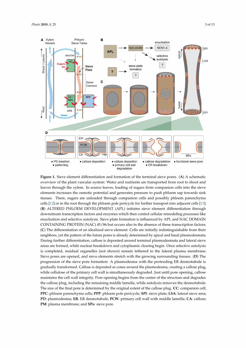
Figure 1. Sieve element differentiation and formation of the terminal sieve pores. (A) A schematicoverview of the plant vascular system: Water and nutrients are transported from root to shoot andleaves through the xylem. In source leaves, loading of sugars from companion cells into the sieveelements increases the osmotic potential and generates pressure to push phloem sap towards sinktissues. There, sugars are unloaded through companion cells and possibly phloem parenchymacells [12] or in the root through the phloem pole pericycle for further transport into adjacent cells [13].(B) ALTERED PHLOEM DEVELOPMENT (APL) initiates sieve element differentiation throughdownstream transcription factors and enzymes which then control cellular remodeling processes likeenucleation and selective autolysis. Sieve plate formation is influenced by APL and NAC DOMAINCONTAINING PROTEIN (NAC) 45/86 but occurs also in the absence of these transcription factors.(C) The differentiation of an idealized sieve element: Cells are initially indistinguishable from theirneighbors, yet the pattern of the future pores is already determined by apical and basal plasmodesmata.During further differentiation, callose is deposited around terminal plasmodesmata and lateral sieveareas are formed, while nuclear breakdown and cytoplasmic clearing begin. Once selective autolysisis completed, residual organelles (not shown) remain tethered to the lateral plasma membrane.Sieve pores are opened, and sieve elements stretch with the growing surrounding tissues. (D) Theprogression of the sieve pore formation: A plasmodesma with the protruding ER desmotubule isgradually transformed. Callose is deposited as cones around the plasmodesma, creating a callose plug,while cellulose of the primary cell wall is simultaneously degraded. Just until pore opening, callosemaintains the cell wall integrity. Pore opening begins from the center of the structure and degradesthe callose plug, including the remaining middle lamella, while autolysis removes the desmotubule.The size of the final pore is determined by the original extent of the callose plug. CC: companion cell;PPC: phloem parenchyma cells; PPP: phloem pole pericycle; SPl: sieve plate; LSA: lateral sieve area;PD: plasmodesma; ER: ER desmotubule; PCW: primary cell wall with middle lamella; CA: callose;PM: plasma membrane; and SPo: sieve pore.
Plants 2019, 8, 25 4 of 13
2. Sieve Pores in the Sieve Plates of Angiosperms
The sieve pores in the apical and basal cell walls or end walls of the sieve elements were firstdescribed in various tree species some 180 years ago by the German forestry biologist TheodorHartig [14]. Collectively, the apical and basal cell walls with their large pores are referred to as “sieveplates”. Most investigations on these sieve plates were descriptive in nature and studied their variableanatomy, their physiological importance with respect to vascular hydraulics, and their development.Perforations of these end walls of conducting cells are found in all vascular plants. Although notcovered in this review, it is worth noting that sieve elements with terminal sieve pores have alsoindependently evolved in the brown algae of the order Laminariales, which do not share a commonmulticellular ancestor with today’s land plants [15,16]. Sieve pores can be rapidly closed through thedeposition of callose. In angiosperms, sieve pore occlusion has also been attributed to agglomerationsof structural P-proteins in the sieve elements. This, however, has lately been questioned. P-proteins donot block translocation through sieve pores and their characteristics were recently reviewed [17].
In opposition to the pores in the apical and basal cell walls, perforations of the side walls aregenerally referred to as “lateral sieve areas” yet do not occur in all sieve elements [18]. In angiosperms,pores in the lateral sieve areas are generally of similar size across species and smaller than the pores inthe sieve plates [19], with the exception of Austrobaileyaceae, a monotypic family found solely in thetropics of Queensland, Australia [20]. Gymnosperms, on the other hand, have so-called sieve cells,in which lateral and end walls do not substantially differ structurally. Here, all pores are of roughlythe same diameter and generally considerably smaller than in angiosperms [21]. Sieve element endwalls in angiosperms may be organized as simple sieve plates, where sieve pores are equally dispersedand are generally bigger, or as compound sieve plates, in which the pores are generally smaller andaggregated into several areas [19]. Additionally, sieve plates may be rather perpendicular to the lateralcell walls or inclined with the latter occupying a larger area and having generally smaller pores [19].
3. Sieve Plates Vary in Size, Pore Diameter, and Overall Architecture
Diameter and number of sieve pores in the sieve plate vary greatly between different species.Particularly large pores with diameters above 10 µmCucurbita sp., 10.3 µm), the genus Tetracera(13.1 µm), and the Tree of Heaven (Ailanthus altissima, 14.3 µm) while many other species have pores ofaround or less than 1 µm [19]. Pore size, sieve plate diameter, and length of the individual sieve elementdetermine the conductivity of the sieve tube. As an approximation, the Hagen–Poiseuille equationfor laminar flow can be used to estimate the hydraulic resistance R through a cylinder, which isinversely proportional to the radius to the power of four (R ∝ 1/r4). Adaptations and refinements ofthe Hagen–Poiseuille equation to the case of the sieve tube demonstrate that the anatomy of the sieveplates is a key contributor to the overall hydraulic resistance [22], contributing around or more than50% of the overall sieve tube hydraulic resistance [23]. For example, sieve plates and individual sievepores in Cucurbita are approximately ten times larger than the ones found in the model plant Arabidopsisthaliana, which results in an approximately 100 times higher hydraulic sieve tube conductivity in theformer. Yet, the effect of the sieve plate anatomy on overall phloem flow is more complex, and a fewrelatively large pores in the center of the Cucurbita sieve plates contribute most to overall sieve plateconductivity, while the smaller lateral pores are hydraulically much less relevant [24].
These hydraulic implications were already noted by Esau and Cheadle in the late 1950s,who proposed that because of the impact of the sieve plate on overall hydraulic resistance, plants likelyevolved towards simpler sieve plates, which were found to generally have bigger pores [19]. However,two recent studies on the morphological features of sieve elements and sieve plates failed to identifysuch an evolutionary trend towards a specific sieve plate type. No correlation between the hydraulicconductivity and the organization of sieve pores in simple or compound sieve plates could be found.Instead, both types of sieve plates were found across the phylogenetic tree, and overall conductivityof the sieve tube correlated with the height of a tree and not with specific pore arrangements [25,26].In fact, sieve plate morphologies appear to be much more variable than originally assumed. In terms
Plants 2019, 8, 25 5 of 13
of impacting hydraulic conductivity across the sieve plate, pore size is far more important than porenumber. Indeed, sieve pore and tube diameters increase towards the base of a plant to increaseconductivity [27]. This developmental plasticity in sieve pore diameters within individuals hasalso been observed in several tree species, allowing them to adjust hydraulic conductivity as theygrow to avoid generating impossibly high phloem turgor pressures [26]. It is noteworthy that ingymnosperms, where sieve element and pore diameters do not increase with tree height, longer sievecells and higher numbers of sieve areas are reported [25]. Such adaptations should also decreaseoverall hydraulic resistance, however to a lesser degree than the sieve pore widening observed in tallangiosperms. This is in accordance with generally lower transport speeds in gymnosperms comparedto angiosperms [28].
4. Sieve Pore Formation during Phloem Development
Sieve pores differentiate from plasmodesmata and, once fully formed, continue to share severalfeatures with them. Plasmodesmata are small membrane-lined symplastic connections betweenneighboring cells. They contain a central endoplasmic reticulum (ER) connection called the desmotubule.The space between the desmotubule and plasma membrane is referred to as the cytoplasmic sleeve andallows intercellular cytosolic exchange [29].
Diameters of plasmodesmata range from 20–50 nm [30], while mature sieve pores are often in themicrometer range [19,24]. For example, assuming a final sieve pore in Cucurbita of approximately 5 µmnecessitates a 100–250 times increase in diameter compared to the original plasmodesma structure,even the rather small sieve pores in Arabidopsis are still between 10–25 times as big as the precedingplasmodesmata. Enlarging an existing plasmodesma to a fully formed sieve pore, therefore, requiresprecisely timed and spatially controlled cell wall remodeling (Figure 1D).
The initial patterning of the end wall through the insertion of plasmodesmata can be consideredthe first step in sieve plate differentiation. Plasmodesmata can be rapidly closed through callose,a linear beta-1,3-glucan which is deposited around their necks, thereby constricting their apertureand effectively closing it [30]. The beginning of the transformation of a plasmodesma into a sievepore appears similar. Callose is deposited around the edges to form a little cone, which protrudesslightly into the sieve element protoplast [31]. Continued callose deposition replaces cellulose inthe primary cell wall, which is being progressively degraded around the plasmodesma. The middlelamella resists hydrolysis and disappears just before pore opening [31]. Eventually, the entirety of thefuture pore is filled with a callose plug, which maintains the cell wall integrity until pore opening [31].Callose degradation commences at the middle lamella in the center of the pore and rapidly hydrolyzesthe callose plug until only residual callose remains around the open pore. Despite the considerablevariation in sieve plate architecture and pore sizes, this progression appears conserved at least withinthe dicotyledons [31,32].
4.1. Lessons from Plasmodesmata
Because of the close relation between plasmodesmata and sieve pores, it is helpful to understandplasmodesmata as such in order to understand their modification into sieve pores. Primaryplasmodesmata are inserted into the cell wall during cytokinesis when individual ER strands aretrapped between secretory vesicles, which fuse at the phragmoplast to give rise to the cell plate [33].Secondary plasmodesmata instead are inserted post-cytokinetically. Since the plasmodesmata densityhardly changes during cell elongation, secondary plasmodesmata are thought to be insertedpredominantly into extending cell walls [34]. Simple plasmodesmata have only one single cytoplasmicconnection but can mature into complex plasmodesmata, which are branched or funnel-shaped [29].Recently, the additional distinction between type I plasmodesmata, which have a very narrowcytoplasmic sleeve, and type II plasmodesmata with nanoscale spokes between the ER desmotubuleand the plasma membrane was introduced [35]. Plasmodesmata have attracted increasing attention
Plants 2019, 8, 25 6 of 13
for their various roles in plant development [36], plant–pathogen interaction [37], and phloemphysiology [38,39], and their characteristic ultrastructure has recently been reviewed [40].
The plasma membrane of plasmodesmata is a distinct micro-domain enriched in sterols andsphingolipids and is characterized by a specific set of proteins [41]. Sterols and sphingolipids can makeup to 30% and 10%, respectively, of all lipids in plant cells and are, therefore, often considered to bestructural components of lipidic membranes rather than, for example, phosphatidylinositol phosphates(PIP) which occur in very low absolute concentrations but may act as specific membrane landmarksfor intracellular trafficking [42]. Both, sterols and sphingolipids have been previously implicated intrafficking to the plasma membrane. For example, the cyclopropylsterol isomerase 1 (cpi1) mutant lacksmost sterols typically occurring in wild-type plants. This results in PIN-FORMED 2 (PIN2) polarityloss due to endocytic defects during cytokinesis and suggests a distinct role for sterols in defining cellpolarity just after the establishment of the cell plate [43]. Intracellular dynamics of auxin transporters,in particularly the PIN proteins, are well studied and their fluorescent protein fusions are often usedas markers for specific plasma membrane domains to elucidate subcellular trafficking towards thesetarget membranes [44].
Biosynthesis of the sphingolipid ceramide can be inhibited using the synthesis inhibitor fumonisinB1 (FB1). This inhibition causes changes in overall lipid composition very similar to hypomorphic,non-lethal mutations in the ceramide biosynthesis genes LONGEVITIY ASSURANCE ONE HOMOLOG1,2,3 (LOH1,2,3). FB1 causes defects, which appear similar to treatment with the recycling inhibitorbrefeldin A (BFA) and prevents proper localization of the auxin carrier AUXIN RESISTANT 1(AUX1) and PIN1 due to interference with the biogenesis of the trans-Golgi network (TGN)/earlyendosomes [45].
4.2. The cher1 Mutant Connects Plasmodesmata Maturation and Sieve Pore Morphogenesis
A clear mechanistic connection between local enrichment in specific lipids and the maturationof plasmodesmata, and by extension sieve pores, is still missing. Yet, there is increasing evidencefor membrane nano-domains enriched in specific lipids (also referred to as “lipid rafts”) aroundplasmodesmata, which are crucial to their formation and function [46]. Significant insights on linkingplasmodesmata and sieve pore maturation to lipid homeostasis originate from the discovery of thecholine transporter-like 1 (cher1) mutants through various forward and reverse genetic approaches.CHER1 is also known as CTL1, yet since CTL1 originally described the unrelated enzyme CHITINASELIKE 1 [47], the name CHER1 should be preferred.
The mutant cher1 was initially identified in a screen for plants affected in vascular development.Sieve pores in cher1 are less numerous and many remained occluded by the ER, which was notproperly removed from the sieve pore aperture. This resulted in impaired translocation and unloadingof cytosolic GFP, expressed from the promoter of SUCROSE TRANSPORTER 2 (SUC2::GFP), which isroutinely used as a marker for symplastic phloem transport. Additionally, cher1 displayed changes inthe amounts of choline and phosphocholine, which are precursors for the major plasma membrane lipidphosphatidylcholine. CHER1 localizes to the TGN/early endosome, the cell plate in dividing cells,and the developing sieve plate in sieve elements. Intriguingly, CHER1 only transiently localizedto newly forming cell walls during cytokinesis but remained at the sieve plate—a localizationwhich, however, was dependent on continued recycling as demonstrated by a treatment with BFA,which caused CHER1-YFP aggregation into larger intracellular structures, known as BFA bodies [48].
An independent approach made use of a ubiquitously expressed viral movement protein fusedto a GFP to specifically label complex plasmodesmata (35S::MP17-GFP). cher1 was identified froma mutagenized population screened for the lack of a GFP signal and hence reduction or absence ofcomplex plasmodesmata. Although cher1 had detectable changes in gross lipid composition, they didnot appear dramatic. Still, cher1 mutants showed a considerable reduction in de novo formed secondaryplasmodesmata [49]. Additionally, proteomic analysis showed that cher1 had a significantly reducedabundance of plasmodesmata proteins [50]. This could suggest that the mild overall lipid defects in
Plants 2019, 8, 25 7 of 13
cher1 may in fact simply mask the subcellular complexities during the plasmodesmata formation andrecruitment of plasmodesmata-specific proteins.
The cher1 mutant also showed defects in the post-Golgi secretion of vesicles to the plasmamembrane, a defect which had been attributed to changes in the lipid homeostasis. This resulted in areduced abundance of several auxin transporters, consequently explaining various developmentaldefects of the cher1 mutant as auxin-defects [51]. It will, therefore, be crucial to untangle the pleiotropicauxin-related defects of the mutant from the role of CHER1 during plasmodesmata and early sievepore formation.
4.3. Origin of the Pore-Forming Plasmodesmata
It may appear a bit blurry as to whether primary or secondary plasmodesmata give rise to thesieve pores. Early electron-microscopy studies could not clarify this [32], more recent studies do notappear to have addressed this early aspect of sieve pore morphogenesis, and reviews of the fieldrefer to the process generally as a restructuring of plasmodesmata without discussing their possibleorigin [2,39]. Since the sieve plate does not extend during differentiation, it has been assumed that theforming sieve plate is dotted by plasmodesmata from its very inception during cytokinesis and, hence,that sieve pores originate from primary, simple plasmodesmata [31]. This appears in contrast to thesieve pore defects in cher1, a mutant which has been reported to be specifically affected in secondaryplasmodesmata [49]. It is imaginable, however, that the pore-forming plasmodesmata in cher1 as suchare unaffected primary plasmodesmata but that their maturation into sieve pores relies on traffickingevents to the sieve plate, which are necessary for these pre-existing plasmodesmata to progress intosieve pores.
4.4. Callose Deposition Replaces Primary Cell Wall
Differentiation from a plasmodesma into a sieve pore progresses through the transient state ofcellulose replacement with callose. This step maintains the cell wall integrity while simultaneouslydelimiting the size of the final pore [31]. It requires the correct localization and activity of cell walldegrading enzymes such as cellulases and pectinases, which the precise identity of, however, has notbeen determined [29]. Cell wall modifying enzymes belong to notoriously large protein families.For example, the Arabidopsis genome encodes for the staggering number of 379 glycosyl hydrolases,of which more than 100 catalyze the breakdown of pectins [52]. There is presently no functional datafor by far most of these enzymes and very little indications which of these may be involved in thespecific task of sieve pore formation.
Callose deposition, in general but also in sieve plates, is presently better understood than cellulosedegradation. The Arabidopsis genome encodes for twelve callose synthases, for which two independentnomenclatures exist. They are either referred to as CALLOSE SYNTHASE (CalS) 1 to 12 [53], which willbe used here, or GLUCAN SYNTHASE-LIKE (GSL) 1 to 12 [54]. Besides around plasmodesmata andthe sieve plates, callose is transiently deposited in the cell plate, where it is replaced once cytokinesis iscompleted [53,55]; it is deposited in growing pollen tubes [56], and around the base of trichomes [57].Callose is also rapidly deposited upon physical damage of plant cells or pathogen infection [58].Comprehensive reviews on the different roles of callose synthesis and depositions in plants have beenpublished previously and remain relevant [54,59,60]
CalS7 (i.e., GSL7) was identified as the callose synthase to be predominantly expressed in thesieve elements, and the corresponding mutant cals7 lacks all callose deposition in sieve tubes [61].Although cals7 mutants still make sieve pores, the final pores are generally smaller and lack thecharacteristic surrounding callose collar [62]. Mutants of cals7 are a bit smaller than wild-type plants,yet their shorter root phenotypes can be largely complemented by the addition of sucrose to the growthmedium [61]. This suggest, that even though callose is largely degraded during the course of sievepore formation, its deposition is important for sieve tube function and carbon transport to the growingroot tip. Accordingly, cals7 mutants accumulate starch in source leaves but are carbon deprived in sink

Plants 2019, 8, 25 8 of 13
tissues [62]. Yet, it has been debated if sieve pores, once they are fully formed, still contain any callose atall and if any observed callose is in fact merely an artefact of the preparations and the rapidly inducedcallose deposition due to the inflicted trauma. When whole plants are rapidly fixed, no callose couldbe observed in mature sieve elements of the duckweed Lemna minor. This has, however, been contestedby Esau and Thorsch, who argued that “the phenomenon is so consistent and orderly in the differentiatingsieve element that it helps to interpret the sequence of events ( . . . )” [31]. Although callose deposition insieve elements of Arabidopsis occurs very fast, sieve plate callose is also observed in rapidly fixedtissues, suggesting that at least in Arabidopsis, the sieve plate contains callose also in a non-challengedstate [61].
Trauma-induced callose deposition in the sieve elements of Arabidopsis occurs also in lateral cellwalls. Intriguingly, this ubiquitous wound-induced callose is equally absent in the cals7 mutant [61].This may suggest that rather than being exclusive to the development of the sieve pores, CalS7mediates callose deposition in sieve elements in general. Yet, depending on developmental andenvironmental cues and necessities, which vary over time, CalS7 may preferentially be active indifferent plasma membrane domains of the sieve element. When the cals7 mutant is complementedwith the dominant active icals3m allele of CalS3 (i.e., GSL12) expressed from the phloem-specific APLpromoter (APL::icals3m), callose deposition not only is overall excessive in the sieve tube but alsoappears preferentially at the sieve plates of differentiating root sieve elements [63]. Unfortunately,callose synthases have proved difficult to use as fluorescent protein fusions. While the expressiondomains of many CalS genes have been identified [64], their precise subcellular localizations anddynamics during callose depositions are still unknown, with the exception of CalS3 [63] and CalS12(i.e., GSL5 or PMR4) [65].
4.5. Pore Opening
Rapid degradation of the callose plugs opens the sieve pores and makes the newly differentiatedsieve element a fully functional member of the sieve tube. Pore opening has been reported to occuraround the time of enucleation [32] or right after enucleation is completed [13]. This is indirectlysupported by transcriptional analyses, which showed that some specific cell wall remodeling enzymesare NAC45/86-dependent while others are not. For example, sieve element callose deposition throughCalS7 appears APL-dependent but NAC45/86-independent while some beta-1,3-glycosyl-hydrolyasesthat degrade callose are direct targets of NAC45/86 [10]. This may imply that callose degradationduring sieve pore opening is at least partially under the same control mechanisms as enucleation butthat the preparatory modifications of the cell wall are distinct events.
It may appear odd why plants first degrade parts of their primary cell wall to replace itwith callose just to degrade the callose plugs shortly after. The chemical and mechano-physicalproperties of cellulose and callose are vastly different. Cellulose is organized in long fibrils and oftenoccurs in a crystalline state, whereas callose is a linear, relatively shorter polymer, which is ratheramorphous [66]. Callose is also softer than cellulose and its addition to a cellulosic matrix even asthe minor polysaccharide reduces the stiffness at least in artificial cellulose–callose mixtures [67].Therefore, callose is considered to be easier to hydrolyze than cellulose, thus allowing the plant arapid collapse of the callose plugs to make a functional pore. Since callose deposition also occurs veryrapidly, this process is reversible and can adapt quickly to external cues [61,63]. The relatively milddevelopmental phenotype of the cals7 mutant suggests that this entire mechanism of callose depositionfor subsequent rapid degradation is not essential for phloem function and plant survival. Yet, the rapidpore opening and closure it allows may have been exceptionally advantageous in eco-physiological andevolutionary terms, analogous to, for example, the Casparian strip of the endodermis: an evolutionarilyhighly conserved feature, which the integrity of is not absolutely crucial for development but highlyrelevant in a physiological context [68,69]. As previously mentioned, gymnosperms have considerablysmaller pores than angiosperms [25]. Intriguingly, just like the Arabidopsis cals7 mutant, not onlyconifers and cycads but also mosses and some ferns lack callose in their sieve cell end walls and only
Plants 2019, 8, 25 9 of 13
produce callose upon wounding [70–72]. It is, therefore, tempting to speculate that the successivedeposition and degradation of callose, which occurs in angiosperms, may have been a key evolutionaryinvention to allow for larger sieve pores.
5. Conclusions and Perspectives
Conceptually, the formation of a sieve pore requires polar cargo delivery to or retention at theapical and basal pore-forming plasmodesmata. Secreted enzymes to degrade the cell wall, such asglucanases and pectinases, have to be properly secreted but kept in place to limit their hydrolyticactivity to the forming sieve pore. This could be achieved either through interactions with integralplasma membrane proteins, which may act as scaffolds, or through direct attachment to the plasmamembrane. Many secreted glucanases and pectinases are, in fact, anchored in the outer leaflet of theplasma membrane through glycosylphosphatidylinositol (GPI) anchors [73] and may be localizedthrough a specific lipid microenvironment. Callose synthases would equally need to be localized tothe forming pore, and indeed, CalS3 localizes preferentially to plasmodesmata [63]. CalS proteins arestructurally similar, and other callose synthases may exhibit similar subcellular distributions.
Since so little is known about the molecular and genetic mechanisms of sieve pore formation,it is worth looking into other tissue-specific cell wall modifications for inspiration. Pit formationin the differentiating metaxylem depends on two mutually exclusive mechanisms of microtubuledepolymerization and stabilization through membrane-to-microtubule tether proteins. The localactivation of Rho of plant GTPase 11 (ROP11) recruits MICROTUBULE DEPLETION DOMAIN1 (MDD1) and Kinesin-13A to locally depolymerize microtubules [74,75]. On the other hand,IQ67 DOMAIN PROTEIN 13 (IQD13) stabilizes microtubules around the active ROP11 domain,thus restricting cortical microtubule depolymerization [76]. Their combined action excludesmicrotubule-guided cellulose synthases during secondary cell wall formation specifically from aplasma membrane region which then gives rise to a pit. As another example, the formation of thelignified Casparian strip in the endodermis requires the local production of reactive oxygen speciesand the positioning of PEROXIDASE 64 (PER64) into a ring around the cell. The localization of thebiosynthetic machinery is determined by the endodermis-specific CASPARIAN STRIP MEMBRANEDOMAIN PROTEIN 1 (CASP1) in the plasma membrane [77,78], which is secreted in a localized fashiontowards a PI(4,5)P2 landmark in the plasma membrane [79]. This mechanism allows the translationof an unstable lipid motif in the cytosolic leaflet of the plasma membrane into a localized polymerdeposition in the cell wall. In the protophloem, although subcellular spatial distributions are unknown,changes in overall PI(4,5)P2 levels have been reported to impede sieve element differentiation [80,81].
It is entirely speculative whether similar mechanisms may govern the formation of the sievepores, but advances in tissue-specific transcriptomics will enable targeted mining for sieve elementdifferentiation factors, and improvements in live cell imaging in resolution and sensitivity already arefacilitating advanced in vivo investigations in deep-lying plant tissues. After the decade-long hiatus instudying sieve plate differentiation and sieve pore formation, it is time to look at them again.
Author Contributions: Conceptualization, L.K. and Y.H.; writing—original draft preparation, L.K.; writing—reviewand editing, L.K. and Y.H.
Funding: L.K. is supported by a postdoctoral fellowship of the Swiss National Science Foundation (FNS;P2LAP3_170862) and is a non-stipendiary fellow of the European Molecular Biology Organization (EMBO;ALTF 1167-2017). Y.H. has been supported by the Finnish Centre of Excellence in Molecular Biology of PrimaryProducers (Academy of Finland CoE program 2014-2019) decision #271832, and his laboratory was funded bythe Gatsby Foundation (GAT3395/PR3), the National Science Foundation Biotechnology and Biological SciencesResearch Council grant (BB/N013158/1), University of Helsinki (award 799992091), and the European ResearchCouncil Advanced Investigator Grant SYMDEV (No. 323052).
Acknowledgments: The authors would like to acknowledge Sofia Otero and other members of the Helariutta labas well as Marie Barberon for input and critical reading of the manuscript.
Conflicts of Interest: The authors declare no conflict of interest.
Plants 2019, 8, 25 10 of 13
References
1. Evert, R.F. Esau’s Plant Anatomy: Meristems, Cells, and Tissues of the Plant Body: Their Structure, Function, andDevelopment – Third Edition; Wiley-Interscience: Hoboken, NJ, USA, 2006; ISBN 0-471-73843-3.
2. Lucas, W.J.; Groover, A.; Lichtenberger, R.; Furuta, K.; Yadav, S.-R.; Helariutta, Y.; He, X.-Q.; Fukuda, H.;Kang, J.; Brady, S.M.; et al. The plant vascular system: Evolution, development and functions. J. Integr.Plant Biol. 2013, 55, 294–388. [CrossRef] [PubMed]
3. De Rybel, B.; Mähönen, A.P.; Helariutta, Y.; Weijers, D. Plant vascular development: From early specificationto differentiation. Nat. Rev. Mol. Cell Biol. 2016, 17, 30–40. [CrossRef] [PubMed]
4. Heo, J.-O.; Blob, B.; Helariutta, Y. Differentiation of conductive cells: A matter of life and death. Curr. Opin.Plant Biol. 2016, 35, 23–29. [CrossRef] [PubMed]
5. Blob, B.; Heo, J.-O.; Helariutta, Y. Phloem differentiation: An integrative model for cell specification. J. Plant Res.2018, 131, 31–36. [CrossRef] [PubMed]
6. Marhava, P.; Bassukas, A.E.L.; Zourelidou, M.; Kolb, M.; Moret, B.; Fastner, A.; Schulze, W.X.; Cattaneo, P.;Hammes, U.Z.; Schwechheimer, C.; et al. A molecular rheostat adjusts auxin flux to promote rootprotophloem differentiation. Nature 2018, 558, 297–300. [CrossRef] [PubMed]
7. Anne, P.; Hardtke, C.S. Phloem function and development—Biophysics meets genetics. Curr. Opin. Plant Biol.2018, 43, 22–28. [CrossRef] [PubMed]
8. Hellmann, E.; Ko, D.; Ruonala, R.; Helariutta, Y. Plant Vascular Tissues-Connecting Tissue Comes in AllShapes. Plants 2018, 7, 109. [CrossRef] [PubMed]
9. Bonke, M.; Thitamadee, S.; Mähönen, A.P.; Hauser, M.-T.; Helariutta, Y. APL regulates vascular tissue identityin Arabidopsis. Nature 2003, 426, 181–186. [CrossRef] [PubMed]
10. Furuta, K.M.; Yadav, S.R.; Lehesranta, S.; Belevich, I.; Miyashima, S.; Heo, J.-O.; Vatén, A.; Lindgren, O.;De Rybel, B.; Van Isterdael, G.; et al. Arabidopsis NAC45/86 direct sieve element morphogenesis culminatingin enucleation. Science 2014, 345, 933–937. [CrossRef]
11. Truernit, E.; Bauby, H.; Dubreucq, B.; Grandjean, O.; Runions, J.; Barthélémy, J.; Palauqui, J.-C.High-resolution whole-mount imaging of three-dimensional tissue organization and gene expression enablesthe study of Phloem development and structure in Arabidopsis. Plant Cell 2008, 20, 1494–1503. [CrossRef][PubMed]
12. Zhang, L.-Y.; Peng, Y.-B.; Pelleschi-Travier, S.; Fan, Y.; Lu, Y.-F.; Lu, Y.-M.; Gao, X.-P.; Shen, Y.-Y.; Delrot, S.;Zhang, D.-P. Evidence for apoplasmic phloem unloading in developing apple fruit. Plant Physiol. 2004, 135,574–586. [CrossRef] [PubMed]
13. Ross-Elliott, T.J.; Jensen, K.H.; Haaning, K.S.; Wager, B.M.; Knoblauch, J.; Howell, A.H.; Mullendore, D.L.;Monteith, A.G.; Paultre, D.; Yan, D.; et al. Phloem unloading in Arabidopsis roots is convective and regulatedby the phloem-pole pericycle. eLife 2017, 6, e24125. [CrossRef] [PubMed]
14. Hartig, T. Vergleichende Untersuchungen über die Organisation des Stammes der einheimischen Waldbäume.In Jahresberichte über die Fortschritte der Forstwissenschaft und forstlichen Naturkunde im Jahre 1836 und 1837;Berlin, Germany, 1837; pp. 125–168.
15. Raven, J.A. Long-distance transport in non-vascular plants. Plant Cell Environ. 2003, 26, 73–85. [CrossRef]16. Knoblauch, J.; Peters, W.S.; Knoblauch, M. The gelatinous extracellular matrix facilitates transport studies
in kelp: Visualization of pressure-induced flow reversal across sieve plates. Ann. Bot. 2016, 117, 599–606.[CrossRef] [PubMed]
17. Knoblauch, M.; Froelich, D.R.; Pickard, W.F.; Peters, W.S. SEORious business: Structural proteins in sievetubes and their involvement in sieve element occlusion. J. Exp. Bot. 2014, 65, 1879–1893. [CrossRef]
18. Behnke, H.D.; Sjolund, R.D. Sieve Elements—Comparative Structure, Induction and Development, 1st ed.;Springer-Verlag: Berlin/Heidelberg, Germany, 1990; ISBN 3-540-50783-3.
19. Esau, K.; Cheadle, V.I. Size of Pores and Their Contents in Sieve Elements of Dicotyledons. Proc. Natl. Acad.Sci. USA 1959, 45, 156–162. [CrossRef]
20. Behnke, H.-D. Sieve element characters and the systematic position of Austrobaileya, Austrobaileyaceae—Withcomments to the distinction and definition of sieve cells and sieve-tube members. Plant Syst. Evol. 1986, 152,101–121. [CrossRef]
21. Evert, R.F.; Alfieri, F.J. Ontogeny and Structure of Coniferous Sieve Cells. Am. J. Bot. 1965, 52, 1058–1066.[CrossRef]

Plants 2019, 8, 25 11 of 13
22. Thompson, M.V.; Holbrook, N.M. Application of a single-solute non-steady-state phloem model to the studyof long-distance assimilate transport. J. Theor. Biol. 2003, 220, 419–455. [CrossRef]
23. Jensen, K.H.; Mullendore, D.L.; Holbrook, N.M.; Bohr, T.; Knoblauch, M.; Bruus, H. Modeling thehydrodynamics of Phloem sieve plates. Front. Plant Sci. 2012, 3, 151. [CrossRef]
24. Mullendore, D.L.; Windt, C.W.; Van As, H.; Knoblauch, M. Sieve tube geometry in relation to phloem flow.Plant Cell 2010, 22, 579–593. [CrossRef]
25. Liesche, J.; Pace, M.R.; Xu, Q.; Li, Y.; Chen, S. Height-related scaling of phloem anatomy and the evolution ofsieve element end wall types in woody plants. New Phytol. 2017, 214, 245–256. [CrossRef] [PubMed]
26. Savage, J.A.; Beecher, S.D.; Clerx, L.; Gersony, J.T.; Knoblauch, J.; Losada, J.M.; Jensen, K.H.; Knoblauch, M.;Holbrook, N.M. Maintenance of carbohydrate transport in tall trees. Nat. Plants 2017, 3, 965–972. [CrossRef][PubMed]
27. Knoblauch, M.; Knoblauch, J.; Mullendore, D.L.; Savage, J.A.; Babst, B.A.; Beecher, S.D.; Dodgen, A.C.;Jensen, K.H.; Holbrook, N.M. Testing the Münch hypothesis of long distance phloem transport in plants.eLife 2016, 5, 826. [CrossRef] [PubMed]
28. Liesche, J.; Windt, C.; Bohr, T.; Schulz, A.; Jensen, K.H. Slower phloem transport in gymnosperm trees can beattributed to higher sieve element resistance. Tree Physiol. 2015, 35, 376–386. [CrossRef] [PubMed]
29. Lucas, W.J.; Ding, B.; van der Schoot, C. Tansley Review No. 58. Plasmodesmata and the SupracellularNature of Plants. New Phytol. 1993, 125, 435–476. [CrossRef]
30. Ehlers, K.; Kollmann, R. Primary and secondary plasmodesmata: Structure, origin, and functioning.Protoplasma 2001, 216, 1. [CrossRef]
31. Esau, K.; Thorsch, J. Sieve Plate Pores and Plasmodesmata, the Communication Channels of the Symplast:Ultrastructural Aspects and Developmental Relations. Am. J. Bot. 1985, 72, 1641–1653. [CrossRef]
32. Esau, K.; Cheadle, V.I.; Risley, E.B. Development of sieve-plate pores. Bot. Gaz. 1962, 123, 233–243. [CrossRef]33. Hepler, P.K. Endoplasmic reticulum in the formation of the cell plate and plasmodesmata. Protoplasma 1982,
111, 121–133. [CrossRef]34. Seagull, R.W. Differences in the frequency and disposition of plasmodesmata resulting from root cell
elongation. Planta 1983, 159, 497–504. [CrossRef] [PubMed]35. Nicolas, W.J.; Grison, M.S.; Trépout, S.; Gaston, A.; Fouché, M.; Cordelières, F.P.; Oparka, K.; Tilsner, J.;
Brocard, L.; Bayer, E.M. Architecture and permeability of post-cytokinesis plasmodesmata lackingcytoplasmic sleeves. Nat. Plants 2017, 3, 17082. [CrossRef] [PubMed]
36. Gallagher, K.L.; Sozzani, R.; Lee, C.-M. Intercellular protein movement: Deciphering the language ofdevelopment. Annu. Rev. Cell Dev. Biol. 2014, 30, 207–233. [CrossRef] [PubMed]
37. Wang, A. Dissecting the molecular network of virus-plant interactions: The complex roles of host factors.Annu. Rev. Phytopathol. 2015, 53, 45–66. [CrossRef] [PubMed]
38. Rockwell, F.E.; Gersony, J.T.; Holbrook, N.M. Where does Münch flow begin? Sucrose transport in thepre-phloem path. Curr. Opin. Plant Biol. 2018, 43, 101–107. [CrossRef] [PubMed]
39. Lee, J.-Y.; Frank, M. Plasmodesmata in phloem: Different gateways for different cargoes. Curr. Opin. Plant Biol.2018, 43, 119–124. [CrossRef]
40. Tilsner, J.; Nicolas, W.; Rosado, A.; Bayer, E.M. Staying Tight: Plasmodesmal Membrane Contact Sites andthe Control of Cell-to-Cell Connectivity in Plants. Annu. Rev. Plant Biol. 2016, 67, 337–364. [CrossRef]
41. Grison, M.S.; Brocard, L.; Fouillen, L.; Nicolas, W.; Wewer, V.; Dörmann, P.; Nacir, H.; Benitez-Alfonso, Y.;Claverol, S.; Germain, V.; et al. Specific membrane lipid composition is important for plasmodesmatafunction in Arabidopsis. Plant Cell 2015, 27, 1228–1250. [CrossRef]
42. Mamode Cassim, A.; Gouguet, P.; Gronnier, J.; Laurent, N.; Germain, V.; Grison, M.; Boutté, Y.;Gerbeau-Pissot, P.; Simon-Plas, F.; Mongrand, S. Plant lipids: Key players of plasma membrane organizationand function. Prog. Lipid Res. 2018, 73, 1–27. [CrossRef]
43. Men, S.; Boutté, Y.; Ikeda, Y.; Li, X.; Palme, K.; Stierhof, Y.-D.; Hartmann, M.-A.; Moritz, T.; Grebe, M.Sterol-dependent endocytosis mediates post-cytokinetic acquisition of PIN2 auxin efflux carrier polarity.Nat. Cell Biol. 2008, 10, 237–244. [CrossRef]
44. Luschnig, C.; Vert, G. The dynamics of plant plasma membrane proteins: PINs and beyond. Development2014, 141, 2924–2938. [CrossRef] [PubMed]
Plants 2019, 8, 25 12 of 13
45. Markham, J.E.; Molino, D.; Gissot, L.; Bellec, Y.; Hématy, K.; Marion, J.; Belcram, K.; Palauqui, J.-C.;Satiat-Jeunemaître, B.; Faure, J.-D. Sphingolipids containing very-long-chain fatty acids define a secretorypathway for specific polar plasma membrane protein targeting in Arabidopsis. Plant Cell 2011, 23, 2362–2378.[CrossRef]
46. Iswanto, A.B.B.; Kim, J.-Y. Lipid Raft, Regulator of Plasmodesmal Callose Homeostasis. Plants 2017, 6, 15.[CrossRef] [PubMed]
47. Zhong, R.; Kays, S.J.; Schroeder, B.P.; Ye, Z.-H. Mutation of a chitinase-like gene causes ectopic depositionof lignin, aberrant cell shapes, and overproduction of ethylene. Plant Cell 2002, 14, 165–179. [CrossRef][PubMed]
48. Dettmer, J.; Ursache, R.; Campilho, A.; Miyashima, S.; Belevich, I.; O’Regan, S.; Mullendore, D.L.; Yadav, S.R.;Lanz, C.; Beverina, L.; et al. CHOLINE TRANSPORTER-LIKE1 is required for sieve plate development tomediate long-distance cell-to-cell communication. Nat. Commun. 2014, 5, 4276. [CrossRef] [PubMed]
49. Kraner, M.E.; Link, K.; Melzer, M.; Ekici, A.B.; Uebe, S.; Tarazona, P.; Feussner, I.; Hofmann, J.; Sonnewald, U.Choline transporter-like1 (CHER1) is crucial for plasmodesmata maturation in Arabidopsis thaliana. Plant J.2017, 89, 394–406. [CrossRef] [PubMed]
50. Kraner, M.E.; Müller, C.; Sonnewald, U. Comparative proteomic profiling of the choline transporter-like1(CHER1) mutant provides insights into plasmodesmata composition of fully developed Arabidopsis thalianaleaves. Plant J. 2017, 92, 696–709. [CrossRef]
51. Wang, Y.; Yang, L.; Tang, Y.; Tang, R.; Jing, Y.; Zhang, C.; Zhang, B.; Li, X.; Cui, Y.; Zhang, C.; et al. Arabidopsischoline transporter-like 1 (CTL1) regulates secretory trafficking of auxin transporters to control seedlinggrowth. PLoS Biol. 2017, 15, e2004310. [CrossRef] [PubMed]
52. Henrissat, B.; Coutinho, P.M.; Davies, G.J. A census of carbohydrate-active enzymes in the genome ofArabidopsis thaliana. Plant Mol. Biol. 2001, 47, 55–72. [CrossRef]
53. Hong, Z.; Delauney, A.J.; Verma, D.P. A cell plate-specific callose synthase and its interaction withphragmoplastin. Plant Cell 2001, 13, 755–768. [CrossRef] [PubMed]
54. Chen, X.-Y.; Kim, J.-Y. Callose synthesis in higher plants. Plant Signal. Behav. 2009, 4, 489–492. [CrossRef]55. Chen, X.-Y.; Liu, L.; Lee, E.; Han, X.; Rim, Y.; Chu, H.; Kim, S.-W.; Sack, F.; Kim, J.-Y. The Arabidopsis
callose synthase gene GSL8 is required for cytokinesis and cell patterning. Plant Physiol. 2009, 150, 105–113.[CrossRef] [PubMed]
56. Enns, L.C.; Kanaoka, M.M.; Torii, K.U.; Comai, L.; Okada, K.; Cleland, R.E. Two callose synthases, GSL1 andGSL5, play an essential and redundant role in plant and pollen development and in fertility. Plant Mol. Biol.2005, 58, 333–349. [CrossRef] [PubMed]
57. Kulich, I.; Vojtíková, Z.; Glanc, M.; Ortmannová, J.; Rasmann, S.; Zárský, V. Cell wall maturationof Arabidopsis trichomes is dependent on exocyst subunit EXO70H4 and involves callose deposition.Plant Physiol. 2015, 168, 120–131. [CrossRef]
58. Jacobs, A.K.; Lipka, V.; Burton, R.A.; Panstruga, R.; Strizhov, N.; Schulze-Lefert, P.; Fincher, G.B. AnArabidopsis Callose Synthase, GSL5, Is Required for Wound and Papillary Callose Formation. Plant Cell2003, 15, 2503–2513. [CrossRef] [PubMed]
59. Ellinger, D.; Voigt, C.A. Callose biosynthesis in Arabidopsis with a focus on pathogen response: What wehave learned within the last decade. Ann. Bot. 2014, 114, 1349–1358. [CrossRef]
60. Zavaliev, R.; Ueki, S.; Epel, B.L.; Citovsky, V. Biology of callose (β-1,3-glucan) turnover at plasmodesmata.Protoplasma 2011, 248, 117–130. [CrossRef]
61. Xie, B.; Wang, X.; Zhu, M.; Zhang, Z.; Hong, Z. CalS7 encodes a callose synthase responsible for callosedeposition in the phloem. Plant J. 2011, 65, 1–14. [CrossRef]
62. Barratt, D.H.P.; Kölling, K.; Graf, A.; Pike, M.; Calder, G.; Findlay, K.; Zeeman, S.C.; Smith, A.M.Callose synthase GSL7 is necessary for normal phloem transport and inflorescence growth in Arabidopsis.Plant Physiol. 2011, 155, 328–341. [CrossRef]
63. Vatén, A.; Dettmer, J.; Wu, S.; Stierhof, Y.-D.; Miyashima, S.; Yadav, S.R.; Roberts, C.J.; Campilho, A.;Bulone, V.; Lichtenberger, R.; et al. Callose biosynthesis regulates symplastic trafficking during rootdevelopment. Dev. Cell 2011, 21, 1144–1155. [CrossRef]
64. Dong, X.; Hong, Z.; Chatterjee, J.; Kim, S.; Verma, D.P.S. Expression of callose synthase genes and itsconnection with Npr1 signaling pathway during pathogen infection. Planta 2008, 229, 87–98. [CrossRef]
Plants 2019, 8, 25 13 of 13
65. Kulich, I.; Vojtíková, Z.; Sabol, P.; Ortmannová, J.; Nedela, V.; Tihlaríková, E.; Zárský, V. Exocyst SubunitEXO70H4 Has a Specific Role in Callose Synthase Secretion and Silica Accumulation. Plant Physiol. 2018,176, 2040–2051. [CrossRef] [PubMed]
66. Piršelová, B.; Matušíková, I. Callose: The plant cell wall polysaccharide with multiple biological functions.Acta Physiol. Plant. 2013, 35, 635–644. [CrossRef]
67. Abou-Saleh, R.H.; Hernandez-Gomez, M.C.; Amsbury, S.; Paniagua, C.; Bourdon, M.; Miyashima, S.;Helariutta, Y.; Fuller, M.; Budtova, T.; Connell, S.D.; et al. Interactions between callose and cellulose revealedthrough the analysis of biopolymer mixtures. Nat. Commun. 2018, 9, 4538. [CrossRef] [PubMed]
68. Pfister, A.; Barberon, M.; Alassimone, J.; Kalmbach, L.; Lee, Y.; Vermeer, J.E.; Yamazaki, M.; Li, G.; Maurel, C.;Takano, J.; et al. A receptor-like kinase mutant with absent endodermal diffusion barrier displays selectivenutrient homeostasis defects. eLife 2014, 3, e03115. [CrossRef] [PubMed]
69. Geldner, N. The endodermis. Annu. Rev. Plant Biol. 2013, 64, 531–558. [CrossRef] [PubMed]70. Neuberger, D.S.; Evert, R.F. Structure and development of sieve areas in the hypocotyl of Pinus resinosa.
Protoplasma 1975, 84, 109–125. [CrossRef]71. Perry, J.W.; Evert, R.F. Structure and development of the sieve elements in Psilotum nudum. Am. J. Bot. 1975,
62, 1038–1052. [CrossRef]72. Scheirer, D.C. Cell Wall Chemistry and Fine Structure in Leptoids of Dendroligotrichum (Bryophyta):
The End Wall. Am. J. Bot. 1978, 65, 1027–1031. [CrossRef]73. Borner, G.H.H.; Lilley, K.S.; Stevens, T.J.; Dupree, P. Identification of glycosylphosphatidylinositol-anchored
proteins in Arabidopsis. A proteomic and genomic analysis. Plant Physiol. 2003, 132, 568–577. [CrossRef]74. Oda, Y.; Fukuda, H. Initiation of cell wall pattern by a Rho- and microtubule-driven symmetry breaking.
Science 2012, 337, 1333–1336. [CrossRef] [PubMed]75. Oda, Y.; Fukuda, H. Rho of plant GTPase signaling regulates the behavior of Arabidopsis kinesin-13A to
establish secondary cell wall patterns. Plant Cell 2013, 25, 4439–4450. [CrossRef] [PubMed]76. Sugiyama, Y.; Wakazaki, M.; Toyooka, K.; Fukuda, H.; Oda, Y. A Novel Plasma Membrane-Anchored Protein
Regulates Xylem Cell-Wall Deposition through Microtubule-Dependent Lateral Inhibition of Rho GTPaseDomains. Curr. Biol. 2017, 27, 2522–2528.e4. [CrossRef] [PubMed]
77. Roppolo, D.; De Rybel, B.; Tendon, V.D.; Pfister, A.; Alassimone, J.; Vermeer, J.E.M.; Yamazaki, M.;Stierhof, Y.-D.; Beeckman, T.; Geldner, N. A novel protein family mediates Casparian strip formationin the endodermis. Nature 2011, 473, 380–383. [CrossRef] [PubMed]
78. Lee, Y.; Rubio, M.C.; Alassimone, J.; Geldner, N. A mechanism for localized lignin deposition in theendodermis. Cell 2013, 153, 402–412. [CrossRef] [PubMed]
79. Kalmbach, L.; Hématy, K.; De Bellis, D.; Barberon, M.; Fujita, S.; Ursache, R.; Daraspe, J.; Geldner, N. Transientcell-specific EXO70A1 activity in the CASP domain and Casparian strip localization. Nat. Plants 2017, 3, 17058.[CrossRef]
80. Rodriguez-Villalon, A.; Gujas, B.; van Wijk, R.; Munnik, T.; Hardtke, C.S. Primary root protophloemdifferentiation requires balanced phosphatidylinositol-4,5-biphosphate levels and systemically affects rootbranching. Development 2015, 142, 1437–1446. [CrossRef]
81. Gujas, B.; Cruz, T.M.D.; Kastanaki, E.; Vermeer, J.E.M.; Munnik, T.; Rodriguez-Villalon, A. Perturbingphosphoinositide homeostasis oppositely affects vascular differentiation in Arabidopsis thaliana roots.Development 2017, 144, 3578–3589. [CrossRef]
© 2019 by the authors. Licensee MDPI, Basel, Switzerland. This article is an open accessarticle distributed under the terms and conditions of the Creative Commons Attribution(CC BY) license (http://creativecommons.org/licenses/by/4.0/).
Copyright © 2022 FDOKUMEN