Symplasmic transport and phloem loading in gymnosperm leaves
10
REVIEW ARTICLE Symplasmic transport and phloem loading in gymnosperm leaves Johannes Liesche & Helle Juel Martens & Alexander Schulz Received: 21 September 2010 / Accepted: 1 November 2010 / Published online: 24 November 2010 # Springer-Verlag 2010 Abstract Despite more than 130 years of research, phloem loading is far from being understood in gymnosperms. In part this is due to the special architecture of their leaves. They differ from angiosperm leaves among others by having a transfusion tissue between bundle sheath and the axial vascular elements. This article reviews the somewhat inaccessible and/or neglected literature and identifies the key points for pre-phloem transport and loading of photo- assimilates. The pre-phloem pathway of assimilates is structurally characterized by a high number of plasmodes- mata between all cell types starting in the mesophyll and continuing via bundle sheath, transfusion parenchyma, Strasburger cells up to the sieve elements. Occurrence of median cavities and branching indicates that primary plasmodesmata get secondarily modified and multiplied during expansion growth. Only functional tests can eluci- date whether this symplasmic pathway is indeed continuous for assimilates, and if phloem loading in gymnosperms is comparable with the symplasmic loading mode in many angiosperm trees. In contrast to angiosperms, the bundle sheath has properties of an endodermis and is equipped with Casparian strips or other wall modifications that form a domain border for any apoplasmic transport. It constitutes a key point of control for nutrient transport, where the opposing flow of mineral nutrients and photoassimilates has to be accommodated in each single cell, bringing to mind the principle of a revolving door. The review lists a number of experiments needed to elucidate the mode of phloem loading in gymnosperms. Keywords Bundle sheath . Endodermis . Plasmodesmata . Transfusion tissue . Strasburger cells . Xylem Introduction In angiosperms, the regulation of phloem loading is usually attributed to the companion cells of minor veins. According to textbooks, transport sugars are actively concentrated in the sieve element-companion cell complexes, leading to an osmotically generated pressure flow from source to sink organs. As active loading strategies, two modes of phloem loading are well characterized: apoplasmic loading, depend- ing on plasma membrane transporters, and symplasmic loading, depending on synthesis of sugar polymers and abundance of plasmodesmata between companion cells and bundle sheath, respectively. The role of cells of the pre- phloem pathway between mesophyll and bundle sheath is easily overlooked. This is evident for the passive symplas- mic loading strategy, where the highest sugar concentra- tions are found in the mesophyll rather than the phloem (see Davidson et al. 2010, this issue). Moreover, only few studies have considered the intersection where the pre- phloem transport of photoassimilates is interwoven with the post-xylem transport of water, so that opposite streams might have influence on each other. For gymnosperms, the mode of phloem loading has not been assessed yet, neither is it known whether both apoplast and symplasm are involved in it. Gymnosperm sieve elements are not associated with companion cells, but with Strasburger cells that are ontogenetically unrelated. How- ever, their ultrastructure and the pore–plasmodesmata Handling Editor: Manfred Heinlein J. Liesche : H. J. Martens : A. Schulz (*) Department of Plant Biology and Biotechnology, University of Copenhagen, Thorvaldsensvej 40, DK-1871, Frederiksberg C, Denmark e-mail: [email protected] Protoplasma (2011) 248:181–190 DOI 10.1007/s00709-010-0239-0
-
Upload
independent -
Category
Documents
-
view
3 -
download
0
Transcript of Symplasmic transport and phloem loading in gymnosperm leaves
REVIEW ARTICLE
Symplasmic transport and phloem loadingin gymnosperm leaves
Johannes Liesche & Helle Juel Martens &
Alexander Schulz
Received: 21 September 2010 /Accepted: 1 November 2010 /Published online: 24 November 2010# Springer-Verlag 2010
Abstract Despite more than 130 years of research, phloemloading is far from being understood in gymnosperms. Inpart this is due to the special architecture of their leaves.They differ from angiosperm leaves among others byhaving a transfusion tissue between bundle sheath and theaxial vascular elements. This article reviews the somewhatinaccessible and/or neglected literature and identifies thekey points for pre-phloem transport and loading of photo-assimilates. The pre-phloem pathway of assimilates isstructurally characterized by a high number of plasmodes-mata between all cell types starting in the mesophyll andcontinuing via bundle sheath, transfusion parenchyma,Strasburger cells up to the sieve elements. Occurrence ofmedian cavities and branching indicates that primaryplasmodesmata get secondarily modified and multipliedduring expansion growth. Only functional tests can eluci-date whether this symplasmic pathway is indeed continuousfor assimilates, and if phloem loading in gymnosperms iscomparable with the symplasmic loading mode in manyangiosperm trees. In contrast to angiosperms, the bundlesheath has properties of an endodermis and is equippedwith Casparian strips or other wall modifications that forma domain border for any apoplasmic transport. It constitutesa key point of control for nutrient transport, where theopposing flow of mineral nutrients and photoassimilateshas to be accommodated in each single cell, bringing tomind the principle of a revolving door. The review lists a
number of experiments needed to elucidate the mode ofphloem loading in gymnosperms.
Keywords Bundle sheath . Endodermis . Plasmodesmata .
Transfusion tissue . Strasburger cells . Xylem
Introduction
In angiosperms, the regulation of phloem loading is usuallyattributed to the companion cells of minor veins. Accordingto textbooks, transport sugars are actively concentrated inthe sieve element-companion cell complexes, leading to anosmotically generated pressure flow from source to sinkorgans. As active loading strategies, two modes of phloemloading are well characterized: apoplasmic loading, depend-ing on plasma membrane transporters, and symplasmicloading, depending on synthesis of sugar polymers andabundance of plasmodesmata between companion cells andbundle sheath, respectively. The role of cells of the pre-phloem pathway between mesophyll and bundle sheath iseasily overlooked. This is evident for the passive symplas-mic loading strategy, where the highest sugar concentra-tions are found in the mesophyll rather than the phloem (seeDavidson et al. 2010, this issue). Moreover, only fewstudies have considered the intersection where the pre-phloem transport of photoassimilates is interwoven with thepost-xylem transport of water, so that opposite streamsmight have influence on each other.
For gymnosperms, the mode of phloem loading has notbeen assessed yet, neither is it known whether both apoplastand symplasm are involved in it. Gymnosperm sieveelements are not associated with companion cells, but withStrasburger cells that are ontogenetically unrelated. How-ever, their ultrastructure and the pore–plasmodesmata
Handling Editor: Manfred Heinlein
J. Liesche :H. J. Martens :A. Schulz (*)Department of Plant Biology and Biotechnology, University ofCopenhagen,Thorvaldsensvej 40,DK-1871, Frederiksberg C, Denmarke-mail: [email protected]
Protoplasma (2011) 248:181–190DOI 10.1007/s00709-010-0239-0
contacts are similar to companion cells (Schulz 1990).Whether and how they might contribute to phloem loadingis unknown. The vascular strands in the leaves of allgymnosperms are separated from the mesophyll by aparticular tissue, the transfusion tissue, and by anendodermis-like bundle sheath. All these special featuresindicate that assimilate loading might involve mechanismsand pathways hitherto not found in angiosperms.
Most angiosperm plant genera could be assigned to oneof three loading types (Rennie and Turgeon 2009). In somecases even species-specific characteristics have been iden-tified (Zhang and Turgeon 2009; Gil 2010). As a generalrule for sucrose transporting angiosperms, active apoplas-mic loading, i.e., sugar uptake into symplasmically isolatedsieve element-companion cell complexes, appears to bepredominant in herbaceous plants, while passive symplas-mic loading, i.e., sugar diffusion from the mesophyll intosieve element-companion cell complexes phloem that arewell-connected to the bundle sheath by plasmodesmata, ismost common in angiosperm trees (Turgeon 2010, see alsoTurgeon and Medville 2010 this issue). All gymnospermsseem to translocate sucrose in the phloem, even though thishas not been studied systematically (Shiroya et al. 1962;Willenbrink and Kollmann 1966; Willenbrink 2002).
Understanding the mechanisms of phloem loading ingymnosperm trees will cast light on the evolution andsignificance of long distance transport mechanisms in landplants, just in the context of recent discussions of passivephloem loading in angiosperm trees (Turgeon 2010).Gymnosperm leaves have fascinated scientists alreadymany years back. Münch, who postulated the mechanismof pressure-driven phloem transport in 1930, was attentivefor the peculiarities of gymnosperm needles and the role ofthe transfusion tissue between bundle sheath and vasculartissue (Fig. 1a). Since older references are often somewhatinaccessible, we chose to compile data and reprint someselected electron micrographs from the relevant literature.Most papers available covered single aspects of thestructure and physiology of gymnosperm leaves only. Theaim of this review is to give an integrated overview of theirtransport pathways, highlight significant control key pointsand identify open questions that have to be studied in orderto understand phloem loading in gymnosperms.
Leaf morphology and anatomy
Taxonomically, gymnosperms are divided in conifers,cycads, Gnetophytes with Gnetum, Welwitschia, and Ephe-dra, and the Ginkgophytes with the only representativeGinkgo biloba. They are mainly evergreen trees, shrubs anda few lianas and well adapted to extreme habitats such asboreal forests with long winter periods, but in some cases
also to tropical or desert environments. Most studies ongymnosperms focus on Pinaceae, by far the most abundantand economically important taxon of gymnosperms. MostPinaceae have needle-shaped leaves, but gymnosperms ingeneral have developed many different leaf shapes such asscales, pinnate, and broad leaves. The xeromorphic charac-ter of all gymnosperm leaves is reflected by an epidermiswith thick cutinized outer walls that is often reinforced byhypodermal sclerenchyma fibers. Stomata are usuallyarranged in rows on the adaxial side and are sunken intothe leaf surface. In most conifers, a variable number of resinducts can be found in the needle periphery. In needles,vascular bundles are arranged in one or two central strands.However, Ginkgo leaf blades show a dichotomous venationpattern, Welwitschia parallel venation with anastomoses andGnetum a reticulate venation with at least five vein classes,similar to most dicots.
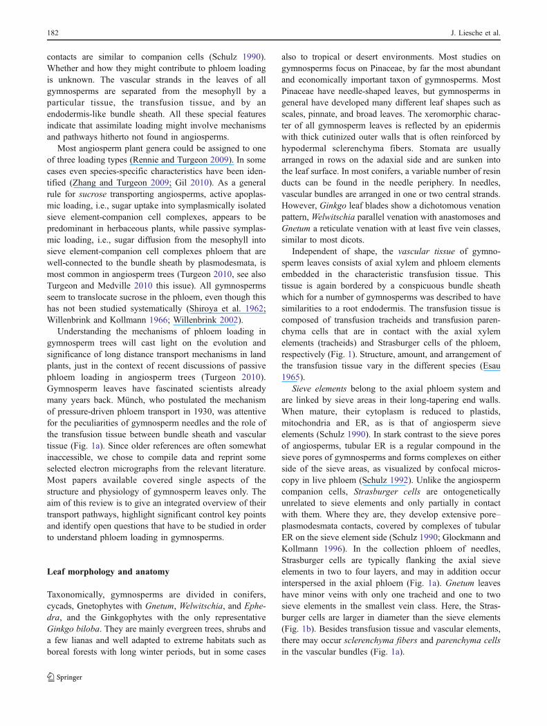
Independent of shape, the vascular tissue of gymno-sperm leaves consists of axial xylem and phloem elementsembedded in the characteristic transfusion tissue. Thistissue is again bordered by a conspicuous bundle sheathwhich for a number of gymnosperms was described to havesimilarities to a root endodermis. The transfusion tissue iscomposed of transfusion tracheids and transfusion paren-chyma cells that are in contact with the axial xylemelements (tracheids) and Strasburger cells of the phloem,respectively (Fig. 1). Structure, amount, and arrangement ofthe transfusion tissue vary in the different species (Esau1965).
Sieve elements belong to the axial phloem system andare linked by sieve areas in their long-tapering end walls.When mature, their cytoplasm is reduced to plastids,mitochondria and ER, as is that of angiosperm sieveelements (Schulz 1990). In stark contrast to the sieve poresof angiosperms, tubular ER is a regular compound in thesieve pores of gymnosperms and forms complexes on eitherside of the sieve areas, as visualized by confocal micros-copy in live phloem (Schulz 1992). Unlike the angiospermcompanion cells, Strasburger cells are ontogeneticallyunrelated to sieve elements and only partially in contactwith them. Where they are, they develop extensive pore–plasmodesmata contacts, covered by complexes of tubularER on the sieve element side (Schulz 1990; Glockmann andKollmann 1996). In the collection phloem of needles,Strasburger cells are typically flanking the axial sieveelements in two to four layers, and may in addition occurinterspersed in the axial phloem (Fig. 1a). Gnetum leaveshave minor veins with only one tracheid and one to twosieve elements in the smallest vein class. Here, the Stras-burger cells are larger in diameter than the sieve elements(Fig. 1b). Besides transfusion tissue and vascular elements,there may occur sclerenchyma fibers and parenchyma cellsin the vascular bundles (Fig. 1a).
182 J. Liesche et al.

The transfusion tracheids are dead cells and a part of thetransfusion tissue surrounding the axial vascular elements.They are characteristic for all leaves of gymnosperms(Fig. 1, de Bary 1877; Strasburger 1891). Transfusiontracheids are not only found in needle-bearing gymno-sperms with one or two central veins, but also in thesmallest veins of Gnetum, which has more than five veinclasses (Fig. 1b). Gambles and Dengler (1982) identifiedfour distinct groups of transfusion tracheids in Pinusresinosa based on their anatomy. Different degree of wallmodification and pit morphology suggest that sometransfusion tracheids are optimized for water transportwhereas others are more important for water storage andsupport. The lignified walls might enable them to maintaintheir shape under great water stress as an adaptation to aridconditions (Gadek and Quinn 1988). The role in waterconductance, first proposed by Worsdell (1897) wasdemonstrated with fluorescent tracers in the transpirationstream (Heimerdinger 1951). In addition to increasingdrought resistance, the capacity to store water in thetransfusion tissue might serve as prevention of frost damageto living cells (Roden et al. 2009).
The transfusion parenchyma forms bridges of livingcells between the outer Strasburger cells and the bundle
sheath (Fig. 1). The ratio of parenchyma cells to tracheidsin the transfusion tissue varies between 18% and 88% inpine species (Lederer 1955). The two cell types can beunderstood as a physiological unit (Canny 1993).
An endodermis-like bundle sheath was described to be aconsistent feature for needles of Pinaceae (Lederer 1955;Esau 1977; see Fig. 2a). Suberization and/or lignification ofradial and transverse walls of the bundle sheath, includingtypical Casparian strips, have indeed been detected inneedles of numerous Pinaceae species by microscopicstaining techniques, electron microscopy and spectroscopicanalysis (Fig. 2a, Carde 1978; Canny 1993; Kaku 1971;Scholz and Bauch 1973; Soda et al. 2000; Wu et al. 2001;Wu et al. 2003). Casparian strips have not been reportedoutside the Pinaceae, but representatives of other gymno-sperm families have often wall modifications in the bundlesheath cells. The walls of bundle sheath cells in Cycasrevoluta leaves are thickened and lignified next to thevascular tissue, and in leaves of G. biloba each of thenumerous vascular bundles has a lignified endodermis(Esau 1977; Lederer 1955; see Fig. 2b). Rehydrationexperiments demonstrated a high radial resistance for waterflow in all tested gymnosperms, which was attributed to thehydraulic isolation of vascular and photosynthetic tissue by
a
b
Mesophyll
Bundle sheath
Casparian strip
Transfusion tracheids
Axial xylem
Post-xylem pathway
Transfusion parenchyma
Strasburger cells
Axial phloem
Pre-phloem pathway
Sclerenchyma
Fig. 1 Illustration of the different cell types and transport pathways ingymnosperm leaves on a schematic drawing of a P. sylvestris needlecross section (a) and an electron micrograph of a Gnetum gnemon fifthclass minor vein (b). The pre-phloem pathway (brown arrows) fromthe mesophyll is symplasmic and crosses bundle sheath, transfusionsparenchyma and Strasburger cells before approaching the sieveelements. The post-xylem pathway (blue arrows) in pines starts in
transfusion tracheids and has to enter the bundle sheath via the innertangential wall, since the radial walls are sealed by the suberizedCasparian strips (red). From the bundle sheath onwards it is notknown to which extent the transpiration stream follows an apoplasmic,symplasmic, or transcellular route towards the sub-stomatal chambers(indicated here by the parallel arrows in a); scale bar 10 μm; a basedon Fig. 23 in Münch (1930)
Symplasmic transport and phloem loading in gymnosperm leaves 183
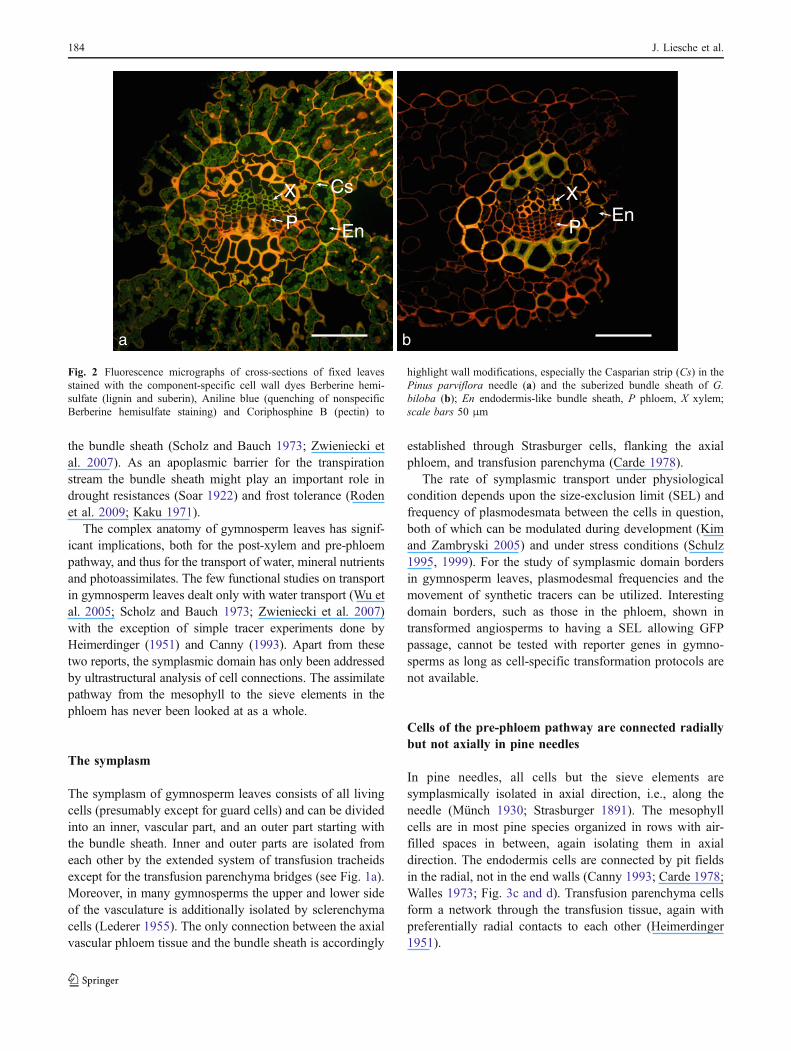
the bundle sheath (Scholz and Bauch 1973; Zwieniecki etal. 2007). As an apoplasmic barrier for the transpirationstream the bundle sheath might play an important role indrought resistances (Soar 1922) and frost tolerance (Rodenet al. 2009; Kaku 1971).
The complex anatomy of gymnosperm leaves has signif-icant implications, both for the post-xylem and pre-phloempathway, and thus for the transport of water, mineral nutrientsand photoassimilates. The few functional studies on transportin gymnosperm leaves dealt only with water transport (Wu etal. 2005; Scholz and Bauch 1973; Zwieniecki et al. 2007)with the exception of simple tracer experiments done byHeimerdinger (1951) and Canny (1993). Apart from thesetwo reports, the symplasmic domain has only been addressedby ultrastructural analysis of cell connections. The assimilatepathway from the mesophyll to the sieve elements in thephloem has never been looked at as a whole.
The symplasm
The symplasm of gymnosperm leaves consists of all livingcells (presumably except for guard cells) and can be dividedinto an inner, vascular part, and an outer part starting withthe bundle sheath. Inner and outer parts are isolated fromeach other by the extended system of transfusion tracheidsexcept for the transfusion parenchyma bridges (see Fig. 1a).Moreover, in many gymnosperms the upper and lower sideof the vasculature is additionally isolated by sclerenchymacells (Lederer 1955). The only connection between the axialvascular phloem tissue and the bundle sheath is accordingly
established through Strasburger cells, flanking the axialphloem, and transfusion parenchyma (Carde 1978).
The rate of symplasmic transport under physiologicalcondition depends upon the size-exclusion limit (SEL) andfrequency of plasmodesmata between the cells in question,both of which can be modulated during development (Kimand Zambryski 2005) and under stress conditions (Schulz1995, 1999). For the study of symplasmic domain bordersin gymnosperm leaves, plasmodesmal frequencies and themovement of synthetic tracers can be utilized. Interestingdomain borders, such as those in the phloem, shown intransformed angiosperms to having a SEL allowing GFPpassage, cannot be tested with reporter genes in gymno-sperms as long as cell-specific transformation protocols arenot available.
Cells of the pre-phloem pathway are connected radiallybut not axially in pine needles
In pine needles, all cells but the sieve elements aresymplasmically isolated in axial direction, i.e., along theneedle (Münch 1930; Strasburger 1891). The mesophyllcells are in most pine species organized in rows with air-filled spaces in between, again isolating them in axialdirection. The endodermis cells are connected by pit fieldsin the radial, not in the end walls (Canny 1993; Carde 1978;Walles 1973; Fig. 3c and d). Transfusion parenchyma cellsform a network through the transfusion tissue, again withpreferentially radial contacts to each other (Heimerdinger1951).
a b
Cs
EnP
XEn
P
X
Fig. 2 Fluorescence micrographs of cross-sections of fixed leavesstained with the component-specific cell wall dyes Berberine hemi-sulfate (lignin and suberin), Aniline blue (quenching of nonspecificBerberine hemisulfate staining) and Coriphosphine B (pectin) to
highlight wall modifications, especially the Casparian strip (Cs) in thePinus parviflora needle (a) and the suberized bundle sheath of G.biloba (b); En endodermis-like bundle sheath, P phloem, X xylem;scale bars 50 μm
184 J. Liesche et al.

Modeling of the hydraulic parameters in leaves ledZwieniecki et al. (2004) to conclude that development ofsingle-veined leaves is accompanied by a high radial waterresistance outside the stele during leaf expansion. Thisconclusion is compatible with the view that water exits fromthe stele primarily through the protoplasts of the endodermis-like bundle sheath. The endodermis layer itself does notallow water transport in axial direction: neither through theapoplast, since the entire end wall of endodermis cells issealed by suberin and/or lignin, nor through the symplasm,since the end walls are not crossed by pit fields/plasmodes-mata (Carde 1978). Accordingly, assimilates entering theendodermis from the mesophyll, have to continue radiallyvia the transfusion parenchyma towards the Strasburger cells.Only the sieve elements then offer an axial transportpathway. The axial isolation of the pre-phloem pathwaymight be important for the stabilization of concentration andpressure gradients over the length of the needle.
Branched plasmodesmata are present at all interfacesof the pre-phloem pathway in gymnosperm leaves
Leaves of a number of different gymnosperm species havebeen studied by electron microscopy. For the pre-phloempathway the micrographs of plasmodesmata indicate unifyingfeatures across all gymnosperm species. We reprinted somemicrographs from less accessible papers and combined themin Fig. 3, which shows that groups of plasmodesmata withmore or less extensive median cavities are typically combinedin fields at all interfaces from mesophyll to phloem:mesophyll–endodermis-transfusion parenchyma–Strasburgercell-sieve element (Fig. 3a–e, g; see also Parameswaran andLiese 1970; Boddi et al. 2002; Carde 1974, 1978; Gamblesand Dengler 1982; Glockmann and Kollmann 1996; Harris1972; Scholz and Bauch 1973; Walles 1973). While mostplasmodesmata on the pre-phloem pathway are located innon-elevated primary pit fields, those linking Strasburgercells are located in conspicuous, dome-shaped wall thicken-ings (Fig. 3g and Carde 1974: Fig. VIc). Staining identifiedthe thickenings to be primary wall depositions and to consistof hemicelluloses, pectin, and callose rather than cellulose(Carde 1974; Glockmann and Kollmann 1996). Branchedplasmodesmata are a regular feature in mesophyll tissue, alsoin angiosperms (see e.g., Kühn et al. 1996) and seem to bethe result of secondary modification of primary plasmodes-mata (see Faulkner and Oparka 2009). However, theformation of large dome-shaped wall thickenings containingnumerous plasmodesmata is characteristic for gymnospermleaves. Only the companion cell side of pore–plasmodesmataunits between sieve element and companion cell of angio-sperms is somewhat comparable, but much less complex,than that of Strasburger cells.
Contacts of the transfusion parenchymawith Strasburger cells
Carde (1973) distinguished between three types of Stras-burger cells: inner ones abutting the sieve elements, themost abundant central ones, and outer ones characterizedby presence of chloroplasts. The inner and central Stras-burger cells can easily be identified based on their dome-shaped wall thickenings, while the chloroplast-containingouter cells have regular pit fields (see Fig. 3e). SinceStrasburger cells also from non-conifers are characterizedby having plasmodesmal connections in dome-shaped wallthickenings (Glockmann and Schulz, unpublished) wesuggest to name only those cells “Strasburger cells” in thegymnosperm leaf which have dome-shaped wall thicken-ing, and to describe the other living parenchyma cells insidethe bundle sheath (with or without chloroplasts) astransfusion parenchyma (see also Fig. 1). Carde himselfcalled the entire system of Strasburger cells flanking theaxial phloem transfer tissue (“Le tissu de transfert”, Carde1973) and assumed that the Strasburger cells play animportant role in the transfer of assimilates to the phloem.
Development of dome-shaped symplasmic contactsin Strasburger cells
A consistent feature of Strasburger cells in all gymno-sperms are the dome-shaped symplasmic contacts towardsother Strasburger cells and towards sieve elements (Carde1974; Gambles and Dengler 1982; Parameswaran and Liese1970). A detailed ultrastructural study on the developmentof these contacts in Metasequoia glyptostroboides byGlockmann and Kollmann (1996) indicated that thesecontacts develop from primary plasmodesmata in the thinwalls between young Strasburger cells (Fig. 3f). Duringdifferentiation and expansion growth of the needle, the wallbecomes thicker and the primary plasmodesmata secondar-ily modified by multiple branching (Fig. 3g, i; seeGlockmann and Kollmann 1996). The only remnant of theprimary plasmodesmata is then the median cavity whichdilates through sheering forces, introduced by expansiongrowth of the needle (Fig. 3i).
Branching might be seen as a means to maintain a highdegree of symplasmic contact between the Strasburgercells. Interestingly, the plasmodesmal orifices have thetypical neck region (Fig. 3h), indicative for the need tocontrol plasmodesmal transport (Glockmann and Kollmann1996). Callose and/or protein interactions were discussed toshape the plasmodesmal neck region, which can be dilatedunder physiological conditions (Schulz 1995, 1999; Lucaset al. 2009). The elaborate and tubular ER system in thedome-shaped wall thickenings might on one hand be a
Symplasmic transport and phloem loading in gymnosperm leaves 185

consequence of the way the original plasmodesmata getsecondarily modified by multiplication of plasmodesmalarms, selective deposition of new wall material and localloosening of the existing wall. This is supported by thedifferent wall composition of the dome-shaped wall regions(Carde 1974; Glockmann and Kollmann 1996; see alsodiscussion in Ehlers and Kollmann 1996, 2001; Faulkner etal. 2008; Faulkner and Oparka 2009; Burch-Smith et al.2010, this issue). On the other hand, the elaborate ERsystem of Strasburger cell plasmodesmata might indicate aninvolvement of the ER in phloem loading (Carde 1974;Glockmann and Kollmann 1996). The presence of ER
complexes on both sides of live sieve areas inspired Schulz(1992, 2005) to discuss that the ER has an active functionin phloem transport of gymnosperms.
Continuity of the pre-phloem pathway with the sieveelements indicates a symplasmic mode of phloemloading
According to the cited electron microscopic papers, there isno indication for an isolation of the phloem from the pre-phloem pathway, otherwise characteristic for the closed
a b d
e g
h
if
c
Fig. 3 Electron micrographs of plasmodesmata at the interfacesbetween two mesophyll cells (a, Boddi et al. 2002), a mesophyll (left)and an endodermis cell (right) (b, Boddi et al. 2002), two endodermiscells (radial wall) (c, Carde 1978; d, Gambles and Dengler 1982), atransfusion parenchyma cell (left) and a Strasburger cell (right) (e,Carde 1974), and two Strasburger cells (f–i, Glockmann andKollmann 1996). All contacts in mature cell walls are characterizedby branched plasmodesmata and more or less extended mediancavities (all but f). Simple plasmodesmata connect young Strasburgercells (f), but get secondarily modified in mature Strasburger cells by
the development of dome-shaped wall thickenings that containcomplex branched plasmodesmal connections (g)–(i). Neck region atthe orifices of these contacts (h). Arrow in d=thickened wall in thecenter of the pit, in g=neck regions, in h=branching site, in i=bulb-like dilation in cavity, arrowheads in g=expanded sleeve regions, Caand cc central cavity, En endodermal cell, ep periplasmatic space, Llipid droplet, M mesophyll cell, Mi mitochondria, MN median nodule,pl plasmalemma, rER rough endoplasmatic reticulum, sER smoothendoplasmatic reticulum, tp desmotuble; scale bars 1 μm (a, b, g, f, i),5 μm (e), 0.1 μm (h)
186 J. Liesche et al.

minor vein configuration of apoplasmic phloem loaders(Gamalei 1989). On the contrary, the pre-phloem pathwayof gymnosperms seems to provide a well-connectedsymplasmic pathway continuous all the way to theconducting sieve elements. The connections between theinner Strasburger cells and the sieve elements are pore–plasmodesmata contacts with many branched plasmodes-mata within the wall thickenings of the Strasburger cell side(Carde 1974; Gambles and Dengler 1982; Glockmann andKollmann 1996; Parameswaran and Liese 1970) andidentical to the ones found in primary and secondary stemphloem of conifers (see Schulz 1990).
Continuity of symplasmic connections would suggestthat sucrose produced in the mesophyll cells reaches thephloem by diffusion, similar to what was proposed formany angiosperm tree species (Turgeon et al. 2001;Turgeon 2010). If so, “phloem loading” happens alreadyon the pre-phloem pathway in gymnosperms (see Turgeonand Medville 1998). Electron microscopy and localizationof plasmodesmal targeted proteins can, however, not tellwhether plasmodesmata are functional. For example, eventhough it is generally agreed that functional guard cells aresymplasmically isolated, reporter protein experiments andrandom electron micrographs might falsely indicate pres-ence of plasmodesmata (Itaya et al. 1998: Fig. 5c; Pallasand Mollenhauer 1972). Evidence for truncation of plas-modesmata towards guard cells was given by serialsectioning (Wille and Lucas 1984).
An indication that the plasmodesmata in the pre-phloempathway of gymnosperms are indeed functional was providedby early experiments with fluorescent tracers. The cytoplas-mic tracer fluorescein, applied to a cut surface on the lowerside of a Pinus sylvestris needle, reached the endodermis,transfusion parenchyma and the phloem after short incuba-tion (Heimerdinger 1951). Whether the tracer approached thesieve elements is not clear, since Heimerdinger did notdiscriminate between the cell types of the phloem. Move-ment of fluorescein is indicative for symplasmic continuity,but cannot detect restrictions in symplasmic domains such asthose resulting from small SEL and/or low plasmodesmalfrequencies.
Opposite transpiration and assimilate streams demandtight control of water, ion and sucrose transportacross the plasma membranes of bundle sheathand transfusion parenchyma
A symplasmic mode of phloem loading, as indicated by thepresence of plasmodesmata all along the pre-phloempathway, raises questions based on the unique anatomy ofgymnosperm leaves. It is dramatically different from thatof angiosperms in two aspects: (1) the particular features of
the bundle sheath and (2) the transfusion tissue betweenbundle sheath and axial vascular elements.
1. The endodermis-like bundle sheath in pines is thebottleneck for outward transport of water and mineralnutrients (Fig. 1: blue arrows). Since the radial wallsblock apoplasmic transport efficiently (Canny 1993),the plasma membrane of each bundle sheath cell has tocontain a sufficient number of aquaporins (Voicu et al.2009) and other transporter proteins to take up forwater and ions from the transfusion tracheids. Also innon-pine gymnosperms does the bundle sheath seem tobe a barrier for water transport, even though they do nothave an obvious Casparian strip. This was establishedby rehydration studies of Metasequoia, Ginkgo, andGnetum leaves (Zwieniecki et al. 2007). The flow ofthe transpiration stream out of the bundle sheathtowards stomata could be apoplasmic, symplasmic ortranscellular. This means that water could re-enter theapoplast and stay there, move through the plasmodes-mata to the neighboring mesophyll cell or get to themesophyll by crossing the plasma membranes of both,bundle sheath cell and mesophyll cell (see blue arrowsin Fig. 1). This situation reflects the general discussionon post-xylem movement of water where a conclusionhas yet to be reached (Sack and Holbrook 2006).
Simultaneous to the outward flow of water, all theinward transport of assimilates from the mesophyll to thetransfusion parenchyma has to pass the bundle sheath (seeFig. 1: brown arrows). Already, Münch (1930) doubted thatthe opposing flow of water and photoassimilates could beaccommodated in the same cells. Once inside the cytosol ofthe bundle sheath, an outward bulk flow of water wouldmeet the inward diffusion of photoassimilates resulting in avery inefficient assimilate transport. The situation appearseven less conceivable in case that the plasmodesmata in theouter bundle sheath wall are involved in water transport.This would imply that water and assimilates had to moveopposite to each other in their cytoplasmic sleeves.
2. The thin network of transfusion parenchyma is the onlysymplasmic contact between the bundle sheath and thephloem for the inward assimilate transport. Thetransfusion parenchyma cells have a large surface areatowards dead transfusion tracheids that are filled withwater and mineral nutrients. The content of solutes inthe apoplast is generally underestimated, and theconcentrations of solutes in the xylem–lumen apoplastare different from those in the cell wall apoplast (Canny1995). A steep gradient in water potential across theplasma membrane of the transfusion parenchyma cellswould certainly lead to a considerable leakage ofsucrose. Indeed, synthetic apoplasmic tracers, fed
Symplasmic transport and phloem loading in gymnosperm leaves 187

through the xylem, end up at the plasma membraneof the transfusion parenchyma and even cross thismembrane after long chase periods (Canny 1993). Asshown by autoradiography, radiolabelled aspartate wascollected by the transfusion parenchyma and returnedquickly via Strasburger cells to the phloem. Some ofthe label appeared also in the mesophyll. This ledCanny (1993) to conclude that active transporters inthe transfusion parenchyma recover solutes from thetranspiration stream. How much sucrose on its waytowards the Strasburger cells might leak into theapoplast of the transfusion tracheids, to be retrievedby membrane transporter activity, has yet to bedetermined.
As a solution to the challenges listed above, anelegant scenario would be the compartmentalization ofwater and assimilate transport in bundle sheath andtransfusion parenchyma. In the context of this review itmight be speculated that water and mineral nutrientscould already be taken up by the transfusion parenchy-ma and access the cytosol en-route to the bundle sheath,as indicated by Canny’s tracer experiments. Obviously,the used fluorochrome sulforhodamine G is an imperfectapoplasmic tracer that over time can cross the plasmamembrane (Canny 1993). Sucrose coming from themesophyll could be taken up at the bundle sheath bythe endoplasmic reticulum, or at any point on the way inthe symplasm between bundle sheath and Strasburgercells. A special role of intercellular ER in phloem loadingof conifers was already discussed by Carde (1974),Glockmann and Kollmann (1996), and Schulz (2005).This role would demand sucrose transporters for sucroseuptake into the ER at the latest in the Strasburger cells,and for efflux of sucrose into the sieve element.
Whether or not sucrose is taken up by the ER at one stepof the pre-phloem pathway, a problem of opposing flows ofwater and assimilates in bundle sheath cells and theirplasmodesmata might actually not exist, since gymno-sperms have xeromorphic leaves. Transpiration might belimited to the extent that outward water transport in thebundle sheath might be diffusion rather than bulk flow.Water release across the outer plasma membrane should betightly controlled by aquaporins and, if plasmodesmata arecontributing, by their SEL. The high abundance of plasmo-desmata is compatible with the assumption that they have asmall SEL. Narrow plasmodesmata will inhibit bulk flow ofwater more than diffusion. Accordingly, if transport in and outof the bundle sheath is dominated by diffusion, there is noneed for compartmentalization of water and assimilates: eachmolecular species would diffuse on its own in the samechannels with a net movement of water outwards, and a netmovement of assimilate inwards.
Feasibility of either compartmentalization or diffusionalflow can only be tested by functional studies of theapoplasmic and symplasmic pathways in gymnospermleaves and by the cloning, characterization and localizationof sucrose transporters possibly involved in assimilateuptake.
Conclusions and open questions
Continuity of symplasmic contacts all the way from meso-phyll to sieve elements indicate a symplasmic mode ofphloem loading in gymnosperms. The endodermis-likebundle sheath seems to play a key role in control of mineralnutrient and assimilate transport, i.e., the post-xylem and thepre-phloem transport. Here, the outward mineral nutrienttransport, driven by transpiration, meets the inward sugartransport, presumably driven by diffusion. Logistically, itwould be the easiest way to separate these two streams bycompartmentalization, where assimilates and water are phys-ically separated. Indeed, the pathways are clearly separated inthe vascular tissue inside the bundle sheath, where they arelimited to the transfusion tracheids and transfusion parenchy-ma, respectively. Here, the pathways are kept separated by theplasma membrane of the transfusion parenchyma whichcontrols any exchange of metabolites between apoplasmicand symplasmic pathway.
For the bundle sheath, it is conceivable that the incomingassimilates are compartmentalized in the endomembranesystem, perhaps even including the vacuoles. Intercellularcontinuity of vacuoles through the plasmodesmal ER wasshown for trichomes by Lazzaro and Thomson (1996).Scholz and Bauch reported the highest osmotic potential ofthe needle to occur in the endodermis-like bundle sheathand made it responsible for the exudation of water fromdetached needles (Scholz and Bauch 1973; see also Carde1978). Without pointing to the compartment in question,this suggests that sugars accumulate in the bundle sheath.Where sucrose would leave the endomembrane system onits inward transport is one of the questions, only furtherfunctional experiments can solve.
The mechanism of phloem loading and transport is anenigma in gymnosperms (Schulz 1992, 2005; Glockmannand Schulz 1999). All gymnosperms studied so far have thesame functional anatomy, and, thus, can be expected tohave a similar phloem loading mechanism, independent ofwhether they have needles, scales or broad leaves.
Open questions derived from the present review are
1. Are there sucrose transporters present in gymnospermsand, if so, where are they localized?
2. Are assimilates compartmentalized in the bundlesheath?
188 J. Liesche et al.

3. What is the role of the extended ER in the plasmodes-mata traversing the dome-shaped symplasmic contactsof Strasburger cells and sieve elements?
4. Is phloem loading active or passive, and does it followan entirely cytosolic route (through the plasmodesmalsleeves), or are there a membrane step and thedesmotubular pathway involved?
In order to solve these questions, it is proposed toclone putative sucrose transporters from gymnosperms,to characterize them and to try to localize them incollection tissue and transport phloem. According to theultrastructural data, it can be assumed that pre-phloemtransport relies on a diffusion gradient from mesophyll toStrasburger cells. If this is the case, any domain bordersand obstacles in this pathway have to be identified. Forthe cytosolic pathway fluorescence redistribution afterphotobleaching (FRAP) and photoactivation experimentscan be utilized to quantify the transport rate betweenselected interfaces (Pina et al. 2009). Preliminary experi-ments indicate that such tracer studies are able todiscriminate symplasmic and apoplasmic phloem loaders(Liesche, Martens, and Schulz, unpublished). For theidentification of a putative vacuolar pathway, FRAPexperiments could be undertaken using a fluorescentvacuolar marker. Autoradiography of leaves after feedingof radiolabelled with CO2 could indicate any activephloem-loading steps (Turgeon and Medville 1998; Ren-nie and Turgeon 2009). Finally, the sugar concentration inthe cytosol and the vacuoles of the pre-phloem pathwayshall be assessed in order to identify those interfaces,where active transport might contribute to pre-phloemtransport and phloem loading.
Acknowledgments We thank Line Svensson Kirk and Astrid S.Andersen for the light microscopic preparations of Ginkgo and Pinusparviflora, respectively (Fig. 2), and Christl Glockmann, University ofKiel, for electron microscopic preparation and micrographs of Gnetumgnemon (Fig. 1b). All comments and suggestions from the anonymousreviewers which led to the improvement of the manuscript aregratefully acknowledged.
Conflict of interest The authors declare that they have no conflict ofinterest.
References
Boddi S, Bonzi LM, Calamassi R (2002) Structure and ultrastructureof Pinus halepensis primary needles. Flora 197(1):10–23
Burch-Smith TM, Stonebloom S, Xu M, Zambryski PC (2010)Plasmodesmata during development: re-examination of theimportance of primary, secondary, and branched plasmodesmatastructure versus function. Protoplasma
Canny MJ (1993) Transfusion tissue of pine needles as a site ofretrieval of solutes from the transpiration stream. New Phytol 123(2):227–232
Canny MJ (1995) Apoplastic water and solute movement: new rulesfor an old space. Annu Rev Plant Physiol Plant Mol Biol 46:215–236
Carde JP (1973) Le tissu de transfert (=cellules de Strasburger) dans lesaiguilles du pin maritime (Pinus pinaster ait.), I. Étude histologiqueet infrastructurale du tissu adulte. J Microsc 17:65–88
Carde JP (1974) Le tissu de transfert (=cellules de Strasburger) dansles aiguilles du pin maritime (Pinus pinaster Ait.), II. Caractèrescytochimiques et infrastructuraux de la paroi et des plasmo-desmes. J Microsc 20:51–72
Carde JP (1978) Ultrastructural studies of Pinus pinaster needles—endodermis. Am J Bot 65(10):1041–1054
Davidson A, Keller F, Turgeon R (2010) Phloem loading, plantgrowth form, and climate. Protoplasma
de Bary A (1877) Vergleichende Anatomie der Vegetationsorgane derPhanerogamen und Farne. Wilhelm Engelmann, Leipzig
Ehlers K, Kollmann R (1996) Formation of branched plasmodesmatain regenerating Solanum nigrum-protoplasts. Planta 199(1):126–138
Ehlers K, Kollmann R (2001) Primary and secondary plasmodesmata:structure, origin, and functioning. Protoplasma 216(1):1–30.doi:10.1007/bf02680127
Esau K (1965) Plant anatomy, 2nd edn. Wiley, New York, 767ppEsau K (1977) Anatomy of seed plants, 2nd edn. Wiley, New York,
550ppFaulkner C, Akman OE, Bell K, Jeffree C, Oparka K (2008) Peeking
into pit fields: a multiple twinning model of secondaryplasmodesmata formation in tobacco. Plant Cell 20(6):1504–1518. doi:10.1105/tpc.107.056903
Faulkner C, Oparka K (2009) Plasmodesmata. Encyclopedia of lifesciences (ELS). Wiley, Chichester
Gadek PA, Quinn CJ (1988) Pitting of transfusion tracheids incupressaceae. Aust J Bot 36(1):81–92
Gamalei Y (1989) Structure and function of leaf minor veins in treesand herbs. Trees 3:96–110
Gambles RL, Dengler RE (1982) The anatomy of the leaf of redpine, Pinus resinosa 2. Vasc tissues Can J Bot 60(12):2804–2824
Gil L (2010) Sucrose transporters play a role in phloem loading ofCMV-infected melon plants that are defined as symplasticloaders. Presentation given at the International conference onPlant Vascular Biology 2010. Columbus, Ohio
Glockmann C, Kollmann R (1996) Structure and development ofcell connections in the phloem of Metasequoia glyptostro-boides needles. 1. Ultrastructural aspects of modified primaryplasmodesmata in Strasburger cells. Protoplasma 193(1–4):191–203
Glockmann C, Schulz A (1999) Phloem loading and transport ingymnosperms: an enigma. In: International conference onassimilate transport and partitioning, Newcastle, Australia,August 15–20 1999. Apstract 7-07, p. 99.
Harris WM (1972) Ultrastructural observations on Pinaceae leafphloem. 1. Spring condition. New Phytol 71(1):169–173
Heimerdinger G (1951) Zur Mikrotopographie der Saftströme im Trans-fusionsgewebe der Koniferennadel. 2. Entwicklungsgeschichte undPhysiologie. Planta 40(2):93–111
Itaya A, Woo YM, Masuta C, Bao YM, Nelson RS, Ding B (1998)Developmental regulation of intercellular protein traffickingthrough plasmodesmata in tobacco leaf epidermis. Plant Physiol118(2):373–385
Kaku S (1971) Possible role of endodermis as a barrier for icepropagation in freezing of pine needles. Plant Cell Physiol 12(6):941–948
Symplasmic transport and phloem loading in gymnosperm leaves 189

Kim I, Zambryski PC (2005) Cell-to-cell communication viaplasmodesmata during Arabidopsis embryogenesis. Curr OpinPlant Biol 8(6):593–599. doi:10.1016/j.pbi.2005.09.013
Kühn C, Quick WP, Schulz A, Riesmeier JW, Sonnewald U, FrommerWB (1996) Companion cell-specific inhibition of the potatosucrose transporter SUT1. Plant Cell Environ 19(10):1115–1123
Lazzaro MD, Thomson WW (1996) The vacuolar-tubular continuumin living trichomes of chickpea (Cicer arietinum) provides arapid means of solute delivery from base to tip. Protoplasma 193(1–4):181–190
Lederer B (1955) Vergleichende Untersuchungen über das Trans-fusionsgewebe einiger rezenter Gymnospermen. In: Huber B (ed)Vergleichend-anatomische Untersuchungen. Gustav Fischer,Jena, pp 1–42
LucasWJ, Ham B-K, Kim J-Y (2009) Plasmodesmata—bridging the gapbetween neighboring plant cells. Trends Cell Biol 19:495–500
Münch E (1930) Die Stoffbewegungen in der Pflanze. Gustav Fischer,Jena
Pallas JE, Mollenhauer HH (1972) Electron-microscopic evidence forplasmodesmata in dicotyledonous guard cells. Science 175(4027):1275–1276
Parameswaran N, Liese W (1970) Cytology of Strasburger cells inconifer needles. Naturwissenschaften 57(1):45–46
Pina A, Errea P, Schulz A, Martens HJ (2009) Cell-to-cell transportthrough plasmodesmata in tree callus cultures. Tree Physiol 29(6):809–818. doi:10.1093/treephys/tpp025
Rennie EA, Turgeon R (2009) A comprehensive picture of phloemloading strategies. P Natl Acad Sci USA 106(33):14162–14167.doi:10.1073/pnas.0902279106
Roden JS, Canny MJ, Huang CX, Ball MC (2009) Frost tolerance andice formation in Pinus radiata needles: ice management by theendodermis and transfusion tissues. Funct Plant Biol 36(2):180–189. doi:10.1071/Fp08247
Sack L, Holbrook NM (2006) Leaf hydraulics. Annu Rev Plant Biol57:361–381
Scholz F, Bauch J (1973) Anatomische und physiologische Untersu-chungen zur Wasserbewegung in Kiefernnadeln. Planta 109(2):105–119. doi:10.1007/bf00386118
Schulz A (1990) Conifers. In: Behnke H-D, Sjolund RD (eds)Comparative structure, induction and development. Springer,Berlin Heidelberg New York, pp 63–88
Schulz A (1992) Living sieve cells of conifers as visualized byconfocal, laser-scanning fluorescence microscopy. Protoplasma166(3–4):153–164
Schulz A (1995) Plasmodesmal widening accompanies the short-termincrease in symplasmic phloem unloading in pea root-tips underosmotic-stress. Protoplasma 188(1–2):22–37
Schulz A (1999) Physiological control of plasmodesmal gating. In:van Bel AJE (ed) Plasmodesmata—stucture, function, role in cellcommunication. Springer, New York, pp 173–204
Schulz A (2005) Role of plasmodesmata in solute loading and unloading.In: Oparka K (ed) Plasmodesmata. Blackwell, Oxford, pp 135–161
Shiroya T, Lister GR, Slankis V, Krotkov G, Nelson CD (1962)Translocation of the products of photosynthesis to roots of pineseedlings. Can J Bot 40:1125–1135
Soar I (1922) The structure and function of the endodermis in theleaves of the abietineae. New Phytol 21(5):269–292. doi:10.1111/j.1469-8137.1922.tb07604.x
Soda C, Bussotti F, Grossoni P, Barnes J, Mori B, Tani C (2000)Impacts of urban levels of ozone on Pinus halepensis foliage.Environ Exp Bot 44(1):69–82
Strasburger E (1891) Ueber den Bau und die Verrichtungen derLeitungsbahnen in den Pflanzen. Gustav Fischer, Jena
Turgeon R (2010) The role of phloem loading reconsidered. PlantPhysiol 152(4):1817–1823. doi:10.1104/pp.110.153023
Turgeon R, Medville R (1998) The absence of phloem loading inwillow leaves. Proc Natl Acad Sci USA 95:12055–12060
Turgeon R, Medville R (2010) Amborella trichopoda, plasmodesmata,and the evolution of phloem loading. Protoplasma
Turgeon R, Medville R, Nixon KC (2001) The evolution of minorvein phloem and phloem loading. Am J Bot 88(8):1331–1339
Voicu MC, Cooke JEK, Zwiazek JZ (2009) Aquaporin geneexpression and apoplastic water flow in bur oak (Quercusmacrocarpa) leaves in relation to the light response of leafhydraulic conductance. J Exp Bot 60(14):4063–4075.doi:10.1093/jxb/erp239
Walles B (1973) On the ultrastructure of needles of Pinus silvestris. l.vol 106. Studia forestalia, suecica. Allmänna Förlaget, Stock-holm
Wille AC, Lucas WJ (1984) Ultrastructural and histochemical studieson guard-cells. Planta 160(2):129–142
Willenbrink J (2002) Assimilate transport in phloem: regulation andmechanism. Russ J Plant Physiol 49:8–15. doi:10.1007/bf00225352
Willenbrink J, Kollmann R (1966) Über den Assimilattransport imPhloem von Metasequoia. Z Pflanzenphysiol 55:42–53
Worsdell WC (1897) Transfusion-tissue: its origin and function in theleaves of gymnospermous plants. Botany 5:301–319
Wu XQ, Zhu JM, Huang RZ, Wang QL, Zheng WJ, Hu YX, Lin JX(2001) Evidence of Casparian strip in the foliar endodermis ofPinus bungeana. Acta Bot Sin 43(10):1081–1084
Wu XQ, Lin JX, Zhu JM, Hu YX, Hartmann K, Schreiber L(2003) Casparian strips in needles of Pinus bungeana:isolation and chemical characterization. Physiol Plant 117(3):421–424
Wu XQ, Lin JX, Lin QQ, Wang J, Schreiber L (2005) Casparian stripsin needles are more solute permeable than endodermal transportbarriers in roots of Pinus bungeana. Plant Cell Physiol 46(11):1799–1808. doi:10.1093/Pcp/Pci194
Zhang CK, Turgeon R (2009) Downregulating the sucrose transporterVPSUT1 in Verbascum phoeniceum does not inhibit phloemloading. P Natl Acad Sci USA 106(44):18849–18854.doi:10.1073/pnas.0904189106
Zwieniecki MA, Boyce CK, Holbrook NM (2004) Functional designspace of single-veined leaves: role of tissue hydraulic propertiesin constraining leaf size and shape. Ann Bot Lond 94(4):507–513. doi:10.1093/Aob/Mch173
Zwieniecki MA, Brodribb TJ, Holbrook NM (2007) Hydraulic designof leaves: insights from rehydration kinetics. Plant Cell Environ30(8):910–921. doi:10.1111/j.1365-3040.2007.001681.x
190 J. Liesche et al.